WO2021010442A1 - Protein and rna interaction evaluation method, interaction modulator evaluation method and interaction modulator detection method, and fusion protein, kit and biosensor using same - Google Patents
Protein and rna interaction evaluation method, interaction modulator evaluation method and interaction modulator detection method, and fusion protein, kit and biosensor using same Download PDFInfo
- Publication number
- WO2021010442A1 WO2021010442A1 PCT/JP2020/027710 JP2020027710W WO2021010442A1 WO 2021010442 A1 WO2021010442 A1 WO 2021010442A1 JP 2020027710 W JP2020027710 W JP 2020027710W WO 2021010442 A1 WO2021010442 A1 WO 2021010442A1
- Authority
- WO
- WIPO (PCT)
- Prior art keywords
- protein
- rna
- fusion
- test
- marker
- Prior art date
Links
Images


Classifications
-
- C—CHEMISTRY; METALLURGY
- C12—BIOCHEMISTRY; BEER; SPIRITS; WINE; VINEGAR; MICROBIOLOGY; ENZYMOLOGY; MUTATION OR GENETIC ENGINEERING
- C12Q—MEASURING OR TESTING PROCESSES INVOLVING ENZYMES, NUCLEIC ACIDS OR MICROORGANISMS; COMPOSITIONS OR TEST PAPERS THEREFOR; PROCESSES OF PREPARING SUCH COMPOSITIONS; CONDITION-RESPONSIVE CONTROL IN MICROBIOLOGICAL OR ENZYMOLOGICAL PROCESSES
- C12Q1/00—Measuring or testing processes involving enzymes, nucleic acids or microorganisms; Compositions therefor; Processes of preparing such compositions
- C12Q1/68—Measuring or testing processes involving enzymes, nucleic acids or microorganisms; Compositions therefor; Processes of preparing such compositions involving nucleic acids
-
- G—PHYSICS
- G01—MEASURING; TESTING
- G01N—INVESTIGATING OR ANALYSING MATERIALS BY DETERMINING THEIR CHEMICAL OR PHYSICAL PROPERTIES
- G01N33/00—Investigating or analysing materials by specific methods not covered by groups G01N1/00 - G01N31/00
- G01N33/48—Biological material, e.g. blood, urine; Haemocytometers
- G01N33/50—Chemical analysis of biological material, e.g. blood, urine; Testing involving biospecific ligand binding methods; Immunological testing
- G01N33/68—Chemical analysis of biological material, e.g. blood, urine; Testing involving biospecific ligand binding methods; Immunological testing involving proteins, peptides or amino acids
-
- C—CHEMISTRY; METALLURGY
- C12—BIOCHEMISTRY; BEER; SPIRITS; WINE; VINEGAR; MICROBIOLOGY; ENZYMOLOGY; MUTATION OR GENETIC ENGINEERING
- C12N—MICROORGANISMS OR ENZYMES; COMPOSITIONS THEREOF; PROPAGATING, PRESERVING, OR MAINTAINING MICROORGANISMS; MUTATION OR GENETIC ENGINEERING; CULTURE MEDIA
- C12N15/00—Mutation or genetic engineering; DNA or RNA concerning genetic engineering, vectors, e.g. plasmids, or their isolation, preparation or purification; Use of hosts therefor
- C12N15/09—Recombinant DNA-technology
- C12N15/11—DNA or RNA fragments; Modified forms thereof; Non-coding nucleic acids having a biological activity
Definitions
- the present invention relates to a method for evaluating the interaction between protein and RNA, a method for evaluating an interaction regulator between protein and RNA, a method for detecting an interaction regulator between protein and RNA, and fusion proteins, kits, and fusion proteins used thereto. Regarding biosensors.
- RNA molecules that bind to specific proteins called aptamers have been artificially created (Wu YX, Kwon YJ, Methods). , 106: 21-28 (2016) (Non-Patent Document 1)).
- aptamers have been applied as molecules that inhibit protein function or as molecular target molecules that constitute drug complexes (Zhu G., Chen X. Adv Drag Dev. 134: 65-78 (2016). ) (Non-Patent Document 2)).
- RNA-binding protein fused to the DNA-binding domain of a split transcription factor constituting the yeast-to-hybrid method and a known RNA that binds to this RNA-binding protein (target RNA of the RNA-binding protein)
- target RNA of the RNA-binding protein RNA that binds to this RNA-binding protein
- the RNA to be verified fused to the molecule is allowed to interact with the RNA to be verified, and the interaction between the RNA-binding protein and the target RNA molecule is resolved to include the DNA-binding domain of the split transcription factor and the RNA to be verified.
- Form ribonucleoprotein Form ribonucleoprotein.
- the protein to be verified in this ribonucleoprotein comes into contact with the protein to be verified fused to the transcriptional activation domain of the split transcription factor, when a protein-RNA interaction occurs, a complex consisting of three molecules A body is formed, and as a result, the DNA binding domain and the transcription activation domain in the complex approach each other, and the complex acquires the transcription activation ability. Therefore, in this method, the protein-RNA interaction can be evaluated using the induction of the expression of the reporter gene by the complex that has acquired the transcriptional activation ability as an index.
- Non-Patent Document 3 the yeast three-hybrid method has been used to measure known protein-RNA interactions, search for new proteins that bind to specific RNA molecules, and search for new RNA molecules that bind to specific proteins.
- the protein-RNA interaction field to be verified is in the cytoplasm, whereas these yeast three-hybrid methods evaluate the interaction in the nuclear environment and the interaction field. There is a divergence in.
- the existing yeast three-hybrid method is known to have a problem that many false positives are detected, which is a serious problem especially in the detection of exploratory interactions (Non-Patent Document 3). ).
- the present invention has been made in view of the above-mentioned problems of the prior art, and is capable of specifically and easily measuring the interaction between protein and RNA in a cytoplasmic or cell-free system, and the interaction between protein and RNA. It is an object of the present invention to provide a method for evaluating an interaction regulator between a protein and RNA, a method for detecting an interaction regulator between a protein and RNA, and a fusion protein, a kit, and a biosensor used for these methods.
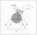
- a system that utilizes a split-type marker protein or a combination-type marker protein that functions in a cytoplasmic or cell-free system specifically, (a) a fusion protein of a test protein and a first element of the marker protein (first (B) Fusion protein of RNA-binding protein and the second element of the marker protein (second fusion protein), and (c) RNA forming a complex with the RNA-binding protein (c)
- a fusion RNA of a complex-forming RNA and a test RNA
- a signal generated due to the expression of the function of the marker protein is detected in a cytoplasmic or cell-free system.
- the present inventors measure the protein-RNA interaction in this system in the presence of a desired substance to increase, decrease, or eliminate the signal caused by the expression of the function of the marker protein. As an index, it was found that it is also possible to evaluate the regulation of protein-RNA interaction by the substance.
- [1] A method for evaluating the interaction between protein and RNA.
- (1) In the cytoplasm or cell-free system (A) A first fusion protein obtained by fusing the test protein and the first element of the marker protein, (B) A second fusion protein obtained by fusing an RNA-binding protein and a second element of the marker protein, and (c) an RNA forming a complex with the RNA-binding protein and a test RNA. Ribonucleoprotein consisting of fused RNA, The process of contacting, (2) A step of detecting a signal caused by the marker protein whose function is expressed by the proximity of the first element and the second element, and (3) A step of determining the interaction between the test protein and the test RNA by detecting the signal. Including When the test protein and the test RNA interact, the first fusion protein and the ribonucleoprotein form a complex, and the first element and the second element A method in which the function of the marker protein is expressed by the proximity of the markers.
- [4] A method for evaluating substances that regulate the interaction between proteins and RNA.
- (1) In the presence of the test substance, in the cytoplasm or cell-free system (A) A first fusion protein formed by fusing a specific protein with the first element of a marker protein, (B) A second fusion protein obtained by fusing an RNA-binding protein and a second element of the marker protein, and (c) an RNA forming a complex with the RNA-binding protein and a specific RNA.
- Ribonucleoprotein consisting of fused RNA
- the process of contacting (2) A step of detecting a signal caused by the marker protein whose function is expressed by the proximity of the first element and the second element, and (3) When the signal is increased as compared with the case where the test substance is absent, the test substance is evaluated as a substance that promotes the interaction between the specific protein and the specific RNA. , The test substance is evaluated as a substance that suppresses the interaction between the specific protein and the specific RNA when the signal is reduced or eliminated as compared with the case where the test substance is absent.
- the first fusion protein and the ribonucleoprotein form a complex
- the first element and the second element A method in which the function of the marker protein is expressed by the proximity of the markers.
- [5] A method for detecting a substance that regulates the interaction between protein and RNA in a sample.
- A A first fusion protein formed by fusing a specific protein with the first element of a marker protein
- B A second fusion protein obtained by fusing an RNA-binding protein and a second element of the marker protein
- c an RNA forming a complex with the RNA-binding protein and a specific RNA.
- Ribonucleoprotein consisting of fused RNA
- the process of contacting (2) A step of detecting a signal caused by the marker protein whose function is expressed by the proximity of the first element and the second element, and (3) A step of detecting a target substance in the test sample by the presence / absence, increase, or decrease of the signal.
- Including The first fusion occurs when the specific RNA interacts with the specific protein in the presence or absence of the target substance, and when the specific protein interacts with the specific RNA.
- a vector containing an RNA-encoding polynucleotide that forms a complex with a sex protein and an insertion site for an adjacent polynucleotide encoding the specific RNA Including, kit.
- C' A polynucleotide encoding a fusion RNA obtained by fusing an RNA forming a complex with the RNA-binding protein and a specific RNA, or a vector expressing the polynucleotide.
- B' A polynucleotide encoding a second fusion protein obtained by fusing an RNA-binding protein and a second element of the marker protein, or a vector expressing the polynucleotide.
- C' A polynucleotide encoding a fusion RNA obtained by fusing an RNA forming a complex with the RNA-binding protein and a specific RNA, or a vector expressing the polynucleotide.
- Transformed cells into which the cells have been introduced, as well as (2) A means for detecting a signal caused by the marker protein whose function is expressed by the proximity of the first element and the second element. Including When the specific protein interacts with the specific RNA, the first fusion protein, the second fusion protein, and the ribonucleoprotein composed of the fusion RNA form a complex, and the said.
- a biosensor in which the function of the marker protein is expressed by the proximity of the first element and the second element.
- the second fusion protein and the fusion RNA have strong binding properties through the binding between the RNA-binding protein contained therein and the complex-forming RNA. And forms a complex (ribonucleoprotein) that can exist stably in the cytoplasm or in a cell-free system.
- a fusion protein first fusion protein and second
- a test protein and the RNA-binding protein are fused to each of the marker proteins divided into a first element and a second element.
- the fusion protein) and the test RNA fused with the complex-forming RNA are brought into contact with each other.
- the function of the marker protein is exhibited only when the first element and the second element interact in close proximity to each other, but at this time, when the test RNA and the test protein interact with each other, the function is exhibited. So to speak, the test protein becomes a capture site (Prey), the test RNA becomes a capture site (Bait), and finally the first fusion protein and the ribonucleoprotein, that is, the above (a).
- the three molecules of (c) to (c) form a complex, and in a cytoplasmic or cell-free system, the first element and the second element are in close proximity to express the function of the marker protein.
- the interaction between the test protein to be verified and the test RNA in the cytoplasmic or cell-free system is caused by the expression of the function of the marker protein, more specifically, the marker protein expressing the function. It is possible to easily evaluate, detect, and measure using the signal generated as an index.
- the expression of background reporter genes and the like is amplified through a two-step gene expression process of transcription and translation, resulting in high levels of false positives.
- the detection of false positives can be sufficiently suppressed, and the interaction between protein and RNA can be sufficiently suppressed as compared with the above-mentioned conventional method. It becomes possible to measure specifically.
- a method for evaluating an interaction between a protein and RNA and a method for evaluating an interaction regulator between a protein and RNA, which can specifically and easily measure the interaction between a protein and RNA in a cytoplasmic or cell-free system.
- methods for detecting protein-RNA interaction regulators, as well as fusion proteins, kits, and biosensors used for them are also useful.
- protein means a molecule in which two or more amino acids are bound by a peptide bond and a modified product thereof. Therefore, it includes not only full-length proteins but also so-called oligopeptides and polypeptides. Modifications of the protein include, for example, phosphorylation, glycosylation, palmitoylation, prenylation (eg, geranylgeranylation), methylation, acetylation, ubiquitination, SUMOylation, hydroxylation, amidation.
- RNA means a molecule in which two or more ribonucleotides are bound by a phosphodiester bond and a modified product thereof.
- modification of RNA include methylation and deamination, and whether it is single-stranded or double-stranded, it has a three-dimensional structure such as a hairpin structure and a hammer head structure. You may be doing it.
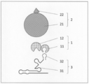
- the "interaction between protein and RNA” indicates the proximity or binding of protein and RNA, and more preferably, the protein and RNA form a complex. Such interactions include not only direct proximity and binding of the protein (test protein 11 or specific protein 11) and RNA (test RNA 32 or specific RNA 32), but also between the protein and the RNA.
- Other molecules eg, other proteins, other nucleic acids, sugars, lipids, low molecular weight compounds (vitamins, coenzymes, hormones, toxins, antibiotics, antibacterial agents, antiviral agents, anticancer agents, carcinogens, It also includes indirect interactions such as forming a complex via (drugs, coenzymes, etc.), metal ions, metal complexes, and complex molecules containing two or more of these molecules.
- the "cytoplasm” is the inside of a eukaryotic cell and a prokaryotic cell (eubacterial cell, archaeal cell), and in the eukaryotic cell, the portion of the cell excluding the nucleus, the prokaryotic cell. Indicates the part of the cell excluding the nucleoid.
- the cytosol according to the present invention may be the cytosol of eukaryotic cells or the cytosol of prokaryotic cells, but the formation of a complex of stable protein and RNA having a lower risk of affecting cell function.
- the cytosol of the eukaryotic cell or the cytosol of the prokaryotic cell is preferable, and more preferably the cytosol of the eukaryotic cell. More preferably, it is the cytosol of eukaryotic cells (the portion of the cytosol of eukaryotic cells excluding intracellular small organs).
- the eukaryotic cells include animal cells (mammalian, fish, birds, reptiles, amphibians, insect cells, etc.), plant cells, algae cells, yeast, and examples of the eubacterial cells include lactic acid bacteria and thermophiles.
- Bacterial bacterium, lactic acid bacterium, hyperthermophilic bacterium, and examples of the archaeal cell include methane bacterium, highly thermophilic bacterium, hyperthermophilic bacterium, and hyperthermophilic bacterium. These cells are present in vivo (eg, DNA encoding a first fusion protein) even when cultured in vitro (eg, cells growing in or on medium). And the DNA in the transgenic animal into which the DNA encoding the second fusion protein and the DNA encoding the fusion RNA have been introduced).
- the "cell-free system” refers to a system without living cells (the eukaryotic cells and prokaryotic cells).
- the cell-free system according to the present invention is not particularly limited as long as it is a system in which the first fusion protein according to the present invention and the ribonucleoprotein (or the second fusion protein and the fusion RNA) can come into contact with each other.
- Reconstituted cell-free protein synthesis system such as PURE System (Shimizu Y. et al., Nature Biotechnology 19: 751-755 (2001)); the molecule of the cell crushed solution or cell extract of the eukaryotic cell or prokaryotic cell. Examples include an in vitro reconstitution system that can be contacted with.
- the "marker protein” refers to a protein or a set of proteins capable of expressing one function as a whole, that is, expressing one function as a whole and finally producing at least one signal. ..
- a marker protein can be divided into at least a first fragment and a second fragment, and its function can be regenerated (expressed) when the first fragment and the second fragment are in close proximity to each other.
- the expression of the function can be detected as a signal (sometimes referred to herein as a "split marker protein”); a combination of two independent first and second proteins.
- the function is expressed when the first protein and the second protein are in close proximity to each other, and the expression of the function can be detected as a signal (in the present specification, in some cases, ". "Combination marker protein”) is included.
- the first element according to the present invention comprises the first fragment and the first protein
- the second element according to the present invention contains the second fragment and the second protein, but the "first".
- Each element, fragment, and protein in an element, fragment, and protein and each element, fragment, and protein in a "second" element, fragment, and protein are such that the first and second correspond to each other. It may be independent of each other and vice versa.
- the function is not particularly limited as long as it can directly or indirectly generate a signal due to the function, and for example, a nutrient synthesis function, a color development (color development) function, a light emitting function, a fluorescence function, and a quenching function.
- a nutrient synthesis function for example, a nutrient synthesis function, a color development (color development) function, a light emitting function, a fluorescence function, and a quenching function.
- Examples include a proteolytic function, a nucleic acid decomposition function, a drug decomposition function, and a redox reaction catalytic function.
- those that can be used as a split-type marker protein that can be divided into a first fragment and a second fragment include, for example, a nutritional requirement marker (tryptophan synthase (TRP1), histidine synthesis).
- Enzyme HIS3
- LYS2 lysine synthase
- MENU2 leucine synthase
- ADE2 adenin synthase
- UAA3 uracil synthase
- fluorescent protein green fluorescent protein (GFP))
- YFP Yellow Fluorescent Protein
- CFP Light Blue Fluorescent Protein
- BFP Blue Fluorescent Protein
- RFP Red Fluorescent Protein
- Clover Ruby, Cherry, Zami Green (AG), Sea Mushroom-Green (UkG), Kusabi Laorange (KO), Midoriishi-cyan (MiCy), etc.
- Luminescent enzyme Luciferase, Iquorin, etc.
- Drug resistance markers Drug resistance markers
- the combinational marker proteins that can be used as a combination of the first protein and the second protein include fluorescence source / quencher pair, fluorescence resonance energy transfer (FRET), and bioluminescence resonance energy.
- FRET fluorescence resonance energy transfer
- BRET acceptor pairs for transfer
- kinase / substrate protein pairs for signal transduction pathway activation and the like.
- fluorescence by the fluorescence source is detected when the fluorescence source and the quencher are separated, but when the fluorescence source and the quencher are close to each other, fluorescence by the fluorescence source is detected. Quenching (ie, reducing or extinguishing fluorescence).
- the fluorescence source include ATTO dye, cyanine dye (eg, Cy3, Cy5), tetramethylrhodamine (eg, TRITC), carboxyfluorescein (FAM), tetrachlorofluorescein (TET), hexachlorofluorescein (HEX), Texas. Examples include, but are not limited to, red and Yakima Yellow.
- the quencher examples include, but are not limited to, dark quencher, BHQ (Black Hole Quencher), IBFQ (Iowa Black FQ), IBRQ (Iowa Black RQ), Eclipse and the like.
- the first protein and the second protein may be independently fluorescent proteins, and when the fluorescent source and / or quencher is other than a protein, the fluorescent sources are independent of each other. Alternatively, it may be a protein that specifically presents a quencher intracellularly. Such a protein can be appropriately prepared by a known method or a method similar thereto.
- FRET fluorescence resonance energy transfer
- Donor / acceptor pairs for FRET include, for example, Clover / mRubi2, BFP / eGFP, BFP / YFP, BFP / DsRed2, CFP / YFP, CFP / DsRed2, Midori Cyanine / Clavira Orange, eGFP / DsRed, eGFP / Rhod-2, FITC / TRITC, FITC / Rhod-2, FITC / Cy3, Alexa488 / Alexa546, Alexa488 / Alexa555, Alexa488 / Cy3, YFP / TRITC, YFP / Cy3, Cy3 / Cy5, Cy3 / Cy5, Cy3 / Cy5.
- the first protein and the second protein may be independently fluorescent proteins, and when the donor and / or acceptor is a fluorescent dye, the fluorescent dye is specifically used intracellularly. It may be the protein to be presented. Such a protein can be appropriately prepared by a known method or a method similar thereto.
- donor / acceptor pair for bioluminescence resonance energy transfer fluorescence by BRET is not observed when the donor and acceptor are separated, but when the donor and acceptor are in close proximity, the donor's emission is emitted. causes excitation and fluorescence of the acceptor (ie, fluorescence by BRET occurs).
- donor / acceptor pairs for BRET include equolin / GFP, firefly-derived luciferase / RFP, firefly-derived luciferase / DsRed2, sea pansy-derived luciferase / YFP, sea pansy-derived luciferase / eGFP, and sea pansy-derived luciferase.
- the first protein and the second protein may be independently luminescent proteins or fluorescent proteins, and when the acceptor is a fluorescent dye, the fluorescent dye is specifically used in the cell. It may be the protein to be presented. Such a protein can be appropriately prepared by a known method or a method similar thereto.
- Each of the amino acid sequences of the marker protein can be mutated in nature (that is, non-artificially), and the mutation can be artificially introduced.
- such a mutant is also used as long as the expression of the function can be detected, that is, the function of the marker protein can be expressed by the proximity of the first element and the second element. be able to.
- the marker protein is a split-type marker protein
- the split position is particularly limited as long as the function of the split-type marker protein can be expressed (regenerated) by the proximity of the first fragment and the second fragment. However, it can be appropriately prepared.
- the marker protein one type may be used alone or two or more types may be used in combination.
- the expression of the function of the marker protein can be detected using this as an index by detecting a signal generated by the marker protein that expresses the function due to the proximity of the first element and the second element.
- the "signal caused by the marker protein expressing the function” is not particularly limited as long as it can be expressed by a changing physical quantity, and is directly generated from the marker protein (for example, quenching, fluorescence, etc.). It may be extinguished (such as quenching) or indirectly generated by the expression of the function of the marker protein (for example, a colony indicating that the cell has survived by the expression of the nutrient synthesis function).
- Examples of such a signal include colonies indicating cell survival, coloration (color development), luminescence, fluorescence, quenching, and the like. Further, the signals include those that can be confirmed with the naked eye and those that can be confirmed by a detection method / device according to the type of signal.
- detection of signal includes detection for confirming the presence or absence of the signal, and quantification or semi-quantification of the amount of the signal.
- the method for detecting the signal is not particularly limited, and a method according to the function of the marker protein and the type of signal derived from the marker protein can be appropriately selected. Further, the increase or decrease of the signal can be measured by quantifying or semi-quantifying the signal. When the signal is quenching, the increase and decrease of the signal indicate an increase and decrease of the degree of quenching, respectively.
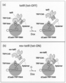
- the detection of the signal can be divided into two fragments, for example, in the cytoplasm of yeast, at position 2-44 (second fragment) and position 45-224 (first fragment), and these
- tryptophan synthase TRP1
- TRP1 tryptophan synthase
- TRP1 which regenerates (expresses) the function as tryptophan synthase when the fragments are close to each other
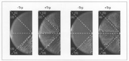
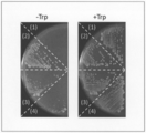
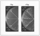
- the marker protein when the second fragment and the first fragment are close to each other Since the function as tryptophan synthase is regenerated (expressed), colonies showing the growth of the yeast in a tryptophan-free medium can be detected as a signal.
- the expression of the function is detected, that is, the function of the marker protein (TRP1) is expressed, while when it is not observed, the expression is said. It can be determined that the expression of the function is not detected, that is, the function of the marker protein is not expressed, or the amount of the signal is quantified or semi-quantified by comparing the amount of the colony with the calibration curve or the like. The degree of expression of can be quantified or semi-quantified.
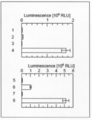
- luciferase (NanoLuc) derived from deep-sea luminescent shrimp is used as the marker protein, it is divided into two fragments, for example, 13 amino acids at the C-terminal (first fragment) and the other (second fragment). And since the function as luciferase is regenerated (expressed) when these fragments are close to each other, luminescence by luciferase can be detected as a signal in cells.
- the expression of the function of the marker protein (NanoLuc) can be detected, or the degree of expression can be quantified or semi-quantified depending on whether or not luminescence is observed (detected) as the signal or the intensity of the luminescence. ..
- the detection of light emission and the quantification or semi-quantification of the intensity can be appropriately selected by a conventionally known method, and can be performed by, for example, a CCD image sensor or a CMOS image sensor equipped with a photomultiplier tube and an optical lens. Further, a method of processing the obtained image by an image analysis program can also be adopted.
- the tryptophan synthase and the luciferase are used in combination as the marker protein, for example, detection of regeneration (expression) of the function of TRP1 by observation of the yeast colony in the tryptophan-free medium and the above-mentioned It is possible to simultaneously detect the regeneration (expression) of the function of NanoLuc by observing the luminescence by luciferase.
- Clover / mRubi2 which is a donor / acceptor pair of FRET
- the function as a combined marker protein when these proteins are close to each other. Is expressed, fluorescence by FRET in cells can be detected as a signal.
- the expression of the function of the marker protein can be detected, or the degree of expression can be quantified or semi-quantified depending on whether or not fluorescence is observed (detected) as the signal or the intensity of the fluorescence.
- the detection of fluorescence and the quantification or semi-quantification of the intensity can be appropriately selected by a conventionally known method, and can be performed by, for example, a fluorescence microscope, a fluorescence spectrophotometer, or a fluorescence plate reader. Further, a method of processing the obtained image by an image analysis program can also be adopted.
- the "RNA-binding protein” is a protein capable of forming a complex with the target RNA by specifically recognizing and binding to the base sequence and structure of the target RNA (complex-forming RNA). is there.
- RNA-binding protein examples include Cas (CRISPER-associated) protein, ribosome protein, spryisosome protein, telomerase protein, ribonuclease P protein, capsid protein derived from RNA virus, and coat protein.
- the RNA-binding protein according to the present invention may be a protein artificially imparted with binding property to RNA.
- RNA-binding protein examples include the tetracycline repressor protein whose target RNA is an RNA aptamer that specifically binds to the tetracycline repressor.
- Cas protein is preferable from the viewpoint of forming a complex of RNA and a stable protein having a lower risk of affecting cell function.
- Cas protein examples include Cas9, Cpf1 (Cas12), Cas12b, CasX (Cas12e), Cas13, and Cas14.
- Cas protein when Cas protein is used as the RNA-binding protein in the present invention, nuclease activity (DNA cleaving ability) is unnecessary. Therefore, as the Cas protein, a Cas protein that has lost a part or all of the nuclease activity may be used (hereinafter, the Cas protein that has lost a part of the nuclease activity is referred to as "nCas” and the nuclease activity. The Cas protein that has lost all of the above is referred to as "dCas").
- the Cas protein typically comprises a domain involved in the cleavage of the target strand (RuvC domain) and a domain involved in the cleavage of the non-target strand (HNH domain), but when the Cas protein is used in the present invention, the Cas protein is included. It is preferable that the nuclease activity of the domain is lost by introducing the mutation into at least one domain.
- a mutation in the case of spCas9 protein (Cas9 protein derived from S. pyogenes), for example, a mutation of the 10th amino acid (aspartic acid) from the N-terminal to alanine (D10A: mutation in the RuvC domain).
- Cas9 proteins of various origins are known (eg, WO2014 / 131833), and their nCas or dCas can be utilized.
- the amino acid sequence and base sequence of Cas9 protein are registered in a public database, for example, GenBank (http://www.ncbi.nlm.nih.gov) (for example, accession number: Q99ZW2.1). Etc.), these can be used in the present invention.
- further mutations for example, mutations for altering PAM recognition, may be introduced into the Cas protein (Benjamin, P. et al., Nature 523, 481-485 (2015); Hirano, S. et al. , Molecular Cell 61,886-894 (2016)).
- RNA forming a complex with an RNA-binding protein has a sequence and structure recognized by the RNA-binding protein. It is an RNA that forms a complex with the RNA-binding protein.
- complex-forming RNA include a guide RNA when the RNA-binding protein is a Cas protein, and the RNA-binding protein is a ribosome protein, a spryisosome protein, a telomerase protein, or a ribonuclease P.
- RNA virus packaging sequences of ribosome RNA, UsnRNA, telomea RNA, ribonuclease P RNA, and RNA virus can be mentioned, respectively.
- a guide RNA is preferable from the viewpoint of forming a complex of a stable protein and RNA having a lower risk of affecting cell function.
- the guide RNA is a combination of crRNA (CRISPR RNA) and tracrRNA (trans-activated CRISPR RNA).
- CRISPR RNA CRISPR RNA
- tracrRNA trans-activated CRISPR RNA
- the crRNA and tracrRNA may be in the form of one molecule or in the form of two molecules.
- crRNA contains a base sequence complementary to a specific base sequence on genomic DNA (targeted RNA sequence) and a base sequence capable of interacting with tracrRNA in this order from the 5'side.
- the targeting RNA sequence is not always necessary because the ability to target genomic DNA is not required.
- the crRNA forms a double-stranded RNA with the tracrRNA in a base sequence capable of interacting with the tracrRNA, and the formed double-stranded RNA interacts with the Cas9 protein.
- the guide RNA may not contain tracrRNA. Examples of the CRISPR / Cas system using such a guide RNA as a component include the CRISPR / Cpf1 system.
- the "fusion protein” is one in which two or more of the above proteins are fused to form one (one molecule) protein.
- the fusion between proteins may be direct or indirect via a linker or spacer protein.
- linker or spacer protein its length is not particularly limited, but it is preferably 1 to 32 amino acid residues independently, and 1 to 17 amino acid residues independently. Is more preferable.
- the fusion protein according to the present invention other functional proteins (for example, epitope tag, affinity tag, solubility improving tag, etc.) are used as long as they do not inhibit the interaction between the target protein and RNA and the expression of the function of the marker protein. It may contain a signal peptide, degron, protease recognition peptide, kinase recognition peptide, etc.).
- the other functional protein can be fused directly or indirectly between the N-terminal, C-terminal, or both sides of the fusion protein, or between the proteins to be fused.
- the other functional protein is not particularly limited, and is appropriately selected according to the function to be imparted to the fusion protein according to the present invention.
- the fusion protein can be obtained, for example, by transcribing and expressing two or more genes encoding the protein as a unit.
- the fusion protein according to the present invention can be produced by appropriately adopting and improving a conventionally known method. For example, as described in the (1) contact step of the following protein-RNA interaction evaluation method, the fusion protein thereof. It can be obtained by a method of chemically synthesizing based on an amino acid sequence by a commercially available synthesizer, or a method of introducing a polynucleotide encoding a fusion protein or a vector expressing the polynucleotide into the cell and expressing it.
- the "first fusion protein” is a fusion protein obtained by fusing a test protein or a specific protein with the first element of the marker protein.
- the first element of the marker protein may be one kind or two or more kinds.
- these elements correspond to at least one of the second elements contained in the second fusion protein.
- the first elements of two or more kinds may be elements derived from the same kind of marker proteins or elements derived from different kinds of marker proteins.
- the fusion of the test protein or the specific protein with the first element is the fusion of the first element to either the N-terminal or C-terminal of the test protein or the specific protein. It may be present, or the first element may be fused to both ends of the test protein or the specific protein.
- the "second fusion protein” is a fusion protein obtained by fusing the RNA-binding protein and the second element of the marker protein.
- the second element of the marker protein may be one kind or two or more kinds.
- the two or more kinds of second elements may be elements derived from the same kind of marker proteins or elements derived from different kinds of marker proteins.
- the fusion of the RNA-binding protein and the second element may be one in which the second element is fused to either the N-terminal or the C-terminal of the RNA-binding protein.
- the RNA-binding protein is the Cas protein
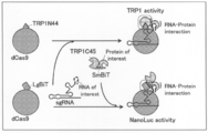
- the amino acid at the 2-44 position of tryptophan synthase (TRP1) (TRP1N44) and tryptophan synthase (TRP1) are added to the C-terminal of the Cas protein as the second element.
- Amino acid at position 45-224 (TRP1C45) 13 amino acids at the C-terminal of luciferase (NanoLuc) derived from deep-sea luminescent shrimp (SmBiT), and 13 amino acids at the C-terminal of luciferase (NanoLuc) derived from deep-sea luminescent shrimp. LgBiT), respectively, are fused.
- these Cas proteins dCas9 and dCas13b are preferable, and dCas9 is more preferable.
- various mutations may be introduced as long as they can bind to the complex-forming RNA.
- these second elements are preferably fused to the Cas protein via a linker, and the length of the linker is not particularly limited, but is preferably 1 to 17 amino acid residues.
- the second fusion protein may be a C-terminal, an N-terminal, or one in which a tag sequence is added at both ends.
- the dCas9-TRP1N44 fusion protein represented by the amino acid sequence of SEQ ID NO: 1
- the dCas9-TRP1C45 fusion protein represented by the amino acid sequence of SEQ ID NO: 2.
- Linker: RS dCas9-LgBiT fusion protein
- linker: RS represented by the amino acid sequence of SEQ ID NO: 3
- Each of these second fusion proteins is capable of binding to the complex-forming RNA, and the function of the marker protein is expressed by the proximity of the first element and the second element.
- it may be a homologue, a mutant, or a partial peptide of the amino acid sequence shown above.
- the homologs include, for example, 85% or more, preferably 90% or more, more preferably 95% or more (for example, 96% or more, 97% or more, 98% or more, 99% or more) with the amino acid sequence shown above.
- a protein consisting of an amino acid sequence having the same identity is included. The identity of the sequence can be evaluated numerically when calculated using BLAST or the like (eg, default or default parameters).
- the mutant consists of an amino acid sequence in which one or more amino acids are substituted, deleted, added or inserted with respect to the amino acid sequence shown above, and can bind to the complex-forming RNA.
- a protein in which the function of the marker protein is expressed by the proximity of the first element and the second element is included.
- plural means, for example, 2 to 150 pieces, preferably 2 to 100 pieces, more preferably 2 to 50 pieces (for example, 2 to 30 pieces, 2 to 10 pieces, 2 to 5 pieces, 2). ⁇ 3 pieces, 2 pieces).
- the "fusion RNA" is one in which two or more of the above RNAs are fused to form one (one molecule) RNA, and in the present invention, the complex-forming RNA and the subject Indicates a fused RNA obtained by fusing a test RNA or a specific RNA.
- the fusion between RNAs may be direct or indirect as long as it does not inhibit the interaction between the protein of interest and RNA and the expression of the function of the marker protein.
- the test RNA or a specific RNA may be fused to either the 3'side or the 5'side of the complex-forming RNA, but the complex-forming RNA may be fused.
- the guide RNA of the Cas protein Is the guide RNA of the Cas protein, the 3'side of the guide RNA (ie, the 3'side if the guide RNA contains trRNA, and the 3'side if it does not contain trRNA and consists only of crRNA. ) Is fused with the test RNA or a specific RNA.
- the fusion RNA can be obtained, for example, by transcribing and expressing two or more genes encoding the RNA as a unit.
- the fusion RNA according to the present invention can be produced by appropriately adopting and improving a conventionally known method.
- the fusion RNA thereof can be obtained by a method of chemically synthesizing based on a base sequence by a commercially available synthesizer, or a method of introducing a polynucleotide encoding fusion RNA or a vector expressing the polynucleotide into the cell and expressing it.
- the "ribonucleoprotein” is a complex of a protein and a ribonucleotide, and in the present invention, it indicates a complex of a second fusion protein and a fusion RNA, and more specifically, the first fusion protein.
- the complex formed through the binding between the RNA-binding protein contained in the fusion protein of 2 and the complex-forming RNA contained in the fusion RNA is shown.
- the binding between the second fusion protein and the fusion RNA that is, the binding between the RNA-binding protein and the complex-forming RNA
- the binding between the second fusion protein and the fusion RNA is the interaction between the target protein and the RNA and the function of the marker protein.
- RNA-binding protein e.g, other proteins, other nucleic acids, sugars, lipids, small molecules
- RNA-binding protein e.g., other proteins, other nucleic acids, sugars, lipids, small molecules
- Compounds (vitamins, coenzymes, hormones, toxins, antibiotics, antibacterial agents, antiviral agents, anticancer agents, carcinogens, drugs, psychotropic agents, etc.), metal ions, metal complexes, and 2 of these It may be indirect, such as forming a complex via a complex molecule containing more than one species of molecule.
- the method for evaluating the interaction between the protein and RNA of the present invention is (1) In the cytoplasm or cell-free system (A) A first fusion protein obtained by fusing the test protein and the first element of the marker protein, (B) A second fusion protein obtained by fusing an RNA-binding protein and a second element of the marker protein, and (c) an RNA forming a complex with the RNA-binding protein and a test RNA.
- Ribonucleoprotein consisting of fused RNA
- the process of contacting (2) A step of detecting a signal caused by the marker protein whose function is expressed by the proximity of the first element and the second element, and (3) A step of determining the interaction between the test protein and the test RNA by detecting the signal.
- the first fusion protein and the ribonucleoprotein form a complex
- the first element and the second element It is a method in which the function of the marker protein is expressed by the proximity of.
- test protein As the "test protein” according to the protein-RNA interaction evaluation method of the present invention, any protein among the above proteins that is desired to measure the interaction with the target RNA (test RNA 32) is used. be able to. Further, as the "test RNA” according to the protein-RNA interaction evaluation method of the present invention, any of the above RNAs that is desired to measure the interaction with the test protein 11 can be used. it can.
- the first fusion protein 1 and the ribonucleoprotein 4 are used as a method of contacting the first fusion protein 1 and the ribonucleoprotein 4.
- the cell those listed as the eukaryotic cell and the prokaryotic cell described above can be used, and the cell-free system is as described above. Further, these introduction into the cytoplasm and expression in the cytoplasm may be transient or constitutive expression depending on the purpose. As the method of introduction or expression, those skilled in the art can appropriately adopt a conventionally known method or a method similar thereto, depending on the type of cell and the like.
- Ribonucleoprotein, and fusion RNA can be used by those skilled in the art, for example, based on the amino acid sequence of the protein, in a cell-free protein synthesis system (eg, reticulated red erythrocyte extract, wheat germ extract), Escherichia coli, animal.
- a polynucleotide encoding a first fusion protein 1 and a ribonucleoprotein 4 (or a second fusion protein 2 and a fusion RNA 3) into a cell or a cell-free system having a gene expression function, respectively.
- Each of these polynucleotides can be chemically synthesized and prepared by a commercially available synthesizer based on its base sequence.
- the expression vector contains one or more regulatory elements that are operably linked to a gene to be expressed (a gene encoded by each of the polynucleotides).
- operably bound means that the gene is operably bound to a regulatory element.
- regulatory elements include promoters, enhancers, internal ribosome entry sites (IRES), and other expression control elements (eg, transcription termination signals such as polyadenylation signals and polyU sequences).
- the regulatory element directs gene expression only in a specific cell, tissue, or organ, even if it directs constitutive expression of the gene in various host cells, for example. It may be a thing. In addition, it may be directed to the expression of a gene only at a specific time, or may be directed to the expression of an artificially inducible gene.
- promoters include polIII promoters (eg, U6 promoter, H1 promoter, and SNR52 promoter), polII promoters (eg, sprouting yeast ADH promoter, CYC promoter, TEF promoter, GPD promoter, GAL promoter, CUP promoter, retrovirus.
- Raus sarcoma virus (RSV) LTR promoter cytomegalovirus (CMV) promoter, SV40 promoter, dihydrofolate reductase promoter, ⁇ -actin promoter, phosphoglycerol kinase (PGK) promoter, and EF1 ⁇ promoter
- polI promoter lac promoter
- trp Examples include a promoter, an araBAD promoter, a T7 promoter, an S6 promoter, or a combination thereof. These promoters can be appropriately selected depending on the type of cell and vector used. For example, when a vector expressing a polynucleotide encoding the fusion RNA according to the present invention is used in yeast, the polII promoter is preferable. ..
- the expression vector examples include a plasmid vector, an episomal vector, and a virus vector.
- the proteins and RNAs encoded in the expression vector are the above-mentioned fusion proteins and fusion RNAs, etc., but from the viewpoint of further improving the expression efficiency of these, the expression vector encoding these is the protein.
- codon-optimized DNA eg, codon-humanized DNA
- a polynucleotide encoding a self-cleaving RNA for example, hammerhead ribozyme
- such an expression vector can be prepared by those skilled in the art by appropriately using known techniques such as a DNA chemical synthesis method and a gene recombination technique.
- a method for introducing the above-mentioned fusion protein, ribonucleoprotein, fusion RNA, polynucleotide (RNA, DNA), and expression vector into cells conventionally known methods are available depending on the type of molecule to be introduced, the type of target cell, and the like. It can be appropriately selected from the method or a method similar thereto. Examples of such an introduction method include a method using a protein introduction reagent, an electroporation method, a microinjection method, a particle gun method, a calcium phosphate method, a liposome method (lipofection method), and a DEAE-dextran method.
- viruses adenovirus, lentivirus, adeno-associated virus, baculovirus, etc.
- agrobacterium method particle gun method
- lithium acetate method lithium acetate method
- spheroplast method heat shock method (chloride) Calcium method
- rubidium chloride method rubidium chloride method
- the first fusion protein 1 and the ribonucleoprotein 4 can be contacted in a cytoplasmic or cell-free system by the above method.
- Ribonucleoprotein 4 is a complex containing a second fusion protein 2 and fusion RNA3.
- Ribonucleoprotein 4 is prepared extracellularly or extracellularly and into a cell or cell-free system. It may be introduced, or the second fusion protein 2 and the fusion RNA 3 may interact with each other in a cell or cell-free system to form a ribonucleoprotein 4.
- the second fusion protein 2 and the fusion RNA 3 form a complex between the RNA-binding protein 21 inherent in each molecule and the complex-forming RNA 31, so that the second fusion protein 2 and the fusion RNA 3 are seconded through such an interaction.
- the fusion protein 2 and the fusion RNA 3 of the above form a ribonucleoprotein 4.
- one molecule of the first fusion protein and one molecule of the second fusion Each element of the marker protein contained in the protein may be one kind alone or two or more kinds, and may be included as the first fusion protein and the second fusion protein, respectively. Depending on the element or combination of proteins, one type may be used alone or may be two or more types, and the number of types of the first fusion protein and the number of types of the second fusion protein may be different. For example, when two types of marker proteins A and B are used, the first element is a fusion of the test protein, the first element A of the marker protein A, and the first element B of the marker protein B.
- the second fusion protein B, which is fused with the second element B, or the ribonucleoprotein B containing the second fusion protein B can be contacted in a cytoplasmic or cell-free system.
- a signal caused by the marker protein whose function is expressed by the proximity of the first element 12 and the second element 22 is detected.
- the combination of such a marker protein, a signal, and a detection method can be appropriately selected, and examples thereof include the above-mentioned combinations.
- the test protein and the marker protein A first.
- the first fusion protein formed by fusing the element A of 1 and the first element B of the marker protein B; the RNA-binding protein and the second element A of the divided marker protein A are fused.
- Ribonucleoprotein A containing a second fusion protein A Ribonucleoprotein A containing a second fusion protein A
- Ribonucleoprotein B containing a second fusion protein B formed by fusing an RNA-binding protein with a second element B of marker protein B
- the test protein and the test RNA interact with each other, the first fusion protein and the ribonucleoprotein A and the other first fusion protein and the ribonucleoprotein B are brought into contact with each other. And, respectively, form complexes A and B, respectively.
- the functions of the marker proteins A and B are expressed when the first element A and the second element A and the first element B and the second element B are close to each other, respectively.
- Multiple detections can be made simultaneously based on the function of each marker protein and the resulting signals (eg, FIG. 9 of the Examples).
- the interaction between the test protein 11 and the test RNA 32 is determined based on the signal detected in the (2) detection step. For example, in the detection step, when a signal caused by the marker protein expressing the function is detected, that is, when the function of the marker protein is detected, the test protein 11 and the test are tested. It can be determined that there is an interaction with RNA32, while if it is not detected, it can be determined that there is no such interaction.
- the detection method is a method that can be quantified or semi-quantitative
- the magnitude of the interaction between the test protein 11 and the test RNA 32 can be determined according to the magnitude of the value (Aspect 1). ).
- a specific protein is used as the test protein 11 and an RNA library is used as the test RNA 32 to interact with the specific protein.
- RNAs that act can be screened (Aspect 2 below).
- a specific RNA as the test RNA 32 and using the protein library as the test protein 11, a protein that interacts with the specific RNA can be screened (Aspect 3 below).
- (Aspect 2) A method for screening RNA that interacts with a particular protein.
- a first fusion protein formed by fusing a specific protein with the first element of a marker protein (B) A second fusion protein obtained by fusing an RNA-binding protein and a second element of the marker protein, and (c) an RNA forming a complex with the RNA-binding protein and a test RNA.
- Ribonucleoprotein consisting of fused RNA The process of contacting, (2) A step of detecting a signal caused by the marker protein whose function is expressed by the proximity of the first element and the second element, and (3) A step of selecting a test RNA that interacts with the specific protein by detecting the signal.
- the first fusion protein and the ribonucleoprotein form a complex
- the first element and the second element A method in which the function of the marker protein is expressed by the proximity of the markers.
- a method for screening proteins that interact with specific RNA (1) In the cytoplasm or cell-free system (A) A first fusion protein obtained by fusing the test protein and the first element of the marker protein, (B) A second fusion protein obtained by fusing an RNA-binding protein and a second element of the marker protein, and (c) an RNA forming a complex with the RNA-binding protein and a specific RNA. Ribonucleoprotein consisting of fused RNA, The process of contacting, (2) A step of detecting a signal caused by the marker protein whose function is expressed by the proximity of the first element and the second element, and (3) A step of selecting a test protein that interacts with the specific RNA by detecting the signal. Including When the test protein and the specific RNA interact, the first fusion protein and the ribonucleoprotein form a complex, and the first element and the second element A method in which the function of the marker protein is expressed by the proximity of the markers.
- a specific protein 11 is used instead of the test protein 11 of the aspect 1.
- a specific RNA 32 is used instead of the test RNA 32 of the aspect 1.
- any protein that desires to search for RNA that interacts with the above protein can be used, and as the specific RNA 32 of the third aspect, the above RNA Of these, any RNA that desires to search for proteins that interact with it can be used.
- the kit for use in the method of evaluating the interaction between the protein and RNA of the present invention is The following (a) to (c): (A) A first fusion protein obtained by fusing a test protein and a first element of a marker protein, a polynucleotide encoding the first fusion protein, a vector expressing the polynucleotide, or a marker protein. A vector containing an insertion site for a polynucleotide encoding the first element of the above and a polynucleotide encoding the test protein adjacent thereto.
- kit for evaluating protein-RNA interaction is the above-mentioned protein-RNA interaction evaluation method.
- a vector expressing a first fusion protein, a polynucleotide encoding the first fusion protein, and a polynucleotide encoding the first fusion protein (a) a vector expressing a first fusion protein, a polynucleotide encoding the first fusion protein, and a polynucleotide encoding the first fusion protein. (B) A second fusion protein, a polynucleotide encoding a second fusion protein, a vector expressing a polynucleotide encoding a second fusion protein, and (c) a fusion RNA, a polynucleotide encoding a fusion RNA.
- the vectors expressing the polynucleotide encoding the fusion RNA are as described above.
- the insertion site of the polynucleotide encoding the first element of the marker protein and the polynucleotide encoding the test protein adjacent thereto as (a).
- a vector containing the above, and a vector containing the insertion site of the polynucleotide encoding the RNA forming a complex with the RNA-binding protein as (c) and the polynucleotide encoding the test RNA adjacent thereto, respectively. can include.
- vectors are, for example, a polynucleotide encoding the test protein 11 of interest (or the specific protein 11 of aspect 2 or the protein library of aspect 3) and / or the test RNA 32 (or the specific RNA 32 of aspect 3).
- the polynucleotide encoding the RNA library of Aspect 2 can be arbitrarily selected and inserted into the insertion site according to the purpose. Examples of the insertion site include restriction enzyme recognition sequences.
- a polynucleotide library may be inserted in advance at the insertion site.
- the kit for evaluating the protein-RNA interaction of the present invention is a first fusion protein in a cytoplasmic or cell-free system by introducing the above (a) to (c) into the cell or cell-free system. Since 1 and ribonucleoprotein 4 (or the second fusion protein 2 and fusion RNA3) can be brought into contact with each other, cells for introducing these, and media and stabilizers necessary for storing and culturing the cells, It may further comprise other ingredients such as preservatives, preservatives and the like.
- cell-free solutions for introducing the above (a) to (c) cell disruption solution, cell extract, reconstituted cell-free protein synthesis solution such as PURE System, the above (a) to (c) It may further be provided with a contactable in vitro reconstitution solution, etc.).
- at least one of the above (a) to (c) may be in a form previously introduced into the cell or cell-free fluid.
- the cells for introducing the above (a) to (c) may or may not be alive, and the above-mentioned (a) to (c) are efficient in advance. Processing for the purpose of introduction, preservation, etc. may be performed.
- a membrane permeation treatment with a surfactant and the like can be mentioned.
- the kit for evaluating protein-RNA interaction of the present invention may further include a standard such as a reagent (reagent for signal detection) for detecting a signal caused by the expression of the function of the marker protein.
- a standard such as a reagent for preparing a calibration curve or an enzyme (a reagent for preparing a calibration curve) may be further provided.
- each fusion protein, fusion RNA, polynucleotide, expression vector, the cell, the cell-free solution, the signal detection reagent, and the calibration line preparation As a standard such as a reagent, other components such as a buffer solution, a stabilizer, a preservative, and a preservative may be added.
- the kit for evaluating the protein-RNA interaction of the present invention may further include an instruction manual which is an instruction for utilizing the expression vector, cells, and cell-free fluid in the method of the present invention.
- the instruction manual describes, for example, the experimental method and conditions of the method of the present invention, and information on each standard of the present invention (for example, information such as a vector map showing a base sequence of a vector, a cloning site, etc.). Information on the origin, properties, culture conditions, etc. of cells) can be included.
- the method for evaluating a substance (test substance) that regulates the interaction between the protein and RNA of the present invention is (1) In the presence of the test substance, in the cytoplasm or cell-free system (A) A first fusion protein formed by fusing a specific protein with the first element of a marker protein, (B) A second fusion protein obtained by fusing an RNA-binding protein and a second element of the marker protein, and (c) an RNA forming a complex with the RNA-binding protein and a specific RNA.
- Ribonucleoprotein consisting of fused RNA
- the process of contacting (2) A step of detecting a signal caused by the marker protein whose function is expressed by the proximity of the first element and the second element, and (3) When the signal is increased as compared with the case where the test substance is absent, the test substance is evaluated as a substance that promotes the interaction between the specific protein and the specific RNA. , The test substance is evaluated as a substance that suppresses the interaction between the specific protein and the specific RNA when the signal is reduced or eliminated as compared with the case where the test substance is absent.
- method for evaluating a substance that regulates the interaction between a protein and RNA of the present invention (hereinafter, sometimes referred to as "method for evaluating a protein-RNA interaction regulator"), (a) a first fusion protein, (b). ) The second fusion protein, (c) the fusion RNA, and the ribonucleoprotein, and the marker protein whose function is expressed by the step of contacting them and the proximity of the first element 12 and the second element 22.
- the steps for detecting the resulting signal include (a) first fusion protein, (b) second fusion protein, (c) fusion RNA, and ribo, respectively, in the above protein-RNA interaction evaluation method.
- the test protein 11 and the test RNA 32 are used. Instead, a specific protein 11 and a specific RNA 32 are used, respectively.
- a combination desired to obtain an evaluation of the effect of the test substance on their interaction is adopted.
- a combination includes, for example, evaluating whether the test substance promotes or inhibits the interaction of a specific protein 11 and a specific RNA 32 known to interact with each other.
- a combination desired to evaluate and search for whether the test substance promotes the interaction can be mentioned.
- RNA virus particle a protein constituting a ribonucleoprotein such as a ribosome, a spryisosome, a telomerase, a ribonuclease P, or an RNA virus particle and an RNA.
- splicing regulators proteins
- microRNA regulators proteins
- RNA LIN28, hnRNP A1, hnRNP L, KSRP and microRNA precursors (RNA)
- RNA microRNA regulators
- test substance examples include proteins, nucleic acids, sugars, lipids, and low molecular weight compounds (vitamins, coenzymes, hormones, toxins, antibiotics, etc.).
- vitaminss, coenzymes, hormones, toxins, antibiotics, etc. include proteins, nucleic acids, sugars, lipids, and low molecular weight compounds (vitamins, coenzymes, hormones, toxins, antibiotics, etc.).
- Antibacterial agents, antiviral agents, anticancer agents, carcinogens, drugs, psychotropic agents, etc.), metal ions, metal complexes, and complex molecules containing two or more of these molecules can be mentioned.
- the test substance is compared with the case where the test substance is not present.
- the test substance is evaluated as a substance that promotes the interaction between the specific protein 11 and the specific RNA 32, while the other
- the test substance is identified as a specific protein 11. It can be evaluated as a substance that suppresses the interaction with RNA32.
- promoting or suppressing the interaction between a specific protein 11 and a specific RNA 32 and "promoting or suppressing the expression of the function of a marker protein” directly make the promotion or suppression (for example).
- the interaction between the specific protein 11 and the specific RNA 32 may be promoted or suppressed).
- a specific protein 11 can be obtained by carrying out the method in parallel using a drug library or the like composed of a large number of chemical substances as a test substance. It is possible to search for a drug that promotes or suppresses the interaction between the protein and a specific RNA32.
- the method of the present invention for detecting a substance (target substance) that regulates the interaction between a protein and RNA in a sample (test sample) is (1) In the presence of the test sample, in the cytoplasm or cell-free system (A) A first fusion protein formed by fusing a specific protein with the first element of a marker protein, (B) A second fusion protein obtained by fusing an RNA-binding protein and a second element of the marker protein, and (c) an RNA forming a complex with the RNA-binding protein and a specific RNA.
- Ribonucleoprotein consisting of fused RNA
- the process of contacting (2) A step of detecting a signal caused by the marker protein whose function is expressed by the proximity of the first element and the second element, and (3) A step of detecting a target substance in the test sample by the presence / absence, increase, or decrease of the signal.
- Including The first fusion occurs when the specific RNA interacts with the specific protein in the presence or absence of the target substance, and when the specific protein interacts with the specific RNA.
- method for detecting a substance that regulates the interaction between a protein and RNA in the sample of the present invention (a) a first fusion protein. , (B) a second fusion protein, (c) a fusion RNA, and a ribonucleoprotein, and the step of contacting them, and the proximity of the first element 12 to the second element 22.
- steps for detecting the signal caused by the marker protein in the above-mentioned protein-RNA interaction evaluation method, (a) first fusion protein, (b) second fusion protein, and (c) fusion RNA, respectively.
- test protein 11 and the subject A specific protein 11 and a specific RNA 32 are used instead of the test RNA 32, respectively.
- the protein and the RNA may interact in the presence or absence of the target substance to be detected.
- a known combination that is, a combination of a specific protein 11 and a specific RNA 32 known to interact in the presence of the substance to be detected, or an interaction (target to be detected) in the absence of the substance to be detected. It is a combination of a particular protein 11 and a particular RNA 32 that is known to (do not interact in the presence of a substance).
- the target substances include, for example, proteins, nucleic acids, sugars, lipids, low molecular weight compounds (vitamins, coenzymes, hormones, toxins, antibiotics, antibacterial agents, etc.
- Antiviral agents, anticancer agents, carcinogens, drugs, psychotropic drugs, etc. metal ions, metal complexes, and complex molecules containing two or more of these molecules can be mentioned.
- the test sample is not particularly limited as long as it is a sample in which the target substance can exist, and for example, human and animal body fluids (saliva, tears, sweat). , Urine, blood, lymph, etc.), plant biofluids, biological culture fluids, extracts of organisms (individuals, organs, tissues, cells, etc.), water in the environment (rivers, lakes, harbors, waterways, groundwater, purified water, sewage) , Drainage, etc.), suspensions of solids (soil, cinders, etc.), suspensions of wiped samples, etc.
- human and animal body fluids saliva, tears, sweat
- Urine, blood, lymph, etc. plant biofluids
- biological culture fluids extracts of organisms (individuals, organs, tissues, cells, etc.)
- water in the environment rivers, lakes, harbors, waterways, groundwater, purified water, sewage) , Drainage, etc.
- suspensions of solids suspensions of wiped samples, etc.
- the combination of these specific proteins 11, the specific RNA 32, and the target substance is, for example, a combination of TetR (protein), TetR aptamer (RNA), and doxycycline; a neurotrophin receptor (protein). ), Neurotrophin receptor aptamer (RNA), and combination of neurotrophins; combinations of ribosomal proteins, ribosomal RNA, and antibiotics.
- the target substance is the sample.
- the signal is not detected, or when the signal is reduced, that is, when the expression of the function of the marker protein is suppressed, the target substance is present. It can be determined that it does not exist in the sample.
- the detection method is a method capable of quantifying or semi-quantitating, it is possible to quantify or semi-quantify the target substance in the sample by using a calibration curve or the like according to the magnitude of the value. it can.
- the signal is detected in the (2) detection step. If, or if the signal is increased, it can be determined that the target substance is not present in the sample, while if it is not detected or decreased, the target substance is said. It can be determined that it is present in the sample.
- the detection method is a method capable of quantifying or semi-quantitating, it is possible to quantify or semi-quantify the target substance in the sample by using a calibration curve or the like according to the magnitude of the value. it can.
- kit for detection method of protein-RNA interaction regulator The kit for use in the method for evaluating a protein-RNA interaction regulator of the present invention and the kit for use in the method for detecting a protein-RNA interaction regulator of the present invention are respectively.
- B A second fusion protein in which an RNA-binding protein and a second element of the marker protein are fused, a polynucleotide encoding the second fusion protein, or a vector expressing the polynucleotide.
- C A fusion RNA formed by fusing an RNA forming a complex with the RNA-binding protein and a specific RNA, a polynucleotide encoding the fusion RNA, a vector expressing the polynucleotide, or the RNA binding.
- (A') A polynucleotide encoding a first fusion protein, which is a fusion of a specific protein and a first element of a marker protein, or a vector expressing the polynucleotide.
- (B') A polynucleotide encoding a second fusion protein obtained by fusing an RNA-binding protein and a second element of the marker protein, or a vector expressing the polynucleotide.
- kits A polynucleotide encoding a fusion RNA obtained by fusing an RNA forming a complex with the RNA-binding protein and a specific RNA, or a vector expressing the polynucleotide. It is also preferable that the kit contains the transformed cells into which the cells have been introduced.
- a kit for use in the method for evaluating a protein-RNA interaction regulator of the present invention and a kit for use in a method for detecting a protein-RNA interaction regulator (hereinafter, in some cases, "evaluation of a protein-RNA interaction regulator”.
- the “detection method kit”) is a cytoplasmic or cell-free system in the (1) contact step of these methods, respectively, in which the first fusion protein 1 and the ribonucleoprotein 4 (or the second fusion protein 2 and) are used.
- a kit used for contacting fusion RNA3 is a kit for use in the method for evaluating a protein-RNA interaction regulator of the present invention and a kit for use in a method for detecting a protein-RNA interaction regulator.
- (a) to (c) refer to the test protein 11 and the test RNA 32 as the above-mentioned specific protein 11 and the specific RNA 32, respectively. Except for the above, the same applies to (a) to (c) mentioned in the above-mentioned kit for evaluating protein-RNA interaction.
- the kit for evaluating / detecting a protein-RNA interaction regulator of the present invention includes cells for introducing the above (a) to (c), and a medium, stabilizer, and storage necessary for storing and culturing the cells. Other ingredients such as agents and preservatives may be further provided.
- cell-free solutions for introducing the above (a) to (c) cell disruption solution, cell extract, reconstituted cell-free protein synthesis solution such as PURE System, the above (a) to (c) It may further be provided with a contactable in vitro reconstitution solution, etc.).
- at least one of the above (a) to (c) may be in a form previously introduced into the cell or cell-free fluid.
- the cells for introducing the above (a) to (c) may or may not be alive, and the above-mentioned (a) to (c) are efficient in advance. Processing for the purpose of introduction, preservation, etc. may be performed.
- a membrane permeation treatment with a surfactant and the like can be mentioned.
- kits for evaluating / detecting a protein-RNA interaction regulator of the present invention is (a') a polynucleotide encoding a first fusion protein, or a polynucleotide expressing the polynucleotide.
- a vector, a polynucleotide encoding (b') a second fusion protein, or a vector expressing the polynucleotide, and (c') a polynucleotide encoding a fusion RNA, or a vector expressing the polynucleotide examples include kits containing transformed cells into which, has been introduced.
- the transformed cell may or may not be alive, and may be a cell disruption solution or a cell extract. Further, the processing for the purpose of preserving the above (a') to (c') may be performed.
- the kit for evaluating / detecting a protein-RNA interaction regulator of the present invention further comprises a standard such as a reagent (reagent for signal detection) for detecting a signal caused by the expression of the function of the marker protein. You may have it.
- a test substance that promotes the interaction between a specific protein and a specific RNA contained in the kit, a test substance that suppresses the reaction, and a test substance that does not promote or suppress the interaction may be further provided.
- the test substance detected by the kit, the test substance not detected, and a standard such as a reagent or an enzyme (reagent for preparing a calibration curve) for preparing a calibration curve used for quantification or semi-quantification may be further provided.
- each fusion protein, fusion RNA, polynucleotide, expression vector, the cell, the cell-free fluid, the transformed cell, and the signal detection Other components such as a buffer solution, a stabilizer, a preservative, and an antiseptic may be added as a standard such as a protein for use, each of the test substances, and the reagent for preparing a calibration line.
- an instruction manual for using the expression vector, the cell, the cell-free solution, the transformed cell, etc. in each method of the present invention may be further provided.
- the instruction manual describes, for example, information on the experimental method and conditions of each method of the present invention, and information on each standard of the present invention (for example, a vector map showing a base sequence of a vector, a cloning site, etc.).
- Information on the origin, properties, culture conditions, etc. of the cells and transformed cells can be included.
- the biosensor of the present invention is a biosensor used in the method for evaluating a protein-RNA interaction regulator and / or the method for detecting a protein-RNA interaction regulator of the present invention.
- B' A polynucleotide encoding a second fusion protein obtained by fusing an RNA-binding protein and a second element of the marker protein, or a vector expressing the polynucleotide.
- C' A polynucleotide encoding a fusion RNA obtained by fusing an RNA forming a complex with the RNA-binding protein and a specific RNA, or a vector expressing the polynucleotide.
- Transformed cells into which the cells have been introduced, as well as (2) A means for detecting a signal caused by the marker protein whose function is expressed by the proximity of the first element and the second element.
- a ribonucleo consisting of the first fusion protein of (a'), the second fusion protein of (b'), and the fusion RNA of (c') It is a biosensor in which the function of the marker protein is expressed when the protein forms a complex and the first element and the second element are in close proximity to each other.
- examples of the transformed cell include those similar to those mentioned in the kit for evaluating / detecting a protein-RNA interaction regulator of the present invention.
- the marker protein and the type of signal are used as a means (detection means) for detecting a signal caused by the marker protein whose function is expressed by the proximity of the first element and the second element. It can be appropriately selected depending on the situation, and is not particularly limited.
- the marker protein is a tryptophan synthase and the signal is a colony indicating cell survival, the growth of yeast in a tryptophan-free medium. Examples include a container capable of discriminating the situation, an optical microscope, and the like.
- the marker protein is luciferase and the signal is fluorescence
- an image sensor or the like equipped with an optical lens capable of observing light emission by luciferase is mentioned.
- the marker protein is a donor / acceptor pair of FRET and the signal is fluorescent
- an image sensor equipped with a fluorescence microscope capable of observing the fluorescence can be mentioned.
- the determination step (3) of the above-mentioned protein-RNA interaction regulator evaluation method or protein-RNA interaction regulator detection method is carried out according to the result of the detection means. Further, the determination means may be provided.
- the determination means is not particularly limited, and examples thereof include an image analysis program and the like.
- the above-mentioned method for evaluating a protein-RNA interaction regulator and / or a method for detecting a protein-RNA interaction regulator can be carried out, for example, a subject to be detected.
- the detection means By simply adding a test substance or a test sample in which a target substance may be present to the growth medium of the transformed cell, the detection means causes the expression of the function of the marker protein in the transformed cell containing the marker protein. It is possible to determine the determination step (3) of the above-mentioned protein-RNA interaction regulator evaluation method or protein-RNA interaction regulator detection method using the detection result as an index.
- the present invention is used in at least one of a method for evaluating the interaction between a protein and RNA of the present invention, a method for evaluating a protein-RNA interaction regulator, and a method for detecting a protein-RNA interaction regulator.
- a fusion protein obtained by fusing the RNA-binding protein and the second element of the marker protein.
- the function of the marker protein is expressed by the proximity of the second element of the marker protein and the other element (that is, the first element) constituting the marker protein.
- the fusion protein is as described as the second fusion protein, including its preferred embodiment.
- a budding yeast strain expressing a fusion protein (dCas9-TRP1N44 fusion protein represented by the amino acid sequence of SEQ ID NO: 1).
- the parent strain of Saccharomyces cerevisiae includes BY4727 [Reference III: Brachmann C.I. B. et al. , Yeast 14: 115-132 (1998)], and for introduction to the HO locus, HO-poly-KanMX4-HO [Reference IV: Voth W. et al. P. et al. , Nucleic Acids Res.
- the region from the promoter to the terminator is the pRS400 series expression vector [Reference V: Mumberg D. et al. et al. , Gene 156: 119-122 (1995)], respectively.
- the Cas9 gene an endonuclease-inactivated Cas9 (dCas9) derived from Streptococcus pyogenes codon-optimized for Saccharomyces cerevisiae [Reference VI: Farzadfard F. et al. et al. , ACS Synth. Biol. (2013)] was adopted.
- the nuclear localization signal added to Cas9 in the document VI was removed.
- the constructed strain will be referred to as "A-share".
- p5G-TRP1C45-GSG5 a protein in which a 17-amino acid glycine-serine linker sequence (amino acid sequence shown in SEQ ID NO: 4) is fused to the C-terminal side of 45-224 amino acids (TRP1C45) of Saccharomyces cerevisiae Trp1 protein is used in the Saccharomyces cerevisiae GPD promoter. It was inserted into an expression vector p415GPD [described in Document V] expressed under control. Hereinafter, the constructed vector will be referred to as "p5G-TRP1C45-GSG5".
- MS2 phage coat protein (MS2CP) codon-optimized for budding yeast on the C-terminal side of the linker of p5G-TRP1C45-GSG5 Non-aggregating mutant (dlFG A81G) [Reference VII: Keryer-Bibens C.I. et al. , Biol Cell, 100: 125-138 (2008)] was inserted into the plasmid "p5G-TRP1C45-GSG5-yMS2CP".
- fusion RNA expression vector An expression vector of fusion RNA in which MS2 stem loop (MS2SL) was linked to the 3'end side of the single-strand guide RNA (gRNA) of dCas9 was constructed.
- An expression unit of fusion RNA excised by a hammerhead ribozyme added to the 5'end and a human hepatitis D virus ribozyme added to the 3'end [Reference VIII: Zalatan J. et al. G. et al. , Cell 160: 339-350 (2015)] was PCR-amplified with the addition of BamHI on the 5'end side and SalI restriction enzyme recognition sites on the 3'end side, and these restriction enzyme sites were used.
- RNA polymerase III For comparison, the vector pJZC583 [Reference VIII] in which a fusion RNA of a guide RNA (gRNA) and an MS2 stem loop (MS2SL) is expressed under the control of a small nucleolar RNA SNR52 promoter transcribed by RNA polymerase III. Description] was adopted.
- gRNA guide RNA
- MS2SL MS2 stem loop
- ⁇ Construction of transformed yeast strain> Competitive cells of strain A were prepared using Frozen-EZ Yeast Transformation II Kit (ZYMO RESEARCH), and a fusion protein expression vector derived from p415GPD (p5G-TRP1C45-GSG5 or p5G-TRP1C45-GSG5-yMS2CP)
- the fusion RNA expression vector p6A-HHRz-gRNA9-MS2SL-HDVRz or p6A-HHRz-gRNA9-MCS-HDVRz
- pJZC583, and pJZC625 were transformed with two types of plasmids.
- the transformed cells were applied to leucine-uracil-free synthetic agar medium SC-LU and statically cultured at 30 ° C. to obtain transformant colonies.
- the same transformant colonies were striked on tryptophan-leucine-uracil-free synthetic agar media SC-WLU (-Trp) and SC-LU (+ Trp), and statically cultured at 30 ° C.
- Table 1 below shows photographs of the appearance of the colonies after culturing under each condition (+ Trp / -Trp) of the transformants into which each vector has been introduced, respectively.
- the numbers in FIG. 2 correspond to the test numbers in Table 1, respectively.
- Test Example 2 Verification of interaction between Cas13b protein and Cas13b guide RNA (gRNA) ⁇ Construction of first fusion protein expression vector> Similar to Test Example 1, expression in which a protein in which a 17-amino acid glycine-serine linker sequence (amino acid sequence shown in SEQ ID NO: 4) is fused to the C-terminal side of TRP1C45 is expressed under the control of the Saccharomyces cerevisiae TEF promoter. It was inserted into the vector p413TEF [described in Document V]. Hereinafter, the constructed vector will be referred to as "p3T-TRP1C45-GSG5".
- RNA-binding protein for verifying the interaction with RNA
- test protein RNA-binding protein
- Prevotella sp. Derived Endonuclease Inactive Cas13b [Reference IX: Cox D. B. T. et al. , Science, 358: 1019-1027 (2017)] was inserted into the C-terminal side of the linker of p3T-TRP1C45-GSG5 to construct a plasmid "p3T-TRP1C45-GSG5-dCas13".
- ⁇ Construction of fusion RNA expression vector> Prevotella sp.
- the plasmid "p6A-HHRz-gRNA9-gCas13-HDVRz” was constructed by inserting the above-mentioned nucleotide sequence into the EcoRI and HindIII sites of p6A-HHRz-gRNA9-MCS-HDVRz of Test Example 1.
- Competent cells of strain A were used as a fusion protein expression vector derived from p413TEFPD (p3T-TRP1C45-GSG5 or p3T-TRP1C45-GSG5-dCas13) and a fusion RNA expression vector derived from p416ADH (p6A-HHRz-gRNA9-MCS-HDVRz or It was transformed with two types of plasmids (p6A-HHRz-gRNA9-gCas13-HDVRz). The transformed cells were applied to a histidine / uracil-free synthetic agar medium SC-HU and statically cultured at 30 ° C. to obtain transformant colonies.
- FIG. 3 shows photographs of the appearance of the colonies after culturing under each condition (+ Trp / -Trp) of the transformant into which each vector was introduced.
- the numbers in FIG. 3 correspond to the test numbers in Table 2, respectively.
- RNA aptamer (TetR Aptamer) that specifically binds to a tetracycline repressor that originally binds to double-stranded DNA [Reference XI: Goldfless S.A. J. .. et al. , Nucleic Acids Res. , 40: described in e64 (2012)] was inserted into the EcoRI and HindIII sites of p6A-HHRz-gRNA9-MCS-HDVRz of Test Example 1 to construct a plasmid "p6A-HHRz-gRNA9-TetRapt-HDVRz".
- Competent cells of strain A of Test Example 1 were used as a tetracycline repressor fusion protein expression vector derived from p415GPD (p5G-TRP1C45-GSG5-ytetR or p5G-TRP1C45-GSG5-yrtetR) and a fusion RNA expression vector derived from p416ADH (p6A).
- p415GPD p5G-TRP1C45-GSG5-ytetR or p5G-TRP1C45-GSG5-yrtetR
- p6A fusion RNA expression vector derived from p416ADH
- FIG. 4 shows photographs of the appearance of the colonies after culturing under each condition (-Dox / + Dox) of the transformant into which each vector was introduced.
- the numbers in FIG. 4 correspond to the test numbers in Table 3, respectively.
- Test Example 4 Quantification of interaction intensity by luciferase assay ⁇ Construction of experimental strain> A protein fragment (11S, LgBiT) in which the dCas9 protein was removed from the HO locus and the 13 amino acids at the C-terminal were removed from NanoLuc (promega) using the same method as for the construction of the A strain of Test Example 1 [Reference XII: Dixon].
- A. S. et al. ACS Chem. Biol.
- strain B TRP1N44 inserted at the HO locus of strain A is replaced with LgBiT (yLgBiT) codon-optimized for Saccharomyces cerevisiae.
- ⁇ Construction of the first fusion protein expression vector > C-terminal 13 amino acids (114, SmBiT) of NanoLuc codon-optimized for Saccharomyces cerevisiae at the N-terminus of the non-aggregating variant (dlFG A81G) of MS2 phage coat protein (MS2CP) codon-optimized for Saccharomyces cerevisiae.
- the gene fragment of the protein fused with [described in XII] was inserted into an expression vector p415GPD [described in Document V] expressed under the control of Saccharomyces cerevisiae GPD promoter to obtain "p5G-ySmBiT-yMS2CP".
- ⁇ Luciferase assay> Transformant colonies were seeded in 1 mL of leucine-uracil-free synthetic liquid medium and cultured with shaking at 30 ° C. overnight. The turbidity of the culture solution was measured, diluted in 1 mL of the same liquid medium so that the turbidity became 0.2, and further cultured with shaking at 30 ° C. for 4 hours. After replacing the culture solution with water, 25 ⁇ L of a sample having a turbidity of 2.8 was prepared, and 25 ⁇ L of the luminescent reagent of Nano-Glo Luciferase Assay Kit prepared at the time of use was added to determine the amount of luminescence (Intensity) after 5 minutes. It was measured. GloMax 96 was used for the measurement, and the exposure time was 60 seconds. Two colonies were prepared for each transformant, and the mean value and standard deviation measured at two points for each sample are shown in FIG. 6 as a result.
- Test Example 5 Simultaneous use of two types of marker proteins ⁇ Construction of experimental strain> Using the B strain of Test Example 4, a fusion protein of dCas9 protein and TRP1N44 described in Test Example 1 (dCas9-TRP1N44 fusion protein) was obtained from the AUR1 gene locus on chromosome 11 under the control of the budding yeast CYC promoter. A budding yeast strain that was further expressed was constructed. PAUR101 (Takara Bio) was used for introduction to the AUR1 locus. Hereinafter, the constructed strain will be referred to as "C strain”.
- the green fluorescent protein Clover which originally does not bind to nucleic acids [Reference XIII: Lee S. et al. , PLoS ONE, 8 (7): described in e67902, 2013] was used as a test protein to construct a plasmid "p5T-TRP1C45-ySmBiT-yClover" inserted into p5T-TRP1C45-ySmBiT.
- ⁇ Construction of fusion RNA expression vector> The plasmid "p26G-HHRz-gRNA9-MS2SL-" in which the fusion RNA in which MS2SL was ligated to the gRNA of dCas9 of Test Example 1 or the fusion RNA in which the MS2 stem-loop portion thereof was replaced with an MCS sequence was inserted into p426GPD [described in Document V].
- "HDVRz” and "p26G-HHRz-gRNA9-MCS-HDVRz” were constructed. Furthermore, the sequence of RNA aptamer (GFP Aptamer) that specifically binds to the green fluorescent protein GFP, which originally does not bind to nucleic acid [Reference XIV: Shii B.
- a competent cell of strain C was prepared using Frozen-EZ Yeast Transformation II Kit (ZYMO RESEARCH), and a fusion protein expression vector derived from p415TEF (p5T-TRP1C45-ySmbiT, p5T-TRP1C45-ySmBiT, p5T-TRP1C45-ySmBiT- -YSmBiT-yClover) and a fusion RNA expression vector derived from p426GPD (p26G-HHRz-gRNA9-MS2SL-HDVRz, p26G-HHRz-gRNA9-MCS-HDVRz, or p26G-HHRz-gRNA9-GFPApt-HDVRz).
- p415TEF p5T-TRP1C45-ySmbiT, p5T-TRP1C45-ySmBiT, p5T-TRP1C45-ySmBiT-
- ⁇ Luciferase assay> The transformant colony was cultured in the same manner as in Test Example 4, and the luciferase activity was measured. Measure the turbidity of the culture medium shake-cultured overnight at 30 ° C. in 1 mL of leucine-uracil-free synthetic liquid medium, dilute to 1 mL of the same liquid medium so that the turbidity becomes 0.2, and further at 30 ° C. It was cultured with shaking. A 25 ⁇ L sample having a turbidity of 2 was prepared, 25 ⁇ L of a luminescent reagent of Nano-Glo Luciferase Assay Kit prepared at the time of use was added, and the luminescence amount (Intensity) after 20 minutes was measured. Each transformant per average samples were measured by preparing triplicate colonies (Luminecence (10 6 RLU)) and its standard deviation as a result is shown in FIG. The numbers in FIG. 8 correspond to the test numbers in Table 4, respectively.
- Test Example 6 Quantitative detection of volume-dependent interactions of ligands (substances that regulate interactions) ⁇ Construction of first fusion protein expression vector> Using the tetR described in Test Example 3 as a test protein, a plasmid "p5T-TRP1C45-ySmBiT-ytetR" inserted into p5T-TRP1C45-ySmBiT was constructed.
- a plasmid "p26G-HHRz-gRNA9-TetRapt-HDVRz" was constructed by inserting the sequence of the fusion RNA of p6A-HHRz-gRNA9-TetRapt-HDVRz of Test Example 3 (gRNA of dCas9, fusion RNA linked with TetR Aptamer) into p426GPD. did.
- Dox is added to the luminescent reagent of Nano-Glo Luciferase Assay Kit prepared at the time of use so that the Dox concentration in the luminescent reagent becomes a 5-fold dilution series from 2 g / L to 64 ⁇ g / L. And used it.
- Samples were prepared for each of the transformant into which p26G-HHRz-gRNA9-MCS-HDVRz was introduced (Comparative Example 19) and the transformant into which p26G-HHRz-gRNA9-TetRapt-HDVRz was introduced (Example 9).
- Figure 10 shows the average value measured (Luminescence (10 5 RLU)) and standard deviation as a result.
- FIG. 11 shows the fusion RNA (a) in which MS2SL is ligated to the gRNA of dCas9, the sequence (b) excised from the deletion substance 1 by the ribozyme, and the sequence (c) excised from the deletion substance 2 by the ribozyme, respectively. ..
- the transformed cells were applied to SC-LU and statically cultured at 30 ° C. to obtain transformant colonies.
- the same transformant colonies were drawn on SC-WLU and SC-LU and statically cultured at 30 ° C.
- the combination of the marker protein (first element (first fragment)) and the test protein in the first fusion protein expression vector, and the combination of the complex-forming RNA and the test RNA in the fusion RNA expression vector are as follows.
- Table 5 shows a photograph of the appearance of the colony after culturing under each condition (+ Trp / ⁇ Trp) of the transformant into which each vector was introduced, respectively.
- the numbers in FIG. 12 correspond to the test numbers in Table 5, respectively.
- TRP1 marker protein Another fusion mode of TRP1 marker protein ⁇ Construction of experimental strain> Using the same method as the construction of strain A described in Test Example 1, the dCas9 protein is expressed as a fusion protein with TRP1C45 (dCas9-TRP1C45 fusion protein represented by the amino acid sequence of SEQ ID NO: 2) from the HO locus. Saccharomyces cerevisiae strain was constructed. Hereinafter, the constructed strain will be referred to as "D strain”.
- ⁇ Construction of the first fusion protein expression vector> Using MS2CP as a test protein, a protein in which a 17-amino acid glycine-serine linker sequence (amino acid sequence shown in SEQ ID NO: 4) and TRP1N44 are fused to the C-terminal side thereof is expressed under the control of the Saccharomyces cerevisiae GPD promoter. It was inserted into the expression vector p415GPD.
- the constructed vector will be referred to as "p5G-yMS2CP-GSG5-TRP1N44".
- the competent cell of strain A was subjected to the first fusion protein expression vector p5G-TRP1C45-GSG5-yMS2CP and the fusion RNA expression vector derived from p416ADH (p6A-HHRz-gRNA9-MCS-HDVRz). Alternatively, it was transformed with two types of plasmids (p6A-HHRz-gRNA9-MS2SL-HDVRz).
- the competent cells of the D strain were used as the first fusion protein expression vector p5G-yMS2CP-GSG5-TRP1N44 and the fusion RNA expression vector derived from p416ADH (p6A-HHRz-gRNA9-MCS-HDVRz or p6A-HHRz-gRNA9-. MS2SL-HDVRz) and two types of plasmids were transformed.
- the transformed cells were applied to SC-LU and statically cultured at 30 ° C. to obtain transformant colonies. The same transformant colonies were drawn on SC-WLU and SC-LU and statically cultured at 30 ° C.
- a yeast strain a combination of a marker protein (first element (first fragment)) and a test protein in the first fusion protein expression vector, and a combination of a complex-forming RNA and a test RNA in the fusion RNA expression vector.
- Table 6 the appearance photographs of the colonies after culturing under each condition (+ Trp / -Trp) of the transformants into which each vector was introduced are shown in FIG.
- the numbers in FIG. 13 correspond to the test numbers in Table 6, respectively.
- Test Example 9 Detection of interaction by FRET ⁇ Construction of experimental strain> Saccharomyces cerevisiae expressing the dCas9 protein as a fusion protein with the green fluorescent protein Clover (second protein) described in Test Example 5 from the HO locus using the same method as the construction of the A strain described in Test Example 1.
- a yeast strain was constructed. Hereinafter, the constructed strain will be referred to as "E strain”.
- a transformant (Example 13) transformed with the vector p26G-HHRz-gRNA9-TetRapt-HDVRz was prepared. Each of the obtained transformants was applied to SC-HU and allowed to stand at 30 ° C. to obtain colonies of the transformants.
- ⁇ FRET assay> The transformant colonies were seeded in 1 mL of a synthetic liquid medium free of histidine and uracil, and cultured with shaking at 30 ° C. overnight. The turbidity of the culture solution was measured, diluted in 1 mL of the same liquid medium so that the turbidity became 0.2, and further cultured with shaking at 30 ° C. for 8 hours. Yeast cells were precipitated by centrifugation, the medium was removed, and 1 mL of sterile water was added for resuspension. Again, yeast cells were precipitated by centrifugation, the supernatant was removed, and 500 ⁇ L of sterile water was added for resuspension.
- fluorescence 1 excitation wavelength 475 nm, fluorescence wavelength 650 nm
- fluorescence 2 excitation wavelength 475 nm, fluorescence wavelength 515 nm
- TRP1N44 as a marker protein (second element (second fragment)
- a 17-amino acid glycine-serine linker sequence as an RNA-binding protein.
- MS2CP as an RNA-binding protein.
- the fused protein was inserted into the expression vector p413TEF expressed under the control of the sprouting yeast TEF promoter.
- the constructed vector will be referred to as "p3T-TRP1N44-GSG5-yMS2CP”.
- ⁇ Construction of fusion RNA expression vector> The single-strand guide RNA (gRNA) of p26G-HHRz-gRNA9-MCS-HDVRz of Test Example 5 and dCas9 of p26G-HHRz-gRNA9-TetRapt-HDVRz of Test Example 6 were replaced with MS2SL, respectively. -HHRz-MS2SL-MCS-HDVRz "and" p26G-HHRz-MS2SL-TetRAPt-HDVRz "were constructed.
- Competent cells of the BY4727 strain were prepared using the Frozen-EZ First Transformation II Kit (ZYMO RESEARCH), and the fusion protein expression vector p5T-TRP1C45-ySmBiT-ytetR of Test Example 6 and the second fusion protein expression vector p3T- It was transformed with two types of plasmids, TRP1N44-GSG5-yMS2CP.
- competent cells of the obtained transformants were prepared and again with a fusion RNA expression vector derived from p426GPD (p26G-HHRz-MS2SL-MCS-HDVRz or p26G-HHRz-MS2SL-TetRapt-HDVRz). Transformed. Further, the transformation was performed in the same manner except that p26G-HHRz-gRNA9-MS2SL-HDVRz of Test Example 5 or p26G-HHRz-gRNA9-TetRapt-HDVRz described in Test Example 6 was used instead of the fusion RNA expression vector. Converted.
- the transformed cells were applied to a synthetic agar medium SC-HLU free of histidine, leucine, and uracil and statically cultured at 30 ° C. to obtain transformant colonies.
- the same transformant colonies were plotted on histidine, tryptophan, leucine, and uracil-free synthetic agar media SC-HWLU (-Trp) and SC-HLU (+ Trp), and statically cultured at 30 ° C.
- a method for evaluating the interaction between protein and RNA which can specifically and easily detect the interaction between protein and RNA in a cytoplasmic or cell-free system, and a method for evaluating the interaction between protein and RNA. It is possible to provide a method for evaluating an interaction regulator, a method for detecting an interaction regulator between a protein and RNA, and a fusion protein, kit, and biosensor used for these methods.
- fusion protein 11 ... test protein / specific protein, 12 ... marker protein (first element, first fragment), 2 ... second fusion protein, 21 ... RNA binding protein, 22 ... Marker protein (second element, second fragment), 3... fusion RNA, 31... complex-forming RNA, 32... test RNA / specific RNA, 4... ribonucleoprotein.
Abstract
Provided is a method for evaluating the interaction of a protein and RNA, said method comprising: (1) a step for bringing the following (a), (b), and (c) into contact in cytoplasm or a cell-free system, in which (a) is a first fusion protein obtained by fusing a subject protein and a first element of a marker protein, (b) is a second fusion protein obtained by fusing an RNA binding protein and a second element of the marker protein, and (c) is a ribonucleoprotein comprising fusion RNA which is obtained by fusing a subject RNA and RNA which forms a composite with the RNA binding protein; (2) a step for detecting a signal caused by the marker protein, which expresses a function by means of the first element and the second element being in close proximity; and (3) a step for determining the interaction of the subject protein and the subject RNA by means of detecting the signal, wherein if the subject protein and the subject RNA interacted, the first fusion protein and the ribonucleoprotein form a composite, and the marker protein function is expressed by means of the first element and the second element being in close proximity.
Description
本発明は、タンパク質とRNAとの相互作用評価方法、タンパク質とRNAとの相互作用調節物質評価方法、及びタンパク質とRNAとの相互作用調節物質検出方法、並びに、これらに用いる融合タンパク質、キット、及びバイオセンサに関する。
The present invention relates to a method for evaluating the interaction between protein and RNA, a method for evaluating an interaction regulator between protein and RNA, a method for detecting an interaction regulator between protein and RNA, and fusion proteins, kits, and fusion proteins used thereto. Regarding biosensors.
細胞内の遺伝子発現制御を理解する上で、タンパク質とRNAとの間の相互作用(タンパク質-RNA間相互作用)の測定は必須の技術である。また、タンパク質-RNA間相互作用を探索的に検出することによって、アプタマーと呼ばれる特定のタンパク質に結合するRNA分子が人為的に創出されている(Wu Y.X.,Kwon Y.J.,Methods,106:21-28(2016)(非特許文献1))。このようなアプタマーは、タンパク質機能の阻害分子として、或いは、薬物複合体を構成する分子標的分子として、応用されている(Zhu G.,Chen X.Adv Drug Deliv Rev.134:65-78(2018)(非特許文献2))。
Measurement of the interaction between protein and RNA (protein-RNA interaction) is an indispensable technique for understanding the regulation of gene expression in cells. In addition, by exploratory detection of protein-RNA interactions, RNA molecules that bind to specific proteins called aptamers have been artificially created (Wu YX, Kwon YJ, Methods). , 106: 21-28 (2016) (Non-Patent Document 1)). Such aptamers have been applied as molecules that inhibit protein function or as molecular target molecules that constitute drug complexes (Zhu G., Chen X. Adv Drag Dev. 134: 65-78 (2018). ) (Non-Patent Document 2)).
試験管内でのタンパク質-RNA間相互作用の測定方法に対して、細胞内の生理的な環境条件でタンパク質-RNA間相互作用を測定する方法として、酵母スリーハイブリッド法がある(Martin F.,Methods,58:367-375(2012)(非特許文献3))。この方法では、酵母ツーハイブリッド法を構成する分割型転写因子のDNA結合ドメインに融合させた既知のRNA結合性タンパク質と、このRNA結合性タンパク質と結合する既知のRNA(RNA結合性タンパク質の標的RNA分子)に融合させた検証対象のRNAとを相互作用させ、前記RNA結合性タンパク質と標的RNA分子との相互作用を解して、分割型転写因子のDNA結合ドメインと検証対象のRNAとを含むリボヌクレオタンパク質を形成させる。このリボヌクレオタンパク質における検証対象のRNAに、分割型転写因子の転写活性化ドメインに融合させた検証対象のタンパク質が接触すると、タンパク質-RNA間の相互作用が生じた場合、3つの分子からなる複合体が形成され、その結果、当該複合体におけるDNA結合ドメインと転写活性化ドメインとが接近し、当該複合体が転写活性化能を獲得する。そのため、この方法では、転写活性化能を獲得した複合体によるレポーター遺伝子の発現誘導を指標に、タンパク質-RNA間相互作用を評価することができる。
In contrast to the method for measuring protein-RNA interaction in vitro, there is a yeast three-hybrid method as a method for measuring protein-RNA interaction under intracellular physiological environmental conditions (Martin F., Patents). , 58: 367-375 (2012) (Non-Patent Document 3)). In this method, a known RNA-binding protein fused to the DNA-binding domain of a split transcription factor constituting the yeast-to-hybrid method and a known RNA that binds to this RNA-binding protein (target RNA of the RNA-binding protein) The RNA to be verified fused to the molecule) is allowed to interact with the RNA to be verified, and the interaction between the RNA-binding protein and the target RNA molecule is resolved to include the DNA-binding domain of the split transcription factor and the RNA to be verified. Form ribonucleoprotein. When the RNA to be verified in this ribonucleoprotein comes into contact with the protein to be verified fused to the transcriptional activation domain of the split transcription factor, when a protein-RNA interaction occurs, a complex consisting of three molecules A body is formed, and as a result, the DNA binding domain and the transcription activation domain in the complex approach each other, and the complex acquires the transcription activation ability. Therefore, in this method, the protein-RNA interaction can be evaluated using the induction of the expression of the reporter gene by the complex that has acquired the transcriptional activation ability as an index.
これまで、酵母スリーハイブリッド法を用いて、既知のタンパク質-RNA間相互作用の測定、並びに、特定のRNA分子に結合する新規タンパク質の探索、及び特定のタンパク質に結合する新規RNA分子の探索がなされてきた(非特許文献3)。しかしながら、多くの場合、検証したいタンパク質-RNA間相互作用の場が細胞質内にあるのに対して、これらの酵母スリーハイブリッド法では、核内環境における相互作用を評価しており、相互作用の場に乖離がある。その上、既存の酵母スリーハイブリッド法では、偽陽性が多く検出されることが問題点として知られており、特に探索的な相互作用の検出において、重大な問題となっている(非特許文献3)。
So far, the yeast three-hybrid method has been used to measure known protein-RNA interactions, search for new proteins that bind to specific RNA molecules, and search for new RNA molecules that bind to specific proteins. (Non-Patent Document 3). However, in many cases, the protein-RNA interaction field to be verified is in the cytoplasm, whereas these yeast three-hybrid methods evaluate the interaction in the nuclear environment and the interaction field. There is a divergence in. In addition, the existing yeast three-hybrid method is known to have a problem that many false positives are detected, which is a serious problem especially in the detection of exploratory interactions (Non-Patent Document 3). ).
細胞質内のタンパク質-RNA間相互作用を探索的に検出する方法としては、免疫沈降法とトランスクリプトーム解析とを複合させた複数種類の方法が知られている(Wheeler E.C.,Van Nostrand E.L.,Yeo G.W.WIREs RNA 9:e1436(2018)(非特許文献4))。しかしながら、このような方法では、細胞が発現しているRNA分子を探索できるのみであり、生物材料の調達が難しい細胞に由来するRNA分子や、人工的なRNA配列を対象としたタンパク質-RNA間相互作用を測定することは困難である。
As a method for exploratory detection of protein-RNA interaction in the cytoplasm, a plurality of types of methods in which immunoprecipitation method and transcriptome analysis are combined are known (Whereer EC, Van Nostrand). EL, Yeo GWWIREs RNA 9: e1436 (2018) (Non-Patent Document 4)). However, such a method can only search for RNA molecules expressed by cells, and RNA molecules derived from cells for which it is difficult to procure biological materials, or between proteins and RNAs targeting artificial RNA sequences. It is difficult to measure the interaction.
本発明は、上記従来技術の有する課題に鑑みてなされたものであり、細胞質又は無細胞系において、タンパク質とRNAとの相互作用を特異的かつ簡便に測定可能な、タンパク質とRNAとの相互作用評価方法、タンパク質とRNAとの相互作用調節物質評価方法、及びタンパク質とRNAとの相互作用調節物質検出方法、並びに、これらに用いる融合タンパク質、キット、及びバイオセンサを提供することを目的とする。
The present invention has been made in view of the above-mentioned problems of the prior art, and is capable of specifically and easily measuring the interaction between protein and RNA in a cytoplasmic or cell-free system, and the interaction between protein and RNA. It is an object of the present invention to provide a method for evaluating an interaction regulator between a protein and RNA, a method for detecting an interaction regulator between a protein and RNA, and a fusion protein, a kit, and a biosensor used for these methods.
本発明者らは、前記目的を達成すべく鋭意研究を重ねた結果、タンパク質-RNA間相互作用の評価において、酵母スリーハイブリッド法のように核内環境で機能する分割型転写因子を用いるのではなく、細胞質又は無細胞系で機能する分割型マーカータンパク質や組み合わせ型マーカータンパク質を利用するシステム、具体的には、(a)被検タンパク質とマーカータンパク質の第1の要素との融合タンパク質(第1の融合タンパク質)、(b)RNA結合性タンパク質と前記マーカータンパク質の第2の要素との融合タンパク質(第2の融合タンパク質)、及び(c)前記RNA結合性タンパク質と複合体を形成するRNA(複合体形成RNA)と被検RNAとの融合RNAを用い、細胞質又は無細胞系において、前記マーカータンパク質(分割型マーカータンパク質又は組み合わせ型マーカータンパク質)の機能の発現に起因して生じるシグナルを検出することにより、前記タンパク質-RNA間の相互作用を測定することが可能であることを見出した。
As a result of intensive studies to achieve the above object, the present inventors may use a split-type transcription factor that functions in the nuclear environment as in the yeast three-hybrid method in the evaluation of protein-RNA interaction. A system that utilizes a split-type marker protein or a combination-type marker protein that functions in a cytoplasmic or cell-free system, specifically, (a) a fusion protein of a test protein and a first element of the marker protein (first (B) Fusion protein of RNA-binding protein and the second element of the marker protein (second fusion protein), and (c) RNA forming a complex with the RNA-binding protein (c) Using a fusion RNA of a complex-forming RNA) and a test RNA, a signal generated due to the expression of the function of the marker protein (divided marker protein or combination marker protein) is detected in a cytoplasmic or cell-free system. By doing so, it was found that it is possible to measure the interaction between the protein and RNA.
また、本発明者らは、このシステムにおけるタンパク質-RNA間相互作用の測定を、所望の物質の存在下で行うことにより、前記マーカータンパク質の機能の発現に起因するシグナルの増加又は減少・消失を指標として、当該物質によるタンパク質-RNA間の相互作用の調節を評価することも可能であることを見出した。
In addition, the present inventors measure the protein-RNA interaction in this system in the presence of a desired substance to increase, decrease, or eliminate the signal caused by the expression of the function of the marker protein. As an index, it was found that it is also possible to evaluate the regulation of protein-RNA interaction by the substance.
さらに、本発明者らは、このシステムにおけるタンパク質-RNA間相互作用の測定において、被検タンパク質及び被検RNAとして特定の物質の存在下で相互作用する又は相互作用しないことが既知の特定のタンパク質及び特定のRNAを用い、これを前記特定の物質が存在し得る試料の存在下で行うことにより、前記マーカータンパク質の機能の発現に起因するシグナルの有無、増加、又は減少を指標として、前記試料中における当該物質の検出をすることも可能であることを見出し、本発明を完成するに至った。
In addition, we have determined that in the measurement of protein-RNA interactions in this system, specific proteins that are known to interact or not interact in the presence of specific substances as test proteins and test RNAs. And by using a specific RNA and performing this in the presence of a sample in which the specific substance can be present, the sample using the presence / absence, increase, or decrease of a signal due to the expression of the function of the marker protein as an index. We have found that it is possible to detect the substance in the substance, and have completed the present invention.
かかる知見により得られた本発明の態様は以下のとおりである。
The aspects of the present invention obtained from such findings are as follows.
[1]タンパク質とRNAとの相互作用を評価する方法であって、
(1)細胞質又は無細胞系において、
(a)被検タンパク質とマーカータンパク質の第1の要素とが融合されてなる第1の融合タンパク質と、
(b)RNA結合性タンパク質と前記マーカータンパク質の第2の要素とが融合されてなる第2の融合タンパク質、及び(c)前記RNA結合性タンパク質と複合体を形成するRNAと被検RNAとが融合されてなる融合RNA、からなるリボヌクレオタンパク質と、
を接触させる工程、
(2)前記第1の要素と前記第2の要素との近接によって機能を発現した前記マーカータンパク質に起因するシグナルを検出する工程、並びに、
(3)前記シグナルの検出により、前記被検タンパク質と前記被検RNAとの相互作用を判定する工程、
を含み、
前記被検タンパク質と前記被検RNAとが相互作用した場合に、前記第1の融合タンパク質と前記リボヌクレオタンパク質とが複合体を形成し、かつ、前記第1の要素と前記第2の要素とが近接することにより前記マーカータンパク質の機能が発現する、方法。 [1] A method for evaluating the interaction between protein and RNA.
(1) In the cytoplasm or cell-free system
(A) A first fusion protein obtained by fusing the test protein and the first element of the marker protein,
(B) A second fusion protein obtained by fusing an RNA-binding protein and a second element of the marker protein, and (c) an RNA forming a complex with the RNA-binding protein and a test RNA. Ribonucleoprotein consisting of fused RNA,
The process of contacting,
(2) A step of detecting a signal caused by the marker protein whose function is expressed by the proximity of the first element and the second element, and
(3) A step of determining the interaction between the test protein and the test RNA by detecting the signal.
Including
When the test protein and the test RNA interact, the first fusion protein and the ribonucleoprotein form a complex, and the first element and the second element A method in which the function of the marker protein is expressed by the proximity of the markers.
[2][1]に記載の方法に用いるための、前記RNA結合性タンパク質と前記マーカータンパク質の前記第2の要素とが融合されてなる融合タンパク質。
[2] A fusion protein obtained by fusing the RNA-binding protein and the second element of the marker protein for use in the method according to [1].
[3][1]に記載の方法に用いるためのキットであり、
下記(a)~(c):
(a)被検タンパク質とマーカータンパク質の第1の要素とが融合されてなる第1の融合タンパク質、前記第1の融合タンパク質をコードするポリヌクレオチド、該ポリヌクレオチドを発現するベクター、又は、マーカータンパク質の第1の要素をコードするポリヌクレオチド及びそれに隣接した前記被検タンパク質をコードするポリヌクレオチドの挿入用部位を含むベクター、
(b)RNA結合性タンパク質と前記マーカータンパク質の第2の要素とが融合されてなる第2の融合タンパク質、前記第2の融合タンパク質をコードするポリヌクレオチド、又は、該ポリヌクレオチドを発現するベクター、
(c)前記RNA結合性タンパク質と複合体を形成するRNAと被検RNAとが融合されてなる融合RNA、前記融合RNAをコードするポリヌクレオチド、該ポリヌクレオチドを発現するベクター、又は、前記RNA結合性タンパク質と複合体を形成するRNAをコードするポリヌクレオチド及びそれに隣接した前記被検RNAをコードするポリヌクレオチドの挿入用部位を含むベクター、
を含む、キット。 [3] A kit for use in the method described in [1].
The following (a) to (c):
(A) A first fusion protein obtained by fusing a test protein and a first element of a marker protein, a polynucleotide encoding the first fusion protein, a vector expressing the polynucleotide, or a marker protein. A vector containing an insertion site for a polynucleotide encoding the first element of the above and a polynucleotide encoding the test protein adjacent thereto.
(B) A second fusion protein in which an RNA-binding protein and a second element of the marker protein are fused, a polynucleotide encoding the second fusion protein, or a vector expressing the polynucleotide.
(C) A fusion RNA formed by fusing an RNA forming a complex with the RNA-binding protein and a test RNA, a polynucleotide encoding the fusion RNA, a vector expressing the polynucleotide, or the RNA binding. A vector containing an insertion site for an RNA encoding an RNA that forms a complex with a sex protein and an adjacent polynucleotide encoding the test RNA.
Including, kit.
[4]タンパク質とRNAとの相互作用を調節する物質を評価する方法であって、
(1)被検物質の存在下で、細胞質又は無細胞系において、
(a)特定のタンパク質とマーカータンパク質の第1の要素とが融合されてなる第1の融合タンパク質と、
(b)RNA結合性タンパク質と前記マーカータンパク質の第2の要素とが融合されてなる第2の融合タンパク質、及び(c)前記RNA結合性タンパク質と複合体を形成するRNAと特定のRNAとが融合されてなる融合RNA、からなるリボヌクレオタンパク質と、
を接触させる工程、
(2)前記第1の要素と前記第2の要素との近接によって機能を発現した前記マーカータンパク質に起因するシグナルを検出する工程、並びに、
(3)前記被検物質が存在しない場合と比較して、前記シグナルが増加した場合に、当該被検物質を、前記特定のタンパク質と前記特定のRNAとの相互作用を促進する物質と評価し、前記被検物質が存在しない場合と比較して、前記シグナルが減少又は消失した場合に、当該被検物質を、前記特定のタンパク質と前記特定のRNAとの相互作用を抑制する物質と評価する工程、
を含み、
前記特定のタンパク質と前記特定のRNAとが相互作用した場合に、前記第1の融合タンパク質と前記リボヌクレオタンパク質とが複合体を形成し、かつ、前記第1の要素と前記第2の要素とが近接することにより前記マーカータンパク質の機能が発現する、方法。 [4] A method for evaluating substances that regulate the interaction between proteins and RNA.
(1) In the presence of the test substance, in the cytoplasm or cell-free system
(A) A first fusion protein formed by fusing a specific protein with the first element of a marker protein,
(B) A second fusion protein obtained by fusing an RNA-binding protein and a second element of the marker protein, and (c) an RNA forming a complex with the RNA-binding protein and a specific RNA. Ribonucleoprotein consisting of fused RNA,
The process of contacting,
(2) A step of detecting a signal caused by the marker protein whose function is expressed by the proximity of the first element and the second element, and
(3) When the signal is increased as compared with the case where the test substance is absent, the test substance is evaluated as a substance that promotes the interaction between the specific protein and the specific RNA. , The test substance is evaluated as a substance that suppresses the interaction between the specific protein and the specific RNA when the signal is reduced or eliminated as compared with the case where the test substance is absent. Process,
Including
When the specific protein and the specific RNA interact with each other, the first fusion protein and the ribonucleoprotein form a complex, and the first element and the second element A method in which the function of the marker protein is expressed by the proximity of the markers.
[5]試料中の、タンパク質とRNAとの相互作用を調節する物質を検出する方法であって、
(1)被検試料の存在下で、細胞質又は無細胞系において、
(a)特定のタンパク質とマーカータンパク質の第1の要素とが融合されてなる第1の融合タンパク質と、
(b)RNA結合性タンパク質と前記マーカータンパク質の第2の要素とが融合されてなる第2の融合タンパク質、及び(c)前記RNA結合性タンパク質と複合体を形成するRNAと特定のRNAとが融合されてなる融合RNA、からなるリボヌクレオタンパク質と、
を接触させる工程、
(2)前記第1の要素と前記第2の要素との近接によって機能を発現した前記マーカータンパク質に起因するシグナルを検出する工程、並びに、
(3)前記シグナルの有無、増加、又は減少により、前記被検試料中の対象物質を検出する工程、
を含み、
前記特定のRNAが前記対象物質の存在下又は非存在下で前記特定のタンパク質と相互作用するものであり、前記特定のタンパク質と前記特定のRNAとが相互作用した場合に、前記第1の融合タンパク質と前記リボヌクレオタンパク質とが複合体を形成し、かつ、前記第1の要素と前記第2の要素とが近接することにより前記マーカータンパク質の機能が発現する、方法。 [5] A method for detecting a substance that regulates the interaction between protein and RNA in a sample.
(1) In the presence of the test sample, in the cytoplasm or cell-free system
(A) A first fusion protein formed by fusing a specific protein with the first element of a marker protein,
(B) A second fusion protein obtained by fusing an RNA-binding protein and a second element of the marker protein, and (c) an RNA forming a complex with the RNA-binding protein and a specific RNA. Ribonucleoprotein consisting of fused RNA,
The process of contacting,
(2) A step of detecting a signal caused by the marker protein whose function is expressed by the proximity of the first element and the second element, and
(3) A step of detecting a target substance in the test sample by the presence / absence, increase, or decrease of the signal.
Including
The first fusion occurs when the specific RNA interacts with the specific protein in the presence or absence of the target substance, and when the specific protein interacts with the specific RNA. A method in which a protein and the ribonucleoprotein form a complex, and the function of the marker protein is expressed by the proximity of the first element and the second element.
[6][4]又は[5]に記載の方法に用いるための、前記RNA結合性タンパク質と前記マーカータンパク質の前記第2の要素とが融合されてなる融合タンパク質。
A fusion protein obtained by fusing the RNA-binding protein and the second element of the marker protein for use in the method according to [6] [4] or [5].
[7][4]又は[5]に記載の方法に用いるためのキットであり、
下記(a)~(c):
(a)特定のタンパク質とマーカータンパク質の第1の要素とが融合されてなる第1の融合タンパク質、前記第1の融合タンパク質をコードするポリヌクレオチド、該ポリヌクレオチドを発現するベクター、又は、マーカータンパク質の第1の要素をコードするポリヌクレオチド及びそれに隣接した前記特定のタンパク質をコードするポリヌクレオチドの挿入用部位を含むベクター、
(b)RNA結合性タンパク質と前記マーカータンパク質の第2の要素とが融合されてなる第2の融合タンパク質、前記第2の融合タンパク質をコードするポリヌクレオチド、又は、該ポリヌクレオチドを発現するベクター、
(c)前記RNA結合性タンパク質と複合体を形成するRNAと特定のRNAとが融合されてなる融合RNA、前記融合RNAをコードするポリヌクレオチド、該ポリヌクレオチドを発現するベクター、又は、前記RNA結合性タンパク質と複合体を形成するRNAをコードするポリヌクレオチド及びそれに隣接した前記特定のRNAをコードするポリヌクレオチドの挿入用部位を含むベクター、
を含む、キット。 [7] A kit for use in the method according to [4] or [5].
The following (a) to (c):
(A) A first fusion protein in which a specific protein and a first element of a marker protein are fused, a polynucleotide encoding the first fusion protein, a vector expressing the polynucleotide, or a marker protein. A vector containing an insertion site for a polynucleotide encoding the first element of the protein and a polynucleotide encoding the particular protein adjacent thereto.
(B) A second fusion protein in which an RNA-binding protein and a second element of the marker protein are fused, a polynucleotide encoding the second fusion protein, or a vector expressing the polynucleotide.
(C) A fusion RNA formed by fusing an RNA forming a complex with the RNA-binding protein and a specific RNA, a polynucleotide encoding the fusion RNA, a vector expressing the polynucleotide, or the RNA binding. A vector containing an RNA-encoding polynucleotide that forms a complex with a sex protein and an insertion site for an adjacent polynucleotide encoding the specific RNA.
Including, kit.
[8][4]又は[5]に記載の方法に用いるためのキットであり、
下記(a’)~(c’):
(a’)特定のタンパク質とマーカータンパク質の第1の要素とが融合されてなる第1の融合タンパク質をコードするポリヌクレオチド、又は、該ポリヌクレオチドを発現するベクター、
(b’)RNA結合性タンパク質と前記マーカータンパク質の第2の要素とが融合されてなる第2の融合タンパク質をコードするポリヌクレオチド、又は、該ポリヌクレオチドを発現するベクター、
(c’)前記RNA結合性タンパク質と複合体を形成するRNAと特定のRNAとが融合されてなる融合RNAをコードするポリヌクレオチド、又は、該ポリヌクレオチドを発現するベクター、
が導入された形質転換細胞を含む、キット。 [8] A kit for use in the method according to [4] or [5].
The following (a') to (c'):
(A') A polynucleotide encoding a first fusion protein, which is a fusion of a specific protein and a first element of a marker protein, or a vector expressing the polynucleotide.
(B') A polynucleotide encoding a second fusion protein obtained by fusing an RNA-binding protein and a second element of the marker protein, or a vector expressing the polynucleotide.
(C') A polynucleotide encoding a fusion RNA obtained by fusing an RNA forming a complex with the RNA-binding protein and a specific RNA, or a vector expressing the polynucleotide.
A kit containing the transformed cells into which the cells have been introduced.
[9][4]又は[5]に記載の方法に用いるバイオセンサであり、
(1)下記(a’)~(c’):
(a’)特定のタンパク質とマーカータンパク質の第1の要素とが融合されてなる第1の融合タンパク質をコードするポリヌクレオチド、又は、該ポリヌクレオチドを発現するベクター、
(b’)RNA結合性タンパク質と前記マーカータンパク質の第2の要素とが融合されてなる第2の融合タンパク質をコードするポリヌクレオチド、又は、該ポリヌクレオチドを発現するベクター、
(c’)前記RNA結合性タンパク質と複合体を形成するRNAと特定のRNAとが融合されてなる融合RNAをコードするポリヌクレオチド、又は、該ポリヌクレオチドを発現するベクター、
が導入された形質転換細胞、並びに、
(2)前記第1の要素と前記第2の要素との近接によって機能を発現した前記マーカータンパク質に起因するシグナルを検出する手段、
を含み、
前記特定のタンパク質と前記特定のRNAとが相互作用した場合に、前記第1の融合タンパク質と前記第2の融合タンパク質及び前記融合RNAからなるリボヌクレオタンパク質とが複合体を形成し、かつ、前記第1の要素と前記第2の要素とが近接することにより前記マーカータンパク質の機能が発現する、バイオセンサ。 [9] A biosensor used in the method according to [4] or [5].
(1) The following (a') to (c'):
(A') A polynucleotide encoding a first fusion protein, which is a fusion of a specific protein and a first element of a marker protein, or a vector expressing the polynucleotide.
(B') A polynucleotide encoding a second fusion protein obtained by fusing an RNA-binding protein and a second element of the marker protein, or a vector expressing the polynucleotide.
(C') A polynucleotide encoding a fusion RNA obtained by fusing an RNA forming a complex with the RNA-binding protein and a specific RNA, or a vector expressing the polynucleotide.
Transformed cells into which the cells have been introduced, as well as
(2) A means for detecting a signal caused by the marker protein whose function is expressed by the proximity of the first element and the second element.
Including
When the specific protein interacts with the specific RNA, the first fusion protein, the second fusion protein, and the ribonucleoprotein composed of the fusion RNA form a complex, and the said. A biosensor in which the function of the marker protein is expressed by the proximity of the first element and the second element.
なお、本発明の構成によって上記目的が達成される理由は以下のとおりである。すなわち、本発明のタンパク質-RNA間相互作用の評価において、第2の融合タンパク質と融合RNAとは、それぞれに内包するRNA結合性タンパク質と複合体形成RNAとの結合を介して、強固な結合性を有し、かつ、細胞質内又は無細胞系で安定に存在できる複合体(リボヌクレオタンパク質)を形成する。かかる複合体を足場として、第1の要素と第2の要素とに分けられたマーカータンパク質のそれぞれに被検タンパク質及び前記RNA結合性タンパク質を融合させた融合タンパク質(第1の融合タンパク質及び第2の融合タンパク質)、並びに、前記複合体形成RNAと融合された被検RNAを接触させる。前記マーカータンパク質は第1の要素と第2の要素とが近接して相互作用した場合のみその機能が発現するが、このとき、前記被検RNAと前記被検タンパク質とが相互作用する場合には、いわば、前記被検タンパク質が捕獲部位(Prey)、前記被検RNAが被捕獲部位(Bait)となって、最終的に、第1の融合タンパク質及び前記リボヌクレオタンパク質、つまり、上記(a)~(c)の3分子が複合体を形成することになり、細胞質又は無細胞系において、第1の要素と第2の要素とが近接して前記マーカータンパク質の機能が発現する。こうして、細胞質又は無細胞系における、検証対象の被検タンパク質と被検RNAとの間の相互作用を、前記マーカータンパク質の機能の発現、より具体的には機能を発現した前記マーカータンパク質に起因して生じるシグナルを指標として、簡便に評価・検出・測定することが可能となる。
The reason why the above object is achieved by the configuration of the present invention is as follows. That is, in the evaluation of the protein-RNA interaction of the present invention, the second fusion protein and the fusion RNA have strong binding properties through the binding between the RNA-binding protein contained therein and the complex-forming RNA. And forms a complex (ribonucleoprotein) that can exist stably in the cytoplasm or in a cell-free system. Using such a complex as a scaffold, a fusion protein (first fusion protein and second) in which a test protein and the RNA-binding protein are fused to each of the marker proteins divided into a first element and a second element. The fusion protein) and the test RNA fused with the complex-forming RNA are brought into contact with each other. The function of the marker protein is exhibited only when the first element and the second element interact in close proximity to each other, but at this time, when the test RNA and the test protein interact with each other, the function is exhibited. So to speak, the test protein becomes a capture site (Prey), the test RNA becomes a capture site (Bait), and finally the first fusion protein and the ribonucleoprotein, that is, the above (a). The three molecules of (c) to (c) form a complex, and in a cytoplasmic or cell-free system, the first element and the second element are in close proximity to express the function of the marker protein. Thus, the interaction between the test protein to be verified and the test RNA in the cytoplasmic or cell-free system is caused by the expression of the function of the marker protein, more specifically, the marker protein expressing the function. It is possible to easily evaluate, detect, and measure using the signal generated as an index.
また、従来の核内環境でタンパク質-RNA間相互作用を測定する方法では、転写及び翻訳の2段階の遺伝子発現過程を経てバックグラウンドのレポーター遺伝子等の発現が増幅され、高レベルで偽陽性が検出されてしまうが、本発明のシステムによれば、核内における転写を介さないため、偽陽性の検出を十分に抑制することができ、上記従来の方法よりもタンパク質とRNAとの相互作用を特異的に測定可能となる。
In addition, in the conventional method of measuring protein-RNA interaction in a nuclear environment, the expression of background reporter genes and the like is amplified through a two-step gene expression process of transcription and translation, resulting in high levels of false positives. Although it is detected, according to the system of the present invention, since it does not mediate transcription in the nucleus, the detection of false positives can be sufficiently suppressed, and the interaction between protein and RNA can be sufficiently suppressed as compared with the above-mentioned conventional method. It becomes possible to measure specifically.
本発明によれば、細胞質又は無細胞系においてタンパク質とRNAとの相互作用を特異的かつ簡便に測定可能な、タンパク質とRNAとの相互作用評価方法、タンパク質とRNAとの相互作用調節物質評価方法、及びタンパク質とRNAとの相互作用調節物質検出方法、並びに、これらに用いる融合タンパク質、キット、及びバイオセンサを提供することが可能となる。
According to the present invention, a method for evaluating an interaction between a protein and RNA and a method for evaluating an interaction regulator between a protein and RNA, which can specifically and easily measure the interaction between a protein and RNA in a cytoplasmic or cell-free system. , And methods for detecting protein-RNA interaction regulators, as well as fusion proteins, kits, and biosensors used for them.
以下、図面を参照しながら本発明の好ましい実施形態を例に挙げてより具体的に説明するが、本発明はこれに限定されるものではない。なお、以下の説明及び図面中、同一又は相当する要素には同一の符号を付し、重複する説明は省略する。
Hereinafter, a preferred embodiment of the present invention will be described in more detail with reference to the drawings, but the present invention is not limited thereto. In the following description and drawings, the same or corresponding elements are designated by the same reference numerals, and duplicate description will be omitted.
本発明において、「タンパク質」とは、2個以上のアミノ酸がペプチド結合により結合した分子及びその修飾体を意味する。したがって、完全長のタンパク質のみならず、いわゆるオリゴペプチドやポリペプチドをも含む。前記タンパク質の修飾としては、例えば、リン酸化、グリコシル化、パルミトイル化、プレニル化(例えば、ゲラニルゲラニル化)、メチル化、アセチル化、ユビキチン化、SUMO化、ヒドロキシル化、アミド化が挙げられる。
In the present invention, "protein" means a molecule in which two or more amino acids are bound by a peptide bond and a modified product thereof. Therefore, it includes not only full-length proteins but also so-called oligopeptides and polypeptides. Modifications of the protein include, for example, phosphorylation, glycosylation, palmitoylation, prenylation (eg, geranylgeranylation), methylation, acetylation, ubiquitination, SUMOylation, hydroxylation, amidation.
本発明において、「RNA」とは、2個以上のリボヌクレオチドがホスホジエステル結合により結合した分子及びその修飾体を意味する。前記RNAの修飾としては、例えば、メチル化、脱アミノ化が挙げられ、また、一本鎖であっても、二本鎖であっても、ヘアピン構造、ハンマーヘッド構造等の3次元構造を有していてもよい。
In the present invention, "RNA" means a molecule in which two or more ribonucleotides are bound by a phosphodiester bond and a modified product thereof. Examples of the modification of RNA include methylation and deamination, and whether it is single-stranded or double-stranded, it has a three-dimensional structure such as a hairpin structure and a hammer head structure. You may be doing it.
本発明に係る「タンパク質とRNAとの相互作用」とは、タンパク質とRNAとの近接又は結合を示し、より好ましくは、タンパク質とRNAとが複合体を形成することを示す。かかる相互作用には、タンパク質(被検タンパク質11又は特定のタンパク質11)とRNA(被検RNA32又は特定のRNA32)との直接的な近接及び結合のみならず、前記タンパク質と前記RNAとの間に他の分子(例えば、他のタンパク質、他の核酸、糖、脂質、低分子化合物(ビタミン、補酵素、ホルモン、毒素、抗生物質、抗菌薬剤、抗ウイルス薬剤、抗がん剤、発がん性物質、麻薬、向精神薬等)、金属イオン、金属錯体、及びこれらのうちの2種以上の分子を含む複合分子)を介して複合体を形成するような、間接的な相互作用も含まれる。
The "interaction between protein and RNA" according to the present invention indicates the proximity or binding of protein and RNA, and more preferably, the protein and RNA form a complex. Such interactions include not only direct proximity and binding of the protein (test protein 11 or specific protein 11) and RNA (test RNA 32 or specific RNA 32), but also between the protein and the RNA. Other molecules (eg, other proteins, other nucleic acids, sugars, lipids, low molecular weight compounds (vitamins, coenzymes, hormones, toxins, antibiotics, antibacterial agents, antiviral agents, anticancer agents, carcinogens, It also includes indirect interactions such as forming a complex via (drugs, coenzymes, etc.), metal ions, metal complexes, and complex molecules containing two or more of these molecules.
本発明において、「細胞質」とは、真核細胞及び原核細胞(真正細菌細胞、古細菌細胞)の細胞内であって、真核細胞においては細胞内のうち核を除いた部分、原核細胞においては細胞内のうち核様体を除いた部分を示す。本発明に係る細胞質としては、真核細胞の細胞質であっても原核細胞の細胞質であってもよいが、細胞機能に影響を与える危険性がより低い安定なタンパク質とRNAとの複合体形成の観点からは、これらの中でも、真核細胞の細胞質又は原核細胞の細胞質基質(原核細胞の細胞質のうち細胞内小器官を除いた部分)であることが好ましく、より好ましくは真核細胞の細胞質、さらに好ましくは真核細胞の細胞質基質(真核細胞の細胞質のうち細胞内小器官を除いた部分)である。前記真核細胞としては、例えば、動物細胞(哺乳類、魚類、鳥類、爬虫類、両生類、昆虫類の細胞等)、植物細胞、藻細胞、酵母が挙げられ、前記真正細菌細胞としては、大腸菌、サルモネラ菌、枯草菌、乳酸菌、高度好熱菌が挙げられ、前記古細菌細胞としては、メタン菌、高度好塩菌、超好熱菌、高熱好酸菌が挙げられる。これらの細胞は、生体外で培養した状態(例えば、培地中又は培地上にて生育している細胞)であっても、生体内に存在する状態(例えば、第1の融合タンパク質をコードするDNAと第2の融合タンパク質をコードするDNAと融合RNAをコードするDNAとが導入されているトランスジェニック動物内の細胞)であってもよい。
In the present invention, the "cytoplasm" is the inside of a eukaryotic cell and a prokaryotic cell (eubacterial cell, archaeal cell), and in the eukaryotic cell, the portion of the cell excluding the nucleus, the prokaryotic cell. Indicates the part of the cell excluding the nucleoid. The cytosol according to the present invention may be the cytosol of eukaryotic cells or the cytosol of prokaryotic cells, but the formation of a complex of stable protein and RNA having a lower risk of affecting cell function. From the viewpoint, among these, the cytosol of the eukaryotic cell or the cytosol of the prokaryotic cell (the portion of the cytosol of the prokaryotic cell excluding the intracellular small organs) is preferable, and more preferably the cytosol of the eukaryotic cell. More preferably, it is the cytosol of eukaryotic cells (the portion of the cytosol of eukaryotic cells excluding intracellular small organs). Examples of the eukaryotic cells include animal cells (mammalian, fish, birds, reptiles, amphibians, insect cells, etc.), plant cells, algae cells, yeast, and examples of the eubacterial cells include lactic acid bacteria and thermophiles. , Bacterial bacterium, lactic acid bacterium, hyperthermophilic bacterium, and examples of the archaeal cell include methane bacterium, highly thermophilic bacterium, hyperthermophilic bacterium, and hyperthermophilic bacterium. These cells are present in vivo (eg, DNA encoding a first fusion protein) even when cultured in vitro (eg, cells growing in or on medium). And the DNA in the transgenic animal into which the DNA encoding the second fusion protein and the DNA encoding the fusion RNA have been introduced).
本発明において、「無細胞系」とは、生きた細胞(前記真核細胞、原核細胞)のない系を示す。本発明に係る無細胞系としては、本発明に係る第1の融合タンパク質とリボヌクレオタンパク質(又は第2の融合タンパク質及び融合RNA)とが接触可能な系であれば特に制限されず、例えば、前記真核細胞又は原核細胞の細胞破砕液、細胞抽出液;PURE System(Shimizu Y.et al.,Nature Biotechnology 19:751-755(2001))等の再構成型無細胞タンパク質合成系;上記分子が接触可能な試験管内再構成系などが挙げられる。
In the present invention, the "cell-free system" refers to a system without living cells (the eukaryotic cells and prokaryotic cells). The cell-free system according to the present invention is not particularly limited as long as it is a system in which the first fusion protein according to the present invention and the ribonucleoprotein (or the second fusion protein and the fusion RNA) can come into contact with each other. Reconstituted cell-free protein synthesis system such as PURE System (Shimizu Y. et al., Nature Biotechnology 19: 751-755 (2001)); the molecule of the cell crushed solution or cell extract of the eukaryotic cell or prokaryotic cell. Examples include an in vitro reconstitution system that can be contacted with.
本発明において、「マーカータンパク質」とは、全体として1つの機能を発現できる、すなわち、全体として1つの機能を発現し、最終的に少なくとも1つのシグナルを生じることができるタンパク質又はタンパク質の集合を示す。このようなマーカータンパク質には、少なくとも第1の断片と第2の断片とに分割可能であって、第1の断片と第2の断片とが近接した場合にその機能が再生(発現)可能なものであり、かつ、前記機能の発現がシグナルとして検出可能なもの(本明細書において、場合により「分割型マーカータンパク質」という);2つの独立した第1のタンパク質と第2のタンパク質との組み合わせであって、第1のタンパク質と第2のタンパク質とが近接した場合にその機能が発現するものであり、かつ、前記機能の発現がシグナルとして検出可能なもの(本明細書において、場合により「組み合わせ型マーカータンパク質」という)が含まれる。
In the present invention, the "marker protein" refers to a protein or a set of proteins capable of expressing one function as a whole, that is, expressing one function as a whole and finally producing at least one signal. .. Such a marker protein can be divided into at least a first fragment and a second fragment, and its function can be regenerated (expressed) when the first fragment and the second fragment are in close proximity to each other. And that the expression of the function can be detected as a signal (sometimes referred to herein as a "split marker protein"); a combination of two independent first and second proteins. The function is expressed when the first protein and the second protein are in close proximity to each other, and the expression of the function can be detected as a signal (in the present specification, in some cases, ". "Combination marker protein") is included.
本発明に係る第1の要素は前記第1の断片及び第1のタンパク質を含み、本発明に係る第2の要素は前記第2の断片及び第2のタンパク質を含むが、「第1の」要素、断片、及びタンパク質における各要素、断片、及びタンパク質と、「第2の」要素、断片、及びタンパク質における各要素、断片、及びタンパク質とは、第1と第2とが互いに対応していればよく、それぞれ独立して、互いに逆であってよい。
The first element according to the present invention comprises the first fragment and the first protein, and the second element according to the present invention contains the second fragment and the second protein, but the "first". Each element, fragment, and protein in an element, fragment, and protein and each element, fragment, and protein in a "second" element, fragment, and protein are such that the first and second correspond to each other. It may be independent of each other and vice versa.
前記機能としては、当該機能に起因して直接的又は間接的にシグナルを生じることができれば特に制限されないが、例えば、栄養合成機能、発色(呈色)機能、発光機能、蛍光機能、消光機能、タンパク質分解機能、核酸分解機能、薬剤分解機能、酸化還元反応触媒機能等が挙げられる。
The function is not particularly limited as long as it can directly or indirectly generate a signal due to the function, and for example, a nutrient synthesis function, a color development (color development) function, a light emitting function, a fluorescence function, and a quenching function. Examples include a proteolytic function, a nucleic acid decomposition function, a drug decomposition function, and a redox reaction catalytic function.
このようなマーカータンパク質のうち、第1の断片と第2の断片とに分割可能な分割型マーカータンパク質として使用可能なものとしては、例えば、栄養要求性マーカー(トリプトファン合成酵素(TRP1)、ヒスチジン合成酵素(HIS3)、リジン合成酵素(LYS2)、メチオニン合成酵素(MET17)、ロイシン合成酵素(LEU2)、アデニン合成酵素(ADE2)、ウラシル合成酵素(URA3)等)、蛍光タンパク質(緑色蛍光タンパク質(GFP)、黄色蛍光タンパク質(YFP)、水色蛍光タンパク質(CFP)、青色蛍光タンパク質(BFP)、赤色蛍光タンパク質(RFP)、クローバー、ルビー、チェリー、アザミグリーン(AG)、ウミキノコ-グリーン(UkG)、クサビラオレンジ(KO)、ミドリイシ-シアン(MiCy)等)、発光酵素(ルシフェラーゼ、イクオリン等)、薬剤耐性マーカー(β-ラクタマーゼ、ネオマイシンリン酸転移酵素、ハイグロマイシンリン酸転移酵素、ブレオマイシン結合タンパク質等)が挙げられ、評価の目的や細胞の種類に応じて適宜選択することができる。
Among such marker proteins, those that can be used as a split-type marker protein that can be divided into a first fragment and a second fragment include, for example, a nutritional requirement marker (tryptophan synthase (TRP1), histidine synthesis). Enzyme (HIS3), lysine synthase (LYS2), methionine synthase (MET17), leucine synthase (LEU2), adenin synthase (ADE2), uracil synthase (URA3), etc.), fluorescent protein (green fluorescent protein (GFP)) ), Yellow Fluorescent Protein (YFP), Light Blue Fluorescent Protein (CFP), Blue Fluorescent Protein (BFP), Red Fluorescent Protein (RFP), Clover, Ruby, Cherry, Zami Green (AG), Sea Mushroom-Green (UkG), Kusabi Laorange (KO), Midoriishi-cyan (MiCy), etc.), Luminescent enzyme (Luciferase, Iquorin, etc.), Drug resistance markers (β-lactamase, Neomycin phosphate transposase, Hyglomycin phosphate transposase, Breomycin binding protein, etc.) Can be appropriately selected according to the purpose of evaluation and the type of cells.
また、前記マーカータンパク質のうち、第1のタンパク質と第2のタンパク質との組み合わせとして使用可能な組み合わせ型マーカータンパク質としては、蛍光源/クエンチャー対、蛍光共鳴エネルギー移動(FRET)及び生物発光共鳴エネルギー移動(BRET)等のためのドナー/アクセプター対、シグナル伝達経路活性化のためのキナーゼ/基質タンパク質対が挙げられる。
Among the marker proteins, the combinational marker proteins that can be used as a combination of the first protein and the second protein include fluorescence source / quencher pair, fluorescence resonance energy transfer (FRET), and bioluminescence resonance energy. Donor / acceptor pairs for transfer (BRET) and the like, kinase / substrate protein pairs for signal transduction pathway activation and the like.
前記蛍光源/クエンチャー対では、蛍光源とクエンチャーとが離れている場合には当該蛍光源による蛍光が検出されるが、蛍光源とクエンチャーとが近接すると、当該蛍光源による蛍光が前記クエンチャーにより消光(すなわち、蛍光の低減又は消滅)する。前記蛍光源としては、例えば、ATTO色素、シアニン色素(例えば、Cy3、Cy5)、テトラメチルローダミン(例えば、TRITC)、カルボキシフルオレセイン(FAM)、テトラクロロフルオレセイン(TET)、ヘキサクロロフルオレセイン(HEX)、テキサスレッド、ヤキマイエロー(Yakima Yellow)などが挙げられるが、これらに制限されない。前記クエンチャーとしては、例えば、dark quencher、BHQ(Black Hole Quencher)、IBFQ(Iowa Blak FQ)、IBRQ(Iowa Blak RQ)、Eclipseなどが挙げられるが、これらに制限されない。第1のタンパク質及び第2のタンパク質としては、それぞれ独立して、蛍光タンパク質であってもよく、前記蛍光源及び/又はクエンチャーがタンパク質以外である場合には、それぞれ独立して、当該蛍光源又はクエンチャーを細胞内で特異的に提示するタンパク質であってもよい。このようなタンパク質は、適宜公知の方法又はそれに準じた方法により調製することができる。
In the fluorescence source / quencher pair, fluorescence by the fluorescence source is detected when the fluorescence source and the quencher are separated, but when the fluorescence source and the quencher are close to each other, fluorescence by the fluorescence source is detected. Quenching (ie, reducing or extinguishing fluorescence). Examples of the fluorescence source include ATTO dye, cyanine dye (eg, Cy3, Cy5), tetramethylrhodamine (eg, TRITC), carboxyfluorescein (FAM), tetrachlorofluorescein (TET), hexachlorofluorescein (HEX), Texas. Examples include, but are not limited to, red and Yakima Yellow. Examples of the quencher include, but are not limited to, dark quencher, BHQ (Black Hole Quencher), IBFQ (Iowa Black FQ), IBRQ (Iowa Black RQ), Eclipse and the like. The first protein and the second protein may be independently fluorescent proteins, and when the fluorescent source and / or quencher is other than a protein, the fluorescent sources are independent of each other. Alternatively, it may be a protein that specifically presents a quencher intracellularly. Such a protein can be appropriately prepared by a known method or a method similar thereto.
蛍光共鳴エネルギー移動(FRET)のためのドナー/アクセプター対では、ドナーとアクセプターとが離れている場合には、FRETによる蛍光は観察されないが、ドナーとアクセプターとが近接すると、前記ドナーの励起が前記アクセプターの励起及び蛍光を引き起こす(すなわち、FRETによる蛍光が生じる)。FRETのためのドナー/アクセプター対としては、例えば、Clover/mRuby2、BFP/eGFP、BFP/YFP、BFP/DsRed2、CFP/YFP、CFP/DsRed2、ミドリイシシアン/クラビラオレンジ、eGFP/DsRed、eGFP/Rhod-2、FITC/TRITC、FITC/Rhod-2、FITC/Cy3、Alexa488/Alexa546、Alexa488/Alexa555、Alexa488/Cy3、YFP/TRITC、YFP/Cy3、Cy3/Cy5、Cy3/Cy5.5、フルオレセイン/テトラメチルローダミン、IAEDANS/FITC、IAEDANS/5-(ヨードアセトアミド)フルオレセイン、フルオレセイン/フルオレセイン、EDANS/ダンシル、トリプトファン/ダンシル、トリプトファン/ピレン、ダンシル/フルオレセイン、ナフタレン/ダンシル、ピレン/クマリン、フィコエリスリン/Cy5などが挙げられるが、これらに制限されない。第1のタンパク質及び第2のタンパク質としては、それぞれ独立して、蛍光タンパク質であってもよく、前記ドナー及び/又はアクセプターが蛍光色素である場合には、当該蛍光色素を細胞内で特異的に提示するタンパク質であってもよい。このようなタンパク質は、適宜公知の方法又はそれに準じた方法により調製することができる。
In donor / acceptor pairs for fluorescence resonance energy transfer (FRET), fluorescence by FRET is not observed when the donor and acceptor are separated, but when the donor and acceptor are in close proximity, the donor's excitation is said. Causes the excitation and fluorescence of the acceptor (ie, fluorescence by FRET occurs). Donor / acceptor pairs for FRET include, for example, Clover / mRubi2, BFP / eGFP, BFP / YFP, BFP / DsRed2, CFP / YFP, CFP / DsRed2, Midori Cyanine / Clavira Orange, eGFP / DsRed, eGFP / Rhod-2, FITC / TRITC, FITC / Rhod-2, FITC / Cy3, Alexa488 / Alexa546, Alexa488 / Alexa555, Alexa488 / Cy3, YFP / TRITC, YFP / Cy3, Cy3 / Cy5, Cy3 / Cy5, Cy3 / Cy5. Tetramethylrhodamine, IAEDANS / FITC, IAEDANS / 5- (iodoacetamide) fluorescein, fluorescein / fluorescein, EDANS / dansyl, tryptophan / dansyl, tryptophan / pyrene, dansyl / fluorescein, naphthalene / dansyl, pyrene / coumarin, phycoerythrin / Examples thereof include, but are not limited to these. The first protein and the second protein may be independently fluorescent proteins, and when the donor and / or acceptor is a fluorescent dye, the fluorescent dye is specifically used intracellularly. It may be the protein to be presented. Such a protein can be appropriately prepared by a known method or a method similar thereto.
生物発光共鳴エネルギー移動(BRET)のためのドナー/アクセプター対では、ドナーとアクセプターとが離れている場合には、BRETによる蛍光は観察されないが、ドナーとアクセプターとが近接すると、前記ドナーの発光が前記アクセプターの励起及び蛍光を引き起こす(すなわち、BRETによる蛍光が生じる)。BRETのためのドナー/アクセプター対としては、例えば、イクオリン/GFP、ホタル由来のルシフェラーゼ/RFP、ホタル由来のルシフェラーゼ/DsRed2、ウミシイタケ由来のルシフェラーゼ/YFP、ウミシイタケ由来のルシフェラーゼ/eGFP、ウミシイタケ由来のルシフェラーゼ/Topaz、深海発光エビ由来のルシフェラーゼ/蛍光リガンド618などが挙げられるが、これらに制限されない。第1のタンパク質及び第2のタンパク質としては、それぞれ独立して、発光タンパク質、或いは蛍光タンパク質であってもよく、前記アクセプターが蛍光色素である場合には、当該蛍光色素を細胞内で特異的に提示するタンパク質であってもよい。このようなタンパク質は、適宜公知の方法又はそれに準じた方法により調製することができる。
In a donor / acceptor pair for bioluminescence resonance energy transfer (BRET), fluorescence by BRET is not observed when the donor and acceptor are separated, but when the donor and acceptor are in close proximity, the donor's emission is emitted. Causes excitation and fluorescence of the acceptor (ie, fluorescence by BRET occurs). Examples of donor / acceptor pairs for BRET include equolin / GFP, firefly-derived luciferase / RFP, firefly-derived luciferase / DsRed2, sea pansy-derived luciferase / YFP, sea pansy-derived luciferase / eGFP, and sea pansy-derived luciferase. Examples include, but are not limited to, Topaz, luciferase / fluorescent ligand 618 derived from deep-sea luminescent shrimp, and the like. The first protein and the second protein may be independently luminescent proteins or fluorescent proteins, and when the acceptor is a fluorescent dye, the fluorescent dye is specifically used in the cell. It may be the protein to be presented. Such a protein can be appropriately prepared by a known method or a method similar thereto.
前記マーカータンパク質のアミノ酸配列は、それぞれ、自然界において(すなわち、非人工的に)変異し得るものであり、人為的に変異を導入することもできる。本発明においては、このような変異体も、前記機能の発現が検出可能なもの、すなわち、第1の要素と第2の要素との近接によってマーカータンパク質の機能が発現可能なものである限り用いることができる。また、前記マーカータンパク質が分割型マーカータンパク質である場合、その分割位置は、第1の断片と第2の断片との近接によって当該分割型マーカータンパク質の機能が発現(再生)可能であれば特に限定されず、適宜調製することができる。さらに、前記マーカータンパク質としては、1種を単独で用いても2種以上を組み合わせて用いてもよい。
Each of the amino acid sequences of the marker protein can be mutated in nature (that is, non-artificially), and the mutation can be artificially introduced. In the present invention, such a mutant is also used as long as the expression of the function can be detected, that is, the function of the marker protein can be expressed by the proximity of the first element and the second element. be able to. When the marker protein is a split-type marker protein, the split position is particularly limited as long as the function of the split-type marker protein can be expressed (regenerated) by the proximity of the first fragment and the second fragment. However, it can be appropriately prepared. Further, as the marker protein, one type may be used alone or two or more types may be used in combination.
前記マーカータンパク質の機能の発現は、第1の要素と第2の要素との近接によって機能を発現したマーカータンパク質に起因して生じるシグナルを検出することにより、これを指標として検出することができる。本発明において「機能を発現したマーカータンパク質に起因するシグナル」としては、変化する物理量で表すことが可能なものであれば特に制限されず、前記マーカータンパク質から直接生じるもの(例えば、発光、蛍光、消光等)であっても、マーカータンパク質が機能を発現したことによって間接的に生じるもの(例えば、栄養合成機能の発現によって細胞が生存したことを示すコロニー等)であってもよい。このようなシグナルとしては、例えば、細胞の生存を示すコロニー、呈色(発色)、発光、蛍光、消光等が挙げられる。また、前記シグナルには、肉眼で確認できるものの他、シグナルの種類に応じた検出方法・装置によって確認できるものも含まれる。
The expression of the function of the marker protein can be detected using this as an index by detecting a signal generated by the marker protein that expresses the function due to the proximity of the first element and the second element. In the present invention, the "signal caused by the marker protein expressing the function" is not particularly limited as long as it can be expressed by a changing physical quantity, and is directly generated from the marker protein (for example, quenching, fluorescence, etc.). It may be extinguished (such as quenching) or indirectly generated by the expression of the function of the marker protein (for example, a colony indicating that the cell has survived by the expression of the nutrient synthesis function). Examples of such a signal include colonies indicating cell survival, coloration (color development), luminescence, fluorescence, quenching, and the like. Further, the signals include those that can be confirmed with the naked eye and those that can be confirmed by a detection method / device according to the type of signal.
本発明において、「シグナルの検出」には、前記シグナルの存在の有無を確認する検出、及び前記シグナルの量の定量若しくは半定量が含まれる。前記シグナルの検出方法としては、特に制限されず、前記マーカータンパク質の機能及びそれに由来するシグナルの種類に応じた方法を適宜選択することができる。また、前記シグナルの増加又は減少は、シグナルを定量若しくは半定量することで測定することができる。なお、前記シグナルが消光であるとき、シグナルの増加及び減少は、それぞれ、消光の度合いの増加及び減少を示す。
In the present invention, "detection of signal" includes detection for confirming the presence or absence of the signal, and quantification or semi-quantification of the amount of the signal. The method for detecting the signal is not particularly limited, and a method according to the function of the marker protein and the type of signal derived from the marker protein can be appropriately selected. Further, the increase or decrease of the signal can be measured by quantifying or semi-quantifying the signal. When the signal is quenching, the increase and decrease of the signal indicate an increase and decrease of the degree of quenching, respectively.
前記シグナルの検出は、例えば、酵母の細胞質において、2-44位(第2の断片)と45-224位(第1の断片)との2つの断片に分割することができ、かつ、これらの断片が近接した場合にトリプトファン合成酵素としての機能が再生(発現)するトリプトファン合成酵素(TRP1)を前記マーカータンパク質として用いた場合には、第2の断片と第1の断片とが近接した場合にトリプトファン合成酵素としての機能が再生(発現)するため、トリプトファン不含有培地での前記酵母の生育を示すコロニーをシグナルとして検出することができる。例えば、前記シグナルとしてコロニーが観察(検出)される場合には、前記機能の発現が検出、すなわち、前記マーカータンパク質(TRP1)の機能が発現したと判定でき、他方、観察されない場合には、前記機能の発現が不検出、すなわち、前記マーカータンパク質の機能が発現しなかったと判定でき、又は、該コロニーの量と検量線等との比較によってシグナルの量を定量若しくは半定量することにより、前記機能の発現の程度を定量若しくは半定量することができる。
The detection of the signal can be divided into two fragments, for example, in the cytoplasm of yeast, at position 2-44 (second fragment) and position 45-224 (first fragment), and these When tryptophan synthase (TRP1), which regenerates (expresses) the function as tryptophan synthase when the fragments are close to each other, is used as the marker protein, when the second fragment and the first fragment are close to each other Since the function as tryptophan synthase is regenerated (expressed), colonies showing the growth of the yeast in a tryptophan-free medium can be detected as a signal. For example, when the colony is observed (detected) as the signal, it can be determined that the expression of the function is detected, that is, the function of the marker protein (TRP1) is expressed, while when it is not observed, the expression is said. It can be determined that the expression of the function is not detected, that is, the function of the marker protein is not expressed, or the amount of the signal is quantified or semi-quantified by comparing the amount of the colony with the calibration curve or the like. The degree of expression of can be quantified or semi-quantified.
また、前記マーカータンパク質として深海発光エビ由来のルシフェラーゼ(NanoLuc)を用いる場合には、例えば、C末端の13アミノ酸(第1の断片)とそれ以外(第2の断片)との2つの断片に分割することができ、かつ、これらの断片が近接した場合にルシフェラーゼとしての機能が再生(発現)するため、細胞においてルシフェラーゼによる発光をシグナルとして検出することができる。前記シグナルとして発光が観察(検出)されるか否か、又は、該発光の強度によって、前記マーカータンパク質(NanoLuc)の機能の発現を検出、又は、発現の程度を定量若しくは半定量することができる。前記発光の検出及び強度の定量若しくは半定量は、従来公知の方法を適宜選択することができ、例えば、光電子倍増管、光学レンズを備えたCCDイメージセンサ又はCMOSイメージセンサにより行うことができる。また、得られた画像を画像解析プログラムにより処理する方法も採用することができる。
When luciferase (NanoLuc) derived from deep-sea luminescent shrimp is used as the marker protein, it is divided into two fragments, for example, 13 amino acids at the C-terminal (first fragment) and the other (second fragment). And since the function as luciferase is regenerated (expressed) when these fragments are close to each other, luminescence by luciferase can be detected as a signal in cells. The expression of the function of the marker protein (NanoLuc) can be detected, or the degree of expression can be quantified or semi-quantified depending on whether or not luminescence is observed (detected) as the signal or the intensity of the luminescence. .. The detection of light emission and the quantification or semi-quantification of the intensity can be appropriately selected by a conventionally known method, and can be performed by, for example, a CCD image sensor or a CMOS image sensor equipped with a photomultiplier tube and an optical lens. Further, a method of processing the obtained image by an image analysis program can also be adopted.
さらに、前記マーカータンパク質として前記トリプトファン合成酵素及び前記ルシフェラーゼを組み合わせて用いる場合には、例えば、上記のトリプトファン不含有培地における前記酵母のコロニーの観察によるTRP1の機能の再生(発現)の検出と、上記のルシフェラーゼによる発光の観察によるNanoLucの機能の再生(発現)の検出とを同時に実施することができる。
Further, when the tryptophan synthase and the luciferase are used in combination as the marker protein, for example, detection of regeneration (expression) of the function of TRP1 by observation of the yeast colony in the tryptophan-free medium and the above-mentioned It is possible to simultaneously detect the regeneration (expression) of the function of NanoLuc by observing the luminescence by luciferase.
また、例えば、前記マーカータンパク質としてFRETのドナー/アクセプター対であるClover/mRuby2を第1のタンパク質/第2のタンパク質として用いる場合には、これらのタンパク質が近接した場合に組み合わせ型マーカータンパク質としての機能が発現するため、細胞におけるFRETによる蛍光をシグナルとして検出することができる。前記シグナルとして蛍光が観察(検出)されるか否か、又は、該蛍光の強度によって、前記マーカータンパク質の機能の発現を検出、又は、発現の程度を定量若しくは半定量することができる。前記蛍光の検出及び強度の定量若しくは半定量は、従来公知の方法を適宜選択することができ、例えば、蛍光顕微鏡、蛍光分光光度計、蛍光プレートリーダーにより行うことができる。また、得られた画像を画像解析プログラムにより処理する方法も採用することができる。
Further, for example, when Clover / mRubi2, which is a donor / acceptor pair of FRET, is used as the first protein / second protein as the marker protein, the function as a combined marker protein when these proteins are close to each other. Is expressed, fluorescence by FRET in cells can be detected as a signal. The expression of the function of the marker protein can be detected, or the degree of expression can be quantified or semi-quantified depending on whether or not fluorescence is observed (detected) as the signal or the intensity of the fluorescence. The detection of fluorescence and the quantification or semi-quantification of the intensity can be appropriately selected by a conventionally known method, and can be performed by, for example, a fluorescence microscope, a fluorescence spectrophotometer, or a fluorescence plate reader. Further, a method of processing the obtained image by an image analysis program can also be adopted.
本発明において、「RNA結合性タンパク質」とは、標的のRNA(複合体形成RNA)の塩基配列及び構造を特異的に認識して結合し、前記標的のRNAと複合体を形成可能なタンパク質である。このようなRNA結合性タンパク質としては、例えば、Cas(CRISPER-associated)タンパク質、リボソームタンパク質、スプライソソームタンパク質、テロメラーゼタンパク質、リボヌクレアーゼPタンパク質、RNAウイルス由来のキャプシドタンパク質やコートタンパク質が挙げられる。また、本発明に係るRNA結合性タンパク質としては、RNAへの結合性が人為的に付与されたタンパク質であってもよい。かかるRNA結合性タンパク質としては、例えば、テトラサイクリンリプレッサーに特異的に結合するRNAアプタマーを標的のRNAとする、当該テトラサイクリンリプレッサータンパク質が挙げられる。これらの中でも、本発明に係るRNA結合性タンパク質としては、細胞機能に影響を与える危険性がより低い安定なタンパク質とRNAとの複合体を形成させる観点から、Casタンパク質が好ましい。
In the present invention, the "RNA-binding protein" is a protein capable of forming a complex with the target RNA by specifically recognizing and binding to the base sequence and structure of the target RNA (complex-forming RNA). is there. Examples of such RNA-binding protein include Cas (CRISPER-associated) protein, ribosome protein, spryisosome protein, telomerase protein, ribonuclease P protein, capsid protein derived from RNA virus, and coat protein. Further, the RNA-binding protein according to the present invention may be a protein artificially imparted with binding property to RNA. Examples of such RNA-binding protein include the tetracycline repressor protein whose target RNA is an RNA aptamer that specifically binds to the tetracycline repressor. Among these, as the RNA-binding protein according to the present invention, Cas protein is preferable from the viewpoint of forming a complex of RNA and a stable protein having a lower risk of affecting cell function.
前記Casタンパク質としては、例えば、Cas9、Cpf1(Cas12)、Cas12b、CasX(Cas12e)、Cas13、Cas14が挙げられる。また、本発明で前記RNA結合性タンパク質としてCasタンパク質を用いる場合には、ヌクレアーゼ活性(DNAの切断能)は不要である。したがって、前記Casタンパク質としては、ヌクレアーゼ活性の一部又は全部を喪失しているCasタンパク質を用いてもよい(以降、ヌクレアーゼ活性の一部を喪失しているCasタンパク質を「nCas」と、ヌクレアーゼ活性の全部を喪失しているCasタンパク質を「dCas」と、それぞれ称する)。Casタンパク質は、典型的には、標的鎖の切断に関与するドメイン(RuvCドメイン)及び非標的鎖の切断に関与するドメイン(HNHドメイン)を含むが、本発明でCasタンパク質を用いる場合には、少なくとも一方のドメインへの変異の導入により、当該ドメインのヌクレアーゼ活性が喪失していることが好ましい。このような変異としては、spCas9タンパク質(S.pyogenes由来のCas9タンパク質)の場合には、例えば、N末端から10番目のアミノ酸(アスパラギン酸)のアラニンへの変異(D10A:RuvCドメイン内の変異)、N末端から840番目のアミノ酸(ヒスチジン)のアラニンへの変異(H840A:HNHドメイン内の変異)、N末端から863番目のアミノ酸(アスパラギン)のアラニンへの変異(N863A:HNHドメイン内の変異)、N末端から762番目のアミノ酸(グルタミン酸)のアラニンへの変異(E762A:RuvCIIドメイン内の変異)、N末端から986番目のアミノ酸(アスパラギン酸)のアラニンへの変異(D986A:RuvCIIIドメイン内の変異)が挙げられる。その他、種々の由来のCas9タンパク質が公知であり(例えば、WO2014/131833)、それらのnCas又はdCasを利用することができる。なお、Cas9タンパク質のアミノ酸配列及び塩基配列は、公開されたデータベース、例えば、GenBank(http://www.ncbi.nlm.nih.gov)に登録されており(例えば、アクセッション番号:Q99ZW2.1等)、本発明においてはこれらを利用することができる。また、前記Casタンパク質には、さらなる変異、例えば、PAM認識を改変するための変異が導入されていてもよい(Benjamin,P.ら、Nature 523,481-485(2015);Hirano,S.ら、Molecular Cell 61,886-894(2016))。
Examples of the Cas protein include Cas9, Cpf1 (Cas12), Cas12b, CasX (Cas12e), Cas13, and Cas14. Further, when Cas protein is used as the RNA-binding protein in the present invention, nuclease activity (DNA cleaving ability) is unnecessary. Therefore, as the Cas protein, a Cas protein that has lost a part or all of the nuclease activity may be used (hereinafter, the Cas protein that has lost a part of the nuclease activity is referred to as "nCas" and the nuclease activity. The Cas protein that has lost all of the above is referred to as "dCas"). The Cas protein typically comprises a domain involved in the cleavage of the target strand (RuvC domain) and a domain involved in the cleavage of the non-target strand (HNH domain), but when the Cas protein is used in the present invention, the Cas protein is included. It is preferable that the nuclease activity of the domain is lost by introducing the mutation into at least one domain. As such a mutation, in the case of spCas9 protein (Cas9 protein derived from S. pyogenes), for example, a mutation of the 10th amino acid (aspartic acid) from the N-terminal to alanine (D10A: mutation in the RuvC domain). , Mutation of amino acid 840th from N-terminal (histidine) to alanine (H840A: mutation in HNH domain), mutation of amino acid 863th from N-terminal (asparagin) to alanine (N863A: mutation in HNH domain) , The mutation of the N-terminal 762th amino acid (glutamic acid) to alanine (E762A: mutation in the RuvCII domain), the mutation of the N-terminal 986th amino acid (aspartic acid) to alanine (D986A: mutation in the RuvCIII domain) ). In addition, Cas9 proteins of various origins are known (eg, WO2014 / 131833), and their nCas or dCas can be utilized. The amino acid sequence and base sequence of Cas9 protein are registered in a public database, for example, GenBank (http://www.ncbi.nlm.nih.gov) (for example, accession number: Q99ZW2.1). Etc.), these can be used in the present invention. In addition, further mutations, for example, mutations for altering PAM recognition, may be introduced into the Cas protein (Benjamin, P. et al., Nature 523, 481-485 (2015); Hirano, S. et al. , Molecular Cell 61,886-894 (2016)).
本発明において、「RNA結合性タンパク質と複合体を形成するRNA(本明細書において、場合により「複合体形成RNA」という)」とは、前記RNA結合性タンパク質に認識される配列及び構造を有し、前記RNA結合性タンパク質と複合体を形成するRNAである。このような複合体形成RNAとしては、例えば、前記RNA結合性タンパク質がCasタンパク質である場合にはそのガイドRNAが挙げられ、前記RNA結合性タンパク質がリボソームタンパク質、スプライソソームタンパク質、テロメラーゼタンパク質、リボヌクレアーゼPタンパク質、RNAウイルス由来のキャプシドタンパク質やコートタンパク質である場合には、それぞれ、リボソームRNA、UsnRNA、テロメアRNA、リボヌクレアーゼP RNA、RNAウイルスのパッケージング配列が挙げられる。これらの中でも、本発明に係る複合体形成RNAとしては、細胞機能に影響を与える危険性がより低い安定なタンパク質とRNAとの複合体を形成させる観点から、ガイドRNAが好ましい。
In the present invention, "RNA forming a complex with an RNA-binding protein (in this specification, sometimes referred to as" complex-forming RNA ")" has a sequence and structure recognized by the RNA-binding protein. It is an RNA that forms a complex with the RNA-binding protein. Examples of such complex-forming RNA include a guide RNA when the RNA-binding protein is a Cas protein, and the RNA-binding protein is a ribosome protein, a spryisosome protein, a telomerase protein, or a ribonuclease P. In the case of a capsid protein or coat protein derived from a protein or RNA virus, packaging sequences of ribosome RNA, UsnRNA, telomea RNA, ribonuclease P RNA, and RNA virus can be mentioned, respectively. Among these, as the complex-forming RNA according to the present invention, a guide RNA is preferable from the viewpoint of forming a complex of a stable protein and RNA having a lower risk of affecting cell function.
前記ガイドRNAは、CRISPR/Cas9システムの場合には、crRNA(CRISPR RNA)とtracrRNA(トランス活性型CRISPR RNA)との組み合わせである。crRNAとtracrRNAとは、一分子の形態であっても、二分子の形態であってもよい。一般に、crRNAは、ゲノムDNA上の特定の塩基配列に対して相補的な塩基配列(標的化RNA配列)とtracrRNAと相互作用可能な塩基配列を、5’側よりこの順で含んでなるが、本発明においては、ゲノムDNAへの標的化能は不要であるため、標的化RNA配列は、必ずしも必要はない。crRNAは、tracrRNAと相互作用可能な塩基配列において、tracrRNAと二本鎖RNAを形成し、形成された二本鎖RNAは、Cas9タンパク質と相互作用する。また、CRISPR/Casシステムによっては、ガイドRNAにtracrRNAを含まない場合がある。このようなガイドRNAを構成要素とするCRISPR/Casシステムとしては、例えば、CRISPR/Cpf1システムが挙げられる。
In the case of the CRISPR / Cas9 system, the guide RNA is a combination of crRNA (CRISPR RNA) and tracrRNA (trans-activated CRISPR RNA). The crRNA and tracrRNA may be in the form of one molecule or in the form of two molecules. Generally, crRNA contains a base sequence complementary to a specific base sequence on genomic DNA (targeted RNA sequence) and a base sequence capable of interacting with tracrRNA in this order from the 5'side. In the present invention, the targeting RNA sequence is not always necessary because the ability to target genomic DNA is not required. The crRNA forms a double-stranded RNA with the tracrRNA in a base sequence capable of interacting with the tracrRNA, and the formed double-stranded RNA interacts with the Cas9 protein. Also, depending on the CRISPR / Cas system, the guide RNA may not contain tracrRNA. Examples of the CRISPR / Cas system using such a guide RNA as a component include the CRISPR / Cpf1 system.
本発明において、「融合タンパク質」とは、2以上の前記タンパク質が融合されて1つ(1分子)のタンパク質を形成しているものである。前記融合タンパク質において、タンパク質間の融合は、直接的なものであってもよく、リンカー又はスぺーサータンパク質を介した間接的なものであってもよい。本発明に係る融合タンパク質がリンカー又はスぺーサータンパク質を含む場合、その長さは特に制限されないが、それぞれ独立に、1~32アミノ酸残基であることが好ましく、1~17アミノ酸残基であることがより好ましい。
In the present invention, the "fusion protein" is one in which two or more of the above proteins are fused to form one (one molecule) protein. In the fusion protein, the fusion between proteins may be direct or indirect via a linker or spacer protein. When the fusion protein according to the present invention contains a linker or spacer protein, its length is not particularly limited, but it is preferably 1 to 32 amino acid residues independently, and 1 to 17 amino acid residues independently. Is more preferable.
また、本発明に係る融合タンパク質としては、目的のタンパク質とRNAとの相互作用及びマーカータンパク質の機能の発現を阻害しない限り、他の機能性タンパク質(例えば、エピトープタグ、アフィニティータグ、溶解度向上タグ、シグナルペプチド、デグロン、プロテアーゼ認識ペプチド、キナーゼ認識ペプチド等)を含むものであってもよい。この場合、他の機能性タンパク質は、融合タンパク質のN末、C末のどちらか一方若しくは両側、又は、融合されるタンパク質の間に、直接的に又は間接的に融合させることができる。他の機能性タンパク質としては特に制限はなく、本発明に係る融合タンパク質に付与したい機能に応じて適宜選択される。
Further, as the fusion protein according to the present invention, other functional proteins (for example, epitope tag, affinity tag, solubility improving tag, etc.) are used as long as they do not inhibit the interaction between the target protein and RNA and the expression of the function of the marker protein. It may contain a signal peptide, degron, protease recognition peptide, kinase recognition peptide, etc.). In this case, the other functional protein can be fused directly or indirectly between the N-terminal, C-terminal, or both sides of the fusion protein, or between the proteins to be fused. The other functional protein is not particularly limited, and is appropriately selected according to the function to be imparted to the fusion protein according to the present invention.
前記融合タンパク質は、例えば、2以上の前記タンパク質をコードする遺伝子を一体として転写・発現させることで得ることができるものである。本発明に係る融合タンパク質は、従来公知の方法を適宜採用、改良することによって生産することができ、例えば、下記のタンパク質-RNA間相互作用評価方法の(1)接触工程において述べるように、そのアミノ酸配列に基づいて市販の合成機によって化学的に合成する方法や、融合タンパク質をコードするポリヌクレオチド又は該ポリヌクレオチドを発現するベクターを前記細胞に導入して発現させる方法によって得ることができる。
The fusion protein can be obtained, for example, by transcribing and expressing two or more genes encoding the protein as a unit. The fusion protein according to the present invention can be produced by appropriately adopting and improving a conventionally known method. For example, as described in the (1) contact step of the following protein-RNA interaction evaluation method, the fusion protein thereof. It can be obtained by a method of chemically synthesizing based on an amino acid sequence by a commercially available synthesizer, or a method of introducing a polynucleotide encoding a fusion protein or a vector expressing the polynucleotide into the cell and expressing it.
本発明において、「第1の融合タンパク質」とは、被検タンパク質又は特定のタンパク質と、前記マーカータンパク質の第1の要素と、が融合されてなる融合タンパク質である。1分子の第1の融合タンパク質において、前記マーカータンパク質の第1の要素としては、1種であっても、2種以上であってもよい。1分子の第1の融合タンパク質あたりに2種以上の第1の要素が含まれる場合、これらの要素は、少なくとも1種が、第2の融合タンパク質に含まれる第2の要素に対応するものであればよく、2種以上の第1の要素は、互いに同種のマーカータンパク質に由来する要素であっても、異なる種のマーカータンパク質に由来する要素であってもよい。第1の融合タンパク質において、被検タンパク質又は特定のタンパク質と第1の要素との融合は、被検タンパク質又は特定のタンパク質のN末及びC末のいずれに第1の要素が融合されたものであってもよく、被検タンパク質又は特定のタンパク質の両末にそれぞれ第1の要素が融合されたものであってもよい。
In the present invention, the "first fusion protein" is a fusion protein obtained by fusing a test protein or a specific protein with the first element of the marker protein. In the first fusion protein of one molecule, the first element of the marker protein may be one kind or two or more kinds. When two or more first elements are contained in one molecule of the first fusion protein, these elements correspond to at least one of the second elements contained in the second fusion protein. The first elements of two or more kinds may be elements derived from the same kind of marker proteins or elements derived from different kinds of marker proteins. In the first fusion protein, the fusion of the test protein or the specific protein with the first element is the fusion of the first element to either the N-terminal or C-terminal of the test protein or the specific protein. It may be present, or the first element may be fused to both ends of the test protein or the specific protein.
本発明において、「第2の融合タンパク質」とは、前記RNA結合性タンパク質と、前記マーカータンパク質の第2の要素と、が融合されてなる融合タンパク質である。1分子の第2の融合タンパク質において、前記マーカータンパク質の第2の要素としては、1種であっても、2種以上であってもよい。1分子の第2の融合タンパク質あたりに、2種以上の第2の要素が含まれる場合、これらの要素は、少なくとも1種が、第1の融合タンパク質に含まれる第1の要素に対応するものであればよく、2種以上の第2の要素は、互いに同種のマーカータンパク質に由来する要素であっても、異なる種のマーカータンパク質に由来する要素であってもよい。第2の融合タンパク質において、RNA結合性タンパク質と第2の要素との融合も、RNA結合性タンパク質のN末及びC末のいずれに第2の要素が融合されたものであってよいが、前記RNA結合性タンパク質が前記Casタンパク質である場合には、該Casタンパク質のC末に第2の要素が融合されたものであることが好ましい。
In the present invention, the "second fusion protein" is a fusion protein obtained by fusing the RNA-binding protein and the second element of the marker protein. In the second fusion protein of one molecule, the second element of the marker protein may be one kind or two or more kinds. When two or more kinds of second elements are contained in one molecule of the second fusion protein, at least one of these elements corresponds to the first element contained in the first fusion protein. As long as it is sufficient, the two or more kinds of second elements may be elements derived from the same kind of marker proteins or elements derived from different kinds of marker proteins. In the second fusion protein, the fusion of the RNA-binding protein and the second element may be one in which the second element is fused to either the N-terminal or the C-terminal of the RNA-binding protein. When the RNA-binding protein is the Cas protein, it is preferable that the C-terminal of the Cas protein is fused with the second element.
このような第2の融合タンパク質として具体的には、Casタンパク質のC末に、第2の要素として、トリプトファン合成酵素(TRP1)の2-44位のアミノ酸(TRP1N44)、トリプトファン合成酵素(TRP1)の45-224位のアミノ酸(TRP1C45)、深海発光エビ由来のルシフェラーゼ(NanoLuc)のC末端の13アミノ酸(SmBiT)、深海発光エビ由来のルシフェラーゼ(NanoLuc)のC末端の13アミノ酸を除去した断片(LgBiT)、がそれぞれ融合されたものが挙げられる。これらのCasタンパク質としては、dCas9、dCas13bが好ましく、dCas9がより好ましい。また、前記複合体形成RNAに結合可能である限り各種変異が導入されていてもよい。さらに、これらの第2の要素は、リンカーを介して前記Casタンパク質に融合されることが好ましく、前記リンカーの長さとしては特に制限されないが、1~17アミノ酸残基であることが好ましい。これらの第2の融合タンパク質としては、C末、N末、又は両端にタグ配列が付加されたものであってもよい。
Specifically, as such a second fusion protein, the amino acid at the 2-44 position of tryptophan synthase (TRP1) (TRP1N44) and tryptophan synthase (TRP1) are added to the C-terminal of the Cas protein as the second element. Amino acid at position 45-224 (TRP1C45), 13 amino acids at the C-terminal of luciferase (NanoLuc) derived from deep-sea luminescent shrimp (SmBiT), and 13 amino acids at the C-terminal of luciferase (NanoLuc) derived from deep-sea luminescent shrimp. LgBiT), respectively, are fused. As these Cas proteins, dCas9 and dCas13b are preferable, and dCas9 is more preferable. In addition, various mutations may be introduced as long as they can bind to the complex-forming RNA. Further, these second elements are preferably fused to the Cas protein via a linker, and the length of the linker is not particularly limited, but is preferably 1 to 17 amino acid residues. The second fusion protein may be a C-terminal, an N-terminal, or one in which a tag sequence is added at both ends.
かかる第2の融合タンパク質として、より具体的には、配列番号:1のアミノ酸配列で示されるdCas9-TRP1N44融合タンパク質(リンカー:RS);配列番号:2のアミノ酸配列で示されるdCas9-TRP1C45融合タンパク質(リンカー:RS);配列番号:3のアミノ酸配列で示されるdCas9-LgBiT融合タンパク質(リンカー:RS)が挙げられる。
As such a second fusion protein, more specifically, the dCas9-TRP1N44 fusion protein (linker: RS) represented by the amino acid sequence of SEQ ID NO: 1; the dCas9-TRP1C45 fusion protein represented by the amino acid sequence of SEQ ID NO: 2. (Linker: RS); dCas9-LgBiT fusion protein (linker: RS) represented by the amino acid sequence of SEQ ID NO: 3 can be mentioned.
これらの第2の融合タンパク質としては、それぞれ、前記複合体形成RNAに結合可能であり、かつ、第1の要素と第2の要素とが近接することによりマーカータンパク質の機能が発現するものである限り、上記に示すアミノ酸配列のホモログ、変異体、又は部分ペプチドであってもよい。前記ホモログとしては、例えば、上記に示すアミノ酸配列と、85%以上、好ましくは90%以上、より好ましくは95%以上(例えば、96%以上、97%以上、98%以上、99%以上)の同一性を有するアミノ酸配列からなるタンパク質が含まれる。配列の同一性は、BLAST等(例えば、デフォルトすなわち初期設定のパラメータ)を用いて計算したときの数値で評価することができる。また、前記変異体としては、上記に示すアミノ酸配列に対して1若しくは複数個のアミノ酸が置換、欠失、付加、又は挿入されたアミノ酸配列からなり、前記複合体形成RNAに結合可能であり、かつ、第1の要素と第2の要素とが近接することによりマーカータンパク質の機能が発現するタンパク質が含まれる。ここで、「複数個」とは、例えば、2~150個、好ましくは2~100個、より好ましくは2~50個(例えば、2~30個、2~10個、2~5個、2~3個、2個)である。
Each of these second fusion proteins is capable of binding to the complex-forming RNA, and the function of the marker protein is expressed by the proximity of the first element and the second element. As long as it is, it may be a homologue, a mutant, or a partial peptide of the amino acid sequence shown above. The homologs include, for example, 85% or more, preferably 90% or more, more preferably 95% or more (for example, 96% or more, 97% or more, 98% or more, 99% or more) with the amino acid sequence shown above. A protein consisting of an amino acid sequence having the same identity is included. The identity of the sequence can be evaluated numerically when calculated using BLAST or the like (eg, default or default parameters). Further, the mutant consists of an amino acid sequence in which one or more amino acids are substituted, deleted, added or inserted with respect to the amino acid sequence shown above, and can bind to the complex-forming RNA. Moreover, a protein in which the function of the marker protein is expressed by the proximity of the first element and the second element is included. Here, "plurality" means, for example, 2 to 150 pieces, preferably 2 to 100 pieces, more preferably 2 to 50 pieces (for example, 2 to 30 pieces, 2 to 10 pieces, 2 to 5 pieces, 2). ~ 3 pieces, 2 pieces).
本発明において、「融合RNA」とは、2以上の前記RNAが融合されて1つ(1分子)のRNAを形成しているものであり、本発明においては、前記複合体形成RNAと、被検RNA又は特定のRNAと、が融合されてなる融合RNAを示す。前記融合RNAにおいて、RNA間の融合は、目的のタンパク質とRNAとの相互作用及びマーカータンパク質の機能の発現を阻害しない限り、直接的なものであっても間接的なものであってもよい。また、前記RNA間の融合においては、前記複合体形成RNAの3’側及び5’側のいずれに被検RNA又は特定のRNAが融合されたものであってもよいが、前記複合体形成RNAがCasタンパク質のガイドRNAである場合、該ガイドRNAの3’側(すなわち、ガイドRNAがtrRNAを含む場合はその3’側であり、trRNAを含まずcrRNAのみからなる場合にはその3’側)に、前記被検RNA又は特定のRNAが融合されたものであることが好ましい。
In the present invention, the "fusion RNA" is one in which two or more of the above RNAs are fused to form one (one molecule) RNA, and in the present invention, the complex-forming RNA and the subject Indicates a fused RNA obtained by fusing a test RNA or a specific RNA. In the fusion RNA, the fusion between RNAs may be direct or indirect as long as it does not inhibit the interaction between the protein of interest and RNA and the expression of the function of the marker protein. Further, in the fusion between the RNAs, the test RNA or a specific RNA may be fused to either the 3'side or the 5'side of the complex-forming RNA, but the complex-forming RNA may be fused. Is the guide RNA of the Cas protein, the 3'side of the guide RNA (ie, the 3'side if the guide RNA contains trRNA, and the 3'side if it does not contain trRNA and consists only of crRNA. ) Is fused with the test RNA or a specific RNA.
前記融合RNAは、例えば、2以上の前記RNAをコードする遺伝子を一体として転写・発現させることで得ることができるものである。本発明に係る融合RNAは、従来公知の方法を適宜採用、改良することによって生産することができ、例えば、下記のタンパク質-RNA間相互作用評価方法の(1)接触工程において述べるように、その塩基配列に基づいて市販の合成機によって化学的に合成する方法や、融合RNAをコードするポリヌクレオチド又は該ポリヌクレオチドを発現するベクターを前記細胞に導入して発現させる方法によって得ることができる。
The fusion RNA can be obtained, for example, by transcribing and expressing two or more genes encoding the RNA as a unit. The fusion RNA according to the present invention can be produced by appropriately adopting and improving a conventionally known method. For example, as described in the (1) contact step of the following protein-RNA interaction evaluation method, the fusion RNA thereof It can be obtained by a method of chemically synthesizing based on a base sequence by a commercially available synthesizer, or a method of introducing a polynucleotide encoding fusion RNA or a vector expressing the polynucleotide into the cell and expressing it.
本発明において、「リボヌクレオタンパク質」とは、タンパク質とリボヌクレオチドとの複合体であり、本発明においては、第2の融合タンパク質と融合RNAとの複合体を示し、より具体的には、第2の融合タンパク質に含まれるRNA結合性タンパク質と融合RNAに含まれる複合体形成RNAとの結合を介して形成される複合体を示す。前記リボヌクレオタンパク質において、第2の融合タンパク質と融合RNAとの結合(すなわち、RNA結合性タンパク質と複合体形成RNAとの結合)は、目的のタンパク質とRNAとの相互作用及びマーカータンパク質の機能の発現を阻害しない限り、直接的なものであっても、前記RNA結合性タンパク質と前記複合体形成RNAとの間に他の分子(例えば、他のタンパク質、他の核酸、糖、脂質、低分子化合物(ビタミン、補酵素、ホルモン、毒素、抗生物質、抗菌薬剤、抗ウイルス薬剤、抗がん剤、発がん性物質、麻薬、向精神薬等)、金属イオン、金属錯体、及びこれらのうちの2種以上の分子を含む複合分子)を介して複合体を形成するような、間接的なものであってもよい。
In the present invention, the "ribonucleoprotein" is a complex of a protein and a ribonucleotide, and in the present invention, it indicates a complex of a second fusion protein and a fusion RNA, and more specifically, the first fusion protein. The complex formed through the binding between the RNA-binding protein contained in the fusion protein of 2 and the complex-forming RNA contained in the fusion RNA is shown. In the ribonucleoprotein, the binding between the second fusion protein and the fusion RNA (that is, the binding between the RNA-binding protein and the complex-forming RNA) is the interaction between the target protein and the RNA and the function of the marker protein. Other molecules (eg, other proteins, other nucleic acids, sugars, lipids, small molecules) between the RNA-binding protein and the complex-forming RNA, even if they are direct, as long as they do not inhibit expression. Compounds (vitamins, coenzymes, hormones, toxins, antibiotics, antibacterial agents, antiviral agents, anticancer agents, carcinogens, drugs, psychotropic agents, etc.), metal ions, metal complexes, and 2 of these It may be indirect, such as forming a complex via a complex molecule containing more than one species of molecule.
<タンパク質-RNA間相互作用評価方法>
本発明のタンパク質とRNAとの相互作用を評価する方法は、
(1)細胞質又は無細胞系において、
(a)被検タンパク質とマーカータンパク質の第1の要素とが融合されてなる第1の融合タンパク質と、
(b)RNA結合性タンパク質と前記マーカータンパク質の第2の要素とが融合されてなる第2の融合タンパク質、及び(c)前記RNA結合性タンパク質と複合体を形成するRNAと被検RNAとが融合されてなる融合RNA、からなるリボヌクレオタンパク質と、
を接触させる工程、
(2)前記第1の要素と前記第2の要素との近接によって機能を発現した前記マーカータンパク質に起因するシグナルを検出する工程、並びに、
(3)前記シグナルの検出により、前記被検タンパク質と前記被検RNAとの相互作用を判定する工程、
を含み、
前記被検タンパク質と前記被検RNAとが相互作用した場合に、前記第1の融合タンパク質と前記リボヌクレオタンパク質とが複合体を形成し、かつ、前記第1の要素と前記第2の要素とが近接することにより前記マーカータンパク質の機能が発現する、方法である。 <Method for evaluating protein-RNA interaction>
The method for evaluating the interaction between the protein and RNA of the present invention is
(1) In the cytoplasm or cell-free system
(A) A first fusion protein obtained by fusing the test protein and the first element of the marker protein,
(B) A second fusion protein obtained by fusing an RNA-binding protein and a second element of the marker protein, and (c) an RNA forming a complex with the RNA-binding protein and a test RNA. Ribonucleoprotein consisting of fused RNA,
The process of contacting,
(2) A step of detecting a signal caused by the marker protein whose function is expressed by the proximity of the first element and the second element, and
(3) A step of determining the interaction between the test protein and the test RNA by detecting the signal.
Including
When the test protein and the test RNA interact, the first fusion protein and the ribonucleoprotein form a complex, and the first element and the second element It is a method in which the function of the marker protein is expressed by the proximity of.
(1)接触工程
本発明のタンパク質とRNAとの相互作用を評価する方法(本明細書において、場合により「タンパク質-RNA間相互作用評価方法」という)においては、図1A~図1Bに示すように、(a)被検タンパク質11と前記マーカータンパク質の第1の要素12とが融合されてなる第1の融合タンパク質1と、(b)RNA結合性タンパク質21と前記マーカータンパク質の第2の要素22とが融合されてなる第2の融合タンパク質2、及び(c)RNA結合性タンパク質21と複合体を形成するRNA(複合体形成RNA)31と被検RNA32とが融合されてなる融合RNA3、からなるリボヌクレオタンパク質4と、を接触させる。 (1) Contact Step In the method for evaluating the interaction between the protein and RNA of the present invention (in the present specification, in some cases, referred to as "protein-RNA interaction evaluation method"), as shown in FIGS. 1A to 1B. In addition, (a) afirst fusion protein 1 in which the test protein 11 and the first element 12 of the marker protein are fused, and (b) an RNA-binding protein 21 and a second element of the marker protein. A second fusion protein 2 in which 22 is fused, and (c) a fusion RNA 3 in which an RNA (complex forming RNA) 31 forming a complex with the RNA-binding protein 21 and a test RNA 32 are fused. It is brought into contact with the ribonucleoprotein 4 composed of.
本発明のタンパク質とRNAとの相互作用を評価する方法(本明細書において、場合により「タンパク質-RNA間相互作用評価方法」という)においては、図1A~図1Bに示すように、(a)被検タンパク質11と前記マーカータンパク質の第1の要素12とが融合されてなる第1の融合タンパク質1と、(b)RNA結合性タンパク質21と前記マーカータンパク質の第2の要素22とが融合されてなる第2の融合タンパク質2、及び(c)RNA結合性タンパク質21と複合体を形成するRNA(複合体形成RNA)31と被検RNA32とが融合されてなる融合RNA3、からなるリボヌクレオタンパク質4と、を接触させる。 (1) Contact Step In the method for evaluating the interaction between the protein and RNA of the present invention (in the present specification, in some cases, referred to as "protein-RNA interaction evaluation method"), as shown in FIGS. 1A to 1B. In addition, (a) a
本発明のタンパク質-RNA間相互作用評価方法に係る「被検タンパク質」としては、上記タンパク質のうち、目的のRNA(被検RNA32)との相互作用を測定することを所望する任意のタンパク質を用いることができる。また、本発明のタンパク質-RNA間相互作用評価方法に係る「被検RNA」としては、上記RNAのうち、被検タンパク質11との相互作用を測定することを所望する任意のRNAを用いることができる。
As the "test protein" according to the protein-RNA interaction evaluation method of the present invention, any protein among the above proteins that is desired to measure the interaction with the target RNA (test RNA 32) is used. be able to. Further, as the "test RNA" according to the protein-RNA interaction evaluation method of the present invention, any of the above RNAs that is desired to measure the interaction with the test protein 11 can be used. it can.
本発明のタンパク質-RNA間相互作用評価方法において、第1の融合タンパク質1とリボヌクレオタンパク質4とを接触させる方法としては、第1の融合タンパク質1とリボヌクレオタンパク質4(又は第2の融合タンパク質2及び融合RNA3)とをそれぞれ細胞の細胞質内又は無細胞系に直接導入する方法;又は、これらをそれぞれコードするポリヌクレオチド、又は該ポリヌクレオチドをそれぞれ発現するベクターを細胞又は遺伝子発現機能を備えた無細胞系に導入して細胞質内で発現させる方法が挙げられる。前記細胞としては、上記の真核細胞及び原核細胞として挙げたものを用いることができ、前記無細胞系としては、上述のとおりである。また、これらの細胞質内への導入及び細胞質内における発現は、目的に応じて、一過性であっても、恒常的な発現であってもよい。前記導入又は発現の方法としては、当業者であれば、細胞の種類などに応じて、従来公知の方法又はそれに準じた方法を適宜採用することができる。
In the protein-RNA interaction evaluation method of the present invention, as a method of contacting the first fusion protein 1 and the ribonucleoprotein 4, the first fusion protein 1 and the ribonucleoprotein 4 (or the second fusion protein) are used. A method of directly introducing 2 and the fusion RNA 3) into the cytoplasm or cell-free system of the cell; or a polynucleotide encoding each of these, or a vector expressing the polynucleotide, respectively, having a cell or gene expression function. Examples thereof include a method of introducing into a cell-free system and expressing it in the cytoplasm. As the cell, those listed as the eukaryotic cell and the prokaryotic cell described above can be used, and the cell-free system is as described above. Further, these introduction into the cytoplasm and expression in the cytoplasm may be transient or constitutive expression depending on the purpose. As the method of introduction or expression, those skilled in the art can appropriately adopt a conventionally known method or a method similar thereto, depending on the type of cell and the like.
例えば、第1の融合タンパク質1とリボヌクレオタンパク質4(又は第2の融合タンパク質2及び融合RNA3)とをそれぞれ細胞の細胞質内又は無細胞系に直接導入する方法を採用する場合、これらの融合タンパク質、リボヌクレオタンパク質、及び融合RNAは、それぞれ、当業者であれば、例えば、タンパク質のアミノ酸配列に基づいて、無細胞タンパク質合成系(例えば、網状赤血球抽出液、小麦胚芽抽出液)、大腸菌、動物細胞、昆虫細胞、植物細胞等を用い、遺伝学的手法により合成し、調製する方法;タンパク質のアミノ酸配列に基づき、市販の合成機によって化学的に合成し、調製する方法;RNAの塩基配列に基づき、市販の合成機によって化学的に合成し、調製する方法;RNAの塩基配列に基づき、市販の合成機によって鋳型DNAを化学的に合成し、試験管内転写により生化学的に調製する方法等によって得ることができる。
For example, when a method of directly introducing the first fusion protein 1 and the ribonucleoprotein 4 (or the second fusion protein 2 and the fusion RNA 3) into the cytoplasm or cell-free system of the cell, respectively, is adopted, these fusion proteins are adopted. , Ribonucleoprotein, and fusion RNA can be used by those skilled in the art, for example, based on the amino acid sequence of the protein, in a cell-free protein synthesis system (eg, reticulated red erythrocyte extract, wheat germ extract), Escherichia coli, animal. Method of synthesizing and preparing by genetic method using cells, insect cells, plant cells, etc .; Method of chemically synthesizing and preparing by a commercially available synthesizer based on the amino acid sequence of protein; Based on this, a method of chemically synthesizing and preparing by a commercially available synthesizer; a method of chemically synthesizing a template DNA by a commercially available synthesizer based on the base sequence of RNA and biochemically preparing by in vitro transcription, etc. Can be obtained by
第1の融合タンパク質1とリボヌクレオタンパク質4(又は第2の融合タンパク質2及び融合RNA3)とをそれぞれコードするポリヌクレオチドを細胞又は遺伝子発現機能を備えた無細胞系に導入する方法を採用する場合、これらのポリヌクレオチドは、それぞれ、その塩基配列に基づいて、市販の合成機によって化学的に合成し、調製することができる。
When adopting a method of introducing a polynucleotide encoding a first fusion protein 1 and a ribonucleoprotein 4 (or a second fusion protein 2 and a fusion RNA 3) into a cell or a cell-free system having a gene expression function, respectively. , Each of these polynucleotides can be chemically synthesized and prepared by a commercially available synthesizer based on its base sequence.
第1の融合タンパク質1とリボヌクレオタンパク質4(又は第2の融合タンパク質2及び融合RNA3)とをそれぞれコードするポリヌクレオチドを発現するベクターを細胞又は遺伝子発現機能を備えた無細胞系に導入する方法を採用する場合、前記発現ベクターは、発現させるべき遺伝子(前記各ポリヌクレオチドにコードされる遺伝子)に作動的に結合している1つ以上の調節エレメントを含む。ここで、「作動可能に結合している」とは、調節エレメントに上記遺伝子が発現可能に結合していることを意味する。「調節エレメント」としては、プロモータ、エンハンサー、内部リボソーム進入部位(IRES)、及び他の発現制御エレメント(例えば、転写終結シグナル、例えば、ポリアデニル化シグナル及びポリU配列)が挙げられる。
A method for introducing a vector expressing a polynucleotide encoding a first fusion protein 1 and a ribonucleoprotein 4 (or a second fusion protein 2 and a fusion RNA 3) into a cell or a cell-free system having a gene expression function. When the expression vector is adopted, the expression vector contains one or more regulatory elements that are operably linked to a gene to be expressed (a gene encoded by each of the polynucleotides). Here, "operably bound" means that the gene is operably bound to a regulatory element. "Regulatory elements" include promoters, enhancers, internal ribosome entry sites (IRES), and other expression control elements (eg, transcription termination signals such as polyadenylation signals and polyU sequences).
前記調節エレメントとしては、目的に応じて、例えば、多様な宿主細胞中での遺伝子の構成的発現を指向するものであっても、特定の細胞、組織、又は器官でのみ遺伝子の発現を指向するものであってもよい。また、特定の時期にのみ遺伝子の発現を指向するものであっても、人為的に誘導可能な遺伝子の発現を指向するものであってもよい。プロモータとしては、例えば、polIIIプロモータ(例えば、U6プロモータ、H1プロモータ、及びSNR52プロモータ)、polIIプロモータ(例えば、出芽酵母ADHプロモータ、CYCプロモータ、TEFプロモータ、GPDプロモータ、GALプロモータ、CUPプロモータ、レトロウイルスのラウス肉腫ウイルス(RSV)LTRプロモータ、サイトメガロウイルス(CMV)プロモータ、SV40プロモータ、ジヒドロ葉酸レダクターゼプロモータ、β-アクチンプロモータ、ホスホグリセロールキナーゼ(PGK)プロモータ、及びEF1αプロモータ)、polIプロモータ、lacプロモータ、trpプロモータ、araBADプロモータ、T7プロモータ、S6プロモータ、又はそれらの組合せが挙げられる。これらのプロモータは、用いる細胞、ベクターの種類に応じて適宜選択することができ、例えば、酵母において本発明に係る融合RNAをコードするポリヌクレオチドを発現するベクターを用いる場合には、polIIプロモータが好ましい。
Depending on the purpose, the regulatory element directs gene expression only in a specific cell, tissue, or organ, even if it directs constitutive expression of the gene in various host cells, for example. It may be a thing. In addition, it may be directed to the expression of a gene only at a specific time, or may be directed to the expression of an artificially inducible gene. Examples of promoters include polIII promoters (eg, U6 promoter, H1 promoter, and SNR52 promoter), polII promoters (eg, sprouting yeast ADH promoter, CYC promoter, TEF promoter, GPD promoter, GAL promoter, CUP promoter, retrovirus. Raus sarcoma virus (RSV) LTR promoter, cytomegalovirus (CMV) promoter, SV40 promoter, dihydrofolate reductase promoter, β-actin promoter, phosphoglycerol kinase (PGK) promoter, and EF1α promoter), polI promoter, lac promoter, trp Examples include a promoter, an araBAD promoter, a T7 promoter, an S6 promoter, or a combination thereof. These promoters can be appropriately selected depending on the type of cell and vector used. For example, when a vector expressing a polynucleotide encoding the fusion RNA according to the present invention is used in yeast, the polII promoter is preferable. ..
前記発現ベクターとしては、例えば、プラスミドベクター、エピソーマルベクター、ウィルスベクターが挙げられる。また、前記発現ベクターにおいてコードされるタンパク質及びRNAは、上述の各融合タンパク質及び融合RNA等であるが、これらの発現効率をより向上させるという観点から、これらをコードする発現ベクターには、当該タンパク質及びRNAを発現させる細胞の種に合わせて、コドンを最適化したDNA(例えば、コドンがヒト化されたDNA)が挿入されていてもよい。また、前記融合RNAをコードする発現ベクターには、自己切断RNA(例えば、ハンマーヘッドリボザイム)等をコードするポリヌクレオチドが挿入されていてもよい。このような発現ベクターは、下記の実施例において示すとおり、当業者であれば、適宜、DNA化学合成法、遺伝子組み換え技術等の公知の技術を利用することにより、調製することができる。
Examples of the expression vector include a plasmid vector, an episomal vector, and a virus vector. Further, the proteins and RNAs encoded in the expression vector are the above-mentioned fusion proteins and fusion RNAs, etc., but from the viewpoint of further improving the expression efficiency of these, the expression vector encoding these is the protein. And codon-optimized DNA (eg, codon-humanized DNA) may be inserted according to the type of cell expressing RNA. In addition, a polynucleotide encoding a self-cleaving RNA (for example, hammerhead ribozyme) or the like may be inserted into the expression vector encoding the fusion RNA. As shown in the examples below, such an expression vector can be prepared by those skilled in the art by appropriately using known techniques such as a DNA chemical synthesis method and a gene recombination technique.
上記の融合タンパク質、リボヌクレオタンパク質、融合RNA、ポリヌクレオチド(RNA、DNA)、及び発現ベクターを細胞に導入する手法としては、導入する分子の種類や対象細胞の種類などに応じて、従来公知の手法又はそれに準じた手法から適宜選択して行うことができる。このような導入手法としては、例えば、タンパク質導入試薬を用いる方法、電気穿孔法(エレクトロポレーション法)、マイクロインジェクション法、パーティクルガン法、リン酸カルシウム法、リポソーム法(リポフェクション法)、DEAE-デキストラン法、カチオン性脂質媒介トランスフェクション、形質導入、ウイルス(アデノウイルス、レンチウイルス、アデノ随伴ウイルス、バキュロウィルス等)、アグロバクテリウム法、パーティクルガン法、酢酸リチウム法、スフェロプラスト法、熱ショック法(塩化カルシウム法、塩化ルビジウム法)等が挙げられる。このような方法は、「Leonard G.Daviset al.,Basic methods in molecular biology,New York:Elsevier,1986」など、多くの標準的研究室マニュアルに記載されている。
As a method for introducing the above-mentioned fusion protein, ribonucleoprotein, fusion RNA, polynucleotide (RNA, DNA), and expression vector into cells, conventionally known methods are available depending on the type of molecule to be introduced, the type of target cell, and the like. It can be appropriately selected from the method or a method similar thereto. Examples of such an introduction method include a method using a protein introduction reagent, an electroporation method, a microinjection method, a particle gun method, a calcium phosphate method, a liposome method (lipofection method), and a DEAE-dextran method. Cationic lipid-mediated transfection, transduction, viruses (adenovirus, lentivirus, adeno-associated virus, baculovirus, etc.), agrobacterium method, particle gun method, lithium acetate method, spheroplast method, heat shock method (chloride) Calcium method, rubidium chloride method) and the like. Such methods are described in many standard laboratory manuals such as "Leonard G. Daviset al., Basic methods in molecular biology, New York: Elsevier, 1986".
本発明のタンパク質-RNA間相互作用評価方法に係る(1)接触工程においては、上記手法によって、細胞質又は無細胞系で、第1の融合タンパク質1とリボヌクレオタンパク質4とを接触させることができる。リボヌクレオタンパク質4は、第2の融合タンパク質2及び融合RNA3を含む複合体であるが、本発明においては、細胞外又は系外でリボヌクレオタンパク質4を調製して細胞内又は無細胞系内に導入してもよく、細胞内又は無細胞系内で第2の融合タンパク質2と融合RNA3とを相互作用させてリボヌクレオタンパク質4を形成させてもよい。このとき、第2の融合タンパク質2と融合RNA3とは、それぞれの分子内に内在するRNA結合性タンパク質21と複合体形成RNA31とが複合体を形成するため、かかる相互作用を介して、第2の融合タンパク質2と融合RNA3とがリボヌクレオタンパク質4を形成する。
In the (1) contact step according to the protein-RNA interaction evaluation method of the present invention, the first fusion protein 1 and the ribonucleoprotein 4 can be contacted in a cytoplasmic or cell-free system by the above method. .. Ribonucleoprotein 4 is a complex containing a second fusion protein 2 and fusion RNA3. In the present invention, ribonucleoprotein 4 is prepared extracellularly or extracellularly and into a cell or cell-free system. It may be introduced, or the second fusion protein 2 and the fusion RNA 3 may interact with each other in a cell or cell-free system to form a ribonucleoprotein 4. At this time, the second fusion protein 2 and the fusion RNA 3 form a complex between the RNA-binding protein 21 inherent in each molecule and the complex-forming RNA 31, so that the second fusion protein 2 and the fusion RNA 3 are seconded through such an interaction. The fusion protein 2 and the fusion RNA 3 of the above form a ribonucleoprotein 4.
また、本発明のタンパク質-RNA間相互作用評価方法に係る(1)接触工程において、2種以上のマーカータンパク質を用いる場合には、1分子の第1の融合タンパク質及び1分子の第2の融合タンパク質に含まれるマーカータンパク質の各要素は、それぞれ、1種を単独であっても、2種以上であってもよく、第1の融合タンパク質及び第2の融合タンパク質としても、それぞれ、含まれるマーカータンパク質の要素や組み合わせによる種類で、1種を単独であっても、2種以上としてもよく、第1の融合タンパク質の種類数と第2の融合タンパク質の種類数とは異なっていてもよい。例えば、2種のマーカータンパク質A及びBを用いる場合には、被検タンパク質と、マーカータンパク質Aの第1の要素Aと、マーカータンパク質Bの第1の要素Bと、が融合されてなる第1の融合タンパク質と;RNA結合性タンパク質とマーカータンパク質Aの第2の要素Aとが融合されてなる第2の融合タンパク質A又はこれを含むリボヌクレオタンパク質Aと;RNA結合性タンパク質とマーカータンパク質Bの第2の要素Bとが融合されてなる第2の融合タンパク質B又はこれを含むリボヌクレオタンパク質Bと、を細胞質又は無細胞系で接触させることができる。
Further, when two or more kinds of marker proteins are used in the (1) contact step according to the protein-RNA interaction evaluation method of the present invention, one molecule of the first fusion protein and one molecule of the second fusion Each element of the marker protein contained in the protein may be one kind alone or two or more kinds, and may be included as the first fusion protein and the second fusion protein, respectively. Depending on the element or combination of proteins, one type may be used alone or may be two or more types, and the number of types of the first fusion protein and the number of types of the second fusion protein may be different. For example, when two types of marker proteins A and B are used, the first element is a fusion of the test protein, the first element A of the marker protein A, and the first element B of the marker protein B. With the fusion protein of; the second fusion protein A formed by fusing the RNA-binding protein and the second element A of the marker protein A, or the ribonucleoprotein A containing the same; the RNA-binding protein and the marker protein B The second fusion protein B, which is fused with the second element B, or the ribonucleoprotein B containing the second fusion protein B can be contacted in a cytoplasmic or cell-free system.
(2)検出工程
本発明のタンパク質-RNA間相互作用評価方法においては、上記の(1)接触工程により、第1の融合タンパク質1とリボヌクレオタンパク質4とを接触させると、被検タンパク質11と被検RNA32とが相互作用する場合には、図1Bに示すように、第1の融合タンパク質1とリボヌクレオタンパク質4とが当該相互作用によって複合体を形成し、第1の要素12(図1Bでは第1の断片12)と第2の要素22(図1Bでは第2の断片22)とが近接することにより、マーカータンパク質の機能が発現(図1Bでは再生)し、それに起因するシグナルが生じる。他方、被検タンパク質11と被検RNA32とが相互作用しない場合には、第1の要素12と第2の要素22とが近接しないため、マーカータンパク質の機能の発現が困難となる。 (2) Detection step In the protein-RNA interaction evaluation method of the present invention, when thefirst fusion protein 1 and the ribonucleoprotein 4 are brought into contact with each other by the above-mentioned (1) contact step, the test protein 11 and When the test RNA 32 interacts, as shown in FIG. 1B, the first fusion protein 1 and the ribonucleoprotein 4 form a complex by the interaction, and the first element 12 (FIG. 1B). Then, when the first fragment 12) and the second element 22 (second fragment 22 in FIG. 1B) are close to each other, the function of the marker protein is expressed (regeneration in FIG. 1B), and a signal resulting from the expression is generated. .. On the other hand, when the test protein 11 and the test RNA 32 do not interact with each other, the first element 12 and the second element 22 do not come close to each other, which makes it difficult to express the function of the marker protein.
本発明のタンパク質-RNA間相互作用評価方法においては、上記の(1)接触工程により、第1の融合タンパク質1とリボヌクレオタンパク質4とを接触させると、被検タンパク質11と被検RNA32とが相互作用する場合には、図1Bに示すように、第1の融合タンパク質1とリボヌクレオタンパク質4とが当該相互作用によって複合体を形成し、第1の要素12(図1Bでは第1の断片12)と第2の要素22(図1Bでは第2の断片22)とが近接することにより、マーカータンパク質の機能が発現(図1Bでは再生)し、それに起因するシグナルが生じる。他方、被検タンパク質11と被検RNA32とが相互作用しない場合には、第1の要素12と第2の要素22とが近接しないため、マーカータンパク質の機能の発現が困難となる。 (2) Detection step In the protein-RNA interaction evaluation method of the present invention, when the
本発明のタンパク質-RNA間相互作用評価方法に係る(2)検出工程においては、かかる第1の要素12と第2の要素22との近接によって機能を発現した前記マーカータンパク質に起因するシグナルを検出する。このようなマーカータンパク質とシグナルと検出方法との組み合わせは、適宜選択することができ、例えば、上述した組み合わせを挙げることができる。
In the (2) detection step according to the protein-RNA interaction evaluation method of the present invention, a signal caused by the marker protein whose function is expressed by the proximity of the first element 12 and the second element 22 is detected. To do. The combination of such a marker protein, a signal, and a detection method can be appropriately selected, and examples thereof include the above-mentioned combinations.
また、上記の(1)接触工程において、上記の2種以上のマーカータンパク質(例えば、2種のマーカータンパク質A及びB)を用いた場合には、例えば、被検タンパク質と、マーカータンパク質Aの第1の要素Aと、マーカータンパク質Bの第1の要素Bと、が融合されてなる第1の融合タンパク質と;RNA結合性タンパク質と分割されたマーカータンパク質Aの第2の要素Aとが融合されてなる第2の融合タンパク質Aを含むリボヌクレオタンパク質Aと;RNA結合性タンパク質とマーカータンパク質Bの第2の要素Bとが融合されてなる第2の融合タンパク質Bを含むリボヌクレオタンパク質Bと;を接触させると、前記被検タンパク質と前記被検RNAとが相互作用する場合には、第1の融合タンパク質とリボヌクレオタンパク質Aとが、及び、他の第1の融合タンパク質とリボヌクレオタンパク質Bとが、それぞれ、複合体A及びBを形成する。このとき、第1の要素Aと第2の要素Aとが、及び、第1の要素Bと第2の要素Bとが、それぞれ近接することでマーカータンパク質A及びBの機能が発現するため、各マーカータンパク質の機能及びそれに起因して生じるシグナルに基づいて複数の検出を同時に行うことができる(例えば、実施例の図9)。
In addition, when the above two or more kinds of marker proteins (for example, two kinds of marker proteins A and B) are used in the above (1) contact step, for example, the test protein and the marker protein A first. The first fusion protein formed by fusing the element A of 1 and the first element B of the marker protein B; the RNA-binding protein and the second element A of the divided marker protein A are fused. Ribonucleoprotein A containing a second fusion protein A; and Ribonucleoprotein B containing a second fusion protein B formed by fusing an RNA-binding protein with a second element B of marker protein B; When the test protein and the test RNA interact with each other, the first fusion protein and the ribonucleoprotein A and the other first fusion protein and the ribonucleoprotein B are brought into contact with each other. And, respectively, form complexes A and B, respectively. At this time, since the functions of the marker proteins A and B are expressed when the first element A and the second element A and the first element B and the second element B are close to each other, respectively. Multiple detections can be made simultaneously based on the function of each marker protein and the resulting signals (eg, FIG. 9 of the Examples).
(3)判定工程
本発明のタンパク質-RNA間相互作用評価方法においては、(2)検出工程で検出された前記シグナルに基づいて、被検タンパク質11と被検RNA32との相互作用を判定する。例えば、前記検出工程において、機能を発現した前記マーカータンパク質に起因するシグナルが検出された場合、すなわち、前記マーカータンパク質の機能が発現したことが検出された場合には、被検タンパク質11と被検RNA32との相互作用が有ると判定することができ、他方、検出されなかった場合には、前記相互作用が無いと判定することができる。また、前記検出方法が定量又は半定量可能な方法である場合には、その値の大小に応じて、被検タンパク質11と被検RNA32との相互作用の大小を判定することができる(態様1)。 (3) Judgment step In the protein-RNA interaction evaluation method of the present invention, the interaction between thetest protein 11 and the test RNA 32 is determined based on the signal detected in the (2) detection step. For example, in the detection step, when a signal caused by the marker protein expressing the function is detected, that is, when the function of the marker protein is detected, the test protein 11 and the test are tested. It can be determined that there is an interaction with RNA32, while if it is not detected, it can be determined that there is no such interaction. When the detection method is a method that can be quantified or semi-quantitative, the magnitude of the interaction between the test protein 11 and the test RNA 32 can be determined according to the magnitude of the value (Aspect 1). ).
本発明のタンパク質-RNA間相互作用評価方法においては、(2)検出工程で検出された前記シグナルに基づいて、被検タンパク質11と被検RNA32との相互作用を判定する。例えば、前記検出工程において、機能を発現した前記マーカータンパク質に起因するシグナルが検出された場合、すなわち、前記マーカータンパク質の機能が発現したことが検出された場合には、被検タンパク質11と被検RNA32との相互作用が有ると判定することができ、他方、検出されなかった場合には、前記相互作用が無いと判定することができる。また、前記検出方法が定量又は半定量可能な方法である場合には、その値の大小に応じて、被検タンパク質11と被検RNA32との相互作用の大小を判定することができる(態様1)。 (3) Judgment step In the protein-RNA interaction evaluation method of the present invention, the interaction between the
また、本発明のタンパク質-RNA間相互作用評価方法として、他の態様としては、被検タンパク質11として特定のタンパク質を用い、被検RNA32としてRNAライブラリーを用いることにより、前記特定のタンパク質と相互作用するRNAをスクリーニングすることができる(下記の態様2)。また逆に、被検RNA32として特定のRNAを用い、被検タンパク質11としてタンパク質ライブラリーを用いることにより、前記特定のRNAと相互作用するタンパク質をスクリーニングすることができる(下記の態様3)。
In addition, as a method for evaluating protein-RNA interaction of the present invention, as another embodiment, a specific protein is used as the test protein 11 and an RNA library is used as the test RNA 32 to interact with the specific protein. RNAs that act can be screened (Aspect 2 below). Conversely, by using a specific RNA as the test RNA 32 and using the protein library as the test protein 11, a protein that interacts with the specific RNA can be screened (Aspect 3 below).
(態様2)
特定のタンパク質と相互作用するRNAをスクリーニングするための方法であって、
(1)細胞質又は無細胞系において、
(a)特定のタンパク質とマーカータンパク質の第1の要素とが融合されてなる第1の融合タンパク質と、
(b)RNA結合性タンパク質と前記マーカータンパク質の第2の要素とが融合されてなる第2の融合タンパク質、及び(c)前記RNA結合性タンパク質と複合体を形成するRNAと被検RNAとが融合されてなる融合RNA、からなるリボヌクレオタンパク質と、
を接触させる工程、
(2)前記第1の要素と前記第2の要素との近接によって機能を発現した前記マーカータンパク質に起因するシグナルを検出する工程、並びに、
(3)前記シグナルの検出により、前記特定のタンパク質と相互作用する被検RNAを選抜する工程、
を含み、
前記特定のタンパク質と前記被検RNAとが相互作用した場合に、前記第1の融合タンパク質と前記リボヌクレオタンパク質とが複合体を形成し、かつ、前記第1の要素と前記第2の要素とが近接することにより前記マーカータンパク質の機能が発現する、方法。 (Aspect 2)
A method for screening RNA that interacts with a particular protein.
(1) In the cytoplasm or cell-free system
(A) A first fusion protein formed by fusing a specific protein with the first element of a marker protein,
(B) A second fusion protein obtained by fusing an RNA-binding protein and a second element of the marker protein, and (c) an RNA forming a complex with the RNA-binding protein and a test RNA. Ribonucleoprotein consisting of fused RNA,
The process of contacting,
(2) A step of detecting a signal caused by the marker protein whose function is expressed by the proximity of the first element and the second element, and
(3) A step of selecting a test RNA that interacts with the specific protein by detecting the signal.
Including
When the specific protein interacts with the test RNA, the first fusion protein and the ribonucleoprotein form a complex, and the first element and the second element A method in which the function of the marker protein is expressed by the proximity of the markers.
(態様3)
特定のRNAと相互作用するタンパク質をスクリーニングするための方法であって、
(1)細胞質又は無細胞系において、
(a)被検タンパク質とマーカータンパク質の第1の要素とが融合されてなる第1の融合タンパク質と、
(b)RNA結合性タンパク質と前記マーカータンパク質の第2の要素とが融合されてなる第2の融合タンパク質、及び(c)前記RNA結合性タンパク質と複合体を形成するRNAと特定のRNAとが融合されてなる融合RNA、からなるリボヌクレオタンパク質と、
を接触させる工程、
(2)前記第1の要素と前記第2の要素との近接によって機能を発現した前記マーカータンパク質に起因するシグナルを検出する工程、並びに、
(3)前記シグナルの検出により、前記特定のRNAと相互作用する被検タンパク質を選抜する工程、
を含み、
前記被検タンパク質と前記特定のRNAとが相互作用した場合に、前記第1の融合タンパク質と前記リボヌクレオタンパク質とが複合体を形成し、かつ、前記第1の要素と前記第2の要素とが近接することにより前記マーカータンパク質の機能が発現する、方法。 (Aspect 3)
A method for screening proteins that interact with specific RNA,
(1) In the cytoplasm or cell-free system
(A) A first fusion protein obtained by fusing the test protein and the first element of the marker protein,
(B) A second fusion protein obtained by fusing an RNA-binding protein and a second element of the marker protein, and (c) an RNA forming a complex with the RNA-binding protein and a specific RNA. Ribonucleoprotein consisting of fused RNA,
The process of contacting,
(2) A step of detecting a signal caused by the marker protein whose function is expressed by the proximity of the first element and the second element, and
(3) A step of selecting a test protein that interacts with the specific RNA by detecting the signal.
Including
When the test protein and the specific RNA interact, the first fusion protein and the ribonucleoprotein form a complex, and the first element and the second element A method in which the function of the marker protein is expressed by the proximity of the markers.
上記の態様2の場合には、態様1の被検タンパク質11に代えて、特定のタンパク質11を用いる。上記の態様3の場合には、態様1の被検RNA32に代えて、特定のRNA32を用いる。この場合、態様2の特定のタンパク質11としては、上記タンパク質のうち、これに相互作用するRNAの探索を所望する任意のタンパク質を用いることができ、態様3の特定のRNA32としては、上記RNAのうち、これに相互作用するタンパク質の探索を所望する任意のRNAを用いることができる。
In the case of the above aspect 2, a specific protein 11 is used instead of the test protein 11 of the aspect 1. In the case of the above aspect 3, a specific RNA 32 is used instead of the test RNA 32 of the aspect 1. In this case, as the specific protein 11 of the second aspect, any protein that desires to search for RNA that interacts with the above protein can be used, and as the specific RNA 32 of the third aspect, the above RNA Of these, any RNA that desires to search for proteins that interact with it can be used.
<タンパク質-RNA間相互作用評価方法用キット>
本発明のタンパク質とRNAとの相互作用を評価する方法に用いるためのキットは、
下記(a)~(c):
(a)被検タンパク質とマーカータンパク質の第1の要素とが融合されてなる第1の融合タンパク質、前記第1の融合タンパク質をコードするポリヌクレオチド、該ポリヌクレオチドを発現するベクター、又は、マーカータンパク質の第1の要素をコードするポリヌクレオチド及びそれに隣接した前記被検タンパク質をコードするポリヌクレオチドの挿入用部位を含むベクター、
(b)RNA結合性タンパク質と前記マーカータンパク質の第2の要素とが融合されてなる第2の融合タンパク質、前記第2の融合タンパク質をコードするポリヌクレオチド、又は、該ポリヌクレオチドを発現するベクター、
(c)前記RNA結合性タンパク質と複合体を形成するRNAと被検RNAとが融合されてなる融合RNA、前記融合RNAをコードするポリヌクレオチド、該ポリヌクレオチドを発現するベクター、又は、前記RNA結合性タンパク質と複合体を形成するRNAをコードするポリヌクレオチド及びそれに隣接した前記被検RNAをコードするポリヌクレオチドの挿入用部位を含むベクター、
を含む、キットである。 <Kit for evaluating protein-RNA interaction>
The kit for use in the method of evaluating the interaction between the protein and RNA of the present invention is
The following (a) to (c):
(A) A first fusion protein obtained by fusing a test protein and a first element of a marker protein, a polynucleotide encoding the first fusion protein, a vector expressing the polynucleotide, or a marker protein. A vector containing an insertion site for a polynucleotide encoding the first element of the above and a polynucleotide encoding the test protein adjacent thereto.
(B) A second fusion protein in which an RNA-binding protein and a second element of the marker protein are fused, a polynucleotide encoding the second fusion protein, or a vector expressing the polynucleotide.
(C) A fusion RNA formed by fusing an RNA forming a complex with the RNA-binding protein and a test RNA, a polynucleotide encoding the fusion RNA, a vector expressing the polynucleotide, or the RNA binding. A vector containing an insertion site for an RNA encoding an RNA that forms a complex with a sex protein and an adjacent polynucleotide encoding the test RNA.
Is a kit that includes.
本発明のタンパク質とRNAとの相互作用を評価する方法に用いるためのキット(以下、場合により「タンパク質-RNA間相互作用評価方法用キット」という)は、上記タンパク質-RNA間相互作用評価方法の(1)接触工程において、細胞質又は無細胞系で、第1の融合タンパク質1とリボヌクレオタンパク質4(又は第2の融合タンパク質2及び融合RNA3)とを接触させるために用いるキットである。
The kit for use in the method for evaluating the interaction between the protein and RNA of the present invention (hereinafter, sometimes referred to as “kit for evaluating protein-RNA interaction”) is the above-mentioned protein-RNA interaction evaluation method. (1) A kit used for contacting a first fusion protein 1 with a ribonucleoprotein 4 (or a second fusion protein 2 and a fusion RNA 3) in a cytoplasmic or cell-free system in a contact step.
本発明のタンパク質-RNA間相互作用評価方法用キットにおいて、(a)第1の融合タンパク質、第1の融合タンパク質をコードするポリヌクレオチド、第1の融合タンパク質をコードするポリヌクレオチドを発現するベクター、(b)第2の融合タンパク質、第2の融合タンパク質をコードするポリヌクレオチド、第2の融合タンパク質をコードするポリヌクレオチドを発現するベクター、並びに、(c)融合RNA、融合RNAをコードするポリヌクレオチド、融合RNAをコードするポリヌクレオチドを発現するベクターとしては、それぞれ、上述のとおりである。
In the kit for evaluating protein-RNA interaction of the present invention, (a) a vector expressing a first fusion protein, a polynucleotide encoding the first fusion protein, and a polynucleotide encoding the first fusion protein. (B) A second fusion protein, a polynucleotide encoding a second fusion protein, a vector expressing a polynucleotide encoding a second fusion protein, and (c) a fusion RNA, a polynucleotide encoding a fusion RNA. , The vectors expressing the polynucleotide encoding the fusion RNA are as described above.
また、本発明のタンパク質-RNA間相互作用評価方法用キットにおいては、(a)としてマーカータンパク質の第1の要素をコードするポリヌクレオチド及びそれに隣接した被検タンパク質をコードするポリヌクレオチドの挿入用部位を含むベクターを、並びに、(c)としてRNA結合性タンパク質と複合体を形成するRNAをコードするポリヌクレオチド及びそれに隣接した前記被検RNAをコードするポリヌクレオチドの挿入用部位を含むベクターを、それぞれ含むことができる。これらのベクターは、例えば、目的の被検タンパク質11(又は態様2の特定のタンパク質11、又は態様3のタンパク質ライブラリー)をコードするポリヌクレオチド及び/又は被検RNA32(又は態様3の特定のRNA32、又は態様2のRNAライブラリー)をコードするポリヌクレオチドを、目的に応じ、任意に選択して前記挿入用部位に挿入することができる。前記挿入用部位としては、例えば、制限酵素認識配列が挙げられる。また、前記挿入用部位には、予めポリヌクレオチドライブラリーが挿入されていてもよい。
Further, in the protein-RNA interaction evaluation method kit of the present invention, the insertion site of the polynucleotide encoding the first element of the marker protein and the polynucleotide encoding the test protein adjacent thereto as (a). A vector containing the above, and a vector containing the insertion site of the polynucleotide encoding the RNA forming a complex with the RNA-binding protein as (c) and the polynucleotide encoding the test RNA adjacent thereto, respectively. Can include. These vectors are, for example, a polynucleotide encoding the test protein 11 of interest (or the specific protein 11 of aspect 2 or the protein library of aspect 3) and / or the test RNA 32 (or the specific RNA 32 of aspect 3). , Or the polynucleotide encoding the RNA library of Aspect 2) can be arbitrarily selected and inserted into the insertion site according to the purpose. Examples of the insertion site include restriction enzyme recognition sequences. In addition, a polynucleotide library may be inserted in advance at the insertion site.
また、本発明のタンパク質-RNA間相互作用評価方法用キットは、前記(a)~(c)を前記細胞又は無細胞系に導入することにより、細胞質又は無細胞系で、第1の融合タンパク質1とリボヌクレオタンパク質4(又は第2の融合タンパク質2及び融合RNA3)とを接触させることができるため、これらを導入するための細胞、及び当該細胞の保存、培養に必要な培地、安定剤、保存剤、防腐剤等の他の成分をさらに備えていてもよい。また、前記(a)~(c)を導入するための無細胞液(細胞破砕液、細胞抽出液、PURE System等の再構成型無細胞タンパク質合成用液、前記(a)~(c)が接触可能な試験管内再構成用液等)をさらに備えていてもよい。この場合、前記(a)~(c)としては、少なくとも1つが、前記細胞又は無細胞液に予め導入された形態であってもよい。また、本発明において、前記(a)~(c)を導入するための細胞としては、生存していても、生存していなくてもよく、予め、前記(a)~(c)の効率的な導入や保存等を目的とした処理がなされていてもよい。例えば、前記(a)~(c)の効率的な導入を目的とした処理としては、界面活性剤による膜透過処理等が挙げられる。
Further, the kit for evaluating the protein-RNA interaction of the present invention is a first fusion protein in a cytoplasmic or cell-free system by introducing the above (a) to (c) into the cell or cell-free system. Since 1 and ribonucleoprotein 4 (or the second fusion protein 2 and fusion RNA3) can be brought into contact with each other, cells for introducing these, and media and stabilizers necessary for storing and culturing the cells, It may further comprise other ingredients such as preservatives, preservatives and the like. In addition, cell-free solutions for introducing the above (a) to (c) (cell disruption solution, cell extract, reconstituted cell-free protein synthesis solution such as PURE System, the above (a) to (c) It may further be provided with a contactable in vitro reconstitution solution, etc.). In this case, at least one of the above (a) to (c) may be in a form previously introduced into the cell or cell-free fluid. Further, in the present invention, the cells for introducing the above (a) to (c) may or may not be alive, and the above-mentioned (a) to (c) are efficient in advance. Processing for the purpose of introduction, preservation, etc. may be performed. For example, as the treatment for the purpose of efficient introduction of the above (a) to (c), a membrane permeation treatment with a surfactant and the like can be mentioned.
本発明のタンパク質-RNA間相互作用評価方法用キットは、前記マーカータンパク質の機能の発現に起因するシグナルを検出するための試薬(シグナル検出用試薬)等の標品をさらに備えていてもよい。また、前記シグナルの検出方法が定量又は半定量可能な方法である場合には、検量線を作成するための試薬や酵素(検量線作成用試薬)等の標品をさらに備えていてもよい。
The kit for evaluating protein-RNA interaction of the present invention may further include a standard such as a reagent (reagent for signal detection) for detecting a signal caused by the expression of the function of the marker protein. Further, when the signal detection method is a method capable of quantifying or semi-quantitating, a standard such as a reagent for preparing a calibration curve or an enzyme (a reagent for preparing a calibration curve) may be further provided.
また、本発明のタンパク質-RNA間相互作用評価方法用キットにおいて、各融合タンパク質、融合RNA、ポリヌクレオチド、発現ベクター、前記細胞、前記無細胞液、前記シグナル検出用試薬、及び前記検量線作成用試薬等の標品としては、緩衝液、安定剤、保存剤、防腐剤等の他の成分が添加してあってもよい。
Further, in the kit for evaluating protein-RNA interaction of the present invention, each fusion protein, fusion RNA, polynucleotide, expression vector, the cell, the cell-free solution, the signal detection reagent, and the calibration line preparation As a standard such as a reagent, other components such as a buffer solution, a stabilizer, a preservative, and a preservative may be added.
さらに、本発明のタンパク質-RNA間相互作用評価方法用キットは、前記発現ベクターや細胞、無細胞液を本発明の方法に利用するための説明書である使用説明書をさらに備えていてもよい。前記使用説明書は、例えば、本発明の方法の実験手法や実験条件、及び本発明の各標品に関する情報(例えば、ベクターの塩基配列やクローニングサイト等が示されているベクターマップ等の情報、細胞の由来、性質、培養条件等の情報)を含むことができる。
Further, the kit for evaluating the protein-RNA interaction of the present invention may further include an instruction manual which is an instruction for utilizing the expression vector, cells, and cell-free fluid in the method of the present invention. .. The instruction manual describes, for example, the experimental method and conditions of the method of the present invention, and information on each standard of the present invention (for example, information such as a vector map showing a base sequence of a vector, a cloning site, etc.). Information on the origin, properties, culture conditions, etc. of cells) can be included.
<タンパク質-RNA間相互作用調節物質評価方法>
本発明のタンパク質とRNAとの相互作用を調節する物質(被検物質)を評価する方法は、
(1)被検物質の存在下で、細胞質又は無細胞系において、
(a)特定のタンパク質とマーカータンパク質の第1の要素とが融合されてなる第1の融合タンパク質と、
(b)RNA結合性タンパク質と前記マーカータンパク質の第2の要素とが融合されてなる第2の融合タンパク質、及び(c)前記RNA結合性タンパク質と複合体を形成するRNAと特定のRNAとが融合されてなる融合RNA、からなるリボヌクレオタンパク質と、
を接触させる工程、
(2)前記第1の要素と前記第2の要素との近接によって機能を発現した前記マーカータンパク質に起因するシグナルを検出する工程、並びに、
(3)前記被検物質が存在しない場合と比較して、前記シグナルが増加した場合に、当該被検物質を、前記特定のタンパク質と前記特定のRNAとの相互作用を促進する物質と評価し、前記被検物質が存在しない場合と比較して、前記シグナルが減少又は消失した場合に、当該被検物質を、前記特定のタンパク質と前記特定のRNAとの相互作用を抑制する物質と評価する工程、
を含み、
前記特定のタンパク質と前記特定のRNAとが相互作用した場合に、前記第1の融合タンパク質と前記リボヌクレオタンパク質とが複合体を形成し、かつ、前記第1の要素と前記第2の要素とが近接することにより前記マーカータンパク質の機能が発現する、方法である。 <Method for evaluating protein-RNA interaction regulator>
The method for evaluating a substance (test substance) that regulates the interaction between the protein and RNA of the present invention is
(1) In the presence of the test substance, in the cytoplasm or cell-free system
(A) A first fusion protein formed by fusing a specific protein with the first element of a marker protein,
(B) A second fusion protein obtained by fusing an RNA-binding protein and a second element of the marker protein, and (c) an RNA forming a complex with the RNA-binding protein and a specific RNA. Ribonucleoprotein consisting of fused RNA,
The process of contacting,
(2) A step of detecting a signal caused by the marker protein whose function is expressed by the proximity of the first element and the second element, and
(3) When the signal is increased as compared with the case where the test substance is absent, the test substance is evaluated as a substance that promotes the interaction between the specific protein and the specific RNA. , The test substance is evaluated as a substance that suppresses the interaction between the specific protein and the specific RNA when the signal is reduced or eliminated as compared with the case where the test substance is absent. Process,
Including
When the specific protein and the specific RNA interact with each other, the first fusion protein and the ribonucleoprotein form a complex, and the first element and the second element It is a method in which the function of the marker protein is expressed by the proximity of.
本発明のタンパク質とRNAとの相互作用を調節する物質を評価する方法(以下、場合により「タンパク質-RNA間相互作用調節物質評価方法」という)において、(a)第1の融合タンパク質、(b)第2の融合タンパク質、(c)融合RNA、及びリボヌクレオタンパク質、並びに、これらを接触させる工程、及び第1の要素12と第2の要素22との近接によって機能を発現した前記マーカータンパク質に起因するシグナルを検出する工程としては、それぞれ、上記のタンパク質-RNA間相互作用評価方法において、(a)第1の融合タンパク質、(b)第2の融合タンパク質、(c)融合RNA、及びリボヌクレオタンパク質、並びに、(1)接触工程、及び(2)検出工程として挙げたとおりであるが、本発明のタンパク質-RNA間相互作用調節物質評価方法においては、被検タンパク質11及び被検RNA32に代えて、それぞれ、特定のタンパク質11及び特定のRNA32を用いる。
In the method for evaluating a substance that regulates the interaction between a protein and RNA of the present invention (hereinafter, sometimes referred to as "method for evaluating a protein-RNA interaction regulator"), (a) a first fusion protein, (b). ) The second fusion protein, (c) the fusion RNA, and the ribonucleoprotein, and the marker protein whose function is expressed by the step of contacting them and the proximity of the first element 12 and the second element 22. The steps for detecting the resulting signal include (a) first fusion protein, (b) second fusion protein, (c) fusion RNA, and ribo, respectively, in the above protein-RNA interaction evaluation method. As mentioned as the nucleoprotein and (1) contact step and (2) detection step, in the protein-RNA interaction regulator evaluation method of the present invention, the test protein 11 and the test RNA 32 are used. Instead, a specific protein 11 and a specific RNA 32 are used, respectively.
本発明のタンパク質-RNA間相互作用調節物質評価方法において、特定のタンパク質11及び特定のRNA32の組み合わせとしては、それらの相互作用に被検物質が及ぼす影響の評価を得ることを所望する組み合わせを採用する。このような組み合わせとしては、例えば、相互作用することが既知の特定のタンパク質11及び特定のRNA32の組み合わせに対して、その相互作用を被検物質が促進するか又は阻害するかを評価することを所望する組み合わせ、及び、相互作用しないことが既知の特定のタンパク質11及び特定のRNA32の組み合わせに対して、その相互作用を被検物質が促進するかを評価・探索することを所望する組み合わせ、が挙げられる。このような特定のタンパク質11及び特定のRNA32の組み合わせとして、より具体的には、例えば、リボソーム、スプライソソーム、テロメラーゼ、リボヌクレアーゼP、RNAウイルス粒子等のリボヌクレオタンパク質を構成するタンパク質とRNAとの組み合わせ;TDP43、FUS、TTN等のスプライシング制御因子(タンパク質)とmRNA前駆体(RNA)との組み合わせ;LIN28、hnRNP A1、hnRNP L、KSRP等のマイクロRNA制御因子(タンパク質)とマイクロRNA前駆体(RNA)との組み合わせ;及びCRISPER-Casタンパク質-RNA複合体を構成するタンパク質とRNAとの組み合わせが挙げられる。
In the protein-RNA interaction regulator evaluation method of the present invention, as the combination of the specific protein 11 and the specific RNA 32, a combination desired to obtain an evaluation of the effect of the test substance on their interaction is adopted. To do. Such a combination includes, for example, evaluating whether the test substance promotes or inhibits the interaction of a specific protein 11 and a specific RNA 32 known to interact with each other. For a desired combination and a combination of a specific protein 11 and a specific RNA 32 known not to interact, a combination desired to evaluate and search for whether the test substance promotes the interaction. Can be mentioned. As a combination of such a specific protein 11 and a specific RNA 32, more specifically, a combination of a protein constituting a ribonucleoprotein such as a ribosome, a spryisosome, a telomerase, a ribonuclease P, or an RNA virus particle and an RNA. Combination of splicing regulators (proteins) such as TDP43, FUS, TTN and pre-mRNA (RNA); microRNA regulators (proteins) such as LIN28, hnRNP A1, hnRNP L, KSRP and microRNA precursors (RNA) ); And combinations of proteins and RNAs that make up the CRISPER-Cas protein-RNA complex.
また、本発明のタンパク質-RNA間相互作用調節物質評価方法において、前記被検物質としては、例えば、タンパク質、核酸、糖、脂質、低分子化合物(ビタミン、補酵素、ホルモン、毒素、抗生物質、抗菌薬剤、抗ウイルス薬剤、抗がん剤、発がん性物質、麻薬、向精神薬等)、金属イオン、金属錯体、及びこれらのうちの2種以上の分子を含む複合分子が挙げられる。
Further, in the method for evaluating a protein-RNA interaction regulator of the present invention, examples of the test substance include proteins, nucleic acids, sugars, lipids, and low molecular weight compounds (vitamins, coenzymes, hormones, toxins, antibiotics, etc.). Antibacterial agents, antiviral agents, anticancer agents, carcinogens, drugs, psychotropic agents, etc.), metal ions, metal complexes, and complex molecules containing two or more of these molecules can be mentioned.
本発明のタンパク質-RNA間相互作用調節物質評価方法の(3)判定工程においては、(2)検出工程により検出された前記シグナルに基づいて、被検物質が存在しない場合と比較して、前記シグナルが増加した場合、すなわち、前記マーカータンパク質の機能の発現が促進された場合に、当該被検物質を、特定のタンパク質11と特定のRNA32との相互作用を促進する物質と評価し、他方、前記被検物質が存在しない場合と比較して、前記シグナルが減少又は消失した場合、すなわち、前記マーカータンパク質の機能の発現が抑制された場合に、当該被検物質を、特定のタンパク質11と特定のRNA32との相互作用を抑制する物質と評価することができる。
In the (3) determination step of the protein-RNA interaction regulator evaluation method of the present invention, based on the signal detected by the (2) detection step, the test substance is compared with the case where the test substance is not present. When the signal is increased, that is, when the expression of the function of the marker protein is promoted, the test substance is evaluated as a substance that promotes the interaction between the specific protein 11 and the specific RNA 32, while the other When the signal is reduced or eliminated as compared with the case where the test substance is absent, that is, when the expression of the function of the marker protein is suppressed, the test substance is identified as a specific protein 11. It can be evaluated as a substance that suppresses the interaction with RNA32.
本発明において、「特定のタンパク質11と特定のRNA32との相互作用の促進又は抑制」及び「マーカータンパク質の機能の発現の促進又は抑制」とは、前記促進又は抑制を、直接的にする(例えば、特定のタンパク質11と特定のRNA32との相互作用部位への結合等によって直接的に促進又は抑制する)ことであっても、間接的にする(例えば、前記被検物質によって特定のタンパク質11と特定のRNA32との相互作用に直接影響を及ぼす物質が生産、誘導された結果、特定のタンパク質11と特定のRNA32との相互作用が促進又は抑制される)ことであってもよい。
In the present invention, "promoting or suppressing the interaction between a specific protein 11 and a specific RNA 32" and "promoting or suppressing the expression of the function of a marker protein" directly make the promotion or suppression (for example). , Even if it is directly promoted or suppressed by binding to the interaction site between the specific protein 11 and the specific RNA 32, etc., it is indirectly (for example, with the specific protein 11 by the test substance). As a result of producing and inducing a substance that directly affects the interaction with a specific RNA 32, the interaction between the specific protein 11 and the specific RNA 32 may be promoted or suppressed).
例えば、本発明のタンパク質-RNA間相互作用調節物質評価方法によれば、多数の化学物質からなる薬剤ライブラリー等を被検物質として、当該方法を並列的に実施することにより、特定のタンパク質11と特定のRNA32との相互作用を促進又は抑制する薬剤を探索することができる。
For example, according to the method for evaluating a protein-RNA interaction regulator of the present invention, a specific protein 11 can be obtained by carrying out the method in parallel using a drug library or the like composed of a large number of chemical substances as a test substance. It is possible to search for a drug that promotes or suppresses the interaction between the protein and a specific RNA32.
<タンパク質-RNA間相互作用調節物質検出方法>
本発明の、試料(被検試料)中のタンパク質とRNAとの相互作用を調節する物質(対象物質)を検出する方法は、
(1)被検試料の存在下で、細胞質又は無細胞系において、
(a)特定のタンパク質とマーカータンパク質の第1の要素とが融合されてなる第1の融合タンパク質と、
(b)RNA結合性タンパク質と前記マーカータンパク質の第2の要素とが融合されてなる第2の融合タンパク質、及び(c)前記RNA結合性タンパク質と複合体を形成するRNAと特定のRNAとが融合されてなる融合RNA、からなるリボヌクレオタンパク質と、
を接触させる工程、
(2)前記第1の要素と前記第2の要素との近接によって機能を発現した前記マーカータンパク質に起因するシグナルを検出する工程、並びに、
(3)前記シグナルの有無、増加、又は減少により、前記被検試料中の対象物質を検出する工程、
を含み、
前記特定のRNAが前記対象物質の存在下又は非存在下で前記特定のタンパク質と相互作用するものであり、前記特定のタンパク質と前記特定のRNAとが相互作用した場合に、前記第1の融合タンパク質と前記リボヌクレオタンパク質とが複合体を形成し、かつ、前記第1の要素と前記第2の要素とが近接することにより前記マーカータンパク質の機能が発現する、方法である。 <Method for detecting protein-RNA interaction regulator>
The method of the present invention for detecting a substance (target substance) that regulates the interaction between a protein and RNA in a sample (test sample) is
(1) In the presence of the test sample, in the cytoplasm or cell-free system
(A) A first fusion protein formed by fusing a specific protein with the first element of a marker protein,
(B) A second fusion protein obtained by fusing an RNA-binding protein and a second element of the marker protein, and (c) an RNA forming a complex with the RNA-binding protein and a specific RNA. Ribonucleoprotein consisting of fused RNA,
The process of contacting,
(2) A step of detecting a signal caused by the marker protein whose function is expressed by the proximity of the first element and the second element, and
(3) A step of detecting a target substance in the test sample by the presence / absence, increase, or decrease of the signal.
Including
The first fusion occurs when the specific RNA interacts with the specific protein in the presence or absence of the target substance, and when the specific protein interacts with the specific RNA. This is a method in which a protein and the ribonucleoprotein form a complex, and the function of the marker protein is expressed by the proximity of the first element and the second element.
本発明の試料中のタンパク質とRNAとの相互作用を調節する物質を検出する方法(以下、場合により「タンパク質-RNA間相互作用調節物質検出方法」という)において、(a)第1の融合タンパク質、(b)第2の融合タンパク質、(c)融合RNA、及びリボヌクレオタンパク質、並びに、これらを接触させる工程、及び第1の要素12と第2の要素22との近接によって機能を発現した前記マーカータンパク質に起因するシグナルを検出する工程としては、それぞれ、上記のタンパク質-RNA間相互作用評価方法において、(a)第1の融合タンパク質、(b)第2の融合タンパク質、(c)融合RNA、及びリボヌクレオタンパク質、並びに、(1)接触工程、及び(2)検出工程として挙げたとおりであるが、本発明のタンパク質-RNA間相互作用調節物質検出方法においては、被検タンパク質11及び被検RNA32に代えて、それぞれ、特定のタンパク質11及び特定のRNA32を用いる。
In the method for detecting a substance that regulates the interaction between a protein and RNA in the sample of the present invention (hereinafter, sometimes referred to as "method for detecting a protein-RNA interaction regulator"), (a) a first fusion protein. , (B) a second fusion protein, (c) a fusion RNA, and a ribonucleoprotein, and the step of contacting them, and the proximity of the first element 12 to the second element 22. As steps for detecting the signal caused by the marker protein, in the above-mentioned protein-RNA interaction evaluation method, (a) first fusion protein, (b) second fusion protein, and (c) fusion RNA, respectively. , And ribonucleoprotein, and (1) contact step, and (2) detection step, but in the protein-RNA interaction regulator detection method of the present invention, the test protein 11 and the subject A specific protein 11 and a specific RNA 32 are used instead of the test RNA 32, respectively.
本発明のタンパク質-RNA間相互作用調節物質検出方法において、特定のタンパク質11及び特定のRNA32の組み合わせは、検出する対象物質の存在下又は非存在下で、タンパク質とRNAとが相互作用することが既知の組み合わせ、つまり、検出する対象物質の存在下で相互作用することが既知の特定のタンパク質11及び特定のRNA32の組み合わせ、又は、検出する対象物質の非存在下で相互作用する(検出する対象物質の存在下で相互作用しない)ことが既知の特定のタンパク質11及び特定のRNA32の組み合わせである。
In the method for detecting a protein-RNA interaction regulator of the present invention, in the combination of a specific protein 11 and a specific RNA 32, the protein and the RNA may interact in the presence or absence of the target substance to be detected. A known combination, that is, a combination of a specific protein 11 and a specific RNA 32 known to interact in the presence of the substance to be detected, or an interaction (target to be detected) in the absence of the substance to be detected. It is a combination of a particular protein 11 and a particular RNA 32 that is known to (do not interact in the presence of a substance).
本発明のタンパク質-RNA間相互作用調節物質検出方法において、前記対象物質としては、例えば、タンパク質、核酸、糖、脂質、低分子化合物(ビタミン、補酵素、ホルモン、毒素、抗生物質、抗菌薬剤、抗ウイルス薬剤、抗がん剤、発がん性物質、麻薬、向精神薬等)、金属イオン、金属錯体、及びこれらのうちの2種以上の分子を含む複合分子が挙げられる。
In the method for detecting a protein-RNA interaction regulator of the present invention, the target substances include, for example, proteins, nucleic acids, sugars, lipids, low molecular weight compounds (vitamins, coenzymes, hormones, toxins, antibiotics, antibacterial agents, etc. Antiviral agents, anticancer agents, carcinogens, drugs, psychotropic drugs, etc.), metal ions, metal complexes, and complex molecules containing two or more of these molecules can be mentioned.
本発明のタンパク質-RNA間相互作用調節物質検出方法において、被検試料としては、前記対象物質が存在し得る試料であれば特に制限されず、例えば、ヒト及び動物の体液(唾液、涙、汗、尿、血液、リンパ液等)、植物生体液、生物培養液、生物(個体、臓器、組織、細胞等)の抽出液、環境中の水(河川、湖沼、港湾、水路、地下水、浄水、下水、排水等)、固形物(土壌、燃え殻等)の懸濁液、拭き取り採取した試料の懸濁液が挙げられる。
In the method for detecting a protein-RNA interaction regulator of the present invention, the test sample is not particularly limited as long as it is a sample in which the target substance can exist, and for example, human and animal body fluids (saliva, tears, sweat). , Urine, blood, lymph, etc.), plant biofluids, biological culture fluids, extracts of organisms (individuals, organs, tissues, cells, etc.), water in the environment (rivers, lakes, harbors, waterways, groundwater, purified water, sewage) , Drainage, etc.), suspensions of solids (soil, cinders, etc.), suspensions of wiped samples, etc.
これらの特定のタンパク質11、特定のRNA32、及び対象物質の組み合わせとしては、より具体的には、例えば、TetR(タンパク質)、TetRアプタマー(RNA)、及びドキシサイクリンの組み合わせ;ニューロトロフィン受容体(タンパク質)、ニューロトロフィン受容体アプタマー(RNA)、及びニューロトロフィンの組み合わせ;リボソームタンパク質、リボソームRNA、及び抗生物質の組み合わせが挙げられる。
More specifically, the combination of these specific proteins 11, the specific RNA 32, and the target substance is, for example, a combination of TetR (protein), TetR aptamer (RNA), and doxycycline; a neurotrophin receptor (protein). ), Neurotrophin receptor aptamer (RNA), and combination of neurotrophins; combinations of ribosomal proteins, ribosomal RNA, and antibiotics.
本発明のタンパク質-RNA間相互作用調節物質検出方法の(3)判定工程においては、検出する対象物質の存在下で特定のタンパク質11と特定のRNA32とが相互作用することが既知の組み合わせである場合には、(2)検出工程において、前記シグナルが検出された場合、又は前記シグナルが増加した場合、すなわち、前記マーカータンパク質の機能の発現が促進された場合には、前記対象物質が前記試料中に存在すると判定することができ、他方、前記シグナルが検出されなかった場合、又は前記シグナルが減少した場合、すなわち、前記マーカータンパク質の機能の発現が抑制された場合には、前記対象物質が前記試料中に存在しないと判定することができる。さらに、前記検出方法が定量又は半定量可能な方法である場合には、その値の大小に応じて、検量線等を用いて、前記試料中における前記対象物質の定量又は半定量をすることができる。また、逆に、検出する対象物質の非存在下で特定のタンパク質11と特定のRNA32とが相互作用することが既知の組み合わせである場合には、(2)検出工程において、前記シグナルが検出された場合、又は前記シグナルが増加した場合には、前記対象物質が前記試料中に存在しないと判定することができ、他方、検出されなかった場合、又は減少した場合には、前記対象物質が前記試料中に存在すると判定することができる。さらに、前記検出方法が定量又は半定量可能な方法である場合には、その値の大小に応じて、検量線等を用いて、前記試料中における前記対象物質の定量又は半定量をすることができる。
In the (3) determination step of the protein-RNA interaction regulator detection method of the present invention, it is a known combination that a specific protein 11 and a specific RNA 32 interact in the presence of the target substance to be detected. In the case of (2), when the signal is detected in the detection step, or when the signal is increased, that is, when the expression of the function of the marker protein is promoted, the target substance is the sample. On the other hand, when the signal is not detected, or when the signal is reduced, that is, when the expression of the function of the marker protein is suppressed, the target substance is present. It can be determined that it does not exist in the sample. Further, when the detection method is a method capable of quantifying or semi-quantitating, it is possible to quantify or semi-quantify the target substance in the sample by using a calibration curve or the like according to the magnitude of the value. it can. On the contrary, when it is a known combination that the specific protein 11 and the specific RNA 32 interact in the absence of the target substance to be detected, the signal is detected in the (2) detection step. If, or if the signal is increased, it can be determined that the target substance is not present in the sample, while if it is not detected or decreased, the target substance is said. It can be determined that it is present in the sample. Further, when the detection method is a method capable of quantifying or semi-quantitating, it is possible to quantify or semi-quantify the target substance in the sample by using a calibration curve or the like according to the magnitude of the value. it can.
<タンパク質-RNA間相互作用調節物質評価方法用キット、タンパク質-RNA間相互作用調節物質検出方法用キット>
本発明のタンパク質-RNA間相互作用調節物質評価方法に用いるためのキット、及びタンパク質-RNA間相互作用調節物質検出方法に用いるためのキットは、それぞれ、
下記(a)~(c):
(a)特定のタンパク質とマーカータンパク質の第1の要素とが融合されてなる第1の融合タンパク質、前記第1の融合タンパク質をコードするポリヌクレオチド、該ポリヌクレオチドを発現するベクター、又は、マーカータンパク質の第1の要素をコードするポリヌクレオチド及びそれに隣接した前記特定のタンパク質をコードするポリヌクレオチドの挿入用部位を含むベクター、
(b)RNA結合性タンパク質と前記マーカータンパク質の第2の要素とが融合されてなる第2の融合タンパク質、前記第2の融合タンパク質をコードするポリヌクレオチド、又は、該ポリヌクレオチドを発現するベクター、
(c)前記RNA結合性タンパク質と複合体を形成するRNAと特定のRNAとが融合されてなる融合RNA、前記融合RNAをコードするポリヌクレオチド、該ポリヌクレオチドを発現するベクター、又は、前記RNA結合性タンパク質と複合体を形成するRNAをコードするポリヌクレオチド及びそれに隣接した前記特定のRNAをコードするポリヌクレオチドの挿入用部位を含むベクター、
を含む、キットである。 <Kit for evaluation method of protein-RNA interaction regulator, kit for detection method of protein-RNA interaction regulator>
The kit for use in the method for evaluating a protein-RNA interaction regulator of the present invention and the kit for use in the method for detecting a protein-RNA interaction regulator of the present invention are respectively.
The following (a) to (c):
(A) A first fusion protein in which a specific protein and a first element of a marker protein are fused, a polynucleotide encoding the first fusion protein, a vector expressing the polynucleotide, or a marker protein. A vector containing an insertion site for a polynucleotide encoding the first element of the protein and a polynucleotide encoding the particular protein adjacent thereto.
(B) A second fusion protein in which an RNA-binding protein and a second element of the marker protein are fused, a polynucleotide encoding the second fusion protein, or a vector expressing the polynucleotide.
(C) A fusion RNA formed by fusing an RNA forming a complex with the RNA-binding protein and a specific RNA, a polynucleotide encoding the fusion RNA, a vector expressing the polynucleotide, or the RNA binding. A vector containing an RNA-encoding polynucleotide that forms a complex with a sex protein and an insertion site for an adjacent polynucleotide encoding the specific RNA.
Is a kit that includes.
また、下記(a’)~(c’):
(a’)特定のタンパク質とマーカータンパク質の第1の要素とが融合されてなる第1の融合タンパク質をコードするポリヌクレオチド、又は、該ポリヌクレオチドを発現するベクター、
(b’)RNA結合性タンパク質と前記マーカータンパク質の第2の要素とが融合されてなる第2の融合タンパク質をコードするポリヌクレオチド、又は、該ポリヌクレオチドを発現するベクター、
(c’)前記RNA結合性タンパク質と複合体を形成するRNAと特定のRNAとが融合されてなる融合RNAをコードするポリヌクレオチド、又は、該ポリヌクレオチドを発現するベクター、
が導入された形質転換細胞を含む、キットであることも好ましい。 In addition, the following (a') to (c'):
(A') A polynucleotide encoding a first fusion protein, which is a fusion of a specific protein and a first element of a marker protein, or a vector expressing the polynucleotide.
(B') A polynucleotide encoding a second fusion protein obtained by fusing an RNA-binding protein and a second element of the marker protein, or a vector expressing the polynucleotide.
(C') A polynucleotide encoding a fusion RNA obtained by fusing an RNA forming a complex with the RNA-binding protein and a specific RNA, or a vector expressing the polynucleotide.
It is also preferable that the kit contains the transformed cells into which the cells have been introduced.
本発明のタンパク質-RNA間相互作用調節物質評価方法に用いるためのキット、及びタンパク質-RNA間相互作用調節物質検出方法に用いるためのキット(以下、場合により「タンパク質-RNA間相互作用調節物質評価・検出方法用キット」という)は、それぞれ、これらの方法の(1)接触工程において、細胞質又は無細胞系で、第1の融合タンパク質1とリボヌクレオタンパク質4(又は第2の融合タンパク質2及び融合RNA3)とを接触させるために用いるキットである。
A kit for use in the method for evaluating a protein-RNA interaction regulator of the present invention and a kit for use in a method for detecting a protein-RNA interaction regulator (hereinafter, in some cases, "evaluation of a protein-RNA interaction regulator". The "detection method kit") is a cytoplasmic or cell-free system in the (1) contact step of these methods, respectively, in which the first fusion protein 1 and the ribonucleoprotein 4 (or the second fusion protein 2 and) are used. A kit used for contacting fusion RNA3).
本発明のタンパク質-RNA間相互作用調節物質評価・検出方法用キットにおいて、(a)~(c)は、それぞれ、被検タンパク質11及び被検RNA32を上記の特定のタンパク質11及び特定のRNA32とすること以外は、上記タンパク質-RNA間相互作用評価方法用キットで挙げた(a)~(c)と同様である。
In the kit for evaluating / detecting a protein-RNA interaction regulator of the present invention, (a) to (c) refer to the test protein 11 and the test RNA 32 as the above-mentioned specific protein 11 and the specific RNA 32, respectively. Except for the above, the same applies to (a) to (c) mentioned in the above-mentioned kit for evaluating protein-RNA interaction.
本発明のタンパク質-RNA間相互作用調節物質評価・検出方法用キットは、前記(a)~(c)を導入するための細胞、及び当該細胞の保存、培養に必要な培地、安定剤、保存剤、防腐剤等の他の成分をさらに備えていてもよい。また、前記(a)~(c)を導入するための無細胞液(細胞破砕液、細胞抽出液、PURE System等の再構成型無細胞タンパク質合成用液、前記(a)~(c)が接触可能な試験管内再構成用液等)をさらに備えていてもよい。この場合、前記(a)~(c)としては、少なくとも1つが、前記細胞又は無細胞液に予め導入された形態であってもよい。さらに、本発明において、前記(a)~(c)を導入するための細胞としては、生存していても、生存していなくてもよく、予め、前記(a)~(c)の効率的な導入や保存等を目的とした処理がなされていてもよい。例えば、前記(a)~(c)の効率的な導入を目的とした処理としては、界面活性剤による膜透過処理等が挙げられる。
The kit for evaluating / detecting a protein-RNA interaction regulator of the present invention includes cells for introducing the above (a) to (c), and a medium, stabilizer, and storage necessary for storing and culturing the cells. Other ingredients such as agents and preservatives may be further provided. In addition, cell-free solutions for introducing the above (a) to (c) (cell disruption solution, cell extract, reconstituted cell-free protein synthesis solution such as PURE System, the above (a) to (c) It may further be provided with a contactable in vitro reconstitution solution, etc.). In this case, at least one of the above (a) to (c) may be in a form previously introduced into the cell or cell-free fluid. Further, in the present invention, the cells for introducing the above (a) to (c) may or may not be alive, and the above-mentioned (a) to (c) are efficient in advance. Processing for the purpose of introduction, preservation, etc. may be performed. For example, as the treatment for the purpose of efficient introduction of the above (a) to (c), a membrane permeation treatment with a surfactant and the like can be mentioned.
このような本発明のタンパク質-RNA間相互作用調節物質評価・検出方法用キットの1つの形態としては、(a’)第1の融合タンパク質をコードするポリヌクレオチド、又は、該ポリヌクレオチドを発現するベクター、(b’)第2の融合タンパク質をコードするポリヌクレオチド、又は、該ポリヌクレオチドを発現するベクター、並びに、(c’)融合RNAをコードするポリヌクレオチド、又は、該ポリヌクレオチドを発現するベクター、が導入された形質転換細胞を含むキットが挙げられる。
One form of the kit for evaluating / detecting a protein-RNA interaction regulator of the present invention is (a') a polynucleotide encoding a first fusion protein, or a polynucleotide expressing the polynucleotide. A vector, a polynucleotide encoding (b') a second fusion protein, or a vector expressing the polynucleotide, and (c') a polynucleotide encoding a fusion RNA, or a vector expressing the polynucleotide. Examples include kits containing transformed cells into which, has been introduced.
前記形質転換細胞としては、上記タンパク質-RNA間相互作用評価方法の(1)接触工程において挙げた、前記(a)~(c)のうちの各融合タンパク質・融合RNAをコードするポリヌクレオチド、又は、各発現ベクターが導入された細胞と同様のものが挙げられる。さらに、本発明において、前記形質転換細胞としては、生存していても、生存していなくてもよく、細胞破砕液や細胞抽出液であってもよい。また、前記(a’)~(c’)の保存等を目的とした処理がなされたものであってもよい。
As the transformed cell, the polynucleotide encoding each fusion protein / fusion RNA of the above (a) to (c) mentioned in the (1) contact step of the protein-RNA interaction evaluation method, or , Similar to the cells into which each expression vector has been introduced. Furthermore, in the present invention, the transformed cell may or may not be alive, and may be a cell disruption solution or a cell extract. Further, the processing for the purpose of preserving the above (a') to (c') may be performed.
さらに、本発明のタンパク質-RNA間相互作用調節物質評価・検出方法用キットは、前記マーカータンパク質の機能の発現に起因するシグナルを検出するための試薬(シグナル検出用試薬)等の標品をさらに備えていてもよい。また、当該キットに含まれる特定のタンパク質と特定のRNAとの相互作用を促進する被験物質、抑制する被験物質、及び促進も抑制もしない被験物質等の標品をさらに備えていてもよく、当該キットによって検出される被験物質、検出されない被験物質、及び定量又は半定量に用いる検量線を作成するための試薬や酵素(検量線作成用試薬)等の標品をさらに備えていてもよい。
Further, the kit for evaluating / detecting a protein-RNA interaction regulator of the present invention further comprises a standard such as a reagent (reagent for signal detection) for detecting a signal caused by the expression of the function of the marker protein. You may have it. In addition, a test substance that promotes the interaction between a specific protein and a specific RNA contained in the kit, a test substance that suppresses the reaction, and a test substance that does not promote or suppress the interaction may be further provided. Further, the test substance detected by the kit, the test substance not detected, and a standard such as a reagent or an enzyme (reagent for preparing a calibration curve) for preparing a calibration curve used for quantification or semi-quantification may be further provided.
また、本発明のタンパク質-RNA間相互作用調節物質評価・検出方法用キットにおいて、各融合タンパク質、融合RNA、ポリヌクレオチド、発現ベクター、前記細胞、前記無細胞液、前記形質転換細胞、前記シグナル検出用試薬、前記各被検物質、及び前記検量線作成用試薬等の標品としては、緩衝液、安定剤、保存剤、防腐剤等の他の成分が添加してあってもよい。
Further, in the kit for evaluating / detecting a protein-RNA interaction regulator of the present invention, each fusion protein, fusion RNA, polynucleotide, expression vector, the cell, the cell-free fluid, the transformed cell, and the signal detection Other components such as a buffer solution, a stabilizer, a preservative, and an antiseptic may be added as a standard such as a protein for use, each of the test substances, and the reagent for preparing a calibration line.
さらに、前記発現ベクター、前記細胞、前記無細胞液、及び前記形質転換細胞等を本発明の各方法に利用するための説明書である使用説明書をさらに備えていてもよい。前記使用説明書は、例えば、本発明の各方法の実験手法や実験条件、及び本発明の各標品に関する情報(例えば、ベクターの塩基配列やクローニングサイト等が示されているベクターマップ等の情報、前記細胞や形質転換細胞の由来、性質、培養条件等の情報)を含むことができる。
Further, an instruction manual for using the expression vector, the cell, the cell-free solution, the transformed cell, etc. in each method of the present invention may be further provided. The instruction manual describes, for example, information on the experimental method and conditions of each method of the present invention, and information on each standard of the present invention (for example, a vector map showing a base sequence of a vector, a cloning site, etc.). , Information on the origin, properties, culture conditions, etc. of the cells and transformed cells) can be included.
<バイオセンサ>
本発明のバイオセンサは、本発明のタンパク質-RNA間相互作用調節物質評価方法、及び/又は、タンパク質-RNA間相互作用調節物質検出方法に用いるバイオセンサであり、
(1)下記(a’)~(c’):
(a’)特定のタンパク質とマーカータンパク質の第1の要素とが融合されてなる第1の融合タンパク質をコードするポリヌクレオチド、又は、該ポリヌクレオチドを発現するベクター、
(b’)RNA結合性タンパク質と前記マーカータンパク質の第2の要素とが融合されてなる第2の融合タンパク質をコードするポリヌクレオチド、又は、該ポリヌクレオチドを発現するベクター、
(c’)前記RNA結合性タンパク質と複合体を形成するRNAと特定のRNAとが融合されてなる融合RNAをコードするポリヌクレオチド、又は、該ポリヌクレオチドを発現するベクター、
が導入された形質転換細胞、並びに、
(2)前記第1の要素と前記第2の要素との近接によって機能を発現した前記マーカータンパク質に起因するシグナルを検出する手段、
を含み、
前記特定のタンパク質と前記特定のRNAとが相互作用した場合に、(a’)の第1の融合タンパク質と(b’)の第2の融合タンパク質及び(c’)の融合RNAからなるリボヌクレオタンパク質とが複合体を形成し、かつ、前記第1の要素と前記第2の要素とが近接することにより前記マーカータンパク質の機能が発現する、バイオセンサである。 <Biosensor>
The biosensor of the present invention is a biosensor used in the method for evaluating a protein-RNA interaction regulator and / or the method for detecting a protein-RNA interaction regulator of the present invention.
(1) The following (a') to (c'):
(A') A polynucleotide encoding a first fusion protein, which is a fusion of a specific protein and a first element of a marker protein, or a vector expressing the polynucleotide.
(B') A polynucleotide encoding a second fusion protein obtained by fusing an RNA-binding protein and a second element of the marker protein, or a vector expressing the polynucleotide.
(C') A polynucleotide encoding a fusion RNA obtained by fusing an RNA forming a complex with the RNA-binding protein and a specific RNA, or a vector expressing the polynucleotide.
Transformed cells into which the cells have been introduced, as well as
(2) A means for detecting a signal caused by the marker protein whose function is expressed by the proximity of the first element and the second element.
Including
When the specific protein interacts with the specific RNA, a ribonucleo consisting of the first fusion protein of (a'), the second fusion protein of (b'), and the fusion RNA of (c') It is a biosensor in which the function of the marker protein is expressed when the protein forms a complex and the first element and the second element are in close proximity to each other.
本発明のバイオセンサにおいて、前記形質転換細胞としては、本発明のタンパク質-RNA間相互作用調節物質評価・検出方法用キットにおいて挙げたものと同様のものが挙げられる。
In the biosensor of the present invention, examples of the transformed cell include those similar to those mentioned in the kit for evaluating / detecting a protein-RNA interaction regulator of the present invention.
本発明のバイオセンサにおいて、第1の要素と第2の要素との近接によって機能を発現した前記マーカータンパク質に起因するシグナルを検出する手段(検出手段)としては、該マーカータンパク質及びシグナルの種類に応じて適宜選択することができ、特に制限されないが、例えば、前記マーカータンパク質がトリプトファン合成酵素であって、前記シグナルが細胞の生存を示すコロニーである場合には、トリプトファン不含有培地における酵母の生育状況を判別可能な容器、光学顕微鏡等が挙げられ、前記マーカータンパク質がルシフェラーゼであって、前記シグナルが蛍光である場合には、ルシフェラーゼによる発光を観察可能な光学レンズを備えたイメージセンサ等が挙げられ、前記マーカータンパク質がFRETのドナー/アクセプター対であって、前記シグナルが蛍光である場合には、当該蛍光を観察可能な蛍光顕微鏡を備えたイメージセンサ等が挙げられる。
In the biosensor of the present invention, as a means (detection means) for detecting a signal caused by the marker protein whose function is expressed by the proximity of the first element and the second element, the marker protein and the type of signal are used. It can be appropriately selected depending on the situation, and is not particularly limited. For example, when the marker protein is a tryptophan synthase and the signal is a colony indicating cell survival, the growth of yeast in a tryptophan-free medium. Examples include a container capable of discriminating the situation, an optical microscope, and the like. When the marker protein is luciferase and the signal is fluorescence, an image sensor or the like equipped with an optical lens capable of observing light emission by luciferase is mentioned. When the marker protein is a donor / acceptor pair of FRET and the signal is fluorescent, an image sensor equipped with a fluorescence microscope capable of observing the fluorescence can be mentioned.
また、本発明のバイオセンサとしては、前記検出手段の結果に応じて上記のタンパク質-RNA間相互作用調節物質評価方法又はタンパク質-RNA間相互作用調節物質検出方法の(3)判定工程を実施する判定手段をさらに備えていてもよい。前記判定手段としては、特に制限されないが、例えば、画像解析プログラム等が挙げられる。
Further, as the biosensor of the present invention, the determination step (3) of the above-mentioned protein-RNA interaction regulator evaluation method or protein-RNA interaction regulator detection method is carried out according to the result of the detection means. Further, the determination means may be provided. The determination means is not particularly limited, and examples thereof include an image analysis program and the like.
本発明のバイオセンサによれば、上記のタンパク質-RNA間相互作用調節物質評価方法、及び/又は、タンパク質-RNA間相互作用調節物質検出方法を実施することができ、例えば、検出対象となる被検物質又は対象物質が存在し得る被検試料を前記形質転換細胞の成育培地に添加するだけで、前記検出手段によって、前記マーカータンパク質を備える該形質転換細胞における該マーカータンパク質の機能の発現に起因するシグナルを検出し、その検出結果を指標として、上記のタンパク質-RNA間相互作用調節物質評価方法又はタンパク質-RNA間相互作用調節物質検出方法の(3)判定工程の判定をすることができる。
According to the biosensor of the present invention, the above-mentioned method for evaluating a protein-RNA interaction regulator and / or a method for detecting a protein-RNA interaction regulator can be carried out, for example, a subject to be detected. By simply adding a test substance or a test sample in which a target substance may be present to the growth medium of the transformed cell, the detection means causes the expression of the function of the marker protein in the transformed cell containing the marker protein. It is possible to determine the determination step (3) of the above-mentioned protein-RNA interaction regulator evaluation method or protein-RNA interaction regulator detection method using the detection result as an index.
<融合タンパク質>
本発明は、本発明のタンパク質とRNAとの相互作用を評価する方法、タンパク質-RNA間相互作用調節物質評価方法、及びタンパク質-RNA間相互作用調節物質検出方法のうちの少なくとも1つの方法に用いるための、前記RNA結合性タンパク質と前記マーカータンパク質の前記第2の要素とが融合されてなる融合タンパク質も提供する。前記マーカータンパク質の第2の要素と、前記マーカータンパク質を構成する他方の要素(すなわち第1の要素)とが近接することにより、当該マーカータンパク質の機能が発現する。かかる融合タンパク質としては、その好ましい態様も含めて、第2の融合タンパク質として挙げたとおりである。 <Fusion protein>
The present invention is used in at least one of a method for evaluating the interaction between a protein and RNA of the present invention, a method for evaluating a protein-RNA interaction regulator, and a method for detecting a protein-RNA interaction regulator. Also provided is a fusion protein obtained by fusing the RNA-binding protein and the second element of the marker protein. The function of the marker protein is expressed by the proximity of the second element of the marker protein and the other element (that is, the first element) constituting the marker protein. The fusion protein is as described as the second fusion protein, including its preferred embodiment.
以下、実施例及び比較例に基づいて本発明をより具体的に説明するが、本発明は以下の実施例に限定されるものではない。
Hereinafter, the present invention will be described in more detail based on Examples and Comparative Examples, but the present invention is not limited to the following Examples.
(試験例1)
MS2 coat proteinとMS2 stem loopとの相互作用の検証、及び融合RNAを転写するプロモータの検討
<実験株の構築>
出芽酵母第4番染色体のHO遺伝子座から、出芽酵母TEFプロモータの制御下でCas9[文献I:Jinek et al.,Science 337:816-821(2012)に記載]のC末端側に出芽酵母Trp1タンパク質の2-44アミノ酸(TRP1N44)[文献II:Tafelmeyer P.et al,Chem.Biol.,11:681-689(2004)に記載]が付加された融合タンパク質(配列番号:1のアミノ酸配列で示されるdCas9-TRP1N44融合タンパク質)を発現する出芽酵母株を構築した。出芽酵母の親株にはBY4727[文献III:Brachmann C.B.et al.,Yeast 14:115-132(1998)に記載]を用い、HO遺伝子座への導入にはHO-poly-KanMX4-HO[文献IV:Voth W.P.et al.,Nucleic Acids Res.29:e59(2001)に記載]を、プロモータからターミネーターまでの領域はpRS400シリーズ発現ベクター[文献V:Mumberg D.et al.,Gene 156:119-122(1995)に記載]を、それぞれ用いた。また、Cas9遺伝子としては、出芽酵母にコドン最適化されたStreptococcus pyogenes由来エンドヌクレアーゼ不活性型Cas9(dCas9)[文献VI:Farzadfard F.et al.,ACS Synth.Biol.(2013)に記載]を採用した。なお、文献VIでCas9に付加されている核局在シグナルは除去した。以後、構築した株を「A株」と呼称する。 (Test Example 1)
Verification of the interaction between MS2 coat protein and MS2 stem loop, and examination of a promoter that transcribes fusion RNA <Construction of experimental strain>
From the HO locus of Saccharomyces cerevisiaechromosome 4, Cas9 [Reference I: Jinek et al.] Under the control of the Saccharomyces cerevisiae TEF promoter. , Science 337: 816-821 (2012)] on the C-terminal side of the 2-44 amino acid of Saccharomyces cerevisiae Trp1 protein (TRP1N44) [Reference II: Tafermeyer P. et al. et al, Chem. Biol. , 11: 681-689 (2004)] was added to construct a budding yeast strain expressing a fusion protein (dCas9-TRP1N44 fusion protein represented by the amino acid sequence of SEQ ID NO: 1). The parent strain of Saccharomyces cerevisiae includes BY4727 [Reference III: Brachmann C.I. B. et al. , Yeast 14: 115-132 (1998)], and for introduction to the HO locus, HO-poly-KanMX4-HO [Reference IV: Voth W. et al. P. et al. , Nucleic Acids Res. 29: described in e59 (2001)], the region from the promoter to the terminator is the pRS400 series expression vector [Reference V: Mumberg D. et al. et al. , Gene 156: 119-122 (1995)], respectively. In addition, as the Cas9 gene, an endonuclease-inactivated Cas9 (dCas9) derived from Streptococcus pyogenes codon-optimized for Saccharomyces cerevisiae [Reference VI: Farzadfard F. et al. et al. , ACS Synth. Biol. (2013)] was adopted. The nuclear localization signal added to Cas9 in the document VI was removed. Hereinafter, the constructed strain will be referred to as "A-share".
MS2 coat proteinとMS2 stem loopとの相互作用の検証、及び融合RNAを転写するプロモータの検討
<実験株の構築>
出芽酵母第4番染色体のHO遺伝子座から、出芽酵母TEFプロモータの制御下でCas9[文献I:Jinek et al.,Science 337:816-821(2012)に記載]のC末端側に出芽酵母Trp1タンパク質の2-44アミノ酸(TRP1N44)[文献II:Tafelmeyer P.et al,Chem.Biol.,11:681-689(2004)に記載]が付加された融合タンパク質(配列番号:1のアミノ酸配列で示されるdCas9-TRP1N44融合タンパク質)を発現する出芽酵母株を構築した。出芽酵母の親株にはBY4727[文献III:Brachmann C.B.et al.,Yeast 14:115-132(1998)に記載]を用い、HO遺伝子座への導入にはHO-poly-KanMX4-HO[文献IV:Voth W.P.et al.,Nucleic Acids Res.29:e59(2001)に記載]を、プロモータからターミネーターまでの領域はpRS400シリーズ発現ベクター[文献V:Mumberg D.et al.,Gene 156:119-122(1995)に記載]を、それぞれ用いた。また、Cas9遺伝子としては、出芽酵母にコドン最適化されたStreptococcus pyogenes由来エンドヌクレアーゼ不活性型Cas9(dCas9)[文献VI:Farzadfard F.et al.,ACS Synth.Biol.(2013)に記載]を採用した。なお、文献VIでCas9に付加されている核局在シグナルは除去した。以後、構築した株を「A株」と呼称する。 (Test Example 1)
Verification of the interaction between MS2 coat protein and MS2 stem loop, and examination of a promoter that transcribes fusion RNA <Construction of experimental strain>
From the HO locus of Saccharomyces cerevisiae
<第1の融合タンパク質発現ベクターの構築>
先ず、出芽酵母Trp1タンパク質の45-224アミノ酸(TRP1C45)のC末端側に17アミノ酸のグリシン-セリンリンカー配列(配列番号:4に記載のアミノ酸配列)を融合させたタンパク質を、出芽酵母GPDプロモータの制御下で発現させる発現ベクターp415GPD[文献Vに記載]に挿入した。以後、構築したベクターを「p5G-TRP1C45-GSG5」と呼称する。次いで、RNAとの相互作用を検証するRNA結合タンパク質(被検タンパク質)のモデルとして、p5G-TRP1C45-GSG5のリンカーのC末端側に出芽酵母にコドン最適化されたMS2ファージコートプロテイン(MS2CP)の非凝集変異体(dlFG A81G)[文献VII:Keryer-Bibens C. et al., Biol Cell, 100:125-138 (2008)に記載]を挿入したプラスミド「p5G-TRP1C45-GSG5-yMS2CP」を構築した。 <Construction of the first fusion protein expression vector>
First, a protein in which a 17-amino acid glycine-serine linker sequence (amino acid sequence shown in SEQ ID NO: 4) is fused to the C-terminal side of 45-224 amino acids (TRP1C45) of Saccharomyces cerevisiae Trp1 protein is used in the Saccharomyces cerevisiae GPD promoter. It was inserted into an expression vector p415GPD [described in Document V] expressed under control. Hereinafter, the constructed vector will be referred to as "p5G-TRP1C45-GSG5". Next, as a model of RNA-binding protein (test protein) for verifying the interaction with RNA, MS2 phage coat protein (MS2CP) codon-optimized for budding yeast on the C-terminal side of the linker of p5G-TRP1C45-GSG5 Non-aggregating mutant (dlFG A81G) [Reference VII: Keryer-Bibens C.I. et al. , Biol Cell, 100: 125-138 (2008)] was inserted into the plasmid "p5G-TRP1C45-GSG5-yMS2CP".
<融合RNA発現ベクターの構築>
dCas9の一本鎖ガイドRNA(gRNA)の3’末端側にMS2ステムループ(MS2SL)が連結した融合RNAの発現ベクターを構築した。5’末端に付加されたハンマーヘッドリボザイムと、3’末端に付加されたヒトD型肝炎ウイルスリボザイムとによって切り出される融合RNAの発現ユニット[文献VIII:Zalatan J.G. et al., Cell 160:339-350 (2015)に記載]を、5’末端側にBamHI、及び3’末端側にSalIの制限酵素認識部位を付加しつつPCR増幅し、これらの制限酵素部位を用いて出芽酵母ADHプロモータの制御下で発現させる発現ベクターp416ADH[文献Vに記載]に挿入した。用いたガイドRNAは出芽酵母のゲノム内に存在しないtetO配列を標的とし、かつ、PAM配列を持たないものとした。以後、このプラスミドを「p6A-HHRz-gRNA9-MS2SL-HDVRz」と呼称する。さらに、p6A-HHRz-gRNA9-MS2SL-HDVRzのMS2ステムループ部分をEcoRI、EcoRV、及びHindIIIの3種類の制限酵素認識配列を連続させた配列(MCS配列;配列番号:5に記載の塩基配列)に置換した発現ベクター「p6A-HHRz-gRNA9-MCS-HDVRz」を構築した。さらに、他のベクターとして、p416ADHとは一部配列が異なるが、同様に出芽酵母ADHプロモータ(ADH-2)の制御下でガイドRNA(gRNA)とMS2ステムループ(MS2SL)との融合RNAが発現するベクターpJZC625[文献VIIIに記載]を採用した。また、比較対象として、RNAポリメラーゼIIIによって転写される核小体低分子 RNA SNR52 プロモータの制御下でガイドRNA(gRNA)とMS2ステムループ(MS2SL)との融合RNAが発現するベクターpJZC583[文献VIIIに記載]を採用した。 <Construction of fusion RNA expression vector>
An expression vector of fusion RNA in which MS2 stem loop (MS2SL) was linked to the 3'end side of the single-strand guide RNA (gRNA) of dCas9 was constructed. An expression unit of fusion RNA excised by a hammerhead ribozyme added to the 5'end and a human hepatitis D virus ribozyme added to the 3'end [Reference VIII: Zalatan J. et al. G. et al. , Cell 160: 339-350 (2015)] was PCR-amplified with the addition of BamHI on the 5'end side and SalI restriction enzyme recognition sites on the 3'end side, and these restriction enzyme sites were used. It was inserted into an expression vector p416ADH [described in Document V] expressed under the control of the Saccharomyces cerevisiae ADH promoter. The guide RNA used targeted a teto sequence that does not exist in the genome of Saccharomyces cerevisiae and did not have a PAM sequence. Hereinafter, this plasmid will be referred to as "p6A-HHRz-gRNA9-MS2SL-HDVRz". Furthermore, a sequence in which the MS2 stem loop portion of p6A-HHRz-gRNA9-MS2SL-HDVRz is contiguous with three types of restriction enzyme recognition sequences of EcoRI, EcoRV, and HindIII (MCS sequence; base sequence shown in SEQ ID NO: 5). The expression vector "p6A-HHRz-gRNA9-MCS-HDVRz" replaced with was constructed. Furthermore, as another vector, although partially different in sequence from p416ADH, a fusion RNA of a guide RNA (gRNA) and an MS2 stem loop (MS2SL) is also expressed under the control of the budding yeast ADH promoter (ADH-2). The vector pJZC625 [described in Document VIII] to be used was adopted. For comparison, the vector pJZC583 [Reference VIII] in which a fusion RNA of a guide RNA (gRNA) and an MS2 stem loop (MS2SL) is expressed under the control of a small nucleolar RNA SNR52 promoter transcribed by RNA polymerase III. Description] was adopted.
<形質転換酵母株の構築>
Frozen-EZ Yeast Transformation II Kit(ZYMO RESEARCH)を用いてA株のコンピテントセルを作成し、p415GPD由来の融合タンパク質発現ベクター(p5G-TRP1C45-GSG5又はp5G-TRP1C45-GSG5-yMS2CP)と、p416ADH由来の融合RNA発現ベクター(p6A-HHRz-gRNA9-MS2SL-HDVRz又はp6A-HHRz-gRNA9-MCS-HDVRz)、pJZC583、及びpJZC625のうちのいずれか1種と、の2種類のプラスミドで形質転換した。形質転換後の細胞をロイシン・ウラシル不含合成寒天培地SC-LUに塗布して30℃で静置培養し、形質転換体のコロニーを得た。同一の形質転換体コロニーをトリプトファン・ロイシン・ウラシル不含合成寒天培地SC-WLU(-Trp)及びSC-LU(+Trp)に画線して、30℃で静置培養した。第1の融合タンパク質発現ベクターにおけるマーカータンパク質(第1の要素(第1の断片))及び被検タンパク質の組み合わせ、並びに、融合RNA発現ベクターにおける複合体形成RNA、被検RNA、及びプロモータの組み合わせを下記の表1に、各ベクターを導入した形質転換体の各条件(+Trp/-Trp)における培養後のコロニーの外観写真を図2に、それぞれ示す。図2中の番号は、それぞれ、表1中の試験番号と対応する。 <Construction of transformed yeast strain>
Competitive cells of strain A were prepared using Frozen-EZ Yeast Transformation II Kit (ZYMO RESEARCH), and a fusion protein expression vector derived from p415GPD (p5G-TRP1C45-GSG5 or p5G-TRP1C45-GSG5-yMS2CP) The fusion RNA expression vector (p6A-HHRz-gRNA9-MS2SL-HDVRz or p6A-HHRz-gRNA9-MCS-HDVRz), pJZC583, and pJZC625 were transformed with two types of plasmids. The transformed cells were applied to leucine-uracil-free synthetic agar medium SC-LU and statically cultured at 30 ° C. to obtain transformant colonies. The same transformant colonies were striked on tryptophan-leucine-uracil-free synthetic agar media SC-WLU (-Trp) and SC-LU (+ Trp), and statically cultured at 30 ° C. The combination of the marker protein (first element (first fragment)) and the test protein in the first fusion protein expression vector, and the combination of the complex-forming RNA, the test RNA, and the promoter in the fusion RNA expression vector. Table 1 below shows photographs of the appearance of the colonies after culturing under each condition (+ Trp / -Trp) of the transformants into which each vector has been introduced, respectively. The numbers in FIG. 2 correspond to the test numbers in Table 1, respectively.

(試験例2)
Cas13bタンパク質とCas13bガイドRNA(gRNA)との相互作用の検証
<第1の融合タンパク質発現ベクターの構築>
試験例1と同様に、TRP1C45のC末端側に17アミノ酸のグリシン-セリンリンカー配列(配列番号:4に記載のアミノ酸配列)を融合させたタンパク質を、出芽酵母TEFプロモータの制御下で発現させる発現ベクターp413TEF[文献Vに記載]に挿入した。以後、構築したベクターを「p3T-TRP1C45-GSG5」と呼称する。次いで、RNAとの相互作用を検証する別のRNA結合タンパク質(被検タンパク質)のモデルとして、Prevotella sp.由来エンドヌクレアーゼ不活性型Cas13b[文献IX:Cox D.B.T.et al.,Science,358:1019-1027(2017)に記載]をp3T-TRP1C45-GSG5のリンカーのC末端側に挿入したプラスミド「p3T-TRP1C45-GSG5-dCas13」を構築した。 (Test Example 2)
Verification of interaction between Cas13b protein and Cas13b guide RNA (gRNA) <Construction of first fusion protein expression vector>
Similar to Test Example 1, expression in which a protein in which a 17-amino acid glycine-serine linker sequence (amino acid sequence shown in SEQ ID NO: 4) is fused to the C-terminal side of TRP1C45 is expressed under the control of the Saccharomyces cerevisiae TEF promoter. It was inserted into the vector p413TEF [described in Document V]. Hereinafter, the constructed vector will be referred to as "p3T-TRP1C45-GSG5". Then, as a model of another RNA-binding protein (test protein) for verifying the interaction with RNA, Prevotella sp. Derived Endonuclease Inactive Cas13b [Reference IX: Cox D. B. T. et al. , Science, 358: 1019-1027 (2017)] was inserted into the C-terminal side of the linker of p3T-TRP1C45-GSG5 to construct a plasmid "p3T-TRP1C45-GSG5-dCas13".
<融合RNA発現ベクターの構築>
Prevotella sp.由来Cas13bに結合するガイドRNAの配列[文献IXに記載]及びその5’末端側にリンカー配列として5’-CAA-3’の3ヌクレオチドの10回繰り返し配列(CAA繰り返し配列;配列番号:6に記載の塩基配列)を試験例1のp6A-HHRz-gRNA9-MCS-HDVRzのEcoRI及びHindIII部位に挿入したプラスミド「p6A-HHRz-gRNA9-gCas13-HDVRz」を構築した。 <Construction of fusion RNA expression vector>
Prevotella sp. A sequence of a guide RNA that binds to the derived Cas13b [described in Document IX] and a 10-fold repeat sequence of 3 nucleotides of 5'-CAA-3'as a linker sequence on the 5'terminal side thereof (CAA repeat sequence; SEQ ID NO: 6). The plasmid "p6A-HHRz-gRNA9-gCas13-HDVRz" was constructed by inserting the above-mentioned nucleotide sequence into the EcoRI and HindIII sites of p6A-HHRz-gRNA9-MCS-HDVRz of Test Example 1.
<形質転換酵母株の構築>
A株のコンピテントセルを、p413TEFPD由来の融合タンパク質発現ベクター(p3T-TRP1C45-GSG5又はp3T-TRP1C45-GSG5-dCas13)と、p416ADH由来の融合RNA発現ベクター(p6A-HHRz-gRNA9-MCS-HDVRz又はp6A-HHRz-gRNA9-gCas13-HDVRz)と、の2種類のプラスミドで形質転換した。形質転換後の細胞をヒスチジン・ウラシル不含合成寒天培地SC-HUに塗布して30℃で静置培養し、形質転換体のコロニーを得た。同一の形質転換体コロニーをヒスチジン・トリプトファン・ウラシル不含合成寒天培地SC-HWU(-Trp)及びSC-HU(+Trp)に画線して、30℃で静置培養した。第1の融合タンパク質発現ベクターにおけるマーカータンパク質(第1の要素(第1の断片))及び被検タンパク質の組み合わせ、並びに、融合RNA発現ベクターにおける複合体形成RNA及び被検RNAの組み合わせを下記の表2に、各ベクターを導入した形質転換体の各条件(+Trp/-Trp)における培養後のコロニーの外観写真を図3に、それぞれ示す。図3中の番号は、それぞれ、表2中の試験番号と対応する。 <Construction of transformed yeast strain>
Competent cells of strain A were used as a fusion protein expression vector derived from p413TEFPD (p3T-TRP1C45-GSG5 or p3T-TRP1C45-GSG5-dCas13) and a fusion RNA expression vector derived from p416ADH (p6A-HHRz-gRNA9-MCS-HDVRz or It was transformed with two types of plasmids (p6A-HHRz-gRNA9-gCas13-HDVRz). The transformed cells were applied to a histidine / uracil-free synthetic agar medium SC-HU and statically cultured at 30 ° C. to obtain transformant colonies. The same transformant colonies were plotted on histidine, tryptophan, and uracil-free synthetic agar media SC-HWU (-Trp) and SC-HU (+ Trp), and statically cultured at 30 ° C. The table below shows the combinations of the marker protein (first element (first fragment)) and the test protein in the first fusion protein expression vector, and the combination of the complex-forming RNA and the test RNA in the fusion RNA expression vector. FIG. 3 shows photographs of the appearance of the colonies after culturing under each condition (+ Trp / -Trp) of the transformant into which each vector was introduced. The numbers in FIG. 3 correspond to the test numbers in Table 2, respectively.

(試験例3)
人為的な探索により取得されたRNA分子(RNAアプタマー)とタンパク質との相互作用の検証、及びリガンド依存的な相互作用の検証(ドキシサイクリンの検出)
<第1の融合タンパク質発現ベクターの構築>
培地中に含まれる化学物質に依存したRNA-タンパク質間の相互作用を検証するため、出芽酵母にコドン最適化されたテトラサイクリンリプレッサー(tetR)、及びそのテトラサイクリンに対する応答性が逆転する誘導体(rev-tetR、rtTA-M2)[文献X:Urlinger S.et al.,Proc Natl Acad Sci USA,97:7963-7968(2000)に記載]を試験例1のp5G-TRP1C45-GSG5のリンカーのC末端側に挿入したプラスミドを構築し、それぞれ、「p5G-TRP1C45-GSG5-ytetR」、及び「p5G-TRP1C45-GSG5-yrtetR」とした。 (Test Example 3)
Verification of the interaction between RNA molecules (RNA aptamers) and proteins obtained by artificial search, and verification of ligand-gated interactions (detection of doxycycline)
<Construction of the first fusion protein expression vector>
To verify the chemical-dependent RNA-protein interactions contained in the medium, a tetracycline repressor (tetR) codon-optimized for Saccharomyces cerevisiae and a derivative (rev-) whose responsiveness to tetracycline is reversed. tetR, rtTA-M2) [Reference X: Urlinger S. et al. , Proc Natl Acad Sci USA, 97: 7963-7868 (2000)] was inserted into the C-terminal side of the linker of p5G-TRP1C45-GSG5 of Test Example 1, respectively, and "p5G-TRP1C45-GSG5" -YtetR "and" p5G-TRP1C45-GSG5-yrtetR ".
<融合RNA発現ベクターの構築>
本来は二本鎖DNAに結合するテトラサイクリンリプレッサーに特異的に結合するRNAアプタマー(TetR Aptamer)の配列[文献XI:Goldfless S.J..et al.,Nucleic Acids Res.,40:e64(2012)に記載]を試験例1のp6A-HHRz-gRNA9-MCS-HDVRzのEcoRI及びHindIII部位に挿入したプラスミド「p6A-HHRz-gRNA9-TetRApt-HDVRz」を構築した。 <Construction of fusion RNA expression vector>
Sequence of RNA aptamer (TetR Aptamer) that specifically binds to a tetracycline repressor that originally binds to double-stranded DNA [Reference XI: Goldfless S.A. J. .. et al. , Nucleic Acids Res. , 40: described in e64 (2012)] was inserted into the EcoRI and HindIII sites of p6A-HHRz-gRNA9-MCS-HDVRz of Test Example 1 to construct a plasmid "p6A-HHRz-gRNA9-TetRapt-HDVRz".
<形質転換酵母株の構築>
試験例1のA株のコンピテントセルを、p415GPD由来のテトラサイクリンリプレッサー融合タンパク質発現ベクター(p5G-TRP1C45-GSG5-ytetR又はp5G-TRP1C45-GSG5-yrtetR)と、p416ADH由来の融合RNA発現ベクター(p6A-HHRz-gRNA9-MCS-HDVRz又はp6A-HHRz-gRNA9-TetRApt-HDVRz)と、の2種類のプラスミドで形質転換した。形質転換後の細胞をSC-LUに塗布して30℃で静置培養し、形質転換体のコロニーを得た。同一の形質転換体コロニーをSC-LU及びテトラサイクリン類縁の化学物質ドキシサイクリン(Dox)を最終濃度で10mg/L含むSC-LU(SC-LU+Dox)に画線して、30℃で静置培養した。その後、SC-LU上で画線培養されたコロニーをSC-LU及び10mg/Lのドキシサイクリンを含むSC-LU(SC-WLU+Dox)に、SC-LU+Dox上で培養されたコロニーをSC-LU及びSC-WLUに、それぞれ画線して、30℃で静置培養した。第1の融合タンパク質発現ベクターにおけるマーカータンパク質(第1の要素(第1の断片))及び被検タンパク質の組み合わせ、並びに、融合RNA発現ベクターにおける複合体形成RNA及び被検RNAの組み合わせを下記の表3に、各ベクターを導入した形質転換体の各条件(-Dox/+Dox)における培養後のコロニーの外観写真を図4に、それぞれ示す。図4中の番号は、それぞれ、表3中の試験番号と対応する。 <Construction of transformed yeast strain>
Competent cells of strain A of Test Example 1 were used as a tetracycline repressor fusion protein expression vector derived from p415GPD (p5G-TRP1C45-GSG5-ytetR or p5G-TRP1C45-GSG5-yrtetR) and a fusion RNA expression vector derived from p416ADH (p6A). -HHRz-gRNA9-MCS-HDVRz or p6A-HHRz-gRNA9-TetRapt-HDVRz) was transformed with two types of plasmids. The transformed cells were applied to SC-LU and statically cultured at 30 ° C. to obtain transformant colonies. The same transformant colony was striked on SC-LU (SC-LU + Dox) containing 10 mg / L of SC-LU and tetracycline-related chemical substance doxycycline (Dox) at a final concentration, and statically cultured at 30 ° C. Then, the colonies cultured on SC-LU were cultivated on SC-LU and SC-LU (SC-WLU + Dox) containing 10 mg / L doxycycline, and the colonies cultivated on SC-LU + Dox were SC-LU and SC. Each image was drawn on WLU and statically cultured at 30 ° C. The table below shows the combinations of the marker protein (first element (first fragment)) and the test protein in the first fusion protein expression vector, and the combination of the complex-forming RNA and the test RNA in the fusion RNA expression vector. FIG. 4 shows photographs of the appearance of the colonies after culturing under each condition (-Dox / + Dox) of the transformant into which each vector was introduced. The numbers in FIG. 4 correspond to the test numbers in Table 3, respectively.

図4に示したように、被検タンパク質のモデルとしてtetRを採用した場合には、ドキシサイクリンを含まない培地で生育し(試験番号3)、図5の上図に示すように、テトラサイクリン(ドキシサイクリン)不存在下で目的遺伝子の発現をする、いわゆる「tet-OFFシステム」が働くことが確認された。他方、被検タンパク質のモデルとしてrev-tetRを採用した場合には、ドキシサイクリンを含む培地で生育し(試験番号4)、図5の下図に示すように、テトラサイクリン(ドキシサイクリン)存在下で目的遺伝子の発現をする、いわゆる「tet-ONシステム」が働くことが確認された。
As shown in FIG. 4, when tetR was adopted as a model of the test protein, it grew in a medium containing no doxycycline (test number 3), and as shown in the upper figure of FIG. 5, tetracycline (doxycycline). It was confirmed that the so-called "tet-OFF system" that expresses the target gene in the absence works. On the other hand, when rev-tetR was adopted as a model of the test protein, it grew in a medium containing doxycycline (test number 4), and as shown in the lower figure of FIG. 5, the target gene was present in the presence of tetracycline (doxycycline). It was confirmed that the so-called "tet-ON system" that expresses works.
(試験例4)
ルシフェラーゼアッセイによる相互作用強度の定量
<実験株の構築>
試験例1のA株の構築と同様の方法を用いて、HO遺伝子座から、dCas9タンパク質を、NanoLuc(プロメガ)からC末端の13アミノ酸を除去したタンパク質断片(11S,LgBiT)[文献XII:Dixon A.S.et al.,ACS Chem.Biol.11:400-408(2016)に記載]との融合タンパク質(配列番号:3のアミノ酸配列で示されるdCas9-LgBiT融合タンパク質)として発現する出芽酵母株を構築した。以後、構築した株を「B株」と呼称する。B株では、A株のHO遺伝子座に挿入されたTRP1N44が、出芽酵母にコドン最適化されたLgBiT(yLgBiT)に置換されている。 (Test Example 4)
Quantification of interaction intensity by luciferase assay <Construction of experimental strain>
A protein fragment (11S, LgBiT) in which the dCas9 protein was removed from the HO locus and the 13 amino acids at the C-terminal were removed from NanoLuc (promega) using the same method as for the construction of the A strain of Test Example 1 [Reference XII: Dixon]. A. S. et al. , ACS Chem. Biol. A budding yeast strain expressed as a fusion protein with [11: 400-408 (2016)] (dCas9-LgBiT fusion protein represented by the amino acid sequence of SEQ ID NO: 3) was constructed. Hereinafter, the constructed strain will be referred to as "B strain". In strain B, TRP1N44 inserted at the HO locus of strain A is replaced with LgBiT (yLgBiT) codon-optimized for Saccharomyces cerevisiae.
<第1の融合タンパク質発現ベクターの構築>
出芽酵母にコドン最適化されたMS2ファージコートプロテイン(MS2CP)の非凝集変異体(dlFG A81G)のN末端に、出芽酵母にコドン最適化されたNanoLucのC末端13アミノ酸(114、SmBiT)[文献XIIに記載]を融合したタンパク質の遺伝子断片を、出芽酵母GPDプロモータの制御下で発現させる発現ベクターp415GPD[文献Vに記載]に挿入し、「p5G-ySmBiT-yMS2CP」とした。 <Construction of the first fusion protein expression vector>
C-terminal 13 amino acids (114, SmBiT) of NanoLuc codon-optimized for Saccharomyces cerevisiae at the N-terminus of the non-aggregating variant (dlFG A81G) of MS2 phage coat protein (MS2CP) codon-optimized for Saccharomyces cerevisiae. The gene fragment of the protein fused with [described in XII] was inserted into an expression vector p415GPD [described in Document V] expressed under the control of Saccharomyces cerevisiae GPD promoter to obtain "p5G-ySmBiT-yMS2CP".
<形質転換酵母株の構築>
Frozen-EZ Yeast Transformation II Kit(ZYMO RESEARCH)を用いてB株のコンピテントセルを作成し、p5G-ySmBiT-yMS2CPとp416ADH由来の融合RNA発現ベクター(p6A-HHRz-gRNA9-MCS-HDVRz)とで形質転換した形質転換体(MCS、比較例12)、及びp5G-ySmBiT-yMS2CPとp416ADH由来の融合RNA発現ベクター(p6A-HHRz-gRNA9-MS2SL-HDVRz)とで形質転換した形質転換体(MS2、実施例6)を作成した。得られた形質転換体をそれぞれSC-LUに塗布して30℃で静置培養し、形質転換体のコロニーを得た。 <Construction of transformed yeast strain>
Competitive cells of strain B were prepared using Frozen-EZ First Transformation II Kit (ZYMO RESEARCH), and a fusion RNA expression vector derived from p5G-ySmBiT-yMS2CP and p416ADH (p6A-HHRz-gRNA9-MCS-HDR). Transformed product (MCS, Comparative Example 12) and a transformant transformed with a fusion RNA expression vector (p6A-HHRz-gRNA9-MS2SL-HDVRz) derived from p5G-ySmBiT-yMS2CP and p416ADH (MS2, Example 6) was created. Each of the obtained transformants was applied to SC-LU and allowed to stand at 30 ° C. to obtain colonies of the transformants.
<ルシフェラーゼアッセイ>
形質転換体コロニーをロイシン・ウラシル不含合成液体培地1mLに播種し、30℃で一晩振盪培養した。培養液の濁度を測定し、濁度が0.2となるよう同じ液体培地1mLに希釈してさらに30℃で4時間振盪培養した。培養液を水に置換した後、濁度2.8の試料を25μL作成し、用時調製したNano-Glo Luciferase Assay Kitの発光試薬を25μL添加して5分間経過後の発光量(Intensity)を測定した。測定にはGloMax 96を用い、露光時間は60秒とした。各形質転換体につき2コロニーずつ試料を調製し、各試料につき2点ずつ測定した平均値及び標準偏差を結果として図6に示す。 <Luciferase assay>
Transformant colonies were seeded in 1 mL of leucine-uracil-free synthetic liquid medium and cultured with shaking at 30 ° C. overnight. The turbidity of the culture solution was measured, diluted in 1 mL of the same liquid medium so that the turbidity became 0.2, and further cultured with shaking at 30 ° C. for 4 hours. After replacing the culture solution with water, 25 μL of a sample having a turbidity of 2.8 was prepared, and 25 μL of the luminescent reagent of Nano-Glo Luciferase Assay Kit prepared at the time of use was added to determine the amount of luminescence (Intensity) after 5 minutes. It was measured. GloMax 96 was used for the measurement, and the exposure time was 60 seconds. Two colonies were prepared for each transformant, and the mean value and standard deviation measured at two points for each sample are shown in FIG. 6 as a result.
(試験例5)
2種類のマーカータンパク質の同時使用
<実験株の構築>
試験例4のB株を用いて、第11番染色体のAUR1遺伝子座から、出芽酵母CYCプロモータの制御下で試験例1に記載のdCas9タンパク質とTRP1N44との融合タンパク質(dCas9-TRP1N44融合タンパク質)をさらに発現する出芽酵母株を構築した。AUR1遺伝子座への導入にはpAUR101(タカラバイオ)を用いた。以後、構築した株を「C株」と呼称する。 (Test Example 5)
Simultaneous use of two types of marker proteins <Construction of experimental strain>
Using the B strain of Test Example 4, a fusion protein of dCas9 protein and TRP1N44 described in Test Example 1 (dCas9-TRP1N44 fusion protein) was obtained from the AUR1 gene locus onchromosome 11 under the control of the budding yeast CYC promoter. A budding yeast strain that was further expressed was constructed. PAUR101 (Takara Bio) was used for introduction to the AUR1 locus. Hereinafter, the constructed strain will be referred to as "C strain".
2種類のマーカータンパク質の同時使用
<実験株の構築>
試験例4のB株を用いて、第11番染色体のAUR1遺伝子座から、出芽酵母CYCプロモータの制御下で試験例1に記載のdCas9タンパク質とTRP1N44との融合タンパク質(dCas9-TRP1N44融合タンパク質)をさらに発現する出芽酵母株を構築した。AUR1遺伝子座への導入にはpAUR101(タカラバイオ)を用いた。以後、構築した株を「C株」と呼称する。 (Test Example 5)
Simultaneous use of two types of marker proteins <Construction of experimental strain>
Using the B strain of Test Example 4, a fusion protein of dCas9 protein and TRP1N44 described in Test Example 1 (dCas9-TRP1N44 fusion protein) was obtained from the AUR1 gene locus on
<第1の融合タンパク質発現ベクターの構築>
マーカータンパク質の第1の要素(第1の断片)として、TRP1C45とSmBiTとをいずれも含む第1の融合タンパク質の発現ベクターを構築した。試験例1の発現ベクターp5G-TRP1C45-GSG5、及びp5G-TRP1C45-GSG5-yMS2CPのリンカー配列(GSG5)をSmBiTに置換した融合タンパク質を、p415TEF[文献Vに記載]に挿入して、「p5T-TRP1C45-ySmBiT」、及び「p5T-TRP1C45-ySmBiT-yMS2CP」を構築した。さらに、本来は核酸に結合しない緑色蛍光タンパク質Clover[文献XIII:Lee S.et al.,PLoS ONE,8(7):e67902,2013に記載]を被検タンパク質としてp5T-TRP1C45-ySmBiTに挿入したプラスミド「p5T-TRP1C45-ySmBiT-yClover」を構築した。 <Construction of the first fusion protein expression vector>
An expression vector of a first fusion protein containing both TRP1C45 and SmBiT was constructed as the first element (first fragment) of the marker protein. A fusion protein in which the linker sequences (GSG5) of the expression vectors p5G-TRP1C45-GSG5 and p5G-TRP1C45-GSG5-yMS2CP of Test Example 1 were replaced with SmBiT was inserted into p415TEF [described in Document V] to insert "p5T-". "TRP1C45-ySmBiT" and "p5T-TRP1C45-ySmBiT-yMS2CP" were constructed. Furthermore, the green fluorescent protein Clover, which originally does not bind to nucleic acids [Reference XIII: Lee S. et al. , PLoS ONE, 8 (7): described in e67902, 2013] was used as a test protein to construct a plasmid "p5T-TRP1C45-ySmBiT-yClover" inserted into p5T-TRP1C45-ySmBiT.
<融合RNA発現ベクターの構築>
試験例1のdCas9のgRNAにMS2SLが連結した融合RNA又はそのMS2ステムループ部分をMCS配列に置換した融合RNAを、p426GPD[文献Vに記載]に挿入したプラスミド「p26G-HHRz-gRNA9-MS2SL-HDVRz」、及び「p26G-HHRz-gRNA9-MCS-HDVRz」を構築した。さらに、本来は核酸に結合しない緑色蛍光タンパク質GFPに特異的に結合するRNAアプタマー(GFP Aptamer)の配列[文献XIV:Shui B.et al.,Nucleic Acids Res,40(5):e39,2012に記載]を被検RNAとしてp26G-HHRz-gRNA9-MCS-HDVRzに挿入したプラスミド「p26G-HHRz-gRNA9-GFPApt-HDVRz」を構築した。 <Construction of fusion RNA expression vector>
The plasmid "p26G-HHRz-gRNA9-MS2SL-" in which the fusion RNA in which MS2SL was ligated to the gRNA of dCas9 of Test Example 1 or the fusion RNA in which the MS2 stem-loop portion thereof was replaced with an MCS sequence was inserted into p426GPD [described in Document V]. "HDVRz" and "p26G-HHRz-gRNA9-MCS-HDVRz" were constructed. Furthermore, the sequence of RNA aptamer (GFP Aptamer) that specifically binds to the green fluorescent protein GFP, which originally does not bind to nucleic acid [Reference XIV: Shii B. et al. , Nucleic Acids Res, 40 (5): e39, 2012] was used as a test RNA to construct a plasmid "p26G-HHRz-gRNA9-GFPApt-HDVRz" inserted into p26G-HHRz-gRNA9-MCS-HDVRz.
<形質転換酵母株の構築>
Frozen-EZ Yeast Transformation II Kit(ZYMO RESEARCH)を用いてC株のコンピテントセルを作成し、p415TEF由来の融合タンパク質発現ベクター(p5T-TRP1C45-ySmBiT、p5T-TRP1C45-ySmBiT-yMS2CP、又はp5T-TRP1C45-ySmBiT-yClover)と、p426GPD由来の融合RNA発現ベクター(p26G-HHRz-gRNA9-MS2SL-HDVRz、p26G-HHRz-gRNA9-MCS-HDVRz、又はp26G-HHRz-gRNA9-GFPApt-HDVRz)と、の2種類のプラスミドで形質転換した。形質転換後の細胞をロイシン・ウラシル不含合成寒天培地SC-LUに塗布して30℃で静置培養し、形質転換体のコロニーを得た。同一の形質転換体コロニーをトリプトファン・ロイシン・ウラシル不含合成寒天培地SC-WLU(-Trp)及びSC-LU(+Trp)に画線して、30℃で静置培養した。第1の融合タンパク質発現ベクターにおけるマーカータンパク質(第1の要素(第1の断片))及び被検タンパク質の組み合わせ、並びに、融合RNA発現ベクターにおける複合体形成RNA、及び被検RNAの組み合わせを下記の表4に、各ベクターを導入した形質転換体の各条件(+Trp/-Trp)における培養後のコロニーの外観写真を図7に、それぞれ示す。図7中の番号は、それぞれ、表4中の試験番号と対応する。 <Construction of transformed yeast strain>
A competent cell of strain C was prepared using Frozen-EZ Yeast Transformation II Kit (ZYMO RESEARCH), and a fusion protein expression vector derived from p415TEF (p5T-TRP1C45-ySmbiT, p5T-TRP1C45-ySmBiT, p5T-TRP1C45-ySmBiT- -YSmBiT-yClover) and a fusion RNA expression vector derived from p426GPD (p26G-HHRz-gRNA9-MS2SL-HDVRz, p26G-HHRz-gRNA9-MCS-HDVRz, or p26G-HHRz-gRNA9-GFPApt-HDVRz). Transformed with different plasmids. The transformed cells were applied to leucine-uracil-free synthetic agar medium SC-LU and statically cultured at 30 ° C. to obtain transformant colonies. The same transformant colonies were striked on tryptophan-leucine-uracil-free synthetic agar media SC-WLU (-Trp) and SC-LU (+ Trp), and statically cultured at 30 ° C. The combination of the marker protein (first element (first fragment)) and the test protein in the first fusion protein expression vector, and the combination of the complex-forming RNA and the test RNA in the fusion RNA expression vector are as follows. Table 4 shows a photograph of the appearance of the colony after culturing under each condition (+ Trp / −Trp) of the transformant into which each vector was introduced, respectively. The numbers in FIG. 7 correspond to the test numbers in Table 4, respectively.

<ルシフェラーゼアッセイ>
形質転換体コロニーを試験例4と同様の方法で培養し、ルシフェラーゼ活性を測定した。ロイシン・ウラシル不含合成液体培地1mL中で30℃にて一晩振盪培養した培養液の濁度を測定し、濁度が0.2となるよう同じ液体培地1mLに希釈してさらに30℃で振盪培養した。濁度2の試料を25μL作成し、用時調製したNano-Glo Luciferase Assay Kitの発光試薬を25μL添加して20分間経過後の発光量(Intensity)を測定した。各形質転換体につき3コロニーずつ試料を調製して測定した平均値(Luminecence(106RLU))及びその標準偏差を結果として図8に示す。図8中の番号は、それぞれ、表4中の試験番号と対応する。 <Luciferase assay>
The transformant colony was cultured in the same manner as in Test Example 4, and the luciferase activity was measured. Measure the turbidity of the culture medium shake-cultured overnight at 30 ° C. in 1 mL of leucine-uracil-free synthetic liquid medium, dilute to 1 mL of the same liquid medium so that the turbidity becomes 0.2, and further at 30 ° C. It was cultured with shaking. A 25 μL sample having a turbidity of 2 was prepared, 25 μL of a luminescent reagent of Nano-Glo Luciferase Assay Kit prepared at the time of use was added, and the luminescence amount (Intensity) after 20 minutes was measured. Each transformant per average samples were measured by preparing triplicate colonies (Luminecence (10 6 RLU)) and its standard deviation as a result is shown in FIG. The numbers in FIG. 8 correspond to the test numbers in Table 4, respectively.
図7及び図8に示したように、マーカータンパク質(TRP1及びNanoLuc)を2種用いた場合には、図9に示すように、TRP1の機能の再生(発現)に起因するシグナル(コロニー)の検出と、NanoLucの機能の再生(発現)に起因するシグナル(蛍光)の検出と、の2種の検出を同時に行うことができることが確認された。
As shown in FIGS. 7 and 8, when two types of marker proteins (TRP1 and NanoLuc) are used, as shown in FIG. 9, the signal (colony) caused by the regeneration (expression) of the function of TRP1 It was confirmed that two types of detection can be performed simultaneously: detection and detection of signal (fluorescence) caused by regeneration (expression) of NanoLuc function.
(試験例6)
リガンド(相互作用を調節する物質)の容量依存的な相互作用の定量的な検出
<第1の融合タンパク質発現ベクターの構築>
試験例3に記載のtetRを被検タンパク質として、p5T-TRP1C45-ySmBiTに挿入したプラスミド「p5T-TRP1C45-ySmBiT-ytetR」を構築した。 (Test Example 6)
Quantitative detection of volume-dependent interactions of ligands (substances that regulate interactions) <Construction of first fusion protein expression vector>
Using the tetR described in Test Example 3 as a test protein, a plasmid "p5T-TRP1C45-ySmBiT-ytetR" inserted into p5T-TRP1C45-ySmBiT was constructed.
<融合RNA発現ベクターの構築>
試験例3のp6A-HHRz-gRNA9-TetRApt-HDVRzの融合RNA(dCas9のgRNA、TetR Aptamerが連結した融合RNA)の配列をp426GPDに挿入したプラスミド「p26G-HHRz-gRNA9-TetRApt-HDVRz」を構築した。 <Construction of fusion RNA expression vector>
A plasmid "p26G-HHRz-gRNA9-TetRapt-HDVRz" was constructed by inserting the sequence of the fusion RNA of p6A-HHRz-gRNA9-TetRapt-HDVRz of Test Example 3 (gRNA of dCas9, fusion RNA linked with TetR Aptamer) into p426GPD. did.
<形質転換酵母株の構築とルシフェラーゼアッセイ>
試験例5に記載の方法を用いて、C株のコンピテントセルを、第1の融合タンパク質発現ベクター(p5T-TRP1C45-ySmBiT-ytetR)と、融合RNA発現ベクター(p26G-HHRz-gRNA9-MCS-HDVRz又はp26G-HHRz-gRNA9-TetRApt-HDVRz)と、の2種類のプラスミドで形質転換して、形質転換体のルシフェラーゼ活性を測定した。ただし、用時調製したNano-Glo Luciferase Assay Kitの発光試薬には、発光試薬中のDox濃度(Dox concentration)が2g/L~64μg/Lまでの5倍希釈系列となるようにあらかじめDoxを添加して使用した。p26G-HHRz-gRNA9-MCS-HDVRzを導入した形質転換体(比較例19)及びp26G-HHRz-gRNA9-TetRApt-HDVRzを導入した形質転換体(実施例9)につき、それぞれ3コロニーずつ試料を調製して測定した平均値(Luminescence(105RLU))及び標準偏差を結果として図10に示す。 <Construction of transformed yeast strain and luciferase assay>
Using the method described in Test Example 5, the competent cells of the C strain were subjected to the first fusion protein expression vector (p5T-TRP1C45-ySmBiT-ytetR) and the fusion RNA expression vector (p26G-HHRz-gRNA9-MCS-). HDVRz or p26G-HHRz-gRNA9-TetRapt-HDVRz) and two types of plasmids were used for transformation, and the luciferase activity of the transformant was measured. However, Dox is added to the luminescent reagent of Nano-Glo Luciferase Assay Kit prepared at the time of use so that the Dox concentration in the luminescent reagent becomes a 5-fold dilution series from 2 g / L to 64 μg / L. And used it. Samples were prepared for each of the transformant into which p26G-HHRz-gRNA9-MCS-HDVRz was introduced (Comparative Example 19) and the transformant into which p26G-HHRz-gRNA9-TetRapt-HDVRz was introduced (Example 9). Figure 10 shows the average value measured (Luminescence (10 5 RLU)) and standard deviation as a result.
(試験例7)
CRISPR/Cas9複合体による標的DNA認識への非依存性
<融合RNA発現ベクターの構築>
試験例1のdCas9のgRNAにMS2SLが連結した融合RNA又はそのMS2ステムループ部分をMCS配列に置換した融合RNAを、p416GPD[文献Vに記載]に挿入したプラスミド「p6G-HHRz-gRNA9-MS2SL-HDVRz」、及び「p6G-HHRz-gRNA9-MCS-HDVRz」を構築した。さらに、p6G-HHRz-gRNA9-MS2SL-HDVRzをもとに、複合体形成RNAとしてCas9のgRNA領域を改変、欠失した融合RNA(欠失体1、欠失体2)の発現ベクター「p6G-HHRz-gRNA9a-MCS-HDVRz(欠失体1(gRNA9a))」、及び「p6G-HHRz-gRNA9b-MCS-HDVRz(欠失体2(gRNA9b))」を構築した。図11に、dCas9のgRNAにMS2SLが連結した融合RNA(a)、並びに、欠失体1からリボザイムによって切り出される配列(b)及び欠失体2からリボザイムによって切り出される配列(c)をそれぞれ示す。 (Test Example 7)
Independence of target DNA recognition by CRISPR / Cas9 complex <Construction of fusion RNA expression vector>
The plasmid "p6G-HHRz-gRNA9-MS2SL-" in which the fusion RNA in which MS2SL was ligated to the gRNA of dCas9 of Test Example 1 or the fusion RNA in which the MS2 stem-loop portion thereof was replaced with an MCS sequence was inserted into p416GPD [described in Document V]. "HDVRz" and "p6G-HHRz-gRNA9-MCS-HDVRz" were constructed. Furthermore, based on p6G-HHRz-gRNA9-MS2SL-HDVRz, the expression vector "p6G-" of the fusion RNA (deletion body 1, deletion body 2) in which the gRNA region of Cas9 was modified and deleted as a complex-forming RNA was modified. "HHRz-gRNA9a-MCS-HDVRz (deletion body 1 (gRNA9a))" and "p6G-HHRz-gRNA9b-MCS-HDVRz (deletion body 2 (gRNA9b))" were constructed. FIG. 11 shows the fusion RNA (a) in which MS2SL is ligated to the gRNA of dCas9, the sequence (b) excised from the deletion substance 1 by the ribozyme, and the sequence (c) excised from the deletion substance 2 by the ribozyme, respectively. ..
CRISPR/Cas9複合体による標的DNA認識への非依存性
<融合RNA発現ベクターの構築>
試験例1のdCas9のgRNAにMS2SLが連結した融合RNA又はそのMS2ステムループ部分をMCS配列に置換した融合RNAを、p416GPD[文献Vに記載]に挿入したプラスミド「p6G-HHRz-gRNA9-MS2SL-HDVRz」、及び「p6G-HHRz-gRNA9-MCS-HDVRz」を構築した。さらに、p6G-HHRz-gRNA9-MS2SL-HDVRzをもとに、複合体形成RNAとしてCas9のgRNA領域を改変、欠失した融合RNA(欠失体1、欠失体2)の発現ベクター「p6G-HHRz-gRNA9a-MCS-HDVRz(欠失体1(gRNA9a))」、及び「p6G-HHRz-gRNA9b-MCS-HDVRz(欠失体2(gRNA9b))」を構築した。図11に、dCas9のgRNAにMS2SLが連結した融合RNA(a)、並びに、欠失体1からリボザイムによって切り出される配列(b)及び欠失体2からリボザイムによって切り出される配列(c)をそれぞれ示す。 (Test Example 7)
Independence of target DNA recognition by CRISPR / Cas9 complex <Construction of fusion RNA expression vector>
The plasmid "p6G-HHRz-gRNA9-MS2SL-" in which the fusion RNA in which MS2SL was ligated to the gRNA of dCas9 of Test Example 1 or the fusion RNA in which the MS2 stem-loop portion thereof was replaced with an MCS sequence was inserted into p416GPD [described in Document V]. "HDVRz" and "p6G-HHRz-gRNA9-MCS-HDVRz" were constructed. Furthermore, based on p6G-HHRz-gRNA9-MS2SL-HDVRz, the expression vector "p6G-" of the fusion RNA (
<形質転換酵母株の構築>
試験例1に記載の方法を用いて、A株のコンピテントセルを、第1の融合タンパク質発現ベクターとしてp5G-TRP1C45-GSG5-yMS2CPと、p416GPDに由来する融合RNA発現ベクター(p6G-HHRz-gRNA9-MS2SL-HDVRz、p6G-HHRz-gRNA9-MCS-HDVRz、p6G-HHRz-gRNA9a-MCS-HDVRz(欠失体1)、又はp6G-HHRz-gRNA9b-MCS-HDVRz(欠失体2))と、の2種類のプラスミドで形質転換した。形質転換後の細胞をSC-LUに塗布して30℃で静置培養し、形質転換体のコロニーを得た。同一の形質転換体コロニーをSC-WLU及びSC-LUに画線して、30℃で静置培養した。第1の融合タンパク質発現ベクターにおけるマーカータンパク質(第1の要素(第1の断片))及び被検タンパク質の組み合わせ、並びに、融合RNA発現ベクターにおける複合体形成RNA、及び被検RNAの組み合わせを下記の表5に、各ベクターを導入した形質転換体の各条件(+Trp/-Trp)における培養後のコロニーの外観写真を図12に、それぞれ示す。図12中の番号は、それぞれ、表5中の試験番号と対応する。 <Construction of transformed yeast strain>
Using the method described in Test Example 1, the competent cells of strain A were used as the first fusion protein expression vector with p5G-TRP1C45-GSG5-yMS2CP and a fusion RNA expression vector derived from p416GPD (p6G-HHRz-gRNA9). -MS2SL-HDVRz, p6G-HHRz-gRNA9-MCS-HDVRz, p6G-HHRz-gRNA9a-MCS-HDVRz (deleter 1), or p6G-HHRz-gRNA9b-MCS-HDVRz (deletion 2)) It was transformed with two types of plasmids. The transformed cells were applied to SC-LU and statically cultured at 30 ° C. to obtain transformant colonies. The same transformant colonies were drawn on SC-WLU and SC-LU and statically cultured at 30 ° C. The combination of the marker protein (first element (first fragment)) and the test protein in the first fusion protein expression vector, and the combination of the complex-forming RNA and the test RNA in the fusion RNA expression vector are as follows. Table 5 shows a photograph of the appearance of the colony after culturing under each condition (+ Trp / −Trp) of the transformant into which each vector was introduced, respectively. The numbers in FIG. 12 correspond to the test numbers in Table 5, respectively.

(試験例8)
TRP1マーカータンパク質の別の融合様式
<実験株の構築>
試験例1に記載のA株の構築と同様の方法を用いて、HO遺伝子座から、dCas9タンパク質をTRP1C45との融合タンパク質(配列番号:2のアミノ酸配列で示されるdCas9-TRP1C45融合タンパク質)として発現する出芽酵母株を構築した。以後、構築した株を「D株」と呼称する。 (Test Example 8)
Another fusion mode of TRP1 marker protein <Construction of experimental strain>
Using the same method as the construction of strain A described in Test Example 1, the dCas9 protein is expressed as a fusion protein with TRP1C45 (dCas9-TRP1C45 fusion protein represented by the amino acid sequence of SEQ ID NO: 2) from the HO locus. Saccharomyces cerevisiae strain was constructed. Hereinafter, the constructed strain will be referred to as "D strain".
<第1の融合タンパク質発現ベクターの構築>
MS2CPを被検タンパク質として、そのC末端側に17アミノ酸のグリシン-セリンリンカー配列(配列番号:4に記載のアミノ酸配列)、及びTRP1N44を融合させたタンパク質を、出芽酵母GPDプロモータの制御下で発現させる発現ベクターp415GPDに挿入した。以後、構築したベクターを「p5G-yMS2CP-GSG5-TRP1N44」と呼称する。 <Construction of the first fusion protein expression vector>
Using MS2CP as a test protein, a protein in which a 17-amino acid glycine-serine linker sequence (amino acid sequence shown in SEQ ID NO: 4) and TRP1N44 are fused to the C-terminal side thereof is expressed under the control of the Saccharomyces cerevisiae GPD promoter. It was inserted into the expression vector p415GPD. Hereinafter, the constructed vector will be referred to as "p5G-yMS2CP-GSG5-TRP1N44".
<形質転換酵母株の構築>
試験例1に記載の方法で、A株のコンピテントセルを、第1の融合タンパク質発現ベクターp5G-TRP1C45-GSG5-yMS2CPと、p416ADH由来の融合RNA発現ベクター(p6A-HHRz-gRNA9-MCS-HDVRz又はp6A-HHRz-gRNA9-MS2SL-HDVRz)と、の2種類のプラスミドで形質転換した。また、D株のコンピテントセルを、第1の融合タンパク質発現ベクターp5G-yMS2CP-GSG5-TRP1N44と、p416ADH由来の融合RNA発現ベクター(p6A-HHRz-gRNA9-MCS-HDVRz又はp6A-HHRz-gRNA9-MS2SL-HDVRz)と、の2種類のプラスミドで形質転換した。形質転換後の細胞をSC-LUに塗布して30℃で静置培養し、形質転換体のコロニーを得た。同一の形質転換体コロニーをSC-WLU及びSC-LUに画線して、30℃で静置培養した。酵母株、第1の融合タンパク質発現ベクターにおけるマーカータンパク質(第1の要素(第1の断片))及び被検タンパク質の組み合わせ、並びに、融合RNA発現ベクターにおける複合体形成RNA、及び被検RNAの組み合わせを下記の表6に、各ベクターを導入した形質転換体の各条件(+Trp/-Trp)における培養後のコロニーの外観写真を図13に、それぞれ示す。図13中の番号は、それぞれ、表6中の試験番号と対応する。 <Construction of transformed yeast strain>
By the method described in Test Example 1, the competent cell of strain A was subjected to the first fusion protein expression vector p5G-TRP1C45-GSG5-yMS2CP and the fusion RNA expression vector derived from p416ADH (p6A-HHRz-gRNA9-MCS-HDVRz). Alternatively, it was transformed with two types of plasmids (p6A-HHRz-gRNA9-MS2SL-HDVRz). In addition, the competent cells of the D strain were used as the first fusion protein expression vector p5G-yMS2CP-GSG5-TRP1N44 and the fusion RNA expression vector derived from p416ADH (p6A-HHRz-gRNA9-MCS-HDVRz or p6A-HHRz-gRNA9-. MS2SL-HDVRz) and two types of plasmids were transformed. The transformed cells were applied to SC-LU and statically cultured at 30 ° C. to obtain transformant colonies. The same transformant colonies were drawn on SC-WLU and SC-LU and statically cultured at 30 ° C. A yeast strain, a combination of a marker protein (first element (first fragment)) and a test protein in the first fusion protein expression vector, and a combination of a complex-forming RNA and a test RNA in the fusion RNA expression vector. In Table 6 below, the appearance photographs of the colonies after culturing under each condition (+ Trp / -Trp) of the transformants into which each vector was introduced are shown in FIG. The numbers in FIG. 13 correspond to the test numbers in Table 6, respectively.

(試験例9)
FRETによる相互作用の検出
<実験株の構築>
試験例1に記載のA株の構築と同様の方法を用いて、HO遺伝子座から、dCas9タンパク質を試験例5に記載の緑色蛍光タンパク質Clover(第2のタンパク質)との融合タンパク質として発現する出芽酵母株を構築した。以後、構築した株を「E株」と呼称する。 (Test Example 9)
Detection of interaction by FRET <Construction of experimental strain>
Saccharomyces cerevisiae expressing the dCas9 protein as a fusion protein with the green fluorescent protein Clover (second protein) described in Test Example 5 from the HO locus using the same method as the construction of the A strain described in Test Example 1. A yeast strain was constructed. Hereinafter, the constructed strain will be referred to as "E strain".
<第1の融合タンパク質発現ベクターの構築>
試験例3に記載のtetRを被検タンパク質として、そのC末端側に17アミノ酸のグリシン-セリンリンカー配列(配列番号:4に記載のアミノ酸配列)、及び赤色蛍光タンパク質mRuby2(第1のタンパク質)[文献XIIIに記載]を融合させたタンパク質を、出芽酵母TEFプロモータの制御下で発現させる発現ベクターp413TEFに挿入した。以後、構築したベクターを「p3T-ytetR-GSG5-yomRuby2」と呼称する。 <Construction of the first fusion protein expression vector>
Using tetR described in Test Example 3 as a test protein, a 17-amino acid glycine-serine linker sequence (amino acid sequence shown in SEQ ID NO: 4) and a red fluorescent protein mRuby2 (first protein) on the C-terminal side thereof [ The protein fused with [described in Ref. XIII] was inserted into the expression vector p413TEF expressed under the control of the sprouting yeast TEF promoter. Hereinafter, the constructed vector will be referred to as "p3T-ytetR-GSG5-yomRubi2".
<形質転換酵母株の構築>
試験例2に記載の方法を用いて、E株のコンピテントセルを、第1の融合タンパク質発現ベクターp3T-ytetR-GSG5-yomRuby2と、試験例5のp426GPD由来の融合RNA発現ベクターp26G-HHRz-gRNA9-MCS-HDVRzと、で形質転換した形質転換体(MCS、比較例25)、並びに、第1の融合タンパク質発現ベクターp3T-ytetR-GSG5-yomRuby2と、試験例6のp426GPD由来の融合RNA発現ベクターp26G-HHRz-gRNA9-TetRApt-HDVRzと、で形質転換した形質転換体(実施例13)を作成した。得られた形質転換体をそれぞれSC-HUに塗布して30℃で静置培養し、形質転換体のコロニーを得た。 <Construction of transformed yeast strain>
Using the method described in Test Example 2, the competent cells of the E strain were subjected to the first fusion protein expression vector p3T-ytetR-GSG5-yomRuby2 and the fusion RNA expression vector p26G-HHRz- derived from p426GPD of Test Example 5. Transformant transformed with gRNA9-MCS-HDVRz (MCS, Comparative Example 25), and fusion RNA expression derived from the first fusion protein expression vector p3T-ytetR-GSG5-yomRuby2 and Test Example 6 p426GPD. A transformant (Example 13) transformed with the vector p26G-HHRz-gRNA9-TetRapt-HDVRz was prepared. Each of the obtained transformants was applied to SC-HU and allowed to stand at 30 ° C. to obtain colonies of the transformants.
<FRETアッセイ>
形質転換体コロニーをヒスチジン・ウラシル不含合成液体培地1mLに播種し、30℃で一晩振盪培養した。培養液の濁度を測定し、濁度が0.2となるよう同じ液体培地1mLに希釈してさらに30℃で8時間振盪培養した。遠心分離により酵母細胞を沈殿させて培地を除去し、滅菌水1mLを加えて再懸濁した。再び、遠心分離により酵母細胞を沈殿させて上清を除去し、滅菌水500μLを加えて再懸濁した。酵母細胞の再懸濁液各100μLについて、蛍光1(励起波長475nm、蛍光波長650nm)、及び蛍光2(励起波長475nm、蛍光波長515nm)の2種類の蛍光強度を測定して、蛍光比率(Fluorescence ratio:蛍光1/蛍光2)を算出した。各形質転換体につき3コロニーずつ試料を調製して算出した蛍光比率の平均値及び標準偏差を結果として図14に示す。図14中のアスタリスクは、有意水準5%以下で有意に実施例13の蛍光比率が高いことを示す。 <FRET assay>
The transformant colonies were seeded in 1 mL of a synthetic liquid medium free of histidine and uracil, and cultured with shaking at 30 ° C. overnight. The turbidity of the culture solution was measured, diluted in 1 mL of the same liquid medium so that the turbidity became 0.2, and further cultured with shaking at 30 ° C. for 8 hours. Yeast cells were precipitated by centrifugation, the medium was removed, and 1 mL of sterile water was added for resuspension. Again, yeast cells were precipitated by centrifugation, the supernatant was removed, and 500 μL of sterile water was added for resuspension. For each 100 μL of resuspension of yeast cells, two types of fluorescence intensities, fluorescence 1 (excitation wavelength 475 nm, fluorescence wavelength 650 nm) and fluorescence 2 (excitation wavelength 475 nm, fluorescence wavelength 515 nm), were measured to determine the fluorescence ratio (Fluorescence). ratio:Fluorescence 1 / Fluorescence 2) was calculated. The average value and standard deviation of the fluorescence ratio calculated by preparing a sample of 3 colonies for each transformant are shown in FIG. 14 as a result. The asterisk in FIG. 14 indicates that the fluorescence ratio of Example 13 is significantly high at the significance level of 5% or less.
形質転換体コロニーをヒスチジン・ウラシル不含合成液体培地1mLに播種し、30℃で一晩振盪培養した。培養液の濁度を測定し、濁度が0.2となるよう同じ液体培地1mLに希釈してさらに30℃で8時間振盪培養した。遠心分離により酵母細胞を沈殿させて培地を除去し、滅菌水1mLを加えて再懸濁した。再び、遠心分離により酵母細胞を沈殿させて上清を除去し、滅菌水500μLを加えて再懸濁した。酵母細胞の再懸濁液各100μLについて、蛍光1(励起波長475nm、蛍光波長650nm)、及び蛍光2(励起波長475nm、蛍光波長515nm)の2種類の蛍光強度を測定して、蛍光比率(Fluorescence ratio:蛍光1/蛍光2)を算出した。各形質転換体につき3コロニーずつ試料を調製して算出した蛍光比率の平均値及び標準偏差を結果として図14に示す。図14中のアスタリスクは、有意水準5%以下で有意に実施例13の蛍光比率が高いことを示す。 <FRET assay>
The transformant colonies were seeded in 1 mL of a synthetic liquid medium free of histidine and uracil, and cultured with shaking at 30 ° C. overnight. The turbidity of the culture solution was measured, diluted in 1 mL of the same liquid medium so that the turbidity became 0.2, and further cultured with shaking at 30 ° C. for 8 hours. Yeast cells were precipitated by centrifugation, the medium was removed, and 1 mL of sterile water was added for resuspension. Again, yeast cells were precipitated by centrifugation, the supernatant was removed, and 500 μL of sterile water was added for resuspension. For each 100 μL of resuspension of yeast cells, two types of fluorescence intensities, fluorescence 1 (excitation wavelength 475 nm, fluorescence wavelength 650 nm) and fluorescence 2 (excitation wavelength 475 nm, fluorescence wavelength 515 nm), were measured to determine the fluorescence ratio (Fluorescence). ratio:
(試験例10)
CRISPR/Cas9複合体によらない検出方法
<第2の融合タンパク質発現ベクターの構築>
TRP1N44をマーカータンパク質(第2の要素(第2の断片))として、このC末端側に17アミノ酸のグリシン-セリンリンカー配列(配列番号:4に記載のアミノ酸配列)、及びRNA結合性タンパク質としてMS2CPを融合させたタンパク質を、出芽酵母TEFプロモータの制御下で発現させる発現ベクターp413TEFに挿入した。以後、構築したベクターを「p3T-TRP1N44-GSG5-yMS2CP」と呼称する。 (Test Example 10)
Detection method not based on CRISPR / Cas9 complex <Construction of second fusion protein expression vector>
Using TRP1N44 as a marker protein (second element (second fragment)), a 17-amino acid glycine-serine linker sequence (amino acid sequence shown in SEQ ID NO: 4) on the C-terminal side, and MS2CP as an RNA-binding protein. The fused protein was inserted into the expression vector p413TEF expressed under the control of the sprouting yeast TEF promoter. Hereinafter, the constructed vector will be referred to as "p3T-TRP1N44-GSG5-yMS2CP".
<融合RNA発現ベクターの構築>
試験例5のp26G-HHRz-gRNA9-MCS-HDVRz、及び試験例6のp26G-HHRz-gRNA9-TetRApt-HDVRzのdCas9の一本鎖ガイドRNA(gRNA)を、それぞれ、MS2SLに置換したプラスミド「p26G-HHRz-MS2SL-MCS-HDVRz」、及び「p26G-HHRz-MS2SL-TetRApt-HDVRz」を構築した。 <Construction of fusion RNA expression vector>
The single-strand guide RNA (gRNA) of p26G-HHRz-gRNA9-MCS-HDVRz of Test Example 5 and dCas9 of p26G-HHRz-gRNA9-TetRapt-HDVRz of Test Example 6 were replaced with MS2SL, respectively. -HHRz-MS2SL-MCS-HDVRz "and" p26G-HHRz-MS2SL-TetRAPt-HDVRz "were constructed.
<形質転換酵母株の構築>
Frozen-EZ Yeast Transformation II Kit(ZYMO RESEARCH)を用いてBY4727株のコンピテントセルを作成し、試験例6の融合タンパク質発現ベクターp5T-TRP1C45-ySmBiT-ytetRと、第2の融合タンパク質発現ベクターp3T-TRP1N44-GSG5-yMS2CPと、の2種類のプラスミドで形質転換した。同じキットを用いて、得られた形質転換体のコンピテントセルを作成し、p426GPD由来の融合RNA発現ベクター(p26G-HHRz-MS2SL-MCS-HDVRz又はp26G-HHRz-MS2SL-TetRApt-HDVRz)で再び形質転換した。また、前記融合RNA発現ベクターに代えて、試験例5のp26G-HHRz-gRNA9-MS2SL-HDVRz又は試験例6に記載のp26G-HHRz-gRNA9-TetRApt-HDVRzを用いたこと以外は同様にして形質転換した。 <Construction of transformed yeast strain>
Competent cells of the BY4727 strain were prepared using the Frozen-EZ First Transformation II Kit (ZYMO RESEARCH), and the fusion protein expression vector p5T-TRP1C45-ySmBiT-ytetR of Test Example 6 and the second fusion protein expression vector p3T- It was transformed with two types of plasmids, TRP1N44-GSG5-yMS2CP. Using the same kit, competent cells of the obtained transformants were prepared and again with a fusion RNA expression vector derived from p426GPD (p26G-HHRz-MS2SL-MCS-HDVRz or p26G-HHRz-MS2SL-TetRapt-HDVRz). Transformed. Further, the transformation was performed in the same manner except that p26G-HHRz-gRNA9-MS2SL-HDVRz of Test Example 5 or p26G-HHRz-gRNA9-TetRapt-HDVRz described in Test Example 6 was used instead of the fusion RNA expression vector. Converted.
形質転換後の細胞をヒスチジン・ロイシン・ウラシル不含合成寒天培地SC-HLUに塗布して30℃で静置培養し、形質転換体のコロニーを得た。同一の形質転換体コロニーをヒスチジン・トリプトファン・ロイシン・ウラシル不含合成寒天培地SC-HWLU(-Trp)及びSC-HLU(+Trp)に画線して、30℃で静置培養した。第1の融合タンパク質発現ベクターにおけるマーカータンパク質(第1の要素(第1の断片))及び被検タンパク質の組み合わせ、第2の融合タンパク質発現ベクターにおけるマーカータンパク質(第2の要素(第2の断片))及びRNA結合性タンパク質の組み合わせ、並びに、融合RNA発現ベクターにおける複合体形成RNA及び被検RNAの組み合わせを、下記の表7に、各ベクターを導入した形質転換体の各条件(+Trp/-Trp)における培養後のコロニーの外観写真を図15に、それぞれ示す。図15中の番号は、それぞれ、表7中の試験番号と対応する。
The transformed cells were applied to a synthetic agar medium SC-HLU free of histidine, leucine, and uracil and statically cultured at 30 ° C. to obtain transformant colonies. The same transformant colonies were plotted on histidine, tryptophan, leucine, and uracil-free synthetic agar media SC-HWLU (-Trp) and SC-HLU (+ Trp), and statically cultured at 30 ° C. Combination of marker protein (first element (first fragment)) and test protein in the first fusion protein expression vector, marker protein in the second fusion protein expression vector (second element (second fragment)) ) And the combination of RNA-binding protein, and the combination of complex-forming RNA and test RNA in the fusion RNA expression vector are shown in Table 7 below for each condition (+ Trp / -Trp) of the transformant into which each vector has been introduced. ), The appearance photographs of the colonies after culturing are shown in FIG. 15, respectively. The numbers in FIG. 15 correspond to the test numbers in Table 7, respectively.

以上説明したように、本発明によれば、細胞質又は無細胞系においてタンパク質とRNAとの相互作用を特異的かつ簡便に検出可能な、タンパク質とRNAとの相互作用評価方法、タンパク質とRNAとの相互作用調節物質評価方法、及びタンパク質とRNAとの相互作用調節物質検出方法、並びに、これらに用いる融合タンパク質、キット、及びバイオセンサを提供することが可能となる。
As described above, according to the present invention, a method for evaluating the interaction between protein and RNA, which can specifically and easily detect the interaction between protein and RNA in a cytoplasmic or cell-free system, and a method for evaluating the interaction between protein and RNA. It is possible to provide a method for evaluating an interaction regulator, a method for detecting an interaction regulator between a protein and RNA, and a fusion protein, kit, and biosensor used for these methods.
1…第1の融合タンパク質、11…被検タンパク質/特定のタンパク質、12…マーカータンパク質(第1の要素、第1の断片)、2…第2の融合タンパク質、21…RNA結合性タンパク質、22…マーカータンパク質(第2の要素、第2の断片)、3…融合RNA、31…複合体形成RNA、32…被検RNA/特定のRNA、4…リボヌクレオタンパク質。
1 ... 1st fusion protein, 11 ... test protein / specific protein, 12 ... marker protein (first element, first fragment), 2 ... second fusion protein, 21 ... RNA binding protein, 22 … Marker protein (second element, second fragment), 3… fusion RNA, 31… complex-forming RNA, 32… test RNA / specific RNA, 4… ribonucleoprotein.
配列番号:1
<223> dCas9-TRP1N44融合タンパク質
配列番号:2
<223> dCas9-TRP1C45融合タンパク質
配列番号:3
<223> dCas9-LgBiT融合タンパク質
配列番号:4
<223> グリシン-セリンリンカー
配列番号:5
<223> MCS配列
配列番号:6
<223> CAA繰り返し配列 SEQ ID NO: 1
<223> dCas9-TRP1N44 fusion protein SEQ ID NO: 2
<223> dCas9-TRP1C45 fusion protein SEQ ID NO: 3
<223> dCas9-LgBiT fusion protein SEQ ID NO: 4
<223> Glycine-serine linker SEQ ID NO: 5
<223> MCS SEQ ID NO: 6
<223> CAA repeating array
Claims (9)
- タンパク質とRNAとの相互作用を評価する方法であって、
(1)細胞質又は無細胞系において、
(a)被検タンパク質とマーカータンパク質の第1の要素とが融合されてなる第1の融合タンパク質と、
(b)RNA結合性タンパク質と前記マーカータンパク質の第2の要素とが融合されてなる第2の融合タンパク質、及び(c)前記RNA結合性タンパク質と複合体を形成するRNAと被検RNAとが融合されてなる融合RNA、からなるリボヌクレオタンパク質と、
を接触させる工程、
(2)前記第1の要素と前記第2の要素との近接によって機能を発現した前記マーカータンパク質に起因するシグナルを検出する工程、並びに、
(3)前記シグナルの検出により、前記被検タンパク質と前記被検RNAとの相互作用を判定する工程、
を含み、
前記被検タンパク質と前記被検RNAとが相互作用した場合に、前記第1の融合タンパク質と前記リボヌクレオタンパク質とが複合体を形成し、かつ、前記第1の要素と前記第2の要素とが近接することにより前記マーカータンパク質の機能が発現する、方法。 A method of evaluating the interaction between proteins and RNA,
(1) In the cytoplasm or cell-free system
(A) A first fusion protein obtained by fusing the test protein and the first element of the marker protein,
(B) A second fusion protein obtained by fusing an RNA-binding protein and a second element of the marker protein, and (c) an RNA forming a complex with the RNA-binding protein and a test RNA. Ribonucleoprotein consisting of fused RNA,
The process of contacting,
(2) A step of detecting a signal caused by the marker protein whose function is expressed by the proximity of the first element and the second element, and
(3) A step of determining the interaction between the test protein and the test RNA by detecting the signal.
Including
When the test protein and the test RNA interact, the first fusion protein and the ribonucleoprotein form a complex, and the first element and the second element A method in which the function of the marker protein is expressed by the proximity of the markers. - 請求項1に記載の方法に用いるための、前記RNA結合性タンパク質と前記マーカータンパク質の前記第2の要素とが融合されてなる融合タンパク質。 A fusion protein obtained by fusing the RNA-binding protein and the second element of the marker protein for use in the method according to claim 1.
- 請求項1に記載の方法に用いるためのキットであり、
下記(a)~(c):
(a)被検タンパク質とマーカータンパク質の第1の要素とが融合されてなる第1の融合タンパク質、前記第1の融合タンパク質をコードするポリヌクレオチド、該ポリヌクレオチドを発現するベクター、又は、マーカータンパク質の第1の要素をコードするポリヌクレオチド及びそれに隣接した前記被検タンパク質をコードするポリヌクレオチドの挿入用部位を含むベクター、
(b)RNA結合性タンパク質と前記マーカータンパク質の第2の要素とが融合されてなる第2の融合タンパク質、前記第2の融合タンパク質をコードするポリヌクレオチド、又は、該ポリヌクレオチドを発現するベクター、
(c)前記RNA結合性タンパク質と複合体を形成するRNAと被検RNAとが融合されてなる融合RNA、前記融合RNAをコードするポリヌクレオチド、該ポリヌクレオチドを発現するベクター、又は、前記RNA結合性タンパク質と複合体を形成するRNAをコードするポリヌクレオチド及びそれに隣接した前記被検RNAをコードするポリヌクレオチドの挿入用部位を含むベクター、
を含む、キット。 A kit for use in the method according to claim 1.
The following (a) to (c):
(A) A first fusion protein obtained by fusing a test protein and a first element of a marker protein, a polynucleotide encoding the first fusion protein, a vector expressing the polynucleotide, or a marker protein. A vector containing an insertion site for a polynucleotide encoding the first element of the above and a polynucleotide encoding the test protein adjacent thereto.
(B) A second fusion protein in which an RNA-binding protein and a second element of the marker protein are fused, a polynucleotide encoding the second fusion protein, or a vector expressing the polynucleotide.
(C) A fusion RNA formed by fusing an RNA forming a complex with the RNA-binding protein and a test RNA, a polynucleotide encoding the fusion RNA, a vector expressing the polynucleotide, or the RNA binding. A vector containing an insertion site for an RNA encoding an RNA that forms a complex with a sex protein and an adjacent polynucleotide encoding the test RNA.
Including, kit. - タンパク質とRNAとの相互作用を調節する物質を評価する方法であって、
(1)被検物質の存在下で、細胞質又は無細胞系において、
(a)特定のタンパク質とマーカータンパク質の第1の要素とが融合されてなる第1の融合タンパク質と、
(b)RNA結合性タンパク質と前記マーカータンパク質の第2の要素とが融合されてなる第2の融合タンパク質、及び(c)前記RNA結合性タンパク質と複合体を形成するRNAと特定のRNAとが融合されてなる融合RNA、からなるリボヌクレオタンパク質と、
を接触させる工程、
(2)前記第1の要素と前記第2の要素との近接によって機能を発現した前記マーカータンパク質に起因するシグナルを検出する工程、並びに、
(3)前記被検物質が存在しない場合と比較して、前記シグナルが増加した場合に、当該被検物質を、前記特定のタンパク質と前記特定のRNAとの相互作用を促進する物質と評価し、前記被検物質が存在しない場合と比較して、前記シグナルが減少又は消失した場合に、当該被検物質を、前記特定のタンパク質と前記特定のRNAとの相互作用を抑制する物質と評価する工程、
を含み、
前記特定のタンパク質と前記特定のRNAとが相互作用した場合に、前記第1の融合タンパク質と前記リボヌクレオタンパク質とが複合体を形成し、かつ、前記第1の要素と前記第2の要素とが近接することにより前記マーカータンパク質の機能が発現する、方法。 A method of evaluating substances that regulate the interaction between proteins and RNA.
(1) In the presence of the test substance, in the cytoplasm or cell-free system
(A) A first fusion protein formed by fusing a specific protein with the first element of a marker protein,
(B) A second fusion protein obtained by fusing an RNA-binding protein and a second element of the marker protein, and (c) an RNA forming a complex with the RNA-binding protein and a specific RNA. Ribonucleoprotein consisting of fused RNA,
The process of contacting,
(2) A step of detecting a signal caused by the marker protein whose function is expressed by the proximity of the first element and the second element, and
(3) When the signal is increased as compared with the case where the test substance is absent, the test substance is evaluated as a substance that promotes the interaction between the specific protein and the specific RNA. , The test substance is evaluated as a substance that suppresses the interaction between the specific protein and the specific RNA when the signal is reduced or eliminated as compared with the case where the test substance is absent. Process,
Including
When the specific protein and the specific RNA interact with each other, the first fusion protein and the ribonucleoprotein form a complex, and the first element and the second element A method in which the function of the marker protein is expressed by the proximity of the markers. - 試料中の、タンパク質とRNAとの相互作用を調節する物質を検出する方法であって、
(1)被検試料の存在下で、細胞質又は無細胞系において、
(a)特定のタンパク質とマーカータンパク質の第1の要素とが融合されてなる第1の融合タンパク質と、
(b)RNA結合性タンパク質と前記マーカータンパク質の第2の要素とが融合されてなる第2の融合タンパク質、及び(c)前記RNA結合性タンパク質と複合体を形成するRNAと特定のRNAとが融合されてなる融合RNA、からなるリボヌクレオタンパク質と、
を接触させる工程、
(2)前記第1の要素と前記第2の要素との近接によって機能を発現した前記マーカータンパク質に起因するシグナルを検出する工程、並びに、
(3)前記シグナルの有無、増加、又は減少により、前記被検試料中の対象物質を検出する工程、
を含み、
前記特定のRNAが前記対象物質の存在下又は非存在下で前記特定のタンパク質と相互作用するものであり、前記特定のタンパク質と前記特定のRNAとが相互作用した場合に、前記第1の融合タンパク質と前記リボヌクレオタンパク質とが複合体を形成し、かつ、前記第1の要素と前記第2の要素とが近接することにより前記マーカータンパク質の機能が発現する、方法。 A method for detecting substances in a sample that regulate the interaction between protein and RNA.
(1) In the presence of the test sample, in the cytoplasm or cell-free system
(A) A first fusion protein formed by fusing a specific protein with the first element of a marker protein,
(B) A second fusion protein obtained by fusing an RNA-binding protein and a second element of the marker protein, and (c) an RNA forming a complex with the RNA-binding protein and a specific RNA. Ribonucleoprotein consisting of fused RNA,
The process of contacting,
(2) A step of detecting a signal caused by the marker protein whose function is expressed by the proximity of the first element and the second element, and
(3) A step of detecting a target substance in the test sample by the presence / absence, increase, or decrease of the signal.
Including
The first fusion occurs when the specific RNA interacts with the specific protein in the presence or absence of the target substance, and when the specific protein interacts with the specific RNA. A method in which a protein and the ribonucleoprotein form a complex, and the function of the marker protein is expressed by the proximity of the first element and the second element. - 請求項4又は5に記載の方法に用いるための、前記RNA結合性タンパク質と前記マーカータンパク質の前記第2の要素とが融合されてなる融合タンパク質。 A fusion protein obtained by fusing the RNA-binding protein and the second element of the marker protein for use in the method according to claim 4 or 5.
- 請求項4又は5に記載の方法に用いるためのキットであり、
下記(a)~(c):
(a)特定のタンパク質とマーカータンパク質の第1の要素とが融合されてなる第1の融合タンパク質、前記第1の融合タンパク質をコードするポリヌクレオチド、該ポリヌクレオチドを発現するベクター、又は、マーカータンパク質の第1の要素をコードするポリヌクレオチド及びそれに隣接した前記特定のタンパク質をコードするポリヌクレオチドの挿入用部位を含むベクター、
(b)RNA結合性タンパク質と前記マーカータンパク質の第2の要素とが融合されてなる第2の融合タンパク質、前記第2の融合タンパク質をコードするポリヌクレオチド、又は、該ポリヌクレオチドを発現するベクター、
(c)前記RNA結合性タンパク質と複合体を形成するRNAと特定のRNAとが融合されてなる融合RNA、前記融合RNAをコードするポリヌクレオチド、該ポリヌクレオチドを発現するベクター、又は、前記RNA結合性タンパク質と複合体を形成するRNAをコードするポリヌクレオチド及びそれに隣接した前記特定のRNAをコードするポリヌクレオチドの挿入用部位を含むベクター、
を含む、キット。 A kit for use in the method according to claim 4 or 5.
The following (a) to (c):
(A) A first fusion protein in which a specific protein and a first element of a marker protein are fused, a polynucleotide encoding the first fusion protein, a vector expressing the polynucleotide, or a marker protein. A vector containing an insertion site for a polynucleotide encoding the first element of the protein and a polynucleotide encoding the particular protein adjacent thereto.
(B) A second fusion protein in which an RNA-binding protein and a second element of the marker protein are fused, a polynucleotide encoding the second fusion protein, or a vector expressing the polynucleotide.
(C) A fusion RNA formed by fusing an RNA forming a complex with the RNA-binding protein and a specific RNA, a polynucleotide encoding the fusion RNA, a vector expressing the polynucleotide, or the RNA binding. A vector containing an RNA-encoding polynucleotide that forms a complex with a sex protein and an insertion site for an adjacent polynucleotide encoding the specific RNA.
Including, kit. - 請求項4又は5に記載の方法に用いるためのキットであり、
下記(a’)~(c’):
(a’)特定のタンパク質とマーカータンパク質の第1の要素とが融合されてなる第1の融合タンパク質をコードするポリヌクレオチド、又は、該ポリヌクレオチドを発現するベクター、
(b’)RNA結合性タンパク質と前記マーカータンパク質の第2の要素とが融合されてなる第2の融合タンパク質をコードするポリヌクレオチド、又は、該ポリヌクレオチドを発現するベクター、
(c’)前記RNA結合性タンパク質と複合体を形成するRNAと特定のRNAとが融合されてなる融合RNAをコードするポリヌクレオチド、又は、該ポリヌクレオチドを発現するベクター、
が導入された形質転換細胞を含む、キット。 A kit for use in the method according to claim 4 or 5.
The following (a') to (c'):
(A') A polynucleotide encoding a first fusion protein, which is a fusion of a specific protein and a first element of a marker protein, or a vector expressing the polynucleotide.
(B') A polynucleotide encoding a second fusion protein obtained by fusing an RNA-binding protein and a second element of the marker protein, or a vector expressing the polynucleotide.
(C') A polynucleotide encoding a fusion RNA obtained by fusing an RNA forming a complex with the RNA-binding protein and a specific RNA, or a vector expressing the polynucleotide.
A kit containing the transformed cells into which the cells have been introduced. - 請求項4又は5に記載の方法に用いるバイオセンサであり、
(1)下記(a’)~(c’):
(a’)特定のタンパク質とマーカータンパク質の第1の要素とが融合されてなる第1の融合タンパク質をコードするポリヌクレオチド、又は、該ポリヌクレオチドを発現するベクター、
(b’)RNA結合性タンパク質と前記マーカータンパク質の第2の要素とが融合されてなる第2の融合タンパク質をコードするポリヌクレオチド、又は、該ポリヌクレオチドを発現するベクター、
(c’)前記RNA結合性タンパク質と複合体を形成するRNAと特定のRNAとが融合されてなる融合RNAをコードするポリヌクレオチド、又は、該ポリヌクレオチドを発現するベクター、
が導入された形質転換細胞、並びに、
(2)前記第1の要素と前記第2の要素との近接によって機能を発現した前記マーカータンパク質に起因するシグナルを検出する手段、
を含み、
前記特定のタンパク質と前記特定のRNAとが相互作用した場合に、前記第1の融合タンパク質と前記第2の融合タンパク質及び前記融合RNAからなるリボヌクレオタンパク質とが複合体を形成し、かつ、前記第1の要素と前記第2の要素とが近接することにより前記マーカータンパク質の機能が発現する、バイオセンサ。 A biosensor used in the method according to claim 4 or 5.
(1) The following (a') to (c'):
(A') A polynucleotide encoding a first fusion protein, which is a fusion of a specific protein and a first element of a marker protein, or a vector expressing the polynucleotide.
(B') A polynucleotide encoding a second fusion protein obtained by fusing an RNA-binding protein and a second element of the marker protein, or a vector expressing the polynucleotide.
(C') A polynucleotide encoding a fusion RNA obtained by fusing an RNA forming a complex with the RNA-binding protein and a specific RNA, or a vector expressing the polynucleotide.
Transformed cells into which the cells have been introduced, as well as
(2) A means for detecting a signal caused by the marker protein whose function is expressed by the proximity of the first element and the second element.
Including
When the specific protein interacts with the specific RNA, the first fusion protein, the second fusion protein, and the ribonucleoprotein composed of the fusion RNA form a complex, and the said. A biosensor in which the function of the marker protein is expressed by the proximity of the first element and the second element.
Priority Applications (1)
| Application Number | Priority Date | Filing Date | Title |
|---|---|---|---|
| JP2021533101A JPWO2021010442A1 (en) | 2019-07-16 | 2020-07-16 |
Applications Claiming Priority (2)
| Application Number | Priority Date | Filing Date | Title |
|---|---|---|---|
| JP2019131198 | 2019-07-16 | ||
| JP2019-131198 | 2019-07-16 |
Publications (1)
| Publication Number | Publication Date |
|---|---|
| WO2021010442A1 true WO2021010442A1 (en) | 2021-01-21 |
Family
ID=74210886
Family Applications (1)
| Application Number | Title | Priority Date | Filing Date |
|---|---|---|---|
| PCT/JP2020/027710 WO2021010442A1 (en) | 2019-07-16 | 2020-07-16 | Protein and rna interaction evaluation method, interaction modulator evaluation method and interaction modulator detection method, and fusion protein, kit and biosensor using same |
Country Status (2)
| Country | Link |
|---|---|
| JP (1) | JPWO2021010442A1 (en) |
| WO (1) | WO2021010442A1 (en) |
Cited By (1)
| Publication number | Priority date | Publication date | Assignee | Title |
|---|---|---|---|---|
| EP4061853A4 (en) * | 2019-11-22 | 2023-12-27 | The Regents of the University of California | Split-enzyme system to detect specific dna in living cells |
Citations (3)
| Publication number | Priority date | Publication date | Assignee | Title |
|---|---|---|---|---|
| JP2009513141A (en) * | 2005-10-27 | 2009-04-02 | トラスティーズ オブ ボストン ユニバーシティ | Real-time in vivo nucleic acid detection with protein complementation |
| JP2009515510A (en) * | 2005-10-27 | 2009-04-16 | トラスティーズ オブ ボストン ユニバーシティ | Activated split polypeptides and methods for their production and use |
| JP2010509194A (en) * | 2006-10-27 | 2010-03-25 | トラスティーズ オブ ボストン ユニバーシティ | Targeted split biomolecule conjugates for the treatment of diseases, malignant lesions, and disorders, and methods for their production |
-
2020
- 2020-07-16 JP JP2021533101A patent/JPWO2021010442A1/ja active Pending
- 2020-07-16 WO PCT/JP2020/027710 patent/WO2021010442A1/en active Application Filing
Patent Citations (3)
| Publication number | Priority date | Publication date | Assignee | Title |
|---|---|---|---|---|
| JP2009513141A (en) * | 2005-10-27 | 2009-04-02 | トラスティーズ オブ ボストン ユニバーシティ | Real-time in vivo nucleic acid detection with protein complementation |
| JP2009515510A (en) * | 2005-10-27 | 2009-04-16 | トラスティーズ オブ ボストン ユニバーシティ | Activated split polypeptides and methods for their production and use |
| JP2010509194A (en) * | 2006-10-27 | 2010-03-25 | トラスティーズ オブ ボストン ユニバーシティ | Targeted split biomolecule conjugates for the treatment of diseases, malignant lesions, and disorders, and methods for their production |
Non-Patent Citations (1)
| Title |
|---|
| SENGUPTA, D. J. ET AL.: "A three-hybrid system to detect RNA-protein interactions in vivo", PROCEEDINGS OF THE NATIONAL ACADEMY OF SCIENCES OF THE UNITED STATES OF AMERICA, vol. 93, 1996, pages 8496 - 8501, XP002038710, DOI: 10.1073/pnas.93.16.8496 * |
Cited By (1)
| Publication number | Priority date | Publication date | Assignee | Title |
|---|---|---|---|---|
| EP4061853A4 (en) * | 2019-11-22 | 2023-12-27 | The Regents of the University of California | Split-enzyme system to detect specific dna in living cells |
Also Published As
| Publication number | Publication date |
|---|---|
| JPWO2021010442A1 (en) | 2021-01-21 |
Similar Documents
| Publication | Publication Date | Title |
|---|---|---|
| US11225659B2 (en) | Type VI-E and type VI-F CRISPR-Cas system and uses thereof | |
| Vickers et al. | Development of a quantitative BRET affinity assay for nucleic acid-protein interactions | |
| Ah-Fong et al. | Vectors for fluorescent protein tagging in Phytophthora: tools for functional genomics and cell biology | |
| US10683559B2 (en) | G-protein coupled receptor (GPCR)-based biosensors and uses thereof | |
| Bai et al. | A protein-independent fluorescent RNA aptamer reporter system for plant genetic engineering | |
| WO2019143529A1 (en) | Engineered promiscuous biotin ligases for efficient proximity labeling | |
| US8975042B2 (en) | Fluorescent and colored proteins and methods for using them | |
| US8552151B2 (en) | Mutant blue fluorescent protein and method of using the same for fluorescence energy transfer and blue fluorescent fish | |
| WO2021010442A1 (en) | Protein and rna interaction evaluation method, interaction modulator evaluation method and interaction modulator detection method, and fusion protein, kit and biosensor using same | |
| US9771402B2 (en) | Fluorescent and colored proteins and methods for using them | |
| WO2017087919A1 (en) | Methods and materials for sensitive detection of target molecules | |
| CN113260700A (en) | Methods of selecting cells based on CRISPR/Cas-controlled integration of a detectable tag with a target protein | |
| EP2689018B1 (en) | Constructs and method for regulating gene expression or for detecting and controlling a dna locus in eukaryotes | |
| US20220195514A1 (en) | Construct for continuous monitoring of live cells | |
| van der Ploeg et al. | Chapter In vivo bacterial morphogenetic protein interactions | |
| US6635440B1 (en) | Method of isolation of RNA-binding compounds | |
| Valencia‐Burton et al. | Visualization of RNA Using Fluorescence Complementation Triggered by Aptamer‐Protein Interactions (RFAP) in Live Bacterial Cells | |
| US20230204572A1 (en) | Engineered Biosensors in an Encapsulated and Deployable System (EBEADS) for Environmental Chemical Detection | |
| US11781173B2 (en) | RNA tagging system for visualization of single mRNA molecules | |
| WO2009009908A1 (en) | A recombmase based screening method for detecting molecular interactions comprising a single plasmid vector with two unique recombmase sites | |
| Maekiniemi | Shedding Light on mRNA Localization in S. cerevisiae: Mechanisms and Methodologies | |
| WO2014188335A1 (en) | Biosensor | |
| Shah | Construction and development of bioluminescent Pseudomonas aeruginosa strains; application in biosensors for preservative efficacy testing | |
| Goldner et al. | Tissue recoil in the early Drosophila embryo is a passive not active process | |
| WO2024015383A1 (en) | Engineered hypoxia biosensors and methods of using the same |
Legal Events
| Date | Code | Title | Description |
|---|---|---|---|
| 121 | Ep: the epo has been informed by wipo that ep was designated in this application |
Ref document number: 20839996 Country of ref document: EP Kind code of ref document: A1 |
|
| ENP | Entry into the national phase |
Ref document number: 2021533101 Country of ref document: JP Kind code of ref document: A |
|
| NENP | Non-entry into the national phase |
Ref country code: DE |
|
| 122 | Ep: pct application non-entry in european phase |
Ref document number: 20839996 Country of ref document: EP Kind code of ref document: A1 |