WO2020179700A1 - Composition destinée à être utilisée dans le traitement de maladies allergiques - Google Patents
Composition destinée à être utilisée dans le traitement de maladies allergiques Download PDFInfo
- Publication number
- WO2020179700A1 WO2020179700A1 PCT/JP2020/008427 JP2020008427W WO2020179700A1 WO 2020179700 A1 WO2020179700 A1 WO 2020179700A1 JP 2020008427 W JP2020008427 W JP 2020008427W WO 2020179700 A1 WO2020179700 A1 WO 2020179700A1
- Authority
- WO
- WIPO (PCT)
- Prior art keywords
- clec10a
- ligand
- panel
- antigen
- asgr1
- Prior art date
Links
- 239000000203 mixture Substances 0.000 title claims abstract description 48
- 208000026935 allergic disease Diseases 0.000 title claims abstract description 35
- 238000011282 treatment Methods 0.000 title abstract description 20
- 239000003446 ligand Substances 0.000 claims abstract description 96
- 101150075175 Asgr1 gene Proteins 0.000 claims abstract description 55
- 102100026292 Asialoglycoprotein receptor 1 Human genes 0.000 claims abstract description 29
- 101710200897 Asialoglycoprotein receptor 1 Proteins 0.000 claims abstract description 28
- 210000004027 cell Anatomy 0.000 claims description 78
- 230000014509 gene expression Effects 0.000 claims description 43
- 239000000427 antigen Substances 0.000 claims description 36
- 102000036639 antigens Human genes 0.000 claims description 36
- 108091007433 antigens Proteins 0.000 claims description 36
- 235000000346 sugar Nutrition 0.000 claims description 35
- 108090000623 proteins and genes Proteins 0.000 claims description 29
- 150000001875 compounds Chemical class 0.000 claims description 25
- 101000785944 Homo sapiens Asialoglycoprotein receptor 1 Proteins 0.000 claims description 24
- 102000051237 human ASGR1 Human genes 0.000 claims description 23
- 238000012360 testing method Methods 0.000 claims description 22
- KUIFHYPNNRVEKZ-VIJRYAKMSA-N O-(N-acetyl-alpha-D-galactosaminyl)-L-threonine Chemical compound OC(=O)[C@@H](N)[C@@H](C)O[C@H]1O[C@H](CO)[C@H](O)[C@H](O)[C@H]1NC(C)=O KUIFHYPNNRVEKZ-VIJRYAKMSA-N 0.000 claims description 19
- 239000000428 dust Substances 0.000 claims description 16
- 210000004102 animal cell Anatomy 0.000 claims description 15
- 102000037865 fusion proteins Human genes 0.000 claims description 14
- 108020001507 fusion proteins Proteins 0.000 claims description 14
- 230000003834 intracellular effect Effects 0.000 claims description 14
- 238000000034 method Methods 0.000 claims description 14
- 241000238876 Acari Species 0.000 claims description 13
- 201000008937 atopic dermatitis Diseases 0.000 claims description 12
- 206010012438 Dermatitis atopic Diseases 0.000 claims description 11
- 208000010668 atopic eczema Diseases 0.000 claims description 11
- 101710155891 Mucin-like protein Proteins 0.000 claims description 10
- 230000000172 allergic effect Effects 0.000 claims description 10
- 208000006673 asthma Diseases 0.000 claims description 5
- 206010002199 Anaphylactic shock Diseases 0.000 claims description 4
- 206010010744 Conjunctivitis allergic Diseases 0.000 claims description 4
- 206010039085 Rhinitis allergic Diseases 0.000 claims description 4
- 208000024780 Urticaria Diseases 0.000 claims description 4
- 208000002205 allergic conjunctivitis Diseases 0.000 claims description 4
- 201000010105 allergic rhinitis Diseases 0.000 claims description 4
- 208000003455 anaphylaxis Diseases 0.000 claims description 4
- 208000024998 atopic conjunctivitis Diseases 0.000 claims description 4
- 208000005577 Gastroenteritis Diseases 0.000 claims description 3
- 201000009961 allergic asthma Diseases 0.000 claims description 3
- 101150000943 Clec10a gene Proteins 0.000 description 153
- 241000699670 Mus sp. Species 0.000 description 61
- 210000003491 skin Anatomy 0.000 description 50
- 241000699666 Mus <mouse, genus> Species 0.000 description 47
- 241000282414 Homo sapiens Species 0.000 description 38
- 210000002540 macrophage Anatomy 0.000 description 29
- 201000004624 Dermatitis Diseases 0.000 description 28
- 238000004458 analytical method Methods 0.000 description 25
- 206010020751 Hypersensitivity Diseases 0.000 description 23
- 230000007815 allergy Effects 0.000 description 20
- 230000000638 stimulation Effects 0.000 description 19
- AQVMLMULKKDILB-UHFFFAOYSA-N 2-[(4-methylpyrimidin-2-yl)sulfanylmethyl]-1,3-benzothiazole Chemical compound CC1=CC=NC(SCC=2SC3=CC=CC=C3N=2)=N1 AQVMLMULKKDILB-UHFFFAOYSA-N 0.000 description 18
- 239000002158 endotoxin Substances 0.000 description 18
- 229920006008 lipopolysaccharide Polymers 0.000 description 18
- 102000004856 Lectins Human genes 0.000 description 16
- 108090001090 Lectins Proteins 0.000 description 16
- 239000002523 lectin Substances 0.000 description 16
- WQZGKKKJIJFFOK-PHYPRBDBSA-N alpha-D-galactose Chemical compound OC[C@H]1O[C@H](O)[C@H](O)[C@@H](O)[C@H]1O WQZGKKKJIJFFOK-PHYPRBDBSA-N 0.000 description 15
- 210000001519 tissue Anatomy 0.000 description 15
- 101100439854 Mus musculus Clec10a gene Proteins 0.000 description 14
- 210000000440 neutrophil Anatomy 0.000 description 14
- 238000011746 C57BL/6J (JAX™ mouse strain) Methods 0.000 description 13
- 210000004443 dendritic cell Anatomy 0.000 description 13
- 229930182830 galactose Natural products 0.000 description 13
- 239000000243 solution Substances 0.000 description 13
- 101000669447 Homo sapiens Toll-like receptor 4 Proteins 0.000 description 12
- HEMHJVSKTPXQMS-UHFFFAOYSA-M Sodium hydroxide Chemical compound [OH-].[Na+] HEMHJVSKTPXQMS-UHFFFAOYSA-M 0.000 description 12
- 102100039360 Toll-like receptor 4 Human genes 0.000 description 12
- 239000000284 extract Substances 0.000 description 12
- 102000004169 proteins and genes Human genes 0.000 description 12
- 241000282412 Homo Species 0.000 description 11
- 101000738771 Homo sapiens Receptor-type tyrosine-protein phosphatase C Proteins 0.000 description 11
- 101001065556 Mus musculus Lymphocyte antigen 6G Proteins 0.000 description 11
- 102100037422 Receptor-type tyrosine-protein phosphatase C Human genes 0.000 description 11
- 208000037265 diseases, disorders, signs and symptoms Diseases 0.000 description 11
- 230000000694 effects Effects 0.000 description 11
- 238000000684 flow cytometry Methods 0.000 description 11
- 150000004676 glycans Chemical class 0.000 description 11
- 230000035772 mutation Effects 0.000 description 11
- 239000000126 substance Substances 0.000 description 11
- 102100026122 High affinity immunoglobulin gamma Fc receptor I Human genes 0.000 description 10
- 101000913074 Homo sapiens High affinity immunoglobulin gamma Fc receptor I Proteins 0.000 description 10
- 239000011324 bead Substances 0.000 description 10
- 201000010099 disease Diseases 0.000 description 10
- 238000002493 microarray Methods 0.000 description 10
- 208000024891 symptom Diseases 0.000 description 10
- 108020004459 Small interfering RNA Proteins 0.000 description 9
- 210000002966 serum Anatomy 0.000 description 9
- 238000010186 staining Methods 0.000 description 9
- 108090000695 Cytokines Proteins 0.000 description 8
- 102000004127 Cytokines Human genes 0.000 description 8
- OVRNDRQMDRJTHS-UHFFFAOYSA-N N-acelyl-D-glucosamine Natural products CC(=O)NC1C(O)OC(CO)C(O)C1O OVRNDRQMDRJTHS-UHFFFAOYSA-N 0.000 description 8
- MBLBDJOUHNCFQT-UHFFFAOYSA-N N-acetyl-D-galactosamine Natural products CC(=O)NC(C=O)C(O)C(O)C(O)CO MBLBDJOUHNCFQT-UHFFFAOYSA-N 0.000 description 8
- MBLBDJOUHNCFQT-LXGUWJNJSA-N N-acetylglucosamine Natural products CC(=O)N[C@@H](C=O)[C@@H](O)[C@H](O)[C@H](O)CO MBLBDJOUHNCFQT-LXGUWJNJSA-N 0.000 description 8
- 238000000692 Student's t-test Methods 0.000 description 8
- 210000004369 blood Anatomy 0.000 description 8
- 239000008280 blood Substances 0.000 description 8
- 239000002299 complementary DNA Substances 0.000 description 8
- 230000008595 infiltration Effects 0.000 description 8
- 238000001764 infiltration Methods 0.000 description 8
- 210000001616 monocyte Anatomy 0.000 description 8
- 239000002674 ointment Substances 0.000 description 8
- 229920000642 polymer Polymers 0.000 description 8
- 230000004044 response Effects 0.000 description 8
- 239000013598 vector Substances 0.000 description 8
- WQZGKKKJIJFFOK-QTVWNMPRSA-N D-mannopyranose Chemical compound OC[C@H]1OC(O)[C@@H](O)[C@@H](O)[C@@H]1O WQZGKKKJIJFFOK-QTVWNMPRSA-N 0.000 description 7
- 108020004414 DNA Proteins 0.000 description 7
- LFQSCWFLJHTTHZ-UHFFFAOYSA-N Ethanol Chemical compound CCO LFQSCWFLJHTTHZ-UHFFFAOYSA-N 0.000 description 7
- WQZGKKKJIJFFOK-GASJEMHNSA-N Glucose Natural products OC[C@H]1OC(O)[C@H](O)[C@@H](O)[C@@H]1O WQZGKKKJIJFFOK-GASJEMHNSA-N 0.000 description 7
- 210000001744 T-lymphocyte Anatomy 0.000 description 7
- 238000007796 conventional method Methods 0.000 description 7
- 239000012228 culture supernatant Substances 0.000 description 7
- 238000003119 immunoblot Methods 0.000 description 7
- HMQPEDMEOBLSQB-UHFFFAOYSA-N n-[2,5-dihydroxy-6-(hydroxymethyl)-4-[3,4,5-trihydroxy-6-(hydroxymethyl)oxan-2-yl]oxyoxan-3-yl]acetamide Chemical compound CC(=O)NC1C(O)OC(CO)C(O)C1OC1C(O)C(O)C(O)C(CO)O1 HMQPEDMEOBLSQB-UHFFFAOYSA-N 0.000 description 7
- 230000037434 nonsense mutation Effects 0.000 description 7
- 239000000523 sample Substances 0.000 description 7
- QTBSBXVTEAMEQO-UHFFFAOYSA-N Acetic acid Chemical compound CC(O)=O QTBSBXVTEAMEQO-UHFFFAOYSA-N 0.000 description 6
- 102000018651 Epithelial Cell Adhesion Molecule Human genes 0.000 description 6
- 108010066687 Epithelial Cell Adhesion Molecule Proteins 0.000 description 6
- 206010061218 Inflammation Diseases 0.000 description 6
- 108020004485 Nonsense Codon Proteins 0.000 description 6
- BQCADISMDOOEFD-UHFFFAOYSA-N Silver Chemical compound [Ag] BQCADISMDOOEFD-UHFFFAOYSA-N 0.000 description 6
- 230000004913 activation Effects 0.000 description 6
- 230000001464 adherent effect Effects 0.000 description 6
- 210000002615 epidermis Anatomy 0.000 description 6
- 238000001114 immunoprecipitation Methods 0.000 description 6
- 230000004054 inflammatory process Effects 0.000 description 6
- 239000002609 medium Substances 0.000 description 6
- 239000008194 pharmaceutical composition Substances 0.000 description 6
- 239000000546 pharmaceutical excipient Substances 0.000 description 6
- -1 polypropylene Polymers 0.000 description 6
- XJMOSONTPMZWPB-UHFFFAOYSA-M propidium iodide Chemical compound [I-].[I-].C12=CC(N)=CC=C2C2=CC=C(N)C=C2[N+](CCC[N+](C)(CC)CC)=C1C1=CC=CC=C1 XJMOSONTPMZWPB-UHFFFAOYSA-M 0.000 description 6
- 102000005962 receptors Human genes 0.000 description 6
- 108020003175 receptors Proteins 0.000 description 6
- 229910052709 silver Inorganic materials 0.000 description 6
- 239000004332 silver Substances 0.000 description 6
- 238000005406 washing Methods 0.000 description 6
- 102100024222 B-lymphocyte antigen CD19 Human genes 0.000 description 5
- 238000002965 ELISA Methods 0.000 description 5
- 208000019028 Epidermal thickening Diseases 0.000 description 5
- 101000980825 Homo sapiens B-lymphocyte antigen CD19 Proteins 0.000 description 5
- 101000942296 Homo sapiens C-type lectin domain family 10 member A Proteins 0.000 description 5
- 101001049180 Mus musculus Killer cell lectin-like receptor subfamily B member 1C Proteins 0.000 description 5
- OVRNDRQMDRJTHS-CBQIKETKSA-N N-Acetyl-D-Galactosamine Chemical compound CC(=O)N[C@H]1[C@@H](O)O[C@H](CO)[C@H](O)[C@@H]1O OVRNDRQMDRJTHS-CBQIKETKSA-N 0.000 description 5
- OVRNDRQMDRJTHS-RTRLPJTCSA-N N-acetyl-D-glucosamine Chemical compound CC(=O)N[C@H]1C(O)O[C@H](CO)[C@@H](O)[C@@H]1O OVRNDRQMDRJTHS-RTRLPJTCSA-N 0.000 description 5
- FAPWRFPIFSIZLT-UHFFFAOYSA-M Sodium chloride Chemical compound [Na+].[Cl-] FAPWRFPIFSIZLT-UHFFFAOYSA-M 0.000 description 5
- 102100021657 Tyrosine-protein phosphatase non-receptor type 6 Human genes 0.000 description 5
- 101710128901 Tyrosine-protein phosphatase non-receptor type 6 Proteins 0.000 description 5
- 239000013566 allergen Substances 0.000 description 5
- 150000001413 amino acids Chemical group 0.000 description 5
- 238000000540 analysis of variance Methods 0.000 description 5
- 239000006071 cream Substances 0.000 description 5
- 239000000499 gel Substances 0.000 description 5
- 239000008103 glucose Substances 0.000 description 5
- 230000001965 increasing effect Effects 0.000 description 5
- 230000002757 inflammatory effect Effects 0.000 description 5
- 239000000047 product Substances 0.000 description 5
- 238000010379 pull-down assay Methods 0.000 description 5
- 238000003753 real-time PCR Methods 0.000 description 5
- QAPSNMNOIOSXSQ-YNEHKIRRSA-N 1-[(2r,4s,5r)-4-[tert-butyl(dimethyl)silyl]oxy-5-(hydroxymethyl)oxolan-2-yl]-5-methylpyrimidine-2,4-dione Chemical compound O=C1NC(=O)C(C)=CN1[C@@H]1O[C@H](CO)[C@@H](O[Si](C)(C)C(C)(C)C)C1 QAPSNMNOIOSXSQ-YNEHKIRRSA-N 0.000 description 4
- 108091003079 Bovine Serum Albumin Proteins 0.000 description 4
- HBBOZFUQJDYASD-UHFFFAOYSA-N Cis-1,3-Dioxide-1,3-Dithiane Natural products OC1C(O)C(O)C(C)OC1OC1C(OC2C(C(O)C(O)C(CO)O2)O)C(CO)OC(O)C1NC(C)=O HBBOZFUQJDYASD-UHFFFAOYSA-N 0.000 description 4
- WSFSSNUMVMOOMR-UHFFFAOYSA-N Formaldehyde Chemical compound O=C WSFSSNUMVMOOMR-UHFFFAOYSA-N 0.000 description 4
- 102000004366 Glucosidases Human genes 0.000 description 4
- 108010056771 Glucosidases Proteins 0.000 description 4
- 102000004457 Granulocyte-Macrophage Colony-Stimulating Factor Human genes 0.000 description 4
- 108010017213 Granulocyte-Macrophage Colony-Stimulating Factor Proteins 0.000 description 4
- 101150082854 Mertk gene Proteins 0.000 description 4
- 239000002033 PVDF binder Substances 0.000 description 4
- 239000000556 agonist Substances 0.000 description 4
- WQZGKKKJIJFFOK-FPRJBGLDSA-N beta-D-galactose Chemical compound OC[C@H]1O[C@@H](O)[C@H](O)[C@@H](O)[C@H]1O WQZGKKKJIJFFOK-FPRJBGLDSA-N 0.000 description 4
- SQVRNKJHWKZAKO-UHFFFAOYSA-N beta-N-Acetyl-D-neuraminic acid Natural products CC(=O)NC1C(O)CC(O)(C(O)=O)OC1C(O)C(O)CO SQVRNKJHWKZAKO-UHFFFAOYSA-N 0.000 description 4
- 239000000872 buffer Substances 0.000 description 4
- 238000010586 diagram Methods 0.000 description 4
- 239000000839 emulsion Substances 0.000 description 4
- 210000003979 eosinophil Anatomy 0.000 description 4
- 238000002474 experimental method Methods 0.000 description 4
- 239000012091 fetal bovine serum Substances 0.000 description 4
- 231100000221 frame shift mutation induction Toxicity 0.000 description 4
- 230000037433 frameshift Effects 0.000 description 4
- 102000048172 human CLEC10A Human genes 0.000 description 4
- 238000011532 immunohistochemical staining Methods 0.000 description 4
- 230000006698 induction Effects 0.000 description 4
- 230000002401 inhibitory effect Effects 0.000 description 4
- 210000004379 membrane Anatomy 0.000 description 4
- 239000012528 membrane Substances 0.000 description 4
- 108020004999 messenger RNA Proteins 0.000 description 4
- 229920002981 polyvinylidene fluoride Polymers 0.000 description 4
- 238000002360 preparation method Methods 0.000 description 4
- SQVRNKJHWKZAKO-OQPLDHBCSA-N sialic acid Chemical compound CC(=O)N[C@@H]1[C@@H](O)C[C@@](O)(C(O)=O)OC1[C@H](O)[C@H](O)CO SQVRNKJHWKZAKO-OQPLDHBCSA-N 0.000 description 4
- 230000011664 signaling Effects 0.000 description 4
- 210000004927 skin cell Anatomy 0.000 description 4
- 239000007787 solid Substances 0.000 description 4
- 239000006228 supernatant Substances 0.000 description 4
- 102000003930 C-Type Lectins Human genes 0.000 description 3
- 108090000342 C-Type Lectins Proteins 0.000 description 3
- 102000002086 C-type lectin-like Human genes 0.000 description 3
- 108050009406 C-type lectin-like Proteins 0.000 description 3
- 108050005493 CD3 protein, epsilon/gamma/delta subunit Proteins 0.000 description 3
- 241000283707 Capra Species 0.000 description 3
- 241000238713 Dermatophagoides farinae Species 0.000 description 3
- IAZDPXIOMUYVGZ-UHFFFAOYSA-N Dimethylsulphoxide Chemical compound CS(C)=O IAZDPXIOMUYVGZ-UHFFFAOYSA-N 0.000 description 3
- 206010015150 Erythema Diseases 0.000 description 3
- 101000946889 Homo sapiens Monocyte differentiation antigen CD14 Proteins 0.000 description 3
- 108090000978 Interleukin-4 Proteins 0.000 description 3
- OKKJLVBELUTLKV-UHFFFAOYSA-N Methanol Chemical compound OC OKKJLVBELUTLKV-UHFFFAOYSA-N 0.000 description 3
- OVRNDRQMDRJTHS-KEWYIRBNSA-N N-acetyl-D-galactosamine Chemical compound CC(=O)N[C@H]1C(O)O[C@H](CO)[C@H](O)[C@@H]1O OVRNDRQMDRJTHS-KEWYIRBNSA-N 0.000 description 3
- OVRNDRQMDRJTHS-FMDGEEDCSA-N N-acetyl-beta-D-glucosamine Chemical compound CC(=O)N[C@H]1[C@H](O)O[C@H](CO)[C@@H](O)[C@@H]1O OVRNDRQMDRJTHS-FMDGEEDCSA-N 0.000 description 3
- KFEUJDWYNGMDBV-LODBTCKLSA-N N-acetyllactosamine Chemical compound O[C@@H]1[C@@H](NC(=O)C)[C@H](O)O[C@H](CO)[C@H]1O[C@H]1[C@H](O)[C@@H](O)[C@@H](O)[C@@H](CO)O1 KFEUJDWYNGMDBV-LODBTCKLSA-N 0.000 description 3
- HESSGHHCXGBPAJ-UHFFFAOYSA-N N-acetyllactosamine Natural products CC(=O)NC(C=O)C(O)C(C(O)CO)OC1OC(CO)C(O)C(O)C1O HESSGHHCXGBPAJ-UHFFFAOYSA-N 0.000 description 3
- 102000000447 Peptide-N4-(N-acetyl-beta-glucosaminyl) Asparagine Amidase Human genes 0.000 description 3
- 108010055817 Peptide-N4-(N-acetyl-beta-glucosaminyl) Asparagine Amidase Proteins 0.000 description 3
- 229920001213 Polysorbate 20 Polymers 0.000 description 3
- 210000001185 bone marrow Anatomy 0.000 description 3
- 239000003795 chemical substances by application Substances 0.000 description 3
- 230000002950 deficient Effects 0.000 description 3
- 230000001419 dependent effect Effects 0.000 description 3
- 230000006866 deterioration Effects 0.000 description 3
- 239000002552 dosage form Substances 0.000 description 3
- 231100000321 erythema Toxicity 0.000 description 3
- LEEIJTHMHDMWLJ-CQSZACIVSA-N ethyl (6r)-6-[(2-chloro-4-fluorophenyl)sulfamoyl]cyclohexene-1-carboxylate Chemical compound CCOC(=O)C1=CCCC[C@H]1S(=O)(=O)NC1=CC=C(F)C=C1Cl LEEIJTHMHDMWLJ-CQSZACIVSA-N 0.000 description 3
- 230000005713 exacerbation Effects 0.000 description 3
- 238000009472 formulation Methods 0.000 description 3
- 210000003494 hepatocyte Anatomy 0.000 description 3
- 239000003112 inhibitor Substances 0.000 description 3
- 239000007788 liquid Substances 0.000 description 3
- 238000010208 microarray analysis Methods 0.000 description 3
- 229950006780 n-acetylglucosamine Drugs 0.000 description 3
- 230000026731 phosphorylation Effects 0.000 description 3
- 238000006366 phosphorylation reaction Methods 0.000 description 3
- 229920002401 polyacrylamide Polymers 0.000 description 3
- 239000000256 polyoxyethylene sorbitan monolaurate Substances 0.000 description 3
- 235000010486 polyoxyethylene sorbitan monolaurate Nutrition 0.000 description 3
- 230000028327 secretion Effects 0.000 description 3
- 238000012163 sequencing technique Methods 0.000 description 3
- 239000011780 sodium chloride Substances 0.000 description 3
- 239000002904 solvent Substances 0.000 description 3
- 238000007619 statistical method Methods 0.000 description 3
- 230000001629 suppression Effects 0.000 description 3
- 241001430294 unidentified retrovirus Species 0.000 description 3
- LJUIOEFZFQRWJG-GHYFRYPYSA-N (2s)-6-amino-2-[[(2s)-6-amino-2-[[(2s)-6-amino-2-[[(2s)-6-amino-2-[[(2s)-2-[[(2r)-2-amino-3-[(2r)-2,3-di(hexadecanoyloxy)propyl]sulfanylpropanoyl]amino]-3-hydroxypropanoyl]amino]hexanoyl]amino]hexanoyl]amino]hexanoyl]amino]hexanoic acid Chemical compound CCCCCCCCCCCCCCCC(=O)OC[C@@H](OC(=O)CCCCCCCCCCCCCCC)CSC[C@H](N)C(=O)N[C@@H](CO)C(=O)N[C@@H](CCCCN)C(=O)N[C@@H](CCCCN)C(=O)N[C@@H](CCCCN)C(=O)N[C@@H](CCCCN)C(O)=O LJUIOEFZFQRWJG-GHYFRYPYSA-N 0.000 description 2
- ZAINTDRBUHCDPZ-UHFFFAOYSA-M Alexa Fluor 546 Chemical compound [H+].[Na+].CC1CC(C)(C)NC(C(=C2OC3=C(C4=NC(C)(C)CC(C)C4=CC3=3)S([O-])(=O)=O)S([O-])(=O)=O)=C1C=C2C=3C(C(=C(Cl)C=1Cl)C(O)=O)=C(Cl)C=1SCC(=O)NCCCCCC(=O)ON1C(=O)CCC1=O ZAINTDRBUHCDPZ-UHFFFAOYSA-M 0.000 description 2
- 102000005427 Asialoglycoprotein Receptor Human genes 0.000 description 2
- 108010002913 Asialoglycoproteins Proteins 0.000 description 2
- 208000019300 CLIPPERS Diseases 0.000 description 2
- 108091033409 CRISPR Proteins 0.000 description 2
- 108091026890 Coding region Proteins 0.000 description 2
- SHZGCJCMOBCMKK-UHFFFAOYSA-N D-mannomethylose Natural products CC1OC(O)C(O)C(O)C1O SHZGCJCMOBCMKK-UHFFFAOYSA-N 0.000 description 2
- 241000238710 Dermatophagoides Species 0.000 description 2
- 238000012286 ELISA Assay Methods 0.000 description 2
- 208000010201 Exanthema Diseases 0.000 description 2
- 108010093031 Galactosidases Proteins 0.000 description 2
- 102000002464 Galactosidases Human genes 0.000 description 2
- PEDCQBHIVMGVHV-UHFFFAOYSA-N Glycerine Chemical compound OCC(O)CO PEDCQBHIVMGVHV-UHFFFAOYSA-N 0.000 description 2
- 102000003886 Glycoproteins Human genes 0.000 description 2
- 108090000288 Glycoproteins Proteins 0.000 description 2
- WZUVPPKBWHMQCE-UHFFFAOYSA-N Haematoxylin Chemical compound C12=CC(O)=C(O)C=C2CC2(O)C1C1=CC=C(O)C(O)=C1OC2 WZUVPPKBWHMQCE-UHFFFAOYSA-N 0.000 description 2
- NTYJJOPFIAHURM-UHFFFAOYSA-N Histamine Chemical compound NCCC1=CN=CN1 NTYJJOPFIAHURM-UHFFFAOYSA-N 0.000 description 2
- 102000009438 IgE Receptors Human genes 0.000 description 2
- 108010073816 IgE Receptors Proteins 0.000 description 2
- 102100022297 Integrin alpha-X Human genes 0.000 description 2
- SHZGCJCMOBCMKK-DHVFOXMCSA-N L-fucopyranose Chemical compound C[C@@H]1OC(O)[C@@H](O)[C@H](O)[C@@H]1O SHZGCJCMOBCMKK-DHVFOXMCSA-N 0.000 description 2
- COLNVLDHVKWLRT-QMMMGPOBSA-N L-phenylalanine Chemical compound OC(=O)[C@@H](N)CC1=CC=CC=C1 COLNVLDHVKWLRT-QMMMGPOBSA-N 0.000 description 2
- PNNNRSAQSRJVSB-UHFFFAOYSA-N L-rhamnose Natural products CC(O)C(O)C(O)C(O)C=O PNNNRSAQSRJVSB-UHFFFAOYSA-N 0.000 description 2
- OUYCCCASQSFEME-QMMMGPOBSA-N L-tyrosine Chemical compound OC(=O)[C@@H](N)CC1=CC=C(O)C=C1 OUYCCCASQSFEME-QMMMGPOBSA-N 0.000 description 2
- 102000005741 Metalloproteases Human genes 0.000 description 2
- 108010006035 Metalloproteases Proteins 0.000 description 2
- 239000004909 Moisturizer Substances 0.000 description 2
- 239000004677 Nylon Substances 0.000 description 2
- 241000283973 Oryctolagus cuniculus Species 0.000 description 2
- 102000003992 Peroxidases Human genes 0.000 description 2
- 108050006002 RNA polymerase sigma factor FliA Proteins 0.000 description 2
- 239000012980 RPMI-1640 medium Substances 0.000 description 2
- 108010090804 Streptavidin Proteins 0.000 description 2
- 241000700605 Viruses Species 0.000 description 2
- 210000000683 abdominal cavity Anatomy 0.000 description 2
- 239000008351 acetate buffer Substances 0.000 description 2
- 208000030961 allergic reaction Diseases 0.000 description 2
- SRHNADOZAAWYLV-XLMUYGLTSA-N alpha-L-Fucp-(1->2)-beta-D-Galp-(1->4)-[alpha-L-Fucp-(1->3)]-beta-D-GlcpNAc Chemical compound O[C@H]1[C@H](O)[C@H](O)[C@H](C)O[C@H]1O[C@H]1[C@H](O[C@H]2[C@@H]([C@@H](NC(C)=O)[C@H](O)O[C@@H]2CO)O[C@H]2[C@H]([C@H](O)[C@H](O)[C@H](C)O2)O)O[C@H](CO)[C@H](O)[C@@H]1O SRHNADOZAAWYLV-XLMUYGLTSA-N 0.000 description 2
- 230000001166 anti-perspirative effect Effects 0.000 description 2
- 230000010056 antibody-dependent cellular cytotoxicity Effects 0.000 description 2
- 239000003213 antiperspirant Substances 0.000 description 2
- 108010006523 asialoglycoprotein receptor Proteins 0.000 description 2
- 238000002869 basic local alignment search tool Methods 0.000 description 2
- SFZBBUSDVJSDGR-XWFYHZIMSA-N beta-D-Galp-(1->4)-[alpha-L-Fucp-(1->3)]-beta-D-GlcpNAc-(1->3)-beta-D-Galp Chemical compound O[C@H]1[C@H](O)[C@H](O)[C@H](C)O[C@H]1O[C@H]1[C@H](O[C@H]2[C@@H]([C@@H](O)[C@@H](O)[C@@H](CO)O2)O)[C@@H](CO)O[C@@H](O[C@H]2[C@H]([C@@H](CO)O[C@@H](O)[C@@H]2O)O)[C@@H]1NC(C)=O SFZBBUSDVJSDGR-XWFYHZIMSA-N 0.000 description 2
- WQZGKKKJIJFFOK-VFUOTHLCSA-N beta-D-glucose Chemical compound OC[C@H]1O[C@@H](O)[C@H](O)[C@@H](O)[C@@H]1O WQZGKKKJIJFFOK-VFUOTHLCSA-N 0.000 description 2
- 230000000903 blocking effect Effects 0.000 description 2
- 239000013592 cell lysate Substances 0.000 description 2
- 238000005119 centrifugation Methods 0.000 description 2
- 238000006243 chemical reaction Methods 0.000 description 2
- 208000021930 chronic lymphocytic inflammation with pontine perivascular enhancement responsive to steroids Diseases 0.000 description 2
- 230000003013 cytotoxicity Effects 0.000 description 2
- 231100000135 cytotoxicity Toxicity 0.000 description 2
- 230000006378 damage Effects 0.000 description 2
- 239000008367 deionised water Substances 0.000 description 2
- 229910021641 deionized water Inorganic materials 0.000 description 2
- 210000004207 dermis Anatomy 0.000 description 2
- 238000011161 development Methods 0.000 description 2
- 231100000673 dose–response relationship Toxicity 0.000 description 2
- 239000003995 emulsifying agent Substances 0.000 description 2
- 238000005516 engineering process Methods 0.000 description 2
- 201000005884 exanthem Diseases 0.000 description 2
- 239000013604 expression vector Substances 0.000 description 2
- 238000002073 fluorescence micrograph Methods 0.000 description 2
- 238000001943 fluorescence-activated cell sorting Methods 0.000 description 2
- 239000012737 fresh medium Substances 0.000 description 2
- 238000010230 functional analysis Methods 0.000 description 2
- 102000035122 glycosylated proteins Human genes 0.000 description 2
- 108091005608 glycosylated proteins Proteins 0.000 description 2
- 210000003958 hematopoietic stem cell Anatomy 0.000 description 2
- 208000007475 hemolytic anemia Diseases 0.000 description 2
- 210000003630 histaminocyte Anatomy 0.000 description 2
- 102000046699 human CD14 Human genes 0.000 description 2
- 230000028993 immune response Effects 0.000 description 2
- 239000012133 immunoprecipitate Substances 0.000 description 2
- 238000001990 intravenous administration Methods 0.000 description 2
- 238000002955 isolation Methods 0.000 description 2
- 238000011813 knockout mouse model Methods 0.000 description 2
- 239000002502 liposome Substances 0.000 description 2
- 239000006210 lotion Substances 0.000 description 2
- 210000001165 lymph node Anatomy 0.000 description 2
- 210000004698 lymphocyte Anatomy 0.000 description 2
- 239000012139 lysis buffer Substances 0.000 description 2
- 238000004519 manufacturing process Methods 0.000 description 2
- 239000003550 marker Substances 0.000 description 2
- 239000011325 microbead Substances 0.000 description 2
- 230000001333 moisturizer Effects 0.000 description 2
- 210000005087 mononuclear cell Anatomy 0.000 description 2
- 229920001778 nylon Polymers 0.000 description 2
- 239000003921 oil Substances 0.000 description 2
- 239000011022 opal Substances 0.000 description 2
- 239000012188 paraffin wax Substances 0.000 description 2
- 210000004303 peritoneum Anatomy 0.000 description 2
- 108040007629 peroxidase activity proteins Proteins 0.000 description 2
- DCWXELXMIBXGTH-QMMMGPOBSA-N phosphonotyrosine Chemical compound OC(=O)[C@@H](N)CC1=CC=C(OP(O)(O)=O)C=C1 DCWXELXMIBXGTH-QMMMGPOBSA-N 0.000 description 2
- 229920000747 poly(lactic acid) Polymers 0.000 description 2
- 238000011321 prophylaxis Methods 0.000 description 2
- 206010037844 rash Diseases 0.000 description 2
- 230000007115 recruitment Effects 0.000 description 2
- 230000001177 retroviral effect Effects 0.000 description 2
- 238000012216 screening Methods 0.000 description 2
- 230000008491 skin homeostasis Effects 0.000 description 2
- 238000002415 sodium dodecyl sulfate polyacrylamide gel electrophoresis Methods 0.000 description 2
- 239000007921 spray Substances 0.000 description 2
- 239000000725 suspension Substances 0.000 description 2
- 230000001225 therapeutic effect Effects 0.000 description 2
- 230000008719 thickening Effects 0.000 description 2
- 206010043554 thrombocytopenia Diseases 0.000 description 2
- 238000011200 topical administration Methods 0.000 description 2
- 230000000699 topical effect Effects 0.000 description 2
- OUYCCCASQSFEME-UHFFFAOYSA-N tyrosine Natural products OC(=O)C(N)CC1=CC=C(O)C=C1 OUYCCCASQSFEME-UHFFFAOYSA-N 0.000 description 2
- XLYOFNOQVPJJNP-UHFFFAOYSA-N water Chemical compound O XLYOFNOQVPJJNP-UHFFFAOYSA-N 0.000 description 2
- WQZGKKKJIJFFOK-SVZMEOIVSA-N (+)-Galactose Chemical compound OC[C@H]1OC(O)[C@H](O)[C@@H](O)[C@H]1O WQZGKKKJIJFFOK-SVZMEOIVSA-N 0.000 description 1
- MZOFCQQQCNRIBI-VMXHOPILSA-N (3s)-4-[[(2s)-1-[[(2s)-1-[[(1s)-1-carboxy-2-hydroxyethyl]amino]-4-methyl-1-oxopentan-2-yl]amino]-5-(diaminomethylideneamino)-1-oxopentan-2-yl]amino]-3-[[2-[[(2s)-2,6-diaminohexanoyl]amino]acetyl]amino]-4-oxobutanoic acid Chemical compound OC[C@@H](C(O)=O)NC(=O)[C@H](CC(C)C)NC(=O)[C@H](CCCN=C(N)N)NC(=O)[C@H](CC(O)=O)NC(=O)CNC(=O)[C@@H](N)CCCCN MZOFCQQQCNRIBI-VMXHOPILSA-N 0.000 description 1
- 229920002818 (Hydroxyethyl)methacrylate Polymers 0.000 description 1
- 108091032973 (ribonucleotides)n+m Proteins 0.000 description 1
- QKNYBSVHEMOAJP-UHFFFAOYSA-N 2-amino-2-(hydroxymethyl)propane-1,3-diol;hydron;chloride Chemical compound Cl.OCC(N)(CO)CO QKNYBSVHEMOAJP-UHFFFAOYSA-N 0.000 description 1
- FWBHETKCLVMNFS-UHFFFAOYSA-N 4',6-Diamino-2-phenylindol Chemical compound C1=CC(C(=N)N)=CC=C1C1=CC2=CC=C(C(N)=N)C=C2N1 FWBHETKCLVMNFS-UHFFFAOYSA-N 0.000 description 1
- DLFVBJFMPXGRIB-UHFFFAOYSA-N Acetamide Chemical compound CC(N)=O DLFVBJFMPXGRIB-UHFFFAOYSA-N 0.000 description 1
- 229920000936 Agarose Polymers 0.000 description 1
- 206010003645 Atopy Diseases 0.000 description 1
- 208000023275 Autoimmune disease Diseases 0.000 description 1
- 241000283690 Bos taurus Species 0.000 description 1
- 206010006474 Bronchopulmonary aspergillosis allergic Diseases 0.000 description 1
- 102100032532 C-type lectin domain family 10 member A Human genes 0.000 description 1
- 210000001239 CD8-positive, alpha-beta cytotoxic T lymphocyte Anatomy 0.000 description 1
- 238000010354 CRISPR gene editing Methods 0.000 description 1
- 101150093802 CXCL1 gene Proteins 0.000 description 1
- 241000282472 Canis lupus familiaris Species 0.000 description 1
- 241000700198 Cavia Species 0.000 description 1
- 241000282693 Cercopithecidae Species 0.000 description 1
- 229920002101 Chitin Polymers 0.000 description 1
- 229920001661 Chitosan Polymers 0.000 description 1
- 108020004705 Codon Proteins 0.000 description 1
- 102000008186 Collagen Human genes 0.000 description 1
- 108010035532 Collagen Proteins 0.000 description 1
- 102000029816 Collagenase Human genes 0.000 description 1
- 108060005980 Collagenase Proteins 0.000 description 1
- 235000005956 Cosmos caudatus Nutrition 0.000 description 1
- 241000699800 Cricetinae Species 0.000 description 1
- 101150031350 Cxcl2 gene Proteins 0.000 description 1
- 101100447432 Danio rerio gapdh-2 gene Proteins 0.000 description 1
- 108010053770 Deoxyribonucleases Proteins 0.000 description 1
- 102000016911 Deoxyribonucleases Human genes 0.000 description 1
- 206010012434 Dermatitis allergic Diseases 0.000 description 1
- 206010012442 Dermatitis contact Diseases 0.000 description 1
- 241000238712 Dermatophagoides microceras Species 0.000 description 1
- QRLVDLBMBULFAL-UHFFFAOYSA-N Digitonin Natural products CC1CCC2(OC1)OC3C(O)C4C5CCC6CC(OC7OC(CO)C(OC8OC(CO)C(O)C(OC9OCC(O)C(O)C9OC%10OC(CO)C(O)C(OC%11OC(CO)C(O)C(O)C%11O)C%10O)C8O)C(O)C7O)C(O)CC6(C)C5CCC4(C)C3C2C QRLVDLBMBULFAL-UHFFFAOYSA-N 0.000 description 1
- KCXVZYZYPLLWCC-UHFFFAOYSA-N EDTA Chemical compound OC(=O)CN(CC(O)=O)CCN(CC(O)=O)CC(O)=O KCXVZYZYPLLWCC-UHFFFAOYSA-N 0.000 description 1
- 241000283086 Equidae Species 0.000 description 1
- 102000009109 Fc receptors Human genes 0.000 description 1
- 108010087819 Fc receptors Proteins 0.000 description 1
- 241000282326 Felis catus Species 0.000 description 1
- 101150112014 Gapdh gene Proteins 0.000 description 1
- 206010051920 Glomerulonephropathy Diseases 0.000 description 1
- 208000024869 Goodpasture syndrome Diseases 0.000 description 1
- 241000282575 Gorilla Species 0.000 description 1
- 206010018691 Granuloma Diseases 0.000 description 1
- 102100034221 Growth-regulated alpha protein Human genes 0.000 description 1
- 208000035186 Hemolytic Autoimmune Anemia Diseases 0.000 description 1
- 208000032843 Hemorrhage Diseases 0.000 description 1
- 229920000209 Hexadimethrine bromide Polymers 0.000 description 1
- 101001069921 Homo sapiens Growth-regulated alpha protein Proteins 0.000 description 1
- 102000003839 Human Proteins Human genes 0.000 description 1
- 108090000144 Human Proteins Proteins 0.000 description 1
- WOBHKFSMXKNTIM-UHFFFAOYSA-N Hydroxyethyl methacrylate Chemical compound CC(=C)C(=O)OCCO WOBHKFSMXKNTIM-UHFFFAOYSA-N 0.000 description 1
- 206010020880 Hypertrophy Diseases 0.000 description 1
- 101150101999 IL6 gene Proteins 0.000 description 1
- 206010021245 Idiopathic thrombocytopenic purpura Diseases 0.000 description 1
- 108090001005 Interleukin-6 Proteins 0.000 description 1
- 108091092195 Intron Proteins 0.000 description 1
- SHZGCJCMOBCMKK-PQMKYFCFSA-N L-Fucose Natural products C[C@H]1O[C@H](O)[C@@H](O)[C@@H](O)[C@@H]1O SHZGCJCMOBCMKK-PQMKYFCFSA-N 0.000 description 1
- 239000012097 Lipofectamine 2000 Substances 0.000 description 1
- 241000218211 Maclura Species 0.000 description 1
- 241000124008 Mammalia Species 0.000 description 1
- 108010090665 Mannosyl-Glycoprotein Endo-beta-N-Acetylglucosaminidase Proteins 0.000 description 1
- 201000005505 Measles Diseases 0.000 description 1
- 102100035877 Monocyte differentiation antigen CD14 Human genes 0.000 description 1
- 101100013967 Mus musculus Gata3 gene Proteins 0.000 description 1
- 206010028980 Neoplasm Diseases 0.000 description 1
- 108091028043 Nucleic acid sequence Proteins 0.000 description 1
- CTQNGGLPUBDAKN-UHFFFAOYSA-N O-Xylene Chemical compound CC1=CC=CC=C1C CTQNGGLPUBDAKN-UHFFFAOYSA-N 0.000 description 1
- 241000282579 Pan Species 0.000 description 1
- 241000282576 Pan paniscus Species 0.000 description 1
- 241001494479 Pecora Species 0.000 description 1
- 206010035664 Pneumonia Diseases 0.000 description 1
- 229920003171 Poly (ethylene oxide) Polymers 0.000 description 1
- 239000002202 Polyethylene glycol Substances 0.000 description 1
- 229920000954 Polyglycolide Polymers 0.000 description 1
- 239000004743 Polypropylene Substances 0.000 description 1
- 239000004793 Polystyrene Substances 0.000 description 1
- 239000004372 Polyvinyl alcohol Substances 0.000 description 1
- 241000282405 Pongo abelii Species 0.000 description 1
- 241000288906 Primates Species 0.000 description 1
- 108090000412 Protein-Tyrosine Kinases Proteins 0.000 description 1
- 102000004022 Protein-Tyrosine Kinases Human genes 0.000 description 1
- 201000004681 Psoriasis Diseases 0.000 description 1
- 101000718529 Saccharolobus solfataricus (strain ATCC 35092 / DSM 1617 / JCM 11322 / P2) Alpha-galactosidase Proteins 0.000 description 1
- 241000555745 Sciuridae Species 0.000 description 1
- 108091081021 Sense strand Proteins 0.000 description 1
- 229920005654 Sephadex Polymers 0.000 description 1
- 239000012507 Sephadex™ Substances 0.000 description 1
- DBMJMQXJHONAFJ-UHFFFAOYSA-M Sodium laurylsulphate Chemical compound [Na+].CCCCCCCCCCCCOS([O-])(=O)=O DBMJMQXJHONAFJ-UHFFFAOYSA-M 0.000 description 1
- 241000282887 Suidae Species 0.000 description 1
- 230000024932 T cell mediated immunity Effects 0.000 description 1
- 208000031981 Thrombocytopenic Idiopathic Purpura Diseases 0.000 description 1
- 102000002689 Toll-like receptor Human genes 0.000 description 1
- 108020000411 Toll-like receptor Proteins 0.000 description 1
- 239000007983 Tris buffer Substances 0.000 description 1
- 229920004890 Triton X-100 Polymers 0.000 description 1
- 239000013504 Triton X-100 Substances 0.000 description 1
- 108060008682 Tumor Necrosis Factor Proteins 0.000 description 1
- 102000000852 Tumor Necrosis Factor-alpha Human genes 0.000 description 1
- 241000700647 Variola virus Species 0.000 description 1
- 239000002250 absorbent Substances 0.000 description 1
- 230000002745 absorbent Effects 0.000 description 1
- 239000002253 acid Substances 0.000 description 1
- 239000004480 active ingredient Substances 0.000 description 1
- 239000000853 adhesive Substances 0.000 description 1
- 230000001070 adhesive effect Effects 0.000 description 1
- 239000002390 adhesive tape Substances 0.000 description 1
- 239000000443 aerosol Substances 0.000 description 1
- 238000001042 affinity chromatography Methods 0.000 description 1
- 238000001261 affinity purification Methods 0.000 description 1
- KDXHLJMVLXJXCW-UHFFFAOYSA-J alcian blue stain Chemical compound [Cl-].[Cl-].[Cl-].[Cl-].[Cu+2].[N-]1C(N=C2C3=CC(CSC(N(C)C)=[N+](C)C)=CC=C3C(N=C3C4=CC=C(CSC(N(C)C)=[N+](C)C)C=C4C(=N4)[N-]3)=N2)=C(C=C(CSC(N(C)C)=[N+](C)C)C=C2)C2=C1N=C1C2=CC(CSC(N(C)C)=[N+](C)C)=CC=C2C4=N1 KDXHLJMVLXJXCW-UHFFFAOYSA-J 0.000 description 1
- GZCGUPFRVQAUEE-KCDKBNATSA-N aldehydo-D-galactose Chemical compound OC[C@@H](O)[C@H](O)[C@H](O)[C@@H](O)C=O GZCGUPFRVQAUEE-KCDKBNATSA-N 0.000 description 1
- 208000006778 allergic bronchopulmonary aspergillosis Diseases 0.000 description 1
- 208000002029 allergic contact dermatitis Diseases 0.000 description 1
- 230000002052 anaphylactic effect Effects 0.000 description 1
- 239000005557 antagonist Substances 0.000 description 1
- 230000003712 anti-aging effect Effects 0.000 description 1
- 239000003963 antioxidant agent Substances 0.000 description 1
- 238000003556 assay Methods 0.000 description 1
- 201000000448 autoimmune hemolytic anemia Diseases 0.000 description 1
- 201000003710 autoimmune thrombocytopenic purpura Diseases 0.000 description 1
- 230000004888 barrier function Effects 0.000 description 1
- 239000002585 base Substances 0.000 description 1
- 230000003796 beauty Effects 0.000 description 1
- KFEUJDWYNGMDBV-RPHKZZMBSA-N beta-D-Galp-(1->4)-D-GlcpNAc Chemical compound O[C@@H]1[C@@H](NC(=O)C)C(O)O[C@H](CO)[C@H]1O[C@H]1[C@H](O)[C@@H](O)[C@@H](O)[C@@H](CO)O1 KFEUJDWYNGMDBV-RPHKZZMBSA-N 0.000 description 1
- 108010005774 beta-Galactosidase Proteins 0.000 description 1
- 229920000249 biocompatible polymer Polymers 0.000 description 1
- 239000007844 bleaching agent Substances 0.000 description 1
- 230000000740 bleeding effect Effects 0.000 description 1
- 210000002798 bone marrow cell Anatomy 0.000 description 1
- 229940046011 buccal tablet Drugs 0.000 description 1
- 239000006189 buccal tablet Substances 0.000 description 1
- 239000007853 buffer solution Substances 0.000 description 1
- 239000004067 bulking agent Substances 0.000 description 1
- 238000010804 cDNA synthesis Methods 0.000 description 1
- 210000000170 cell membrane Anatomy 0.000 description 1
- 239000006285 cell suspension Substances 0.000 description 1
- 239000003153 chemical reaction reagent Substances 0.000 description 1
- 230000003399 chemotactic effect Effects 0.000 description 1
- 239000012459 cleaning agent Substances 0.000 description 1
- 238000010367 cloning Methods 0.000 description 1
- 238000000576 coating method Methods 0.000 description 1
- 229920001436 collagen Polymers 0.000 description 1
- 229960002424 collagenase Drugs 0.000 description 1
- 239000003086 colorant Substances 0.000 description 1
- 230000000295 complement effect Effects 0.000 description 1
- 238000012790 confirmation Methods 0.000 description 1
- 229920001577 copolymer Polymers 0.000 description 1
- 239000002537 cosmetic Substances 0.000 description 1
- 238000005520 cutting process Methods 0.000 description 1
- 230000016396 cytokine production Effects 0.000 description 1
- 230000001086 cytosolic effect Effects 0.000 description 1
- 230000003247 decreasing effect Effects 0.000 description 1
- 230000007123 defense Effects 0.000 description 1
- 230000003111 delayed effect Effects 0.000 description 1
- 229940124447 delivery agent Drugs 0.000 description 1
- 238000004925 denaturation Methods 0.000 description 1
- 230000036425 denaturation Effects 0.000 description 1
- 239000002781 deodorant agent Substances 0.000 description 1
- 238000011033 desalting Methods 0.000 description 1
- 238000001514 detection method Methods 0.000 description 1
- UVYVLBIGDKGWPX-KUAJCENISA-N digitonin Chemical compound O([C@@H]1[C@@H]([C@]2(CC[C@@H]3[C@@]4(C)C[C@@H](O)[C@H](O[C@H]5[C@@H]([C@@H](O)[C@@H](O[C@H]6[C@@H]([C@@H](O[C@H]7[C@@H]([C@@H](O)[C@H](O)CO7)O)[C@H](O)[C@@H](CO)O6)O[C@H]6[C@@H]([C@@H](O[C@H]7[C@@H]([C@@H](O)[C@H](O)[C@@H](CO)O7)O)[C@@H](O)[C@@H](CO)O6)O)[C@@H](CO)O5)O)C[C@@H]4CC[C@H]3[C@@H]2[C@@H]1O)C)[C@@H]1C)[C@]11CC[C@@H](C)CO1 UVYVLBIGDKGWPX-KUAJCENISA-N 0.000 description 1
- UVYVLBIGDKGWPX-UHFFFAOYSA-N digitonine Natural products CC1C(C2(CCC3C4(C)CC(O)C(OC5C(C(O)C(OC6C(C(OC7C(C(O)C(O)CO7)O)C(O)C(CO)O6)OC6C(C(OC7C(C(O)C(O)C(CO)O7)O)C(O)C(CO)O6)O)C(CO)O5)O)CC4CCC3C2C2O)C)C2OC11CCC(C)CO1 UVYVLBIGDKGWPX-UHFFFAOYSA-N 0.000 description 1
- 239000003085 diluting agent Substances 0.000 description 1
- 238000010790 dilution Methods 0.000 description 1
- 239000012895 dilution Substances 0.000 description 1
- 208000035475 disorder Diseases 0.000 description 1
- 239000002270 dispersing agent Substances 0.000 description 1
- 239000006185 dispersion Substances 0.000 description 1
- 238000010494 dissociation reaction Methods 0.000 description 1
- 230000005593 dissociations Effects 0.000 description 1
- 229940079593 drug Drugs 0.000 description 1
- 239000003814 drug Substances 0.000 description 1
- 230000002500 effect on skin Effects 0.000 description 1
- 238000001378 electrochemiluminescence detection Methods 0.000 description 1
- 238000001962 electrophoresis Methods 0.000 description 1
- 238000010828 elution Methods 0.000 description 1
- 239000012645 endogenous antigen Substances 0.000 description 1
- YQGOJNYOYNNSMM-UHFFFAOYSA-N eosin Chemical compound [Na+].OC(=O)C1=CC=CC=C1C1=C2C=C(Br)C(=O)C(Br)=C2OC2=C(Br)C(O)=C(Br)C=C21 YQGOJNYOYNNSMM-UHFFFAOYSA-N 0.000 description 1
- 230000036566 epidermal hyperplasia Effects 0.000 description 1
- 210000003237 epithelioid cell Anatomy 0.000 description 1
- 230000000763 evoking effect Effects 0.000 description 1
- 238000010195 expression analysis Methods 0.000 description 1
- 238000000605 extraction Methods 0.000 description 1
- 201000001155 extrinsic allergic alveolitis Diseases 0.000 description 1
- 208000024711 extrinsic asthma Diseases 0.000 description 1
- 239000003889 eye drop Substances 0.000 description 1
- 229940012356 eye drops Drugs 0.000 description 1
- 239000003885 eye ointment Substances 0.000 description 1
- 239000000945 filler Substances 0.000 description 1
- 239000000796 flavoring agent Substances 0.000 description 1
- 235000013355 food flavoring agent Nutrition 0.000 description 1
- 238000005194 fractionation Methods 0.000 description 1
- 239000012634 fragment Substances 0.000 description 1
- 239000003205 fragrance Substances 0.000 description 1
- 230000006870 function Effects 0.000 description 1
- 239000003349 gelling agent Substances 0.000 description 1
- 230000002068 genetic effect Effects 0.000 description 1
- 230000007614 genetic variation Effects 0.000 description 1
- 230000000762 glandular Effects 0.000 description 1
- 235000011187 glycerol Nutrition 0.000 description 1
- 238000010438 heat treatment Methods 0.000 description 1
- 125000005842 heteroatom Chemical group 0.000 description 1
- 229960001340 histamine Drugs 0.000 description 1
- 238000000265 homogenisation Methods 0.000 description 1
- 210000005260 human cell Anatomy 0.000 description 1
- 230000028996 humoral immune response Effects 0.000 description 1
- 125000002887 hydroxy group Chemical group [H]O* 0.000 description 1
- 230000009610 hypersensitivity Effects 0.000 description 1
- 208000022098 hypersensitivity pneumonitis Diseases 0.000 description 1
- 230000006872 improvement Effects 0.000 description 1
- 238000000338 in vitro Methods 0.000 description 1
- 238000011534 incubation Methods 0.000 description 1
- 230000001939 inductive effect Effects 0.000 description 1
- 208000030603 inherited susceptibility to asthma Diseases 0.000 description 1
- 238000007912 intraperitoneal administration Methods 0.000 description 1
- 239000007951 isotonicity adjuster Substances 0.000 description 1
- 230000003902 lesion Effects 0.000 description 1
- 239000000865 liniment Substances 0.000 description 1
- 244000144972 livestock Species 0.000 description 1
- 230000004777 loss-of-function mutation Effects 0.000 description 1
- 239000000314 lubricant Substances 0.000 description 1
- 239000006166 lysate Substances 0.000 description 1
- 230000001404 mediated effect Effects 0.000 description 1
- 238000000386 microscopy Methods 0.000 description 1
- 230000004048 modification Effects 0.000 description 1
- 238000012986 modification Methods 0.000 description 1
- 239000002324 mouth wash Substances 0.000 description 1
- 229940051866 mouthwash Drugs 0.000 description 1
- 210000004967 non-hematopoietic stem cell Anatomy 0.000 description 1
- 239000002773 nucleotide Substances 0.000 description 1
- 125000003729 nucleotide group Chemical group 0.000 description 1
- 229920001542 oligosaccharide Polymers 0.000 description 1
- 150000002482 oligosaccharides Chemical class 0.000 description 1
- 230000003204 osmotic effect Effects 0.000 description 1
- 238000004806 packaging method and process Methods 0.000 description 1
- 235000015927 pasta Nutrition 0.000 description 1
- 239000006072 paste Substances 0.000 description 1
- 230000003950 pathogenic mechanism Effects 0.000 description 1
- 230000001575 pathological effect Effects 0.000 description 1
- 230000000149 penetrating effect Effects 0.000 description 1
- 210000003819 peripheral blood mononuclear cell Anatomy 0.000 description 1
- COLNVLDHVKWLRT-UHFFFAOYSA-N phenylalanine Natural products OC(=O)C(N)CC1=CC=CC=C1 COLNVLDHVKWLRT-UHFFFAOYSA-N 0.000 description 1
- DCWXELXMIBXGTH-UHFFFAOYSA-N phosphotyrosine Chemical compound OC(=O)C(N)CC1=CC=C(OP(O)(O)=O)C=C1 DCWXELXMIBXGTH-UHFFFAOYSA-N 0.000 description 1
- 230000000704 physical effect Effects 0.000 description 1
- 239000004014 plasticizer Substances 0.000 description 1
- 229920001308 poly(aminoacid) Polymers 0.000 description 1
- 229920003229 poly(methyl methacrylate) Polymers 0.000 description 1
- 229920002239 polyacrylonitrile Polymers 0.000 description 1
- 229920001610 polycaprolactone Polymers 0.000 description 1
- 229920001223 polyethylene glycol Polymers 0.000 description 1
- 229920000139 polyethylene terephthalate Polymers 0.000 description 1
- 239000005020 polyethylene terephthalate Substances 0.000 description 1
- 239000004848 polyfunctional curative Substances 0.000 description 1
- 239000004626 polylactic acid Substances 0.000 description 1
- 239000004926 polymethyl methacrylate Substances 0.000 description 1
- 229920001155 polypropylene Polymers 0.000 description 1
- 229920002223 polystyrene Polymers 0.000 description 1
- 229920002451 polyvinyl alcohol Polymers 0.000 description 1
- 239000003755 preservative agent Substances 0.000 description 1
- 230000002265 prevention Effects 0.000 description 1
- 239000003380 propellant Substances 0.000 description 1
- 239000012264 purified product Substances 0.000 description 1
- 238000011002 quantification Methods 0.000 description 1
- 239000000985 reactive dye Substances 0.000 description 1
- 230000009467 reduction Effects 0.000 description 1
- 230000001105 regulatory effect Effects 0.000 description 1
- 206010039073 rheumatoid arthritis Diseases 0.000 description 1
- 102200088286 rs397509403 Human genes 0.000 description 1
- 201000000306 sarcoidosis Diseases 0.000 description 1
- 230000037390 scarring Effects 0.000 description 1
- 206010040400 serum sickness Diseases 0.000 description 1
- 239000002453 shampoo Substances 0.000 description 1
- 230000016160 smooth muscle contraction Effects 0.000 description 1
- 241000894007 species Species 0.000 description 1
- 230000003595 spectral effect Effects 0.000 description 1
- 230000003393 splenic effect Effects 0.000 description 1
- 230000004936 stimulating effect Effects 0.000 description 1
- 238000003756 stirring Methods 0.000 description 1
- 239000006190 sub-lingual tablet Substances 0.000 description 1
- 229940098466 sublingual tablet Drugs 0.000 description 1
- 150000008163 sugars Chemical class 0.000 description 1
- 230000000475 sunscreen effect Effects 0.000 description 1
- 239000000516 sunscreening agent Substances 0.000 description 1
- 238000005211 surface analysis Methods 0.000 description 1
- 238000013268 sustained release Methods 0.000 description 1
- 239000012730 sustained-release form Substances 0.000 description 1
- 230000009885 systemic effect Effects 0.000 description 1
- 201000000596 systemic lupus erythematosus Diseases 0.000 description 1
- 239000002562 thickening agent Substances 0.000 description 1
- 230000000451 tissue damage Effects 0.000 description 1
- 231100000827 tissue damage Toxicity 0.000 description 1
- 238000013518 transcription Methods 0.000 description 1
- 230000035897 transcription Effects 0.000 description 1
- 238000012546 transfer Methods 0.000 description 1
- 230000032258 transport Effects 0.000 description 1
- LENZDBCJOHFCAS-UHFFFAOYSA-N tris Chemical compound OCC(N)(CO)CO LENZDBCJOHFCAS-UHFFFAOYSA-N 0.000 description 1
- 210000004881 tumor cell Anatomy 0.000 description 1
- 125000001493 tyrosinyl group Chemical group [H]OC1=C([H])C([H])=C(C([H])=C1[H])C([H])([H])C([H])(N([H])[H])C(*)=O 0.000 description 1
- 238000010200 validation analysis Methods 0.000 description 1
- 230000002792 vascular Effects 0.000 description 1
- 229920002554 vinyl polymer Polymers 0.000 description 1
- 230000003442 weekly effect Effects 0.000 description 1
- 239000012224 working solution Substances 0.000 description 1
- 239000008096 xylene Substances 0.000 description 1
Images


Classifications
-
- A—HUMAN NECESSITIES
- A61—MEDICAL OR VETERINARY SCIENCE; HYGIENE
- A61K—PREPARATIONS FOR MEDICAL, DENTAL OR TOILETRY PURPOSES
- A61K39/00—Medicinal preparations containing antigens or antibodies
- A61K39/35—Allergens
-
- C—CHEMISTRY; METALLURGY
- C07—ORGANIC CHEMISTRY
- C07K—PEPTIDES
- C07K14/00—Peptides having more than 20 amino acids; Gastrins; Somatostatins; Melanotropins; Derivatives thereof
- C07K14/435—Peptides having more than 20 amino acids; Gastrins; Somatostatins; Melanotropins; Derivatives thereof from animals; from humans
- C07K14/705—Receptors; Cell surface antigens; Cell surface determinants
- C07K14/7056—Lectin superfamily, e.g. CD23, CD72
-
- A—HUMAN NECESSITIES
- A61—MEDICAL OR VETERINARY SCIENCE; HYGIENE
- A61K—PREPARATIONS FOR MEDICAL, DENTAL OR TOILETRY PURPOSES
- A61K35/00—Medicinal preparations containing materials or reaction products thereof with undetermined constitution
- A61K35/12—Materials from mammals; Compositions comprising non-specified tissues or cells; Compositions comprising non-embryonic stem cells; Genetically modified cells
- A61K35/14—Blood; Artificial blood
- A61K35/17—Lymphocytes; B-cells; T-cells; Natural killer cells; Interferon-activated or cytokine-activated lymphocytes
-
- A—HUMAN NECESSITIES
- A61—MEDICAL OR VETERINARY SCIENCE; HYGIENE
- A61K—PREPARATIONS FOR MEDICAL, DENTAL OR TOILETRY PURPOSES
- A61K38/00—Medicinal preparations containing peptides
- A61K38/16—Peptides having more than 20 amino acids; Gastrins; Somatostatins; Melanotropins; Derivatives thereof
- A61K38/17—Peptides having more than 20 amino acids; Gastrins; Somatostatins; Melanotropins; Derivatives thereof from animals; from humans
- A61K38/1703—Peptides having more than 20 amino acids; Gastrins; Somatostatins; Melanotropins; Derivatives thereof from animals; from humans from vertebrates
- A61K38/1709—Peptides having more than 20 amino acids; Gastrins; Somatostatins; Melanotropins; Derivatives thereof from animals; from humans from vertebrates from mammals
- A61K38/1735—Mucins, e.g. human intestinal mucin
-
- A—HUMAN NECESSITIES
- A61—MEDICAL OR VETERINARY SCIENCE; HYGIENE
- A61P—SPECIFIC THERAPEUTIC ACTIVITY OF CHEMICAL COMPOUNDS OR MEDICINAL PREPARATIONS
- A61P1/00—Drugs for disorders of the alimentary tract or the digestive system
- A61P1/04—Drugs for disorders of the alimentary tract or the digestive system for ulcers, gastritis or reflux esophagitis, e.g. antacids, inhibitors of acid secretion, mucosal protectants
-
- A—HUMAN NECESSITIES
- A61—MEDICAL OR VETERINARY SCIENCE; HYGIENE
- A61P—SPECIFIC THERAPEUTIC ACTIVITY OF CHEMICAL COMPOUNDS OR MEDICINAL PREPARATIONS
- A61P11/00—Drugs for disorders of the respiratory system
- A61P11/02—Nasal agents, e.g. decongestants
-
- A—HUMAN NECESSITIES
- A61—MEDICAL OR VETERINARY SCIENCE; HYGIENE
- A61P—SPECIFIC THERAPEUTIC ACTIVITY OF CHEMICAL COMPOUNDS OR MEDICINAL PREPARATIONS
- A61P11/00—Drugs for disorders of the respiratory system
- A61P11/06—Antiasthmatics
-
- A—HUMAN NECESSITIES
- A61—MEDICAL OR VETERINARY SCIENCE; HYGIENE
- A61P—SPECIFIC THERAPEUTIC ACTIVITY OF CHEMICAL COMPOUNDS OR MEDICINAL PREPARATIONS
- A61P17/00—Drugs for dermatological disorders
-
- A—HUMAN NECESSITIES
- A61—MEDICAL OR VETERINARY SCIENCE; HYGIENE
- A61P—SPECIFIC THERAPEUTIC ACTIVITY OF CHEMICAL COMPOUNDS OR MEDICINAL PREPARATIONS
- A61P17/00—Drugs for dermatological disorders
- A61P17/04—Antipruritics
-
- A—HUMAN NECESSITIES
- A61—MEDICAL OR VETERINARY SCIENCE; HYGIENE
- A61P—SPECIFIC THERAPEUTIC ACTIVITY OF CHEMICAL COMPOUNDS OR MEDICINAL PREPARATIONS
- A61P27/00—Drugs for disorders of the senses
- A61P27/02—Ophthalmic agents
-
- A—HUMAN NECESSITIES
- A61—MEDICAL OR VETERINARY SCIENCE; HYGIENE
- A61P—SPECIFIC THERAPEUTIC ACTIVITY OF CHEMICAL COMPOUNDS OR MEDICINAL PREPARATIONS
- A61P27/00—Drugs for disorders of the senses
- A61P27/02—Ophthalmic agents
- A61P27/14—Decongestants or antiallergics
-
- A—HUMAN NECESSITIES
- A61—MEDICAL OR VETERINARY SCIENCE; HYGIENE
- A61P—SPECIFIC THERAPEUTIC ACTIVITY OF CHEMICAL COMPOUNDS OR MEDICINAL PREPARATIONS
- A61P27/00—Drugs for disorders of the senses
- A61P27/16—Otologicals
-
- A—HUMAN NECESSITIES
- A61—MEDICAL OR VETERINARY SCIENCE; HYGIENE
- A61P—SPECIFIC THERAPEUTIC ACTIVITY OF CHEMICAL COMPOUNDS OR MEDICINAL PREPARATIONS
- A61P37/00—Drugs for immunological or allergic disorders
- A61P37/08—Antiallergic agents
-
- C—CHEMISTRY; METALLURGY
- C07—ORGANIC CHEMISTRY
- C07K—PEPTIDES
- C07K14/00—Peptides having more than 20 amino acids; Gastrins; Somatostatins; Melanotropins; Derivatives thereof
- C07K14/435—Peptides having more than 20 amino acids; Gastrins; Somatostatins; Melanotropins; Derivatives thereof from animals; from humans
- C07K14/705—Receptors; Cell surface antigens; Cell surface determinants
- C07K14/70503—Immunoglobulin superfamily
- C07K14/7051—T-cell receptor (TcR)-CD3 complex
-
- C—CHEMISTRY; METALLURGY
- C12—BIOCHEMISTRY; BEER; SPIRITS; WINE; VINEGAR; MICROBIOLOGY; ENZYMOLOGY; MUTATION OR GENETIC ENGINEERING
- C12N—MICROORGANISMS OR ENZYMES; COMPOSITIONS THEREOF; PROPAGATING, PRESERVING, OR MAINTAINING MICROORGANISMS; MUTATION OR GENETIC ENGINEERING; CULTURE MEDIA
- C12N15/00—Mutation or genetic engineering; DNA or RNA concerning genetic engineering, vectors, e.g. plasmids, or their isolation, preparation or purification; Use of hosts therefor
- C12N15/09—Recombinant DNA-technology
- C12N15/11—DNA or RNA fragments; Modified forms thereof; Non-coding nucleic acids having a biological activity
- C12N15/62—DNA sequences coding for fusion proteins
-
- C—CHEMISTRY; METALLURGY
- C12—BIOCHEMISTRY; BEER; SPIRITS; WINE; VINEGAR; MICROBIOLOGY; ENZYMOLOGY; MUTATION OR GENETIC ENGINEERING
- C12N—MICROORGANISMS OR ENZYMES; COMPOSITIONS THEREOF; PROPAGATING, PRESERVING, OR MAINTAINING MICROORGANISMS; MUTATION OR GENETIC ENGINEERING; CULTURE MEDIA
- C12N15/00—Mutation or genetic engineering; DNA or RNA concerning genetic engineering, vectors, e.g. plasmids, or their isolation, preparation or purification; Use of hosts therefor
- C12N15/09—Recombinant DNA-technology
- C12N15/63—Introduction of foreign genetic material using vectors; Vectors; Use of hosts therefor; Regulation of expression
- C12N15/79—Vectors or expression systems specially adapted for eukaryotic hosts
- C12N15/85—Vectors or expression systems specially adapted for eukaryotic hosts for animal cells
- C12N15/86—Viral vectors
-
- A—HUMAN NECESSITIES
- A61—MEDICAL OR VETERINARY SCIENCE; HYGIENE
- A61K—PREPARATIONS FOR MEDICAL, DENTAL OR TOILETRY PURPOSES
- A61K38/00—Medicinal preparations containing peptides
-
- C—CHEMISTRY; METALLURGY
- C07—ORGANIC CHEMISTRY
- C07K—PEPTIDES
- C07K2319/00—Fusion polypeptide
- C07K2319/01—Fusion polypeptide containing a localisation/targetting motif
- C07K2319/02—Fusion polypeptide containing a localisation/targetting motif containing a signal sequence
-
- C—CHEMISTRY; METALLURGY
- C07—ORGANIC CHEMISTRY
- C07K—PEPTIDES
- C07K2319/00—Fusion polypeptide
- C07K2319/01—Fusion polypeptide containing a localisation/targetting motif
- C07K2319/03—Fusion polypeptide containing a localisation/targetting motif containing a transmembrane segment
-
- C—CHEMISTRY; METALLURGY
- C07—ORGANIC CHEMISTRY
- C07K—PEPTIDES
- C07K2319/00—Fusion polypeptide
- C07K2319/30—Non-immunoglobulin-derived peptide or protein having an immunoglobulin constant or Fc region, or a fragment thereof, attached thereto
Definitions
- the present invention relates to compositions for use in treating allergic diseases.
- Dust mites are the main allergens for allergic diseases such as atopic dermatitis and asthma (Non-Patent Documents 1 and 2).
- the NC/Nga mouse is a mouse strain that is sensitive to HDM, and develops more severe dermatitis against HDM compared to other strains (Non-patent Document 3). However, little is known about its pathogenic mechanism.
- the present invention provides a composition for use in treating an allergic disease.
- the present inventors have revealed that Clec10a is involved in the onset and deterioration of dermatitis due to dust mites in mice.
- the present inventors also revealed that the structural and functional counterpart of Clec10a is Asgr1 in humans, and that Asgr1 is involved in the onset and exacerbation of dermatitis caused by dust mites in humans. did.
- the inventors further revealed that dust mites contain a substance that binds to mouse Clec10a and human Asgr1 and suppresses the development of allergies (for example, Clec10a ligand or Asgr1 ligand). ..
- the ligand of Clec10a contains an O-linked sugar chain, particularly T antigen (Gal ⁇ (1-3) GalNAc) and Tn antigen ( ⁇ GalNAc), and ASGR1 binds to any of these. Revealed.
- the present invention is based on these findings.
- the allergic disease is one or more selected from the group consisting of atopic dermatitis, allergic rhinitis, urticaria, allergic asthma, allergic conjunctivitis, allergic gastroenteritis and anaphylactic shock.
- the composition according to any one of (1) to (3) above, wherein the ligand contains either or both of T antigen and Tn antigen.
- the following inventions are also provided.
- (1A) A composition comprising a ligand for the asialoglycoprotein receptor 1 (Asgr1) for use in treating allergic diseases.
- the allergic disease is one or more selected from the group consisting of atopic dermatitis, allergic rhinitis, urticaria, allergic asthma, allergic conjunctivitis, allergic gastroenteritis and anaphylactic shock.
- ligand comprises a polymeric scaffold that presents at least one sugar chain selected from the group consisting of T antigen, Tn antigen, LeA, and Lex.
- the composition according to. (6A) The composition according to any one of (1A) to (4A) above, wherein the ligand is a mucin-like protein or mucin.
- (7A) The composition according to (1A) to (6A) above, wherein the ligand is a ligand for human asialoglycoprotein receptor 1.
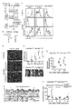
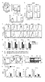
- FIG. 1 is a diagram showing that nonsense mutations in Clec10a in NC / Nga mice cause HDMI-induced dermatitis.
- Panel a shows the CleclOa gene, C57BL/6J CleclOa (NM_010796), NC/Nga-CleclOa c. 706T / T (c.706T / T), and NC / Nga-Clec10a c.
- the DNA sequence of the nonsense mutation site (c.706) in 706T / C (c.706T / C) mice is shown.
- the total length of Clec10a is represented by a plurality of white squares and a line penetrating them, the white squares represent the coding region of the gene, and the lines represent introns.
- CTLD stands for c-type lectin-like domain.
- Panel b shows a schematic diagram of each Clec10a of a C57BL / 6J mouse and an NC / Nga mouse.
- TM represents a transmembrane domain.
- Panel c is a C57BL / 6J mouse, NC / Nga-Clec10a c. 706T / T mouse, NC / Nga-Clec10a c.
- Macrophages (MP) (CD64 + MerTK +), conventional DC (cDC) (CD64-MerTK) in PI-CD45 + MHCII + Lineage (CD3, CD19, NK1.1, and Ly-6G) -EpCAM-cells of the dorsal skin of 706T / C mice. -), And the expression of Clec10a on the cell surface of monocyte-derived dendritic cells (CD64-MerTK lo ). The shaded histogram shows staining with an isotype control antibody (Ab). Panel d is a C57BL / 6J mouse, NC / Nga-Clec10a c.
- Panel e shows the dermatitis score
- panel f shows the appearance on day 14
- panels g and h show the tissue section (hematoxylin-eosin staining) and epithelial thickness, respectively.
- Panel i shows neutrophils (CD11b + Ly-6G + ), eosinophils (CD11b + SIGLEC-F + ), and Ly-6C hi monocytes (CD11b + Ly-6G - SIGLEC-F - Ly-6C hi). ) Indicates the total number of individuals.
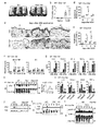
- FIG. 2 shows that Clec10a inhibits an HDMI-induced immune response.
- Panels a to f show the results of applying HDM ointment to the dorsal skin of C57BL / 6J wild-type mice and Clec10a ⁇ / ⁇ mice twice a week.
- Panels c and d show a comparison between wild-type and Clec10a ⁇ / ⁇ mice.
- Panel e shows flow cytometry that identifies neutrophils (CD11b + Ly-6G + ) in the skin CD45 + cells of wild-type and Clec10a ⁇ / ⁇ mice at specific time points.
- Panel f is the result of quantitative RT-PCR of mRNA obtained from cutaneous WT and Clec10a ⁇ / ⁇ MHCII+ MPs and DCs 3 hours after topical application of HDM.
- CBA cost-benefit bead array
- Panel h shows the results of stimulating wild-type, Trr4-/-, and Clec10a ⁇ / ⁇ BMMPs for the time specified by HDMI and then subjecting the lysates to immunoprecipitation (IP) with Clec10a antibodies. .. Immunoblot analysis (IB) was performed using antibodies against phosphorylated tyrosine (pTyr) or Clec10a.
- CBA cytometric bead array
- Panel j stimulated wild type or CleclOa-/- CD115+-enriched BMMPs with HDM for a specified time and then using phosphorylated Syk (pSyk; Y 519/520 ) or a monoclonal antibody to Syk (mAb).
- pSyk phosphorylated Syk
- mAb monoclonal antibody to Syk
- Panel k shows BMMPs transfected with wild type or Y3F Clec10a or empty vector (EV) transfected with HDM, cell lysates immunoprecipitated with mAb against Clec10a and against Syk, SHP-1 or Clec10a. The result of having immunoblotted with an antibody is shown.
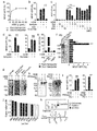
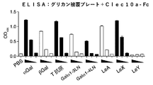
- FIG. 3 shows that Clec10a recognizes the mucin-like protein of HDMI.
- Panels a to d are rat IgG2a or anti-Clec10a mAb (panels a and b) or galactose (Gal), glucose (Glc) or mannose (Man) (panel c) in mouse Clec10a-CD3 ⁇ reporter cells or parent reporter cells. Expression of GFP after stimulation with an HDM coated plate in the presence or absence of); or after stimulation with a galactose (GALase) or glucosidase (GLCase) or untreated HDM coated plate (panel d). Indicates.
- Panels e-i are Clec10a ligands (Clec10a-L) (panel e) in HDM pulled down (PD) with Clec10a-Fc or control human antibody, each fraction based on the size of Clec10a-L (panel f), or 3 shows GFP expression in mouse CleclOa-CD3 ⁇ reporter cells or control reporter cells after stimulation with CleclOa-L (panel i) before and after treatment with PNGase F or NaOH.
- Statistical analysis was performed using PBS-stimulated samples used as controls (panel f).
- Clec10a-L is immunoblotted with Clec10a-Fc before (panels f-h) or post-treatment (panel h) with PNGase F or NaOH, or silver stained with or without alcyan blue (panel h).
- Panel j shows a lectin microarray analysis of Clec10a-L. The black bar indicates the lectin that binds to Gal ⁇ (1-3) GalNAc (T antigen). GalNAc means N-acetylgalactosamine and GlcNAc means N-acetylglucosamine.
- Panel k shows a schematic of HDM Clec10a-L.
- T means T antigen (Gal ⁇ (1-3) GalNAc)
- Tn means Tn antigen ( ⁇ GalNAc)
- LacNAc means N-acetyl-D-lactosamine (Gal ⁇ (1-4) GlcNAc).
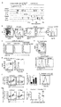
- FIG. 4 shows that human Asgr1 is the structural and functional counterpart of mouse Clec10a.
- Panel c shows the amino acid sequences of the intracellular regions of mouse CleclOa, human Asgrl, and human CleclOa. The putative hemITAM sequence is underlined.
- Panel d shows GFP expression in human Asgr1-CD3 ⁇ reporter cells stimulated with CleclOa ligand coated plates obtained by pulling down from HDM with mouse CleclOa-Fc.
- Panel e shows the results of staining tissue sections of human skin with anti-Asgr1 antibody, anti-CD68 mAb and DAPI.
- E means epidermis
- D means dermis.
- the scale bar is 100 ⁇ m.
- Panel g shows the association between ASGR1 expression (GSE5667) and serum IgE titers in the skin of healthy individuals, psoriasis patients, and atopic dermatitis patients.
- Panel h shows a hypothetical model for the role of C-type lectin receptors in maintaining skin homeostasis upon exposure to HDM in mice and humans.
- * represents p ⁇ 0.05, ** represents P ⁇ 0.01, and *** represents p ⁇ 0.001 (one-sided ANOVA test (panel b), independent two-sided Student's t-test ( Panel f) or two-tailed Pearson correlation test (panel g)).
- Data represent mean ⁇ SEM.
- FIG. 5 shows the results of feature analysis of Clec10a in C57BL / 6J and NC / Nga mice.
- Panel a shows the results of gene expression in hematopoietic cells and tissues of C57BL / 6J mice based on BioGPS analysis.
- Panels f and g show Flag-tagged Cle10a c. 706C - IRES -GFP or Flag-tagged Clec10a c. Surface analysis of transformants of RAW264.7 expressing 706T - IRES -GFP (panel f) and intracellular expression of Flag tag by flow cytometry (panel g) are shown. Panel h is NC/Nga Clec10a c. 706T/T (T/T), Clec10a c. 706 T / C (T / C), and Clec10a c.
- 706 C / C shows the expression of Clec10a mRNA in the skin of mouse.
- Panel j sets the serum IgE titer at the specified time point to Clec10a c. 706T/T (T/T), Clec10a c. The result of comparison with 706 T /C (T/C) is shown.
- FIG. 6 shows the phenotype of HDMI-induced dermatitis in Clec10a-deficient mice.
- Dust mites (HDM) ointment was applied to the dorsal skin of wild type C57BL/6J and CleclOa-/- mice twice weekly.
- Panel a shows the results of flow cytometry analysis of each cell population after staining the skin cells recovered at a specified time point with an antibody against PI, anti-CD45 antibody, or marker molecule.
- the markers for each cell were as follows: eosinophils (CD11b + Sigma-F + ), and Ly-6C hi monocytes (CD11b + Ly-6G - Sigma-F - Ly-6C hi ), CD3 + CD4 + T.
- Panel c is a quantitative representation of mRNA expression levels of the designated molecules in wild-type and CleclOa-/- type CD4+ T cells (CD3 + CD4 + ) sorted from axillary and inguinal lymph nodes at day 6.
- the results of RT-PCR are shown. ** represents P ⁇ 0.01 and *** represents p ⁇ 0.001 (independent two-sided Student's t-test). Data represent mean ⁇ SEM.
- FIG. 7 shows the results of characteristic analysis of wild-type and Clec10a ⁇ / ⁇ type MPs.
- Panel a shows MP (PI ⁇ CD45 + MHCII + Lineage (CD3, CD19, NK1.1, and Ly-6G) ⁇ EpCAM ⁇ CD64 + ) and DC obtained from mouse skin 3 hours after topical application of HDM.
- 2 shows a gate strategy for sorting (PI ⁇ CD45 + MHCII + Lineage ⁇ EpCAM ⁇ CD64 ⁇ ).
- Panel b shows a gate strategy for sorting CD115 + BMMP from wild-type and Clec10a ⁇ / ⁇ mice.
- Panel c shows the results of staining BMMPs from wild-type and CleclOa-/-mice with antibodies to CD115, CleclOa, the designated MP marker, and TLR4.
- CD115 + cells were gated and the expression of each molecule was analyzed by flow cytometry. Shaded histograms are stained with isotype control antibody.
- Panel e shows the amino acid sequence of the intracellular region of the wild-type and Y3F mutant Clec10a. The estimated hemITAM sequence is underlined.
- Panels f and g show the expression of Clec10a on the cell surface of BMMP transfected with cDNA or empty vector encoding wild-type and Y3F mutant Clec10a. Shaded histograms show staining with isotype control antibody (panel f). Transfected cells were stimulated with HDM, lysed, immunoprecipitated (IP) with anti-Clec10a antibody, and immunoblotted with anti-phosphorylated tyrosine (pTyr) and anti-Clec10a antibodies (panel g). Arrowheads indicate the molecule of interest (black) and the antibody used for IP (IP-Ab) (white).
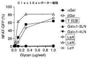
- FIG. 8 shows the establishment of Clec10a-CD3 ⁇ reporter cells and Clec10a-FC chimeric protein.
- Panels a and b are Clec10a after stimulation with a plate coated with Lewis X at the specified dose (panel a) and a plate coated with Lewis X (10 ⁇ g / mL) or Lewis Y (10 ⁇ g / mL).
- FIG. 9 shows the results of HDMI stimulation of human Clec10a-CD3 ⁇ reporter cells and the knockdown efficiency of human ASGR1.
- Panels a and b are humans after stimulation with HDM-coated plates in the absence (panel a) or presence (panel b) of galactose (Gal), glucose (Glc), or mannose (Man).
- 3 shows GFP expression in CleclOa-CD3 ⁇ reporter cells.
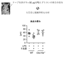
- FIG. 10 is a diagram showing that the Clec10a ligand (Clec10a-L) improves LPS-induced dermatitis.
- Panels a and b show tissue on day 5 after daily application of LPS to the back skin of C57BL / 6J wild-type (WT) and Clec10a ⁇ / ⁇ mice in the presence or absence of Clec10a-L. And epidermis thickening, respectively.
- Panel c shows neutrophils (CD45 + CD11b + Ly-6G + ) in the skin of WT and Clec10a ⁇ / ⁇ mice 6 hours after LPS was applied to the dorsal skin in the presence or absence of CleclOa-L. The number (/cm 2 ) is shown.
- FIG. 11 shows a scheme for determining Clec10a-L.
- FIG. 12 shows the results of an ELISA assay that examined whether Clec10a-Fc binds to a polymer scaffold-coated plate that presents the various glycans displayed.
- FIG. 13 shows the results of a reporter assay using Clec10a reporter cells to determine whether a polymeric scaffold presenting the various displayed glycans activates Clec10a.
- FIG. 14 shows the results of a therapeutic experiment in which the effect of administration of a T-presenting polymer scaffold on LPS-induced epidermal inflammation was examined.
- a "subject” can be a mammal, for example, pets such as dogs, cats, rabbits, hamsters, guinea pigs, and squirrels; livestock such as cows, pigs, horses, sheep, and goats; monkeys. Includes primates such as chimpanzees, orangutans, gorillas, bonobos, and humans.
- treatment is used in the sense of including therapeutic treatment and prophylactic treatment.
- treatment can be used to include suppression of deterioration of a disease or condition, delay of deterioration of a disease or condition, improvement of a disease or condition, or cure of a disease or condition.
- prophylaxis can be used to mean suppressing the onset of a disease or condition, or delaying the onset of a disease or condition.
- allergy means a systemic or local disorder to a living body based on an immune response. Allergies are broadly divided into allergies based on humoral immune response by blood antibodies (types I, II, and III) and allergies based on cell-mediated immunity by sensitized lymphocytes (type IV).
- Type I allergy is an allergy also called immediate type allergy or anaphylactic type. IgE is involved, and IgE binds to the IgE receptor (Fc ⁇ RI) that mast cells and eosinophils in blood and tissues have on the cell surface, and when an allergen binds to this, histamine, etc. from mast cells and eosinophils Is released, causing an allergic reaction (eg, smooth muscle contraction, vascular hyperpermeability, and glandular hypersecretion).
- Type I allergies include atopic bronchial asthma, allergic rhinitis, urticaria, allergic conjunctivitis, atopic dermatitis, and anaphylactic shock. In type I allergy, house dust, mites and the like are known to invade the body to cause an allergic reaction (oral, inhalation, transdermal, and intravenous routes may be used).
- Type II allergy is based on cytotoxicity caused by the reaction of IgG or IgM with cell and tissue antigens and the like, and complement binding to this.
- Type II allergy also includes antibody-dependent cellular cytotoxicity (ADCC), in which macrophages and killer cells having IgG Fc receptors bind to IgG bound to an antigen on the cell membrane and damage the cells.
- ADCC antibody-dependent cellular cytotoxicity
- Type II allergies include hemolytic anemia due to incompatible blood transfusion, autoimmune hemolytic anemia, idiopathic thrombocytopenic purpura, drug-induced hemolytic anemia, thrombocytopenia, thrombocytopenia, and Goodpasture disease group. Can be mentioned.
- Type III allergy also called immune complex type or Arthus type, is based on tissue damage caused by an immune complex (Immunocomplex) of a soluble antigen with IgG or IgM.
- Type III allergies include serum sickness, autoimmune diseases such as systemic lupus erythematosus and rheumatoid arthritis, glomerulonephropathy, hypersensitivity pneumonia, and allergic bronchopulmonary aspergillosis.
- Type IV allergy also called delayed allergy, is based on the reaction of sensitized T cells with antigens, which releases cytokines from the sensitized T cells and causes cytotoxicity. It is also based on damage to virus-infected cells, tumor cells, and grafts by killer T cells.
- Type IV allergies include allergic contact dermatitis, atopic dermatitis, hypersensitivity pneumonitis, tuberculous cavities, clefts, epithelioid cell granulomas lesions of sarcoidosis, smallpox rash, and measles rash.
- asialoglycoprotein receptor refers to a glycoprotein in which the sialic acid at the terminal end of the sugar chain of the protein has been removed and the galactose residue inside is exposed as a terminal group, that is, asialoglycoprotein It is a receptor that binds to AGP).
- the asialoglycoprotein receptor is present on the surface of hepatocytes and binds to AGP in blood to remove AGP from blood.
- Asialoglycoprotein receptor 1 (ASGR1) is also referred to as type C lectin domain family member H1 or CLEC4H1.
- a representative example of a human ASGR1 protein may be a protein having an amino acid sequence registered with GenBank under accession number CAG46849.1.
- ASGR1 is used to include the ortholog of human ASGR1.
- Clec10a is also referred to as C-type lectin domain family 10, member A, and is a molecule that recognizes a sugar chain and functions as a biological defense system of a host. Clec10a can specifically bind to galactose or N-acetylgalactosamine.
- the "house dust mite” is a mite of the genus Dermatophagoides.
- the main species of dust mites are Dermatophagoides farinae, Dermatophagoides microceras, Dermatophagoides mediterraneus pteronyus, and Dermatophaeneys pteronysphaerus.
- the term “antigen” means a substance that provides an epitope to which a lectin can react in the case of an antibody or sugar.
- “antigen” means, according to its ordinary meaning, a sugar chain that provides an epitope with which a lectin can react. Therefore, the term "antigen" in the context of glycans means providing the glycans to the surface of the glycans as an epitope with which the lectin can react, as is found in natural glycans and glycoproteins. To do.
- the "ligand” refers to a substance to which a receptor binds.
- the ligand may regulate a downstream signal of the receptor by binding to the receptor.
- a substance that positively regulates a downstream signal of a receptor is called an "agonist”.
- a substance that negatively regulates a downstream signal of a receptor is called an "antagonist”.
- GalNAc means N-acetylgalactosamine
- GlcNAc means N-acetylglucosamine
- Clec10a is involved in the onset and exacerbation of dermatitis caused by dust mites in mice, and Asgr1 is involved in humans.
- the inventors also revealed from the results of functional analysis and homology analysis that human Asgr1 is the original structural and functional counterpart of mouse Clec10a.
- dust mites contain substances that bind to and activate mouse Clec10a and human Asgr1 and suppress the development of allergies (eg, Clec10a ligand or Asgr1 ligand).
- the ligand of Clec10a contains an O-linked sugar chain, particularly T antigen (Gal ⁇ (1-3) GalNAc) and Tn antigen ( ⁇ GalNAc), and ASGR1 binds to any of these. Revealed.
- the present inventors have also revealed that the CleclOa ligand suppresses TLR4 signaling.
- the T antigen (Gal ⁇ (1-3) GalNAc) and the Tn antigen ( ⁇ GalNAc) are sugar chains having the structures shown in the schematic diagram shown in the following formula (I), respectively.
- the binding of Asgr1 to the T and Tn antigens, respectively, is consistent with Asgr1's affinity for galactose or N-acetylgalactosamine.
- Asgr1 binds to a sugar chain selected from the group consisting of T antigen, LeA, and LeX, and thereby transmits a signal into the cell.
- sugar chains may be presented on a polymeric scaffold.
- sugar chains are polylactic acid, polyacrylamide, polyvinyl, polyvinyl alcohol, polymethyl methacrylate, polyacrylonitrile, polystyrene, polypropylene, polyethylene terephthalate, nylon, collagen, hydroxyethyl methacrylate, chitosan, chitin, polyethylene oxide, polyethylene glycol.
- Polyamino acids, polylactides, polyglycolides, polycaprolactones, and copolymers thereof may be linked on the side chains of a polymeric scaffold having as a backbone and presented to Clec10a.
- each polymer is not particularly limited as long as it has manageable physical properties (for example, viscosity, osmotic pressure, etc.), but for example, it has a weight average molecular weight of 1 kDa to 100 kDa, 5 kDa to 50 kDa, 10 kDa to 40 kDa, or 20 to 40 kDa.
- a biocompatible polymer having a can be used as a backbone.
- a nonsense mutation or frameshift mutation into Clec10a, the mouse counterpart of asialoglycoprotein receptor 1 (Asgr1), resulted in an excessive response to HDM.
- an extract (purified product) containing a CleclOa ligand was prepared and contacted with human Asgr1-CD3 ⁇ reporter cells, Asgrl responded to the CleclOa ligand in a concentration-dependent manner. This activation was also concentration-dependently counteracted by the addition of galactose to the system.
- the asialoglycoprotein receptor 1 (Asgr1) ligand can be used in the treatment of diseases or symptoms (eg, inflammation and allergic diseases) induced by TLR4 signals such as dust mite-induced allergies.
- TLR4 signals such as dust mite-induced allergies.
- the present invention provides a composition for use in treating an allergic disease, which comprises a ligand of asialoglycoprotein receptor 1 (Asgr1).
- a composition comprising a ligand for the asialoglycoprotein receptor 1 (Asgr1) for use in the treatment of diseases or symptoms (eg, inflammation and allergic diseases) induced by activation of TLR4 signaling.
- the TLR signal can be activated by the TLR4 ligand.
- TLR4 ligands include lipopolysaccharide (LPS) and lipoteichoic acid and agonists that are analogs thereof. Therefore, the ligand of asialoglycoprotein receptor 1 (Asgr1) can be used for the treatment of diseases or conditions (eg, inflammation and allergic diseases) induced by allergens containing these TLR4 ligands. Therefore, according to the present invention, a composition for use in treating a disease or symptom induced by an allergen containing a TLR4 ligand, which comprises the ligand for asialoglycoprotein receptor 1 (Asgr1). Provided.
- LPS lipopolysaccharide
- Asgr1 lipoteichoic acid and agonists that are analogs thereof. Therefore, the ligand of asialoglycoprotein receptor 1 (Asgr1) can be used for the treatment of diseases or conditions (eg, inflammation and allergic diseases) induced by allergens containing these TLR4 ligands. Therefore,
- Examples of the ligand for the asialoglycoprotein receptor 1 include a sugar chain in which sialic acid is released and has LewisX at the end, and a protein having the sugar chain.
- Examples of the ligand of the asialoglycoprotein receptor 1 (Asgr1) include a sugar chain in which sialic acid is released and has a T antigen or Tn antigen at the end, and a protein having the sugar chain.
- T antigen means Gal ⁇ (1-3)GalNAc
- Tn antigen means ⁇ GalNAc.
- the ligand for the asialoglycoprotein receptor 1 As a ligand for the asialoglycoprotein receptor 1 (Asgr1), sialic acid is released, and 1, 2 or all O-linked sugars selected from the group consisting of Lewis X antigen, T antigen, and Tn antigen at the end. It can be a chain or a protein having the sugar chain.
- the mucin-like protein has the above-mentioned O-linked sugar chain, and has a sugar chain selected from the group consisting of Lewis X antigen, T antigen, and Tn antigen.
- the ligand for asialoglycoprotein receptor 1 can be a mucin-like protein or mucin.
- the protein as a ligand for asialoglycoprotein receptor 1 (Asgr1) may be a mucin-like protein or mucin.
- Asgr1 is highly expressed in hepatocytes, and the aged protein (asiaroglycoprotein) desialylated in the body is taken into the hepatocytes and removed from the blood. Therefore, all such desialylated glycoproteins can be used as ligands for the asialoglycoprotein receptor 1. Asgr1 binds to and reacts with sugar chains having at least one or both of T antigen and Tn antigen. Therefore, a sugar chain having at least one or both of the T antigen and the Tn antigen or a protein having the same can be used as a ligand for the asialoglycoprotein receptor 1.
- All allergen-containing substances can also be obtained by affinity purification based on binding affinity with Clec10a (eg, mouse Clec10a or human Asgr1). it can. Elution of the Clec10a ligand from Clec10a can be performed using, for example, galactose. The eluate of CleclOa ligand may be dialyzed against saline.
- a fusion protein containing the extracellular region, transmembrane region, and intracellular region of CD3 ⁇ of human asialoglycoprotein receptor 1 transmitted the CD3 ⁇ signal downstream in the presence of the ligand of asialoglycoprotein receptor 1. Therefore, a fusion protein containing the extracellular region of human asialoglycoprotein receptor 1, transmembrane region, and intracellular region of CD3 ⁇ can be identified as a substance to be used for treating allergic diseases. For example, an animal cell expressing a fusion protein containing an extracellular region of human asialoglycoprotein receptor 1, a transmembrane region, and an intracellular region of CD3 ⁇ , which operates with a promoter (eg, NFAT promoter) activated by a CD3 ⁇ signal.
- a promoter eg, NFAT promoter
- the test compound is a compound that binds to human asialoglycoprotein receptor 1 by contacting the compound with an animal cell having a gene encoding a reporter that is operably linked.
- the obtained substance is a substance to be used for treating an allergic disease by using regulatory signaling via inhibitory TAM as an index.
- the compound human Asgr1 ligand candidate compound
- the compound can be applied to LPS-induced dermatitis to confirm the dermatitis suppressing effect.
- the present invention provides a fusion protein containing an extracellular region of human asialoglycoprotein receptor 1, a transmembrane region, and an intracellular region of CD3 ⁇ , and an animal cell expressing the fusion protein.
- the animal cell may also have a gene encoding a reporter operably linked to a promoter activated by the CD3 ⁇ signal.
- the present invention further provides an animal cell that expresses a fusion protein containing an extracellular region of human asialoglycoprotein receptor 1, a transmembrane region, and an intracellular region of CD3 ⁇ , which is operably linked to a promoter activated by a CD3 ⁇ signal.
- test compound may comprise a ligand for the asialoglycoprotein receptor 1 (Asgr1).
- test compound may be at least one sugar chain in which the ligand is selected from the group consisting of T and Tn antigens.
- the ligand can be a mucin-like protein or mucin.
- the ligand can be a ligand for human asialoglycoprotein receptor 1.
- An animal cell expressing a fusion protein comprising the extracellular region of human asialoglycoprotein receptor 1 of the present invention, a transmembrane region and an intracellular region of CD3 ⁇ , which is a reporter operably linked to a promoter activated by a CD3 ⁇ signal.
- the animal cell having the gene encoding the can also be used for compound screening. Therefore, in the present invention, it is an animal cell that is a screening method for a human Asgr1 ligand or agonist and expresses a fusion protein containing an extracellular region, a transmembrane region, and an intracellular region of CD3 ⁇ of human asialoglycoprotein receptor 1.
- test compound is indicated as a candidate for a human Asgr1 ligand or agonist.
- the animal cell may preferably be a human cell.
- composition of the present invention may be a composition such as a personal care composition and a pharmaceutical composition.
- the pharmaceutical composition examples include pharmaceutical compositions for topical administration, which can be used in the present invention.
- the pharmaceutical composition for topical administration may be a pharmaceutical composition for mucosal application or body surface application, for example, eye drops, eye ointment, sublingual tablet, buccal tablet, troche, mouthwash, spray. , Aerosols, and inhalants; Solution formulations such as liquids, salves, glycerin, alcohol, liquids, and coatings; dispersions such as emulsions, suspensions, liniments, lotions, sprays, and liposomes.
- Formulations Semi-solid formulations such as ointments, plasters, patches, adhesive tapes, pasta, paps, creams, oils, and sticks; and extracts (soft extracts, dry extracts), and Examples include leachate preparations such as tinctures.
- the pharmaceutical composition may contain a pharmaceutically acceptable excipient.
- Pharmaceutically acceptable excipients include solvents, bases, diluents, bulking agents, fillers, and shapers; solubilizers, solubilizers, buffers, isotonic agents, emulsifiers, suspensions. Turbidants, dispersants, thickeners, gelling agents, hardeners, absorbents, adhesives, elastics, plasticizers, sustained release agents, and propellants; antioxidants, preservatives, moisturizers, shading Examples include agents, antistatics, fragrances, flavoring agents, colorants, and palliatives.
- Examples of personal care compositions include skin care, antiperspirants, deodorants, beauty products, cosmetics, and hair care products.
- Personal care compositions include moisturizers, conditioners, anti-aging agents, whitening agents, sunscreens, antiperspirants, shaving compositions, post-shaving compositions, foundations, lipsticks, lipsticks, hair styling products, shampoos, etc. Examples include cleaning agents and lubricants.
- the personal care composition may be used in personal care products. Examples of personal care products include underwear, diapers, tissues, wipes, masks, patches and the like.
- the composition may include an excipient in addition to the active ingredient.
- the composition can be in a dosage form suitable for administration forms such as intravenous administration, transdermal administration, oral administration, enteral administration, and intraperitoneal administration.
- the compositions of the invention may be administered by transdermal administration, in the form of, for example, gels, emulsions, creams, liquids, pastes, lotions, liposome creams and the like (eg, skin). Scientific composition).
- the composition can be an ointment.
- a dermatologically acceptable excipient can be used as an excipient, and a dosage form suitable for this can be used.
- transmucosal administration an excipient that is acceptable for mucosal application can be used, and a dosage form suitable for this can be used.
- an Asgr1 ligand in the manufacture of a composition for use in treating an allergic disease is provided.
- a method for treating an allergic disease in a subject in need thereof comprising administering to the subject an effective amount of an Asgr1 ligand.
- a method for preventing an allergic disease, or suppressing the occurrence thereof comprises administering to the subject an effective amount of Asgr1 ligand.
- Method (1) Preparation of skin cells Skin cells are minced by sandwiching a thinly ground dorsal skin sample and containing 200 U / mL Collagenase II, 50 U / mL DNase and 10% fetal bovine serum (FBS). Was incubated in RPMI-1640 medium for 60 minutes. Further dissociation and homogenization were performed using the gentleMACS Dissociator (Miltenyi Biotec). The obtained cells were filtered through a 55 ⁇ m nylon mesh to obtain a single cell suspension.
- FBS fetal bovine serum
- immunohistochemical staining skin samples taken from mice were embedded in Tissue Tissue Optimal Cutting Tissue Company Compound (Sakura Finetek Japan) and stored at -80 ° C. A 4 ⁇ m thick section was also used for immunohistochemical staining.
- the cells were washed with PBS (PBS-T, pH 7.4) containing 0.05% Tween-20, soaked, and incubated with Blocking One Histo (Nacalai) for 10 minutes at room temperature.
- the sections were then washed with PBS-T, incubated overnight with anti-Clec 10a mAb at 4 ° C., washed with PBS-T and incubated with Alexa Fluor 546-labeled anti-rat IgG polyclonal antibody for 1 hour. After washing with PBS-T, sections were stained with DAPI nuclei. Healthy human skin tissue was collected from the periphery of the patient's tumor area, fixed with formalin, and embedded in paraffin to make 4 ⁇ m thick sections.
- Sections were deparaffinized with xylene, rehydrated with ethanol and blocked with methanol to block endogenous peroxidase. Sections were stained with anti-CD69 antibody and anti-Asgr1 antibody according to the manufacturer's manual for the Opal 4-Color Automation IHC Kit (PerkinElmer, NEL820001KT). Briefly, sections are incubated at 95 ° C. for 15 minutes, washed with TBS (TBS-T) (Takara Bio, T9142) containing 0.05% Tween-20, and treated with a blocking solution at room temperature for 10 minutes. did. Sections were incubated overnight at 4 ° C.
- Serum IgE antibody was measured using a capture antibody against mouse IgE (R35-72), biotinylated anti-mouse IgE (R35-118), and subsequent HRP-labeled streptavidin (GE Healthcare, RPN1231V). .. Purified mouse IgE (C38-2, BD Biosciences) was used as a standard. Serum IgG1 was measured using a capture antibody against mouse IgG1 (A85-3) and an HRP-labeled antibody against mouse IgG1. Purified mouse IgG1 (107.3, BD Biosciences) was used as a standard. Serum IgG2c was detected by mouse ELISA Quantification Set (Bethyl, E90-136).
- BMMPs bone marrow macrophages
- GM-CSF R&D Systems
- IL-4 Wako
- Cultured below in RPMI 1640 complete medium with 10% FBS.
- 70% of non-adherent cells were removed and fresh medium containing GM-CSF and IL-4 was added.
- 100% non-adherent cells were removed by washing with PBS and fresh medium containing GM-CSF and IL-4 was added.
- all non-adherent cells were removed by PBS washing and the adherent cells were used for later experiments.
- CD115 + BMMP was concentrated using anti-CD115 antibody (BioLegend) and anti-rat IgG microbeads (Miltenyi Biotec).
- Skin MP is CD45 + MHCII + CD3 - CD19 - NK1.1 - Ly-6G - EpCAM - CD64 sorted by +, DC is, CD45 + MHCII + CD3 - CD19 - NK1.1 - Ly-6G - EpCAM - CD64 - ) Sorted.
- BMMP was infected on days 2 and 4 with virus supernatant supplemented with polybrene (8 ⁇ g / mL, Sigma-Aldrich). After centrifugation, the supernatant containing the virus was removed and replaced with fresh BMMP medium. On day 5, the medium was replaced with fresh BMMP medium, and on day 7, non-adherent cells were removed by PBS washing and adherent cells were used in the experiment.
- CleclOa-Fc Chimera A mouse CleclOa chimeric construct (CleclOa-Fc) was prepared by cloning the extracellular region of mouse CleclOa into a pME18S expression vector containing the Fc region of human IgGl. Clec10a-Fc was transfected into HEK293T cells using Lipofectamine 2000 (Thermo Fisher Scientific) and then the medium was replaced with GIT medium (KOHJIN BIO). Clec10a-Fc was purified from culture supernatant using protein A agarose (Bio-Rad Laboratories).
- BMMP was stimulated with HDM extract (100 ⁇ g/mL) at 37° C. for 0 minutes, 10 minutes, or 30 minutes. Stimulated BMMP was dissolved in 1% NP-40 lysis buffer and separated by SDS-PAGE. It was transcribed on a PVDF membrane by electroblot, immunoblotted with anti-phosphorylated Syk antibody and anti-Syk antibody, and detected with HRP-labeled anti-rabbit IgG antibody. To analyze tyrosine phosphorylation of Clec10a, BMMP was stimulated with HDMI extract (100 ⁇ g / mL) at 37 ° C. for 0, 10 or 30 minutes.
- HDMI extract 100 ⁇ g / mL
- BMMP Stimulated BMMP was dissolved in 1% NP-40 lysis buffer and subjected to immunoprecipitation with anti-Clec 10a mAb. Immunoprecipitates were developed by SDS-PAGE, transcribed on PVDF membrane and immunoblotted with HRP-labeled anti-phosphotyrosine antibody or anti-CleclOa antibody and HRP-labeled anti-rabbit IgG antibody. To analyze the association of CleclOa with Syk or SHP-1, BMMPs were pretreated in the presence or absence of 5 mM Syk inhibitor IV (Merck, 574714) for 30 minutes at 37°C and extracted with HDM extract. (100 ⁇ g / mL) was stimulated at 37 ° C.
- Syk inhibitor IV Merck, 574714
- the Clec10a ligand is 0 after 16 hours of pretreatment at 37 ° C. and 5 minutes of thermal denaturation at 95 ° C.
- peptide N-glycosidase F peptide N-glycosidase F
- Pretreatment at 40 ° C. for 16 hours in the presence or absence of .05 M NaOH then separated by SDS-PAGE and transferred to PVDF membranes, biotinylated Clec10a-Fc and HRP. Immunoblots were performed using labeled streptavidin.
- Reporter cells are in the presence or absence of anti-Clec 10a mAb, Lewis X (GlycoTech), Lees Y (GlycoTech), galactose (Sigma-Aldrich), glucose (Sigma-Aldrich) or mannose (Sigma-Aldrich). Incubated. Reporter cells were also treated with or not treated with galactosidase (R & D Systems, 5704-GH or 5549-GH), or glucosidase (R & D Systems, 8329-GH), a Clec10a ligand-coated plate, or a size-fractionated Clec10a ligand. The cells were cultured on a treated HDMI extract coated plate. Activation of NFAT-GFP was monitored by flow cytometry.
- Lectin microarrays were prepared by a non-contact microarray printing robot (MicroSys 4000; Genomic Solutions) according to the conventional method. Samples were fluorescently labeled with Cy3 Mono-Reactive dye (GE) and excess Cy3 was removed using a Sephadex G-25 desalting column (GE). 10 times with a probe solution (25 mM Tris-HCl, pH 7.5, 140 mM NaCl) (TBS) containing 2.7 mM KCl, 1 mM CaCl 2 , 1 mM MnCl 2 , and 1% Triton X-100.
- GE Cy3 Mono-Reactive dye
- TBS Sephadex G-25 desalting column
- Gal D-galactose
- GalNAc N-acetyl-galactosamine
- GlcNAc N-acetyl-glucosamine
- Fuc L-fucose
- Sia Sialic acid
- LacNAc N-acetylamine
- JOM J-OIL MILLS, INC
- Vector VECTOR LABORATORIES
- Seikagaku SEIKAGAKU CORPORATION
- EY EY LABORATORIES, INC
- AIST AIST
- Example 1 Exome analysis of atopic dermatitis model mouse
- NC / Nga mouse which is a model mouse of atopic dermatitis
- the NC/Nga mouse was purchased from Charles River, Japan. DNA was extracted from blood obtained from NC / Nga mice under conditions suitable for extraction of mouse DNA using QIAamp DNA blood Mini Kit (QIAGEN, Venlo, Netherlands). The exome analysis of the obtained DNA was performed as follows.
- the SureSelect Library prep kit post-pool version 4; Agilent Technologies, Santa Clara, CA) and the SureSelect DNA Manual All Exon Kit manufactured by the SureSelect Mouse Manual (Agilent), and the technicians using the Allexon Kit (Agilent Tech).
- the obtained DNA library was subjected to emulsion PCR (SOLiD EZ Bead Emulsifier kit; Thermo Fisher Scientific, Waltham, MA) to generate clonal DNA fragments on beads, and then subjected to bead enrichment (SOLiD EZ. Bead Emulsion kit; Thermo Fisher Scientific, Waltham, MA).
- SOLiD EZ. Bead Emulsion kit Thermo Fisher Scientific, Waltham, MA
- the resulting beads were sequenced on a SOLiD 5500xl sequencer as single-ended 60 bp reads.
- the obtained read was aligned with the mouse reference genome (NCBI37 / mm9) using LifeScop version 2.5.1 (Life Technologies) to obtain a BAM file.
- Variant calls were performed using SAMtool according to the protocol described in Genome Analysi Toolkit, Picard (http://broadinstitute.github.io/picard), and only leads mapped to unique positions on the reference genome were analyzed. Using. Mutation sites were annotated using ANNOVER software. Genetic variation in inbred mouse strains other than NC/Nga was demonstrated by release REL-1211 (http://www.sanger.ac.uk/science/data/mouse-genomics-project) numbered according to the NCBIm37 assembly. Obtained from Mouse Genome Information website (http://www.infomatics.jax.org/). The sequence was carried out on ABI 3130xl Genetic Analyzer (Thermo Fisher Scientific) using BigDye Terminator v1.1 Cycle Sequencing Kit (Thermo Fisher Sequencing). The primers used are shown in Table 4.
- 70,772 mutations were identified in the exon region of the NC/Nga mouse genome relative to the reference genome. Mutations with low quality values obtained by GATK output were removed to give 64,518 mutations. Loss of function mutations, including nonsense and frameshift mutations, were selected using ANNOVER software. Then, 35 nonsense mutations and 48 frameshift mutations (inducing stop codons) were identified. For each of these 83 genes, 46 mutations in 43 genes were selected for further sequence validation.
- Clec10a was selected from seven genes based on the information in the BioGPS database (see panel a in FIG. 5).
- Clec10a of NC / Nga mice was a nonsense mutation (ie, Q236X) in which C at position 706 in the nucleotide sequence of NM_010769 was T.
- Example 2 Expression analysis and functional analysis of Clec10a
- Clec10a is a member of the type II C-type lectin receptor family and detects galactose moieties at the ends of foreign and endogenous antigens.
- CleclOa c. in NC/Nga mice c. 706C> T is present in the coding region of the C-type lectin-like domain (panels a and b in FIG. 1).
- Clec10a showed that in skin CD45 + MHCII + lineage ⁇ EpCAM ⁇ cells, macrophages (MP) (CD64 + MerTK + ), conventional dendritic cells (cDC) (CD64 ⁇ MerTK ⁇ ), And it was revealed that it was expressed on the cell surface of mononuclear cell-derived DC (CD64 - MerTK lo ) and on the macrophages of the peritoneum of C57BL / 6J (it was not expressed in the peritoneum of NC / Nga mice). (See panel c of FIG. 1 and panels b and e of FIG. 5).
- C57BL / 6J mice of the non-hematopoietic cells of the skin CD45 -
- CD45 + MHCII- cells CD45 + MHCII + (Lin, EpCAM) + cells
- neutrophils CD45 + CD11b + Ly-6G +
- Clec10a was not observed (see panels c and d in FIG. 5). Similar results were obtained by immunohistochemical staining, and expression of Clec10a was observed in the skin cells of C57BL / 6J mice, but not in NC / Nga mice (panel d in FIG. 1). ..
- Clec10a expression in NC / Nga mice were further analyzed. Specifically, the cDNA of Clec10a (derived from C57BL / 6J mouse and derived from NC / Nga mouse) having a flag tag and a sequence encoding IRES-GFP at the 3'end was transfected into RAW264.7 macrophages. The Flag tag was expressed on the cell surface of macrophages expressing Clec10a derived from C57BL / 6J mice, but not on the cell surface of macrophages expressing Clec10a derived from NC / Nga mice. (See panel f in FIG. 5).
- Clec10a c A mutant mouse having 706C (hereinafter, sometimes referred to as "NC/Nga mouse CleclOa c.706T>C ") was prepared by a conventional method using the CRISPR/Cas9 system (see panel a in Fig. 1 ). Clec10a c. In the skin of mutant mice with 706C, mRNA expression Clec10a is, Clec10a c. There was a significant increase compared to mice with 706T (see panel h in FIG.
- Clec10a-deficient mice (Clec10a ⁇ / ⁇ mice) were prepared against the background of C57BL/6J mice.
- HDM was topically applied to the dorsal skin of wild type C57BL/6J mice and Clec10a deficient mice (Clec10a ⁇ / ⁇ ).
- CleclOa-/- mice showed dermatitis such as erythema, dryness and enhancement of skin at 5 to 6 days after HDM treatment as compared with wild type mice. Exacerbation was shown (see Figure 2 panels ad).
- CleclOa-/- mice had increased neutrophil infiltration in the skin 1 to 3 days after HDM treatment compared to wild-type mice (see Figure 2 panel e).
- the numbers of cells of other bone marrow and lymphocyte lineages were comparable in wild type and CleclOa-/- mice (see Figure 6 panel a).
- Clec10a is expressed on skin MHCII + macrophages (MP) and dendritic cells (DC), these cells were sorted from skin cell samples of wild-type and Clec10a ⁇ / ⁇ mice topically treated with HDM ( See panel a in FIG. 7).
- the gene expression levels of Il6, Cxcl1, and Cxcl2 were higher in Clec10a ⁇ / ⁇ MP than in wild-type MP, but in DC, these gene expression levels were similar between wild-type and Clec10a ⁇ / ⁇ (in DC). See panel f in FIG. 2).
- Bone marrow macrophages were prepared and characterized from wild type and CleclOa-/- C57BL/6J mice, respectively (see panel b in Figure 7). Expression levels of MP markers and TLR4 in CD115 + BMMP were comparable in wild-type and Clec10a ⁇ / ⁇ mice. After HDM stimulation in vitro, Clec10a ⁇ / ⁇ BMMP secreted more neutrophil chemotactic substances such as IL-6, CXCL1, CCL3 and TNF- ⁇ than wild-type BMMP (Fig. 2). See panel g). Similarly, Clec10a c.
- the BMMP of 706 T / T NC / Nga mice was described in Clec10a c. It showed greater cytokine production than 706 T / C BMMP (see panel d in FIG. 7). These results demonstrate that cell surface expression of Clec10a inhibits the production of inflammatory cytokines from skin MP after stimulation with HDM.
- HDMI stimulation causes activation of splenic tyrosine kinase (Syk) in wild-type BMMP, but no activation of Syk was observed in Clec10a ⁇ / ⁇ BMMP (see panel j in FIG. 2).
- HDMI stimulation evoked recruitment of Syk and the protein tyrosine phosphatase SHP-1 to Clec10a, which was dependent on the tyrosine residue of Clec10a (see panel k in FIG. 2).
- BMMP treatment with a Syk inhibitor suppressed HDMI-induced recruitment of SHP-1 to Clec10a (panel l in FIG. 2), which means that the event is dependent on Syk activation. Suggests. This result is consistent with the control signaling via inhibitory ITAM.
- Example 3 Analysis of Clec10a Ligand
- a chimeric fusion protein containing the extracellular and transmembrane portions of Clec10a fused to the cytoplasmic portion of CD3 ⁇ was tested.
- NFAT-GFP reporter cells (Clec10a reporter cells) to be expressed were prepared.
- Mouse CleclOa reporter cells expressed GFP in response to Lewis X, an oligosaccharide ligand of CleclOa, but not Lewis Y (see Figure 8 panels a and b).
- mouse Clec10a reporter cells expressed GFP in response to the HDMI coated plate in a dose-dependent manner (panel a in FIG. 3).
- Clec10a ligand Clec10a-L
- a chimeric fusion protein containing the extracellular portion (Clec10a-Fc) of mouse Clec10a fused with the Fc portion of human IgG1 was prepared, and this fusion protein is Lewis. It was not bound to Y but to Lewis X (panel c in FIG. 8).
- a pull-down assay from HDMI using Clec10a-Fc isolated Clec10a-L with a molecular weight of approximately 225 kDa, which was able to induce GFP expression in mouse Clec10a reporter cells (FIG. 3). Panel e and f).
- CleclOa-L was stained with Alcian blue and silver, but not silver alone (see panel g in Figure 3), indicating that CleclOa-L is a highly glycosylated protein.
- Treatment with NaOH to degrade O-linked glycans inhibited Clec10a-Fc from binding to the epitope (see panel h in FIG. 3) and also inhibited induction of GFP expression in mouse Clec10a reporter cells (see panel h in FIG. 3). See panel i in FIG. 3).
- PNGase F which decomposes N-linked glycans, showed no effect on CleclOa-L (see panels h and i in Figure 3).
- Clec10a recognizes the O-linked glycan of Clec10a-L.
- Clec10a-L contains T antigen (Gal ⁇ (1-3) GalNAc) and Tn antigen ( ⁇ GalNAc), and LacNAc epitope that binds to mucin-type O glycan (Gal ⁇ (1-4)). It was shown to have GlcNAc) (panel j in FIG. 3 and Table 6).
- Control 1 Pull-down assay sample from buffer solution using Clec10a-Fc 3
- Control 2 Pull-down assay sample from HDMI using human IgG1
- Clec10a is bound most strongly to a lectin which recognizes a multivalent T and Tn antigen of high-density Makurura-Pomifera (Maclura pomifera (MPA)).
- Maclura pomifera MPA
- analysis by glycan microarray revealed that Clec10a-Fc binds to T and Tn antigens (see Table 7), and it was suggested that Clec10a-L is a mucin-like protein (panel in FIG. 3). k)).
- Example 4 Relationship between human Asgr1 and mouse Clec10a
- BLAST Basic Local Alignment Search Tool
- NCBI National Center for Biotechnology Information
- the mouse Clec10a is a tool for analyzing homology of amino acid sequences.
- human proteins that showed the highest homology in the amino acid sequence of the C-type lectin-like domain (CTLD) were human Asgr1 and Clec10a (encoded by Gene IDs: 432 and 10462, respectively).
- Human Asgr1 reporter cells and human Clec10a reporter cells expressed GFP in response to HDM stimulation see panel a in FIG. 4 and panel a in FIG.
- C-type lectin receptors such as Asgr1 in humans and Clec10a in mice recognize mucin-like proteins in HDM and play an important role in maintaining skin homeostasis against inflammation induced by HDM. Understood (see panel h in Figure 4).
- Example 5 Treatment of Allergic Symptoms with CleclOa Ligand From the results of the above Examples, CleclOa (Asgr1 in humans) is involved in the control of HDM-induced allergic symptoms, and ligand stimulation to CleclOa (Asgrl in humans) was observed. It was shown to be involved in the suppression of allergic symptoms. In this example, allergic symptoms were treated with a ligand for Clec10a and the results were observed.
- TLR4-induced dermatitis is induced.
- tissue sections were prepared by a conventional method and stained with hematoxylin and eosin. Then, the epidermal thickening and the number of neutrophils in the skin were counted.
- the panel b of FIG. 10 also showed the same result as the panel a of FIG.
- Neutrophil infiltration into the skin tissue was observed by a conventional method.
- neutrophil infiltration was observed in the LPS-treated group in wild-type (WT) mice, but in the group treated with LPS plus CleclOa ligand. Neutrophil infiltration was suppressed.
- Clec10a ⁇ / ⁇ mice neutrophil infiltration could not be suppressed by the Clec10a ligand. From these results, it was clarified that the Clec10a ligand exhibits an inhibitory effect on dermatitis.
- HDMI is known to contain TLR4 ligands, which are thought to induce inflammatory and allergic symptoms such as dermatitis. Also in HDM stimulation, Clec10a ligand binds to Clec10a (Asgr1 ligand binds to Asgr1 which is its functional counterpart in humans), and suppressing the TLR4 signal results in reducing inflammatory and allergic symptoms. Conceivable.
- Example 6 Detection of Human Asgr1 Ligand
- the binding profile of CleclOa-L to the lectin was determined by lectin array to predict the glycan structure in CleclOa-L (upper left panel of Fig. 11) and CleclOa.
- a scheme (upper right panel of FIG. 11) is shown in which the binding profile of glycans to is determined by the glycan array.
- FIG. 11 these results are considered together, and it is shown that five glycans ( ⁇ Gal, ⁇ Gal, T antigen, LeA, and LeX) can be identified as Clec10a ligand candidates.
- Gal ⁇ 1-3LN and Gal ⁇ 1-4LN were added to identify seven Clec10a ligand candidates.
- a polymeric scaffold (with a modification rate of 20% as described in the above formula) that presents glycans at the position of “Sugar” in the above formula.
- the PBS solution containing the wells was introduced into a 96-well plate, the wells were coated with glycans by incubating overnight at room temperature, and then the unbound polymer was washed. Then, a 10% fetal bovine serum-containing PBS solution (50 ⁇ L / well) containing 1 ⁇ g / ⁇ L of Clec10a-Fc was added to the glycan-coated well.
- T antigen which is one of them, can suppress skin inflammation against lipopolysaccharide (LPS).
- Polymeric scaffolds presenting 50 ⁇ g LPS and 20 ⁇ g T antigen were injected intradermally daily on the backs of mice, and epidermal thickness was observed on day 5. The result was as shown in FIG. As shown in FIG. 14, the increase in skin thickness by LPS was suppressed by T antigen.
- the thickness of the epidermis was equal to or greater than that of WT.
- the effect of suppressing the thickness of the epidermis by the T antigen was not observed in the Clec10a knockout mouse, it was clarified that the effect of suppressing the inflammation by the T antigen was mediated by Clec10a.
Landscapes
- Health & Medical Sciences (AREA)
- Life Sciences & Earth Sciences (AREA)
- Chemical & Material Sciences (AREA)
- Engineering & Computer Science (AREA)
- Organic Chemistry (AREA)
- General Health & Medical Sciences (AREA)
- Medicinal Chemistry (AREA)
- Bioinformatics & Cheminformatics (AREA)
- Animal Behavior & Ethology (AREA)
- Public Health (AREA)
- Pharmacology & Pharmacy (AREA)
- Veterinary Medicine (AREA)
- Genetics & Genomics (AREA)
- Immunology (AREA)
- Zoology (AREA)
- Nuclear Medicine, Radiotherapy & Molecular Imaging (AREA)
- General Chemical & Material Sciences (AREA)
- Chemical Kinetics & Catalysis (AREA)
- Cell Biology (AREA)
- Biomedical Technology (AREA)
- Biotechnology (AREA)
- Molecular Biology (AREA)
- Biochemistry (AREA)
- Biophysics (AREA)
- Wood Science & Technology (AREA)
- General Engineering & Computer Science (AREA)
- Epidemiology (AREA)
- Gastroenterology & Hepatology (AREA)
- Proteomics, Peptides & Aminoacids (AREA)
- Pulmonology (AREA)
- Virology (AREA)
- Toxicology (AREA)
- Microbiology (AREA)
- Plant Pathology (AREA)
- Hematology (AREA)
- Developmental Biology & Embryology (AREA)
- Physics & Mathematics (AREA)
- Ophthalmology & Optometry (AREA)
- Dermatology (AREA)
- Marine Sciences & Fisheries (AREA)
Abstract
La présente invention concerne une composition pharmaceutique destinée à être utilisée dans le traitement de maladies allergiques. Cette composition destinée à être utilisée dans le traitement de maladies allergiques contient un ligand pour le récepteur de l'asialoglycoprotéine 1 (Asgr1).
Priority Applications (3)
| Application Number | Priority Date | Filing Date | Title |
|---|---|---|---|
| US17/435,175 US20220153806A1 (en) | 2019-03-01 | 2020-02-28 | Composition for use in treatment of allergic diseases |
| EP20765494.8A EP3932421A4 (fr) | 2019-03-01 | 2020-02-28 | Composition destinée à être utilisée dans le traitement de maladies allergiques |
| JP2021504064A JPWO2020179700A1 (fr) | 2019-03-01 | 2020-02-28 |
Applications Claiming Priority (4)
| Application Number | Priority Date | Filing Date | Title |
|---|---|---|---|
| JP2019037353 | 2019-03-01 | ||
| JP2019-037353 | 2019-03-01 | ||
| JP2019086233 | 2019-04-26 | ||
| JP2019-086233 | 2019-04-26 |
Publications (1)
| Publication Number | Publication Date |
|---|---|
| WO2020179700A1 true WO2020179700A1 (fr) | 2020-09-10 |
Family
ID=72338683
Family Applications (1)
| Application Number | Title | Priority Date | Filing Date |
|---|---|---|---|
| PCT/JP2020/008427 WO2020179700A1 (fr) | 2019-03-01 | 2020-02-28 | Composition destinée à être utilisée dans le traitement de maladies allergiques |
Country Status (4)
| Country | Link |
|---|---|
| US (1) | US20220153806A1 (fr) |
| EP (1) | EP3932421A4 (fr) |
| JP (1) | JPWO2020179700A1 (fr) |
| WO (1) | WO2020179700A1 (fr) |
Citations (4)
| Publication number | Priority date | Publication date | Assignee | Title |
|---|---|---|---|---|
| JP2010534200A (ja) * | 2007-07-20 | 2010-11-04 | キャンサー・リサーチ・テクノロジー・リミテッド | C型レクチンを介する免疫 |
| JP2010534828A (ja) * | 2007-07-23 | 2010-11-11 | ビオエクセル エスピーエー | スクリーニング、療法及び診断 |
| US20130024961A1 (en) * | 2009-12-01 | 2013-01-24 | Christopher Burlak | Methods of Modulating Thrombocytopenia and Modified Transgenic Pigs |
| JP2017507945A (ja) * | 2014-02-28 | 2017-03-23 | アラコス インコーポレイテッド | シグレック−8関連疾患を処置するための方法および組成物 |
Family Cites Families (1)
| Publication number | Priority date | Publication date | Assignee | Title |
|---|---|---|---|---|
| JP6836500B2 (ja) * | 2014-05-16 | 2021-03-03 | ベイラー リサーチ インスティテュートBaylor Research Institute | 自己免疫状態および炎症状態を治療するための方法および組成物 |
-
2020
- 2020-02-28 US US17/435,175 patent/US20220153806A1/en active Pending
- 2020-02-28 EP EP20765494.8A patent/EP3932421A4/fr active Pending
- 2020-02-28 JP JP2021504064A patent/JPWO2020179700A1/ja active Pending
- 2020-02-28 WO PCT/JP2020/008427 patent/WO2020179700A1/fr unknown
Patent Citations (4)
| Publication number | Priority date | Publication date | Assignee | Title |
|---|---|---|---|---|
| JP2010534200A (ja) * | 2007-07-20 | 2010-11-04 | キャンサー・リサーチ・テクノロジー・リミテッド | C型レクチンを介する免疫 |
| JP2010534828A (ja) * | 2007-07-23 | 2010-11-11 | ビオエクセル エスピーエー | スクリーニング、療法及び診断 |
| US20130024961A1 (en) * | 2009-12-01 | 2013-01-24 | Christopher Burlak | Methods of Modulating Thrombocytopenia and Modified Transgenic Pigs |
| JP2017507945A (ja) * | 2014-02-28 | 2017-03-23 | アラコス インコーポレイテッド | シグレック−8関連疾患を処置するための方法および組成物 |
Non-Patent Citations (8)
| Title |
|---|
| "GenBank", Database accession no. CAG46849.1 |
| BIEBER, T. N. ENGL. J. MED., vol. 358, 2008, pages 1483 - 1494 |
| JACQUET, A., TRENDS MOL. MED., vol. 17, 2011, pages 604 - 611 |
| KAZUMASA KANEMARU: "Clec10a regulates mite- induced dermatitis", SCI IMMUNOL, vol. 4, no. 42, 6 December 2019 (2019-12-06), pages 1 - 14, XP009531092, ISSN: 2470-9468, DOI: 10.1126/sciimmunol.aax6908 * |
| See also references of EP3932421A4 |
| TOSHIHIKO KATOH: "Inhibition mechanism of inflammation through the interaction between siglec and mucin sugar chains, Role of mucin sugar chains in allergic immune response", KAGAKU TO SEIBUTSU , vol. 54, no. 6, 20 May 2016 (2016-05-20), pages 375 - 6, XP055855324, ISSN: 0453-073X, DOI: 10.1271/kagakutoseibutsu.54.375 * |
| YAMAMOTO, M. ET AL., ARCH. DERMATOL. RES., vol. 301, 2009, pages 739 - 746 |
| YOSHIYA TSUNODA, TAKUMI KIWAMOTO, NOBUYUKI HIZAWA, TOSHIHIKO KATOH: "Glycans carried by airway mucins are endogenous ligands for Siglec-F and induce eosinophil apoptosis", CLINICAL IMMUNOLOGY & ALLERGOLOGY, vol. 65, no. 5, 1 May 2016 (2016-05-01), JP , pages 468 - 471, XP009541557, ISSN: 1881-1930 * |
Also Published As
| Publication number | Publication date |
|---|---|
| EP3932421A1 (fr) | 2022-01-05 |
| EP3932421A4 (fr) | 2023-03-22 |
| JPWO2020179700A1 (fr) | 2020-09-10 |
| US20220153806A1 (en) | 2022-05-19 |
Similar Documents
| Publication | Publication Date | Title |
|---|---|---|
| JP6712701B2 (ja) | 癌治療用医薬組成物 | |
| Scandiuzzi et al. | Tissue-expressed B7-H1 critically controls intestinal inflammation | |
| Taylor et al. | The mannose receptor: linking homeostasis and immunity through sugar recognition | |
| EP2400001B1 (fr) | Produits géniques d'expression différentielle dans les tumeurs et leur utilisation | |
| EP2392593B1 (fr) | Produits géniques d'expression différentielle dans les tumeurs et leur utilisation | |
| EP2889312B1 (fr) | Traitement de l'athérosclérose par modulation de l'annexin-1 (lipocortine 1) | |
| Xiao et al. | The IL-2/Anti-IL-2 complex attenuates cardiac ischaemia-reperfusion injury through expansion of regulatory T cells | |
| EP2392352B1 (fr) | Utilisation de cd93 ou d'un fragment soluble de celui-ci | |
| Simhadri et al. | CD300c is an activating receptor expressed on human monocytes | |
| Claus et al. | The TWEAK/Fn14 pathway is required for calcineurin inhibitor toxicity of the kidneys | |
| EP2808028B1 (fr) | Modulateur d'activité, agent médicinal le comprenant, utilisation de souris à gène déficient cd300a et anticorps anti-cd300a | |
| Zhang et al. | FimH confers mannose-targeting ability to Bacillus Calmette-Guerin for improved immunotherapy in bladder cancer | |
| WO2020179700A1 (fr) | Composition destinée à être utilisée dans le traitement de maladies allergiques | |
| Holsti et al. | Regulation of postsurgical fibrosis by the programmed death-1 inhibitory pathway | |
| WO2023143425A1 (fr) | Méthode d'amélioration de troubles cognitifs | |
| Van Dixhoorn et al. | Characterization of complement C6 deficiency in a PVG/c rat strain | |
| US20240158861A1 (en) | Methods and compositions for treating cell senescence accumulation related disease | |
| JP6310397B2 (ja) | 中皮腫の治療方法 | |
| Ronda et al. | Anti-fibroblast antibodies in systemic sclerosis | |
| WO2021085295A1 (fr) | Inhibiteur de réponse immunitaire | |
| Qiao et al. | Activation of the C3a anaphylatoxin receptor inhibits keratinocyte proliferation by regulating keratin 6, keratin 16, and keratin 17 in psoriasis | |
| Becker | Mechanisms of tissue-specific T cell tolerance in diabetes | |
| McNamee et al. | Enhancing central noradrenergic tone promotes IL-10 production and signalling in the CNS via ß-adrenoceptor activation (Abstract No. 20) | |
| Chen et al. | Agonistic anti-DCIR antibody inhibits ITAM-mediated inflammatory signaling and promotes immune resolution | |
| WO2021022228A1 (fr) | Méthodes et compositions de traitement et de prévention d'endommagement de la peau |
Legal Events
| Date | Code | Title | Description |
|---|---|---|---|
| 121 | Ep: the epo has been informed by wipo that ep was designated in this application |
Ref document number: 20765494 Country of ref document: EP Kind code of ref document: A1 |
|
| ENP | Entry into the national phase |
Ref document number: 2021504064 Country of ref document: JP Kind code of ref document: A |
|
| NENP | Non-entry into the national phase |
Ref country code: DE |
|
| ENP | Entry into the national phase |
Ref document number: 2020765494 Country of ref document: EP Effective date: 20211001 |