WO2020179700A1 - Composition for use in treatment of allergic diseases - Google Patents
Composition for use in treatment of allergic diseases Download PDFInfo
- Publication number
- WO2020179700A1 WO2020179700A1 PCT/JP2020/008427 JP2020008427W WO2020179700A1 WO 2020179700 A1 WO2020179700 A1 WO 2020179700A1 JP 2020008427 W JP2020008427 W JP 2020008427W WO 2020179700 A1 WO2020179700 A1 WO 2020179700A1
- Authority
- WO
- WIPO (PCT)
- Prior art keywords
- clec10a
- ligand
- panel
- antigen
- asgr1
- Prior art date
Links
- 239000000203 mixture Substances 0.000 title claims abstract description 48
- 208000026935 allergic disease Diseases 0.000 title claims abstract description 35
- 238000011282 treatment Methods 0.000 title abstract description 20
- 239000003446 ligand Substances 0.000 claims abstract description 96
- 101150075175 Asgr1 gene Proteins 0.000 claims abstract description 55
- 102100026292 Asialoglycoprotein receptor 1 Human genes 0.000 claims abstract description 29
- 101710200897 Asialoglycoprotein receptor 1 Proteins 0.000 claims abstract description 28
- 210000004027 cell Anatomy 0.000 claims description 78
- 230000014509 gene expression Effects 0.000 claims description 43
- 239000000427 antigen Substances 0.000 claims description 36
- 102000036639 antigens Human genes 0.000 claims description 36
- 108091007433 antigens Proteins 0.000 claims description 36
- 235000000346 sugar Nutrition 0.000 claims description 35
- 108090000623 proteins and genes Proteins 0.000 claims description 29
- 150000001875 compounds Chemical class 0.000 claims description 25
- 101000785944 Homo sapiens Asialoglycoprotein receptor 1 Proteins 0.000 claims description 24
- 102000051237 human ASGR1 Human genes 0.000 claims description 23
- 238000012360 testing method Methods 0.000 claims description 22
- KUIFHYPNNRVEKZ-VIJRYAKMSA-N O-(N-acetyl-alpha-D-galactosaminyl)-L-threonine Chemical compound OC(=O)[C@@H](N)[C@@H](C)O[C@H]1O[C@H](CO)[C@H](O)[C@H](O)[C@H]1NC(C)=O KUIFHYPNNRVEKZ-VIJRYAKMSA-N 0.000 claims description 19
- 239000000428 dust Substances 0.000 claims description 16
- 210000004102 animal cell Anatomy 0.000 claims description 15
- 102000037865 fusion proteins Human genes 0.000 claims description 14
- 108020001507 fusion proteins Proteins 0.000 claims description 14
- 230000003834 intracellular effect Effects 0.000 claims description 14
- 238000000034 method Methods 0.000 claims description 14
- 241000238876 Acari Species 0.000 claims description 13
- 201000008937 atopic dermatitis Diseases 0.000 claims description 12
- 206010012438 Dermatitis atopic Diseases 0.000 claims description 11
- 208000010668 atopic eczema Diseases 0.000 claims description 11
- 101710155891 Mucin-like protein Proteins 0.000 claims description 10
- 230000000172 allergic effect Effects 0.000 claims description 10
- 208000006673 asthma Diseases 0.000 claims description 5
- 206010002199 Anaphylactic shock Diseases 0.000 claims description 4
- 206010010744 Conjunctivitis allergic Diseases 0.000 claims description 4
- 206010039085 Rhinitis allergic Diseases 0.000 claims description 4
- 208000024780 Urticaria Diseases 0.000 claims description 4
- 208000002205 allergic conjunctivitis Diseases 0.000 claims description 4
- 201000010105 allergic rhinitis Diseases 0.000 claims description 4
- 208000003455 anaphylaxis Diseases 0.000 claims description 4
- 208000024998 atopic conjunctivitis Diseases 0.000 claims description 4
- 208000005577 Gastroenteritis Diseases 0.000 claims description 3
- 201000009961 allergic asthma Diseases 0.000 claims description 3
- 101150000943 Clec10a gene Proteins 0.000 description 153
- 241000699670 Mus sp. Species 0.000 description 61
- 210000003491 skin Anatomy 0.000 description 50
- 241000699666 Mus <mouse, genus> Species 0.000 description 47
- 241000282414 Homo sapiens Species 0.000 description 38
- 210000002540 macrophage Anatomy 0.000 description 29
- 201000004624 Dermatitis Diseases 0.000 description 28
- 238000004458 analytical method Methods 0.000 description 25
- 206010020751 Hypersensitivity Diseases 0.000 description 23
- 230000007815 allergy Effects 0.000 description 20
- 230000000638 stimulation Effects 0.000 description 19
- AQVMLMULKKDILB-UHFFFAOYSA-N 2-[(4-methylpyrimidin-2-yl)sulfanylmethyl]-1,3-benzothiazole Chemical compound CC1=CC=NC(SCC=2SC3=CC=CC=C3N=2)=N1 AQVMLMULKKDILB-UHFFFAOYSA-N 0.000 description 18
- 239000002158 endotoxin Substances 0.000 description 18
- 229920006008 lipopolysaccharide Polymers 0.000 description 18
- 102000004856 Lectins Human genes 0.000 description 16
- 108090001090 Lectins Proteins 0.000 description 16
- 239000002523 lectin Substances 0.000 description 16
- WQZGKKKJIJFFOK-PHYPRBDBSA-N alpha-D-galactose Chemical compound OC[C@H]1O[C@H](O)[C@H](O)[C@@H](O)[C@H]1O WQZGKKKJIJFFOK-PHYPRBDBSA-N 0.000 description 15
- 210000001519 tissue Anatomy 0.000 description 15
- 101100439854 Mus musculus Clec10a gene Proteins 0.000 description 14
- 210000000440 neutrophil Anatomy 0.000 description 14
- 238000011746 C57BL/6J (JAX™ mouse strain) Methods 0.000 description 13
- 210000004443 dendritic cell Anatomy 0.000 description 13
- 229930182830 galactose Natural products 0.000 description 13
- 239000000243 solution Substances 0.000 description 13
- 101000669447 Homo sapiens Toll-like receptor 4 Proteins 0.000 description 12
- HEMHJVSKTPXQMS-UHFFFAOYSA-M Sodium hydroxide Chemical compound [OH-].[Na+] HEMHJVSKTPXQMS-UHFFFAOYSA-M 0.000 description 12
- 102100039360 Toll-like receptor 4 Human genes 0.000 description 12
- 239000000284 extract Substances 0.000 description 12
- 102000004169 proteins and genes Human genes 0.000 description 12
- 241000282412 Homo Species 0.000 description 11
- 101000738771 Homo sapiens Receptor-type tyrosine-protein phosphatase C Proteins 0.000 description 11
- 101001065556 Mus musculus Lymphocyte antigen 6G Proteins 0.000 description 11
- 102100037422 Receptor-type tyrosine-protein phosphatase C Human genes 0.000 description 11
- 208000037265 diseases, disorders, signs and symptoms Diseases 0.000 description 11
- 230000000694 effects Effects 0.000 description 11
- 238000000684 flow cytometry Methods 0.000 description 11
- 150000004676 glycans Chemical class 0.000 description 11
- 230000035772 mutation Effects 0.000 description 11
- 239000000126 substance Substances 0.000 description 11
- 102100026122 High affinity immunoglobulin gamma Fc receptor I Human genes 0.000 description 10
- 101000913074 Homo sapiens High affinity immunoglobulin gamma Fc receptor I Proteins 0.000 description 10
- 239000011324 bead Substances 0.000 description 10
- 201000010099 disease Diseases 0.000 description 10
- 238000002493 microarray Methods 0.000 description 10
- 208000024891 symptom Diseases 0.000 description 10
- 108020004459 Small interfering RNA Proteins 0.000 description 9
- 210000002966 serum Anatomy 0.000 description 9
- 238000010186 staining Methods 0.000 description 9
- 108090000695 Cytokines Proteins 0.000 description 8
- 102000004127 Cytokines Human genes 0.000 description 8
- OVRNDRQMDRJTHS-UHFFFAOYSA-N N-acelyl-D-glucosamine Natural products CC(=O)NC1C(O)OC(CO)C(O)C1O OVRNDRQMDRJTHS-UHFFFAOYSA-N 0.000 description 8
- MBLBDJOUHNCFQT-UHFFFAOYSA-N N-acetyl-D-galactosamine Natural products CC(=O)NC(C=O)C(O)C(O)C(O)CO MBLBDJOUHNCFQT-UHFFFAOYSA-N 0.000 description 8
- MBLBDJOUHNCFQT-LXGUWJNJSA-N N-acetylglucosamine Natural products CC(=O)N[C@@H](C=O)[C@@H](O)[C@H](O)[C@H](O)CO MBLBDJOUHNCFQT-LXGUWJNJSA-N 0.000 description 8
- 238000000692 Student's t-test Methods 0.000 description 8
- 210000004369 blood Anatomy 0.000 description 8
- 239000008280 blood Substances 0.000 description 8
- 239000002299 complementary DNA Substances 0.000 description 8
- 230000008595 infiltration Effects 0.000 description 8
- 238000001764 infiltration Methods 0.000 description 8
- 210000001616 monocyte Anatomy 0.000 description 8
- 239000002674 ointment Substances 0.000 description 8
- 229920000642 polymer Polymers 0.000 description 8
- 230000004044 response Effects 0.000 description 8
- 239000013598 vector Substances 0.000 description 8
- WQZGKKKJIJFFOK-QTVWNMPRSA-N D-mannopyranose Chemical compound OC[C@H]1OC(O)[C@@H](O)[C@@H](O)[C@@H]1O WQZGKKKJIJFFOK-QTVWNMPRSA-N 0.000 description 7
- 108020004414 DNA Proteins 0.000 description 7
- LFQSCWFLJHTTHZ-UHFFFAOYSA-N Ethanol Chemical compound CCO LFQSCWFLJHTTHZ-UHFFFAOYSA-N 0.000 description 7
- WQZGKKKJIJFFOK-GASJEMHNSA-N Glucose Natural products OC[C@H]1OC(O)[C@H](O)[C@@H](O)[C@@H]1O WQZGKKKJIJFFOK-GASJEMHNSA-N 0.000 description 7
- 210000001744 T-lymphocyte Anatomy 0.000 description 7
- 238000007796 conventional method Methods 0.000 description 7
- 239000012228 culture supernatant Substances 0.000 description 7
- 238000003119 immunoblot Methods 0.000 description 7
- HMQPEDMEOBLSQB-UHFFFAOYSA-N n-[2,5-dihydroxy-6-(hydroxymethyl)-4-[3,4,5-trihydroxy-6-(hydroxymethyl)oxan-2-yl]oxyoxan-3-yl]acetamide Chemical compound CC(=O)NC1C(O)OC(CO)C(O)C1OC1C(O)C(O)C(O)C(CO)O1 HMQPEDMEOBLSQB-UHFFFAOYSA-N 0.000 description 7
- 230000037434 nonsense mutation Effects 0.000 description 7
- 239000000523 sample Substances 0.000 description 7
- QTBSBXVTEAMEQO-UHFFFAOYSA-N Acetic acid Chemical compound CC(O)=O QTBSBXVTEAMEQO-UHFFFAOYSA-N 0.000 description 6
- 102000018651 Epithelial Cell Adhesion Molecule Human genes 0.000 description 6
- 108010066687 Epithelial Cell Adhesion Molecule Proteins 0.000 description 6
- 206010061218 Inflammation Diseases 0.000 description 6
- 108020004485 Nonsense Codon Proteins 0.000 description 6
- BQCADISMDOOEFD-UHFFFAOYSA-N Silver Chemical compound [Ag] BQCADISMDOOEFD-UHFFFAOYSA-N 0.000 description 6
- 230000004913 activation Effects 0.000 description 6
- 230000001464 adherent effect Effects 0.000 description 6
- 210000002615 epidermis Anatomy 0.000 description 6
- 238000001114 immunoprecipitation Methods 0.000 description 6
- 230000004054 inflammatory process Effects 0.000 description 6
- 239000002609 medium Substances 0.000 description 6
- 239000008194 pharmaceutical composition Substances 0.000 description 6
- 239000000546 pharmaceutical excipient Substances 0.000 description 6
- -1 polypropylene Polymers 0.000 description 6
- XJMOSONTPMZWPB-UHFFFAOYSA-M propidium iodide Chemical compound [I-].[I-].C12=CC(N)=CC=C2C2=CC=C(N)C=C2[N+](CCC[N+](C)(CC)CC)=C1C1=CC=CC=C1 XJMOSONTPMZWPB-UHFFFAOYSA-M 0.000 description 6
- 102000005962 receptors Human genes 0.000 description 6
- 108020003175 receptors Proteins 0.000 description 6
- 229910052709 silver Inorganic materials 0.000 description 6
- 239000004332 silver Substances 0.000 description 6
- 238000005406 washing Methods 0.000 description 6
- 102100024222 B-lymphocyte antigen CD19 Human genes 0.000 description 5
- 238000002965 ELISA Methods 0.000 description 5
- 208000019028 Epidermal thickening Diseases 0.000 description 5
- 101000980825 Homo sapiens B-lymphocyte antigen CD19 Proteins 0.000 description 5
- 101000942296 Homo sapiens C-type lectin domain family 10 member A Proteins 0.000 description 5
- 101001049180 Mus musculus Killer cell lectin-like receptor subfamily B member 1C Proteins 0.000 description 5
- OVRNDRQMDRJTHS-CBQIKETKSA-N N-Acetyl-D-Galactosamine Chemical compound CC(=O)N[C@H]1[C@@H](O)O[C@H](CO)[C@H](O)[C@@H]1O OVRNDRQMDRJTHS-CBQIKETKSA-N 0.000 description 5
- OVRNDRQMDRJTHS-RTRLPJTCSA-N N-acetyl-D-glucosamine Chemical compound CC(=O)N[C@H]1C(O)O[C@H](CO)[C@@H](O)[C@@H]1O OVRNDRQMDRJTHS-RTRLPJTCSA-N 0.000 description 5
- FAPWRFPIFSIZLT-UHFFFAOYSA-M Sodium chloride Chemical compound [Na+].[Cl-] FAPWRFPIFSIZLT-UHFFFAOYSA-M 0.000 description 5
- 102100021657 Tyrosine-protein phosphatase non-receptor type 6 Human genes 0.000 description 5
- 101710128901 Tyrosine-protein phosphatase non-receptor type 6 Proteins 0.000 description 5
- 239000013566 allergen Substances 0.000 description 5
- 150000001413 amino acids Chemical group 0.000 description 5
- 238000000540 analysis of variance Methods 0.000 description 5
- 239000006071 cream Substances 0.000 description 5
- 239000000499 gel Substances 0.000 description 5
- 239000008103 glucose Substances 0.000 description 5
- 230000001965 increasing effect Effects 0.000 description 5
- 230000002757 inflammatory effect Effects 0.000 description 5
- 239000000047 product Substances 0.000 description 5
- 238000010379 pull-down assay Methods 0.000 description 5
- 238000003753 real-time PCR Methods 0.000 description 5
- QAPSNMNOIOSXSQ-YNEHKIRRSA-N 1-[(2r,4s,5r)-4-[tert-butyl(dimethyl)silyl]oxy-5-(hydroxymethyl)oxolan-2-yl]-5-methylpyrimidine-2,4-dione Chemical compound O=C1NC(=O)C(C)=CN1[C@@H]1O[C@H](CO)[C@@H](O[Si](C)(C)C(C)(C)C)C1 QAPSNMNOIOSXSQ-YNEHKIRRSA-N 0.000 description 4
- 108091003079 Bovine Serum Albumin Proteins 0.000 description 4
- HBBOZFUQJDYASD-UHFFFAOYSA-N Cis-1,3-Dioxide-1,3-Dithiane Natural products OC1C(O)C(O)C(C)OC1OC1C(OC2C(C(O)C(O)C(CO)O2)O)C(CO)OC(O)C1NC(C)=O HBBOZFUQJDYASD-UHFFFAOYSA-N 0.000 description 4
- WSFSSNUMVMOOMR-UHFFFAOYSA-N Formaldehyde Chemical compound O=C WSFSSNUMVMOOMR-UHFFFAOYSA-N 0.000 description 4
- 102000004366 Glucosidases Human genes 0.000 description 4
- 108010056771 Glucosidases Proteins 0.000 description 4
- 102000004457 Granulocyte-Macrophage Colony-Stimulating Factor Human genes 0.000 description 4
- 108010017213 Granulocyte-Macrophage Colony-Stimulating Factor Proteins 0.000 description 4
- 101150082854 Mertk gene Proteins 0.000 description 4
- 239000002033 PVDF binder Substances 0.000 description 4
- 239000000556 agonist Substances 0.000 description 4
- WQZGKKKJIJFFOK-FPRJBGLDSA-N beta-D-galactose Chemical compound OC[C@H]1O[C@@H](O)[C@H](O)[C@@H](O)[C@H]1O WQZGKKKJIJFFOK-FPRJBGLDSA-N 0.000 description 4
- SQVRNKJHWKZAKO-UHFFFAOYSA-N beta-N-Acetyl-D-neuraminic acid Natural products CC(=O)NC1C(O)CC(O)(C(O)=O)OC1C(O)C(O)CO SQVRNKJHWKZAKO-UHFFFAOYSA-N 0.000 description 4
- 239000000872 buffer Substances 0.000 description 4
- 238000010586 diagram Methods 0.000 description 4
- 239000000839 emulsion Substances 0.000 description 4
- 210000003979 eosinophil Anatomy 0.000 description 4
- 238000002474 experimental method Methods 0.000 description 4
- 239000012091 fetal bovine serum Substances 0.000 description 4
- 231100000221 frame shift mutation induction Toxicity 0.000 description 4
- 230000037433 frameshift Effects 0.000 description 4
- 102000048172 human CLEC10A Human genes 0.000 description 4
- 238000011532 immunohistochemical staining Methods 0.000 description 4
- 230000006698 induction Effects 0.000 description 4
- 230000002401 inhibitory effect Effects 0.000 description 4
- 210000004379 membrane Anatomy 0.000 description 4
- 239000012528 membrane Substances 0.000 description 4
- 108020004999 messenger RNA Proteins 0.000 description 4
- 229920002981 polyvinylidene fluoride Polymers 0.000 description 4
- 238000002360 preparation method Methods 0.000 description 4
- SQVRNKJHWKZAKO-OQPLDHBCSA-N sialic acid Chemical compound CC(=O)N[C@@H]1[C@@H](O)C[C@@](O)(C(O)=O)OC1[C@H](O)[C@H](O)CO SQVRNKJHWKZAKO-OQPLDHBCSA-N 0.000 description 4
- 230000011664 signaling Effects 0.000 description 4
- 210000004927 skin cell Anatomy 0.000 description 4
- 239000007787 solid Substances 0.000 description 4
- 239000006228 supernatant Substances 0.000 description 4
- 102000003930 C-Type Lectins Human genes 0.000 description 3
- 108090000342 C-Type Lectins Proteins 0.000 description 3
- 102000002086 C-type lectin-like Human genes 0.000 description 3
- 108050009406 C-type lectin-like Proteins 0.000 description 3
- 108050005493 CD3 protein, epsilon/gamma/delta subunit Proteins 0.000 description 3
- 241000283707 Capra Species 0.000 description 3
- 241000238713 Dermatophagoides farinae Species 0.000 description 3
- IAZDPXIOMUYVGZ-UHFFFAOYSA-N Dimethylsulphoxide Chemical compound CS(C)=O IAZDPXIOMUYVGZ-UHFFFAOYSA-N 0.000 description 3
- 206010015150 Erythema Diseases 0.000 description 3
- 101000946889 Homo sapiens Monocyte differentiation antigen CD14 Proteins 0.000 description 3
- 108090000978 Interleukin-4 Proteins 0.000 description 3
- OKKJLVBELUTLKV-UHFFFAOYSA-N Methanol Chemical compound OC OKKJLVBELUTLKV-UHFFFAOYSA-N 0.000 description 3
- OVRNDRQMDRJTHS-KEWYIRBNSA-N N-acetyl-D-galactosamine Chemical compound CC(=O)N[C@H]1C(O)O[C@H](CO)[C@H](O)[C@@H]1O OVRNDRQMDRJTHS-KEWYIRBNSA-N 0.000 description 3
- OVRNDRQMDRJTHS-FMDGEEDCSA-N N-acetyl-beta-D-glucosamine Chemical compound CC(=O)N[C@H]1[C@H](O)O[C@H](CO)[C@@H](O)[C@@H]1O OVRNDRQMDRJTHS-FMDGEEDCSA-N 0.000 description 3
- KFEUJDWYNGMDBV-LODBTCKLSA-N N-acetyllactosamine Chemical compound O[C@@H]1[C@@H](NC(=O)C)[C@H](O)O[C@H](CO)[C@H]1O[C@H]1[C@H](O)[C@@H](O)[C@@H](O)[C@@H](CO)O1 KFEUJDWYNGMDBV-LODBTCKLSA-N 0.000 description 3
- HESSGHHCXGBPAJ-UHFFFAOYSA-N N-acetyllactosamine Natural products CC(=O)NC(C=O)C(O)C(C(O)CO)OC1OC(CO)C(O)C(O)C1O HESSGHHCXGBPAJ-UHFFFAOYSA-N 0.000 description 3
- 102000000447 Peptide-N4-(N-acetyl-beta-glucosaminyl) Asparagine Amidase Human genes 0.000 description 3
- 108010055817 Peptide-N4-(N-acetyl-beta-glucosaminyl) Asparagine Amidase Proteins 0.000 description 3
- 229920001213 Polysorbate 20 Polymers 0.000 description 3
- 210000001185 bone marrow Anatomy 0.000 description 3
- 239000003795 chemical substances by application Substances 0.000 description 3
- 230000002950 deficient Effects 0.000 description 3
- 230000001419 dependent effect Effects 0.000 description 3
- 230000006866 deterioration Effects 0.000 description 3
- 239000002552 dosage form Substances 0.000 description 3
- 231100000321 erythema Toxicity 0.000 description 3
- LEEIJTHMHDMWLJ-CQSZACIVSA-N ethyl (6r)-6-[(2-chloro-4-fluorophenyl)sulfamoyl]cyclohexene-1-carboxylate Chemical compound CCOC(=O)C1=CCCC[C@H]1S(=O)(=O)NC1=CC=C(F)C=C1Cl LEEIJTHMHDMWLJ-CQSZACIVSA-N 0.000 description 3
- 230000005713 exacerbation Effects 0.000 description 3
- 238000009472 formulation Methods 0.000 description 3
- 210000003494 hepatocyte Anatomy 0.000 description 3
- 239000003112 inhibitor Substances 0.000 description 3
- 239000007788 liquid Substances 0.000 description 3
- 238000010208 microarray analysis Methods 0.000 description 3
- 229950006780 n-acetylglucosamine Drugs 0.000 description 3
- 230000026731 phosphorylation Effects 0.000 description 3
- 238000006366 phosphorylation reaction Methods 0.000 description 3
- 229920002401 polyacrylamide Polymers 0.000 description 3
- 239000000256 polyoxyethylene sorbitan monolaurate Substances 0.000 description 3
- 235000010486 polyoxyethylene sorbitan monolaurate Nutrition 0.000 description 3
- 230000028327 secretion Effects 0.000 description 3
- 238000012163 sequencing technique Methods 0.000 description 3
- 239000011780 sodium chloride Substances 0.000 description 3
- 239000002904 solvent Substances 0.000 description 3
- 238000007619 statistical method Methods 0.000 description 3
- 230000001629 suppression Effects 0.000 description 3
- 241001430294 unidentified retrovirus Species 0.000 description 3
- LJUIOEFZFQRWJG-GHYFRYPYSA-N (2s)-6-amino-2-[[(2s)-6-amino-2-[[(2s)-6-amino-2-[[(2s)-6-amino-2-[[(2s)-2-[[(2r)-2-amino-3-[(2r)-2,3-di(hexadecanoyloxy)propyl]sulfanylpropanoyl]amino]-3-hydroxypropanoyl]amino]hexanoyl]amino]hexanoyl]amino]hexanoyl]amino]hexanoic acid Chemical compound CCCCCCCCCCCCCCCC(=O)OC[C@@H](OC(=O)CCCCCCCCCCCCCCC)CSC[C@H](N)C(=O)N[C@@H](CO)C(=O)N[C@@H](CCCCN)C(=O)N[C@@H](CCCCN)C(=O)N[C@@H](CCCCN)C(=O)N[C@@H](CCCCN)C(O)=O LJUIOEFZFQRWJG-GHYFRYPYSA-N 0.000 description 2
- ZAINTDRBUHCDPZ-UHFFFAOYSA-M Alexa Fluor 546 Chemical compound [H+].[Na+].CC1CC(C)(C)NC(C(=C2OC3=C(C4=NC(C)(C)CC(C)C4=CC3=3)S([O-])(=O)=O)S([O-])(=O)=O)=C1C=C2C=3C(C(=C(Cl)C=1Cl)C(O)=O)=C(Cl)C=1SCC(=O)NCCCCCC(=O)ON1C(=O)CCC1=O ZAINTDRBUHCDPZ-UHFFFAOYSA-M 0.000 description 2
- 102000005427 Asialoglycoprotein Receptor Human genes 0.000 description 2
- 108010002913 Asialoglycoproteins Proteins 0.000 description 2
- 208000019300 CLIPPERS Diseases 0.000 description 2
- 108091033409 CRISPR Proteins 0.000 description 2
- 108091026890 Coding region Proteins 0.000 description 2
- SHZGCJCMOBCMKK-UHFFFAOYSA-N D-mannomethylose Natural products CC1OC(O)C(O)C(O)C1O SHZGCJCMOBCMKK-UHFFFAOYSA-N 0.000 description 2
- 241000238710 Dermatophagoides Species 0.000 description 2
- 238000012286 ELISA Assay Methods 0.000 description 2
- 208000010201 Exanthema Diseases 0.000 description 2
- 108010093031 Galactosidases Proteins 0.000 description 2
- 102000002464 Galactosidases Human genes 0.000 description 2
- PEDCQBHIVMGVHV-UHFFFAOYSA-N Glycerine Chemical compound OCC(O)CO PEDCQBHIVMGVHV-UHFFFAOYSA-N 0.000 description 2
- 102000003886 Glycoproteins Human genes 0.000 description 2
- 108090000288 Glycoproteins Proteins 0.000 description 2
- WZUVPPKBWHMQCE-UHFFFAOYSA-N Haematoxylin Chemical compound C12=CC(O)=C(O)C=C2CC2(O)C1C1=CC=C(O)C(O)=C1OC2 WZUVPPKBWHMQCE-UHFFFAOYSA-N 0.000 description 2
- NTYJJOPFIAHURM-UHFFFAOYSA-N Histamine Chemical compound NCCC1=CN=CN1 NTYJJOPFIAHURM-UHFFFAOYSA-N 0.000 description 2
- 102000009438 IgE Receptors Human genes 0.000 description 2
- 108010073816 IgE Receptors Proteins 0.000 description 2
- 102100022297 Integrin alpha-X Human genes 0.000 description 2
- SHZGCJCMOBCMKK-DHVFOXMCSA-N L-fucopyranose Chemical compound C[C@@H]1OC(O)[C@@H](O)[C@H](O)[C@@H]1O SHZGCJCMOBCMKK-DHVFOXMCSA-N 0.000 description 2
- COLNVLDHVKWLRT-QMMMGPOBSA-N L-phenylalanine Chemical compound OC(=O)[C@@H](N)CC1=CC=CC=C1 COLNVLDHVKWLRT-QMMMGPOBSA-N 0.000 description 2
- PNNNRSAQSRJVSB-UHFFFAOYSA-N L-rhamnose Natural products CC(O)C(O)C(O)C(O)C=O PNNNRSAQSRJVSB-UHFFFAOYSA-N 0.000 description 2
- OUYCCCASQSFEME-QMMMGPOBSA-N L-tyrosine Chemical compound OC(=O)[C@@H](N)CC1=CC=C(O)C=C1 OUYCCCASQSFEME-QMMMGPOBSA-N 0.000 description 2
- 102000005741 Metalloproteases Human genes 0.000 description 2
- 108010006035 Metalloproteases Proteins 0.000 description 2
- 239000004909 Moisturizer Substances 0.000 description 2
- 239000004677 Nylon Substances 0.000 description 2
- 241000283973 Oryctolagus cuniculus Species 0.000 description 2
- 102000003992 Peroxidases Human genes 0.000 description 2
- 108050006002 RNA polymerase sigma factor FliA Proteins 0.000 description 2
- 239000012980 RPMI-1640 medium Substances 0.000 description 2
- 108010090804 Streptavidin Proteins 0.000 description 2
- 241000700605 Viruses Species 0.000 description 2
- 210000000683 abdominal cavity Anatomy 0.000 description 2
- 239000008351 acetate buffer Substances 0.000 description 2
- 208000030961 allergic reaction Diseases 0.000 description 2
- SRHNADOZAAWYLV-XLMUYGLTSA-N alpha-L-Fucp-(1->2)-beta-D-Galp-(1->4)-[alpha-L-Fucp-(1->3)]-beta-D-GlcpNAc Chemical compound O[C@H]1[C@H](O)[C@H](O)[C@H](C)O[C@H]1O[C@H]1[C@H](O[C@H]2[C@@H]([C@@H](NC(C)=O)[C@H](O)O[C@@H]2CO)O[C@H]2[C@H]([C@H](O)[C@H](O)[C@H](C)O2)O)O[C@H](CO)[C@H](O)[C@@H]1O SRHNADOZAAWYLV-XLMUYGLTSA-N 0.000 description 2
- 230000001166 anti-perspirative effect Effects 0.000 description 2
- 230000010056 antibody-dependent cellular cytotoxicity Effects 0.000 description 2
- 239000003213 antiperspirant Substances 0.000 description 2
- 108010006523 asialoglycoprotein receptor Proteins 0.000 description 2
- 238000002869 basic local alignment search tool Methods 0.000 description 2
- SFZBBUSDVJSDGR-XWFYHZIMSA-N beta-D-Galp-(1->4)-[alpha-L-Fucp-(1->3)]-beta-D-GlcpNAc-(1->3)-beta-D-Galp Chemical compound O[C@H]1[C@H](O)[C@H](O)[C@H](C)O[C@H]1O[C@H]1[C@H](O[C@H]2[C@@H]([C@@H](O)[C@@H](O)[C@@H](CO)O2)O)[C@@H](CO)O[C@@H](O[C@H]2[C@H]([C@@H](CO)O[C@@H](O)[C@@H]2O)O)[C@@H]1NC(C)=O SFZBBUSDVJSDGR-XWFYHZIMSA-N 0.000 description 2
- WQZGKKKJIJFFOK-VFUOTHLCSA-N beta-D-glucose Chemical compound OC[C@H]1O[C@@H](O)[C@H](O)[C@@H](O)[C@@H]1O WQZGKKKJIJFFOK-VFUOTHLCSA-N 0.000 description 2
- 230000000903 blocking effect Effects 0.000 description 2
- 239000013592 cell lysate Substances 0.000 description 2
- 238000005119 centrifugation Methods 0.000 description 2
- 238000006243 chemical reaction Methods 0.000 description 2
- 208000021930 chronic lymphocytic inflammation with pontine perivascular enhancement responsive to steroids Diseases 0.000 description 2
- 230000003013 cytotoxicity Effects 0.000 description 2
- 231100000135 cytotoxicity Toxicity 0.000 description 2
- 230000006378 damage Effects 0.000 description 2
- 239000008367 deionised water Substances 0.000 description 2
- 229910021641 deionized water Inorganic materials 0.000 description 2
- 210000004207 dermis Anatomy 0.000 description 2
- 238000011161 development Methods 0.000 description 2
- 231100000673 dose–response relationship Toxicity 0.000 description 2
- 239000003995 emulsifying agent Substances 0.000 description 2
- 238000005516 engineering process Methods 0.000 description 2
- 201000005884 exanthem Diseases 0.000 description 2
- 239000013604 expression vector Substances 0.000 description 2
- 238000002073 fluorescence micrograph Methods 0.000 description 2
- 238000001943 fluorescence-activated cell sorting Methods 0.000 description 2
- 239000012737 fresh medium Substances 0.000 description 2
- 238000010230 functional analysis Methods 0.000 description 2
- 102000035122 glycosylated proteins Human genes 0.000 description 2
- 108091005608 glycosylated proteins Proteins 0.000 description 2
- 210000003958 hematopoietic stem cell Anatomy 0.000 description 2
- 208000007475 hemolytic anemia Diseases 0.000 description 2
- 210000003630 histaminocyte Anatomy 0.000 description 2
- 102000046699 human CD14 Human genes 0.000 description 2
- 230000028993 immune response Effects 0.000 description 2
- 239000012133 immunoprecipitate Substances 0.000 description 2
- 238000001990 intravenous administration Methods 0.000 description 2
- 238000002955 isolation Methods 0.000 description 2
- 238000011813 knockout mouse model Methods 0.000 description 2
- 239000002502 liposome Substances 0.000 description 2
- 239000006210 lotion Substances 0.000 description 2
- 210000001165 lymph node Anatomy 0.000 description 2
- 210000004698 lymphocyte Anatomy 0.000 description 2
- 239000012139 lysis buffer Substances 0.000 description 2
- 238000004519 manufacturing process Methods 0.000 description 2
- 239000003550 marker Substances 0.000 description 2
- 239000011325 microbead Substances 0.000 description 2
- 230000001333 moisturizer Effects 0.000 description 2
- 210000005087 mononuclear cell Anatomy 0.000 description 2
- 229920001778 nylon Polymers 0.000 description 2
- 239000003921 oil Substances 0.000 description 2
- 239000011022 opal Substances 0.000 description 2
- 239000012188 paraffin wax Substances 0.000 description 2
- 210000004303 peritoneum Anatomy 0.000 description 2
- 108040007629 peroxidase activity proteins Proteins 0.000 description 2
- DCWXELXMIBXGTH-QMMMGPOBSA-N phosphonotyrosine Chemical compound OC(=O)[C@@H](N)CC1=CC=C(OP(O)(O)=O)C=C1 DCWXELXMIBXGTH-QMMMGPOBSA-N 0.000 description 2
- 229920000747 poly(lactic acid) Polymers 0.000 description 2
- 238000011321 prophylaxis Methods 0.000 description 2
- 206010037844 rash Diseases 0.000 description 2
- 230000007115 recruitment Effects 0.000 description 2
- 230000001177 retroviral effect Effects 0.000 description 2
- 238000012216 screening Methods 0.000 description 2
- 230000008491 skin homeostasis Effects 0.000 description 2
- 238000002415 sodium dodecyl sulfate polyacrylamide gel electrophoresis Methods 0.000 description 2
- 239000007921 spray Substances 0.000 description 2
- 239000000725 suspension Substances 0.000 description 2
- 230000001225 therapeutic effect Effects 0.000 description 2
- 230000008719 thickening Effects 0.000 description 2
- 206010043554 thrombocytopenia Diseases 0.000 description 2
- 238000011200 topical administration Methods 0.000 description 2
- 230000000699 topical effect Effects 0.000 description 2
- OUYCCCASQSFEME-UHFFFAOYSA-N tyrosine Natural products OC(=O)C(N)CC1=CC=C(O)C=C1 OUYCCCASQSFEME-UHFFFAOYSA-N 0.000 description 2
- XLYOFNOQVPJJNP-UHFFFAOYSA-N water Chemical compound O XLYOFNOQVPJJNP-UHFFFAOYSA-N 0.000 description 2
- WQZGKKKJIJFFOK-SVZMEOIVSA-N (+)-Galactose Chemical compound OC[C@H]1OC(O)[C@H](O)[C@@H](O)[C@H]1O WQZGKKKJIJFFOK-SVZMEOIVSA-N 0.000 description 1
- MZOFCQQQCNRIBI-VMXHOPILSA-N (3s)-4-[[(2s)-1-[[(2s)-1-[[(1s)-1-carboxy-2-hydroxyethyl]amino]-4-methyl-1-oxopentan-2-yl]amino]-5-(diaminomethylideneamino)-1-oxopentan-2-yl]amino]-3-[[2-[[(2s)-2,6-diaminohexanoyl]amino]acetyl]amino]-4-oxobutanoic acid Chemical compound OC[C@@H](C(O)=O)NC(=O)[C@H](CC(C)C)NC(=O)[C@H](CCCN=C(N)N)NC(=O)[C@H](CC(O)=O)NC(=O)CNC(=O)[C@@H](N)CCCCN MZOFCQQQCNRIBI-VMXHOPILSA-N 0.000 description 1
- 229920002818 (Hydroxyethyl)methacrylate Polymers 0.000 description 1
- 108091032973 (ribonucleotides)n+m Proteins 0.000 description 1
- QKNYBSVHEMOAJP-UHFFFAOYSA-N 2-amino-2-(hydroxymethyl)propane-1,3-diol;hydron;chloride Chemical compound Cl.OCC(N)(CO)CO QKNYBSVHEMOAJP-UHFFFAOYSA-N 0.000 description 1
- FWBHETKCLVMNFS-UHFFFAOYSA-N 4',6-Diamino-2-phenylindol Chemical compound C1=CC(C(=N)N)=CC=C1C1=CC2=CC=C(C(N)=N)C=C2N1 FWBHETKCLVMNFS-UHFFFAOYSA-N 0.000 description 1
- DLFVBJFMPXGRIB-UHFFFAOYSA-N Acetamide Chemical compound CC(N)=O DLFVBJFMPXGRIB-UHFFFAOYSA-N 0.000 description 1
- 229920000936 Agarose Polymers 0.000 description 1
- 206010003645 Atopy Diseases 0.000 description 1
- 208000023275 Autoimmune disease Diseases 0.000 description 1
- 241000283690 Bos taurus Species 0.000 description 1
- 206010006474 Bronchopulmonary aspergillosis allergic Diseases 0.000 description 1
- 102100032532 C-type lectin domain family 10 member A Human genes 0.000 description 1
- 210000001239 CD8-positive, alpha-beta cytotoxic T lymphocyte Anatomy 0.000 description 1
- 238000010354 CRISPR gene editing Methods 0.000 description 1
- 101150093802 CXCL1 gene Proteins 0.000 description 1
- 241000282472 Canis lupus familiaris Species 0.000 description 1
- 241000700198 Cavia Species 0.000 description 1
- 241000282693 Cercopithecidae Species 0.000 description 1
- 229920002101 Chitin Polymers 0.000 description 1
- 229920001661 Chitosan Polymers 0.000 description 1
- 108020004705 Codon Proteins 0.000 description 1
- 102000008186 Collagen Human genes 0.000 description 1
- 108010035532 Collagen Proteins 0.000 description 1
- 102000029816 Collagenase Human genes 0.000 description 1
- 108060005980 Collagenase Proteins 0.000 description 1
- 235000005956 Cosmos caudatus Nutrition 0.000 description 1
- 241000699800 Cricetinae Species 0.000 description 1
- 101150031350 Cxcl2 gene Proteins 0.000 description 1
- 101100447432 Danio rerio gapdh-2 gene Proteins 0.000 description 1
- 108010053770 Deoxyribonucleases Proteins 0.000 description 1
- 102000016911 Deoxyribonucleases Human genes 0.000 description 1
- 206010012434 Dermatitis allergic Diseases 0.000 description 1
- 206010012442 Dermatitis contact Diseases 0.000 description 1
- 241000238712 Dermatophagoides microceras Species 0.000 description 1
- QRLVDLBMBULFAL-UHFFFAOYSA-N Digitonin Natural products CC1CCC2(OC1)OC3C(O)C4C5CCC6CC(OC7OC(CO)C(OC8OC(CO)C(O)C(OC9OCC(O)C(O)C9OC%10OC(CO)C(O)C(OC%11OC(CO)C(O)C(O)C%11O)C%10O)C8O)C(O)C7O)C(O)CC6(C)C5CCC4(C)C3C2C QRLVDLBMBULFAL-UHFFFAOYSA-N 0.000 description 1
- KCXVZYZYPLLWCC-UHFFFAOYSA-N EDTA Chemical compound OC(=O)CN(CC(O)=O)CCN(CC(O)=O)CC(O)=O KCXVZYZYPLLWCC-UHFFFAOYSA-N 0.000 description 1
- 241000283086 Equidae Species 0.000 description 1
- 102000009109 Fc receptors Human genes 0.000 description 1
- 108010087819 Fc receptors Proteins 0.000 description 1
- 241000282326 Felis catus Species 0.000 description 1
- 101150112014 Gapdh gene Proteins 0.000 description 1
- 206010051920 Glomerulonephropathy Diseases 0.000 description 1
- 208000024869 Goodpasture syndrome Diseases 0.000 description 1
- 241000282575 Gorilla Species 0.000 description 1
- 206010018691 Granuloma Diseases 0.000 description 1
- 102100034221 Growth-regulated alpha protein Human genes 0.000 description 1
- 208000035186 Hemolytic Autoimmune Anemia Diseases 0.000 description 1
- 208000032843 Hemorrhage Diseases 0.000 description 1
- 229920000209 Hexadimethrine bromide Polymers 0.000 description 1
- 101001069921 Homo sapiens Growth-regulated alpha protein Proteins 0.000 description 1
- 102000003839 Human Proteins Human genes 0.000 description 1
- 108090000144 Human Proteins Proteins 0.000 description 1
- WOBHKFSMXKNTIM-UHFFFAOYSA-N Hydroxyethyl methacrylate Chemical compound CC(=C)C(=O)OCCO WOBHKFSMXKNTIM-UHFFFAOYSA-N 0.000 description 1
- 206010020880 Hypertrophy Diseases 0.000 description 1
- 101150101999 IL6 gene Proteins 0.000 description 1
- 206010021245 Idiopathic thrombocytopenic purpura Diseases 0.000 description 1
- 108090001005 Interleukin-6 Proteins 0.000 description 1
- 108091092195 Intron Proteins 0.000 description 1
- SHZGCJCMOBCMKK-PQMKYFCFSA-N L-Fucose Natural products C[C@H]1O[C@H](O)[C@@H](O)[C@@H](O)[C@@H]1O SHZGCJCMOBCMKK-PQMKYFCFSA-N 0.000 description 1
- 239000012097 Lipofectamine 2000 Substances 0.000 description 1
- 241000218211 Maclura Species 0.000 description 1
- 241000124008 Mammalia Species 0.000 description 1
- 108010090665 Mannosyl-Glycoprotein Endo-beta-N-Acetylglucosaminidase Proteins 0.000 description 1
- 201000005505 Measles Diseases 0.000 description 1
- 102100035877 Monocyte differentiation antigen CD14 Human genes 0.000 description 1
- 101100013967 Mus musculus Gata3 gene Proteins 0.000 description 1
- 206010028980 Neoplasm Diseases 0.000 description 1
- 108091028043 Nucleic acid sequence Proteins 0.000 description 1
- CTQNGGLPUBDAKN-UHFFFAOYSA-N O-Xylene Chemical compound CC1=CC=CC=C1C CTQNGGLPUBDAKN-UHFFFAOYSA-N 0.000 description 1
- 241000282579 Pan Species 0.000 description 1
- 241000282576 Pan paniscus Species 0.000 description 1
- 241001494479 Pecora Species 0.000 description 1
- 206010035664 Pneumonia Diseases 0.000 description 1
- 229920003171 Poly (ethylene oxide) Polymers 0.000 description 1
- 239000002202 Polyethylene glycol Substances 0.000 description 1
- 229920000954 Polyglycolide Polymers 0.000 description 1
- 239000004743 Polypropylene Substances 0.000 description 1
- 239000004793 Polystyrene Substances 0.000 description 1
- 239000004372 Polyvinyl alcohol Substances 0.000 description 1
- 241000282405 Pongo abelii Species 0.000 description 1
- 241000288906 Primates Species 0.000 description 1
- 108090000412 Protein-Tyrosine Kinases Proteins 0.000 description 1
- 102000004022 Protein-Tyrosine Kinases Human genes 0.000 description 1
- 201000004681 Psoriasis Diseases 0.000 description 1
- 101000718529 Saccharolobus solfataricus (strain ATCC 35092 / DSM 1617 / JCM 11322 / P2) Alpha-galactosidase Proteins 0.000 description 1
- 241000555745 Sciuridae Species 0.000 description 1
- 108091081021 Sense strand Proteins 0.000 description 1
- 229920005654 Sephadex Polymers 0.000 description 1
- 239000012507 Sephadex™ Substances 0.000 description 1
- DBMJMQXJHONAFJ-UHFFFAOYSA-M Sodium laurylsulphate Chemical compound [Na+].CCCCCCCCCCCCOS([O-])(=O)=O DBMJMQXJHONAFJ-UHFFFAOYSA-M 0.000 description 1
- 241000282887 Suidae Species 0.000 description 1
- 230000024932 T cell mediated immunity Effects 0.000 description 1
- 208000031981 Thrombocytopenic Idiopathic Purpura Diseases 0.000 description 1
- 102000002689 Toll-like receptor Human genes 0.000 description 1
- 108020000411 Toll-like receptor Proteins 0.000 description 1
- 239000007983 Tris buffer Substances 0.000 description 1
- 229920004890 Triton X-100 Polymers 0.000 description 1
- 239000013504 Triton X-100 Substances 0.000 description 1
- 108060008682 Tumor Necrosis Factor Proteins 0.000 description 1
- 102000000852 Tumor Necrosis Factor-alpha Human genes 0.000 description 1
- 241000700647 Variola virus Species 0.000 description 1
- 239000002250 absorbent Substances 0.000 description 1
- 230000002745 absorbent Effects 0.000 description 1
- 239000002253 acid Substances 0.000 description 1
- 239000004480 active ingredient Substances 0.000 description 1
- 239000000853 adhesive Substances 0.000 description 1
- 230000001070 adhesive effect Effects 0.000 description 1
- 239000002390 adhesive tape Substances 0.000 description 1
- 239000000443 aerosol Substances 0.000 description 1
- 238000001042 affinity chromatography Methods 0.000 description 1
- 238000001261 affinity purification Methods 0.000 description 1
- KDXHLJMVLXJXCW-UHFFFAOYSA-J alcian blue stain Chemical compound [Cl-].[Cl-].[Cl-].[Cl-].[Cu+2].[N-]1C(N=C2C3=CC(CSC(N(C)C)=[N+](C)C)=CC=C3C(N=C3C4=CC=C(CSC(N(C)C)=[N+](C)C)C=C4C(=N4)[N-]3)=N2)=C(C=C(CSC(N(C)C)=[N+](C)C)C=C2)C2=C1N=C1C2=CC(CSC(N(C)C)=[N+](C)C)=CC=C2C4=N1 KDXHLJMVLXJXCW-UHFFFAOYSA-J 0.000 description 1
- GZCGUPFRVQAUEE-KCDKBNATSA-N aldehydo-D-galactose Chemical compound OC[C@@H](O)[C@H](O)[C@H](O)[C@@H](O)C=O GZCGUPFRVQAUEE-KCDKBNATSA-N 0.000 description 1
- 208000006778 allergic bronchopulmonary aspergillosis Diseases 0.000 description 1
- 208000002029 allergic contact dermatitis Diseases 0.000 description 1
- 230000002052 anaphylactic effect Effects 0.000 description 1
- 239000005557 antagonist Substances 0.000 description 1
- 230000003712 anti-aging effect Effects 0.000 description 1
- 239000003963 antioxidant agent Substances 0.000 description 1
- 238000003556 assay Methods 0.000 description 1
- 201000000448 autoimmune hemolytic anemia Diseases 0.000 description 1
- 201000003710 autoimmune thrombocytopenic purpura Diseases 0.000 description 1
- 230000004888 barrier function Effects 0.000 description 1
- 239000002585 base Substances 0.000 description 1
- 230000003796 beauty Effects 0.000 description 1
- KFEUJDWYNGMDBV-RPHKZZMBSA-N beta-D-Galp-(1->4)-D-GlcpNAc Chemical compound O[C@@H]1[C@@H](NC(=O)C)C(O)O[C@H](CO)[C@H]1O[C@H]1[C@H](O)[C@@H](O)[C@@H](O)[C@@H](CO)O1 KFEUJDWYNGMDBV-RPHKZZMBSA-N 0.000 description 1
- 108010005774 beta-Galactosidase Proteins 0.000 description 1
- 229920000249 biocompatible polymer Polymers 0.000 description 1
- 239000007844 bleaching agent Substances 0.000 description 1
- 230000000740 bleeding effect Effects 0.000 description 1
- 210000002798 bone marrow cell Anatomy 0.000 description 1
- 229940046011 buccal tablet Drugs 0.000 description 1
- 239000006189 buccal tablet Substances 0.000 description 1
- 239000007853 buffer solution Substances 0.000 description 1
- 239000004067 bulking agent Substances 0.000 description 1
- 238000010804 cDNA synthesis Methods 0.000 description 1
- 210000000170 cell membrane Anatomy 0.000 description 1
- 239000006285 cell suspension Substances 0.000 description 1
- 239000003153 chemical reaction reagent Substances 0.000 description 1
- 230000003399 chemotactic effect Effects 0.000 description 1
- 239000012459 cleaning agent Substances 0.000 description 1
- 238000010367 cloning Methods 0.000 description 1
- 238000000576 coating method Methods 0.000 description 1
- 229920001436 collagen Polymers 0.000 description 1
- 229960002424 collagenase Drugs 0.000 description 1
- 239000003086 colorant Substances 0.000 description 1
- 230000000295 complement effect Effects 0.000 description 1
- 238000012790 confirmation Methods 0.000 description 1
- 229920001577 copolymer Polymers 0.000 description 1
- 239000002537 cosmetic Substances 0.000 description 1
- 238000005520 cutting process Methods 0.000 description 1
- 230000016396 cytokine production Effects 0.000 description 1
- 230000001086 cytosolic effect Effects 0.000 description 1
- 230000003247 decreasing effect Effects 0.000 description 1
- 230000007123 defense Effects 0.000 description 1
- 230000003111 delayed effect Effects 0.000 description 1
- 229940124447 delivery agent Drugs 0.000 description 1
- 238000004925 denaturation Methods 0.000 description 1
- 230000036425 denaturation Effects 0.000 description 1
- 239000002781 deodorant agent Substances 0.000 description 1
- 238000011033 desalting Methods 0.000 description 1
- 238000001514 detection method Methods 0.000 description 1
- UVYVLBIGDKGWPX-KUAJCENISA-N digitonin Chemical compound O([C@@H]1[C@@H]([C@]2(CC[C@@H]3[C@@]4(C)C[C@@H](O)[C@H](O[C@H]5[C@@H]([C@@H](O)[C@@H](O[C@H]6[C@@H]([C@@H](O[C@H]7[C@@H]([C@@H](O)[C@H](O)CO7)O)[C@H](O)[C@@H](CO)O6)O[C@H]6[C@@H]([C@@H](O[C@H]7[C@@H]([C@@H](O)[C@H](O)[C@@H](CO)O7)O)[C@@H](O)[C@@H](CO)O6)O)[C@@H](CO)O5)O)C[C@@H]4CC[C@H]3[C@@H]2[C@@H]1O)C)[C@@H]1C)[C@]11CC[C@@H](C)CO1 UVYVLBIGDKGWPX-KUAJCENISA-N 0.000 description 1
- UVYVLBIGDKGWPX-UHFFFAOYSA-N digitonine Natural products CC1C(C2(CCC3C4(C)CC(O)C(OC5C(C(O)C(OC6C(C(OC7C(C(O)C(O)CO7)O)C(O)C(CO)O6)OC6C(C(OC7C(C(O)C(O)C(CO)O7)O)C(O)C(CO)O6)O)C(CO)O5)O)CC4CCC3C2C2O)C)C2OC11CCC(C)CO1 UVYVLBIGDKGWPX-UHFFFAOYSA-N 0.000 description 1
- 239000003085 diluting agent Substances 0.000 description 1
- 238000010790 dilution Methods 0.000 description 1
- 239000012895 dilution Substances 0.000 description 1
- 208000035475 disorder Diseases 0.000 description 1
- 239000002270 dispersing agent Substances 0.000 description 1
- 239000006185 dispersion Substances 0.000 description 1
- 238000010494 dissociation reaction Methods 0.000 description 1
- 230000005593 dissociations Effects 0.000 description 1
- 229940079593 drug Drugs 0.000 description 1
- 239000003814 drug Substances 0.000 description 1
- 230000002500 effect on skin Effects 0.000 description 1
- 238000001378 electrochemiluminescence detection Methods 0.000 description 1
- 238000001962 electrophoresis Methods 0.000 description 1
- 238000010828 elution Methods 0.000 description 1
- 239000012645 endogenous antigen Substances 0.000 description 1
- YQGOJNYOYNNSMM-UHFFFAOYSA-N eosin Chemical compound [Na+].OC(=O)C1=CC=CC=C1C1=C2C=C(Br)C(=O)C(Br)=C2OC2=C(Br)C(O)=C(Br)C=C21 YQGOJNYOYNNSMM-UHFFFAOYSA-N 0.000 description 1
- 230000036566 epidermal hyperplasia Effects 0.000 description 1
- 210000003237 epithelioid cell Anatomy 0.000 description 1
- 230000000763 evoking effect Effects 0.000 description 1
- 238000010195 expression analysis Methods 0.000 description 1
- 238000000605 extraction Methods 0.000 description 1
- 201000001155 extrinsic allergic alveolitis Diseases 0.000 description 1
- 208000024711 extrinsic asthma Diseases 0.000 description 1
- 239000003889 eye drop Substances 0.000 description 1
- 229940012356 eye drops Drugs 0.000 description 1
- 239000003885 eye ointment Substances 0.000 description 1
- 239000000945 filler Substances 0.000 description 1
- 239000000796 flavoring agent Substances 0.000 description 1
- 235000013355 food flavoring agent Nutrition 0.000 description 1
- 238000005194 fractionation Methods 0.000 description 1
- 239000012634 fragment Substances 0.000 description 1
- 239000003205 fragrance Substances 0.000 description 1
- 230000006870 function Effects 0.000 description 1
- 239000003349 gelling agent Substances 0.000 description 1
- 230000002068 genetic effect Effects 0.000 description 1
- 230000007614 genetic variation Effects 0.000 description 1
- 230000000762 glandular Effects 0.000 description 1
- 235000011187 glycerol Nutrition 0.000 description 1
- 238000010438 heat treatment Methods 0.000 description 1
- 125000005842 heteroatom Chemical group 0.000 description 1
- 229960001340 histamine Drugs 0.000 description 1
- 238000000265 homogenisation Methods 0.000 description 1
- 210000005260 human cell Anatomy 0.000 description 1
- 230000028996 humoral immune response Effects 0.000 description 1
- 125000002887 hydroxy group Chemical group [H]O* 0.000 description 1
- 230000009610 hypersensitivity Effects 0.000 description 1
- 208000022098 hypersensitivity pneumonitis Diseases 0.000 description 1
- 230000006872 improvement Effects 0.000 description 1
- 238000000338 in vitro Methods 0.000 description 1
- 238000011534 incubation Methods 0.000 description 1
- 230000001939 inductive effect Effects 0.000 description 1
- 208000030603 inherited susceptibility to asthma Diseases 0.000 description 1
- 238000007912 intraperitoneal administration Methods 0.000 description 1
- 239000007951 isotonicity adjuster Substances 0.000 description 1
- 230000003902 lesion Effects 0.000 description 1
- 239000000865 liniment Substances 0.000 description 1
- 244000144972 livestock Species 0.000 description 1
- 230000004777 loss-of-function mutation Effects 0.000 description 1
- 239000000314 lubricant Substances 0.000 description 1
- 239000006166 lysate Substances 0.000 description 1
- 230000001404 mediated effect Effects 0.000 description 1
- 238000000386 microscopy Methods 0.000 description 1
- 230000004048 modification Effects 0.000 description 1
- 238000012986 modification Methods 0.000 description 1
- 239000002324 mouth wash Substances 0.000 description 1
- 229940051866 mouthwash Drugs 0.000 description 1
- 210000004967 non-hematopoietic stem cell Anatomy 0.000 description 1
- 239000002773 nucleotide Substances 0.000 description 1
- 125000003729 nucleotide group Chemical group 0.000 description 1
- 229920001542 oligosaccharide Polymers 0.000 description 1
- 150000002482 oligosaccharides Chemical class 0.000 description 1
- 230000003204 osmotic effect Effects 0.000 description 1
- 238000004806 packaging method and process Methods 0.000 description 1
- 235000015927 pasta Nutrition 0.000 description 1
- 239000006072 paste Substances 0.000 description 1
- 230000003950 pathogenic mechanism Effects 0.000 description 1
- 230000001575 pathological effect Effects 0.000 description 1
- 230000000149 penetrating effect Effects 0.000 description 1
- 210000003819 peripheral blood mononuclear cell Anatomy 0.000 description 1
- COLNVLDHVKWLRT-UHFFFAOYSA-N phenylalanine Natural products OC(=O)C(N)CC1=CC=CC=C1 COLNVLDHVKWLRT-UHFFFAOYSA-N 0.000 description 1
- DCWXELXMIBXGTH-UHFFFAOYSA-N phosphotyrosine Chemical compound OC(=O)C(N)CC1=CC=C(OP(O)(O)=O)C=C1 DCWXELXMIBXGTH-UHFFFAOYSA-N 0.000 description 1
- 230000000704 physical effect Effects 0.000 description 1
- 239000004014 plasticizer Substances 0.000 description 1
- 229920001308 poly(aminoacid) Polymers 0.000 description 1
- 229920003229 poly(methyl methacrylate) Polymers 0.000 description 1
- 229920002239 polyacrylonitrile Polymers 0.000 description 1
- 229920001610 polycaprolactone Polymers 0.000 description 1
- 229920001223 polyethylene glycol Polymers 0.000 description 1
- 229920000139 polyethylene terephthalate Polymers 0.000 description 1
- 239000005020 polyethylene terephthalate Substances 0.000 description 1
- 239000004848 polyfunctional curative Substances 0.000 description 1
- 239000004626 polylactic acid Substances 0.000 description 1
- 239000004926 polymethyl methacrylate Substances 0.000 description 1
- 229920001155 polypropylene Polymers 0.000 description 1
- 229920002223 polystyrene Polymers 0.000 description 1
- 229920002451 polyvinyl alcohol Polymers 0.000 description 1
- 239000003755 preservative agent Substances 0.000 description 1
- 230000002265 prevention Effects 0.000 description 1
- 239000003380 propellant Substances 0.000 description 1
- 239000012264 purified product Substances 0.000 description 1
- 238000011002 quantification Methods 0.000 description 1
- 239000000985 reactive dye Substances 0.000 description 1
- 230000009467 reduction Effects 0.000 description 1
- 230000001105 regulatory effect Effects 0.000 description 1
- 206010039073 rheumatoid arthritis Diseases 0.000 description 1
- 102200088286 rs397509403 Human genes 0.000 description 1
- 201000000306 sarcoidosis Diseases 0.000 description 1
- 230000037390 scarring Effects 0.000 description 1
- 206010040400 serum sickness Diseases 0.000 description 1
- 239000002453 shampoo Substances 0.000 description 1
- 230000016160 smooth muscle contraction Effects 0.000 description 1
- 241000894007 species Species 0.000 description 1
- 230000003595 spectral effect Effects 0.000 description 1
- 230000003393 splenic effect Effects 0.000 description 1
- 230000004936 stimulating effect Effects 0.000 description 1
- 238000003756 stirring Methods 0.000 description 1
- 239000006190 sub-lingual tablet Substances 0.000 description 1
- 229940098466 sublingual tablet Drugs 0.000 description 1
- 150000008163 sugars Chemical class 0.000 description 1
- 230000000475 sunscreen effect Effects 0.000 description 1
- 239000000516 sunscreening agent Substances 0.000 description 1
- 238000005211 surface analysis Methods 0.000 description 1
- 238000013268 sustained release Methods 0.000 description 1
- 239000012730 sustained-release form Substances 0.000 description 1
- 230000009885 systemic effect Effects 0.000 description 1
- 201000000596 systemic lupus erythematosus Diseases 0.000 description 1
- 239000002562 thickening agent Substances 0.000 description 1
- 230000000451 tissue damage Effects 0.000 description 1
- 231100000827 tissue damage Toxicity 0.000 description 1
- 238000013518 transcription Methods 0.000 description 1
- 230000035897 transcription Effects 0.000 description 1
- 238000012546 transfer Methods 0.000 description 1
- 230000032258 transport Effects 0.000 description 1
- LENZDBCJOHFCAS-UHFFFAOYSA-N tris Chemical compound OCC(N)(CO)CO LENZDBCJOHFCAS-UHFFFAOYSA-N 0.000 description 1
- 210000004881 tumor cell Anatomy 0.000 description 1
- 125000001493 tyrosinyl group Chemical group [H]OC1=C([H])C([H])=C(C([H])=C1[H])C([H])([H])C([H])(N([H])[H])C(*)=O 0.000 description 1
- 238000010200 validation analysis Methods 0.000 description 1
- 230000002792 vascular Effects 0.000 description 1
- 229920002554 vinyl polymer Polymers 0.000 description 1
- 230000003442 weekly effect Effects 0.000 description 1
- 239000012224 working solution Substances 0.000 description 1
- 239000008096 xylene Substances 0.000 description 1
Images


Classifications
-
- A—HUMAN NECESSITIES
- A61—MEDICAL OR VETERINARY SCIENCE; HYGIENE
- A61K—PREPARATIONS FOR MEDICAL, DENTAL OR TOILETRY PURPOSES
- A61K39/00—Medicinal preparations containing antigens or antibodies
- A61K39/35—Allergens
-
- C—CHEMISTRY; METALLURGY
- C07—ORGANIC CHEMISTRY
- C07K—PEPTIDES
- C07K14/00—Peptides having more than 20 amino acids; Gastrins; Somatostatins; Melanotropins; Derivatives thereof
- C07K14/435—Peptides having more than 20 amino acids; Gastrins; Somatostatins; Melanotropins; Derivatives thereof from animals; from humans
- C07K14/705—Receptors; Cell surface antigens; Cell surface determinants
- C07K14/7056—Lectin superfamily, e.g. CD23, CD72
-
- A—HUMAN NECESSITIES
- A61—MEDICAL OR VETERINARY SCIENCE; HYGIENE
- A61K—PREPARATIONS FOR MEDICAL, DENTAL OR TOILETRY PURPOSES
- A61K35/00—Medicinal preparations containing materials or reaction products thereof with undetermined constitution
- A61K35/12—Materials from mammals; Compositions comprising non-specified tissues or cells; Compositions comprising non-embryonic stem cells; Genetically modified cells
- A61K35/14—Blood; Artificial blood
- A61K35/17—Lymphocytes; B-cells; T-cells; Natural killer cells; Interferon-activated or cytokine-activated lymphocytes
-
- A—HUMAN NECESSITIES
- A61—MEDICAL OR VETERINARY SCIENCE; HYGIENE
- A61K—PREPARATIONS FOR MEDICAL, DENTAL OR TOILETRY PURPOSES
- A61K38/00—Medicinal preparations containing peptides
- A61K38/16—Peptides having more than 20 amino acids; Gastrins; Somatostatins; Melanotropins; Derivatives thereof
- A61K38/17—Peptides having more than 20 amino acids; Gastrins; Somatostatins; Melanotropins; Derivatives thereof from animals; from humans
- A61K38/1703—Peptides having more than 20 amino acids; Gastrins; Somatostatins; Melanotropins; Derivatives thereof from animals; from humans from vertebrates
- A61K38/1709—Peptides having more than 20 amino acids; Gastrins; Somatostatins; Melanotropins; Derivatives thereof from animals; from humans from vertebrates from mammals
- A61K38/1735—Mucins, e.g. human intestinal mucin
-
- A—HUMAN NECESSITIES
- A61—MEDICAL OR VETERINARY SCIENCE; HYGIENE
- A61P—SPECIFIC THERAPEUTIC ACTIVITY OF CHEMICAL COMPOUNDS OR MEDICINAL PREPARATIONS
- A61P1/00—Drugs for disorders of the alimentary tract or the digestive system
- A61P1/04—Drugs for disorders of the alimentary tract or the digestive system for ulcers, gastritis or reflux esophagitis, e.g. antacids, inhibitors of acid secretion, mucosal protectants
-
- A—HUMAN NECESSITIES
- A61—MEDICAL OR VETERINARY SCIENCE; HYGIENE
- A61P—SPECIFIC THERAPEUTIC ACTIVITY OF CHEMICAL COMPOUNDS OR MEDICINAL PREPARATIONS
- A61P11/00—Drugs for disorders of the respiratory system
- A61P11/02—Nasal agents, e.g. decongestants
-
- A—HUMAN NECESSITIES
- A61—MEDICAL OR VETERINARY SCIENCE; HYGIENE
- A61P—SPECIFIC THERAPEUTIC ACTIVITY OF CHEMICAL COMPOUNDS OR MEDICINAL PREPARATIONS
- A61P11/00—Drugs for disorders of the respiratory system
- A61P11/06—Antiasthmatics
-
- A—HUMAN NECESSITIES
- A61—MEDICAL OR VETERINARY SCIENCE; HYGIENE
- A61P—SPECIFIC THERAPEUTIC ACTIVITY OF CHEMICAL COMPOUNDS OR MEDICINAL PREPARATIONS
- A61P17/00—Drugs for dermatological disorders
-
- A—HUMAN NECESSITIES
- A61—MEDICAL OR VETERINARY SCIENCE; HYGIENE
- A61P—SPECIFIC THERAPEUTIC ACTIVITY OF CHEMICAL COMPOUNDS OR MEDICINAL PREPARATIONS
- A61P17/00—Drugs for dermatological disorders
- A61P17/04—Antipruritics
-
- A—HUMAN NECESSITIES
- A61—MEDICAL OR VETERINARY SCIENCE; HYGIENE
- A61P—SPECIFIC THERAPEUTIC ACTIVITY OF CHEMICAL COMPOUNDS OR MEDICINAL PREPARATIONS
- A61P27/00—Drugs for disorders of the senses
- A61P27/02—Ophthalmic agents
-
- A—HUMAN NECESSITIES
- A61—MEDICAL OR VETERINARY SCIENCE; HYGIENE
- A61P—SPECIFIC THERAPEUTIC ACTIVITY OF CHEMICAL COMPOUNDS OR MEDICINAL PREPARATIONS
- A61P27/00—Drugs for disorders of the senses
- A61P27/02—Ophthalmic agents
- A61P27/14—Decongestants or antiallergics
-
- A—HUMAN NECESSITIES
- A61—MEDICAL OR VETERINARY SCIENCE; HYGIENE
- A61P—SPECIFIC THERAPEUTIC ACTIVITY OF CHEMICAL COMPOUNDS OR MEDICINAL PREPARATIONS
- A61P27/00—Drugs for disorders of the senses
- A61P27/16—Otologicals
-
- A—HUMAN NECESSITIES
- A61—MEDICAL OR VETERINARY SCIENCE; HYGIENE
- A61P—SPECIFIC THERAPEUTIC ACTIVITY OF CHEMICAL COMPOUNDS OR MEDICINAL PREPARATIONS
- A61P37/00—Drugs for immunological or allergic disorders
- A61P37/08—Antiallergic agents
-
- C—CHEMISTRY; METALLURGY
- C07—ORGANIC CHEMISTRY
- C07K—PEPTIDES
- C07K14/00—Peptides having more than 20 amino acids; Gastrins; Somatostatins; Melanotropins; Derivatives thereof
- C07K14/435—Peptides having more than 20 amino acids; Gastrins; Somatostatins; Melanotropins; Derivatives thereof from animals; from humans
- C07K14/705—Receptors; Cell surface antigens; Cell surface determinants
- C07K14/70503—Immunoglobulin superfamily
- C07K14/7051—T-cell receptor (TcR)-CD3 complex
-
- C—CHEMISTRY; METALLURGY
- C12—BIOCHEMISTRY; BEER; SPIRITS; WINE; VINEGAR; MICROBIOLOGY; ENZYMOLOGY; MUTATION OR GENETIC ENGINEERING
- C12N—MICROORGANISMS OR ENZYMES; COMPOSITIONS THEREOF; PROPAGATING, PRESERVING, OR MAINTAINING MICROORGANISMS; MUTATION OR GENETIC ENGINEERING; CULTURE MEDIA
- C12N15/00—Mutation or genetic engineering; DNA or RNA concerning genetic engineering, vectors, e.g. plasmids, or their isolation, preparation or purification; Use of hosts therefor
- C12N15/09—Recombinant DNA-technology
- C12N15/11—DNA or RNA fragments; Modified forms thereof; Non-coding nucleic acids having a biological activity
- C12N15/62—DNA sequences coding for fusion proteins
-
- C—CHEMISTRY; METALLURGY
- C12—BIOCHEMISTRY; BEER; SPIRITS; WINE; VINEGAR; MICROBIOLOGY; ENZYMOLOGY; MUTATION OR GENETIC ENGINEERING
- C12N—MICROORGANISMS OR ENZYMES; COMPOSITIONS THEREOF; PROPAGATING, PRESERVING, OR MAINTAINING MICROORGANISMS; MUTATION OR GENETIC ENGINEERING; CULTURE MEDIA
- C12N15/00—Mutation or genetic engineering; DNA or RNA concerning genetic engineering, vectors, e.g. plasmids, or their isolation, preparation or purification; Use of hosts therefor
- C12N15/09—Recombinant DNA-technology
- C12N15/63—Introduction of foreign genetic material using vectors; Vectors; Use of hosts therefor; Regulation of expression
- C12N15/79—Vectors or expression systems specially adapted for eukaryotic hosts
- C12N15/85—Vectors or expression systems specially adapted for eukaryotic hosts for animal cells
- C12N15/86—Viral vectors
-
- A—HUMAN NECESSITIES
- A61—MEDICAL OR VETERINARY SCIENCE; HYGIENE
- A61K—PREPARATIONS FOR MEDICAL, DENTAL OR TOILETRY PURPOSES
- A61K38/00—Medicinal preparations containing peptides
-
- C—CHEMISTRY; METALLURGY
- C07—ORGANIC CHEMISTRY
- C07K—PEPTIDES
- C07K2319/00—Fusion polypeptide
- C07K2319/01—Fusion polypeptide containing a localisation/targetting motif
- C07K2319/02—Fusion polypeptide containing a localisation/targetting motif containing a signal sequence
-
- C—CHEMISTRY; METALLURGY
- C07—ORGANIC CHEMISTRY
- C07K—PEPTIDES
- C07K2319/00—Fusion polypeptide
- C07K2319/01—Fusion polypeptide containing a localisation/targetting motif
- C07K2319/03—Fusion polypeptide containing a localisation/targetting motif containing a transmembrane segment
-
- C—CHEMISTRY; METALLURGY
- C07—ORGANIC CHEMISTRY
- C07K—PEPTIDES
- C07K2319/00—Fusion polypeptide
- C07K2319/30—Non-immunoglobulin-derived peptide or protein having an immunoglobulin constant or Fc region, or a fragment thereof, attached thereto
Definitions
- the present invention relates to compositions for use in treating allergic diseases.
- Dust mites are the main allergens for allergic diseases such as atopic dermatitis and asthma (Non-Patent Documents 1 and 2).
- the NC/Nga mouse is a mouse strain that is sensitive to HDM, and develops more severe dermatitis against HDM compared to other strains (Non-patent Document 3). However, little is known about its pathogenic mechanism.
- the present invention provides a composition for use in treating an allergic disease.
- the present inventors have revealed that Clec10a is involved in the onset and deterioration of dermatitis due to dust mites in mice.
- the present inventors also revealed that the structural and functional counterpart of Clec10a is Asgr1 in humans, and that Asgr1 is involved in the onset and exacerbation of dermatitis caused by dust mites in humans. did.
- the inventors further revealed that dust mites contain a substance that binds to mouse Clec10a and human Asgr1 and suppresses the development of allergies (for example, Clec10a ligand or Asgr1 ligand). ..
- the ligand of Clec10a contains an O-linked sugar chain, particularly T antigen (Gal ⁇ (1-3) GalNAc) and Tn antigen ( ⁇ GalNAc), and ASGR1 binds to any of these. Revealed.
- the present invention is based on these findings.
- the allergic disease is one or more selected from the group consisting of atopic dermatitis, allergic rhinitis, urticaria, allergic asthma, allergic conjunctivitis, allergic gastroenteritis and anaphylactic shock.
- the composition according to any one of (1) to (3) above, wherein the ligand contains either or both of T antigen and Tn antigen.
- the following inventions are also provided.
- (1A) A composition comprising a ligand for the asialoglycoprotein receptor 1 (Asgr1) for use in treating allergic diseases.
- the allergic disease is one or more selected from the group consisting of atopic dermatitis, allergic rhinitis, urticaria, allergic asthma, allergic conjunctivitis, allergic gastroenteritis and anaphylactic shock.
- ligand comprises a polymeric scaffold that presents at least one sugar chain selected from the group consisting of T antigen, Tn antigen, LeA, and Lex.
- the composition according to. (6A) The composition according to any one of (1A) to (4A) above, wherein the ligand is a mucin-like protein or mucin.
- (7A) The composition according to (1A) to (6A) above, wherein the ligand is a ligand for human asialoglycoprotein receptor 1.
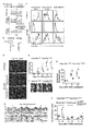
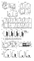
- FIG. 1 is a diagram showing that nonsense mutations in Clec10a in NC / Nga mice cause HDMI-induced dermatitis.
- Panel a shows the CleclOa gene, C57BL/6J CleclOa (NM_010796), NC/Nga-CleclOa c. 706T / T (c.706T / T), and NC / Nga-Clec10a c.
- the DNA sequence of the nonsense mutation site (c.706) in 706T / C (c.706T / C) mice is shown.
- the total length of Clec10a is represented by a plurality of white squares and a line penetrating them, the white squares represent the coding region of the gene, and the lines represent introns.
- CTLD stands for c-type lectin-like domain.
- Panel b shows a schematic diagram of each Clec10a of a C57BL / 6J mouse and an NC / Nga mouse.
- TM represents a transmembrane domain.
- Panel c is a C57BL / 6J mouse, NC / Nga-Clec10a c. 706T / T mouse, NC / Nga-Clec10a c.
- Macrophages (MP) (CD64 + MerTK +), conventional DC (cDC) (CD64-MerTK) in PI-CD45 + MHCII + Lineage (CD3, CD19, NK1.1, and Ly-6G) -EpCAM-cells of the dorsal skin of 706T / C mice. -), And the expression of Clec10a on the cell surface of monocyte-derived dendritic cells (CD64-MerTK lo ). The shaded histogram shows staining with an isotype control antibody (Ab). Panel d is a C57BL / 6J mouse, NC / Nga-Clec10a c.
- Panel e shows the dermatitis score
- panel f shows the appearance on day 14
- panels g and h show the tissue section (hematoxylin-eosin staining) and epithelial thickness, respectively.
- Panel i shows neutrophils (CD11b + Ly-6G + ), eosinophils (CD11b + SIGLEC-F + ), and Ly-6C hi monocytes (CD11b + Ly-6G - SIGLEC-F - Ly-6C hi). ) Indicates the total number of individuals.
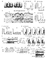
- FIG. 2 shows that Clec10a inhibits an HDMI-induced immune response.
- Panels a to f show the results of applying HDM ointment to the dorsal skin of C57BL / 6J wild-type mice and Clec10a ⁇ / ⁇ mice twice a week.
- Panels c and d show a comparison between wild-type and Clec10a ⁇ / ⁇ mice.
- Panel e shows flow cytometry that identifies neutrophils (CD11b + Ly-6G + ) in the skin CD45 + cells of wild-type and Clec10a ⁇ / ⁇ mice at specific time points.
- Panel f is the result of quantitative RT-PCR of mRNA obtained from cutaneous WT and Clec10a ⁇ / ⁇ MHCII+ MPs and DCs 3 hours after topical application of HDM.
- CBA cost-benefit bead array
- Panel h shows the results of stimulating wild-type, Trr4-/-, and Clec10a ⁇ / ⁇ BMMPs for the time specified by HDMI and then subjecting the lysates to immunoprecipitation (IP) with Clec10a antibodies. .. Immunoblot analysis (IB) was performed using antibodies against phosphorylated tyrosine (pTyr) or Clec10a.
- CBA cytometric bead array
- Panel j stimulated wild type or CleclOa-/- CD115+-enriched BMMPs with HDM for a specified time and then using phosphorylated Syk (pSyk; Y 519/520 ) or a monoclonal antibody to Syk (mAb).
- pSyk phosphorylated Syk
- mAb monoclonal antibody to Syk
- Panel k shows BMMPs transfected with wild type or Y3F Clec10a or empty vector (EV) transfected with HDM, cell lysates immunoprecipitated with mAb against Clec10a and against Syk, SHP-1 or Clec10a. The result of having immunoblotted with an antibody is shown.
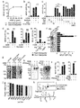
- FIG. 3 shows that Clec10a recognizes the mucin-like protein of HDMI.
- Panels a to d are rat IgG2a or anti-Clec10a mAb (panels a and b) or galactose (Gal), glucose (Glc) or mannose (Man) (panel c) in mouse Clec10a-CD3 ⁇ reporter cells or parent reporter cells. Expression of GFP after stimulation with an HDM coated plate in the presence or absence of); or after stimulation with a galactose (GALase) or glucosidase (GLCase) or untreated HDM coated plate (panel d). Indicates.
- Panels e-i are Clec10a ligands (Clec10a-L) (panel e) in HDM pulled down (PD) with Clec10a-Fc or control human antibody, each fraction based on the size of Clec10a-L (panel f), or 3 shows GFP expression in mouse CleclOa-CD3 ⁇ reporter cells or control reporter cells after stimulation with CleclOa-L (panel i) before and after treatment with PNGase F or NaOH.
- Statistical analysis was performed using PBS-stimulated samples used as controls (panel f).
- Clec10a-L is immunoblotted with Clec10a-Fc before (panels f-h) or post-treatment (panel h) with PNGase F or NaOH, or silver stained with or without alcyan blue (panel h).
- Panel j shows a lectin microarray analysis of Clec10a-L. The black bar indicates the lectin that binds to Gal ⁇ (1-3) GalNAc (T antigen). GalNAc means N-acetylgalactosamine and GlcNAc means N-acetylglucosamine.
- Panel k shows a schematic of HDM Clec10a-L.
- T means T antigen (Gal ⁇ (1-3) GalNAc)
- Tn means Tn antigen ( ⁇ GalNAc)
- LacNAc means N-acetyl-D-lactosamine (Gal ⁇ (1-4) GlcNAc).
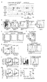
- FIG. 4 shows that human Asgr1 is the structural and functional counterpart of mouse Clec10a.
- Panel c shows the amino acid sequences of the intracellular regions of mouse CleclOa, human Asgrl, and human CleclOa. The putative hemITAM sequence is underlined.
- Panel d shows GFP expression in human Asgr1-CD3 ⁇ reporter cells stimulated with CleclOa ligand coated plates obtained by pulling down from HDM with mouse CleclOa-Fc.
- Panel e shows the results of staining tissue sections of human skin with anti-Asgr1 antibody, anti-CD68 mAb and DAPI.
- E means epidermis
- D means dermis.
- the scale bar is 100 ⁇ m.
- Panel g shows the association between ASGR1 expression (GSE5667) and serum IgE titers in the skin of healthy individuals, psoriasis patients, and atopic dermatitis patients.
- Panel h shows a hypothetical model for the role of C-type lectin receptors in maintaining skin homeostasis upon exposure to HDM in mice and humans.
- * represents p ⁇ 0.05, ** represents P ⁇ 0.01, and *** represents p ⁇ 0.001 (one-sided ANOVA test (panel b), independent two-sided Student's t-test ( Panel f) or two-tailed Pearson correlation test (panel g)).
- Data represent mean ⁇ SEM.
- FIG. 5 shows the results of feature analysis of Clec10a in C57BL / 6J and NC / Nga mice.
- Panel a shows the results of gene expression in hematopoietic cells and tissues of C57BL / 6J mice based on BioGPS analysis.
- Panels f and g show Flag-tagged Cle10a c. 706C - IRES -GFP or Flag-tagged Clec10a c. Surface analysis of transformants of RAW264.7 expressing 706T - IRES -GFP (panel f) and intracellular expression of Flag tag by flow cytometry (panel g) are shown. Panel h is NC/Nga Clec10a c. 706T/T (T/T), Clec10a c. 706 T / C (T / C), and Clec10a c.
- 706 C / C shows the expression of Clec10a mRNA in the skin of mouse.
- Panel j sets the serum IgE titer at the specified time point to Clec10a c. 706T/T (T/T), Clec10a c. The result of comparison with 706 T /C (T/C) is shown.
- FIG. 6 shows the phenotype of HDMI-induced dermatitis in Clec10a-deficient mice.
- Dust mites (HDM) ointment was applied to the dorsal skin of wild type C57BL/6J and CleclOa-/- mice twice weekly.
- Panel a shows the results of flow cytometry analysis of each cell population after staining the skin cells recovered at a specified time point with an antibody against PI, anti-CD45 antibody, or marker molecule.
- the markers for each cell were as follows: eosinophils (CD11b + Sigma-F + ), and Ly-6C hi monocytes (CD11b + Ly-6G - Sigma-F - Ly-6C hi ), CD3 + CD4 + T.
- Panel c is a quantitative representation of mRNA expression levels of the designated molecules in wild-type and CleclOa-/- type CD4+ T cells (CD3 + CD4 + ) sorted from axillary and inguinal lymph nodes at day 6.
- the results of RT-PCR are shown. ** represents P ⁇ 0.01 and *** represents p ⁇ 0.001 (independent two-sided Student's t-test). Data represent mean ⁇ SEM.
- FIG. 7 shows the results of characteristic analysis of wild-type and Clec10a ⁇ / ⁇ type MPs.
- Panel a shows MP (PI ⁇ CD45 + MHCII + Lineage (CD3, CD19, NK1.1, and Ly-6G) ⁇ EpCAM ⁇ CD64 + ) and DC obtained from mouse skin 3 hours after topical application of HDM.
- 2 shows a gate strategy for sorting (PI ⁇ CD45 + MHCII + Lineage ⁇ EpCAM ⁇ CD64 ⁇ ).
- Panel b shows a gate strategy for sorting CD115 + BMMP from wild-type and Clec10a ⁇ / ⁇ mice.
- Panel c shows the results of staining BMMPs from wild-type and CleclOa-/-mice with antibodies to CD115, CleclOa, the designated MP marker, and TLR4.
- CD115 + cells were gated and the expression of each molecule was analyzed by flow cytometry. Shaded histograms are stained with isotype control antibody.
- Panel e shows the amino acid sequence of the intracellular region of the wild-type and Y3F mutant Clec10a. The estimated hemITAM sequence is underlined.
- Panels f and g show the expression of Clec10a on the cell surface of BMMP transfected with cDNA or empty vector encoding wild-type and Y3F mutant Clec10a. Shaded histograms show staining with isotype control antibody (panel f). Transfected cells were stimulated with HDM, lysed, immunoprecipitated (IP) with anti-Clec10a antibody, and immunoblotted with anti-phosphorylated tyrosine (pTyr) and anti-Clec10a antibodies (panel g). Arrowheads indicate the molecule of interest (black) and the antibody used for IP (IP-Ab) (white).
- FIG. 8 shows the establishment of Clec10a-CD3 ⁇ reporter cells and Clec10a-FC chimeric protein.
- Panels a and b are Clec10a after stimulation with a plate coated with Lewis X at the specified dose (panel a) and a plate coated with Lewis X (10 ⁇ g / mL) or Lewis Y (10 ⁇ g / mL).
- FIG. 9 shows the results of HDMI stimulation of human Clec10a-CD3 ⁇ reporter cells and the knockdown efficiency of human ASGR1.
- Panels a and b are humans after stimulation with HDM-coated plates in the absence (panel a) or presence (panel b) of galactose (Gal), glucose (Glc), or mannose (Man).
- 3 shows GFP expression in CleclOa-CD3 ⁇ reporter cells.
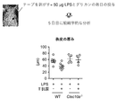
- FIG. 10 is a diagram showing that the Clec10a ligand (Clec10a-L) improves LPS-induced dermatitis.
- Panels a and b show tissue on day 5 after daily application of LPS to the back skin of C57BL / 6J wild-type (WT) and Clec10a ⁇ / ⁇ mice in the presence or absence of Clec10a-L. And epidermis thickening, respectively.
- Panel c shows neutrophils (CD45 + CD11b + Ly-6G + ) in the skin of WT and Clec10a ⁇ / ⁇ mice 6 hours after LPS was applied to the dorsal skin in the presence or absence of CleclOa-L. The number (/cm 2 ) is shown.
- FIG. 11 shows a scheme for determining Clec10a-L.
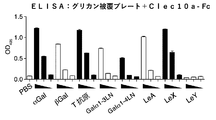
- FIG. 12 shows the results of an ELISA assay that examined whether Clec10a-Fc binds to a polymer scaffold-coated plate that presents the various glycans displayed.
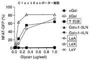
- FIG. 13 shows the results of a reporter assay using Clec10a reporter cells to determine whether a polymeric scaffold presenting the various displayed glycans activates Clec10a.
- FIG. 14 shows the results of a therapeutic experiment in which the effect of administration of a T-presenting polymer scaffold on LPS-induced epidermal inflammation was examined.
- a "subject” can be a mammal, for example, pets such as dogs, cats, rabbits, hamsters, guinea pigs, and squirrels; livestock such as cows, pigs, horses, sheep, and goats; monkeys. Includes primates such as chimpanzees, orangutans, gorillas, bonobos, and humans.
- treatment is used in the sense of including therapeutic treatment and prophylactic treatment.
- treatment can be used to include suppression of deterioration of a disease or condition, delay of deterioration of a disease or condition, improvement of a disease or condition, or cure of a disease or condition.
- prophylaxis can be used to mean suppressing the onset of a disease or condition, or delaying the onset of a disease or condition.
- allergy means a systemic or local disorder to a living body based on an immune response. Allergies are broadly divided into allergies based on humoral immune response by blood antibodies (types I, II, and III) and allergies based on cell-mediated immunity by sensitized lymphocytes (type IV).
- Type I allergy is an allergy also called immediate type allergy or anaphylactic type. IgE is involved, and IgE binds to the IgE receptor (Fc ⁇ RI) that mast cells and eosinophils in blood and tissues have on the cell surface, and when an allergen binds to this, histamine, etc. from mast cells and eosinophils Is released, causing an allergic reaction (eg, smooth muscle contraction, vascular hyperpermeability, and glandular hypersecretion).
- Type I allergies include atopic bronchial asthma, allergic rhinitis, urticaria, allergic conjunctivitis, atopic dermatitis, and anaphylactic shock. In type I allergy, house dust, mites and the like are known to invade the body to cause an allergic reaction (oral, inhalation, transdermal, and intravenous routes may be used).
- Type II allergy is based on cytotoxicity caused by the reaction of IgG or IgM with cell and tissue antigens and the like, and complement binding to this.
- Type II allergy also includes antibody-dependent cellular cytotoxicity (ADCC), in which macrophages and killer cells having IgG Fc receptors bind to IgG bound to an antigen on the cell membrane and damage the cells.
- ADCC antibody-dependent cellular cytotoxicity
- Type II allergies include hemolytic anemia due to incompatible blood transfusion, autoimmune hemolytic anemia, idiopathic thrombocytopenic purpura, drug-induced hemolytic anemia, thrombocytopenia, thrombocytopenia, and Goodpasture disease group. Can be mentioned.
- Type III allergy also called immune complex type or Arthus type, is based on tissue damage caused by an immune complex (Immunocomplex) of a soluble antigen with IgG or IgM.
- Type III allergies include serum sickness, autoimmune diseases such as systemic lupus erythematosus and rheumatoid arthritis, glomerulonephropathy, hypersensitivity pneumonia, and allergic bronchopulmonary aspergillosis.
- Type IV allergy also called delayed allergy, is based on the reaction of sensitized T cells with antigens, which releases cytokines from the sensitized T cells and causes cytotoxicity. It is also based on damage to virus-infected cells, tumor cells, and grafts by killer T cells.
- Type IV allergies include allergic contact dermatitis, atopic dermatitis, hypersensitivity pneumonitis, tuberculous cavities, clefts, epithelioid cell granulomas lesions of sarcoidosis, smallpox rash, and measles rash.
- asialoglycoprotein receptor refers to a glycoprotein in which the sialic acid at the terminal end of the sugar chain of the protein has been removed and the galactose residue inside is exposed as a terminal group, that is, asialoglycoprotein It is a receptor that binds to AGP).
- the asialoglycoprotein receptor is present on the surface of hepatocytes and binds to AGP in blood to remove AGP from blood.
- Asialoglycoprotein receptor 1 (ASGR1) is also referred to as type C lectin domain family member H1 or CLEC4H1.
- a representative example of a human ASGR1 protein may be a protein having an amino acid sequence registered with GenBank under accession number CAG46849.1.
- ASGR1 is used to include the ortholog of human ASGR1.
- Clec10a is also referred to as C-type lectin domain family 10, member A, and is a molecule that recognizes a sugar chain and functions as a biological defense system of a host. Clec10a can specifically bind to galactose or N-acetylgalactosamine.
- the "house dust mite” is a mite of the genus Dermatophagoides.
- the main species of dust mites are Dermatophagoides farinae, Dermatophagoides microceras, Dermatophagoides mediterraneus pteronyus, and Dermatophaeneys pteronysphaerus.
- the term “antigen” means a substance that provides an epitope to which a lectin can react in the case of an antibody or sugar.
- “antigen” means, according to its ordinary meaning, a sugar chain that provides an epitope with which a lectin can react. Therefore, the term "antigen" in the context of glycans means providing the glycans to the surface of the glycans as an epitope with which the lectin can react, as is found in natural glycans and glycoproteins. To do.
- the "ligand” refers to a substance to which a receptor binds.
- the ligand may regulate a downstream signal of the receptor by binding to the receptor.
- a substance that positively regulates a downstream signal of a receptor is called an "agonist”.
- a substance that negatively regulates a downstream signal of a receptor is called an "antagonist”.
- GalNAc means N-acetylgalactosamine
- GlcNAc means N-acetylglucosamine
- Clec10a is involved in the onset and exacerbation of dermatitis caused by dust mites in mice, and Asgr1 is involved in humans.
- the inventors also revealed from the results of functional analysis and homology analysis that human Asgr1 is the original structural and functional counterpart of mouse Clec10a.
- dust mites contain substances that bind to and activate mouse Clec10a and human Asgr1 and suppress the development of allergies (eg, Clec10a ligand or Asgr1 ligand).
- the ligand of Clec10a contains an O-linked sugar chain, particularly T antigen (Gal ⁇ (1-3) GalNAc) and Tn antigen ( ⁇ GalNAc), and ASGR1 binds to any of these. Revealed.
- the present inventors have also revealed that the CleclOa ligand suppresses TLR4 signaling.
- the T antigen (Gal ⁇ (1-3) GalNAc) and the Tn antigen ( ⁇ GalNAc) are sugar chains having the structures shown in the schematic diagram shown in the following formula (I), respectively.
- the binding of Asgr1 to the T and Tn antigens, respectively, is consistent with Asgr1's affinity for galactose or N-acetylgalactosamine.
- Asgr1 binds to a sugar chain selected from the group consisting of T antigen, LeA, and LeX, and thereby transmits a signal into the cell.
- sugar chains may be presented on a polymeric scaffold.
- sugar chains are polylactic acid, polyacrylamide, polyvinyl, polyvinyl alcohol, polymethyl methacrylate, polyacrylonitrile, polystyrene, polypropylene, polyethylene terephthalate, nylon, collagen, hydroxyethyl methacrylate, chitosan, chitin, polyethylene oxide, polyethylene glycol.
- Polyamino acids, polylactides, polyglycolides, polycaprolactones, and copolymers thereof may be linked on the side chains of a polymeric scaffold having as a backbone and presented to Clec10a.
- each polymer is not particularly limited as long as it has manageable physical properties (for example, viscosity, osmotic pressure, etc.), but for example, it has a weight average molecular weight of 1 kDa to 100 kDa, 5 kDa to 50 kDa, 10 kDa to 40 kDa, or 20 to 40 kDa.
- a biocompatible polymer having a can be used as a backbone.
- a nonsense mutation or frameshift mutation into Clec10a, the mouse counterpart of asialoglycoprotein receptor 1 (Asgr1), resulted in an excessive response to HDM.
- an extract (purified product) containing a CleclOa ligand was prepared and contacted with human Asgr1-CD3 ⁇ reporter cells, Asgrl responded to the CleclOa ligand in a concentration-dependent manner. This activation was also concentration-dependently counteracted by the addition of galactose to the system.
- the asialoglycoprotein receptor 1 (Asgr1) ligand can be used in the treatment of diseases or symptoms (eg, inflammation and allergic diseases) induced by TLR4 signals such as dust mite-induced allergies.
- TLR4 signals such as dust mite-induced allergies.
- the present invention provides a composition for use in treating an allergic disease, which comprises a ligand of asialoglycoprotein receptor 1 (Asgr1).
- a composition comprising a ligand for the asialoglycoprotein receptor 1 (Asgr1) for use in the treatment of diseases or symptoms (eg, inflammation and allergic diseases) induced by activation of TLR4 signaling.
- the TLR signal can be activated by the TLR4 ligand.
- TLR4 ligands include lipopolysaccharide (LPS) and lipoteichoic acid and agonists that are analogs thereof. Therefore, the ligand of asialoglycoprotein receptor 1 (Asgr1) can be used for the treatment of diseases or conditions (eg, inflammation and allergic diseases) induced by allergens containing these TLR4 ligands. Therefore, according to the present invention, a composition for use in treating a disease or symptom induced by an allergen containing a TLR4 ligand, which comprises the ligand for asialoglycoprotein receptor 1 (Asgr1). Provided.
- LPS lipopolysaccharide
- Asgr1 lipoteichoic acid and agonists that are analogs thereof. Therefore, the ligand of asialoglycoprotein receptor 1 (Asgr1) can be used for the treatment of diseases or conditions (eg, inflammation and allergic diseases) induced by allergens containing these TLR4 ligands. Therefore,
- Examples of the ligand for the asialoglycoprotein receptor 1 include a sugar chain in which sialic acid is released and has LewisX at the end, and a protein having the sugar chain.
- Examples of the ligand of the asialoglycoprotein receptor 1 (Asgr1) include a sugar chain in which sialic acid is released and has a T antigen or Tn antigen at the end, and a protein having the sugar chain.
- T antigen means Gal ⁇ (1-3)GalNAc
- Tn antigen means ⁇ GalNAc.
- the ligand for the asialoglycoprotein receptor 1 As a ligand for the asialoglycoprotein receptor 1 (Asgr1), sialic acid is released, and 1, 2 or all O-linked sugars selected from the group consisting of Lewis X antigen, T antigen, and Tn antigen at the end. It can be a chain or a protein having the sugar chain.
- the mucin-like protein has the above-mentioned O-linked sugar chain, and has a sugar chain selected from the group consisting of Lewis X antigen, T antigen, and Tn antigen.
- the ligand for asialoglycoprotein receptor 1 can be a mucin-like protein or mucin.
- the protein as a ligand for asialoglycoprotein receptor 1 (Asgr1) may be a mucin-like protein or mucin.
- Asgr1 is highly expressed in hepatocytes, and the aged protein (asiaroglycoprotein) desialylated in the body is taken into the hepatocytes and removed from the blood. Therefore, all such desialylated glycoproteins can be used as ligands for the asialoglycoprotein receptor 1. Asgr1 binds to and reacts with sugar chains having at least one or both of T antigen and Tn antigen. Therefore, a sugar chain having at least one or both of the T antigen and the Tn antigen or a protein having the same can be used as a ligand for the asialoglycoprotein receptor 1.
- All allergen-containing substances can also be obtained by affinity purification based on binding affinity with Clec10a (eg, mouse Clec10a or human Asgr1). it can. Elution of the Clec10a ligand from Clec10a can be performed using, for example, galactose. The eluate of CleclOa ligand may be dialyzed against saline.
- a fusion protein containing the extracellular region, transmembrane region, and intracellular region of CD3 ⁇ of human asialoglycoprotein receptor 1 transmitted the CD3 ⁇ signal downstream in the presence of the ligand of asialoglycoprotein receptor 1. Therefore, a fusion protein containing the extracellular region of human asialoglycoprotein receptor 1, transmembrane region, and intracellular region of CD3 ⁇ can be identified as a substance to be used for treating allergic diseases. For example, an animal cell expressing a fusion protein containing an extracellular region of human asialoglycoprotein receptor 1, a transmembrane region, and an intracellular region of CD3 ⁇ , which operates with a promoter (eg, NFAT promoter) activated by a CD3 ⁇ signal.
- a promoter eg, NFAT promoter
- the test compound is a compound that binds to human asialoglycoprotein receptor 1 by contacting the compound with an animal cell having a gene encoding a reporter that is operably linked.
- the obtained substance is a substance to be used for treating an allergic disease by using regulatory signaling via inhibitory TAM as an index.
- the compound human Asgr1 ligand candidate compound
- the compound can be applied to LPS-induced dermatitis to confirm the dermatitis suppressing effect.
- the present invention provides a fusion protein containing an extracellular region of human asialoglycoprotein receptor 1, a transmembrane region, and an intracellular region of CD3 ⁇ , and an animal cell expressing the fusion protein.
- the animal cell may also have a gene encoding a reporter operably linked to a promoter activated by the CD3 ⁇ signal.
- the present invention further provides an animal cell that expresses a fusion protein containing an extracellular region of human asialoglycoprotein receptor 1, a transmembrane region, and an intracellular region of CD3 ⁇ , which is operably linked to a promoter activated by a CD3 ⁇ signal.
- test compound may comprise a ligand for the asialoglycoprotein receptor 1 (Asgr1).
- test compound may be at least one sugar chain in which the ligand is selected from the group consisting of T and Tn antigens.
- the ligand can be a mucin-like protein or mucin.
- the ligand can be a ligand for human asialoglycoprotein receptor 1.
- An animal cell expressing a fusion protein comprising the extracellular region of human asialoglycoprotein receptor 1 of the present invention, a transmembrane region and an intracellular region of CD3 ⁇ , which is a reporter operably linked to a promoter activated by a CD3 ⁇ signal.
- the animal cell having the gene encoding the can also be used for compound screening. Therefore, in the present invention, it is an animal cell that is a screening method for a human Asgr1 ligand or agonist and expresses a fusion protein containing an extracellular region, a transmembrane region, and an intracellular region of CD3 ⁇ of human asialoglycoprotein receptor 1.
- test compound is indicated as a candidate for a human Asgr1 ligand or agonist.
- the animal cell may preferably be a human cell.
- composition of the present invention may be a composition such as a personal care composition and a pharmaceutical composition.
- the pharmaceutical composition examples include pharmaceutical compositions for topical administration, which can be used in the present invention.
- the pharmaceutical composition for topical administration may be a pharmaceutical composition for mucosal application or body surface application, for example, eye drops, eye ointment, sublingual tablet, buccal tablet, troche, mouthwash, spray. , Aerosols, and inhalants; Solution formulations such as liquids, salves, glycerin, alcohol, liquids, and coatings; dispersions such as emulsions, suspensions, liniments, lotions, sprays, and liposomes.
- Formulations Semi-solid formulations such as ointments, plasters, patches, adhesive tapes, pasta, paps, creams, oils, and sticks; and extracts (soft extracts, dry extracts), and Examples include leachate preparations such as tinctures.
- the pharmaceutical composition may contain a pharmaceutically acceptable excipient.
- Pharmaceutically acceptable excipients include solvents, bases, diluents, bulking agents, fillers, and shapers; solubilizers, solubilizers, buffers, isotonic agents, emulsifiers, suspensions. Turbidants, dispersants, thickeners, gelling agents, hardeners, absorbents, adhesives, elastics, plasticizers, sustained release agents, and propellants; antioxidants, preservatives, moisturizers, shading Examples include agents, antistatics, fragrances, flavoring agents, colorants, and palliatives.
- Examples of personal care compositions include skin care, antiperspirants, deodorants, beauty products, cosmetics, and hair care products.
- Personal care compositions include moisturizers, conditioners, anti-aging agents, whitening agents, sunscreens, antiperspirants, shaving compositions, post-shaving compositions, foundations, lipsticks, lipsticks, hair styling products, shampoos, etc. Examples include cleaning agents and lubricants.
- the personal care composition may be used in personal care products. Examples of personal care products include underwear, diapers, tissues, wipes, masks, patches and the like.
- the composition may include an excipient in addition to the active ingredient.
- the composition can be in a dosage form suitable for administration forms such as intravenous administration, transdermal administration, oral administration, enteral administration, and intraperitoneal administration.
- the compositions of the invention may be administered by transdermal administration, in the form of, for example, gels, emulsions, creams, liquids, pastes, lotions, liposome creams and the like (eg, skin). Scientific composition).
- the composition can be an ointment.
- a dermatologically acceptable excipient can be used as an excipient, and a dosage form suitable for this can be used.
- transmucosal administration an excipient that is acceptable for mucosal application can be used, and a dosage form suitable for this can be used.
- an Asgr1 ligand in the manufacture of a composition for use in treating an allergic disease is provided.
- a method for treating an allergic disease in a subject in need thereof comprising administering to the subject an effective amount of an Asgr1 ligand.
- a method for preventing an allergic disease, or suppressing the occurrence thereof comprises administering to the subject an effective amount of Asgr1 ligand.
- Method (1) Preparation of skin cells Skin cells are minced by sandwiching a thinly ground dorsal skin sample and containing 200 U / mL Collagenase II, 50 U / mL DNase and 10% fetal bovine serum (FBS). Was incubated in RPMI-1640 medium for 60 minutes. Further dissociation and homogenization were performed using the gentleMACS Dissociator (Miltenyi Biotec). The obtained cells were filtered through a 55 ⁇ m nylon mesh to obtain a single cell suspension.
- FBS fetal bovine serum
- immunohistochemical staining skin samples taken from mice were embedded in Tissue Tissue Optimal Cutting Tissue Company Compound (Sakura Finetek Japan) and stored at -80 ° C. A 4 ⁇ m thick section was also used for immunohistochemical staining.
- the cells were washed with PBS (PBS-T, pH 7.4) containing 0.05% Tween-20, soaked, and incubated with Blocking One Histo (Nacalai) for 10 minutes at room temperature.
- the sections were then washed with PBS-T, incubated overnight with anti-Clec 10a mAb at 4 ° C., washed with PBS-T and incubated with Alexa Fluor 546-labeled anti-rat IgG polyclonal antibody for 1 hour. After washing with PBS-T, sections were stained with DAPI nuclei. Healthy human skin tissue was collected from the periphery of the patient's tumor area, fixed with formalin, and embedded in paraffin to make 4 ⁇ m thick sections.
- Sections were deparaffinized with xylene, rehydrated with ethanol and blocked with methanol to block endogenous peroxidase. Sections were stained with anti-CD69 antibody and anti-Asgr1 antibody according to the manufacturer's manual for the Opal 4-Color Automation IHC Kit (PerkinElmer, NEL820001KT). Briefly, sections are incubated at 95 ° C. for 15 minutes, washed with TBS (TBS-T) (Takara Bio, T9142) containing 0.05% Tween-20, and treated with a blocking solution at room temperature for 10 minutes. did. Sections were incubated overnight at 4 ° C.
- Serum IgE antibody was measured using a capture antibody against mouse IgE (R35-72), biotinylated anti-mouse IgE (R35-118), and subsequent HRP-labeled streptavidin (GE Healthcare, RPN1231V). .. Purified mouse IgE (C38-2, BD Biosciences) was used as a standard. Serum IgG1 was measured using a capture antibody against mouse IgG1 (A85-3) and an HRP-labeled antibody against mouse IgG1. Purified mouse IgG1 (107.3, BD Biosciences) was used as a standard. Serum IgG2c was detected by mouse ELISA Quantification Set (Bethyl, E90-136).
- BMMPs bone marrow macrophages
- GM-CSF R&D Systems
- IL-4 Wako
- Cultured below in RPMI 1640 complete medium with 10% FBS.
- 70% of non-adherent cells were removed and fresh medium containing GM-CSF and IL-4 was added.
- 100% non-adherent cells were removed by washing with PBS and fresh medium containing GM-CSF and IL-4 was added.
- all non-adherent cells were removed by PBS washing and the adherent cells were used for later experiments.
- CD115 + BMMP was concentrated using anti-CD115 antibody (BioLegend) and anti-rat IgG microbeads (Miltenyi Biotec).
- Skin MP is CD45 + MHCII + CD3 - CD19 - NK1.1 - Ly-6G - EpCAM - CD64 sorted by +, DC is, CD45 + MHCII + CD3 - CD19 - NK1.1 - Ly-6G - EpCAM - CD64 - ) Sorted.
- BMMP was infected on days 2 and 4 with virus supernatant supplemented with polybrene (8 ⁇ g / mL, Sigma-Aldrich). After centrifugation, the supernatant containing the virus was removed and replaced with fresh BMMP medium. On day 5, the medium was replaced with fresh BMMP medium, and on day 7, non-adherent cells were removed by PBS washing and adherent cells were used in the experiment.
- CleclOa-Fc Chimera A mouse CleclOa chimeric construct (CleclOa-Fc) was prepared by cloning the extracellular region of mouse CleclOa into a pME18S expression vector containing the Fc region of human IgGl. Clec10a-Fc was transfected into HEK293T cells using Lipofectamine 2000 (Thermo Fisher Scientific) and then the medium was replaced with GIT medium (KOHJIN BIO). Clec10a-Fc was purified from culture supernatant using protein A agarose (Bio-Rad Laboratories).
- BMMP was stimulated with HDM extract (100 ⁇ g/mL) at 37° C. for 0 minutes, 10 minutes, or 30 minutes. Stimulated BMMP was dissolved in 1% NP-40 lysis buffer and separated by SDS-PAGE. It was transcribed on a PVDF membrane by electroblot, immunoblotted with anti-phosphorylated Syk antibody and anti-Syk antibody, and detected with HRP-labeled anti-rabbit IgG antibody. To analyze tyrosine phosphorylation of Clec10a, BMMP was stimulated with HDMI extract (100 ⁇ g / mL) at 37 ° C. for 0, 10 or 30 minutes.
- HDMI extract 100 ⁇ g / mL
- BMMP Stimulated BMMP was dissolved in 1% NP-40 lysis buffer and subjected to immunoprecipitation with anti-Clec 10a mAb. Immunoprecipitates were developed by SDS-PAGE, transcribed on PVDF membrane and immunoblotted with HRP-labeled anti-phosphotyrosine antibody or anti-CleclOa antibody and HRP-labeled anti-rabbit IgG antibody. To analyze the association of CleclOa with Syk or SHP-1, BMMPs were pretreated in the presence or absence of 5 mM Syk inhibitor IV (Merck, 574714) for 30 minutes at 37°C and extracted with HDM extract. (100 ⁇ g / mL) was stimulated at 37 ° C.
- Syk inhibitor IV Merck, 574714
- the Clec10a ligand is 0 after 16 hours of pretreatment at 37 ° C. and 5 minutes of thermal denaturation at 95 ° C.
- peptide N-glycosidase F peptide N-glycosidase F
- Pretreatment at 40 ° C. for 16 hours in the presence or absence of .05 M NaOH then separated by SDS-PAGE and transferred to PVDF membranes, biotinylated Clec10a-Fc and HRP. Immunoblots were performed using labeled streptavidin.
- Reporter cells are in the presence or absence of anti-Clec 10a mAb, Lewis X (GlycoTech), Lees Y (GlycoTech), galactose (Sigma-Aldrich), glucose (Sigma-Aldrich) or mannose (Sigma-Aldrich). Incubated. Reporter cells were also treated with or not treated with galactosidase (R & D Systems, 5704-GH or 5549-GH), or glucosidase (R & D Systems, 8329-GH), a Clec10a ligand-coated plate, or a size-fractionated Clec10a ligand. The cells were cultured on a treated HDMI extract coated plate. Activation of NFAT-GFP was monitored by flow cytometry.
- Lectin microarrays were prepared by a non-contact microarray printing robot (MicroSys 4000; Genomic Solutions) according to the conventional method. Samples were fluorescently labeled with Cy3 Mono-Reactive dye (GE) and excess Cy3 was removed using a Sephadex G-25 desalting column (GE). 10 times with a probe solution (25 mM Tris-HCl, pH 7.5, 140 mM NaCl) (TBS) containing 2.7 mM KCl, 1 mM CaCl 2 , 1 mM MnCl 2 , and 1% Triton X-100.
- GE Cy3 Mono-Reactive dye
- TBS Sephadex G-25 desalting column
- Gal D-galactose
- GalNAc N-acetyl-galactosamine
- GlcNAc N-acetyl-glucosamine
- Fuc L-fucose
- Sia Sialic acid
- LacNAc N-acetylamine
- JOM J-OIL MILLS, INC
- Vector VECTOR LABORATORIES
- Seikagaku SEIKAGAKU CORPORATION
- EY EY LABORATORIES, INC
- AIST AIST
- Example 1 Exome analysis of atopic dermatitis model mouse
- NC / Nga mouse which is a model mouse of atopic dermatitis
- the NC/Nga mouse was purchased from Charles River, Japan. DNA was extracted from blood obtained from NC / Nga mice under conditions suitable for extraction of mouse DNA using QIAamp DNA blood Mini Kit (QIAGEN, Venlo, Netherlands). The exome analysis of the obtained DNA was performed as follows.
- the SureSelect Library prep kit post-pool version 4; Agilent Technologies, Santa Clara, CA) and the SureSelect DNA Manual All Exon Kit manufactured by the SureSelect Mouse Manual (Agilent), and the technicians using the Allexon Kit (Agilent Tech).
- the obtained DNA library was subjected to emulsion PCR (SOLiD EZ Bead Emulsifier kit; Thermo Fisher Scientific, Waltham, MA) to generate clonal DNA fragments on beads, and then subjected to bead enrichment (SOLiD EZ. Bead Emulsion kit; Thermo Fisher Scientific, Waltham, MA).
- SOLiD EZ. Bead Emulsion kit Thermo Fisher Scientific, Waltham, MA
- the resulting beads were sequenced on a SOLiD 5500xl sequencer as single-ended 60 bp reads.
- the obtained read was aligned with the mouse reference genome (NCBI37 / mm9) using LifeScop version 2.5.1 (Life Technologies) to obtain a BAM file.
- Variant calls were performed using SAMtool according to the protocol described in Genome Analysi Toolkit, Picard (http://broadinstitute.github.io/picard), and only leads mapped to unique positions on the reference genome were analyzed. Using. Mutation sites were annotated using ANNOVER software. Genetic variation in inbred mouse strains other than NC/Nga was demonstrated by release REL-1211 (http://www.sanger.ac.uk/science/data/mouse-genomics-project) numbered according to the NCBIm37 assembly. Obtained from Mouse Genome Information website (http://www.infomatics.jax.org/). The sequence was carried out on ABI 3130xl Genetic Analyzer (Thermo Fisher Scientific) using BigDye Terminator v1.1 Cycle Sequencing Kit (Thermo Fisher Sequencing). The primers used are shown in Table 4.
- 70,772 mutations were identified in the exon region of the NC/Nga mouse genome relative to the reference genome. Mutations with low quality values obtained by GATK output were removed to give 64,518 mutations. Loss of function mutations, including nonsense and frameshift mutations, were selected using ANNOVER software. Then, 35 nonsense mutations and 48 frameshift mutations (inducing stop codons) were identified. For each of these 83 genes, 46 mutations in 43 genes were selected for further sequence validation.
- Clec10a was selected from seven genes based on the information in the BioGPS database (see panel a in FIG. 5).
- Clec10a of NC / Nga mice was a nonsense mutation (ie, Q236X) in which C at position 706 in the nucleotide sequence of NM_010769 was T.
- Example 2 Expression analysis and functional analysis of Clec10a
- Clec10a is a member of the type II C-type lectin receptor family and detects galactose moieties at the ends of foreign and endogenous antigens.
- CleclOa c. in NC/Nga mice c. 706C> T is present in the coding region of the C-type lectin-like domain (panels a and b in FIG. 1).
- Clec10a showed that in skin CD45 + MHCII + lineage ⁇ EpCAM ⁇ cells, macrophages (MP) (CD64 + MerTK + ), conventional dendritic cells (cDC) (CD64 ⁇ MerTK ⁇ ), And it was revealed that it was expressed on the cell surface of mononuclear cell-derived DC (CD64 - MerTK lo ) and on the macrophages of the peritoneum of C57BL / 6J (it was not expressed in the peritoneum of NC / Nga mice). (See panel c of FIG. 1 and panels b and e of FIG. 5).
- C57BL / 6J mice of the non-hematopoietic cells of the skin CD45 -
- CD45 + MHCII- cells CD45 + MHCII + (Lin, EpCAM) + cells
- neutrophils CD45 + CD11b + Ly-6G +
- Clec10a was not observed (see panels c and d in FIG. 5). Similar results were obtained by immunohistochemical staining, and expression of Clec10a was observed in the skin cells of C57BL / 6J mice, but not in NC / Nga mice (panel d in FIG. 1). ..
- Clec10a expression in NC / Nga mice were further analyzed. Specifically, the cDNA of Clec10a (derived from C57BL / 6J mouse and derived from NC / Nga mouse) having a flag tag and a sequence encoding IRES-GFP at the 3'end was transfected into RAW264.7 macrophages. The Flag tag was expressed on the cell surface of macrophages expressing Clec10a derived from C57BL / 6J mice, but not on the cell surface of macrophages expressing Clec10a derived from NC / Nga mice. (See panel f in FIG. 5).
- Clec10a c A mutant mouse having 706C (hereinafter, sometimes referred to as "NC/Nga mouse CleclOa c.706T>C ") was prepared by a conventional method using the CRISPR/Cas9 system (see panel a in Fig. 1 ). Clec10a c. In the skin of mutant mice with 706C, mRNA expression Clec10a is, Clec10a c. There was a significant increase compared to mice with 706T (see panel h in FIG.
- Clec10a-deficient mice (Clec10a ⁇ / ⁇ mice) were prepared against the background of C57BL/6J mice.
- HDM was topically applied to the dorsal skin of wild type C57BL/6J mice and Clec10a deficient mice (Clec10a ⁇ / ⁇ ).
- CleclOa-/- mice showed dermatitis such as erythema, dryness and enhancement of skin at 5 to 6 days after HDM treatment as compared with wild type mice. Exacerbation was shown (see Figure 2 panels ad).
- CleclOa-/- mice had increased neutrophil infiltration in the skin 1 to 3 days after HDM treatment compared to wild-type mice (see Figure 2 panel e).
- the numbers of cells of other bone marrow and lymphocyte lineages were comparable in wild type and CleclOa-/- mice (see Figure 6 panel a).
- Clec10a is expressed on skin MHCII + macrophages (MP) and dendritic cells (DC), these cells were sorted from skin cell samples of wild-type and Clec10a ⁇ / ⁇ mice topically treated with HDM ( See panel a in FIG. 7).
- the gene expression levels of Il6, Cxcl1, and Cxcl2 were higher in Clec10a ⁇ / ⁇ MP than in wild-type MP, but in DC, these gene expression levels were similar between wild-type and Clec10a ⁇ / ⁇ (in DC). See panel f in FIG. 2).
- Bone marrow macrophages were prepared and characterized from wild type and CleclOa-/- C57BL/6J mice, respectively (see panel b in Figure 7). Expression levels of MP markers and TLR4 in CD115 + BMMP were comparable in wild-type and Clec10a ⁇ / ⁇ mice. After HDM stimulation in vitro, Clec10a ⁇ / ⁇ BMMP secreted more neutrophil chemotactic substances such as IL-6, CXCL1, CCL3 and TNF- ⁇ than wild-type BMMP (Fig. 2). See panel g). Similarly, Clec10a c.
- the BMMP of 706 T / T NC / Nga mice was described in Clec10a c. It showed greater cytokine production than 706 T / C BMMP (see panel d in FIG. 7). These results demonstrate that cell surface expression of Clec10a inhibits the production of inflammatory cytokines from skin MP after stimulation with HDM.
- HDMI stimulation causes activation of splenic tyrosine kinase (Syk) in wild-type BMMP, but no activation of Syk was observed in Clec10a ⁇ / ⁇ BMMP (see panel j in FIG. 2).
- HDMI stimulation evoked recruitment of Syk and the protein tyrosine phosphatase SHP-1 to Clec10a, which was dependent on the tyrosine residue of Clec10a (see panel k in FIG. 2).
- BMMP treatment with a Syk inhibitor suppressed HDMI-induced recruitment of SHP-1 to Clec10a (panel l in FIG. 2), which means that the event is dependent on Syk activation. Suggests. This result is consistent with the control signaling via inhibitory ITAM.
- Example 3 Analysis of Clec10a Ligand
- a chimeric fusion protein containing the extracellular and transmembrane portions of Clec10a fused to the cytoplasmic portion of CD3 ⁇ was tested.
- NFAT-GFP reporter cells (Clec10a reporter cells) to be expressed were prepared.
- Mouse CleclOa reporter cells expressed GFP in response to Lewis X, an oligosaccharide ligand of CleclOa, but not Lewis Y (see Figure 8 panels a and b).
- mouse Clec10a reporter cells expressed GFP in response to the HDMI coated plate in a dose-dependent manner (panel a in FIG. 3).
- Clec10a ligand Clec10a-L
- a chimeric fusion protein containing the extracellular portion (Clec10a-Fc) of mouse Clec10a fused with the Fc portion of human IgG1 was prepared, and this fusion protein is Lewis. It was not bound to Y but to Lewis X (panel c in FIG. 8).
- a pull-down assay from HDMI using Clec10a-Fc isolated Clec10a-L with a molecular weight of approximately 225 kDa, which was able to induce GFP expression in mouse Clec10a reporter cells (FIG. 3). Panel e and f).
- CleclOa-L was stained with Alcian blue and silver, but not silver alone (see panel g in Figure 3), indicating that CleclOa-L is a highly glycosylated protein.
- Treatment with NaOH to degrade O-linked glycans inhibited Clec10a-Fc from binding to the epitope (see panel h in FIG. 3) and also inhibited induction of GFP expression in mouse Clec10a reporter cells (see panel h in FIG. 3). See panel i in FIG. 3).
- PNGase F which decomposes N-linked glycans, showed no effect on CleclOa-L (see panels h and i in Figure 3).
- Clec10a recognizes the O-linked glycan of Clec10a-L.
- Clec10a-L contains T antigen (Gal ⁇ (1-3) GalNAc) and Tn antigen ( ⁇ GalNAc), and LacNAc epitope that binds to mucin-type O glycan (Gal ⁇ (1-4)). It was shown to have GlcNAc) (panel j in FIG. 3 and Table 6).
- Control 1 Pull-down assay sample from buffer solution using Clec10a-Fc 3
- Control 2 Pull-down assay sample from HDMI using human IgG1
- Clec10a is bound most strongly to a lectin which recognizes a multivalent T and Tn antigen of high-density Makurura-Pomifera (Maclura pomifera (MPA)).
- Maclura pomifera MPA
- analysis by glycan microarray revealed that Clec10a-Fc binds to T and Tn antigens (see Table 7), and it was suggested that Clec10a-L is a mucin-like protein (panel in FIG. 3). k)).
- Example 4 Relationship between human Asgr1 and mouse Clec10a
- BLAST Basic Local Alignment Search Tool
- NCBI National Center for Biotechnology Information
- the mouse Clec10a is a tool for analyzing homology of amino acid sequences.
- human proteins that showed the highest homology in the amino acid sequence of the C-type lectin-like domain (CTLD) were human Asgr1 and Clec10a (encoded by Gene IDs: 432 and 10462, respectively).
- Human Asgr1 reporter cells and human Clec10a reporter cells expressed GFP in response to HDM stimulation see panel a in FIG. 4 and panel a in FIG.
- C-type lectin receptors such as Asgr1 in humans and Clec10a in mice recognize mucin-like proteins in HDM and play an important role in maintaining skin homeostasis against inflammation induced by HDM. Understood (see panel h in Figure 4).
- Example 5 Treatment of Allergic Symptoms with CleclOa Ligand From the results of the above Examples, CleclOa (Asgr1 in humans) is involved in the control of HDM-induced allergic symptoms, and ligand stimulation to CleclOa (Asgrl in humans) was observed. It was shown to be involved in the suppression of allergic symptoms. In this example, allergic symptoms were treated with a ligand for Clec10a and the results were observed.
- TLR4-induced dermatitis is induced.
- tissue sections were prepared by a conventional method and stained with hematoxylin and eosin. Then, the epidermal thickening and the number of neutrophils in the skin were counted.
- the panel b of FIG. 10 also showed the same result as the panel a of FIG.
- Neutrophil infiltration into the skin tissue was observed by a conventional method.
- neutrophil infiltration was observed in the LPS-treated group in wild-type (WT) mice, but in the group treated with LPS plus CleclOa ligand. Neutrophil infiltration was suppressed.
- Clec10a ⁇ / ⁇ mice neutrophil infiltration could not be suppressed by the Clec10a ligand. From these results, it was clarified that the Clec10a ligand exhibits an inhibitory effect on dermatitis.
- HDMI is known to contain TLR4 ligands, which are thought to induce inflammatory and allergic symptoms such as dermatitis. Also in HDM stimulation, Clec10a ligand binds to Clec10a (Asgr1 ligand binds to Asgr1 which is its functional counterpart in humans), and suppressing the TLR4 signal results in reducing inflammatory and allergic symptoms. Conceivable.
- Example 6 Detection of Human Asgr1 Ligand
- the binding profile of CleclOa-L to the lectin was determined by lectin array to predict the glycan structure in CleclOa-L (upper left panel of Fig. 11) and CleclOa.
- a scheme (upper right panel of FIG. 11) is shown in which the binding profile of glycans to is determined by the glycan array.
- FIG. 11 these results are considered together, and it is shown that five glycans ( ⁇ Gal, ⁇ Gal, T antigen, LeA, and LeX) can be identified as Clec10a ligand candidates.
- Gal ⁇ 1-3LN and Gal ⁇ 1-4LN were added to identify seven Clec10a ligand candidates.
- a polymeric scaffold (with a modification rate of 20% as described in the above formula) that presents glycans at the position of “Sugar” in the above formula.
- the PBS solution containing the wells was introduced into a 96-well plate, the wells were coated with glycans by incubating overnight at room temperature, and then the unbound polymer was washed. Then, a 10% fetal bovine serum-containing PBS solution (50 ⁇ L / well) containing 1 ⁇ g / ⁇ L of Clec10a-Fc was added to the glycan-coated well.
- T antigen which is one of them, can suppress skin inflammation against lipopolysaccharide (LPS).
- Polymeric scaffolds presenting 50 ⁇ g LPS and 20 ⁇ g T antigen were injected intradermally daily on the backs of mice, and epidermal thickness was observed on day 5. The result was as shown in FIG. As shown in FIG. 14, the increase in skin thickness by LPS was suppressed by T antigen.
- the thickness of the epidermis was equal to or greater than that of WT.
- the effect of suppressing the thickness of the epidermis by the T antigen was not observed in the Clec10a knockout mouse, it was clarified that the effect of suppressing the inflammation by the T antigen was mediated by Clec10a.
Abstract
The present invention provides a composition for use in treatment of allergic diseases. This composition for use in treatment of allergic diseases contains a ligand for asialoglycoprotein receptor 1 (Asgr1).
Description
本発明は、アレルギー疾患を処置することに用いるための組成物に関する。
The present invention relates to compositions for use in treating allergic diseases.
チリダニ(HDM)は、アトピー性皮膚炎や喘息などのアレルギー疾患の主なアレルゲンである(非特許文献1および2)。NC/NgaマウスはHDMに対して感受性を有するマウス系統であり、HDMに対して他の系統と比較してより重度の皮膚炎を発症する(非特許文献3)。しかしながら、その発症機序についてはほとんど明らかにされていない。
Dust mites (HDM) are the main allergens for allergic diseases such as atopic dermatitis and asthma (Non-Patent Documents 1 and 2). The NC/Nga mouse is a mouse strain that is sensitive to HDM, and develops more severe dermatitis against HDM compared to other strains (Non-patent Document 3). However, little is known about its pathogenic mechanism.
本発明は、アレルギー疾患を処置することに用いるための組成物を提供する。
The present invention provides a composition for use in treating an allergic disease.
本発明者らは、チリダニによる皮膚炎の発症や悪化に、マウスでは、Clec10aが関与することを明らかにした。本発明者らはまた、ヒトでは、Clec10aの構造的機能的カウンターパートがAsgr1であることを明らかにし、また、チリダニによる皮膚炎の発症や悪化に、ヒトでは、Asgr1が関与することを明らかにした。発明者らはさらに、チリダニには、マウスClec10aおよびヒトAsgr1に結合する物質であって、かつ、アレルギー発生を抑制する物質(例えば、Clec10aリガンドまたはAsgr1リガンド)が含まれていることを明らかにした。発明者らはさらに、Clec10aのリガンドが、O結合型糖鎖、特に、T抗原(Galβ(1-3)GalNAc)、Tn抗原(αGalNAc)を含み、ASGR1がこれらのいずれにも結合することを明らかにした。本発明はこれらの知見に基づくものである。
The present inventors have revealed that Clec10a is involved in the onset and deterioration of dermatitis due to dust mites in mice. The present inventors also revealed that the structural and functional counterpart of Clec10a is Asgr1 in humans, and that Asgr1 is involved in the onset and exacerbation of dermatitis caused by dust mites in humans. did. The inventors further revealed that dust mites contain a substance that binds to mouse Clec10a and human Asgr1 and suppresses the development of allergies (for example, Clec10a ligand or Asgr1 ligand). .. The inventors further stated that the ligand of Clec10a contains an O-linked sugar chain, particularly T antigen (Galβ (1-3) GalNAc) and Tn antigen (αGalNAc), and ASGR1 binds to any of these. Revealed. The present invention is based on these findings.
すなわち、本発明によれば、例えば以下に示される発明が提供される。
(1)アシアロ糖タンパク質受容体1(Asgr1)のリガンドを含む、アレルギー疾患を処置することに用いるための組成物。
(2)アレルギー疾患が、アトピー性皮膚炎、アレルギー性鼻炎、じんましん、アレルギー性喘息、アレルギー性結膜炎、アレルギー性胃腸炎およびアナフィラキシーショックからなる群から選択される1以上である、上記(1)に記載の組成物。
(3)アレルギー疾患が、チリダニを原因とするものである、上記(1)または(2)に記載の組成物。
(4)リガンドが、T抗原およびTn抗原のいずれか、または両方を含む、上記(1)~(3)のいずれかに記載の組成物。
(5)リガンドが、T抗原およびTn抗原からなる群から選択される糖鎖である、上記(1)~(3)のいずれかに記載の組成物。
(6)リガンドが、ムチン様タンパク質またはムチンである、上記(1)~(4)のいずれかに記載の組成物。
(7)リガンドが、ヒトアシアロ糖タンパク質受容体1のリガンドである、上記(1)~(6)のに記載の組成物。
本発明によればまた、以下の発明が提供される。
(1A)アシアロ糖タンパク質受容体1(Asgr1)のリガンドを含む、アレルギー疾患を処置することに用いるための組成物。
(2A)アレルギー疾患が、アトピー性皮膚炎、アレルギー性鼻炎、じんましん、アレルギー性喘息、アレルギー性結膜炎、アレルギー性胃腸炎およびアナフィラキシーショックからなる群から選択される1以上である、上記(1A)に記載の組成物。
(3A)アレルギー疾患が、チリダニを原因とするものである、上記(1A)または(2A)に記載の組成物。
(4A)リガンドが、T抗原、Tn抗原、LeA、およびLexからなる群から選択される少なくとも1種の糖鎖を含む、上記(1A)~(3A)のいずれかに記載の組成物。
(5A)リガンドが、T抗原、Tn抗原、LeA、およびLexからなる群から選択される少なくとも1種の糖鎖を提示するポリマー性の足場を含む、上記(1A)~(3A)のいずれかに記載の組成物。
(6A)リガンドが、ムチン様タンパク質またはムチンである、上記(1A)~(4A)のいずれかに記載の組成物。
(7A)リガンドが、ヒトアシアロ糖タンパク質受容体1のリガンドである、上記(1A)~(6A)のに記載の組成物。
(8A)ヒトアシアロ糖タンパク質受容体1の細胞外領域と膜貫通領域とCD3ζの細胞内領域を含む融合タンパク質を発現する動物細胞であって、CD3ζシグナルで活性化するプロモーターと作動可能に連結したレポーターをコードする遺伝子を有する動物細胞。
(9A)被検化合物が、ヒトアシアロ糖タンパク質受容体1を活性化する化合物であることを検査する方法であって、
上記(8A)に記載の細胞と被検化合物とを接触させることと、
レポーターの発現レベルが増強した場合に、当該被検化合物がヒトアシアロ糖タンパク質受容体1に結合する化合物であると決定すること
を含む、方法。 That is, according to the present invention, for example, the invention shown below is provided.
(1) A composition containing a ligand for asialoglycoprotein receptor 1 (Asgr1) for use in treating allergic diseases.
(2) In (1) above, the allergic disease is one or more selected from the group consisting of atopic dermatitis, allergic rhinitis, urticaria, allergic asthma, allergic conjunctivitis, allergic gastroenteritis and anaphylactic shock. The composition described.
(3) The composition according to (1) or (2) above, wherein the allergic disease is caused by dust mites.
(4) The composition according to any one of (1) to (3) above, wherein the ligand contains either or both of T antigen and Tn antigen.
(5) The composition according to any one of (1) to (3) above, wherein the ligand is a sugar chain selected from the group consisting of T antigen and Tn antigen.
(6) The composition according to any one of (1) to (4) above, wherein the ligand is a mucin-like protein or mucin.
(7) The composition according to (1) to (6) above, wherein the ligand is a ligand forhuman asialoglycoprotein receptor 1.
According to the present invention, the following inventions are also provided.
(1A) A composition comprising a ligand for the asialoglycoprotein receptor 1 (Asgr1) for use in treating allergic diseases.
(2A) In (1A) above, the allergic disease is one or more selected from the group consisting of atopic dermatitis, allergic rhinitis, urticaria, allergic asthma, allergic conjunctivitis, allergic gastroenteritis and anaphylactic shock. The composition described.
(3A) The composition according to (1A) or (2A) above, wherein the allergic disease is caused by dust mites.
(4A) The composition according to any of (1A) to (3A) above, wherein the ligand comprises at least one sugar chain selected from the group consisting of T antigen, Tn antigen, LeA, and Lex.
(5A) Any of (1A) to (3A) above, wherein the ligand comprises a polymeric scaffold that presents at least one sugar chain selected from the group consisting of T antigen, Tn antigen, LeA, and Lex. The composition according to.
(6A) The composition according to any one of (1A) to (4A) above, wherein the ligand is a mucin-like protein or mucin.
(7A) The composition according to (1A) to (6A) above, wherein the ligand is a ligand forhuman asialoglycoprotein receptor 1.
(8A) An animal cell expressing a fusion protein containing an extracellular region, a transmembrane region, and an intracellular region of CD3ζ ofhuman asialoglycoprotein receptor 1, which is operably linked to a promoter activated by a CD3ζ signal. An animal cell that has a gene encoding.
(9A) A method for inspecting that the test compound is a compound that activateshuman asialoglycoprotein receptor 1.
Contacting the cell according to (8A) with a test compound,
A method comprising determining that the test compound is a compound that binds tohuman asialoglycoprotein receptor 1 when the expression level of the reporter is enhanced.
(1)アシアロ糖タンパク質受容体1(Asgr1)のリガンドを含む、アレルギー疾患を処置することに用いるための組成物。
(2)アレルギー疾患が、アトピー性皮膚炎、アレルギー性鼻炎、じんましん、アレルギー性喘息、アレルギー性結膜炎、アレルギー性胃腸炎およびアナフィラキシーショックからなる群から選択される1以上である、上記(1)に記載の組成物。
(3)アレルギー疾患が、チリダニを原因とするものである、上記(1)または(2)に記載の組成物。
(4)リガンドが、T抗原およびTn抗原のいずれか、または両方を含む、上記(1)~(3)のいずれかに記載の組成物。
(5)リガンドが、T抗原およびTn抗原からなる群から選択される糖鎖である、上記(1)~(3)のいずれかに記載の組成物。
(6)リガンドが、ムチン様タンパク質またはムチンである、上記(1)~(4)のいずれかに記載の組成物。
(7)リガンドが、ヒトアシアロ糖タンパク質受容体1のリガンドである、上記(1)~(6)のに記載の組成物。
本発明によればまた、以下の発明が提供される。
(1A)アシアロ糖タンパク質受容体1(Asgr1)のリガンドを含む、アレルギー疾患を処置することに用いるための組成物。
(2A)アレルギー疾患が、アトピー性皮膚炎、アレルギー性鼻炎、じんましん、アレルギー性喘息、アレルギー性結膜炎、アレルギー性胃腸炎およびアナフィラキシーショックからなる群から選択される1以上である、上記(1A)に記載の組成物。
(3A)アレルギー疾患が、チリダニを原因とするものである、上記(1A)または(2A)に記載の組成物。
(4A)リガンドが、T抗原、Tn抗原、LeA、およびLexからなる群から選択される少なくとも1種の糖鎖を含む、上記(1A)~(3A)のいずれかに記載の組成物。
(5A)リガンドが、T抗原、Tn抗原、LeA、およびLexからなる群から選択される少なくとも1種の糖鎖を提示するポリマー性の足場を含む、上記(1A)~(3A)のいずれかに記載の組成物。
(6A)リガンドが、ムチン様タンパク質またはムチンである、上記(1A)~(4A)のいずれかに記載の組成物。
(7A)リガンドが、ヒトアシアロ糖タンパク質受容体1のリガンドである、上記(1A)~(6A)のに記載の組成物。
(8A)ヒトアシアロ糖タンパク質受容体1の細胞外領域と膜貫通領域とCD3ζの細胞内領域を含む融合タンパク質を発現する動物細胞であって、CD3ζシグナルで活性化するプロモーターと作動可能に連結したレポーターをコードする遺伝子を有する動物細胞。
(9A)被検化合物が、ヒトアシアロ糖タンパク質受容体1を活性化する化合物であることを検査する方法であって、
上記(8A)に記載の細胞と被検化合物とを接触させることと、
レポーターの発現レベルが増強した場合に、当該被検化合物がヒトアシアロ糖タンパク質受容体1に結合する化合物であると決定すること
を含む、方法。 That is, according to the present invention, for example, the invention shown below is provided.
(1) A composition containing a ligand for asialoglycoprotein receptor 1 (Asgr1) for use in treating allergic diseases.
(2) In (1) above, the allergic disease is one or more selected from the group consisting of atopic dermatitis, allergic rhinitis, urticaria, allergic asthma, allergic conjunctivitis, allergic gastroenteritis and anaphylactic shock. The composition described.
(3) The composition according to (1) or (2) above, wherein the allergic disease is caused by dust mites.
(4) The composition according to any one of (1) to (3) above, wherein the ligand contains either or both of T antigen and Tn antigen.
(5) The composition according to any one of (1) to (3) above, wherein the ligand is a sugar chain selected from the group consisting of T antigen and Tn antigen.
(6) The composition according to any one of (1) to (4) above, wherein the ligand is a mucin-like protein or mucin.
(7) The composition according to (1) to (6) above, wherein the ligand is a ligand for
According to the present invention, the following inventions are also provided.
(1A) A composition comprising a ligand for the asialoglycoprotein receptor 1 (Asgr1) for use in treating allergic diseases.
(2A) In (1A) above, the allergic disease is one or more selected from the group consisting of atopic dermatitis, allergic rhinitis, urticaria, allergic asthma, allergic conjunctivitis, allergic gastroenteritis and anaphylactic shock. The composition described.
(3A) The composition according to (1A) or (2A) above, wherein the allergic disease is caused by dust mites.
(4A) The composition according to any of (1A) to (3A) above, wherein the ligand comprises at least one sugar chain selected from the group consisting of T antigen, Tn antigen, LeA, and Lex.
(5A) Any of (1A) to (3A) above, wherein the ligand comprises a polymeric scaffold that presents at least one sugar chain selected from the group consisting of T antigen, Tn antigen, LeA, and Lex. The composition according to.
(6A) The composition according to any one of (1A) to (4A) above, wherein the ligand is a mucin-like protein or mucin.
(7A) The composition according to (1A) to (6A) above, wherein the ligand is a ligand for
(8A) An animal cell expressing a fusion protein containing an extracellular region, a transmembrane region, and an intracellular region of CD3ζ of
(9A) A method for inspecting that the test compound is a compound that activates
Contacting the cell according to (8A) with a test compound,
A method comprising determining that the test compound is a compound that binds to
本明細書では、「対象」とは、哺乳動物であり得、例えば、イヌ、ネコ、ウサギ、ハムスター、モルモット、およびリス等のペット;ウシ、ブタ、ウマ、ヒツジ、およびヤギ等の家畜;サル、チンパンジー、オランウータン、ゴリラ、ボノボ、およびヒト等の霊長類を含む。
As used herein, a "subject" can be a mammal, for example, pets such as dogs, cats, rabbits, hamsters, guinea pigs, and squirrels; livestock such as cows, pigs, horses, sheep, and goats; monkeys. Includes primates such as chimpanzees, orangutans, gorillas, bonobos, and humans.
本明細書では、「処置」とは、治療的処置および予防的処置が含まれる意味で用いられる。本明細書では、治療とは、疾患もしくは状態の悪化の抑制、疾患もしくは状態の悪化の遅延、疾患もしくは状態の改善、または疾患もしくは状態の治癒を含む意味で用いられ得る。本明細書では、予防とは、疾患もしくは状態の発症の抑制、または疾患もしくは状態の発症の遅延を含む意味で用いられ得る。
In the present specification, "treatment" is used in the sense of including therapeutic treatment and prophylactic treatment. As used herein, treatment can be used to include suppression of deterioration of a disease or condition, delay of deterioration of a disease or condition, improvement of a disease or condition, or cure of a disease or condition. As used herein, prophylaxis can be used to mean suppressing the onset of a disease or condition, or delaying the onset of a disease or condition.
本明細書では、「アレルギー」とは、免疫反応に基づく生体に対する全身的または局所的な障害を意味する。アレルギーは、血中抗体による液性免疫反応に基づくアレルギー(I型、II型、およびIII型)と感作リンパ球による細胞性免疫に基づくアレルギー(IV型)とに大別される。
As used herein, "allergy" means a systemic or local disorder to a living body based on an immune response. Allergies are broadly divided into allergies based on humoral immune response by blood antibodies (types I, II, and III) and allergies based on cell-mediated immunity by sensitized lymphocytes (type IV).
I型アレルギーは、即時型アレルギーまたはアナフィラキシー型とも呼ばれるアレルギーである。IgEが関与し、血中や組織中のマスト細胞や好塩気球が細胞表面に有するIgE受容体(FcεRI)とIgEが結合し、これにアレルゲンが結合すると、マスト細胞や好塩気球からヒスタミン等の化学伝達物質が放出され、アレルギー反応(例えば、平滑筋収縮、血管透過性亢進、および腺分泌亢進)が引き起こされる。I型アレルギーとしては、アトピー性気管支喘息、アレルギー性鼻炎、じんましん、アレルギー性結膜炎、アトピー性皮膚炎、およびアナフィラキシーショックが挙げられる。I型アレルギーでは、ハウスダスト、ダニ等が、体内に侵入してアレルギー反応を起こすことが知られている(経口、吸入、経皮、および経静脈等の経路で侵入し得る)。
Type I allergy is an allergy also called immediate type allergy or anaphylactic type. IgE is involved, and IgE binds to the IgE receptor (FcεRI) that mast cells and eosinophils in blood and tissues have on the cell surface, and when an allergen binds to this, histamine, etc. from mast cells and eosinophils Is released, causing an allergic reaction (eg, smooth muscle contraction, vascular hyperpermeability, and glandular hypersecretion). Type I allergies include atopic bronchial asthma, allergic rhinitis, urticaria, allergic conjunctivitis, atopic dermatitis, and anaphylactic shock. In type I allergy, house dust, mites and the like are known to invade the body to cause an allergic reaction (oral, inhalation, transdermal, and intravenous routes may be used).
II型アレルギーは、細胞および組織の抗原等とIgGまたはIgMとが反応し、ここに補体が結合することによって生じる細胞傷害に基づくものである。細胞膜の抗原に結合したIgGに対して、IgG Fc受容体を有するマクロファージやキラー細胞などが結合してその細胞を傷害する抗体依存性細胞性障害(ADCC)もII型アレルギーに含まれる。II型アレルギーとしては、不適合輸血による溶血性貧血、自己免疫性溶血性貧血、特発性血小板減少性紫斑病、薬剤性溶血性貧血、願粒球減少症、血小板減少症、およびGoodpasture症侯群が挙げられる。III型アレルギーは、免疫複合体型またはArthus型とも呼ばれ、可溶性抗原とIgGまたはIgMとの免疫複合体(Immunocomplex)による組織障害に基づく。III型アレルギーとしては、血清病、全身性エリテマトーデスおよび関節性リウマチなどの自己免疫疾患、糸球体腎症、過敏性肺炎、アレルギー性気管支肺アスペルギルス症が挙げられる。IV型アレルギーは、遅延型アレルギーとも呼ばれ、感作T細胞と抗原との反応により、感作T細胞からサイトカインが放出されて細胞傷害が生じることに基づく。また、キラーT細胞によるウイルス感染細胞、腫瘍細胞、移植片に対する障害にも基づくものである。IV型アレルギーとしては、アレルギー性接触性皮膚炎、アトピー性皮膚炎、過敏性肺炎、結核性空洞、癩、サルコイドーシスの類上皮細胞性肉芽腫病変、天然痘発疹、および麻疹発疹が挙げられる。
Type II allergy is based on cytotoxicity caused by the reaction of IgG or IgM with cell and tissue antigens and the like, and complement binding to this. Type II allergy also includes antibody-dependent cellular cytotoxicity (ADCC), in which macrophages and killer cells having IgG Fc receptors bind to IgG bound to an antigen on the cell membrane and damage the cells. Type II allergies include hemolytic anemia due to incompatible blood transfusion, autoimmune hemolytic anemia, idiopathic thrombocytopenic purpura, drug-induced hemolytic anemia, thrombocytopenia, thrombocytopenia, and Goodpasture disease group. Can be mentioned. Type III allergy, also called immune complex type or Arthus type, is based on tissue damage caused by an immune complex (Immunocomplex) of a soluble antigen with IgG or IgM. Type III allergies include serum sickness, autoimmune diseases such as systemic lupus erythematosus and rheumatoid arthritis, glomerulonephropathy, hypersensitivity pneumonia, and allergic bronchopulmonary aspergillosis. Type IV allergy, also called delayed allergy, is based on the reaction of sensitized T cells with antigens, which releases cytokines from the sensitized T cells and causes cytotoxicity. It is also based on damage to virus-infected cells, tumor cells, and grafts by killer T cells. Type IV allergies include allergic contact dermatitis, atopic dermatitis, hypersensitivity pneumonitis, tuberculous cavities, clefts, epithelioid cell granulomas lesions of sarcoidosis, smallpox rash, and measles rash.
本明細書では、「アシアロ糖タンパク質受容体」とは、タンパク質の糖鎖の最末端のシアル酸が除去され、その内側のガラクトース残基が末端基として露出した糖タンパク質、すなわち、アシアロ糖タンパク質(AGP)に結合する受容体である。アシアロ糖タンパク質受容体は、肝細胞表面に存在し、血液中のAGPと結合して血中からAGPを除去する。アシアロ糖タンパク質受容体1(ASGR1)は、C型レクチンドメインファミリーメンバーH1またはCLEC4H1とも呼ばれる。ヒトのASGR1タンパク質の代表例は、GenBankに登録番号CAG46849.1で登録されたアミノ酸配列を有するタンパク質で有り得る。本明細書では、「ASGR1」と述べる場合には、ヒトのASGR1のオーソログを含む意味で用いられる。
本明細書では、「Clec10a」は、C型レクチンドメインファミリー10、メンバーAとも呼ばれ、糖鎖を認識して宿主の生体防御システムとして機能する分子である。Clec10aは、ガラクトースまたはN-アセチルガラクトサミンと特異的に結合し得る。 As used herein, the term “asialoglycoprotein receptor” refers to a glycoprotein in which the sialic acid at the terminal end of the sugar chain of the protein has been removed and the galactose residue inside is exposed as a terminal group, that is, asialoglycoprotein It is a receptor that binds to AGP). The asialoglycoprotein receptor is present on the surface of hepatocytes and binds to AGP in blood to remove AGP from blood. Asialoglycoprotein receptor 1 (ASGR1) is also referred to as type C lectin domain family member H1 or CLEC4H1. A representative example of a human ASGR1 protein may be a protein having an amino acid sequence registered with GenBank under accession number CAG46849.1. In the present specification, the term "ASGR1" is used to include the ortholog of human ASGR1.
In the present specification, "Clec10a" is also referred to as C-typelectin domain family 10, member A, and is a molecule that recognizes a sugar chain and functions as a biological defense system of a host. Clec10a can specifically bind to galactose or N-acetylgalactosamine.
本明細書では、「Clec10a」は、C型レクチンドメインファミリー10、メンバーAとも呼ばれ、糖鎖を認識して宿主の生体防御システムとして機能する分子である。Clec10aは、ガラクトースまたはN-アセチルガラクトサミンと特異的に結合し得る。 As used herein, the term “asialoglycoprotein receptor” refers to a glycoprotein in which the sialic acid at the terminal end of the sugar chain of the protein has been removed and the galactose residue inside is exposed as a terminal group, that is, asialoglycoprotein It is a receptor that binds to AGP). The asialoglycoprotein receptor is present on the surface of hepatocytes and binds to AGP in blood to remove AGP from blood. Asialoglycoprotein receptor 1 (ASGR1) is also referred to as type C lectin domain family member H1 or CLEC4H1. A representative example of a human ASGR1 protein may be a protein having an amino acid sequence registered with GenBank under accession number CAG46849.1. In the present specification, the term "ASGR1" is used to include the ortholog of human ASGR1.
In the present specification, "Clec10a" is also referred to as C-type
本明細書では、「チリダニ」(house dust mite)とは、ヒョウヒダニ属(Dermatophagoides属)のダニである。チリダニの主な種は、コナヒョウヒダニ(Dermatophagoides farinae)、ダーマトファゴイデス・ミクロセラス(Dermatophagoides microceras)、ヤケヒョウヒダニ(Dermatophagoides pteronyssinus)、およびユーログリファス・メイネイ(Euroglyphus maynei)である。
In the present specification, the "house dust mite" is a mite of the genus Dermatophagoides. The main species of dust mites are Dermatophagoides farinae, Dermatophagoides microceras, Dermatophagoides mediterraneus pteronyus, and Dermatophaeneys pteronysphaerus.
本明細書では、「抗原」とは、抗体または糖の場合はレクチンが反応し得るエピトープを提供する物質を意味する。糖鎖の文脈において「抗原」は、通常の語義に従って、レクチンが反応し得るエピトープを提供する糖鎖を意味する。従って、糖鎖の文脈において「抗原」と述べた場合には、天然の糖鎖および糖タンパク質において認められるように、レクチンが反応し得るエピトープとして糖鎖を糖鎖の表層に提供することを意味する。
As used herein, the term “antigen” means a substance that provides an epitope to which a lectin can react in the case of an antibody or sugar. In the context of sugar chains, “antigen” means, according to its ordinary meaning, a sugar chain that provides an epitope with which a lectin can react. Therefore, the term "antigen" in the context of glycans means providing the glycans to the surface of the glycans as an epitope with which the lectin can react, as is found in natural glycans and glycoproteins. To do.
本明細書では、「リガンド」とは、受容体が結合する相手の物質をいう。リガンドは、受容体に結合することによって、受容体の下流シグナルを制御し得る。本明細書では、受容体の下流シグナルを正に制御する物質は、「アゴニスト」と呼ばれる。本明細書ではまた、受容体の下流シグナルを負に制御する物質は、「アンタゴニスト」と呼ばれる。
In the present specification, the "ligand" refers to a substance to which a receptor binds. The ligand may regulate a downstream signal of the receptor by binding to the receptor. As used herein, a substance that positively regulates a downstream signal of a receptor is called an "agonist". Also herein, a substance that negatively regulates a downstream signal of a receptor is called an "antagonist".
本明細書では、「GalNAc」とは、N-アセチルガラクトサミンを意味し、「GlcNAc」とは、N-アセチルグルコサミンを意味する。
In the present specification, "GalNAc" means N-acetylgalactosamine, and "GlcNAc" means N-acetylglucosamine.
本発明者らによれば、チリダニによる皮膚炎の発症や悪化に、マウスでは、Clec10aが関与し、ヒトでは、Asgr1が関与することを明らかにした。発明者らはまた、機能性解析および相同性解析の結果から、ヒトAsgr1がマウスのClec10aの本来の構造的機能的カウンターパートであることを明らかにした。発明者らはまた、チリダニには、マウスClec10aおよびヒトAsgr1に結合してこれを活性化し、アレルギー発生を抑制する物質(例えば、Clec10aリガンドまたはAsgr1リガンド)が含まれていることを明らかにした。発明者らはさらに、Clec10aのリガンドが、O結合型糖鎖、特に、T抗原(Galβ(1-3)GalNAc)、Tn抗原(αGalNAc)を含み、ASGR1がこれらのいずれにも結合することを明らかにした。本発明者らはまた、Clec10aのリガンドがTLR4シグナルを抑制することを明らかにした。
According to the present inventors, it was clarified that Clec10a is involved in the onset and exacerbation of dermatitis caused by dust mites in mice, and Asgr1 is involved in humans. The inventors also revealed from the results of functional analysis and homology analysis that human Asgr1 is the original structural and functional counterpart of mouse Clec10a. The inventors also revealed that dust mites contain substances that bind to and activate mouse Clec10a and human Asgr1 and suppress the development of allergies (eg, Clec10a ligand or Asgr1 ligand). The inventors further stated that the ligand of Clec10a contains an O-linked sugar chain, particularly T antigen (Galβ (1-3) GalNAc) and Tn antigen (αGalNAc), and ASGR1 binds to any of these. Revealed. The present inventors have also revealed that the CleclOa ligand suppresses TLR4 signaling.
T抗原(Galβ(1-3)GalNAc)、およびTn抗原(αGalNAc)はそれぞれ、以下式(I)に示される模式図の通りの構造を有する糖鎖である。Asgr1が、T抗原およびTn抗原にそれぞれ結合することは、Asgr1がガラクトースまたはN-アセチルガラクトサミンに対して親和性を有することと整合する。
The T antigen (Galβ (1-3) GalNAc) and the Tn antigen (αGalNAc) are sugar chains having the structures shown in the schematic diagram shown in the following formula (I), respectively. The binding of Asgr1 to the T and Tn antigens, respectively, is consistent with Asgr1's affinity for galactose or N-acetylgalactosamine.

また、Asgr1は、T抗原、LeA、およびLeXからなる群から選択される糖鎖にも結合し、これによりシグナルを細胞内へ伝達する。
Also, Asgr1 binds to a sugar chain selected from the group consisting of T antigen, LeA, and LeX, and thereby transmits a signal into the cell.

糖鎖は、ポリマー性の足場上に提示されていてもよい。例えば、糖鎖は、ポリ乳酸、ポリアクリルアミド、ポリビニル、ポリビニルアルコール、ポリメチルメタクリレート、ポリアクリルニトリル、ポリスチレン、ポリプロピレン、ポリエチレンテレフタラート、ナイロン、コラーゲン、ヒドロキシエチルメタクリレート、キトサン、キチン、ポリエチレンオキシド、ポリエチレングリコール、ポリアミノ酸、ポリラクチド、ポリグリコリド、ポリカプロラクトン、およびこれらの共重合体などの生体適合性高分子をバックボーンとして有するポリマー性の足場の側鎖上に連結し、Clec10aに提示されてもよい。ボリマー性の足場上に提示された糖鎖が、Clec10aまたはAsgr1を活性化させるか否かは、後述するCD3ζレポーター細胞を用いて確認することができる。上記においてそれぞれのポリマーは、投与可能な物性(例えば、粘度、浸透圧等)であれば特に限定されないが、例えば、1kDa~100kDa、5kDa~50kDa、10kDa~40kDa、または20~40kDの重量平均分子量を有する生体適合性高分子をバックボーンとして用いることができる。
The sugar chain may be presented on a polymeric scaffold. For example, sugar chains are polylactic acid, polyacrylamide, polyvinyl, polyvinyl alcohol, polymethyl methacrylate, polyacrylonitrile, polystyrene, polypropylene, polyethylene terephthalate, nylon, collagen, hydroxyethyl methacrylate, chitosan, chitin, polyethylene oxide, polyethylene glycol. , Polyamino acids, polylactides, polyglycolides, polycaprolactones, and copolymers thereof, may be linked on the side chains of a polymeric scaffold having as a backbone and presented to Clec10a. Whether or not the sugar chain presented on the volima scaffold activates Clec10a or Asgr1 can be confirmed by using the CD3ζ reporter cell described later. In the above, each polymer is not particularly limited as long as it has manageable physical properties (for example, viscosity, osmotic pressure, etc.), but for example, it has a weight average molecular weight of 1 kDa to 100 kDa, 5 kDa to 50 kDa, 10 kDa to 40 kDa, or 20 to 40 kDa. A biocompatible polymer having a can be used as a backbone.
本発明によれば、アシアロ糖タンパク質受容体1(Asgr1)のマウスカウンターパートであるClec10aにナンセンス変異またはフレームシフト変異を導入すると、HDMに対する応答が過剰になった。また、Clec10aのリガンドを含む抽出物(精製物)を調製し、ヒトAsgr1-CD3ζレポーター細胞に接触させると、Asgr1は、濃度依存的にClec10aのリガンドに応答した。また、この活性化は、ガラクトースの系への添加によって濃度依存的に打ち消された。このことから、アシアロ糖タンパク質受容体1(Asgr1)のリガンド)は、チリダニにより誘発されるアレルギーなどのTLR4シグナルにより誘発される疾患または症状(例えば、炎症およびアレルギー疾患)の処置に用いることができると結論できる。
従って、本発明によれば、アシアロ糖タンパク質受容体1(Asgr1)のリガンドを含む、アレルギー疾患を処置することに用いるための組成物が提供される。本発明によれば、アシアロ糖タンパク質受容体1(Asgr1)のリガンドを含む、TLR4シグナルの活性化により誘発される疾患または症状(例えば、炎症およびアレルギー疾患)の処置に用いるための組成物が提供される。TLRシグナルは、TLR4リガンドにより活性化されうる。TLR4リガンドとしては、リポ多糖(LPS)およびリポテイコ酸並びにこれらのアナログであるアゴニストが挙げられる。したがって、これらのTLR4リガンドを含むアレルゲンにより誘発される疾患または症状(例えば、炎症およびアレルギー疾患)の処置に、アシアロ糖タンパク質受容体1(Asgr1)のリガンドを用いることができる。したがって、本発明によれば、TLR4リガンドを含むアレルゲンにより誘発される疾患または症状を処置することに用いるための組成物であって、アシアロ糖タンパク質受容体1(Asgr1)のリガンドを含む組成物が提供される。 According to the present invention, the introduction of a nonsense mutation or frameshift mutation into Clec10a, the mouse counterpart of asialoglycoprotein receptor 1 (Asgr1), resulted in an excessive response to HDM. In addition, when an extract (purified product) containing a CleclOa ligand was prepared and contacted with human Asgr1-CD3ζ reporter cells, Asgrl responded to the CleclOa ligand in a concentration-dependent manner. This activation was also concentration-dependently counteracted by the addition of galactose to the system. From this, the asialoglycoprotein receptor 1 (Asgr1) ligand) can be used in the treatment of diseases or symptoms (eg, inflammation and allergic diseases) induced by TLR4 signals such as dust mite-induced allergies. Can be concluded.
Accordingly, the present invention provides a composition for use in treating an allergic disease, which comprises a ligand of asialoglycoprotein receptor 1 (Asgr1). According to the invention, there is provided a composition comprising a ligand for the asialoglycoprotein receptor 1 (Asgr1) for use in the treatment of diseases or symptoms (eg, inflammation and allergic diseases) induced by activation of TLR4 signaling. To be done. The TLR signal can be activated by the TLR4 ligand. TLR4 ligands include lipopolysaccharide (LPS) and lipoteichoic acid and agonists that are analogs thereof. Therefore, the ligand of asialoglycoprotein receptor 1 (Asgr1) can be used for the treatment of diseases or conditions (eg, inflammation and allergic diseases) induced by allergens containing these TLR4 ligands. Therefore, according to the present invention, a composition for use in treating a disease or symptom induced by an allergen containing a TLR4 ligand, which comprises the ligand for asialoglycoprotein receptor 1 (Asgr1). Provided.
アシアロ糖タンパク質受容体1(Asgr1)のリガンドとしては、シアル酸が離脱し、末端にLewisXを有する糖鎖、および当該糖鎖を有するタンパク質が挙げられる。アシアロ糖タンパク質受容体1(Asgr1)のリガンドとしては、シアル酸が離脱し、末端にT抗原またはTn抗原を有する糖鎖、および当該糖鎖を有するタンパク質が挙げられる。T抗原は、Galβ(1-3)GalNAcを意味し、Tn抗原は、αGalNAcを意味する。アシアロ糖タンパク質受容体1(Asgr1)のリガンドとしては、シアル酸が離脱し、末端に、Lewis X抗原、T抗原、およびTn抗原からなる群から選択される1、2または全てのO結合型糖鎖、または当該糖鎖を有するタンパク質であり得る。ムチン様タンパク質は、上記O結合型糖鎖を有し、Lewis X抗原、T抗原、およびTn抗原からなる群から選択される糖鎖を有する。従って、アシアロ糖タンパク質受容体1のリガンドは、ムチン様タンパク質またはムチンであり得る。アシアロ糖タンパク質受容体1(Asgr1)のリガンドとしての前記タンパク質は、ムチン様タンパク質またはムチンであり得る。
Examples of the ligand for the asialoglycoprotein receptor 1 (Asgr1) include a sugar chain in which sialic acid is released and has LewisX at the end, and a protein having the sugar chain. Examples of the ligand of the asialoglycoprotein receptor 1 (Asgr1) include a sugar chain in which sialic acid is released and has a T antigen or Tn antigen at the end, and a protein having the sugar chain. T antigen means Galβ(1-3)GalNAc, and Tn antigen means αGalNAc. As a ligand for the asialoglycoprotein receptor 1 (Asgr1), sialic acid is released, and 1, 2 or all O-linked sugars selected from the group consisting of Lewis X antigen, T antigen, and Tn antigen at the end. It can be a chain or a protein having the sugar chain. The mucin-like protein has the above-mentioned O-linked sugar chain, and has a sugar chain selected from the group consisting of Lewis X antigen, T antigen, and Tn antigen. Thus, the ligand for asialoglycoprotein receptor 1 can be a mucin-like protein or mucin. The protein as a ligand for asialoglycoprotein receptor 1 (Asgr1) may be a mucin-like protein or mucin.
生体内においては、肝細胞にAsgr1が高発現しており、体内で脱シアリル化した老化したタンパク質(アシアロ糖タンパク質)を肝細胞内に取り込んで血中から除去する。従って、このような脱シアリル化した糖タンパク質は、全てアシアロ糖タンパク質受容体1のリガンドとして用い得る。Asgr1は、T抗原及びTn抗原の少なくともいずれかまたは両方を有する糖鎖に結合し、反応する。従って、T抗原及びTn抗原の少なくともいずれかまたは両方を有する糖鎖またはこれを有するタンパク質は、全てアシアロ糖タンパク質受容体1のリガンドとして用い得る。また、全てアシアロ糖タンパク質受容体1のリガンドは、アレルゲンを含有する物質(例えば、HDM抽出物)をClec10a(例えば、マウスClec10aまたはヒトAsgr1)との結合親和性に基づくアフィニティー精製によっても得ることができる。Clec10aからのClec10aリガンドの溶出は、例えば、ガラクトースを用いて行うことができる。Clec10aリガンドの溶出物は生理食塩水で透析してもよい。
In the living body, Asgr1 is highly expressed in hepatocytes, and the aged protein (asiaroglycoprotein) desialylated in the body is taken into the hepatocytes and removed from the blood. Therefore, all such desialylated glycoproteins can be used as ligands for the asialoglycoprotein receptor 1. Asgr1 binds to and reacts with sugar chains having at least one or both of T antigen and Tn antigen. Therefore, a sugar chain having at least one or both of the T antigen and the Tn antigen or a protein having the same can be used as a ligand for the asialoglycoprotein receptor 1. All allergen-containing substances (eg, HDM extracts) can also be obtained by affinity purification based on binding affinity with Clec10a (eg, mouse Clec10a or human Asgr1). it can. Elution of the Clec10a ligand from Clec10a can be performed using, for example, galactose. The eluate of CleclOa ligand may be dialyzed against saline.
ヒトアシアロ糖タンパク質受容体1の細胞外領域と膜貫通領域とCD3ζの細胞内領域を含む融合タンパク質は、アシアロ糖タンパク質受容体1のリガンドの存在下でCD3ζシグナルを下流に伝えた。従って、ヒトアシアロ糖タンパク質受容体1の細胞外領域と膜貫通領域とCD3ζの細胞内領域を含む融合タンパク質は、アレルギー疾患を処置することに用いるための物質を確認することができる。例えば、ヒトアシアロ糖タンパク質受容体1の細胞外領域と膜貫通領域とCD3ζの細胞内領域を含む融合タンパク質を発現する動物細胞であって、CD3ζシグナルで活性化するプロモーター(例えば、NFATプロモーター)と作動可能に連結したレポーターをコードする遺伝子を有する動物細胞と、化合物を接触させることによって、被検化合物が、ヒトアシアロ糖タンパク質受容体1に結合する化合物であることを確認することができる。また、抑制性ITAMを介する制御性シグナリングを指標として、得られた物質がアレルギー疾患を処置することに用いるための物質であることを確認することができる。
また、化合物(ヒトAsgr1のリガンドの候補化合物)のアレルギー抑制効果については、例えば、LPS誘導性の皮膚炎に対して、化合物を適用して皮膚炎の抑制効果を確認することができる。
このように、本発明では、ヒトアシアロ糖タンパク質受容体1の細胞外領域と膜貫通領域とCD3ζの細胞内領域を含む融合タンパク質、および当該融合タンパク質を発現する動物細胞が提供される。本発明ではまた、当該動物細胞は、CD3ζシグナルで活性化するプロモーターと作動可能に連結したレポーターをコードする遺伝子を有するものであってもよい。本発明ではさらに、ヒトアシアロ糖タンパク質受容体1の細胞外領域と膜貫通領域とCD3ζの細胞内領域を含む融合タンパク質を発現する動物細胞であって、CD3ζシグナルで活性化するプロモーターと作動可能に連結したレポーターをコードする遺伝子を有する動物細胞と、化合物を接触させることによって、被検化合物が、ヒトアシアロ糖タンパク質受容体1に結合する化合物であることを確認する方法が提供される。
本発明のある態様では、被検化合物は、アシアロ糖タンパク質受容体1(Asgr1)のリガンドを含み得る。本発明のある態様では、被検化合物は、前記リガンドが、T抗原およびTn抗原からなる群から選択される少なくとも1種の糖鎖であり得る。本発明のある態様では、前記リガンドが、ムチン様タンパク質またはムチンであり得る。本発明のある態様では、前記リガンドが、ヒトアシアロ糖タンパク質受容体1のリガンドであり得る。
本発明のヒトアシアロ糖タンパク質受容体1の細胞外領域と膜貫通領域とCD3ζの細胞内領域を含む融合タンパク質を発現する動物細胞であって、CD3ζシグナルで活性化するプロモーターと作動可能に連結したレポーターをコードする遺伝子を有する動物細胞は、化合物のスクリーニングに用いることもできる。従って、本発明では、ヒトAsgr1リガンドまたはアゴニストのスクリーニング方法であって、ヒトアシアロ糖タンパク質受容体1の細胞外領域と膜貫通領域とCD3ζの細胞内領域を含む融合タンパク質を発現する動物細胞であって、CD3ζシグナルで活性化するプロモーターと作動可能に連結したレポーターをコードする遺伝子を有する動物細胞と被検化合物とを接触させることを含む、方法が提供される。レポーター活性が認められた場合には、当該被検化合物は、ヒトAsgr1リガンドまたはアゴニストの候補であることが示される。
動物細胞は、好ましくは、ヒト細胞であり得る。 The fusion protein containing the extracellular region, transmembrane region, and intracellular region of CD3ζ ofhuman asialoglycoprotein receptor 1 transmitted the CD3ζ signal downstream in the presence of the ligand of asialoglycoprotein receptor 1. Therefore, a fusion protein containing the extracellular region of human asialoglycoprotein receptor 1, transmembrane region, and intracellular region of CD3ζ can be identified as a substance to be used for treating allergic diseases. For example, an animal cell expressing a fusion protein containing an extracellular region of human asialoglycoprotein receptor 1, a transmembrane region, and an intracellular region of CD3ζ, which operates with a promoter (eg, NFAT promoter) activated by a CD3ζ signal. It is possible to confirm that the test compound is a compound that binds to human asialoglycoprotein receptor 1 by contacting the compound with an animal cell having a gene encoding a reporter that is operably linked. In addition, it can be confirmed that the obtained substance is a substance to be used for treating an allergic disease by using regulatory signaling via inhibitory TAM as an index.
Regarding the allergic inhibitory effect of the compound (human Asgr1 ligand candidate compound), for example, the compound can be applied to LPS-induced dermatitis to confirm the dermatitis suppressing effect.
Thus, the present invention provides a fusion protein containing an extracellular region ofhuman asialoglycoprotein receptor 1, a transmembrane region, and an intracellular region of CD3ζ, and an animal cell expressing the fusion protein. In the present invention, the animal cell may also have a gene encoding a reporter operably linked to a promoter activated by the CD3ζ signal. The present invention further provides an animal cell that expresses a fusion protein containing an extracellular region of human asialoglycoprotein receptor 1, a transmembrane region, and an intracellular region of CD3ζ, which is operably linked to a promoter activated by a CD3ζ signal. A method for confirming that the test compound is a compound that binds to the human asialoglycoprotein receptor 1 is provided by contacting the compound with an animal cell having a gene encoding the reporter.
In some aspects of the invention, the test compound may comprise a ligand for the asialoglycoprotein receptor 1 (Asgr1). In some aspects of the invention, the test compound may be at least one sugar chain in which the ligand is selected from the group consisting of T and Tn antigens. In some aspects of the invention, the ligand can be a mucin-like protein or mucin. In some aspects of the invention, the ligand can be a ligand forhuman asialoglycoprotein receptor 1.
An animal cell expressing a fusion protein comprising the extracellular region ofhuman asialoglycoprotein receptor 1 of the present invention, a transmembrane region and an intracellular region of CD3ζ, which is a reporter operably linked to a promoter activated by a CD3ζ signal. The animal cell having the gene encoding the can also be used for compound screening. Therefore, in the present invention, it is an animal cell that is a screening method for a human Asgr1 ligand or agonist and expresses a fusion protein containing an extracellular region, a transmembrane region, and an intracellular region of CD3ζ of human asialoglycoprotein receptor 1. , Contacting an animal cell having a gene encoding a reporter operably linked to a promoter activated by the CD3ζ signal with a test compound. If reporter activity is observed, the test compound is indicated as a candidate for a human Asgr1 ligand or agonist.
The animal cell may preferably be a human cell.
また、化合物(ヒトAsgr1のリガンドの候補化合物)のアレルギー抑制効果については、例えば、LPS誘導性の皮膚炎に対して、化合物を適用して皮膚炎の抑制効果を確認することができる。
このように、本発明では、ヒトアシアロ糖タンパク質受容体1の細胞外領域と膜貫通領域とCD3ζの細胞内領域を含む融合タンパク質、および当該融合タンパク質を発現する動物細胞が提供される。本発明ではまた、当該動物細胞は、CD3ζシグナルで活性化するプロモーターと作動可能に連結したレポーターをコードする遺伝子を有するものであってもよい。本発明ではさらに、ヒトアシアロ糖タンパク質受容体1の細胞外領域と膜貫通領域とCD3ζの細胞内領域を含む融合タンパク質を発現する動物細胞であって、CD3ζシグナルで活性化するプロモーターと作動可能に連結したレポーターをコードする遺伝子を有する動物細胞と、化合物を接触させることによって、被検化合物が、ヒトアシアロ糖タンパク質受容体1に結合する化合物であることを確認する方法が提供される。
本発明のある態様では、被検化合物は、アシアロ糖タンパク質受容体1(Asgr1)のリガンドを含み得る。本発明のある態様では、被検化合物は、前記リガンドが、T抗原およびTn抗原からなる群から選択される少なくとも1種の糖鎖であり得る。本発明のある態様では、前記リガンドが、ムチン様タンパク質またはムチンであり得る。本発明のある態様では、前記リガンドが、ヒトアシアロ糖タンパク質受容体1のリガンドであり得る。
本発明のヒトアシアロ糖タンパク質受容体1の細胞外領域と膜貫通領域とCD3ζの細胞内領域を含む融合タンパク質を発現する動物細胞であって、CD3ζシグナルで活性化するプロモーターと作動可能に連結したレポーターをコードする遺伝子を有する動物細胞は、化合物のスクリーニングに用いることもできる。従って、本発明では、ヒトAsgr1リガンドまたはアゴニストのスクリーニング方法であって、ヒトアシアロ糖タンパク質受容体1の細胞外領域と膜貫通領域とCD3ζの細胞内領域を含む融合タンパク質を発現する動物細胞であって、CD3ζシグナルで活性化するプロモーターと作動可能に連結したレポーターをコードする遺伝子を有する動物細胞と被検化合物とを接触させることを含む、方法が提供される。レポーター活性が認められた場合には、当該被検化合物は、ヒトAsgr1リガンドまたはアゴニストの候補であることが示される。
動物細胞は、好ましくは、ヒト細胞であり得る。 The fusion protein containing the extracellular region, transmembrane region, and intracellular region of CD3ζ of
Regarding the allergic inhibitory effect of the compound (human Asgr1 ligand candidate compound), for example, the compound can be applied to LPS-induced dermatitis to confirm the dermatitis suppressing effect.
Thus, the present invention provides a fusion protein containing an extracellular region of
In some aspects of the invention, the test compound may comprise a ligand for the asialoglycoprotein receptor 1 (Asgr1). In some aspects of the invention, the test compound may be at least one sugar chain in which the ligand is selected from the group consisting of T and Tn antigens. In some aspects of the invention, the ligand can be a mucin-like protein or mucin. In some aspects of the invention, the ligand can be a ligand for
An animal cell expressing a fusion protein comprising the extracellular region of
The animal cell may preferably be a human cell.
本発明の組成物は、パーソナルケア組成物および医薬組成物などの組成物であり得る。
The composition of the present invention may be a composition such as a personal care composition and a pharmaceutical composition.
医薬組成物としては、例えば、局所投与用の医薬組成物が挙げられ、本発明に用いることができる。局所投与用の医薬組成物は、粘膜適用または体表適用のための医薬組成物であり得、例えば、点眼剤、眼軟膏剤、舌下錠、バッカル錠、トローチ剤、含そう剤、スプレー剤、エアロゾル剤、および吸入剤;液剤、灌注剤、グリセリン剤、酒精剤、水剤、および塗布剤などの溶液製剤;乳剤、懸濁剤、リニメント剤、ローション、スプレー剤、およびリポソーム剤などの分散製剤;軟膏剤、硬膏剤、貼付剤、粘着テープ剤、パスタ剤、パップ剤、クリーム剤、油剤、およびスティック剤などの半固形製剤;並びに、エキス剤(軟エキス剤、乾燥エキス剤)、およびチンキ剤などの浸出製剤が挙げられる。
Examples of the pharmaceutical composition include pharmaceutical compositions for topical administration, which can be used in the present invention. The pharmaceutical composition for topical administration may be a pharmaceutical composition for mucosal application or body surface application, for example, eye drops, eye ointment, sublingual tablet, buccal tablet, troche, mouthwash, spray. , Aerosols, and inhalants; Solution formulations such as liquids, salves, glycerin, alcohol, liquids, and coatings; dispersions such as emulsions, suspensions, liniments, lotions, sprays, and liposomes. Formulations; Semi-solid formulations such as ointments, plasters, patches, adhesive tapes, pasta, paps, creams, oils, and sticks; and extracts (soft extracts, dry extracts), and Examples include leachate preparations such as tinctures.
本発明によれば、医薬組成物は、薬学的に許容可能な賦形剤を含んでいてもよい。薬学的に許容可能な賦形剤としては、溶剤、基剤、希釈剤、増量剤、充填剤、および補形剤;溶解補助剤、可溶化剤、緩衝剤、等張化剤、乳化剤、懸濁化剤、分散剤、増粘剤、ゲル化剤、硬化剤、吸収剤、粘着剤、弾性剤、可塑剤、徐放化剤、および噴射剤;抗酸化剤、保存剤、保湿剤、遮光剤、帯電防止剤、芳香剤、着香剤、着色剤、および緩和剤が挙げられる。
According to the present invention, the pharmaceutical composition may contain a pharmaceutically acceptable excipient. Pharmaceutically acceptable excipients include solvents, bases, diluents, bulking agents, fillers, and shapers; solubilizers, solubilizers, buffers, isotonic agents, emulsifiers, suspensions. Turbidants, dispersants, thickeners, gelling agents, hardeners, absorbents, adhesives, elastics, plasticizers, sustained release agents, and propellants; antioxidants, preservatives, moisturizers, shading Examples include agents, antistatics, fragrances, flavoring agents, colorants, and palliatives.
パーソナルケア組成物としては、例えば、スキンケア、制汗剤、脱臭剤、美容品、化粧品、およびヘアケア製品が挙げられる。パーソナルケア組成物としては、保湿剤、コンディショナー、老化防止剤、美白剤、日焼け止め剤、制汗剤、ひげそり用組成物、ひげそり後用組成物、ファンデーション、リップスティック、口紅、整髪料、シャンプー、洗浄剤、および潤滑剤が挙げられる。パーソナルケア組成物は、パーソナルケア製品に用いられ得る。パーソナルケア製品としては、下着、おむつ、ティッシュ、拭き取り布、マスク、パッチ等が挙げられる。組成物は、有効成分に加えて賦形剤を含んでいてもよい。組成物は、例えば、静脈投与、経皮投与、経口投与、経腸投与、および腹腔内投与などの投与形態に適した剤形にすることができる。皮膚炎の予防および/または治療に関しては、経皮投与により本発明の組成物を投与してもよく、例えば、ゲル、エマルション、クリーム、液体、ペースト、ローション、リポソームクリーム等の形態(例えば、皮膚科学的組成物)であり得る。ある態様では、組成物は、軟膏であり得る。経皮投与の場合には、賦形剤としては、皮膚科学的に許容可能な賦形剤を用いることができ、これに適した剤形を用いることができる。経粘膜投与の場合には、賦形剤としては、粘膜適用上許容可能な賦形剤を用いることができ、これに適した剤形を用いることができる。
Examples of personal care compositions include skin care, antiperspirants, deodorants, beauty products, cosmetics, and hair care products. Personal care compositions include moisturizers, conditioners, anti-aging agents, whitening agents, sunscreens, antiperspirants, shaving compositions, post-shaving compositions, foundations, lipsticks, lipsticks, hair styling products, shampoos, etc. Examples include cleaning agents and lubricants. The personal care composition may be used in personal care products. Examples of personal care products include underwear, diapers, tissues, wipes, masks, patches and the like. The composition may include an excipient in addition to the active ingredient. The composition can be in a dosage form suitable for administration forms such as intravenous administration, transdermal administration, oral administration, enteral administration, and intraperitoneal administration. For the prevention and / or treatment of dermatitis, the compositions of the invention may be administered by transdermal administration, in the form of, for example, gels, emulsions, creams, liquids, pastes, lotions, liposome creams and the like (eg, skin). Scientific composition). In some embodiments, the composition can be an ointment. In the case of transdermal administration, a dermatologically acceptable excipient can be used as an excipient, and a dosage form suitable for this can be used. In the case of transmucosal administration, an excipient that is acceptable for mucosal application can be used, and a dosage form suitable for this can be used.
本発明によれば、アレルギー疾患を処置することに用いるための組成物の製造における、Asgr1リガンドの使用が提供される。
According to the present invention, the use of an Asgr1 ligand in the manufacture of a composition for use in treating an allergic disease is provided.
本発明によれば、アレルギー疾患をその必要のある対象において処置する方法であって、当該対象にAsgr1リガンドの有効量を投与することを含む、方法が提供される。
According to the present invention, there is provided a method for treating an allergic disease in a subject in need thereof, comprising administering to the subject an effective amount of an Asgr1 ligand.
本発明によれば、アレルギー疾患を予防する、またはその発生を抑制する方法であって、
当該対象にAsgr1リガンドの有効量を投与することを含む、方法が提供される。 According to the present invention, a method for preventing an allergic disease, or suppressing the occurrence thereof,
A method is provided that comprises administering to the subject an effective amount of Asgr1 ligand.
当該対象にAsgr1リガンドの有効量を投与することを含む、方法が提供される。 According to the present invention, a method for preventing an allergic disease, or suppressing the occurrence thereof,
A method is provided that comprises administering to the subject an effective amount of Asgr1 ligand.
方法
(1)皮膚細胞の調製
皮膚細胞は、薄く削った背側皮膚サンプルを挟みでミンチにし、200 U/mLのコラゲナーゼII、50 U/mLのDNaseおよび10% ウシ胎児血清(FBS)を含有するRPMI-1640培地中で60分間インキュベートした。gentleMACS Dissociator (Miltenyi Biotec)を用いて更なる解離とホモジェナイズを行った。得られた細胞は、55μmナイロンメッシュでろ過し、単一細胞懸濁液として得た。 Method (1) Preparation of skin cells Skin cells are minced by sandwiching a thinly ground dorsal skin sample and containing 200 U / mL Collagenase II, 50 U / mL DNase and 10% fetal bovine serum (FBS). Was incubated in RPMI-1640 medium for 60 minutes. Further dissociation and homogenization were performed using the gentleMACS Dissociator (Miltenyi Biotec). The obtained cells were filtered through a 55 μm nylon mesh to obtain a single cell suspension.
(2)フローサイトメトリ
フローサイトメトリとセルソーティングはそれぞれ、FACS LSRFortessaおよびFACS Aria (BD Biosciences)を用いて実施した。FlowJo software(Tree Star)をデータの分析に用いた。死細胞は、ヨウ化プロピジウム溶液(シグマアルドリッチ、P4864)を用いて染色し、除去した。 (2) Flow cytometry Flow cytometry and cell sorting were performed using FACS LSR Fortessa and FACS Aria (BD Biosciences), respectively. FlowJo software (Tree Star) was used to analyze the data. Dead cells were stained and removed with propidium iodide solution (Sigma-Aldrich, P4864).
フローサイトメトリとセルソーティングはそれぞれ、FACS LSRFortessaおよびFACS Aria (BD Biosciences)を用いて実施した。FlowJo software(Tree Star)をデータの分析に用いた。死細胞は、ヨウ化プロピジウム溶液(シグマアルドリッチ、P4864)を用いて染色し、除去した。 (2) Flow cytometry Flow cytometry and cell sorting were performed using FACS LSR Fortessa and FACS Aria (BD Biosciences), respectively. FlowJo software (Tree Star) was used to analyze the data. Dead cells were stained and removed with propidium iodide solution (Sigma-Aldrich, P4864).
(3)組織学および免疫組織化学染色
組織学的分析に関しては、マウスから採取した背側皮膚サンプルをホルマリン固定し、パラフィン包埋して、4μm厚切片を作製した。切片をヘマトキシリン-エオシン染色して光学顕微鏡で分析した。上皮の厚みは、マウス1匹につき5領域で、領域1つにつき5箇所で測定した。
免疫組織化学染色に関しては、マウスから採取した皮膚サンプルをTissueTek Optimal Cutting Temperature Compound(Sakura Finetek Japan)中に包埋し、-80℃で保存した。免疫組織化学染色においても4μm厚の切片を用いた。0.05%のTween-20を含むPBS(PBS-T, pH7.4)で洗浄し、染みこませて、Blocking One Histo(Nacalai)を用いて室温で10分間インキュベートした。その後、切片をPBS-Tで洗浄し、抗Clec10a mAbを用いて4℃で一晩インキュベートし、PBS-Tで洗浄し、Alexa Fluor 546標識抗ラットIgG ポリクローナル抗体を用いて1時間インキュベートした。PBS-Tを用いた洗浄の後に、切片は、DAPIを用いて核を染色した。
ヒトの健常皮膚組織は、患者の腫瘍領域の周辺から採取し、ホルマリン固定し、パラフィン包埋して4μm厚の切片にした。切片をキシレンで脱パラフィン処理し、エタノールで再水和させて、メタノールを用いて内在性のペルオキシダーゼを遮断した。Opal 4-Color Automation IHC Kit(PerkinElmer、NEL820001KT)の製造者マニュアルに従って、切片を抗CD69抗体および抗Asgr1抗体を用いて染色した。簡単には、切片を95℃で15分間インキュベートし、0.05%のTween-20を含むTBS(TBS-T)(Takara Bio, T9142)で洗浄し、ブロッキング溶液を用いて室温で10分間処理した。切片を抗CD68抗体またはマウスIgG1抗体を用いて4℃で一晩インキュベートし、TBS-Tを用いて洗浄し、湿式チャンバー内でOpalpolymer HRPを用いて室温で30分間処理した。TBS-Tを用いた洗浄の後で、切片をOpal Fluorophore Working Solutionを用いて湿式チャンバー内で室温で10分間インキュベートし、TBS-Tで洗浄した。1回目の染色のための抗体は、95℃で加熱することにより切片から除去した。切片は、その後、1回目の染色と同様に抗Asgr1抗体またはウサギIgG抗体で染色した。切片は、spectral DAPI溶液を用いて核染色した。 (3) Histology and immunohistochemical staining For histological analysis, dorsal skin samples collected from mice were fixed with formalin and embedded in paraffin to prepare 4 μm-thick sections. The sections were stained with hematoxylin-eosin and analyzed by light microscopy. Epithelial thickness was measured in 5 regions per mouse and 5 sites per region.
For immunohistochemical staining, skin samples taken from mice were embedded in Tissue Tissue Optimal Cutting Tissue Company Compound (Sakura Finetek Japan) and stored at -80 ° C. A 4 μm thick section was also used for immunohistochemical staining. The cells were washed with PBS (PBS-T, pH 7.4) containing 0.05% Tween-20, soaked, and incubated with Blocking One Histo (Nacalai) for 10 minutes at room temperature. The sections were then washed with PBS-T, incubated overnight with anti-Clec 10a mAb at 4 ° C., washed with PBS-T and incubated with Alexa Fluor 546-labeled anti-rat IgG polyclonal antibody for 1 hour. After washing with PBS-T, sections were stained with DAPI nuclei.
Healthy human skin tissue was collected from the periphery of the patient's tumor area, fixed with formalin, and embedded in paraffin to make 4 μm thick sections. Sections were deparaffinized with xylene, rehydrated with ethanol and blocked with methanol to block endogenous peroxidase. Sections were stained with anti-CD69 antibody and anti-Asgr1 antibody according to the manufacturer's manual for the Opal 4-Color Automation IHC Kit (PerkinElmer, NEL820001KT). Briefly, sections are incubated at 95 ° C. for 15 minutes, washed with TBS (TBS-T) (Takara Bio, T9142) containing 0.05% Tween-20, and treated with a blocking solution at room temperature for 10 minutes. did. Sections were incubated overnight at 4 ° C. with anti-CD68 antibody or mouse IgG1 antibody, washed with TBS-T, and treated in a wet chamber with Operamer HRP for 30 minutes at room temperature. After washing with TBS-T, sections were incubated with Opal Fluorophore Working Solution for 10 minutes at room temperature in a wet chamber and washed with TBS-T. Antibodies for the first staining were removed from the sections by heating at 95 ° C. Sections were then stained with anti-Asgr1 antibody or rabbit IgG antibody as in the first stain. Sections were nuclear stained with spectral DAPI solution.
(4)チリダニ(HDM)誘発皮膚炎
最初の誘発(0日目)において、麻酔したマウスの背中の皮膚を電子バリカンを用いて除毛し、残存した毛を毛髪除去クリームを用いて脱毛した。剃った背中の皮膚に対して100mgのHDM(Dermatophagoides farinae)の軟膏(Biostir, Japan)を局所投与した。2回目の誘発からは、HDM軟膏投与の2時間前に背側皮膚に対して150μLの4%のドデシル硫酸ナトリウムを適用することにより、皮膚のバリア機能を破壊した。これらの手順は、週2回繰り返された。いくつかの因子(紅斑/出血および瘢痕/乾燥)は、3日目、6日目、14日目、および21日目において、製造者指示書(Biostir)に従い、0(無し)、1(軽度)、2(中度)または3(重度)の評価基準に従ってスコアリングした。ここのスコアの合計を全体の皮膚炎スコアとした。 (4) Dust Mite (HDM)-Induced Dermatitis In the first induction (day 0), the skin on the back of anesthetized mice was hair-removed using an electronic clipper, and the remaining hair was dehaired using a hair-removing cream. 100 mg of HDM (Dermatophagoides farinae) ointment (Biosir, Japan) was locally administered to the shaved back skin. From the second induction, the barrier function of the skin was disrupted by applying 150 μL of 4% sodium dodecyl sulfate to thedorsal skin 2 hours prior to the administration of HDMI ointment. These procedures were repeated twice a week. Several factors (erythema / bleeding and scarring / dryness) were 0 (none), 1 (mild) on days 3, 6, 14, and 21 according to the manufacturer's instructions (Biosir). ), Scored according to 2 (moderate) or 3 (severe) criteria. The total score here was taken as the overall dermatitis score.
最初の誘発(0日目)において、麻酔したマウスの背中の皮膚を電子バリカンを用いて除毛し、残存した毛を毛髪除去クリームを用いて脱毛した。剃った背中の皮膚に対して100mgのHDM(Dermatophagoides farinae)の軟膏(Biostir, Japan)を局所投与した。2回目の誘発からは、HDM軟膏投与の2時間前に背側皮膚に対して150μLの4%のドデシル硫酸ナトリウムを適用することにより、皮膚のバリア機能を破壊した。これらの手順は、週2回繰り返された。いくつかの因子(紅斑/出血および瘢痕/乾燥)は、3日目、6日目、14日目、および21日目において、製造者指示書(Biostir)に従い、0(無し)、1(軽度)、2(中度)または3(重度)の評価基準に従ってスコアリングした。ここのスコアの合計を全体の皮膚炎スコアとした。 (4) Dust Mite (HDM)-Induced Dermatitis In the first induction (day 0), the skin on the back of anesthetized mice was hair-removed using an electronic clipper, and the remaining hair was dehaired using a hair-removing cream. 100 mg of HDM (Dermatophagoides farinae) ointment (Biosir, Japan) was locally administered to the shaved back skin. From the second induction, the barrier function of the skin was disrupted by applying 150 μL of 4% sodium dodecyl sulfate to the
(5)RAW264.7の形質転換体の樹立
C57BL/6JマウスまたはNC/NgaマウスからClec10aのcDNAを作製して、Flagタグをコードする配列で標識して、pMXs-IRES-GFPレトロウイルスベクターにサブクローニングした。C57BL/6J型またはNC/Nga型のClec10aを安定的に発現するRAW264.7の形質転換体は、常法に基づいて樹立した。 (5) Establishment of RAW264.7 Transformant Cle10a cDNA was prepared from C57BL/6J mouse or NC/Nga mouse, labeled with a sequence encoding Flag tag, and transformed into pMXs-IRES-GFP retrovirus vector. Subcloned. A transformant of RAW264.7 that stably expresses C57BL / 6J type or NC / Nga type Clec10a was established by a conventional method.
C57BL/6JマウスまたはNC/NgaマウスからClec10aのcDNAを作製して、Flagタグをコードする配列で標識して、pMXs-IRES-GFPレトロウイルスベクターにサブクローニングした。C57BL/6J型またはNC/Nga型のClec10aを安定的に発現するRAW264.7の形質転換体は、常法に基づいて樹立した。 (5) Establishment of RAW264.7 Transformant Cle10a cDNA was prepared from C57BL/6J mouse or NC/Nga mouse, labeled with a sequence encoding Flag tag, and transformed into pMXs-IRES-GFP retrovirus vector. Subcloned. A transformant of RAW264.7 that stably expresses C57BL / 6J type or NC / Nga type Clec10a was established by a conventional method.
(6)ELISAアッセイ
血清IgE抗体は、マウスIgE(R35-72)に対する捕捉抗体とビオチン化抗マウスIgE(R35-118)と、その後のHRP標識ストレプトアビジン(GE Healthcare、RPN1231V)を用いて測定した。精製マウスIgE(C38-2、BD Biosciences)を標準として用いた。血清IgG1をマウスIgG1(A85-3)に対する捕捉抗体とマウスIgG1に対するHRP標識抗体とを用いて測定した。精製マウスIgG1(107.3、BD Biosciences)を標準として用いた。血清IgG2cは、マウスELISA Quantitation Set(Bethyl, E90-136)によって検出した。 (6) ELISA assay Serum IgE antibody was measured using a capture antibody against mouse IgE (R35-72), biotinylated anti-mouse IgE (R35-118), and subsequent HRP-labeled streptavidin (GE Healthcare, RPN1231V). .. Purified mouse IgE (C38-2, BD Biosciences) was used as a standard. Serum IgG1 was measured using a capture antibody against mouse IgG1 (A85-3) and an HRP-labeled antibody against mouse IgG1. Purified mouse IgG1 (107.3, BD Biosciences) was used as a standard. Serum IgG2c was detected by mouse ELISA Quantification Set (Bethyl, E90-136).
血清IgE抗体は、マウスIgE(R35-72)に対する捕捉抗体とビオチン化抗マウスIgE(R35-118)と、その後のHRP標識ストレプトアビジン(GE Healthcare、RPN1231V)を用いて測定した。精製マウスIgE(C38-2、BD Biosciences)を標準として用いた。血清IgG1をマウスIgG1(A85-3)に対する捕捉抗体とマウスIgG1に対するHRP標識抗体とを用いて測定した。精製マウスIgG1(107.3、BD Biosciences)を標準として用いた。血清IgG2cは、マウスELISA Quantitation Set(Bethyl, E90-136)によって検出した。 (6) ELISA assay Serum IgE antibody was measured using a capture antibody against mouse IgE (R35-72), biotinylated anti-mouse IgE (R35-118), and subsequent HRP-labeled streptavidin (GE Healthcare, RPN1231V). .. Purified mouse IgE (C38-2, BD Biosciences) was used as a standard. Serum IgG1 was measured using a capture antibody against mouse IgG1 (A85-3) and an HRP-labeled antibody against mouse IgG1. Purified mouse IgG1 (107.3, BD Biosciences) was used as a standard. Serum IgG2c was detected by mouse ELISA Quantification Set (Bethyl, E90-136).
(7)骨髄マクロファージ(BMMP)の調製
骨髄細胞を培養皿(Corning、430166または430167)上で、10 ng/mLのGM-CSF(R&D Systems)および7ng/mLのIL-4(Wako)の存在下で、10%FBSを含むRPMI1640完全培地中で培養した。2日目に70%の非接着細胞を除去し、GM-CSFとIL-4を含む新鮮な培地を添加した。5日目に、100%の非接着細胞をPBSで洗浄することによって除去し、GM-CSFとIL-4を含む新鮮な培地を添加した。7日目に、全ての非接着細胞をPBS洗浄により除去して、接着細胞を後の実験に用いた。サイトカインの分泌とSykのリン酸化の分析に関しては、CD115+のBMMPを抗CD115抗体(BioLegend)および抗ラットIgGマイクロビーズ(Miltenyi Biotec)を用いて濃縮した。 (7) Preparation of bone marrow macrophages (BMMPs) The presence of 10 ng/mL GM-CSF (R&D Systems) and 7 ng/mL IL-4 (Wako) on culture dishes (Corning, 430166 or 430167) of bone marrow cells. Cultured below in RPMI 1640 complete medium with 10% FBS. Onday 2, 70% of non-adherent cells were removed and fresh medium containing GM-CSF and IL-4 was added. On day 5, 100% non-adherent cells were removed by washing with PBS and fresh medium containing GM-CSF and IL-4 was added. On day 7, all non-adherent cells were removed by PBS washing and the adherent cells were used for later experiments. For analysis of cytokine secretion and phosphorylation of Syk, CD115 + BMMP was concentrated using anti-CD115 antibody (BioLegend) and anti-rat IgG microbeads (Miltenyi Biotec).
骨髄細胞を培養皿(Corning、430166または430167)上で、10 ng/mLのGM-CSF(R&D Systems)および7ng/mLのIL-4(Wako)の存在下で、10%FBSを含むRPMI1640完全培地中で培養した。2日目に70%の非接着細胞を除去し、GM-CSFとIL-4を含む新鮮な培地を添加した。5日目に、100%の非接着細胞をPBSで洗浄することによって除去し、GM-CSFとIL-4を含む新鮮な培地を添加した。7日目に、全ての非接着細胞をPBS洗浄により除去して、接着細胞を後の実験に用いた。サイトカインの分泌とSykのリン酸化の分析に関しては、CD115+のBMMPを抗CD115抗体(BioLegend)および抗ラットIgGマイクロビーズ(Miltenyi Biotec)を用いて濃縮した。 (7) Preparation of bone marrow macrophages (BMMPs) The presence of 10 ng/mL GM-CSF (R&D Systems) and 7 ng/mL IL-4 (Wako) on culture dishes (Corning, 430166 or 430167) of bone marrow cells. Cultured below in RPMI 1640 complete medium with 10% FBS. On
(8)サイトカイン分泌の分析
CD115+のBMMPを0.5 μMのTAK-242(Merck)存在下または非存在下で、100 μg/mLのHDM抽出物(Dermatophagoides farinae)(COSMO BIO)、1ng/mLのリポポリサッカライド、または200 pg/mL Pam2CSK4で15分間刺激した。6時間の刺激後に、培養上清を回収し、サイトカインそれぞれの濃度をcytometric bead array analysis(BD Biosciences)を用いて決定した。 (8) Analysis ofCytokine Secretion 100 μg / mL HDM extract (Dermatophagoides farinae) (COSMO BIO), 1 ng / in the presence or absence of 0.5 μM TAK-242 (Merck) of BMMP of CD115 +. Stimulation was performed with mL lipopolycytokines or 200 pg / mL Pam2CSK4 for 15 minutes. After stimulation for 6 hours, the culture supernatant was collected and the concentration of each cytokine was determined using cytometric bead array analysis (BD Biosciences).
CD115+のBMMPを0.5 μMのTAK-242(Merck)存在下または非存在下で、100 μg/mLのHDM抽出物(Dermatophagoides farinae)(COSMO BIO)、1ng/mLのリポポリサッカライド、または200 pg/mL Pam2CSK4で15分間刺激した。6時間の刺激後に、培養上清を回収し、サイトカインそれぞれの濃度をcytometric bead array analysis(BD Biosciences)を用いて決定した。 (8) Analysis of
(9)cDNAの合成とリアルタイムPCR(RT-PCR)
全RNAを皮膚組織またはフローサイトメトリによってソートされた細胞からIsogen reagent(Nippon Gene)を用いて抽出した。皮膚MPはCD45+MHCII+CD3-CD19-NK1.1-Ly-6G-EpCAM-CD64+でソートし、DCは、CD45+MHCII+CD3-CD19-NK1.1-Ly-6G-EpCAM-CD64-)でソートした。CD3+CD4+細胞も取得した。cDNAは、High Capacity RNA-to-cDNA Kit(Applied Biosystems)を用いて合成した。Clec10aと炎症性サイトカインの遺伝子発現は、定量的RT-PCR法によってSYBR Gree Master Mix(Applied Biosystems)および特異的プライマーを用いて測定した。Gapdhの発現レベルを標準化データの内部参照として用いた。用いたプライマー配列は以下表1の通りであった。

(9) cDNA synthesis and real-time PCR (RT-PCR)
Total RNA was extracted from skin tissue or cells sorted by flow cytometry using Isogen reagent (Nippon Gene). Skin MP is CD45 + MHCII + CD3 - CD19 - NK1.1 - Ly-6G - EpCAM - CD64 sorted by +, DC is, CD45 + MHCII + CD3 - CD19 - NK1.1 - Ly-6G - EpCAM - CD64 - ) Sorted. CD3 + CD4 + cells were also obtained. The cDNA was synthesized using the High Capacity RNA-to- cDNA Kit (Applied Biosystems). Gene expression of Clec10a and inflammatory cytokines was measured by quantitative RT-PCR using SYBR Green Master Mix (Applied Biosystems) and specific primers. The expression level of Gapdh was used as an internal reference for standardized data. The primer sequences used are shown in Table 1 below.
全RNAを皮膚組織またはフローサイトメトリによってソートされた細胞からIsogen reagent(Nippon Gene)を用いて抽出した。皮膚MPはCD45+MHCII+CD3-CD19-NK1.1-Ly-6G-EpCAM-CD64+でソートし、DCは、CD45+MHCII+CD3-CD19-NK1.1-Ly-6G-EpCAM-CD64-)でソートした。CD3+CD4+細胞も取得した。cDNAは、High Capacity RNA-to-cDNA Kit(Applied Biosystems)を用いて合成した。Clec10aと炎症性サイトカインの遺伝子発現は、定量的RT-PCR法によってSYBR Gree Master Mix(Applied Biosystems)および特異的プライマーを用いて測定した。Gapdhの発現レベルを標準化データの内部参照として用いた。用いたプライマー配列は以下表1の通りであった。
Total RNA was extracted from skin tissue or cells sorted by flow cytometry using Isogen reagent (Nippon Gene). Skin MP is CD45 + MHCII + CD3 - CD19 - NK1.1 - Ly-6G - EpCAM - CD64 sorted by +, DC is, CD45 + MHCII + CD3 - CD19 - NK1.1 - Ly-6G - EpCAM - CD64 - ) Sorted. CD3 + CD4 + cells were also obtained. The cDNA was synthesized using the High Capacity RNA-to- cDNA Kit (Applied Biosystems). Gene expression of Clec10a and inflammatory cytokines was measured by quantitative RT-PCR using SYBR Green Master Mix (Applied Biosystems) and specific primers. The expression level of Gapdh was used as an internal reference for standardized data. The primer sequences used are shown in Table 1 below.
(10)レトロウイルスによる遺伝子導入
野生型Clec10aのcDNAをpMXs-puro retroviral vector (Cell Biolabs)にサブクローニングした。部位特異的Clec10a変異体を作製するため、センス鎖のPCRプライマーをフェニルアラニン(TTC)をチロシン(TAC)の代わりに含むように設計した。得られた変異型cDNAは、シークエンシングにより確認した。レトロウイルスは、293GPパッケージング細胞を野生型または変異体Y3FのcDNA、またはVSV-G発現ベクターpCMV-VSV-Gを用いてトランスフェクションすることによって得られた。BMMPを、2日目および4日目に、ポリブレン(8μg/mL, Sigma-Aldrich)を添加したウイルス上清を用いて感染させた。遠心後、ウイルスを含む上清を除去して、新鮮なBMMP培地に置き換えた。5日目に培地を新鮮なBMMP培地に置き換え、7日目に非接着細胞をPBS洗浄によって除去して、接着細胞を実験に用いた。 (10) Gene transfer by retrovirus The cDNA of wild-type Clec10a was subcloned into pMXs-puro retroviral vector (Cell Biolabs). To generate site-specific Clec10a variants, the sense strand PCR primers were designed to contain phenylalanine (TTC) instead of tyrosine (TAC). The obtained mutant cDNA was confirmed by sequencing. Retroviruses were obtained by transfecting 293GP packaging cells with wild-type or mutant Y3F cDNA, or the VSV-G expression vector pCMV-VSV-G. BMMP was infected on days 2 and 4 with virus supernatant supplemented with polybrene (8 μg / mL, Sigma-Aldrich). After centrifugation, the supernatant containing the virus was removed and replaced with fresh BMMP medium. On day 5, the medium was replaced with fresh BMMP medium, and on day 7, non-adherent cells were removed by PBS washing and adherent cells were used in the experiment.
野生型Clec10aのcDNAをpMXs-puro retroviral vector (Cell Biolabs)にサブクローニングした。部位特異的Clec10a変異体を作製するため、センス鎖のPCRプライマーをフェニルアラニン(TTC)をチロシン(TAC)の代わりに含むように設計した。得られた変異型cDNAは、シークエンシングにより確認した。レトロウイルスは、293GPパッケージング細胞を野生型または変異体Y3FのcDNA、またはVSV-G発現ベクターpCMV-VSV-Gを用いてトランスフェクションすることによって得られた。BMMPを、2日目および4日目に、ポリブレン(8μg/mL, Sigma-Aldrich)を添加したウイルス上清を用いて感染させた。遠心後、ウイルスを含む上清を除去して、新鮮なBMMP培地に置き換えた。5日目に培地を新鮮なBMMP培地に置き換え、7日目に非接着細胞をPBS洗浄によって除去して、接着細胞を実験に用いた。 (10) Gene transfer by retrovirus The cDNA of wild-type Clec10a was subcloned into pMXs-puro retroviral vector (Cell Biolabs). To generate site-specific Clec10a variants, the sense strand PCR primers were designed to contain phenylalanine (TTC) instead of tyrosine (TAC). The obtained mutant cDNA was confirmed by sequencing. Retroviruses were obtained by transfecting 293GP packaging cells with wild-type or mutant Y3F cDNA, or the VSV-G expression vector pCMV-VSV-G. BMMP was infected on
(11)Clec10a-Fcキメラの調製
マウスClec10aキメラ構築物(Clec10a-Fc)は、ヒトIgG1のFc領域を含むpME18S発現ベクター中にマウスClec10aの細胞外領域を連結させるようにクローニングすることによって作製した。Clec10a-FcをLipofectamine 2000(Thermo Fisher Scientific)を用いてHEK293T細胞にトランスフェクションし、その後、培地をGIT medium(KOHJIN BIO)に置き換えた。Clec10a-Fcは、protein A agarose(Bio-Rad Laboratories)を用いて培養上清から精製した。 (11) Preparation of CleclOa-Fc Chimera A mouse CleclOa chimeric construct (CleclOa-Fc) was prepared by cloning the extracellular region of mouse CleclOa into a pME18S expression vector containing the Fc region of human IgGl. Clec10a-Fc was transfected into HEK293T cells using Lipofectamine 2000 (Thermo Fisher Scientific) and then the medium was replaced with GIT medium (KOHJIN BIO). Clec10a-Fc was purified from culture supernatant using protein A agarose (Bio-Rad Laboratories).
マウスClec10aキメラ構築物(Clec10a-Fc)は、ヒトIgG1のFc領域を含むpME18S発現ベクター中にマウスClec10aの細胞外領域を連結させるようにクローニングすることによって作製した。Clec10a-FcをLipofectamine 2000(Thermo Fisher Scientific)を用いてHEK293T細胞にトランスフェクションし、その後、培地をGIT medium(KOHJIN BIO)に置き換えた。Clec10a-Fcは、protein A agarose(Bio-Rad Laboratories)を用いて培養上清から精製した。 (11) Preparation of CleclOa-Fc Chimera A mouse CleclOa chimeric construct (CleclOa-Fc) was prepared by cloning the extracellular region of mouse CleclOa into a pME18S expression vector containing the Fc region of human IgGl. Clec10a-Fc was transfected into HEK293T cells using Lipofectamine 2000 (Thermo Fisher Scientific) and then the medium was replaced with GIT medium (KOHJIN BIO). Clec10a-Fc was purified from culture supernatant using protein A agarose (Bio-Rad Laboratories).
(12)Clec10aリガンドの単離
150 mM NaCl, 50 mM Tris, 1mM CaCl2, および0.01% Tween 20を含む緩衝液に溶解させたHDM抽出物をClec10a-Fcを用いてプルダウンアッセイに供した。Clec10a-Fcに結合したリガンドは、30 mM EDTAまたは200 mM ガラクトースを用いて溶出させた。溶出物は、PBSを外液として用い、centrifugal filter unit(Merck, UFC503024)を用いて透析し、Clec10a-Lと命名し、Clec10aリガンドの一つとして用いた。 (12) Isolation 0.99 mM NaCl in Clec10a ligand, 50 mM Tris, and subjected 1 mM CaCl 2, and HDM extracts dissolved in a buffer containing 0.01% Tween 20 in the pull-down assay using Clec10a-Fc .. Ligand bound to Clec10a-Fc was eluted with 30 mM EDTA or 200 mM galactose. The eluate was dialyzed with a centrifugal filter unit (Merck, UFC503024) using PBS as an external solution, named CleclOa-L, and used as one of CleclOa ligands.
150 mM NaCl, 50 mM Tris, 1mM CaCl2, および0.01% Tween 20を含む緩衝液に溶解させたHDM抽出物をClec10a-Fcを用いてプルダウンアッセイに供した。Clec10a-Fcに結合したリガンドは、30 mM EDTAまたは200 mM ガラクトースを用いて溶出させた。溶出物は、PBSを外液として用い、centrifugal filter unit(Merck, UFC503024)を用いて透析し、Clec10a-Lと命名し、Clec10aリガンドの一つとして用いた。 (12) Isolation 0.99 mM NaCl in Clec10a ligand, 50 mM Tris, and subjected 1 mM CaCl 2, and HDM extracts dissolved in a buffer containing 0.01
(13)アルシアンブルー染色と銀染色
Clec10aリガンドは、SDS-PAGEによって展開させた。電気泳動直後にゲルを脱イオン水と10%酢酸緩衝液(10%酢酸および30%エタノールを含む脱イオン水)で洗浄し、アルシアンブルー溶液(pH2.5)(Wako、013-13801)を用いて、または用いずに、室温で2時間染色し、その後、3%酢酸緩衝液および10%酢酸緩衝液で脱染した。ゲルを製造者指示書に従って銀染色した(Pierce Silver Stain Kit, Thermo Fisher Scientific)。 (13) Alcian blue stain and silver stain The Clec10a ligand was developed by SDS-PAGE. Immediately after electrophoresis, the gel is washed with deionized water and 10% acetic acid buffer (deionized water containing 10% acetic acid and 30% ethanol), and Alcian blue solution (pH 2.5) (Wako, 031-13801) is added. Stain with or without room temperature for 2 hours and then destain with 3% acetate buffer and 10% acetate buffer. Gels were silver-stained according to the manufacturer's instructions (Pierce Silver Stein Kit, Thermo Fisher Scientific).
Clec10aリガンドは、SDS-PAGEによって展開させた。電気泳動直後にゲルを脱イオン水と10%酢酸緩衝液(10%酢酸および30%エタノールを含む脱イオン水)で洗浄し、アルシアンブルー溶液(pH2.5)(Wako、013-13801)を用いて、または用いずに、室温で2時間染色し、その後、3%酢酸緩衝液および10%酢酸緩衝液で脱染した。ゲルを製造者指示書に従って銀染色した(Pierce Silver Stain Kit, Thermo Fisher Scientific)。 (13) Alcian blue stain and silver stain The Clec10a ligand was developed by SDS-PAGE. Immediately after electrophoresis, the gel is washed with deionized water and 10% acetic acid buffer (deionized water containing 10% acetic acid and 30% ethanol), and Alcian blue solution (pH 2.5) (Wako, 031-13801) is added. Stain with or without room temperature for 2 hours and then destain with 3% acetate buffer and 10% acetate buffer. Gels were silver-stained according to the manufacturer's instructions (Pierce Silver Stein Kit, Thermo Fisher Scientific).
(14)Clec10aリガンドの分画
Clec10aリガンドをSDS-PAGEによって展開させ、サイズにしたがって分離する方式でゲルを切り出した。切り出されたゲルを機械的に粉砕し、攪拌しながらPBS中で一晩インキュベートした。17,400gで10分間の遠心後に上清を回収して、PBSを外液として用い、centrifugal filter unit(Merck, UFC503024)を用いて透析した。 (14) Fractionation of CleclOa ligand The CleclOa ligand was developed by SDS-PAGE, and the gel was cut out by a method of separating according to size. The cut gel was mechanically ground and incubated overnight in PBS with stirring. The supernatant was collected after centrifugation at 17,400 g for 10 minutes, and dialyzed using PBS as an external solution and a centrifugal filter unit (Merck, UFC503024).
Clec10aリガンドをSDS-PAGEによって展開させ、サイズにしたがって分離する方式でゲルを切り出した。切り出されたゲルを機械的に粉砕し、攪拌しながらPBS中で一晩インキュベートした。17,400gで10分間の遠心後に上清を回収して、PBSを外液として用い、centrifugal filter unit(Merck, UFC503024)を用いて透析した。 (14) Fractionation of CleclOa ligand The CleclOa ligand was developed by SDS-PAGE, and the gel was cut out by a method of separating according to size. The cut gel was mechanically ground and incubated overnight in PBS with stirring. The supernatant was collected after centrifugation at 17,400 g for 10 minutes, and dialyzed using PBS as an external solution and a centrifugal filter unit (Merck, UFC503024).
(15)イムノブロット
Sykのリン酸化を分析するために、BMMPをHDM抽出物(100μg/mL)で37℃で0分、10分、または30分間刺激した。刺激したBMMPを1% NP-40溶解緩衝液で溶解させ、SDS-PAGEによって分離した。PVDF膜上に電気ブロットによって転写して、抗リン酸化Syk抗体および抗Syk抗体を用いてイムノブロットし、HRP標識抗ウサギIgG抗体を用いて検出した。
Clec10aのチロシンリン酸化を分析するために、BMMPをHDM抽出物(100μg/mL)で37℃で0分、10分、または30分間刺激した。刺激したBMMPを1% NP-40溶解緩衝液で溶解させ、抗Clec10a mAbを用いた免疫沈降に供した。免疫沈降物は、SDS-PAGEによって展開し、PVDF膜上に転写され、HRP標識抗リン酸化チロシン抗体、または抗Clec10a抗体とHRP標識抗ウサギIgG抗体を用いて、イムノブロットした。
Clec10aとSykまたはSHP-1との会合を分析するため、BMMPを5 mMのSyk inhibitor IV(Merck, 574714)の存在下、または非存在下で37℃で30分間、前処理し、HDM抽出物(100μg/mL)で37℃で0分、10分、または30分間刺激した。0.2%ジギトニン緩衝液で溶解して、抗Clec10a mAbを用いて免疫沈降した。免疫沈降物は、先に記載したようにPVDF膜上に転写し、抗Syk抗体、抗SHP-1抗体、または抗Clec10a抗体を用いてイムノブロットし、その後、HRP標識抗ウサギIgG抗体またはヤギIgG抗体で検出した。タンパク質は全て、enhanced chemiluminescence(Thermo Fisher Scientific)を用いて検出した。
Clec10aリガンドは、ペプチドN-グリコシダーゼF(PNGase F PRIMTM, NZS1、N-Zyme Scientific)の存在下もしくは非存在下での37℃で16時間の前処理、95℃で5分間の熱変成の後に0.05 MのNaOHの存在下もしくは非存在下での40℃で16時間の前処理をして、その後、SDS-PAGEによって分離し、PVDF膜状に転写して、ビオチン化Clec10a-FcとHRP標識ストレプトアビジンを用いてイムノブロットした。 (15) Immunoblot In order to analyze phosphorylation of Syk, BMMP was stimulated with HDM extract (100 μg/mL) at 37° C. for 0 minutes, 10 minutes, or 30 minutes. Stimulated BMMP was dissolved in 1% NP-40 lysis buffer and separated by SDS-PAGE. It was transcribed on a PVDF membrane by electroblot, immunoblotted with anti-phosphorylated Syk antibody and anti-Syk antibody, and detected with HRP-labeled anti-rabbit IgG antibody.
To analyze tyrosine phosphorylation of Clec10a, BMMP was stimulated with HDMI extract (100 μg / mL) at 37 ° C. for 0, 10 or 30 minutes. Stimulated BMMP was dissolved in 1% NP-40 lysis buffer and subjected to immunoprecipitation with anti-Clec 10a mAb. Immunoprecipitates were developed by SDS-PAGE, transcribed on PVDF membrane and immunoblotted with HRP-labeled anti-phosphotyrosine antibody or anti-CleclOa antibody and HRP-labeled anti-rabbit IgG antibody.
To analyze the association of CleclOa with Syk or SHP-1, BMMPs were pretreated in the presence or absence of 5 mM Syk inhibitor IV (Merck, 574714) for 30 minutes at 37°C and extracted with HDM extract. (100 μg / mL) was stimulated at 37 ° C. for 0, 10 or 30 minutes. It was dissolved in 0.2% digitonin buffer and immunoprecipitated using anti-Clec 10a mAb. The immunoprecipitate is transcribed onto a PVDF membrane as described above and immunoblotted with anti-Syk antibody, anti-SHP-1 antibody, or anti-Clec10a antibody, followed by HRP-labeled anti-rabbit IgG antibody or goat IgG. Detected with antibody. All proteins were detected using enhanced chemiluminescence (Thermo Fisher Scientific).
The Clec10a ligand is 0 after 16 hours of pretreatment at 37 ° C. and 5 minutes of thermal denaturation at 95 ° C. in the presence or absence of the peptide N-glycosidase F (PNGS F PRIMTM, NZS1, N-Zyme Scientific). Pretreatment at 40 ° C. for 16 hours in the presence or absence of .05 M NaOH, then separated by SDS-PAGE and transferred to PVDF membranes, biotinylated Clec10a-Fc and HRP. Immunoblots were performed using labeled streptavidin.
(16)レポーター細胞の樹立と刺激
ヒトCD3ζの細胞内領域は、LL Lanier(University of California, San Francisco)から提供されたベクターから取得した。マウスもしくはヒトClec10aの細胞外領域またはヒトAsgr1の細胞外領域は、pMXs-puro retroviral vector中にサブクローニングした。2B4-NFAT-GFPレポーター細胞は、H. Arase博士(University of Osaka)から提供された。マウスまたはヒトClec10aを安定発現する2B4-NFAT-GFPレポーター細胞は、以前記載された通りに作製した。レポーター細胞は、抗Clec10a mAb、Lewis X(GlycoTech)、Lewes Y(GlycoTech)、ガラクトース(Sigma-Aldrich)、グルコース(Sigma-Aldrich)もしくはマンノース(Sigma-Aldrich)の存在下または非存在下で18時間インキュベートした。レポーター細胞はまた、ガラクトシダーゼ(R&D Systems、5704-GHまたは5549-GH)、もしくはグルコシダーゼ(R&D Systems、8329-GH)、Clec10aリガンド被覆プレート、またはサイズ分画されたClec10aリガンドで処理された、または未処理のHDM抽出物被覆プレート上で、培養された。NFAT-GFPの活性化は、フローサイトメトリによってモニターされた。 (16) Establishment and Stimulation of Reporter Cells The intracellular region of human CD3ζ was obtained from a vector provided by LL Lanier (University of California, San Francisco). The extracellular region of mouse or human Clec10a or the extracellular region of human Asgr1 was subcloned into pMXs-puro retroviral vector. 2B4-NFAT-GFP reporter cells are described in H. Courtesy of Dr. Arase (University of Osaka). 2B4-NFAT-GFP reporter cells that stably express mouse or human Clec10a were generated as previously described. Reporter cells are in the presence or absence of anti-Clec 10a mAb, Lewis X (GlycoTech), Lees Y (GlycoTech), galactose (Sigma-Aldrich), glucose (Sigma-Aldrich) or mannose (Sigma-Aldrich). Incubated. Reporter cells were also treated with or not treated with galactosidase (R & D Systems, 5704-GH or 5549-GH), or glucosidase (R & D Systems, 8329-GH), a Clec10a ligand-coated plate, or a size-fractionated Clec10a ligand. The cells were cultured on a treated HDMI extract coated plate. Activation of NFAT-GFP was monitored by flow cytometry.
ヒトCD3ζの細胞内領域は、LL Lanier(University of California, San Francisco)から提供されたベクターから取得した。マウスもしくはヒトClec10aの細胞外領域またはヒトAsgr1の細胞外領域は、pMXs-puro retroviral vector中にサブクローニングした。2B4-NFAT-GFPレポーター細胞は、H. Arase博士(University of Osaka)から提供された。マウスまたはヒトClec10aを安定発現する2B4-NFAT-GFPレポーター細胞は、以前記載された通りに作製した。レポーター細胞は、抗Clec10a mAb、Lewis X(GlycoTech)、Lewes Y(GlycoTech)、ガラクトース(Sigma-Aldrich)、グルコース(Sigma-Aldrich)もしくはマンノース(Sigma-Aldrich)の存在下または非存在下で18時間インキュベートした。レポーター細胞はまた、ガラクトシダーゼ(R&D Systems、5704-GHまたは5549-GH)、もしくはグルコシダーゼ(R&D Systems、8329-GH)、Clec10aリガンド被覆プレート、またはサイズ分画されたClec10aリガンドで処理された、または未処理のHDM抽出物被覆プレート上で、培養された。NFAT-GFPの活性化は、フローサイトメトリによってモニターされた。 (16) Establishment and Stimulation of Reporter Cells The intracellular region of human CD3ζ was obtained from a vector provided by LL Lanier (University of California, San Francisco). The extracellular region of mouse or human Clec10a or the extracellular region of human Asgr1 was subcloned into pMXs-puro retroviral vector. 2B4-NFAT-GFP reporter cells are described in H. Courtesy of Dr. Arase (University of Osaka). 2B4-NFAT-GFP reporter cells that stably express mouse or human Clec10a were generated as previously described. Reporter cells are in the presence or absence of anti-Clec 10a mAb, Lewis X (GlycoTech), Lees Y (GlycoTech), galactose (Sigma-Aldrich), glucose (Sigma-Aldrich) or mannose (Sigma-Aldrich). Incubated. Reporter cells were also treated with or not treated with galactosidase (R & D Systems, 5704-GH or 5549-GH), or glucosidase (R & D Systems, 8329-GH), a Clec10a ligand-coated plate, or a size-fractionated Clec10a ligand. The cells were cultured on a treated HDMI extract coated plate. Activation of NFAT-GFP was monitored by flow cytometry.
(17)レクチンマイクロアレイ分析
レクチンマイクロアレイは、従前の方法に従い、非接触マイクロアレイプリントロボット(MicroSys4000; Genomic Solutions)を用いて作製した。サンプルをCy3 Mono-Reactive dye(GE)で蛍光標識し、過剰なCy3をSephadex G-25の脱塩カラム(GE)を用いて除去した。2.7 mM KCl, 1 mM CaCl2, 1 mM MnCl2, および1% Triton X-100を含むプローブ溶液(25 mM Tris-HCl, pH 7.5, 140 mM NaCl)(TBS)で10倍に希釈した後に、Cy3標識サンプルをレクチンマイクロアレイに適用し、20℃で一晩インキュベートした。プローブ溶液で洗浄し、蛍光像をevanescent-field activated fluorescence scanner(Bio-Rad scan 200、Rexxam Co. Ltd.)を用いて取得した。3連スポットのレクチンシグナルを各タンパク質サンプルについて平均して、96のレクチンの平均値に対して正規化した。レクチンのリストは、表2に示される通りであった。 (17) Lectin Microarray Analysis Lectin microarrays were prepared by a non-contact microarray printing robot (MicroSys 4000; Genomic Solutions) according to the conventional method. Samples were fluorescently labeled with Cy3 Mono-Reactive dye (GE) and excess Cy3 was removed using a Sephadex G-25 desalting column (GE). 10 times with a probe solution (25 mM Tris-HCl, pH 7.5, 140 mM NaCl) (TBS) containing 2.7 mM KCl, 1 mM CaCl 2 , 1 mM MnCl 2 , and 1% Triton X-100. After dilution, Cy3-labeled samples were applied to Lectin microarrays and incubated overnight at 20 ° C. After washing with a probe solution, a fluorescence image was obtained using an evanescent-field activated fluoresence scanner (Bio-Rad scan 200, Rexxam Co. Ltd.). Triple-spot lectin signals were averaged for each protein sample and normalized to an average of 96 lectins. The list of lectins was as shown in Table 2.
レクチンマイクロアレイは、従前の方法に従い、非接触マイクロアレイプリントロボット(MicroSys4000; Genomic Solutions)を用いて作製した。サンプルをCy3 Mono-Reactive dye(GE)で蛍光標識し、過剰なCy3をSephadex G-25の脱塩カラム(GE)を用いて除去した。2.7 mM KCl, 1 mM CaCl2, 1 mM MnCl2, および1% Triton X-100を含むプローブ溶液(25 mM Tris-HCl, pH 7.5, 140 mM NaCl)(TBS)で10倍に希釈した後に、Cy3標識サンプルをレクチンマイクロアレイに適用し、20℃で一晩インキュベートした。プローブ溶液で洗浄し、蛍光像をevanescent-field activated fluorescence scanner(Bio-Rad scan 200、Rexxam Co. Ltd.)を用いて取得した。3連スポットのレクチンシグナルを各タンパク質サンプルについて平均して、96のレクチンの平均値に対して正規化した。レクチンのリストは、表2に示される通りであった。 (17) Lectin Microarray Analysis Lectin microarrays were prepared by a non-contact microarray printing robot (MicroSys 4000; Genomic Solutions) according to the conventional method. Samples were fluorescently labeled with Cy3 Mono-Reactive dye (GE) and excess Cy3 was removed using a Sephadex G-25 desalting column (GE). 10 times with a probe solution (25 mM Tris-HCl, pH 7.5, 140 mM NaCl) (TBS) containing 2.7 mM KCl, 1 mM CaCl 2 , 1 mM MnCl 2 , and 1% Triton X-100. After dilution, Cy3-labeled samples were applied to Lectin microarrays and incubated overnight at 20 ° C. After washing with a probe solution, a fluorescence image was obtained using an evanescent-field activated fluoresence scanner (Bio-Rad scan 200, Rexxam Co. Ltd.). Triple-spot lectin signals were averaged for each protein sample and normalized to an average of 96 lectins. The list of lectins was as shown in Table 2.


2 特異性のデータは、フロンタルアフィニティークロマトグラフィーおよび糖コンジュゲートマイクロアレイによって得られた。
3 略語:JOM(J-OIL MILLS, INC)、Vector(VECTOR LABORATORIES)、Seikagaku(SEIKAGAKU CORPORATION)、EY(EY LABORATORIES, INC)、AIST(産業技術総合研究所)
Two- specificity data were obtained by frontal affinity chromatography and sugar-conjugated microarrays.
3 Abbreviations: JOM (J-OIL MILLS, INC), Vector (VECTOR LABORATORIES), Seikagaku (SEIKAGAKU CORPORATION), EY (EY LABORATORIES, INC), AIST (AIST)
(18)糖コンジュゲートマイクロアレイ分析
98種の糖コンジュゲートを含む糖コンジュゲートマイクロアレイ(表3)を従前の方法に従い、非接触マイクロアレイプリントロボット(MicroSys4000; Genomic Solutions)を用いて作製した。Clec10a-Fc(10 μg/mL)をCy3標識ヤギ抗ヒトIgG, Fc(Jackson、109-165-098)(1 μg/mL)を事前に複合体形成をさせ、糖コンジュゲートマイクロアレイ(80μL/ウェル)と20℃で一晩インキュベートした。 (18) Sugar Conjugate Microarray Analysis A sugar conjugate microarray containing 98 kinds of sugar conjugates (Table 3) was prepared using a non-contact microarray printing robot (MicroSys 4000; Genomic Solutions) according to the conventional method. CleclOa-Fc (10 µg/mL) was pre-complexed with Cy3-labeled goat anti-human IgG, Fc (Jackson, 109-165-098) (1 µg/mL), and sugar-conjugated microarray (80 µL/well). ) And 20 ° C. overnight.
98種の糖コンジュゲートを含む糖コンジュゲートマイクロアレイ(表3)を従前の方法に従い、非接触マイクロアレイプリントロボット(MicroSys4000; Genomic Solutions)を用いて作製した。Clec10a-Fc(10 μg/mL)をCy3標識ヤギ抗ヒトIgG, Fc(Jackson、109-165-098)(1 μg/mL)を事前に複合体形成をさせ、糖コンジュゲートマイクロアレイ(80μL/ウェル)と20℃で一晩インキュベートした。 (18) Sugar Conjugate Microarray Analysis A sugar conjugate microarray containing 98 kinds of sugar conjugates (Table 3) was prepared using a non-contact microarray printing robot (MicroSys 4000; Genomic Solutions) according to the conventional method. CleclOa-Fc (10 µg/mL) was pre-complexed with Cy3-labeled goat anti-human IgG, Fc (Jackson, 109-165-098) (1 µg/mL), and sugar-conjugated microarray (80 µL/well). ) And 20 ° C. overnight.


(19)ヒトAsgr1のノックダウン
CD14+の単球を抗CD14マイクロビーズ(Miltenyi Biotec、130-050-201)を用いて末梢血単核球から濃縮し、GM-CSF存在下で2日間培養した。ASGR1に対して特異的なsiRNA(SMARTpool Accell siRNA, Dharmacon)または対照siRNAとsiRNA送達剤との混合物で処理した。その後、100μg/mLのHDM抽出物で単球を6時間刺激した。培養上清中の各種サイトカインの濃度をcytometric bead array analysis(BD Biosciences)を用いて決定した。 (19) Human Asgr1 knockdown CD14 + monocytes were concentrated from peripheral blood mononuclear cells using anti-CD14 microbeads (Miltenyi Biotec, 130-050-201) and cultured in the presence of GM-CSF for 2 days. .. It was treated with siRNA specific for ASGR1 (SMARTpool Accell siRNA, Dharmacon) or a mixture of control siRNA and siRNA delivery agent. The monocytes were then stimulated with 100 μg / mL HDMI extract for 6 hours. The concentrations of various cytokines in the culture supernatant were determined using cytometric bead array analysis (BD Biosciences).
CD14+の単球を抗CD14マイクロビーズ(Miltenyi Biotec、130-050-201)を用いて末梢血単核球から濃縮し、GM-CSF存在下で2日間培養した。ASGR1に対して特異的なsiRNA(SMARTpool Accell siRNA, Dharmacon)または対照siRNAとsiRNA送達剤との混合物で処理した。その後、100μg/mLのHDM抽出物で単球を6時間刺激した。培養上清中の各種サイトカインの濃度をcytometric bead array analysis(BD Biosciences)を用いて決定した。 (19) Human Asgr1 knockdown CD14 + monocytes were concentrated from peripheral blood mononuclear cells using anti-CD14 microbeads (Miltenyi Biotec, 130-050-201) and cultured in the presence of GM-CSF for 2 days. .. It was treated with siRNA specific for ASGR1 (SMARTpool Accell siRNA, Dharmacon) or a mixture of control siRNA and siRNA delivery agent. The monocytes were then stimulated with 100 μg / mL HDMI extract for 6 hours. The concentrations of various cytokines in the culture supernatant were determined using cytometric bead array analysis (BD Biosciences).
(20)ヒト皮膚遺伝子発現のデータ
ヒトの皮膚およびアトピー性皮膚炎の病理学的状態におけるヒトASGR1の発現を分析するために、公開されたマイクロアレイデータ(GSE5667)を用いた。 (20) Human Skin Gene Expression Data Published microarray data (GSE5667) was used to analyze the expression of human ASGR1 in human skin and pathological conditions of atopic dermatitis.
ヒトの皮膚およびアトピー性皮膚炎の病理学的状態におけるヒトASGR1の発現を分析するために、公開されたマイクロアレイデータ(GSE5667)を用いた。 (20) Human Skin Gene Expression Data Published microarray data (GSE5667) was used to analyze the expression of human ASGR1 in human skin and pathological conditions of atopic dermatitis.
(21)統計学的解析
統計学的解析は、両側Student′s t-検定(GraphPad Prism 5)、事後テューキー・クラマー検定とANOVA検定(GraphPad Prism 5)、またはピアソン相関検定(両側、GraphPad Prism 5)を用いて行った。 (21) Statistical analysis Statistical analysis can be performed by two-sided Student's t-test (GraphPad Prism 5), post-Tuky-Kramer test and ANOVA test (GraphPad Prism 5), or Pearson correlation test (two-sided,GraphPad Prism 5). ) Was used.
統計学的解析は、両側Student′s t-検定(GraphPad Prism 5)、事後テューキー・クラマー検定とANOVA検定(GraphPad Prism 5)、またはピアソン相関検定(両側、GraphPad Prism 5)を用いて行った。 (21) Statistical analysis Statistical analysis can be performed by two-sided Student's t-test (GraphPad Prism 5), post-Tuky-Kramer test and ANOVA test (GraphPad Prism 5), or Pearson correlation test (two-sided,
実施例1:アトピー性皮膚炎モデルマウスのエクソーム解析
本実施例では、アトピー性皮膚炎のモデルマウスであるNC/Ngaマウスのエクソーム解析を実施して、アトピー性皮膚炎の原因遺伝子を特定した。 Example 1: Exome analysis of atopic dermatitis model mouse In this example, an exome analysis of NC / Nga mouse, which is a model mouse of atopic dermatitis, was performed to identify the causative gene of atopic dermatitis.
本実施例では、アトピー性皮膚炎のモデルマウスであるNC/Ngaマウスのエクソーム解析を実施して、アトピー性皮膚炎の原因遺伝子を特定した。 Example 1: Exome analysis of atopic dermatitis model mouse In this example, an exome analysis of NC / Nga mouse, which is a model mouse of atopic dermatitis, was performed to identify the causative gene of atopic dermatitis.
NC/Ngaマウスは、チャールズリバー、ジャパンから購入した。QIAamp DNA blood Mini Kit (QIAGEN, Venlo, Netherlands)を用いて、マウスDNAの抽出に適した条件下でNC/Ngaマウスから得られた血液からDNAを抽出した。得られたDNAのエクソーム解析は、以下の通り実施した。SureSelect Library prep kit (post-pool version 4; Agilent Technologies, Santa Clara, CA)とSureSelect Mouse All Exon Kit (Agilent Technologies)を用いて、その製造者マニュアルにしたがってDNAライブラリを得た。得られたDNAライブラリをエマルションPCR (SOLiD EZ Bead Emulsifier kit; Thermo Fisher Scientific, Waltham, MA)に供して、ビーズ上にクローン性のDNA断片を生成させ、その後、ビーズエンリッチメントに供した(SOLiD EZ Bead Enrichment kit; Thermo Fisher Scientific, Waltham, MA)。得られたビーズは、SOLiD 5500xl シークエンサー上で、単一末端の60bpのリードとしてシークエンスした。得られたリードは、マウス参照ゲノム(NCBI37/mm9)に対してLifeScope version 2.5.1 (Life Technolofies)を用いてアラインメントして、BAMファイルを得た。バリアントコールをGenome Analyssi Toolkit, Picard (http://broadinstitute.github.io/picard)に記載されたプロトコルに従ってSAMtoolを用いて実施し、参照ゲノム上で固有の位置にマップされたリードのみを分析に用いた。変異箇所は、ANNOVERソフトウェアを用いてアノテーションした。NC/Nga以外の近交系マウスにおける遺伝的変動は、NCBIm37アッセンブリに従って付番されたリリースREL-1211(http://www.sanger.ac.uk/science/data/mouse-genomes-project)およびMouse Genome Informatics website (http://www.informatics.jax.org/)から取得された。シークエンスは、ABI 3130xl Genetic Analyzer (Thermo Fisher Scientific)上で、BigDye Terminator v1.1 Cycle Sequencing Kit (Thermo Fisher Scientific)を用いて実施した。用いたプライマーは表4の通りであった。
The NC/Nga mouse was purchased from Charles River, Japan. DNA was extracted from blood obtained from NC / Nga mice under conditions suitable for extraction of mouse DNA using QIAamp DNA blood Mini Kit (QIAGEN, Venlo, Netherlands). The exome analysis of the obtained DNA was performed as follows. The SureSelect Library prep kit (post-pool version 4; Agilent Technologies, Santa Clara, CA) and the SureSelect DNA Manual All Exon Kit manufactured by the SureSelect Mouse Manual (Agilent), and the technicians using the Allexon Kit (Agilent Tech). The obtained DNA library was subjected to emulsion PCR (SOLiD EZ Bead Emulsifier kit; Thermo Fisher Scientific, Waltham, MA) to generate clonal DNA fragments on beads, and then subjected to bead enrichment (SOLiD EZ. Bead Emulsion kit; Thermo Fisher Scientific, Waltham, MA). The resulting beads were sequenced on a SOLiD 5500xl sequencer as single-ended 60 bp reads. The obtained read was aligned with the mouse reference genome (NCBI37 / mm9) using LifeScop version 2.5.1 (Life Technologies) to obtain a BAM file. Variant calls were performed using SAMtool according to the protocol described in Genome Analysi Toolkit, Picard (http://broadinstitute.github.io/picard), and only leads mapped to unique positions on the reference genome were analyzed. Using. Mutation sites were annotated using ANNOVER software. Genetic variation in inbred mouse strains other than NC/Nga was demonstrated by release REL-1211 (http://www.sanger.ac.uk/science/data/mouse-genomics-project) numbered according to the NCBIm37 assembly. Obtained from Mouse Genome Information website (http://www.infomatics.jax.org/). The sequence was carried out on ABI 3130xl Genetic Analyzer (Thermo Fisher Scientific) using BigDye Terminator v1.1 Cycle Sequencing Kit (Thermo Fisher Sequencing). The primers used are shown in Table 4.

NC/Ngaマウスのゲノムのエキソン領域には、参照ゲノムに対して70,772個の変異が同定された。GATK出力により得られた低品質の値を伴う変異を取り除き、64,518個の変異を得た。ANNOVERソフトウェアを用いてナンセンス変異およびフレームシフト変異を含む機能喪失変異を選択した。そして、35のナンセンス変異と48のフレームシフト変異(ストップコドンが誘発される)が同定された。これらの83遺伝子のそれぞれについて43遺伝子中の46の変異について更なるシークエンスバリデーションのために選択した。
70,772 mutations were identified in the exon region of the NC/Nga mouse genome relative to the reference genome. Mutations with low quality values obtained by GATK output were removed to give 64,518 mutations. Loss of function mutations, including nonsense and frameshift mutations, were selected using ANNOVER software. Then, 35 nonsense mutations and 48 frameshift mutations (inducing stop codons) were identified. For each of these 83 genes, 46 mutations in 43 genes were selected for further sequence validation.
また、46の変異のうち、24がシークエンスによって確認された。結果は表5に示される通りであった。
Also, of the 46 mutations, 24 were confirmed by sequence. The results are as shown in Table 5.

17の近交系マウス系統と2の日本のペットマウスに存在する変異を除外し、7遺伝子中の7つの変異に着目した。(表5の太字参照)。造血細胞における発現の観点から、BioGPSデータベースの情報に基づいて、7つの遺伝子からClec10aを選択した(図5のパネルa参照)。NC/NgaマウスのClec10aは、NM_010769の塩基配列における706番目のCがTになるナンセンス変異(すなわち、Q236X)であった。
We excluded mutations existing in 17 inbred mouse strains and 2 Japanese pet mice, and focused on 7 mutations in 7 genes. (See bold in Table 5). From the viewpoint of expression in hematopoietic cells, Clec10a was selected from seven genes based on the information in the BioGPS database (see panel a in FIG. 5). Clec10a of NC / Nga mice was a nonsense mutation (ie, Q236X) in which C at position 706 in the nucleotide sequence of NM_010769 was T.
実施例2:Clec10aの発現解析と機能解析
Clec10aは、II型のC型レクチン受容体ファミリーの一員であり、外来および内在性抗原の末端のガラクトース部分を検知する。NC/NgaマウスにおけるClec10aのc.706C>Tは、C型レクチン様ドメインのコード領域に存在する(図1のパネルaおよびb)。フローサイトメトリによる分析から、Clec10aが、皮膚CD45+MHCII+リニエージ-EpCAM-細胞においては、マクロファージ(MP)(CD64+MerTK+)、従来型の樹状細胞(cDC)(CD64-MerTK-)、および単核球由来DC(CD64-MerTKlo)の細胞表面、並びにC57BL/6Jの腹腔のマクロファージ上に発現していることが明らかとなった(NC/Ngaマウスの腹腔には発現していなかった)(図1のパネルc、図5のパネルb,e参照)。C57BL/6Jマウスの皮膚の非造血細胞(CD45-)、CD45+MHCII-細胞、CD45+MHCII+(Lin, EpCAM)+細胞、および好中球(CD45+CD11b+Ly-6G+)では、Clec10aの発現は認められなかった(図5のパネルc,d参照)。免疫組織化学染色によっても同様の結果が得られ、C57BL/6Jマウスの皮膚細胞においてはClec10aの発現が認められたが、NC/Ngaマウスではその発現は認められなかった(図1のパネルd)。 Example 2: Expression analysis and functional analysis of Clec10a Clec10a is a member of the type II C-type lectin receptor family and detects galactose moieties at the ends of foreign and endogenous antigens. CleclOa c. in NC/Nga mice c. 706C> T is present in the coding region of the C-type lectin-like domain (panels a and b in FIG. 1). Flow cytometric analysis revealed that Clec10a showed that in skin CD45 + MHCII + lineage − EpCAM − cells, macrophages (MP) (CD64 + MerTK + ), conventional dendritic cells (cDC) (CD64 − MerTK − ), And it was revealed that it was expressed on the cell surface of mononuclear cell-derived DC (CD64 - MerTK lo ) and on the macrophages of the peritoneum of C57BL / 6J (it was not expressed in the peritoneum of NC / Nga mice). (See panel c of FIG. 1 and panels b and e of FIG. 5). C57BL / 6J mice of the non-hematopoietic cells of the skin (CD45 -), CD45 + MHCII- cells, CD45 + MHCII + (Lin, EpCAM) + cells, and neutrophils (CD45 + CD11b + Ly-6G +) In, Clec10a Was not observed (see panels c and d in FIG. 5). Similar results were obtained by immunohistochemical staining, and expression of Clec10a was observed in the skin cells of C57BL / 6J mice, but not in NC / Nga mice (panel d in FIG. 1). ..
Clec10aは、II型のC型レクチン受容体ファミリーの一員であり、外来および内在性抗原の末端のガラクトース部分を検知する。NC/NgaマウスにおけるClec10aのc.706C>Tは、C型レクチン様ドメインのコード領域に存在する(図1のパネルaおよびb)。フローサイトメトリによる分析から、Clec10aが、皮膚CD45+MHCII+リニエージ-EpCAM-細胞においては、マクロファージ(MP)(CD64+MerTK+)、従来型の樹状細胞(cDC)(CD64-MerTK-)、および単核球由来DC(CD64-MerTKlo)の細胞表面、並びにC57BL/6Jの腹腔のマクロファージ上に発現していることが明らかとなった(NC/Ngaマウスの腹腔には発現していなかった)(図1のパネルc、図5のパネルb,e参照)。C57BL/6Jマウスの皮膚の非造血細胞(CD45-)、CD45+MHCII-細胞、CD45+MHCII+(Lin, EpCAM)+細胞、および好中球(CD45+CD11b+Ly-6G+)では、Clec10aの発現は認められなかった(図5のパネルc,d参照)。免疫組織化学染色によっても同様の結果が得られ、C57BL/6Jマウスの皮膚細胞においてはClec10aの発現が認められたが、NC/Ngaマウスではその発現は認められなかった(図1のパネルd)。 Example 2: Expression analysis and functional analysis of Clec10a Clec10a is a member of the type II C-type lectin receptor family and detects galactose moieties at the ends of foreign and endogenous antigens. CleclOa c. in NC/Nga mice c. 706C> T is present in the coding region of the C-type lectin-like domain (panels a and b in FIG. 1). Flow cytometric analysis revealed that Clec10a showed that in skin CD45 + MHCII + lineage − EpCAM − cells, macrophages (MP) (CD64 + MerTK + ), conventional dendritic cells (cDC) (CD64 − MerTK − ), And it was revealed that it was expressed on the cell surface of mononuclear cell-derived DC (CD64 - MerTK lo ) and on the macrophages of the peritoneum of C57BL / 6J (it was not expressed in the peritoneum of NC / Nga mice). (See panel c of FIG. 1 and panels b and e of FIG. 5). C57BL / 6J mice of the non-hematopoietic cells of the skin (CD45 -), CD45 + MHCII- cells, CD45 + MHCII + (Lin, EpCAM) + cells, and neutrophils (CD45 + CD11b + Ly-6G +) In, Clec10a Was not observed (see panels c and d in FIG. 5). Similar results were obtained by immunohistochemical staining, and expression of Clec10a was observed in the skin cells of C57BL / 6J mice, but not in NC / Nga mice (panel d in FIG. 1). ..
NC/NgaマウスにおけるClec10aの発現の特徴を更に分析した。具体的には、3′末端にFlagタグとIRES-GFPをコードする配列を有するClec10a(C57BL/6Jマウス由来およびNC/Ngaマウス由来)のcDNAをRAW264.7マクロファージにトランスフェクションした。Flagタグは、C57BL/6Jマウス由来のClec10aを発現するマクロファージの細胞表面に発現したが、NC/Ngaマウス由来のClec10aを発現するマクロファージの細胞表面には発現しなかった。(図5のパネルf参照)。しかしながら、NC/Ngaマウス由来のClec10aを発現するマクロファージにおいては、Flagの細胞内における発現が認められた(図5のパネルg参照)。これらの結果は、Clec10aにおける変異が、細胞表面へのClec10aの輸送に負の影響を与えたことを示唆するものである。
The characteristics of Clec10a expression in NC / Nga mice were further analyzed. Specifically, the cDNA of Clec10a (derived from C57BL / 6J mouse and derived from NC / Nga mouse) having a flag tag and a sequence encoding IRES-GFP at the 3'end was transfected into RAW264.7 macrophages. The Flag tag was expressed on the cell surface of macrophages expressing Clec10a derived from C57BL / 6J mice, but not on the cell surface of macrophages expressing Clec10a derived from NC / Nga mice. (See panel f in FIG. 5). However, intracellular expression of Flag was observed in macrophages expressing Clec10a derived from NC/Nga mice (see panel g in FIG. 5). These results suggest that mutations in Clec10a had a negative effect on the transport of Clec10a to the cell surface.
次に、このClec10aの変異が、チリダニ(House dust mite, HDM)による皮膚炎に関与しているか否かを確認した。そこで、天然の配列(Clec10ac.706T)の代わりに、Clec10ac.706Cを有する変異体マウス(以下、「NC/NgaマウスClec10ac.706T>C」ということがある)を、CRISPR/Cas9システムを用いて常法に従って作製した(図1のパネルa参照)。Clec10ac.706Cを有する変異体マウスの皮膚においては、Clec10aのmRNA発現が、Clec10ac.706Tを有するマウスと比較して、有意に増加していた(図5のパネルh参照)。Clec10ac.706TとClec10ac.706Cをヘテロで有する変異体マウス(Clec10ac.706T/C)では、皮膚および腹腔におけるマクロファージの表面上でのClec10aの発現が回復した(図1のパネルc、d、図5のパネルi参照)。加えて、紅斑、乾燥、および皮膚厚の増大などのHDM誘発皮膚炎の症状が変異体マウス(Clec10ac.706T/C)において軽減した(図1のパネルe~h)。更に、HDM処理6日後における皮膚への好中球の浸潤が、野生型Clec10a(Clec10ac.706T/T)を有するNC/Ngaマウスと比較して、変異体マウス(Clec10ac.706T/C)において減少していた(図1のパネルi)。これに対して、IgEの血清レベルは、両者間で同等であった。これらの結果は、Clec10a変異体(Clec10ac.706T>C)は、HDM誘発皮膚炎の原因となること、およびNC/NgaマウスにおいてはTh2応答を増加させないことを示唆する。
Next, it was confirmed whether or not this mutation of Clec10a was involved in dermatitis caused by dust mites (HDM). Therefore, instead of the natural sequence (Clec10a c.706T ), Clec10a c. A mutant mouse having 706C (hereinafter, sometimes referred to as "NC/Nga mouse CleclOa c.706T>C ") was prepared by a conventional method using the CRISPR/Cas9 system (see panel a in Fig. 1 ). Clec10a c. In the skin of mutant mice with 706C, mRNA expression Clec10a is, Clec10a c. There was a significant increase compared to mice with 706T (see panel h in FIG. 5). Clec10a c. 706T and Clec10a c. In mutant mice having 706C hetero (Clec10a c.706T / C ), the expression of Clec10a on the surface of macrophages in the skin and abdominal cavity was restored (see panels c and d in FIG. 1 and panel i in FIG. 5). .. In addition, symptoms of HDM-induced dermatitis, such as erythema, dryness, and increased skin thickness, were alleviated in mutant mice (Clec10a c.706T/C ) (FIG. 1, panels eh). Furthermore, the infiltration of neutrophils into the skin 6 days after HDM treatment was higher than that of the mutant mouse (Clec10a c.706T/C ) compared to the NC/Nga mouse having wild-type Clec10a (Clec10a c.706T/T ). Was decreased in the panel (Fig. 1, panel i). In contrast, serum levels of IgE were comparable between the two. These results suggest that the Clec10a mutant (Clec10a c.706T> C ) is responsible for HDMI- induced dermatitis and does not increase the Th2 response in NC / Nga mice.
HDM誘発皮膚炎におけるClec10aの細胞表面発現の関与を調べるため、C57BL/6Jマウスを背景としてClec10a欠損マウス(Clec10a-/-マウス)を作製した。野生型C57BL/6JマウスとClec10a欠損マウス(Clec10a-/-)の背側の皮膚にHDMを局所で適用した。NC/Ngaマウスの皮膚において認められたように、Clec10a-/-マウスは、HDM処理後5~6日目において、野生型マウスと比較して、紅斑、乾燥および皮膚の増強などの皮膚炎の増悪を示した(図2のパネルa~d参照)。加えて、HDM処理後1~3日目には野生型マウスと比較して、Clec10a-/-マウスでは、皮膚における好中球の浸潤が増加した(図2のパネルe参照)。これに対して、他の骨髄およびリンパ球系列の細胞の数は、野生型とClec10a-/-マウスと
で同等であった(図6のパネルa参照)。 In order to investigate the involvement of cell surface expression of Clec10a in HDM-induced dermatitis, Clec10a-deficient mice (Clec10a −/− mice) were prepared against the background of C57BL/6J mice. HDM was topically applied to the dorsal skin of wild type C57BL/6J mice and Clec10a deficient mice (Clec10a −/− ). As observed in the skin of NC/Nga mice, CleclOa-/- mice showed dermatitis such as erythema, dryness and enhancement of skin at 5 to 6 days after HDM treatment as compared with wild type mice. Exacerbation was shown (see Figure 2 panels ad). In addition, CleclOa-/- mice had increased neutrophil infiltration in theskin 1 to 3 days after HDM treatment compared to wild-type mice (see Figure 2 panel e). In contrast, the numbers of cells of other bone marrow and lymphocyte lineages were comparable in wild type and CleclOa-/- mice (see Figure 6 panel a).
で同等であった(図6のパネルa参照)。 In order to investigate the involvement of cell surface expression of Clec10a in HDM-induced dermatitis, Clec10a-deficient mice (Clec10a −/− mice) were prepared against the background of C57BL/6J mice. HDM was topically applied to the dorsal skin of wild type C57BL/6J mice and Clec10a deficient mice (Clec10a −/− ). As observed in the skin of NC/Nga mice, CleclOa-/- mice showed dermatitis such as erythema, dryness and enhancement of skin at 5 to 6 days after HDM treatment as compared with wild type mice. Exacerbation was shown (see Figure 2 panels ad). In addition, CleclOa-/- mice had increased neutrophil infiltration in the
HDM処理後2週間では、野生型におけるよりもClec10a-/-マウスにおいて血清IgG1レベルが高く、IgE価およびIgG2c価は野生型とClec10a-/-マウスとで同等であった(図6のパネルb参照)。加えて、HDM処理した皮膚の流入領域リンパ節(draining lymph node)におけるIl4およびGata3のCD4+T細胞による発現は、野生型とClec10a-/-マウスとで同等であった(図6のパネルc参照)。従って、Clec10aは、Th2応答を縮小させるというよりもむしろ、好中球の浸潤を減少させることによって皮膚炎を抑制すると考えられる。
Two weeks after HDMI treatment, serum IgG1 levels were higher in Clec10a − / − mice than in wild type, and IgE and IgG2c titers were comparable between wild type and Clec10a − / − mice (panel b in FIG. 6). reference). In addition, the expression of Il4 and Gata3 by CD4 + T cells in the draining lymph node of HDM-treated skin was comparable between wild-type and CleclOa-/- mice (Fig. 6, panel c). reference). Therefore, Clec10a is believed to suppress dermatitis by reducing neutrophil infiltration rather than reducing Th2 response.
Clec10aは、皮膚のMHCII+のマクロファージ(MP)および樹状細胞(DC)に発現するため、HDMで局所処置した野生型マウスおよびClec10a-/-マウスの皮膚細胞サンプルからこれらの細胞をソーティングした(図7のパネルa参照)。Il6、Cxcl1、およびCxcl2の遺伝子発現レベルは、野生型MPよりもClec10a-/-MPにおいて高かったが、DCにおいては野生型とClec10a-/-とでこれらの遺伝子発現レベルは同等であった(図2のパネルf参照)。これらの結果は、DCよりもMPにおけるClec10aがHDM誘発皮膚炎の抑制に関与していることを示唆するものである。骨髄マクロファージ(BMMP)を野生型マウスおよびClec10a-/- C57BL/6Jマウスからそれぞれ調製して特性解析をした(図7のパネルb参照)。CD115+BMMPにおけるMPマーカーおよびTLR4の発現レベルは、野生型マウスおよびClec10a-/-マウスとで同等であった。インビトロにおけるHDM刺激の後で、Clec10a-/- BMMPは、IL-6、CXCL1、CCL3およびTNF-αなどの好中球の化学走性物質を野生型BMMPよりも大量に分泌した(図2のパネルg参照)。同様に、Clec10ac.706T/TのNC/NgaマウスのBMMPは、Clec10ac.706T/CのBMMPよりも大量のサイトカイン産生を示した(図7のパネルd参照)。これらの結果から、Clec10aの細胞表面での発現がHDMによる刺激の後における皮膚MPからの炎症性サイトカインの産生を阻害することが実証された。
Since Clec10a is expressed on skin MHCII + macrophages (MP) and dendritic cells (DC), these cells were sorted from skin cell samples of wild-type and Clec10a − / − mice topically treated with HDM ( See panel a in FIG. 7). The gene expression levels of Il6, Cxcl1, and Cxcl2 were higher in Clec10a − / − MP than in wild-type MP, but in DC, these gene expression levels were similar between wild-type and Clec10a − / − (in DC). See panel f in FIG. 2). These results suggest that Clec10a in MP rather than DC is involved in the suppression of HDMI-induced dermatitis. Bone marrow macrophages (BMMPs) were prepared and characterized from wild type and CleclOa-/- C57BL/6J mice, respectively (see panel b in Figure 7). Expression levels of MP markers and TLR4 in CD115 + BMMP were comparable in wild-type and Clec10a − / − mice. After HDM stimulation in vitro, Clec10a − / − BMMP secreted more neutrophil chemotactic substances such as IL-6, CXCL1, CCL3 and TNF-α than wild-type BMMP (Fig. 2). See panel g). Similarly, Clec10a c. The BMMP of 706 T / T NC / Nga mice was described in Clec10a c. It showed greater cytokine production than 706 T / C BMMP (see panel d in FIG. 7). These results demonstrate that cell surface expression of Clec10a inhibits the production of inflammatory cytokines from skin MP after stimulation with HDM.
HDM刺激は、野生型のBMMPにおいて脾臓チロシンキナーゼ(Syk)の活性化を引き起こすが、Clec10a-/-のBMMPではSykの活性化は認められなかった(図2のパネルj参照)。更に、HDM刺激はSykとタンパク質チロシンホスファターゼSHP-1のClec10aへのリクルートを引き起し、このリクルートはClec10aのチロシン残基に依存した(図2のパネルk参照)。Syk阻害剤によるBMMP処理は、HDMで誘発されるSHP-1のClec10aへのリクルートを抑制したが(図2のパネルl)、このことは、当該イベントがSykの活性化に依存していることを示唆する。抑制性ITAMを介する制御性シグナリングとも一貫する結果である。
HDMI stimulation causes activation of splenic tyrosine kinase (Syk) in wild-type BMMP, but no activation of Syk was observed in Clec10a − / − BMMP (see panel j in FIG. 2). In addition, HDMI stimulation evoked recruitment of Syk and the protein tyrosine phosphatase SHP-1 to Clec10a, which was dependent on the tyrosine residue of Clec10a (see panel k in FIG. 2). BMMP treatment with a Syk inhibitor suppressed HDMI-induced recruitment of SHP-1 to Clec10a (panel l in FIG. 2), which means that the event is dependent on Syk activation. Suggests. This result is consistent with the control signaling via inhibitory ITAM.
実施例3:Clec10aリガンドの解析
Clec10aがHDM中に含まれるグリコシル化タンパク質を認識するかを試験するために、CD3ζの細胞質部分に融合したClec10aの細胞外部分と膜貫通部分を含むキメラ融合タンパク質を発現するNFAT-GFPレポーター細胞(Clec10aレポーター細胞)を作製した。マウスClec10aレポーター細胞はLewis X(Clec10aのオリゴ糖リガンド)に応答してGFPを発現させたが、Lewis Yには応答を示さなかった(図8のパネルaおよびb参照)。また、マウスClec10aレポーター細胞は、用量依存的にHDM被覆プレートに応答してGFPを発現させた(図3のパネルa)。加えて、抗Clec10aモノクローナル抗体(mAb)またはガラクトースを用いたレポーター細胞の前処理は、GFPの発現を抑制したが、グルコースやマンノースの前処理は、GFPの発現を抑制しなかった(図3のパネルbおよびc)。更にまた、ガラクトシダーゼを用いたHDMの処理は、レポーター細胞におけるGFP発現を引き起こすその能力を低下させたが、グルコシダーゼでは、そのような低下は認められなかった(図3のパネルd)。これらの結果は、Clec10aが直接的にHDMのガラクトシル化部分に結合することを示すものである。 Example 3: Analysis of Clec10a Ligand To test whether Clec10a recognizes glycosylated proteins contained in HDM, a chimeric fusion protein containing the extracellular and transmembrane portions of Clec10a fused to the cytoplasmic portion of CD3ζ was tested. NFAT-GFP reporter cells (Clec10a reporter cells) to be expressed were prepared. Mouse CleclOa reporter cells expressed GFP in response to Lewis X, an oligosaccharide ligand of CleclOa, but not Lewis Y (see Figure 8 panels a and b). In addition, mouse Clec10a reporter cells expressed GFP in response to the HDMI coated plate in a dose-dependent manner (panel a in FIG. 3). In addition, pretreatment of reporter cells with anti-Clec10a monoclonal antibody (mAb) or galactose suppressed GFP expression, but pretreatment with glucose and mannose did not suppress GFP expression (FIG. 3). Panels b and c). Furthermore, treatment of HDM with galactosidase reduced its ability to induce GFP expression in reporter cells, whereas glucosidase did not show such reduction (FIG. 3, panel d). These results indicate that Clec10a binds directly to the galactosylated moiety of HDMI.
Clec10aがHDM中に含まれるグリコシル化タンパク質を認識するかを試験するために、CD3ζの細胞質部分に融合したClec10aの細胞外部分と膜貫通部分を含むキメラ融合タンパク質を発現するNFAT-GFPレポーター細胞(Clec10aレポーター細胞)を作製した。マウスClec10aレポーター細胞はLewis X(Clec10aのオリゴ糖リガンド)に応答してGFPを発現させたが、Lewis Yには応答を示さなかった(図8のパネルaおよびb参照)。また、マウスClec10aレポーター細胞は、用量依存的にHDM被覆プレートに応答してGFPを発現させた(図3のパネルa)。加えて、抗Clec10aモノクローナル抗体(mAb)またはガラクトースを用いたレポーター細胞の前処理は、GFPの発現を抑制したが、グルコースやマンノースの前処理は、GFPの発現を抑制しなかった(図3のパネルbおよびc)。更にまた、ガラクトシダーゼを用いたHDMの処理は、レポーター細胞におけるGFP発現を引き起こすその能力を低下させたが、グルコシダーゼでは、そのような低下は認められなかった(図3のパネルd)。これらの結果は、Clec10aが直接的にHDMのガラクトシル化部分に結合することを示すものである。 Example 3: Analysis of Clec10a Ligand To test whether Clec10a recognizes glycosylated proteins contained in HDM, a chimeric fusion protein containing the extracellular and transmembrane portions of Clec10a fused to the cytoplasmic portion of CD3ζ was tested. NFAT-GFP reporter cells (Clec10a reporter cells) to be expressed were prepared. Mouse CleclOa reporter cells expressed GFP in response to Lewis X, an oligosaccharide ligand of CleclOa, but not Lewis Y (see Figure 8 panels a and b). In addition, mouse Clec10a reporter cells expressed GFP in response to the HDMI coated plate in a dose-dependent manner (panel a in FIG. 3). In addition, pretreatment of reporter cells with anti-Clec10a monoclonal antibody (mAb) or galactose suppressed GFP expression, but pretreatment with glucose and mannose did not suppress GFP expression (FIG. 3). Panels b and c). Furthermore, treatment of HDM with galactosidase reduced its ability to induce GFP expression in reporter cells, whereas glucosidase did not show such reduction (FIG. 3, panel d). These results indicate that Clec10a binds directly to the galactosylated moiety of HDMI.
HDMにおけるClec10aのリガンド(Clec10a-L)の分析のため、ヒトIgG1のFc部分と融合したマウスClec10aの細胞外部分(Clec10a-Fc)を含むキメラ融合タンパク質を作製したが、この融合タンパク質は、Lewis Yには結合せずLewis Xに結合した(図8のパネルc)。Clec10a-Fcを用いてHDMからのプルダウンアッセイによって、分子量約225kDaのClec10a-Lが単離されたが、Clec10a-Lは、マウスClec10aレポーター細胞におけるGFPの発現を誘導することができた(図3のパネルeおよびf参照)。Clec10a-Lはアルシアンブルーと銀により染色されたが、銀単独では染色されず(図3のパネルg参照)、このことは、Clec10a-Lが高度に糖修飾されたタンパク質であることを示す。O結合型グリカンを分解するNaOHでの処理は、Clec10a-Fcがエピトープに結合することを阻害し(図3のパネルh参照)、また、マウスClec10aレポーター細胞におけるGFPの発現の誘導を阻害した(図3のパネルi参照)。これに対して、N結合型グリカンを分解するPNGase FはClec10a-Lに対して何らの効果も示さなかった(図3のパネルhおよびi参照)。これらの結果は、Clec10aがClec10a-LのO結合型グリカンを認識することを示唆するものである。また、レクチンマイクロアレイによる分析では、Clec10a-LがT抗原(Galβ(1-3)GalNAc)およびTn抗原(αGalNAc)を含むこと、およびムチン型Oグリカンに結合するLacNAcエピトープ(Galβ(1-4)GlcNAc)を有することが示された(図3のパネルj、および表6)。
For the analysis of Clec10a ligand (Clec10a-L) in HDM, a chimeric fusion protein containing the extracellular portion (Clec10a-Fc) of mouse Clec10a fused with the Fc portion of human IgG1 was prepared, and this fusion protein is Lewis. It was not bound to Y but to Lewis X (panel c in FIG. 8). A pull-down assay from HDMI using Clec10a-Fc isolated Clec10a-L with a molecular weight of approximately 225 kDa, which was able to induce GFP expression in mouse Clec10a reporter cells (FIG. 3). Panel e and f). CleclOa-L was stained with Alcian blue and silver, but not silver alone (see panel g in Figure 3), indicating that CleclOa-L is a highly glycosylated protein. .. Treatment with NaOH to degrade O-linked glycans inhibited Clec10a-Fc from binding to the epitope (see panel h in FIG. 3) and also inhibited induction of GFP expression in mouse Clec10a reporter cells (see panel h in FIG. 3). See panel i in FIG. 3). In contrast, PNGase F, which decomposes N-linked glycans, showed no effect on CleclOa-L (see panels h and i in Figure 3). These results suggest that Clec10a recognizes the O-linked glycan of Clec10a-L. Further, in the analysis by the lectin microarray, Clec10a-L contains T antigen (Galβ (1-3) GalNAc) and Tn antigen (αGalNAc), and LacNAc epitope that binds to mucin-type O glycan (Galβ (1-4)). It was shown to have GlcNAc) (panel j in FIG. 3 and Table 6).

2 Control 1:Clec10a-Fcを用いた緩衝液からのプルダウンアッセイサンプル
3 Control 2:ヒトIgG1を用いたHDMからのプルダウンアッセイサンプル
2 Control 1: Pull-down assay sample from buffer solution using Clec10a-Fc
3 Control 2: Pull-down assay sample from HDMI using human IgG1
Clec10aは、高密度の多価TおよびTn抗原を認識するマクルラ・ポミフェラ(Maclura pomifera (MPA))のレクチンに最も強く結合した。他方で、TおよびTn抗原にClec10a-Fcが結合することがグリカンマイクロアレイによる分析で明らかとなり(表7参照)、また、Clec10a-Lがムチン様タンパク質であることが示唆された(図3のパネルk参照)。
Clec10a is bound most strongly to a lectin which recognizes a multivalent T and Tn antigen of high-density Makurura-Pomifera (Maclura pomifera (MPA)). On the other hand, analysis by glycan microarray revealed that Clec10a-Fc binds to T and Tn antigens (see Table 7), and it was suggested that Clec10a-L is a mucin-like protein (panel in FIG. 3). k)).


実施例4:ヒトAsgr1とマウスClec10aの関係
ところで、アミノ酸配列の相同性を解析するツールであり、米国生物工学情報センター(NCBI)から提供されるBasic Local Alignment Search Tool (BLAST)によると、マウスClec10aとC型レクチン様ドメイン(CTLD)のアミノ酸配列において、最も高い相同性を示したヒトタンパク質は、ヒトAsgr1およびClec10aであった(それぞれ、Gene ID: 432および10462によりコードされる)。ヒトAsgr1レポーター細胞とヒトClec10aレポーター細胞は、HDM刺激に応答してGFPを発現したが(図4のパネルaおよび図9のパネルa参照)、ガラクトースの追加は、Asgr1レポーター細胞においてのみ、HDMに誘発されるGFPの発現を阻害した(図4のパネルbおよび図9のパネルb参照)。また、hemITAM配列(YxxL)は、ヒトClec10aでは見出されず、ヒトAsgr1でしか見出すことができなかった(図4のパネルc参照)。これらの結果は、マウスにおけるClec10aの機能的構造的なカウンターパートは、ヒトClec10aではなく、むしろヒトAsgr1であることを示すものである。ヒトAsgr1レポーター細胞は、HDMにおけるマウスClec10aのリガンド(Clec10a-L)に応答してGFPを発現した(図4のパネルd参照)。タンパク質の発現を確認すると、マウスの皮膚におけるClec10aの発現と同様に、ヒトAsgr1はヒトの皮膚においてMPに発現した(図4のパネルe参照)。siRNAによるヒト単核球由来培養MPにおけるAsgr1のノックダウンは、HDMに対する応答において、炎症性サイトカインの分泌を増強した(図4のパネルfおよび図9参照)。このことは、ヒトにおいては、Asgr1がHDM誘発皮膚炎を制御していることを示唆するものである。さらには、転写データ(GSE5667)によれば、アレルギー性皮膚炎の患者において皮膚のAsgr1の発現と、HDMに対する感受性と関連する血清IgEレベルとに逆相関が認められた(図4のパネルg参照)。これらの結果から、ヒトにおけるAsgr1とマウスにおけるClec10aなどのC型レクチン受容体がHDMにおけるムチン様タンパク質を認識し、HDMにより誘発される炎症に対する皮膚の恒常性維持において重要な役割を果たしていることが理解された(図4のパネルh参照)。 Example 4: Relationship between human Asgr1 and mouse Clec10a By the way, according to the Basic Local Alignment Search Tool (BLAST) provided by the National Center for Biotechnology Information (NCBI), the mouse Clec10a is a tool for analyzing homology of amino acid sequences. And the human proteins that showed the highest homology in the amino acid sequence of the C-type lectin-like domain (CTLD) were human Asgr1 and Clec10a (encoded by Gene IDs: 432 and 10462, respectively). Human Asgr1 reporter cells and human Clec10a reporter cells expressed GFP in response to HDM stimulation (see panel a in FIG. 4 and panel a in FIG. 9), but the addition of galactose to HDM only in Asgr1 reporter cells. Inhibited GFP expression induced (see panel b in FIG. 4 and panel b in FIG. 9). Further, the hemITAM sequence (YxxL) was not found in human CleclOa, and could only be found in human Asgrl (see panel c in Figure 4). These results indicate that the functional and structural counterpart of Clec10a in mice is not human Clec10a, but rather human Asgr1. Human Asgr1 reporter cells expressed GFP in response to the mouse Clec10a ligand (Clec10a-L) in HDM (see panel d in FIG. 4). Upon confirmation of protein expression, human Asgr1 was expressed in MP in human skin, similar to the expression of Clec10a in mouse skin (see panel e in FIG. 4). Knockdown of Asgr1 in human mononuclear cell-derived cultured MP with siRNA enhanced the secretion of inflammatory cytokines in response to HDMI (see panel f and FIG. 9 in FIG. 4). This suggests that Asgr1 controls HDMI-induced dermatitis in humans. Furthermore, transcription data (GSE5667) showed an inverse correlation between skin Asgr1 expression and serum IgE levels associated with HDM susceptibility in patients with allergic dermatitis (see panel g in FIG. 4). ). These results indicate that C-type lectin receptors such as Asgr1 in humans and Clec10a in mice recognize mucin-like proteins in HDM and play an important role in maintaining skin homeostasis against inflammation induced by HDM. Understood (see panel h in Figure 4).
ところで、アミノ酸配列の相同性を解析するツールであり、米国生物工学情報センター(NCBI)から提供されるBasic Local Alignment Search Tool (BLAST)によると、マウスClec10aとC型レクチン様ドメイン(CTLD)のアミノ酸配列において、最も高い相同性を示したヒトタンパク質は、ヒトAsgr1およびClec10aであった(それぞれ、Gene ID: 432および10462によりコードされる)。ヒトAsgr1レポーター細胞とヒトClec10aレポーター細胞は、HDM刺激に応答してGFPを発現したが(図4のパネルaおよび図9のパネルa参照)、ガラクトースの追加は、Asgr1レポーター細胞においてのみ、HDMに誘発されるGFPの発現を阻害した(図4のパネルbおよび図9のパネルb参照)。また、hemITAM配列(YxxL)は、ヒトClec10aでは見出されず、ヒトAsgr1でしか見出すことができなかった(図4のパネルc参照)。これらの結果は、マウスにおけるClec10aの機能的構造的なカウンターパートは、ヒトClec10aではなく、むしろヒトAsgr1であることを示すものである。ヒトAsgr1レポーター細胞は、HDMにおけるマウスClec10aのリガンド(Clec10a-L)に応答してGFPを発現した(図4のパネルd参照)。タンパク質の発現を確認すると、マウスの皮膚におけるClec10aの発現と同様に、ヒトAsgr1はヒトの皮膚においてMPに発現した(図4のパネルe参照)。siRNAによるヒト単核球由来培養MPにおけるAsgr1のノックダウンは、HDMに対する応答において、炎症性サイトカインの分泌を増強した(図4のパネルfおよび図9参照)。このことは、ヒトにおいては、Asgr1がHDM誘発皮膚炎を制御していることを示唆するものである。さらには、転写データ(GSE5667)によれば、アレルギー性皮膚炎の患者において皮膚のAsgr1の発現と、HDMに対する感受性と関連する血清IgEレベルとに逆相関が認められた(図4のパネルg参照)。これらの結果から、ヒトにおけるAsgr1とマウスにおけるClec10aなどのC型レクチン受容体がHDMにおけるムチン様タンパク質を認識し、HDMにより誘発される炎症に対する皮膚の恒常性維持において重要な役割を果たしていることが理解された(図4のパネルh参照)。 Example 4: Relationship between human Asgr1 and mouse Clec10a By the way, according to the Basic Local Alignment Search Tool (BLAST) provided by the National Center for Biotechnology Information (NCBI), the mouse Clec10a is a tool for analyzing homology of amino acid sequences. And the human proteins that showed the highest homology in the amino acid sequence of the C-type lectin-like domain (CTLD) were human Asgr1 and Clec10a (encoded by Gene IDs: 432 and 10462, respectively). Human Asgr1 reporter cells and human Clec10a reporter cells expressed GFP in response to HDM stimulation (see panel a in FIG. 4 and panel a in FIG. 9), but the addition of galactose to HDM only in Asgr1 reporter cells. Inhibited GFP expression induced (see panel b in FIG. 4 and panel b in FIG. 9). Further, the hemITAM sequence (YxxL) was not found in human CleclOa, and could only be found in human Asgrl (see panel c in Figure 4). These results indicate that the functional and structural counterpart of Clec10a in mice is not human Clec10a, but rather human Asgr1. Human Asgr1 reporter cells expressed GFP in response to the mouse Clec10a ligand (Clec10a-L) in HDM (see panel d in FIG. 4). Upon confirmation of protein expression, human Asgr1 was expressed in MP in human skin, similar to the expression of Clec10a in mouse skin (see panel e in FIG. 4). Knockdown of Asgr1 in human mononuclear cell-derived cultured MP with siRNA enhanced the secretion of inflammatory cytokines in response to HDMI (see panel f and FIG. 9 in FIG. 4). This suggests that Asgr1 controls HDMI-induced dermatitis in humans. Furthermore, transcription data (GSE5667) showed an inverse correlation between skin Asgr1 expression and serum IgE levels associated with HDM susceptibility in patients with allergic dermatitis (see panel g in FIG. 4). ). These results indicate that C-type lectin receptors such as Asgr1 in humans and Clec10a in mice recognize mucin-like proteins in HDM and play an important role in maintaining skin homeostasis against inflammation induced by HDM. Understood (see panel h in Figure 4).
実施例5:Clec10aリガンドによるアレルギー症状の処置
これまでの実施例の結果から、Clec10a(ヒトにおいてはAsgr1)がHDM誘発アレルギー症状の制御に関わっており、Clec10a(ヒトにおいてはAsgr1)に対するリガンド刺激がアレルギー症状の抑制に関与することが示された。本実施例では、Clec10aに対するリガンドでアレルギー症状を処置し、結果を観察した。 Example 5: Treatment of Allergic Symptoms with CleclOa Ligand From the results of the above Examples, CleclOa (Asgr1 in humans) is involved in the control of HDM-induced allergic symptoms, and ligand stimulation to CleclOa (Asgrl in humans) was observed. It was shown to be involved in the suppression of allergic symptoms. In this example, allergic symptoms were treated with a ligand for Clec10a and the results were observed.
これまでの実施例の結果から、Clec10a(ヒトにおいてはAsgr1)がHDM誘発アレルギー症状の制御に関わっており、Clec10a(ヒトにおいてはAsgr1)に対するリガンド刺激がアレルギー症状の抑制に関与することが示された。本実施例では、Clec10aに対するリガンドでアレルギー症状を処置し、結果を観察した。 Example 5: Treatment of Allergic Symptoms with CleclOa Ligand From the results of the above Examples, CleclOa (Asgr1 in humans) is involved in the control of HDM-induced allergic symptoms, and ligand stimulation to CleclOa (Asgrl in humans) was observed. It was shown to be involved in the suppression of allergic symptoms. In this example, allergic symptoms were treated with a ligand for Clec10a and the results were observed.
最初の誘発(0日目)において、麻酔したマウス(C57BL/6JのWT)及びClec10a-/-マウス)の背中の皮膚を電子バリカンを用いて除毛し、残存した毛を毛髪除去クリームを用いて脱毛した。剃った背中の皮膚に対してテープストリッピングした後、50μgのLPSをClec10a-Lの存在下もしくは非存在下で局所投与した。Clec10aリガンドは、上記(12)Clec10aリガンドの単離に記載される通りの方法で取得した。テープストリッピング以降の手順は、毎日繰り返された。これにより、皮膚炎を誘発させた。表皮肥厚の解析は投与後5日目に行い、好中球浸潤の解析は投与後6時間後に行った。この系では、TLR4誘導性の皮膚炎が誘発される。
誘発された皮膚炎を観察するため、常法により組織切片を作製し、ヘマトキシリン・エオシン染色を行った。その後、表皮肥厚と皮膚における好中球の数を計数した。 At the first induction (day 0), the back skin of anesthetized mice (C57BL/6J WT) and Clec10a −/− mice was shaved using an electronic clipper, and the remaining hair was treated with a hair removal cream. I lost my hair. After tape stripping on the shaved back skin, 50 μg of LPS was topically administered in the presence or absence of Clec10a-L. The Clec10a ligand was obtained by the method described in (12) Isolation of the Clec10a ligand above. The procedure after tape stripping was repeated daily. This induced dermatitis. Analysis of epidermal thickening was performed 5 days after administration, and analysis of neutrophil infiltration was performed 6 hours after administration. In this system, TLR4-induced dermatitis is induced.
In order to observe the induced dermatitis, tissue sections were prepared by a conventional method and stained with hematoxylin and eosin. Then, the epidermal thickening and the number of neutrophils in the skin were counted.
結果は図10に示される通りであった。図10のパネルaに示されるようにLPSで処置した皮膚では、皮膚炎が誘発され、表皮の肥厚が増大する傾向が認められた(中央上の写真)。これに対して、Clec10aリガンドを投与した野生型マウスでは、表皮肥厚が顕著に低減し(右上の写真)、LPSによる表皮肥厚増大の効果を抑制した。これに対して、Clec10a-/-マウスにおいては、LPSによる表皮肥厚増大の効果は認められたが、Clec10aリガンドの投与によっては表皮肥厚増大の効果を抑制することができなかった。図10のパネルbでは、表皮肥厚をグラフ化したものである。図10のパネルbにおいても、図10パネルaと同様の結果が示された。皮膚組織に対する好中球の浸潤を常法により観察した。結果は、図10のパネルcに示されるように、野生型(WT)マウスにおいてLPS処置群において好中球の浸潤が観察されたが、LPSに加えてClec10aリガンドで処置した群においては好中球浸潤が抑制された。これに対してClec10a-/-マウスにおいては、好中球浸潤をClec10aリガンドによっては抑制することができなかった。これらの結果から、Clec10aリガンドは、皮膚炎の抑制効果を示すことが明らかとなった。
HDMには、TLR4リガンドが含まれることが知られており、これによって皮膚炎などの炎症症状やアレルギー症状が誘発されると考えられている。HDM刺激においても、Clec10aにClec10aリガンドが結合し(ヒトにおいてはその機能的カウンターパートであるAsgr1にAsgr1リガンドが結合し)、TLR4シグナルを抑制することが炎症症状やアレルギー症状の軽減効果をもたらすと考えられる。 The results were as shown in FIG. In the skin treated with LPS as shown in panel a of FIG. 10, dermatitis was induced and the thickening of the epidermis tended to increase (upper center photo). On the other hand, in wild-type mice to which the Clec10a ligand was administered, epidermal thickening was significantly reduced (upper right photo), and the effect of LPS on increasing epidermal thickening was suppressed. On the other hand, in Clec10a −/− mice, the effect of increasing epidermal hypertrophy by LPS was observed, but the effect of increasing epidermal hyperplasia could not be suppressed by administration of Clec10a ligand. Panel b of FIG. 10 is a graph of epidermal thickening. The panel b of FIG. 10 also showed the same result as the panel a of FIG. Neutrophil infiltration into the skin tissue was observed by a conventional method. As shown in panel c of FIG. 10, neutrophil infiltration was observed in the LPS-treated group in wild-type (WT) mice, but in the group treated with LPS plus CleclOa ligand. Neutrophil infiltration was suppressed. On the other hand, in Clec10a − / − mice, neutrophil infiltration could not be suppressed by the Clec10a ligand. From these results, it was clarified that the Clec10a ligand exhibits an inhibitory effect on dermatitis.
HDMI is known to contain TLR4 ligands, which are thought to induce inflammatory and allergic symptoms such as dermatitis. Also in HDM stimulation, Clec10a ligand binds to Clec10a (Asgr1 ligand binds to Asgr1 which is its functional counterpart in humans), and suppressing the TLR4 signal results in reducing inflammatory and allergic symptoms. Conceivable.
実施例6:ヒトAsgr1リガンドの検出
図11では、Clec10a-Lのレクチンに対する結合プロファイルをレクチンアレイによって決定し、Clec10a-L中のグリカン構造を予測するスキーム(図11の左上パネル)、および、Clec10aに対するグリカンの結合プロファイルをグリカンアレイによって決定するスキーム(図11の右上パネル)が示されている。図11では、これらの結果を合わせて考察し、5つのグリカン(αGal、βGal、T抗原、LeA、およびLeX)をClec10aのリガンド候補として特定できることを示している。図11では、Galα1-3LNとGalα1-4LNを加えて7つのClec10aのリガンド候補を特定した。 Example 6 Detection of Human Asgr1 Ligand In FIG. 11, the binding profile of CleclOa-L to the lectin was determined by lectin array to predict the glycan structure in CleclOa-L (upper left panel of Fig. 11) and CleclOa. A scheme (upper right panel of FIG. 11) is shown in which the binding profile of glycans to is determined by the glycan array. In FIG. 11, these results are considered together, and it is shown that five glycans (αGal, βGal, T antigen, LeA, and LeX) can be identified as Clec10a ligand candidates. In FIG. 11, Galα1-3LN and Galα1-4LN were added to identify seven Clec10a ligand candidates.
図11では、Clec10a-Lのレクチンに対する結合プロファイルをレクチンアレイによって決定し、Clec10a-L中のグリカン構造を予測するスキーム(図11の左上パネル)、および、Clec10aに対するグリカンの結合プロファイルをグリカンアレイによって決定するスキーム(図11の右上パネル)が示されている。図11では、これらの結果を合わせて考察し、5つのグリカン(αGal、βGal、T抗原、LeA、およびLeX)をClec10aのリガンド候補として特定できることを示している。図11では、Galα1-3LNとGalα1-4LNを加えて7つのClec10aのリガンド候補を特定した。 Example 6 Detection of Human Asgr1 Ligand In FIG. 11, the binding profile of CleclOa-L to the lectin was determined by lectin array to predict the glycan structure in CleclOa-L (upper left panel of Fig. 11) and CleclOa. A scheme (upper right panel of FIG. 11) is shown in which the binding profile of glycans to is determined by the glycan array. In FIG. 11, these results are considered together, and it is shown that five glycans (αGal, βGal, T antigen, LeA, and LeX) can be identified as Clec10a ligand candidates. In FIG. 11, Galα1-3LN and Galα1-4LN were added to identify seven Clec10a ligand candidates.
グリカン被覆プレートに対してClec10aを適用するELISA系を常法により構築して、各グリカンとClec10aとの結合性を検討した。ELISA系においては、ポリマー性足場に複数のグリカンを結合させることによって多価効果が奏されることに期待し、ポリマー性足場にグリカンを結合させ、Clec10aに提示させた。本実施例では、ポリマー性足場としては、ポリアクリルアミド(重量平均分子量:30kDa)を用いた。具体的には、以下のようにポリアクリルアミド側鎖のOH基に対してグリカンを修飾し、これによってグリカンをClec10aに提示した。以下式の通りである。
An ELISA system in which Clec10a was applied to a glycan-coated plate was constructed by a conventional method, and the binding property between each glycan and Clec10a was examined. In the ELISA system, it was expected that a polyvalent effect would be produced by binding a plurality of glycans to the polymer scaffold, and the glycans were bound to the polymer scaffold and presented to Clec10a. In this example, polyacrylamide (weight average molecular weight: 30 kDa) was used as the polymer scaffold. Specifically, a glycan was modified on the OH group of the polyacrylamide side chain as described below, and thereby the glycan was presented to CleclOa. It is as follows.

上記のようにして上記式の「Sugar」の位置にグリカンを提示するポリマー性足場(上記式の通り修飾率20%であった)を、1μg/ウェル、0.1μg/ウェル、0.01μg/ウェル含む、PBS溶液を96穴プレートに導入し、室温で一晩インキュベートすることによってウェルをグリカンで被覆した後に、非結合のポリマーを洗浄した。その後、1μg/μLのClec10a-Fcを含む10%ウシ胎児血清含PBS溶液(50μL/ウェル)をグリカン被覆ウェルに添加した。室温で1時間インキュベートした後に、非結合のClec10a-Fcを洗浄し、ペルオキシダーゼ標識抗Fc抗体でプレート表面に結合したClec10a-Fcを定量した。陰性対象として、LeYを導入した上記ポリマーを用いた。
As described above, 1 μg / well, 0.1 μg / well, 0.01 μg / well of a polymeric scaffold (with a modification rate of 20% as described in the above formula) that presents glycans at the position of “Sugar” in the above formula. The PBS solution containing the wells was introduced into a 96-well plate, the wells were coated with glycans by incubating overnight at room temperature, and then the unbound polymer was washed. Then, a 10% fetal bovine serum-containing PBS solution (50 μL / well) containing 1 μg / μL of Clec10a-Fc was added to the glycan-coated well. After incubation at room temperature for 1 hour, unbound Clec10a-Fc was washed and Clec10a-Fc bound to the plate surface was quantified with a peroxidase-labeled anti-Fc antibody. As a negative object, the above-mentioned polymer into which LeY was introduced was used.
結果は、図12に示される通りであった。図12に示されるように、リガンド候補の糖鎖すべてがClec10a-Fcと結合した。すなわち、すべてのリガンド候補が、リガンドであることが確認された。
The results were as shown in FIG. As shown in FIG. 12, all sugar chains of candidate ligands bound to Clec10a-Fc. That is, it was confirmed that all the candidate ligands were ligands.
次いで、これらのリガンドが、Clec10aを活性化させるかをClec10a-CD3ζレポーター細胞を用いて確認した。すると、図13に示されるように、T抗原、LeA、およびLeXは、用量依存的にClec10aを活性化させ、NFATレポーターの発現を増強した。
Next, it was confirmed using Clec10a-CD3ζ reporter cells whether these ligands activate Clec10a. Then, as shown in FIG. 13, the T antigen, LeA, and LeX activated Clec10a in a dose-dependent manner and enhanced the expression of the NFAT reporter.
上記の実験からClec10aを活性化するグリカンが明らかになった。そのうちの一つであるT抗原がリポポリサッカライド(LPS)に対する皮膚炎症を抑制できるかを検討した。マウスの背中に50μgのLPSと20μgのT抗原を提示するポリマー性足場を毎日皮内注射し、5日目に表皮の厚みを観察した。結果は、図14に示される通りであった。図14に示されるように、LPSによる皮膚の厚みの増加は、T抗原によって抑制された。また、Clec10aノックアウトマウスでは、表皮の厚みはWTと同等以上であった。ここで、T抗原による表皮の厚みの抑制の効果はClec10aノックアウトマウスにおいては認められなかったことから、T抗原による炎症の抑制効果は、Clec10aを介するものであることが明らかとなった。
From the above experiment, the glycan that activates Clec10a was clarified. It was examined whether T antigen, which is one of them, can suppress skin inflammation against lipopolysaccharide (LPS). Polymeric scaffolds presenting 50 μg LPS and 20 μg T antigen were injected intradermally daily on the backs of mice, and epidermal thickness was observed on day 5. The result was as shown in FIG. As shown in FIG. 14, the increase in skin thickness by LPS was suppressed by T antigen. In the CleclOa knockout mouse, the thickness of the epidermis was equal to or greater than that of WT. Here, since the effect of suppressing the thickness of the epidermis by the T antigen was not observed in the Clec10a knockout mouse, it was clarified that the effect of suppressing the inflammation by the T antigen was mediated by Clec10a.
Claims (9)
- アシアロ糖タンパク質受容体1(Asgr1)のリガンドを含む、アレルギー疾患を処置することに用いるための組成物。 A composition for use in treating an allergic disease, which comprises a ligand of asialoglycoprotein receptor 1 (Asgr1).
- アレルギー疾患が、アトピー性皮膚炎、アレルギー性鼻炎、じんましん、アレルギー性喘息、アレルギー性結膜炎、アレルギー性胃腸炎およびアナフィラキシーショックからなる群から選択される1以上である、請求項1に記載の組成物。 The composition according to claim 1, wherein the allergic disease is one or more selected from the group consisting of atopic dermatitis, allergic rhinitis, urticaria, allergic asthma, allergic conjunctivitis, allergic gastroenteritis and anaphylactic shock. ..
- アレルギー疾患が、チリダニを原因とするものである、請求項1または2に記載の組成物。 The composition according to claim 1 or 2, wherein the allergic disease is caused by dust mites.
- リガンドが、T抗原、Tn抗原、LeA、およびLexからなる群から選択される少なくとも1種の糖鎖を含む、請求項1~3のいずれか一項に記載の組成物。 The composition according to any one of claims 1 to 3, wherein the ligand contains at least one sugar chain selected from the group consisting of T antigen, Tn antigen, LeA, and Lex.
- リガンドが、T抗原、Tn抗原、LeA、およびLexからなる群から選択される少なくとも1種の糖鎖を提示するポリマー性の足場を含む、請求項1~3のいずれか一項に記載の組成物。 The composition according to any one of claims 1 to 3, wherein the ligand comprises a polymeric scaffold displaying at least one sugar chain selected from the group consisting of T antigen, Tn antigen, LeA, and Lex. Stuff.
- リガンドが、ムチン様タンパク質またはムチンである、請求項1~4のいずれか一項に記載の組成物。 The composition according to any one of claims 1 to 4, wherein the ligand is a mucin-like protein or mucin.
- リガンドが、ヒトアシアロ糖タンパク質受容体1のリガンドである、請求項1~6のいずれか一項に記載の組成物。 The composition according to any one of claims 1 to 6, wherein the ligand is a ligand of human asialoglycoprotein receptor 1.
- ヒトアシアロ糖タンパク質受容体1の細胞外領域と膜貫通領域とCD3ζの細胞内領域を含む融合タンパク質を発現する動物細胞であって、CD3ζシグナルで活性化するプロモーターと作動可能に連結したレポーターをコードする遺伝子を有する動物細胞。 An animal cell expressing a fusion protein comprising an extracellular region of human asialoglycoprotein receptor 1, a transmembrane region and an intracellular region of CD3ζ, which encodes a reporter operably linked to a promoter activated by a CD3ζ signal. Animal cells that carry the gene.
- 被検化合物が、ヒトアシアロ糖タンパク質受容体1を活性化する化合物であることを検査する方法であって、
請求項8に記載の細胞と被検化合物とを接触させることと、
レポーターの発現レベルが増強した場合に、当該被検化合物がヒトアシアロ糖タンパク質受容体1に結合する化合物であると決定すること
を含む、方法。
A method for testing that the test compound is a compound that activates human asialoglycoprotein receptor 1.
Contacting the cells according to claim 8 with a test compound,
A method comprising determining that the test compound is a compound that binds to human asialoglycoprotein receptor 1 when the expression level of the reporter is enhanced.
Priority Applications (3)
| Application Number | Priority Date | Filing Date | Title |
|---|---|---|---|
| JP2021504064A JPWO2020179700A1 (en) | 2019-03-01 | 2020-02-28 | |
| EP20765494.8A EP3932421A4 (en) | 2019-03-01 | 2020-02-28 | Composition for use in treatment of allergic diseases |
| US17/435,175 US20220153806A1 (en) | 2019-03-01 | 2020-02-28 | Composition for use in treatment of allergic diseases |
Applications Claiming Priority (4)
| Application Number | Priority Date | Filing Date | Title |
|---|---|---|---|
| JP2019037353 | 2019-03-01 | ||
| JP2019-037353 | 2019-03-01 | ||
| JP2019086233 | 2019-04-26 | ||
| JP2019-086233 | 2019-04-26 |
Publications (1)
| Publication Number | Publication Date |
|---|---|
| WO2020179700A1 true WO2020179700A1 (en) | 2020-09-10 |
Family
ID=72338683
Family Applications (1)
| Application Number | Title | Priority Date | Filing Date |
|---|---|---|---|
| PCT/JP2020/008427 WO2020179700A1 (en) | 2019-03-01 | 2020-02-28 | Composition for use in treatment of allergic diseases |
Country Status (4)
| Country | Link |
|---|---|
| US (1) | US20220153806A1 (en) |
| EP (1) | EP3932421A4 (en) |
| JP (1) | JPWO2020179700A1 (en) |
| WO (1) | WO2020179700A1 (en) |
Citations (4)
| Publication number | Priority date | Publication date | Assignee | Title |
|---|---|---|---|---|
| JP2010534200A (en) * | 2007-07-20 | 2010-11-04 | キャンサー・リサーチ・テクノロジー・リミテッド | Immunity via C-type lectin |
| JP2010534828A (en) * | 2007-07-23 | 2010-11-11 | ビオエクセル エスピーエー | Screening, therapy and diagnosis |
| US20130024961A1 (en) * | 2009-12-01 | 2013-01-24 | Christopher Burlak | Methods of Modulating Thrombocytopenia and Modified Transgenic Pigs |
| JP2017507945A (en) * | 2014-02-28 | 2017-03-23 | アラコス インコーポレイテッド | Methods and compositions for treating Siglec-8 related diseases |
Family Cites Families (1)
| Publication number | Priority date | Publication date | Assignee | Title |
|---|---|---|---|---|
| WO2015175957A1 (en) * | 2014-05-16 | 2015-11-19 | Baylor Research Institute | Methods and compositions for treating autoimmune and inflammatory conditions |
-
2020
- 2020-02-28 US US17/435,175 patent/US20220153806A1/en active Pending
- 2020-02-28 JP JP2021504064A patent/JPWO2020179700A1/ja active Pending
- 2020-02-28 WO PCT/JP2020/008427 patent/WO2020179700A1/en unknown
- 2020-02-28 EP EP20765494.8A patent/EP3932421A4/en active Pending
Patent Citations (4)
| Publication number | Priority date | Publication date | Assignee | Title |
|---|---|---|---|---|
| JP2010534200A (en) * | 2007-07-20 | 2010-11-04 | キャンサー・リサーチ・テクノロジー・リミテッド | Immunity via C-type lectin |
| JP2010534828A (en) * | 2007-07-23 | 2010-11-11 | ビオエクセル エスピーエー | Screening, therapy and diagnosis |
| US20130024961A1 (en) * | 2009-12-01 | 2013-01-24 | Christopher Burlak | Methods of Modulating Thrombocytopenia and Modified Transgenic Pigs |
| JP2017507945A (en) * | 2014-02-28 | 2017-03-23 | アラコス インコーポレイテッド | Methods and compositions for treating Siglec-8 related diseases |
Non-Patent Citations (8)
| Title |
|---|
| "GenBank", Database accession no. CAG46849.1 |
| BIEBER, T. N. ENGL. J. MED., vol. 358, 2008, pages 1483 - 1494 |
| JACQUET, A., TRENDS MOL. MED., vol. 17, 2011, pages 604 - 611 |
| KAZUMASA KANEMARU: "Clec10a regulates mite- induced dermatitis", SCI IMMUNOL, vol. 4, no. 42, 6 December 2019 (2019-12-06), pages 1 - 14, XP009531092, ISSN: 2470-9468, DOI: 10.1126/sciimmunol.aax6908 * |
| See also references of EP3932421A4 |
| TOSHIHIKO KATOH: "Inhibition mechanism of inflammation through the interaction between siglec and mucin sugar chains, Role of mucin sugar chains in allergic immune response", KAGAKU TO SEIBUTSU , vol. 54, no. 6, 20 May 2016 (2016-05-20), pages 375 - 6, XP055855324, ISSN: 0453-073X, DOI: 10.1271/kagakutoseibutsu.54.375 * |
| YAMAMOTO, M. ET AL., ARCH. DERMATOL. RES., vol. 301, 2009, pages 739 - 746 |
| YOSHIYA TSUNODA, TAKUMI KIWAMOTO, NOBUYUKI HIZAWA, TOSHIHIKO KATOH: "Glycans carried by airway mucins are endogenous ligands for Siglec-F and induce eosinophil apoptosis", CLINICAL IMMUNOLOGY & ALLERGOLOGY, vol. 65, no. 5, 1 May 2016 (2016-05-01), JP , pages 468 - 471, XP009541557, ISSN: 1881-1930 * |
Also Published As
| Publication number | Publication date |
|---|---|
| EP3932421A1 (en) | 2022-01-05 |
| US20220153806A1 (en) | 2022-05-19 |
| EP3932421A4 (en) | 2023-03-22 |
| JPWO2020179700A1 (en) | 2020-09-10 |
Similar Documents
| Publication | Publication Date | Title |
|---|---|---|
| JP6712701B2 (en) | Pharmaceutical composition for treating cancer | |
| Taylor et al. | The mannose receptor: linking homeostasis and immunity through sugar recognition | |
| Scandiuzzi et al. | Tissue-expressed B7-H1 critically controls intestinal inflammation | |
| EP2400001B1 (en) | In tumours differentially expressed gene products and use of the same. | |
| EP2392593B1 (en) | Differential in tumour gene products and use of same | |
| EP2889312B1 (en) | Treatment of atherosclerosis by modulating annexin-1 (lipocortin 1) | |
| Kolly et al. | Inflammatory role of ASC in antigen-induced arthritis is independent of caspase-1, NALP-3, and IPAF | |
| Xiao et al. | The IL-2/Anti-IL-2 complex attenuates cardiac ischaemia-reperfusion injury through expansion of regulatory T cells | |
| EP2392352B1 (en) | Cd93 or use of soluble fragment thereof | |
| KR20140114859A (en) | Partial mhc constructs and methods of use | |
| Simhadri et al. | CD300c is an activating receptor expressed on human monocytes | |
| Claus et al. | The TWEAK/Fn14 pathway is required for calcineurin inhibitor toxicity of the kidneys | |
| EP2808028B1 (en) | Activity modulator, medicinal agent comprising same, use of cd300a gene-deficient mouse, and anti-cd300a antibody | |
| Zhang et al. | FimH confers mannose-targeting ability to Bacillus Calmette-Guerin for improved immunotherapy in bladder cancer | |
| WO2020179700A1 (en) | Composition for use in treatment of allergic diseases | |
| Holsti et al. | Regulation of postsurgical fibrosis by the programmed death-1 inhibitory pathway | |
| WO2023143425A1 (en) | Method for improving cognitive disorders | |
| Van Dixhoorn et al. | Characterization of complement C6 deficiency in a PVG/c rat strain | |
| WO2014088040A1 (en) | Method for treating mesothelioma | |
| Ronda et al. | Anti-fibroblast antibodies in systemic sclerosis | |
| US20240158861A1 (en) | Methods and compositions for treating cell senescence accumulation related disease | |
| WO2021085295A1 (en) | Immune response suppressor | |
| Qiao et al. | Activation of the C3a anaphylatoxin receptor inhibits keratinocyte proliferation by regulating keratin 6, keratin 16, and keratin 17 in psoriasis | |
| Becker | Mechanisms of tissue-specific T cell tolerance in diabetes | |
| McNamee et al. | Enhancing central noradrenergic tone promotes IL-10 production and signalling in the CNS via ß-adrenoceptor activation (Abstract No. 20) |
Legal Events
| Date | Code | Title | Description |
|---|---|---|---|
| 121 | Ep: the epo has been informed by wipo that ep was designated in this application |
Ref document number: 20765494 Country of ref document: EP Kind code of ref document: A1 |
|
| ENP | Entry into the national phase |
Ref document number: 2021504064 Country of ref document: JP Kind code of ref document: A |
|
| NENP | Non-entry into the national phase |
Ref country code: DE |
|
| ENP | Entry into the national phase |
Ref document number: 2020765494 Country of ref document: EP Effective date: 20211001 |