WO2020122195A1 - Procédé de production de cellules à modification génomique - Google Patents
Procédé de production de cellules à modification génomique Download PDFInfo
- Publication number
- WO2020122195A1 WO2020122195A1 PCT/JP2019/048781 JP2019048781W WO2020122195A1 WO 2020122195 A1 WO2020122195 A1 WO 2020122195A1 JP 2019048781 W JP2019048781 W JP 2019048781W WO 2020122195 A1 WO2020122195 A1 WO 2020122195A1
- Authority
- WO
- WIPO (PCT)
- Prior art keywords
- sequence
- allele
- indel
- cells
- guide rna
- Prior art date
Links
Images


Classifications
-
- C—CHEMISTRY; METALLURGY
- C12—BIOCHEMISTRY; BEER; SPIRITS; WINE; VINEGAR; MICROBIOLOGY; ENZYMOLOGY; MUTATION OR GENETIC ENGINEERING
- C12N—MICROORGANISMS OR ENZYMES; COMPOSITIONS THEREOF; PROPAGATING, PRESERVING, OR MAINTAINING MICROORGANISMS; MUTATION OR GENETIC ENGINEERING; CULTURE MEDIA
- C12N15/00—Mutation or genetic engineering; DNA or RNA concerning genetic engineering, vectors, e.g. plasmids, or their isolation, preparation or purification; Use of hosts therefor
- C12N15/09—Recombinant DNA-technology
- C12N15/87—Introduction of foreign genetic material using processes not otherwise provided for, e.g. co-transformation
- C12N15/90—Stable introduction of foreign DNA into chromosome
- C12N15/902—Stable introduction of foreign DNA into chromosome using homologous recombination
-
- C—CHEMISTRY; METALLURGY
- C12—BIOCHEMISTRY; BEER; SPIRITS; WINE; VINEGAR; MICROBIOLOGY; ENZYMOLOGY; MUTATION OR GENETIC ENGINEERING
- C12N—MICROORGANISMS OR ENZYMES; COMPOSITIONS THEREOF; PROPAGATING, PRESERVING, OR MAINTAINING MICROORGANISMS; MUTATION OR GENETIC ENGINEERING; CULTURE MEDIA
- C12N15/00—Mutation or genetic engineering; DNA or RNA concerning genetic engineering, vectors, e.g. plasmids, or their isolation, preparation or purification; Use of hosts therefor
- C12N15/09—Recombinant DNA-technology
- C12N15/10—Processes for the isolation, preparation or purification of DNA or RNA
- C12N15/102—Mutagenizing nucleic acids
-
- C—CHEMISTRY; METALLURGY
- C07—ORGANIC CHEMISTRY
- C07K—PEPTIDES
- C07K14/00—Peptides having more than 20 amino acids; Gastrins; Somatostatins; Melanotropins; Derivatives thereof
- C07K14/435—Peptides having more than 20 amino acids; Gastrins; Somatostatins; Melanotropins; Derivatives thereof from animals; from humans
- C07K14/46—Peptides having more than 20 amino acids; Gastrins; Somatostatins; Melanotropins; Derivatives thereof from animals; from humans from vertebrates
- C07K14/47—Peptides having more than 20 amino acids; Gastrins; Somatostatins; Melanotropins; Derivatives thereof from animals; from humans from vertebrates from mammals
- C07K14/4701—Peptides having more than 20 amino acids; Gastrins; Somatostatins; Melanotropins; Derivatives thereof from animals; from humans from vertebrates from mammals not used
- C07K14/4702—Regulators; Modulating activity
- C07K14/4703—Inhibitors; Suppressors
-
- C—CHEMISTRY; METALLURGY
- C07—ORGANIC CHEMISTRY
- C07K—PEPTIDES
- C07K14/00—Peptides having more than 20 amino acids; Gastrins; Somatostatins; Melanotropins; Derivatives thereof
- C07K14/435—Peptides having more than 20 amino acids; Gastrins; Somatostatins; Melanotropins; Derivatives thereof from animals; from humans
- C07K14/78—Connective tissue peptides, e.g. collagen, elastin, laminin, fibronectin, vitronectin, cold insoluble globulin [CIG]
-
- C—CHEMISTRY; METALLURGY
- C12—BIOCHEMISTRY; BEER; SPIRITS; WINE; VINEGAR; MICROBIOLOGY; ENZYMOLOGY; MUTATION OR GENETIC ENGINEERING
- C12N—MICROORGANISMS OR ENZYMES; COMPOSITIONS THEREOF; PROPAGATING, PRESERVING, OR MAINTAINING MICROORGANISMS; MUTATION OR GENETIC ENGINEERING; CULTURE MEDIA
- C12N15/00—Mutation or genetic engineering; DNA or RNA concerning genetic engineering, vectors, e.g. plasmids, or their isolation, preparation or purification; Use of hosts therefor
- C12N15/09—Recombinant DNA-technology
- C12N15/11—DNA or RNA fragments; Modified forms thereof; Non-coding nucleic acids having a biological activity
- C12N15/113—Non-coding nucleic acids modulating the expression of genes, e.g. antisense oligonucleotides; Antisense DNA or RNA; Triplex- forming oligonucleotides; Catalytic nucleic acids, e.g. ribozymes; Nucleic acids used in co-suppression or gene silencing
-
- C—CHEMISTRY; METALLURGY
- C12—BIOCHEMISTRY; BEER; SPIRITS; WINE; VINEGAR; MICROBIOLOGY; ENZYMOLOGY; MUTATION OR GENETIC ENGINEERING
- C12N—MICROORGANISMS OR ENZYMES; COMPOSITIONS THEREOF; PROPAGATING, PRESERVING, OR MAINTAINING MICROORGANISMS; MUTATION OR GENETIC ENGINEERING; CULTURE MEDIA
- C12N15/00—Mutation or genetic engineering; DNA or RNA concerning genetic engineering, vectors, e.g. plasmids, or their isolation, preparation or purification; Use of hosts therefor
- C12N15/09—Recombinant DNA-technology
- C12N15/87—Introduction of foreign genetic material using processes not otherwise provided for, e.g. co-transformation
- C12N15/90—Stable introduction of foreign DNA into chromosome
- C12N15/902—Stable introduction of foreign DNA into chromosome using homologous recombination
- C12N15/907—Stable introduction of foreign DNA into chromosome using homologous recombination in mammalian cells
-
- C—CHEMISTRY; METALLURGY
- C12—BIOCHEMISTRY; BEER; SPIRITS; WINE; VINEGAR; MICROBIOLOGY; ENZYMOLOGY; MUTATION OR GENETIC ENGINEERING
- C12N—MICROORGANISMS OR ENZYMES; COMPOSITIONS THEREOF; PROPAGATING, PRESERVING, OR MAINTAINING MICROORGANISMS; MUTATION OR GENETIC ENGINEERING; CULTURE MEDIA
- C12N9/00—Enzymes; Proenzymes; Compositions thereof; Processes for preparing, activating, inhibiting, separating or purifying enzymes
- C12N9/14—Hydrolases (3)
- C12N9/16—Hydrolases (3) acting on ester bonds (3.1)
- C12N9/22—Ribonucleases RNAses, DNAses
-
- G—PHYSICS
- G01—MEASURING; TESTING
- G01N—INVESTIGATING OR ANALYSING MATERIALS BY DETERMINING THEIR CHEMICAL OR PHYSICAL PROPERTIES
- G01N21/00—Investigating or analysing materials by the use of optical means, i.e. using sub-millimetre waves, infrared, visible or ultraviolet light
- G01N21/62—Systems in which the material investigated is excited whereby it emits light or causes a change in wavelength of the incident light
- G01N21/63—Systems in which the material investigated is excited whereby it emits light or causes a change in wavelength of the incident light optically excited
- G01N21/64—Fluorescence; Phosphorescence
- G01N21/6486—Measuring fluorescence of biological material, e.g. DNA, RNA, cells
-
- G—PHYSICS
- G16—INFORMATION AND COMMUNICATION TECHNOLOGY [ICT] SPECIALLY ADAPTED FOR SPECIFIC APPLICATION FIELDS
- G16B—BIOINFORMATICS, i.e. INFORMATION AND COMMUNICATION TECHNOLOGY [ICT] SPECIALLY ADAPTED FOR GENETIC OR PROTEIN-RELATED DATA PROCESSING IN COMPUTATIONAL MOLECULAR BIOLOGY
- G16B30/00—ICT specially adapted for sequence analysis involving nucleotides or amino acids
-
- C—CHEMISTRY; METALLURGY
- C07—ORGANIC CHEMISTRY
- C07K—PEPTIDES
- C07K2319/00—Fusion polypeptide
- C07K2319/60—Fusion polypeptide containing spectroscopic/fluorescent detection, e.g. green fluorescent protein [GFP]
-
- C—CHEMISTRY; METALLURGY
- C12—BIOCHEMISTRY; BEER; SPIRITS; WINE; VINEGAR; MICROBIOLOGY; ENZYMOLOGY; MUTATION OR GENETIC ENGINEERING
- C12N—MICROORGANISMS OR ENZYMES; COMPOSITIONS THEREOF; PROPAGATING, PRESERVING, OR MAINTAINING MICROORGANISMS; MUTATION OR GENETIC ENGINEERING; CULTURE MEDIA
- C12N2310/00—Structure or type of the nucleic acid
- C12N2310/10—Type of nucleic acid
- C12N2310/20—Type of nucleic acid involving clustered regularly interspaced short palindromic repeats [CRISPRs]
-
- C—CHEMISTRY; METALLURGY
- C12—BIOCHEMISTRY; BEER; SPIRITS; WINE; VINEGAR; MICROBIOLOGY; ENZYMOLOGY; MUTATION OR GENETIC ENGINEERING
- C12N—MICROORGANISMS OR ENZYMES; COMPOSITIONS THEREOF; PROPAGATING, PRESERVING, OR MAINTAINING MICROORGANISMS; MUTATION OR GENETIC ENGINEERING; CULTURE MEDIA
- C12N2320/00—Applications; Uses
- C12N2320/30—Special therapeutic applications
- C12N2320/34—Allele or polymorphism specific uses
-
- C—CHEMISTRY; METALLURGY
- C12—BIOCHEMISTRY; BEER; SPIRITS; WINE; VINEGAR; MICROBIOLOGY; ENZYMOLOGY; MUTATION OR GENETIC ENGINEERING
- C12N—MICROORGANISMS OR ENZYMES; COMPOSITIONS THEREOF; PROPAGATING, PRESERVING, OR MAINTAINING MICROORGANISMS; MUTATION OR GENETIC ENGINEERING; CULTURE MEDIA
- C12N2320/00—Applications; Uses
- C12N2320/50—Methods for regulating/modulating their activity
- C12N2320/53—Methods for regulating/modulating their activity reducing unwanted side-effects
-
- C—CHEMISTRY; METALLURGY
- C12—BIOCHEMISTRY; BEER; SPIRITS; WINE; VINEGAR; MICROBIOLOGY; ENZYMOLOGY; MUTATION OR GENETIC ENGINEERING
- C12N—MICROORGANISMS OR ENZYMES; COMPOSITIONS THEREOF; PROPAGATING, PRESERVING, OR MAINTAINING MICROORGANISMS; MUTATION OR GENETIC ENGINEERING; CULTURE MEDIA
- C12N2800/00—Nucleic acids vectors
- C12N2800/80—Vectors containing sites for inducing double-stranded breaks, e.g. meganuclease restriction sites
Definitions
- the present invention relates to the field of genome editing.
- the present invention particularly relates to a method for producing a cell in which only one allele is genome-edited, a guide RNA that can be used in the production method, an expression vector, and a kit, and a method for predicting a genome editing pattern.
- the present invention relates to a method for analyzing a genome editing pattern, and cells that can be used in the analysis method.
- CRISPR Clustered, regularly arranged short palindromic repeat sequences
- Cas Cas (CRISPR-associated) genes
- CRISPRs are often derived from phage or plasmid DNA and consist of short conserved repeats of 24-48 bp interspersed with unique variable DNA sequences called spacers of similar size.
- spacers unique variable DNA sequences
- a gene group encoding the Cas protein family exists near the repeat and spacer sequences.
- exogenous DNA is cleaved into fragments of about 30 bp by the Cas protein family and inserted into CRISPR.
- Cas1 and Cas2 proteins which are one of the Cas protein families, recognize a nucleotide sequence called proto-spacer adjacent motif (PAM) of exogenous DNA, cut its upstream and insert it into the host CRISPR sequence. It becomes the immune memory of bacteria.
- the RNA (called pre-crRNA) generated by transcribing the CRISPR sequence including immune memory is a member of the Cas protein family by pairing with partially complementary RNA (trans-activating crRNA: tracrRNA). Incorporated into Cas9 protein.
- the pre-crRNA and tracrRNA incorporated in Cas9 are cleaved by RNAaseIII to form a small RNA fragment (CRISPR-RNAs:crRNAs) containing an exogenous sequence (guide sequence), and a Cas9-crRNA-tracrRNA complex is formed.
- CRISPR-RNAs:crRNAs small RNA fragment
- the Cas9-crRNA-tracrRNA complex binds to exogenous invasive DNA complementary to crRNA, and the Cas9 protein, which is a DNA-cleaving enzyme (nuclease), cleaves the exogenous invasive DNA so that the DNA invades from the outside. Suppress and eliminate the function of.
- Cas9 protein recognizes the PAM sequence in exogenous invasive DNA and cleaves the double-stranded DNA upstream so that it becomes a blunt end.
- the length and base sequence of the PAM sequence vary depending on the bacterial species, and Streptococcus pyogenes (S. pyogenes) recognizes 3 bases of "NGG”.
- Streptococcus thermophilus (S. thermophilus) has two Cas9s, and each recognizes 5 to 6 bases of "NGGNG" or "NNAGAA” as a PAM sequence. (N represents any base).
- N represents any base).
- the number of bp upstream of the PAM sequence to be cleaved depends on the bacterial species. Most Cas9 orthologs, including pyogenes, cleave 3 bases upstream of the PAM sequence.
- RNA-guided nuclease RNA-guided nuclease: RGN
- the CRISPR/Cas system includes type I, type II, and type III, but the type II CRISPR/Cas system is mainly used for genome editing, and in type II, Cas9 protein is used as RGN. S.
- the Cas9 protein derived from Pyogenes recognizes three bases, NGG, as a PAM sequence, the upstream of the sequence can be cleaved as long as there is a sequence in which two guanines are aligned.
- the method using the CRISPR/Cas system only needs to synthesize a short sgRNA having a sequence homologous to the target DNA sequence, and genome editing can be performed using a single protein Cas9 protein. Therefore, it is not necessary to synthesize a large protein that is different for each DNA sequence such as the zinc finger nuclease (ZFN) and the transactivator-like agonist (TALEN) that have been conventionally used, and the genome editing can be performed simply and quickly.
- ZFN zinc finger nuclease
- TALEN transactivator-like agonist
- NHEJ non-homologous End-Joining Repair
- the present invention includes the following aspects.
- A (a1) a guide RNA in which one or more nucleotide residues are added to the 5′ end of the spacer sequence, and (a2) a spacer sequence having a mismatch of one base or a plurality of bases with respect to the target sequence.
- a guide RNA, and (a3) at least one selected from the group consisting of the expression vectors of the guide RNAs of (a1) or (a2) above; and (B) selected from the group consisting of a Cas protein and its expression vector.
- a method for producing a cell in which only one side allele is genome-edited which comprises the step of introducing at least one of [2]
- the (A) is a spacer sequence in which one or more nucleotide residues are added to the 5'end of the spacer sequence, and the spacer sequence has a mismatch of one base or a plurality of bases with the target sequence.
- a method for producing a cell, in which only one unilateral allele according to [1], which is a guide RNA or an expression vector encoding the same, is subjected to genome editing.
- [3] A method for producing a cell in which only one allele of one side is genome-edited according to [1] or [2], wherein the nucleotide residues added to the 5'end of the spacer sequence are all the same nucleotide residue. .
- [4] The method for producing a cell in which only one side allele is genome-edited, according to [3], wherein the nucleotide residue added to the 5'end of the spacer sequence is a cytosine residue or a guanine residue.
- [5] The method for producing a cell in which only the one-sided allele according to any one of [1] to [4] is genome-edited, further comprising (C) a step of introducing the donor vector into the cell.
- kits for producing a cell in which only one allele is genome-edited which comprises at least one kind selected from the group consisting of a guide RNA containing and (a3) an expression vector of the guide RNA of (a1) or (a2) above.
- the production kit according to [12] further including at least one selected from the group consisting of (B) Cas protein and its expression vector.
- [14] (i) introducing guide RNA or its expression vector and Cas protein or its expression vector into cells to perform genome editing; (ii) extracting DNA from the cells subjected to the genome editing; iii) a step of amplifying a DNA fragment containing a target region from the DNA, (iv) a sequence analysis of the amplified DNA fragment to obtain an indel induction rate (P) to the target region, and (v) A genome editing pattern including a step of obtaining an indel induction rate (mono) to only one side allele and an indel induction rate (bi) to both alleles by the formula (m) or (m1) and (b) or (b1). Prediction method.
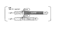
- One allele contains a chimeric gene in which a localized protein coding sequence, a cleavage site, and a first fluorescent protein coding sequence are linked in-frame in this order, and the other allele contains the above-mentioned site.
- a cell comprising a chimeric gene in which the localized protein coding sequence, the cleavage site, and the second fluorescent protein coding sequence are linked in-frame in this order.
- a method for producing a cell in which only one allele is genome-edited and a guide RNA, an expression vector and a kit that can be used in the method. Furthermore, a method for predicting a genome editing pattern, a method for analyzing a genome editing pattern, and a cell that can be used in the analysis method are provided.
- FIG. 1A is a diagram explaining the gene composition of the AIMS cells prepared in the examples.
- FIG. 1B is a diagram illustrating an indel evaluation method by AIMS.
- FIG. 1C shows the P2A peptide coding sequence used in the preparation of AIMS cells in Examples, and the aP2A sequence in which a silent mutation was introduced into the sequence. It is a figure which shows the structure of the plasmid used for the genome editing in the Example. It is a figure which shows the operation procedure after transfection of the plasmid used for genome editing in an Example.
- FIGS. 3A and 3B show the results of investigating whether the introduction rate of the both-side allele indel and the one-side allele indel is changed by introducing a single base mismatch with the target sequence into the spacer sequence of sgRNA.
- FIG. 3A shows an example of the spacer sequence used in the examples.
- FIG. 3B shows the results of indel pattern analysis using Tbx3-P2A-AIMS as AIMS cells.
- FIG. 3C shows the results of indel pattern analysis using Tbx3-P2A-AIMS as AIMS cells.
- FIG. 3D shows the result of indel pattern analysis using Cdh1-aP2A-AIMS as AIMS cells.
- FIG. 4A to 4F show the results of investigating whether or not the introduction rate of the both-side allele indel and the one-side allele indel is changed by adding a nucleotide residue to the 5'end of the spacer sequence of sgRNA.
- FIG. 4A shows an example of sgRNA used in the examples.
- FIG. 4B shows the results of indel pattern analysis using Cdh1-P2A-AIMS as AIMS cells.
- FIG. 4C shows the results of indel pattern analysis using Cdh1-P2A-AIMS as AIMS cells.
- FIG. 4D shows the results of investigating whether the introduction rate of the both-side allele indel and the one-side allele indel changes by changing the amount of the sgRNA expression plasmid to be transfected.
- FIG. 4E shows the results of investigating whether the introduction rate of the both-side allele indel and the one-side allele indel changes by changing the amount of the sgRNA expression plasmid to be transfected.
- the Cas9 and puromycin resistance gene expression plasmid and the sgRNA expression plasmid were separated into separate plasmids, and only the amount of the sgRNA expression plasmid was changed.
- FIG. 4F shows the results of genome editing targeting Rosa26 and albumin gene (Alb).
- FIGS. 5A to 5C show the results of evaluating the introduction rate of homologous recombination containing no indel using AIMS cells.
- FIG. 5A is a diagram illustrating the outline of the method used in Example 5.
- FIG. 5B shows the results of homologous recombination using sgRAN containing a spacer sequence having a single base mismatch.
- FIG. 5C shows the result of homologous recombination using sgRAN in which cytosine was added to the 5'end of the spacer sequence.
- FIG. 6A to 6C show the results of investigating whether or not the introduction rate of the both-side allele indel and the one-side allele indel is changed by combining the introduction of a single-base mismatch and the addition of a nucleotide residue to the 5'end.
- FIG. 6A shows the results using sgRNA having a single base mismatch with the target sequence.
- FIG. 6B shows the results using sgRNA having a spacer sequence having a single base mismatch with the target sequence and having 10 cytosines added to the 5′ end.
- FIG. 6C shows the results using sgRNA having a spacer sequence having a single base mismatch with the target sequence and having 25 cytosines added to the 5′ end.
- FIGS. 3B to 3D, FIG. 4B, and FIGS. 6A to 6C show the results of comparison between the predicted value of Pre-Demo-Prediction and the measured value.
- FIG. 8A shows the operation protocol of Pre-Demo-Prediction.
- FIG. 8B shows a predicted value (left diagram) and an actually measured value (right diagram) of Pre-Demo-Prediction. The result of having investigated whether the off-target effect can be suppressed by adding a nucleotide residue to the 5'end of a spacer sequence is shown.
- FIG. 8A shows the operation protocol of Pre-Demo-Prediction.
- FIG. 8B shows a predicted value (left diagram) and an actually measured value (right diagram) of Pre-Demo-Prediction.
- FIG. 9A shows the result of calculating the indel induction rate (P) by the formula (1) described in Example 7 based on the data of FIGS. 6A to 6C.
- FIG. 9B shows the results of verifying the effect of suppressing the off-target action by adding cytosine to the 5'end of the spacer sequence.
- the sgRNA expression vector in which 0, 10, or 25 cytosines were added to the 5′ end of the spacer sequence for the target sequence (GAGTCCGAGCAGAAGAAGAA: 5 to 24 of SEQ ID NO: 83) in the EMX1 gene was introduced into HEK293T and turned on.
- FIG. 9B shows the results of verifying the effect of suppressing the off-target action by adding cytosine to the 5'end of the spacer sequence. Under the same conditions as in FIG. 9B, the number of cytosine additions at the 5'end of the spacer sequence was 0, 5, 10, 15, 20, 25 or 30.
- 10A-10C show the results of a repair test for inherited disease mutations in progressive ossifying fibrodysplasia (FOP).
- FOP progressive ossifying fibrodysplasia
- FIG. 10A shows an outline of the method for repairing FOP inherited disease mutation.
- FIG. 10B shows the results of evaluation of the HDR induction efficiency when human iPS cells having a FOP inherited disease mutation (wt/R206H) were used to induce the HDR that repairs the mutant allele (R206H).
- FIG. 10C shows the results of evaluation of indel introduction efficiency by genome editing targeting the mutant allele (R206H) in human iPS cells having a FOP inherited disease mutation (wt/R206H).
- 11A to 11D show the results of the FOP hereditary disease model production test.
- FIG. 11A shows an outline of a method for inducing FOP inherited disease mutation.
- FIG. 11A shows an outline of a method for inducing FOP inherited disease mutation.
- FIG. 11B shows the results of evaluating the HDR induction efficiency when the mouse ES cells (wt/wt) were used to induce the HDR that induces the mutant allele (R206H).
- FIG. 11C shows the results of evaluating the indel introduction efficiency by genome editing targeting the Acvr1 gene (wt) in mouse ES cells (wt/wt).
- FIG. 11D is a photograph showing an abnormal bone (arrow) formed in a chimeric mouse produced by microinjecting mouse ES cells having a FOP inherited disease mutation (wt/R206H) produced by HDR induction into mouse fertilized eggs. Is.
- FIG. 12A shows the result of a cytotoxicity evaluation test using AIMS.
- FIG. 12B shows the results of a cytotoxicity evaluation test by genome editing targeting ACVR1.
- the horizontal axis is the P value calculated from 253 data obtained by AIMS, and the vertical axis is the ratio of both-sided allele indel (Bi; upper figure), one-sided allele indel (Mono; middle figure), and no indel (Nono; lower figure).
- FIG. 13A shows the results of predicting the ratios of both-sided allele indel (Bi; upper figure), one-sided allele indel (Mono; middle figure), and no indel (None; lower figure) with the mathematical formulas obtained from the graph of FIG. 13A. Actual measured value (horizontal axis).
- the results of genome editing targeting P2A with Cdh1-P2A-AIMS are shown.
- the above figure shows actual data when genome editing was performed by targeting P2A with Cdh1-P2A-AIMS.
- the following figure shows an indel pattern prediction graph obtained based on the method for predicting a genome editing pattern according to one embodiment of the present invention. An outline of the method for producing Compound heterozygoous is shown.
- the target position of P2A-sgRNA1 and Cdh1-sgRNA4 is shown.
- An outline of the method for producing Compound heterozygoous is shown.
- the genomic structure after genome editing with P2A-sgRNA1 and Cdh1-sgRNA4 and the annealing position of each primer are shown.
- the result of the in vitro cleavage assay with sgRNA to which 0, 10, or 25 cytosines are added is shown.
- polynucleotide and “nucleic acid” are used interchangeably and refer to a nucleotide polymer in which the nucleotides are linked by phosphodiester bonds.
- the “polynucleotide” and “nucleic acid” may be DNA, RNA, or may be composed of a combination of DNA and RNA.
- polynucleotide and “nucleic acid” may be polymers of natural nucleotides, and include natural nucleotides and non-natural nucleotides (at least one of natural nucleotide analogues, base moieties, sugar moieties and phosphate moieties).
- nucleotide May be a polymer with a modified nucleotide (for example, phosphorothioate skeleton), or may be a polymer of a non-natural nucleotide.
- base sequence of “polynucleotide” or “nucleic acid” is described by a generally accepted one-letter code unless otherwise specified. Unless otherwise specified, base sequences are described from the 5′ side to the 3′ side.
- nucleotide residues constituting the “polynucleotide” or the “nucleic acid” may be simply described as adenine, thymine, cytosine, guanine, uracil, or the like, or their one-letter codes.
- the term “gene” refers to a polynucleotide containing at least one open reading frame that encodes a particular protein. Genes can include both exons and introns.
- polypeptide In this specification, the terms “polypeptide”, “peptide” and “protein” are used interchangeably and refer to a polymer of amino acids linked by amide bonds.
- the “polypeptide”, “peptide” or “protein” may be a polymer of natural amino acids or a polymer of natural amino acids and unnatural amino acids (chemical analogs of natural amino acids, modified derivatives, etc.). Well, it may be a polymer of unnatural amino acids. Unless otherwise specified, amino acid sequences are written from the N-terminal side toward the C-terminal side.
- allele refers to a pair of genes existing at the same locus on a pair of chromosomes or a pair of base sequences existing at the same locus.
- the pair of genes does not necessarily have to be alleles, and the pair of base sequences need not necessarily have different base sequences.
- double-sided allele refers to both of the pair of genes or the pair of base sequences, and the term “one-sided allele” refers to either one of the pair of genes or the pair of base sequences. Point to.
- Genome editing refers to inducing a mutation at a desired position (target region) on the genome.
- Genome editing may involve the use of nucleases engineered to cleave DNA in the target region.
- a site-specific nuclease induces a double-strand break (DSB) in the DNA of the target region, followed by homologous recombination repair (Homologous Directed Repair: HDR) and non-homologous end rejoining (Non-).
- HDR homologous Directed Repair
- Non- non-homologous end rejoining
- the genome is repaired by an endogenous process of the cell, such as Homologous End-Joining Repair (NHEJ).
- NHEJ Homologous End-Joining Repair
- NHEJ is a repair method in which double-strand-cut ends are ligated without using a template DNA for repair, and insertion and/or deletion (indel) is frequently induced during repair.
- HDR is a repair mechanism using a template DNA for repair, and it is also possible to introduce a desired mutation into a target region.
- the genome editing technique for example, the CRISPR/Cas system is preferably exemplified.
- the term “repair template DNA” refers to DNA used for repairing double-strand breaks in DNA and capable of homologous recombination with DNA around the target region.
- the term “donor vector” refers to foreign DNA used as a template DNA for repair.
- the donor vector contains a base sequence flanking the target region as a homology arm.
- a homology arm consisting of a base sequence adjacent to the 5'side of a target region is referred to as a "5'arm”
- a homology arm consisting of a base sequence adjacent to the 3'side of a target sequence is referred to as a "3'arm”. May be listed.
- the donor vector can include a desired nucleotide sequence between the 5'arm and the 3'arm.
- the length of each homology arm is preferably 3 kb or more, and usually about 5 to 10 kb.
- the lengths of the 5'arm and the 3'arm may be the same or different, but are preferably the same.
- safe harbor region refers to a region on the genome that has been demonstrated to allow the insertion of foreign DNA without exhibiting a harmful effect on cells.
- Known examples of safe harbor regions include AAVS1 in humans and Rosa26 in mice.
- Cas protein refers to a CRISPR-associated protein.
- the Cas protein forms a complex with the guide RNA and exhibits endonuclease activity or nickase activity.
- the Cas protein is not particularly limited, and examples thereof include Cas9 protein, Cpf1 protein, C2c1 protein, C2c2 protein, and C2c3 protein.
- Cas proteins include wild-type Cas proteins and their homologs (paralogs and orthologs), and variants thereof, as long as they exhibit endonuclease activity or nickase activity in cooperation with guide RNA.
- the Cas proteins are those involved in the class 2 CRISPR/Cas system, more preferably those involved in type II CRISPR/Cas system.
- a preferred example of Cas protein is Cas9 protein.
- Cas9 protein refers to the Cas protein involved in the Type II CRISPR/Cas system.
- the Cas9 protein shows an activity of forming a complex with a guide RNA and cooperating with the guide RNA to cleave the DNA in the target region.
- the Cas9 protein includes the wild-type Cas9 protein and homologs thereof (paralogs and orthologs), and mutants thereof as long as they have the above-mentioned activity.
- the wild-type Cas9 protein has a RuvC domain and an HNH domain as nuclease domains, but the Cas9 protein in the present specification may be one in which either the RuvC domain or the HNH domain is inactivated.
- the biological species from which the Cas9 protein is derived is not particularly limited, but bacteria belonging to the genus Streptococcus, the genus Staphylococcus, the genus Neisseria, or the genus Treponema are preferably exemplified. More specifically, S. pyogenes, S.; thermophilus, S. aureus, N.A. meningitidis, or T.
- a Cas9 protein derived from denticola or the like is preferably exemplified.
- the Cas9 protein is S. It is a Cas9 protein derived from Pyogenes.
- S. As the amino acid sequence of the Cas9 protein of Pyogenes, that registered in UniProt with the accession number Q99ZW2 can be used.
- S. An example of the coding sequence for the Cas9 protein of Pyogenes is shown in SEQ ID NO:9.
- the nucleotide sequence shown in SEQ ID NO: 9 is S. 3xFlag and a nuclear transfer signal were added to the 5'end of the Cas9 protein of Pyogenes, and a nuclear transfer signal was added to the 3'end.
- the terms "guide RNA” and "gRNA” are used interchangeably and refer to RNA that is capable of forming a complex with a Cas protein and directing the Cas protein to a target region.
- the guide RNA comprises CRISPR RNA (crRNA) and trans-activated CRISPR RNA (tracrRNA).
- crRNA is involved in binding to a target region on the genome, and tracrRNA is involved in binding to Cas protein.
- the crRNA comprises a spacer sequence and a repeat sequence, the spacer sequence binding to the complementary strand of the target sequence in the target region.
- the tracrRNA comprises an anti-repeat sequence and a 3'tail sequence.
- the anti-repeat sequence has a sequence complementary to the repeat sequence of crRNA, forms a base pair with the repeat sequence, and the 3'tail sequence usually forms three stem loops.
- the guide RNA may be a single-stranded guide RNA (sgRNA) in which the 3′ end of crRNA is linked to the 5′ end of tracrRNA, and crRNA and tracrRNA are separate RNA molecules, and a repeat sequence and an antirepeat sequence are used as bases. It may be a pair.
- the guide RNA is sgRNA.
- the repeat sequence of crRNA and the sequence of tracrRNA can be appropriately selected according to the type of Cas protein, and those derived from the same bacterial species as Cas protein can be used.
- the length of sgRNA can be about 50 to 220 nucleotides (nt), preferably about 60 to 180 nt, more preferably about 80 to 120 nt.
- the length of crRNA can be about 25 to 70 bases including the spacer sequence, and preferably about 25 to 50 nt.
- the length of tracrRNA can be about 10 to 130 nt, preferably about 30 to 80 nt.
- the repeat sequence of crRNA may be the same as that in the bacterial species from which the Cas protein is derived, or may be one in which a part of the 3'end has been deleted.
- the tracrRNA may have the same sequence as the mature tracrRNA in the bacterial species from which the Cas protein is derived, or may be a truncated form in which the 5′ end and/or the 3′ end of the mature tracrRNA is cleaved.
- the tracrRNA may be a truncated form obtained by removing about 1 to 40 nucleotide residues from the 3'end of the mature tracrRNA.
- the tracrRNA may be a truncated form in which about 1 to 80 nucleotide residues are removed from the 5′ end of the mature tracrRNA.
- the tracrRNA may be, for example, a truncated form in which about 1 to 20 nucleotide residues are removed from the 5'end and about 1 to 40 nucleotide residues are removed from the 3'end.
- crRNA repeat sequences and tracrRNA sequences for sgRNA design have been proposed, and a person skilled in the art can design an sgRNA based on known techniques (for example, Jinek et al. (2012) Science, 337, 816-21; Mali et al.
- target sequence refers to a DNA sequence in the genome that is the target of cleavage by Cas protein.
- the target sequence needs to be a sequence adjacent to the 5'side of the protospacer flanking motif (PAM).
- PAM protospacer flanking motif
- As the target sequence a sequence of 17 to 30 bases (preferably 18 to 25 bases, more preferably 19 to 22 bases, still more preferably 20 bases) immediately adjacent to the 5'side of PAM is usually selected.
- a known design tool such as CRISPR DESIGN (crispr.mit.edu/) can be used for designing the target sequence.
- target region refers to a genomic region including a target sequence and its complementary strand.
- the terms “protospacer flanking motif” and “PAM” are used interchangeably and refer to a sequence recognized by a Cas protein upon DNA cleavage by the Cas protein.
- the sequence and position of PAM differ depending on the type of Cas protein.
- PAM should be immediately adjacent to the 3'side of the target sequence.
- the sequence of PAM corresponding to the Cas9 protein differs depending on the bacterial species from which the Cas9 protein is derived. For example, S.
- the PAM corresponding to the Cas9 protein of P. pyogenes is "NGG” and S.
- the PAM corresponding to the Thermophilus Cas9 protein is "NNAGAA", and S.
- the PAM corresponding to the Cas9 protein of Aureus is "NGRRT” or “NGGRR(N)", and The PAM corresponding to the Cas9 protein of Meningitidis is "NNNNGATT", and T. It is "NAAAAAC” corresponding to the Cas9 protein of denticola ("R” is A or G; “N” is A, T, G or C).
- the terms “spacer sequence” and “guide sequence” are used interchangeably and refer to sequences contained in guide RNA that are capable of binding to the complementary strand of the target sequence.
- the spacer sequence is the same sequence as the target sequence (however, T in the target sequence becomes U in the spacer sequence).
- the spacer sequence may contain a single or multiple base mismatch with the target sequence. When a mismatch of a plurality of bases is included, it may have a mismatch at an adjacent position or a mismatch at a distant position.
- the spacer sequence may contain a 1-5 base mismatch to the target sequence.
- the spacer sequence may contain a single base mismatch to the target sequence.
- the spacer sequence is arranged on the 5′ side of crRNA.
- mismatch refers to the spacer sequence containing a base different from the target sequence, or a base different from the target sequence.
- the spacer sequence contains a single base mismatch means that the spacer sequence differs by one base compared to the target sequence.
- the term “indel” means insertions and/or deletions.
- the term “bilateral allele indel” refers to a state in which indels have been generated in the target regions of bilateral alleles by genome editing.
- the term “one-sided allele indel” refers to a state in which indels are generated only in the target region of the one-sided allele due to genome editing.
- the term “frameshift indel” refers to an indel that causes a frameshift.
- the term “in-frame indel” refers to indel that does not cause frame shifting.
- AIMS means Allele-specific Indel Monitor System, and refers to a technique capable of detecting indel in an allele-specific manner.
- AIMS cell means a cell constructed to perform AIMS, and refers to a cell capable of detecting indel in an allele-specific manner.
- chimeric gene refers to a polynucleotide in which the coding sequences of two or more different proteins are linked in-frame.
- chimeric protein refers to a protein expressed from a chimeric gene.
- localized protein refers to a protein that is localized in a certain part of a cell (eg, nucleus or cell membrane).
- nuclear localization protein means a protein localized in the nucleus
- cell membrane localization protein means a protein localized in the cell membrane.
- cleavage site refers to an amino acid sequence or a nucleotide sequence, which this sequence directs for cleavage, for example because it is recognized by and/or can be cleaved by a cleavage enzyme.
- the polypeptide chain is cleaved by hydrolysis of one or more peptide bonds connecting amino acids.
- the polynucleotide chain is cleaved by hydrolysis of one or more phosphodiester bonds between the nucleotides. Cleavage of the peptide bond or phosphodiester bond may result from chemical or enzymatic cleavage.
- Enzymatic cleavage in the case of polynucleotide chains, refers to the cleavage of the polynucleotide, eg achieved by restriction endonucleases (eg type I, type II, type III, type IV or artificial restriction enzymes).
- restriction endonucleases eg type I, type II, type III, type IV or artificial restriction enzymes.
- the polypeptide chain include endopeptidase, exopeptidase or protease (eg, serine-protease, cysteine-protease, metallo-protease, threonine-protease, aspartate-protease, glutamic acid-protease) and the like, but are not limited thereto. Not, refers to the cleavage of the polypeptide achieved by the proteolytic enzyme.
- endopeptidase cleavage site means that this sequence is cleaved by an endopeptidase (eg trypsin, pepsin, elastase, thrombin, collagenase, furin, thermolysin, endopeptidase V8, cathepsin) or Refers to a cleavage site within the amino acid sequence that is cleavable.
- endopeptidase eg trypsin, pepsin, elastase, thrombin, collagenase, furin, thermolysin, endopeptidase V8, cathepsin
- the cleavage site may be cleaved by an autoprotease, ie a protease that cleaves peptide bonds within the same protein molecule that also includes the protease.
- autoproteases include flavivirus NS2 protease or birnavirus VP4 protease.
- the term "cleavage site" refers to an amino acid or nucleotide sequence that prevents the formation of peptide bonds between amino acids or phosphodiester bonds between nucleotides. For example, the formation of peptide bonds is prevented due to the concomitant self-processing of the polypeptide, resulting in two discontinuous translation products resulting from a single translation event in a single open reading frame. obtain.
- such self-processing is caused by a pseudo stop codon sequence, which induces the translation complex to move from one codon to the next without forming a peptide bond, a "ribosome.” It is achieved by "skip".
- sequences that induce ribosome skipping include viral 2A peptides or 2A-like peptides used by several families of viruses, including picornaviruses, insect viruses, Aphtoviridae, rotaviruses and trypanosomes. In the present specification, both are collectively referred to as “2A peptide”), but are not limited thereto.
- the term "self-cleavage site” means that the sequence is cleaved or cleavable without any additional molecules, or of a peptide bond or phosphodiester bond within the sequence. Refers to a cleavage site within an amino acid sequence or nucleotide sequence where formation is prevented in the first step (eg, via self-processing concurrent with translation as described above).
- the cleavage site typically comprises a few amino acids, or is encoded by several codons (eg, in such a case, the "cleavage site” is not translated into a protein but causes interference with translation). It Therefore, the cleavage site also serves the purpose of being a peptide linker, ie sterically separating the two peptides. Thus, in some embodiments, the "cleavage site” is both the peptide linker and also provides the cleavage function described above. In this embodiment, the cleavage site may include additional N and/or C terminal amino acids.
- the term “2A peptide” refers to a viral 2A peptide or 2A-like peptide.
- the 2A peptide is a peptide that is cleaved by the peptidase or ribosome skip mechanism.
- Examples of the 2A peptide include 2A peptide (F2A) derived from foot-and-mouth disease virus (FMDV), 2A peptide (E2A) derived from equine rhinitis A virus (ERAV), 2A peptide (P2A) derived from Porcine teschovirus (PTV-1), and Examples include 2A peptide (T2A) derived from Thosea virus (TaV).
- the term "genome editing pattern” refers to the induction state of genome editing in each allele of the target region of the cell targeted for genome editing. That is, it means a state in which genome editing is induced in both alleles or genome editing is induced only in one allele.
- operably linked with respect to a polynucleotide means that the first base sequence is located sufficiently close to the second base sequence, and the first base sequence is the second base sequence or It means that it can affect the region under the control of the second nucleotide sequence.
- a polynucleotide operably linked to a promoter means that the polynucleotide is linked so that it is expressed under the control of the promoter.
- the term “expressible state” refers to a state in which a polynucleotide can be transcribed in a cell into which the polynucleotide has been introduced.
- expression vector refers to a vector containing a target polynucleotide, which is provided with a system that makes the target polynucleotide expressible in cells into which the vector has been introduced.
- Cas protein expression vector means a vector capable of expressing Cas protein in cells into which the vector has been introduced.
- the “guide RNA expression vector” means a vector capable of expressing guide RNA in cells into which the vector has been introduced.
- silent mutation refers to a gene mutation in which the amino acid sequence of the encoded protein is unchanged.
- sequence identity means the insertion and deletion of two base sequences or amino acid sequences so that the corresponding bases or amino acids are most matched. It is determined as the ratio of the matched bases or amino acids to the entire base sequence or the entire amino acid sequence, excluding the gaps in the obtained alignment, by arranging them in parallel with each other with a gap in between.
- sequence identity between base sequences or amino acid sequences can be determined using various homology search software known in the art. For example, the value of sequence identity of a nucleotide sequence can be obtained by calculation based on the alignment obtained by the known homology search software BLASTN, and the value of sequence identity of an amino acid sequence can be obtained by a known homology search. It can be obtained by calculation based on the alignment obtained by the software BLASTP.
- the present invention provides: (A) (a1) a guide RNA in which one or more nucleotide residues are added to the 5′ end of a spacer sequence, (a2) a single or multiple bases relative to a target sequence. A guide RNA containing a spacer sequence having a mismatch, and (a3) at least one member selected from the group consisting of the expression vector of the guide RNA of (a1) or (a2) above; (B) A method for producing a cell in which only one allele is genome-edited, which comprises a step of introducing at least one selected from the group consisting of Cas protein and its expression vector into a cell.
- RNA a guide RNA in which one or more nucleotide residues are added to the 5′ end of the spacer sequence, (a2) a spacer having a mismatch of one base or a plurality of bases with respect to the target sequence
- a guide RNA containing a sequence and (a3) at least one selected from the group consisting of the expression vectors of the guide RNA of (a1) or (a2) above are used.
- the guide RNA (a1) is a guide RNA in which one or more nucleotide residues are added to the 5'end of the spacer sequence.
- the spacer sequence is not particularly limited as long as it is a spacer sequence that targets any target sequence.
- the length of the spacer sequence may be a length corresponding to the target sequence, and a sequence of usually 17 to 30 bases, preferably 18 to 25 bases, more preferably 19 to 21 bases, still more preferably 20 bases is selected.
- the spacer sequence has the same sequence as the target sequence (however, "T” in the target sequence becomes “U” in the spacer sequence), but it has a mismatch as long as it has the ability to bind to the complementary strand of the target sequence. You may have. In general, mismatches on the 5'side of the spacer sequence are permissive.
- a spacer sequence having a single base mismatch with the target sequence is preferable, as in the guide RNA (a2) described later.
- the number of nucleotide residues added to the 5'end of the spacer sequence is one or more, and is not particularly limited, and examples thereof include a range of 1 to 50. ..
- the number of nucleotide residues to be added can be appropriately set depending on the type of spacer sequence.
- the number of nucleotide residues added can be 5 or more, 10 or more, 15 or more, 20 or more, 25 or more, or the like.
- the rate of genome editing with only one allele can be further increased.
- the upper limit of the number of additional nucleotide residues is not particularly limited, but since the ratio of genome editing is not changed by only one allele, it can be 50 or less, preferably 40 or less, more preferably 35 or less. preferable.
- a preferable range of the number of additional nucleotide residues is, for example, 5 to 50, and is 5 to 40, 5 to 35, 10 to 40, 10 to 35, 15 to 35, or 20 to 30. It is preferable that the number is individual.
- the type of additional nucleotide residue is not particularly limited, but, for example, all the same nucleotide residues can be used.
- the additional nucleotide residue can be selected from the group consisting of polyadenine (polyA), polyuracil (polyU), polycytosine (polyC), and polyguanine (polyG).
- the type of additional nucleotide residue is preferably poly-C (all cytosine residues) or poly-G (all guanine residues), because the rate of genome editing with only one allele is improved. Is more preferable.
- the additional polynucleotide residue is usually a complementary sequence of the terminator sequence that stops transcription from the promoter used. Does not include. For example, when the U6 promoter is used, the transcription is stopped when 5 consecutive thymines are present, and therefore the additional nucleotide residue usually does not include a sequence of 5 or more consecutive uracils.
- the guide RNA of (a2) is a guide RNA containing a spacer sequence having a mismatch of one base or a plurality of bases with respect to the target sequence.
- the spacer sequence of the guide RNA of (a2) has a mismatch of 1 base or a plurality of bases with respect to an arbitrary target sequence.
- the multiple base mismatch is, for example, a mismatch of 2 to 5 bases, preferably a mismatch of 2 to 4 bases, more preferably a mismatch of 2 or 3 bases, still more preferably a mismatch of 2 bases.
- the length of the spacer sequence is the same as that of the guide RNA of (a1) above, but 20 bases is particularly preferable.
- the position where the spacer sequence has a mismatch of 1 base or a plurality of bases with respect to the target sequence is not particularly limited.
- the spacer sequence when the spacer sequence has 20 bases, there may be a mismatch in any of the 1st to 20th bases counting from the 3'end side to the 5'end side, and for example, there is a mismatch in the 1st to 17th bases. be able to.
- the spacer sequence has a single base mismatch within the above range, the rate of genome editing with only one allele can be further increased.
- a mismatch can be present in one or more bases selected from the group consisting of the 2nd to 6th, the 8th to 9th seeds, and the 15 to 17th, counting from the 3'end side to the 5'end side. ..
- the base in the mismatch base is not particularly limited as long as it is a base different from the base in the target sequence.
- the mismatch base in the spacer sequence can be, for example, a pyrimidine base (cytosine or uracil) if the base in the target sequence is a purine base (adenine or guanine).
- the mismatch base in the spacer sequence can be a purine base (adenine or guanine), for example, if the base in the target sequence is a pyrimidine base (cytosine or thymine).
- the mismatch base in the spacer sequence may be uracil if the base in the target sequence is adenine, adenine if the base in the target sequence is thymine, cytosine if the base in the target sequence is guanine, and cytosine if the base in the target sequence is cytosine. It can be guanine, for example.
- the guide RNA used in the production method of the present embodiment may have the features of the guide RNAs (a1) and (a2) described above. That is, the guide RNA is a spacer sequence in which one or more nucleotide residues are added to the 5′ end of the spacer sequence, and the spacer sequence has a mismatch of 1 base or a plurality of bases with respect to the target sequence. It may be RNA.
- the guide RNA is a spacer sequence in which one or more nucleotide residues are added to the 5′ end of the spacer sequence, and the spacer sequence has a mismatch of 1 base or a plurality of bases with respect to the target sequence. It may be RNA.
- the number and type of additional nucleotide residues are the same as those described above in “ ⁇ (a1) Guide RNA>>”.
- the position and type of the mismatch bases are the same as those described in ⁇ Guide RNA of (a2)>> above.
- the guide RNA expression vector of (a1), (a2) or (a12) may be used instead of the guide RNA of (a1), (a2) or (a12). .
- the production method of the present embodiment uses the guide RNA expression vector of (a1), (a2) or (a12) above.
- the expression vector of the guide RNA of (a1), (a2) or (a12) controls the expression of the guide RNA coding sequence and the sequence encoding the guide RNA of (a1), (a2) or (a12) above. It is preferable to include a promoter. In the expression vector, the guide RNA coding sequence is operably linked to the promoter.
- the promoter is not particularly limited, and for example, a pol II type promoter can be used, but from the viewpoint of more accurate transcription of relatively short RNA, pol II is used.
- III-type promoters are preferred.
- the pol III promoter is not particularly limited, but examples thereof include mouse and human U6-snRNA promoters, human H1-RNase P RNA promoters, human valine-tRNA promoters, and the like. When using the U6 promoter, it is preferable that the 5'end of the guide RNA is "G" for initiation of transcription.
- the guide RNA when the guide RNA is the above-mentioned (a1) or (a12) guide RNA, an additional “G” should be added to the 5′ end of the 5 to 50 nucleotide residues added to the 5′ end of the spacer sequence. Is preferred. Further, when the guide RNA is the guide RNA of the above (a2), it is preferable to select a spacer sequence having a “G” at the 5′ end or add “G” to the 5′ end of the spacer sequence.
- the expression vector optionally includes an enhancer, a poly A addition signal, a marker gene, a replication origin, and a gene encoding a protein that binds to the replication origin and controls replication. You can leave.
- the “marker gene” refers to a gene that enables selection and selection of cells by introducing the marker gene into cells. Specific examples of the marker gene include a drug resistance gene, a fluorescent protein gene, a luminescent enzyme gene, and a chromogenic enzyme gene. These may be used alone or in combination of two or more.
- the drug resistance gene examples include, for example, a puromycin resistance gene, a geneticin resistance gene, a neomycin resistance gene, a tetracycline resistance gene, a kanamycin resistance gene, a zeocin resistance gene, a hygromycin resistance gene, and a chloramphenicol resistance gene.
- a puromycin resistance gene examples include a puromycin resistance gene, a geneticin resistance gene, a neomycin resistance gene, a tetracycline resistance gene, a kanamycin resistance gene, a zeocin resistance gene, a hygromycin resistance gene, and a chloramphenicol resistance gene.
- the fluorescent protein gene examples include a green fluorescent protein (GFP) gene, a yellow fluorescent protein (YFP) gene, a red fluorescent protein (RFP) gene, and the like.
- GFP green fluorescent protein
- YFP yellow fluorescent protein
- RFP red fluorescent protein
- the luminescent enzyme gene examples include a luci
- the type of expression vector is not particularly limited, and a known expression vector can be used.
- Examples of the expression vector include a plasmid vector and a virus vector.
- the plasmid vector is not particularly limited, as long as it is a plasmid vector that can be expressed in the cell for which the genome is to be edited.
- a commonly used plasmid vector for expressing animal cells can be used.
- Examples of plasmid vectors for expressing animal cells include, but are not limited to, pX459, pA1-11, pXT1, pRc/CMV, pRc/RSV, pcDNAI/Neo, and the like.
- the viral vector examples include retrovirus (including lentivirus) vector, adenovirus vector, adeno-associated virus vector, Sendai virus vector, herpesvirus vector, vaccinia virus vector, poxvirus vector, poliovirus vector, Sylbis virus vector. , Rhabdovirus vector, paramyxovirus vector, orthomyxovirus vector and the like.
- the expression vector is preferably a plasmid vector.
- the Cas protein is not particularly limited as long as it is used in the CRISPR/Cas system.
- various substances can be used that can form a complex with a guide RNA and can be induced to the target region by the guide RNA to double-strand the DNA in the target region.
- the Cas protein is preferably Cas9 protein, and S. More preferred is the Cas9 protein of Pyogenes.
- the Cas protein may be a mutant of the wild-type Cas protein as long as it forms a complex with the guide RNA and exhibits endonuclease activity or nickase activity (hereinafter referred to as “Cas protein activity”).
- Cas protein activity examples include the following proteins (b1) or (b2).
- (B2) One or more (for example, 2 to 100, preferably 2 to 50, more preferably 2 to 20 and still more preferably 2 to 10) relative to the amino acid sequence of the wild-type Cas protein, and further A protein having an amino acid sequence in which 2 to 5 amino acids, particularly preferably 2 amino acids) are substituted, deleted, added, or inserted, and has Cas protein activity.
- an expression vector of Cas protein may be used instead of the Cas protein.
- the production method of the present embodiment uses a Cas protein expression vector.
- the Cas protein expression vector preferably contains a Cas protein coding sequence and a promoter that controls the expression of the Cas protein coding sequence.
- the Cas protein coding sequence is operably linked to the promoter.
- the promoter is not particularly limited, and various pol II promoters can be used, for example.
- the pol II type promoter is not particularly limited, and examples thereof include CMV promoter, EF1 promoter, SV40 promoter, MSCV promoter, hTERT promoter, ⁇ -actin promoter, CAG promoter, and CBh promoter.
- the expression vector contains, in addition to the Cas protein coding sequence and promoter, optionally an enhancer, a poly A addition signal, a marker gene, an origin of replication, a gene encoding a protein that binds to the origin of replication and controls replication. You may stay. Examples of the marker gene include the same as above.
- the type of expression vector is not particularly limited, and a known expression vector can be used.
- the expression vector include a plasmid vector and a virus vector. Examples of these vectors are the same as above. Of these, the expression vector is preferably a plasmid vector.
- the Cas protein coding sequence contained in the expression vector may be codon-optimized depending on the organism species from which the cell into which the expression vector is introduced is derived.
- codon optimization refers to replacement of at least one codon in the original nucleotide sequence with a codon used more frequently in the target species while maintaining the original amino acid sequence.
- the codon usage table is provided, for example, in "Codon Usage” provided by Kazusa DNA Research Institute. Database” (www.kazusa.or.jp/codon/), and these tables can be used to optimize codons.
- Computer algorithms for codon optimizing particular sequences for expression in particular animal species are also available, eg, in Gene Forge (Atatage; Jacobus, PA).
- the guide RNA expression vector and the Cas protein expression vector may be the same expression vector. That is, in the production method of the present embodiment, an expression vector containing a guide RNA coding sequence and a Cas protein coding sequence in an expressible state can be used. In the expression vector, the guide RNA coding sequence and the Cas protein coding sequence are preferably functionally linked to different promoters.
- the production method of the present embodiment includes a step of (A) introducing the guide RNA of (a1), (a2) or (a12) or the expression vector thereof and (B) the Cas protein or the expression vector thereof into cells. .
- the cells into which the above (A) and (B) are introduced are not particularly limited, and desired cells to be the target of genome editing can be used.
- the organism from which the cells are derived is not particularly limited, and mammals such as humans, monkeys, mice, rats, dogs, cats, rabbits, cows, horses, pigs, goats, and sheep; avian animals such as chickens; snakes, lizards, etc.
- Plants such as yeast and fungus; bacteria such as Escherichia coli, Bacillus subtilis, and cyanobacteria.
- the type of cell is not particularly limited, and examples thereof include cells derived from various tissues or having various properties, such as blood cells, hematopoietic stem cells/progenitor cells, gametes (sperms, eggs), fertilized eggs, fibroblasts, epithelial cells, blood vessels. Examples thereof include endothelial cells, nerve cells, hepatocytes, keratinocytes, muscle cells, epidermal cells, endocrine cells, tissue stem cells, iPS cells, ES cells and cancer cells. Further, cells having various inherited diseases such as sickle cell disease, Huntington's chorea, Duchenne muscular dystrophy, progressive ossifying fibrodysplasia (FOP) and the like can be mentioned.
- FOP progressive ossifying fibrodysplasia
- the method of introducing (A) and (B) above is not particularly limited, and can be appropriately selected depending on the target cell and the type of material (whether it is nucleic acid, protein, etc.).
- Examples of methods for introducing the expression vector into cells include lipofection method, microinjection method, DEAE dextran method, gene gun method, electroporation method, calcium phosphate method and the like.
- examples of the method for infecting cells with the viral vector include the polybrene method.
- RNA transfection reagent such as Lipofectamine (registered trademark) Messenger MAX (manufactured by Life Technologies) can be used.
- the method of introducing the protein into cells is not particularly limited, and a known method can be appropriately selected and used. Examples of such a method include a method using a protein introduction reagent, a method using a protein introduction domain (PTD) fusion protein, and a microinjection method.
- a method using a protein introduction reagent a method using a protein introduction domain (PTD) fusion protein, and a microinjection method.
- PTD protein introduction domain
- the above (A) and (B) may be introduced into cells at the same time, may be introduced sequentially, or may be introduced separately after a certain period of time. In a preferred embodiment, the above (A) and (B) are simultaneously introduced into cells.
- the manufacturing method of the present embodiment may include any step in addition to the above-mentioned introducing step.
- the optional step includes, for example, the step of (C) introducing the donor vector into cells.
- the donor vector contains a base sequence flanking the target region as a homology arm.
- the donor vector can include a desired nucleotide sequence (hereinafter, sometimes referred to as “knock-in sequence”) between the 5′ arm and the 3′ arm.
- the knock-in sequence is not particularly limited and can be any sequence.
- the knock-in sequence may be, for example, a gene knockout sequence, a base substitution sequence, or an arbitrary gene sequence. When the knock-in sequence is an arbitrary gene sequence, it is preferable to set the target sequence in the safe harbor region.
- the donor vector may be a circular DNA vector (for example, a plasmid vector) or a linear DNA vector.
- the donor vector may include other sequences in addition to the homology arm and knockin sequences. Examples of other sequences include a marker gene, a replication origin, a gene encoding a protein that binds to the replication origin and controls replication. Examples of the marker gene include the same as above.
- the method of introducing the donor vector is not particularly limited and can be appropriately selected depending on the target cell.
- Examples of the method for introducing the donor vector into cells include lipofection method, microinjection method, DEAE dextran method, gene gun method, electroporation method, calcium phosphate method and the like.
- the donor vector may be introduced into the cells simultaneously with the above (A) and (B), may be introduced sequentially, or may be introduced after a certain period of time after the introduction of (A) and (B). Good.
- the donor vector is introduced into cells at the same time as (A) and (B) above.
- the optional step may be a step of culturing the cell after introducing the above-mentioned (A) and (B) and, if necessary, the above-mentioned (C) into the cell. Culturing of cells may be carried out under appropriate culture conditions depending on the type of cells.
- the above (A), (B) and/or (C) is a vector containing a drug resistance marker
- the culture may be performed in the presence of the drug. By culturing in the presence of the drug, genome-edited cells can be efficiently selected.
- cells may be cloned by diluting or plating the cell culture medium.
- the optional step may be a step of introducing the above-mentioned (A) and (B) and, if necessary, the above-mentioned (C) into a cell and then analyzing a genome editing pattern.
- the method for analyzing the genome editing pattern is not particularly limited, but after the introduction step, for example, after appropriately performing the culture step, the cell culture medium is plated, and DNA is extracted from the resulting colony to sequence the target region. Examples include a method of performing analysis. By performing a sequence analysis of the target regions of both alleles, it can be confirmed whether or not the cell is a cell in which only one allele is genome-edited.
- the guide RNA (a1) a guide RNA in which one or more nucleotide residues are added to the 5'end of a spacer sequence, or (a2) one base or a plurality of bases relative to a target sequence
- the guide RNA containing the spacer sequence having the mismatch of it is possible to increase the proportion of cells in which the genome is edited with only one allele during genome editing. Therefore, according to the production method of the present embodiment, it is possible to efficiently produce cells whose genome has been edited with only one allele. Further, according to the production method of the present embodiment, it is possible to suppress the cytotoxicity due to the introduction of sgRNA, Cas9 and the like.
- the present invention provides (A) (a1) a guide RNA in which one or more nucleotide residues are added to the 5'end of a spacer sequence, (a2) one or more bases relative to a target sequence. At least one selected from the group consisting of a guide RNA containing a spacer sequence having a mismatch, and (a3) the guide vector of (a1) or (a2), and (B) a Cas protein and its expression vector.
- a method of editing a genome of only one unilateral allele which comprises the step of introducing at least one selected from the group consisting of:
- the invention provides a guide RNA in which one or more nucleotide residues are added to the 5'end of the spacer sequence.
- the guide RNA of the present embodiment is the same as the guide RNA of (a1) described in the above section [Method for producing cells in which only one allele is genome-edited] ⁇ (A) Guide RNA>.
- the spacer sequence is preferably a sequence having a mismatch of 1 base or a plurality of bases with the target sequence. That is, it is preferably the guide RNA of the above (a12).
- the present invention provides a guide RNA expression vector in which one or more nucleotide residues are added to the 5'end of the spacer sequence.
- the expression vector of the present embodiment is the same as the expression vector of the guide RNA of (a1) or (a12) described in the section [Method for producing cells in which only one allele is genome-edited] ⁇ (A) Guide RNA> described above. Is.
- the expression vector of the present embodiment may further contain a Cas protein coding sequence (preferably Cas9 protein coding sequence) in an expressible state.
- the present invention provides: (A) (a1) a guide RNA in which one or more nucleotide residues are added to the 5′ end of a spacer sequence; Only one allele containing the guide RNA containing a spacer sequence having a mismatch and (a3) at least one selected from the group consisting of the expression vectors of the guide RNAs of (a1) or (a2) was edited.
- a cell manufacturing kit is provided. The production kit preferably further includes (B) at least one selected from the group consisting of Cas protein and its expression vector.
- the guide RNAs of (a1) and (a2) are the same as the guide RNAs of (a1) and (a2) described in the section [Method for producing cells in which only one allele is genome-edited] ⁇ (A) Guide RNA> described above. It is the same.
- the guide RNA may be the guide RNA described in (a12) above.
- the expression vector of (a3) is the same as the expression vector of the guide RNA of (a3) described in the section [Method for producing cells in which only one allele is genome edited] ⁇ (A) Guide RNA>.
- the Cas protein and the expression vector thereof in (B) are the same as those described in the above [Method for producing cells in which only one allele is genome-edited] ⁇ (B) Cas protein>.
- the guide RNA coding sequence and the Cas protein coding sequence may be contained in the same expression vector in an expressible state.
- the kit of the present embodiment may have other configurations in addition to the above (A) and (B). Other configurations are not particularly limited, and examples thereof include instructions for producing cells in which only one allele is genome-edited, reagents used for introducing an expression vector into cells, and the like.
- the present invention provides (i) a step of introducing a guide RNA or an expression vector thereof, and a Cas protein or an expression vector thereof into a cell to perform genome editing, and (ii) DNA from the cell subjected to the genome editing. And (iii) amplifying a DNA fragment containing a target region from the DNA, (iv) analyzing the sequence of the amplified DNA fragment, and determining an indel induction rate (P) to the target region.
- Step (i) is a step of introducing a guide RNA or an expression vector thereof and a Cas protein or an expression vector thereof into cells to perform genome editing.
- the guide RNA is not particularly limited, and a guide RNA usable in the CRISPR/Cas system may be used.
- the spacer sequence is not particularly limited as long as it is a spacer sequence having an arbitrary target sequence as a target sequence.
- the expression vector of the guide RNA can be prepared in the same manner as the expression vector described in the section [Method for producing cells in which only one allele is genome-edited] ⁇ (A) Guide RNA>.
- the Cas protein is not particularly limited, and a Cas protein that can be used in the CRISPR/Cas system may be used.
- the Cas protein is preferably the Cas9 protein, more preferably S. It is the Cas9 protein of Pyogenes.
- the Cas protein expression vector can be prepared in the same manner as the expression vector described in the above section [Method for producing cells in which only one allele is genome-edited] ⁇ (B) Cas protein>.
- the guide RNA and Cas protein, or their expression vector can be introduced into cells in the same manner as the method described in the above [Method for producing cells in which only one allele is genome-edited] ⁇ Introduction step>. After introduction into cells, culturing may be carried out as appropriate.
- the expression vector of the guide RNA and the Cas protein is introduced into a cell and the expression vector has a drug resistance marker, the cell into which the expression vector is introduced is selected by culturing the cell in the presence of the agent. You may.
- Step (ii) is a step of extracting DNA from the cells that have undergone genome editing in step (i).
- the DNA extraction method is not particularly limited, and a known DNA extraction method may be used.
- Examples of the DNA extraction method include a phenol/chloroform extraction method and a method of heating under alkaline conditions (eg, 99° C. for 10 minutes in the presence of 50 mM NaOH). Further, a commercially available DNA extraction kit or the like can also be used.
- Step (iii) is a step of amplifying a DNA fragment containing a target region from the DNA extracted in step (ii).
- the method for amplifying the target region is not particularly limited, and a known nucleic acid fragment amplification method may be used.
- the nucleic acid fragment amplification method include a PCR method and an isothermal amplification method.
- a primer capable of amplifying the target region can be designed, and the DNA fragment in the target region can be amplified by the PCR method or the isothermal amplification method.
- the length of the amplified DNA fragment is not particularly limited as long as it includes the target region, but can be, for example, about 20 to 1000 bp, usually about 350 to 750 bp.
- the amplified DNA fragment may be cloned using a commercially available cloning vector or the like. Furthermore, a cloning vector having the amplified DNA fragment inserted therein may be introduced into Escherichia coli or the like, and the E. coli may be cultured to form colonies. DNA may be extracted from the thus obtained colonies and subjected to the sequence analysis in step (iv).
- the number of colonies to be subjected to sequence analysis is not particularly limited, but may be, for example, about 10 to 200, about 20 to 100, or about 20 to 50.
- Step (iv) is a step of performing sequence analysis of the DNA fragment amplified in step (iii) to determine the indel induction rate (P) to the target region.
- the method of sequence analysis of the DNA fragment is not particularly limited, and a known sequence analysis method may be used.
- a commercially available sequencer can be used for sequence analysis, and DNA sequencing can be performed according to the method recommended by the manufacturer.
- the presence or absence of indel in the amplified DNA fragment may be analyzed by T7E1 assay, or the sequence analysis by sequencer and T7E1 assay may be used in combination.
- the indel induction rate (P) to the target region can be calculated by the following formula (p) based on the result of sequence analysis.
- the indel induction rate (P) obtained in the step (iv) By substituting the value of the indel induction rate (P) obtained in the step (iv) into the above formula (m) or (m1) and (b) or (b1), the indel induction rate (mono) for only one side allele (mono) ) And the indel induction rate (bi) to both alleles can be determined, respectively.
- the values of indel induction rate (mono) and indel induction rate (bi) obtained by the method of the present embodiment are close to the respective indel induction rates confirmed by actual tests. ing.
- the prediction method of the present embodiment it is possible to predict the indel induction rate to only one allele and the indel induction rate to both alleles with a simple operation. Therefore, by performing the prediction method of this embodiment using an arbitrary guide RNA, it is possible to predict the genome editing pattern by genome editing using the guide RNA.
- the invention comprises a chimeric gene in one of which an localization protein coding sequence, a cleavage site coding sequence, and a first fluorescent protein coding sequence are linked in-frame in this order.
- first and second are descriptions for convenience.
- the cell of the present embodiment is a cell (AIMS cell) that can be used for analysis of a genome editing pattern by AIMS described later.
- AIMS cell a cell that can be used for analysis of a genome editing pattern by AIMS described later.
- the localized protein can be appropriately selected depending on the cell to be subjected to genome editing.
- the localized protein may be a naturally-occurring protein as long as it is a protein that is localized in the cell, and artificial proteins (for example, mutant proteins or truncated proteins of the naturally-localized protein, localization signals Including peptides).
- the native protein may be an endogenous protein of the target cell or an exogenous protein (localized protein derived from another organism).
- the localized protein is preferably an endogenous protein of the target cell, which is always expressed.
- the localization protein may be a nuclear localization protein or a cell membrane localization protein.
- nuclear localization proteins include various transcription factors and various transcription regulatory factors.
- specific examples include the TBX family such as TBX3 protein and the SOX family such as SOX2 protein.
- cell membrane localization proteins include various cell membrane receptors, various cell membrane antigens, and the like.
- specific examples include the cadherin family such as E-cadherin and the SSEA family such as SSEA4.
- the localized protein coding sequence is not particularly limited as long as it has a nucleotide sequence encoding the localized protein, and may contain silent mutations.
- the localized protein coding sequence may be one that utilizes the endogenous gene sequence of the endogenous localized protein or may be exogenous DNA.
- the localized protein coding sequence is exogenous DNA (eg, exogenous natural localized protein coding sequence, artificial localized protein coding sequence, etc.)
- the localized protein coding sequence is constitutive in the target cell. It may be one that is operably linked to a promoter that is expressed in.
- the cleavage site is not particularly limited, and examples thereof include a self-cleavage site or an endopeptidase cleavage site, and a specific example thereof is a 2A peptide coding sequence.
- the 2A peptide is not particularly limited, and a known 2A peptide or 2A-like peptide can be used.
- a 2A peptide selected from the group consisting of P2A peptide, F2A peptide, E2A peptide, and T2A peptide can be used.
- the cleavage site encodes a self-cleaving sequence such as a ribosome skip or an amino acid sequence such as a peptidase recognition sequence, it may include silent mutations.
- the 2A peptide coding sequence is not particularly limited as long as it has a base sequence that encodes the 2A peptide, and may include silent mutations.
- the 2A peptide coding sequence may be codon-optimized depending on the target cell.
- the P2A peptide coding sequence and the coding sequence (aP2A) containing a silent mutation in the P2A peptide coding sequence are shown in SEQ ID NO: 14 and SEQ ID NO: 15, respectively.
- the first fluorescent protein is not particularly limited, and a known fluorescent protein can be used.
- the fluorescent proteins for example, Sirius, BFP, EBFP, ECFP, mTurquoise, TagCFP, AmCyan, mTFP1, MidoriishiCyan, CFP, TurboGFP, AcGFP, TagGFP, Azami-Green, ZsGreen, EmGFP, GFP, EGFP, GFP2, HyPer, TagYFP , EYFP, Venus, YFP, PhiYFP, PhiYFP-m, TurboYFP, ZsYellow, mBanana, KusabiraOrange, mOrange, TurboRFP, tdTomato, DsRed, DsRed-Express, DsRed2, TagRFP, DsRed-Monomer, AsRed, AsRed2, mStrawberry, TurboFP602, RFP , ERFP, mRFP1, JRed, Killer
- the first fluorescent protein coding sequence is not particularly limited as long as it has a nucleotide sequence encoding a fluorescent protein, and may include silent mutations. Further, it may be codon-optimized depending on the target cell.
- the fluorescent protein coding sequence the tdTomato coding sequence and the Venus coding sequence are shown in SEQ ID NO: 16 and SEQ ID NO: 17, respectively.
- the second fluorescent protein is a fluorescent protein different from the first fluorescent protein.
- the second fluorescent protein is not particularly limited as long as it is different from the first fluorescent protein, and a known fluorescent protein can be used. Examples of the fluorescent protein include the same as those exemplified for the first fluorescent protein.
- the second fluorescent protein preferably has a fluorescence wavelength different from that of the first fluorescent protein.
- the second fluorescent protein coding sequence is not particularly limited as long as it has a nucleotide sequence encoding a fluorescent protein, and may include silent mutations. Further, it may be codon-optimized depending on the target cell.
- the cell of this embodiment has a localization protein coding sequence, a cleavage site (for example, a 2A peptide coding sequence), a first fluorescent protein in one allele (hereinafter, sometimes referred to as “first allele”).
- the coding sequence and a chimeric gene (hereinafter, sometimes referred to as “first chimeric gene”) linked in-frame in this order are included.
- the other allele hereinafter, sometimes referred to as “second allele” has a localization protein coding sequence, a cleavage site (eg, 2A peptide coding sequence), and a second fluorescent protein coding sequence.
- the cleavage sites eg 2A peptide coding sequence
- the localized protein coding sequences contained in the first chimeric gene and the second chimeric gene are preferably the same.
- the first chimeric gene and the second chimeric gene are located at the loci of the endogenous localization protein gene of the cell.
- the first chimeric gene and the second chimeric gene may be located at the same locus within the safe harbor region.
- the first chimeric gene and the second chimeric gene are present on the first allele and the second allele, respectively, in an expressible state in the cell.
- AIMS cells containing the first chimeric gene and the second chimeric gene can be prepared by using a technique such as genome editing.
- a technique such as genome editing.
- a donor vector (knock-in vector) containing the first chimeric gene and the second chimeric gene is prepared, respectively.
- the knock-in vector can be produced by a known method.
- the homology arm can be appropriately designed according to the position on the genome where the chimeric gene is inserted.
- the homology arm is preferably the nucleotide sequence of the endogenous localized protein gene and the region adjacent to the localized protein gene.
- the homology arm of the knock-in vector is the nucleotide sequence of the endogenous localized protein gene and the region adjacent to the localized protein gene
- the target sequence is, for example, selected from the sequences in the endogenous localized protein gene. To be done.
- the guide RNA or the expression vector thereof, the Cas protein or the expression vector thereof, and the knockin vector of the first chimeric gene and the knockin vector of the second chimeric gene are introduced into cells.
- the expression vectors may be the same expression vector. Examples of the introduction method include the same methods as those described in the above [Method for producing cells in which only one allele is genome-edited] ⁇ Introduction step>. After introduction into cells, culturing may be carried out as appropriate.
- the expression vector of the guide RNA and the Cas protein When the expression vector of the guide RNA and the Cas protein is introduced into a cell and the expression vector has a drug resistance marker, the cell into which the expression vector is introduced is selected by culturing the cell in the presence of the agent. You may. Even when each knock-in vector of the first and second chimeric genes has a drug resistance marker, cells in which the knock-in vector has been introduced may be selected by culturing the cells in the presence of the drug.
- the selection of fluorescent cells can be performed by a known method, for example, using a flow cytometer, a fluorescence microscope or the like. In the obtained cells, it can be confirmed by PCR or the like that the first chimeric gene and the second chimeric gene are inserted into the respective loci of interest of the first and second alleles, respectively.
- the chimeric protein expressed from the first chimeric gene and the second chimeric gene is cleaved at the cleavage site (for example, 2A peptide). Since the first fluorescent protein and the second fluorescent protein are separated from the localized protein, respectively, they are distributed in the entire cell without being localized in the cell (see “wt” in FIG. 1A). On the other hand, when a frame shift indel occurs at the cleavage site (eg, 2A peptide coding sequence) as a result of genome editing targeting the cleavage site (eg, 2A peptide coding sequence), a fluorescent protein is not produced.
- the cleavage site eg, 2A peptide coding sequence
- the target cells used for producing AIMS cells are not particularly limited, and desired cells can be used.
- the organism from which the cells are derived is not particularly limited, and mammals such as humans, monkeys, mice, rats, dogs, cats, rabbits, cows, horses, pigs, goats, and sheep; avian animals such as chickens; snakes, lizards, etc.
- the type of the target cell is not particularly limited, and examples thereof include cells of various tissues or having various properties, such as blood cells, hematopoietic stem cells/progenitor cells, gametes (sperms, eggs), fertilized eggs, fibroblasts, epithelial cells, Examples include vascular endothelial cells, nerve cells, hepatocytes, keratinocytes, muscle cells, epidermal cells, endocrine cells, tissue stem cells, iPS cells, ES cells, cancer cells and the like.
- the target cells may be primary culture cells or cell lines that have been immortalized. Examples of cell lines include human-derived Hela cells, African green monkey-derived COS7 cells, mouse-derived 3T3 cells, hamster-derived CHO cells, rat-derived PC12 cells, and the like.
- the genome editing pattern is analyzed using the above characteristics of the cell of the present embodiment.
- the present invention provides (I) a guide RNA or an expression vector thereof targeting the cleavage site (for example, a 2A peptide coding sequence), and a Cas protein or the same in the cell (AIMS cell) of the above embodiment. Based on the step of introducing the expression vector and performing genome editing, (II) the step (I) and then analyzing the fluorescence pattern of the cell, and (III) the fluorescence pattern analyzed in the step (II) Provided is a method for analyzing a genome editing pattern, which comprises the step of determining the genome editing pattern.
- the method for analyzing the genome editing pattern of the present embodiment is characterized by using the above AIMS cells.
- the genome editing pattern can be analyzed by a simple method.
- Step (I) is a step of introducing a guide RNA targeting a cleavage site (for example, a 2A peptide coding sequence) or an expression vector thereof, and a Cas protein or its expression vector into AIMS cells to perform genome editing.
- a guide RNA targeting a cleavage site for example, a 2A peptide coding sequence
- a Cas protein or its expression vector into AIMS cells to perform genome editing.
- the guide RNA that targets the cleavage site can be appropriately designed according to the base sequence of the cleavage site (for example, 2A peptide coding sequence) possessed by the AIMS cell used.
- the target sequence is exemplified by the sequence shown in SEQ ID NO: 14.
- Examples of the method for introducing the guide RNA or the expression vector thereof and the Cas protein or the expression vector thereof include the same methods as those described in the above-mentioned [Method for producing cells in which only one allele is genome-edited] ⁇ Introduction step>. ..
- the expression vectors may be the same expression vector. After introduction into cells, culturing may be carried out as appropriate. When the expression vector of the guide RNA and the Cas protein is introduced into a cell and the expression vector has a drug resistance marker, the cell into which the expression vector is introduced is selected by culturing the cell in the presence of the agent. You may. Alternatively, cells may be cloned by diluting or plating the cell culture medium.
- the step (II) is a step of analyzing the fluorescence pattern of the AIMS cell after the step (I).
- Analysis of the fluorescence pattern of AIMS cells after genome editing can be performed by a known method.
- Examples of the method for analyzing the fluorescence pattern include observation with a fluorescence microscope and a method using a flow cytometer. From the viewpoint of accurately analyzing the fluorescence pattern, for example, observation with a fluorescence microscope is preferable.
- the analysis method is not particularly limited as long as the fluorescence pattern can be analyzed. Table 1 shows the fluorescence patterns that can be detected by this step.
- the step (III) is a step of determining a genome editing pattern based on the fluorescence pattern analyzed in the step (II).
- the genome editing pattern can be determined as shown in Table 2.
- fluorescence patterns (2) and (3) it is determined that only the second allele is edited in the genome.
- fluorescence patterns (4) and (7) it is determined that only the first allele is edited in the genome.
- fluorescence (5), (6), (8) and (9) it is determined that the genome is edited in both the first allele and the second allele.
- fluorescence pattern (1) it is determined that none of the alleles have undergone genome editing.
- the genome editing pattern can be analyzed by a simple method. Furthermore, the genome editing patterns analyzed in individual cells can be totaled, and the ratio of genome editing in both alleles and the ratio of genome editing in only one allele can be calculated. Therefore, for example, it is possible to analyze whether or not the newly designed guide RNA easily induces genome editing with both alleles or only one allele. Therefore, the method of this embodiment is useful for developing a novel guide RNA for inducing a desired genome editing pattern.
- the present invention provides the treatment, alleviation and/or prevention of a genetic disease by genome editing of a unilateral allele achieved by the embodiments of the present invention described above. That is, a homozygous mutation or a heterozygous mutation (including a compound heterozygous mutation), which is a cause of an inherited disease, is repaired by genome editing of the one-sided allele achieved by the embodiment of the present invention.
- the disease can be treated, alleviated and/or prevented.
- the disease gene alone is treated, mitigated and/or treated by genome editing only the unilateral allele according to the embodiment of the present invention by the genome editing method. Can be prevented.
- a subject has a disease gene homozygously, one of the alleles is homozygous for a homozygous disease gene to be heterozygous with a disease gene and a normal gene by editing the genome of only one unilateral allele according to the embodiment of the present invention. It is possible to treat, alleviate and/or prevent hereditary diseases by shaping the normal protein expressed by the repair allele.
- the genome editing of the one-sided allele achieved by the embodiment of the present invention has high therapeutic safety because it can suppress the off-target effect while maintaining the on-target genome editing activity.
- the hereditary disease that can be treated, alleviated and/or prevented by genome editing of the one-sided allele according to the embodiment of the present invention is not particularly limited as long as it is a disease caused by a gene mutation, and examples thereof include cancer and sickle shape. Examples include, but are not limited to, erythrocytosis, Huntington's chorea, Duchenne muscular dystrophy, progressive ossifying fibrodysplasia (FOP), and the like.
- cytotoxicity due to genome editing is suppressed, so that it is possible to favorably treat a genetic disease.
- genome editing using a guide RNA in which one or more nucleotide residues are added to the 5'end of the spacer sequence for the target sequence in the (a1) disease gene described below, or a genome editing using the expression vector of the guide RNA It is possible to suppress the cytotoxicity caused by.
- the present invention is, for example, (A) (a1) a guide RNA in which one or more nucleotide residues are added to the 5'end of a spacer sequence for the target sequence in the disease gene, (a2) a mismatch of one or more bases with the target sequence
- a guide RNA comprising a spacer sequence having: and (a3) at least one member selected from the group consisting of the expression vector of the guide RNA of (a1) or (a2) above, (B) at least one member selected from the group consisting of Cas protein and its expression vector;
- the present invention provides a method for treating, alleviating and/or preventing a genetic disease, which comprises the step of administering to a subject.
- the disease may be a disease caused by a heterozygous mutation (including a complex heterozygous mutation) or may be a disease caused by a homozygous mutation.
- the administration method of (A) and (B) is not particularly limited as long as the genome editing of the one-sided allele can occur, and may be oral administration or parenteral administration. Examples of the parenteral administration form include intravenous injection, intravenous drip injection, subcutaneous injection, intradermal injection, intraperitoneal injection and the like. (A) and (B) may be administered simultaneously or separately.
- the doses of (A) and (B) vary depending on the degree of disease, age of the subject, sex, weight, sensitivity difference, administration method, administration time, administration interval, administration period, properties of preparation, type of active ingredient, etc. However, those skilled in the art can set it appropriately.
- the present invention is, for example, (A) (a1) a guide RNA in which one or more nucleotide residues are added to the 5'end of a spacer sequence for the target sequence in the disease gene, (a2) a mismatch of one or more bases with the target sequence At least one selected from the group consisting of a guide RNA comprising a spacer sequence having: and (a3) an expression vector of the guide RNA of (a1) or (a2) above,
- the present invention provides a pharmaceutical composition for treating, alleviating and/or preventing a genetic disease, which comprises:
- the pharmaceutical composition further comprises (B) at least one selected from the group consisting of Cas protein and its expression vector, May be included.
- the pharmaceutical composition is (B) at least one selected from the group consisting of Cas protein and its expression vector, May be used in combination with another pharmaceutical composition comprising.
- the another pharmaceutical composition may be administered simultaneously with the pharmaceutical composition of the present invention or may be administered separately.
- the pharmaceutical composition of the present invention may be formulated by incorporating a pharmaceutically acceptable carrier and the like.
- the pharmaceutically acceptable carrier include excipients, binders, disintegrants, lubricants, colorants, flavoring agents, stabilizers, emulsifiers, absorption promoters, surfactants, pH adjusting agents. , Antiseptics, and antioxidants.
- the disease may be a disease caused by a heterozygous mutation (including a complex heterozygous mutation) or may be a disease caused by a homozygous mutation.
- the dosage form of the pharmaceutical composition of the present invention is not particularly limited, and it can be administered orally or parenterally.
- parenteral administration examples include intravenous injection, intravenous drip injection, subcutaneous injection, intradermal injection, intraperitoneal injection and the like.
- the dose of the pharmaceutical composition of the present invention varies depending on the degree of disease, age, sex, weight of body, sensitivity difference, administration method, administration time, administration interval, administration period, properties of preparation, type of active ingredient, etc. Those skilled in the art can set it appropriately.
- the present invention is, for example, (A) (a1) a guide RNA having one or more nucleotide residues added to the 5'end of a spacer sequence for a target sequence in a disease gene, for use in the treatment, alleviation and/or prevention of a hereditary disease, (A2) selected from the group consisting of a guide RNA containing a spacer sequence having a mismatch of one base or a plurality of bases with the target sequence, and (a3) an expression vector of the guide RNA of (a1) or (a2) above. At least one selected from the group consisting of (B) Cas protein and its expression vector, I will provide a.
- the disease may be a disease caused by a heterozygous mutation (including a complex heterozygous mutation) or may be a disease caused by a homozygous mutation.
- the present invention is, for example, In the manufacture of therapeutic, palliative and/or prophylactic agents for genetic disorders, (A) (a1) A guide RNA in which one or more nucleotide residues are added to the 5'end of a spacer sequence for a target sequence in a disease gene, (a2) a mismatch of one or more bases with respect to the target sequence A guide RNA containing a spacer sequence having the following: (a3) at least one member selected from the group consisting of the expression vectors of the above (a1) or (a2) guide RNA; and (B) a Cas protein and its expression vector At least one selected from the group consisting of To provide the use of.
- the disease may be a disease caused by a heterozygous mutation (including a complex heterozygous mutation) or may be a disease caused by a homozygous mutation.
- the disease gene is repaired by introducing (A) and (B) instead of (A) and (B).
- Cells derived from the subject may be used. That is, by introducing (A) and (B) described above into cells obtained from a subject, homozygous mutations or heterozygous mutations (including complex heterozygous mutations) that are the cause of inherited diseases in cells can be introduced. , Is repaired by genome editing of the unilateral allele achieved by the embodiment of the present invention.
- By returning cells in which this disease gene has been repaired hereinafter referred to as “repair cells”) into the subject, it is possible to treat, alleviate and/or prevent hereditary diseases by expressing normal proteins from the repair cells. it can.
- the present invention provides a method for producing a hereditary disease model cell by genome editing of a unilateral allele achieved by the above-described embodiment of the present invention, and a model cell produced by the method. That is, with respect to a gene known to be a causative gene of an inherited disease or a gene suspected to be a causative gene of an inherited disease, the normal gene of the gene is targeted to achieve the above-mentioned embodiment of the present invention. By genetically editing the one-sided allele, an indel or a desired mutation can be introduced into the one-sided allele to produce a hereditary disease model cell.
- the cell to be subjected to genome editing may have a normal gene in a homozygous type, or may have a normal gene and a disease gene in a heterozygous type.
- the inherited disease that the inherited disease model cell of the present embodiment has is not particularly limited as long as it is a disease caused by a gene mutation, and examples thereof include cancer, sickle cell disease, Huntington's chorea, Duchenne muscular dystrophy, and progression. Ossifying fibrodysplasia (FOP) and the like, but are not limited thereto.
- the present invention is, for example, (A) (a1) A guide RNA in which one or more nucleotide residues are added to the 5'end of a spacer sequence for a target sequence in a normal gene of a causative gene of an inherited disease or a gene suspected of having the same, (a2) At least one selected from the group consisting of a guide RNA containing a spacer sequence having a mismatch of one base or a plurality of bases with the target sequence, and (a3) the expression vector of the guide RNA of (a1) or (a2) above. Seeds, (B) at least one member selected from the group consisting of Cas protein and its expression vector; A method for producing a genetic disease model cell, which comprises the step of administering (introducing) to a cell in vitro.
- the disease may be a disease caused by a heterozygous mutation (including a complex heterozygous mutation) or may be a disease caused by a homozygous mutation.
- the cells to which (A) and (B) are administered may be primary culture cells or immortalized cell lines. Examples of cell lines include human-derived Hela cells, African green monkey-derived COS7 cells, mouse-derived 3T3 cells, hamster-derived CHO cells, rat-derived PC12 cells, and the like.
- the cells to which (A) and (B) are administered may be pluripotent stem cells, pluripotent stem cells, unipotent stem cells, ES cells, or iPS cells.
- the cells to which (A) and (B) are administered are, for example, human, mouse, rat, guinea pig, hamster, rabbit, dog, pig, cow, horse, sheep, monkey, or chicken cells. (A) and (B) may be administered simultaneously or separately.
- the administration method (introduction method) of (A) and (B) into cells is not particularly limited, and a known method can be used. For example, a method similar to the method described in the section [Method for producing cells in which only one allele is genome-edited] ⁇ Introduction step> is mentioned.
- the present invention provides a method for producing a non-human model animal of a genetic disease by genome editing of a unilateral allele achieved by the above-described embodiment of the present invention, and a non-human model animal produced by the method.
- I will provide a. That is, with respect to a gene known to be a causative gene of an inherited disease or a gene suspected to be a causative gene of an inherited disease, the normal gene of the gene is targeted to achieve the above-mentioned embodiment of the present invention.
- a non-human animal model of hereditary disease can be produced.
- the animal to be subjected to genome editing may have a normal gene in a homozygous type, or may have a normal gene and a disease gene in a heterozygous type.
- the genetic disease of the genetic disease model animal of the present embodiment is not particularly limited as long as it is a disease caused by a gene mutation, for example, cancer, sickle cell disease, Huntington's disease, Duchenne muscular dystrophy, progression Ossifying fibrodysplasia (FOP) and the like, but are not limited thereto.
- the present invention is, for example, (A) (a1) A guide RNA in which one or more nucleotide residues are added to the 5'end of a spacer sequence for a target sequence in a normal gene of a causative gene of an inherited disease or a gene suspected of having the same, (a2) At least one selected from the group consisting of a guide RNA containing a spacer sequence having a mismatch of one base or a plurality of bases with the target sequence, and (a3) the expression vector of the guide RNA of (a1) or (a2) above.
- Seeds (B) at least one member selected from the group consisting of Cas protein and its expression vector;
- the present invention provides a method for producing a non-human model animal of a genetic disease, which comprises the step of administering the above to a non-human animal.
- the present invention may be a method for producing a non-human model animal of a genetic disease, which comprises a step of administering the genetic disease model cell to a non-human animal.
- the present invention provides a non-human model animal of a genetic disease, which comprises the step of injecting the (A) and (B) or the genetic disease model cell into a fertilized egg of a non-human animal. It may be a method of manufacturing.
- the disease may be a disease caused by a heterozygous mutation (including a complex heterozygous mutation) or may be a disease caused by a homozygous mutation.
- Non-human animals to which (A) and (B) are administered are available in the art such as mice, rats, guinea pigs, hamsters, rabbits, dogs, pigs, cows, horses, sheep, monkeys and chickens. Any experimental animal can be used.
- the administration method of (A) and (B) is not particularly limited as long as the genome editing of the one-sided allele can occur, and may be oral administration or parenteral administration. Examples of the form of parenteral administration include intravenous injection, intravenous drip injection, subcutaneous injection, intradermal injection, intraperitoneal injection and the like.
- (A) and (B) may be administered simultaneously or separately.
- the doses of (A) and (B) vary depending on the type of non-human animal to be administered, age/month, sex, body weight, sensitivity difference, administration method, administration timing, administration interval, administration period, etc. A trader can set it appropriately.
- P2A-tdTomato allele P2A-tdTomato chimeric gene knocked-in allele
- P2A-Venus allele P2A-Venus chimeric gene knocked-in allele
- P2A-Neo allele P2A-Neo chimeric gene knocked-in plasmid: PRIS all-in-one CRIS , SgRNA, and a plasmid expressing a selectable marker p:RCP: all-in-one CRISPR plasmid having a puromycin resistance gene (Puro) as a selectable marker
- PX459 pSpCas9(BB)-2A-Puro(PX459)V2.0 plasmid (Addgene, plasmid) #62988)
- P2A_PX459 PX459 with a spacer sequence targeting the P2A peptide coding sequence inserted at the B
- Tbx3-P2A-AIMS Tbx3 loci of both alleles, Tbx3-P2A-tdTomato chimeric gene and Tbx3-, respectively.
- AIMS cell having P2A-Venus chimeric gene Cdh1-aP2A-AIMS: AIMS cell having Cdh1-aP2A-tdTomato chimeric gene and Cdh1-aP2A-Venus chimeric gene at Cdh1 loci of both alleles
- Example 1 Culturing of mouse ES cells
- Mouse ES cells were cultured in DMEM (Dulbecco's modified Eagle medium; Nacalai Tesque).
- DMEM used for the culture was 2 mM Glutamax (Nacalai Tesque), 1 ⁇ non-essential amino acid (NEAA) (Nacalai Tesque), 1 mM sodium pyruvate, 100 U/mL penicillin, 100 ⁇ g/mL streptomycin (P/S).
- FIG. 1 is a diagram for explaining the outline of the AIMS constructed in this embodiment.
- FIG. 1A is a diagram for explaining the gene composition of the AIMS cells prepared in this example, and
- FIG. 1B is a diagram for explaining the indel evaluation method by AIMS.
- Cdh1 E-cadherin gene
- Tbx3 TBX3 protein gene
- the P2A peptide coding sequence is used as the cleavage site coding sequence.
- the tdTomato gene is used as the first fluorescent protein coding sequence
- the Venus gene is used as the second fluorescent protein coding sequence. Therefore, the first chimeric gene in the first allele has the structure of Cdh1-P2A-tdTomato or Tbx3-P2A-tdTomato, and the second chimeric gene in the second allele is Cdh1-P2A-Venus or Tbx3.
- -It has the structure of P2A-Venus (Fig. 1A).
- the chimeric protein expressed from each chimeric gene is cleaved with the P2A peptide sequence to separate into a localized protein and a fluorescent protein. Therefore, the fluorescent protein is distributed throughout the cell without being localized (FIG. 2B “wt”). Therefore, when the wild type (wt) AIMS cells are observed with a fluorescence microscope or the like, fluorescence is observed throughout the cells.
- the chimeric protein expressed from the first or second chimeric gene is not cleaved by the P2A peptide sequence and is localized in the cell depending on the type of the localized protein.
- the localized protein is Tbx3
- the AIMS protein is localized in the nucleus (Fig. 1B in-frame, Tbx3-AIMS).
- the localization protein is Cdh1
- the AIMS protein localizes to the cell membrane (Fig. 1B in-frame, Cdh1-AIMS).
- FIG. 1C shows the P2A coding sequence used to generate AIMS cells in this example.
- the area surrounded by "target” in the figure is the target sequence of sgRNA.
- the sequence shown as aP2A is a sequence in which a silent mutation is introduced into the P2A coding sequence. Either the P2A coding sequence or the aP2A coding sequence was used in the preparation of AIMS cells described below.
- Cdh1 (Construction of knock-in plasmid for AIMS cell production)
- the 5'arm and 3'arm of Cdh1 were ligated to a plasmid containing the P2A-Venus chimeric gene or a plasmid containing the P2A-tdTomato chimeric gene to prepare a Cdh1-P2A-tdTomato KI vector and a Cdh1-P2A-tdTomato KI vector. did.
- the 5′ arm of Cdh1 was designed so that the ends of the Cdh1 coding sequence were linked in-frame to the P2A coding sequence so as to produce E-cadherin (Cdh1) and fluorescent protein (tdTomato or Venus) independently.
- Tbx3-P2A-tdTomato KI vector and Tbx3-P2A-tdTomato KI were prepared in the same manner as above except that the 5′ and 3′ arms of Cdh1 were replaced by the 5′ and 3′ arms of Tbx3.
- a vector was created.
- a Cdh1-aP2A-tdTomato KI vector and a Cdh1-aP2A-tdTomato KI vector were prepared in the same manner as above except that the aP2A sequence was used instead of the P2A sequence.
- an sgRNA adapter linker (SEQ ID NOs: 65 and 66) containing a coding sequence for a spacer sequence targeting the sequence in Cdh was ligated to the BpiI site of PX459 to prepare an all-in-one CRISPR plasmid for producing AIMS cells. did.
- the sgRNA targeting the downstream of the stop codon was designed using CRISPR DESIGN (crispr.mit.edu/).
- Opti-MEM medium ThermoFisher SCIENTIFIC
- 250 ng of the above-mentioned three kinds of plasmids and 1 ⁇ L of P3000 reagent were added to another 25 ⁇ L of Opti-MEM medium and mixed. These mixtures were mixed together and incubated at room temperature for 5 minutes. Then, ES cells were added to the seeded 24-well plate. After the cells spontaneously settled to the bottom of the plate, the plate was centrifuged at 600 g at 37°C for 1 hour and then incubated overnight. After 1-2 days of transfection, cells were treated with puromycin (12 ⁇ g/mL) for 24-48 hours.
- Puromycin-resistant cells selected by puromycin treatment were cultured for several days in the absence of puromycin, and a dual color-positive colony was collected. The genotype of the collected colony was confirmed by PCR. Cells confirmed to have introduced the Cdh1-P2A-tdTomat chimeric gene and the Cdh1-P2A-Venus chimeric gene in both alleles were used as Cdh1-P2A-AIMS.
- Tbx3-Tbx3-P2A-Venus KI vector and Tbx3-P2A-tdTomato KI vector were used instead of Cdh1-P2A-Venus KI vector and Cdh1-P2A-tdTomato KI vector.
- P2A-AIMS was made.
- aP2A-AIMS was prepared.
- Indel pattern analysis using AIMS cells Analysis of the indel pattern using AIMS cells was performed as follows.
- the target sequence was set within the P2A peptide coding sequence or within the aP2A sequence, and sgRNA was designed.
- the coding sequence of the spacer sequence of the sgRNA was ligated to the BpiI site of PX459 to prepare p:RCP.
- the structure of p:RCP used in the examples is shown in FIG. 2A.
- P:RCP was transfected into AIMS cells (Cdh1-P2A-AIMS, Tbx3-P2A-AIMS, or Cdh1-aP2A-AIMS) using Lipofectamine® 3000 (ThermoFisher SCIENTIFIC). Transfection and puromycin treatment were performed in the same manner as in the above-mentioned "(Preparation of AIMS cells)" except that AIMS cells were used as cells and p:RCP was used as a plasmid. Puromycin-resistant cells selected by puromycin treatment were cultured for several days in the absence of puromycin, and then passaged and cloned.
- FIG. 2B shows a timeline from transfection to fluorescence analysis. Indel pattern analysis of 30-170 clones was performed in a single transfection. For one type of sgRNA, transfection and subsequent indel pattern analysis were performed at least 3 times.
- Example 3 Evaluation of mismatch method It was examined whether the introduction rate of the both-side allele indel and the one-side allele indel changes by introducing a single-base mismatch to the target sequence into the spacer sequence of sgRNA.
- a 1-base mismatch spacer sequence was designed by replacing any one base of the P2A target sequence (TAACTTCAGCCTGTCGAAGC: SEQ ID NO: 95) with a different base (see FIG. 3A).
- An adapter sequence for ligation to the BpiI site of PX459 was added to the coding sequence of the spacer sequence having a 1-base mismatch at the base (position 1) adjacent to PAM to prepare an adapter linker for 1-base mismatch sgRNA (sequence).
- Each adapter linker for sgRNA was produced.
- the sequences of adapter linkers for sgRNA that do not have a mismatch with the P2A target sequence are shown in SEQ ID NOs: 67 and 68.
- a single base mismatch spacer sequence in which any one of the target sequences of aP2A (TAGTCTACTAAAAACAAGCCG: SEQ ID NO: 96) is replaced with a different base is designed, and an adapter sequence is added to the coding sequence of the spacer sequence.
- Adapter linkers for single base mismatch sgRNA were prepared respectively.
- the sequences of the adapter linkers for 1-base mismatch sgRNA having a 1-base mismatch at the base (1st position) adjacent to PAM with respect to the target sequence of aP2A are shown in SEQ ID NOs: 73 and 74.
- Other 1-base mismatch adapter linkers for sgRNA are similar except that the position of the mismatch is different.
- FIG. 3B shows the results of indel pattern analysis using Tbx3-P2A-AIMS as AIMS cells.
- the horizontal axis of the graph in FIG. 3B shows the P2A target sequence, and the base after substitution in P2A (mismatch) is shown below it.
- the distance from the PAM of the substituted bases (the position of each base when the base adjacent to the PAM is 1) is shown at the top of the graph.
- P is when P2A_PX459 (no mismatch) is used
- “N” is when PX459 (no spacer sequence) is used.
- P2A_PX459 was used, almost 100% of the two-sided allele indel was introduced.
- FIG. 3C shows the results of indel pattern analysis using Tbx3-P2A-AIMS as AIMS cells.
- the position of the target sequence from PAM is shown in the upper part of the graph of FIG. 3C, and the horizontal axis of the graph shows the bases in the spacer sequence corresponding to the position. Underlined bases are bases that match the target sequence.
- a difference was observed in the indel introduction rate, the both-side allele indel introduction rate, and the one-side allele indel introduction rate depending on the position at which the mismatch was introduced and the type of mismatch base.
- FIG. 3D shows the results of indel pattern analysis using Cdh1-aP2A-AIMS as AIMS cells.
- the notation of the graph in FIG. 3D is the same as that in FIG. 3B.
- the perfectly matched sgRNA was used, almost 100% of the two-sided allele indel was introduced.
- aP2A (mismatch)_PX459 was used, the introduction rate of the one-sided allele indel was high. Further, there was a difference in the tendency of introducing indel depending on the position where the mismatch was introduced. A difference was observed in the introduction tendency of indel from FIG. 3B.
- the ratio of the one-sided allele indel can be adjusted by selecting the position for introducing a single-base mismatch and the type of mismatch base.
- P2A(15G) and P2A(15U) sgRNA adapter linkers were prepared. These sgRNA adapter linkers were respectively inserted into the BpiI sites of the PX459 plasmid to prepare P2A(5′-added)_PX459.
- the sequences of the adapter linker for sgRNA that add 15G to the 5′ end of the spacer sequence are shown in SEQ ID NOs: 75 and 76.
- the other adapter linkers for sgRNA also have the same sequence except that the number and type of nucleotide residues added to the 5'end of the spacer sequence are different.
- the sequences of adapter linkers for sRNA that add 5C to the 5'end of the spacer sequence targeting aP2A are shown in SEQ ID NOs: 81 and 82.
- the P2A(5′-added)_PX459 produced above was introduced into each AIMS cell, and indel was evaluated.
- FIG. 4B shows the results of indel pattern analysis using Cdh1-P2A-AIMS as AIMS cells.
- the horizontal axis of the graph in FIG. 4B shows nucleotide residues added to the 5′ end of the target sequence.
- P2A_PX459(0C) when P2A_PX459(0C) was used, almost 100% of the two-sided allele indel was introduced.
- the indel introduction rate decreased as the number of added cytosines increased. Further, as the number of added cytosines increased, the introduction rate of the one-sided allele indel increased. However, when the number of added cytosines was 25 or more, there was no difference in the tendency to introduce indel.
- FIG. 4C shows the results of indel pattern analysis using Cdh1-P2A-AIMS as AIMS cells.
- the notation of the graph in FIG. 4C is the same as that in FIG. 4B.
- a difference was observed in the introduction rate of the indel and the introduction rate of the one-sided allele indel depending on the type of the added nucleotide.
- A adenosine
- the introduction rate of indel was hardly reduced, and the introduction rate of unilateral allele indel was also low.
- the introduction rate of indel decreased and the introduction rate of the one-sided allele indel increased in the order of guanosine (G) and cytosine (C).
- uridine (U) was added, the introduction rate of indel was remarkably reduced, and introduction of both-sides allele indel was hardly observed.
- P2A(3′ addition)_PX459 was prepared in the same manner as above except that the sequence of the adapter linker for sgRNA (SEQ ID NOS: 77 to 80) that adds 5C or 10C to the 3′ end of the spacer sequence was used. , AIMS cells were introduced and the indel introduction rate was evaluated. As a result, the indel introduction rate was almost 0% regardless of whether 5C or 10C was added to the 3'end. It is considered that this is because when the sequence is added to the 3'end of the spacer sequence, an extra sequence is interposed between the spacer sequence and the PAM sequence.
- P2A_PX459 was transfected using Tbx3-P2A-AIMS as mouse ES cells for AIMS.
- the amount of P2A_PX459 used for transfection was 2.5 ng, 25 ng, 250 ng, or 2500 ng.
- FIG. 4D The results are shown in Fig. 4D.
- the left figure shows the results of indel analysis
- the right figure shows the number of colonies obtained after transfection of P2A_PX459.
- the number of colonies decreased. It is considered that this is because the introduction rate of P2A_PX459 into AIMS cells decreases with a decrease in the amount of p:RCP used during transfection, and the number of cells that become puromycin resistant decreases.
- puromycin resistant cells almost 100% of the both-sides allele indel was introduced.
- PX459 (del_Cas9-T2A-Puro) was prepared by cutting PX459 with KpnI and NotI, blunting the cut ends with T4 polymerase, and ligation.
- the coding sequence of the spacer sequence targeting aP2A was inserted into the BpiI site of PX459 (del_Cas9-T2A-Puro) to prepare aP2A_PX459 (del_Cas9-T2A-Puro).
- AIMS cells were co-transfected with varying amounts of aP2A_PX459 (del_Cas9-T2A-Puro) (0-250 ng) along with a constant amount (250 ng) of PX459 (no spacer sequence).
- Cdh1-aP2A-AIMS was used for AIMS cells.
- adapter linker (F) 5′-CACCGCCCCCCCACCGGGTCTTCCGAGAAGACCT-3′ (SEQ ID NO: 59) (R)5′-AAACAGGTCTTCTCGAAGACCCGGTGGGGGGGGC-3′ (SEQ ID NO: 60) 25C (23A) adapter linker: (F) 5'-CACCGCCCCCCCCCCCCCCCCACCGGGTCTTCGAGAAGACCT-3' (SEQ ID NO: 61) (R)5′-AAACAGGTCTTCTCGAAGACCCGGTGGGGGGGGGGGGGGGGGGGGGGGGC-3′ (SEQ ID NO: 62)
- a coding sequence of a spacer sequence targeting Rosa26 or a coding sequence of a spacer sequence targeting albumin gene (Alb) is inserted, and Rosa26_PX459( 10C(8A)), Rosa26_PX459(25C(23A)), Alb_PX459(10C(8A)), and Alb_PX459(25C(23A)), respectively.
- Sequences of sgRNA-producing linkers targeting Rosa26 are shown in SEQ ID NOs: 87 and 88.
- Sequences of sgRNA-producing linkers targeting Alb are shown in SEQ ID NOs: 89 and 90.
- the Rosa26_PX459(10C(8A)), Rosa26_PX459(25C(23A)), Alb_PX459(10C(8A)), or Alb_PX459(25C(23A)) prepared above was introduced into wild-type ES cells. Transfection and puromycin treatment were carried out in the same manner as in the method described in "(Preparation of AIMS cells)" above. Puromycin-resistant cells selected by puromycin treatment were cultured for several days in the absence of puromycin, and then passaged and cloned. The genome was recovered from the cloned cell colony, and the presence or absence of indel of each allele was determined by PCR and sequence analysis.
- Example 2 As described in “(indel pattern analysis using AIMS cells)” in Example 2 except that wild-type mouse ES cells were used as the cells for transfection and the plasmid prepared above was used as the plasmid for transfection. Similar to the method, transfection and puromycin treatment were performed to select puromycin resistant cells. The obtained puromycin-resistant cells were cultured for several days in the absence of puromycin, and then subcultured to obtain cloned colonies. Genomic DNA was extracted from the colonies, a DNA fragment containing the target sequence of sgRNA was amplified by PCR, and sequence analysis was performed to determine the presence or absence of indel. Sequences of PCR primers for indel analysis when targeting Rosa26 are shown in SEQ ID NOs: 55 and 56. Sequences of PCR primers for indel analysis when targeting the albumin gene are shown in SEQ ID NOs: 57 and 58.
- FIG. 4F The results are shown in Figure 4F.
- the horizontal axis of the graph in FIG. 4F shows the nucleotide residues added to the 5′ end of the target sequence. Similar to the results in FIG. 4B, almost 100% of the two-sided allele indel was introduced without nucleotide addition (0C).
- the indel introduction rate decreased as the number of added cytosines increased.
- the addition rate of cytosine increased the introduction rate of the one-sided allele indel. From these results, it was confirmed that, even when the endogenous gene was targeted, the introduction rate of the one-sided allele indel was increased by adding the nucleotide residue to the 5'end of the target sequence.
- FIG. 5A is a diagram illustrating the outline of the method used in this test.
- Tbx3-P2A-Neo KI vector and p:RCP targeting a sequence within the P2A coding sequence are co-transfected into Tbx3-P2A-AIMS.
- Tbx3-P2A-AIMS in which both plasmids have been introduced, first, sgRNA and Cas9 targeting P2A are expressed from the all-in-one CRISPR plasmid, and the P2A coding sequence is cleaved by these. Then, by homologous recombination with the Tbx3-P2A-Neo KI vector, the P2A-Neo chimeric gene is knocked in downstream of the Tbx3 gene (see the left diagram in FIG. 5A). After co-transfection, by culturing in the presence of puromycin and then culturing in the presence of geneticin, cells in which the P2A-Neo chimera gene is knocked in can be selected by genome editing.
- the P2A-Neo chimera gene was amplified by PCR using Rosa26 mT/mG plasmid (Addgene, plasmid #17787) as a template.
- the sequences of the primers used for PCR are shown in SEQ ID NOs: 30 and 32.
- the P2A-Neo chimeric gene was replaced with the P2A-tdTomato chimeric gene of the Tbx3-P2A-tdTomato KI vector to prepare a Tbx3-P2A-Neo KI vector.
- P:RCP containing the coding sequence of the spacer sequence to be tested and the Tbx3-P2A-Neo KI vector were cotransfected into Tbx3-P2A-AIMS, and treated with puromycin.
- the conditions for cotransfection and puromycin treatment are the same as the method described in "(indel pattern analysis using AIMS cells)" in Example 2.
- puromycin was removed and a medium containing geneticin (400 ⁇ g/mL, GIBCO) was added to select geneticin resistant cells.
- the obtained Geneticin resistant cells were cloned and 9 clones were genotyped. As a result, knock-in of the P2A-Neo chimeric gene was confirmed in all 9 clones.
- FIG. 5B The results are shown in Figure 5B.
- the horizontal axis of the graph in FIG. 5B shows the positions of the bases substituted for the P2A target sequence (the positions of the respective bases when the base adjacent to PAM is 1).
- a of the target sequence was replaced by T, T by A, C by G, and G by C.
- P is the case where P2A_PX459 (no mismatch) is used.
- HR+wt has homologous recombination (HR) in one allele and does not contain indel in the other allele
- HR+indel has homologous recombination (HR) in one allele. And has indel in the other allele.
- "Unknown" is something other than them.
- FIG. 5C The results are shown in Figure 5C.
- the horizontal axis of the graph in FIG. 5C shows nucleotide residues added to the 5′ end of the target sequence.
- “0C” is the case where P2A_PX459 (without 5'addition) is used.
- the meanings of “HR+wt”, “HR+indel”, and “unknown” are the same as in FIG. 5B.
- FIG. 5C when P2A_PX459 was used, there was almost 100% indel homologous recombination.
- P2A(5′)_PX459 was used, the rate of homologous recombination without indel was increased.
- Example 6 Combination of mismatch method and 5'nucleotide addition method
- the introduction rate of the one-side allele indel was improved. Tested to increase. Tbx3-P2A-AIMS was used as the AIMS cell.
- FIG. 6A shows the results using sgRNA having a single base mismatch with the target sequence of P2A (SEQ ID NO: 95).
- P2A (mismatch)_PX459 was introduced into Tbx3-P2A-AIMS, and indel was evaluated. The distance from the PAM of the substituted bases (the position of each base when the base adjacent to the PAM is 1) is shown at the top of the graph. “P” is the case where P2A_PX459 (no mismatch) is used.
- a of the target sequence was replaced by T, T by A, C by G, and G by C. Similar to FIG.
- FIG. 6B shows the results using an sgRNA having a spacer sequence having a single base mismatch with the target sequence of P2A (SEQ ID NO: 95) and having 10 cytosines added at the 5′ end.
- P2A(mismatch_10C)_PX459 was introduced into Tbx3-P2A-AIMS, and indel was evaluated.
- the method of describing the graph and the method of substituting the base are the same as in the case of FIG. 6A.
- “P” is the case where P2A(10C(8A))_PX459 is used.
- P2A(10C(8A)) means that the 8th cytosine of 10 cytosines added to the 5'end of the target sequence of P2A was replaced with adenosine.
- FIG. 6B the introduction rate of the one-sided allele indel was increased overall as compared with the result of FIG. 6A.
- FIG. 6C shows the results using an sgRNA having a spacer sequence having a single base mismatch with the target sequence of P2A (SEQ ID NO: 95) and having 25 cytosines added at the 5′ end.
- P2A(mismatch_25C)_PX459 was introduced into Tbx3-P2A-AIMS, and indel was evaluated.
- the method of describing the graph and the method of substituting the base are the same as in the case of FIG. 6A. “P” is the case where P2A(25C(23A))_PX459 is used.
- P2A(25C(23A)) means that the 23rd cytosine of the 25 cytosines added to the 5'end of the P2A target sequence was replaced with adenosine.
- the indel introduction rate was decreased as compared with the results of FIGS. 6A and 6B.
- the introduction rate of the both-sides allele indel was greatly reduced.
- the number of mismatch positions in which only the one-sided allele indel was induced was increased.
- the P value in each sgRNA was calculated by the above formula (1) from the results of FIGS. 3B to 3D, FIG. 4B, and FIGS. 6A to 6C.
- the frequency of the one-sided allele indel in each sgRNA was calculated by the above formula (m).
- FIG. 7 shows the correlation between the frequency of the one-sided allele indel calculated by the formula (m) and the frequency of the one-sided allele indel actually detected in FIGS. 3B to 3D, FIG. 4B, and FIGS. 6A to 6C. ..
- Pre-Demo-Prediction is a simple test method for calculating the P value by the above formula (1).
- Pre-Demo-Prediction was performed according to the protocol shown in FIG. 8A.
- the coding sequence of the spacer sequence for any target sequence was ligated to the BpiI site of PX459 to prepare p:RCP.
- the p:RCP was transfected into cells in the same manner as in the method described in “(indel pattern analysis using AIMS cells)” in Example 2, and treated with puromycin to select puromycin-resistant cells.
- the obtained puromycin-resistant cell DNA was extracted, the region containing the target sequence was amplified by PCR, and the amplified fragment was inserted into T-Easy Vector (pGEM-T Easy Vector System; Promega) for cloning. Sequence analysis of 20 to 30 clones was performed, and the ratio of clones in which indel was confirmed was calculated. This value was defined as the indel induction rate (P). Further, the calculated P value was used to predict the frequency of the one-sided allele indel, the frequency of the two-sided allele indel, and the frequency without the indel by the above formulas (m), (b), and (n).
- P indel induction rate
- the albumin gene (Alb) is selected as a target gene for genome editing, and a sgRNA linker (SEQ ID NOs: 89, 90) containing a coding sequence of a spacer sequence targeting Alb is used, and Alb_PX459, Alb( 10C(8A))_PX459 and Alb(25C(23A))_PX459 were produced. These were introduced into wild-type ES cells, respectively, and Pre-Demo-Prediction was performed as described above. The prediction result is shown in the left diagram of FIG. 8B.
- Alb_PX459 transfection and puromycin treatment were performed to obtain a puromycin-resistant cell colony.
- DNA was extracted from each puromycin-resistant cell colony, the region containing the target sequence was sequenced, and the indel pattern in each clone was analyzed. From the results, the frequency of the one-sided allele indel, the frequency of the two-sided allele indel, and the frequency without the indel were obtained. The results are shown in the right diagram of FIG. 8B.
- the predicted value by Pre-Demo-Prediction was similar to the actual indel pattern. From this result, it was shown that the indel pattern can be predicted by Pre-Demo-Prediction.
- Example 9 Suppression of off-target effect by 5'nucleotide addition method (test using sgRNA having single base mismatch) Genome editing of the target sequence by sgRNA having a single base mismatch can be considered as off-target genome editing because the target sequence and the spacer sequence do not match. Therefore, it was examined whether the off-target effect can be suppressed by the 5'nucleotide addition method.
- FIG. 9A shows the result of calculating the indel induction rate (P) by the above formula (1) based on the data of FIGS. 6A to 6C. Genomic editing of the target sequence with sgRNA having a single base mismatch at positions 1-20 can be considered as off-target genomic editing. As shown in FIG.
- the addition of cytosine to the 5′ end could suppress the off-target effect.
- the indel induction rate (P) is almost 1 when cytosine is not added.
- the indel induction rate (P) was significantly reduced.
- the indel induction rate (P) did not decrease so much even when 10 cytosines were added. This result indicates that the 5′ nucleotide addition method can suppress the off-target effect while maintaining the on-target genome editing activity.
- the coding sequence of the spacer sequence targeting the EMX1 gene was inserted into the 10C(8A) linker PX459 or 25C(23A) linker PX459 produced in Example 4 above to obtain EMX1_PX459(10C(8A)) and EMX1_PX459(25C). (23A)) was produced.
- the linkers used to make the plasmids are shown in SEQ ID NOs:83,84. These plasmids were introduced into HEK293T cells. Transfection and puromycin treatment were performed in the same manner as in the method described in "(Preparation of AIMS cells)" above. Confirmation of Indel was performed by T7E1 assay and sequence analysis.
- DNA extracted from the transfected and puromycin-treated HEK293T cells was amplified by PCR (EMX1 on-target region amplification primer: SEQ ID NO: 85, 86; MFAR off-target region amplification primer: SEQ ID NO: 92, 93).
- 0C the amplified fragment was inserted into T-easy Vector and cloned.
- the indel induction rate (P) was then determined by assaying with T7E1 enzyme (NEB) and sequence analysis.
- the PCR products amplified from (P) other than 0C were directly subjected to T7E1 assay, and the (P) value was calculated from the ratio of the amount of cleaved bands, which is shown in FIG. 9B.
- the coding sequence of the spacer sequence that targets the EMX1 gene is inserted, and EMX1_PX459(5C(3A)), EMX1_PX459(10C(8A)), EMX1_PX459(15C(13A)), EMX1_PX459(20C(18A)), EMX1_PX459(25C(23A)) and EMX1_PX459(30C(28A)) were produced, respectively.
- the linkers used to make the plasmids are shown in SEQ ID NOs: 117-124. These plasmids were introduced into HEK293T cells.
- the PCR products amplified from (P) other than 0C were directly subjected to T7E1 assay, and the (P) value was calculated from the ratio of the amount of cleaved bands.
- the indel induction rate was significantly reduced in the off-target region of the MFAP1 gene than in the target sequence (on-target region) of the on-target EMX1 gene.
- the off-target effect can be reduced by adding a nucleotide to the 5'end.
- Example 10 Repair test for inherited disease mutation in progressive ossifying fibrodysplasia (FOP) (cell line) Human iPS cells (wt/R206H) having a FOP inherited disease mutation ((Arginine (CGC) at position 206 was mutated to histidine (CAC): R206H)) in one of the alleles of the ACVR1 gene were used.
- FOP progressive ossifying fibrodysplasia
- FOP inherited disease mutation repair method An outline of the FOP inherited disease mutation repair method is shown in FIG. 10A.
- a selective target sequence for the mutant allele (R206H) of the ACVR1 gene (GGCTC[A]CCAGATTTACACTGT: SEQ ID NO: 112; [] indicates a mutant base) was selected, and a DNA (adapter sequence added to the target sequence was selected. Sequence number 100, 101) was produced.
- a sgRNA containing and a plasmid for editing a mutant allele (R206H) expressing Cas9 were obtained.
- these plasmids were introduced into human iPS cells (wt/R206H)
- Cas9 preferentially cleaves the mutant allele (R206H) (FIG. 10A, dot arrow).
- ssODN single-stranded oligo donor DNA having the nucleotide sequence shown in SEQ ID NO: 102 was prepared as a template DNA for repairing the mutant allele.
- the ssODN has a silent mutation [G] 2 bases 5'upstream of [G] in addition to [G] that repairs the mutation of R206H.
- the silent mutation [G] prevents further indel introduction after genome repair (Fig. 10A, black arrow). Further, it is possible to confirm the presence of the ACVR1 gene (wt/correct) having the wt allele and the repaired allele (correct).
- the deletion of the mutant allele (R206H) and long deletion often occur (unknown indel) due to the action of Cas9.
- the silent mutation [G] does not exist, the clone having wt/unknown indel may be mistakenly recognized as the clone having wt/correct.
- the silent mutation [G] introduces a cleavage site for the restriction enzyme BstUI, so that the repair clone can be easily genotyped.
- Each plasmid and the ssODN were introduced into human iPS cells (wt/R206H) to induce HDR (Homology Directed Repair).
- FIG. 10C The result is shown in FIG. 10C. It was confirmed that the indel induction efficiency was remarkably reduced in the cytosine-added sgRNA (5C, 10C, 15C) as compared with the sgRNA in which cytosine was not added to the 5'end (0C). It was shown that this reduction of indel induction efficiency was necessary for the acquisition of wt/correct clones (Fig. 10B).
- FIG. 11A The outline of the FOP inherited disease mutation induction method is shown in FIG. 11A.
- a target sequence for the Acvr1 gene (wt/wt) was selected, and a DNA (SEQ ID NOs: 105 and 106) in which an adapter sequence was added to the target sequence was prepared.
- ssODN having the nucleotide sequence shown in SEQ ID NO: 107 was prepared as a template DNA for mutation induction.
- the ssODN has [A] that induces a mutation of R206H.
- Each plasmid and the ssODN were introduced into mouse ES cells (wt/wt) to induce HDR (Homology Directed Repair).
- the plasmid can induce indel to the mutant allele (R206H) of the heterozygous mutant clone (wt/R206H) (Fig. 11A, white arrow), but due to the off-target suppression effect of 5'nucleotide-added sgRNA, the mutant allele Induction of indel to (R206H) is suppressed. Therefore, it was considered that the heterozygous mutant clone (wt/R206H) could be efficiently obtained.
- FIG. 11B “overall” is the ratio of clones containing genotypes (indel/R206H, wt/R206H+indel, etc.) other than wt/R206H among the clones in which HDR was induced.
- HDR induction efficiency was highest in sgRNA (5C) in which 5 cytosines were added to the 5′ end.
- the activity of Cas9 decreased due to an increase in the number of added cytosines (FIG. 11C)
- the HDR induction efficiency also decreased.
- the acquisition rate of the heterozygous mutant clone (wt/R206H) increased with a decrease in Cas9 activity due to an increase in the number of added cytosines (FIG. 11C).
- FIG. 11C The result is shown in FIG. 11C. It was confirmed that the indel induction efficiency decreased as the number of added cytosines increased.
- pX459 is a negative control using sgRNA having no spacer sequence.
- Example 12 Cytotoxicity evaluation test (cytotoxicity evaluation test using AIMS) In the same manner as in Example 4, AIMS cell genome editing was performed by targeting the P2A target sequence (TAACTTCAGCCCTGCTGAAGC: SEQ ID NO: 95). As the spacer sequence, a sequence in which 0 to 20 cytosines were added to the 5'end of the P2A target sequence was used. After editing the genome, the number of cells was counted.
- P2A target sequence TAACTTCAGCCCTGCTGAAGC: SEQ ID NO: 95.
- spacer sequence a sequence in which 0 to 20 cytosines were added to the 5'end of the P2A target sequence was used. After editing the genome, the number of cells was counted.
- FIG. 12A The result is shown in FIG. 12A.
- the number of cells was reduced to 1/5 as compared with the sgRNA with addition of 5 cytosines (5C).
- FIG. 4B the indel induction efficiencies of sgRNA(0C) and sgRNA(5C) were almost 100%. Therefore, it was suggested that cytosine addition can suppress cytotoxicity that is not involved in genomic cleavage and indel induction efficiency.
- Example 13 Calculation of indel induction rate (Probability: P) and prediction of indel frequency 253 data (composed of all of C-added gRNA, mismatch gRNA, and C-added +mismatch gRNA) obtained from AIMS were obtained, Based on these data, the P value was calculated by the above formula (1). The calculated P value is plotted on the horizontal axis, and the indel data value P of each of Bi, Meno, and None is plotted on the vertical axis. From this graph, a quadratic function formula was obtained (FIG. 13A). When the P value is applied to these obtained mathematical expressions, the indel ratios of Bi, Mono, and None can be predicted.
- FIG. 13B shows the relationship between the actual measured value and the predicted value by the above formula.
- the actual measured values Bi indel (P), Mono indel (P), and None (P) are plotted on the horizontal axis, and the predicted value (Prediction) is plotted on the vertical axis.
- the measured value and the predicted value showed a high correlation.
- the upper diagram of FIG. 13C shows actual data when genome editing was performed with Cdh1-P2A-AIMS targeting P2A.
- the genome extracted in the experiment of the upper diagram of FIG. 13C was subjected to PCR, and the bacterial (Escherichia coli) assay (Example 8: Pre- FIG. 13B shows an indel pattern prediction graph obtained by finding the P value from Demo-Prediction) and applying it to the mathematical formula in FIG. 13A.
- the lower figure of FIG. 13C substantially agrees with the upper figure of FIG.
- Example 14 Prediction by Compound heterozygous two target sequences of P2A-sgRNA1 (linker SEQ ID NOs: 67 and 68) and Cdh1-sgRNA4 (linker SEQ ID NOs: 125 and 126) for Cdh1-P2A-AIMS ES cells was set (FIG. 14A). Each unilateral allele indel was generated in a different allele (trans relationship), and a compound heterozygous indel clone was produced by one recombination operation (FIG. 14B). Genotypes were determined by the fluorescence pattern (for P2A site: no need for sequencing) and the PCR product sequence by PCR with the set primers (endogenous Cdh1 gene region).
- Genotype of the Cdh1-sgRNA4 target region of Tomato allele is Pr. KW1287 (SEQ ID NO: 127) and Pr. KW1118 (SEQ ID NO: 54).
- the genotype of the Cdh1-sgRNA4 target region of Venus allele was Pr. KW558 (SEQ ID NO: 39) and Pr. KW1118 (SEQ ID NO: 54).
- P2A-sgRNA1 and Cdh1-sgRNA4 were tested with a combination of 0 cytosine-added sgRNA (0C) and 25 cytosine-added sgRNA (25C). Even if [0C] or only Cdh1 is [25C], the compound heterozygoous indel clone could not be obtained. 21 clones were obtained between [25C]. Therefore, it was shown that the induction of Mono indel due to the decreased activity is essential for both targets.
- Example 15 In Vitro Cleavage Assay P. Using the KW13-3 plasmid (SEQ ID NO: 4) as a template, Pr. KW541 (SEQ ID NO: 41) and Pr. PCR was performed using KW607 (SEQ ID NO: 128) as a primer. A complex of sgRNA and Cas9 was allowed to act on the PCR product of 300 ng (951 bp) in vitro to measure the DNA-cleaving activity of C-added gRNA-Cas9 (cleavage DNA: 741 bp and 210 bp). [0C]sgRNA, [10C]sgRNA, and [25C]sgRNA were produced by the following procedure.
- a method for producing a cell in which only one allele is genome-edited and a guide RNA, an expression vector and a kit that can be used in the method. Furthermore, a method for predicting a genome editing pattern, a method for analyzing a genome editing pattern, and a cell that can be used in the analysis method are provided.
Abstract
Priority Applications (5)
| Application Number | Priority Date | Filing Date | Title |
|---|---|---|---|
| CN201980091726.1A CN113423823A (zh) | 2018-12-12 | 2019-12-12 | 经基因组编辑的细胞的制造方法 |
| US17/311,137 US20220025405A1 (en) | 2018-12-12 | 2019-12-12 | Production method for genome-edited cells |
| JP2020559324A JPWO2020122195A1 (ja) | 2018-12-12 | 2019-12-12 | ゲノム編集された細胞の製造方法 |
| EP19895355.6A EP3896155A4 (fr) | 2018-12-12 | 2019-12-12 | Procédé de production de cellules à modification génomique |
| CA3122465A CA3122465A1 (fr) | 2018-12-12 | 2019-12-12 | Procede de production de cellules a modification genomique |
Applications Claiming Priority (2)
| Application Number | Priority Date | Filing Date | Title |
|---|---|---|---|
| JP2018-232946 | 2018-12-12 | ||
| JP2018232946 | 2018-12-12 |
Publications (1)
| Publication Number | Publication Date |
|---|---|
| WO2020122195A1 true WO2020122195A1 (fr) | 2020-06-18 |
Family
ID=71076488
Family Applications (1)
| Application Number | Title | Priority Date | Filing Date |
|---|---|---|---|
| PCT/JP2019/048781 WO2020122195A1 (fr) | 2018-12-12 | 2019-12-12 | Procédé de production de cellules à modification génomique |
Country Status (6)
| Country | Link |
|---|---|
| US (1) | US20220025405A1 (fr) |
| EP (1) | EP3896155A4 (fr) |
| JP (1) | JPWO2020122195A1 (fr) |
| CN (1) | CN113423823A (fr) |
| CA (1) | CA3122465A1 (fr) |
| WO (1) | WO2020122195A1 (fr) |
Cited By (1)
| Publication number | Priority date | Publication date | Assignee | Title |
|---|---|---|---|---|
| JP2022100201A (ja) * | 2020-12-23 | 2022-07-05 | 浙江大学 | 感染性植物ラブドウイルスベクターおよび植物における非トランスジェニックのゲノム部位特異的編集の方法 |
Citations (4)
| Publication number | Priority date | Publication date | Assignee | Title |
|---|---|---|---|---|
| WO2014093661A2 (fr) | 2012-12-12 | 2014-06-19 | The Broad Institute, Inc. | Systèmes crispr-cas et procédés pour modifier l'expression de produits de gène |
| JP2015523856A (ja) * | 2012-05-25 | 2015-08-20 | ザ リージェンツ オブ ザ ユニバーシティ オブ カリフ | Rna依存性標的dna修飾およびrna依存性転写調節のための方法および組成物 |
| JP2016500003A (ja) * | 2012-10-23 | 2016-01-07 | ツールゲン インコーポレイテッド | 標的dnaに特異的なガイドrnaおよびcasタンパク質コード核酸またはcasタンパク質を含む、標的dnaを切断するための組成物、ならびにその使用 |
| WO2018060238A1 (fr) * | 2016-09-29 | 2018-04-05 | F. Hoffmann-La Roche Ag | Procédé d'analyse et d'optimisation de modules d'édition de gènes et d'approches d'administration |
Family Cites Families (3)
| Publication number | Priority date | Publication date | Assignee | Title |
|---|---|---|---|---|
| CA3109801C (fr) * | 2013-08-22 | 2024-01-09 | Andrew Cigan | Modification du genome des plantes a l'aide de systemes d'arn de guida /endonuclease cas et leurs procedes d'utilisation |
| CN106191116B (zh) * | 2016-08-22 | 2019-10-08 | 西北农林科技大学 | 基于CRISPR/Cas9的外源基因敲入整合系统及其建立方法和应用 |
| US20180282722A1 (en) * | 2016-11-21 | 2018-10-04 | Massachusetts Institute Of Technology | Chimeric DNA:RNA Guide for High Accuracy Cas9 Genome Editing |
-
2019
- 2019-12-12 CA CA3122465A patent/CA3122465A1/fr active Pending
- 2019-12-12 EP EP19895355.6A patent/EP3896155A4/fr active Pending
- 2019-12-12 CN CN201980091726.1A patent/CN113423823A/zh active Pending
- 2019-12-12 US US17/311,137 patent/US20220025405A1/en active Pending
- 2019-12-12 WO PCT/JP2019/048781 patent/WO2020122195A1/fr unknown
- 2019-12-12 JP JP2020559324A patent/JPWO2020122195A1/ja active Pending
Patent Citations (4)
| Publication number | Priority date | Publication date | Assignee | Title |
|---|---|---|---|---|
| JP2015523856A (ja) * | 2012-05-25 | 2015-08-20 | ザ リージェンツ オブ ザ ユニバーシティ オブ カリフ | Rna依存性標的dna修飾およびrna依存性転写調節のための方法および組成物 |
| JP2016500003A (ja) * | 2012-10-23 | 2016-01-07 | ツールゲン インコーポレイテッド | 標的dnaに特異的なガイドrnaおよびcasタンパク質コード核酸またはcasタンパク質を含む、標的dnaを切断するための組成物、ならびにその使用 |
| WO2014093661A2 (fr) | 2012-12-12 | 2014-06-19 | The Broad Institute, Inc. | Systèmes crispr-cas et procédés pour modifier l'expression de produits de gène |
| WO2018060238A1 (fr) * | 2016-09-29 | 2018-04-05 | F. Hoffmann-La Roche Ag | Procédé d'analyse et d'optimisation de modules d'édition de gènes et d'approches d'administration |
Non-Patent Citations (7)
| Title |
|---|
| ANDERSON EMILY M.; HAUPT AMANDA; SCHIEL JOHN A.; CHOU ELDON; MACHADO HIDEVALDO B.; STREZOSKA ŽAKLINA; LENGER STEVE; MCCLELLAND SHA: "Systematic analysis of CRISPR-Cas9 mismatch tolerance reveals low levels of off-target activity", J. BIOTECHNOL., vol. 211, 17 July 2015 (2015-07-17), pages 56 - 65, XP029273071, ISSN: 0168-1656, DOI: 10.1016/j.jbiotec.2015.06.427 * |
| CONG ET AL., SCIENCE, vol. 339, no. 6121, 2013, pages 819 - 23 |
| HWANG ET AL., NAT. BIOTECHNOL., vol. 31, no. 3, 2013, pages 227 - 9 |
| JINEK ET AL., ELIFE, vol. 2, 2013, pages e00471 |
| JINEK ET AL., SCIENCE, vol. 337, 2012, pages 816 - 21 |
| KAWAMATA, MASAKI: "3P-0595 Development of genome-editing techniques that allele-selective by CRISPR-Cas9 activity", 42ND ANNUAL MEETING OF THE MOLECULAR BIOLOGY SOCIETY OF JAPAN; FUKUOKA; DECEMBER 3-6, 2019, vol. 42, 1 November 2019 (2019-11-01) - 6 December 2019 (2019-12-06), JP, pages 3P - 0595, XP009528416 * |
| See also references of EP3896155A4 |
Cited By (1)
| Publication number | Priority date | Publication date | Assignee | Title |
|---|---|---|---|---|
| JP2022100201A (ja) * | 2020-12-23 | 2022-07-05 | 浙江大学 | 感染性植物ラブドウイルスベクターおよび植物における非トランスジェニックのゲノム部位特異的編集の方法 |
Also Published As
| Publication number | Publication date |
|---|---|
| US20220025405A1 (en) | 2022-01-27 |
| CA3122465A1 (fr) | 2020-06-18 |
| EP3896155A4 (fr) | 2022-08-17 |
| EP3896155A1 (fr) | 2021-10-20 |
| JPWO2020122195A1 (ja) | 2021-10-21 |
| CN113423823A (zh) | 2021-09-21 |
Similar Documents
| Publication | Publication Date | Title |
|---|---|---|
| CN115651927B (zh) | 编辑rna的方法和组合物 | |
| US9574209B2 (en) | Enhanced nucleic acid constructs for eukaryotic gene expression | |
| KR20220004674A (ko) | Rna를 편집하기 위한 방법 및 조성물 | |
| JP7461368B2 (ja) | タウの播種または凝集の遺伝的修飾因子を同定するためのcrispr/casスクリーニングプラットフォーム | |
| US11781131B2 (en) | CRISPR/Cas dropout screening platform to reveal genetic vulnerabilities associated with tau aggregation | |
| US9809839B2 (en) | Method for concentrating cells that are genetically altered by nucleases | |
| JP2019505209A (ja) | 遺伝子修飾アッセイ | |
| WO2020122195A1 (fr) | Procédé de production de cellules à modification génomique | |
| US20220162648A1 (en) | Compositions and methods for improved gene editing | |
| RU2808829C2 (ru) | Платформа crispr/cas для исключающего скрининга для выявления генетических уязвимостей, связанных с агрегацией тау-белка |
Legal Events
| Date | Code | Title | Description |
|---|---|---|---|
| 121 | Ep: the epo has been informed by wipo that ep was designated in this application |
Ref document number: 19895355 Country of ref document: EP Kind code of ref document: A1 |
|
| ENP | Entry into the national phase |
Ref document number: 3122465 Country of ref document: CA |
|
| ENP | Entry into the national phase |
Ref document number: 2020559324 Country of ref document: JP Kind code of ref document: A |
|
| NENP | Non-entry into the national phase |
Ref country code: DE |
|
| ENP | Entry into the national phase |
Ref document number: 2019895355 Country of ref document: EP Effective date: 20210712 |