WO2020105954A1 - 가변파장의 피부 영상 정보를 이용한 휴대용 피부질환 진단장치 - Google Patents
가변파장의 피부 영상 정보를 이용한 휴대용 피부질환 진단장치Info
- Publication number
- WO2020105954A1 WO2020105954A1 PCT/KR2019/015525 KR2019015525W WO2020105954A1 WO 2020105954 A1 WO2020105954 A1 WO 2020105954A1 KR 2019015525 W KR2019015525 W KR 2019015525W WO 2020105954 A1 WO2020105954 A1 WO 2020105954A1
- Authority
- WO
- WIPO (PCT)
- Prior art keywords
- skin
- wavelength
- skin disease
- lighting
- variable
- Prior art date
Links
Images


Classifications
-
- A—HUMAN NECESSITIES
- A61—MEDICAL OR VETERINARY SCIENCE; HYGIENE
- A61B—DIAGNOSIS; SURGERY; IDENTIFICATION
- A61B5/00—Measuring for diagnostic purposes; Identification of persons
- A61B5/0059—Measuring for diagnostic purposes; Identification of persons using light, e.g. diagnosis by transillumination, diascopy, fluorescence
-
- A—HUMAN NECESSITIES
- A61—MEDICAL OR VETERINARY SCIENCE; HYGIENE
- A61B—DIAGNOSIS; SURGERY; IDENTIFICATION
- A61B5/00—Measuring for diagnostic purposes; Identification of persons
- A61B5/44—Detecting, measuring or recording for evaluating the integumentary system, e.g. skin, hair or nails
- A61B5/441—Skin evaluation, e.g. for skin disorder diagnosis
Definitions
- the present invention relates to a portable skin disease diagnosis apparatus using variable-wavelength skin image information, and more specifically, as a handy type, when diagnosing skin of a diagnosed patient, a skin disease of a diagnosed patient using light scattered through a contact light source of a variable wavelength.
- the base shape of can be checked, and the external shape of the variable wavelength can be used to check the appearance of skin disease using light reflected from the surface of the skin, and the tilted surface of the skin disease area of the diagnoser is provided by providing the sloped illumination of the variable wavelength ( Side)
- Side By checking the shape, it is possible to diagnose at least one or more diseases of skin pigment disease, infectious disease, and skin cancer, and by obtaining 3D shape information using photometric, it is variable to provide a three-dimensional impression of shape
- the present invention relates to a portable skin disease diagnosis device using wavelength image information.
- the 3D shape extraction technology is a digital technique for extracting a 3D shape of an object existing in the real world.
- a method of manually displaying a 3D shape of an object based on conventional computer graphics an experienced designer
- the three-dimensional model is produced by hand, which takes a lot of time and has many disadvantages in terms of quality depending on the designer's proficiency.
- it has the disadvantage of having to go through a process of measuring every day.
- the three-dimensional shape extraction technology of objects can be divided into contact type and non-contact type.
- contact type three-dimensional shape extraction high-precision three-dimensional measurement is performed by measuring three-dimensional coordinates with the measurement sensor touching the measurement part of the object to be restored. Measurement data can be obtained, but measurement is impossible in the case of an object whose shape changes when a pressure is applied or a high-temperature object.
- a non-contact 3D shape extraction technique has been proposed.
- Non-contact 3D shape extraction technology is a method of extracting a 3D shape using light reflected or transmitted from an object.
- 3D shape of the product itself or the surface can be extracted in real time without contact. The development of technology for a system capable of being continuously required.
- the photometric stereo technique is a technique of sequentially applying multiple lights to a target object and extracting the three-dimensional shape of the object using at least three images obtained through the camera. The more the number of lights, the more reliable it is. It is possible to extract the three-dimensional shape of the object.
- cameras, sensors, and the like which provide comprehensive information on conventional skin, simply photograph the operator by manually contacting the skin and acquire the information.
- a skin diagnosis device using a diagnostic lamp for diagnosing a skin condition is often used by irradiating light with a specific wavelength to the skin and analyzing a specific fluorescence color represented by the skin irradiated with the light.
- the ultraviolet lamp is mainly used as the diagnostic lamp.
- a device for diagnosing the skin condition of the face which is commonly found in cosmetics shops, etc., is equipped with a diagnostic lamp in a box in which the front is open, and a cylindrical black cloth that does not transmit light well along the outer circumferential surface of the open front of the box.
- Such a conventional skin diagnosis device is not guaranteed to be darkened, so there is a fear that measurement and diagnosis cannot be efficiently performed. Also, since the subject has to wrap the cloth around the head as described above, the shape of the head may be damaged, so the diagnosis itself is reluctant. Or you can think of such a process.
- biopsy is a pain and a wound to remove skin tissue, and a lot of cost and time need.
- the present invention has been proposed in view of the problems of the prior art as described above, and the first object of the present invention is a handy type, and when diagnosing the skin of a diagnosed person, the skin of the diagnosed person using light scattered through a contact light source having a variable wavelength.
- the underlying shape of the disease can be checked, and the appearance of the skin disease can be checked using the light reflected from the surface of the skin through an ambient light source of variable wavelength, and the tilted side of the skin disease area of the diagnoser is provided by providing sloped illumination of the variable wavelength.
- Side By being able to confirm the shape, it is intended to diagnose at least one or more diseases of skin pigment disease, infectious disease, and skin cancer.
- the second object of the present invention is to diagnose skin diseases, in particular skin cancer, by using a difference between an image that illuminates light in a wavelength band absorbed by a skin disease and an image that illuminates light in a wavelength band that is not absorbed by a skin disease.
- the third object of the present invention is to obtain an image for diagnosis of a skin disease object optically.
- the fourth object of the present invention is to provide three-dimensional shape of a shape by acquiring three-dimensional shape information by using a photometric of a variable wavelength method.
- a portable skin disease diagnosis apparatus using variable wavelength skin image information according to an embodiment of the present invention
- a main body case 100 having a predetermined length and width that can be held by a user;
- a camera unit 200 formed on an upper side of the main body case to photograph a video image
- It includes a plurality of wavelength-variable fluorescence module 310 that is formed at a predetermined interval around the outer periphery of the camera unit to irradiate light to the skin, the first illumination group 300 for providing illumination to the camera unit when emitting light;
- a cap 400 coupled to the front of the main body case to maintain a certain distance from the object to be photographed;
- the lighting control signal is provided to the first lighting group to turn on each optical element of the corresponding wavelength variable band according to two or more wavelength changes in each wavelength variable fluorescent module 310, and the camera unit It comprises a variable wavelength skin disease control unit 600 for providing an imaging signal;
- the tunable fluorescent module 310 is characterized in that two or more optical elements (310a ⁇ 310n) that emit light of different wavelength bands are combined.
- the skin disease object described above is a skin surface that is expected to be skin cancer
- a more precise skin disease condition can be diagnosed with respect to a region where the camera is located, thereby preventing skin diagnosis errors that may be mistaken for points. There will be.
- the probability of diagnosing skin disease by diagnosing skin diseases, particularly skin cancer by using the difference between an image that lights up the wavelength band absorbed by a variable wavelength-based skin disease and an image that lights up a wavelength band that is not absorbed by a skin disease It exerts a better effect of raising it even more.
- the present invention is characterized by acquiring an image for diagnosing a skin disease object optically, so that it is possible to obtain fine shape information of a skin disease when diagnosing a skin disease through various variable wavelength bands.
- a point light source image can be obtained by providing illumination for a photometric, it provides an effect of stereoscopically identifying a skin disease site.
- 1 is an exemplary view showing the shape of a general point and skin cancer.
- Figure 2 is an exemplary diagram for diagnosing skin cancer with a handy skin cancer diagnosis device in a hospital.
- FIG. 3 is a perspective view of a portable skin disease diagnosis apparatus using variable wavelength skin image information according to an embodiment of the present invention.
- Figure 4 is a perspective view from a different angle of the portable skin disease diagnosis apparatus using variable wavelength skin image information according to an embodiment of the present invention.
- FIG. 5 is an exemplary view showing two or more optical elements constituting a wavelength tunable optical module in an illumination group of a portable skin disease diagnosis apparatus using variable wavelength skin image information according to an embodiment of the present invention.
- Figure 6 is a perspective view showing the cap and the lighting group of the portable skin disease diagnosis apparatus using variable wavelength skin image information according to an embodiment of the present invention.
- FIG. 7 is an exemplary view showing an example of diagnosing a skin disease through scattered light, inclined light, and reflected light of a portable skin disease diagnosis apparatus using variable wavelength skin image information according to an embodiment of the present invention.
- variable wavelength fluorescence module of a portable skin disease diagnosis apparatus using variable wavelength skin image information according to an embodiment of the present invention.
- FIG. 9 is formed in the first illumination group 300, the second illumination group 500, and the third illumination group 700 of a portable skin disease diagnosis apparatus using variable wavelength skin image information according to an embodiment of the present invention.
- FIG. 10 is formed in the first lighting group 300, the second lighting group 500, the third lighting group 700 of the portable skin disease diagnosis apparatus using variable wavelength skin image information according to an embodiment of the present invention
- FIG. 11 is a block diagram of a variable wavelength skin disease control unit 600 of a portable skin disease diagnosis apparatus using variable wavelength skin image information according to an embodiment of the present invention.
- FIG. 12 is an absorption rate on a video image taken by a first illumination group light source, a second illumination group light source, and a third illumination group light source of a portable skin disease diagnosis apparatus using variable wavelength skin image information according to an embodiment of the present invention. Graph comparing.
- FIG. 13 is an exemplary view in which the first illumination group and the second illumination group of the portable skin disease diagnosis apparatus using variable wavelength skin image information according to another embodiment of the present invention are arranged on the same plane.
- variable wavelength skin disease control unit 600 variable wavelength skin disease control unit
- a portable skin disease diagnosis apparatus using variable wavelength skin image information according to an embodiment of the present invention
- a main body case 100 having a predetermined length and width that can be held by a user;
- a camera unit 200 formed on an upper side of the main body case to photograph a video image
- It includes a plurality of wavelength-variable fluorescence module 310 that is formed at a predetermined interval around the outer periphery of the camera unit to irradiate light to the skin, the first illumination group 300 for providing illumination to the camera unit when emitting light;
- a cap 400 coupled to the front of the main body case to maintain a certain distance from the object to be photographed;
- the lighting control signal is provided to the first lighting group to turn on each optical element of the corresponding wavelength variable band according to two or more wavelength changes in each wavelength variable fluorescent module 310, and the camera unit It comprises a variable wavelength skin disease control unit 600 for providing an imaging signal;
- the tunable fluorescent module 310 is characterized in that two or more optical elements (310a ⁇ 310n) that emit light of different wavelength bands are combined.
- a plurality of wavelength-variable fluorescence modules 510 that irradiate light to the skin is formed at regular intervals around the outer periphery of the cap or the first lighting group, and when the light is emitted, the second lighting for providing illumination to the camera unit Group 500; is further configured to include,
- the tunable fluorescence module 510 is characterized in that two or more optical elements 510a to 510n that emit light of different wavelength bands are combined.
- the second lighting group 500 formed on the cap 400 is formed to be inclined at regular intervals, and includes a plurality of tunable fluorescence modules 510 that irradiate light to the skin. It is configured to further include a third illumination group 700 to provide an inclined light to provide an inclined face (side) shape of the skin disease area of the diagnosed person,
- the tunable fluorescent module 510 is characterized in that two or more optical elements 710a to 710n that emit light of different wavelength bands are combined.
- the first lighting group 300, the second lighting group 500 and the third lighting group 700 are identical to the first lighting group 300, the second lighting group 500 and the third lighting group 700.
- variable wavelength skin disease control unit 600 Under the control of the variable wavelength skin disease control unit 600, a variable wavelength among two or more optical elements 310a to 310n, 510a to 510n, and 710a to 710n coupled to a plurality of wavelength variable fluorescence modules 310, 510, and 710 According to the present invention, it is characterized in that each optical element of a specific wavelength band is simultaneously lit or each optical element of a specific wavelength band is sequentially lit.
- variable wavelength skin disease control unit 600 controls the variable wavelength skin disease control unit 600.
- variable lighting scenario information storage unit 630 that includes the set (programmed) variable lighting lighting scenario information
- variable lighting lighting scenario information includes simultaneous lighting information of a specific wavelength band light source for each group and sequential lighting information of a specific wavelength band light source for each group.
- variable lighting scenario information is obtained from the variable lighting scenario information storage unit by the variable wavelength skin disease control unit 600 to perform variable wavelength lighting control for each group.
- the portable skin disease diagnosis device In addition, the portable skin disease diagnosis device, and
- It is characterized by being capable of diagnosing at least one or more of skin pigment disease, infectious disease and skin cancer.
- variable wavelength skin disease control unit 600 controls the variable wavelength skin disease control unit 600.
- the light source of a wavelength band that is not absorbed by the skin disease in the selected illumination group is turned on, and then a photographing signal is provided to the camera unit to obtain a video image
- It is characterized by determining the difference in absorption rate of the two acquired image images, and diagnosing as a skin disease when the determined difference in absorption rate exceeds a set threshold difference value.
- variable wavelength skin disease control unit 600 the variable wavelength skin disease control unit 600
- a light source in a wavelength band that is not absorbed by a skin disease in any one of the lighting groups except the selected lighting group is turned on, and then a photographing signal is provided to the camera unit to obtain a video image
- It is characterized by determining the difference in absorption rate of the two acquired image images, and diagnosing as a skin disease when the determined difference in absorption rate exceeds a set threshold difference value.
- the second lighting group 500 is the first lighting group 500.
- the scattered light is provided to the camera unit 200,
- the camera unit 200 The camera unit 200,
- the light scattered through the contact light source of the second lighting group 500 can be used to check the underlying shape of the skin disease
- the appearance of the skin disease can be confirmed by using light reflected from the surface of the skin through the ambient light source of the first lighting group 300.
- optical elements 310a to 310n, 510a to 510n, and 710a that emit light of different wavelength bands formed in the first lighting group 300, the second lighting group 500, and the third lighting group 700 ⁇ 710n
- At least two or more light source elements of a white light source element, an infrared light source element, a UV light source element, a yellow light source element, a red light source element, a green light source element, and a blue light source element are formed at regular intervals.
- the second lighting group 500 is arranged to have a larger circle diameter than the circle diameter of the first lighting group 300,
- the second lighting group 500 includes a plurality of wavelength-tunable fluorescence modules 510 that are formed at regular intervals and irradiate light to the skin.
- the tunable fluorescence module 510 is characterized in that two or more optical elements 510a to 510n that emit light of different wavelength bands are combined.
- FIG. 3 is a perspective view of a portable skin disease diagnosis apparatus using variable wavelength skin image information according to an embodiment of the present invention.
- FIG. 4 is a perspective view of a portable skin disease diagnosis apparatus using variable wavelength skin image information according to an embodiment of the present invention, as viewed from different angles.
- the main body case 100 has a certain length and width that can be held by the user.
- the photographing button unit 110 is configured on one side.
- the photographing button is configured at any one of the lower side, the side and the upper side of the main body case.
- the lighting can be automatically turned on when the power is supplied, and the camera unit can be started without the configuration of the shooting button unit.
- a photographing button unit a plurality of keys for manipulation are provided, and a selection signal according to a user's key selection is generated and transmitted to the control unit 600 for variable wavelength skin disease.
- a pointing device such as a touch pad or an input device such as a touch screen can be used.
- the camera is configured to be installed inside the lights of the first lighting group 300 to take an image of the surface of the skin disease to be diagnosed. In a preferred embodiment, it is characterized in that it is formed in the central region, preferably.
- One general camera can be installed and configured, but two or more can be installed and configured as needed, and in another embodiment, a TOF camera is installed and configured.
- a TOF camera is installed and configured as a camera, it is possible to acquire 3D information, specifically, 3D distance information.
- the TOF (TIME OF FLIGHT) has color information of RGB and a distance value that is a T value, and thus can replace a conventional stereo camera, thereby providing a cost reduction effect.
- a lens part may be further included on one side of the camera part.
- a plurality of predetermined intervals are arranged around the outer periphery of the camera unit to install and configure the first lighting group 300 to provide illumination to the camera unit.
- the first illumination group 300 irradiates light to the skin disease site
- the variable wavelength skin disease control unit 600 irradiates light to the suspected skin disease site through the first illumination group 300 and the light.
- the irradiated skin disease state is captured by the camera unit to obtain a skin image
- the variable wavelength skin disease control unit 600 diagnoses the skin disease condition from the acquired skin disease image.
- the first lighting group 300 is formed at regular intervals around the periphery of the camera unit and includes a plurality of tunable fluorescent modules 310 that irradiate light to the skin.
- the tunable fluorescence module 310 is characterized in that two or more optical elements (310a ⁇ 310n) that emit light of different wavelength bands are combined.
- the device 300c has a structure in which a green LED device 300d irradiating light in a wavelength range of 700 to 800 nm is coupled.
- the near-infrared LED device 300e, the white LED device 300f, and the like may be additionally configured.
- optical elements 310a to 310n, 510a to 510n that emit light of different wavelength bands formed in the first lighting group 300 and the second lighting group 500 and the third lighting group 700, which will be described later. , 710a ⁇ 710n),
- At least two or more light source elements of a white light source element, an infrared light source element, a UV light source element, a yellow light source element, a red light source element, a green light source element, and a blue light source element are formed at regular intervals.
- the cap 400 is coupled to the front of the main body case to maintain a certain distance from the skin disease site, which is an object to be photographed, but is brought into contact with the skin of the diagnostician.
- the cap by coupling the cap to the front of the main body case, the light illuminated by the first lighting group is provided to the skin disease object.
- the second lighting group 500 is formed on the cap, for example, on the lower side of the cap, and is arranged at a plurality of intervals to provide illumination to the camera unit.
- the second lighting group 500 includes a plurality of wavelength-tunable fluorescence modules 510 that irradiate light to the skin, and when it emits light, it provides illumination to the camera unit, similar to the first lighting group.
- the variable wavelength fluorescence module 510 is characterized in that two or more optical elements 510a to 510n that emit light of different wavelength bands are combined.
- variable wavelength skin disease control unit 600 provides a lighting control signal to the first lighting group according to the set variable lighting lighting scenario information, the wavelength of each wavelength variable fluorescence module 310 according to at least two wavelengths of the corresponding wavelength Each optical element of the variable band is turned on, and a photographing signal is provided to the camera unit.
- the set variable lighting lighting scenario information may mean programmed information, or information manipulated by a user.
- FIG. 6 is a perspective view showing a cap and lighting groups of a portable skin disease diagnosis apparatus using variable wavelength skin image information according to an embodiment of the present invention.
- the second lighting group is formed on the cap, for example, on the lower side of the cap, that is, above the skin contact surface of the cap.
- the material of the cap may be formed of a transparent material, or may be formed of an opaque material as necessary.
- the third lighting group to be described below may be formed above the second lighting group.
- FIG. 7 is an exemplary view illustrating an example of diagnosing a skin disease through scattered light, inclined light, and reflected light of a portable skin disease diagnosis apparatus using variable wavelength skin image information according to an embodiment of the present invention.
- FIG. 7 located on the upper side of the second lighting group 500 formed on the cap 400, is formed by being arranged in a plurality of predetermined intervals at an inclined, camera unit It characterized in that it is configured to further include a third illumination group 700 for providing a tilted surface (side) shape of the skin disease area of the diagnosis by providing the oblique illumination to.
- a third illumination group 700 for providing a tilted surface (side) shape of the skin disease area of the diagnosis by providing the oblique illumination to.
- the third lighting group 700 is positioned on the upper side of the second lighting group, and is formed by being arranged in a plurality of inclined intervals, thereby providing an inclined light to the camera unit so that the inclined surface (side) of the skin disease region of the diagnosed person It will provide shape.
- the portable skin disease diagnosis apparatus using the variable wavelength skin image information of the present invention uses the variable wavelength skin image information of the present invention
- It is characterized by being capable of diagnosing at least one or more of skin pigment disease, infectious disease and skin cancer.
- Skin pigment disease means, for example, spots, freckles, etc.
- infectious disease means acne bacteria, fungi, and the like.
- the first lighting group 300 provides light reflected on the surface of the skin to the camera unit 200, and the second lighting group 500 penetrates the skin according to the skin contact of the diagnostician and then scatters. Characterized in that the provided light to the camera unit 200.
- the camera unit 200 may check the underlying shape of the skin disease using light scattered through the contact light source of the second lighting group 500, and the skin surface through the peripheral light source of the first lighting group 300. It is characterized by being able to confirm the appearance of skin diseases using reflected light.
- the second illumination group 500 provides a contact light source on the skin surface, it is possible to check the underlying shape of the skin disease using scattered light.
- the size of the skin base of skin cancer is 'A'.
- the first lighting group 300 can check the appearance of the skin disease using light reflected from the skin surface through an ambient light source.
- a plurality of third lighting groups 700 are formed to be inclined on the upper side of the second lighting group at regular intervals, thereby providing inclined illumination to the camera unit, thereby providing an inclined surface (side) shape of the skin disease area of the diagnosed person. Is done.
- a dark field image can be obtained, thereby providing an effect of making the skin cancer region stand out, thereby providing an effect to stereoscopically check the skin cancer region.
- a skin disease object is a skin surface that is expected to be skin cancer
- two or more optical elements 310a to 310n that emit light of different wavelength bands configured in the first lighting group 300 and the second lighting group 500 and the third lighting group 700. , 510a ⁇ 510n, 710a ⁇ 710n),
- At least two or more light source elements of a white light source element, an infrared light source element, a UV light source element, a yellow light source element, a red light source element, a green light source element, and a blue light source element are formed at regular intervals.
- a white light source is arranged, an infrared light source is arranged at regular intervals on one side of the white light source, and a UV light source is arranged on one side of the infrared light source.
- a lighting group when configured with illuminations of different wavelength bands at regular intervals, various images can be obtained.
- FIG. 8 a general image of a skin disease site through a white light source and a skin disease through an infrared light source It is possible to acquire an IR image of a site through a UV light source.
- each of a specific wavelength band according to the wavelength variation of the two or more optical elements (310a ⁇ 310n) coupled to a plurality of variable wavelength fluorescence module 310 It is characterized in that the optical elements are turned on simultaneously or each optical element of a specific wavelength band is sequentially turned on.
- four optical elements in a wavelength range providing blue light among optical elements existing in the first first lighting group 300 (lighting each optical element in a specific wavelength band at the same time according to the above-described variable wavelength) 300a) simultaneously, and after 1 second, after simultaneously lighting four optical elements 500a of the wavelength band providing blue light among the optical elements existing in the second lighting group 500, after 2 seconds, after 3 seconds, the third lighting Of the optical elements present in the group 700, four optical elements 700a of a wavelength band providing blue light are simultaneously lit.
- variable wavelength skin disease control unit 600 Thereafter, under the control of the variable wavelength skin disease control unit 600, four optical elements 300c of a wavelength band providing red light among the optical elements existing in the first lighting group 300 are simultaneously lit (from the initial point of view). After 3 seconds), after 1 second (after 4 seconds from the initial point of view), after simultaneously lighting four optical elements 500c of a wavelength band providing red light among the optical elements existing in the second lighting group 500, 2 At the beginning (after 5 seconds from the initial point of time), four optical elements 700c of a wavelength band providing blue light among the optical elements existing in the third lighting group 700 are simultaneously lit.
- the white optical element, the green optical element, the infrared optical element, and the UV optical element are simultaneously lit.
- the first optical element of the four optical elements 300a of the wavelength band providing blue light among the optical elements existing in the first first lighting group 300 is turned on, and after 0.3 seconds, the second optical element is turned on.
- the ruler is turned on, and after 0.6 seconds, the third optical element is turned on, and after 0.9 seconds, the fourth optical element is turned on.
- the first optical element among the four four optical elements 500a in the wavelength band providing blue light among the optical elements existing in the second lighting group 500 is turned on, and after 1.3 seconds, the second optical element is turned on. After 1.6 seconds, the third optical element is turned on, and after 1.9 seconds, the fourth optical element is turned on.
- the first optical element among the four four optical elements 700a of the wavelength band providing blue light among the optical elements existing in the third lighting group 700 is turned on, and after 2.3 seconds, the second optical element is turned on.
- the third optical element is turned on, and after 2.9 seconds, the fourth optical element is turned on.
- the control unit for variable-wavelength skin disease 600 by the control of the control unit for variable-wavelength skin disease 600, light of the first optical group of the four optical elements 300a of the wavelength band providing red light among the optical elements existing in the first lighting group 300 is turned on. , After 3.3 seconds, the second optical element is turned on, after 3.6 seconds, the third optical element is turned on, and after 3.9 seconds, the fourth optical element is turned on.
- the first optical element among the four four optical elements 500a of the wavelength band providing red light among the optical elements existing in the second lighting group 500 is turned on, and the second second optical element is turned on after 4.3 seconds.
- the third optical element is turned on, and after 4.9 seconds, the fourth optical element is turned on.
- the first optical element among the four four optical elements 700a of the wavelength band providing red light among the optical elements existing in the third lighting group 700 is turned on, and the second optical element is turned on after 5.3 seconds.
- the third optical element is turned on, and after 5.9 seconds, the fourth optical element is turned on.
- the white optical element, the green optical element, the infrared optical element, and the UV optical element are also sequentially turned on according to the variable wavelength control.
- FIG. 11 is a block diagram of a variable wavelength skin disease control unit 600 of a portable skin disease diagnosis apparatus using variable wavelength skin image information according to an embodiment of the present invention.
- variable wavelength skin disease control unit 600 the variable wavelength skin disease control unit 600
- variable wavelength skin disease control unit 600 the variable wavelength skin disease control unit 600
- variable lighting scenario information storage module 630 that includes the set (programmed) variable lighting lighting scenario information
- variable lighting lighting scenario information includes simultaneous lighting information of a specific wavelength band light source for each group and sequential lighting information of a specific wavelength band light source for each group.
- variable lighting lighting scenario information is obtained from the variable lighting scenario information storage module from the variable wavelength skin disease control unit 600 to perform variable wavelength lighting control for each group.
- variable lighting scenario information storage module 630 stores the captured skin disease image images for each wavelength band.
- a plurality of wavelength variable fluorescent modules (310, 510) formed in the first lighting group 300, the second lighting group 500, and the third lighting group 700 through the simultaneous lighting control module 610 for each wavelength band, 710), two or more optical elements (310a to 310n, 510a to 510n, and 710a to 710n) that simultaneously emit light having the same wavelength are controlled to be lighted.
- the present invention is characterized in that the optically obtains an image for diagnosing a skin disease object, thereby obtaining fine shape information of the skin disease when diagnosing the skin disease through various variable wavelength bands.
- a point light source image can be obtained by providing illumination for a photometric, it provides an effect of stereoscopically identifying a skin disease site.
- variable wavelength skin disease control unit 600 the variable wavelength skin disease control unit 600
- a 3D shape generation module To obtain 3D information, a 3D shape generation module, a surface shape generation module, and a 3D shape surface registration module are further included.
- the 3D shape generation module is to generate a 3D shape by obtaining a 3D value when the camera is a TOF camera, and the surface shape generation module is to generate a surface shape based on photometric image information. .
- the surface shape information is matched with the 3D shape information generated from the 3D shape generation module and the surface shape generation module by the 3D shape surface matching module.
- variable wavelength skin disease control unit 600 the variable wavelength skin disease control unit 600
- variable lighting scenario information storage module 630 is a wireless communication module 640 for transmitting the skin disease image image stored for each wavelength band to the external terminal 2000 through wireless communication; characterized in that further comprises a.
- variable lighting scenario information storage module 630 the shape information of the target surface of the skin disease stored in the variable lighting scenario information storage module 630 is transmitted to an external terminal through wireless communication.
- the cloud server by providing the matched 3D stereoscopic image to the cloud server 3000 configured at a remote location, the cloud server generates surface diagnosis information for skin diseases.
- users can obtain the corresponding surface diagnosis information by accessing the cloud server anytime, anywhere through an external terminal, for example, a smart device, having the surface diagnosis information of the skin disease.
- the diagnostic app is mounted on the smart device.
- variable wavelength skin disease control unit 600 the variable wavelength skin disease control unit 600
- variable wavelength skin disease control unit 600 specifically, the skin disease diagnosis unit,
- the light source of a wavelength band that is not absorbed by the skin disease in the selected illumination group is turned on, and then a photographing signal is provided to the camera unit to obtain a video image
- It is characterized by determining the difference in absorption rate of the two acquired image images, and diagnosing as a skin disease when the determined difference in absorption rate exceeds a set threshold difference value.
- a photographing signal is provided to the camera unit to obtain a video image.
- the green light source which is a light source of a wavelength band that is not absorbed by the skin disease in the selected first lighting group, is turned on, and a photographing signal is provided to the camera unit to obtain a video image.
- the difference in absorption rate between the two acquired image images is determined, and if the determined difference in absorption rate exceeds the set threshold difference value, it is diagnosed as a skin disease.
- the absorption rate is calculated by analyzing the first image image obtained, and the light absorption rate is 55% and the first light group green light source is As a result of analyzing the second image image obtained when it is lit and calculating the absorption rate, when the absorption rate is 10%, the threshold difference value is set to 40%.
- the absorption rate in general, when the light emission is 45% in the case of 100% light emission, the absorption rate is 55%, which means that the skin disease (eg, skin cancer) is absorbed.
- variable wavelength skin disease control unit 600 specifically, the skin disease diagnosis unit,
- a light source in a wavelength band that is not absorbed by a skin disease in any one of the lighting groups except the selected lighting group is turned on, and then a photographing signal is provided to the camera unit to obtain a video image
- It is characterized by determining the difference in absorption rate of the two acquired image images, and diagnosing as a skin disease when the determined difference in absorption rate exceeds a set threshold difference value.
- a red light source which is a light source of a wavelength band absorbed by a skin disease in the first lighting group, and then providing a photographing signal to a camera unit to obtain a video image do.
- the green light source which is a light source of a wavelength band that is not absorbed by skin diseases in the second lighting group among the second lighting group and the third lighting group except for the selected first lighting group, is turned on, and then the camera unit is turned on.
- a video image is obtained by providing a shooting signal.
- the difference in absorption rate between the two acquired image images is determined, and if the determined difference in absorption rate exceeds the set threshold difference value, it is diagnosed as a skin disease.
- the first light group red light source when the first light group red light source is turned on, the first light image obtained is analyzed and the absorption rate is calculated. As a result, the light absorption rate is 55% and the second light group green light source is As a result of analyzing the second image image obtained when it is lit and calculating the absorption rate, when the absorption rate is 10%, the threshold difference value is set to 40%.
- the above-mentioned absorption rate threshold difference value is collected by configuring the machine learning skin disease learning unit for setting and learning the set number of skin disease (eg, skin cancer) images using a machine learning technique in the cloud server 3000 By learning the absorption rate in the skin disease image, the optimal absorption rate threshold difference value is set to increase the accuracy of skin disease diagnosis.
- skin disease eg, skin cancer
- the accuracy of skin disease diagnosis is further enhanced through the machine learning skin disease learning department.
- FIG. 13 is an exemplary view in which the first light group and the second light group of the portable skin disease diagnosis apparatus using variable wavelength skin image information according to another embodiment of the present invention are arranged on the same plane.
- the diameter of the layer on which the second lighting group 500 is disposed is larger than the diameter of the layer on which the first lighting group 300 is disposed.
- the second illumination group is arranged on the same plane as the first illumination group formed around the camera unit.
- the second lighting group 500 includes a plurality of tunable fluorescence modules 510 that are formed at regular intervals to irradiate light to the skin, and provide light to the camera unit when emitting light, but the tunable fluorescence module 510 is characterized in that two or more optical elements (510a ⁇ 510n) that emit light of different wavelength bands are combined.
- the illumination group is placed on the same plane in two rows (for example, the first illumination group and the second illumination group), and the optical element is turned on by contacting the skin with the cap 400 to provide illumination to the camera unit. Will be.
- the structure of the cap may have a funnel shape that increases in diameter toward the lower side.
- the present invention when diagnosing the skin of a diagnosed person, it is possible to confirm the underlying shape of the skin disease of the diagnosed person using light scattered through a contact light source of a variable wavelength, and reflect from the surface of the skin through a peripheral light source of the variable wavelength. At least one of skin pigmentation disease, infectious disease, and skin cancer can be confirmed by using the light to be used to check the appearance of skin disease, and providing a tilted illumination of variable wavelength to check the shape of the inclined side (side) of the skin disease area of the diagnosed person.
- the portable skin disease diagnosis apparatus using variable-wavelength skin image information is a handy type, and when diagnosing the skin of a diagnosed person, check the basis shape of the skin disease of the diagnosed person using light scattered through a contact light source of the variable wavelength. It is possible to check the appearance of skin diseases using the light reflected from the skin surface through the ambient light source of the variable wavelength, and to provide the sloped illumination of the variable wavelength to check the shape of the inclined side (side) of the skin disease area of the diagnosed person.
- skin pigment disease, infectious disease, the effect of diagnosing at least one or more diseases of skin cancer, and at an affordable price anyone can easily photograph the surface of a skin disease object and check the surface condition in a three-dimensional shape. In the case of skin cancer, since it has the effect of increasing the probability of skin cancer diagnosis, it is also an industrially available invention.
Landscapes
- Health & Medical Sciences (AREA)
- Life Sciences & Earth Sciences (AREA)
- Heart & Thoracic Surgery (AREA)
- Medical Informatics (AREA)
- Biophysics (AREA)
- Pathology (AREA)
- Engineering & Computer Science (AREA)
- Biomedical Technology (AREA)
- Veterinary Medicine (AREA)
- Physics & Mathematics (AREA)
- Molecular Biology (AREA)
- Surgery (AREA)
- Animal Behavior & Ethology (AREA)
- General Health & Medical Sciences (AREA)
- Public Health (AREA)
- Dermatology (AREA)
- Measuring And Recording Apparatus For Diagnosis (AREA)
- Measurement Of The Respiration, Hearing Ability, Form, And Blood Characteristics Of Living Organisms (AREA)
Abstract
본 발명은 가변파장의 피부 영상 정보를 이용한 휴대용 피부질환 진단장치에 관한 것으로서, 더욱 상세하게는 핸디 타입으로서, 진단자의 피부 진단시, 가변파장의 접촉광원을 통해 산란되는 빛을 이용해 진단자의 피부 질환의 기저 형상을 확인할 수 있으며, 가변파장의 주변 광원을 통해 피부 표면에서 반사되는 빛을 이용해 피부 질환의 외형을 확인할 수 있으며, 가변파장의 경사 조명을 제공하여 진단자의 피부 질환 부위의 기울어진 면(측면) 형상을 확인할 수 있음으로써, 피부 색소 질환, 감염성 질환, 피부암 중 적어도 어느 하나 이상의 질환을 진단할 수 있으며, 포토 메트릭을 이용하여 3차원 형상 정보를 획득함으로써, 형상의 입체감을 제공하기 위한 가변파장의 피부 영상 정보를 이용한 휴대용 피부질환 진단장치에 관한 것이다.
Description
본 발명은 가변파장의 피부 영상 정보를 이용한 휴대용 피부질환 진단장치에 관한 것으로서, 더욱 상세하게는 핸디 타입으로서, 진단자의 피부 진단시, 가변파장의 접촉광원을 통해 산란되는 빛을 이용해 진단자의 피부 질환의 기저 형상을 확인할 수 있으며, 가변파장의 주변 광원을 통해 피부 표면에서 반사되는 빛을 이용해 피부 질환의 외형을 확인할 수 있으며, 가변파장의 경사 조명을 제공하여 진단자의 피부 질환 부위의 기울어진 면(측면) 형상을 확인할 수 있음으로써, 피부 색소 질환, 감염성 질환, 피부암 중 적어도 어느 하나 이상의 질환을 진단할 수 있으며, 포토 메트릭을 이용하여 3차원 형상 정보를 획득함으로써, 형상의 입체감을 제공하기 위한 가변파장의 피부 영상 정보를 이용한 휴대용 피부질환 진단장치에 관한 것이다.
3차원 형상 추출 기술은 실세계에 존재하는 물체에 대한 3차원 형상을 추출하는 디지털 기술로서, 종래의 컴퓨터 그래픽스에 기반하여 수작업에 의해 대상물체의 3차원 형상을 시현하는 방식의 경우에는 숙련된 디자이너가 수작업으로 3차원 모델을 제작하므로 시간이 많이 소요되고 디자이너의 숙련도에 따라서 품질의 차이가 많은 단점이 있으며, 실세계에 존재하는 물체를 모델링할 때는 일일이 측정을 하는 과정을 거쳐야 하는 단점이 있다.
이와 같은 문제점을 해결하고자 제시된 것이 3차원 형상 추출 기술로써, 이미 많은 응용 분야에서 활용되고 있다.
물체의 3차원 형상 추출 기술은 접촉식과 비접촉식으로 구분할 수 있는데, 접촉식 3차원 형상 추출의 경우에는 복원 대상 물체의 측정부위를 측정 센서가 맞닿은 상태에서 3차원 좌표를 측정하는 방식으로 고정밀의 3차원 측정 데이터를 획득할 수는 있으나 압력이 가해지면 형태가 변하는 물체나 고온 물체의 경우에는 측정이 불가능하여 이에 대한 대안으로 비접촉식 3차원 형상 추출 기술이 제시되고 있다.
비접촉식 3차원 형상 추출 기술은 물체에서 반사하거나 투과되는 광을 이용하여 3차원 형상을 추출하는 방식으로서, 여러 산업 분야에서는 제품 품질 관리를 위해 제품 자체 또는 표면의 3차원 형상을 비접촉식이면서 실시간으로 추출할 수 있는 시스템에 대한 기술 개발이 꾸준히 요구되고 있는 실정이다.
포토메트릭 스테레오(Phometric Stereo) 기법은 대상 물체에 여러 개의 조명을 순차적으로 적용하고 카메라를 통해 획득한 최소 3개 이상의 영상을 이용하여 물체의 3차원 형상을 추출하는 기법이며 조명의 개수가 많을수록 보다 신뢰성 있게 물체의 3차원 형상을 추출할 수 있다.
최근에는 RGB 카메라와 R(빨강색), G(초록색), B(파란색)의 서로 다른 3개의 파장대역의 조명을 동시에 물체에 조사하여 얻은 3장의 영상에 포토메트릭 스테레오 기법을 적용하여 3차원 형상 정보를 추출하는 기법이 개발된 상황이다.
한편, 피부 미용에 대한 관심이 증가하면서 많은 사용자들이 피부관리실이나 마사지샵, 피부과의원 등 다양한 경로를 통하여 피부의 상태를 확인하고, 적절한 화장품이나 약품 등을 이용하여 피부관리를 하게 되었다.
사람들의 피부 상태는 계절에 따라서도 크게 달라지며, 개인마다 편차가 심하기 때문에 적절한 처방을 위해서는 정확한 진단이 무엇보다 중요하다.
피부 상태의 진단을 위하여 피부과의원이나 클리닉 등지에는 전문적인 의료장비들이 구비되어 모공의 확대사진을 촬영하는 등의 진단 행위를 가능케 하고 있다.
그러나, 피부 미용에 대한 관심의 증가로 인해 사용자들의 피부에 대한 지식수준도 함께 증가하였으며, 사용자들은 자신의 피부의 상태에 대해서 또는 피부의 상태변화에 대해서 보다 상세하게 알기를 원하게 되었다.
그럼에도 불구하고 단순히 피부 상태를 모니터링하고, 미용제품 등의 사용에 따른 변화추이를 파악하기 위하여 정기적으로 클리닉 등에 방문하는 것은 매우 번거로운 일이라 하겠다.
따라서, 사용자들이 휴대하면서 간단히 자신의 피부상태를 확인하고 진단할 수 있도록 하는 방안의 마련이 필요하다.
또한, 종래의 피부에 대한 종합적인 정보를 제공하는 카메라, 센서 등은 단순히 운영자가 수동으로 피부에 접촉하여 촬영하고 정보를 획득한다.
또한, 얼굴 피부에 대한 대략의 상태를 측정하는 경우에는 고해상도 카메라로 2D 영상을 획득하여 분석하여 왔고, 얼굴 피부에 대한 종합적인 정보를 획득하기 위해서는 고해상도 카메라 외에 다른 센서를 이용하여 접촉식 국부 촬영 센서로 재촬영해야 하는 번거로움이 존재했다.
이런 경우, 고해상도 카메라 앞에서 얼굴 피부 촬영 후 기구로부터 얼굴을 분리한 뒤 접촉식 국부 촬영 카메라를 수동으로 밀착하여 촬영하는 과정을 거쳐야 하는 불편함이 있었다.
한편, 최근 화장품 가게나 피부과, 또한 피부관리소 등에서는 자신의 피부 상태를 측정, 진단하여 자신의 피부 상태에 맞는 화장품을 용이하게 선택할 수 있도록 하거나 또는 자신의 피부 상태에 있어서의 문제점을 발견하고 그 해결책을 찾는데 도움을 주는 다양한 형태의 피부진단 장치를 볼 수가 있다.
이 중에서도, 특히 피부에 특정 파장의 광선을 조사하여 상기 광선에 조사된 피부가 나타내는 특유의 형광 색을 분석함으로써 피부 상태를 진단하는 진단용 램프를 이용하는 피부진단 장치가 흔히 사용되고 있다.
상기 진단용 램프로서 주로 사용하는 것은 자외선 램프이다.
자외선 램프를 이용하는 종래의 피부진단 장치는, 자외선이 원하는 피부 부위로 효율적으로 조사될 수 있도록, 자외선이 조사되는 경우 그 주위를 암실화(暗室化) 할 필요가 있다.
이에 따라, 특히 화장품 가게 등에서 흔히 볼 수 있는 얼굴의 피부 상태 진단 장치는 전면이 개방된 상자 내에 진단용 램프가 장착되고, 상기 상자의 개방된 전면부의 외주면을 따라 빛이 잘 투과되지 않는 원통형의 검정색 천을 부착하여, 피부 상태의 진단을 받고자 하는 피검자가 상기 천을 머리에 둘러쓰고 얼굴을 상기 진단용 램프가 장착된 상자에 가까이 가져감으로써, 주위를 어둡게 하여 피부 상태의 측정 및 진단이 이루어지도록 하였다.
이와 같은 종래의 피부진단 장치는 암실화가 확실히 보장되지 않아 측정 및 진단이 효율적으로 이루어지지 못할 염려가 있고, 또한 피검자가 상기와 같이 머리에 천을 둘러써야 하므로 머리 모양이 망가질 염려가 있어 진단 자체를 꺼리거나 그러한 과정을 번거롭게 생각할 수 있다.
또한, 천이 찢어지거나 더러워지는 경우 이를 자주 교체해야 하는 불편이 있다.
한편, 최근 피부에 광을 조사하여 이를 촬영한 후 그 광 정보를 분석하여 피부의 상태를 검진하려는 시도가 이루어지고 있으나, 어떠한 광을 어떻게 조사하고 어떠한 광을 어떻게 검출해야 하는지, 그리고 검출한 광 정보를 어떻게 분석하여야 하는지에 대해 정립된 방법이 없어, 이러한 원리를 이용한 피부 진단 방법과 그 장치에 대한 시급한 개발이 요구된다.
또한, 종래 일반적인 3차원 대상체 표면 촬영장치의 경우, 외형 사이즈가 크며, 가격이 고가이므로 일반 피부샵이나, 개인 사용자들이 소지하지 못하는 어려움이 있어 수시로 자신의 피부 상태를 체크할 수는 없었으며, 고가의 장치도 다양한 조명 파장대를 제공할 수 없어 대상체 표면에 대한 다양한 트러블 상태를 확인할 수 없는 문제점이 존재하였다.
따라서, 보다 간편하면서도 정밀하게 피부 질환의 표면 상태를 영상이미지로 제공할 수 있는 장치를 제공할 필요성이 대두되고 있는 실정이다.
한편, 미국에서는 피부암으로 인하여 매시간마다 1명꼴로 피부암으로 인하여 사망하고 있다.
그리고, 매년 5명 중 1명이 피부암 진단을 받으며, 5백 만명이 피부암으로 인해 치료를 받고 있으므로 피부암은 매우 심각한 사회적 문제로 대두되고 있다.
이러한 피부암은 자각 증상이 없기 때문에 주기적으로 확인하는 것이 중요하다.
도 1에 도시한 바와 같이, 피부암은 주로 육안이나 현미경으로 주의깊게 살펴 보고, 피부암이 의심스러울 경우 조직검사를 하게 되는데, 특히 조직검사는 피부조직을 떼어내는 고통과 상처, 그리고 많은 비용과 시간이 필요하다.
근래에는 레이저의 분광특성을 이용하여 피부암을 진단하는 기술도 개발되었지만, 이러한 방법들은 모두 병원을 방문해야 한다.
즉, 주기적으로 확인하기 위해서 매번 병원에 가는 것은 어려운 게 현실이다.
또한, 도 1에 도시한 바와 같이, 일반인들이 일반 점과 피부암이 그 형상과 크기가 비슷하므로 육안으로 판단한다는 것은 불가능하다.
도 2의 경우에는 병원에 가도 주치의에 의해 핸디 타입 진단기로 눈으로 확인하여 피부암을 진단하게 되지만, 적중률은 90% 미만으로 좀 더 정확한 진단 기술이 필요한 실정이다.
<선행기술문헌>
(등록특허문헌) 대한민국공개특허공보 제10-2005-0083197호
따라서 본 발명은 상기와 같은 종래 기술의 문제점을 감안하여 제안된 것으로서, 본 발명의 제1 목적은 핸디 타입으로서, 진단자의 피부 진단시, 가변파장의 접촉광원을 통해 산란되는 빛을 이용해 진단자의 피부 질환의 기저 형상을 확인할 수 있으며, 가변파장의 주변 광원을 통해 피부 표면에서 반사되는 빛을 이용해 피부 질환의 외형을 확인할 수 있으며, 가변파장의 경사 조명을 제공하여 진단자의 피부 질환 부위의 기울어진 면(측면) 형상을 확인할 수 있음으로써, 피부 색소 질환, 감염성 질환, 피부암 중 적어도 어느 하나 이상의 질환을 진단할 수 있도록 하는데 있다.
본 발명의 제2 목적은 피부 질환이 흡수하는 파장대의 조명을 점등한 영상과 피부 질환이 흡수하지 않는 파장대의 조명을 점등한 영상의 차이를 이용하여 피부 질환 특히, 피부암을 진단하는데 있다.
본 발명의 제3 목적은 광학식으로 피부 질환 대상체 진단용 이미지를 획득하는 것이다.
본 발명의 제4 목적은 파장 가변 방식의 포토 메트릭을 이용하여 3차원 형상 정보를 획득함으로써, 형상의 입체감을 제공하는데 있다.
본 발명이 해결하고자 하는 과제를 달성하기 위하여, 본 발명의 일실시예에 따른 가변파장의 피부 영상 정보를 이용한 휴대용 피부질환 진단장치는,
사용자에 의해 파지할 수 있는 일정 길이와 폭을 가지고 있는 본체케이스(100)와;
상기 본체케이스의 상측에 형성되어 영상 이미지를 촬영하기 위한 카메라부(200)와;
상기 카메라부 외곽 주변으로 일정 간격 형성되어 피부에 광을 조사하는 다수의 파장가변형광모듈(310)을 포함하며, 발광시, 상기 카메라부에 조명을 제공하기 위한 제1조명그룹(300)과;
상기 본체케이스의 전방에 결합되어 촬영 대상체와의 일정 거리를 유지시키되, 진단자의 피부에 접촉되는 캡(400)과;
설정된 가변 조명 점등 시나리오 정보에 따라 제1조명그룹에 조명 제어 신호를 제공하여 각각의 파장가변형광모듈(310)에서 2회 이상의 파장 가변에 따라 해당 파장 가변대의 각각의 광소자를 점등시키며, 카메라부에 촬영 신호를 제공하기 위한 가변파장피부질환용제어부(600);를 포함하여 구성되되,
상기 파장가변형광모듈(310)은 서로 다른 파장대의 광을 발광하는 둘 이상의 광소자(310a ~ 310n)가 결합되어 이루어진 것을 특징으로 한다.
본 발명에 따른 가변파장의 피부 영상 정보를 이용한 휴대용 피부질환 진단장치는,
핸디 타입으로서, 진단자의 피부 진단시, 가변파장의 접촉광원을 통해 산란되는 빛을 이용해 진단자의 피부 질환의 기저 형상을 확인할 수 있으며, 가변파장의 주변 광원을 통해 피부 표면에서 반사되는 빛을 이용해 피부 질환의 외형을 확인할 수 있으며, 가변파장의 경사 조명을 제공하여 진단자의 피부 질환 부위의 기울어진 면(측면) 형상을 확인할 수 있음으로써, 피부 색소 질환, 감염성 질환, 피부암 중 적어도 어느 하나 이상의 질환을 진단할 수 있는 효과, 저렴한 가격에 누구나 쉽게 피부 질환 대상체의 표면을 촬영하여 3차원 형상으로 표면 상태를 확인할 수 있는 효과와 피부암일 경우에 피부암 진단 확률을 높이는 효과를 발휘하게 된다.
실시예로서, 상기한 피부 질환 대상체가 피부암으로 예상되는 피부 표면일 경우에 카메라가 위치한 부위에 대하여 보다 정밀한 피부 질환 상태를 진단할 수 있게 됨으로써, 점으로 오인할 수 있는 피부 진단 오류를 방지할 수 있게 된다.
또한, 가변파장 기반의 피부 질환이 흡수하는 파장대의 조명을 점등한 영상과 피부 질환이 흡수하지 않는 파장대의 조명을 점등한 영상의 차이를 이용하여 피부질환 특히, 피부암을 진단함으로써, 피부 질환 진단 확률을 더욱 높이는 더 나은 효과를 발휘한다.
또한, 본 발명은 광학식으로 피부 질환 대상체 진단용 이미지를 획득하는 것을 특징으로 함으로써, 다양한 가변 파장대를 통해 피부 질환 진단시 피부 질환의 미세한 형상 정보를 획득할 수 있게 된다.
또한, 캡에 측면 조명을 구성하여 다크 필드 영상을 획득할 수 있으므로 피부 질환 부위를 도드라지게 하는 효과를 제공한다.
또한, 포토 메트릭을 위한 조명을 제공하여 점 광원 영상을 획득할 수 있으므로 피부 질환 부위를 입체적으로 확인할 수 있는 효과를 제공한다.
도 1은 일반 점과 피부암의 형상을 나타낸 예시도.
도 2는 병원에서 핸디 피부암 진단장치로 피부암을 진단하는 예시도.
도 3은 본 발명의 일실시예에 따른 가변파장의 피부 영상 정보를 이용한 휴대용 피부질환 진단장치의 사시도.
도 4는 본 발명의 일실시예에 따른 가변파장의 피부 영상 정보를 이용한 휴대용 피부질환 진단장치의 다른 각도에서 바라본 사시도.
도 5는 본 발명의 일실시예에 따른 가변파장의 피부 영상 정보를 이용한 휴대용 피부질환 진단장치의 조명그룹 내 파장가변형광모듈을 구성하고 있는 둘 이상의 광소자를 나타낸 예시도.
도 6은 본 발명의 일실시예에 따른 가변파장의 피부 영상 정보를 이용한 휴대용 피부질환 진단장치의 캡과 조명그룹들을 나타낸 사시도.
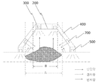
도 7은 본 발명의 일실시예에 따른 가변파장의 피부 영상 정보를 이용한 휴대용 피부질환 진단장치의 산란광, 경사광, 반사광을 통해 피부 질환을 진단하는 예시를 나타낸 예시도.
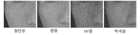
도 8은 본 발명의 일실시예에 따른 가변파장의 피부 영상 정보를 이용한 휴대용 피부질환 진단장치의 파장가변형광모듈을 통해 다양한 영상 이미지를 획득할 수 있음을 나타낸 예시도.
도 9는 본 발명의 일실시예에 따른 가변파장의 피부 영상 정보를 이용한 휴대용 피부질환 진단장치의 제1조명그룹(300), 제2조명그룹(500), 제3조명그룹(700)에 형성된 다수 파장가변형광모듈들 내에 형성된 광소자들의 동시 점등 시나리오 예시도.
도 10은 본 발명의 일실시예에 따른 가변파장의 피부 영상 정보를 이용한 휴대용 피부질환 진단장치의 제1조명그룹(300), 제2조명그룹(500), 제3조명그룹(700)에 형성된 다수 파장가변형광모듈들 내에 형성된 광소자들의 순차적 점등 시나리오 예시도.
도 11은 본 발명의 일실시예에 따른 가변파장의 피부 영상 정보를 이용한 휴대용 피부질환 진단장치의 가변파장피부질환용제어부(600) 블록도.
도 12는 본 발명의 일실시예에 따른 가변파장의 피부 영상 정보를 이용한 휴대용 피부질환 진단장치의 제1조명그룹 광원, 제2조명그룹 광원, 제3조명그룹 광원에 의해 촬영된 영상 이미지 상의 흡수율을 비교한 그래프.
도 13은 본 발명의 다른 일실시예에 따른 가변파장의 피부 영상 정보를 이용한 휴대용 피부질환 진단장치의 제1조명그룹과 제2조명그룹이 동일 평면 상에 배열되는 예시도.
<부호의 설명>
100 : 본체케이스
200 : 카메라부
300 : 제1조명그룹
400 : 캡
500 : 제2조명그룹
600 : 가변파장피부질환용제어부
700 : 제3조명그룹
이하의 내용은 단지 본 발명의 원리를 예시한다. 그러므로 당업자는 비록 본 명세서에 명확히 설명되거나 도시되지 않았지만, 본 발명의 원리를 구현하고 본 발명의 개념과 범위에 포함된 다양한 장치를 발명할 수 있는 것이다.
또한, 본 명세서에 열거된 모든 조건부 용어 및 실시 예들은 원칙적으로, 본 발명의 개념이 이해되도록 하기 위한 목적으로만 명백히 의도되고, 이와 같이 특별히 열거된 실시 예들 및 상태들에 제한적이지 않는 것으로 이해되어야 한다.
본 발명의 일실시예에 따른 가변파장의 피부 영상 정보를 이용한 휴대용 피부질환 진단장치는,
사용자에 의해 파지할 수 있는 일정 길이와 폭을 가지고 있는 본체케이스(100)와;
상기 본체케이스의 상측에 형성되어 영상 이미지를 촬영하기 위한 카메라부(200)와;
상기 카메라부 외곽 주변으로 일정 간격 형성되어 피부에 광을 조사하는 다수의 파장가변형광모듈(310)을 포함하며, 발광시, 상기 카메라부에 조명을 제공하기 위한 제1조명그룹(300)과;
상기 본체케이스의 전방에 결합되어 촬영 대상체와의 일정 거리를 유지시키되, 진단자의 피부에 접촉되는 캡(400)과;
설정된 가변 조명 점등 시나리오 정보에 따라 제1조명그룹에 조명 제어 신호를 제공하여 각각의 파장가변형광모듈(310)에서 2회 이상의 파장 가변에 따라 해당 파장 가변대의 각각의 광소자를 점등시키며, 카메라부에 촬영 신호를 제공하기 위한 가변파장피부질환용제어부(600);를 포함하여 구성되되,
상기 파장가변형광모듈(310)은 서로 다른 파장대의 광을 발광하는 둘 이상의 광소자(310a ~ 310n)가 결합되어 이루어진 것을 특징으로 한다.
또한, 상기 캡 혹은 제1조명그룹 외곽 주변에 일정 간격 형성되어 피부에 광을 조사하는 다수의 파장가변형광모듈(510)을 포함하며, 발광시, 상기 카메라부에 조명을 제공하기 위한 제2조명그룹(500);을 더 포함하여 구성되되,
상기 파장가변형광모듈(510)은 서로 다른 파장대의 광을 발광하는 둘 이상의 광소자(510a ~ 510n)가 결합되어 이루어진 것을 특징으로 한다.
또한, 상기 캡(400)에 형성된 제2조명그룹(500)의 상측에 일정 간격 경사지게 형성되고, 피부에 광을 조사하는 다수의 파장가변형광모듈(510)을 포함하며, 발광시, 상기 카메라부에 경사 조명을 제공하게 되어 진단자의 피부 질환 부위의 기울어진 면(측면) 형상을 제공하기 위한 제3조명그룹(700);을 더 포함하여 구성되되,
상기 파장가변형광모듈(510)은 서로 다른 파장대의 광을 발광하는 둘 이상의 광소자(710a ~ 710n)가 결합되어 이루어진 것을 특징으로 한다.
이때, 상기 제1조명그룹(300)과 제2조명그룹(500) 및 제3조명그룹(700)은,
가변파장피부질환용제어부(600)의 제어에 의해, 복수의 파장가변형광모듈(310, 510, 710)에 결합된 둘 이상의 광소자(310a ~ 310n, 510a ~ 510n, 710a ~ 710n) 중 파장 가변에 따라 특정 파장대의 각각의 광소자를 동시에 점등시키거나, 특정 파장대의 각각의 광소자를 순차적으로 점등시키는 것을 특징으로 한다.
또한, 상기 가변파장피부질환용제어부(600)는,
제1조명그룹(300), 제2조명그룹(500) 혹은 제1조명그룹(300), 제2조명그룹(500), 제3조명그룹(700)에 형성된 복수의 파장가변형광모듈(310, 510, 710)에 결합된 둘 이상의 광소자(310a ~ 310n, 510a ~ 510n, 710a ~ 710n)에 파장 가변 제어를 수행하여 일정 시간 간격으로 특정 파장대의 광원이 동시에 점등되도록 함으로써, 파장 가변 제어에 따라 복수의 파장가변형광모듈(310, 510, 710)들의 동일한 파장대로 발광되는 광소자가 동시에 점등되도록 하는 파장대별동시점등제어모듈(610);
제1조명그룹(300), 제2조명그룹(500) 혹은 제1조명그룹(300), 제2조명그룹(500), 제3조명그룹(700)에 형성된 복수의 파장가변형광모듈(310, 510, 710)에 결합된 둘 이상의 광소자(310a ~ 310n, 510a ~ 510n, 710a ~ 710n)에 특정 파장대의 광원이 순차적으로 점등되도록 함으로써, 파장 가변 제어에 따라 복수의 파장가변형광모듈(310, 510, 710)들의 특정 파장대로 발광되는 광소자가 순차적으로 점등되도록 하는 파장대별순차점등제어모듈(620); 중 적어도 어느 하나 이상의 모듈을 포함하여 구성되는 것을 특징으로 한다.
이때, 설정된(프로그램된) 가변 조명 점등 시나리오 정보를 포함하고 있는 가변조명시나리오정보저장부(630);를 더 포함하여 구성될 경우에,
상기 가변 조명 점등 시나리오 정보에는 그룹별 특정 파장대 광원의 동시 점등 정보, 그룹별 특정 파장대 광원의 순차 점등 정보를 포함한다.
이때, 상기 가변조명시나리오정보저장부로부터 가변 조명 점등 시나리오 정보를 가변파장피부질환용제어부(600)에서 획득하여 그룹별 가변파장 조명 제어를 수행하게 되는 것을 특징으로 한다.
또한, 상기 휴대용 피부질환 진단장치는,
피부 색소 질환, 감염성 질환, 피부암 중 적어도 어느 하나 이상의 질환을 진단할 수 있는 것을 특징으로 한다.
또한, 상기 가변파장피부질환용제어부(600)는,
제1조명그룹(300), 제2조명그룹(500), 제3조명그룹(700) 중 어느 하나의 조명그룹을 선택하고, 해당 조명그룹 내의 피부 질환이 흡수하는 파장대의 광원을 점등시킨 후, 카메라부에 촬영 신호를 제공하여 영상 이미지를 획득하며,
일정 시간 경과 후, 상기 선택된 조명그룹 내의 피부 질환이 흡수하지 않는 파장대의 광원을 점등시킨 후, 카메라부에 촬영 신호를 제공하여 영상 이미지를 획득하며,
상기 획득된 2 개의 영상 이미지의 흡수율 차이값을 판단하고, 판단된 흡수율 차이값이 설정된 임계 차이값을 벗어날 경우에 피부 질환으로 진단하는 것을 특징으로 한다.
한편, 상기 가변파장피부질환용제어부(600)는,
제1조명그룹(300), 제2조명그룹(500), 제3조명그룹(700) 중 어느 하나의 조명그룹을 선택하고, 해당 조명그룹 내의 피부 질환이 흡수하는 파장대의 광원을 점등시킨 후, 카메라부에 촬영 신호를 제공하여 영상 이미지를 획득하며,
일정 시간 경과후, 상기 선택된 조명그룹을 제외한 조명그룹 중 어느 하나의 조명그룹 내의 피부 질환이 흡수하지 않는 파장대의 광원을 점등시킨 후, 카메라부에 촬영 신호를 제공하여 영상 이미지를 획득하며,
상기 획득된 2 개의 영상 이미지의 흡수율 차이값을 판단하고, 판단된 흡수율 차이값이 설정된 임계 차이값을 벗어날 경우에 피부 질환으로 진단하는 것을 특징으로 한다.
이때, 상기 제1조명그룹(300)은,
피부 표면에 반사되는 빛을 카메라부(200)로 제공하며,
제2조명그룹(500)은,
진단자의 피부 접촉에 따라 피부 투과 후, 산란되는 빛을 카메라부(200)로 제공하는 것을 특징으로 하며,
상기 카메라부(200)는,
제2조명그룹(500)의 접촉 광원을 통해 산란되는 빛을 이용해 피부 질환의 기저 형상을 확인할 수 있으며,
제1조명그룹(300)의 주변 광원을 통해 피부 표면에서 반사되는 빛을 이용해 피부 질환의 외형을 확인할 수 있는 것을 특징으로 한다.
또한, 상기 제1조명그룹(300)과 제2조명그룹(500) 및 제3조명그룹(700)에 구성된 서로 다른 파장대의 광을 발광하는 둘 이상의 광소자(310a ~ 310n, 510a ~ 510n, 710a ~ 710n)는,
백색 광원소자, 적외선 광원소자, UV 광원소자, 옐로우 광원소자, 레드 광원소자, 그린 광원소자, 블루 광원소자 중 적어도 2개 이상의 광원소자들을 일정 간격 다수 형성하는 것을 특징으로 한다.
한편, 다른 실시예에 따라, 상기 제1조명그룹(300)의 원 지름보다 더 큰 원 지름을 가지도록 제2조명그룹(500)을 배치하는 것을 특징으로 하며,
상기 제2조명그룹(500)에는 일정 간격 형성되어 피부에 광을 조사하는 다수의 파장가변형광모듈(510)을 포함하며, 발광시, 카메라부에 조명을 제공하되,
상기 파장가변형광모듈(510)은 서로 다른 파장대의 광을 발광하는 둘 이상의 광소자(510a ~ 510n)가 결합되어 이루어진 것을 특징으로 한다.
이하, 본 발명에 의한 가변파장의 피부 영상 정보를 이용한 휴대용 피부질환 진단장치의 실시예를 통해 상세히 설명하도록 한다.
도 3은 본 발명의 일실시예에 따른 가변파장의 피부 영상 정보를 이용한 휴대용 피부질환 진단장치의 사시도이다.
도 4는 본 발명의 일실시예에 따른 가변파장의 피부 영상 정보를 이용한 휴대용 피부질환 진단장치의 다른 각도에서 바라본 사시도이다.
도 3 내지 도 4에 도시한 바와 같이, 본 발명인 가변파장의 피부 영상 정보를 이용한 휴대용 피부질환 진단장치는, 본체케이스(100), 카메라부(200), 제1조명그룹(300), 캡(400), 가변파장피부질환용제어부(600)를 포함하여 구성하게 된다.
구체적으로 설명하면, 상기 본체케이스(100)는 사용자에 의해 파지할 수 있는 일정 길이와 폭을 가지고 있게 된다.
바람직하게는 일측에 촬영버튼부(110)를 구성하고 있게 된다.
예를 들어, 본체케이스의 하측, 측면, 상측 중 어느 한 부위에 촬영버튼부를 구성하게 되는 것이다.
즉, 사용자가 파지할 경우에 버튼을 누르기 편리한 부위에 설치 구성하게 되는 것이다.
한편, 촬영버튼부의 구성없이, 전원이 공급되면 자동적으로 조명을 점등시키고, 카메라부를 동작시켜 촬영을 시작할 수도 있음은 자명한 사실이다.
또한, 촬영버튼부로서, 조작을 위한 복수의 키를 제공하며, 사용자의 키선택에 따른 선택 신호를 발생하여 가변파장피부질환용제어부(600)로 전달하는데, 이러한 촬영버튼부로는 버튼, 키패드, 터치패드와 같은 포인팅 장치, 터치스크린 등의 입력장치가 사용될 수 있다.
본 발명의 예시에서는 촬영버튼부를 누를 경우에 동작하는 것으로 설명하였다.
또한, 상기 카메라부(200)를 본체케이스의 내부 전방에 설치 구성함으로써, 촬영버튼부를 누를 경우에 영상 이미지를 촬영하게 되는 것이다.
상기 카메라는 제1조명그룹(300)의 조명들의 내측에 설치 구성되어 진단하고자 하는 피부 질환 표면의 영상을 촬영하게 되는데, 바람직한 실시예로는 정 중앙 부위에 형성하는 것을 특징으로 하며, 바람직하게는 일반 카메라를 한 개 설치 구성할 수 있으나, 필요에 따라 두 개 이상 설치 구성할 수 있으며, 다른 실시예로는 TOF카메라를 설치 구성하게 된다.
한편, 카메라로 TOF카메라를 설치 구성하게 되면, 3차원 정보를 획득할 수 있는데, 구체적으로 3차원 거리 정보를 획득할 수 있게 된다.
상기 TOF(TIME OF FLIGHT)는 RGB의 색정보와 T값인 거리값을 가지게 되므로 종래의 스테레오 카메라를 대체할 수 있게 되어 원가 절감 효과를 제공할 수 있게 된다.
그리고, 상기 카메라부의 일측에 렌즈부를 더 포함하여 구성할 수도 있다.
또한, 상기 카메라부 외곽 주변으로 일정 간격 다수 배열되어 카메라부에 조명을 제공하기 위한 제1조명그룹(300)을 설치 구성하게 된다.
이때, 제1조명그룹(300)은 피부 질환 부위에 광을 조사하게 되며, 가변파장피부질환용제어부(600)는 제1조명그룹(300)을 통하여 피부 질환 의심 부위에 광을 조사하고, 광이 조사된 피부 질환의 상태를 카메라부로 촬영하여 피부 영상을 획득하게 되며, 가변파장피부질환용제어부(600)는 획득한 피부 질환 영상으로 피부 질환 상태를 진단하게 된다.
그리고, 도 5에 도시한 바와 같이, 상기 제1조명그룹(300)은 카메라부 외곽 주변으로 일정 간격 형성되어 피부에 광을 조사하는 다수의 파장가변형광모듈(310)을 포함하여 구성되게 되는데, 상기 파장가변형광모듈(310)은 서로 다른 파장대의 광을 발광하는 둘 이상의 광소자(310a ~ 310n)가 결합되어 이루어진 것을 특징으로 한다.
예를 들어, 400 ~ 470nm의 파장대의 광을 조사하는 블루 LED 소자(300a), 580 ~ 595nm의 파장대의 광을 조사하는 옐로우 LED 소자(300b), 620 ~ 670nm의 파장대의 광을 조사하는 레드 LED 소자(300c), 700 ~ 800nm의 파장대의 광을 조사하는 녹색 LED 소자(300d)가 결합되는 구조를 가지게 된다.
또한, 필요에 따라 근적외선 LED 소자(300e), 화이트 LED 소자(300f),... 등을 추가적으로 구성할 수도 있을 것이다.
즉, 상기 제1조명그룹(300)과 후술할 제2조명그룹(500) 및 제3조명그룹(700)에 구성된 서로 다른 파장대의 광을 발광하는 둘 이상의 광소자(310a ~ 310n, 510a ~ 510n, 710a ~ 710n)는,
백색 광원소자, 적외선 광원소자, UV 광원소자, 옐로우 광원소자, 레드 광원소자, 그린 광원소자, 블루 광원소자 중 적어도 2개 이상의 광원소자들을 일정 간격 다수 형성하는 것을 특징으로 한다.
그리고, 캡(400)을 상기 본체케이스의 전방에 결합시켜 촬영 대상체인 피부 질환 부위와의 일정 거리를 유지시키되, 진단자의 피부에 접촉되게 되는 것이다.
즉, 도 4에 도시한 바와 같이, 본체케이스의 전방에 캡을 결합시키게 됨으로써, 제1조명그룹에 의해 점등되는 조명을 피부 질환 대상체 주변으로 빛을 제공하게 되는 것이다.
또한, 상기 캡에, 예를 들어, 캡의 하측에 제2조명그룹(500)을 형성하되, 일정 간격 다수 배열되어 카메라부에 조명을 제공하게 되는 것이다.
이때, 상기 제2조명그룹(500)은 피부에 광을 조사하는 다수의 파장가변형광모듈(510)을 포함하며, 발광시, 상기 카메라부에 조명을 제공하게 되는데, 제1조명그룹과 마찬가지로 상기 파장가변형광모듈(510)은 서로 다른 파장대의 광을 발광하는 둘 이상의 광소자(510a ~ 510n)가 결합되어 이루어진 것을 특징으로 한다.
또한, 가변파장피부질환용제어부(600)는 설정된 가변 조명 점등 시나리오 정보에 따라 제1조명그룹에 조명 제어 신호를 제공하여 각각의 파장가변형광모듈(310)에서 2회 이상의 파장 가변에 따라 해당 파장 가변대의 각각의 광소자를 점등시키며, 카메라부에 촬영 신호를 제공하게 된다.
상기 설정된 가변 조명 점등 시나리오 정보는 프로그램된 정보를 의미할 수 있으며, 사용자에 의해 조작된 정보를 의미할 수 있다.
도 6은 본 발명의 일실시예에 따른 가변파장의 피부 영상 정보를 이용한 휴대용 피부질환 진단장치의 캡과 조명그룹들을 나타낸 사시도이다.
도 6에 도시한 바와 같이, 캡에, 예를 들어, 캡의 하측에 즉, 캡의 피부 접촉면 보다 상측으로 제2조명그룹을 형성하게 되는 것이다.
이때, 캡의 재질은 투명 재질로 형성할 수 있으며, 필요에 따라 불투명 재질로 형성할 수도 있다.
상기와 같이, 불투명 재질로 형성할 경우에 하기에서 설명할 제3조명그룹을 제2조명그룹의 상측으로 형성할 수 있을 것이다.
도 7은 본 발명의 일실시예에 따른 가변파장의 피부 영상 정보를 이용한 휴대용 피부질환 진단장치의 산란광, 경사광, 반사광을 통해 피부 질환을 진단하는 예시를 나타낸 예시도이다.
한편, 도 7에 도시한 바와 같이, 본 발명의 부가적인 양태에 따라, 상기 캡(400)에 형성된 제2조명그룹(500)의 상측에 위치하되, 경사지게 일정 간격 다수 배열되어 형성됨으로써, 카메라부에 경사 조명을 제공하게 되어 진단자의 피부 질환 부위의 기울어진 면(측면) 형상을 제공하기 위한 제3조명그룹(700);을 더 포함하여 구성되는 것을 특징으로 한다.
즉, 제3조명그룹(700)을 제2조명그룹의 상측에 위치시키되, 경사지게 일정 간격 다수 배열되어 형성됨으로써, 카메라부에 경사 조명을 제공하게 되어 진단자의 피부 질환 부위의 기울어진 면(측면) 형상을 제공하게 되는 것이다.
상기와 같은 구성을 통해, 본 발명의 가변파장의 피부 영상 정보를 이용한 휴대용 피부질환 진단장치는,
피부 색소 질환, 감염성 질환, 피부암 중 적어도 어느 하나 이상의 질환을 진단할 수 있는 것을 특징으로 한다.
피부 색소 질환이란, 예를 들어, 기미, 주근깨 등을 의미하며, 감염성 질환이란, 여드름균, 곰팡이 등을 의미한다.
또한, 상기와 같은 구성 중 제1조명그룹(300)은 피부 표면에 반사되는 빛을 카메라부(200)로 제공하며, 제2조명그룹(500)은 진단자의 피부 접촉에 따라 피부 투과 후, 산란되는 빛을 카메라부(200)로 제공하는 것을 특징으로 한다.
따라서, 카메라부(200)는 제2조명그룹(500)의 접촉 광원을 통해 산란되는 빛을 이용해 피부 질환의 기저 형상을 확인할 수 있으며, 제1조명그룹(300)의 주변 광원을 통해 피부 표면에서 반사되는 빛을 이용해 피부 질환의 외형을 확인할 수 있는 것을 특징으로 한다.
도 7에 도시한 바와 같이, 제2조명그룹(500)이 피부 표면에 접촉 광원을 제공하게 되므로 산란되는 빛을 이용해 피부 질환의 기저 형상을 확인할 수 있게 된다.
예를 들어, 피부암의 피부 기저의 크기가 'A'임을 진단할 수 있다.
또한, 제1조명그룹(300)이 주변 광원을 통해 피부 표면에서 반사되는 빛을 이용해 피부 질환의 외형을 확인할 수 있게 된다.
예를 들어, 피부암의 외형의 크기가 'B'임을 진단할 수 있게 된다.
또한, 제3조명그룹(700)을 제2조명그룹의 상측에 경사지게 일정 간격 다수 배열되어 형성됨으로써, 카메라부에 경사 조명을 제공하게 되어 진단자의 피부 질환 부위의 기울어진 면(측면) 형상을 제공하게 된다.
즉, 측면 조명을 제공하게 됨으로써, 다크 필드 영상을 획득할 수 있으므로 피부암 부위를 도드라지게 하는 효과를 제공하게 되어 피부암 부위를 입체적으로 확인할 수 있는 효과를 제공하는 것이다.
상기한 실시예와 같이, 구성하게 되면 예를 들어 피부 질환 대상체가 피부암으로 예상되는 피부 표면일 경우에 카메라가 위치한 부위에 대하여 보다 정밀한 피부암 상태를 진단할 수 있게 됨으로써, 점으로 오인할 수 있는 피부 진단 오류를 방지할 수 있게 된다.
한편, 부가적인 양태에 따라, 상기 제1조명그룹(300)과 제2조명그룹(500) 및 제3조명그룹(700)에 구성된 서로 다른 파장대의 광을 발광하는 둘 이상의 광소자(310a ~ 310n, 510a ~ 510n, 710a ~ 710n)는,
백색 광원소자, 적외선 광원소자, UV 광원소자, 옐로우 광원소자, 레드 광원소자, 그린 광원소자, 블루 광원소자 중 적어도 2개 이상의 광원소자들을 일정 간격 다수 형성하는 것을 특징으로 한다.
예를 들어, 백색 광원을 배열하고 있으며, 백색 광원의 일측에 적외선 광원을 일정 간격 배열하고 있으며, 적외선 광원의 일측에 UV 광원을 배열하고 있는 것이다.
상기와 같이, 조명 그룹을 다른 파장대의 조명들로 일정 간격 구성하게 되면 다양한 영상을 획득할 수 있게 되는데, 도 8과 같이, 백색 광원을 통해 피부 질환 부위의 일반 영상을, 적외선 광원을 통해 피부질환 부위의 IR 영상을, UV 광원을 통해 UV 영상을 획득할 수 있게 되는 것이다.
따라서, 예를 들어, 피부암의 경우, 다양한 파장대의 조명들을 통해 다양한 영상을 획득하여 피부암을 진단할 수 있도록 하는 것이다.
한편, 본 발명의 가변파장피부질환용제어부(600)의 제어에 의해, 복수의 파장가변형광모듈(310)에 결합된 둘 이상의 광소자(310a ~ 310n) 중 파장 가변에 따라 특정 파장대의 각각의 광소자를 동시에 점등시키거나, 특정 파장대의 각각의 광소자를 순차적으로 점등시키는 것을 특징으로 한다.
도 9를 참조하여 설명하면, 상기한 파장 가변에 따라 특정 파장대의 각각의 광소자를 동시에 점등은 최초 제1조명그룹(300) 내에 존재하는 광소자 중 블루 광을 제공하는 파장대의 4개의 광소자(300a)를 동시 점등시킨 후, 1초후, 제2조명그룹(500) 내에 존재하는 광소자 중 블루 광을 제공하는 파장대의 4개의 광소자(500a)를 동시 점등시킨 후, 2초후, 제3조명그룹(700) 내에 존재하는 광소자 중 블루 광을 제공하는 파장대의 4개의 광소자(700a)를 동시 점등시킨다.
이후, 가변파장피부질환용제어부(600)의 제어에 의해, 제1조명그룹(300) 내에 존재하는 광소자 중 레드 광을 제공하는 파장대의 4개의 광소자(300c)를 동시 점등(최초 시점부터는 3초후)시킨 후, 1초후(최초 시점부터는 4초후), 제2조명그룹(500) 내에 존재하는 광소자 중 레드 광을 제공하는 파장대의 4개의 광소자(500c)를 동시 점등시킨 후, 2초후(최초 시점부터는 5초후), 제3조명그룹(700) 내에 존재하는 광소자 중 블루 광을 제공하는 파장대의 4개의 광소자(700c)를 동시 점등시킨다.
상기와 같은 식으로 화이트 광소자, 그린 광소자, 적외선 광소자, UV 광소자등으로 동시에 점등시키게 되는 것이다.
따라서, 다양한 파장대의 조명을 제공한 후, 촬영된 영상 이미지를 통해 피부 질환의 진단시 참조할 수 있도록 하는 것이다.
한편, 특정 파장대의 각각의 광소자를 순차적으로 점등시키는 것을 도 10을 참조하여 설명하도록 한다.
도 10을 참조하여 설명하면, 최초 제1조명그룹(300) 내에 존재하는 광소자 중 블루 광을 제공하는 파장대의 4개의 광소자(300a) 중 1번 광소자를 점등시키며, 0.3초후, 2번 광소자를 점등시키며, 0.6초후, 3번 광소자를 점등시키며, 0.9초후 4번 광소자를 점등시키게 된다.
이후, 1초후, 제2조명그룹(500) 내에 존재하는 광소자 중 블루 광을 제공하는 파장대의 4개의 4개 광소자(500a) 중 1번 광소자를 점등시키며, 1.3초후, 2번 광소자를 점등시키며, 1.6초후, 3번 광소자를 점등시키며, 1.9초후 4번 광소자를 점등시키게 된다.
이후, 2초후, 제3조명그룹(700) 내에 존재하는 광소자 중 블루 광을 제공하는 파장대의 4개의 4개 광소자(700a) 중 1번 광소자를 점등시키며, 2.3초후, 2번 광소자를 점등시키며, 2.6초후, 3번 광소자를 점등시키며, 2.9초후 4번 광소자를 점등시키게 된다.
이후, 가변파장피부질환용제어부(600)의 제어에 의해, 제1조명그룹(300) 내에 존재하는 광소자 중 레드 광을 제공하는 파장대의 4개의 광소자(300a) 중 1번 광소자를 점등시키며, 3.3초후, 2번 광소자를 점등시키며, 3.6초후, 3번 광소자를 점등시키며, 3.9초후 4번 광소자를 점등시키게 된다.
이후, 4초후, 제2조명그룹(500) 내에 존재하는 광소자 중 레드 광을 제공하는 파장대의 4개의 4개 광소자(500a) 중 1번 광소자를 점등시키며, 4.3초후, 2번 광소자를 점등시키며, 4.6초후, 3번 광소자를 점등시키며, 4.9초후 4번 광소자를 점등시키게 된다.
이후, 5초후, 제3조명그룹(700) 내에 존재하는 광소자 중 레드 광을 제공하는 파장대의 4개의 4개 광소자(700a) 중 1번 광소자를 점등시키며, 5.3초후, 2번 광소자를 점등시키며, 5.6초후, 3번 광소자를 점등시키며, 5.9초후 4번 광소자를 점등시키게 된다.
상기와 같은 식으로 화이트 광소자, 그린 광소자, 적외선 광소자, UV 광소자등도 가변 파장 제어에 따라 순차적으로 점등시키게 되는 것이다.
이러한 경우에 포토 메트릭을 위한 조명을 제공하게 됨으로써, 점 광원 영상을 획득할 수 있으므로 피부 질환 부위를 입체적으로 확인할 수 있는 효과를 제공한다.
도 11은 본 발명의 일실시예에 따른 가변파장의 피부 영상 정보를 이용한 휴대용 피부질환 진단장치의 가변파장피부질환용제어부(600) 블록도이다.
도 11에 도시한 바와 같이, 상기 가변파장피부질환용제어부(600)는,
제1조명그룹(300), 제2조명그룹(500) 혹은 제1조명그룹(300), 제2조명그룹(500), 제3조명그룹(700)에 형성된 복수의 파장가변형광모듈(310, 510, 710)에 결합된 둘 이상의 광소자(310a ~ 310n, 510a ~ 510n, 710a ~ 710n)에 파장 가변 제어를 수행하여 일정 시간 간격으로 특정 파장대의 광원이 동시에 점등되도록 함으로써, 파장 가변 제어에 따라 복수의 파장가변형광모듈(310, 510, 710)들의 동일한 파장대로 발광되는 광소자가 동시에 점등되도록 하는 파장대별동시점등제어모듈(610);
제1조명그룹(300), 제2조명그룹(500) 혹은 제1조명그룹(300), 제2조명그룹(500), 제3조명그룹(700)에 형성된 복수의 파장가변형광모듈(310, 510, 710)에 결합된 둘 이상의 광소자(310a ~ 310n, 510a ~ 510n, 710a ~ 710n)에 특정 파장대의 광원이 순차적으로 점등되도록 함으로써, 파장 가변 제어에 따라 복수의 파장가변형광모듈(310)들의 특정 파장대로 발광되는 광소자가 순차적으로 점등되도록 하는 파장대별순차점등제어모듈(620); 중 적어도 어느 하나 이상의 모듈을 포함하여 구성되게 된다.
즉, 상기한 파장대별동시점등제어모듈(610) 및 파장대별순차점등제어모듈(620)에 따라 광소자들의 조명 점등 시나리오는 도 9 내지 도 10을 참조하여 상기와 같이 구체적으로 설명하였다.
한편, 상기 가변파장피부질환용제어부(600)는,
설정된(프로그램된) 가변 조명 점등 시나리오 정보를 포함하고 있는 가변조명시나리오정보저장모듈(630);을 더 포함하여 구성될 경우에,
상기 가변 조명 점등 시나리오 정보에는 그룹별 특정 파장대 광원의 동시 점등 정보, 그룹별 특정 파장대 광원의 순차 점등 정보를 포함한다.
따라서, 상기 가변조명시나리오정보저장모듈로부터 가변 조명 점등 시나리오 정보를 가변파장피부질환용제어부(600)에서 획득하여 그룹별 가변파장 조명 제어를 수행하게 되는 것이다.
또한, 가변조명시나리오정보저장모듈(630)에는 촬영된 피부질환 영상 이미지를 파장대별로 저장하고 있게 된다.
구체적으로는 파장대별동시점등제어모듈(610)을 통해 제1조명그룹(300), 제2조명그룹(500), 제3조명그룹(700)에 형성된 복수의 파장가변형광모듈(310, 510, 710)에 결합된 둘 이상의 광소자(310a ~ 310n, 510a ~ 510n, 710a ~ 710n)에 동일한 파장대로 발광되는 광소자가 동시에 점등되는 제어를 수행하게 된다.
그리고, 파장대별순차점등제어모듈(620)을 통해 제1조명그룹(300), 제2조명그룹(500), 제3조명그룹(700)에 형성된 복수의 파장가변형광모듈)에 결합된 둘 이상의 광소자에 특정 파장대의 광원이 순차적으로 점등되도록 하는 제어를 수행하게 된다.
결국, 상기와 같이, 조명을 동시에 파장 가변하거나, 조명을 순차적으로 점등시킴으로써, 카메라로부터 특정 파장대의 영상을 획득하거나, 특정 파장대의 포토 메트릭 영상을 획득할 수 있게 되는 것이다.
따라서, 본 발명은 광학식으로 피부 질환 대상체 진단용 이미지를 획득하는 것을 특징으로 함으로써, 다양한 가변 파장대를 통해 피부 질환 진단시 피부 질환의 미세한 형상 정보를 획득할 수 있게 된다.
또한, 포토 메트릭을 위한 조명을 제공하여 점 광원 영상을 획득할 수 있으므로 피부 질환 부위를 입체적으로 확인할 수 있는 효과를 제공한다.
한편, 상기 가변파장피부질환용제어부(600)는,
3차원 정보를 획득하기 위하여, 3차원형상생성모듈, 표면형상생성모듈, 3차원형상표면정합모듈을 더 포함하게 된다.
구체적으로, 상기 3차원형상생성모듈은 카메라가 TOF 카메라일 경우에 3D 값을 획득하여 3차원 형상을 생성하게 되는 것이며, 상기 표면형상생성모듈은 포토 메트릭 영상 정보를 토대로 표면 형상을 생성하게 되는 것이다.
이때, 3차원형상표면정합모듈에 의해 상기 3차원형상생성모듈과 표면형상생성모듈로부터 생성된 3차원 형상 정보에 표면 형상 정보를 정합하게 되는 것이다.
즉, 정합하게 되면 피부암 표면의 미세한 형상과 질감을 제공할 수 있게 되는 것이다.
한편, 본 발명의 또 다른 부가적인 양태에 따라, 상기 가변파장피부질환용제어부(600)는,
가변조명시나리오정보저장모듈(630)에 파장대별로 저장된 피부질환 영상 이미지를 무선 통신을 통해 외부단말기(2000)로 전송하기 위한 무선통신모듈(640);을 더 포함하여 구성되는 것을 특징으로 한다.
따라서, 가변조명시나리오정보저장모듈(630)에 저장된 피부질환 대상 표면의 형상 정보를 무선 통신을 통해 외부단말기로 전송하게 되는 것이다.
상기와 같이, 구성하게 되면, 외부단말기(2000)의 화면을 통해 현재 촬영되고 있은 피부질환 대상 표면의 영상 정보를 실시간으로 확인할 수 있게 된다.
또한, 정합된 3차원 입체 영상을 원격지에 구성된 클라우드서버(3000)로 제공하게 됨으로써, 클라우드서버에서 피부질환 대상 표면 진단 정보를 생성하게 된다.
상기와 같이, 구성하게 되면, 사용자들이 해당 피부질환 표면 진단 정보를 자신이 가지고 있는 외부단말기 예를 들어, 스마트기기를 통해 언제 어디서든지 클라우드서버에 접속하여 해당 표면 진단 정보를 획득하게 된다.
이를 위하여 진단앱을 스마트기기에 탑재하게 된다.
즉, 보다 정밀한 피부 상태를 진단할 수 있게 됨으로써, 피부 진단 오류를 방지할 수 있게 된다.
한편, 가변파장피부질환용제어부(600)는,
제1조명그룹의 점등에 의해 획득된 영상 이미지 정보, 제2조명그룹의 점등에 의해 획득된 영상 이미지 정보, 제3조명그룹의 점등에 의해 획득된 영상 이미지 정보를 추출하여 영상 이미지 정보별 흡수율을 계산하여 흡수율 차이값이 임계 차이값보다 높을 경우에 피부 질환으로 진단하기 위한 피부질환진단부를 더 포함하여 구성된다.
좀 더 구체적으로 설명하자면, 상기 가변파장피부질환용제어부(600)는, 구체적으로 상기 피부질환진단부는,
제1조명그룹(300), 제2조명그룹(500), 제3조명그룹(700) 중 어느 하나의 조명그룹을 선택하고, 해당 조명그룹 내의 피부 질환이 흡수하는 파장대의 광원을 점등시킨 후, 카메라부에 촬영 신호를 제공하여 영상 이미지를 획득하며,
일정 시간 경과후, 상기 선택된 조명그룹 내의 피부 질환이 흡수하지 않는 파장대의 광원을 점등시킨 후, 카메라부에 촬영 신호를 제공하여 영상 이미지를 획득하며,
상기 획득된 2 개의 영상 이미지의 흡수율 차이값을 판단하고, 판단된 흡수율 차이값이 설정된 임계 차이값을 벗어날 경우에 피부 질환으로 진단하는 것을 특징으로 한다.
예를 들어, 제1조명그룹(300)을 선택하고, 해당 조명그룹 내의 피부 질환이 흡수하는 파장대의 광원인 레드 광원을 점등시킨 후, 카메라부에 촬영 신호를 제공하여 영상 이미지를 획득하게 된다.
이후, 일정 시간 경과 후, 상기 선택된 제1조명그룹 내의 피부 질환이 흡수하지 않는 파장대의 광원인 그린 광원을 점등시킨 후, 카메라부에 촬영 신호를 제공하여 영상 이미지를 획득하게 된다.
이후, 상기 획득된 2 개의 영상 이미지의 흡수율 차이값을 판단하고, 판단된 흡수율 차이값이 설정된 임계 차이값을 벗어날 경우에 피부 질환으로 진단하는 것이다.
좀 더 구체적으로 도 12를 참조하여 설명하자면, 제1조명그룹 레드 광원이 점등될 경우에 획득된 제1 영상 이미지를 분석하여 흡수율을 계산한 결과, 흡수율이 55%, 제1조명그룹 그린 광원이 점등될 경우에 획득된 제2 영상 이미지를 분석하여 흡수율을 계산한 결과, 흡수율이 10%일 경우에, 임계 차이값은 40%로 설정되었다.
이때, 제1 영상 이미지 흡수율 55%와 제2 영상 이미지 흡수율 10% 간에 차이값이 45%로서 임계 차이값인 40%를 초과하였으므로 이를 피부 질환(예를 들어, 피부암)으로 분석(진단)하게 되는 것이다.
상기한 흡수율 계산의 경우, 일반적으로 발광 100%일 경우에 수광 45%라면, 흡수율은 55%가 되는 것이므로 이는 피부 질환(예를 들어, 피부암)이 흡수하였음을 의미하기 때문이다.
상기한 영상 이미지를 통해 흡수율을 계산하는 기술은 영상 이미지를 처리하는 당업자들이게는 알려진 기술이므로 상세한 설명은 생략한다.
한편, 다른 조명그룹 점등 예시로서, 상기 가변파장피부질환용제어부(600)는, 구체적으로 상기 피부질환진단부는,
제1조명그룹(300), 제2조명그룹(500), 제3조명그룹(700) 중 어느 하나의 조명그룹을 선택하고, 해당 조명그룹 내의 피부 질환이 흡수하는 파장대의 광원을 점등시킨 후, 카메라부에 촬영 신호를 제공하여 영상 이미지를 획득하며,
일정 시간 경과후, 상기 선택된 조명그룹을 제외한 조명그룹 중 어느 하나의 조명그룹 내의 피부 질환이 흡수하지 않는 파장대의 광원을 점등시킨 후, 카메라부에 촬영 신호를 제공하여 영상 이미지를 획득하며,
상기 획득된 2 개의 영상 이미지의 흡수율 차이값을 판단하고, 판단된 흡수율 차이값이 설정된 임계 차이값을 벗어날 경우에 피부 질환으로 진단하는 것을 특징으로 한다.
예를 들어, 제1조명그룹(300)을 선택하고, 해당 제1조명그룹 내의 피부 질환이 흡수하는 파장대의 광원인 레드 광원을 점등시킨 후, 카메라부에 촬영 신호를 제공하여 영상 이미지를 획득하게 된다.
이후, 일정 시간 경과 후, 상기 선택된 제1조명그룹을 제외한 제2조명그룹 및 제3조명그룹 중 제2조명그룹 내의 피부 질환이 흡수하지 않는 파장대의 광원인 그린 광원을 점등시킨 후, 카메라부에 촬영 신호를 제공하여 영상 이미지를 획득하게 된다.
이후, 상기 획득된 2 개의 영상 이미지의 흡수율 차이값을 판단하고, 판단된 흡수율 차이값이 설정된 임계 차이값을 벗어날 경우에 피부 질환으로 진단하는 것이다.
좀 더 구체적으로 도 12를 참조하여 설명하자면, 제1조명그룹 레드 광원이 점등될 경우에 획득된 제1 영상 이미지를 분석하여 흡수율을 계산한 결과, 흡수율이 55%, 제2조명그룹 그린 광원이 점등될 경우에 획득된 제2 영상 이미지를 분석하여 흡수율을 계산한 결과, 흡수율이 10%일 경우에, 임계 차이값은 40%로 설정되었다.
이때, 제1 영상 이미지 흡수율 55%와 제2 영상 이미지 흡수율 10% 간에 차이값이 45%로서 임계 차이값인 40%를 초과하였으므로 이를 피부 질환(예를 들어, 피부암)으로 분석(진단)하게 되는 것이다.
한편, 일반적인 점의 경우에는 도 12의 우측 그래프에 도시하였듯이, 조명그룹 광원 간의 흡수율 차이가 크지 않은 특성을 가지고 있다.
이는 점들의 경우 빛을 흡수하는 확률이 피부암보다 현저히 적기 때문에 제1조명그룹 광원, 제2조명그룹 광원, 제3조명그룹 광원들에 의해 점등 후 촬영된 영상 이미지 분석시 흡수율 차이가 크지 않게 된다.
한편, 상기한 흡수율 임계 차이값은 설정된 피부질환(예를 들어, 피부암) 이미지 갯수를 머신 러닝기법을 이용해 학습하여 설정하기 위한 머신러닝피부질환학습부;를 클라우드서버(3000)에 구성함으로써, 수집된 피부질환 이미지 내의 흡수율을 학습하여 최적의 흡수율 임계 차이값을 설정하게 되어 피부질환 진단의 정확성을 높이도록 하게 된다.
즉, 머신러닝피부질환학습부를 통해 피부질환 진단 정확성을 더욱 높이게 되는 것이다.
도 13은 본 발명의 다른 일실시예에 따른 가변파장의 피부 영상 정보를 이용한 휴대용 피부질환 진단장치의 제1조명그룹과 제2조명그룹이 동일 평면 상에 배열되는 예시도이다.
예를 들어, 제1조명그룹(300)이 배치되는 레이어의 지름보다 제2조명그룹(500)이 배치되는 레이어의 지름이 더 크도록 형성하게 된다.
즉, 카메라부의 주변으로 형성되는 제1조명그룹과 동일 평면상에 제2조명그룹을 배치하게 되는 것이다.
이때, 상기 제2조명그룹(500)에는 일정 간격 형성되어 피부에 광을 조사하는 다수의 파장가변형광모듈(510)을 포함하며, 발광시, 카메라부에 조명을 제공하되, 상기 파장가변형광모듈(510)은 서로 다른 파장대의 광을 발광하는 둘 이상의 광소자(510a ~ 510n)가 결합되어 이루어진 것을 특징으로 한다.
따라서, 2열(예를 들어, 제1조명그룹과 제2조명그룹)로 조명그룹을 동일한 평면 상에 위치시키며, 캡(400)으로 피부에 접촉하여 광소자를 점등시켜 카메라부에 조명을 제공하게 되는 것이다.
이때, 바람직하게 상기 캡의 구조는 하측으로 갈수록 지름이 넓어지는 깔대기 형상을 가질 수도 있다.
본 발명에 의하면, 핸디 타입으로서, 진단자의 피부 진단시, 가변파장의 접촉광원을 통해 산란되는 빛을 이용해 진단자의 피부 질환의 기저 형상을 확인할 수 있으며, 가변파장의 주변 광원을 통해 피부 표면에서 반사되는 빛을 이용해 피부 질환의 외형을 확인할 수 있으며, 가변파장의 경사 조명을 제공하여 진단자의 피부 질환 부위의 기울어진 면(측면) 형상을 확인할 수 있음으로써, 피부 색소 질환, 감염성 질환, 피부암 중 적어도 어느 하나 이상의 질환을 진단할 수 있는 효과, 저렴한 가격에 누구나 쉽게 피부 질환 대상체의 표면을 촬영하여 3차원 형상으로 표면 상태를 확인할 수 있는 효과와 피부암일 경우에 피부암 진단 확률을 높이는 효과를 발휘하게 된다.
또한, 이상에서는 본 발명의 바람직한 실시예에 대하여 도시하고 설명하였지만, 본 발명은 상술한 특정의 실시 예에 한정되지 아니하며, 청구범위에서 청구하는 본 발명의 요지를 벗어남이 없이 당해 발명이 속하는 기술분야에서 통상의 지식을 가진 자에 의해 다양한 변형 실시가 가능한 것은 물론이고, 이러한 변형 실시들은 본 발명의 기술적 사상이나 전망으로부터 개별적으로 이해되어서는 안될 것이다.
본 발명에 따른 가변파장의 피부 영상 정보를 이용한 휴대용 피부질환 진단장치는, 핸디 타입으로서, 진단자의 피부 진단시, 가변파장의 접촉광원을 통해 산란되는 빛을 이용해 진단자의 피부 질환의 기저 형상을 확인할 수 있으며, 가변파장의 주변 광원을 통해 피부 표면에서 반사되는 빛을 이용해 피부 질환의 외형을 확인할 수 있으며, 가변파장의 경사 조명을 제공하여 진단자의 피부 질환 부위의 기울어진 면(측면) 형상을 확인할 수 있음으로써, 피부 색소 질환, 감염성 질환, 피부암 중 적어도 어느 하나 이상의 질환을 진단할 수 있는 효과, 저렴한 가격에 누구나 쉽게 피부 질환 대상체의 표면을 촬영하여 3차원 형상으로 표면 상태를 확인할 수 있는 효과와 피부암일 경우에 피부암 진단 확률을 높이는 효과를 발휘하게 되므로, 산업상 이용가능성도 높은 발명이다.
Claims (10)
- 가변파장의 피부 영상 정보를 이용한 휴대용 피부질환 진단장치에 있어서,사용자에 의해 파지할 수 있는 일정 길이와 폭을 가지고 있는 본체케이스(100)와;상기 본체케이스의 상측에 형성되어 영상 이미지를 촬영하기 위한 카메라부(200)와;상기 카메라부 외곽 주변으로 일정 간격 형성되어 피부에 광을 조사하는 다수의 파장가변형광모듈(310)을 포함하며, 발광시, 상기 카메라부에 조명을 제공하기 위한 제1조명그룹(300)과;상기 본체케이스의 전방에 결합되어 촬영 대상체와의 일정 거리를 유지시키되, 진단자의 피부에 접촉되는 캡(400)과;설정된 가변 조명 점등 시나리오 정보에 따라 제1조명그룹에 조명 제어 신호를 제공하여 각각의 파장가변형광모듈(310)에서 2회 이상의 파장 가변에 따라 해당 파장 가변대의 각각의 광소자를 점등시키며, 카메라부에 촬영 신호를 제공하기 위한 가변파장피부질환용제어부(600);를 포함하여 구성되되,상기 파장가변형광모듈(310)은 서로 다른 파장대의 광을 발광하는 둘 이상의 광소자(310a ~ 310n)가 결합되어 이루어진 것을 특징으로 하는 가변파장의 피부 영상 정보를 이용한 휴대용 피부질환 진단장치.
- 제 1항에 있어서,상기 캡 혹은 제1조명그룹 외곽 주변에 일정 간격 형성되어 피부에 광을 조사하는 다수의 파장가변형광모듈(510)을 포함하며, 발광시, 상기 카메라부에 조명을 제공하기 위한 제2조명그룹(500);을 더 포함하여 구성되되,상기 파장가변형광모듈(510)은 서로 다른 파장대의 광을 발광하는 둘 이상의 광소자(510a ~ 510n)가 결합되어 이루어진 것을 특징으로 하는 가변파장의 피부 영상 정보를 이용한 휴대용 피부질환 진단장치.
- 제 1항 또는 제 2항에 있어서,상기 캡(400)에 형성된 제2조명그룹(500)의 상측에 일정 간격 경사지게 형성되고, 피부에 광을 조사하는 다수의 파장가변형광모듈(510)을 포함하며, 발광시, 상기 카메라부에 경사 조명을 제공하게 되어 진단자의 피부 질환 부위의 기울어진 면(측면) 형상을 제공하기 위한 제3조명그룹(700);을 더 포함하여 구성되되,상기 파장가변형광모듈(510)은 서로 다른 파장대의 광을 발광하는 둘 이상의 광소자(710a ~ 710n)가 결합되어 이루어진 것을 특징으로 하는 가변파장의 피부 영상 정보를 이용한 휴대용 피부질환 진단장치.
- 제 1항 내지 제 3항 중 어느 한 항에 있어서,상기 제1조명그룹(300)과 제2조명그룹(500) 및 제3조명그룹(700)은,가변파장피부질환용제어부(600)의 제어에 의해, 복수의 파장가변형광모듈(310, 510, 710)에 결합된 둘 이상의 광소자(310a ~ 310n, 510a ~ 510n, 710a ~ 710n) 중 파장 가변에 따라 특정 파장대의 각각의 광소자를 동시에 점등시키거나, 특정 파장대의 각각의 광소자를 순차적으로 점등시키는 것을 특징으로 하는 가변파장의 피부 영상 정보를 이용한 휴대용 피부질환 진단장치.
- 제 1항 내지 제 3항 중 어느 한 항에 있어서,휴대용 피부질환 진단장치는,피부 색소 질환, 감염성 질환, 피부암 중 적어도 어느 하나 이상의 질환을 진단할 수 있는 것을 특징으로 하는 가변파장의 피부 영상 정보를 이용한 휴대용 피부질환 진단장치.
- 제 1항 내지 제 3항 중 어느 한 항에 있어서,상기 가변파장피부질환용제어부(600)는,제1조명그룹(300), 제2조명그룹(500), 제3조명그룹(700) 중 어느 하나의 조명그룹을 선택하고, 해당 조명그룹 내의 피부 질환이 흡수하는 파장대의 광원을 점등시킨 후, 카메라부에 촬영 신호를 제공하여 영상 이미지를 획득하며,일정 시간 경과 후, 상기 선택된 조명그룹 내의 피부 질환이 흡수하지 않는 파장대의 광원을 점등시킨 후, 카메라부에 촬영 신호를 제공하여 영상 이미지를 획득하며,상기 획득된 2 개의 영상 이미지의 흡수율 차이값을 판단하고, 판단된 흡수율 차이값이 설정된 임계 차이값을 벗어날 경우에 피부 질환으로 진단하는 것을 특징으로 하는 가변파장의 피부 영상 정보를 이용한 휴대용 피부질환 진단장치.
- 제 1항 내지 제 3항 중 어느 한 항에 있어서,상기 가변파장피부질환용제어부(600)는,제1조명그룹(300), 제2조명그룹(500), 제3조명그룹(700) 중 어느 하나의 조명그룹을 선택하고, 해당 조명그룹 내의 피부 질환이 흡수하는 파장대의 광원을 점등시킨 후, 카메라부에 촬영 신호를 제공하여 영상 이미지를 획득하며,일정 시간 경과후, 상기 선택된 조명그룹을 제외한 조명그룹 중 어느 하나의 조명그룹 내의 피부 질환이 흡수하지 않는 파장대의 광원을 점등시킨 후, 카메라부에 촬영 신호를 제공하여 영상 이미지를 획득하며,상기 획득된 2 개의 영상 이미지의 흡수율 차이값을 판단하고, 판단된 흡수율 차이값이 설정된 임계 차이값을 벗어날 경우에 피부 질환으로 진단하는 것을 특징으로 하는 가변파장의 피부 영상 정보를 이용한 휴대용 피부질환 진단장치.
- 제 1항 내지 제 3항 중 어느 한 항에 있어서,상기 제1조명그룹(300)은,피부 표면에 반사되는 빛을 카메라부(200)로 제공하며,제2조명그룹(500)은,진단자의 피부 접촉에 따라 피부 투과 후, 산란되는 빛을 카메라부(200)로 제공하는 것을 특징으로 하며,상기 카메라부(200)는,제2조명그룹(500)의 접촉 광원을 통해 산란되는 빛을 이용해 피부 질환의 기저 형상을 확인할 수 있으며,제1조명그룹(300)의 주변 광원을 통해 피부 표면에서 반사되는 빛을 이용해 피부 질환의 외형을 확인할 수 있는 것을 특징으로 하는 가변파장의 피부 영상 정보를 이용한 휴대용 피부질환 진단장치.
- 제 1항 내지 제 3항 중 어느 한 항에 있어서,상기 제1조명그룹(300)과 제2조명그룹(500) 및 제3조명그룹(700)에 구성된 서로 다른 파장대의 광을 발광하는 둘 이상의 광소자(310a ~ 310n, 510a ~ 510n, 710a ~ 710n)는,백색 광원소자, 적외선 광원소자, UV 광원소자, 옐로우 광원소자, 레드 광원소자, 그린 광원소자, 블루 광원소자 중 적어도 2개 이상의 광원소자들을 일정 간격 다수 형성하는 것을 특징으로 하는 가변파장의 피부 영상 정보를 이용한 휴대용 피부질환 진단장치.
- 제 1항에 있어서,상기 제1조명그룹(300)의 원 지름보다 더 큰 원 지름을 가지도록 제2조명그룹(500)을 배치하는 것을 특징으로 하며,상기 제2조명그룹(500)에는 일정 간격 형성되어 피부에 광을 조사하는 다수의 파장가변형광모듈(510)을 포함하며, 발광시, 카메라부에 조명을 제공하되,상기 파장가변형광모듈(510)은 서로 다른 파장대의 광을 발광하는 둘 이상의 광소자(510a ~ 510n)가 결합되어 이루어진 것을 특징으로 하는 가변파장의 피부 영상 정보를 이용한 휴대용 피부질환 진단장치.
Applications Claiming Priority (2)
| Application Number | Priority Date | Filing Date | Title |
|---|---|---|---|
| KR10-2018-0142256 | 2018-11-19 | ||
| KR1020180142256A KR102036045B1 (ko) | 2018-11-19 | 2018-11-19 | 가변파장의 피부 영상 정보를 이용한 휴대용 피부질환 진단장치 |
Publications (1)
| Publication Number | Publication Date |
|---|---|
| WO2020105954A1 true WO2020105954A1 (ko) | 2020-05-28 |
Family
ID=68423321
Family Applications (1)
| Application Number | Title | Priority Date | Filing Date |
|---|---|---|---|
| PCT/KR2019/015525 WO2020105954A1 (ko) | 2018-11-19 | 2019-11-14 | 가변파장의 피부 영상 정보를 이용한 휴대용 피부질환 진단장치 |
Country Status (2)
| Country | Link |
|---|---|
| KR (1) | KR102036045B1 (ko) |
| WO (1) | WO2020105954A1 (ko) |
Cited By (1)
| Publication number | Priority date | Publication date | Assignee | Title |
|---|---|---|---|---|
| WO2022241330A3 (en) * | 2021-05-13 | 2023-01-19 | Banter C Bruce | Wheal and flare analyzing system |
Families Citing this family (1)
| Publication number | Priority date | Publication date | Assignee | Title |
|---|---|---|---|---|
| KR102566912B1 (ko) | 2021-05-24 | 2023-08-14 | 파이 주식회사 | 디지탈 확대 스코프용 접촉형 이중 구조 캡 |
Citations (5)
| Publication number | Priority date | Publication date | Assignee | Title |
|---|---|---|---|---|
| WO2010049907A2 (en) * | 2008-10-31 | 2010-05-06 | L'oreal | A portable skin diagnosis device |
| KR20110054413A (ko) * | 2009-11-17 | 2011-05-25 | 재단법인 철원플라즈마 산업기술연구원 | 피부 진단 및 치료 장치와 이를 이용한 피부 진단 및 치료 방법 |
| KR20160147585A (ko) * | 2015-06-15 | 2016-12-23 | 서울바이오시스 주식회사 | 피부 상태 진단 방법 및 이를 이용하는 피부 상태 진단 장치 |
| KR101766417B1 (ko) * | 2016-03-14 | 2017-08-08 | 주식회사 한국지식재산경영평가원 | 모바일을 이용한 광조사형 피부 케어 장치 |
| JP2018510665A (ja) * | 2014-12-30 | 2018-04-19 | ネクサス エキスペリティズィ イ バダニア ドクター ジャセク ステピエンNexus Ekspertyzy I Badania Dr Jacek Stepien | 接触式熱光学構造体、及び皮膚アレルギー反応におけるヒスタミン誘発過熱皮下反応の大きさを非侵襲的イメージングするためのその利用、記録装置、及びアレルギー反応診断方法 |
Family Cites Families (6)
| Publication number | Priority date | Publication date | Assignee | Title |
|---|---|---|---|---|
| KR100609823B1 (ko) | 2003-11-07 | 2006-08-08 | 아람휴비스(주) | 피부 진단 시스템 |
| KR20080109501A (ko) * | 2007-06-13 | 2008-12-17 | (주)웨버인스트루먼트 | 아토피진단의료용 카메라 |
| KR20130040349A (ko) * | 2011-10-14 | 2013-04-24 | 주식회사메드스타 | 스코프 탈착식 의료용 유무선 영상 진단장치 |
| KR20140028415A (ko) * | 2012-08-29 | 2014-03-10 | 한국전자통신연구원 | 피부분석을 위한 3차원 얼굴 모델 생성 장치 및 그 방법 |
| KR101583659B1 (ko) * | 2014-12-05 | 2016-01-11 | 주식회사 미르기술 | 3차원 형상 측정 장치 |
| KR101738035B1 (ko) * | 2016-10-20 | 2017-05-19 | 장현재 | 피부의 3차원 입체 영상을 획득하는 3차원 촬영장치 |
-
2018
- 2018-11-19 KR KR1020180142256A patent/KR102036045B1/ko active IP Right Grant
-
2019
- 2019-11-14 WO PCT/KR2019/015525 patent/WO2020105954A1/ko active Application Filing
Patent Citations (5)
| Publication number | Priority date | Publication date | Assignee | Title |
|---|---|---|---|---|
| WO2010049907A2 (en) * | 2008-10-31 | 2010-05-06 | L'oreal | A portable skin diagnosis device |
| KR20110054413A (ko) * | 2009-11-17 | 2011-05-25 | 재단법인 철원플라즈마 산업기술연구원 | 피부 진단 및 치료 장치와 이를 이용한 피부 진단 및 치료 방법 |
| JP2018510665A (ja) * | 2014-12-30 | 2018-04-19 | ネクサス エキスペリティズィ イ バダニア ドクター ジャセク ステピエンNexus Ekspertyzy I Badania Dr Jacek Stepien | 接触式熱光学構造体、及び皮膚アレルギー反応におけるヒスタミン誘発過熱皮下反応の大きさを非侵襲的イメージングするためのその利用、記録装置、及びアレルギー反応診断方法 |
| KR20160147585A (ko) * | 2015-06-15 | 2016-12-23 | 서울바이오시스 주식회사 | 피부 상태 진단 방법 및 이를 이용하는 피부 상태 진단 장치 |
| KR101766417B1 (ko) * | 2016-03-14 | 2017-08-08 | 주식회사 한국지식재산경영평가원 | 모바일을 이용한 광조사형 피부 케어 장치 |
Cited By (1)
| Publication number | Priority date | Publication date | Assignee | Title |
|---|---|---|---|---|
| WO2022241330A3 (en) * | 2021-05-13 | 2023-01-19 | Banter C Bruce | Wheal and flare analyzing system |
Also Published As
| Publication number | Publication date |
|---|---|
| KR102036045B1 (ko) | 2019-10-24 |
Similar Documents
| Publication | Publication Date | Title |
|---|---|---|
| WO2020101380A1 (ko) | 광학식 피부질환 진단장치 | |
| WO2017155265A1 (ko) | 다중 분광 이미징 디바이스 | |
| WO2017188589A2 (en) | Mobile hyperspectral camera system and human skin monitoring using a mobile hyperspectral camera system | |
| WO2016204432A1 (ko) | 전자 디바이스 | |
| WO2019083116A1 (ko) | 다중 광원을 이용한 피부 상태 측정 장치 | |
| EP1433418A1 (en) | Skin diagnostic imaging method and apparatus | |
| WO2020105954A1 (ko) | 가변파장의 피부 영상 정보를 이용한 휴대용 피부질환 진단장치 | |
| WO2019088555A1 (ko) | 전자 장치 및 이를 이용한 눈의 충혈도 판단 방법 | |
| US8315692B2 (en) | Multi-spectral imaging spectrometer for early detection of skin cancer | |
| WO2016159710A1 (ko) | 모바일 다중 분광 이미징 디바이스, 다중 분광 이미지 획득용 사용자 단말, 분광 이미지 분석 서버 및 그 방법 | |
| WO2017135564A1 (ko) | 전자장치, 휴대단말기 및 그 제어방법 | |
| JP2008304466A (ja) | 画像処理システム | |
| WO2013157673A1 (en) | Optical coherence tomography and control method for the same | |
| WO2019135475A1 (en) | Electronic apparatus and control method thereof | |
| WO2021153858A1 (ko) | 비정형 피부질환 영상데이터를 활용한 판독보조장치 | |
| CN104586355A (zh) | 测定装置 | |
| WO2022145999A1 (ko) | 인공지능 기반의 자궁경부암 검진 서비스 시스템 | |
| KR102036043B1 (ko) | 스마트기기 기반의 피부질환 진단이 가능한 광학식 피부질환 진단장치 | |
| WO2021241795A1 (ko) | 실시간으로 관찰 및 촬영이 동시에 가능한 더마스코프 장치 | |
| WO2022060133A1 (ko) | 이미지 처리 장치 및 이미지 처리 방법 | |
| US11219926B2 (en) | Modulable system for identifying and certifying garments | |
| WO2020256319A1 (ko) | 이미지를 이용한 비접촉 산소포화도 측정 시스템 및 그 구동 방법 | |
| WO2019083154A1 (ko) | 개별 제어되는 마이크로 led 픽셀들을 갖는 플래쉬 유닛을 포함하는 촬상 장치 및 피부 진단용 촬상 장치 | |
| WO2017119726A2 (ko) | 렌즈모듈 및 이를 포함하는 홍채 인식용 카메라모듈, 홍채 인식 카메라 시스템 및 이를 포함하는 단말기 | |
| KR102036048B1 (ko) | 스마트기기 기반의 가변파장의 피부 영상 정보를 이용한 휴대용 피부질환 진단장치 |
Legal Events
| Date | Code | Title | Description |
|---|---|---|---|
| 121 | Ep: the epo has been informed by wipo that ep was designated in this application |
Ref document number: 19888034 Country of ref document: EP Kind code of ref document: A1 |
|
| NENP | Non-entry into the national phase |
Ref country code: DE |
|
| 32PN | Ep: public notification in the ep bulletin as address of the adressee cannot be established |
Free format text: NOTING OF LOSS OF RIGHTS PURSUANT TO RULE 112(1) EPC (EPO FORM 1205 DATED 11.10.2021) |
|
| 122 | Ep: pct application non-entry in european phase |
Ref document number: 19888034 Country of ref document: EP Kind code of ref document: A1 |