WO2020071489A1 - Méthode d'examen du cancer urothélial - Google Patents
Méthode d'examen du cancer urothélialInfo
- Publication number
- WO2020071489A1 WO2020071489A1 PCT/JP2019/039134 JP2019039134W WO2020071489A1 WO 2020071489 A1 WO2020071489 A1 WO 2020071489A1 JP 2019039134 W JP2019039134 W JP 2019039134W WO 2020071489 A1 WO2020071489 A1 WO 2020071489A1
- Authority
- WO
- WIPO (PCT)
- Prior art keywords
- protein
- biomarker
- receptor
- adhesion molecule
- cancer
- Prior art date
Links
Images


Classifications
-
- A—HUMAN NECESSITIES
- A61—MEDICAL OR VETERINARY SCIENCE; HYGIENE
- A61K—PREPARATIONS FOR MEDICAL, DENTAL OR TOILETRY PURPOSES
- A61K38/00—Medicinal preparations containing peptides
- A61K38/16—Peptides having more than 20 amino acids; Gastrins; Somatostatins; Melanotropins; Derivatives thereof
-
- A—HUMAN NECESSITIES
- A61—MEDICAL OR VETERINARY SCIENCE; HYGIENE
- A61K—PREPARATIONS FOR MEDICAL, DENTAL OR TOILETRY PURPOSES
- A61K45/00—Medicinal preparations containing active ingredients not provided for in groups A61K31/00 - A61K41/00
-
- A—HUMAN NECESSITIES
- A61—MEDICAL OR VETERINARY SCIENCE; HYGIENE
- A61P—SPECIFIC THERAPEUTIC ACTIVITY OF CHEMICAL COMPOUNDS OR MEDICINAL PREPARATIONS
- A61P13/00—Drugs for disorders of the urinary system
- A61P13/10—Drugs for disorders of the urinary system of the bladder
-
- A—HUMAN NECESSITIES
- A61—MEDICAL OR VETERINARY SCIENCE; HYGIENE
- A61P—SPECIFIC THERAPEUTIC ACTIVITY OF CHEMICAL COMPOUNDS OR MEDICINAL PREPARATIONS
- A61P35/00—Antineoplastic agents
-
- G—PHYSICS
- G01—MEASURING; TESTING
- G01N—INVESTIGATING OR ANALYSING MATERIALS BY DETERMINING THEIR CHEMICAL OR PHYSICAL PROPERTIES
- G01N33/00—Investigating or analysing materials by specific methods not covered by groups G01N1/00 - G01N31/00
- G01N33/15—Medicinal preparations ; Physical properties thereof, e.g. dissolubility
-
- G—PHYSICS
- G01—MEASURING; TESTING
- G01N—INVESTIGATING OR ANALYSING MATERIALS BY DETERMINING THEIR CHEMICAL OR PHYSICAL PROPERTIES
- G01N33/00—Investigating or analysing materials by specific methods not covered by groups G01N1/00 - G01N31/00
- G01N33/48—Biological material, e.g. blood, urine; Haemocytometers
- G01N33/50—Chemical analysis of biological material, e.g. blood, urine; Testing involving biospecific ligand binding methods; Immunological testing
- G01N33/53—Immunoassay; Biospecific binding assay; Materials therefor
- G01N33/574—Immunoassay; Biospecific binding assay; Materials therefor for cancer
-
- G—PHYSICS
- G01—MEASURING; TESTING
- G01N—INVESTIGATING OR ANALYSING MATERIALS BY DETERMINING THEIR CHEMICAL OR PHYSICAL PROPERTIES
- G01N33/00—Investigating or analysing materials by specific methods not covered by groups G01N1/00 - G01N31/00
- G01N33/48—Biological material, e.g. blood, urine; Haemocytometers
- G01N33/50—Chemical analysis of biological material, e.g. blood, urine; Testing involving biospecific ligand binding methods; Immunological testing
- G01N33/68—Chemical analysis of biological material, e.g. blood, urine; Testing involving biospecific ligand binding methods; Immunological testing involving proteins, peptides or amino acids
-
- C—CHEMISTRY; METALLURGY
- C12—BIOCHEMISTRY; BEER; SPIRITS; WINE; VINEGAR; MICROBIOLOGY; ENZYMOLOGY; MUTATION OR GENETIC ENGINEERING
- C12N—MICROORGANISMS OR ENZYMES; COMPOSITIONS THEREOF; PROPAGATING, PRESERVING, OR MAINTAINING MICROORGANISMS; MUTATION OR GENETIC ENGINEERING; CULTURE MEDIA
- C12N15/00—Mutation or genetic engineering; DNA or RNA concerning genetic engineering, vectors, e.g. plasmids, or their isolation, preparation or purification; Use of hosts therefor
- C12N15/09—Recombinant DNA-technology
- C12N15/11—DNA or RNA fragments; Modified forms thereof; Non-coding nucleic acids having a biological activity
- C12N15/113—Non-coding nucleic acids modulating the expression of genes, e.g. antisense oligonucleotides; Antisense DNA or RNA; Triplex- forming oligonucleotides; Catalytic nucleic acids, e.g. ribozymes; Nucleic acids used in co-suppression or gene silencing
Definitions
- the present invention relates to a method for testing urothelial carcinoma and the like.
- Urine cytology and cystoscopy are used as examination methods for urothelial cancer.
- the sensitivity of urine cytology is as low as 40-50%, and Katsutoscope is more invasive.
- exosomes (30-120 nm membrane structures), transport them out of the cells, and reach cells at distant sites. It has been shown to affect the microenvironment of tumor abduction, such as promoting metastasis and immunosuppression. Exosomes are found to be stably present in blood and urine, and are expected to be applied to diagnosis and treatment.
- Non-Patent Document 1 describes that miR-21-5p in urinary exosomes can be a biomarker for urothelial carcinoma. .
- An object of the present invention is to provide a biomarker for urothelial cancer and a method for using the same.
- the present inventors have found that a specific membrane protein group in a urine sample collected from a subject is useful as a biomarker for urothelial cancer.
- the present invention has been completed. That is, the present invention includes the following embodiments.
- Item 1 A method for testing urothelial cancer, (1) Claudin-4, Heat shock protein HSP 90-beta, Epidermal growth factor receptor, Epihelial cell adhesion molecule, HLA class I histocompatibility antigen, B-35 alpha chain, Chloride intracellular channel protein 1, Syndecan-1, Intercellular adhesion molecule 1, Myristoylated alanine-rich C-kinase substrate, Niban-like protein 1, Solute carrier family 12 member 7, MARCKS-related protein, Tight junction protein ZO-2, Ephrin type-A receptor 2, Calreticulin, Gamma-enolase, Calpain-2 catalytic subunit, Protein diaphanous homolog 1, Serine / threonine-protein kinase LMTK2, Protein S100-P, Complement decay-accelerating factor (DAF-1), and Receptor-type tyrosine- detecting at least one biomarker selected from the group consisting of protein phosphatase F (PTPRF-1).
- the biomarker is Heat shock protein HSP 90-beta, Epidermal growth factor receptor, Epithelial cell adhesion molecule, HLA class I histocompatibility antigen, B-35 alpha chain, Solute carrier family 12 member 7, Gamma-enolase ⁇ , 3.
- the test method according to item 1 or 2 which is at least one biomarker selected from the group consisting of Serine / threonine-protein-kinase-LMTK2.
- the biomarker is claudin-4, Heatshock protein HSP 90-beta, Epidermal growth factor receptor, Epihelial cell adhesion molecule, HLA class I histocompatibility antigen, B-35 alpha chain, Chloride intracellular channel protein, Syndecan-1 Item 3.
- the test method according to Item 1 or 2 which is at least one biomarker selected from the group consisting of Intercellular @ adhesion @ molecule1.
- the biomarker is Claudin-4, Heat shock protein HSP 90-beta, Epidermal growth factor receptor, Epihelial cell adhesion molecule, HLA class I histocompatibility antigen, B-35 alpha chain, Syndecan-1, Intercellular adhesion molecule1 Item 3.
- the inspection method according to Item 1 or 2 which is at least one biomarker selected from the group consisting of -rich -C-kinase substrate, Niban-like protein 1, MARCKS-related protein, and Calreticulin.
- Item 6 when the amount or concentration of the biomarker detected in the step (1) is equal to or more than a cutoff value, the subject is susceptible to invasive cancer among urothelial cancers.
- Item 7. The test method according to any of (1) to (6), wherein the biomarkers are two or more kinds.
- Item 8 The test method according to any one of Items 1 to 7, wherein the urine sample is extracellular vesicles of urine.
- Item 9. ⁇ The method according to any one of Items 1 to 8, wherein the urothelial cancer is bladder cancer.
- Item 10. ⁇ The test method according to any one of Items 1 to 9, wherein the subject is a human.
- Claudin-4 Heat shock protein HSP 90-beta, Epidermal growth factor receptor, Epithelial cell adhesion molecule, HLA class I histocompatibility antigen, B-35 alpha chain, Chloride intracellular channel protein 1, Syndecan-1, Interdecan-1 alanine-rich C-kinase substrate, Niban-like protein 1, SoluteARCcarrier family 12 member 7, MARCKS-related protein, Tight junctionulinproteinGZO-2, Ephrin -type-Alyticreceptor 2, Calreticulin, Gamma-enolase, Calpain-2lyticcatalytic selected from the group consisting of subunit, Protein diaphanous homolog 1, Serine / threonine-protein kinase LMTK2, Protein S100-P, Complement decay-accelerating factor (DAF-1), and Receptor-type tyrosine-protein phosphatase F (PTPRF-1)
- a test agent for urothelial cancer comprising an agent for detecting at least
- HSP 90-beta Heat shock protein HSP 90-beta, Epidermal growth factor receptor, Epithelial cell adhesion molecule, HLA class I histocompatibility antigen, B-35 alpha chain, Chloride intracellular channel protein 1, Syndecan-1, Interdecan-1 alanine-rich C-kinase substrate, Niban-like protein 1, SoluteARCcarrier family 12 member 7, MARCKS-related protein, Tight junctionulinproteinGZO-2, Ephrin -type-Alyticreceptor 2, Calreticulin, Gamma-enolase, Calpain-2lyticcatalytic selected from the group consisting of subunit, Protein diaphanous homolog 1, Serine / threonine-protein kinase LMTK2, Protein S100-P, Complement decay-accelerating factor (DAF-1), and Receptor-type tyrosine-protein phosphatase F (PTPRF-1)
- a test kit for urothelial cancer comprising a detection agent for at
- Claudin-4 Heat shock protein HSP 90-beta, Epidermal growth factor receptor, Epithelial cell adhesion molecule, HLA class I histocompatibility antigen, B-35 alpha chain, Chloride intracellular channel protein 1, Syndecan-1, Interdecan-1 alanine-rich C-kinase substrate, Niban-like protein 1, SoluteARCcarrier family 12 member 7, MARCKS-related protein, Tight junctionulinproteinGZO-2, Ephrin -type-Alyticreceptor 2, Calreticulin, Gamma-enolase, Calpain-2lyticcatalytic selected from the group consisting of subunit, Protein diaphanous homolog 1, Serine / threonine-protein kinase LMTK2, Protein S100-P, Complement decay-accelerating factor (DAF-1), and Receptor-type tyrosine-protein phosphatase F (PTPRF-1)
- Item 14 In urine samples collected from animals treated with the test substance, Claudin-4, Heat ⁇ shock protein HSP 90-beta, Epidermal growth factor receptor, Epihelial cell adhesion molecule, HLA class I histocompatibility antigen, B-35 alpha chain, Chloride intracellular channel protein 1, Syndecan-1, Intercellular adhesion molecule 1, Myristoylated alanine-rich C-kinase substrate, Niban-like protein 1, Solute carrier family 12, member 7, MARKS-related protein, Tight junction protein ZO-2 type-A receptor 2, Calreticulin, Gamma-enolase, Calpain-2 catalytic subunit, Protein diaphanous homolog 1, Serine / threonine-protein kinase LMTK2, Protein S100-P, Complement decay-accelerating factor (DAF-1), and Receptor- Prediction of urothelial cancer, based on the amount or concentration of at least one biomarker selected from the group consisting of type tyrosine-protein
- Item 15 In urine samples collected from animals treated with the test substance, Claudin-4, Heat ⁇ shock protein HSP 90-beta, Epidermal growth factor receptor, Epihelial cell adhesion molecule, HLA class I histocompatibility antigen, B-35 alpha chain, Chloride intracellular channel protein 1, Syndecan-1, Intercellular adhesion molecule 1, Myristoylated alanine-rich C-kinase substrate, Niban-like protein 1, Solute carrier family 12, member 7, MARKS-related protein, Tight junction protein ZO-2 type-A receptor 2, Calreticulin, Gamma-enolase, Calpain-2 catalytic subunit, Protein diaphanous homolog 1, Serine / threonine-protein kinase LMTK2, Protein S100-P, Complement decay-accelerating factor (DAF-1), and Receptor- Induction of urothelial cancer using the amount or concentration of at least one biomarker selected from the group consisting of type tyrosine-protein phosphatas
- a urothelial cancer biomarker can be provided. Utilization of the biomarker makes it easier and more efficient to test urothelial cancer. Furthermore, by using the biomarker, it is possible to prevent or treat urothelial cancer, to screen an active ingredient of a preventive or therapeutic agent for urothelial cancer, and the like.
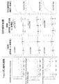
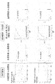
- FIG. 9 shows the quantitative results of Test Example 2 (shotgun analysis results), the quantitative results of Test Example 4 (SRM analysis results), and the ROC curves of Claudin-4, Heatshock protein HSP90-beta, and Epidermal growthfactor receptor. .
- the horizontal axis indicates, from the left, healthy subjects, cystitis, bladder cancer (superficial cancer), and bladder cancer (invasive cancer) from the left.
- the horizontal axis in the graph of the comparison results between the two groups of the quantification results of the SRM analysis results shows healthy subjects and bladder cancer from the left, and the horizontal axis in the graph of the comparison results between the three groups shows the healthy subjects from the left.
- Bladder cancer superficial cancer
- bladder cancer invasive cancer
- the vertical axis indicates the quantitative value.
- the vertical axis indicates sensitivity (positive rate), and the horizontal axis indicates a value obtained by subtracting specificity from 1 (1-specificity) (false positive rate).
- Quantitative results of Test Example 2 shotgun analysis results
- Quantitative results of Test Example 4 SRM analysis results
- ROC curves The description of each graph is the same as that of FIG.
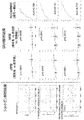
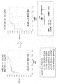
- FIG. 2 shows the quantitative results of Test Example 2 (shotgun analysis results), the quantitative results of Test Example 4 (SRM analysis results), and the ROC curves for Tight junction protein ZO-2, Ephrin type-A receptor II, and Calreticulin.
- the description of each graph is the same as that of FIG. 5 shows the quantification result of Test Example 2 (shotgun analysis result), the quantification result of Test Example 4 (SRM analysis result), and the ROC curve for Gamma-enolase and Calpain-2 catalytic subunit. The description of each graph is the same as that of FIG.
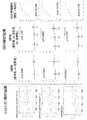
- TMT analysis results shows the quantification results of Test Example 2 (shotgun analysis results), the quantification results of Test Example 4 (SRM analysis results), and the ROC curves for Protein diaphanous homolog 1 and Serine / threonine-protein kinase LMTK2.
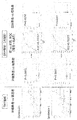
- SRM analysis results shows the quantification results of Test Example 6 (TMT analysis results), the quantification results of Test Example 6 (SRM analysis results), and the ROC curves for Protein S100-P and Complement Decay-accelerating Factor (DAF-1).
- DAF-1 Complement Decay-accelerating Factor
- a graph of the comparison result between the two groups (the vertical axis indicates the quantitative value; the horizontal axis indicates a healthy person and bladder cancer from the left), and a graph of the ROC curve (vertical axis) Indicates the sensitivity (positive rate), the horizontal axis indicates the value obtained by subtracting the specificity from 1 (1-specificity) (false positive rate), and the graph of the comparison results between the two groups (the vertical axis indicates the quantitative value)
- the hematuria patient and bladder cancer are shown from the left in the horizontal axis).
- test method for urothelial cancer relates to a method for testing urothelial cancer, comprising: (1) selecting from a specific group of membrane proteins in a urine sample collected from a subject;
- the present invention relates to a test method (hereinafter, also referred to as “test method of the present invention”) including a step of detecting at least one type of biomarker (step 1).
- test method of the present invention including a step of detecting at least one type of biomarker (step 1).
- the "urothelial cancer" to be tested is not particularly limited as long as it occurs in the urinary tract (renal pelvis, ureter, bladder, urethra).
- urothelial cancer include bladder cancer, renal pelvis / ureteral cancer, and the like.
- a bladder cancer is preferable as a test object.
- urothelial cancer includes various stages of cancer, and includes superficial cancer, invasive cancer and the like.
- the subject is the target organism of the test method of the present invention, and the species is not particularly limited.
- the species of the subject include various mammals such as humans, monkeys, mice, rats, dogs, cats and rabbits, and humans are preferred.
- the state of the subject is not particularly limited.
- the subject may be, for example, a sample that is not known to have urothelial carcinoma, a sample that has already been determined to have urothelial carcinoma by another method, or a subject that has urothelial carcinoma Samples that have already been determined to have not been performed by another method, samples that are being treated for urothelial cancer, and the like can be mentioned.
- the urine sample is not particularly limited as long as it is urine and a sample derived therefrom (a urine-derived sample).
- the urine-derived sample is a sample obtained by subjecting urine to any operation (separation, purification, drug addition, and the like).
- the urine sample is preferably a urine-derived sample.
- urine-derived sample urine extracellular vesicles are preferable.
- the urine sample may be used alone or in combination of two or more.
- Extracellular vesicles are not particularly limited as long as they are membrane vesicles secreted and released from cells.
- Extracellular vesicles are usually defined as membrane vesicles that carry intracellular and intracellular communication by transporting intracellular proteins and genetic information (mRNA, microRNA, etc.) outside the cell. You. Examples of extracellular vesicles include exosomes, microvesicles, apoptotic bodies, ectosomes, microparticles, secretory microvesicles, and the like. Of these, exosomes are preferred.
- Extracellular vesicles can be purified, separated, concentrated, etc. from urine according to or according to known methods.
- Methods for purifying, separating, concentrating, etc. extracellular vesicles include, for example, ultracentrifugation (eg, pellet down, sucrose cushion, density gradient centrifugation, etc.), methods using immunoaffinity carriers, gel filtration, Flow fractionation, FACS and the like can be mentioned.
- Purification, separation, concentration and the like of extracellular vesicles can also be performed using a commercially available kit. These methods may be employed alone or in combination of two or more.
- the detection target in step (1) is at least one biomarker selected from a specific membrane protein group (in the present specification, these may be collectively referred to as “target biomarker”).
- This specific group of membrane proteins is a urothelial carcinoma-specific biomarker, which can be used as an indicator to distinguish urothelial carcinoma.
- This particular group of membrane proteins includes Claudin-4, Heat shock protein HSP 90-beta, Epidermal growth factor receptor, Epihelial cell adhesion molecule, HLA class I histocompatibility antigen, B-35 alpha chain, Chloride intracellular channel can-1 and yn 1, Intercellular hesadhesion molecule 1, Myristoylated alanine-rich C-kinase substrate, Niban-like protein 1, Solute carrier family 12 member 7, MARCKS-related protein, Tight junction protein ZO-2, Ephrin type-A receptor 2, Calreticulin Gamma-enolase, Calpain-2 catalytic subunit, Protein diaphanous homolog 1, Serine / threonine-protein kinase LMTK2, Protein S100-P, Complement decay-accelerating factor (DAF-1), and Receptor-type tyrosine-protein phosphatase F (TPRF) -1). These are a group of proteins whose amounts in urothelial cancer samples are higher than
- HSP 90-beta preferably Heat ⁇ shock protein HSP 90-beta, Epidermal growth factor receptor, Epithelial cell adhesion molecule, HLA class I histocompatibility antigen, B-35 alpha chain, Solute carrier family 12 member 7, Gamma-enolase, Protein diaphanous homolog, 1, Serine / threonine-protein kinase, LMTK2, and pit, preferably helical, and helical.
- Heat Shock Protein HSP 90-beta preferably Heat Shock Protein HSP 90-beta, Syndecan-1, Ephrin Examples include type-A receptor 2, Calreticulin, protein S100-P, complement decay-accelerating factor (DAF-1), and receptor-type tyrosine-protein phosphatase F (PTPRF-1).
- Claudin-4 Heat shock protein HSP 90-beta, Epidermal growth factor receptor, Epihelial cell adhesion molecule, HLA class I histocompatibility antigen, B -35 chain, Chloride intracellular channel protein 1, Syndecan-1, Intercellular adhesion molecule 1, etc., more preferably Claudin-4, Heat shock protein HSP 90-beta, Epidermal growth factor receptor, Epihelial cell adhesion molecule, class I histocompatibility antigen, B-35 alpha chain and the like, more preferably Claudin-4, Heat shock protein HSP 90-beta, Epidermal growth factor receptor, Epihelial cell adhesion molecule, and particularly preferably Claudin-4 Is mentioned.
- membrane proteins from the viewpoint of being more suitable for invasive cancer differentiation, etc., preferably Claudin-4, Heat ⁇ shock protein HSP 90-beta, Epidermal growth factor receptor, Epihelial cell adhesion molecule, HLA class I histocompatibility antigen , B-35 alpha chain, Syndecan-1, Intercellular adhesion molecule 1, Myristoylated alanine-rich C-kinase substrate, Niban-like protein 1, MARCKS-related protein, Calreticulin, etc., more preferably Claudin-4, Epihelial cell adhesion molecule, Syndecan-1, Intercellular adhesion molecule 1, Myristoylated alanine-rich C-kinase substrate, MARCKS-related protein, Calreticulin, etc., more preferably, Claudin-4, Epihelial cell adhesion molecule, Calreticulin, etc. .
- the proteins of the above-mentioned membrane protein group can be specified by their names, or can be specified as orthologs of the human proteins indicated by the names.
- the number of target biomarkers in step (1) may be only one, or may be a combination of two or more. Testing for urothelial carcinoma by combining more (eg, 2 or more, 3 or more, 4 or more, 5 or more, 7 or more, 10 or more, 15 or more, 19) Etc. can be performed more accurately.
- Detection is usually performed by measuring the amount or concentration of the target biomarker.
- concentration is not limited to the absolute concentration, but may be a relative concentration, a weight per unit volume, or raw data measured to know the absolute concentration.
- the method for detecting the target biomarker is not particularly limited as long as it can specifically detect a part or all of the target biomarker.
- Specific examples of the detection method include, for example, mass spectrometry for detecting a peptide constituting the target biomarker, immunological measurement using an antibody that specifically recognizes the target biomarker, and the like.
- the amino acid sequence information of the target biomarker can be obtained by searching the EBI (http://www.ebi.ac.uk/IPI/IPIhelp.html) database based on the UniProtKB accession number. .
- Suitable examples of the immunological assay include immunohistochemical staining, ELISA, sandwich ELISA, EIA, RIA, and Western blotting.
- ELISA immunohistochemical staining
- sandwich ELISA sandwich ELISA
- EIA extracellular vesicle marker
- RIA Western blotting
- extracellular vesicle marker such as CD9
- target biomarker an antibody against a target biomarker
- Mass spectrometry is a method in which a peptide sample is converted into gaseous ions using an ion source (ionization), and the peptide sample is ionized by moving it in a vacuum and using electromagnetic force or by a time-of-flight difference in the analysis unit.
- a measurement method using a mass spectrometer that can be separated and detected according to the ratio, and ionization using an ion source includes EI, CI, FD, FAB, MALDI, and ESI.
- the method of separating the ionized peptide sample in the analyzer can be selected from magnetic field deflection type, quadrupole type, ion trap type, time of flight (TOF) type, Fourier transform A separation method such as an ion cyclotron resonance type can be appropriately selected.
- tandem mass spectrometry (MS / MS) or triple quadrupole mass spectrometry combining two or more mass spectrometry methods can be used.
- the sample is a sample containing a phosphorylated peptide
- the sample can be concentrated using iron ion-immobilized affinity chromatography (Fe-IMAC) before the sample is introduced into the mass spectrometer.
- Fe-IMAC iron ion-immobilized affinity chromatography
- the peptide constituting the target biomarker can be separated and purified by liquid chromatography (LC) or HPLC to obtain a sample. Further, the detection unit and the data processing method can be appropriately selected.
- LC liquid chromatography
- the detection unit and the data processing method can be appropriately selected.
- a stable isotope-labeled peptide in which one or more of the amino acids in the peptide constituting the target biomarker to be detected contains one or more of 15 N, 13 C, 18 O, and 2 H If so, the type, position, number, etc. of the amino acids can be appropriately selected, and such a stable isotope-labeled peptide can be prepared using the F-moc method (Amblard., Et al. Methods Mol Biol.
- iTRAQ registered trademark
- ICAT registered trademark
- ICPL registered trademark
- NBS (Registered trademark) reagent
- a labeled antibody an antibody that binds to the primary antibody
- a radioisotope such as 125I, a fluorescent substance, an enzyme such as horseradish peroxidase (HRP), etc.
- HRP horseradish peroxidase
- the amount and / or concentration of the target biomarker which is a detection index of urothelial cancer, can be provided, whereby the detection of urothelial cancer can be performed. Can assist you.
- test results of the test method of the present invention including the step (1) are used to determine the therapeutic effect, elucidate the pathology of urothelial cancer, predict the prognosis of urothelial cancer, stratify patients, select treatment methods (individualization) It can be used for evaluation of refractory and remodeling in urothelial cancer, differentiation of urothelial cancer histology, phenotype, etc.
- (2) the subject suffers from urothelial cancer when the amount or concentration of the biomarker detected in the step (1) is equal to or higher than a cutoff value. It is preferable to include a step of determining that the operation is performed. According to the test method of the present invention including the step 2, urothelial cancer can be determined.
- Cut-off value, sensitivity, specificity, positive predictive value can be appropriately set by those skilled in the art from the viewpoint of negative predictive value, for example, collected from a subject not suffering from urothelial cancer
- a predetermined value or a predetermined value can be used.
- the cut-off value is, for example, the amount and / or concentration of the target biomarker in a urine sample collected from a subject not suffering from urothelial cancer (average, median, ) Can be, for example, 1 (or a value greater than 1) to 3 times.
- the determination can be made by a method using a discriminant.
- the discriminant can be any discriminant analysis method that can create a discriminant for discriminating between urothelial cancer and healthy, such as Fisher's discriminant analysis, nonlinear discriminant analysis by Mahalanobis distance, neural network, Support Vector Although it can be created using a Machine (SVM), it is not limited to these specific examples.
- SVM Machine
- the determination can be made using a method such as a neural network, a k-nearest neighbor method, a decision tree, and a logistic regression analysis.
- the cut-off value is set to, for example, the amount of the target biomarker in a past sample of the same sample and / or By setting the value based on the concentration, the therapeutic effect of urothelial cancer can be determined.
- the biomarker is Claudin-4, Heat shock protein HSP 90-beta, Epidermal growth factor receptor, Epihelial cell adhesion molecule, HLA class I histocompatibility antigen, B-35 alpha chain, Syndecan -1, Intercellular adhesion molecule 1, Myristoylated alanine-rich C-kinase substrate, Niban-like protein 1, MARCKS-related protein, and at least one biomarker selected from the group consisting of Calreticulin, (3 A) determining that the subject is suffering from invasive cancer among urothelial cancers when the amount or concentration of the biomarker detected in the step (1) is equal to or greater than a cutoff value; It is preferred to include.
- Step 3 may also include a step of determining that the subject has a superficial cancer among urothelial carcinomas when the amount or concentration of the biomarker is equal to or lower than the cutoff value. According to the test method of the present invention including the step 3, it is possible to distinguish whether the urothelial cancer is a superficial cancer or an invasive cancer.
- the cut-off value can be appropriately set by those skilled in the art from the viewpoints of sensitivity, specificity, positive predictive value, negative predictive value, and the like.
- urothelial cancer (superficial cancer and / or invasion
- the value may be a predetermined value or a predetermined value based on the amount and / or concentration of the target biomarker in a urine sample collected from a subject not suffering from (H).
- the cut-off value may be determined, for example, by measuring the amount and / or concentration of the target biomarker in a urine sample collected from a subject not suffering from urothelial cancer (superficial cancer and / or invasive cancer).
- the value can be, for example, 1 (or a value exceeding 1) to 3 times the average value, the median value, or the like).
- the cut-off value is set to, for example, a target biomarker in a past sample of the same sample.
- the therapeutic effect of urothelial cancer (invasive cancer) can be determined by setting the value based on the amount and / or concentration of
- the subject can be treated with urothelial carcinoma (superficial cancer and / or invasion). Diagnosing urothelial carcinoma with higher accuracy by combining the test method of the present invention with the step of applying a urothelial carcinoma diagnosis by a doctor when it is determined that can do.
- the test method of the present invention can more accurately detect urothelial cancer, by combining the above-described steps with the test method of the present invention, it is possible to more efficiently and more accurately detect urothelial cancer. Yes "can be diagnosed.
- the subject suffers from urothelial cancer (superficial cancer and / or invasive cancer) by the test method of the present invention including the treatment step (2) and / or the step (3) for urothelial cancer. If it is determined that the urothelial cancer is present, the urothelial cancer (superficial cancer) may be further examined by the method of the present invention or as described in “2. And / or invasive cancer), the combination of the test method of the present invention and the step of applying a diagnosis by a physician further provides (3) urothelial cancer. By performing a step of treating the disease for the subject determined or diagnosed as being present, the disease of the subject can be treated.
- test method of the present invention can more accurately detect urothelial cancer
- test method of the present invention or the combination of the test method of the present invention and a step of applying a diagnosis by a doctor By combining step 3, a subject suffering from urothelial cancer can be treated more efficiently and more reliably.
- the method of treating urothelial cancer is not particularly limited, and various known treatment methods can be adopted.
- the treatment method include chemotherapy, surgical treatment, radiation treatment, and immunotherapy. These can be performed according to a known method.
- the therapeutic agent used for chemotherapy is not particularly limited, and various anticancer agents can be used.
- the anticancer agent include an alkylating agent, an antimetabolite, a microtubule inhibitor, an antibiotic anticancer agent, a topoisomerase inhibitor, a platinum preparation, a molecular target drug, a hormonal agent, and a biological preparation.
- the alkylating agent include cyclophosphamide, ifosfamide, nitrosourea, dacarbazine, temozolomide, nimustine, busulfan, melphalan, procarbazine, ranimustine and the like.
- antimetabolites for example, enositabine, carmofur, capecitabine, tegafur, tegafur uracil, tegafur gimeracil oteracil potassium, gemcitabine, cytarabine, cytarabine octophosphate, nelarabine, fluorouracil, fludarabetin, penmetrexet, penmetrexet, penmetrexetate Cladribine, doxyfluridine, hydroxycarbamide, mercaptopurine and the like.
- the microtubule inhibitor include alkaloid anticancer agents such as vincristine, and taxane anticancer agents such as docetaxel and paclitaxel.
- antibiotic anticancer agent examples include mitomycin C, doxorubicin, epirubicin, daunorubicin, bleomycin, actinomycin D, aclarubicin, idarubicin, pirarubicin, peplomycin, mitoxantrone, amrubicin, dinostatin stimaramar and the like.
- topoisomerase inhibitor examples include CPT-11 having a topoisomerase I inhibitory action, irinotecan, nogitecan, etoposide and sobuzoxane having a topoisomerase II inhibitory action.
- platinum preparation examples include cisplatin, nedaplatin, oxaliplatin, carboplatin and the like.
- Hormonal agents include, for example, dexamethasone, finasteride, tamoxifen, astrozole, exemestane, ethinylestradiol, chlormadinone, goserelin, bicalutamide, flutamide, blednisolone, leuprorelin, letrozole, estramustine, toremifene, phosphesterol, mitotane, Examples include methyltestosterone, medroxyprogesterone, mepithiostane, and the like.
- the biologic include interferon ⁇ , ⁇ and ⁇ , interleukin 2, ubenimex, dried BCG and the like.
- Examples of the molecular target drug include nivolumab, pembrolizumab, rituximab, alemtuzumab, trastuzumab, cetuximab, panitumumab, imatinib, dasatinib, nilotinib, gefitinib, erlotinib, temsirolimus, bevacizumab, and GF. Ibritumomab ozogamicin, ibritumomab tiuxetane, tamibarotene, tretinoin and the like.
- human epidermal growth factor receptor 2 inhibitor In addition to the targeted drugs specified here, human epidermal growth factor receptor 2 inhibitor, epidermal growth factor receptor inhibitor, Bcr-Abl tyrosine kinase inhibitor, epidermal growth factor tyrosine kinase inhibitor, mTOR inhibitor Drugs, inhibitors of angiogenesis such as vascular endothelial growth factor receptor 2 inhibitor ( ⁇ -VEGFR-2 antibody), various tyrosine kinase inhibitors such as MAP kinase inhibitor, inhibitors targeting cytokines, Molecular targeting drugs such as proteasome inhibitors and antibody-anticancer drug combinations can also be included. These inhibitors also include antibodies. One, two, or three or more therapeutic agents can be used in combination.
- test agent for urothelial cancer test kit
- the present invention provides, in one embodiment, a test agent for urothelial cancer, comprising a detection agent for at least one biomarker selected from a specific membrane protein group (In this specification, it may be referred to as “the test agent of the present invention”). Hereinafter, this will be described.
- the detection agent is not particularly limited as long as it can specifically detect the target biomarker.
- Examples of the detection agent include an antibody against a target biomarker.
- the detection agent may be modified as long as its function is not significantly impaired.
- modifications include addition of a label, for example, a fluorescent dye, an enzyme, a protein, a radioisotope, a chemiluminescent substance, biotin, and the like.
- the fluorescent dye used in the present invention generally used are those which label nucleotides and are used for the detection and quantification of nucleic acids.
- HEX 4,7,2 ', 4', 5 ', 7 '-hexachloro-6-carboxylfluorescein, green fluorescent dye
- fluorescein fluorescein
- NED trade name, manufactured by Applied Biosystems, yellow fluorescent dye
- 6-FAM trade name, manufactured by Applied Biosystems, yellow
- Green fluorescent dye rhodamine or a derivative thereof (eg, tetramethylrhodamine (TMR)), but is not limited thereto.
- a method for labeling nucleotides with a fluorescent dye an appropriate one of known labeling methods can be used [see Nature Biotechnology, ⁇ 14, ⁇ 303-308 ⁇ (1996)].
- a commercially available fluorescent labeling kit can be used (for example, oligonucleotide ECL '3'-oligolabeling system, manufactured by Amersham Pharmacia).
- the detection agent can be used by immobilizing it on an arbitrary solid phase. Therefore, the test agent of the present invention can be provided in the form of a substrate on which a detection agent is immobilized (for example, an ELISA plate or the like on which an antibody is immobilized).
- a substrate on which a detection agent is immobilized for example, an ELISA plate or the like on which an antibody is immobilized.
- the solid phase used for immobilization is not particularly limited as long as it can immobilize an antibody or the like, and examples thereof include a glass plate, a nylon membrane, microbeads, a silicon chip, a capillary, and other substrates. it can. Immobilization of the detection agent on the solid phase is not particularly limited.
- the antibody is not particularly limited as long as it selectively (specifically) recognizes the target biomarker.
- “selectively (specifically) recognizes” means that a target biomarker can be specifically detected by, for example, a Western blotting method or an ELISA method, but is not limited thereto. May be used as long as it can be determined that the above-mentioned detected substance is derived from the target biomarker.
- Antibodies include a part of the above-mentioned antibodies having antigen-binding properties such as polyclonal antibodies, monoclonal antibodies, chimeric antibodies, single-chain antibodies, or Fab fragments or fragments generated by Fab expression libraries.
- Antibodies of the present invention also include antibodies that have antigen-binding properties to a polypeptide that is at least contiguous, usually at least 8, preferably at least 15, and more preferably at least 20 amino acids in the amino acid sequence of the target biomarker.
- the antibodies of the present invention can also be produced according to these conventional methods (Current Protocols in Molecular Biology, Chapters 11.12 to 11.13 (2000)).
- the antibody of the present invention is a polyclonal antibody, an oligopeptide having a partial amino acid sequence of the target biomarker using a target biomarker expressed and purified in E. coli or the like according to a conventional method, or Can be synthesized, immunized to a non-human animal such as a rabbit, and obtained from the serum of the immunized animal according to a conventional method.
- a target biomarker used as an immunizing antigen for the production of an antibody is based on known gene sequence information, DNA cloning, construction of each plasmid, transfection into a host, culturing of a transformant, and protein cultivation from the culture. It can be obtained by a collection operation. These operations are performed according to methods known to those skilled in the art, or methods described in the literature (Molecular Cloning, T. Maniatis et al., CSH Laboratory (1983), DNA Cloning, DM. Glover, IRL Press (1985)). Can be done.
- a recombinant DNA capable of expressing a gene encoding a target biomarker in a desired host cell is prepared, and this is introduced into a host cell, transformed, and the transformant is cultured. Then, by recovering the target protein from the obtained culture, a protein as an immunizing antigen for producing the antibody of the present invention can be obtained. Further, the partial peptide of the target biomarker can also be produced by a general chemical synthesis method (peptide synthesis) according to known gene sequence information.
- the antibody of the present invention may be prepared using an oligopeptide having a partial amino acid sequence of the target biomarker.
- the oligo (poly) peptide used for the production of such an antibody does not need to have a functional biological activity, but desirably has the same immunogenic properties as the target biomarker.
- An oligo (poly) peptide preferably having this immunogenic property and comprising at least 8 amino acids, preferably 15 amino acids, more preferably 20 amino acids in the amino acid sequence of the target biomarker can be exemplified.
- Production of an antibody against such an oligo (poly) peptide can also be carried out by increasing the immunological reaction using various adjuvants depending on the host.
- adjuvants include, but are not limited to, Freund's adjuvant, mineral gels such as aluminum hydroxide, and surfaces such as lysolecithin, pluronic polyols, polyanions, peptides, oil emulsions, keyhole limpet hemocyanin and dinitrophenol.
- Active substances include human adjuvants such as BCG (Bacillus Calmette-Guerin) and Corynebacterium-Parvum.
- the test agent of the present invention may be in the form of a composition.
- the composition may contain other components as necessary.
- Other components include, for example, bases, carriers, solvents, dispersants, emulsifiers, buffers, stabilizers, excipients, binders, disintegrants, lubricants, thickeners, humectants, colorants, fragrances And chelating agents.
- the test agent of the present invention may be in the form of a kit.
- the kit may contain, in addition to the above-described detection agent or the above-described composition containing the same, a kit that can be used for detection of a target biomarker in a urine sample of a subject.
- Specific examples of such substances include various reagents (eg, secondary antibodies, buffers, etc.), instruments (eg, instruments for purification, separation, and concentration of extracellular vesicles (eg, columns)).
- the present invention comprises a suppressor of at least one biomarker selected from a specific group of membrane proteins, for preventing or treating urothelial cancer
- the present invention also relates to an agent (hereinafter, also referred to as “agent of the present invention”). Hereinafter, this will be described.
- an example of the inhibitor is an antibody against the target biomarker.
- the antibody those similar to the antibodies described in the above-mentioned “4. Test agent and test kit for urothelial cancer” can be used.
- Another example of the inhibitor is an expression inhibitor of the target biomarker.
- the expression inhibitor of the target biomarker is not particularly limited as long as it can suppress the expression level of the target biomarker, its mRNA, its precursor, and the like.
- gene-specific small interfering RNA siRNA
- miRNA Gene-specific microRNA
- gene-specific antisense nucleic acid of the target biomarker their expression vector
- gene-specific ribozyme of the target biomarker gene of the target biomarker by CRISPR / Cas system Editing agents and the like.
- the expression suppression refers to the target biomarker, the expression level of its mRNA and the like, for example, 1/2, 1/3, 1/5, 1/10, 1/20, 1/30, 1/50, 1 / This means that the expression is suppressed to 100, 1/200, 1/300, 1/500, 1/1000, and 1 / 10,000 or less.
- the gene siRNA of the target biomarker is not particularly limited as long as it is a double-stranded RNA molecule that specifically suppresses the expression of the gene encoding the target biomarker.
- the siRNA preferably has a length of, for example, 18 bases or more, 19 bases or more, 20 bases or more, or 21 bases or more. Further, the siRNA preferably has a length of, for example, 25 bases or less, 24 bases or less, 23 bases or less, or 22 bases or less. It is assumed that the upper limit and the lower limit of the length of the siRNA described here are arbitrarily combined.
- the lower limit is 18 bases
- the upper limit is 25 bases, 24 bases, 23 bases, or 22 bases
- the lower limit is 19 bases
- the upper limit is 25 bases, 24 bases, 23 bases, or 22 bases.
- a length with a lower limit of 20 bases and an upper limit of 25 bases, 24 bases, 23 bases, or 22 bases; a lower limit of 21 bases, and an upper limit of 25 bases, 24 bases, 23 bases, or 22 bases A combination of lengths that are bases is envisioned.
- SiRNA may be shRNA (small hairpin RNA).
- shRNAs can be designed such that a portion forms a stem-loop structure.
- shRNA has a sequence of a certain region as sequence a and a complementary strand to sequence a as sequence b, so that these sequences are present in one RNA strand such that sequence a, spacer, and sequence b are in this order.
- sequence a is a sequence of a partial region of the base sequence encoding the target biomarker to be targeted.
- the target region is not particularly limited, and any region can be a candidate.
- the length of sequence a is 19 to 25 bases, preferably 19 to 21 bases.
- the gene-specific siRNA of the target biomarker may have an additional base at the 5 'or 3' end.
- the length of the additional base is usually about 2 to 4 bases.
- the additional base may be DNA or RNA, but use of DNA may improve the stability of the nucleic acid in some cases.
- Such additional base sequences include, for example, ug-3 ', uu-3', tg-3 ', tt-3', ggg-3 ', guuu-3', gttt-3 ', ttttt-3 ', Uuuuu-3' and the like, but are not limited thereto.
- the siRNA may have a protruding portion sequence (overhang) at the 3 ′ end, and specific examples include those to which dTdT (dT represents deoxythymidine) has been added. In addition, blunt ends (blunt ends) without terminal addition may be used.
- the sense strand and the antisense strand may have different numbers of bases. For example, “asymmetrical interfering RNA (the antisense strand has an overhanging sequence at the 3 ′ end and the 5 ′ end) aiRNA)].
- a typical aiRNA has an antisense strand consisting of 21 bases, a sense strand consisting of 15 bases, and an overhang structure of 3 bases at each end of the antisense strand.
- the position of the target sequence of the gene-specific siRNA of the subject biomarker is not particularly limited, but in one embodiment, up to about 50 bases from the 5'-UTR and start codon, and from regions other than the 3'-UTR It is desirable to select a target sequence.
- BLAST http://www.ncbi.nlm.nih.gov/BLAST/ ) It is preferable to check the specificity of the selected target sequence using homology search software.
- a sense strand having a TT or UU 3 ′ terminal overhang at 19-21 bases after AA (or NA), a sequence complementary to the 19-21 base and TT or A double-stranded RNA consisting of an antisense strand having a 3'-terminal overhang of UU may be designed as siRNA.
- shRNA which is a precursor of siRNA, appropriately selects an arbitrary linker sequence (for example, about 5 to 25 bases) capable of forming a loop structure, and connects the sense strand and the antisense strand via the linker sequence. It can be designed by connecting.
- siRNA and / or shRNA can be searched using search software provided free of charge on various websites. Examples of such sites include the following. SiRNA Target Finder provided by Ambion (http://www.ambion.com/jp/techlib/misc/siRNA_finder.html) pSilencer (registered trademark) Expression Vector Insert Design Tool (http://www.ambion.com/jp/techlib/misc/psilencer_converter.html) GeneSeer provided by RNAi Codex (http://codex.cshl.edu/scripts/newsearchhairpin.cgi).
- the siRNA is obtained by synthesizing the sense strand and the antisense strand of the target sequence on the mRNA with a DNA / RNA automatic synthesizer and denaturing them in a suitable annealing buffer at about 90 to about 95 ° C. for about 1 minute. It can be prepared by annealing at about 30 to about 70 ° C. for about 1 to about 8 hours. Alternatively, it can be prepared by synthesizing shRNA that is a precursor of siRNA and cleaving it using an RNA-cleaving protein dicer.
- the gene-specific miRNA of the target biomarker is optional as long as it inhibits translation of the gene encoding the target biomarker.
- the miRNA may pair with the 3 'untranslated region (UTR) of the target and inhibit its translation.
- the miRNA may be any of pri-miRNA (primary miRNA), pre-miRNA (precursor miRNA), and mature miRNA.
- the length of the miRNA is not particularly limited, the length of the pri-miRNA is usually several hundred to several thousand bases, the length of the pre-miRNA is usually 50 to 80 bases, and the length of the mature miRNA is usually 18 ⁇ 30 bases.
- the gene-specific miRNA of the biomarker of interest is preferably a pre-miRNA or a mature miRNA, more preferably a mature miRNA.
- a gene-specific miRNA of the target biomarker may be synthesized by a known method, or may be purchased from a company that provides synthetic RNA.
- the gene-specific antisense nucleic acid of the target biomarker is a nucleic acid containing a base sequence complementary to or substantially complementary to the base sequence of the mRNA of the gene encoding the target biomarker or a part thereof, and the mRNA It is a nucleic acid having a function of suppressing the synthesis of a target biomarker by forming a specific and stable double strand and binding thereto.
- the antisense nucleic acid may be any of DNA, RNA, and DNA / RNA chimera.
- RNA DNA hybrid formed by the target RNA and the antisense DNA is recognized by endogenous ribonuclease H (RNase H) and causes the target RNA to be selectively degraded. Therefore, in the case of antisense DNA directed to degradation by RNase H, the target sequence may be not only the sequence in the mRNA but also the sequence of the intron region in the initial translation product of the gene of the target biomarker.
- the intron sequence can be determined by comparing the genomic sequence with the cDNA base sequence of the gene of the target biomarker using a homology search program such as BLAST or FASTA.
- the length of the target region of the gene-specific antisense nucleic acid of the target biomarker is not limited as long as the antisense nucleic acid hybridizes and as a result, translation into the target biomarker is inhibited.
- the gene-specific antisense nucleic acid of the target biomarker may be the entire sequence or a partial sequence of the mRNA encoding the target biomarker. Considering the ease of synthesis, antigenicity, and the ability to enter cells, oligonucleotides consisting of about 10 to about 40 bases, particularly about 15 to about 30 bases, are preferred, but not limited thereto.
- the 5 'end hairpin loop, 5' end untranslated region, translation initiation codon, protein coding region, ORF translation stop codon, 3 'end untranslated region, 3' end palindrome of the gene of the target biomarker A region or a 3 ′ end hairpin loop may be selected as a preferred target region for an antisense nucleic acid, but is not limited thereto.
- the gene-specific antisense nucleic acid of the target biomarker not only hybridizes to the mRNA and early transcript of the target biomarker gene and inhibits translation into proteins, but also binds to these double-stranded DNA genes. To form a triplex, thereby inhibiting the transcription into RNA (antigene).
- the nucleotide molecules constituting the gene-specific siRNA of the target biomarker, the gene-specific miRNA of the target biomarker, and the gene-specific antisense nucleic acid of the target biomarker have stability (chemical and / or counterpart enzyme) and Various chemical modifications may be included to improve specific activity (affinity with RNA). For example, in order to prevent degradation by a hydrolase such as nuclease, a phosphate residue (phosphate) of each nucleotide constituting an antisense nucleic acid is replaced with, for example, phosphorothioate (PS), methylphosphonate (methylphosphonate), or phosphorodithioate.
- PS phosphorothioate
- methylphosphonate methylphosphonate
- phosphorodithioate phosphorodithioate
- the base moiety pyrimidine, purine
- a part of the nucleotide molecules constituting the siRNA or the miRNA may be replaced with a natural type DNA.
- the gene-specific siRNA of the target biomarker, the gene-specific miRNA of the target biomarker, and the gene-specific antisense nucleic acid of the target biomarker are mRNA or initial based on the cDNA sequence or genomic DNA sequence of the gene of the target biomarker. It can be prepared by determining the target sequence of the transcript and synthesizing a sequence complementary thereto using a commercially available automatic DNA / RNA synthesizer. In addition, antisense nucleic acids containing the various modifications described above can be chemically synthesized by any known method.
- the gene-specific siRNA of the target biomarker For expression vectors of the gene-specific siRNA of the target biomarker, the gene-specific miRNA of the target biomarker, or the gene-specific antisense nucleic acid of the target biomarker, the gene-specific siRNA of the target biomarker, the gene specificity of the target biomarker
- the target miRNA or the gene-specific antisense nucleic acid of the target biomarker is incorporated in an expressible state.
- the expression vector comprises a promoter sequence and a gene-specific siRNA of the target biomarker, a gene-specific miRNA of the target biomarker, or a coding sequence of the gene-specific antisense nucleic acid of the target biomarker (optionally And a polynucleotide containing a transcription termination signal sequence) and, if necessary, other sequences.
- the promoter is not particularly limited, and for example, CMV promoter, EF1 promoter, SV40 promoter, MSCV promoter, hTERT promoter, ⁇ -actin promoter, RNA ⁇ ⁇ ⁇ ⁇ ⁇ ⁇ polymerase II (polII) -based promoter such as CAG promoter; mouse and human U6-snRNA promoter, Human H1-RNase P RNA promoter, human valine-tRNA promoter, and other RNA polymerase III (polIII) promoters and the like. Of these, from the viewpoint that transcription of short RNAs can be performed accurately, polIII promoters are preferred. preferable.
- the other sequence is not particularly limited, and various known sequences that can be included in the expression vector can be used. Examples of such a sequence include, for example, an origin of replication and a drug resistance gene. The types of drug resistance genes and types of vectors can be the same as those described above.
- RNA-specific ribozyme of the target biomarker includes a gene-specific ribozyme of the target biomarker.
- "Ribozyme” in a narrow sense means RNA having an enzymatic activity for cleaving a nucleic acid, but in the present application, it includes DNA as long as it has a sequence-specific nucleic acid cleaving activity.
- the most versatile ribozyme nucleic acids include self-splicing RNAs found in infectious RNAs such as viroids and viruses, and hammerhead and hairpin types are known.
- the hammerhead type exhibits enzymatic activity at about 40 bases, and the bases at both ends adjacent to the hammerhead structure (about 10 bases in total) are converted into a sequence complementary to the desired cleavage site of mRNA. By doing so, it is possible to specifically cleave only the target mRNA. Since this type of ribozyme nucleic acid uses only RNA as a substrate, it has the advantage that it does not attack genomic DNA. When the mRNA of the target biomarker gene has a double-stranded structure by itself, a single target sequence can be obtained by using a hybrid ribozyme linked to an RNA motif derived from a viral nucleic acid that can specifically bind to RNA helicase.
- the application target of the agent of the present invention is not particularly limited, and includes, for example, various mammals such as human, monkey, mouse, rat, dog, cat, rabbit, pig, horse, cow, sheep, goat, deer and the like. .
- the form of the agent of the present invention is not particularly limited, and may take a form usually used in each application depending on the use of the agent of the present invention.
- a tablet including an intraorally disintegrating tablet, a chewable tablet, an effervescent tablet, a troche, a jelly-like drop, etc.
- Pills granules, fine granules, powders, hard capsules, soft capsules, dry syrups, liquids (including drinks, suspensions and syrups), and preparations suitable for oral ingestion such as jellies
- Dosage form oral dosage form
- nasal drops inhalants, rectal suppositories, inserts, enemas, jellies, injections, patches, lotions, creams, etc. Oral formulation).
- a liquid, gel or solid food for example, juice, soft drink, tea, soup, soy milk, salad oil, dressing, yogurt, jelly, pudding, sprinkle, powdered milk for childcare , Cake mix, powdered or liquid dairy products, bread, cookies and the like.
- a liquid solution, emulsion, suspension, etc.
- semi-solid gel, cream, paste, etc.
- solid tablet, particulate, capsule, Film, kneaded material, molten solid, waxy solid, elastic solid, etc.
- dentifrice teethpaste, liquid dentifrice, liquid dentifrice, powder dentifrice, etc.
- mouthwash Coating agents, patches, mouth fresheners, foods (eg, chewing gum, tablet confectionery, candy, gummy, film, troche, etc.) and the like can be mentioned.
- the agent of the present invention may further contain other components as necessary.
- Other components are not particularly limited as long as they can be blended in, for example, a medicine, a food composition, an oral composition, a health enhancer, a nutritional supplement (such as a supplement), and the like.
- the content of the inhibitor of the target biomarker of the agent of the present invention depends on the type, use, use mode, application target, state of the application target, and the like of the inhibitor, and is not limited. It can be 100% by weight, preferably 0.001 to 50% by weight.
- the amount of application (for example, administration, ingestion, inoculation, etc.) of the composition of the present invention is not particularly limited as long as it is an effective amount that exhibits a medicinal effect, and is generally 0.1 to 1000 per day as the weight of the active ingredient. mg / kg body weight.
- the above-mentioned dose is preferably administered once a day or divided into two to three times a day, and may be appropriately increased or decreased depending on age, disease state and symptoms.
- Method for screening active ingredient of agent for preventing or treating urothelial cancer is selected from a specific group of membrane proteins in a urine sample collected from an animal treated with a test substance.
- a method for screening an active ingredient of a prophylactic or therapeutic agent for urothelial cancer using an amount or concentration of at least one biomarker as an index in the present specification, this may be referred to as “the active ingredient screening method of the present invention”. There is). Hereinafter, this will be described.
- the measurement of the amount or concentration of the body fluid, urine sample, specific membrane protein group, urothelial carcinoma, target biomarker, etc. is the same as the definition in the above “1.
- Animal species are not particularly limited. Examples of animal species include various mammals such as humans, monkeys, mice, rats, dogs, cats, and rabbits, and humans are preferable.
- any of naturally occurring compounds or artificially produced compounds can be widely used.
- a purified compound but also a composition in which various kinds of compounds are mixed, and an extract of animals and plants can be used.
- the compound is not limited to a low molecular compound, but also includes a high molecular compound such as a protein, a nucleic acid, and a polysaccharide.
- the active ingredient screening method of the present invention more specifically, when the biomarker as an index is at least one biomarker selected from a specific membrane protein group, the value of the index, the test substance If the amount or concentration of the corresponding biomarker in a urine sample collected from an animal that has not been treated with (a control value) is lower than the test substance, the active ingredient of a preventive or therapeutic agent for urothelial cancer (or As a candidate for an active ingredient of a prophylactic or therapeutic agent for urothelial cancer).
- Corresponding biomarker means the same protein as the target biomarker used as an index.
- Low means, for example, that the index value is 1/2, 1/5, 1/10, 1/20, 1/50, 1/100 of the control value.
- the present invention provides, in one embodiment, at least one selected from a specific group of membrane proteins in a urine sample collected from an animal treated with a test substance.
- the present invention relates to a method for evaluating the induction or malignancy of urothelial carcinoma using the amount or concentration of a biomarker of an species as an index (in this specification, the method is sometimes referred to as “the toxicity evaluation method of the present invention”). Hereinafter, this will be described.
- the toxicity evaluation method of the present invention when the biomarker as an index is at least one biomarker selected from a specific membrane protein group, the value of the index, the test substance, When the amount or concentration (control value) of the corresponding biomarker in a urine sample collected from an untreated animal is higher, the test substance is determined to have urothelial cancer-induced or aggressiveness. Process.
- Corresponding biomarker means the same protein as the target biomarker used as an index.
- “High” means that, for example, the index value is twice, five times, ten times, twenty times, fifty times, and one hundred times the control value.
- Test example 1 Preparation of exosome fraction 1 As subjects, seven human subjects diagnosed with bladder cancer (superficial cancer and invasive cancer) and four healthy human subjects were employed. The background of the subject is as follows.
- Natural urine was collected from the subject, and the exosome fraction was prepared from the urine.
- the exosome fraction was prepared by a stepwise ultracentrifugation method. Specifically, the procedure was performed as follows. Urine was diluted with PBS and centrifuged (2000 g, 10 ° C., 30 minutes). Thereafter, the supernatant was centrifuged (17000 g, 10 ° C., 30 minutes). The supernatant was passed through a 0.22 ⁇ m filter and subjected to ultracentrifugation (130,000 g, @ 10 ° C., 90 minutes).
- the pellet was suspended in 1 ml of PBS, placed in the upper layer of a 30% sucrose solution, and subjected to sucrose cushion ultracentrifugation (130,000 g, @ 4 ° C, 70 minutes). The sucrose layer was collected and ultracentrifuged (130,000 g, 4 ° C, 70 minutes). The pellet was further ultracentrifuged under the same conditions. The finally obtained pellet was suspended in PBS to obtain a urinary exosome fraction.
- exosome fraction was observed with an electron microscope in which the exosome marker (CD9) was labeled with gold, and the exosome marker (CD63 and CD9) was detected by Western blot. As a result, it was confirmed that exosomes were obtained.
- the number and particle size of exosomes were measured. Specifically, measurement was performed using a nanosite (Nippon Quantum Design, Inc., Nanoparticle Tracking Analysis (NTA) Version 2.3 Build 0025). This is an analysis based on the difference in Brownian motion speed for each particle size. It tracks the movement of each scattered light reflected on the screen and tracks the movement speed (diffusion coefficient) in the liquid. Can be calculated. As a result, it was confirmed that the particle diameter was approximately 200 nm or less.
- TMT label-LC-MS / MS proteomics analysis was performed according to the published literature (Journal of Proteome Research., 2017, 16 (2), pp 1077-1086.). The outline is as follows. A peptide sample obtained by trypsin digestion of the protein in the exosome fraction obtained in Test Example 1 was labeled with a TMT 10-plex reagent. The labeled sample was fractionated with an SCX column, and subjected to LC-MS / MS analysis using a mass spectrometer (LTQ-Orbitrap XL, manufactured by Thermo Fisher Scientific). The obtained raw data is analyzed using an analysis program (Proteome Discoverer ver.1.3 (Thermo Fisher Scientific) equipped with Mascot v2.3.1 (Matrix Science) for UniProt / SwissProt), and quantification of various proteins Data obtained.
- TMT 10-plex reagent The labeled sample was fractionated with an SCX column, and subjected to LC-MS / MS analysis using a mass spectrometer
- 110 We identified 110 proteins that were highly expressed in urinary exosomes of bladder cancer subjects (Fold change> 1.5 (p ⁇ 0.1) for healthy subjects). Out of 110 proteins highly expressed in urinary exosomes, (1) 20 proteins (Fold change> 2, p ⁇ 0.05) that are particularly strongly expressed in urine of cancer patients and (2) cancer patients 37 proteins expressed in urine (Fold change> 2,0.1p ⁇ 0.1) or (FC> 1.5, p ⁇ 0.05), and (3) a protein reported to be derived from a bladder cancer cell line in Exocarta Alternatively, a total of 90 proteins, 33 of which were associated with bladder cancer prognosis (p ⁇ 0.1) in TCGA data, were selected for further analysis.
- Test example 3 Preparation of exosome fraction 2 As subjects, 40 human subjects diagnosed with bladder cancer (superficial cancer and invasive cancer) and 30 healthy human subjects were employed. The background of the subject is as follows.
- Test example 4 Proteomics analysis 2 (SRM / MRM)
- the 71 proteins selected in Test Example 2 were subjected to SRM proteomics analysis in accordance with the published literature (Molecular & Cellular Proteomics 13: 10.1074 / mcp. M113.037093, 1471-1484, 2014.).
- the outline is as follows. Based on the amino acid sequence information of the protein, one or two peptides (trypsin digested fragments) that are specifically detected by the SRM method are selected, and a stable isotope-labeled peptide (SI peptide) consisting of the same amino acid sequence as the peptide for each is selected. ) was used as an internal standard peptide.
- SI peptide stable isotope-labeled peptide
- the protein in the urinary exosome fraction obtained in Test Example 3 was digested with trypsin, mixed with an internal standard peptide, and subjected to relative quantitative analysis by SRM using a mass spectrometer (TSQ Vantage, manufactured by Thermo Fisher Scientific). .
- the vertical axis is used as the sensitivity (positive rate), and the horizontal axis is used as the value obtained by subtracting the specificity from 1 (1-specificity) (false positive rate) using the statistical software JMP. Created.
- Table 3 shows the results of evaluating the level of expression in cystitis based on the quantitative results of Test Example 2 and the results obtained by summarizing the AUC values.
- Test Example 5 Preparation of exosome fraction 3 As subjects, 49 human subjects diagnosed as having bladder cancer (superficial cancer and invasive cancer) and 48 non-cancer subjects were employed. The background of the subject is as follows.
- TMT analysis TMT analysis, SRM / MRM
- SRM / MRM analysis TMT analysis and SRM / MRM analysis were performed in the same manner as in Test Examples 2 and 4, using the urinary exosome fraction protein obtained in Test Example 5.
- three additional proteins Protein S100-P, Complement decay-accelerating factor (DAF-1), Receptor-type tyrosine-protein) phosphatase F (PTPRF-1) was selected as a biomarker.
- DAF-1 Complement decay-accelerating factor
- PPRF-1 Receptor-type tyrosine-protein phosphatase F
- the vertical axis is used as the sensitivity (positive rate), and the horizontal axis is used as the value obtained by subtracting the specificity from 1 (1-specificity) (false positive rate) using the statistical software JMP. Created. Examples of quantitative results are shown in FIGS. 8 to 11 also show the results of TMT analysis.
- Test Example 7 Determination of bladder cancer Serine / threonine-protein kinase LMTK2, Syndecan-1 (SDC1), Ephrin type-A receptor 2 (EPHA2), Epidermal growth factor receptor (EGFR2), and Calreticulin (CALR3) combined with logistic regression analysis An expression for judging the presence or absence of bladder cancer was created.
- SDC1 Syndecan-1
- EPHA2 Ephrin type-A receptor 2
- EGFR2 Epidermal growth factor receptor
- CALR3 Calreticulin
Landscapes
- Health & Medical Sciences (AREA)
- Life Sciences & Earth Sciences (AREA)
- Engineering & Computer Science (AREA)
- Chemical & Material Sciences (AREA)
- Immunology (AREA)
- General Health & Medical Sciences (AREA)
- Medicinal Chemistry (AREA)
- Molecular Biology (AREA)
- Urology & Nephrology (AREA)
- Biomedical Technology (AREA)
- Hematology (AREA)
- Pharmacology & Pharmacy (AREA)
- Public Health (AREA)
- Food Science & Technology (AREA)
- Animal Behavior & Ethology (AREA)
- Physics & Mathematics (AREA)
- Analytical Chemistry (AREA)
- Biochemistry (AREA)
- Veterinary Medicine (AREA)
- General Physics & Mathematics (AREA)
- Pathology (AREA)
- Microbiology (AREA)
- Cell Biology (AREA)
- Bioinformatics & Cheminformatics (AREA)
- Biotechnology (AREA)
- Proteomics, Peptides & Aminoacids (AREA)
- Nuclear Medicine, Radiotherapy & Molecular Imaging (AREA)
- Organic Chemistry (AREA)
- General Chemical & Material Sciences (AREA)
- Chemical Kinetics & Catalysis (AREA)
- Epidemiology (AREA)
- Biophysics (AREA)
- Gastroenterology & Hepatology (AREA)
- Hospice & Palliative Care (AREA)
- Oncology (AREA)
- Investigating Or Analysing Biological Materials (AREA)
- Measuring Or Testing Involving Enzymes Or Micro-Organisms (AREA)
- Medicines That Contain Protein Lipid Enzymes And Other Medicines (AREA)
Abstract
La présente invention porte sur le problème consistant à fournir un biomarqueur du cancer urothélial et sa méthode d'utilisation. Ce problème est résolu par au moins un type de biomarqueur du cancer urothélial choisi dans le groupe constitué par la claudine-4, la protéine de choc thermique HSP 90-bêta, le récepteur du facteur de croissance épidermique, une molécule d'adhérence des cellules épithéliales, l'antigène d'histocompatibilité HLA de classe I, la chaîne B-35 alpha, la protéine des canaux chlorure intracellulaire 1, le syndécane-1, la molécule d'adhérence intercellulaire 1, un substrat C-kinase riche en alanine myristoylée, la protéine de type niban 1, l'élément 7 de la famille de transporteurs de soluté 12, une protéine apparentée à MARCKS, la protéine des jonctions serrées ZO-2, le récepteur à éphrine de type A 2, la calréticuline, la gamma-énolase, la sous-unité catalytique de la calpaïne-2, la protéine diaphane homologue 1, la sérine/thréonine-protéine kinase LMTK2, la protéine S100-P, le facteur accélérateur de la dégradation du complément (DAF-1) et la protéine tyrosine phosphatase de type récepteur F (PTPRF-1).
Priority Applications (2)
| Application Number | Priority Date | Filing Date | Title |
|---|---|---|---|
| JP2020550548A JPWO2020071489A1 (ja) | 2018-10-04 | 2019-10-03 | 尿路上皮がんを検査する方法 |
| JP2023189715A JP2024010173A (ja) | 2018-10-04 | 2023-11-07 | 尿路上皮がんを検査する方法 |
Applications Claiming Priority (2)
| Application Number | Priority Date | Filing Date | Title |
|---|---|---|---|
| JP2018-188733 | 2018-10-04 | ||
| JP2018188733 | 2018-10-04 |
Publications (1)
| Publication Number | Publication Date |
|---|---|
| WO2020071489A1 true WO2020071489A1 (fr) | 2020-04-09 |
Family
ID=70054640
Family Applications (1)
| Application Number | Title | Priority Date | Filing Date |
|---|---|---|---|
| PCT/JP2019/039134 WO2020071489A1 (fr) | 2018-10-04 | 2019-10-03 | Méthode d'examen du cancer urothélial |
Country Status (2)
| Country | Link |
|---|---|
| JP (2) | JPWO2020071489A1 (fr) |
| WO (1) | WO2020071489A1 (fr) |
Cited By (1)
| Publication number | Priority date | Publication date | Assignee | Title |
|---|---|---|---|---|
| WO2022030626A1 (fr) * | 2020-08-06 | 2022-02-10 | 株式会社Lsiメディエンス | Méthode de diagnostic du cancer de la vessie |
Citations (8)
| Publication number | Priority date | Publication date | Assignee | Title |
|---|---|---|---|---|
| JPS639594B2 (fr) * | 1983-07-08 | 1988-02-29 | Deitsupusooru Kk | |
| WO2004040313A1 (fr) * | 2002-11-01 | 2004-05-13 | Tss Biotech Inc. | Marqueur tumoral des carcinomes urotheliaux |
| US20040241653A1 (en) * | 2001-12-31 | 2004-12-02 | Elena Feinstein | Methods for identifying marker genes for cancer |
| WO2007026895A1 (fr) * | 2005-09-02 | 2007-03-08 | Toray Industries, Inc. | Kit et procédé pour détecter le cancer urothélial |
| WO2014061419A1 (fr) * | 2012-10-17 | 2014-04-24 | 公立大学法人奈良県立医科大学 | Marqueur de cancer innovant et son utilisation |
| JP2016538884A (ja) * | 2013-11-21 | 2016-12-15 | パシフィック エッジ リミテッド | 遺伝子型及び表現型バイオマーカーを用いる無症候性血尿を有する患者のトリアージ |
| WO2017153381A1 (fr) * | 2016-03-09 | 2017-09-14 | Cézanne S.A.S. | Chromogranine a comme marqueur du cancer de la vessie |
| JP2018090608A (ja) * | 2013-03-01 | 2018-06-14 | 公益財団法人ヒューマンサイエンス振興財団 | 抗体 |
-
2019
- 2019-10-03 WO PCT/JP2019/039134 patent/WO2020071489A1/fr active Application Filing
- 2019-10-03 JP JP2020550548A patent/JPWO2020071489A1/ja active Pending
-
2023
- 2023-11-07 JP JP2023189715A patent/JP2024010173A/ja active Pending
Patent Citations (8)
| Publication number | Priority date | Publication date | Assignee | Title |
|---|---|---|---|---|
| JPS639594B2 (fr) * | 1983-07-08 | 1988-02-29 | Deitsupusooru Kk | |
| US20040241653A1 (en) * | 2001-12-31 | 2004-12-02 | Elena Feinstein | Methods for identifying marker genes for cancer |
| WO2004040313A1 (fr) * | 2002-11-01 | 2004-05-13 | Tss Biotech Inc. | Marqueur tumoral des carcinomes urotheliaux |
| WO2007026895A1 (fr) * | 2005-09-02 | 2007-03-08 | Toray Industries, Inc. | Kit et procédé pour détecter le cancer urothélial |
| WO2014061419A1 (fr) * | 2012-10-17 | 2014-04-24 | 公立大学法人奈良県立医科大学 | Marqueur de cancer innovant et son utilisation |
| JP2018090608A (ja) * | 2013-03-01 | 2018-06-14 | 公益財団法人ヒューマンサイエンス振興財団 | 抗体 |
| JP2016538884A (ja) * | 2013-11-21 | 2016-12-15 | パシフィック エッジ リミテッド | 遺伝子型及び表現型バイオマーカーを用いる無症候性血尿を有する患者のトリアージ |
| WO2017153381A1 (fr) * | 2016-03-09 | 2017-09-14 | Cézanne S.A.S. | Chromogranine a comme marqueur du cancer de la vessie |
Non-Patent Citations (2)
| Title |
|---|
| KUWADA, MASAOMI: "Review of molecular targeting therapy for bladder cancer by monoclonal antibody that recognizes extracellular domain of Claudin-4", PROGRAM ABSTRACTS OF ACADEMIC AND GENERAL CONFERENCE OF THE JAPANESE ASSOCIATION FOR METASTASIS RESEARCH, vol. 24, 2015, pages 7 - 6 * |
| TOERZSOEK, PETER ET AL.: "Claudins and Ki-67: Potential Markers to Differentiate Low-and High- Grade Transitional Cell Carcinomas of the Urinary Bladder", JOURNAL OF HISTOCHEMISTRY & CYTOCHEMISTRY, vol. 59, no. 11, November 2011 (2011-11-01), pages 1022 - 1030, XP055699387 * |
Cited By (1)
| Publication number | Priority date | Publication date | Assignee | Title |
|---|---|---|---|---|
| WO2022030626A1 (fr) * | 2020-08-06 | 2022-02-10 | 株式会社Lsiメディエンス | Méthode de diagnostic du cancer de la vessie |
Also Published As
| Publication number | Publication date |
|---|---|
| JPWO2020071489A1 (ja) | 2021-09-24 |
| JP2024010173A (ja) | 2024-01-23 |
Similar Documents
| Publication | Publication Date | Title |
|---|---|---|
| US20190359983A1 (en) | Targeted oligonucleotides | |
| Chen et al. | Silence of long noncoding RNA NEAT1 exerts suppressive effects on immunity during sepsis by promoting microRNA‐125‐dependent MCEMP1 downregulation | |
| JP2018102299A (ja) | 膀胱癌の処置および診断のための方法および組成物 | |
| CN104080924A (zh) | 用于治疗和诊断乳腺癌的方法和组合物 | |
| TW201632207A (zh) | 細胞穿透抗體 | |
| Wang et al. | MicroRNA microarray-based identification of involvement of miR-155 and miR-19a in development of oral lichen planus (OLP) by modulating Th1/Th2 balance via targeting eNOS and toll-like receptor 2 (TLR2) | |
| JP2024010173A (ja) | 尿路上皮がんを検査する方法 | |
| WO2022064049A1 (fr) | Procédé pour diagnostiquer une infection à brucella | |
| KR20220170379A (ko) | 암의 진단용 신규한 바이오마커 및 이의 용도 | |
| US20130295581A1 (en) | Methods and Compositions for the Treatment and Diagnosis of Breast Cancer | |
| WO2018168769A1 (fr) | Biomarqueur de fibrose tissulaire | |
| WO2018168779A1 (fr) | Biomarqueur de la bronchopneumopathie chronique obstructive | |
| JP2019158753A (ja) | リンパ脈管筋腫症バイオマーカー | |
| WO2020004557A1 (fr) | Biomarqueurs de maladies pulmonaires obstructives | |
| JP2018151276A (ja) | 老化バイオマーカー | |
| WO2020162441A1 (fr) | Biomarqueur de maladie granulomateuse | |
| JP6839707B2 (ja) | Gpr160を過剰発現する癌の予防、診断および治療 | |
| JP2016079170A (ja) | 細胞遊走調節に関する疾患の予防・治療剤および肺間質の疾患の疾患活動性判定・予後評価 | |
| WO2023153496A1 (fr) | Biomarqueur de l'asthme éosinophilique ou bronchique | |
| WO2023027186A1 (fr) | Biomarqueur de fibrose pulmonaire | |
| KR102042332B1 (ko) | 간암의 재발 및 예후 예측용 tcirg1 마커 및 이의 용도 | |
| KR101150410B1 (ko) | 간암 진단용 마커로서의 s100p의 용도 | |
| KR101385783B1 (ko) | 간암 진단 마커 및 치료제로서의 dbc1 | |
| US11858998B2 (en) | Glycome factors driving melanoma progression | |
| WO2022085687A1 (fr) | Agent de prévention ou de traitement de maladies des voies respiratoires, procédé de détection, agent de détection, procédé de criblage de principe actif d'agent de prévention ou de traitement, et procédé d'évaluation d'inductibilité ou de risques d'aggravation |
Legal Events
| Date | Code | Title | Description |
|---|---|---|---|
| 121 | Ep: the epo has been informed by wipo that ep was designated in this application |
Ref document number: 19869150 Country of ref document: EP Kind code of ref document: A1 |
|
| ENP | Entry into the national phase |
Ref document number: 2020550548 Country of ref document: JP Kind code of ref document: A |
|
| NENP | Non-entry into the national phase |
Ref country code: DE |
|
| 122 | Ep: pct application non-entry in european phase |
Ref document number: 19869150 Country of ref document: EP Kind code of ref document: A1 |