WO2020071489A1 - Method for examining urothelial cancer - Google Patents
Method for examining urothelial cancerInfo
- Publication number
- WO2020071489A1 WO2020071489A1 PCT/JP2019/039134 JP2019039134W WO2020071489A1 WO 2020071489 A1 WO2020071489 A1 WO 2020071489A1 JP 2019039134 W JP2019039134 W JP 2019039134W WO 2020071489 A1 WO2020071489 A1 WO 2020071489A1
- Authority
- WO
- WIPO (PCT)
- Prior art keywords
- protein
- biomarker
- receptor
- adhesion molecule
- cancer
- Prior art date
Links
Images


Classifications
-
- A—HUMAN NECESSITIES
- A61—MEDICAL OR VETERINARY SCIENCE; HYGIENE
- A61K—PREPARATIONS FOR MEDICAL, DENTAL OR TOILETRY PURPOSES
- A61K38/00—Medicinal preparations containing peptides
- A61K38/16—Peptides having more than 20 amino acids; Gastrins; Somatostatins; Melanotropins; Derivatives thereof
-
- A—HUMAN NECESSITIES
- A61—MEDICAL OR VETERINARY SCIENCE; HYGIENE
- A61K—PREPARATIONS FOR MEDICAL, DENTAL OR TOILETRY PURPOSES
- A61K45/00—Medicinal preparations containing active ingredients not provided for in groups A61K31/00 - A61K41/00
-
- A—HUMAN NECESSITIES
- A61—MEDICAL OR VETERINARY SCIENCE; HYGIENE
- A61P—SPECIFIC THERAPEUTIC ACTIVITY OF CHEMICAL COMPOUNDS OR MEDICINAL PREPARATIONS
- A61P13/00—Drugs for disorders of the urinary system
- A61P13/10—Drugs for disorders of the urinary system of the bladder
-
- A—HUMAN NECESSITIES
- A61—MEDICAL OR VETERINARY SCIENCE; HYGIENE
- A61P—SPECIFIC THERAPEUTIC ACTIVITY OF CHEMICAL COMPOUNDS OR MEDICINAL PREPARATIONS
- A61P35/00—Antineoplastic agents
-
- G—PHYSICS
- G01—MEASURING; TESTING
- G01N—INVESTIGATING OR ANALYSING MATERIALS BY DETERMINING THEIR CHEMICAL OR PHYSICAL PROPERTIES
- G01N33/00—Investigating or analysing materials by specific methods not covered by groups G01N1/00 - G01N31/00
- G01N33/15—Medicinal preparations ; Physical properties thereof, e.g. dissolubility
-
- G—PHYSICS
- G01—MEASURING; TESTING
- G01N—INVESTIGATING OR ANALYSING MATERIALS BY DETERMINING THEIR CHEMICAL OR PHYSICAL PROPERTIES
- G01N33/00—Investigating or analysing materials by specific methods not covered by groups G01N1/00 - G01N31/00
- G01N33/48—Biological material, e.g. blood, urine; Haemocytometers
- G01N33/50—Chemical analysis of biological material, e.g. blood, urine; Testing involving biospecific ligand binding methods; Immunological testing
- G01N33/53—Immunoassay; Biospecific binding assay; Materials therefor
- G01N33/574—Immunoassay; Biospecific binding assay; Materials therefor for cancer
-
- G—PHYSICS
- G01—MEASURING; TESTING
- G01N—INVESTIGATING OR ANALYSING MATERIALS BY DETERMINING THEIR CHEMICAL OR PHYSICAL PROPERTIES
- G01N33/00—Investigating or analysing materials by specific methods not covered by groups G01N1/00 - G01N31/00
- G01N33/48—Biological material, e.g. blood, urine; Haemocytometers
- G01N33/50—Chemical analysis of biological material, e.g. blood, urine; Testing involving biospecific ligand binding methods; Immunological testing
- G01N33/68—Chemical analysis of biological material, e.g. blood, urine; Testing involving biospecific ligand binding methods; Immunological testing involving proteins, peptides or amino acids
-
- C—CHEMISTRY; METALLURGY
- C12—BIOCHEMISTRY; BEER; SPIRITS; WINE; VINEGAR; MICROBIOLOGY; ENZYMOLOGY; MUTATION OR GENETIC ENGINEERING
- C12N—MICROORGANISMS OR ENZYMES; COMPOSITIONS THEREOF; PROPAGATING, PRESERVING, OR MAINTAINING MICROORGANISMS; MUTATION OR GENETIC ENGINEERING; CULTURE MEDIA
- C12N15/00—Mutation or genetic engineering; DNA or RNA concerning genetic engineering, vectors, e.g. plasmids, or their isolation, preparation or purification; Use of hosts therefor
- C12N15/09—Recombinant DNA-technology
- C12N15/11—DNA or RNA fragments; Modified forms thereof; Non-coding nucleic acids having a biological activity
- C12N15/113—Non-coding nucleic acids modulating the expression of genes, e.g. antisense oligonucleotides; Antisense DNA or RNA; Triplex- forming oligonucleotides; Catalytic nucleic acids, e.g. ribozymes; Nucleic acids used in co-suppression or gene silencing
Definitions
- the present invention relates to a method for testing urothelial carcinoma and the like.
- Urine cytology and cystoscopy are used as examination methods for urothelial cancer.
- the sensitivity of urine cytology is as low as 40-50%, and Katsutoscope is more invasive.
- exosomes (30-120 nm membrane structures), transport them out of the cells, and reach cells at distant sites. It has been shown to affect the microenvironment of tumor abduction, such as promoting metastasis and immunosuppression. Exosomes are found to be stably present in blood and urine, and are expected to be applied to diagnosis and treatment.
- Non-Patent Document 1 describes that miR-21-5p in urinary exosomes can be a biomarker for urothelial carcinoma. .
- An object of the present invention is to provide a biomarker for urothelial cancer and a method for using the same.
- the present inventors have found that a specific membrane protein group in a urine sample collected from a subject is useful as a biomarker for urothelial cancer.
- the present invention has been completed. That is, the present invention includes the following embodiments.
- Item 1 A method for testing urothelial cancer, (1) Claudin-4, Heat shock protein HSP 90-beta, Epidermal growth factor receptor, Epihelial cell adhesion molecule, HLA class I histocompatibility antigen, B-35 alpha chain, Chloride intracellular channel protein 1, Syndecan-1, Intercellular adhesion molecule 1, Myristoylated alanine-rich C-kinase substrate, Niban-like protein 1, Solute carrier family 12 member 7, MARCKS-related protein, Tight junction protein ZO-2, Ephrin type-A receptor 2, Calreticulin, Gamma-enolase, Calpain-2 catalytic subunit, Protein diaphanous homolog 1, Serine / threonine-protein kinase LMTK2, Protein S100-P, Complement decay-accelerating factor (DAF-1), and Receptor-type tyrosine- detecting at least one biomarker selected from the group consisting of protein phosphatase F (PTPRF-1).
- the biomarker is Heat shock protein HSP 90-beta, Epidermal growth factor receptor, Epithelial cell adhesion molecule, HLA class I histocompatibility antigen, B-35 alpha chain, Solute carrier family 12 member 7, Gamma-enolase ⁇ , 3.
- the test method according to item 1 or 2 which is at least one biomarker selected from the group consisting of Serine / threonine-protein-kinase-LMTK2.
- the biomarker is claudin-4, Heatshock protein HSP 90-beta, Epidermal growth factor receptor, Epihelial cell adhesion molecule, HLA class I histocompatibility antigen, B-35 alpha chain, Chloride intracellular channel protein, Syndecan-1 Item 3.
- the test method according to Item 1 or 2 which is at least one biomarker selected from the group consisting of Intercellular @ adhesion @ molecule1.
- the biomarker is Claudin-4, Heat shock protein HSP 90-beta, Epidermal growth factor receptor, Epihelial cell adhesion molecule, HLA class I histocompatibility antigen, B-35 alpha chain, Syndecan-1, Intercellular adhesion molecule1 Item 3.
- the inspection method according to Item 1 or 2 which is at least one biomarker selected from the group consisting of -rich -C-kinase substrate, Niban-like protein 1, MARCKS-related protein, and Calreticulin.
- Item 6 when the amount or concentration of the biomarker detected in the step (1) is equal to or more than a cutoff value, the subject is susceptible to invasive cancer among urothelial cancers.
- Item 7. The test method according to any of (1) to (6), wherein the biomarkers are two or more kinds.
- Item 8 The test method according to any one of Items 1 to 7, wherein the urine sample is extracellular vesicles of urine.
- Item 9. ⁇ The method according to any one of Items 1 to 8, wherein the urothelial cancer is bladder cancer.
- Item 10. ⁇ The test method according to any one of Items 1 to 9, wherein the subject is a human.
- Claudin-4 Heat shock protein HSP 90-beta, Epidermal growth factor receptor, Epithelial cell adhesion molecule, HLA class I histocompatibility antigen, B-35 alpha chain, Chloride intracellular channel protein 1, Syndecan-1, Interdecan-1 alanine-rich C-kinase substrate, Niban-like protein 1, SoluteARCcarrier family 12 member 7, MARCKS-related protein, Tight junctionulinproteinGZO-2, Ephrin -type-Alyticreceptor 2, Calreticulin, Gamma-enolase, Calpain-2lyticcatalytic selected from the group consisting of subunit, Protein diaphanous homolog 1, Serine / threonine-protein kinase LMTK2, Protein S100-P, Complement decay-accelerating factor (DAF-1), and Receptor-type tyrosine-protein phosphatase F (PTPRF-1)
- a test agent for urothelial cancer comprising an agent for detecting at least
- HSP 90-beta Heat shock protein HSP 90-beta, Epidermal growth factor receptor, Epithelial cell adhesion molecule, HLA class I histocompatibility antigen, B-35 alpha chain, Chloride intracellular channel protein 1, Syndecan-1, Interdecan-1 alanine-rich C-kinase substrate, Niban-like protein 1, SoluteARCcarrier family 12 member 7, MARCKS-related protein, Tight junctionulinproteinGZO-2, Ephrin -type-Alyticreceptor 2, Calreticulin, Gamma-enolase, Calpain-2lyticcatalytic selected from the group consisting of subunit, Protein diaphanous homolog 1, Serine / threonine-protein kinase LMTK2, Protein S100-P, Complement decay-accelerating factor (DAF-1), and Receptor-type tyrosine-protein phosphatase F (PTPRF-1)
- a test kit for urothelial cancer comprising a detection agent for at
- Claudin-4 Heat shock protein HSP 90-beta, Epidermal growth factor receptor, Epithelial cell adhesion molecule, HLA class I histocompatibility antigen, B-35 alpha chain, Chloride intracellular channel protein 1, Syndecan-1, Interdecan-1 alanine-rich C-kinase substrate, Niban-like protein 1, SoluteARCcarrier family 12 member 7, MARCKS-related protein, Tight junctionulinproteinGZO-2, Ephrin -type-Alyticreceptor 2, Calreticulin, Gamma-enolase, Calpain-2lyticcatalytic selected from the group consisting of subunit, Protein diaphanous homolog 1, Serine / threonine-protein kinase LMTK2, Protein S100-P, Complement decay-accelerating factor (DAF-1), and Receptor-type tyrosine-protein phosphatase F (PTPRF-1)
- Item 14 In urine samples collected from animals treated with the test substance, Claudin-4, Heat ⁇ shock protein HSP 90-beta, Epidermal growth factor receptor, Epihelial cell adhesion molecule, HLA class I histocompatibility antigen, B-35 alpha chain, Chloride intracellular channel protein 1, Syndecan-1, Intercellular adhesion molecule 1, Myristoylated alanine-rich C-kinase substrate, Niban-like protein 1, Solute carrier family 12, member 7, MARKS-related protein, Tight junction protein ZO-2 type-A receptor 2, Calreticulin, Gamma-enolase, Calpain-2 catalytic subunit, Protein diaphanous homolog 1, Serine / threonine-protein kinase LMTK2, Protein S100-P, Complement decay-accelerating factor (DAF-1), and Receptor- Prediction of urothelial cancer, based on the amount or concentration of at least one biomarker selected from the group consisting of type tyrosine-protein
- Item 15 In urine samples collected from animals treated with the test substance, Claudin-4, Heat ⁇ shock protein HSP 90-beta, Epidermal growth factor receptor, Epihelial cell adhesion molecule, HLA class I histocompatibility antigen, B-35 alpha chain, Chloride intracellular channel protein 1, Syndecan-1, Intercellular adhesion molecule 1, Myristoylated alanine-rich C-kinase substrate, Niban-like protein 1, Solute carrier family 12, member 7, MARKS-related protein, Tight junction protein ZO-2 type-A receptor 2, Calreticulin, Gamma-enolase, Calpain-2 catalytic subunit, Protein diaphanous homolog 1, Serine / threonine-protein kinase LMTK2, Protein S100-P, Complement decay-accelerating factor (DAF-1), and Receptor- Induction of urothelial cancer using the amount or concentration of at least one biomarker selected from the group consisting of type tyrosine-protein phosphatas
- a urothelial cancer biomarker can be provided. Utilization of the biomarker makes it easier and more efficient to test urothelial cancer. Furthermore, by using the biomarker, it is possible to prevent or treat urothelial cancer, to screen an active ingredient of a preventive or therapeutic agent for urothelial cancer, and the like.
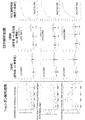
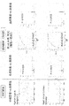
- FIG. 9 shows the quantitative results of Test Example 2 (shotgun analysis results), the quantitative results of Test Example 4 (SRM analysis results), and the ROC curves of Claudin-4, Heatshock protein HSP90-beta, and Epidermal growthfactor receptor. .
- the horizontal axis indicates, from the left, healthy subjects, cystitis, bladder cancer (superficial cancer), and bladder cancer (invasive cancer) from the left.
- the horizontal axis in the graph of the comparison results between the two groups of the quantification results of the SRM analysis results shows healthy subjects and bladder cancer from the left, and the horizontal axis in the graph of the comparison results between the three groups shows the healthy subjects from the left.
- Bladder cancer superficial cancer
- bladder cancer invasive cancer
- the vertical axis indicates the quantitative value.
- the vertical axis indicates sensitivity (positive rate), and the horizontal axis indicates a value obtained by subtracting specificity from 1 (1-specificity) (false positive rate).
- Quantitative results of Test Example 2 shotgun analysis results
- Quantitative results of Test Example 4 SRM analysis results
- ROC curves The description of each graph is the same as that of FIG.
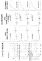
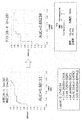
- FIG. 2 shows the quantitative results of Test Example 2 (shotgun analysis results), the quantitative results of Test Example 4 (SRM analysis results), and the ROC curves for Tight junction protein ZO-2, Ephrin type-A receptor II, and Calreticulin.
- the description of each graph is the same as that of FIG. 5 shows the quantification result of Test Example 2 (shotgun analysis result), the quantification result of Test Example 4 (SRM analysis result), and the ROC curve for Gamma-enolase and Calpain-2 catalytic subunit. The description of each graph is the same as that of FIG.
- TMT analysis results shows the quantification results of Test Example 2 (shotgun analysis results), the quantification results of Test Example 4 (SRM analysis results), and the ROC curves for Protein diaphanous homolog 1 and Serine / threonine-protein kinase LMTK2.
- SRM analysis results shows the quantification results of Test Example 6 (TMT analysis results), the quantification results of Test Example 6 (SRM analysis results), and the ROC curves for Protein S100-P and Complement Decay-accelerating Factor (DAF-1).
- DAF-1 Complement Decay-accelerating Factor
- a graph of the comparison result between the two groups (the vertical axis indicates the quantitative value; the horizontal axis indicates a healthy person and bladder cancer from the left), and a graph of the ROC curve (vertical axis) Indicates the sensitivity (positive rate), the horizontal axis indicates the value obtained by subtracting the specificity from 1 (1-specificity) (false positive rate), and the graph of the comparison results between the two groups (the vertical axis indicates the quantitative value)
- the hematuria patient and bladder cancer are shown from the left in the horizontal axis).
- test method for urothelial cancer relates to a method for testing urothelial cancer, comprising: (1) selecting from a specific group of membrane proteins in a urine sample collected from a subject;
- the present invention relates to a test method (hereinafter, also referred to as “test method of the present invention”) including a step of detecting at least one type of biomarker (step 1).
- test method of the present invention including a step of detecting at least one type of biomarker (step 1).
- the "urothelial cancer" to be tested is not particularly limited as long as it occurs in the urinary tract (renal pelvis, ureter, bladder, urethra).
- urothelial cancer include bladder cancer, renal pelvis / ureteral cancer, and the like.
- a bladder cancer is preferable as a test object.
- urothelial cancer includes various stages of cancer, and includes superficial cancer, invasive cancer and the like.
- the subject is the target organism of the test method of the present invention, and the species is not particularly limited.
- the species of the subject include various mammals such as humans, monkeys, mice, rats, dogs, cats and rabbits, and humans are preferred.
- the state of the subject is not particularly limited.
- the subject may be, for example, a sample that is not known to have urothelial carcinoma, a sample that has already been determined to have urothelial carcinoma by another method, or a subject that has urothelial carcinoma Samples that have already been determined to have not been performed by another method, samples that are being treated for urothelial cancer, and the like can be mentioned.
- the urine sample is not particularly limited as long as it is urine and a sample derived therefrom (a urine-derived sample).
- the urine-derived sample is a sample obtained by subjecting urine to any operation (separation, purification, drug addition, and the like).
- the urine sample is preferably a urine-derived sample.
- urine-derived sample urine extracellular vesicles are preferable.
- the urine sample may be used alone or in combination of two or more.
- Extracellular vesicles are not particularly limited as long as they are membrane vesicles secreted and released from cells.
- Extracellular vesicles are usually defined as membrane vesicles that carry intracellular and intracellular communication by transporting intracellular proteins and genetic information (mRNA, microRNA, etc.) outside the cell. You. Examples of extracellular vesicles include exosomes, microvesicles, apoptotic bodies, ectosomes, microparticles, secretory microvesicles, and the like. Of these, exosomes are preferred.
- Extracellular vesicles can be purified, separated, concentrated, etc. from urine according to or according to known methods.
- Methods for purifying, separating, concentrating, etc. extracellular vesicles include, for example, ultracentrifugation (eg, pellet down, sucrose cushion, density gradient centrifugation, etc.), methods using immunoaffinity carriers, gel filtration, Flow fractionation, FACS and the like can be mentioned.
- Purification, separation, concentration and the like of extracellular vesicles can also be performed using a commercially available kit. These methods may be employed alone or in combination of two or more.
- the detection target in step (1) is at least one biomarker selected from a specific membrane protein group (in the present specification, these may be collectively referred to as “target biomarker”).
- This specific group of membrane proteins is a urothelial carcinoma-specific biomarker, which can be used as an indicator to distinguish urothelial carcinoma.
- This particular group of membrane proteins includes Claudin-4, Heat shock protein HSP 90-beta, Epidermal growth factor receptor, Epihelial cell adhesion molecule, HLA class I histocompatibility antigen, B-35 alpha chain, Chloride intracellular channel can-1 and yn 1, Intercellular hesadhesion molecule 1, Myristoylated alanine-rich C-kinase substrate, Niban-like protein 1, Solute carrier family 12 member 7, MARCKS-related protein, Tight junction protein ZO-2, Ephrin type-A receptor 2, Calreticulin Gamma-enolase, Calpain-2 catalytic subunit, Protein diaphanous homolog 1, Serine / threonine-protein kinase LMTK2, Protein S100-P, Complement decay-accelerating factor (DAF-1), and Receptor-type tyrosine-protein phosphatase F (TPRF) -1). These are a group of proteins whose amounts in urothelial cancer samples are higher than
- HSP 90-beta preferably Heat ⁇ shock protein HSP 90-beta, Epidermal growth factor receptor, Epithelial cell adhesion molecule, HLA class I histocompatibility antigen, B-35 alpha chain, Solute carrier family 12 member 7, Gamma-enolase, Protein diaphanous homolog, 1, Serine / threonine-protein kinase, LMTK2, and pit, preferably helical, and helical.
- Heat Shock Protein HSP 90-beta preferably Heat Shock Protein HSP 90-beta, Syndecan-1, Ephrin Examples include type-A receptor 2, Calreticulin, protein S100-P, complement decay-accelerating factor (DAF-1), and receptor-type tyrosine-protein phosphatase F (PTPRF-1).
- Claudin-4 Heat shock protein HSP 90-beta, Epidermal growth factor receptor, Epihelial cell adhesion molecule, HLA class I histocompatibility antigen, B -35 chain, Chloride intracellular channel protein 1, Syndecan-1, Intercellular adhesion molecule 1, etc., more preferably Claudin-4, Heat shock protein HSP 90-beta, Epidermal growth factor receptor, Epihelial cell adhesion molecule, class I histocompatibility antigen, B-35 alpha chain and the like, more preferably Claudin-4, Heat shock protein HSP 90-beta, Epidermal growth factor receptor, Epihelial cell adhesion molecule, and particularly preferably Claudin-4 Is mentioned.
- membrane proteins from the viewpoint of being more suitable for invasive cancer differentiation, etc., preferably Claudin-4, Heat ⁇ shock protein HSP 90-beta, Epidermal growth factor receptor, Epihelial cell adhesion molecule, HLA class I histocompatibility antigen , B-35 alpha chain, Syndecan-1, Intercellular adhesion molecule 1, Myristoylated alanine-rich C-kinase substrate, Niban-like protein 1, MARCKS-related protein, Calreticulin, etc., more preferably Claudin-4, Epihelial cell adhesion molecule, Syndecan-1, Intercellular adhesion molecule 1, Myristoylated alanine-rich C-kinase substrate, MARCKS-related protein, Calreticulin, etc., more preferably, Claudin-4, Epihelial cell adhesion molecule, Calreticulin, etc. .
- the proteins of the above-mentioned membrane protein group can be specified by their names, or can be specified as orthologs of the human proteins indicated by the names.
- the number of target biomarkers in step (1) may be only one, or may be a combination of two or more. Testing for urothelial carcinoma by combining more (eg, 2 or more, 3 or more, 4 or more, 5 or more, 7 or more, 10 or more, 15 or more, 19) Etc. can be performed more accurately.
- Detection is usually performed by measuring the amount or concentration of the target biomarker.
- concentration is not limited to the absolute concentration, but may be a relative concentration, a weight per unit volume, or raw data measured to know the absolute concentration.
- the method for detecting the target biomarker is not particularly limited as long as it can specifically detect a part or all of the target biomarker.
- Specific examples of the detection method include, for example, mass spectrometry for detecting a peptide constituting the target biomarker, immunological measurement using an antibody that specifically recognizes the target biomarker, and the like.
- the amino acid sequence information of the target biomarker can be obtained by searching the EBI (http://www.ebi.ac.uk/IPI/IPIhelp.html) database based on the UniProtKB accession number. .
- Suitable examples of the immunological assay include immunohistochemical staining, ELISA, sandwich ELISA, EIA, RIA, and Western blotting.
- ELISA immunohistochemical staining
- sandwich ELISA sandwich ELISA
- EIA extracellular vesicle marker
- RIA Western blotting
- extracellular vesicle marker such as CD9
- target biomarker an antibody against a target biomarker
- Mass spectrometry is a method in which a peptide sample is converted into gaseous ions using an ion source (ionization), and the peptide sample is ionized by moving it in a vacuum and using electromagnetic force or by a time-of-flight difference in the analysis unit.
- a measurement method using a mass spectrometer that can be separated and detected according to the ratio, and ionization using an ion source includes EI, CI, FD, FAB, MALDI, and ESI.
- the method of separating the ionized peptide sample in the analyzer can be selected from magnetic field deflection type, quadrupole type, ion trap type, time of flight (TOF) type, Fourier transform A separation method such as an ion cyclotron resonance type can be appropriately selected.
- tandem mass spectrometry (MS / MS) or triple quadrupole mass spectrometry combining two or more mass spectrometry methods can be used.
- the sample is a sample containing a phosphorylated peptide
- the sample can be concentrated using iron ion-immobilized affinity chromatography (Fe-IMAC) before the sample is introduced into the mass spectrometer.
- Fe-IMAC iron ion-immobilized affinity chromatography
- the peptide constituting the target biomarker can be separated and purified by liquid chromatography (LC) or HPLC to obtain a sample. Further, the detection unit and the data processing method can be appropriately selected.
- LC liquid chromatography
- the detection unit and the data processing method can be appropriately selected.
- a stable isotope-labeled peptide in which one or more of the amino acids in the peptide constituting the target biomarker to be detected contains one or more of 15 N, 13 C, 18 O, and 2 H If so, the type, position, number, etc. of the amino acids can be appropriately selected, and such a stable isotope-labeled peptide can be prepared using the F-moc method (Amblard., Et al. Methods Mol Biol.
- iTRAQ registered trademark
- ICAT registered trademark
- ICPL registered trademark
- NBS (Registered trademark) reagent
- a labeled antibody an antibody that binds to the primary antibody
- a radioisotope such as 125I, a fluorescent substance, an enzyme such as horseradish peroxidase (HRP), etc.
- HRP horseradish peroxidase
- the amount and / or concentration of the target biomarker which is a detection index of urothelial cancer, can be provided, whereby the detection of urothelial cancer can be performed. Can assist you.
- test results of the test method of the present invention including the step (1) are used to determine the therapeutic effect, elucidate the pathology of urothelial cancer, predict the prognosis of urothelial cancer, stratify patients, select treatment methods (individualization) It can be used for evaluation of refractory and remodeling in urothelial cancer, differentiation of urothelial cancer histology, phenotype, etc.
- (2) the subject suffers from urothelial cancer when the amount or concentration of the biomarker detected in the step (1) is equal to or higher than a cutoff value. It is preferable to include a step of determining that the operation is performed. According to the test method of the present invention including the step 2, urothelial cancer can be determined.
- Cut-off value, sensitivity, specificity, positive predictive value can be appropriately set by those skilled in the art from the viewpoint of negative predictive value, for example, collected from a subject not suffering from urothelial cancer
- a predetermined value or a predetermined value can be used.
- the cut-off value is, for example, the amount and / or concentration of the target biomarker in a urine sample collected from a subject not suffering from urothelial cancer (average, median, ) Can be, for example, 1 (or a value greater than 1) to 3 times.
- the determination can be made by a method using a discriminant.
- the discriminant can be any discriminant analysis method that can create a discriminant for discriminating between urothelial cancer and healthy, such as Fisher's discriminant analysis, nonlinear discriminant analysis by Mahalanobis distance, neural network, Support Vector Although it can be created using a Machine (SVM), it is not limited to these specific examples.
- SVM Machine
- the determination can be made using a method such as a neural network, a k-nearest neighbor method, a decision tree, and a logistic regression analysis.
- the cut-off value is set to, for example, the amount of the target biomarker in a past sample of the same sample and / or By setting the value based on the concentration, the therapeutic effect of urothelial cancer can be determined.
- the biomarker is Claudin-4, Heat shock protein HSP 90-beta, Epidermal growth factor receptor, Epihelial cell adhesion molecule, HLA class I histocompatibility antigen, B-35 alpha chain, Syndecan -1, Intercellular adhesion molecule 1, Myristoylated alanine-rich C-kinase substrate, Niban-like protein 1, MARCKS-related protein, and at least one biomarker selected from the group consisting of Calreticulin, (3 A) determining that the subject is suffering from invasive cancer among urothelial cancers when the amount or concentration of the biomarker detected in the step (1) is equal to or greater than a cutoff value; It is preferred to include.
- Step 3 may also include a step of determining that the subject has a superficial cancer among urothelial carcinomas when the amount or concentration of the biomarker is equal to or lower than the cutoff value. According to the test method of the present invention including the step 3, it is possible to distinguish whether the urothelial cancer is a superficial cancer or an invasive cancer.
- the cut-off value can be appropriately set by those skilled in the art from the viewpoints of sensitivity, specificity, positive predictive value, negative predictive value, and the like.
- urothelial cancer (superficial cancer and / or invasion
- the value may be a predetermined value or a predetermined value based on the amount and / or concentration of the target biomarker in a urine sample collected from a subject not suffering from (H).
- the cut-off value may be determined, for example, by measuring the amount and / or concentration of the target biomarker in a urine sample collected from a subject not suffering from urothelial cancer (superficial cancer and / or invasive cancer).
- the value can be, for example, 1 (or a value exceeding 1) to 3 times the average value, the median value, or the like).
- the cut-off value is set to, for example, a target biomarker in a past sample of the same sample.
- the therapeutic effect of urothelial cancer (invasive cancer) can be determined by setting the value based on the amount and / or concentration of
- the subject can be treated with urothelial carcinoma (superficial cancer and / or invasion). Diagnosing urothelial carcinoma with higher accuracy by combining the test method of the present invention with the step of applying a urothelial carcinoma diagnosis by a doctor when it is determined that can do.
- the test method of the present invention can more accurately detect urothelial cancer, by combining the above-described steps with the test method of the present invention, it is possible to more efficiently and more accurately detect urothelial cancer. Yes "can be diagnosed.
- the subject suffers from urothelial cancer (superficial cancer and / or invasive cancer) by the test method of the present invention including the treatment step (2) and / or the step (3) for urothelial cancer. If it is determined that the urothelial cancer is present, the urothelial cancer (superficial cancer) may be further examined by the method of the present invention or as described in “2. And / or invasive cancer), the combination of the test method of the present invention and the step of applying a diagnosis by a physician further provides (3) urothelial cancer. By performing a step of treating the disease for the subject determined or diagnosed as being present, the disease of the subject can be treated.
- test method of the present invention can more accurately detect urothelial cancer
- test method of the present invention or the combination of the test method of the present invention and a step of applying a diagnosis by a doctor By combining step 3, a subject suffering from urothelial cancer can be treated more efficiently and more reliably.
- the method of treating urothelial cancer is not particularly limited, and various known treatment methods can be adopted.
- the treatment method include chemotherapy, surgical treatment, radiation treatment, and immunotherapy. These can be performed according to a known method.
- the therapeutic agent used for chemotherapy is not particularly limited, and various anticancer agents can be used.
- the anticancer agent include an alkylating agent, an antimetabolite, a microtubule inhibitor, an antibiotic anticancer agent, a topoisomerase inhibitor, a platinum preparation, a molecular target drug, a hormonal agent, and a biological preparation.
- the alkylating agent include cyclophosphamide, ifosfamide, nitrosourea, dacarbazine, temozolomide, nimustine, busulfan, melphalan, procarbazine, ranimustine and the like.
- antimetabolites for example, enositabine, carmofur, capecitabine, tegafur, tegafur uracil, tegafur gimeracil oteracil potassium, gemcitabine, cytarabine, cytarabine octophosphate, nelarabine, fluorouracil, fludarabetin, penmetrexet, penmetrexet, penmetrexetate Cladribine, doxyfluridine, hydroxycarbamide, mercaptopurine and the like.
- the microtubule inhibitor include alkaloid anticancer agents such as vincristine, and taxane anticancer agents such as docetaxel and paclitaxel.
- antibiotic anticancer agent examples include mitomycin C, doxorubicin, epirubicin, daunorubicin, bleomycin, actinomycin D, aclarubicin, idarubicin, pirarubicin, peplomycin, mitoxantrone, amrubicin, dinostatin stimaramar and the like.
- topoisomerase inhibitor examples include CPT-11 having a topoisomerase I inhibitory action, irinotecan, nogitecan, etoposide and sobuzoxane having a topoisomerase II inhibitory action.
- platinum preparation examples include cisplatin, nedaplatin, oxaliplatin, carboplatin and the like.
- Hormonal agents include, for example, dexamethasone, finasteride, tamoxifen, astrozole, exemestane, ethinylestradiol, chlormadinone, goserelin, bicalutamide, flutamide, blednisolone, leuprorelin, letrozole, estramustine, toremifene, phosphesterol, mitotane, Examples include methyltestosterone, medroxyprogesterone, mepithiostane, and the like.
- the biologic include interferon ⁇ , ⁇ and ⁇ , interleukin 2, ubenimex, dried BCG and the like.
- Examples of the molecular target drug include nivolumab, pembrolizumab, rituximab, alemtuzumab, trastuzumab, cetuximab, panitumumab, imatinib, dasatinib, nilotinib, gefitinib, erlotinib, temsirolimus, bevacizumab, and GF. Ibritumomab ozogamicin, ibritumomab tiuxetane, tamibarotene, tretinoin and the like.
- human epidermal growth factor receptor 2 inhibitor In addition to the targeted drugs specified here, human epidermal growth factor receptor 2 inhibitor, epidermal growth factor receptor inhibitor, Bcr-Abl tyrosine kinase inhibitor, epidermal growth factor tyrosine kinase inhibitor, mTOR inhibitor Drugs, inhibitors of angiogenesis such as vascular endothelial growth factor receptor 2 inhibitor ( ⁇ -VEGFR-2 antibody), various tyrosine kinase inhibitors such as MAP kinase inhibitor, inhibitors targeting cytokines, Molecular targeting drugs such as proteasome inhibitors and antibody-anticancer drug combinations can also be included. These inhibitors also include antibodies. One, two, or three or more therapeutic agents can be used in combination.
- test agent for urothelial cancer test kit
- the present invention provides, in one embodiment, a test agent for urothelial cancer, comprising a detection agent for at least one biomarker selected from a specific membrane protein group (In this specification, it may be referred to as “the test agent of the present invention”). Hereinafter, this will be described.
- the detection agent is not particularly limited as long as it can specifically detect the target biomarker.
- Examples of the detection agent include an antibody against a target biomarker.
- the detection agent may be modified as long as its function is not significantly impaired.
- modifications include addition of a label, for example, a fluorescent dye, an enzyme, a protein, a radioisotope, a chemiluminescent substance, biotin, and the like.
- the fluorescent dye used in the present invention generally used are those which label nucleotides and are used for the detection and quantification of nucleic acids.
- HEX 4,7,2 ', 4', 5 ', 7 '-hexachloro-6-carboxylfluorescein, green fluorescent dye
- fluorescein fluorescein
- NED trade name, manufactured by Applied Biosystems, yellow fluorescent dye
- 6-FAM trade name, manufactured by Applied Biosystems, yellow
- Green fluorescent dye rhodamine or a derivative thereof (eg, tetramethylrhodamine (TMR)), but is not limited thereto.
- a method for labeling nucleotides with a fluorescent dye an appropriate one of known labeling methods can be used [see Nature Biotechnology, ⁇ 14, ⁇ 303-308 ⁇ (1996)].
- a commercially available fluorescent labeling kit can be used (for example, oligonucleotide ECL '3'-oligolabeling system, manufactured by Amersham Pharmacia).
- the detection agent can be used by immobilizing it on an arbitrary solid phase. Therefore, the test agent of the present invention can be provided in the form of a substrate on which a detection agent is immobilized (for example, an ELISA plate or the like on which an antibody is immobilized).
- a substrate on which a detection agent is immobilized for example, an ELISA plate or the like on which an antibody is immobilized.
- the solid phase used for immobilization is not particularly limited as long as it can immobilize an antibody or the like, and examples thereof include a glass plate, a nylon membrane, microbeads, a silicon chip, a capillary, and other substrates. it can. Immobilization of the detection agent on the solid phase is not particularly limited.
- the antibody is not particularly limited as long as it selectively (specifically) recognizes the target biomarker.
- “selectively (specifically) recognizes” means that a target biomarker can be specifically detected by, for example, a Western blotting method or an ELISA method, but is not limited thereto. May be used as long as it can be determined that the above-mentioned detected substance is derived from the target biomarker.
- Antibodies include a part of the above-mentioned antibodies having antigen-binding properties such as polyclonal antibodies, monoclonal antibodies, chimeric antibodies, single-chain antibodies, or Fab fragments or fragments generated by Fab expression libraries.
- Antibodies of the present invention also include antibodies that have antigen-binding properties to a polypeptide that is at least contiguous, usually at least 8, preferably at least 15, and more preferably at least 20 amino acids in the amino acid sequence of the target biomarker.
- the antibodies of the present invention can also be produced according to these conventional methods (Current Protocols in Molecular Biology, Chapters 11.12 to 11.13 (2000)).
- the antibody of the present invention is a polyclonal antibody, an oligopeptide having a partial amino acid sequence of the target biomarker using a target biomarker expressed and purified in E. coli or the like according to a conventional method, or Can be synthesized, immunized to a non-human animal such as a rabbit, and obtained from the serum of the immunized animal according to a conventional method.
- a target biomarker used as an immunizing antigen for the production of an antibody is based on known gene sequence information, DNA cloning, construction of each plasmid, transfection into a host, culturing of a transformant, and protein cultivation from the culture. It can be obtained by a collection operation. These operations are performed according to methods known to those skilled in the art, or methods described in the literature (Molecular Cloning, T. Maniatis et al., CSH Laboratory (1983), DNA Cloning, DM. Glover, IRL Press (1985)). Can be done.
- a recombinant DNA capable of expressing a gene encoding a target biomarker in a desired host cell is prepared, and this is introduced into a host cell, transformed, and the transformant is cultured. Then, by recovering the target protein from the obtained culture, a protein as an immunizing antigen for producing the antibody of the present invention can be obtained. Further, the partial peptide of the target biomarker can also be produced by a general chemical synthesis method (peptide synthesis) according to known gene sequence information.
- the antibody of the present invention may be prepared using an oligopeptide having a partial amino acid sequence of the target biomarker.
- the oligo (poly) peptide used for the production of such an antibody does not need to have a functional biological activity, but desirably has the same immunogenic properties as the target biomarker.
- An oligo (poly) peptide preferably having this immunogenic property and comprising at least 8 amino acids, preferably 15 amino acids, more preferably 20 amino acids in the amino acid sequence of the target biomarker can be exemplified.
- Production of an antibody against such an oligo (poly) peptide can also be carried out by increasing the immunological reaction using various adjuvants depending on the host.
- adjuvants include, but are not limited to, Freund's adjuvant, mineral gels such as aluminum hydroxide, and surfaces such as lysolecithin, pluronic polyols, polyanions, peptides, oil emulsions, keyhole limpet hemocyanin and dinitrophenol.
- Active substances include human adjuvants such as BCG (Bacillus Calmette-Guerin) and Corynebacterium-Parvum.
- the test agent of the present invention may be in the form of a composition.
- the composition may contain other components as necessary.
- Other components include, for example, bases, carriers, solvents, dispersants, emulsifiers, buffers, stabilizers, excipients, binders, disintegrants, lubricants, thickeners, humectants, colorants, fragrances And chelating agents.
- the test agent of the present invention may be in the form of a kit.
- the kit may contain, in addition to the above-described detection agent or the above-described composition containing the same, a kit that can be used for detection of a target biomarker in a urine sample of a subject.
- Specific examples of such substances include various reagents (eg, secondary antibodies, buffers, etc.), instruments (eg, instruments for purification, separation, and concentration of extracellular vesicles (eg, columns)).
- the present invention comprises a suppressor of at least one biomarker selected from a specific group of membrane proteins, for preventing or treating urothelial cancer
- the present invention also relates to an agent (hereinafter, also referred to as “agent of the present invention”). Hereinafter, this will be described.
- an example of the inhibitor is an antibody against the target biomarker.
- the antibody those similar to the antibodies described in the above-mentioned “4. Test agent and test kit for urothelial cancer” can be used.
- Another example of the inhibitor is an expression inhibitor of the target biomarker.
- the expression inhibitor of the target biomarker is not particularly limited as long as it can suppress the expression level of the target biomarker, its mRNA, its precursor, and the like.
- gene-specific small interfering RNA siRNA
- miRNA Gene-specific microRNA
- gene-specific antisense nucleic acid of the target biomarker their expression vector
- gene-specific ribozyme of the target biomarker gene of the target biomarker by CRISPR / Cas system Editing agents and the like.
- the expression suppression refers to the target biomarker, the expression level of its mRNA and the like, for example, 1/2, 1/3, 1/5, 1/10, 1/20, 1/30, 1/50, 1 / This means that the expression is suppressed to 100, 1/200, 1/300, 1/500, 1/1000, and 1 / 10,000 or less.
- the gene siRNA of the target biomarker is not particularly limited as long as it is a double-stranded RNA molecule that specifically suppresses the expression of the gene encoding the target biomarker.
- the siRNA preferably has a length of, for example, 18 bases or more, 19 bases or more, 20 bases or more, or 21 bases or more. Further, the siRNA preferably has a length of, for example, 25 bases or less, 24 bases or less, 23 bases or less, or 22 bases or less. It is assumed that the upper limit and the lower limit of the length of the siRNA described here are arbitrarily combined.
- the lower limit is 18 bases
- the upper limit is 25 bases, 24 bases, 23 bases, or 22 bases
- the lower limit is 19 bases
- the upper limit is 25 bases, 24 bases, 23 bases, or 22 bases.
- a length with a lower limit of 20 bases and an upper limit of 25 bases, 24 bases, 23 bases, or 22 bases; a lower limit of 21 bases, and an upper limit of 25 bases, 24 bases, 23 bases, or 22 bases A combination of lengths that are bases is envisioned.
- SiRNA may be shRNA (small hairpin RNA).
- shRNAs can be designed such that a portion forms a stem-loop structure.
- shRNA has a sequence of a certain region as sequence a and a complementary strand to sequence a as sequence b, so that these sequences are present in one RNA strand such that sequence a, spacer, and sequence b are in this order.
- sequence a is a sequence of a partial region of the base sequence encoding the target biomarker to be targeted.
- the target region is not particularly limited, and any region can be a candidate.
- the length of sequence a is 19 to 25 bases, preferably 19 to 21 bases.
- the gene-specific siRNA of the target biomarker may have an additional base at the 5 'or 3' end.
- the length of the additional base is usually about 2 to 4 bases.
- the additional base may be DNA or RNA, but use of DNA may improve the stability of the nucleic acid in some cases.
- Such additional base sequences include, for example, ug-3 ', uu-3', tg-3 ', tt-3', ggg-3 ', guuu-3', gttt-3 ', ttttt-3 ', Uuuuu-3' and the like, but are not limited thereto.
- the siRNA may have a protruding portion sequence (overhang) at the 3 ′ end, and specific examples include those to which dTdT (dT represents deoxythymidine) has been added. In addition, blunt ends (blunt ends) without terminal addition may be used.
- the sense strand and the antisense strand may have different numbers of bases. For example, “asymmetrical interfering RNA (the antisense strand has an overhanging sequence at the 3 ′ end and the 5 ′ end) aiRNA)].
- a typical aiRNA has an antisense strand consisting of 21 bases, a sense strand consisting of 15 bases, and an overhang structure of 3 bases at each end of the antisense strand.
- the position of the target sequence of the gene-specific siRNA of the subject biomarker is not particularly limited, but in one embodiment, up to about 50 bases from the 5'-UTR and start codon, and from regions other than the 3'-UTR It is desirable to select a target sequence.
- BLAST http://www.ncbi.nlm.nih.gov/BLAST/ ) It is preferable to check the specificity of the selected target sequence using homology search software.
- a sense strand having a TT or UU 3 ′ terminal overhang at 19-21 bases after AA (or NA), a sequence complementary to the 19-21 base and TT or A double-stranded RNA consisting of an antisense strand having a 3'-terminal overhang of UU may be designed as siRNA.
- shRNA which is a precursor of siRNA, appropriately selects an arbitrary linker sequence (for example, about 5 to 25 bases) capable of forming a loop structure, and connects the sense strand and the antisense strand via the linker sequence. It can be designed by connecting.
- siRNA and / or shRNA can be searched using search software provided free of charge on various websites. Examples of such sites include the following. SiRNA Target Finder provided by Ambion (http://www.ambion.com/jp/techlib/misc/siRNA_finder.html) pSilencer (registered trademark) Expression Vector Insert Design Tool (http://www.ambion.com/jp/techlib/misc/psilencer_converter.html) GeneSeer provided by RNAi Codex (http://codex.cshl.edu/scripts/newsearchhairpin.cgi).
- the siRNA is obtained by synthesizing the sense strand and the antisense strand of the target sequence on the mRNA with a DNA / RNA automatic synthesizer and denaturing them in a suitable annealing buffer at about 90 to about 95 ° C. for about 1 minute. It can be prepared by annealing at about 30 to about 70 ° C. for about 1 to about 8 hours. Alternatively, it can be prepared by synthesizing shRNA that is a precursor of siRNA and cleaving it using an RNA-cleaving protein dicer.
- the gene-specific miRNA of the target biomarker is optional as long as it inhibits translation of the gene encoding the target biomarker.
- the miRNA may pair with the 3 'untranslated region (UTR) of the target and inhibit its translation.
- the miRNA may be any of pri-miRNA (primary miRNA), pre-miRNA (precursor miRNA), and mature miRNA.
- the length of the miRNA is not particularly limited, the length of the pri-miRNA is usually several hundred to several thousand bases, the length of the pre-miRNA is usually 50 to 80 bases, and the length of the mature miRNA is usually 18 ⁇ 30 bases.
- the gene-specific miRNA of the biomarker of interest is preferably a pre-miRNA or a mature miRNA, more preferably a mature miRNA.
- a gene-specific miRNA of the target biomarker may be synthesized by a known method, or may be purchased from a company that provides synthetic RNA.
- the gene-specific antisense nucleic acid of the target biomarker is a nucleic acid containing a base sequence complementary to or substantially complementary to the base sequence of the mRNA of the gene encoding the target biomarker or a part thereof, and the mRNA It is a nucleic acid having a function of suppressing the synthesis of a target biomarker by forming a specific and stable double strand and binding thereto.
- the antisense nucleic acid may be any of DNA, RNA, and DNA / RNA chimera.
- RNA DNA hybrid formed by the target RNA and the antisense DNA is recognized by endogenous ribonuclease H (RNase H) and causes the target RNA to be selectively degraded. Therefore, in the case of antisense DNA directed to degradation by RNase H, the target sequence may be not only the sequence in the mRNA but also the sequence of the intron region in the initial translation product of the gene of the target biomarker.
- the intron sequence can be determined by comparing the genomic sequence with the cDNA base sequence of the gene of the target biomarker using a homology search program such as BLAST or FASTA.
- the length of the target region of the gene-specific antisense nucleic acid of the target biomarker is not limited as long as the antisense nucleic acid hybridizes and as a result, translation into the target biomarker is inhibited.
- the gene-specific antisense nucleic acid of the target biomarker may be the entire sequence or a partial sequence of the mRNA encoding the target biomarker. Considering the ease of synthesis, antigenicity, and the ability to enter cells, oligonucleotides consisting of about 10 to about 40 bases, particularly about 15 to about 30 bases, are preferred, but not limited thereto.
- the 5 'end hairpin loop, 5' end untranslated region, translation initiation codon, protein coding region, ORF translation stop codon, 3 'end untranslated region, 3' end palindrome of the gene of the target biomarker A region or a 3 ′ end hairpin loop may be selected as a preferred target region for an antisense nucleic acid, but is not limited thereto.
- the gene-specific antisense nucleic acid of the target biomarker not only hybridizes to the mRNA and early transcript of the target biomarker gene and inhibits translation into proteins, but also binds to these double-stranded DNA genes. To form a triplex, thereby inhibiting the transcription into RNA (antigene).
- the nucleotide molecules constituting the gene-specific siRNA of the target biomarker, the gene-specific miRNA of the target biomarker, and the gene-specific antisense nucleic acid of the target biomarker have stability (chemical and / or counterpart enzyme) and Various chemical modifications may be included to improve specific activity (affinity with RNA). For example, in order to prevent degradation by a hydrolase such as nuclease, a phosphate residue (phosphate) of each nucleotide constituting an antisense nucleic acid is replaced with, for example, phosphorothioate (PS), methylphosphonate (methylphosphonate), or phosphorodithioate.
- PS phosphorothioate
- methylphosphonate methylphosphonate
- phosphorodithioate phosphorodithioate
- the base moiety pyrimidine, purine
- a part of the nucleotide molecules constituting the siRNA or the miRNA may be replaced with a natural type DNA.
- the gene-specific siRNA of the target biomarker, the gene-specific miRNA of the target biomarker, and the gene-specific antisense nucleic acid of the target biomarker are mRNA or initial based on the cDNA sequence or genomic DNA sequence of the gene of the target biomarker. It can be prepared by determining the target sequence of the transcript and synthesizing a sequence complementary thereto using a commercially available automatic DNA / RNA synthesizer. In addition, antisense nucleic acids containing the various modifications described above can be chemically synthesized by any known method.
- the gene-specific siRNA of the target biomarker For expression vectors of the gene-specific siRNA of the target biomarker, the gene-specific miRNA of the target biomarker, or the gene-specific antisense nucleic acid of the target biomarker, the gene-specific siRNA of the target biomarker, the gene specificity of the target biomarker
- the target miRNA or the gene-specific antisense nucleic acid of the target biomarker is incorporated in an expressible state.
- the expression vector comprises a promoter sequence and a gene-specific siRNA of the target biomarker, a gene-specific miRNA of the target biomarker, or a coding sequence of the gene-specific antisense nucleic acid of the target biomarker (optionally And a polynucleotide containing a transcription termination signal sequence) and, if necessary, other sequences.
- the promoter is not particularly limited, and for example, CMV promoter, EF1 promoter, SV40 promoter, MSCV promoter, hTERT promoter, ⁇ -actin promoter, RNA ⁇ ⁇ ⁇ ⁇ ⁇ ⁇ polymerase II (polII) -based promoter such as CAG promoter; mouse and human U6-snRNA promoter, Human H1-RNase P RNA promoter, human valine-tRNA promoter, and other RNA polymerase III (polIII) promoters and the like. Of these, from the viewpoint that transcription of short RNAs can be performed accurately, polIII promoters are preferred. preferable.
- the other sequence is not particularly limited, and various known sequences that can be included in the expression vector can be used. Examples of such a sequence include, for example, an origin of replication and a drug resistance gene. The types of drug resistance genes and types of vectors can be the same as those described above.
- RNA-specific ribozyme of the target biomarker includes a gene-specific ribozyme of the target biomarker.
- "Ribozyme” in a narrow sense means RNA having an enzymatic activity for cleaving a nucleic acid, but in the present application, it includes DNA as long as it has a sequence-specific nucleic acid cleaving activity.
- the most versatile ribozyme nucleic acids include self-splicing RNAs found in infectious RNAs such as viroids and viruses, and hammerhead and hairpin types are known.
- the hammerhead type exhibits enzymatic activity at about 40 bases, and the bases at both ends adjacent to the hammerhead structure (about 10 bases in total) are converted into a sequence complementary to the desired cleavage site of mRNA. By doing so, it is possible to specifically cleave only the target mRNA. Since this type of ribozyme nucleic acid uses only RNA as a substrate, it has the advantage that it does not attack genomic DNA. When the mRNA of the target biomarker gene has a double-stranded structure by itself, a single target sequence can be obtained by using a hybrid ribozyme linked to an RNA motif derived from a viral nucleic acid that can specifically bind to RNA helicase.
- the application target of the agent of the present invention is not particularly limited, and includes, for example, various mammals such as human, monkey, mouse, rat, dog, cat, rabbit, pig, horse, cow, sheep, goat, deer and the like. .
- the form of the agent of the present invention is not particularly limited, and may take a form usually used in each application depending on the use of the agent of the present invention.
- a tablet including an intraorally disintegrating tablet, a chewable tablet, an effervescent tablet, a troche, a jelly-like drop, etc.
- Pills granules, fine granules, powders, hard capsules, soft capsules, dry syrups, liquids (including drinks, suspensions and syrups), and preparations suitable for oral ingestion such as jellies
- Dosage form oral dosage form
- nasal drops inhalants, rectal suppositories, inserts, enemas, jellies, injections, patches, lotions, creams, etc. Oral formulation).
- a liquid, gel or solid food for example, juice, soft drink, tea, soup, soy milk, salad oil, dressing, yogurt, jelly, pudding, sprinkle, powdered milk for childcare , Cake mix, powdered or liquid dairy products, bread, cookies and the like.
- a liquid solution, emulsion, suspension, etc.
- semi-solid gel, cream, paste, etc.
- solid tablet, particulate, capsule, Film, kneaded material, molten solid, waxy solid, elastic solid, etc.
- dentifrice teethpaste, liquid dentifrice, liquid dentifrice, powder dentifrice, etc.
- mouthwash Coating agents, patches, mouth fresheners, foods (eg, chewing gum, tablet confectionery, candy, gummy, film, troche, etc.) and the like can be mentioned.
- the agent of the present invention may further contain other components as necessary.
- Other components are not particularly limited as long as they can be blended in, for example, a medicine, a food composition, an oral composition, a health enhancer, a nutritional supplement (such as a supplement), and the like.
- the content of the inhibitor of the target biomarker of the agent of the present invention depends on the type, use, use mode, application target, state of the application target, and the like of the inhibitor, and is not limited. It can be 100% by weight, preferably 0.001 to 50% by weight.
- the amount of application (for example, administration, ingestion, inoculation, etc.) of the composition of the present invention is not particularly limited as long as it is an effective amount that exhibits a medicinal effect, and is generally 0.1 to 1000 per day as the weight of the active ingredient. mg / kg body weight.
- the above-mentioned dose is preferably administered once a day or divided into two to three times a day, and may be appropriately increased or decreased depending on age, disease state and symptoms.
- Method for screening active ingredient of agent for preventing or treating urothelial cancer is selected from a specific group of membrane proteins in a urine sample collected from an animal treated with a test substance.
- a method for screening an active ingredient of a prophylactic or therapeutic agent for urothelial cancer using an amount or concentration of at least one biomarker as an index in the present specification, this may be referred to as “the active ingredient screening method of the present invention”. There is). Hereinafter, this will be described.
- the measurement of the amount or concentration of the body fluid, urine sample, specific membrane protein group, urothelial carcinoma, target biomarker, etc. is the same as the definition in the above “1.
- Animal species are not particularly limited. Examples of animal species include various mammals such as humans, monkeys, mice, rats, dogs, cats, and rabbits, and humans are preferable.
- any of naturally occurring compounds or artificially produced compounds can be widely used.
- a purified compound but also a composition in which various kinds of compounds are mixed, and an extract of animals and plants can be used.
- the compound is not limited to a low molecular compound, but also includes a high molecular compound such as a protein, a nucleic acid, and a polysaccharide.
- the active ingredient screening method of the present invention more specifically, when the biomarker as an index is at least one biomarker selected from a specific membrane protein group, the value of the index, the test substance If the amount or concentration of the corresponding biomarker in a urine sample collected from an animal that has not been treated with (a control value) is lower than the test substance, the active ingredient of a preventive or therapeutic agent for urothelial cancer (or As a candidate for an active ingredient of a prophylactic or therapeutic agent for urothelial cancer).
- Corresponding biomarker means the same protein as the target biomarker used as an index.
- Low means, for example, that the index value is 1/2, 1/5, 1/10, 1/20, 1/50, 1/100 of the control value.
- the present invention provides, in one embodiment, at least one selected from a specific group of membrane proteins in a urine sample collected from an animal treated with a test substance.
- the present invention relates to a method for evaluating the induction or malignancy of urothelial carcinoma using the amount or concentration of a biomarker of an species as an index (in this specification, the method is sometimes referred to as “the toxicity evaluation method of the present invention”). Hereinafter, this will be described.
- the toxicity evaluation method of the present invention when the biomarker as an index is at least one biomarker selected from a specific membrane protein group, the value of the index, the test substance, When the amount or concentration (control value) of the corresponding biomarker in a urine sample collected from an untreated animal is higher, the test substance is determined to have urothelial cancer-induced or aggressiveness. Process.
- Corresponding biomarker means the same protein as the target biomarker used as an index.
- “High” means that, for example, the index value is twice, five times, ten times, twenty times, fifty times, and one hundred times the control value.
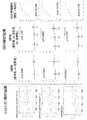
- Test example 1 Preparation of exosome fraction 1 As subjects, seven human subjects diagnosed with bladder cancer (superficial cancer and invasive cancer) and four healthy human subjects were employed. The background of the subject is as follows.
- Natural urine was collected from the subject, and the exosome fraction was prepared from the urine.
- the exosome fraction was prepared by a stepwise ultracentrifugation method. Specifically, the procedure was performed as follows. Urine was diluted with PBS and centrifuged (2000 g, 10 ° C., 30 minutes). Thereafter, the supernatant was centrifuged (17000 g, 10 ° C., 30 minutes). The supernatant was passed through a 0.22 ⁇ m filter and subjected to ultracentrifugation (130,000 g, @ 10 ° C., 90 minutes).
- the pellet was suspended in 1 ml of PBS, placed in the upper layer of a 30% sucrose solution, and subjected to sucrose cushion ultracentrifugation (130,000 g, @ 4 ° C, 70 minutes). The sucrose layer was collected and ultracentrifuged (130,000 g, 4 ° C, 70 minutes). The pellet was further ultracentrifuged under the same conditions. The finally obtained pellet was suspended in PBS to obtain a urinary exosome fraction.
- exosome fraction was observed with an electron microscope in which the exosome marker (CD9) was labeled with gold, and the exosome marker (CD63 and CD9) was detected by Western blot. As a result, it was confirmed that exosomes were obtained.
- the number and particle size of exosomes were measured. Specifically, measurement was performed using a nanosite (Nippon Quantum Design, Inc., Nanoparticle Tracking Analysis (NTA) Version 2.3 Build 0025). This is an analysis based on the difference in Brownian motion speed for each particle size. It tracks the movement of each scattered light reflected on the screen and tracks the movement speed (diffusion coefficient) in the liquid. Can be calculated. As a result, it was confirmed that the particle diameter was approximately 200 nm or less.
- TMT label-LC-MS / MS proteomics analysis was performed according to the published literature (Journal of Proteome Research., 2017, 16 (2), pp 1077-1086.). The outline is as follows. A peptide sample obtained by trypsin digestion of the protein in the exosome fraction obtained in Test Example 1 was labeled with a TMT 10-plex reagent. The labeled sample was fractionated with an SCX column, and subjected to LC-MS / MS analysis using a mass spectrometer (LTQ-Orbitrap XL, manufactured by Thermo Fisher Scientific). The obtained raw data is analyzed using an analysis program (Proteome Discoverer ver.1.3 (Thermo Fisher Scientific) equipped with Mascot v2.3.1 (Matrix Science) for UniProt / SwissProt), and quantification of various proteins Data obtained.
- TMT 10-plex reagent The labeled sample was fractionated with an SCX column, and subjected to LC-MS / MS analysis using a mass spectrometer
- 110 We identified 110 proteins that were highly expressed in urinary exosomes of bladder cancer subjects (Fold change> 1.5 (p ⁇ 0.1) for healthy subjects). Out of 110 proteins highly expressed in urinary exosomes, (1) 20 proteins (Fold change> 2, p ⁇ 0.05) that are particularly strongly expressed in urine of cancer patients and (2) cancer patients 37 proteins expressed in urine (Fold change> 2,0.1p ⁇ 0.1) or (FC> 1.5, p ⁇ 0.05), and (3) a protein reported to be derived from a bladder cancer cell line in Exocarta Alternatively, a total of 90 proteins, 33 of which were associated with bladder cancer prognosis (p ⁇ 0.1) in TCGA data, were selected for further analysis.
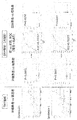
- Test example 3 Preparation of exosome fraction 2 As subjects, 40 human subjects diagnosed with bladder cancer (superficial cancer and invasive cancer) and 30 healthy human subjects were employed. The background of the subject is as follows.
- Test example 4 Proteomics analysis 2 (SRM / MRM)
- the 71 proteins selected in Test Example 2 were subjected to SRM proteomics analysis in accordance with the published literature (Molecular & Cellular Proteomics 13: 10.1074 / mcp. M113.037093, 1471-1484, 2014.).
- the outline is as follows. Based on the amino acid sequence information of the protein, one or two peptides (trypsin digested fragments) that are specifically detected by the SRM method are selected, and a stable isotope-labeled peptide (SI peptide) consisting of the same amino acid sequence as the peptide for each is selected. ) was used as an internal standard peptide.
- SI peptide stable isotope-labeled peptide
- the protein in the urinary exosome fraction obtained in Test Example 3 was digested with trypsin, mixed with an internal standard peptide, and subjected to relative quantitative analysis by SRM using a mass spectrometer (TSQ Vantage, manufactured by Thermo Fisher Scientific). .
- the vertical axis is used as the sensitivity (positive rate), and the horizontal axis is used as the value obtained by subtracting the specificity from 1 (1-specificity) (false positive rate) using the statistical software JMP. Created.
- Table 3 shows the results of evaluating the level of expression in cystitis based on the quantitative results of Test Example 2 and the results obtained by summarizing the AUC values.
- Test Example 5 Preparation of exosome fraction 3 As subjects, 49 human subjects diagnosed as having bladder cancer (superficial cancer and invasive cancer) and 48 non-cancer subjects were employed. The background of the subject is as follows.
- TMT analysis TMT analysis, SRM / MRM
- SRM / MRM analysis TMT analysis and SRM / MRM analysis were performed in the same manner as in Test Examples 2 and 4, using the urinary exosome fraction protein obtained in Test Example 5.
- three additional proteins Protein S100-P, Complement decay-accelerating factor (DAF-1), Receptor-type tyrosine-protein) phosphatase F (PTPRF-1) was selected as a biomarker.
- DAF-1 Complement decay-accelerating factor
- PPRF-1 Receptor-type tyrosine-protein phosphatase F
- the vertical axis is used as the sensitivity (positive rate), and the horizontal axis is used as the value obtained by subtracting the specificity from 1 (1-specificity) (false positive rate) using the statistical software JMP. Created. Examples of quantitative results are shown in FIGS. 8 to 11 also show the results of TMT analysis.
- Test Example 7 Determination of bladder cancer Serine / threonine-protein kinase LMTK2, Syndecan-1 (SDC1), Ephrin type-A receptor 2 (EPHA2), Epidermal growth factor receptor (EGFR2), and Calreticulin (CALR3) combined with logistic regression analysis An expression for judging the presence or absence of bladder cancer was created.
- SDC1 Syndecan-1
- EPHA2 Ephrin type-A receptor 2
- EGFR2 Epidermal growth factor receptor
- CALR3 Calreticulin
Abstract
The present invention addresses the problem of providing a urothelial cancer biomarker and a method for using the same. This problem is solved by at least one kind of urothelial cancer biomarker selected from the group consisting of claudin-4, heat shock protein HSP 90-beta, epidermal growth factor receptor, epithelial cell adhesion molecule, HLA class I histocompatibility antigen, B-35 alpha chain, chloride intracellular channel protein 1, syndecan-1, intercellular adhesion molecule 1, myristoylated alanine-rich C-kinase substrate, niban-like protein 1, solute carrier family 12 member 7, MARCKS-related protein, tight junction protein ZO-2, ephrin type-A receptor 2, calreticulin, gamma-enolase, calpain-2 catalytic subunit, protein diaphanous homolog 1, serine/threonine-protein kinase LMTK2, protein S100-P, complement decay-accelerating factor (DAF-1) and receptor-type tyrosine-protein phosphatase F (PTPRF-1).
Description
本発明は、尿路上皮がんを検査する方法等に関する。
(4) The present invention relates to a method for testing urothelial carcinoma and the like.
進行性の勝脱がん、腎孟尿管がんなどの尿路上皮がんの予後は悪く、完治は望むことができない。ただ、早期に発見された場合であれば、手術による根治が見込まれるため、早期発見早期治療が重要となる。尿路上皮がんの検査方法としては尿細胞診及び膀胱鏡が施行されている。しかし、尿細胞診の感度は40~50%と低く、さらに勝統鏡は侵襲が高い。
The prognosis of urothelial carcinoma such as progressive victory cancer and renal pelvic and ureteral cancer is poor, and complete cure cannot be expected. However, if it is detected early, it is expected that surgery will cure it, so early detection and early treatment are important. Urine cytology and cystoscopy are used as examination methods for urothelial cancer. However, the sensitivity of urine cytology is as low as 40-50%, and Katsutoscope is more invasive.
近年、正常上皮細胞またはがん細胞が自らのタンパクや核酸などをエクソソームと呼ばれる小胞(30・120nmの膜の構造体)に内包して細胞外へ輸送し、離れた部位の細胞に到達し転移の促進や免疫抑制など腫蕩罵囲の微小環境に影響を及ぼすことが明らかになってきた。エクソソームは血液や尿中に安定して存在していることが明らかになり、診断や治療への応用が期待されている。
In recent years, normal epithelial cells or cancer cells encapsulate their proteins and nucleic acids in vesicles called exosomes (30-120 nm membrane structures), transport them out of the cells, and reach cells at distant sites. It has been shown to affect the microenvironment of tumor abduction, such as promoting metastasis and immunosuppression. Exosomes are found to be stably present in blood and urine, and are expected to be applied to diagnosis and treatment.
尿中エクソソームを診断に利用し得ることが報告された例として、非特許文献1には、尿中エクソソーム内のmiR-21-5pは尿路上皮癌のバイオマーカーとなり得ることが記載されている。
As an example in which urinary exosomes can be used for diagnosis, Non-Patent Document 1 describes that miR-21-5p in urinary exosomes can be a biomarker for urothelial carcinoma. .
本発明は、尿路上皮がんのバイオマーカー及びその利用方法を提供することを課題とする。
と す る An object of the present invention is to provide a biomarker for urothelial cancer and a method for using the same.
本発明者は上記課題に鑑みて鋭意研究した結果、被検体から採取された尿試料における特定の膜タンパク質群が、尿路上皮がんのバイオマーカーとして有用であることを見出した。この知見に基づいてさらに研究を進めた結果、本発明が完成した。即ち、本発明は、下記の態様を包含する。
As a result of intensive studies in view of the above problems, the present inventors have found that a specific membrane protein group in a urine sample collected from a subject is useful as a biomarker for urothelial cancer. As a result of further research based on this finding, the present invention has been completed. That is, the present invention includes the following embodiments.
項1. 尿路上皮がんを検査する方法であって、
(1)被検体から採取された尿試料における、Claudin-4、Heat shock protein HSP 90-beta、Epidermal growth factor receptor、Epithelial cell adhesion molecule、HLA class I histocompatibility antigen, B-35 alpha chain、Chloride intracellular channel protein 1、Syndecan-1、Intercellular adhesion molecule 1、Myristoylated alanine-rich C-kinase substrate、Niban-like protein 1、Solute carrier family 12 member 7、MARCKS-related protein、Tight junction protein ZO-2、Ephrin type-A receptor 2、Calreticulin、Gamma-enolase、Calpain-2 catalytic subunit、Protein diaphanous homolog 1、Serine/threonine-protein kinase LMTK2、Protein S100-P、Complement decay-accelerating factor(DAF-1)、及びReceptor-type tyrosine-protein phosphatase F(PTPRF-1)からなる群より選択される少なくとも1種のバイオマーカーを検出する工程、を含む、検査方法。Item 1. A method for testing urothelial cancer,
(1) Claudin-4, Heat shock protein HSP 90-beta, Epidermal growth factor receptor, Epihelial cell adhesion molecule, HLA class I histocompatibility antigen, B-35 alpha chain, Chlorideintracellular channel protein 1, Syndecan-1, Intercellular adhesion molecule 1, Myristoylated alanine-rich C-kinase substrate, Niban-like protein 1, Solute carrier family 12 member 7, MARCKS-related protein, Tight junction protein ZO-2, Ephrin type-A receptor 2, Calreticulin, Gamma-enolase, Calpain-2 catalytic subunit, Protein diaphanous homolog 1, Serine / threonine-protein kinase LMTK2, Protein S100-P, Complement decay-accelerating factor (DAF-1), and Receptor-type tyrosine- detecting at least one biomarker selected from the group consisting of protein phosphatase F (PTPRF-1).
(1)被検体から採取された尿試料における、Claudin-4、Heat shock protein HSP 90-beta、Epidermal growth factor receptor、Epithelial cell adhesion molecule、HLA class I histocompatibility antigen, B-35 alpha chain、Chloride intracellular channel protein 1、Syndecan-1、Intercellular adhesion molecule 1、Myristoylated alanine-rich C-kinase substrate、Niban-like protein 1、Solute carrier family 12 member 7、MARCKS-related protein、Tight junction protein ZO-2、Ephrin type-A receptor 2、Calreticulin、Gamma-enolase、Calpain-2 catalytic subunit、Protein diaphanous homolog 1、Serine/threonine-protein kinase LMTK2、Protein S100-P、Complement decay-accelerating factor(DAF-1)、及びReceptor-type tyrosine-protein phosphatase F(PTPRF-1)からなる群より選択される少なくとも1種のバイオマーカーを検出する工程、を含む、検査方法。
(1) Claudin-4, Heat shock protein HSP 90-beta, Epidermal growth factor receptor, Epihelial cell adhesion molecule, HLA class I histocompatibility antigen, B-35 alpha chain, Chloride
項2. さらに、(2)前記工程(1)で検出されたバイオマーカーの量又は濃度がカットオフ値以上である場合に、前記被検体が尿路上皮がんに罹患していると判定する工程を含む、項1に記載の検査方法。
Item 2. Furthermore, (2) a step of determining that the subject is suffering from urothelial cancer when the amount or concentration of the biomarker detected in the step (1) is equal to or more than a cutoff value The inspection method according to item 1.
項3. 前記バイオマーカーが、Heat shock protein HSP 90-beta、Epidermal growth factor receptor、Epithelial cell adhesion molecule、HLA class I histocompatibility antigen, B-35 alpha chain、Solute carrier family 12 member 7、Gamma-enolase、Protein diaphanous homolog 1、及びSerine/threonine-protein kinase LMTK2からなる群より選択される少なくとも1種のバイオマーカーである、項1又は2に記載の検査方法。
Item 3. The biomarker is Heat shock protein HSP 90-beta, Epidermal growth factor receptor, Epithelial cell adhesion molecule, HLA class I histocompatibility antigen, B-35 alpha chain, Solute carrier family 12 member 7, Gamma-enolase 、, 3. The test method according to item 1 or 2, which is at least one biomarker selected from the group consisting of Serine / threonine-protein-kinase-LMTK2.
項4. 前記バイオマーカーが、Claudin-4、Heat shock protein HSP 90-beta、Epidermal growth factor receptor、Epithelial cell adhesion molecule、HLA class I histocompatibility antigen, B-35 alpha chain、Chloride intracellular channel protein 1、Syndecan-1、及びIntercellular adhesion molecule 1からなる群より選択される少なくとも1種のバイオマーカーである、項1又は2に記載の検査方法。
Item 4. The biomarker is claudin-4, Heatshock protein HSP 90-beta, Epidermal growth factor receptor, Epihelial cell adhesion molecule, HLA class I histocompatibility antigen, B-35 alpha chain, Chloride intracellular channel protein, Syndecan-1 Item 3. The test method according to Item 1 or 2, which is at least one biomarker selected from the group consisting of Intercellular @ adhesion @ molecule1.
項5. 前記バイオマーカーが、Claudin-4、Heat shock protein HSP 90-beta、Epidermal growth factor receptor、Epithelial cell adhesion molecule、HLA class I histocompatibility antigen, B-35 alpha chain、Syndecan-1、Intercellular adhesion molecule 1、Myristoylated alanine-rich C-kinase substrate、Niban-like protein 1、MARCKS-related protein、及びCalreticulinからなる群より選択される少なくとも1種のバイオマーカーである、項1又は2に記載の検査方法。
Item 5. The biomarker is Claudin-4, Heat shock protein HSP 90-beta, Epidermal growth factor receptor, Epihelial cell adhesion molecule, HLA class I histocompatibility antigen, B-35 alpha chain, Syndecan-1, Intercellular adhesion molecule1 Item 3. The inspection method according to Item 1 or 2, which is at least one biomarker selected from the group consisting of -rich -C-kinase substrate, Niban-like protein 1, MARCKS-related protein, and Calreticulin.
項6. さらに、(3)前記工程(1)で検出されたバイオマーカーの量又は濃度がカットオフ値以上である場合に、前記被検体が尿路上皮がんの中でも浸潤がんに罹患していると判定する工程を含む、項5に記載の検査方法。
Item 6. Furthermore, (3) when the amount or concentration of the biomarker detected in the step (1) is equal to or more than a cutoff value, the subject is susceptible to invasive cancer among urothelial cancers. Item 6. The inspection method according to Item 5, including a determining step.
項7. 前記バイオマーカーが2種以上である、項1~6のいずれかに記載の検査方法。
Item 7. (8) The test method according to any of (1) to (6), wherein the biomarkers are two or more kinds.
項8. 前記尿試料が尿の細胞外小胞である、項1~7のいずれかに記載の検査方法。
Item 8. Item 8. The test method according to any one of Items 1 to 7, wherein the urine sample is extracellular vesicles of urine.
項9. 前記尿路上皮がんが膀胱がんである、項1~8のいずれかに記載の検査方法。
Item 9.検 査 The method according to any one of Items 1 to 8, wherein the urothelial cancer is bladder cancer.
項10. 前記被検体がヒトである、項1~9のいずれかに記載の検査方法。
Item 10.検 査 The test method according to any one of Items 1 to 9, wherein the subject is a human.
項11. Claudin-4、Heat shock protein HSP 90-beta、Epidermal growth factor receptor、Epithelial cell adhesion molecule、HLA class I histocompatibility antigen, B-35 alpha chain、Chloride intracellular channel protein 1、Syndecan-1、Intercellular adhesion molecule 1、Myristoylated alanine-rich C-kinase substrate、Niban-like protein 1、Solute carrier family 12 member 7、MARCKS-related protein、Tight junction protein ZO-2、Ephrin type-A receptor 2、Calreticulin、Gamma-enolase、Calpain-2 catalytic subunit、Protein diaphanous homolog 1、Serine/threonine-protein kinase LMTK2、Protein S100-P、Complement decay-accelerating factor(DAF-1)、及びReceptor-type tyrosine-protein phosphatase F(PTPRF-1)からなる群より選択される少なくとも1種のバイオマーカーの検出剤を含む、尿路上皮がんの検査薬。
Item 11. Claudin-4, Heat shock protein HSP 90-beta, Epidermal growth factor receptor, Epithelial cell adhesion molecule, HLA class I histocompatibility antigen, B-35 alpha chain, Chloride intracellular channel protein 1, Syndecan-1, Interdecan-1 alanine-rich C-kinase substrate, Niban-like protein 1, SoluteARCcarrier family 12 member 7, MARCKS-related protein, Tight junctionulinproteinGZO-2, Ephrin -type-Alyticreceptor 2, Calreticulin, Gamma-enolase, Calpain-2lyticcatalytic selected from the group consisting of subunit, Protein diaphanous homolog 1, Serine / threonine-protein kinase LMTK2, Protein S100-P, Complement decay-accelerating factor (DAF-1), and Receptor-type tyrosine-protein phosphatase F (PTPRF-1) A test agent for urothelial cancer, comprising an agent for detecting at least one biomarker to be performed.
項12. Claudin-4、Heat shock protein HSP 90-beta、Epidermal growth factor receptor、Epithelial cell adhesion molecule、HLA class I histocompatibility antigen, B-35 alpha chain、Chloride intracellular channel protein 1、Syndecan-1、Intercellular adhesion molecule 1、Myristoylated alanine-rich C-kinase substrate、Niban-like protein 1、Solute carrier family 12 member 7、MARCKS-related protein、Tight junction protein ZO-2、Ephrin type-A receptor 2、Calreticulin、Gamma-enolase、Calpain-2 catalytic subunit、Protein diaphanous homolog 1、Serine/threonine-protein kinase LMTK2、Protein S100-P、Complement decay-accelerating factor(DAF-1)、及びReceptor-type tyrosine-protein phosphatase F(PTPRF-1)からなる群より選択される少なくとも1種のバイオマーカーの検出剤を含む、尿路上皮がんの検査キット。
Item 12. Claudin-4, Heat shock protein HSP 90-beta, Epidermal growth factor receptor, Epithelial cell adhesion molecule, HLA class I histocompatibility antigen, B-35 alpha chain, Chloride intracellular channel protein 1, Syndecan-1, Interdecan-1 alanine-rich C-kinase substrate, Niban-like protein 1, SoluteARCcarrier family 12 member 7, MARCKS-related protein, Tight junctionulinproteinGZO-2, Ephrin -type-Alyticreceptor 2, Calreticulin, Gamma-enolase, Calpain-2lyticcatalytic selected from the group consisting of subunit, Protein diaphanous homolog 1, Serine / threonine-protein kinase LMTK2, Protein S100-P, Complement decay-accelerating factor (DAF-1), and Receptor-type tyrosine-protein phosphatase F (PTPRF-1) A test kit for urothelial cancer, comprising a detection agent for at least one biomarker to be performed.
項13. Claudin-4、Heat shock protein HSP 90-beta、Epidermal growth factor receptor、Epithelial cell adhesion molecule、HLA class I histocompatibility antigen, B-35 alpha chain、Chloride intracellular channel protein 1、Syndecan-1、Intercellular adhesion molecule 1、Myristoylated alanine-rich C-kinase substrate、Niban-like protein 1、Solute carrier family 12 member 7、MARCKS-related protein、Tight junction protein ZO-2、Ephrin type-A receptor 2、Calreticulin、Gamma-enolase、Calpain-2 catalytic subunit、Protein diaphanous homolog 1、Serine/threonine-protein kinase LMTK2、Protein S100-P、Complement decay-accelerating factor(DAF-1)、及びReceptor-type tyrosine-protein phosphatase F(PTPRF-1)からなる群より選択される少なくとも1種のバイオマーカーの抑制剤を含有する、尿路上皮がんの予防又は治療剤。
Item 13. Claudin-4, Heat shock protein HSP 90-beta, Epidermal growth factor receptor, Epithelial cell adhesion molecule, HLA class I histocompatibility antigen, B-35 alpha chain, Chloride intracellular channel protein 1, Syndecan-1, Interdecan-1 alanine-rich C-kinase substrate, Niban-like protein 1, SoluteARCcarrier family 12 member 7, MARCKS-related protein, Tight junctionulinproteinGZO-2, Ephrin -type-Alyticreceptor 2, Calreticulin, Gamma-enolase, Calpain-2lyticcatalytic selected from the group consisting of subunit, Protein diaphanous homolog 1, Serine / threonine-protein kinase LMTK2, Protein S100-P, Complement decay-accelerating factor (DAF-1), and Receptor-type tyrosine-protein phosphatase F (PTPRF-1) An agent for preventing or treating urothelial cancer, comprising an inhibitor of at least one biomarker to be used.
項14. 被検物質で処理された動物から採取された尿試料における、Claudin-4、Heat shock protein HSP 90-beta、Epidermal growth factor receptor、Epithelial cell adhesion molecule、HLA class I histocompatibility antigen, B-35 alpha chain、Chloride intracellular channel protein 1、Syndecan-1、Intercellular adhesion molecule 1、Myristoylated alanine-rich C-kinase substrate、Niban-like protein 1、Solute carrier family 12 member 7、MARCKS-related protein、Tight junction protein ZO-2、Ephrin type-A receptor 2、Calreticulin、Gamma-enolase、Calpain-2 catalytic subunit、Protein diaphanous homolog 1、Serine/threonine-protein kinase LMTK2、Protein S100-P、Complement decay-accelerating factor(DAF-1)、及びReceptor-type tyrosine-protein phosphatase F(PTPRF-1)からなる群より選択される少なくとも1種のバイオマーカーの量又は濃度を指標とする、尿路上皮がんの予防又は治療剤の有効成分のスクリーニング方法。
Item 14. In urine samples collected from animals treated with the test substance, Claudin-4, Heat 、 shock protein HSP 90-beta, Epidermal growth factor receptor, Epihelial cell adhesion molecule, HLA class I histocompatibility antigen, B-35 alpha chain, Chloride intracellular channel protein 1, Syndecan-1, Intercellular adhesion molecule 1, Myristoylated alanine-rich C-kinase substrate, Niban-like protein 1, Solute carrier family 12, member 7, MARKS-related protein, Tight junction protein ZO-2 type-A receptor 2, Calreticulin, Gamma-enolase, Calpain-2 catalytic subunit, Protein diaphanous homolog 1, Serine / threonine-protein kinase LMTK2, Protein S100-P, Complement decay-accelerating factor (DAF-1), and Receptor- Prediction of urothelial cancer, based on the amount or concentration of at least one biomarker selected from the group consisting of type tyrosine-protein phosphatase F (PTPRF-1) Or screening method of the active ingredient of the therapeutic agent.
項15. 被検物質で処理された動物から採取された尿試料における、Claudin-4、Heat shock protein HSP 90-beta、Epidermal growth factor receptor、Epithelial cell adhesion molecule、HLA class I histocompatibility antigen, B-35 alpha chain、Chloride intracellular channel protein 1、Syndecan-1、Intercellular adhesion molecule 1、Myristoylated alanine-rich C-kinase substrate、Niban-like protein 1、Solute carrier family 12 member 7、MARCKS-related protein、Tight junction protein ZO-2、Ephrin type-A receptor 2、Calreticulin、Gamma-enolase、Calpain-2 catalytic subunit、Protein diaphanous homolog 1、Serine/threonine-protein kinase LMTK2、Protein S100-P、Complement decay-accelerating factor(DAF-1)、及びReceptor-type tyrosine-protein phosphatase F(PTPRF-1)からなる群より選択される少なくとも1種のバイオマーカーの量又は濃度を指標とする、尿路上皮がんの誘発性又は増悪性の評価方法。
Item 15. In urine samples collected from animals treated with the test substance, Claudin-4, Heat 、 shock protein HSP 90-beta, Epidermal growth factor receptor, Epihelial cell adhesion molecule, HLA class I histocompatibility antigen, B-35 alpha chain, Chloride intracellular channel protein 1, Syndecan-1, Intercellular adhesion molecule 1, Myristoylated alanine-rich C-kinase substrate, Niban-like protein 1, Solute carrier family 12, member 7, MARKS-related protein, Tight junction protein ZO-2 type-A receptor 2, Calreticulin, Gamma-enolase, Calpain-2 catalytic subunit, Protein diaphanous homolog 1, Serine / threonine-protein kinase LMTK2, Protein S100-P, Complement decay-accelerating factor (DAF-1), and Receptor- Induction of urothelial cancer using the amount or concentration of at least one biomarker selected from the group consisting of type tyrosine-protein phosphatase F (PTPRF-1) as an index Gender or exacerbation of the evaluation method.
本発明によれば、尿路上皮がんのバイオマーカーを提供することができる。該バイオマーカーを利用することにより、尿路上皮がんの検査等を、より簡便、且つより効率的に行うことができる。さらに、該バイオマーカーを利用することにより、尿路上皮がんの予防又は治療、尿路上皮がんの予防又は治療剤の有効成分のスクリーニング等を図ることも可能である。
According to the present invention, a urothelial cancer biomarker can be provided. Utilization of the biomarker makes it easier and more efficient to test urothelial cancer. Furthermore, by using the biomarker, it is possible to prevent or treat urothelial cancer, to screen an active ingredient of a preventive or therapeutic agent for urothelial cancer, and the like.
本明細書中において、「含有」及び「含む」なる表現については、「含有」、「含む」、「実質的にからなる」及び「のみからなる」という概念を含む。
に お い て In this specification, the expressions “contain” and “contain” include the concepts of “contain”, “contain”, “consisting essentially of” and “consisting only of”.
1.尿路上皮がんの検査方法
本発明は、その一態様において、尿路上皮がんを検査する方法であって、(1)被検体から採取された尿試料における、特定の膜タンパク質群より選択される少なくとも1種のバイオマーカーを検出する工程(工程1)を含む、検査方法(本明細書において、「本発明の検査方法」と示すこともある。)に関する。以下、これについて説明する。 1. Test method for urothelial cancer In one embodiment, the present invention relates to a method for testing urothelial cancer, comprising: (1) selecting from a specific group of membrane proteins in a urine sample collected from a subject; The present invention relates to a test method (hereinafter, also referred to as “test method of the present invention”) including a step of detecting at least one type of biomarker (step 1). Hereinafter, this will be described.
本発明は、その一態様において、尿路上皮がんを検査する方法であって、(1)被検体から採取された尿試料における、特定の膜タンパク質群より選択される少なくとも1種のバイオマーカーを検出する工程(工程1)を含む、検査方法(本明細書において、「本発明の検査方法」と示すこともある。)に関する。以下、これについて説明する。 1. Test method for urothelial cancer In one embodiment, the present invention relates to a method for testing urothelial cancer, comprising: (1) selecting from a specific group of membrane proteins in a urine sample collected from a subject; The present invention relates to a test method (hereinafter, also referred to as “test method of the present invention”) including a step of detecting at least one type of biomarker (step 1). Hereinafter, this will be described.
1-1.工程(1)
検査対象である「尿路上皮がん」は、尿路(腎盂、尿管、膀胱、尿道)に発生するがんである限り特に制限されない。尿路上皮がんとしては、例えば膀胱がん、腎盂・尿管がん等が挙げられる。これらの中でも、検査対象としては、膀胱がんが好ましい。また、尿路上皮がんには、各ステージのがんがふくまれ、表在がん、浸潤がん等が含まれる。 1-1. Process (1)
The "urothelial cancer" to be tested is not particularly limited as long as it occurs in the urinary tract (renal pelvis, ureter, bladder, urethra). Examples of urothelial cancer include bladder cancer, renal pelvis / ureteral cancer, and the like. Among these, a bladder cancer is preferable as a test object. In addition, urothelial cancer includes various stages of cancer, and includes superficial cancer, invasive cancer and the like.
被検体は、本発明の検査方法の対象生物であり、その生物種は特に制限されない。被検体の生物種としては、例えばヒト、サル、マウス、ラット、イヌ、ネコ、ウサギなどの種々の哺乳類動物が挙げられ、好ましくはヒトが挙げられる。
(4) The subject is the target organism of the test method of the present invention, and the species is not particularly limited. Examples of the species of the subject include various mammals such as humans, monkeys, mice, rats, dogs, cats and rabbits, and humans are preferred.
被検体の状態は、特に制限されない。被検体としては、例えば尿路上皮がんに罹患しているかどうか不明な検体、尿路上皮がんに罹患していると既に別の方法により判定されている検体、尿路上皮がんに罹患していないと既に別の方法により判定されている検体、尿路上皮がんの治療中の検体等が挙げられる。
状態 The state of the subject is not particularly limited. The subject may be, for example, a sample that is not known to have urothelial carcinoma, a sample that has already been determined to have urothelial carcinoma by another method, or a subject that has urothelial carcinoma Samples that have already been determined to have not been performed by another method, samples that are being treated for urothelial cancer, and the like can be mentioned.
尿試料は、尿及びそれに由来する試料(尿由来試料)である限り特に制限されない。尿由来試料は、尿に何らからの操作(分離、精製、薬剤添加等)をして得られる試料である。尿試料としては、尿由来試料が好ましい。尿由来試料としては、尿の細胞外小胞が好ましい。尿試料は、1種単独で採用してもよいし、2種以上を組み合わせて採用してもよい。
(4) The urine sample is not particularly limited as long as it is urine and a sample derived therefrom (a urine-derived sample). The urine-derived sample is a sample obtained by subjecting urine to any operation (separation, purification, drug addition, and the like). The urine sample is preferably a urine-derived sample. As the urine-derived sample, urine extracellular vesicles are preferable. The urine sample may be used alone or in combination of two or more.
細胞外小胞は、細胞から分泌、放出等される膜小胞である限り特に制限されない。細胞外小胞は、通常は、細胞内のタンパク質や遺伝情報(mRNA, microRNA等)を細胞外に運搬することにより、局所や全身における細胞間の情報伝達を担っている膜小胞として定義される。細胞外小胞としては、例えばエクソソーム、微小小胞体、アポトーシス小体、エクトソーム、マイクロパーティクル、分泌マイクロベシクル等が挙げられる。これらの中でもエクソソームが好ましい。
外 Extracellular vesicles are not particularly limited as long as they are membrane vesicles secreted and released from cells. Extracellular vesicles are usually defined as membrane vesicles that carry intracellular and intracellular communication by transporting intracellular proteins and genetic information (mRNA, microRNA, etc.) outside the cell. You. Examples of extracellular vesicles include exosomes, microvesicles, apoptotic bodies, ectosomes, microparticles, secretory microvesicles, and the like. Of these, exosomes are preferred.
細胞外小胞は、尿から、公知の方法に従って又は準じて、精製、分離、濃縮等することができる。細胞外小胞を精製、分離、濃縮等する方法としては、例えば超遠心法(例えばペレットダウン法、スクロースクッション法、密度勾配遠心法等)、イムノアフィニティー担体を用いる方法、ゲルろ過法、フィールド・フロー分画法、FACS法等が挙げられる。また、細胞外小胞の精製、分離、濃縮等は、市販のキットを用いて行うことも可能である。これらの方法は、1種単独で採用してもよいし、2種以上を組み合わせて採用してもよい。
外 Extracellular vesicles can be purified, separated, concentrated, etc. from urine according to or according to known methods. Methods for purifying, separating, concentrating, etc. extracellular vesicles include, for example, ultracentrifugation (eg, pellet down, sucrose cushion, density gradient centrifugation, etc.), methods using immunoaffinity carriers, gel filtration, Flow fractionation, FACS and the like can be mentioned. Purification, separation, concentration and the like of extracellular vesicles can also be performed using a commercially available kit. These methods may be employed alone or in combination of two or more.
工程(1)の検出対象は、特定の膜タンパク質群より選択される少なくとも1種のバイオマーカー(本明細書において、これらをまとめて「対象バイオマーカー」と示すこともある。)である。
検 出 The detection target in step (1) is at least one biomarker selected from a specific membrane protein group (in the present specification, these may be collectively referred to as “target biomarker”).
この特定の膜タンパク質群は、尿路上皮がん特異的なバイオマーカーであり、これを指標とすることにより尿路上皮がんを鑑別可能である。
This specific group of membrane proteins is a urothelial carcinoma-specific biomarker, which can be used as an indicator to distinguish urothelial carcinoma.
この特定の膜タンパク質群は、Claudin-4、Heat shock protein HSP 90-beta、Epidermal growth factor receptor、Epithelial cell adhesion molecule、HLA class I histocompatibility antigen, B-35 alpha chain、Chloride intracellular channel protein 1、Syndecan-1、Intercellular adhesion molecule 1、Myristoylated alanine-rich C-kinase substrate、Niban-like protein 1、Solute carrier family 12 member 7、MARCKS-related protein、Tight junction protein ZO-2、Ephrin type-A receptor 2、Calreticulin、Gamma-enolase、Calpain-2 catalytic subunit、Protein diaphanous homolog 1、Serine/threonine-protein kinase LMTK2、Protein S100-P、Complement decay-accelerating factor(DAF-1)、及びReceptor-type tyrosine-protein phosphatase F(PTPRF-1)からなるタンパク質群である。これらは、尿路上皮がん検体における量が健常検体における量よりも高いタンパク質群である。
This particular group of membrane proteins includes Claudin-4, Heat shock protein HSP 90-beta, Epidermal growth factor receptor, Epihelial cell adhesion molecule, HLA class I histocompatibility antigen, B-35 alpha chain, Chloride intracellular channel can-1 and yn 1, Intercellular hesadhesion molecule 1, Myristoylated alanine-rich C-kinase substrate, Niban-like protein 1, Solute carrier family 12 member 7, MARCKS-related protein, Tight junction protein ZO-2, Ephrin type-A receptor 2, Calreticulin Gamma-enolase, Calpain-2 catalytic subunit, Protein diaphanous homolog 1, Serine / threonine-protein kinase LMTK2, Protein S100-P, Complement decay-accelerating factor (DAF-1), and Receptor-type tyrosine-protein phosphatase F (TPRF) -1). These are a group of proteins whose amounts in urothelial cancer samples are higher than in healthy samples.
上記膜タンパク質群の中でも、尿路上皮炎での発現が比較的低い(がんと炎症との鑑別により適している)という観点等から、好ましくはHeat shock protein HSP 90-beta、Epidermal growth factor receptor、Epithelial cell adhesion molecule、HLA class I histocompatibility antigen, B-35 alpha chain、Solute carrier family 12 member 7、Gamma-enolase、Protein diaphanous homolog 1、Serine/threonine-protein kinase LMTK2等が挙げられ、より好ましくはEpithelial cell adhesion molecule、Solute carrier family 12 member 7、Gamma-enolase、Protein diaphanous homolog 1、Serine/threonine-protein kinase LMTK2等が挙げられる。
Among the above membrane proteins, from the viewpoint of relatively low expression in urotheliitis (more suitable for distinguishing cancer from inflammation), preferably Heat 等 shock protein HSP 90-beta, Epidermal growth factor receptor, Epithelial cell adhesion molecule, HLA class I histocompatibility antigen, B-35 alpha chain, Solute carrier family 12 member 7, Gamma-enolase, Protein diaphanous homolog, 1, Serine / threonine-protein kinase, LMTK2, and pit, preferably helical, and helical. Adhesion molecule, Solute carrier family 12 member 7, Gamma-enolase, Protein diaphanous homolog 1, Serine / threonine-protein kinase LMTK2 and the like.
上記膜タンパク質群の中でも、血尿被検体での発現が比較的低い(がんと血尿との鑑別により適している)という観点等から、好ましくはHeat shock protein HSP 90-beta、Syndecan-1、Ephrin type-A receptor 2、Calreticulin、Protein S100-P、Complement decay-accelerating factor(DAF-1)、Receptor-type tyrosine-protein phosphatase F(PTPRF-1)等が挙げられる。
Among the above-mentioned membrane protein groups, from the viewpoint of relatively low expression in hematuria subjects (more suitable for distinguishing cancer from hematuria), preferably Heat Shock Protein HSP 90-beta, Syndecan-1, Ephrin Examples include type-A receptor 2, Calreticulin, protein S100-P, complement decay-accelerating factor (DAF-1), and receptor-type tyrosine-protein phosphatase F (PTPRF-1).
上記膜タンパク質群の中でも、AUC(感度、特異度)の観点等から、好ましくはClaudin-4、Heat shock protein HSP 90-beta、Epidermal growth factor receptor、Epithelial cell adhesion molecule、HLA class I histocompatibility antigen, B-35 alpha chain、Chloride intracellular channel protein 1、Syndecan-1、Intercellular adhesion molecule 1等が挙げられ、より好ましくはClaudin-4、Heat shock protein HSP 90-beta、Epidermal growth factor receptor、Epithelial cell adhesion molecule、HLA class I histocompatibility antigen, B-35 alpha chain等が挙げられ、さらに好ましくはClaudin-4、Heat shock protein HSP 90-beta、Epidermal growth factor receptor、Epithelial cell adhesion molecule等が挙げられ、特に好ましくはClaudin-4が挙げられる。
Among the above-mentioned membrane proteins, from the viewpoint of AUC (sensitivity, specificity) and the like, preferably, Claudin-4, Heat shock protein HSP 90-beta, Epidermal growth factor receptor, Epihelial cell adhesion molecule, HLA class I histocompatibility antigen, B -35 chain, Chloride intracellular channel protein 1, Syndecan-1, Intercellular adhesion molecule 1, etc., more preferably Claudin-4, Heat shock protein HSP 90-beta, Epidermal growth factor receptor, Epihelial cell adhesion molecule, class I histocompatibility antigen, B-35 alpha chain and the like, more preferably Claudin-4, Heat shock protein HSP 90-beta, Epidermal growth factor receptor, Epihelial cell adhesion molecule, and particularly preferably Claudin-4 Is mentioned.
上記膜タンパク質群の中でも、浸潤がんの鑑別により適しているという観点等から、好ましくはClaudin-4、Heat shock protein HSP 90-beta、Epidermal growth factor receptor、Epithelial cell adhesion molecule、HLA class I histocompatibility antigen, B-35 alpha chain、Syndecan-1、Intercellular adhesion molecule 1、Myristoylated alanine-rich C-kinase substrate、Niban-like protein 1、MARCKS-related protein、Calreticulin等が挙げられ、より好ましくはClaudin-4、Epithelial cell adhesion molecule、Syndecan-1、Intercellular adhesion molecule 1、Myristoylated alanine-rich C-kinase substrate、MARCKS-related protein、Calreticulin等が挙げられ、さらに好ましくはClaudin-4、Epithelial cell adhesion molecule、Calreticulin等が挙げられる。
Among the membrane proteins, from the viewpoint of being more suitable for invasive cancer differentiation, etc., preferably Claudin-4, Heat 、 shock protein HSP 90-beta, Epidermal growth factor receptor, Epihelial cell adhesion molecule, HLA class I histocompatibility antigen , B-35 alpha chain, Syndecan-1, Intercellular adhesion molecule 1, Myristoylated alanine-rich C-kinase substrate, Niban-like protein 1, MARCKS-related protein, Calreticulin, etc., more preferably Claudin-4, Epihelial cell adhesion molecule, Syndecan-1, Intercellular adhesion molecule 1, Myristoylated alanine-rich C-kinase substrate, MARCKS-related protein, Calreticulin, etc., more preferably, Claudin-4, Epihelial cell adhesion molecule, Calreticulin, etc. .
上記膜タンパク質群のタンパク質は、その名称で特定することができる、或いは該名称で示されるヒトタンパク質のオーソログとして特定することができる。
タ ン パ ク 質 The proteins of the above-mentioned membrane protein group can be specified by their names, or can be specified as orthologs of the human proteins indicated by the names.
工程(1)における対象バイオマーカーの数は、1種のみでもよいが、2種以上の組み合わせであってもよい。より多く(例えば2種以上、3種以上、4種以上、5種以上、7種以上、10種以上、15種以上、19種)の検出対象を組み合わせることにより、尿路上皮がんの検査等を、より正確に行うことが可能になる。
数 The number of target biomarkers in step (1) may be only one, or may be a combination of two or more. Testing for urothelial carcinoma by combining more (eg, 2 or more, 3 or more, 4 or more, 5 or more, 7 or more, 10 or more, 15 or more, 19) Etc. can be performed more accurately.
検出は、通常は、対象バイオマーカーの量又は濃度を測定することによって行われる。「濃度」とは、絶対濃度に限らず、相対濃度や、単位体積辺りの重量や、絶対濃度を知るために測定した生データなどでもよい。
Detection is usually performed by measuring the amount or concentration of the target biomarker. The “concentration” is not limited to the absolute concentration, but may be a relative concentration, a weight per unit volume, or raw data measured to know the absolute concentration.
対象バイオマーカーを検出する方法としては、対象バイオマーカーの一部又は全部を特異的に検出できる方法であれば特に制限されない。検出方法としては、具体的には、例えば、対象バイオマーカーを構成するペプチドを検出する質量分析法、対象バイオマーカーを特異的に認識する抗体を用いた免疫学的測定法等を挙げることができる。なお、対象バイオマーカーのアミノ酸配列情報は、UniProtKBアクセッション番号を基に、EBI(http://www.ebi.ac.uk/IPI/IPIhelp.html)のデータベースで検索することにより得ることができる。
方法 The method for detecting the target biomarker is not particularly limited as long as it can specifically detect a part or all of the target biomarker. Specific examples of the detection method include, for example, mass spectrometry for detecting a peptide constituting the target biomarker, immunological measurement using an antibody that specifically recognizes the target biomarker, and the like. . The amino acid sequence information of the target biomarker can be obtained by searching the EBI (http://www.ebi.ac.uk/IPI/IPIhelp.html) database based on the UniProtKB accession number. .
免疫学的測定法としては、免疫組織化学染色法、ELISA法、サンドイッチELISA法、EIA法、RIA法、ウェスタンブロッティング法等を好適に例示することができる。尿試料として細胞外小胞を採用する場合であれば、例えば、細胞外小胞マーカー(CD9等)に対する抗体と対象バイオマーカーに対する抗体とを使用したサンドイッチELISA法を採用することができる。
好 適 Suitable examples of the immunological assay include immunohistochemical staining, ELISA, sandwich ELISA, EIA, RIA, and Western blotting. When extracellular vesicles are used as a urine sample, for example, a sandwich ELISA method using an antibody against an extracellular vesicle marker (such as CD9) and an antibody against a target biomarker can be used.
質量分析法とは、ペプチド試料を、イオン源を用いて気体状のイオンとし(イオン化)、分析部において、真空中で運動させ電磁気力を用いて、あるいは飛行時間差によりイオン化したペプチド試料を質量電荷比に応じて分離し、検出できる質量分析計を用いた測定方法のことをいい、イオン源を用いてイオン化する方法としては、EI法、CI法、FD法、FAB法、MALDI法、ESI法などの方法を適宜選択することができ、また、分析部において、イオン化したペプチド試料を分離する方法としては、磁場偏向型、四重極型、イオントラップ型、飛行時間(TOF)型、フーリエ変換イオンサイクロトロン共鳴型などの分離方法を適宜選択することができる。また、2以上の質量分析法を組み合わせたタンデム型質量分析(MS/MS)やトリプル四重極型質量分析を利用することができる。また、サンプルがリン酸化したペプチドを含む試料の場合、質量分析計へのサンプル導入前に、サンプルを鉄イオン固定化アフィニティークロマトグラフィー(Fe-IMAC)を用いて濃縮することができる。また、液体クロマトグラフ(LC)やHPLCにより、対象バイオマーカーを構成するペプチドを分離・精製してサンプルとすることができる。また、検出部やデータ処理方法も適宜選択することができる。なお、質量分析法を用いて対象バイオマーカーを構成するペプチドを質量分析法で検出・定量する場合、かかるペプチドと同一のアミノ酸配列からなる、濃度が既知の安定同位体で標識したペプチドを内部標準とすることができる。かかる安定同位体標識ペプチドとしては、検出する対象バイオマーカーを構成するペプチドにおけるアミノ酸の1個以上が、15N,13C,18O,及び2Hのいずれか1以上を含む安定同位体標識ペプチドであれば、アミノ酸の種類、位置、数などは適宜選択することができ、かかる安定同位体標識ペプチドは、安定同位元素により標識されたアミノ酸を用いてF-moc法(Amblard., et al. Methods Mol Biol.298:3-24(2005))等の適当な手段で化学合成することができるが、iTRAQ(登録商標)試薬、ICAT(登録商標)試薬、ICPL(登録商標)試薬、NBS(登録商標)試薬などの標識試薬を用いて作製することもできる。
Mass spectrometry is a method in which a peptide sample is converted into gaseous ions using an ion source (ionization), and the peptide sample is ionized by moving it in a vacuum and using electromagnetic force or by a time-of-flight difference in the analysis unit. A measurement method using a mass spectrometer that can be separated and detected according to the ratio, and ionization using an ion source includes EI, CI, FD, FAB, MALDI, and ESI. The method of separating the ionized peptide sample in the analyzer can be selected from magnetic field deflection type, quadrupole type, ion trap type, time of flight (TOF) type, Fourier transform A separation method such as an ion cyclotron resonance type can be appropriately selected. In addition, tandem mass spectrometry (MS / MS) or triple quadrupole mass spectrometry combining two or more mass spectrometry methods can be used. When the sample is a sample containing a phosphorylated peptide, the sample can be concentrated using iron ion-immobilized affinity chromatography (Fe-IMAC) before the sample is introduced into the mass spectrometer. In addition, the peptide constituting the target biomarker can be separated and purified by liquid chromatography (LC) or HPLC to obtain a sample. Further, the detection unit and the data processing method can be appropriately selected. When detecting and quantifying the peptide constituting the target biomarker by mass spectrometry using mass spectrometry, a peptide having the same amino acid sequence as the peptide and labeled with a stable isotope of known concentration is used as an internal standard. It can be. As such a stable isotope-labeled peptide, a stable isotope-labeled peptide in which one or more of the amino acids in the peptide constituting the target biomarker to be detected contains one or more of 15 N, 13 C, 18 O, and 2 H If so, the type, position, number, etc. of the amino acids can be appropriately selected, and such a stable isotope-labeled peptide can be prepared using the F-moc method (Amblard., Et al. Methods Mol Biol. 298: 3-24 (2005)), etc., and can be chemically synthesized using iTRAQ (registered trademark) reagent, ICAT (registered trademark) reagent, ICPL (registered trademark) reagent, NBS ( (Registered trademark) reagent or the like.
ウェスタンブロット法は、一次抗体を用いた後、二次抗体として125Iなどの放射性同位元素、蛍光物質、ホースラディッシュペルオキシターゼ(HRP)などの酵素等で標識した標識抗体(一次抗体に結合する抗体)を用い、得られる標識化合物の放射性同位元素、蛍光物質などの標識物質に由来するシグナルを放射線測定器、蛍光検出器などで検出し、測定する方法が例示される。
In the Western blot method, after using a primary antibody, a labeled antibody (an antibody that binds to the primary antibody) labeled with a radioisotope such as 125I, a fluorescent substance, an enzyme such as horseradish peroxidase (HRP), etc. as a secondary antibody is used. A method of detecting and measuring a signal derived from a labeling substance such as a radioisotope or a fluorescent substance of the obtained labeled compound using a radiometer, a fluorescence detector or the like is exemplified.
工程(1)を含む本発明の検査方法によれば、尿路上皮がんの検出指標である対象バイオマーカーの量及び/又は濃度を提供することができ、これにより尿路上皮がんの検出などを補助することができる。
According to the test method of the present invention including the step (1), the amount and / or concentration of the target biomarker, which is a detection index of urothelial cancer, can be provided, whereby the detection of urothelial cancer can be performed. Can assist you.
工程(1)を含む本発明の検査方法による検査結果は、治療効果判定、尿路上皮がんの病態解明、尿路上皮がんの予後予測、患者層別化、治療方法の選択(個別化医療、治療反応性)、尿路上皮がんにおける難治化や、リモデリングの評価、尿路上皮がんの組織型や表現型等の鑑別等に利用し得る。
The test results of the test method of the present invention including the step (1) are used to determine the therapeutic effect, elucidate the pathology of urothelial cancer, predict the prognosis of urothelial cancer, stratify patients, select treatment methods (individualization) It can be used for evaluation of refractory and remodeling in urothelial cancer, differentiation of urothelial cancer histology, phenotype, etc.
1-2.工程(2)
本発明の検査方法は、一態様として、(2)前記工程(1)で検出されたバイオマーカーの量又は濃度がカットオフ値以上である場合に、前記被検体が尿路上皮がんに罹患していると判定する工程を含むことが好ましい。該工程2を含む本発明の検査方法によれば、尿路上皮がんを判定することが可能となる。 1-2. Process (2)
In one embodiment of the test method of the present invention, (2) the subject suffers from urothelial cancer when the amount or concentration of the biomarker detected in the step (1) is equal to or higher than a cutoff value. It is preferable to include a step of determining that the operation is performed. According to the test method of the present invention including thestep 2, urothelial cancer can be determined.
本発明の検査方法は、一態様として、(2)前記工程(1)で検出されたバイオマーカーの量又は濃度がカットオフ値以上である場合に、前記被検体が尿路上皮がんに罹患していると判定する工程を含むことが好ましい。該工程2を含む本発明の検査方法によれば、尿路上皮がんを判定することが可能となる。 1-2. Process (2)
In one embodiment of the test method of the present invention, (2) the subject suffers from urothelial cancer when the amount or concentration of the biomarker detected in the step (1) is equal to or higher than a cutoff value. It is preferable to include a step of determining that the operation is performed. According to the test method of the present invention including the
カットオフ値は、感度、特異度、陽性的中率、陰性的中率などの観点から当業者が適宜設定することができ、例えば、尿路上皮がんに罹患していない被検体から採取された尿試料における対象バイオマーカーの量及び/又は濃度に基づいて、その都度定められた値、或いは予め定められた値とすることができる。カットオフ値は、例えば、尿路上皮がんに罹患していない被検体から採取された尿試料における対象バイオマーカーの量及び/又は濃度(被検体が複数の場合は、平均値、中央値など)の、例えば1(又は1を超える値)~3倍の値とすることができる。
Cut-off value, sensitivity, specificity, positive predictive value, can be appropriately set by those skilled in the art from the viewpoint of negative predictive value, for example, collected from a subject not suffering from urothelial cancer Based on the amount and / or concentration of the target biomarker in the urine sample, a predetermined value or a predetermined value can be used. The cut-off value is, for example, the amount and / or concentration of the target biomarker in a urine sample collected from a subject not suffering from urothelial cancer (average, median, ) Can be, for example, 1 (or a value greater than 1) to 3 times.
また、工程(2)においては、判別式を用いた方法で判定することもできる。判別式は、尿路上皮がんと健常とを区別的に判別する判別式を作成することができる任意の判別分析法、例えばフィッシャーの判別分析、マハラノビス距離による非線形判別分析、ニューラルネットワーク、Support Vector Machine(SVM)などを用いて作成できるが、これらの具体例に限定されない。
判定 す る In step (2), the determination can be made by a method using a discriminant. The discriminant can be any discriminant analysis method that can create a discriminant for discriminating between urothelial cancer and healthy, such as Fisher's discriminant analysis, nonlinear discriminant analysis by Mahalanobis distance, neural network, Support Vector Although it can be created using a Machine (SVM), it is not limited to these specific examples.
さらに、工程(2)において、例えばニューラルネットワーク、k-近傍法、決定木、ロジスティック回帰分析などの手法を利用して判定することもできる。
Furthermore, in the step (2), the determination can be made using a method such as a neural network, a k-nearest neighbor method, a decision tree, and a logistic regression analysis.
工程(2)の好ましい一態様においては、被検体が尿路上皮がんの治療中の検体である場合、カットオフ値を、例えば同一検体についての過去の試料における対象バイオマーカーの量及び/又は濃度に基づいた値とすることにより、尿路上皮がんの治療効果を判定することができる。
In one preferred embodiment of the step (2), when the subject is a subject undergoing treatment for urothelial carcinoma, the cut-off value is set to, for example, the amount of the target biomarker in a past sample of the same sample and / or By setting the value based on the concentration, the therapeutic effect of urothelial cancer can be determined.
1-3.工程(3)
本発明の検査方法は、一態様として、バイオマーカーが、Claudin-4、Heat shock protein HSP 90-beta、Epidermal growth factor receptor、Epithelial cell adhesion molecule、HLA class I histocompatibility antigen, B-35 alpha chain、Syndecan-1、Intercellular adhesion molecule 1、Myristoylated alanine-rich C-kinase substrate、Niban-like protein 1、MARCKS-related protein、及びCalreticulinからなる群より選択される少なくとも1種のバイオマーカーである場合に、(3)前記工程(1)で検出されたバイオマーカーの量又は濃度がカットオフ値以上である場合に、前記被検体が尿路上皮がんの中でも浸潤がんに罹患していると判定する工程を含むことが好ましい。該工程3は、バイオマーカーの量又は濃度がカットオフ値以下である場合に、前記被検体が尿路上皮がんの中でも表在がんに罹患していると判定する工程も含み得る。該工程3を含む本発明の検査方法によれば、尿路上皮がんが表在がんであるか、或いは浸潤がんであるかを鑑別することができる。 1-3. Process (3)
The test method of the present invention, as one embodiment, the biomarker is Claudin-4, Heat shock protein HSP 90-beta, Epidermal growth factor receptor, Epihelial cell adhesion molecule, HLA class I histocompatibility antigen, B-35 alpha chain, Syndecan -1,Intercellular adhesion molecule 1, Myristoylated alanine-rich C-kinase substrate, Niban-like protein 1, MARCKS-related protein, and at least one biomarker selected from the group consisting of Calreticulin, (3 A) determining that the subject is suffering from invasive cancer among urothelial cancers when the amount or concentration of the biomarker detected in the step (1) is equal to or greater than a cutoff value; It is preferred to include. Step 3 may also include a step of determining that the subject has a superficial cancer among urothelial carcinomas when the amount or concentration of the biomarker is equal to or lower than the cutoff value. According to the test method of the present invention including the step 3, it is possible to distinguish whether the urothelial cancer is a superficial cancer or an invasive cancer.
本発明の検査方法は、一態様として、バイオマーカーが、Claudin-4、Heat shock protein HSP 90-beta、Epidermal growth factor receptor、Epithelial cell adhesion molecule、HLA class I histocompatibility antigen, B-35 alpha chain、Syndecan-1、Intercellular adhesion molecule 1、Myristoylated alanine-rich C-kinase substrate、Niban-like protein 1、MARCKS-related protein、及びCalreticulinからなる群より選択される少なくとも1種のバイオマーカーである場合に、(3)前記工程(1)で検出されたバイオマーカーの量又は濃度がカットオフ値以上である場合に、前記被検体が尿路上皮がんの中でも浸潤がんに罹患していると判定する工程を含むことが好ましい。該工程3は、バイオマーカーの量又は濃度がカットオフ値以下である場合に、前記被検体が尿路上皮がんの中でも表在がんに罹患していると判定する工程も含み得る。該工程3を含む本発明の検査方法によれば、尿路上皮がんが表在がんであるか、或いは浸潤がんであるかを鑑別することができる。 1-3. Process (3)
The test method of the present invention, as one embodiment, the biomarker is Claudin-4, Heat shock protein HSP 90-beta, Epidermal growth factor receptor, Epihelial cell adhesion molecule, HLA class I histocompatibility antigen, B-35 alpha chain, Syndecan -1,
カットオフ値は、感度、特異度、陽性的中率、陰性的中率などの観点から当業者が適宜設定することができ、例えば、尿路上皮がん(表在がん及び/又は浸潤がん)に罹患していない被検体から採取された尿試料における対象バイオマーカーの量及び/又は濃度に基づいて、その都度定められた値、或いは予め定められた値とすることができる。カットオフ値は、例えば、尿路上皮がん(表在がん及び/又は浸潤がん)に罹患していない被検体から採取された尿試料における対象バイオマーカーの量及び/又は濃度(被検体が複数の場合は、平均値、中央値など)の、例えば1(又は1を超える値)~3倍の値とすることができる。
The cut-off value can be appropriately set by those skilled in the art from the viewpoints of sensitivity, specificity, positive predictive value, negative predictive value, and the like. For example, urothelial cancer (superficial cancer and / or invasion The value may be a predetermined value or a predetermined value based on the amount and / or concentration of the target biomarker in a urine sample collected from a subject not suffering from (H). The cut-off value may be determined, for example, by measuring the amount and / or concentration of the target biomarker in a urine sample collected from a subject not suffering from urothelial cancer (superficial cancer and / or invasive cancer). In the case where there are a plurality of values, the value can be, for example, 1 (or a value exceeding 1) to 3 times the average value, the median value, or the like).
工程(3)の好ましい一態様においては、被検体が尿路上皮がん(浸潤がん)の治療中の検体である場合、カットオフ値を、例えば同一検体についての過去の試料における対象バイオマーカーの量及び/又は濃度に基づいた値とすることにより、尿路上皮がん(浸潤がん)の治療効果を判定することができる。
In a preferred embodiment of the step (3), when the subject is a subject undergoing treatment for urothelial carcinoma (invasive cancer), the cut-off value is set to, for example, a target biomarker in a past sample of the same sample. The therapeutic effect of urothelial cancer (invasive cancer) can be determined by setting the value based on the amount and / or concentration of
2.尿路上皮がんのより高い精度での診断
工程(2)及び/又は工程(3)を含む本発明の検査方法により、被検体が尿路上皮がん(表在がん及び/又は浸潤がん)に罹患していると判定された場合、本発明の検査方法に、さらに尿路上皮がんの医師による診断を適用する工程を組み合わせることによって、より高い精度で尿路上皮がんを診断することができる。また、本発明の検査方法はより正確に尿路上皮がんを検出できるので、本発明の検査方法に上記工程を組み合わせることによって、より効率的且つより正確に「尿路上皮がんに罹患している」と診断できる。 2. According to the test method of the present invention including the diagnostic step (2) and / or the step (3) with higher accuracy for urothelial carcinoma , the subject can be treated with urothelial carcinoma (superficial cancer and / or invasion). Diagnosing urothelial carcinoma with higher accuracy by combining the test method of the present invention with the step of applying a urothelial carcinoma diagnosis by a doctor when it is determined that can do. In addition, since the test method of the present invention can more accurately detect urothelial cancer, by combining the above-described steps with the test method of the present invention, it is possible to more efficiently and more accurately detect urothelial cancer. Yes "can be diagnosed.
工程(2)及び/又は工程(3)を含む本発明の検査方法により、被検体が尿路上皮がん(表在がん及び/又は浸潤がん)に罹患していると判定された場合、本発明の検査方法に、さらに尿路上皮がんの医師による診断を適用する工程を組み合わせることによって、より高い精度で尿路上皮がんを診断することができる。また、本発明の検査方法はより正確に尿路上皮がんを検出できるので、本発明の検査方法に上記工程を組み合わせることによって、より効率的且つより正確に「尿路上皮がんに罹患している」と診断できる。 2. According to the test method of the present invention including the diagnostic step (2) and / or the step (3) with higher accuracy for urothelial carcinoma , the subject can be treated with urothelial carcinoma (superficial cancer and / or invasion). Diagnosing urothelial carcinoma with higher accuracy by combining the test method of the present invention with the step of applying a urothelial carcinoma diagnosis by a doctor when it is determined that can do. In addition, since the test method of the present invention can more accurately detect urothelial cancer, by combining the above-described steps with the test method of the present invention, it is possible to more efficiently and more accurately detect urothelial cancer. Yes "can be diagnosed.
3.尿路上皮がんの治療
工程(2)及び/又は工程(3)を含む本発明の検査方法により被検体が尿路上皮がん(表在がん及び/又は浸潤がん)に罹患していると判定された場合は本発明の検査方法に対してさらに、或いは上記「2.尿路上皮がんのより高い精度での診断」に記載の様に尿路上皮がん(表在がん及び/又は浸潤がん)に罹患していると診断された場合は本発明の検査方法と医師による診断を適用する工程との組合せに対してさらに、(3)尿路上皮がんに罹患していると判定又は診断された被検体に対して、該疾患の治療を行う工程を行うことによって、被検体の該疾患を治療することが可能となる。また、本発明の検査方法はより正確に尿路上皮がんを検出できるので、本発明の検査方法に対して、或いは本発明の検査方法と医師による診断を適用する工程との組合せに対して工程3を組み合わせることによって、尿路上皮がんに罹患している被検体をより効率的に、より確実に治療できる。 3. The subject suffers from urothelial cancer (superficial cancer and / or invasive cancer) by the test method of the present invention including the treatment step (2) and / or the step (3) for urothelial cancer. If it is determined that the urothelial cancer is present, the urothelial cancer (superficial cancer) may be further examined by the method of the present invention or as described in “2. And / or invasive cancer), the combination of the test method of the present invention and the step of applying a diagnosis by a physician further provides (3) urothelial cancer. By performing a step of treating the disease for the subject determined or diagnosed as being present, the disease of the subject can be treated. In addition, since the test method of the present invention can more accurately detect urothelial cancer, the test method of the present invention or the combination of the test method of the present invention and a step of applying a diagnosis by a doctor By combiningstep 3, a subject suffering from urothelial cancer can be treated more efficiently and more reliably.
工程(2)及び/又は工程(3)を含む本発明の検査方法により被検体が尿路上皮がん(表在がん及び/又は浸潤がん)に罹患していると判定された場合は本発明の検査方法に対してさらに、或いは上記「2.尿路上皮がんのより高い精度での診断」に記載の様に尿路上皮がん(表在がん及び/又は浸潤がん)に罹患していると診断された場合は本発明の検査方法と医師による診断を適用する工程との組合せに対してさらに、(3)尿路上皮がんに罹患していると判定又は診断された被検体に対して、該疾患の治療を行う工程を行うことによって、被検体の該疾患を治療することが可能となる。また、本発明の検査方法はより正確に尿路上皮がんを検出できるので、本発明の検査方法に対して、或いは本発明の検査方法と医師による診断を適用する工程との組合せに対して工程3を組み合わせることによって、尿路上皮がんに罹患している被検体をより効率的に、より確実に治療できる。 3. The subject suffers from urothelial cancer (superficial cancer and / or invasive cancer) by the test method of the present invention including the treatment step (2) and / or the step (3) for urothelial cancer. If it is determined that the urothelial cancer is present, the urothelial cancer (superficial cancer) may be further examined by the method of the present invention or as described in “2. And / or invasive cancer), the combination of the test method of the present invention and the step of applying a diagnosis by a physician further provides (3) urothelial cancer. By performing a step of treating the disease for the subject determined or diagnosed as being present, the disease of the subject can be treated. In addition, since the test method of the present invention can more accurately detect urothelial cancer, the test method of the present invention or the combination of the test method of the present invention and a step of applying a diagnosis by a doctor By combining
尿路上皮がんの治療方法は、特に制限されず、公知の治療方法を各種採用することができる。治療方法としては、例えば化学療法、外科治療法、放射線治療法、免疫療法などが挙げられる。これらは公知の方法に従って実施することができる。
The method of treating urothelial cancer is not particularly limited, and various known treatment methods can be adopted. Examples of the treatment method include chemotherapy, surgical treatment, radiation treatment, and immunotherapy. These can be performed according to a known method.
化学療法に用いられる治療薬としては、特に制限されず、各種抗がん剤を用いることができる。抗がん剤としては、例えばアルキル化剤、代謝拮抗剤、微小管阻害剤、抗生物質抗がん剤、トポイソメラーゼ阻害剤、白金製剤、分子標的薬、ホルモン剤、生物製剤などが挙げられる。アルキル化剤としては、例えばシクロホスファミド、イホスファミド、ニトロソウレア、ダカルバジン、テモゾロミド、ニムスチン、ブスルファン、メルファラン、プロカルバジン、ラニムスチンなどが挙げられる。代謝拮抗剤としては、例えば、エノシタビン、カルモフール、カペシタビン、テガフール、テガフール・ウラシル、テガフール・ギメラシル・オテラシルカリウム、ゲムシタビン、シタラビン、シタラビンオクホスファート、ネララビン、フルオロウラシル、フルダラビン、ペメトレキセド、ペントスタチン、メトトレキサート、クラドリビン、ドキシフルリジン、ヒドロキシカルバミド、メルカプトプリンなどが挙げられる。微小管阻害剤としては、例えば、ビンクリスチンなどのアルカロイド系抗がん剤、ドセタキセル、パクリタキセルなどのタキサン系抗がん剤が挙げられる。抗生物質抗がん剤としては、例えば、マイトマイシンC、ドキソルビシン、エピルビシン、ダウノルビシン、ブレオマイシン、アクチノマイシンD、アクラルビシン、イダルビシン、ピラルビシン、ペプロマイシン、ミトキサントロン、アムルビシン、ジノスタチンスチマラマーなどが挙げられる。トポイソメラーゼ阻害剤としてはトポイソメラーゼI阻害作用を有するCPT-11、イリノテカン、ノギテカン、トポイソメラーゼII阻害作用をもつエトポシド、ソブゾキサンが挙げられる。白金製剤としては、例えば、シスプラチン、ネダプラチン、オキサリプラチン、カルボプラチンなどが挙げられる。ホルモン剤としては、例えば、デキサメタゾン、フィナステリド、タモキシフェン、アストロゾール、エキセメスタン、エチニルエストラジオール、クロルマジノン、ゴセレリン、ビカルタミド、フルタミド、ブレドニゾロン、リュープロレリン、レトロゾール、エストラムスチン、トレミフェン、ホスフェストロール、ミトタン、メチルテストステロン、メドロキシプロゲステロン、メピチオスタンなどが挙げられる。生物製剤としては、例えば、インターフェロンα、βおよびγ、インターロイキン2、ウベニメクス、乾燥BCGなどが挙げられる。分子標的薬としては、例えば、ニボルマブ、ペンブロリズマブ、リツキシマブ、アレムツズマブ、トラスツズマブ、セツキシマブ、パニツムマブ、イマチニブ、ダサチニブ、ニロチニブ、ゲフィチニブ、エルロチニブ、テムシロリムス、ベバシズマブ、VEGF trap、スニチニブ、ソラフェニブ、トシツズマブ、ボルテゾミブ、ゲムツズマブ・オゾガマイシン、イブリツモマブ・オゾガマイシン、イブリツモマブチウキセタン、タミバロテン、トレチノインなどが挙げられる。ここに特定する分子標的薬以外にも、ヒト上皮性増殖因子受容体2阻害剤、上皮性増殖因子受容体阻害剤、Bcr-Ablチロシンキナーゼ阻害剤、上皮性増殖因子チロシンキナーゼ阻害剤、mTOR阻害剤、血管内皮増殖因子受容体2阻害剤(α-VEGFR-2抗体)などの血管新生を標的にした阻害剤、MAPキナーゼ阻害剤などの各種チロシンキナーゼ阻害剤、サイトカインを標的とした阻害剤、プロテアソーム阻害剤、抗体-抗がん剤配合体などの分子標的薬なども含めることができる。これら阻害剤には抗体も含む。治療薬は、1種、2種、又は3種以上を組み合わせて用いることができる。
治療 The therapeutic agent used for chemotherapy is not particularly limited, and various anticancer agents can be used. Examples of the anticancer agent include an alkylating agent, an antimetabolite, a microtubule inhibitor, an antibiotic anticancer agent, a topoisomerase inhibitor, a platinum preparation, a molecular target drug, a hormonal agent, and a biological preparation. Examples of the alkylating agent include cyclophosphamide, ifosfamide, nitrosourea, dacarbazine, temozolomide, nimustine, busulfan, melphalan, procarbazine, ranimustine and the like. As antimetabolites, for example, enositabine, carmofur, capecitabine, tegafur, tegafur uracil, tegafur gimeracil oteracil potassium, gemcitabine, cytarabine, cytarabine octophosphate, nelarabine, fluorouracil, fludarabetin, penmetrexet, penmetrexet, penmetrexetate Cladribine, doxyfluridine, hydroxycarbamide, mercaptopurine and the like. Examples of the microtubule inhibitor include alkaloid anticancer agents such as vincristine, and taxane anticancer agents such as docetaxel and paclitaxel. Examples of the antibiotic anticancer agent include mitomycin C, doxorubicin, epirubicin, daunorubicin, bleomycin, actinomycin D, aclarubicin, idarubicin, pirarubicin, peplomycin, mitoxantrone, amrubicin, dinostatin stimaramar and the like. Examples of the topoisomerase inhibitor include CPT-11 having a topoisomerase I inhibitory action, irinotecan, nogitecan, etoposide and sobuzoxane having a topoisomerase II inhibitory action. Examples of the platinum preparation include cisplatin, nedaplatin, oxaliplatin, carboplatin and the like. Hormonal agents include, for example, dexamethasone, finasteride, tamoxifen, astrozole, exemestane, ethinylestradiol, chlormadinone, goserelin, bicalutamide, flutamide, blednisolone, leuprorelin, letrozole, estramustine, toremifene, phosphesterol, mitotane, Examples include methyltestosterone, medroxyprogesterone, mepithiostane, and the like. Examples of the biologic include interferon α, β and γ, interleukin 2, ubenimex, dried BCG and the like. Examples of the molecular target drug include nivolumab, pembrolizumab, rituximab, alemtuzumab, trastuzumab, cetuximab, panitumumab, imatinib, dasatinib, nilotinib, gefitinib, erlotinib, temsirolimus, bevacizumab, and GF. Ibritumomab ozogamicin, ibritumomab tiuxetane, tamibarotene, tretinoin and the like. In addition to the targeted drugs specified here, human epidermal growth factor receptor 2 inhibitor, epidermal growth factor receptor inhibitor, Bcr-Abl tyrosine kinase inhibitor, epidermal growth factor tyrosine kinase inhibitor, mTOR inhibitor Drugs, inhibitors of angiogenesis such as vascular endothelial growth factor receptor 2 inhibitor (α-VEGFR-2 antibody), various tyrosine kinase inhibitors such as MAP kinase inhibitor, inhibitors targeting cytokines, Molecular targeting drugs such as proteasome inhibitors and antibody-anticancer drug combinations can also be included. These inhibitors also include antibodies. One, two, or three or more therapeutic agents can be used in combination.
4.尿路上皮がんの検査薬、検査キット
本発明は、その一態様において、特定の膜タンパク質群より選択される少なくとも1種のバイオマーカーの検出剤を含む、尿路上皮がんの検査薬(本明細書において、「本発明の検査薬」と示すこともある。)に関する。以下、これについて説明する。 Four. Test agent for urothelial cancer, test kit The present invention provides, in one embodiment, a test agent for urothelial cancer, comprising a detection agent for at least one biomarker selected from a specific membrane protein group ( In this specification, it may be referred to as “the test agent of the present invention”). Hereinafter, this will be described.
本発明は、その一態様において、特定の膜タンパク質群より選択される少なくとも1種のバイオマーカーの検出剤を含む、尿路上皮がんの検査薬(本明細書において、「本発明の検査薬」と示すこともある。)に関する。以下、これについて説明する。 Four. Test agent for urothelial cancer, test kit The present invention provides, in one embodiment, a test agent for urothelial cancer, comprising a detection agent for at least one biomarker selected from a specific membrane protein group ( In this specification, it may be referred to as “the test agent of the present invention”). Hereinafter, this will be described.
特定の膜タンパク質群、尿路上皮がん等については、上記「1.尿路上皮がんの検査方法」における定義と同様である。
The specific membrane protein group, urothelial cancer, etc. are the same as defined in “1.
検出剤は、対象バイオマーカーを特異的に検出できるものである限り特に制限されない。該検出剤としては、例えば対象バイオマーカーに対する抗体等が挙げられる。
The detection agent is not particularly limited as long as it can specifically detect the target biomarker. Examples of the detection agent include an antibody against a target biomarker.
検出剤は、その機能が著しく損なわれない限りにおいて、修飾が施されていてもよい。修飾としては、例えば、標識物、例えば蛍光色素、酵素、タンパク質、放射性同位体、化学発光物質、ビオチン等の付加が挙げられる。
(4) The detection agent may be modified as long as its function is not significantly impaired. Examples of the modification include addition of a label, for example, a fluorescent dye, an enzyme, a protein, a radioisotope, a chemiluminescent substance, biotin, and the like.
本発明において用いられる蛍光色素としては、一般にヌクレオチドを標識して、核酸の検出や定量に用いられるものが好適に使用でき、例えば、HEX(4,7,2’,4’,5’,7’-hexachloro-6-carboxylfluorescein、緑色蛍光色素)、フルオレセイン(fluorescein)、NED(商品名、アプライドバイオシステムズ社製、黄色蛍光色素)、あるいは、6-FAM(商品名、アプライドバイオシステムズ社製、黄緑色蛍光色素)、ローダミン(rhodamin)またはその誘導体〔例えば、テトラメチルローダミン(TMR)〕を挙げることができるが、これらに限定されない。蛍光色素でヌクレオチドを標識する方法は、公知の標識法のうち適当なものを使用することができる〔Nature Biotechnology, 14, 303-308 (1996)参照〕。また、市販の蛍光標識キットを使用することもできる(例えば、アマシャム・ファルマシア社製、オリゴヌクレオチドECL 3’-オリゴラベリングシステム等)。
As the fluorescent dye used in the present invention, generally used are those which label nucleotides and are used for the detection and quantification of nucleic acids. For example, HEX (4,7,2 ', 4', 5 ', 7 '-hexachloro-6-carboxylfluorescein, green fluorescent dye), fluorescein (fluorescein), NED (trade name, manufactured by Applied Biosystems, yellow fluorescent dye), or 6-FAM (trade name, manufactured by Applied Biosystems, yellow) Green fluorescent dye), rhodamine or a derivative thereof (eg, tetramethylrhodamine (TMR)), but is not limited thereto. As a method for labeling nucleotides with a fluorescent dye, an appropriate one of known labeling methods can be used [see Nature Biotechnology, {14, {303-308} (1996)]. Alternatively, a commercially available fluorescent labeling kit can be used (for example, oligonucleotide ECL '3'-oligolabeling system, manufactured by Amersham Pharmacia).
検出剤は、任意の固相に固定化して用いることもできる。このため本発明の検査薬は、検出剤を固定化した基板(例えば抗体を固定化したELISAプレート等)の形態として提供することができる。
(4) The detection agent can be used by immobilizing it on an arbitrary solid phase. Therefore, the test agent of the present invention can be provided in the form of a substrate on which a detection agent is immobilized (for example, an ELISA plate or the like on which an antibody is immobilized).
固定化に使用される固相は、抗体等を固定化できるものであれば特に制限されることなく、例えばガラス板、ナイロンメンブレン、マイクロビーズ、シリコンチップ、キャピラリーまたはその他の基板等を挙げることができる。固相への検出剤の固定は、特に制限されない。
The solid phase used for immobilization is not particularly limited as long as it can immobilize an antibody or the like, and examples thereof include a glass plate, a nylon membrane, microbeads, a silicon chip, a capillary, and other substrates. it can. Immobilization of the detection agent on the solid phase is not particularly limited.
抗体は、対象バイオマーカーを選択的に(特異的に)認識するものであれば、特に限定されない。ここで、「選択的に(特異的に)認識する」とは、例えばウェスタンブロット法やELISA法において、対象バイオマーカーが特異的に検出できることを意味するが、それに限定されることなく、当業者が上記検出物が対象バイオマーカーに由来するものであると判断できるものであればよい。
The antibody is not particularly limited as long as it selectively (specifically) recognizes the target biomarker. Here, “selectively (specifically) recognizes” means that a target biomarker can be specifically detected by, for example, a Western blotting method or an ELISA method, but is not limited thereto. May be used as long as it can be determined that the above-mentioned detected substance is derived from the target biomarker.
抗体には、ポリクローナル抗体、モノクローナル抗体、キメラ抗体、一本鎖抗体、またはFabフラグメントやFab発現ライブラリーによって生成されるフラグメントなどのように抗原結合性を有する上記抗体の一部が包含される。対象バイオマーカーのアミノ酸配列のうち少なくとも連続する、通常8アミノ酸、好ましくは15アミノ酸、より好ましくは20アミノ酸からなるポリペプチドに対して抗原結合性を有する抗体も、本発明の抗体に含まれる。
Antibodies include a part of the above-mentioned antibodies having antigen-binding properties such as polyclonal antibodies, monoclonal antibodies, chimeric antibodies, single-chain antibodies, or Fab fragments or fragments generated by Fab expression libraries. Antibodies of the present invention also include antibodies that have antigen-binding properties to a polypeptide that is at least contiguous, usually at least 8, preferably at least 15, and more preferably at least 20 amino acids in the amino acid sequence of the target biomarker.
これらの抗体の製造方法は、すでに周知であり、本発明の抗体もこれらの常法に従って製造することができる(Current protocols in Molecular Biology , Chapter 11.12~11.13(2000))。具体的には、本発明の抗体がポリクローナル抗体の場合には、常法に従って大腸菌等で発現し精製した対象バイオマーカーを用いて、あるいは常法に従って当該対象バイオマーカーの部分アミノ酸配列を有するオリゴペプチドを合成して、家兎等の非ヒト動物に免疫し、該免疫動物の血清から常法に従って得ることが可能である。一方、モノクローナル抗体の場合には、常法に従って大腸菌等で発現し精製した対象バイオマーカー、あるいは対象バイオマーカーの部分アミノ酸配列を有するオリゴペプチドをマウス等の非ヒト動物に免疫し、得られた脾臓細胞と骨髄腫細胞とを細胞融合させて調製したハイブリドーマ細胞の中から得ることができる(Current protocols in Molecular Biology edit. Ausubel et al. (1987) Publish. John Wiley and Sons. Section 11.4~11.11)。
The methods for producing these antibodies are already well known, and the antibodies of the present invention can also be produced according to these conventional methods (Current Protocols in Molecular Biology, Chapters 11.12 to 11.13 (2000)). Specifically, when the antibody of the present invention is a polyclonal antibody, an oligopeptide having a partial amino acid sequence of the target biomarker using a target biomarker expressed and purified in E. coli or the like according to a conventional method, or Can be synthesized, immunized to a non-human animal such as a rabbit, and obtained from the serum of the immunized animal according to a conventional method. On the other hand, in the case of a monoclonal antibody, a spleen obtained by immunizing a non-human animal such as a mouse with a target biomarker expressed and purified in Escherichia coli or the like according to a conventional method, or an oligopeptide having a partial amino acid sequence of the target biomarker. It can be obtained from hybridoma cells prepared by cell fusion of cells and myeloma cells (Current Protocols in Molecular Biology edit. Ausubel et al. (1987) Publish. John Wiley and Sons. Section 11.1.4 to 11.11).
抗体の作製に免疫抗原として使用される対象バイオマーカーは、公知の遺伝子配列情報に基づいて、DNAクローニング、各プラスミドの構築、宿主へのトランスフェクション、形質転換体の培養および培養物からのタンパク質の回収の操作により得ることができる。これらの操作は、当業者に既知の方法、あるいは文献記載の方法(Molecular Cloning, T.Maniatis et al., CSH Laboratory (1983), DNA Cloning, DM. Glover, IRL PRESS (1985))などに準じて行うことができる。
A target biomarker used as an immunizing antigen for the production of an antibody is based on known gene sequence information, DNA cloning, construction of each plasmid, transfection into a host, culturing of a transformant, and protein cultivation from the culture. It can be obtained by a collection operation. These operations are performed according to methods known to those skilled in the art, or methods described in the literature (Molecular Cloning, T. Maniatis et al., CSH Laboratory (1983), DNA Cloning, DM. Glover, IRL Press (1985)). Can be done.
具体的には、対象バイオマーカーをコードする遺伝子が所望の宿主細胞中で発現できる組み換えDNA(発現ベクター)を作製し、これを宿主細胞に導入して形質転換し、該形質転換体を培養して、得られる培養物から、目的タンパク質を回収することによって、本発明抗体の製造のための免疫抗原としてのタンパク質を得ることができる。また対象バイオマーカーの部分ペプチドは、公知の遺伝子配列情報に従って、一般的な化学合成法(ペプチド合成)によって製造することもできる。
Specifically, a recombinant DNA (expression vector) capable of expressing a gene encoding a target biomarker in a desired host cell is prepared, and this is introduced into a host cell, transformed, and the transformant is cultured. Then, by recovering the target protein from the obtained culture, a protein as an immunizing antigen for producing the antibody of the present invention can be obtained. Further, the partial peptide of the target biomarker can also be produced by a general chemical synthesis method (peptide synthesis) according to known gene sequence information.
また本発明の抗体は、対象バイオマーカーの部分アミノ酸配列を有するオリゴペプチドを用いて調製されるものであってよい。かかる抗体の製造のために用いられるオリゴ(ポリ)ペプチドは、機能的な生物活性を有することは要しないが、対象バイオマーカーと同様な免疫原特性を有するものであることが望ましい。好ましくはこの免疫原特性を有し、且つ対象バイオマーカーのアミノ酸配列において少なくとも連続する8アミノ酸、好ましくは15アミノ酸、より好ましくは20アミノ酸からなるオリゴ(ポリ)ペプチドを例示することができる。
The antibody of the present invention may be prepared using an oligopeptide having a partial amino acid sequence of the target biomarker. The oligo (poly) peptide used for the production of such an antibody does not need to have a functional biological activity, but desirably has the same immunogenic properties as the target biomarker. An oligo (poly) peptide preferably having this immunogenic property and comprising at least 8 amino acids, preferably 15 amino acids, more preferably 20 amino acids in the amino acid sequence of the target biomarker can be exemplified.
かかるオリゴ(ポリ)ペプチドに対する抗体の製造は、宿主に応じて種々のアジュバントを用いて免疫学的反応を高めることによって行うこともできる。限定はされないが、そのようなアジュバントには、フロイントアジュバント、水酸化アルミニウムのようなミネラルゲル、並びにリゾレシチン、プルロニックポリオル、ポリアニオン、ペプチド、油乳剤、キーホールリンペットヘモシアニン及びジニトロフェノールのような表面活性物質、BCG(カルメット-ゲラン桿菌)やコリネバクテリウム-パルヴムなどのヒトアジュバントが含まれる。
抗体 Production of an antibody against such an oligo (poly) peptide can also be carried out by increasing the immunological reaction using various adjuvants depending on the host. Such adjuvants include, but are not limited to, Freund's adjuvant, mineral gels such as aluminum hydroxide, and surfaces such as lysolecithin, pluronic polyols, polyanions, peptides, oil emulsions, keyhole limpet hemocyanin and dinitrophenol. Active substances include human adjuvants such as BCG (Bacillus Calmette-Guerin) and Corynebacterium-Parvum.
本発明の検査薬は、組成物の形態であってもよい。該組成物には、必要に応じて他の成分が含まれていてもよい。他の成分としては、例えば基剤、担体、溶剤、分散剤、乳化剤、緩衝剤、安定剤、賦形剤、結合剤、崩壊剤、滑沢剤、増粘剤、保湿剤、着色料、香料、キレート剤等が挙げられる。
検 査 The test agent of the present invention may be in the form of a composition. The composition may contain other components as necessary. Other components include, for example, bases, carriers, solvents, dispersants, emulsifiers, buffers, stabilizers, excipients, binders, disintegrants, lubricants, thickeners, humectants, colorants, fragrances And chelating agents.
本発明の検査薬は、キットの形態であってもよい。該キットには、上記検出剤或いはこれを含む上記組成物のほかに、被検体の尿試料における対象バイオマーカーの検出に使用し得るものを含んでいてもよい。このようなものの具体例としては、各種試薬(例えば二次抗体、緩衝液等)、器具(例えば細胞外小胞の精製、分離、濃縮用器具(例えばカラム等))等が挙げられる。
検 査 The test agent of the present invention may be in the form of a kit. The kit may contain, in addition to the above-described detection agent or the above-described composition containing the same, a kit that can be used for detection of a target biomarker in a urine sample of a subject. Specific examples of such substances include various reagents (eg, secondary antibodies, buffers, etc.), instruments (eg, instruments for purification, separation, and concentration of extracellular vesicles (eg, columns)).
5.尿路上皮がんの予防又は治療剤
本発明は、その一態様において、特定の膜タンパク質群より選択される少なくとも1種のバイオマーカーの抑制剤を含有する、尿路上皮がんの予防又は治療剤(本明細書において、「本発明の剤」と示すこともある。)に関する。以下、これについて説明する。 Five. Preventive or therapeutic agent for urothelial cancer In one embodiment, the present invention comprises a suppressor of at least one biomarker selected from a specific group of membrane proteins, for preventing or treating urothelial cancer The present invention also relates to an agent (hereinafter, also referred to as “agent of the present invention”). Hereinafter, this will be described.
本発明は、その一態様において、特定の膜タンパク質群より選択される少なくとも1種のバイオマーカーの抑制剤を含有する、尿路上皮がんの予防又は治療剤(本明細書において、「本発明の剤」と示すこともある。)に関する。以下、これについて説明する。 Five. Preventive or therapeutic agent for urothelial cancer In one embodiment, the present invention comprises a suppressor of at least one biomarker selected from a specific group of membrane proteins, for preventing or treating urothelial cancer The present invention also relates to an agent (hereinafter, also referred to as “agent of the present invention”). Hereinafter, this will be described.
特定の膜タンパク質群等については、上記「1.尿路上皮がんの検査方法」における定義と同様である。
The specific membrane protein group and the like are the same as defined in the above “1.
抑制剤としては、例えば対象バイオマーカーに対する抗体が挙げられる。該抗体については、上記「4.尿路上皮がんの検査薬、検査キット」で説明した抗体と同様のものを使用することができる。
An example of the inhibitor is an antibody against the target biomarker. As the antibody, those similar to the antibodies described in the above-mentioned “4. Test agent and test kit for urothelial cancer” can be used.
抑制剤の別の例としては、対象バイオマーカーの発現抑制剤が挙げられる。
Another example of the inhibitor is an expression inhibitor of the target biomarker.
対象バイオマーカーの発現抑制剤としては、対象バイオマーカー、そのmRNA、その前駆体などの発現量を抑制し得るものである限り特に制限されず、例えば対象バイオマーカーの遺伝子特異的small interfering RNA(siRNA)、対象バイオマーカーの遺伝子特異的microRNA(miRNA)、対象バイオマーカーの遺伝子特異的アンチセンス核酸、これらの発現ベクター; 対象バイオマーカーの遺伝子特異的リボザイム; CRISPR/Casシステムによる対象バイオマーカーの遺伝子遺伝子編集剤などが挙げられる。
The expression inhibitor of the target biomarker is not particularly limited as long as it can suppress the expression level of the target biomarker, its mRNA, its precursor, and the like. For example, gene-specific small interfering RNA (siRNA ), Gene-specific microRNA (miRNA) of the target biomarker, gene-specific antisense nucleic acid of the target biomarker, their expression vector; gene-specific ribozyme of the target biomarker; gene of the target biomarker by CRISPR / Cas system Editing agents and the like.
なお、発現抑制とは、対象バイオマーカー、そのmRNAなどの発現量を、例えば1/2、1/3、1/5、1/10、1/20、1/30、1/50、1/100、1/200、1/300、1/500、1/1000、1/10000以下に抑制することを意味し、これらの発現量を0とすることをも包含する。
In addition, the expression suppression refers to the target biomarker, the expression level of its mRNA and the like, for example, 1/2, 1/3, 1/5, 1/10, 1/20, 1/30, 1/50, 1 / This means that the expression is suppressed to 100, 1/200, 1/300, 1/500, 1/1000, and 1 / 10,000 or less.
対象バイオマーカーの遺伝子siRNAは、対象バイオマーカーをコードする遺伝子の発現を特異的に抑制する二本鎖RNA分子である限り特に制限されない。一実施形態において、siRNAは、例えば、18塩基以上、19塩基以上、20塩基以上、又は21塩基以上の長さであることが好ましい。また、siRNAは、例えば、25塩基以下、24塩基以下、23塩基以下、又は22塩基以下の長さであることが好ましい。ここに記載するsiRNAの長さの上限値及び下限値は任意に組み合わせることが想定される。例えば、下限が18塩基であり、上限が25塩基、24塩基、23塩基、又は22塩基である長さ;下限が19塩基であり、上限が25塩基、24塩基、23塩基、又は22塩基である長さ;下限が20塩基であり、上限が25塩基、24塩基、23塩基、又は22塩基である長さ;下限が21塩基であり、上限が25塩基、24塩基、23塩基、又は22塩基である長さの組み合わせが想定される。
遺 伝 子 The gene siRNA of the target biomarker is not particularly limited as long as it is a double-stranded RNA molecule that specifically suppresses the expression of the gene encoding the target biomarker. In one embodiment, the siRNA preferably has a length of, for example, 18 bases or more, 19 bases or more, 20 bases or more, or 21 bases or more. Further, the siRNA preferably has a length of, for example, 25 bases or less, 24 bases or less, 23 bases or less, or 22 bases or less. It is assumed that the upper limit and the lower limit of the length of the siRNA described here are arbitrarily combined. For example, the lower limit is 18 bases, the upper limit is 25 bases, 24 bases, 23 bases, or 22 bases; the lower limit is 19 bases, and the upper limit is 25 bases, 24 bases, 23 bases, or 22 bases. A length with a lower limit of 20 bases and an upper limit of 25 bases, 24 bases, 23 bases, or 22 bases; a lower limit of 21 bases, and an upper limit of 25 bases, 24 bases, 23 bases, or 22 bases A combination of lengths that are bases is envisioned.
siRNAは、shRNA(small hairpin RNA)であっても良い。shRNAは、その一部がステムループ構造を形成するように設計することができる。例えば、shRNAは、ある領域の配列を配列aとし、配列aに対する相補鎖を配列bとすると、配列a、スペーサー、配列bの順になるようにこれらの配列が一本のRNA鎖に存在するようにし、全体で45~60塩基の長さとなるように設計することができる。配列aは、標的となる対象バイオマーカーをコードする塩基配列の一部の領域の配列であり、標的領域は特に限定されず、任意の領域を候補にすることが可能である。そして配列aの長さは19~25塩基、好ましくは19~21塩基である。
SiRNA may be shRNA (small hairpin RNA). shRNAs can be designed such that a portion forms a stem-loop structure. For example, shRNA has a sequence of a certain region as sequence a and a complementary strand to sequence a as sequence b, so that these sequences are present in one RNA strand such that sequence a, spacer, and sequence b are in this order. And can be designed to have a total length of 45 to 60 bases. The sequence a is a sequence of a partial region of the base sequence encoding the target biomarker to be targeted. The target region is not particularly limited, and any region can be a candidate. The length of sequence a is 19 to 25 bases, preferably 19 to 21 bases.
対象バイオマーカーの遺伝子特異的siRNAは、5’又は3’末端に、付加的な塩基を有していてもよい。該付加的塩基の長さは、通常2~4塩基程度である。該付加的塩基は、DNAでもRNAでもよいが、DNAを用いると核酸の安定性を向上させることができる場合がある。このような付加的塩基の配列としては、例えばug-3’、uu-3’、tg-3’、tt-3’、ggg-3’、guuu-3’、gttt-3’、ttttt-3’、uuuuu-3’などの配列が挙げられるが、これらに限定されるものではない。
遺 伝 子 The gene-specific siRNA of the target biomarker may have an additional base at the 5 'or 3' end. The length of the additional base is usually about 2 to 4 bases. The additional base may be DNA or RNA, but use of DNA may improve the stability of the nucleic acid in some cases. Such additional base sequences include, for example, ug-3 ', uu-3', tg-3 ', tt-3', ggg-3 ', guuu-3', gttt-3 ', ttttt-3 ', Uuuuu-3' and the like, but are not limited thereto.
siRNAは、3'末端に突出部配列(オーバーハング)を有していてもよく、具体的には、dTdT(dTはデオキシチミジンを表わす)を付加したものが挙げられる。また、末端付加がない平滑末端(ブラントエンド)であってもよい。siRNAは、センス鎖とアンチセンス鎖が異なる塩基数であってもよく、例えば、アンチセンス鎖が3'末端及び5'末端に突出部配列(オーバーハング)を有している「asymmetrical interfering RNA(aiRNA)」を挙げることができる。典型的なaiRNAは、アンチセンス鎖が21塩基からなり、センス鎖が15塩基からなり、アンチセンス鎖の両端で各々3塩基のオーバーハング構造をとる。
The siRNA may have a protruding portion sequence (overhang) at the 3 ′ end, and specific examples include those to which dTdT (dT represents deoxythymidine) has been added. In addition, blunt ends (blunt ends) without terminal addition may be used. In the siRNA, the sense strand and the antisense strand may have different numbers of bases. For example, “asymmetrical interfering RNA (the antisense strand has an overhanging sequence at the 3 ′ end and the 5 ′ end) aiRNA)]. A typical aiRNA has an antisense strand consisting of 21 bases, a sense strand consisting of 15 bases, and an overhang structure of 3 bases at each end of the antisense strand.
対象バイオマーカーの遺伝子特異的siRNAの標的配列の位置は特に制限されるわけではないが、一実施形態において、5’-UTR及び開始コドンから約50塩基まで、並びに3’-UTR以外の領域から標的配列を選択することが望ましい。選択された標的配列の候補群について、標的以外のmRNAにおいて16-17塩基の連続した配列に相同性がないかどうかを、BLAST(http://www.ncbi.nlm.nih.gov/BLAST/)などのホモロジー検索ソフトを用いて調べ、選択した標的配列の特異性を確認することが好ましい。特異性が確認された標的配列について、AA(もしくはNA)以降の19-21塩基にTTもしくはUUの3’末端オーバーハングを有するセンス鎖と、該19-21塩基に相補的な配列及びTTもしくはUUの3’末端オーバーハングを有するアンチセンス鎖とからなる2本鎖RNAをsiRNAとして設計してもよい。また、siRNAの前駆体であるshRNAは、ループ構造を形成しうる任意のリンカー配列(例えば、5-25塩基程度)を適宜選択し、上記センス鎖とアンチセンス鎖とを該リンカー配列を介して連結することにより設計することができる。
The position of the target sequence of the gene-specific siRNA of the subject biomarker is not particularly limited, but in one embodiment, up to about 50 bases from the 5'-UTR and start codon, and from regions other than the 3'-UTR It is desirable to select a target sequence. For the selected target sequence candidate group, BLAST (http://www.ncbi.nlm.nih.gov/BLAST/ ), It is preferable to check the specificity of the selected target sequence using homology search software. For the target sequence for which specificity has been confirmed, a sense strand having a TT or UU 3 ′ terminal overhang at 19-21 bases after AA (or NA), a sequence complementary to the 19-21 base and TT or A double-stranded RNA consisting of an antisense strand having a 3'-terminal overhang of UU may be designed as siRNA. In addition, shRNA, which is a precursor of siRNA, appropriately selects an arbitrary linker sequence (for example, about 5 to 25 bases) capable of forming a loop structure, and connects the sense strand and the antisense strand via the linker sequence. It can be designed by connecting.
siRNA及び/又はshRNAの配列は、種々のwebサイト上に無料で提供される検索ソフトを用いて検索が可能である。このようなサイトとしては、例えば、以下を挙げることができる。
Ambionが提供するsiRNA Target Finder(http://www.ambion.com/jp/techlib/misc/siRNA_finder.html)pSilencer(登録商標)
Expression Vector用インサートデザインツール(http://www.ambion.com/jp/techlib/misc/psilencer_converter.html)
RNAi Codexが提供するGeneSeer(http://codex.cshl.edu/scripts/newsearchhairpin.cgi)。 The sequence of siRNA and / or shRNA can be searched using search software provided free of charge on various websites. Examples of such sites include the following.
SiRNA Target Finder provided by Ambion (http://www.ambion.com/jp/techlib/misc/siRNA_finder.html) pSilencer (registered trademark)
Expression Vector Insert Design Tool (http://www.ambion.com/jp/techlib/misc/psilencer_converter.html)
GeneSeer provided by RNAi Codex (http://codex.cshl.edu/scripts/newsearchhairpin.cgi).
siRNAは、mRNA上の標的配列のセンス鎖及びアンチセンス鎖をDNA/RNA自動合成機でそれぞれ合成し、適当なアニーリング緩衝液中、約90~約95℃で約1分程度変性させた後、約30~約70℃で約1~約8時間アニーリングさせることにより調製することができる。また、siRNAの前駆体となるshRNAを合成し、これを、RNA切断タンパク質ダイサー(dicer)を用いて切断することにより調製することもできる。
The siRNA is obtained by synthesizing the sense strand and the antisense strand of the target sequence on the mRNA with a DNA / RNA automatic synthesizer and denaturing them in a suitable annealing buffer at about 90 to about 95 ° C. for about 1 minute. It can be prepared by annealing at about 30 to about 70 ° C. for about 1 to about 8 hours. Alternatively, it can be prepared by synthesizing shRNA that is a precursor of siRNA and cleaving it using an RNA-cleaving protein dicer.
対象バイオマーカーの遺伝子特異的miRNAは、対象バイオマーカーをコードする遺伝子の翻訳を阻害する限り任意である。例えば、miRNAは、siRNAのように標的mRNAを切断するのではなく、標的の3’非翻訳領域(UTR)に対合してその翻訳を阻害してもよい。miRNAは、pri-miRNA(primary miRNA)、pre-miRNA(precursor miRNA)、及び成熟miRNAのいずれでもよい。miRNAの長さは特に制限されず、pri-miRNAの長さは通常数百~数千塩基であり、pre-miRNAの長さは通常50~80塩基であり、成熟miRNAの長さは通常18~30塩基である。一実施形態において、対象バイオマーカーの遺伝子特異的miRNAは、好ましくはpre-miRNA又は成熟miRNAであり、より好ましくは成熟miRNAである。このような対象バイオマーカーの遺伝子特異的miRNAは、公知の手法で合成してもよく、合成RNAを提供する会社から購入してもよい。
遺 伝 子 The gene-specific miRNA of the target biomarker is optional as long as it inhibits translation of the gene encoding the target biomarker. For example, instead of cleaving the target mRNA as siRNA does, the miRNA may pair with the 3 'untranslated region (UTR) of the target and inhibit its translation. The miRNA may be any of pri-miRNA (primary miRNA), pre-miRNA (precursor miRNA), and mature miRNA. The length of the miRNA is not particularly limited, the length of the pri-miRNA is usually several hundred to several thousand bases, the length of the pre-miRNA is usually 50 to 80 bases, and the length of the mature miRNA is usually 18 ~ 30 bases. In one embodiment, the gene-specific miRNA of the biomarker of interest is preferably a pre-miRNA or a mature miRNA, more preferably a mature miRNA. Such a gene-specific miRNA of the target biomarker may be synthesized by a known method, or may be purchased from a company that provides synthetic RNA.
対象バイオマーカーの遺伝子特異的アンチセンス核酸とは、対象バイオマーカーをコードする遺伝子のmRNAの塩基配列と相補的もしくは実質的に相補的な塩基配列又はその一部を含む核酸であって、該mRNAと特異的かつ安定した二重鎖を形成して結合することにより、対象バイオマーカー合成を抑制する機能を有する核酸である。アンチセンス核酸はDNA、RNA、DNA/RNAキメラのいずれでもよい。アンチセンス核酸がDNAの場合、標的RNAとアンチセンスDNAとによって形成されるRNA:DNAハイブリッドは、内在性リボヌクレアーゼH(RNase H)に認識されて標的RNAの選択的な分解を引き起こす。したがって、RNase Hによる分解を指向するアンチセンスDNAの場合、標的配列は、mRNA中の配列だけでなく、対象バイオマーカーの遺伝子の初期翻訳産物におけるイントロン領域の配列であってもよい。イントロン配列は、ゲノム配列と、対象バイオマーカーの遺伝子のcDNA塩基配列とをBLAST、FASTAなどのホモロジー検索プログラムを用いて比較することにより、決定することができる。
The gene-specific antisense nucleic acid of the target biomarker is a nucleic acid containing a base sequence complementary to or substantially complementary to the base sequence of the mRNA of the gene encoding the target biomarker or a part thereof, and the mRNA It is a nucleic acid having a function of suppressing the synthesis of a target biomarker by forming a specific and stable double strand and binding thereto. The antisense nucleic acid may be any of DNA, RNA, and DNA / RNA chimera. When the antisense nucleic acid is DNA, an RNA: DNA hybrid formed by the target RNA and the antisense DNA is recognized by endogenous ribonuclease H (RNase H) and causes the target RNA to be selectively degraded. Therefore, in the case of antisense DNA directed to degradation by RNase H, the target sequence may be not only the sequence in the mRNA but also the sequence of the intron region in the initial translation product of the gene of the target biomarker. The intron sequence can be determined by comparing the genomic sequence with the cDNA base sequence of the gene of the target biomarker using a homology search program such as BLAST or FASTA.
対象バイオマーカーの遺伝子特異的アンチセンス核酸の標的領域は、該アンチセンス核酸がハイブリダイズすることにより、結果として対象バイオマーカーへの翻訳が阻害されるものであればその長さは制限されない。対象バイオマーカーの遺伝子特異的アンチセンス核酸は、対象バイオマーカーをコードするmRNAの全配列であっても部分配列であってもよい。合成の容易さや抗原性、細胞内移行性の問題などを考慮すれば、約10~約40塩基、特に約15~約30塩基からなるオリゴヌクレオチドが好ましいが、これらに限定されるものではない。より具体的には、対象バイオマーカーの遺伝子の5’端ヘアピンループ、5’端非翻訳領域、翻訳開始コドン、タンパク質コード領域、ORF翻訳終止コドン、3’端非翻訳領域、3’端パリンドローム領域又は3’端ヘアピンループなどをアンチセンス核酸の好ましい標的領域として選択しうるが、それらに限定されるものではない。
標的 The length of the target region of the gene-specific antisense nucleic acid of the target biomarker is not limited as long as the antisense nucleic acid hybridizes and as a result, translation into the target biomarker is inhibited. The gene-specific antisense nucleic acid of the target biomarker may be the entire sequence or a partial sequence of the mRNA encoding the target biomarker. Considering the ease of synthesis, antigenicity, and the ability to enter cells, oligonucleotides consisting of about 10 to about 40 bases, particularly about 15 to about 30 bases, are preferred, but not limited thereto. More specifically, the 5 'end hairpin loop, 5' end untranslated region, translation initiation codon, protein coding region, ORF translation stop codon, 3 'end untranslated region, 3' end palindrome of the gene of the target biomarker A region or a 3 ′ end hairpin loop may be selected as a preferred target region for an antisense nucleic acid, but is not limited thereto.
対象バイオマーカーの遺伝子特異的アンチセンス核酸は、対象バイオマーカーの遺伝子のmRNAや初期転写産物とハイブリダイズしてタンパク質への翻訳を阻害するだけでなく、二本鎖DNAであるこれらの遺伝子と結合して三重鎖(トリプレックス)を形成し、RNAへの転写を阻害し得るもの(アンチジーン(antigene))であってもよい。
The gene-specific antisense nucleic acid of the target biomarker not only hybridizes to the mRNA and early transcript of the target biomarker gene and inhibits translation into proteins, but also binds to these double-stranded DNA genes. To form a triplex, thereby inhibiting the transcription into RNA (antigene).
上記した対象バイオマーカーの遺伝子特異的siRNA、対象バイオマーカーの遺伝子特異的miRNA、及び対象バイオマーカーの遺伝子特異的アンチセンス核酸を構成するヌクレオチド分子は、安定性(化学的及び/又は対酵素)や比活性(RNAとの親和性)を向上させるために、種々の化学修飾を含んでもよい。例えば、ヌクレアーゼなどの加水分解酵素による分解を防ぐために、アンチセンス核酸を構成する各ヌクレオチドのリン酸残基(ホスフェート)を、例えば、ホスホロチオエート(phosphorothioate; PS)、メチルホスホネート(methylphosphonate)、ホスホロジチオネート(phosphorodithioate)などの化学修飾リン酸残基に置換することができる。また、各ヌクレオチドの糖(リボース)の2’位の水酸基を、-OR(R=CH3(2’-O-Me)、CH2CH2OCH3(2’-O-MOE)、CH2CH2NHC(NH)NH2、CH2CONHCH3、又はCH2CH2CNなど)に置換してもよい。さらに、塩基部分(ピリミジン、プリン)に化学修飾を施してもよく、例えば、ピリミジン塩基の5位へのメチル基やカチオン性官能基の導入、あるいは2位のカルボニル基のチオカルボニルへの置換などを施してもよい。また、siRNAやmiRNAを構成するヌクレオチド分子の一部は、天然型のDNAに置換されていてもよい。
The nucleotide molecules constituting the gene-specific siRNA of the target biomarker, the gene-specific miRNA of the target biomarker, and the gene-specific antisense nucleic acid of the target biomarker have stability (chemical and / or counterpart enzyme) and Various chemical modifications may be included to improve specific activity (affinity with RNA). For example, in order to prevent degradation by a hydrolase such as nuclease, a phosphate residue (phosphate) of each nucleotide constituting an antisense nucleic acid is replaced with, for example, phosphorothioate (PS), methylphosphonate (methylphosphonate), or phosphorodithioate. It can be substituted with a chemically modified phosphate residue such as a phosphorodithioate. Further, the hydroxyl group at the 2 ′ position of the sugar (ribose) of each nucleotide is represented by —OR (R = CH 3 (2′-O-Me), CH 2 CH 2 OCH 3 (2′-O-MOE), CH 2 CH 2 NHC (NH) NH 2 , CH 2 CONHCH 3 , or CH 2 CH 2 CN). Further, the base moiety (pyrimidine, purine) may be chemically modified, for example, introduction of a methyl group or a cationic functional group at the 5-position of the pyrimidine base, or substitution of the carbonyl group at the 2-position with thiocarbonyl. May be applied. In addition, a part of the nucleotide molecules constituting the siRNA or the miRNA may be replaced with a natural type DNA.
対象バイオマーカーの遺伝子特異的siRNA、対象バイオマーカーの遺伝子特異的miRNA、及び対象バイオマーカーの遺伝子特異的アンチセンス核酸などは、対象バイオマーカーの遺伝子のcDNA配列もしくはゲノミックDNA配列に基づいてmRNAもしくは初期転写産物の標的配列を決定し、市販のDNA/RNA自動合成機を用いて、これに相補的な配列を合成することにより調製することができる。また、上記した各種修飾を含むアンチセンス核酸も、いずれも公知の手法により、化学的に合成することができる。
The gene-specific siRNA of the target biomarker, the gene-specific miRNA of the target biomarker, and the gene-specific antisense nucleic acid of the target biomarker are mRNA or initial based on the cDNA sequence or genomic DNA sequence of the gene of the target biomarker. It can be prepared by determining the target sequence of the transcript and synthesizing a sequence complementary thereto using a commercially available automatic DNA / RNA synthesizer. In addition, antisense nucleic acids containing the various modifications described above can be chemically synthesized by any known method.
対象バイオマーカーの遺伝子特異的siRNA、対象バイオマーカーの遺伝子特異的miRNA、又は対象バイオマーカーの遺伝子特異的アンチセンス核酸の発現ベクターについては、対象バイオマーカーの遺伝子特異的siRNA、対象バイオマーカーの遺伝子特異的miRNA、又は対象バイオマーカーの遺伝子特異的アンチセンス核酸が発現可能な状態で組み込まれている限りにおいて特に限定されない。典型的には、該発現ベクターは、プロモーター配列、及び対象バイオマーカーの遺伝子特異的siRNA、対象バイオマーカーの遺伝子特異的miRNA、又は対象バイオマーカーの遺伝子特異的アンチセンス核酸のコード配列(必要に応じて、さらに転写終結シグナル配列)を含むポリヌクレオチド、必要に応じて他の配列を含む。プロモーターは、特に制限されず、例えばCMVプロモーター、EF1プロモーター、SV40プロモーター、MSCVプロモーター、hTERTプロモーター、βアクチンプロモーター、CAGプロモーターなどのRNA polymerase II(polII)系プロモーター; マウス及びヒトのU6-snRNAプロモーター、ヒトH1-RNase P RNAプロモーター、ヒトバリン-tRNAプロモーターなどのRNA polymerase III(polIII)系プロモーターなどが挙げられ、これらの中でも、短いRNAの転写を正確に行うことができるという観点から、polIII系プロモーターが好ましい。他の配列としては、特に制限されず、発現ベクターが含み得る公知の配列を各種採用することができる。このような配列の一例としては、例えば複製起点、薬剤耐性遺伝子などが挙げられる。また、薬剤耐性遺伝子の種類及びベクターの種類は上述のものを例示できる。
For expression vectors of the gene-specific siRNA of the target biomarker, the gene-specific miRNA of the target biomarker, or the gene-specific antisense nucleic acid of the target biomarker, the gene-specific siRNA of the target biomarker, the gene specificity of the target biomarker There is no particular limitation as long as the target miRNA or the gene-specific antisense nucleic acid of the target biomarker is incorporated in an expressible state. Typically, the expression vector comprises a promoter sequence and a gene-specific siRNA of the target biomarker, a gene-specific miRNA of the target biomarker, or a coding sequence of the gene-specific antisense nucleic acid of the target biomarker (optionally And a polynucleotide containing a transcription termination signal sequence) and, if necessary, other sequences. The promoter is not particularly limited, and for example, CMV promoter, EF1 promoter, SV40 promoter, MSCV promoter, hTERT promoter, β-actin promoter, RNA プ ロ モ ー タ ー polymerase II (polII) -based promoter such as CAG promoter; mouse and human U6-snRNA promoter, Human H1-RNase P RNA promoter, human valine-tRNA promoter, and other RNA polymerase III (polIII) promoters and the like. Of these, from the viewpoint that transcription of short RNAs can be performed accurately, polIII promoters are preferred. preferable. The other sequence is not particularly limited, and various known sequences that can be included in the expression vector can be used. Examples of such a sequence include, for example, an origin of replication and a drug resistance gene. The types of drug resistance genes and types of vectors can be the same as those described above.
対象バイオマーカーの遺伝子発現抑制剤の別の例としては、対象バイオマーカーの遺伝子特異的リボザイムなどが挙げられる。「リボザイム」とは、狭義には、核酸を切断する酵素活性を有するRNAを意味するが、本願では配列特異的な核酸切断活性を有する限りDNAをも包含する。リボザイム核酸として最も汎用性の高いものは、ウイロイドやウイルソイドなどの感染性RNAに見られるセルフスプライシングRNAがあり、ハンマーヘッド型やヘアピン型などが知られている。ハンマーヘッド型は約40塩基程度で酵素活性を発揮し、ハンマーヘッド構造をとる部分に隣接する両端の数塩基ずつ(合わせて約10塩基程度)をmRNAの所望の切断部位と相補的な配列にすることにより、標的mRNAのみを特異的に切断することが可能である。このタイプのリボザイム核酸は、RNAのみを基質とするので、ゲノムDNAを攻撃することがないという利点を有する。対象バイオマーカーの遺伝子のmRNAが自身で二本鎖構造をとる場合には、RNAヘリカーゼと特異的に結合し得るウイルス核酸由来のRNAモチーフを連結したハイブリッドリボザイムを用いることにより、標的配列を一本鎖にすることができる[Proc. Natl. Acad. Sci. USA, 98(10): 5572-5577 (2001)]。さらに、リボザイムを、それをコードするDNAを含む発現ベクターの形態で使用する場合には、転写産物の細胞質への移行を促進するために、tRNAを改変した配列をさらに連結したハイブリッドリボザイムとすることもできる[Nucleic Acids Res., 29(13): 2780-2788 (2001)]。
別 Another example of the gene expression inhibitor of the target biomarker includes a gene-specific ribozyme of the target biomarker. "Ribozyme" in a narrow sense means RNA having an enzymatic activity for cleaving a nucleic acid, but in the present application, it includes DNA as long as it has a sequence-specific nucleic acid cleaving activity. The most versatile ribozyme nucleic acids include self-splicing RNAs found in infectious RNAs such as viroids and viruses, and hammerhead and hairpin types are known. The hammerhead type exhibits enzymatic activity at about 40 bases, and the bases at both ends adjacent to the hammerhead structure (about 10 bases in total) are converted into a sequence complementary to the desired cleavage site of mRNA. By doing so, it is possible to specifically cleave only the target mRNA. Since this type of ribozyme nucleic acid uses only RNA as a substrate, it has the advantage that it does not attack genomic DNA. When the mRNA of the target biomarker gene has a double-stranded structure by itself, a single target sequence can be obtained by using a hybrid ribozyme linked to an RNA motif derived from a viral nucleic acid that can specifically bind to RNA helicase. It can be chained [Proc. Natl. Acad. Sci. USA, 98 (10): 5572-5577 (2001)]. Furthermore, when the ribozyme is used in the form of an expression vector containing the DNA encoding the ribozyme, a hybrid ribozyme in which a sequence modified from tRNA is further linked in order to promote the transfer of a transcript to the cytoplasm. [Nucleic Acids Res., 29 (13): 2780-2788 (2001)].
本発明の剤の適用対象は特に限定されず、例えば、ヒト、サル、マウス、ラット、イヌ、ネコ、ウサギ、ブタ、ウマ、ウシ、ヒツジ、ヤギ、シカなどの種々の哺乳類動物などが挙げられる。
The application target of the agent of the present invention is not particularly limited, and includes, for example, various mammals such as human, monkey, mouse, rat, dog, cat, rabbit, pig, horse, cow, sheep, goat, deer and the like. .
本発明の剤の形態は、特に限定されず、本発明の剤の用途に応じて、各用途において通常使用される形態をとることができる。
形態 The form of the agent of the present invention is not particularly limited, and may take a form usually used in each application depending on the use of the agent of the present invention.
形態としては、用途が医薬、健康増進剤、栄養補助剤(サプリメントなど)などである場合は、例えば錠剤(口腔内側崩壊錠、咀嚼可能錠、発泡錠、トローチ剤、ゼリー状ドロップ剤などを含む)、丸剤、顆粒剤、細粒剤、散剤、硬カプセル剤、軟カプセル剤、ドライシロップ剤、液剤(ドリンク剤、懸濁剤、シロップ剤を含む)、ゼリー剤などの経口摂取に適した製剤形態(経口製剤形態)、点鼻剤、吸入剤、肛門坐剤、挿入剤、浣腸剤、ゼリー剤、注射剤、貼付剤、ローション剤、クリーム剤などの非経口摂取に適した製剤形態(非経口製剤形態)が挙げられる。
When the application is a drug, a health enhancer, a nutritional supplement (such as a supplement), and the like, for example, a tablet (including an intraorally disintegrating tablet, a chewable tablet, an effervescent tablet, a troche, a jelly-like drop, etc.) ), Pills, granules, fine granules, powders, hard capsules, soft capsules, dry syrups, liquids (including drinks, suspensions and syrups), and preparations suitable for oral ingestion such as jellies Dosage form (oral dosage form), such as nasal drops, inhalants, rectal suppositories, inserts, enemas, jellies, injections, patches, lotions, creams, etc. Oral formulation).
形態としては、用途が食品組成物の場合は、液状、ゲル状あるいは固形状の食品、例えばジュース、清涼飲料、茶、スープ、豆乳、サラダ油、ドレッシング、ヨーグルト、ゼリー、プリン、ふりかけ、育児用粉乳、ケーキミックス、粉末状または液状の乳製品、パン、クッキーなどが挙げられる。
As a form, when the use is a food composition, a liquid, gel or solid food, for example, juice, soft drink, tea, soup, soy milk, salad oil, dressing, yogurt, jelly, pudding, sprinkle, powdered milk for childcare , Cake mix, powdered or liquid dairy products, bread, cookies and the like.
形態としては、用途が口腔用組成物である場合は、例えば液体(溶液、乳液、懸濁液など)、半固体(ゲル、クリーム、ペーストなど)、固体(錠剤、粒子状剤、カプセル剤、フィルム剤、混練物、溶融固体、ロウ状固体、弾性固体など)などの任意の形態、より具体的には、歯磨剤(練歯磨、液体歯磨、液状歯磨、粉歯磨など)、洗口剤、塗布剤、貼付剤、口中清涼剤、食品(例えば、チューインガム、錠菓、キャンディ、グミ、フィルム、トローチなど)などが挙げられる。
When the application is an oral composition, for example, a liquid (solution, emulsion, suspension, etc.), semi-solid (gel, cream, paste, etc.), solid (tablet, particulate, capsule, Film, kneaded material, molten solid, waxy solid, elastic solid, etc.), more specifically, dentifrice (toothpaste, liquid dentifrice, liquid dentifrice, powder dentifrice, etc.), mouthwash, Coating agents, patches, mouth fresheners, foods (eg, chewing gum, tablet confectionery, candy, gummy, film, troche, etc.) and the like can be mentioned.
本発明の剤は、必要に応じてさらに他の成分を含んでいてもよい。他の成分としては、例えば医薬、食品組成物、口腔用組成物、健康増進剤、栄養補助剤(サプリメントなど)などに配合され得る成分である限り特に限定されるものではないが、例えば基剤、担体、溶剤、分散剤、乳化剤、緩衝剤、安定剤、賦形剤、結合剤、崩壊剤、滑沢剤、増粘剤、保湿剤、着色料、香料、キレート剤などが挙げられる。
剤 The agent of the present invention may further contain other components as necessary. Other components are not particularly limited as long as they can be blended in, for example, a medicine, a food composition, an oral composition, a health enhancer, a nutritional supplement (such as a supplement), and the like. , Carriers, solvents, dispersants, emulsifiers, buffers, stabilizers, excipients, binders, disintegrants, lubricants, thickeners, humectants, colorants, fragrances, chelating agents and the like.
本発明の剤の対象バイオマーカーの抑制剤の含有量は、抑制剤の種類、用途、使用態様、適用対象、適用対象の状態などに左右されるものであり、限定はされないが、例えば0.0001~100重量%、好ましくは0.001~50重量%とすることができる。
The content of the inhibitor of the target biomarker of the agent of the present invention depends on the type, use, use mode, application target, state of the application target, and the like of the inhibitor, and is not limited. It can be 100% by weight, preferably 0.001 to 50% by weight.
本発明の組成物の適用(例えば、投与、摂取、接種など)量は、薬効を発現する有効量であれば特に限定されず、通常は、有効成分の重量として、一般に一日あたり0.1~1000 mg/kg体重である。上記投与量は1日1回又は2~3回に分けて投与するのが好ましく、年齢、病態、症状により適宜増減することもできる。
The amount of application (for example, administration, ingestion, inoculation, etc.) of the composition of the present invention is not particularly limited as long as it is an effective amount that exhibits a medicinal effect, and is generally 0.1 to 1000 per day as the weight of the active ingredient. mg / kg body weight. The above-mentioned dose is preferably administered once a day or divided into two to three times a day, and may be appropriately increased or decreased depending on age, disease state and symptoms.
6.尿路上皮がんの予防又は治療剤の有効成分のスクリーニング方法
本発明は、その一態様において、被検物質で処理された動物から採取された尿試料における、特定の膜タンパク質群より選択される少なくとも1種のバイオマーカーの量又は濃度を指標とする、尿路上皮がんの予防又は治療剤の有効成分のスクリーニング方法(本明細書において、「本発明の有効成分スクリーニング方法」と示すこともある。)に関する。以下、これについて説明する。 6. Method for screening active ingredient of agent for preventing or treating urothelial cancer The present invention, in one embodiment, is selected from a specific group of membrane proteins in a urine sample collected from an animal treated with a test substance. A method for screening an active ingredient of a prophylactic or therapeutic agent for urothelial cancer using an amount or concentration of at least one biomarker as an index (in the present specification, this may be referred to as “the active ingredient screening method of the present invention”. There is). Hereinafter, this will be described.
本発明は、その一態様において、被検物質で処理された動物から採取された尿試料における、特定の膜タンパク質群より選択される少なくとも1種のバイオマーカーの量又は濃度を指標とする、尿路上皮がんの予防又は治療剤の有効成分のスクリーニング方法(本明細書において、「本発明の有効成分スクリーニング方法」と示すこともある。)に関する。以下、これについて説明する。 6. Method for screening active ingredient of agent for preventing or treating urothelial cancer The present invention, in one embodiment, is selected from a specific group of membrane proteins in a urine sample collected from an animal treated with a test substance. A method for screening an active ingredient of a prophylactic or therapeutic agent for urothelial cancer using an amount or concentration of at least one biomarker as an index (in the present specification, this may be referred to as “the active ingredient screening method of the present invention”. There is). Hereinafter, this will be described.
体液、尿試料、特定の膜タンパク質群、尿路上皮がん、対象バイオマーカーの量又は濃度の測定等については、上記「1.尿路上皮がんの検査方法」における定義と同様である。
The measurement of the amount or concentration of the body fluid, urine sample, specific membrane protein group, urothelial carcinoma, target biomarker, etc. is the same as the definition in the above “1.
動物の生物種は特に制限されない。動物の生物種としては、例えばヒト、サル、マウス、ラット、イヌ、ネコ、ウサギなどの種々の哺乳類動物が挙げられ、好ましくはヒトが挙げられる。
Animal species are not particularly limited. Examples of animal species include various mammals such as humans, monkeys, mice, rats, dogs, cats, and rabbits, and humans are preferable.
被検物質としては、天然に存在する化合物又は人工に作られた化合物を問わず広く使用することができる。また、精製された化合物に限らず、多種の化合物を混合した組成物や、動植物の抽出液も使用することができる。化合物には、低分子化合物に限らず、タンパク質、核酸、多糖類等の高分子化合物も包含される。
As the test substance, any of naturally occurring compounds or artificially produced compounds can be widely used. In addition, not only a purified compound but also a composition in which various kinds of compounds are mixed, and an extract of animals and plants can be used. The compound is not limited to a low molecular compound, but also includes a high molecular compound such as a protein, a nucleic acid, and a polysaccharide.
本発明の有効成分スクリーニング方法は、より具体的には、指標とするバイオマーカーが、特定の膜タンパク質群より選択される少なくとも1種のバイオマーカーである場合、上記指標の値が、被検物質で処理されていない動物から採取された尿試料における対応バイオマーカーの量又は濃度(対照値)よりも低い場合に、前記被検物質を尿路上皮がんの予防又は治療剤の有効成分(或いは、尿路上皮がんの予防又は治療剤の有効成分の候補物質)として選択する工程を含む。
The active ingredient screening method of the present invention, more specifically, when the biomarker as an index is at least one biomarker selected from a specific membrane protein group, the value of the index, the test substance If the amount or concentration of the corresponding biomarker in a urine sample collected from an animal that has not been treated with (a control value) is lower than the test substance, the active ingredient of a preventive or therapeutic agent for urothelial cancer (or As a candidate for an active ingredient of a prophylactic or therapeutic agent for urothelial cancer).
対応バイオマーカーとは、指標としている対象バイオマーカーと同じタンパク質を意味する。
Corresponding biomarker means the same protein as the target biomarker used as an index.
「低い」とは、例えば指標の値が、対照値の1/2、1/5、1/10、1/20、1/50、1/100であることを意味する。
“Low” means, for example, that the index value is 1/2, 1/5, 1/10, 1/20, 1/50, 1/100 of the control value.
7.尿路上皮がんの誘発性又は増悪性の評価方法
本発明は、その一態様において、被検物質で処理された動物から採取された尿試料における、特定の膜タンパク質群より選択される少なくとも1種のバイオマーカーの量又は濃度を指標とする、尿路上皮がんの誘発性又は増悪性の評価方法(本明細書において、「本発明の毒性評価方法」と示すこともある。)に関する。以下、これについて説明する。 7. The present invention provides, in one embodiment, at least one selected from a specific group of membrane proteins in a urine sample collected from an animal treated with a test substance. The present invention relates to a method for evaluating the induction or malignancy of urothelial carcinoma using the amount or concentration of a biomarker of an species as an index (in this specification, the method is sometimes referred to as “the toxicity evaluation method of the present invention”). Hereinafter, this will be described.
本発明は、その一態様において、被検物質で処理された動物から採取された尿試料における、特定の膜タンパク質群より選択される少なくとも1種のバイオマーカーの量又は濃度を指標とする、尿路上皮がんの誘発性又は増悪性の評価方法(本明細書において、「本発明の毒性評価方法」と示すこともある。)に関する。以下、これについて説明する。 7. The present invention provides, in one embodiment, at least one selected from a specific group of membrane proteins in a urine sample collected from an animal treated with a test substance. The present invention relates to a method for evaluating the induction or malignancy of urothelial carcinoma using the amount or concentration of a biomarker of an species as an index (in this specification, the method is sometimes referred to as “the toxicity evaluation method of the present invention”). Hereinafter, this will be described.
体液、尿試料、特定の膜タンパク質群、尿路上皮がん、対象バイオマーカーの量又は濃度の測定、動物の生物種、被検物質等については、上記「1.尿路上皮がんの検査方法」及び「6.尿路上皮がんの予防又は治療剤の有効成分のスクリーニング方法」における定義と同様である。
For body fluids, urine samples, specific membrane protein groups, urothelial cancer, measurement of the amount or concentration of target biomarkers, animal species, test substances, etc., see “1. Method "and" 6. Screening method for active ingredient of preventive or therapeutic agent for urothelial cancer ".
本発明の毒性評価方法は、より具体的には、指標とするバイオマーカーが、特定の膜タンパク質群より選択される少なくとも1種のバイオマーカーである場合、上記指標の値が、被検物質で処理されていない動物から採取された尿試料における対応バイオマーカーの量又は濃度(対照値)よりも高い場合に、前記被検物質を尿路上皮がんの誘発性又は増悪性があると判定する工程を含む。
More specifically, the toxicity evaluation method of the present invention, when the biomarker as an index is at least one biomarker selected from a specific membrane protein group, the value of the index, the test substance, When the amount or concentration (control value) of the corresponding biomarker in a urine sample collected from an untreated animal is higher, the test substance is determined to have urothelial cancer-induced or aggressiveness. Process.
対応バイオマーカーとは、指標としている対象バイオマーカーと同じタンパク質を意味する。
Corresponding biomarker means the same protein as the target biomarker used as an index.
「高い」とは、例えば指標の値が、対照値の2倍、5倍、10倍、20倍、50倍、100倍であることを意味する。
“High” means that, for example, the index value is twice, five times, ten times, twenty times, fifty times, and one hundred times the control value.
以下に、実施例に基づいて本発明を詳細に説明するが、本発明はこれらの実施例によって限定されるものではない。
Hereinafter, the present invention will be described in detail with reference to examples, but the present invention is not limited to these examples.
試験例1.エクソソーム画分の調製1
被検体として、膀胱がん(表在がん、及び浸潤がん)であると診断されたヒト被検体7名、及び健常ヒト被検体4名を採用した。被検体の背景は以下の通りである。 Test example 1. Preparation ofexosome fraction 1
As subjects, seven human subjects diagnosed with bladder cancer (superficial cancer and invasive cancer) and four healthy human subjects were employed. The background of the subject is as follows.
被検体として、膀胱がん(表在がん、及び浸潤がん)であると診断されたヒト被検体7名、及び健常ヒト被検体4名を採用した。被検体の背景は以下の通りである。 Test example 1. Preparation of
As subjects, seven human subjects diagnosed with bladder cancer (superficial cancer and invasive cancer) and four healthy human subjects were employed. The background of the subject is as follows.

被検体から自然尿を採取し、尿からエクソソーム画分の調製をした。エクソソーム画分の調製は段階的超遠心法により行った。具体的には次のようにして行った。尿をPBSで希釈して、遠心 (2000g、10℃、30分)した。その後、上清を遠心 (17000g,10℃、30分)した。その上清を0.22μmのフィルターに通し、超遠心 (130000g, 10℃、90分)した。ペレットをPBSで1mlに懸濁し、30%スクロース溶液の上層に留置し、スクロースクッション超遠心を施行(130000g, 4℃、70分)した。スクロース層を回収し、さらに超遠心を施行 (130000g, 4℃、70分)した。ペレットをさらに同条件で超遠心した。最終的に得られたペレットをPBSで懸濁して、尿中エクソソーム画分を得た。
自然 Natural urine was collected from the subject, and the exosome fraction was prepared from the urine. The exosome fraction was prepared by a stepwise ultracentrifugation method. Specifically, the procedure was performed as follows. Urine was diluted with PBS and centrifuged (2000 g, 10 ° C., 30 minutes). Thereafter, the supernatant was centrifuged (17000 g, 10 ° C., 30 minutes). The supernatant was passed through a 0.22 μm filter and subjected to ultracentrifugation (130,000 g, @ 10 ° C., 90 minutes). The pellet was suspended in 1 ml of PBS, placed in the upper layer of a 30% sucrose solution, and subjected to sucrose cushion ultracentrifugation (130,000 g, @ 4 ° C, 70 minutes). The sucrose layer was collected and ultracentrifuged (130,000 g, 4 ° C, 70 minutes). The pellet was further ultracentrifuged under the same conditions. The finally obtained pellet was suspended in PBS to obtain a urinary exosome fraction.
続いて、得られたエクソソーム画分について、エクソソームマーカー(CD9)を金標識した電子顕微鏡観察、及びウェスタンブロットによるエクソソームマーカー(CD63及びCD9)の検出を行った。その結果、エクソソームが得られていることを確認できた。
Subsequently, the obtained exosome fraction was observed with an electron microscope in which the exosome marker (CD9) was labeled with gold, and the exosome marker (CD63 and CD9) was detected by Western blot. As a result, it was confirmed that exosomes were obtained.
さらに、得られたエクソソーム画分について、エクソソームの粒子数及び粒子径を測定した。具体的には、ナノサイト(日本カンタム・デザイン株式会社、Nanoparticle Tracking Analysis (NTA) Version 2.3 Build 0025)を使用して測定した。これは、粒子径ごとのブラウン運動速度の違いをもとに解析するものであり、画面に映る散乱光1つ1つの動きを追尾(トラッキング)し、各々の移動速度(拡散係数)から液中における粒子径(流体力学径)を算出することができる。その結果、粒子径は概ね200nm以下であることを確認した。
Further, with respect to the obtained exosome fraction, the number and particle size of exosomes were measured. Specifically, measurement was performed using a nanosite (Nippon Quantum Design, Inc., Nanoparticle Tracking Analysis (NTA) Version 2.3 Build 0025). This is an analysis based on the difference in Brownian motion speed for each particle size. It tracks the movement of each scattered light reflected on the screen and tracks the movement speed (diffusion coefficient) in the liquid. Can be calculated. As a result, it was confirmed that the particle diameter was approximately 200 nm or less.
試験例2.プロテオミクス解析(TMTラベル、LC-MS/MS)
既報の文献(Journal of Proteome Research., 2017, 16 (2), pp 1077-1086.)に従って、TMTラベル-LC-MS/MSプロテオミクス解析を行った。概要は次のとおりである。試験例1で得られたエクソソーム画分中のタンパク質をトリプシン消化して得られたペプチドサンプルを、TMT 10-plex試薬でラベルした。ラベル化サンプルをSCXカラムで分画し、質量分析計(LTQ-Orbitrap XL、Thermo Fisher Scientific社製)を用いてLC-MS/MS解析した。得られた生データを、解析プログラム(UniProt/SwissProtに対するMascot v2.3.1(Matrix Science社製)を備える、Proteome Discoverer ver.1.3(Thermo Fisher Scientific社製))を用いて解析し、各種タンパク質の定量データを得た。 Test example 2. Proteomics analysis (TMT label, LC-MS / MS)
TMT label-LC-MS / MS proteomics analysis was performed according to the published literature (Journal of Proteome Research., 2017, 16 (2), pp 1077-1086.). The outline is as follows. A peptide sample obtained by trypsin digestion of the protein in the exosome fraction obtained in Test Example 1 was labeled with a TMT 10-plex reagent. The labeled sample was fractionated with an SCX column, and subjected to LC-MS / MS analysis using a mass spectrometer (LTQ-Orbitrap XL, manufactured by Thermo Fisher Scientific). The obtained raw data is analyzed using an analysis program (Proteome Discoverer ver.1.3 (Thermo Fisher Scientific) equipped with Mascot v2.3.1 (Matrix Science) for UniProt / SwissProt), and quantification of various proteins Data obtained.
膀胱がん被検体の尿中エクソソームで高発現(健常被検体に対するFold change > 1.5(p < 0.1))している110タンパク質を同定した。尿中エクソソームで高発現している110タンパク質の中から、(1)がん患者尿中で特に強く発現しているタンパク(Fold change >2,p<0.05)20個、(2)がん患者尿中でやや強く発現しているタンパク(Fold change >2, p<0.1)又は(FC>1.5, p<0.05)37個、及び(3)Exocartaで膀胱がん細胞株由来という報告があるタンパク又はTCGA dataで膀胱がんの予後と関連(p<0.1)していたタンパク33個の、計90個のタンパク質をさらなる解析対象として選定した。
110 We identified 110 proteins that were highly expressed in urinary exosomes of bladder cancer subjects (Fold change> 1.5 (p <0.1) for healthy subjects). Out of 110 proteins highly expressed in urinary exosomes, (1) 20 proteins (Fold change> 2, p <0.05) that are particularly strongly expressed in urine of cancer patients and (2) cancer patients 37 proteins expressed in urine (Fold change> 2,0.1p <0.1) or (FC> 1.5, p <0.05), and (3) a protein reported to be derived from a bladder cancer cell line in Exocarta Alternatively, a total of 90 proteins, 33 of which were associated with bladder cancer prognosis (p <0.1) in TCGA data, were selected for further analysis.
試験例3.エクソソーム画分の調製2
被検体として、膀胱がん(表在がん、及び浸潤がん)であると診断されたヒト被検体40名、及び健常ヒト被検体30名を採用した。被検体の背景は以下の通りである。 Test example 3. Preparation ofexosome fraction 2
As subjects, 40 human subjects diagnosed with bladder cancer (superficial cancer and invasive cancer) and 30 healthy human subjects were employed. The background of the subject is as follows.
被検体として、膀胱がん(表在がん、及び浸潤がん)であると診断されたヒト被検体40名、及び健常ヒト被検体30名を採用した。被検体の背景は以下の通りである。 Test example 3. Preparation of
As subjects, 40 human subjects diagnosed with bladder cancer (superficial cancer and invasive cancer) and 30 healthy human subjects were employed. The background of the subject is as follows.

被検体から自然尿を採取し、試験例1と同様の方法により尿中エクソソーム画分の調製した。
自然 Natural urine was collected from the subject, and a urinary exosome fraction was prepared in the same manner as in Test Example 1.
試験例4.プロテオミクス解析2(SRM/MRM)
試験例2で選定した71個のタンパク質について、既報の文献(Molecular & Cellular Proteomics 13: 10.1074/mcp.M113.037093, 1471-1484, 2014.)に従って、SRMプロテオミクス解析を行った。概要は次のとおりである。タンパク質のアミノ酸配列情報を基に、SRM法で特異的に検出されるペプチド(トリプシン消化断片)を1又は2種類ずつ選択し、それぞれに対するペプチドと同じアミノ酸配列からなる安定同位体標識ペプチド(SIペプチド)を、内部標準ペプチドとして用いた。試験例3で得られた尿中エクソソーム画分のタンパク質をトリプシン消化して、内部標準ペプチドと混合し、質量分析計(TSQ Vantage、Thermo Fisher Scientific社製)を用いたSRM法により相対定量解析した。得られた定量データに基づいて、膀胱がんで高発現している膜タンパク質19個(Claudin-4、Heat shock protein HSP 90-beta、Epidermal growth factor receptor、Epithelial cell adhesion molecule、HLA class I histocompatibility antigen, B-35 alpha chain、Chloride intracellular channel protein 1、Syndecan-1、Intercellular adhesion molecule 1、Myristoylated alanine-rich C-kinase substrate、Niban-like protein 1、Solute carrier family 12 member 7、MARCKS-related protein、Tight junction protein ZO-2、Ephrin type-A receptor 2、Calreticulin、Gamma-enolase、Calpain-2 catalytic subunit、Protein diaphanous homolog 1、及びSerine/threonine-protein kinase LMTK2)をバイオマーカーとして選定した。 Test example 4. Proteomics analysis 2 (SRM / MRM)
The 71 proteins selected in Test Example 2 were subjected to SRM proteomics analysis in accordance with the published literature (Molecular & Cellular Proteomics 13: 10.1074 / mcp. M113.037093, 1471-1484, 2014.). The outline is as follows. Based on the amino acid sequence information of the protein, one or two peptides (trypsin digested fragments) that are specifically detected by the SRM method are selected, and a stable isotope-labeled peptide (SI peptide) consisting of the same amino acid sequence as the peptide for each is selected. ) Was used as an internal standard peptide. The protein in the urinary exosome fraction obtained in Test Example 3 was digested with trypsin, mixed with an internal standard peptide, and subjected to relative quantitative analysis by SRM using a mass spectrometer (TSQ Vantage, manufactured by Thermo Fisher Scientific). . Based on the obtained quantitative data, 19 membrane proteins highly expressed in bladder cancer (Claudin-4, Heat shock protein HSP 90-beta, Epidermal growth factor receptor, Epihelial cell adhesion molecule, HLA class I histocompatibility antigen, B-35 alpha chain, Chlorideintracellular channel protein 1, Syndecan-1, Intercellular adhesion molecule 1, Myristoylated alanine-rich C-kinase substrate, Niban-like protein 1, Solute carrier family 12 member 7, MARCKS-related protein, Tight junction Protein ZO-2, Ephrin type-A receptor 2, Calreticulin, Gamma-enolase, Calpain-2 catalytic subunit, Protein diaphanous homolog 1, and Serine / threonine-protein kinase LMTK2) were selected as biomarkers.
試験例2で選定した71個のタンパク質について、既報の文献(Molecular & Cellular Proteomics 13: 10.1074/mcp.M113.037093, 1471-1484, 2014.)に従って、SRMプロテオミクス解析を行った。概要は次のとおりである。タンパク質のアミノ酸配列情報を基に、SRM法で特異的に検出されるペプチド(トリプシン消化断片)を1又は2種類ずつ選択し、それぞれに対するペプチドと同じアミノ酸配列からなる安定同位体標識ペプチド(SIペプチド)を、内部標準ペプチドとして用いた。試験例3で得られた尿中エクソソーム画分のタンパク質をトリプシン消化して、内部標準ペプチドと混合し、質量分析計(TSQ Vantage、Thermo Fisher Scientific社製)を用いたSRM法により相対定量解析した。得られた定量データに基づいて、膀胱がんで高発現している膜タンパク質19個(Claudin-4、Heat shock protein HSP 90-beta、Epidermal growth factor receptor、Epithelial cell adhesion molecule、HLA class I histocompatibility antigen, B-35 alpha chain、Chloride intracellular channel protein 1、Syndecan-1、Intercellular adhesion molecule 1、Myristoylated alanine-rich C-kinase substrate、Niban-like protein 1、Solute carrier family 12 member 7、MARCKS-related protein、Tight junction protein ZO-2、Ephrin type-A receptor 2、Calreticulin、Gamma-enolase、Calpain-2 catalytic subunit、Protein diaphanous homolog 1、及びSerine/threonine-protein kinase LMTK2)をバイオマーカーとして選定した。 Test example 4. Proteomics analysis 2 (SRM / MRM)
The 71 proteins selected in Test Example 2 were subjected to SRM proteomics analysis in accordance with the published literature (Molecular & Cellular Proteomics 13: 10.1074 / mcp. M113.037093, 1471-1484, 2014.). The outline is as follows. Based on the amino acid sequence information of the protein, one or two peptides (trypsin digested fragments) that are specifically detected by the SRM method are selected, and a stable isotope-labeled peptide (SI peptide) consisting of the same amino acid sequence as the peptide for each is selected. ) Was used as an internal standard peptide. The protein in the urinary exosome fraction obtained in Test Example 3 was digested with trypsin, mixed with an internal standard peptide, and subjected to relative quantitative analysis by SRM using a mass spectrometer (TSQ Vantage, manufactured by Thermo Fisher Scientific). . Based on the obtained quantitative data, 19 membrane proteins highly expressed in bladder cancer (Claudin-4, Heat shock protein HSP 90-beta, Epidermal growth factor receptor, Epihelial cell adhesion molecule, HLA class I histocompatibility antigen, B-35 alpha chain, Chloride
さらに、定量結果に基づき、縦軸を感度(陽性率)とし、横軸を1から特異度を減じた値(1-特異度)(偽陽性率)とするROC曲線を統計ソフトJMPを用いて作成した。
Furthermore, based on the quantitative results, the vertical axis is used as the sensitivity (positive rate), and the horizontal axis is used as the value obtained by subtracting the specificity from 1 (1-specificity) (false positive rate) using the statistical software JMP. Created.
定量結果を図1~7に示す。図1~7には、合わせて試験例2の定量結果(ショットガン解析結果)も示す。
The quantitative results are shown in Figs. 1 to 7 also show the quantitative results (shotgun analysis results) of Test Example 2.
また、試験例2の定量結果より膀胱炎での発現の高低を評価した結果、及びAUC値をまとめた結果を表3に示す。
Table 3 shows the results of evaluating the level of expression in cystitis based on the quantitative results of Test Example 2 and the results obtained by summarizing the AUC values.

試験例5.エクソソーム画分の調製3
被検体として、膀胱がん(表在がん、及び浸潤がん)であると診断されたヒト被検体49名、及び非がん被検体48名を採用した。被検体の背景は以下の通りである。 Test Example 5 Preparation ofexosome fraction 3
As subjects, 49 human subjects diagnosed as having bladder cancer (superficial cancer and invasive cancer) and 48 non-cancer subjects were employed. The background of the subject is as follows.
被検体として、膀胱がん(表在がん、及び浸潤がん)であると診断されたヒト被検体49名、及び非がん被検体48名を採用した。被検体の背景は以下の通りである。 Test Example 5 Preparation of
As subjects, 49 human subjects diagnosed as having bladder cancer (superficial cancer and invasive cancer) and 48 non-cancer subjects were employed. The background of the subject is as follows.

被検体から自然尿を採取し、試験例1と同様の方法により尿中エクソソーム画分の調製した。
自然 Natural urine was collected from the subject, and a urinary exosome fraction was prepared in the same manner as in Test Example 1.
試験例6.プロテオミクス解析3(TMT解析、SRM/MRM)
試験例5で得られた尿中エクソソーム画分のタンパク質を用いて、試験例2及び4と同様にして、TMT解析及びSRM/MRM解析を行った。得られた定量データに基づいて、膀胱がんで高発現している膜タンパク質として、追加で、3つのタンパク質(Protein S100-P、Complement decay-accelerating factor(DAF-1)、Receptor-type tyrosine-protein phosphatase F(PTPRF-1))をバイオマーカーとして選定した。 Test Example 6. Proteomics analysis 3 (TMT analysis, SRM / MRM)
TMT analysis and SRM / MRM analysis were performed in the same manner as in Test Examples 2 and 4, using the urinary exosome fraction protein obtained in Test Example 5. Based on the obtained quantitative data, three additional proteins (Protein S100-P, Complement decay-accelerating factor (DAF-1), Receptor-type tyrosine-protein) phosphatase F (PTPRF-1)) was selected as a biomarker.
さらに、定量結果に基づき、縦軸を感度(陽性率)とし、横軸を1から特異度を減じた値(1-特異度)(偽陽性率)とするROC曲線を統計ソフトJMPを用いて作成した。定量結果の例を図8~11に示す。図8~11には、合わせてTMT解析結果も示す。
Furthermore, based on the quantitative results, the vertical axis is used as the sensitivity (positive rate), and the horizontal axis is used as the value obtained by subtracting the specificity from 1 (1-specificity) (false positive rate) using the statistical software JMP. Created. Examples of quantitative results are shown in FIGS. 8 to 11 also show the results of TMT analysis.
試験例7.膀胱がん判定
Serine/threonine-protein kinase LMTK2、Syndecan-1(SDC1)、Ephrin type-A receptor 2(EPHA2)、Epidermal growth factor receptor(EGFR2)、及びCalreticulin(CALR3)を組み合わせ、ロジスティック回帰解析にて膀胱がんの有無の判定式を作成した。 Test Example 7 Determination of bladder cancer Serine / threonine-protein kinase LMTK2, Syndecan-1 (SDC1), Ephrin type-A receptor 2 (EPHA2), Epidermal growth factor receptor (EGFR2), and Calreticulin (CALR3) combined with logistic regression analysis An expression for judging the presence or absence of bladder cancer was created.
Serine/threonine-protein kinase LMTK2、Syndecan-1(SDC1)、Ephrin type-A receptor 2(EPHA2)、Epidermal growth factor receptor(EGFR2)、及びCalreticulin(CALR3)を組み合わせ、ロジスティック回帰解析にて膀胱がんの有無の判定式を作成した。 Test Example 7 Determination of bladder cancer Serine / threonine-protein kinase LMTK2, Syndecan-1 (SDC1), Ephrin type-A receptor 2 (EPHA2), Epidermal growth factor receptor (EGFR2), and Calreticulin (CALR3) combined with logistic regression analysis An expression for judging the presence or absence of bladder cancer was created.
結果を図12に示す。LMTK2、SDC、EPHA2、EGFR2、及びCALR3を組み合わせることにより診断精度が上昇することが分かった。
The results are shown in FIG. It was found that the diagnostic accuracy was increased by combining LMTK2, SDC, EPHA2, EGFR2, and CALR3.
Claims (15)
- 尿路上皮がんを検査する方法であって、
(1)被検体から採取された尿試料における、Claudin-4、Heat shock protein HSP 90-beta、Epidermal growth factor receptor、Epithelial cell adhesion molecule、HLA class I histocompatibility antigen, B-35 alpha chain、Chloride intracellular channel protein 1、Syndecan-1、Intercellular adhesion molecule 1、Myristoylated alanine-rich C-kinase substrate、Niban-like protein 1、Solute carrier family 12 member 7、MARCKS-related protein、Tight junction protein ZO-2、Ephrin type-A receptor 2、Calreticulin、Gamma-enolase、Calpain-2 catalytic subunit、Protein diaphanous homolog 1、Serine/threonine-protein kinase LMTK2、Protein S100-P、Complement decay-accelerating factor(DAF-1)、及びReceptor-type tyrosine-protein phosphatase F(PTPRF-1)からなる群より選択される少なくとも1種のバイオマーカーを検出する工程、を含む、検査方法。 A method for testing urothelial cancer,
(1) Claudin-4, Heat shock protein HSP 90-beta, Epidermal growth factor receptor, Epihelial cell adhesion molecule, HLA class I histocompatibility antigen, B-35 alpha chain, Chloride intracellular channel protein 1, Syndecan-1, Intercellular adhesion molecule 1, Myristoylated alanine-rich C-kinase substrate, Niban-like protein 1, Solute carrier family 12 member 7, MARCKS-related protein, Tight junction protein ZO-2, Ephrin type-A receptor 2, Calreticulin, Gamma-enolase, Calpain-2 catalytic subunit, Protein diaphanous homolog 1, Serine / threonine-protein kinase LMTK2, Protein S100-P, Complement decay-accelerating factor (DAF-1), and Receptor-type tyrosine- detecting at least one biomarker selected from the group consisting of protein phosphatase F (PTPRF-1). - さらに、(2)前記工程(1)で検出されたバイオマーカーの量又は濃度がカットオフ値以上である場合に、前記被検体が尿路上皮がんに罹患していると判定する工程を含む、請求項1に記載の検査方法。 Furthermore, (2) a step of determining that the subject is suffering from urothelial cancer when the amount or concentration of the biomarker detected in the step (1) is equal to or more than a cutoff value The inspection method according to claim 1.
- 前記バイオマーカーが、Heat shock protein HSP 90-beta、Epidermal growth factor receptor、Epithelial cell adhesion molecule、HLA class I histocompatibility antigen, B-35 alpha chain、Solute carrier family 12 member 7、Gamma-enolase、Protein diaphanous homolog 1、及びSerine/threonine-protein kinase LMTK2からなる群より選択される少なくとも1種のバイオマーカーである、請求項1又は2に記載の検査方法。 The biomarker is Heat shock protein HSP 90-beta, Epidermal growth factor receptor, Epithelial cell adhesion molecule, HLA class I histocompatibility antigen, B-35 alpha chain, Solute carrier family 12 member 7, Gamma-enolase 、, The test method according to claim 1 or 2, wherein the test method is at least one biomarker selected from the group consisting of Serine / threonine-protein-kinase-LMTK2.
- 前記バイオマーカーが、Claudin-4、Heat shock protein HSP 90-beta、Epidermal growth factor receptor、Epithelial cell adhesion molecule、HLA class I histocompatibility antigen, B-35 alpha chain、Chloride intracellular channel protein 1、Syndecan-1、及びIntercellular adhesion molecule 1からなる群より選択される少なくとも1種のバイオマーカーである、請求項1又は2に記載の検査方法。 The biomarker is claudin-4, Heatshock protein HSP 90-beta, Epidermal growth factor receptor, Epihelial cell adhesion molecule, HLA class I histocompatibility antigen, B-35 alpha chain, Chloride intracellular channel protein, Syndecan-1 The test method according to claim 1 or 2, wherein the test method is at least one biomarker selected from the group consisting of Intercellular {adhesion} molecule} 1.
- 前記バイオマーカーが、Claudin-4、Heat shock protein HSP 90-beta、Epidermal growth factor receptor、Epithelial cell adhesion molecule、HLA class I histocompatibility antigen, B-35 alpha chain、Syndecan-1、Intercellular adhesion molecule 1、Myristoylated alanine-rich C-kinase substrate、Niban-like protein 1、MARCKS-related protein、及びCalreticulinからなる群より選択される少なくとも1種のバイオマーカーである、請求項1又は2に記載の検査方法。 The biomarker is Claudin-4, Heat shock protein HSP 90-beta, Epidermal growth factor receptor, Epithelial cell adhesion molecule, HLA class I histocompatibility antigen, B-35 alpha chain, Syndecan-1, Intercellular adhesion molecule1 The test method according to claim 1 or 2, which is at least one biomarker selected from the group consisting of -rich -C-kinase substrate, Niban-like protein 1, MARCKS-related protein, and Calreticulin.
- さらに、(3)前記工程(1)で検出されたバイオマーカーの量又は濃度がカットオフ値以上である場合に、前記被検体が尿路上皮がんの中でも浸潤がんに罹患していると判定する工程を含む、請求項5に記載の検査方法。 Further, (3) when the amount or concentration of the biomarker detected in the step (1) is equal to or more than a cutoff value, the subject is afflicted with invasive cancer among urothelial cancers. The inspection method according to claim 5, further comprising a determining step.
- 前記バイオマーカーが2種以上である、請求項1~6のいずれかに記載の検査方法。 The test method according to any one of claims 1 to 6, wherein the biomarkers are two or more types.
- 前記尿試料が尿の細胞外小胞である、請求項1~7のいずれかに記載の検査方法。 The test method according to any one of claims 1 to 7, wherein the urine sample is urine extracellular vesicles.
- 前記尿路上皮がんが膀胱がんである、請求項1~8のいずれかに記載の検査方法。 The test method according to any one of claims 1 to 8, wherein the urothelial cancer is bladder cancer.
- 前記被検体がヒトである、請求項1~9のいずれかに記載の検査方法。 The test method according to any one of claims 1 to 9, wherein the subject is a human.
- Claudin-4、Heat shock protein HSP 90-beta、Epidermal growth factor receptor、Epithelial cell adhesion molecule、HLA class I histocompatibility antigen, B-35 alpha chain、Chloride intracellular channel protein 1、Syndecan-1、Intercellular adhesion molecule 1、Myristoylated alanine-rich C-kinase substrate、Niban-like protein 1、Solute carrier family 12 member 7、MARCKS-related protein、Tight junction protein ZO-2、Ephrin type-A receptor 2、Calreticulin、Gamma-enolase、Calpain-2 catalytic subunit、Protein diaphanous homolog 1、Serine/threonine-protein kinase LMTK2、Protein S100-P、Complement decay-accelerating factor(DAF-1)、及びReceptor-type tyrosine-protein phosphatase F(PTPRF-1)からなる群より選択される少なくとも1種のバイオマーカーの検出剤を含む、尿路上皮がんの検査薬。 Claudin-4, Heat shock protein HSP 90-beta, Epidermal growth factor receptor, Epithial cell adhesion molecule, HLA class I histocompatibility antigen, B-35 alpha chain, Chloride intracellular channel protein 1, Syndecan-1 alanine-rich C-kinase substrate, Niban-like protein 1, SoluteARCcarrier family 12 member 7, MARCKS-related protein, Tight junctionulinproteinGZO-2, Ephrin -type-Alyticreceptor 2, Calreticulin, Gamma-enolase, Calpain-2lyticcatalytic selected from the group consisting of subunit, Protein diaphanous homolog 1, Serine / threonine-protein kinase LMTK2, Protein S100-P, Complement decay-accelerating factor (DAF-1), and Receptor-type tyrosine-protein phosphatase F (PTPRF-1) A test agent for urothelial cancer, comprising an agent for detecting at least one biomarker to be performed.
- Claudin-4、Heat shock protein HSP 90-beta、Epidermal growth factor receptor、Epithelial cell adhesion molecule、HLA class I histocompatibility antigen, B-35 alpha chain、Chloride intracellular channel protein 1、Syndecan-1、Intercellular adhesion molecule 1、Myristoylated alanine-rich C-kinase substrate、Niban-like protein 1、Solute carrier family 12 member 7、MARCKS-related protein、Tight junction protein ZO-2、Ephrin type-A receptor 2、Calreticulin、Gamma-enolase、Calpain-2 catalytic subunit、Protein diaphanous homolog 1、Serine/threonine-protein kinase LMTK2、Protein S100-P、Complement decay-accelerating factor(DAF-1)、及びReceptor-type tyrosine-protein phosphatase F(PTPRF-1)からなる群より選択される少なくとも1種のバイオマーカーの検出剤を含む、尿路上皮がんの検査キット。 Claudin-4, Heat shock protein HSP 90-beta, Epidermal growth factor receptor, Epithial cell adhesion molecule, HLA class I histocompatibility antigen, B-35 alpha chain, Chloride intracellular channel protein 1, Syndecan-1 alanine-rich C-kinase substrate, Niban-like protein 1, SoluteARCcarrier family 12 member 7, MARCKS-related protein, Tight junctionulinproteinGZO-2, Ephrin -type-Alyticreceptor 2, Calreticulin, Gamma-enolase, Calpain-2lyticcatalytic selected from the group consisting of subunit, Protein diaphanous homolog 1, Serine / threonine-protein kinase LMTK2, Protein S100-P, Complement decay-accelerating factor (DAF-1), and Receptor-type tyrosine-protein phosphatase F (PTPRF-1) A test kit for urothelial cancer, comprising a detection agent for at least one biomarker to be performed.
- Claudin-4、Heat shock protein HSP 90-beta、Epidermal growth factor receptor、Epithelial cell adhesion molecule、HLA class I histocompatibility antigen, B-35 alpha chain、Chloride intracellular channel protein 1、Syndecan-1、Intercellular adhesion molecule 1、Myristoylated alanine-rich C-kinase substrate、Niban-like protein 1、Solute carrier family 12 member 7、MARCKS-related protein、Tight junction protein ZO-2、Ephrin type-A receptor 2、Calreticulin、Gamma-enolase、Calpain-2 catalytic subunit、Protein diaphanous homolog 1、Serine/threonine-protein kinase LMTK2、Protein S100-P、Complement decay-accelerating factor(DAF-1)、及びReceptor-type tyrosine-protein phosphatase F(PTPRF-1)からなる群より選択される少なくとも1種のバイオマーカーの抑制剤を含有する、尿路上皮がんの予防又は治療剤。 Claudin-4, Heat shock protein HSP 90-beta, Epidermal growth factor receptor, Epithial cell adhesion molecule, HLA class I histocompatibility antigen, B-35 alpha chain, Chloride intracellular channel protein 1, Syndecan-1 alanine-rich C-kinase substrate, Niban-like protein 1, SoluteARCcarrier family 12 member 7, MARCKS-related protein, Tight junctionulinproteinGZO-2, Ephrin -type-Alyticreceptor 2, Calreticulin, Gamma-enolase, Calpain-2lyticcatalytic selected from the group consisting of subunit, Protein diaphanous homolog 1, Serine / threonine-protein kinase LMTK2, Protein S100-P, Complement decay-accelerating factor (DAF-1), and Receptor-type tyrosine-protein phosphatase F (PTPRF-1) An agent for preventing or treating urothelial cancer, comprising an inhibitor of at least one biomarker to be used.
- 被検物質で処理された動物から採取された尿試料における、Claudin-4、Heat shock protein HSP 90-beta、Epidermal growth factor receptor、Epithelial cell adhesion molecule、HLA class I histocompatibility antigen, B-35 alpha chain、Chloride intracellular channel protein 1、Syndecan-1、Intercellular adhesion molecule 1、Myristoylated alanine-rich C-kinase substrate、Niban-like protein 1、Solute carrier family 12 member 7、MARCKS-related protein、Tight junction protein ZO-2、Ephrin type-A receptor 2、Calreticulin、Gamma-enolase、Calpain-2 catalytic subunit、Protein diaphanous homolog 1、Serine/threonine-protein kinase LMTK2、Protein S100-P、Complement decay-accelerating factor(DAF-1)、及びReceptor-type tyrosine-protein phosphatase F(PTPRF-1)からなる群より選択される少なくとも1種のバイオマーカーの量又は濃度を指標とする、尿路上皮がんの予防又は治療剤の有効成分のスクリーニング方法。 In urine samples collected from animals treated with the test substance, Claudin-4, Heat 、 shock protein HSP 90-beta, Epidermal growth factor receptor, Epihelial cell adhesion molecule, HLA class I histocompatibility antigen, B-35 alpha chain, Chloride intracellular channel protein 1, Syndecan-1, Intercellular adhesion molecule 1, Myristoylated alanine-rich C-kinase substrate, Niban-like protein 1, Solute carrier family 12, member 7, MARKS-related protein, Tight junction protein ZO-2 type-A receptor 2, Calreticulin, Gamma-enolase, Calpain-2 catalytic subunit, Protein diaphanous homolog 1, Serine / threonine-protein kinase LMTK2, Protein S100-P, Complement decay-accelerating factor (DAF-1), and Receptor- Prevention of urothelial cancer, based on the amount or concentration of at least one biomarker selected from the group consisting of type tyrosine-protein phosphatase F (PTPRF-1) The screening method of the active ingredient of a therapeutic agent.
- 被検物質で処理された動物から採取された尿試料における、Claudin-4、Heat shock protein HSP 90-beta、Epidermal growth factor receptor、Epithelial cell adhesion molecule、HLA class I histocompatibility antigen, B-35 alpha chain、Chloride intracellular channel protein 1、Syndecan-1、Intercellular adhesion molecule 1、Myristoylated alanine-rich C-kinase substrate、Niban-like protein 1、Solute carrier family 12 member 7、MARCKS-related protein、Tight junction protein ZO-2、Ephrin type-A receptor 2、Calreticulin、Gamma-enolase、Calpain-2 catalytic subunit、Protein diaphanous homolog 1、Serine/threonine-protein kinase LMTK2、Protein S100-P、Complement decay-accelerating factor(DAF-1)、及びReceptor-type tyrosine-protein phosphatase F(PTPRF-1)からなる群より選択される少なくとも1種のバイオマーカーの量又は濃度を指標とする、尿路上皮がんの誘発性又は増悪性の評価方法。 In urine samples collected from animals treated with the test substance, Claudin-4, Heat 、 shock protein HSP 90-beta, Epidermal growth factor receptor, Epihelial cell adhesion molecule, HLA class I histocompatibility antigen, B-35 alpha chain, Chloride intracellular channel protein 1, Syndecan-1, Intercellular adhesion molecule 1, Myristoylated alanine-rich C-kinase substrate, Niban-like protein 1, Solute carrier family 12, member 7, MARKS-related protein, Tight junction protein ZO-2 type-A receptor 2, Calreticulin, Gamma-enolase, Calpain-2 catalytic subunit, Protein diaphanous homolog 1, Serine / threonine-protein kinase LMTK2, Protein S100-P, Complement decay-accelerating factor (DAF-1), and Receptor- Induction of urothelial cancer using the amount or concentration of at least one biomarker selected from the group consisting of type tyrosine-protein phosphatase F (PTPRF-1) as an index Or exacerbation of the evaluation method.
Priority Applications (2)
| Application Number | Priority Date | Filing Date | Title |
|---|---|---|---|
| JP2020550548A JPWO2020071489A1 (en) | 2018-10-04 | 2019-10-03 | How to test for urothelial cancer |
| JP2023189715A JP2024010173A (en) | 2018-10-04 | 2023-11-07 | Method of testing for urothelial cancer |
Applications Claiming Priority (2)
| Application Number | Priority Date | Filing Date | Title |
|---|---|---|---|
| JP2018188733 | 2018-10-04 | ||
| JP2018-188733 | 2018-10-04 |
Publications (1)
| Publication Number | Publication Date |
|---|---|
| WO2020071489A1 true WO2020071489A1 (en) | 2020-04-09 |
Family
ID=70054640
Family Applications (1)
| Application Number | Title | Priority Date | Filing Date |
|---|---|---|---|
| PCT/JP2019/039134 WO2020071489A1 (en) | 2018-10-04 | 2019-10-03 | Method for examining urothelial cancer |
Country Status (2)
| Country | Link |
|---|---|
| JP (2) | JPWO2020071489A1 (en) |
| WO (1) | WO2020071489A1 (en) |
Cited By (1)
| Publication number | Priority date | Publication date | Assignee | Title |
|---|---|---|---|---|
| WO2022030626A1 (en) * | 2020-08-06 | 2022-02-10 | 株式会社Lsiメディエンス | Diagnosis method for bladder cancer |
Citations (8)
| Publication number | Priority date | Publication date | Assignee | Title |
|---|---|---|---|---|
| JPS639594B2 (en) * | 1983-07-08 | 1988-02-29 | Deitsupusooru Kk | |
| WO2004040313A1 (en) * | 2002-11-01 | 2004-05-13 | Tss Biotech Inc. | Urothelial cancer tumor marker |
| US20040241653A1 (en) * | 2001-12-31 | 2004-12-02 | Elena Feinstein | Methods for identifying marker genes for cancer |
| WO2007026895A1 (en) * | 2005-09-02 | 2007-03-08 | Toray Industries, Inc. | Kit and method for detection of urothelial cancer |
| WO2014061419A1 (en) * | 2012-10-17 | 2014-04-24 | 公立大学法人奈良県立医科大学 | Novel cancer marker and utilization thereof |
| JP2016538884A (en) * | 2013-11-21 | 2016-12-15 | パシフィック エッジ リミテッド | Triage of patients with asymptomatic hematuria using genotype and phenotype biomarkers |
| WO2017153381A1 (en) * | 2016-03-09 | 2017-09-14 | Cézanne S.A.S. | Chromogranin a as a marker for bladder cancer |
| JP2018090608A (en) * | 2013-03-01 | 2018-06-14 | 公益財団法人ヒューマンサイエンス振興財団 | Antibodies |
-
2019
- 2019-10-03 WO PCT/JP2019/039134 patent/WO2020071489A1/en active Application Filing
- 2019-10-03 JP JP2020550548A patent/JPWO2020071489A1/en active Pending
-
2023
- 2023-11-07 JP JP2023189715A patent/JP2024010173A/en active Pending
Patent Citations (8)
| Publication number | Priority date | Publication date | Assignee | Title |
|---|---|---|---|---|
| JPS639594B2 (en) * | 1983-07-08 | 1988-02-29 | Deitsupusooru Kk | |
| US20040241653A1 (en) * | 2001-12-31 | 2004-12-02 | Elena Feinstein | Methods for identifying marker genes for cancer |
| WO2004040313A1 (en) * | 2002-11-01 | 2004-05-13 | Tss Biotech Inc. | Urothelial cancer tumor marker |
| WO2007026895A1 (en) * | 2005-09-02 | 2007-03-08 | Toray Industries, Inc. | Kit and method for detection of urothelial cancer |
| WO2014061419A1 (en) * | 2012-10-17 | 2014-04-24 | 公立大学法人奈良県立医科大学 | Novel cancer marker and utilization thereof |
| JP2018090608A (en) * | 2013-03-01 | 2018-06-14 | 公益財団法人ヒューマンサイエンス振興財団 | Antibodies |
| JP2016538884A (en) * | 2013-11-21 | 2016-12-15 | パシフィック エッジ リミテッド | Triage of patients with asymptomatic hematuria using genotype and phenotype biomarkers |
| WO2017153381A1 (en) * | 2016-03-09 | 2017-09-14 | Cézanne S.A.S. | Chromogranin a as a marker for bladder cancer |
Non-Patent Citations (2)
| Title |
|---|
| KUWADA, MASAOMI: "Review of molecular targeting therapy for bladder cancer by monoclonal antibody that recognizes extracellular domain of Claudin-4", PROGRAM ABSTRACTS OF ACADEMIC AND GENERAL CONFERENCE OF THE JAPANESE ASSOCIATION FOR METASTASIS RESEARCH, vol. 24, 2015, pages 7 - 6 * |
| TOERZSOEK, PETER ET AL.: "Claudins and Ki-67: Potential Markers to Differentiate Low-and High- Grade Transitional Cell Carcinomas of the Urinary Bladder", JOURNAL OF HISTOCHEMISTRY & CYTOCHEMISTRY, vol. 59, no. 11, November 2011 (2011-11-01), pages 1022 - 1030, XP055699387 * |
Cited By (1)
| Publication number | Priority date | Publication date | Assignee | Title |
|---|---|---|---|---|
| WO2022030626A1 (en) * | 2020-08-06 | 2022-02-10 | 株式会社Lsiメディエンス | Diagnosis method for bladder cancer |
Also Published As
| Publication number | Publication date |
|---|---|
| JPWO2020071489A1 (en) | 2021-09-24 |
| JP2024010173A (en) | 2024-01-23 |
Similar Documents
| Publication | Publication Date | Title |
|---|---|---|
| US20190359983A1 (en) | Targeted oligonucleotides | |
| JP2018102299A (en) | Method and composition for treating and diagnosing bladder cancer | |
| US20140235486A1 (en) | Methods and Compositions for the Treatment and Diagnosis of Breast Cancer | |
| WO2011113047A2 (en) | Compositions and methods for characterizing breast cancer | |
| Wang et al. | MicroRNA microarray-based identification of involvement of miR-155 and miR-19a in development of oral lichen planus (OLP) by modulating Th1/Th2 balance via targeting eNOS and toll-like receptor 2 (TLR2) | |
| JP2024010173A (en) | Method of testing for urothelial cancer | |
| WO2022064049A1 (en) | Method for diagnosing brucella infection | |
| KR20220170379A (en) | Novel biomarkers for the diagnosis of cancer and uses thereof | |
| EP3124035A1 (en) | Novel cancer antigen eef2 | |
| EP3390669A1 (en) | Biomarkers of chronic lymphocytic leukemia and use thereof | |
| WO2018168769A1 (en) | Tissue fibrosis biomarker | |
| US20130295581A1 (en) | Methods and Compositions for the Treatment and Diagnosis of Breast Cancer | |
| WO2018168779A1 (en) | Biomarker for chronic obstructive pulmonary disease | |
| JP2019158753A (en) | Biomarkers for lymphangioleiomyomatosis | |
| WO2020004557A1 (en) | Obstructive pulmonary disease biomarkers | |
| JP2018151276A (en) | Aging biomarker | |
| WO2020162441A1 (en) | Granulomatous disease biomarker | |
| JP6839707B2 (en) | Prevention, diagnosis and treatment of cancers that overexpress GPR160 | |
| WO2023153496A1 (en) | Eosinophilia or bronchial asthma biomarker | |
| WO2023027186A1 (en) | Pulmonary fibrosis biomarker | |
| KR102042332B1 (en) | TCIRG1 biomarker for diagnosing recurrent and prognosis of liver cancer and use thereof | |
| KR101150410B1 (en) | Use of S100P as a hepatocellular carcinomar diagnostic marker | |
| KR101385783B1 (en) | Deleted in breast cancer-1 as a marker for the diagnosis of hepatocellular carcinomas and as a therapeutic agent thereof | |
| US11858998B2 (en) | Glycome factors driving melanoma progression | |
| WO2022085687A1 (en) | Prophylactic or therapeutic agent for respiratory disease, examination method, examination reagent, method for screening active ingredient of prophylactic or therapeutic agent, and method for evaluating induction or exacerbation |
Legal Events
| Date | Code | Title | Description |
|---|---|---|---|
| 121 | Ep: the epo has been informed by wipo that ep was designated in this application |
Ref document number: 19869150 Country of ref document: EP Kind code of ref document: A1 |
|
| ENP | Entry into the national phase |
Ref document number: 2020550548 Country of ref document: JP Kind code of ref document: A |
|
| NENP | Non-entry into the national phase |
Ref country code: DE |
|
| 122 | Ep: pct application non-entry in european phase |
Ref document number: 19869150 Country of ref document: EP Kind code of ref document: A1 |