WO2020004535A1 - 細胞等の高感度検出用センサーチップ - Google Patents
細胞等の高感度検出用センサーチップ Download PDFInfo
- Publication number
- WO2020004535A1 WO2020004535A1 PCT/JP2019/025553 JP2019025553W WO2020004535A1 WO 2020004535 A1 WO2020004535 A1 WO 2020004535A1 JP 2019025553 W JP2019025553 W JP 2019025553W WO 2020004535 A1 WO2020004535 A1 WO 2020004535A1
- Authority
- WO
- WIPO (PCT)
- Prior art keywords
- sensor chip
- cell
- binding molecule
- microorganism
- sulfosuccinimide
- Prior art date
- Legal status (The legal status is an assumption and is not a legal conclusion. Google has not performed a legal analysis and makes no representation as to the accuracy of the status listed.)
- Ceased
Links
Images


Classifications
-
- G—PHYSICS
- G01—MEASURING; TESTING
- G01N—INVESTIGATING OR ANALYSING MATERIALS BY DETERMINING THEIR CHEMICAL OR PHYSICAL PROPERTIES
- G01N21/00—Investigating or analysing materials by the use of optical means, i.e. using sub-millimetre waves, infrared, visible or ultraviolet light
- G01N21/17—Systems in which incident light is modified in accordance with the properties of the material investigated
- G01N21/41—Refractivity; Phase-affecting properties, e.g. optical path length
-
- G—PHYSICS
- G01—MEASURING; TESTING
- G01N—INVESTIGATING OR ANALYSING MATERIALS BY DETERMINING THEIR CHEMICAL OR PHYSICAL PROPERTIES
- G01N33/00—Investigating or analysing materials by specific methods not covered by groups G01N1/00 - G01N31/00
- G01N33/48—Biological material, e.g. blood, urine; Haemocytometers
- G01N33/50—Chemical analysis of biological material, e.g. blood, urine; Testing involving biospecific ligand binding methods; Immunological testing
- G01N33/53—Immunoassay; Biospecific binding assay; Materials therefor
- G01N33/543—Immunoassay; Biospecific binding assay; Materials therefor with an insoluble carrier for immobilising immunochemicals
-
- G—PHYSICS
- G01—MEASURING; TESTING
- G01N—INVESTIGATING OR ANALYSING MATERIALS BY DETERMINING THEIR CHEMICAL OR PHYSICAL PROPERTIES
- G01N37/00—Details not covered by any other group of this subclass
Definitions
- the present invention provides a sensor chip for detecting an interaction between a binding molecule and a surface molecule of a cell, a microorganism, a vesicle or a virus secreted by the cell or the microorganism, and suppressing the non-specific interaction,
- the present invention relates to an interaction detecting device and the like provided with the sensor chip.
- the diagnosis of malignant tumors and the like is made through preliminary observations based on image information such as macroscopic observation, X-rays, computed tomography (CT) or ultrasonic waves, and microscopic observation of the tissue structure using a pathological tissue specimen Is ultimately determined by
- diagnosis based on such information is performed on the basis of a physician's judgment criteria, so that there is a possibility that a considerable amount of misdiagnosis may occur, and in some cases, a fatal medical accident may occur. Therefore, in order to reduce the possibility of misdiagnosis, information on the abnormality of a gene in a suspected tissue and the presence / absence of a tumor marker is further added to make a comprehensive judgment.
- Tumor markers have been actively studied in recent years, and refer to tumor-associated antigens, enzymes, specific proteins, metabolites, oncogenes, oncogene products, tumor suppressor genes, and the like.
- carcinoembryonic antigen CEA, sugar Proteins CA19-9 and CA125, prostate-specific antigen PSA, and thyroid-produced peptide hormone calcitonin have been used as tumor markers in some cancers for cancer diagnosis.
- Many tumor markers to be detected are humoral (blood, lymph, urine, etc.) markers, and the detection can be performed by known means.
- the immunological detection method uses an antigen-antibody reaction to detect a tumor marker, and is generally not only excellent in detection accuracy but also a rapid, simple and economical detection method.
- a surface plasmon resonance device capable of measuring a reaction and a binding amount between biomolecules of an antibody and an antigen and performing kinetic analysis by applying a surface plasmon resonance phenomenon and capturing a change in resonance angle in real time.
- SPR surface plasmon resonance device
- Tumor markers include not only humoral markers but also markers that are expressed on the surface of cancer cell membranes.Thus, if the above method is applied to detect a tumor marker anchored on the cell membrane, the cell membrane of the cells may Since a non-specific interaction occurs with the antibody, there is a problem that the non-specific interaction must be suppressed.
- the present inventors have already found that the use of a mixture of gelatin and an antibody can suppress the non-specific interaction.
- the expression level of the tumor marker varies depending on the tumor marker and the cells that express it, in the case of cells with a low expression level of the tumor marker, it is necessary to ensure that the number of cells serving as test samples is larger than usual. The problem was that the invasion was large.
- a tumor marker may be transiently expressed in cells and the cell may be detected.
- Non-Patent Document 1 In order to establish an experimental system, many expression cells are required, and there is an economic problem. In view of the above, in order to detect cells that express low levels of a membrane protein, it is necessary not only to suppress non-specific interactions between the cell membrane of the cells and the antibody, but also to increase the level of the target membrane protein. It needed to be able to interact with sensitivity.
- the present invention provides a sensor chip for detecting the interaction of a binding molecule with a target cell, a microorganism, a vesicle or a virus surface molecule secreted by the cell or the microorganism with high sensitivity while suppressing non-specific interaction. It is an object of the present invention to provide the interaction detecting device and the like provided with the sensor chip.
- the present inventors mixed an antibody (anti-c-kit antibody or negative antibody) with gelatin, spotted the antibody on the sensor chip, and chemically bound the antibody onto the sensor chip.
- a test sample containing mouse bone marrow-derived mast cells known to express c-kit on the cell membrane was brought into contact with a sensor chip, and the reflection of both anti-c-kit antibody and negative antibody was determined by surface plasmon resonance. The rate change was confirmed. As a result, a change in the reflectance of the negative antibody could not be confirmed, but when the number of cells in the test sample was small, a change in the reflectance of the anti-c-kit antibody could not be confirmed.
- the present inventors have found that the method of chemically binding an antibody mixed with gelatin can suppress nonspecific interaction between the antibody and the cell membrane, but has a problem in the sensitivity of antigen detection by the antibody. Recognized. Therefore, the present inventors provide a method for detecting a cell, a microorganism, a vesicle or a virus secreted by the cell or the microorganism that expresses an antigen of interest with high sensitivity while suppressing non-specific interaction. I did my best research. At that time, the inventors noted that 3% of the constituent amino acids of gelatin were lysine, which is a basic amino acid.
- an antibody can be chemically bonded to gelatin via the amino group of lysine of gelatin, the steric hindrance between antibodies will be reduced by maintaining the distance so that the antibodies do not become overcrowded on the chip, and as a result It was thought that the sensitivity of antigen detection by an antibody could be improved.
- the present inventors coated the sensor chip with gelatin and conjugated the antibody and the gelatin using bis (sulfosuccinimidyl) suberic acid, disodium salt. No change in reflectance was confirmed, and it was confirmed that the sensitivity of antigen detection by the antibody was improved about 100-fold as compared to the above method.
- the support is pre-coated with gelatin or polysaccharide and then the binding molecules for the surface molecules of cells, microorganisms, vesicles or viruses secreted by the cell or microorganism are chemically coated on gelatin or polysaccharide.
- the detection sensitivity of the binding molecule to the surface molecule is improved while suppressing the nonspecific interaction of the binding molecule with cells, microorganisms, vesicles or viruses secreted by the cell or the microorganism.
- the present invention [1] having a support, Having a gelatin layer or a polysaccharide layer on the support, Having a divalent cross-linking agent chemically bonded on the gelatin layer or the polysaccharide layer, A sensor chip comprising a binding molecule chemically bonded to the bivalent crosslinking agent; [2] The sensor chip according to [1], wherein the polysaccharide is an acidic polysaccharide, a basic polysaccharide, or a mixture thereof; [3]
- the divalent cross-linking agent has the formula 1
- n a natural number
- the divalent crosslinking agent is bis (sulfosuccinimidyl) suberic acid, disodium salt (Bis (sulfosuccinimidyl) suberate, disodium salt (BS3)) or bis (sulfosuccinimidyl) glutaric acid,
- the divalent cross-linking agent is represented by Formula 3
- n represents a natural number
- the divalent cross-linking agent is N- (6-maleimidocaproyloxy) succinimide (EMC), N- (4-maleimidobutyryloxy) succinimide (N- (4-Maleimidobutyryloxy) succinimide (GMBS)), N- (8-Maleimidocapryloxy) succinimide (HMCS), N- (11-maleimidoundecanoyloxy) succinimide (N- (11-Maleimidoundecanoyloxy) succinimide (KMUS)), N- (6-maleimidocaproyloxy) sulfosuccinimide, sodium salt (N- (6-Maleimidocaproyloxy) sulfo
- Sensor chip [12] having a support, Having a gelatin layer on the support, A sensor chip having binding molecules directly or indirectly chemically bonded on the gelatin layer; [13] A cell, a microorganism, a vesicle or a virus secreted by the cell or the microorganism, comprising a measurement unit including the sensor chip according to any one of [1] to [12] and a main body.
- a device for detecting the interaction between a surface molecule and a binding molecule [14] A method for specifically detecting a cell, a microorganism, a vesicle or a virus secreted by the cell or the microorganism, comprising the following steps: (1) forming a gelatin layer on a support; (2) directly or indirectly chemically bonding a binding molecule onto the gelatin layer; (3) bringing a test sample into contact with the binding molecule; (4) detecting the interaction of the binding molecule with a cell, microorganism, vesicle or virus surface molecule secreted by the cell or microorganism in the test sample; I will provide a.
- binding molecules chemically bound on the gelatin layer or the polysaccharide layer are bound to cells, microorganisms, vesicles or viruses secreted by the cells or the microorganisms. Non-specific interactions can be suppressed. Further, the binding molecules are chemically bonded to each other on the gelatin layer or the polysaccharide layer while maintaining an appropriate distance, so that cells, microorganisms, vesicles or viruses secreted by the cells or the microorganisms of the binding molecules are formed.
- the detection sensitivity to surface molecules can be improved.
- the binding molecule when the binding molecule is chemically bonded to the gelatin layer or the polysaccharide layer, for example, the binding molecule is linked to the gelatin layer or the polysaccharide layer via a bivalent cross-linking agent.
- the distance between the binding molecule and the gelatin layer or the polysaccharide layer is equal to the length of the carbon chain of the divalent cross-linking agent, and the spatial freedom of the binding molecule also increases, which also contributes to the improvement of detection sensitivity. it can.
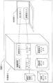
- FIG. 2 is a diagram illustrating a configuration of a microarray type SPRi device.
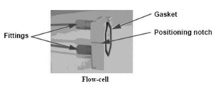
- FIG. 2 is a diagram showing a Flow-cell attached to a microarray type SPRi apparatus (Horiba, Ltd .: OpenPlex).
- FIG. 2 is a diagram showing a sensor chip (HORIBA, Ltd .: CS-HD) dedicated to a microarray type SPRi apparatus (HORIBA, Ltd .: OpenPlex).
- the shaded area indicates the part where the antibody is chemically bonded.
- the hexagonal frame indicates the place where the gasket in FIG. 2 contacts.
- FIG. 4 is a diagram showing a change in reflectance due to an interaction between an anti-c-kit antibody or goat IgG chemically bound to a sensor chip in a state of being mixed with gelatin and c-kit on a BMMC surface (non-specific adsorption suppression method). . Changes in the reflectance of the anti-c-kit antibody and goat IgG are shown by SPR images 600 seconds after BMMC was injected into the device.
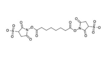
- FIG. 2 is a view showing a structural formula of bis (sulfosuccinimidyl) suberate.
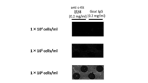
- FIG. 4 is a diagram showing a high sensitivity detection method. Changes in the reflectance of the anti-c-kit antibody and goat IgG are shown by SPR images 600 seconds after BMMC was injected into the device.
- the present invention Having a support, Having a gelatin layer on the support, Provided is a sensor chip (hereinafter, sometimes referred to as the sensor chip 1 of the present invention), which has a binding molecule directly or indirectly chemically bonded on the gelatin layer.
- the sensor chip of the present invention is a device having the above characteristics, wherein an interaction between a binding molecule and a surface molecule of a cell, a microorganism, a vesicle or a virus secreted by the cell or the microorganism is generated on the device.
- Device is
- the sensor chip 1 of the present invention has a support.
- the support used for the sensor chip 1 of the present invention is not particularly limited as long as it can be used in an immunological method or a surface plasmon resonance method, and examples thereof include synthetic resins such as polystyrene, polyacrylamide, and silicon; Glass, a prism covered with a metal thin film, a nitrocellulose film, and the like can be mentioned, and a prism covered with a metal thin film is preferable.
- the prism covered with the metal thin film is composed of the prism and metal deposited on one side of the prism, and can be used for the surface plasmon resonance method.
- Examples of the shape of the prism include a trapezoid, a triangle, and a circle (half pillar).
- the refractive index of the prism is usually 1.5 to 1.8.
- Examples of the metal deposited on one side of the prism include gold, silver, copper, and aluminum.
- a carboxy group and an ester of N-hydroxysuccinimide may be chemically bonded to the surface of the support. For example, when the support is covered with a thin gold film, an alkane having a thiol group at one end and a carboxy group at the other end is used as the first layer, and the carboxy group is reacted with N-hydroxysuccinimide. By performing the esterification, the carboxy group and the ester of N-hydroxysuccinimide can be exposed on the surface of the gold thin film.
- the sensor chip 1 of the present invention has a gelatin layer.
- the gelatin that can be used for the sensor chip 1 of the present invention is not particularly limited as long as it can suppress nonspecific interaction of the binding molecule with cells, microorganisms, vesicles or viruses secreted by the cells or the microorganisms. Examples thereof include gelatin extracted from collagen derived from the skin, bone, tendon and the like of non-human mammals (eg, cows and pigs) or the skin, bone, scales and the like of fish (eg, salmon). Extraction of gelatin from collagen may be performed by a known method. When bones and scales are used, the bones and scales are decalcified, solubilized, extracted, and purified.
- skin or tendon In the case where skin or tendon is used, it is performed by directly solubilizing the skin or tendon without decalcification, and extracting and purifying it.
- Decalcification can be performed by immersing bones and scales in dilute hydrochloric acid for about one week, and the resulting treated product (ossein) can be directly used for solubilization.
- ossein the resulting treated product
- calcium phosphate (apatite and the like) bound to collagen in bones and scales can be removed. Since collagen derived from ossein, skin and tendon forms a fibrous collagen helical structure and is stabilized (poorly water-soluble) by strong cross-linking, solubilization is required to gelatinize it. Become.
- an alkali treatment or an acid treatment can be performed.
- the alkali treatment can be performed by immersing the ossein, skin or tendon in a lime solution for 1 to 3 months.
- collagen can be partially and selectively hydrolyzed to a state where it can be extracted into warm water.
- the acid treatment can be performed by immersing ossein, skin or tendon in hydrochloric acid, sulfuric acid, phosphoric acid, or the like for several days, and the acid treatment can also make collagen extractable.
- the conditions of immersion (time, temperature, etc.) in the lime solution or acid can be appropriately determined by those skilled in the art.
- Extraction and purification can be carried out by eluting the gelatin from the solubilized ossein, skin or tendon with warm water, purifying and drying.
- examples of the gelatin that can be used for the sensor chip 1 of the present invention include commercially available gelatin.
- Commercially available gelatin that can be used for the sensor chip 1 of the present invention includes, for example, gelatin (purified powder) product code (Nacalai Tesque, Inc., product code 16631-05).
- the sensor chip 1 of the present invention has a gelatin layer on a support.
- the gelatin layer of the sensor chip 1 of the present invention is a layer formed based on gelatin. After heating heated distilled water to the gelatin to prepare a gelatin solution, the gelatin solution is diluted with heated distilled water to a desired concentration. It can be formed by applying an appropriate amount of liquid to the surface of the support and allowing it to stand at room temperature overnight.
- the concentration of gelatin when applied is usually 0.05% to 1%, preferably 0.1% to 1%.
- the thickness of the gelatin layer formed on the sensor chip 1 of the present invention is usually less than 100 nm.
- the sensor chip 1 of the present invention has a binding molecule.
- the binding molecule that can be used for the sensor chip 1 of the present invention is a molecule capable of specifically recognizing and interacting with a surface molecule of a cell, a microorganism, a vesicle or a virus secreted by the cell or the microorganism to be detected.
- proteins and nucleic acid aptamers include, for example, antibodies, cell adhesion factors (eg, integrins), lectins, and the like.
- the antibody is a polyclonal or monoclonal antibody.
- Antibodies may include antibodies derived from any mammals, and may further belong to any immunoglobulin class of IgG, IgA, IgM, IgD or IgE, but are preferably IgG. It is.
- As the antibody a commercially available antibody that interacts with the target surface molecule or an antibody stored in a research institution may be used. Alternatively, those skilled in the art can produce an antibody according to a conventionally known method.
- Antibodies include natural antibodies such as polyclonal antibodies and monoclonal antibodies (mAbs) described above, chimeric antibodies that can be produced using gene recombination techniques, humanized antibodies and single-chain antibodies, and these antibodies. Fragments.
- the antibody fragment means a partial region of the aforementioned antibody, and specifically includes Fab, Fab ′, F (ab ′) 2 , scAb, scFv, or scFv-Fc.
- cell adhesion factors include integrins. Integrin is not particularly limited as long as it is a heterodimer consisting of two subunits of an ⁇ chain and a ⁇ chain. ⁇ D ⁇ 2, ⁇ 5 ⁇ 1, ⁇ V ⁇ 1, ⁇ V ⁇ 3, ⁇ V ⁇ 5, ⁇ V ⁇ 6, ⁇ V ⁇ 8, ⁇ IIb ⁇ 3, ⁇ 4 ⁇ 1, ⁇ 4 ⁇ 7, ⁇ 9 ⁇ 1, ⁇ D ⁇ 2, ⁇ L ⁇ 2, ⁇ M ⁇ 2, ⁇ X ⁇ 2, ⁇ E ⁇ 7 and the like.
- the lectin is not particularly limited as long as it is a sugar-binding protein or glycoprotein other than an antibody that has the property of aggregating cells or glycoconjugates.
- lectins for example, SBA (Soybean Agglutinin), LCA (Lens culinaris Agglutinin), AAL (Aleuria aurantia Lectin), UEA (Ulex europaeus Agglutinin), PNA (Peanut Agglutinin), WGA (Wheat Ager Aglutinin) A) and the like.
- Nucleic acid aptamers can be RNA, DNA, modified nucleic acids, or mixtures thereof. Nucleic acid aptamers can also be in linear or circular form.
- a sugar residue eg, ribose
- examples of the site to be modified in the sugar residue include those in which the 2'-position, 3'-position and / or 4'-position hydroxy group of the sugar residue is replaced with another atom. Examples of the type of modification include fluorination, alkoxylation, O-allylation, S-alkylation, S-allylation, and amination.
- the sugar residue may be a BNA (Bridged Nucleic Acid) (LNA: Linked Nucleic Acid) having a crosslinked structure at the 2'-position and the 4'-position.
- the binding molecule is chemically bonded directly or indirectly to the gelatin layer formed on the surface of the support to form a layer based on the binding molecule.
- Chemical binding of the binding molecule onto the gelatin layer is performed by known means.
- the amino acid residue-binding molecule of gelatin is chemically bonded directly onto the gelatin layer, for example, when the binding molecule is a protein, one carboxy group at the C-terminus of the protein and one at the carboxy group of the acidic amino acid residue.
- a method can be considered in which gelatin is brought into contact with the active ester to form an amide bond between the carboxy group of the protein and the amino group of the lysine residue of the gelatin, thereby chemically bonding the protein to the gelatin layer.
- the activity of proteins such as enzymes and antibodies may be reduced by the reduction treatment.
- the binding molecule is an oligonucleotide
- An active ester is formed on the carboxy group in the same manner as described above, and gelatin is brought into contact with the active ester to form an amide bond between the carboxy group of the oligonucleotide and the amino group of a lysine residue of the gelatin.
- Nucleotides can also be chemically bonded onto the gelatin layer. Further, that the binding molecule is chemically bonded indirectly on the gelatin layer means that the binding molecule is chemically bonded on the gelatin layer via a cross-linking molecule.
- the cross-linking molecule examples include a divalent cross-linking agent.
- a method of chemically bonding a binding molecule to a gelatin layer via a divalent cross-linking agent is described below. Therefore, in the sensor chip 1 of the present invention, when the binding molecule is chemically bonded to the gelatin layer by the divalent crosslinking agent, the sensor chip is the same as the sensor chip 2 of the present invention described later.
- the present invention also provides Having a support, Having a binding site optimal combination layer on the support, Having a divalent cross-linking agent chemically bonded on the bonding site optimum blending layer, Having a binding molecule chemically bonded to the divalent cross-linking agent, a sensor chip, more specifically, Having a support, Having a gelatin layer or a polysaccharide layer on the support, Having a divalent cross-linking agent chemically bonded on the gelatin layer or the polysaccharide layer, provided is a sensor chip (hereinafter, sometimes referred to as a sensor chip 2 of the present invention), characterized by having a binding molecule chemically bonded to the bivalent crosslinking agent.
- the support used for the sensor chip 2 of the present invention may be the same as the support used for the sensor chip 1 of the present invention.
- the sensor chip 2 of the present invention has a binding site optimum blending layer.
- the optimum binding site compounding layer is a three-dimensional structure of the bonding molecules by maintaining a distance between the bonding molecules chemically bonded via the divalent crosslinking agent so as not to be overcrowded on the support.
- the layer may be formed of any material as long as the layer has an effect of reducing the obstacle, but is preferably a layer formed of a material having a carboxy group or an amino group.
- Specific materials used for the binding site optimum formulation layer include gelatin or polysaccharides. Gelatin usable for the sensor chip 2 of the present invention may be the same as the gelatin used for the sensor chip 1 of the present invention.
- the polysaccharide that can be used in the sensor chip 2 of the present invention is not particularly limited as long as the nonspecific interaction of the binding molecule with cells, microorganisms, vesicles or viruses secreted by the cells or the microorganisms can be suppressed.
- a polysaccharide having an amino group (basic polysaccharide), a polysaccharide having a carboxy group (acidic polysaccharide) or a mixture thereof is preferable.
- Basic polysaccharides include chitosan.
- Examples of the acidic polysaccharide include carrageenan, pectin, gum arabic, xanthan gum, gellan gum, agar, and tragacanth gum.
- mixtures can be obtained by mixing a basic polysaccharide and an acidic polysaccharide.
- a preferred polysaccharide that can be used for the sensor chip 2 of the present invention is chitosan.
- Polysaccharides can be extracted from raw materials such as plants and seaweed by hot water extraction or the like.
- the sensor chip 2 of the present invention is characterized by having a binding site optimum compounding layer (preferably a gelatin or polysaccharide layer, hereinafter the same) on a support.
- the gelatin layer of the sensor chip 2 of the present invention may be formed by the same method as the sensor chip 1 of the present invention.
- the polysaccharide layer of the sensor chip 2 of the present invention is prepared by adding heated distilled water to the polysaccharide to prepare a polysaccharide solution, and then diluting the polysaccharide to a desired concentration with the heated distilled water, and forming the solution on the surface of the support.
- a polysaccharide layer can be formed on the support by applying an appropriate amount and allowing to stand.
- the sensor chip 2 of the present invention has a divalent cross-linking agent chemically bonded on the binding site optimum blending layer, and a layer based on the divalent cross-linking agent is formed.
- the bivalent cross-linking agent that can be used in the sensor chip 2 of the present invention is a cross-linking agent having two functional groups so as to be able to connect the binding site optimal combination layer formed on the sensor chip 2 of the present invention to the binding molecule.
- the binding molecule can be given spatial freedom by providing a certain distance between the binding molecule and the binding site optimum blending layer.
- Such divalent crosslinkers include homodivalent crosslinkers.
- the homobivalent crosslinking agent is not particularly limited as long as the two functional groups are the same.
- n a natural number
- n represents a natural number
- n is not particularly limited as long as it is a natural number, but is preferably 1 to 100, more preferably 3 to 45, and still more preferably 3 to 6.
- Specific compounds represented by the formula 1 include, for example, a compound in which n is 6, bis (sulfosuccinimidyl) suberic acid, disodium salt (Bis (sulfosuccinimidyl) suberate, disodium salt (BS3)), Or a compound wherein n is 3, bis (sulfosuccinimidyl) glutaric acid, bis (sulfosuccinimidyl) glutarate, disodium salt (BS2G), and the like.
- Another homobivalent crosslinking agent is represented by Formula 3
- n represents a natural number.
- n is not particularly limited as long as it is a natural number, but is preferably 3 to 25, and more preferably 4 to 24.
- Specific compounds represented by Formula 3 include, for example, compounds represented by Formula 4
- a heterodivalent crosslinking agent may be mentioned.
- the heterodivalent crosslinking agent is not particularly limited as long as the two functional groups are different functional groups.
- -(6-maleimidocaproyloxy) succinimide N- (6-Maleimidocaproyloxy) succinimide (EMCS)
- N- (4-maleimidobutyryloxy) succinimide N- (4-Maleimidobutyryloxy) succinimide (GMBS)
- N -(8-Maleimidocapryloxy) succinimide N- (8-Maleimidocapryloxy) succinimide (HMCS)
- N- (11-Maleimidocapryloxy) succinimide HMCS
- N- (11-Maleimidoundecanoyloxy) succinimide KMUS
- N-(6-maleimidocaproyloxy) sulfosuccinimide, sodium salt N-(6-maleimidoc
- heterobivalent crosslinking agent includes a heterobivalent crosslinking agent in which the functional group at one end of the homobivalent crosslinking agent is substituted with avidin.
- the sensor chip 2 of the present invention is characterized in that it has a divalent cross-linking agent chemically bonded on the bonding site optimum blending layer formed on the surface of the support.
- the divalent crosslinking agent of the sensor chip 2 of the present invention is prepared by dissolving the divalent crosslinking agent in a solvent, spotting an appropriate amount on the surface of the binding site optimal blending layer, and allowing it to react at room temperature for 1 hour. It can be chemically bonded to the layer.
- the concentration of the divalent crosslinking agent at the time of spotting is usually 0.001 mM to 100 mM, preferably 0.01 mM to 10 mM.
- the divalent cross-linking agent is bis (sulfosuccinimidyl) suberic acid, bis (sulfosuccinimidyl) suberate, disodium salt (BS3)
- both ester bonds of the compound are unstable. Active ester, easily reacts with an amino group, and forms an amide bond between the carboxy group constituting the active ester and the amino group. Therefore, by contacting the divalent cross-linking agent with the binding site optimum blending layer, an amide bond is formed between the amino group of the binding site optimum blending layer and the carboxy group of one of the active esters of the divalent crosslinking agent.
- the valence crosslinking agent can be chemically bonded on the bonding site optimum blending layer. Also, at this time, the other active ester of the divalent crosslinking agent maintains an unreacted state.
- the sensor chip 2 of the present invention has a binding molecule.
- the binding molecule used for the sensor chip 2 of the present invention may be the same as the binding molecule used for the sensor chip 1 of the present invention.
- the binding molecule is chemically bonded to the bivalent cross-linking agent chemically bonded to the sensor chip 2 of the present invention.
- Chemical bonding between the binding molecule and the divalent crosslinking agent is performed by a known means.
- a chemical bond can be formed by dissolving the binding molecule in a solvent, spotting a suitable amount on a bivalent cross-linking agent chemically bonded on the bonding site optimum blending layer, and allowing it to react overnight at room temperature.
- the concentration of the binding molecule at the time of spotting is usually 0.1 mg / ml to 1 mg / ml, preferably 0.2 mg / ml.
- the divalent cross-linking agent is bis (sulfosuccinimidyl) suberic acid, disodium salt (Bis (sulfosuccinimidyl) suberate, disodium salt (BS3)), and the binding molecule is an antibody
- the unreacted active ester of succinimidyl) suberic acid, disodium salt (Bis (sulfosuccinimidyl) suberate, disodium salt (BS3) easily reacts with the amino group of the lysine residue of the antibody, and the unreacted active ester Therefore, an amide bond is formed between the carboxy group and the amino group of the binding molecule.
- the lysine residue of the binding molecule is brought into contact by contacting the binding molecule with a divalent cross-linking agent chemically bonded on the binding site optimum combination layer.
- An amide bond can be formed between the amino group of the group and the carboxy group of the unreacted active ester of the diester to form a chemical bond between the binding molecule and the divalent cross-linking agent.
- Sex rack The agent formed a crosslink by reacting with the antibody which is a binding molecule and the amino group of the gelatin layer, but reacted with the carboxy group of the binding molecule and the gelatin layer (or polysaccharide layer) to form a crosslink. It may be formed.
- the gelatin layer or the binding site optimum compounding layer formed on the surface of the support is composed of cells or microorganisms of the binding molecule, secreted by the cells or the microorganism. It has the effect of suppressing non-specific interactions with vesicles or viruses.
- the binding molecule is chemically bound to the gelatin layer or the binding site optimum blending layer while maintaining an appropriate distance between the binding molecules, whereby the cell, the microorganism, the cell or the microorganism of the binding molecule is bound.
- the detection sensitivity to the surface molecules of vesicles or viruses secreted by E. coli is improved.
- the binding molecule when the binding molecule is chemically bonded to the gelatin layer or the binding site optimal blending layer, the binding molecule is linked to the gelatin or the material forming the binding site optimal blending layer via a bivalent crosslinking agent. As a result, there is a distance between the binding molecule and the gelatin layer or between the layers where the binding site is optimally compounded, the distance corresponding to the length of the carbon chain of the divalent cross-linking agent.
- the sensor chip 1 or the sensor chip 2 of the present invention can be provided as a sensor chip for detecting an interaction between a binding molecule and a surface molecule of a cell, a microorganism, a vesicle or a virus secreted by the cell or the microorganism. .
- the cell is not particularly limited as long as it is a eukaryotic cell, and is defined as a concept including animal cells, plant cells, and fungi. Among them, animal cells are preferable.
- Animal cells include, but are not limited to, for example, hepatocytes, spleen cells, nerve cells, glial cells, pancreatic ⁇ cells, bone marrow cells, mesangial cells, Langerhans cells, epidermal cells, epithelial cells, goblet cells , Endothelial cells, smooth muscle cells, fibroblasts, fibroblasts, muscle cells, adipocytes, immune cells (eg, macrophages, T cells, B cells, natural killer cells, mast cells, neutrophils, basophils, Acidocytes, monocytes), megakaryocytes, synovial cells, chondrocytes, osteocytes, osteoblasts, osteoclasts, mammary cells, hepatocytes or stromal cells, or precursors, stem cells or cancer
- the surface molecule that interacts with the binding molecule in the cell is not particularly limited as long as it is a surface antigen, and examples thereof include proteins, sugar chains, lipids, glycoproteins, and glycolipids. Among them, proteins and sugar chains are preferable.
- proteins and sugar chains are preferable.
- the protein include c-kit.
- sugar chain include sugar chain antigen 125 (CA125), carcinoembryonic antigen (CEA), and sialyl Tn antigen.
- Microorganisms include eubacteria (hereinafter simply referred to as "bacteria”) and archaebacteria, of which bacteria are preferred.
- the bacterium is not particularly limited as long as it is a prokaryote having a cell membrane composed of a fatty acid ester of glycerol 3-phosphate, and may be a gram-negative bacterium or a gram-positive bacterium.
- Gram-negative bacteria examples include, but are not limited to, Neisseria (Neisseria), Branhamella (Branhamella), Haemophilus (Haemophilus), Bordetella (Bordetella), Escherichia (Escherichia), Citrobacter Genera (Citrobacter), Salmonella (Salmonella), Sigeria (Shigella), Klebsiella (Klebsiella), Enterobacter (Enterobacter), Serratia (Serratia), Hafnia (Hafnia), Proteus (Proteus), Morganella (Morganella), Providencia (Providencia), Yersinia (Yersinia), Campylobacter (Campylobacter), Vibrio (Vibrio), Aeromonas (Aeromonas), Pseudomonas (Pseudomonas), Xanthomonas (Xanthomonas), Acinetobacter (A
- the Gram-positive bacteria are not limited to the following, for example, Staphylococcus (Staphylococcus), Streptococcus (Streptococcus), Enterococcus (Enterococcus), Corynebacterium (Corynebacterium), Bacillus (Bacillus) ), Listeria, Listeria, Peptococcus, Peptostreptococcus, Clostridium, Clostridium, Eubacterium, Propionibacterium, Lactobacillus And bacteria belonging to the same.
- the surface molecule interacting with the binding molecule in the microorganism is not particularly limited as long as it is a molecule exposed on the cell membrane, and examples thereof include O antigen, H antigen, and K antigen.
- the vesicles secreted by cells or microorganisms are not particularly limited as long as they have a lipid bilayer as the outermost membrane, and extracellular vesicles (eg, exosomes, membrane vesicles, exosome-like vesicles, etc.) Is mentioned.
- the surface molecule that interacts with the binding molecule in the vesicle is not particularly limited as long as it is a molecule exposed on the vesicle membrane, for example, tetraspanin, CD9, CD63, CD81, major histocompatibility complex ( major histocompatibility complex), c-Kit, fucose, sugar chains containing sialic acid, and the like.
- Viruses include capsid-type viruses and envelope-type viruses.
- the surface molecule that interacts with the binding molecule in the virus is not particularly limited as long as it is a molecule exposed on the virus surface, and examples include influenza virus hemagglutinin and neuraminidase.
- the present invention also relates to a measuring unit including the sensor chip 1 or the sensor chip 2 of the present invention (hereinafter also referred to as a measuring unit of the present invention) and a main unit (hereinafter referred to as a main unit of the present invention).
- a measuring unit of the present invention includes the sensor chip 1 or the sensor chip 2 of the present invention (hereinafter also referred to as a measuring unit of the present invention) and a main unit (hereinafter referred to as a main unit of the present invention).
- Device for detecting an interaction between a surface molecule of a cell, a microorganism, a vesicle or a virus secreted by the cell or the microorganism, and a binding molecule hereinafter sometimes referred to as the device of the present invention. I will provide a.
- the support used for the sensor chip 1 or the sensor chip 2 of the present invention included in the measuring section of the present invention is preferably made of a prism and a metal deposited on one side surface of the prism.
- the prism and the metal deposited on one side of the prism may be the same as the metal described in the sensor chip 1 or the sensor chip 2 of the present invention.
- the measurement unit of the present invention is induced on the sensor chip 1 or the sensor chip 2 of the present invention by an interaction between a binding molecule and a surface molecule of a cell, a microorganism, a vesicle or a virus secreted by the cell or the microorganism.
- a sensor for detecting reflected light due to the SPR phenomenon may be further provided.
- the measuring section of the present invention may further include a calculating section for calculating and outputting a change amount of the reflected light detected by the sensor as a reflectance (%).
- the main body of the present invention can receive a signal output based on the amount of change in reflected light detected by the measuring unit of the present invention and display it to an operator.
- the main body of the present invention may further include a display unit that receives the calculated change in the reflectance from the calculation unit, converts the change into a color image, and outputs the color image.
- the main body of the present invention may further include a controller for adjusting the rotation angle of the reflecting mirror and the intensity of the irradiation light source.
- the arithmetic unit, the display unit, the control unit, and some of the functions thereof may be provided in any of the measuring unit of the present invention and the main unit of the present invention. Since the device of the present invention can also confirm a change in color tone at a place where a binding molecule is not chemically bonded, non-specific interaction of a cell, a microorganism, a vesicle secreted by the cell or the microorganism, or a virus with a sensor chip is performed. Can also be checked.
- FIG. 1 A microarray type SPRi device is shown in FIG. 1 as a more specific example of the device of the present invention.
- the device 1 shown in FIG. 1 mainly includes a measuring unit 2 and a main unit 3.
- the measuring unit 2 includes a sensor chip 21, a sensor 22, a calculating unit 23, a waste liquid bottle 24, a buffer bottle 25, a liquid sending pump 26, a deaerator 27, and a test sample insertion port 28. It is mainly composed.
- the main body 3 mainly includes a display unit 31 and a control unit 32.
- the sensor chip 21 is a device that causes an SPR phenomenon induced by the interaction of a binding molecule with a surface molecule of a cell, a microorganism, a vesicle secreted by the cell or the microorganism, or a virus. Applicable.
- the sensor 22 detects, as reflected light, an SPR phenomenon induced by the interaction of a binding molecule with a surface molecule of a cell, a microorganism, a vesicle secreted by the cell or the microorganism, or a virus.
- a CCD camera as a sensor detects reflected light intensity at each reflected light detection position as a current value and a voltage value.
- the calculation unit 23 calculates and outputs a change amount of the reflected light detected by the sensor 22 as a reflectance (%).
- the waste liquid bottle 24 is a bottle for disposing of the test sample and the buffer that have passed through the sensor chip 21 in the measuring unit 2.
- the buffer bottle 25 is a buffer bottle for passing the sensor chip 21 in the measuring unit 2.
- the liquid sending pump 26 is a pump for sending the buffer of the buffer bottle.
- the degassing device 27 is a device for degassing the buffer.
- the test sample insertion port 28 is an insertion port for introducing a test sample such as a cell into the measuring unit 2.
- the display unit 31 receives the change in the reflectance calculated by the calculation unit 23, converts the change into a color tone image, and outputs the image.
- the control unit 32 adjusts the rotation angle of the reflector provided in the measurement unit 2 and the intensity of the irradiation light source.
- the present invention also provides a method (detection method 1 of the present invention) for specifically detecting a cell, a microorganism, a vesicle or a virus secreted by the cell or the microorganism, comprising the following steps. (1) Forming a gelatin layer on a support. (2) Direct or indirect chemical bonding of the binding molecule onto the gelatin layer. (3) bringing a test sample into contact with the binding molecule. (4) detecting the interaction of the binding molecule with a cell, microorganism, vesicle or virus surface molecule secreted by the cell or microorganism in the test sample.
- Detection method 1 of the present invention includes forming a gelatin layer on a support.
- the support and gelatin used in the detection method 1 of the present invention may be the same as the support and gelatin used in the sensor chip 1 of the present invention.
- a method of forming a gelatin layer on a support is to prepare a gelatin solution by adding heated distilled water to the gelatin, and then dilute the solution to a desired concentration with heated distilled water. It can be carried out by spreading an appropriate amount on the surface of the body and allowing it to stand at room temperature overnight.
- the concentration of gelatin at the time of coating is usually 0.05% to 1%, preferably 0.1% to 1%.
- the thickness of the gelatin layer formed by coating the support is usually less than 100 nm.
- Detection method 1 of the present invention involves chemically binding a binding molecule to a gelatin layer.
- the binding molecule used in the detection method 1 of the present invention and the method of direct or indirect chemical bonding of the binding molecule onto the gelatin layer are the same as those used in the sensor chip 1 of the present invention. It may be.
- Detection method 1 of the present invention includes contacting a test sample with a binding molecule.
- the test sample used in the detection method 1 of the present invention can be used without particular limitation as long as it is a sample containing cells, microorganisms to be detected, vesicles or viruses secreted by the cells or the microorganisms.
- Test samples are obtained by centrifuging body fluids (blood, saliva, tears, urine, sweat, etc.), cell cultures, cultured cells, cultured microorganisms, environmental water, soil, foodstuffs, etc. in animals (preferably mammals), It is prepared by filtering, size exclusion chromatography and the like.
- a test sample having a higher concentration of cells, microorganisms, vesicles or viruses secreted by the cells or the microorganisms in the test sample can be prepared.
- the time, temperature, and number of times of contact with the binding molecule can be appropriately determined by those skilled in the art. For example, the contact is performed at room temperature, 1 to 3 times for 10 minutes to 2 hours. be able to.
- the detection method 1 of the present invention includes detecting an interaction between a binding molecule and a cell, a microorganism, a vesicle or a virus secreted by the cell or the microorganism in a test sample.
- a method for detecting an interaction between a surface molecule and a binding molecule is not particularly limited, and examples thereof include an immunological method (ELISA method) and a surface plasmon resonance (SPR) method. Is mentioned.
- the detection by the immunological method is not particularly limited, and may comprise a cell, a microorganism, a vesicle secreted by the cell or the microorganism, or a virus-binding molecule in a test sample. Any measurement method may be used as long as the method detects the complex by immunological means. Examples of the immunological method include, for example, in a batch system and a flow system, cells, microorganisms, vesicles or virus surface molecules secreted by the cells or the microorganisms that have interacted with the binding molecule on the gelatin layer by ELISA. Any method may be used as long as it is used as an antigen, and detection is performed by allowing an antibody to interact with the antigen.
- the interaction of the antibody with the antigen can be detected using a labeling agent.
- a labeling agent for example, a radioisotope, an enzyme, a fluorescent substance, a luminescent substance and the like are used.
- the radioisotope for example, [ 125 I], [ 131 I], [ 3 H], [ 14 C] and the like are used.
- the above enzyme a stable enzyme having a large specific activity is preferable.
- ⁇ -galactosidase, ⁇ -glucosidase, alkaline phosphatase, peroxidase, malate dehydrogenase and the like are used.
- fluorescent substance for example, fluorescamine, fluorescein isothiocyanate and the like are used.
- luminescent substance for example, luminol, a luminol derivative, luciferin, lucigenin and the like are used.
- a biotin-avidin system can be used for binding the antibody to the labeling agent.
- a test sample is reacted with a binding molecule chemically bonded to the gelatin layer (primary reaction), and further reacted with a labeled secondary antibody against the surface molecule (secondary reaction).
- primary reaction a binding molecule chemically bonded to the gelatin layer
- secondary reaction a labeled secondary antibody against the surface molecule
- the primary reaction and the secondary reaction may be performed in the reverse order, may be performed simultaneously, or may be performed at staggered times.
- the detection by the surface plasmon resonance method is such that after a test sample is brought into contact with a gelatin layer, a support is irradiated with light of a specific wavelength from a specific angle, and a change in the resonance angle is used as an index.
- the method is not particularly limited as long as it is a method for determining the presence or absence of the interaction of the surface molecule with the binding molecule chemically bonded to the gelatin layer.
- the present invention also provides a method for specifically detecting a cell, a microorganism, a vesicle or a virus secreted by the cell or the microorganism (the detection method 2 of the present invention), comprising the following steps. (1) Forming a binding site optimum blending layer on a support. (2) chemically bonding a divalent cross-linking agent to the bonding site optimum blending layer; (3) chemically bonding the binding molecule to the divalent crosslinking agent; (4) bringing a test sample into contact with the binding molecule. (5) detecting the interaction of the binding molecule with a cell, microorganism, vesicle or virus surface molecule secreted by the cell or microorganism in the test sample.
- Detection method 2 of the present invention includes forming a binding site optimum blending layer, preferably a gelatin layer or a polysaccharide layer, on a support.
- the support used in the detection method 1 of the present invention, the material forming the binding site optimum blending layer, the gelatin and the polysaccharide are the support used in the sensor chip 2 of the present invention, the material forming the binding site optimal blending layer, It can be similar to gelatin and polysaccharides.
- a method of forming a gelatin layer on a support may be the same as the method used in the detection method 1 of the present invention.
- a method for forming a polysaccharide layer on a support is to prepare a polysaccharide solution by adding heated distilled water to the polysaccharide, and then dilute to a desired concentration with heated distilled water. Then, an appropriate amount is applied to the surface of the support and allowed to stand, whereby a polysaccharide layer can be formed on the support.
- Detection method 2 of the present invention includes chemically bonding a divalent cross-linking agent on the binding site optimum combination layer.
- the divalent crosslinking agent used in the detection method 2 of the present invention may be the same as the divalent crosslinking agent used in the sensor chip 2 of the present invention.
- the method of chemically bonding a divalent cross-linking agent onto the binding site optimum blending layer comprises dissolving the divalent crosslinking agent in a solvent and spotting an appropriate amount on the surface of the binding site optimal blending layer. For 1 hour at room temperature.
- the concentration of the divalent crosslinking agent at the time of spotting is usually 0.001 mM to 100 mM, preferably 0.01 mM to 10 mM.
- Detection method 2 of the present invention involves chemically bonding a binding molecule and a divalent crosslinking agent.
- the binding molecule used in the detection method 2 of the present invention may be the same as the binding molecule used in the sensor chip 2 of the present invention.
- the binding molecule is chemically bound to the divalent cross-linking agent chemically bound on the binding site optimum blending layer.
- Chemical bonding between the binding molecule and the divalent crosslinking agent is performed by a known means.
- a chemical bond can be obtained by dissolving the binding molecule in a solvent, spotting an appropriate amount of the binding molecule on a bivalent crosslinking agent, and allowing the mixture to react overnight at room temperature.
- the concentration of the binding molecule at the time of spotting is usually 0.1 mg / ml to 1 mg / ml, preferably 0.2 mg / ml.
- Detection method 2 of the present invention includes contacting a test sample with the binding molecule.
- the test sample used in the detection method 2 of the present invention may be the same as the test sample used in the detection method 1 of the present invention.
- the time, temperature, and number of times of contact with the binding molecule can be appropriately determined by those skilled in the art. For example, the contact can be performed at 10 to 2 hours at room temperature, 1 to 3 times. it can.
- the detection method 2 of the present invention includes detecting an interaction between the binding molecule and a cell, a microorganism, a vesicle or a virus secreted by the cell or the microorganism in the test sample.
- the method of detecting the interaction between the surface molecule and the binding molecule may be the same as the method used in the detection method 1 of the present invention.
- two or more different binding molecules are chemically bonded to a gelatin layer or a divalent cross-linking agent so as to be differently arranged on the sensor chip of the present invention.
- a test sample is brought into contact with a sensor chip of the present invention in which an antibody is chemically bonded to at least one spot, and a lectin is chemically bonded to another at least one spot, and cells, microorganisms,
- an antibody is chemically bonded to at least one spot
- a lectin is chemically bonded to another at least one spot
- cells, microorganisms The interaction of the antibody with the surface antigen of vesicles or viruses secreted by the cell or the microorganism and the interaction of the lectin with the sugar chain can also be detected.
- the present invention also provides a method for specifically detecting a cell, a microorganism, a vesicle or a virus secreted by the cell or the microorganism, comprising the following steps. (1) Forming a gelatin layer on a support.
- the present invention also provides a method for specifically detecting a cell, a microorganism, a vesicle or a virus secreted by the cell or the microorganism, comprising the following steps. (1) Forming a binding site optimum blending layer on a support.
- the method allows rapid diagnosis of a test sample, for example, because two or more different surface molecules of a cell, a microorganism, a vesicle or a virus secreted by the cell or the microorganism of the test sample can be detected simultaneously.
- the sugar chain on the cell membrane surface and the surface antigen can be simultaneously detected, so that the diagnosis can be performed quickly.
- the cell detection sensor using Surface Plasmon Resonance is a microarray type SPRi device (Horiba, Ltd .: OpenPlex) and a dedicated sensor chip (Horiba, Ltd.) Mfg .: CS-HD; a succinimide-activated carboxy group chemically bonded sensor chip).
- the constructed sensor can measure the amount of change in reflected light associated with the SPR phenomenon induced by the interaction of cell surface molecules with the antibody on the chip surface as the reflectance (%) every 3 seconds.
- the reflectance change of the SPR can be observed as a spot image.
- the chip has a surface area of 12 mm ⁇ 23 mm, a feature is that many spots can be arranged in parallel by adjusting the spot diameter (spot amount) of the ligand solution for chemical bonding.
- BMMC mouse bone marrow-derived mast cells
- a goat antibody (goat antibody; R & D systems Inc., AB108C) was used as a negative antibody.
- Gelatin having an effect of suppressing non-specific interaction between the antibody and the cells was mixed with the antibody, spotted on the chip surface at 10 nL using a spotter, and allowed to stand for 16 hours for chemical bonding.
- the chip surface was washed with Dulbecco's PBS (-) (hereinafter abbreviated as PBS), filled with 1% gelatin dissolved PBS on the chip surface, allowed to stand at room temperature for 1 hour, and blocked.
- the blocked chip was washed three times with PBS and then mounted on the device.
- the contact of the buffer or BMMC to the chip surface was performed via Flow-cell (FIG. 2).
- the flow-cell was contact-fixed to the chip at a position where the entire gasket was completely covered by the chip (FIG. 3). Further, among the planes of the Flow-cell, the plane surrounded by the Gasket frame is recessed by 80 ⁇ m from the plane around the Gasket frame. As a result, when the chip comes into contact with the Flow-cell, a spatial gap having a width of 80 ⁇ m is generated between the plane surrounded by the Gasket frame of the Flow-cell and the chip surface. Therefore, the buffer etc. sent from one polyvinyl chloride tube (380 ⁇ m inside diameter) connected to the Flow-cell via Fitting comes into contact with the chip surface by filling the 80 ⁇ m wide space, It is discharged from the other polyvinyl chloride tube.
- PBS buffer A
- buffer A buffer A
- BMMC was suspended in buffer A to a concentration of 1 ⁇ 10 4 , 1 ⁇ 10 5 and 1 ⁇ 10 6 cells / ml. After injecting the cells of each concentration into the device, the solution was sent for 480 seconds, and immediately thereafter, only buffer A was sent for 480 seconds.
- Example Cell detection using a sensor chip that has been previously blocked with gelatin and further chemically bonded to the antibody via BS3 (a new high-sensitivity detection method) Since gelatin is derived from an extracellular matrix, it has excellent compatibility with cells. Therefore, by using gelatin as a blocking agent, the hydrophilicity of the chip surface, that is, the bound water near the molecule due to hydrogen bonding increases, and the binding to the hydrophobic portion of the protein which is present on the cell membrane surface in large numbers is suppressed, Non-specific interactions between cells and antibodies are suppressed. To make a chip with this property, the surface was blocked by completely covering it with gelatin. However, since the chip is completely blocked with gelatin, an antibody to be used for cell detection cannot be bound to the chip as it is.
- BS3 Bis (sulfosuccinimidyl) suberate, disodium salt (hereinafter, BS3) (FIG. 5) was used as a divalent reactive reagent.
- BS3 is a diester of suberic acid and sulfo-N-hydroxysuccinimide, and has a property of easily forming an amide bond between a carboxy group of suberic acid and an amino group of a lysine residue constituting a protein or polypeptide. Accordingly, BS3 can serve as a linker that links lysine residues present in about 3% of all amino acids constituting gelatin to lysine residues of the antibody.
- a cell concentration dependency test was performed as in the comparative example.
- Cells and antibodies used were the same as in the comparative example.
- 500% of 0.1% gelatin dissolved in PBS was applied on the chip to chemically bind the gelatin, and then reacted overnight at room temperature to block the chip surface. After the reaction, the chip was washed three times with distilled water, and water remaining on the chip surface was completely blown off with a blower. Next, a divalent reactive reagent 1 mM BS3 dissolved in PBS was spotted on the chip surface with a spotter, and reacted for 1 hour at room temperature.
- the chip was washed three times with water, and water remaining on the chip surface was completely blown off with a blower.
- the antibody was spotted with the spotter at the same position as the spot where the bivalent reactive reagent was spotted, and reacted overnight at room temperature.
- the chip was washed three times with PBS, and water remaining on the chip surface was completely removed with a blower, and used for the test.
- Buffer A was supplied as a running buffer to the device equipped with the chip at a flow rate of 25 ⁇ L / min, the surface of the chip was conditioned, and the reflectance at the time of stabilization was set to 0%.
- BMMC was suspended in buffer A to a concentration of 1 ⁇ 10 4 , 1 ⁇ 10 5 and 1 ⁇ 10 6 cells / ml. After injecting the cells of each concentration into the device, the solution was fed for 480 seconds, and then immediately washed with buffer A alone for 480 seconds. As a result, at 1 ⁇ 10 4 , 1 ⁇ 10 5 , 1 ⁇ 10 6 cells / ml concentration to detect the specific interaction between the positive antibody anti-c-Kit antibody and BMMC, and the BMMC concentration 1 ⁇ 10 4. At 1, 1 ⁇ 10 5 and 1 ⁇ 10 6 cells / ml, the spot of the negative antibody did not turn white, indicating that the nonspecific interaction was suppressed by the gelatin effect (FIG.
- the new high-sensitivity detection method increased sensitivity 100-fold in cell concentration.
- the fact that the proportion of lysine residues present in gelatin is about 3% means that antibodies linked via BS3 are kept at a distance so that they do not become overcrowded on the chip, and the antibodies are highly sensitive to antigens. It is considered that the detection was made possible.
- the distance between the chip and the antibody is increased by the length of the carbon chain of the bivalent reactive reagent, and the degree of freedom of the antibody is also increased. It is considered that this contributed to the sensitivity detection.
- the non-specific interaction of the binding molecule with cells, microorganisms, vesicles or viruses secreted by the cell or the microorganism is suppressed, and the cells of the binding molecule, microorganism, Detection sensitivity to vesicles or virus surface molecules secreted by the cell or the microorganism can be improved.
Landscapes
- Health & Medical Sciences (AREA)
- Immunology (AREA)
- Life Sciences & Earth Sciences (AREA)
- Chemical & Material Sciences (AREA)
- Engineering & Computer Science (AREA)
- Biochemistry (AREA)
- General Health & Medical Sciences (AREA)
- General Physics & Mathematics (AREA)
- Analytical Chemistry (AREA)
- Pathology (AREA)
- Physics & Mathematics (AREA)
- Urology & Nephrology (AREA)
- Hematology (AREA)
- Molecular Biology (AREA)
- Biomedical Technology (AREA)
- Biotechnology (AREA)
- Cell Biology (AREA)
- Microbiology (AREA)
- Food Science & Technology (AREA)
- Medicinal Chemistry (AREA)
- Apparatus Associated With Microorganisms And Enzymes (AREA)
- Investigating Or Analysing Materials By Optical Means (AREA)
Abstract
本発明は、目的の細胞、微生物、該細胞もしくは該微生物が分泌する小胞またはウイルス表面分子と結合性分子の相互作用を、非特異的相互作用を抑制しながら、高感度に検出するセンサーチップ、該センサーチップを備えた該相互作用検出装置を提供する。
Description
本発明は、細胞、微生物、該細胞もしくは該微生物が分泌する小胞またはウイルスの表面分子と結合性分子の相互作用を、非特異的相互作用を抑制しながら、高感度に検出するセンサーチップ、該センサーチップを備えた該相互作用検出装置等に関する。
現在、悪性腫瘍等の診断は、肉眼観察、X線、CT(Computed Tomography)または超音波等による画像情報に基づく予備的判断が行われ、病理組織標本を用いた組織構造を顕微鏡的に観察することによって最終的に判断される。しかし、これらの情報に基づく診断は、医師の判断基準に基づいて行われるため少なからず誤診が生じる可能性があり、場合によっては致命的な医療事故につながる虞もある。そこで、誤診の可能性を小さくするために、さらに被疑組織内の遺伝子の異常、腫瘍マーカーの有無に関する情報を加えて、総合的に判断されるようになってきている。
腫瘍マーカーは、近年研究が盛んであり、腫瘍に関連する抗原、酵素、特定のタンパク質、代謝産物、腫瘍遺伝子、腫瘍遺伝子生産物及び腫瘍抑制遺伝子などを指し、例えば、癌胎児性抗原CEA、糖タンパク質CA19-9、CA125、前立腺特異抗原PSA、甲状腺で産生されるペプチドホルモンであるカルシトニンなどが一部の癌で腫瘍マーカーとして癌診断に活用されている。検出の対象となる腫瘍マーカーには体液性(血液、リンパ液、尿等)マーカーが多く、その検出は公知の手段によって実施することができる。例えば、免疫学的検出法は、抗原抗体反応を利用して腫瘍マーカーの検出を行うもので、一般に検出精度が優れているばかりでなく、迅速、簡便かつ経済的な検出法である。また、近年、表面プラズモン共鳴現象を応用し、共鳴角度変化をリアルタイムでとらえることにより、抗体および抗原の生体分子間の反応および結合量の測定および速度論解析をすることができる表面プラズモン共鳴装置(SPR(surface plasmon resonance)装置)が様々な研究および検査等で利用されており、腫瘍マーカーの検査にも応用されている。これらの方法は、抗体を支持体に化学結合させることによって、安価かつ大量に被験試料を処理できるという大きな利点を有する。
しかし、全ての腫瘍マーカーに対して上記の方法を適用できるわけではない。哺乳動細胞の直径は10μm以上である場合が多く、抗体に対して極めて大きい。そのため、細胞と抗体を接触させた場合、抗体の特異性に関わらず、細胞膜が抗体に直接相互作用してしまうことが知られていた。腫瘍マーカーには体液性マーカーだけでなく、癌細胞膜表面に発現するマーカーも存在するため、細胞膜にアンカーされた腫瘍マーカーを検出するために上記の方法を適用しようとすれば、該細胞の細胞膜と抗体との間に非特異的な相互作用が生じるため、該非特異的相互作用を抑制しなければならないという課題があった。この課題に対して、本発明者らは、ゼラチンと抗体の混合物を用いることによって、該非特異的相互作用を抑制できることを既に見出している。しかし、腫瘍マーカーの発現量は腫瘍マーカーやそれを発現する細胞によって変わるため、腫瘍マーカーの発現量が低い細胞の場合は、被験試料となる細胞の数を通常より多く確保する必要があり、被験者への侵襲が大きいことが問題であった。また、例えば、抗腫瘍剤の開発のための基礎研究では、細胞に腫瘍マーカーを一過性発現させ、その検出を行うことがある。しかし、哺乳動物細胞では、異種膜タンパク質を一過性発現させた場合でも、その発現量は哺乳動物細胞中の全タンパク質のわずか2.5%にとどまる(非特許文献1)。従って、実験系として成立させるためには、多くの発現細胞が必要となり、経済的課題があった。以上の点から、膜タンパク質を低発現する細胞を検出するためには、細胞の細胞膜と抗体との間に非特異的な相互作用を抑制するだけでなく、抗体が目的とする膜タンパク質に高感度に相互作用できる必要があった。
Randal J. Kaufman, Mol Biotechnol. Vol.16(2), 151-160, 2000
本発明は、目的の細胞、微生物、該細胞もしくは該微生物が分泌する小胞またはウイルス表面分子と結合性分子の相互作用を、非特異的相互作用を抑制しながら、高感度に検出するセンサーチップ、該センサーチップを備えた該相互作用検出装置等を提供することを目的とする。
本発明者らは、抗体(抗c-kit抗体または陰性抗体)とゼラチンを混合し、センサーチップ上にスポットすることによって抗体をセンサーチップ上に化学結合させた後、センサーチップをゼラチンでブロッキングし、c-kitを細胞膜上に発現していることが既知であるマウス骨髄由来肥満細胞を含む被験試料をセンサーチップに接触させ、表面プラズモン共鳴法によって抗c-kit抗体および陰性抗体の両者の反射率の変化を確認した。その結果、陰性抗体の反射率の変化は確認できなかったものの、被験試料中の細胞数が少ない場合は、抗c-kit抗体の反射率変化も確認できなかった。本結果について、本発明者らは、ゼラチンと混合した抗体を化学結合させる方法は、抗体と細胞膜間の非特異的相互作用を抑制できるが、抗体による抗原の検出感度に課題を抱えていることを認識した。そこで本発明者らは、非特異的相互作用を抑制しながら、目的とする抗原を表面に発現する細胞、微生物、該細胞もしくは該微生物が分泌する小胞またはウイルスを感度よく検出する方法を提供すべく鋭意研究を行った。その際、発明者らは、ゼラチンの構成アミノ酸の3%は塩基性アミノ酸であるリジンであることに着目した。ゼラチンのリジンのアミノ基を介して抗体をゼラチン上に化学結合させることができれば、抗体同士がチップ上で過密にならないような距離が保たれることによって抗体同士の立体障害が減弱され、その結果、抗体による抗原の検出感度を向上できると考えた。本着想を検証すべく、本発明者らは、センサーチップをゼラチンで被覆し、ビス(スルホスクシンイミジル)スベリン酸, 二ナトリウム塩を用いて抗体と該ゼラチンを連結した結果、陰性抗体の反射率の変化は確認できず、かつ上記の方法と比べて、抗体による抗原の検出感度が約100倍向上したことを確認した。上記の事実から、支持体を予めゼラチンまたは多糖類で被覆し、その後、細胞、微生物、該細胞もしくは該微生物が分泌する小胞またはウイルスの表面分子に対する結合性分子をゼラチンまたは多糖類上に化学結合させることによって、該結合性分子の細胞、微生物、該細胞もしくは該微生物が分泌する小胞またはウイルスに対する非特異的相互作用を抑制しつつ、該結合性分子の該表面分子に対する検出感度を向上できることを見出し、本発明を完成した。
すなわち、本発明は、
[1]支持体を有し、
該支持体上にゼラチン層または多糖類層を有し、
該ゼラチン層または該多糖類層上に化学結合した二価性架橋剤を有し、
該二価性架橋剤と化学結合した結合性分子を有することを特徴とする、センサーチップ;
[2]前記多糖類が、酸性多糖類、塩基性多糖類またはそれらの混合物である、[1]に記載のセンサーチップ;
[3]前記二価性架橋剤が、式1
[1]支持体を有し、
該支持体上にゼラチン層または多糖類層を有し、
該ゼラチン層または該多糖類層上に化学結合した二価性架橋剤を有し、
該二価性架橋剤と化学結合した結合性分子を有することを特徴とする、センサーチップ;
[2]前記多糖類が、酸性多糖類、塩基性多糖類またはそれらの混合物である、[1]に記載のセンサーチップ;
[3]前記二価性架橋剤が、式1
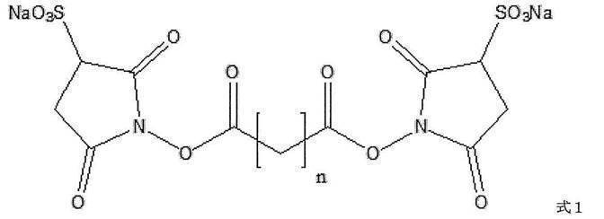
(式1中、nは自然数を表す)によって表される化合物、または式2
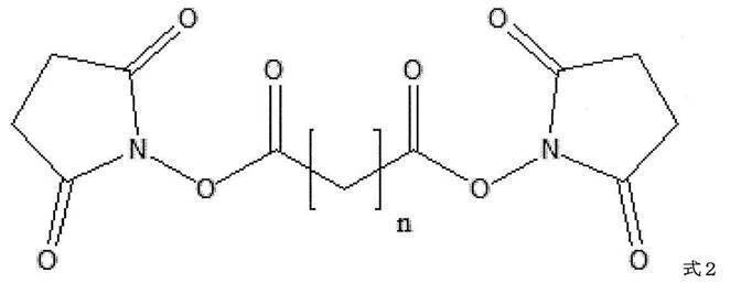
(式2中、nは自然数を表す)によって表される化合物
である、[1]または[2]に記載のセンサーチップ;
[4]前記式1または式2によって表される化合物において、nが1~100である、[3]に記載のセンサーチップ;
[5]前記二価性架橋剤が、ビス(スルホスクシンイミジル)スベリン酸, 二ナトリウム塩(Bis(sulfosuccinimidyl)suberate, disodium salt(BS3))またはビス(スルホスクシンイミジル)グルタル酸, 二ナトリウム塩(Bis(sulfosuccinimidyl)glutarate, disodium salt(BS2G))である、[3]に記載のセンサーチップ;
[6]前記二価性架橋剤が、式3
である、[1]または[2]に記載のセンサーチップ;
[4]前記式1または式2によって表される化合物において、nが1~100である、[3]に記載のセンサーチップ;
[5]前記二価性架橋剤が、ビス(スルホスクシンイミジル)スベリン酸, 二ナトリウム塩(Bis(sulfosuccinimidyl)suberate, disodium salt(BS3))またはビス(スルホスクシンイミジル)グルタル酸, 二ナトリウム塩(Bis(sulfosuccinimidyl)glutarate, disodium salt(BS2G))である、[3]に記載のセンサーチップ;
[6]前記二価性架橋剤が、式3
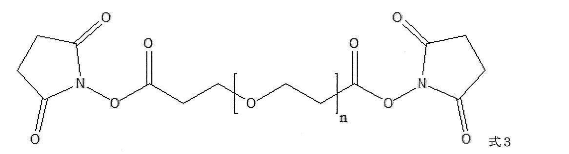
(式3中、nは自然数を表す)によって表される化合物である、[1]または[2]に記載のセンサーチップ;
[7]前記式3によって表される化合物において、nが3~25である、[6]に記載のセンサーチップ;
[8]前記二価性架橋剤が、N-(6-マレイミドカプロイルオキシ)スクシンイミド(N-(6-Maleimidocaproyloxy)succinimide(EMCS))、N-(4-マレイミドブチリルオキシ)スクシンイミド(N-(4-Maleimidobutyryloxy)succinimide(GMBS))、N-(8-マレイミドカプリルオキシ)スクシンイミド(N-(8-Maleimidocapryloxy)succinimide(HMCS))、N-(11-マレイミドウンデカノイルオキシ)スクシンイミド(N-(11-Maleimidoundecanoyloxy)succinimide(KMUS))、N-(6-マレイミドカプロイルオキシ)スルホスクシンイミド, ナトリウム塩(N-(6-Maleimidocaproyloxy)sulfosuccinimide, sodium salt(Sulfo-EMCS))、N-(4-マレイミドブチリルオキシ)スルホスクシンイミド, ナトリウム塩(N-(4-Maleimidobutyryloxy)sulfosuccinimide, sodium salt(Sulfo-GMBS))、N-(8-マレイミドカプリルオキシ)スルホスクシンイミド, ナトリウム塩(N-(8-Maleimidocapryloxy)sulfosuccinimide, sodium salt(Sulfo-HMCS))、N-(11-マレイミドウンデカノイルオキシ)スルホスクシンイミド, ナトリウム塩(N-(11-Maleimidoundecanoyloxy)sulfosuccinimide, sodium salt(Sulfo-KMUS))、N-[(4-マレイミドメチル)シクロヘキシルカルボニルオキシ]スルホスクシンイミド, ナトリウム塩(N-[(4-Maleimidomethyl)cyclohexylcarbonyloxy]sulfosuccinimide, sodium salt(Sulfo-SMCC))、N-スクシンイミジル 3-(2-ピリジルジチオ)プロピオン酸(N-Succinimidyl 3-(2-pyridyldithio)propionate(SPDP))、N-{6-[3-(2-ピリジルジチオ)プロピオンアミド]ヘキサノイルオキシ}スルホスクシンイミド, ナトリウム塩(N-{6-[3-(2-Pyridyldithio)propionamido]hexanoyloxy}sulfosuccinimide, sodium salt(Sulfo-AC5-SPDP))、ジチオビス(スルホスクシンイミジル プロパン酸), 二ナトリウム塩(Dithiobis(sulfosuccinimidyl propionate), disodium salt(DTSSP))、ジチオビス(スクシンイミジル プロパン酸)(Dithiobis(succinimidyl propionate)(DSP))、ジチオビス(スクシンイミジル ウンデカン酸)(Dithiobis(succinimidyl undecanoate))、ジチオビス(スクシンイミジル オクタン酸)(Dithiobis(succinimidyl octanoate))、およびジチオビス(スクシンイミジル ヘキサン酸)(Dithiobis(succinimidyl hexanoate))からなる群から選択される、[1]または[2]に記載のセンサーチップ;
[9]前記結合性分子が、抗体、細胞接着因子、レクチンおよび核酸アプタマーからなる群から選択される、[1]~[8]のいずれか1つに記載のセンサーチップ;
[10]前記支持体が、金属薄膜に覆われたプリズムである、[1]~[9]のいずれか1つに記載のセンサーチップ;
[11]細胞、微生物、該細胞もしくは該微生物が分泌する小胞またはウイルスの表面分子と結合性分子の相互作用検出用センサーチップである、[1]~[10]のいずれか1つに記載のセンサーチップ;
[12]支持体を有し、
該支持体上にゼラチン層を有し、
該ゼラチン層上に直接的に又は間接的に化学結合した結合性分子を有することを特徴とする、センサーチップ;
[13][1]~[12]のいずれか1つに記載のセンサーチップを含む計測部と、本体部とを備えた、細胞、微生物、該細胞もしくは該微生物が分泌する小胞またはウイルスの表面分子と結合性分子の相互作用検出装置;
[14]以下の工程を含む、細胞、微生物、該細胞もしくは該微生物が分泌する小胞またはウイルスを特異的に検出する方法:
(1)支持体上にゼラチン層を形成すること、
(2)結合性分子を該ゼラチン層上に直接的に又は間接的に化学結合させること、
(3)被験試料を該結合性分子に接触させること、
(4)該被験試料中の細胞、微生物、該細胞もしくは該微生物が分泌する小胞またはウイルスの表面分子と該結合性分子の相互作用を検出すること;
を提供する。
[7]前記式3によって表される化合物において、nが3~25である、[6]に記載のセンサーチップ;
[8]前記二価性架橋剤が、N-(6-マレイミドカプロイルオキシ)スクシンイミド(N-(6-Maleimidocaproyloxy)succinimide(EMCS))、N-(4-マレイミドブチリルオキシ)スクシンイミド(N-(4-Maleimidobutyryloxy)succinimide(GMBS))、N-(8-マレイミドカプリルオキシ)スクシンイミド(N-(8-Maleimidocapryloxy)succinimide(HMCS))、N-(11-マレイミドウンデカノイルオキシ)スクシンイミド(N-(11-Maleimidoundecanoyloxy)succinimide(KMUS))、N-(6-マレイミドカプロイルオキシ)スルホスクシンイミド, ナトリウム塩(N-(6-Maleimidocaproyloxy)sulfosuccinimide, sodium salt(Sulfo-EMCS))、N-(4-マレイミドブチリルオキシ)スルホスクシンイミド, ナトリウム塩(N-(4-Maleimidobutyryloxy)sulfosuccinimide, sodium salt(Sulfo-GMBS))、N-(8-マレイミドカプリルオキシ)スルホスクシンイミド, ナトリウム塩(N-(8-Maleimidocapryloxy)sulfosuccinimide, sodium salt(Sulfo-HMCS))、N-(11-マレイミドウンデカノイルオキシ)スルホスクシンイミド, ナトリウム塩(N-(11-Maleimidoundecanoyloxy)sulfosuccinimide, sodium salt(Sulfo-KMUS))、N-[(4-マレイミドメチル)シクロヘキシルカルボニルオキシ]スルホスクシンイミド, ナトリウム塩(N-[(4-Maleimidomethyl)cyclohexylcarbonyloxy]sulfosuccinimide, sodium salt(Sulfo-SMCC))、N-スクシンイミジル 3-(2-ピリジルジチオ)プロピオン酸(N-Succinimidyl 3-(2-pyridyldithio)propionate(SPDP))、N-{6-[3-(2-ピリジルジチオ)プロピオンアミド]ヘキサノイルオキシ}スルホスクシンイミド, ナトリウム塩(N-{6-[3-(2-Pyridyldithio)propionamido]hexanoyloxy}sulfosuccinimide, sodium salt(Sulfo-AC5-SPDP))、ジチオビス(スルホスクシンイミジル プロパン酸), 二ナトリウム塩(Dithiobis(sulfosuccinimidyl propionate), disodium salt(DTSSP))、ジチオビス(スクシンイミジル プロパン酸)(Dithiobis(succinimidyl propionate)(DSP))、ジチオビス(スクシンイミジル ウンデカン酸)(Dithiobis(succinimidyl undecanoate))、ジチオビス(スクシンイミジル オクタン酸)(Dithiobis(succinimidyl octanoate))、およびジチオビス(スクシンイミジル ヘキサン酸)(Dithiobis(succinimidyl hexanoate))からなる群から選択される、[1]または[2]に記載のセンサーチップ;
[9]前記結合性分子が、抗体、細胞接着因子、レクチンおよび核酸アプタマーからなる群から選択される、[1]~[8]のいずれか1つに記載のセンサーチップ;
[10]前記支持体が、金属薄膜に覆われたプリズムである、[1]~[9]のいずれか1つに記載のセンサーチップ;
[11]細胞、微生物、該細胞もしくは該微生物が分泌する小胞またはウイルスの表面分子と結合性分子の相互作用検出用センサーチップである、[1]~[10]のいずれか1つに記載のセンサーチップ;
[12]支持体を有し、
該支持体上にゼラチン層を有し、
該ゼラチン層上に直接的に又は間接的に化学結合した結合性分子を有することを特徴とする、センサーチップ;
[13][1]~[12]のいずれか1つに記載のセンサーチップを含む計測部と、本体部とを備えた、細胞、微生物、該細胞もしくは該微生物が分泌する小胞またはウイルスの表面分子と結合性分子の相互作用検出装置;
[14]以下の工程を含む、細胞、微生物、該細胞もしくは該微生物が分泌する小胞またはウイルスを特異的に検出する方法:
(1)支持体上にゼラチン層を形成すること、
(2)結合性分子を該ゼラチン層上に直接的に又は間接的に化学結合させること、
(3)被験試料を該結合性分子に接触させること、
(4)該被験試料中の細胞、微生物、該細胞もしくは該微生物が分泌する小胞またはウイルスの表面分子と該結合性分子の相互作用を検出すること;
を提供する。
支持体に予めゼラチン層または多糖類層を形成することによって、該ゼラチン層または該多糖類層上に化学結合した結合性分子の細胞、微生物、該細胞もしくは該微生物が分泌する小胞またはウイルスに対する非特異的相互作用を抑制することができる。また、ゼラチン層または多糖類層上に結合性分子同士が適度な距離を保って化学結合されることによって、該結合性分子の細胞、微生物、該細胞もしくは該微生物が分泌する小胞またはウイルスの表面分子に対する検出感度を向上できる。さらに、結合性分子をゼラチン層または多糖類層上に化学結合する際に、例えば二価性架橋剤を介して該結合性分子と該ゼラチン層または該多糖類層を連結することによって、該結合性分子と該ゼラチン層または該多糖類層間に該二価性架橋剤の炭素鎖の長さの分だけ距離が生じ、該結合性分子の空間的自由度が増すことも検出感度の向上に寄与できる。
本発明は、
支持体を有し、
該支持体上にゼラチン層を有し、
該ゼラチン層上に直接的に又は間接的に化学結合した結合性分子を有することを特徴とする、センサーチップ(以下、本発明のセンサーチップ1と記載する場合もある)を提供する。本発明のセンサーチップは、上記の特徴を有するデバイスであって、該デバイス上で細胞、微生物、該細胞もしくは該微生物が分泌する小胞またはウイルスの表面分子と結合性分子の相互作用を生じさせるデバイスである。
支持体を有し、
該支持体上にゼラチン層を有し、
該ゼラチン層上に直接的に又は間接的に化学結合した結合性分子を有することを特徴とする、センサーチップ(以下、本発明のセンサーチップ1と記載する場合もある)を提供する。本発明のセンサーチップは、上記の特徴を有するデバイスであって、該デバイス上で細胞、微生物、該細胞もしくは該微生物が分泌する小胞またはウイルスの表面分子と結合性分子の相互作用を生じさせるデバイスである。
本発明のセンサーチップ1は支持体を有する。本発明のセンサーチップ1に用いられる支持体は、免疫学的方法または表面プラズモン共鳴法で使用されうる支持体であれば特に制限はないが、例えば、ポリスチレン、ポリアクリルアミド、シリコン等の合成樹脂、ガラス、金属薄膜に覆われたプリズム、ニトロセルロース膜等が挙げられ、好ましくは、金属薄膜に覆われたプリズムである。金属薄膜に覆われたプリズムは、プリズムと該プリズムの一側面に成膜される金属から構成されており、表面プラズモン共鳴法に使用することができる。プリズムの形状は台形、三角形および円形(半柱形)などが挙げられる。また、プリズムの屈折率は通常1.5~1.8である。プリズムの一側面に成膜される金属としては、金、銀、銅、アルミニウムなどが挙げられる。支持体の表面は、カルボキシ基とN-ヒドロキシスクシンイミドのエステルが化学結合されていてもよい。例えば、支持体が金薄膜に覆われている場合、一方の末端にチオール基を有し、他方の末端にカルボキシ基を持つアルカンを第一層とし、そのカルボキシ基とN-ヒドロキシスクシンイミドとを反応させエステル化することによって、金薄膜表面にカルボキシ基とN-ヒドロキシスクシンイミドのエステルを露出させることができる。
本発明のセンサーチップ1は、ゼラチン層を有する。本発明のセンサーチップ1に使用可能なゼラチンは、結合性分子の細胞、微生物、該細胞もしくは該微生物が分泌する小胞またはウイルスに対する非特異的相互作用を抑制することができれば、特に制限されないが、例えば、非ヒト哺乳類(例:ウシ、ブタ等)の皮、骨、腱等または魚類(例、サケ等)の皮、骨、鱗等由来のコラーゲンから抽出されたゼラチンが挙げられる。コラーゲンからのゼラチン抽出は、公知の方法で行ってよく、骨や鱗を用いる場合は、骨や鱗を脱灰処理後、可溶化処理し、抽出、精製することによって行われる。また、皮や腱を用いる場合は、脱灰処理することなく、直接皮や腱を可溶化処理し、抽出、精製することによって行われる。脱灰は、骨や鱗を希塩酸に1週間程度浸漬することによって行うことができ、得られた処理物(オセイン)はそのまま可溶化処理に供することができる。該脱灰処理によって、骨や鱗中のコラーゲンに結合したリン酸力ルシウム(アパタイト等)を除去することができる。オセイン、皮および腱由来のコラーゲンは繊維状のコラーゲンヘリックス構造を形成しており、強固な架橋結合により安定化(難水溶性)しているため、ゼラチン化するためには可溶化処理が必要となる。可溶化処理としては、アルカリ処理または酸処理を実施することができる。アルカリ処理は、オセイン、皮または腱を石灰溶液に1~3ヶ月浸漬することによって行うことができる。該アルカリ処理によって、コラーゲンを部分的かつ選択的に加水分解し、温水に抽出可能な状態にすることができる。また、酸処理は、オセイン、皮または腱を塩酸、硫酸またはリン酸などに数日間浸漬することによって行うことができ、該酸処理によっても、コラーゲンを抽出可能な状態にすることができる。石灰溶液または酸による浸漬条件(時間、温度等)は当業者が適宜決定することができる。抽出および精製は、可溶化されたオセイン、皮または腱から温水にてゼラチンを溶出させ、精製し、乾燥させることによって行うことができる。また、本発明のセンサーチップ1に使用可能なゼラチンとしては、市販品のゼラチンも挙げられる。本発明のセンサーチップ1に使用可能な市販のゼラチンとしては、例えば、ゼラチン(精製粉末)商品コード(ナカライテスク株式会社、商品コード16631-05)等が挙げられる。
本発明のセンサーチップ1は、支持体上にゼラチン層を有することを特徴とする。本発明のセンサーチップ1のゼラチン層は、ゼラチンに基づいて形成される層であり、前記ゼラチンに加熱した蒸留水を加えてゼラチン溶液を作製した後、加熱した蒸留水で所望の濃度に希釈し、支持体の表面に適量塗沫し、室温で一晩静置することによって形成させることができる。塗沫する際のゼラチンの濃度は通常0.05 %~1 %、好ましくは0.1 %~1 %である。本発明のセンサーチップ1に形成されるゼラチン層の厚みは、通常100 nm未満である。
本発明のセンサーチップ1は、結合性分子を有する。本発明のセンサーチップ1に使用可能な結合性分子は、検出対象となる細胞、微生物、該細胞もしくは該微生物が分泌する小胞またはウイルスの表面分子を特異的に認識し、相互作用できる分子であれば、特に制限はないが、例えば、タンパク質、核酸アプタマーが挙げられる。タンパク質としては、例えば、抗体、細胞接着因子(例えば、インテグリン)、レクチンなどが挙げられる。
抗体は、ポリクローナル抗体またはモノクローナル抗体である。また、抗体は、あらゆる哺乳動物由来の抗体を包含するものであってよく、さらに、IgG、IgA、IgM、IgDまたはIgEのいずれの免疫グロブリンクラスに属するものであってもよいが、好ましくはIgGである。抗体は目的の表面分子に相互作用する市販の抗体や研究機関に保存されている抗体を使用してもよい。あるいは、当業者であれば、従来公知の方法に従って、抗体を作製することができる。また、抗体には、前記のポリクローナル抗体、モノクローナル抗体(mAb)等の天然型抗体、遺伝子組換技術を用いて製造され得るキメラ抗体、ヒト化抗体や一本鎖抗体に加えて、これらの抗体の断片が含まれる。抗体の断片とは、前述の抗体の一部分の領域を意味し、具体的にはFab、Fab’、F(ab’)2、scAb、scFv、またはscFv-Fc等を包含する。
細胞接着因子としては、例えば、インテグリンが挙げられる。インテグリンは、α鎖とβ鎖の2つのサブユニットからなるヘテロダイマーであれば特に制限はなく、例えば、インテグリンα1β1、α2β1、α3β1、α6β1、α7β1、α6β4、α10β1、α11β1、αLβ2、αMβ2、αXβ2、αDβ2、α5β1、αVβ1、αVβ3、αVβ5、αVβ6、αVβ8、αIIbβ3、α4β1、α4β7、α9β1、αDβ2、αLβ2、αMβ2、αXβ2、αEβ7などが挙げられる。
レクチンは、細胞または複合糖質を凝集する性質を有する、抗体以外の、糖結合性のタンパク質または糖タンパク質であれば、特に制限されない。レクチンとしては、例えば、SBA(Soybean Agglutinin)、LCA(Lens culinaris Agglutinin)、AAL(Aleuria aurantia Lectin)、UEA(Ulex europaeus Agglutinin)、PNA(Peanut Agglutinin)、WGA(Wheat Germ Agglutinin)、Con A(Concanavalin A)などが挙げられる。
核酸アプタマーは、RNA、DNA、修飾核酸又はそれらの混合物であり得る。核酸アプタマーはまた、直鎖状又は環状の形態であり得る。核酸アプタマーがRNAである場合、安定性、薬物送達性等を高めるため、各ヌクレオチドの糖残基(例、リボース)が修飾されたものであってもよい。糖残基において修飾される部位としては、例えば、糖残基の2’位、3’位及び/又は4’位のヒドロキシ基を他の原子に置き換えたものなどが挙げられる。修飾の種類としては、例えば、フルオロ化、アルコキシ化、O-アリル化、S-アルキル化、S-アリル化、アミノ化が挙げられる。また、糖残基については、2’位及び4’位で架橋構造を形成したBNA:Bridged nucleic acid(LNA:Linked nucleic acid)とすることもできる。
結合性分子は、支持体の表面に形成されたゼラチン層上に直接的に又は間接的に化学結合し、結合性分子に基づいた層が形成される。結合性分子のゼラチン層上への化学結合は、公知の手段によって行われる。ゼラチンのアミノ酸残基結合性分子がゼラチン層上に直接的に化学結合される場合、例えば、結合性分子がタンパク質である場合、タンパク質のC末端のカルボキシ基、酸性アミノ酸残基のカルボキシ基に1-エチル-3-(3-ジメチルアミノプロピル)カルボジイミド塩酸塩(EDC)およびN-ヒドロキシスクシンジアミド (NHS)に反応させることにより、不安定なエステル結合(活性エステル)を形成させ、その上で、該活性エステルにゼラチンを接触させることによって、タンパク質のカルボキシ基とゼラチンのリジン残基のアミノ基間にアミド結合を形成させて、タンパク質をゼラチン層上へ化学結合させる方法が考えられる。しかし、この場合、還元処理により酵素や抗体といったタンパク質の活性が低くなることがある。また、結合性分子がオリゴヌクレオチドである場合、オリゴヌクレオチドの5’末端または3’末端の炭素がカルボキシ基で修飾されていることが好ましい。カルボキシ基に上記と同様の方法により、活性エステルを形成させ、該活性エステルにゼラチンを接触させることによって、オリゴヌクレオチドのカルボキシ基とゼラチンのリジン残基のアミノ基間にアミド結合を形成させ、オリゴヌクレオチドをゼラチン層上へ化学結合させることもできる。また、結合性分子がゼラチン層上に間接的に化学結合されるとは、結合性分子が架橋分子を介してゼラチン層上に化学結合されることをいう。架橋分子としては、例えば、二価性架橋剤が挙げられる、二価性架橋剤を介して結合性分子をゼラチン層上に化学結合させる方法は後述の通りである。従って、本発明のセンサーチップ1において、二価性架橋剤によって、結合性分子がゼラチン層上に化学結合される場合、該センサーチップは後述する本発明のセンサーチップ2と同一である。
本発明はまた、
支持体を有し、
該支持体上に結合部位最適配合層を有し、
該結合部位最適配合層上に化学結合した二価性架橋剤を有し、
該二価性架橋剤と化学結合した結合性分子を有することを特徴とする、センサーチップ、より具体的には、
支持体を有し、
該支持体上にゼラチン層または多糖類層を有し、
該ゼラチン層または該多糖類層上に化学結合した二価性架橋剤を有し、
該二価性架橋剤と化学結合した結合性分子を有することを特徴とする、センサーチップ(以下、本発明のセンサーチップ2と記載する場合もある)を提供する。
支持体を有し、
該支持体上に結合部位最適配合層を有し、
該結合部位最適配合層上に化学結合した二価性架橋剤を有し、
該二価性架橋剤と化学結合した結合性分子を有することを特徴とする、センサーチップ、より具体的には、
支持体を有し、
該支持体上にゼラチン層または多糖類層を有し、
該ゼラチン層または該多糖類層上に化学結合した二価性架橋剤を有し、
該二価性架橋剤と化学結合した結合性分子を有することを特徴とする、センサーチップ(以下、本発明のセンサーチップ2と記載する場合もある)を提供する。
本発明のセンサーチップ2に用いられる支持体は、本発明のセンサーチップ1に用いられる支持体と同様であってよい。
本発明のセンサーチップ2は、結合部位最適配合層を有する。ここで、結合部位最適配合層とは、二価性架橋剤を介して化学結合される結合性分子同士に支持体上で過密にならないような距離を保たせることによって、結合性分子同士の立体障害を減弱させる効果を有する限り、どのような材料によって形成される層であってもよいが、カルボキシ基またはアミノ基を有する材料によって形成される層であることが好ましい。結合部位最適配合層に使用される具体的な材料としては、ゼラチンまたは多糖類が挙げられる。本発明のセンサーチップ2に使用可能なゼラチンは、本発明のセンサーチップ1に用いられるゼラチンと同様であってよい。本発明のセンサーチップ2に使用可能な多糖類は、結合性分子の細胞、微生物、該細胞もしくは該微生物が分泌する小胞またはウイルスに対する非特異的相互作用を抑制することができれば、特に制限されないが、その中でもアミノ基を有する多糖類(塩基性多糖類)、カルボキシ基を有する多糖類(酸性多糖類)またはそれらの混合物が好ましい。塩基性多糖類としては、キトサンが挙げられる。酸性多糖類としては、例えば、カラギーナン、ペクチン、アラビアガム、キサンタンガム、ジェランガム、寒天、トラガントガムなどが挙げられる。それらの混合物としては、塩基性多糖類と酸性多糖類を混合することによって得られる。本発明のセンサーチップ2に使用可能な好ましい多糖類は、キトサンである。多糖類は原料の植物、海藻等から熱水抽出などによって抽出することができる。
本発明のセンサーチップ2は、支持体上に上記の通りに得られた結合部位最適配合層(好ましくは、ゼラチンまたは多糖類の層。以下、同様である)を有することを特徴とする。本発明のセンサーチップ2のゼラチン層は、本発明のセンサーチップ1と同様の手法によって形成されてよい。また、本発明のセンサーチップ2の多糖類層は、前記多糖類に加熱した蒸留水を加えて多糖類溶液を作製した後、加熱した蒸留水で所望の濃度に希釈し、支持体の表面に適量塗布し、静置することによって、支持体上に多糖類層を形成させることができる。
本発明のセンサーチップ2は、結合部位最適配合層上に化学結合した二価性架橋剤を有し、二価性架橋剤に基づいた層が形成される。本発明のセンサーチップ2に使用可能な二価性架橋剤は、本発明のセンサーチップ2に形成された結合部位最適配合層と結合性分子を連結可能なように官能基を2つ備える架橋剤であって、結合性分子と結合部位最適配合層間に一定の距離を付与することによって結合性分子に空間的自由を与えることができるものであれば、特に制限はない。そのような二価性架橋剤としては、ホモ二価性架橋剤が挙げられる。ホモ二価性架橋剤は、2つの官能基が同一であれば特に制限されず、例えば、式1
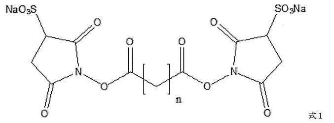
(式1中、nは自然数を表す)によって表される化合物、または式2
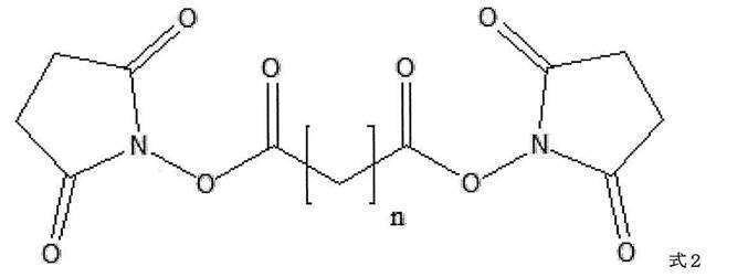
(式2中、nは自然数を表す)によって表される化合物が挙げられる。前記式1または式2によって表される化合物において、nは自然数であれば特に制限されないが、好ましくは1~100、より好ましくは3~45、さらに好ましくは3~6である。式1によって表される具体的な化合物としては、例えば、nが6である化合物、ビス(スルホスクシンイミジル)スベリン酸, 二ナトリウム塩(Bis(sulfosuccinimidyl)suberate, disodium salt(BS3))、またはnが3である化合物、ビス(スルホスクシンイミジル)グルタル酸, 二ナトリウム塩(Bis(sulfosuccinimidyl)glutarate, disodium salt(BS2G))などが挙げられる。
別のホモ二価性架橋剤としては、式3

(式3中、nは自然数を表す)によって表される化合物が挙げられる。前記式3によって表される化合物において、nは自然数であれば特に制限されないが、好ましくは3~25、より好ましくは4~24である。式3によって表される具体的な化合物としては、例えば、式4によって表される化合物
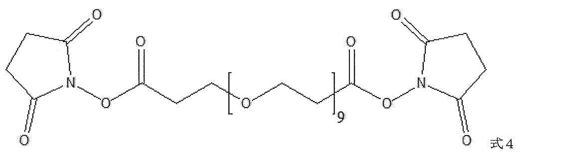
が挙げられる。
また、さらに別の二価性架橋剤としては、ヘテロ二価性架橋剤が挙げられる、ヘテロ二価性架橋剤は、2つの官能基が異なる官能基であれば特に制限されず、例えば、N-(6-マレイミドカプロイルオキシ)スクシンイミド(N-(6-Maleimidocaproyloxy)succinimide(EMCS))、N-(4-マレイミドブチリルオキシ)スクシンイミド(N-(4-Maleimidobutyryloxy)succinimide(GMBS))、N-(8-マレイミドカプリルオキシ)スクシンイミド(N-(8-Maleimidocapryloxy)succinimide(HMCS))、N-(11-マレイミドウンデカノイルオキシ)スクシンイミド(N-(11-Maleimidoundecanoyloxy)succinimide(KMUS))、N-(6-マレイミドカプロイルオキシ)スルホスクシンイミド, ナトリウム塩(N-(6-Maleimidocaproyloxy)sulfosuccinimide, sodium salt(Sulfo-EMCS))、N-(4-マレイミドブチリルオキシ)スルホスクシンイミド, ナトリウム塩(N-(4-Maleimidobutyryloxy)sulfosuccinimide, sodium salt(Sulfo-GMBS))、N-(8-マレイミドカプリルオキシ)スルホスクシンイミド, ナトリウム塩(N-(8-Maleimidocapryloxy)sulfosuccinimide, sodium salt(Sulfo-HMCS))、N-(11-マレイミドウンデカノイルオキシ)スルホスクシンイミド, ナトリウム塩(N-(11-Maleimidoundecanoyloxy)sulfosuccinimide, sodium salt(Sulfo-KMUS))、N-[(4-マレイミドメチル)シクロヘキシルカルボニルオキシ]スルホスクシンイミド, ナトリウム塩(N-[(4-Maleimidomethyl)cyclohexylcarbonyloxy]sulfosuccinimide, sodium salt(Sulfo-SMCC))、N-スクシンイミジル 3-(2-ピリジルジチオ)プロピオン酸(N-Succinimidyl 3-(2-pyridyldithio)propionate(SPDP))、N-{6-[3-(2-ピリジルジチオ)プロピオンアミド]ヘキサノイルオキシ}スルホスクシンイミド, ナトリウム塩(N-{6-[3-(2-Pyridyldithio)propionamido]hexanoyloxy}sulfosuccinimide, sodium salt(Sulfo-AC5-SPDP))、ジチオビス(スルホスクシンイミジル プロパン酸), 二ナトリウム塩(Dithiobis(sulfosuccinimidyl propionate), disodium salt(DTSSP))、ジチオビス(スクシンイミジル プロパン酸)(Dithiobis(succinimidyl propionate)(DSP))、ジチオビス(スクシンイミジル ウンデカン酸)(Dithiobis(succinimidyl undecanoate))、ジチオビス(スクシンイミジル オクタン酸)(Dithiobis(succinimidyl octanoate))、ジチオビス(スクシンイミジル ヘキサン酸)(Dithiobis(succinimidyl hexanoate))等が挙げられる。
また、ヘテロ二価性架橋剤の別の態様としては、前記のホモ二価性架橋剤の一端の官能基がアビジンに置換されたヘテロ二価性架橋剤が挙げられる。
本発明のセンサーチップ2は、支持体の表面に形成された結合部位最適配合層上に化学結合された二価性架橋剤を有することを特徴とする。本発明のセンサーチップ2の二価性架橋剤は、前記二価性架橋剤を溶媒に溶解させ、結合部位最適配合層の表面に適量スポットし、1時間室温で反応させることによって結合部位最適配合層に化学結合させることができる。スポットする際の二価性架橋剤の濃度は通常0.001 mM~100 mM、好ましくは0.01 mM~10 mMである。例えば、二価性架橋剤がビス(スルホスクシンイミジル)スベリン酸, 二ナトリウム塩(Bis(sulfosuccinimidyl)suberate, disodium salt(BS3))である場合、該化合物の2つのエステル結合は共に不安定な活性エステルであり、アミノ基と容易に反応し、活性エステルを構成するカルボキシ基と前記アミノ基の間でアミド結合を形成する。従って、二価性架橋剤を結合部位最適配合層と接触させることによって、結合部位最適配合層のアミノ基と二価性架橋剤の一方の活性エステルのカルボキシ基間にアミド結合を形成させ、二価性架橋剤を結合部位最適配合層上に化学結合させることができる。また、この際、二価性架橋剤の他方の活性エステルは未反応状態を維持している。
本発明のセンサーチップ2は、結合性分子を有する。本発明のセンサーチップ2に用いられる結合性分子は、本発明のセンサーチップ1に用いられる結合性分子と同様であってよい。
結合性分子は、本発明のセンサーチップ2に化学結合された二価性架橋剤と化学結合される。結合性分子と二価性架橋剤の化学結合は、公知の手段によって行われる。例えば、前記結合性分子を溶媒に溶解させ、結合部位最適配合層上に化学結合された二価性架橋剤上に適量スポットし、一晩室温で反応させることによって化学結合させることができる。スポットする際の結合性分子の濃度は、抗体の場合、通常0.1 mg/ml~1 mg/ml、好ましくは0.2 mg/ml、である。例えば、二価性架橋剤がビス(スルホスクシンイミジル)スベリン酸, 二ナトリウム塩(Bis(sulfosuccinimidyl)suberate, disodium salt(BS3))であり、結合性分子が抗体である場合、ビス(スルホスクシンイミジル)スベリン酸, 二ナトリウム塩(Bis(sulfosuccinimidyl)suberate, disodium salt(BS3)の未反応の活性エステルは、抗体のリジン残基のアミノ基と容易に反応し、未反応の活性エステルのカルボキシ基と前記アミノ基の間でアミド結合を形成する。従って、結合性分子を結合部位最適配合層上に化学結合された二価性架橋剤と接触させることによって、結合性分子のリジン残基のアミノ基とジエステルの未反応の活性エステルのカルボキシ基間にアミド結合を形成させ、結合性分子と二価性架橋剤とを化学結合させることができる。なお、後述の実施例では二価性架橋剤は、結合性分子である抗体およびゼラチン層のアミノ基と反応して架橋を形成するものであったが、結合性分子およびゼラチン層(または多糖類層)のカルボキシ基と反応して架橋を形成するものであっても良い。
以上の方法によって得られる本発明のセンサーチップ1またはセンサーチップ2では、支持体表面に形成されたゼラチン層または結合部位最適配合層が、結合性分子の細胞、微生物、該細胞もしくは該微生物が分泌する小胞またはウイルスに対する非特異的相互作用を抑制させる効果を有する。また、該結合性分子は、結合性分子同士が適度な距離を保ってゼラチン層または結合部位最適配合層上に化学結合されることよって、該結合性分子の細胞、微生物、該細胞もしくは該微生物が分泌する小胞またはウイルスの表面分子に対する検出感度が向上される。さらに、結合性分子をゼラチン層または結合部位最適配合層上に化学結合する際に、二価性架橋剤を介して結合性分子と該ゼラチンまたは該結合部位最適配合層を形成する材料を連結させることによって、結合性分子とゼラチン層または結合部位最適配合層間に二価性架橋剤の炭素鎖の長さの分だけ距離が生じ、結合性分子の空間的自由度が増すことも検出感度の向上に寄与できる。従って、本発明のセンサーチップ1またはセンサーチップ2は、細胞、微生物、該細胞もしくは該微生物が分泌する小胞またはウイルスの表面分子と結合性分子の相互作用検出用センサーチップとして提供することができる。
細胞としては、真核生物の細胞であれば特に制限はなく、動物細胞、植物細胞、真菌を含む概念として定義するが、その中でも動物細胞が好ましい。動物細胞としては、以下に制限されるものではないが、例えば、肝細胞、脾細胞、神経細胞、グリア細胞、膵β細胞、骨髄細胞、メサンギウム細胞、ランゲルハンス細胞、表皮細胞、上皮細胞、杯細胞、内皮細胞、平滑筋細胞、線維芽細胞、線維細胞、筋細胞、脂肪細胞、免疫細胞(例、マクロファージ、T細胞、B細胞、ナチュラルキラー細胞、肥満細胞、好中球、好塩基球、好酸球、単球)、巨核球、滑膜細胞、軟骨細胞、骨細胞、骨芽細胞、破骨細胞、乳腺細胞、肝細胞もしくは間質細胞、またはこれら細胞の前駆細胞、幹細胞もしくはガン細胞などが挙げられ、その中でも肥満細胞が好ましい。また、遺伝的に組み換えられた上記細胞もまた好ましく用いられる。
細胞において結合性分子と相互作用する表面分子としては、表面抗原であれば特に制限はないが、タンパク質、糖鎖、脂質、糖タンパク質、糖脂質などが挙げられる。その中でもタンパク質、糖鎖が好ましい。タンパク質としては、例えば、c-kitが挙げられる。糖鎖としては、例えば、糖鎖抗原125(CA125)、がん胎児性抗原(CEA)、シアリルTn抗原などが挙げられる。
微生物としては、真正細菌(以下、単なる「細菌」と記載する)、古細菌が挙げられるが、その中でも細菌が好ましい。細菌は、グリセロール3-リン酸の脂肪酸エステルより構成される細胞膜を持つ原核生物であれば特に制限はなく、グラム陰性細菌であってもよいし、グラム陽性細菌であってもよい。グラム陰性細菌としては、以下に制限されるものではないが、例えば、ナイセリア属(Neisseria)、ブランハメラ属(Branhamella)、ヘモフィルス属(Haemophilus)、ボルデテラ属(Bordetella)、エシェリキア属(Escherichia)、シトロバクター属(Citrobacter)、サルモネラ属(Salmonella)、シゲリア属(Shigella)、クレブシエラ属(Klebsiella)、エンテロバクター属(Enterobacter)、セラチア属(Serratia)、ハフニア属(Hafnia)、プロテウス属(Proteus)、モルガネラ属(Morganella)、プロビデンシア属(Providencia)、エルシニア属(Yersinia)、キャンピロバクター属(Campylobacter)、ビブリオ属(Vibrio)、エロモナス属(Aeromonas)、シュードモナス属(Pseudomonas)、キサントモナス属(Xanthomonas)、アシネトバクター属(Acinetobacter)、フラボバクテリウム属(Flavobacterium)、ブルセラ属(Brucella)、レジオネラ属(Legionella)、ベイロネラ属(Veillonella)、バクテロイデス属(Bacteroides)、フゾバクテリウム属(Fusobacterium)などに属する細菌が挙げられる。グラム陽性細菌としては、以下に制限されるものではないが、例えば、スタフィロコッカス属(Staphylococcus)、ストレプトコッカス属(Streptococcus)、エンテロコッカス属(Enterococcus)、コリネバクテリウム属(Corynebacterium)、バシラス属(Bacillus)、リステリア属(Listeria)、ペプトコッカス属(Peptococcus)、ペプトストレプトコッカス属(Peptostreptococcus)、クロストリジウム属(Clostridium)、ユーバクテリウム属(Eubacterium)、プロピオニバクテリウム属(Propionibacterium)、ラクトバシラス属(Lactobacillus)などに属する細菌が挙げられる。
微生物において結合性分子と相互作用する表面分子としては、細胞膜に露出している分子であれば特に制限はないが、例えば、O抗原、H抗原、K抗原などが挙げられる。
細胞もしくは微生物が分泌する小胞としては、最外膜として脂質二重膜を有する小胞であれば特に制限はなく、細胞外小胞(例えば、エクソソーム、膜小胞、エクソソーム様小胞等)が挙げられる。
小胞において結合性分子と相互作用する表面分子としては、小胞膜に露出している分子であれば特に制限はないが、例えば、テトラスパニン、CD9、CD63、CD81、主要組織適合遺伝子複合体(major histocompatibility complex)、c-Kit、フコース、シアル酸を含む糖鎖などが挙げられる。
ウイルスとしては、カプシドタイプのウイルスとエンベロープタイプのウイルスが挙げられる。
ウイルスにおいて結合性分子と相互作用する表面分子としては、ウイルス表面に露出している分子であれば特に制限はないが、例えば、インフルエンザウイルスのヘマグルチニンやノイラミニダーゼなどが挙げられる。
本発明はまた、本発明のセンサーチップ1またはセンサーチップ2を含む計測部(以下、本発明の計測部と記載する場合もある)と、本体部(以下、本発明の本体部と記載する場合もある)とを備えた、細胞、微生物、該細胞もしくは該微生物が分泌する小胞またはウイルスの表面分子と結合性分子の相互作用検出装置(以下、本発明の装置と記載する場合もある)を提供する。
本発明の計測部に含まれる本発明のセンサーチップ1またはセンサーチップ2に用いられる支持体は、好ましくは、プリズムと該プリズムの一側面に成膜される金属から構成される。プリズムと該プリズムの一側面に成膜される金属は、本発明のセンサーチップ1またはセンサーチップ2において記載した金属と同様であってよい。また、本発明の計測部は、細胞、微生物、該細胞もしくは該微生物が分泌する小胞またはウイルスの表面分子と結合性分子の相互作用によって本発明のセンサーチップ1またはセンサーチップ2に誘起されるSPR現象に伴う反射光を検出するセンサーをさらに備えてもよい。また、本発明の計測部は上記センサーによって検出した反射光の変化量を反射率(%)として計算し、出力する演算部をさらに備えてもよい。
本発明の本体部は、本発明の計測部によって検出された反射光の変化量に基づいて出力された信号を受け付けて作業者に表示することができる。また、本発明の本体部は計算された反射率の変化を演算部から受け付けて色調イメージに変換して出力する表示部をさらに備えてもよい。さらに、本発明の本体部は反射鏡の回転角度や照射光源の強度を調整する制御部をさらに備えてもよい。
本発明の装置において、演算部、表示部、制御部、それらの一部の機能は、本発明の計測部、本発明の本体部のいずれに備わるように構成しても良い。本発明の装置は、結合性分子が化学結合されていない箇所の色調変化も確認できるため、細胞、微生物、該細胞もしくは該微生物が分泌する小胞またはウイルスのセンサーチップへの非特異的相互作用の有無を確認することもできる。
より具体的な本発明の装置の一例として、マイクロアレイ型SPRi装置を図1に示す。
図1に示された装置1は、計測部2と、本体部3とから主として構成されている。計測部2は、センサーチップ21と、センサー22と、演算部23と、廃液用ボトル24と、バッファー用ボトル25と、送液ポンプ26と、脱気装置27と、被験試料挿入口28とから主として構成されている。本体部3は、表示部31と、制御部32とから主として構成されている。
センサーチップ21は、細胞、微生物、該細胞もしくは該微生物が分泌する小胞またはウイルスの表面分子と結合性分子の相互作用によって誘起されるSPR現象が生じさせるデバイスであり、本発明のセンサーチップに該当する。センサー22は、細胞、微生物、該細胞もしくは該微生物が分泌する小胞またはウイルスの表面分子と結合性分子の相互作用によって誘起されるSPR現象を反射光として検出する。具体的にはセンサーとしてCCDカメラが、各反射光検出位置における反射光強度を電流値と電圧値として検出する。演算部23は、上記センサー22によって検出した反射光の変化量を反射率(%)として計算し、出力する。廃液用ボトル24は、計測部2でセンサーチップ21を通過した被験試料やバッファーの廃棄用ボトルである。バッファー用ボトル25は、計測部2でセンサーチップ21を通過させるためのバッファー用ボトルである。送液ポンプ26は、バッファー用ボトルのバッファーを送液するためのポンプである。脱気装置27は、バッファーを脱気するための装置である。被験試料挿入口28は、細胞等の被験試料を計測部2に投入するための挿入口である。
表示部31は、演算部23によって計算された反射率の変化を受け付けて色調イメージに変換して出力する。制御部32は、計測部2に備え付けられた反射鏡の回転角度や照射光源の強度を調整する。
本発明はまた、以下の工程を含む、細胞、微生物、該細胞もしくは該微生物が分泌する小胞またはウイルスを特異的に検出する方法(本発明の検出方法1)を提供する。
(1)支持体上にゼラチン層を形成すること。
(2)結合性分子を該ゼラチン層上に直接的に又は間接的に化学結合させること。
(3)被験試料を該結合性分子に接触させること。
(4)該被験試料中の細胞、微生物、該細胞もしくは該微生物が分泌する小胞またはウイルスの表面分子と該結合性分子の相互作用を検出すること。
(1)支持体上にゼラチン層を形成すること。
(2)結合性分子を該ゼラチン層上に直接的に又は間接的に化学結合させること。
(3)被験試料を該結合性分子に接触させること。
(4)該被験試料中の細胞、微生物、該細胞もしくは該微生物が分泌する小胞またはウイルスの表面分子と該結合性分子の相互作用を検出すること。
本発明の検出方法1は、支持体上にゼラチン層を形成することを含む。本発明の検出方法1に用いられる支持体およびゼラチンは、本発明のセンサーチップ1に用いられる支持体およびゼラチンと同様であってよい。本発明の検出方法1において、支持体上にゼラチン層を形成する方法は、前記ゼラチンに加熱した蒸留水を加えてゼラチン溶液を作製した後、加熱した蒸留水で所望の濃度に希釈し、支持体の表面に適量塗沫し、室温で一晩静置することによって実施することができる。被覆する際のゼラチンの濃度は通常0.05 %~1%、好ましくは0.1 %~1 %である。支持体を被覆することによって形成されるゼラチン層の厚みは、通常100 nm未満である。
本発明の検出方法1は、結合性分子をゼラチン層上に化学結合させることを含む。本発明の検出方法1に用いられる結合性分子および結合性分子のゼラチン層上への直接的または間接的化学結合方法は、本発明のセンサーチップ1に用いられる結合性分子および化学結合方法と同様であってよい。
本発明の検出方法1は、被験試料を結合性分子に接触させることを含む。本発明の検出方法1に用いられる被験試料は、検出対象となる細胞、微生物、該細胞もしくは該微生物が分泌する小胞またはウイルスを含む試料であれば特に制限なく用いることができる。被験試料は、動物(好ましくは、哺乳動物)における体液(血液、唾液、涙液、尿、汗など)、細胞培養液、培養細胞、培養微生物、環境水、土壌、食料品などを遠心処理、フィルター処理、サイズ排除クロマトグラフィー等することによって調製される。これらの方法を用いることにより、被験試料中の細胞、微生物、該細胞もしくは該微生物が分泌する小胞またはウイルスの濃度がより高い被験試料を調製することができる。また、被験試料を結合性分子に接触させる際は、結合性分子に接触させる時間、温度、回数は当業者が適宜決定できるが、例えば、10分から2時間、室温、1~3回で接触させることができる。
本発明の検出方法1は、被験試料中の細胞、微生物、該細胞もしくは該微生物が分泌する小胞またはウイルスの表面分子と結合性分子の相互作用を検出することを含む。本発明の検出方法1において、表面分子と結合性分子の相互作用を検出する方法は、特に制限されるものではないが、例えば、免疫学的方法(ELISA法)または表面プラズモン共鳴(SPR)法が挙げられる。
本発明の検出方法1において、免疫学的方法による検出は、特に制限されるものではなく、被験試料中の細胞、微生物、該細胞もしくは該微生物が分泌する小胞またはウイルスと結合性分子からなる複合体を免疫学的手段により検出する方法であれば、いずれの測定法を用いてもよい。免疫学的方法としては、例えば、バッチ系、フロー系を問わず、ELISAによってゼラチン層上の結合性分子と相互作用した細胞、微生物、該細胞もしくは該微生物が分泌する小胞またはウイルスの表面分子を抗原とし、該抗原に対して抗体を相互作用させることで検出する手法であれば良い。
免疫学的方法において、抗原に対する抗体の相互作用は、標識剤を用いて検出することができる。標識剤としては、例えば、放射性同位元素、酵素、蛍光物質、発光物質などが用いられる。放射性同位元素としては、例えば、〔125I〕、〔131I〕、〔3H〕、〔14C〕などが用いられる。上記酵素としては、安定で比活性の大きなものが好ましく、例えば、β-ガラクトシダーゼ、β-グルコシダーゼ、アルカリフォスファターゼ、ペルオキシダーゼ、リンゴ酸脱水素酵素などが用いられる。蛍光物質としては、例えば、フルオレスカミン、フルオレッセンイソチオシアネートなどが用いられる。発光物質としては、例えば、ルミノール、ルミノール誘導体、ルシフェリン、ルシゲニンなどが用いられる。さらに、抗体と標識剤との結合にビオチン-アビジン系を用いることもできる。
ELISAのうち、サンドイッチ法の場合、ゼラチン層に化学結合された結合性分子に被験試料を反応させ(1次反応)、さらに該表面分子に対する標識二次抗体を反応させ(2次反応)た後、ゼラチン層上の標識剤の量(活性)を測定することにより、被験試料中の細胞、微生物、該細胞もしくは該微生物が分泌する小胞またはウイルスを特定することができる。1次反応と2次反応は逆の順序に行っても、また、同時に行なってもよいし時間をずらして行なってもよい。
本発明の検出方法1において、表面プラズモン共鳴法による検出は、ゼラチン層に被験試料を接触させた後、支持体に特定の波長の光を特定の角度から照射し、共鳴角度の変化を指標にして、ゼラチン層に化学結合した結合性分子への表面分子の相互作用の有無を判定する方法であれば、特に制限されない。
本発明はまた、以下の工程を含む、細胞、微生物、該細胞もしくは該微生物が分泌する小胞またはウイルスを特異的に検出する方法(本発明の検出方法2)を提供する。
(1)支持体上に結合部位最適配合層を形成すること。
(2)該結合部位最適配合層上に二価性架橋剤を化学結合させること。
(3)結合性分子と該二価性架橋剤とを化学結合させること。
(4)被験試料を該結合性分子に接触させること。
(5)該被験試料中の細胞、微生物、該細胞もしくは該微生物が分泌する小胞またはウイルスの表面分子と該結合性分子の相互作用を検出すること。
より具体的には、
(1’)支持体上にゼラチン層または多糖類層を形成すること。
(2’)該ゼラチン層または該多糖類層上に二価性架橋剤を化学結合させること。
(3’)結合性分子と該二価性架橋剤とを化学結合させること。
(4’)被験試料を該結合性分子に接触させること。
(5’)該被験試料中の細胞、微生物、該細胞もしくは該微生物が分泌する小胞またはウイルスの表面分子と該結合性分子の相互作用を検出すること。
(1)支持体上に結合部位最適配合層を形成すること。
(2)該結合部位最適配合層上に二価性架橋剤を化学結合させること。
(3)結合性分子と該二価性架橋剤とを化学結合させること。
(4)被験試料を該結合性分子に接触させること。
(5)該被験試料中の細胞、微生物、該細胞もしくは該微生物が分泌する小胞またはウイルスの表面分子と該結合性分子の相互作用を検出すること。
より具体的には、
(1’)支持体上にゼラチン層または多糖類層を形成すること。
(2’)該ゼラチン層または該多糖類層上に二価性架橋剤を化学結合させること。
(3’)結合性分子と該二価性架橋剤とを化学結合させること。
(4’)被験試料を該結合性分子に接触させること。
(5’)該被験試料中の細胞、微生物、該細胞もしくは該微生物が分泌する小胞またはウイルスの表面分子と該結合性分子の相互作用を検出すること。
本発明の検出方法2は、支持体上に結合部位最適配合層、好ましくはゼラチン層または多糖類層を形成することを含む。本発明の検出方法1に用いられる支持体、結合部位最適配合層を形成する材料、ゼラチンおよび多糖類は、本発明のセンサーチップ2に用いられる支持体、結合部位最適配合層を形成する材料、ゼラチンおよび多糖類と同様であってよい。本発明の検出方法2において、支持体上にゼラチン層を形成する方法は、本発明の検出方法1に用いられる方法と同様であってよい。本発明の検出方法2において、支持体上に多糖類層を形成する方法は、前記多糖類に加熱した蒸留水を加えて多糖類溶液を作製した後、加熱した蒸留水で所望の濃度に希釈し、支持体の表面に適量塗布し、静置することによって、支持体上に多糖類層を形成させることができる。
本発明の検出方法2は、結合部位最適配合層上に二価性架橋剤を化学結合させることを含む。本発明の検出方法2に用いられる二価性架橋剤は、本発明のセンサーチップ2に用いられる二価性架橋剤と同様であってよい。本発明の検出方法2において、結合部位最適配合層上に二価性架橋剤を化学結合させる方法は、前記二価性架橋剤を溶媒に溶解させ、結合部位最適配合層の表面に適量スポットし、1時間室温で反応させることによって実施することができる。スポットする際の二価性架橋剤の濃度は通常0.001 mM~100 mM、好ましくは0.01 mM~10 mMである。
本発明の検出方法2は、結合性分子と二価性架橋剤とを化学結合させることを含む。本発明の検出方法2に用いられる結合性分子は、本発明のセンサーチップ2に用いられる結合性分子と同様であってよい。本発明の検出方法2において、結合性分子は、結合部位最適配合層上に化学結合された二価性架橋剤と化学結合される。結合性分子と二価性架橋剤との化学結合は、公知の手段によって行われる。例えば、前記結合性分子を溶媒に溶解させ、二価性架橋剤上に適量スポットし、一晩室温で反応させることによって化学結合させることができる。スポットする際の結合性分子の濃度は、抗体の場合、通常0.1 mg/ml~1mg/ml、好ましくは0.2 mg/mlである。
本発明の検出方法2は、被験試料を該結合性分子に接触させることを含む。本発明の検出方法2に用いられる被験試料は、本発明の検出方法1に用いられる被験試料と同様であってよい。被験試料を結合性分子に接触させる際は、結合性分子に接触させる時間、温度、回数は当業者が適宜決定できるが、例えば、10分から2時間、室温、1~3回で接触させることができる。
本発明の検出方法2は、該被験試料中の細胞、微生物、該細胞もしくは該微生物が分泌する小胞またはウイルスの表面分子と該結合性分子の相互作用を検出することを含む。本発明の検出方法2において、表面分子と結合性分子の相互作用を検出する方法は、本発明の検出方法1に用いられる方法と同様であってよい。
さらに、本発明の検出方法1または検出方法2では、2つ以上の異なる結合性分子を本発明のセンサーチップ上で異なる配置になるようにゼラチン層上または二価性架橋剤に化学結合させることによって、各結合性分子が細胞、微生物、該細胞もしくは該微生物が分泌する小胞またはウイルスに存在する複数の表面分子と相互作用するかどうかを同時に検証することができる。例えば、少なくとも1つのスポットに対して抗体を化学結合させ、別の少なくとも1つのスポットにレクチンを化学結合させた本発明のセンサーチップに、被験試料を接触させて該被験試料中の細胞、微生物、該細胞もしくは該微生物が分泌する小胞またはウイルスの表面抗原と前記抗体の相互作用、及び糖鎖と前記レクチンの相互作用を検出することもできる。従って、本発明はまた、以下の工程を含む、細胞、微生物、該細胞もしくは該微生物が分泌する小胞またはウイルスを特異的に検出する方法を提供する。
(1)支持体上にゼラチン層を形成すること。
(2)2つ以上の異なる結合性分子を互いに異なる配置になるように該ゼラチン層上に直接的に又は間接的に化学結合させること。
(3)被験試料を該2つ以上の異なる結合性分子に接触させること。
(4)該被験試料中の細胞、微生物、該細胞もしくは該微生物が分泌する小胞またはウイルスの2つ以上の異なる表面分子と該2つ以上の異なる結合性分子の相互作用を検出すること。
本発明はまた、以下の工程を含む、細胞、微生物、該細胞もしくは該微生物が分泌する小胞またはウイルスを特異的に検出する方法を提供する。
(1)支持体上に結合部位最適配合層を形成すること。
(2)該結合部位最適配合層上に二価性架橋剤を化学結合させること。
(3)2つ以上の異なる結合性分子を支持体上で互いに異なる配置になるように該二価性架橋剤と化学結合させること。
(4)被験試料を該2つ以上の異なる結合性分子に接触させること。
(5)該被験試料中の細胞、微生物、該細胞もしくは該微生物が分泌する小胞またはウイルスの2つ以上の異なる表面分子と該2つ以上の異なる結合性分子の相互作用を検出すること。
より具体的には、
(1’)支持体上にゼラチン層または多糖類層を形成すること。
(2’)該ゼラチン層または該多糖類層上に二価性架橋剤を化学結合させること。
(3’)2つ以上の異なる結合性分子を支持体上で互いに異なる配置になるように該二価性架橋剤と化学結合させること。
(4’)被験試料を該結合性分子に接触させること。
(5’)該被験試料中の細胞、微生物、該細胞もしくは該微生物が分泌する小胞またはウイルスの2つ以上の異なる表面分子と該2つ以上の異なる結合性分子の相互作用を検出すること。
本方法により、例えば、被験試料の細胞、微生物、該細胞もしくは該微生物が分泌する小胞またはウイルスの2つ以上の異なる表面分子を同時に検出することができるため、被験試料を迅速に診断することができる。具体的にはがん細胞の診断において、細胞膜表面の糖鎖と表面抗原を同時に検出することができるので、その診断を迅速に行うことができるようになる。
(1)支持体上にゼラチン層を形成すること。
(2)2つ以上の異なる結合性分子を互いに異なる配置になるように該ゼラチン層上に直接的に又は間接的に化学結合させること。
(3)被験試料を該2つ以上の異なる結合性分子に接触させること。
(4)該被験試料中の細胞、微生物、該細胞もしくは該微生物が分泌する小胞またはウイルスの2つ以上の異なる表面分子と該2つ以上の異なる結合性分子の相互作用を検出すること。
本発明はまた、以下の工程を含む、細胞、微生物、該細胞もしくは該微生物が分泌する小胞またはウイルスを特異的に検出する方法を提供する。
(1)支持体上に結合部位最適配合層を形成すること。
(2)該結合部位最適配合層上に二価性架橋剤を化学結合させること。
(3)2つ以上の異なる結合性分子を支持体上で互いに異なる配置になるように該二価性架橋剤と化学結合させること。
(4)被験試料を該2つ以上の異なる結合性分子に接触させること。
(5)該被験試料中の細胞、微生物、該細胞もしくは該微生物が分泌する小胞またはウイルスの2つ以上の異なる表面分子と該2つ以上の異なる結合性分子の相互作用を検出すること。
より具体的には、
(1’)支持体上にゼラチン層または多糖類層を形成すること。
(2’)該ゼラチン層または該多糖類層上に二価性架橋剤を化学結合させること。
(3’)2つ以上の異なる結合性分子を支持体上で互いに異なる配置になるように該二価性架橋剤と化学結合させること。
(4’)被験試料を該結合性分子に接触させること。
(5’)該被験試料中の細胞、微生物、該細胞もしくは該微生物が分泌する小胞またはウイルスの2つ以上の異なる表面分子と該2つ以上の異なる結合性分子の相互作用を検出すること。
本方法により、例えば、被験試料の細胞、微生物、該細胞もしくは該微生物が分泌する小胞またはウイルスの2つ以上の異なる表面分子を同時に検出することができるため、被験試料を迅速に診断することができる。具体的にはがん細胞の診断において、細胞膜表面の糖鎖と表面抗原を同時に検出することができるので、その診断を迅速に行うことができるようになる。
以下において、実施例により本発明をより具体的に説明するが、この発明はこれらに限定されるものではない。
表面プラズモン共鳴(SPR)による細胞検出センサーセンサーの構築
表面プラズモン共鳴(SPR)による細胞検出センサーセンサーは、マイクロアレイ型SPRi装置((株)堀場製作所:OpenPlex)と装置専用のセンサーチップ((株)堀場製作所:CS-HD;スクシンイミドで活性化されたカルボキシ基を化学結合させたセンサーチップ)を用いて構築した。構築したセンサーは、チップ表面の抗体への細胞の表面分子の相互作用によって誘起されるSPR現象に伴う反射光の変化量を反射率(%)として、3秒毎に測定することができる。同時に、SPRの反射率変化をスポットイメージとして観察することができる。またチップは12 mm×23 mmの表面積があるので、化学結合させるためのリガンド溶液のスポット径(スポット量)を調整することで多数のスポットを並列できる特徴がある。
表面プラズモン共鳴(SPR)による細胞検出センサーセンサーは、マイクロアレイ型SPRi装置((株)堀場製作所:OpenPlex)と装置専用のセンサーチップ((株)堀場製作所:CS-HD;スクシンイミドで活性化されたカルボキシ基を化学結合させたセンサーチップ)を用いて構築した。構築したセンサーは、チップ表面の抗体への細胞の表面分子の相互作用によって誘起されるSPR現象に伴う反射光の変化量を反射率(%)として、3秒毎に測定することができる。同時に、SPRの反射率変化をスポットイメージとして観察することができる。またチップは12 mm×23 mmの表面積があるので、化学結合させるためのリガンド溶液のスポット径(スポット量)を調整することで多数のスポットを並列できる特徴がある。
比較例 ゼラチン存在下で抗体を化学結合したセンサーチップによる細胞検出(非特異的相互作用抑制法)
抗体をチップに化学結合させた後、一般的なブロッキング剤であるウシ血清アルブミン(BSA)でチップをブロッキングし、細胞をチップに接触させた場合、抗原に特異的な抗体および陰性抗体の間に反射率の差がほとんど生じず、細胞結合検出が困難になる。この理由は、BSAは疎水性が高いため、水素結合による分子近傍の結合水は少なくなる。そのため細胞膜表面に多数存在するタンパク質の疎水性部分がBSAと疎水的に結合しやすくなり非特異的な相互作用を生じると考えられる。この問題を解決するため、BSAより疎水性が低く、親水性が高いゼラチンを抗体に混ぜて使用することで、細胞と抗体間の非特異的相互作用を抑制し細胞検出が可能になる。しかし、細胞や組換型細胞を検出するには細胞上にわずかに発現している表面抗原を検出する必要がある。そこで本比較例では、上記方法による細胞濃度依存性試験を実施した。
細胞はマウス骨髄から組換型IL-3で誘導したマウス骨髄由来肥満細胞(BMMC)を使用した。BMMC表面上のc-Kit発現は既知であることから、BMMC検出には、表面抗原c-Kitに対する抗体(抗c-Kit抗体; R&D systems Inc., AF1356)を用いた。陰性抗体としてはヤギ抗体(ヤギ抗体;R&D systems Inc., AB108C)を用いた。抗体と細胞間の非特異的相互作用抑制効果を有するゼラチンと抗体を混合し、スポッターを用いてチップ表面に10 nLスポットし、16時間静置することで化学結合させた。ダルベコのPBS(-)(以下、PBSと略記)でチップ表面を洗浄し、1%ゼラチンを溶解したPBSをチップ表面に満たして1時間室温で静置し、ブロッキングした。ブロッキングしたチップを、PBSで3回洗浄後、装置に装着した。チップ表面へのバッファーまたはBMMCの接触は、Flow-cell(図2)を介して行った。Flow-cellは、Gasket全体がチップに完全に覆われるような位置(図3)で、チップに接触固定した。また、Flow-cellの平面のうち、Gasketの枠に囲まれた平面は、Gasketの枠の周囲の平面よりも、80 μm凹んでいる。結果的に、Flow-cellと接触したチップは、Flow-cellのGasketの枠に囲まれた平面とチップ表面の間に幅80 μmの空間的隙間が生じる。従って、Flow-cellにFittingを介して連結された片方のポリ塩化ビニルチューブ(内径380 μm)から送液されたバッファー等は、幅80 μmの空間的隙間を満たすことによってチップ表面に接触し、もう片方のポリ塩化ビニルチューブから排出される。チップを装着した装置には、ランニングバッファーとして 0.1%ゼラチンと0.02%Tween20を含んだPBS(バッファーA)を25 μL/分の流速で送液し、チップ表面をコンディショニングし、安定化した時点の反射率を0%とした。次に、BMMCを1×104、1×105、1×106 cells/ml濃度になるようにバッファーAに懸濁した。各濃度の細胞を装置に注入後、480秒間送液し、その後ただちにバッファーAのみを480秒間送液した。その結果、1×104、1×105 cells/ml濃度において陽性抗体の抗c-Kit抗体とBMMC間の特異的な相互作用を検出できず、1×106 cells/ml濃度でわずかに特異的な相互作用を検出できた(図4)。一方、BMMC濃度1×104、1×105、1×106cells/mlにおいて、スポットが白くならず、ゼラチン効果によって陰性抗体のヤギ抗体とBMMC間の非特異的相互作用は抑制された(図4)。以上の結果は、本比較例の方法では、被験試料中の細胞数を比較的多くしなければ、BMMC上のc-Kit抗原と抗体の相互作用をほとんど検出できないことを示している。従って、抗体を用いた細胞の表面抗原の検出系を確立するためには、抗体と細胞間の非特異的相互作用を抑制し、かつ抗体と細胞の表面抗原の相互作用を高感度に検出するための方法を見出す必要があることが判明した。
抗体をチップに化学結合させた後、一般的なブロッキング剤であるウシ血清アルブミン(BSA)でチップをブロッキングし、細胞をチップに接触させた場合、抗原に特異的な抗体および陰性抗体の間に反射率の差がほとんど生じず、細胞結合検出が困難になる。この理由は、BSAは疎水性が高いため、水素結合による分子近傍の結合水は少なくなる。そのため細胞膜表面に多数存在するタンパク質の疎水性部分がBSAと疎水的に結合しやすくなり非特異的な相互作用を生じると考えられる。この問題を解決するため、BSAより疎水性が低く、親水性が高いゼラチンを抗体に混ぜて使用することで、細胞と抗体間の非特異的相互作用を抑制し細胞検出が可能になる。しかし、細胞や組換型細胞を検出するには細胞上にわずかに発現している表面抗原を検出する必要がある。そこで本比較例では、上記方法による細胞濃度依存性試験を実施した。
細胞はマウス骨髄から組換型IL-3で誘導したマウス骨髄由来肥満細胞(BMMC)を使用した。BMMC表面上のc-Kit発現は既知であることから、BMMC検出には、表面抗原c-Kitに対する抗体(抗c-Kit抗体; R&D systems Inc., AF1356)を用いた。陰性抗体としてはヤギ抗体(ヤギ抗体;R&D systems Inc., AB108C)を用いた。抗体と細胞間の非特異的相互作用抑制効果を有するゼラチンと抗体を混合し、スポッターを用いてチップ表面に10 nLスポットし、16時間静置することで化学結合させた。ダルベコのPBS(-)(以下、PBSと略記)でチップ表面を洗浄し、1%ゼラチンを溶解したPBSをチップ表面に満たして1時間室温で静置し、ブロッキングした。ブロッキングしたチップを、PBSで3回洗浄後、装置に装着した。チップ表面へのバッファーまたはBMMCの接触は、Flow-cell(図2)を介して行った。Flow-cellは、Gasket全体がチップに完全に覆われるような位置(図3)で、チップに接触固定した。また、Flow-cellの平面のうち、Gasketの枠に囲まれた平面は、Gasketの枠の周囲の平面よりも、80 μm凹んでいる。結果的に、Flow-cellと接触したチップは、Flow-cellのGasketの枠に囲まれた平面とチップ表面の間に幅80 μmの空間的隙間が生じる。従って、Flow-cellにFittingを介して連結された片方のポリ塩化ビニルチューブ(内径380 μm)から送液されたバッファー等は、幅80 μmの空間的隙間を満たすことによってチップ表面に接触し、もう片方のポリ塩化ビニルチューブから排出される。チップを装着した装置には、ランニングバッファーとして 0.1%ゼラチンと0.02%Tween20を含んだPBS(バッファーA)を25 μL/分の流速で送液し、チップ表面をコンディショニングし、安定化した時点の反射率を0%とした。次に、BMMCを1×104、1×105、1×106 cells/ml濃度になるようにバッファーAに懸濁した。各濃度の細胞を装置に注入後、480秒間送液し、その後ただちにバッファーAのみを480秒間送液した。その結果、1×104、1×105 cells/ml濃度において陽性抗体の抗c-Kit抗体とBMMC間の特異的な相互作用を検出できず、1×106 cells/ml濃度でわずかに特異的な相互作用を検出できた(図4)。一方、BMMC濃度1×104、1×105、1×106cells/mlにおいて、スポットが白くならず、ゼラチン効果によって陰性抗体のヤギ抗体とBMMC間の非特異的相互作用は抑制された(図4)。以上の結果は、本比較例の方法では、被験試料中の細胞数を比較的多くしなければ、BMMC上のc-Kit抗原と抗体の相互作用をほとんど検出できないことを示している。従って、抗体を用いた細胞の表面抗原の検出系を確立するためには、抗体と細胞間の非特異的相互作用を抑制し、かつ抗体と細胞の表面抗原の相互作用を高感度に検出するための方法を見出す必要があることが判明した。
実施例 予めゼラチンでブロッキングし、さらにBS3を介して抗体を化学結合させたセンサーチップよる細胞検出(新規高感度検出法)
ゼラチンは、細胞外マトリックス由来であるため、細胞との相性が優れている。そのことから、ゼラチンをブロッキング剤として使用することにより、チップ表面の親水性すなわち水素結合による分子近傍の結合水が増加し、細胞膜表面に多数存在するタンパク質の疎水性部分との結合が抑制され、細胞と抗体間の非特異的相互作用が抑制される。この特性をもつチップを作製するため、その表面をゼラチンで完全に覆うことによってブロッキングした。しかし、チップは完全にゼラチンでブロッキングされているため、このままでは細胞検出に用いるための抗体をチップに結合させることができない。これを解決するために、二価性反応性試薬としてBis(sulfosuccinimidyl)suberate, disodium salt(以下、BS3)(図5)を使用した。BS3は、スベリン酸とサルフォ-N-ヒドロキシコハク酸イミドのジエステルであり、スベリン酸のカルボキシ基とタンパク質またはポリペプチドを構成するリジン残基のアミノ基と容易にアミド結合を形成する性質を有する。従って、BS3は、ゼラチンを構成する全アミノ酸に約3%存在するリジン残基と抗体のリジン残基を連結させるリンカーの役割を果たすことができる。このチップを用いて、比較例と同様に、細胞濃度依存性試験を実施した。
細胞および抗体は、比較例と同様のものを使用した。チップ作製は、ゼラチンを化学的に結合させるために、PBSに溶解させた0.1% ゼラチンをチップ上に500 μl塗沫後、一晩室温にて反応させチップ表面をブロッキングした。反応後、チップを蒸留水で3回洗浄しブロアでチップ表面に残った水分を完全に吹き飛ばした。次に、PBSで溶解させた二価性反応性試薬1 mM BS3をスポッターでチップ表面にスポットし、1時間室温で反応させた。反応後、チップを水で3回洗浄し、ブロアでチップ表面に残った水分を完全に吹き飛ばした。次に二価性反応性試薬をスポットした場所と同位置に抗体をスポッターでスポットし、一晩室温にて反応させた。反応後、チップをPBSで3回洗浄し、ブロアでチップ表面に残った水分を完全に飛ばし、試験に使用した。チップを装着した装置には、ランニングバッファーとしてバッファーAを25 μL/分の流速で送液し、チップ表面をコンディショニングし、安定化した時点の反射率を0%とした。次に、BMMCを1×104、1×105、1×106 cells/ml濃度になるようにバッファーAに懸濁した。各濃度の細胞を装置に注入後、480秒間送液し、その後ただちにバッファーAのみで480秒間洗浄した。その結果、1×104、1×105、1×106 cells/ml濃度において陽性抗体の抗c-Kit抗体とBMMC間の特異的な相互作用を検出し、かつ、BMMC濃度1×104、1×105、1×106cells/mlにおいて、陰性抗体のスポットが白くならないため、ゼラチン効果によって非特異的相互作用が抑制されたことが分かった(図6)。また、比較例との比較から、新規高感度検出法は細胞濃度において100倍感度が増加した。このことから、新規高感度検出法は細胞の高感度検出にも成功したことも示している。ゼラチンに存在するリジン残基の割合が約3%であるという点は、BS3を介して連結される抗体がチップ上で過密にならないような距離を保つように維持し、抗体が抗原を高感度に検出することを可能にさせたと考えられる。さらに、二価性反応性試薬によって抗体とゼラチンを連結することによって、二価性反応性試薬の炭素鎖の長さの分だけチップと抗体に距離が生じ、抗体の自由度が増すことも高感度検出に寄与したと考えられる。
ゼラチンは、細胞外マトリックス由来であるため、細胞との相性が優れている。そのことから、ゼラチンをブロッキング剤として使用することにより、チップ表面の親水性すなわち水素結合による分子近傍の結合水が増加し、細胞膜表面に多数存在するタンパク質の疎水性部分との結合が抑制され、細胞と抗体間の非特異的相互作用が抑制される。この特性をもつチップを作製するため、その表面をゼラチンで完全に覆うことによってブロッキングした。しかし、チップは完全にゼラチンでブロッキングされているため、このままでは細胞検出に用いるための抗体をチップに結合させることができない。これを解決するために、二価性反応性試薬としてBis(sulfosuccinimidyl)suberate, disodium salt(以下、BS3)(図5)を使用した。BS3は、スベリン酸とサルフォ-N-ヒドロキシコハク酸イミドのジエステルであり、スベリン酸のカルボキシ基とタンパク質またはポリペプチドを構成するリジン残基のアミノ基と容易にアミド結合を形成する性質を有する。従って、BS3は、ゼラチンを構成する全アミノ酸に約3%存在するリジン残基と抗体のリジン残基を連結させるリンカーの役割を果たすことができる。このチップを用いて、比較例と同様に、細胞濃度依存性試験を実施した。
細胞および抗体は、比較例と同様のものを使用した。チップ作製は、ゼラチンを化学的に結合させるために、PBSに溶解させた0.1% ゼラチンをチップ上に500 μl塗沫後、一晩室温にて反応させチップ表面をブロッキングした。反応後、チップを蒸留水で3回洗浄しブロアでチップ表面に残った水分を完全に吹き飛ばした。次に、PBSで溶解させた二価性反応性試薬1 mM BS3をスポッターでチップ表面にスポットし、1時間室温で反応させた。反応後、チップを水で3回洗浄し、ブロアでチップ表面に残った水分を完全に吹き飛ばした。次に二価性反応性試薬をスポットした場所と同位置に抗体をスポッターでスポットし、一晩室温にて反応させた。反応後、チップをPBSで3回洗浄し、ブロアでチップ表面に残った水分を完全に飛ばし、試験に使用した。チップを装着した装置には、ランニングバッファーとしてバッファーAを25 μL/分の流速で送液し、チップ表面をコンディショニングし、安定化した時点の反射率を0%とした。次に、BMMCを1×104、1×105、1×106 cells/ml濃度になるようにバッファーAに懸濁した。各濃度の細胞を装置に注入後、480秒間送液し、その後ただちにバッファーAのみで480秒間洗浄した。その結果、1×104、1×105、1×106 cells/ml濃度において陽性抗体の抗c-Kit抗体とBMMC間の特異的な相互作用を検出し、かつ、BMMC濃度1×104、1×105、1×106cells/mlにおいて、陰性抗体のスポットが白くならないため、ゼラチン効果によって非特異的相互作用が抑制されたことが分かった(図6)。また、比較例との比較から、新規高感度検出法は細胞濃度において100倍感度が増加した。このことから、新規高感度検出法は細胞の高感度検出にも成功したことも示している。ゼラチンに存在するリジン残基の割合が約3%であるという点は、BS3を介して連結される抗体がチップ上で過密にならないような距離を保つように維持し、抗体が抗原を高感度に検出することを可能にさせたと考えられる。さらに、二価性反応性試薬によって抗体とゼラチンを連結することによって、二価性反応性試薬の炭素鎖の長さの分だけチップと抗体に距離が生じ、抗体の自由度が増すことも高感度検出に寄与したと考えられる。
本発明のセンサーチップを用いることによって、結合性分子の細胞、微生物、該細胞もしくは該微生物が分泌する小胞またはウイルスに対する非特異的相互作用を抑制し、かつ該結合性分子の細胞、微生物、該細胞もしくは該微生物が分泌する小胞またはウイルスの表面分子に対する検出感度を向上できる。本効果によって、癌細胞膜表面に発現する腫瘍マーカーの臨床検査のために、被験者から大量の細胞を取得する必要がなく、身体的負担の軽減に資する。また、異種膜タンパク質を一過性発現させた細胞を用いた実験系の確立の際にも、予め準備しなければならない一過性発現細胞の細胞数を減らすことができる。本出願は、日本で出願された特願2018-123270(出願日:平成30年6月28日)および特願2018-133710(出願日:平成30年7月13日)を基礎としており、その内容はすべて本明細書に包含されるものとする。
Claims (14)
- 支持体を有し、
該支持体上にゼラチン層または多糖類層を有し、
該ゼラチン層または該多糖類層上に化学結合した二価性架橋剤を有し、
該二価性架橋剤と化学結合した結合性分子を有することを特徴とする、センサーチップ。 - 前記多糖類が、酸性多糖類、塩基性多糖類またはそれらの混合物である、請求項1に記載のセンサーチップ。
- 前記二価性架橋剤が、式1
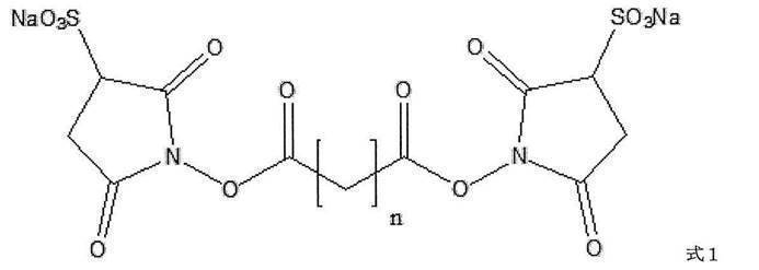
(式1中、nは自然数を表す)によって表される化合物、または式2

(式2中、nは自然数を表す)によって表される化合物
である、請求項1または2に記載のセンサーチップ。 - 前記式1または式2によって表される化合物において、nが1~100である、請求項3に記載のセンサーチップ。
- 前記二価性架橋剤が、ビス(スルホスクシンイミジル)スベリン酸, 二ナトリウム塩(Bis(sulfosuccinimidyl)suberate, disodium salt(BS3))またはビス(スルホスクシンイミジル)グルタル酸, 二ナトリウム塩(Bis(sulfosuccinimidyl)glutarate, disodium salt(BS2G))である、請求項3に記載のセンサーチップ。
- 前記二価性架橋剤が、式3
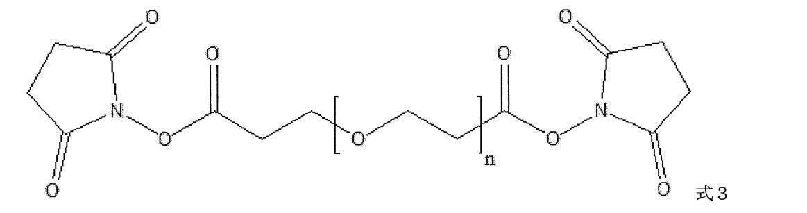
(式3中、nは自然数を表す)によって表される化合物である、請求項1または2に記載のセンサーチップ。 - 前記式3によって表される化合物において、nが3~25である、請求項6に記載のセンサーチップ。
- 前記二価性架橋剤が、N-(6-マレイミドカプロイルオキシ)スクシンイミド(N-(6-Maleimidocaproyloxy)succinimide(EMCS))、N-(4-マレイミドブチリルオキシ)スクシンイミド(N-(4-Maleimidobutyryloxy)succinimide(GMBS))、N-(8-マレイミドカプリルオキシ)スクシンイミド(N-(8-Maleimidocapryloxy)succinimide(HMCS))、N-(11-マレイミドウンデカノイルオキシ)スクシンイミド(N-(11-Maleimidoundecanoyloxy)succinimide(KMUS))、N-(6-マレイミドカプロイルオキシ)スルホスクシンイミド, ナトリウム塩(N-(6-Maleimidocaproyloxy)sulfosuccinimide, sodium salt(Sulfo-EMCS))、N-(4-マレイミドブチリルオキシ)スルホスクシンイミド, ナトリウム塩(N-(4-Maleimidobutyryloxy)sulfosuccinimide, sodium salt(Sulfo-GMBS))、N-(8-マレイミドカプリルオキシ)スルホスクシンイミド, ナトリウム塩(N-(8-Maleimidocapryloxy)sulfosuccinimide, sodium salt(Sulfo-HMCS))、N-(11-マレイミドウンデカノイルオキシ)スルホスクシンイミド, ナトリウム塩(N-(11-Maleimidoundecanoyloxy)sulfosuccinimide, sodium salt(Sulfo-KMUS))、N-[(4-マレイミドメチル)シクロヘキシルカルボニルオキシ]スルホスクシンイミド, ナトリウム塩(N-[(4-Maleimidomethyl)cyclohexylcarbonyloxy]sulfosuccinimide, sodium salt(Sulfo-SMCC))、N-スクシンイミジル 3-(2-ピリジルジチオ)プロピオン酸(N-Succinimidyl 3-(2-pyridyldithio)propionate(SPDP))、N-{6-[3-(2-ピリジルジチオ)プロピオンアミド]ヘキサノイルオキシ}スルホスクシンイミド, ナトリウム塩(N-{6-[3-(2-Pyridyldithio)propionamido]hexanoyloxy}sulfosuccinimide, sodium salt(Sulfo-AC5-SPDP))、ジチオビス(スルホスクシンイミジル プロパン酸), 二ナトリウム塩(Dithiobis(sulfosuccinimidyl propionate), disodium salt(DTSSP))、ジチオビス(スクシンイミジル プロパン酸)(Dithiobis(succinimidyl propionate)(DSP))、ジチオビス(スクシンイミジル ウンデカン酸)(Dithiobis(succinimidyl undecanoate))、ジチオビス(スクシンイミジル オクタン酸)(Dithiobis(succinimidyl octanoate))、およびジチオビス(スクシンイミジル ヘキサン酸)(Dithiobis(succinimidyl hexanoate))からなる群から選択される、請求項1または2に記載のセンサーチップ。
- 前記結合性分子が、抗体、細胞接着因子、レクチンおよび核酸アプタマーからなる群から選択される、請求項1~8のいずれか1項に記載のセンサーチップ。
- 前記支持体が、金属薄膜に覆われたプリズムである、請求項1~9のいずれか1項に記載のセンサーチップ。
- 細胞、微生物、該細胞もしくは該微生物が分泌する小胞またはウイルスの表面分子と結合性分子の相互作用検出用センサーチップである、請求項1~10のいずれか1項に記載のセンサーチップ。
- 支持体を有し、
該支持体上にゼラチン層を有し、
該ゼラチン層上に直接的に又は間接的に化学結合した結合性分子を有することを特徴とする、センサーチップ。 - 請求項1~12のいずれか1項に記載のセンサーチップを含む計測部と、本体部とを備えた、細胞、微生物、該細胞もしくは該微生物が分泌する小胞またはウイルスの表面分子と結合性分子の相互作用検出装置。
- 以下の工程を含む、細胞、微生物、該細胞もしくは該微生物が分泌する小胞またはウイルスを特異的に検出する方法:
(1)支持体上にゼラチン層を形成すること、
(2)結合性分子を該ゼラチン層上に直接的に又は間接的に化学結合させること、
(3)被験試料を該結合性分子に接触させること、
(4)該被験試料中の細胞、微生物、該細胞もしくは該微生物が分泌する小胞またはウイルスの表面分子と該結合性分子の相互作用を検出すること。
Priority Applications (1)
| Application Number | Priority Date | Filing Date | Title |
|---|---|---|---|
| JP2020527622A JPWO2020004535A1 (ja) | 2018-06-28 | 2019-06-27 | 細胞等の高感度検出用センサーチップ |
Applications Claiming Priority (4)
| Application Number | Priority Date | Filing Date | Title |
|---|---|---|---|
| JP2018-123270 | 2018-06-28 | ||
| JP2018123270 | 2018-06-28 | ||
| JP2018133710 | 2018-07-13 | ||
| JP2018-133710 | 2018-07-13 |
Publications (1)
| Publication Number | Publication Date |
|---|---|
| WO2020004535A1 true WO2020004535A1 (ja) | 2020-01-02 |
Family
ID=68985041
Family Applications (1)
| Application Number | Title | Priority Date | Filing Date |
|---|---|---|---|
| PCT/JP2019/025553 Ceased WO2020004535A1 (ja) | 2018-06-28 | 2019-06-27 | 細胞等の高感度検出用センサーチップ |
Country Status (2)
| Country | Link |
|---|---|
| JP (1) | JPWO2020004535A1 (ja) |
| WO (1) | WO2020004535A1 (ja) |
Citations (8)
| Publication number | Priority date | Publication date | Assignee | Title |
|---|---|---|---|---|
| JP2003240781A (ja) * | 2001-11-30 | 2003-08-27 | Eastman Kodak Co | タンパク質アレイを作製するためのゼラチンベースの基体およびその製造方法 |
| JP2008249502A (ja) * | 2007-03-30 | 2008-10-16 | Fujifilm Corp | 生理活性物質を固定化した固体基板 |
| JP2010286466A (ja) * | 2008-10-24 | 2010-12-24 | Fujifilm Corp | 断片化抗体固定化担体及びその製造方法 |
| JP2011196979A (ja) * | 2010-03-24 | 2011-10-06 | Sumitomo Bakelite Co Ltd | 表面プラズモン共鳴センサー用測定チップ |
| WO2014109327A1 (ja) * | 2013-01-11 | 2014-07-17 | コニカミノルタ株式会社 | センサーチップの製造方法 |
| US20140256918A1 (en) * | 2013-01-18 | 2014-09-11 | Sanofi | Method for immobilizing membrane proteins on surfaces |
| US20140268159A1 (en) * | 2013-03-14 | 2014-09-18 | The Florida International University Board Of Trustees | Surface plasmon resonance based sensing devices and methods for real-time analysis of analyte secretion from living cells |
| WO2017104626A1 (ja) * | 2015-12-15 | 2017-06-22 | 株式会社堀場製作所 | 担体に固相化された抗体から微生物、細胞、該微生物もしくは該細胞が分泌する小胞またはウイルスを除去する方法 |
-
2019
- 2019-06-27 WO PCT/JP2019/025553 patent/WO2020004535A1/ja not_active Ceased
- 2019-06-27 JP JP2020527622A patent/JPWO2020004535A1/ja active Pending
Patent Citations (8)
| Publication number | Priority date | Publication date | Assignee | Title |
|---|---|---|---|---|
| JP2003240781A (ja) * | 2001-11-30 | 2003-08-27 | Eastman Kodak Co | タンパク質アレイを作製するためのゼラチンベースの基体およびその製造方法 |
| JP2008249502A (ja) * | 2007-03-30 | 2008-10-16 | Fujifilm Corp | 生理活性物質を固定化した固体基板 |
| JP2010286466A (ja) * | 2008-10-24 | 2010-12-24 | Fujifilm Corp | 断片化抗体固定化担体及びその製造方法 |
| JP2011196979A (ja) * | 2010-03-24 | 2011-10-06 | Sumitomo Bakelite Co Ltd | 表面プラズモン共鳴センサー用測定チップ |
| WO2014109327A1 (ja) * | 2013-01-11 | 2014-07-17 | コニカミノルタ株式会社 | センサーチップの製造方法 |
| US20140256918A1 (en) * | 2013-01-18 | 2014-09-11 | Sanofi | Method for immobilizing membrane proteins on surfaces |
| US20140268159A1 (en) * | 2013-03-14 | 2014-09-18 | The Florida International University Board Of Trustees | Surface plasmon resonance based sensing devices and methods for real-time analysis of analyte secretion from living cells |
| WO2017104626A1 (ja) * | 2015-12-15 | 2017-06-22 | 株式会社堀場製作所 | 担体に固相化された抗体から微生物、細胞、該微生物もしくは該細胞が分泌する小胞またはウイルスを除去する方法 |
Also Published As
| Publication number | Publication date |
|---|---|
| JPWO2020004535A1 (ja) | 2021-08-05 |
Similar Documents
| Publication | Publication Date | Title |
|---|---|---|
| Marcos-Silva et al. | Characterization of binding epitopes of CA125 monoclonal antibodies | |
| Steinkühler et al. | Membrane fluctuations and acidosis regulate cooperative binding of ‘marker of self’protein CD47 with the macrophage checkpoint receptor SIRPα | |
| Tanaka et al. | Proteoglycans are attractive biomarkers and therapeutic targets in hepatocellular carcinoma | |
| Baruch et al. | The breast cancer-associated MUC1 gene generates both a receptor and its cognate binding protein | |
| Hofmann et al. | COSMC knockdown mediated aberrant O-glycosylation promotes oncogenic properties in pancreatic cancer | |
| Remmers et al. | Aberrant expression of mucin core proteins and o-linked glycans associated with progression of pancreatic cancer | |
| Saeland et al. | The C-type lectin MGL expressed by dendritic cells detects glycan changes on MUC1 in colon carcinoma | |
| Oyelaran et al. | Microarrays with varying carbohydrate density reveal distinct subpopulations of serum antibodies | |
| Mazal et al. | Monoclonal antibodies toward different Tn-amino acid backbones display distinct recognition patterns on human cancer cells. Implications for effective immuno-targeting of cancer | |
| Goto et al. | A new melanoma antigen fatty acid–binding protein 7, involved in proliferation and invasion, is a potential target for immunotherapy and molecular target therapy | |
| Li et al. | Resolving conflicting data on expression of the Tn antigen and implications for clinical trials with cancer vaccines | |
| CN106661110B (zh) | 抗muc1抗体或其抗原结合片段及其用途 | |
| Dituri et al. | Proteoglycans in cancer: friends or enemies? A special focus on hepatocellular carcinoma | |
| US20210322513A1 (en) | Method for detecting cancer cells, reagent for introducing substance into cancer cells, and composition for treating cancer | |
| El Masri et al. | Extracellular endosulfatase Sulf-2 harbors a chondroitin/dermatan sulfate chain that modulates its enzyme activity | |
| Liu et al. | Endothelial discoidin domain receptor 1 senses flow to modulate YAP activation | |
| Böcker et al. | Binding characteristics of galectin-3 fusion proteins | |
| JP2012211904A (ja) | タンパク質 | |
| JP6967578B2 (ja) | 免疫チェックポイント阻害剤pd−1及びpd−l1に対する抗体治療をモニタリングするためのイムノアッセイ及び操作されたタンパク質 | |
| Degen et al. | Tenascin‐W, a new marker of cancer stroma, is elevated in sera of colon and breast cancer patients | |
| JP2020201202A (ja) | エクソソームの表面分子および内包分子を検出する方法 | |
| Corzana et al. | Detection of tumor‐associated autoantibodies in the sera of pancreatic cancer patients using engineered MUC1 glycopeptide nanoparticle probes | |
| Kuznetsova et al. | Versican-thrombospondin-1 binding in vitro and colocalization in microfibrils induced by inflammation on vascular smooth muscle cells | |
| Kaltner et al. | Prototype chicken galectins revisited: characterization of a third protein with distinctive hydrodynamic behaviour and expression pattern in organs of adult animals | |
| Freitas et al. | A multivalent CD44 glycoconjugate vaccine candidate for cancer immunotherapy |
Legal Events
| Date | Code | Title | Description |
|---|---|---|---|
| 121 | Ep: the epo has been informed by wipo that ep was designated in this application |
Ref document number: 19826595 Country of ref document: EP Kind code of ref document: A1 |
|
| ENP | Entry into the national phase |
Ref document number: 2020527622 Country of ref document: JP Kind code of ref document: A |
|
| NENP | Non-entry into the national phase |
Ref country code: DE |
|
| 122 | Ep: pct application non-entry in european phase |
Ref document number: 19826595 Country of ref document: EP Kind code of ref document: A1 |