WO2019198461A1 - 超音波造影及び近赤外蛍光造影の両方が可能な造影剤 - Google Patents
超音波造影及び近赤外蛍光造影の両方が可能な造影剤 Download PDFInfo
- Publication number
- WO2019198461A1 WO2019198461A1 PCT/JP2019/012003 JP2019012003W WO2019198461A1 WO 2019198461 A1 WO2019198461 A1 WO 2019198461A1 JP 2019012003 W JP2019012003 W JP 2019012003W WO 2019198461 A1 WO2019198461 A1 WO 2019198461A1
- Authority
- WO
- WIPO (PCT)
- Prior art keywords
- contrast
- contrast agent
- liposome
- infrared
- infrared fluorescence
- Prior art date
- Legal status (The legal status is an assumption and is not a legal conclusion. Google has not performed a legal analysis and makes no representation as to the accuracy of the status listed.)
- Ceased
Links
Images






Classifications
-
- A—HUMAN NECESSITIES
- A61—MEDICAL OR VETERINARY SCIENCE; HYGIENE
- A61K—PREPARATIONS FOR MEDICAL, DENTAL OR TOILETRY PURPOSES
- A61K49/00—Preparations for testing in vivo
-
- A—HUMAN NECESSITIES
- A61—MEDICAL OR VETERINARY SCIENCE; HYGIENE
- A61K—PREPARATIONS FOR MEDICAL, DENTAL OR TOILETRY PURPOSES
- A61K49/00—Preparations for testing in vivo
- A61K49/06—Nuclear magnetic resonance [NMR] contrast preparations; Magnetic resonance imaging [MRI] contrast preparations
- A61K49/18—Nuclear magnetic resonance [NMR] contrast preparations; Magnetic resonance imaging [MRI] contrast preparations characterised by a special physical form, e.g. emulsions, microcapsules, liposomes
-
- A—HUMAN NECESSITIES
- A61—MEDICAL OR VETERINARY SCIENCE; HYGIENE
- A61K—PREPARATIONS FOR MEDICAL, DENTAL OR TOILETRY PURPOSES
- A61K9/00—Medicinal preparations characterised by special physical form
- A61K9/10—Dispersions; Emulsions
- A61K9/127—Synthetic bilayered vehicles, e.g. liposomes or liposomes with cholesterol as the only non-phosphatidyl surfactant
Definitions
- the present invention relates to a contrast agent capable of both ultrasonic contrast and near infrared fluorescence contrast.
- ultrasound contrast imaging an ultrasound pulse that travels in a living body is reflected at a place where the living tissue changes (more precisely, where the acoustic impedance changes), and the reflected wave is processed and imaged (imaging).
- a contrast agent is used.
- a contrast agent is administered to a living tissue (for example, blood vessels or lymphatic vessels) to enhance reflection, thereby obtaining a new biological tissue image (for example, blood vessel or lymphatic distribution image) that has not been obtained so far. .
- ultrasound has the advantage that there is no or very little damage to living tissue.
- the ultrasound contrast media depends on the size of living tissue (eg, liver, kidney, pancreas) , Deep in the gallbladder, stomach, stomach, duodenum, skin, etc. (specifically, when imaging from the skin surface, maximum depth: tens of mm, fan-shaped when imaging from the surface of organs such as the liver and heart)
- living tissue eg, liver, kidney, pancreas
- maximum depth tens of mm, fan-shaped when imaging from the surface of organs such as the liver and heart
- it is attracting attention because it reaches the microvascular system and the lymphatic system with a maximum depth of about 100 mm (see Non-Patent Documents 1 and 2).
- a near-infrared fluorescent contrast agent is similarly administered to a living tissue, and near-infrared excitation light is irradiated from the outside.
- the contrast agent that has received the excitation light emits near-infrared light having a wavelength different from that of the excitation light when returning to the original ground state after being excited.
- Near-infrared light is relatively thick (specifically, a maximum thickness of about 10 mm) and transmits through living tissue, so that irradiation and reflected light or transmitted light can be received from the outside.
- Non-Patent Documents 3 and 4 are attracting attention.
- the liposome of Non-Patent Document 4 is a liposome bound to indocyanine green (ICG), which is a near-infrared fluorescent contrast pigment that is the main role of a near-infrared fluorescent contrast agent, or an ICG derivative labeled with an alkyl chain.
- ICG indocyanine green
- Liposomes are bound for the purpose of enlarging the dye molecules and causing the dye to stay and accumulate in the lymph nodes. After all, the liposome of Non-Patent Document 4 is completely different from the present invention regardless of ultrasound contrast.
- the “liposome containing fluorescent dye” in Patent Document 1 is the same as the liposome in Non-Patent Document 4. Basically, the liposome of Patent Document 1 is also a near-infrared fluorescent contrast agent, which is completely different from the present invention regardless of ultrasonic contrast.
- the liposome complex of Patent Document 2 is also structurally a liposome particle bound with indocyanine green (ICG) or the like, but contains a drug in the liposome particle.
- ICG indocyanine green
- DDS drug delivery system
- a deep region is locally, that is, a relatively narrow range (width: several tens of millimeters from the body surface) and a relatively high resolution (depending on the frequency of the ultrasound, resolution: approximately several hundred ⁇ m). It is difficult to see a wide range (cons).
- near-infrared fluorescence imaging can observe a shallow region in a relatively wide range (width: several hundred mm) with high resolution (resolution: approximately several tens to several hundreds ⁇ m). The scope of application is narrow (cons).
- the present inventors complemented the disadvantages of both, observed a relatively wide range of shallow region by near infrared fluorescence imaging, found the local to be seen (so to navigate), at the same position, This time, I thought that a new diagnostic method would be possible if the deep region could be observed locally and with high resolution by ultrasonic contrast.
- the present inventors have conceived for the first time in the world that an ultrasonic contrast agent and a near-infrared fluorescent contrast agent are combined into one contrast agent. It came to be accomplished.
- ultrasonic contrast agents are mainly composed of liposomes (particles).
- a contrast agent is a dispersion (suspension) composed of liposome particles as a dispersoid and physiological saline as a dispersion medium, and contains a gas inside the liposome particles.
- Liposomes are artificially made by imitating a lipid bilayer membrane of a cell membrane, and are composed of a phospholipid film and an inclusion substance (described later).
- a hydrophilic site phosphocholine, etc., shown by ⁇ in FIG. 1
- the phospholipid membrane In the absence of gas, the phospholipid membrane is stabilized so that the two are close together on the front and back, and the membrane has a double structure, forming liposome particles. Since the inside of the liposome particles is also hydrophilic, water or an aqueous solution enters here.
- liposome particles in which a part of a sphere is a monolayer film and the other part is a bilayer film can be produced.
- gas is confined in the space surrounded by the single layer film, and water or an aqueous solution enters the remaining space.
- the present invention succeeded in mixing the liposome with a near-infrared fluorescent dye, which is the main component of the near-infrared fluorescent contrast agent, and binding it to a monolayer film or a bilayer film (liposome particles) during or after the liposome production. It came to be accomplished.
- an ultrasonic contrast agent comprising a liposome particle as a dispersoid and a physiological saline as a dispersion medium, the diameter of the liposome particle being nano-sized or micro-sized, and containing a gas inside the liposome particle.
- a near-infrared fluorescent contrast agent comprising an infrared fluorescent dye, Contrast agent capable of both ultrasonic contrast and near-infrared fluorescence contrast, wherein the liposome particles and the near-infrared fluorescent dye are bonded " I will provide a.
- the contrast agent capable of both ultrasonic contrast and near-infrared fluorescence contrast according to the present invention may be referred to as “bilateral contrast agent”.
- the contrast agent of the present invention is generally a suspension composed of liposome particles as a dispersoid and physiological saline as a dispersion medium.
- the present invention provides the world's first contrast agent capable of both ultrasonic contrast and near infrared fluorescence contrast.
- the contrast agent of the present invention is a contrast agent capable of both ultrasonic contrast and near-infrared fluorescence contrast, and can reach both a shallow region and a deep region with a single injection, and the agent is relatively large at the molecular level. Since it has liposome particles, diffusion is slow even during near-infrared fluorescence imaging, and a clear image can be seen over time. Therefore, it is possible to discover a local area to be viewed over time and observe a deep region locally and with high resolution at the same position by ultrasonic contrast. Therefore, a new diagnostic method can be provided.
- the above-described diagnostic method can be carried out by a single injection. Risk can be reduced.
- the contrast agent of the present invention can be roughly classified into two sizes, that is, a nanometer (nm) size or a micrometer ( ⁇ m) size, depending on the particle size of the liposome.
- nanometer (nm) size it passes through the tissue gap and is absorbed into the lymphatic vessels. From these characteristics, it is possible to visualize the lymph flow from the administration by subcutaneous injection to the lymphatic vessels and lymph nodes, and it is expected to greatly contribute to the functional diagnosis of the lymphatic system near the body surface and the organ surface.
- ultrasonic imaging uses a technique called Kupper imaging that uses the effect of macrophages on cells to image a tumor, but the same applies to near-infrared fluorescence imaging by using the contrast agent developed in the present invention. It is expected to perform imaging.
- the pharmacokinetic monitoring of the drug delivery system can be performed by near-infrared fluorescence. After confirming that it has reached cancer), the drug is released by irradiating powerful ultrasound to destroy the liposome (the liposome collapses due to resonance vibration of the internal gas excited by the ultrasound) It becomes possible to administer the drug directly only to the affected part (for example, cancer).
- Example 1 It is an image figure of the contrast agent (liposome particle) of Example 1 (nanometer size). 3 is a graph showing the particle size distribution of the contrast agent of Example 1. It is the conceptual diagram which looked at the evaluation apparatus used by evaluation of the contrast agent of Example 1 from the side.
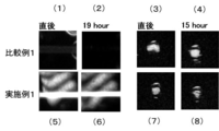
- Example 1 and Comparative Example 1 were circulated, the fluorescence images of the channels observed and photographed from above using a near-infrared fluorescence observation apparatus [FIGS. 4 (1), (2), (5), (6 )] And tomographic images of the flow path [FIGS. 4 (3), (4), (7), (8)] taken by the ultrasonic diagnostic imaging apparatus.
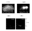
- (1) is an appearance photograph of a pig foot, (2) an image of near-infrared fluorescence contrast, (3) is an appearance photograph of a lymph node part of the pig foot of (1), and (4) is near-infrared fluorescence. It is a fluorescence image of contrast. It is an ultrasound contrast image. Note that (1) is an image before contrast medium injection. It is an image figure of the contrast agent (liposome particle) of Example 2 (micrometer size). (1) is a photograph of the simplified flow path system of FIG. 3 (side view) taken from above, (2) and (3) are Example 2, Comparative Example 2 and PBS (saline) in the same system.
- FIG. 4 is a fluorescence image of the flow channel when the system is circulated and the system (FIG.
- FIG. 3 side view
- (4) is a cut obtained by cutting the simple flow channel system of FIG. 3 (side view) vertically.
- It is an image figure of a surface.
- (5) and (6) are tomographic images of the flow path taken by the ultrasonic diagnostic imaging apparatus, and are the same plane as (4).
- It is an illustration which shows a mode that a contrast medium is injected into a chicken breast.
- (1) is an appearance photograph of the chicken breast, and (2) is a fluorescent image of the chicken breast.
- (3) is a tomographic image of the same breast breast by ultrasound contrast (before contrast medium administration)
- (4) is a tomographic image of the same breast breast by ultrasound contrast (before contrast medium administration). It is the bright field image (photograph) observed using the optical microscope.
- Liposome-based ultrasound contrast agents that constitute contrast agents capable of both ultrasound contrast and near-infrared fluorescence contrast (both-purpose contrast agents) are already known per se, and the raw material of liposomes (particles) (phospholipids) , Lipids modified with polyethylene glycol (PEG-lipid, etc.) are readily available, and devices for preparing liposomes are also commercially available.
- dispersoid liposomes (particles) are dispersed in physiological saline as a dispersion medium, and the whole image is a dispersion (suspension).
- the liposome is composed of a phospholipid film and an inclusion substance.
- Examples of the phospholipid constituting the film include distearyl-phosphatidylcholine (DSPC), dimyristoyl-phosphatidylethanolamine (DMPE), dipalmitoylphosphatidylethanolamine (DPPE), dioleylphosphatidylethanolamine (DOPE), distearoylphosphatidyl.
- DSPC distearyl-phosphatidylcholine
- DMPE dimyristoyl-phosphatidylethanolamine
- DPPE dipalmitoylphosphatidylethanolamine
- DOPE dioleylphosphatidylethanolamine
- Phosphatidylethanolamines such as ethanolamine (DSPE), diarachidoyl phosphatidylethanolamine (DAPE) or dilinoleyl phosphatidylethanolamine (DLPE); dilauroyl-phosphatidylcholine (DLPC), dimyristoyl-phosphatidylcholine (DMPC), dipalmitoyl-phosphatidylcholine (DPPC), diarachidoyl-phosphatidylcholine (DAPC), dioleyl-phosphatidyl Phosphatidylcholines such as phosphorus (DOPC); dimyristoyl phosphatidylserine (DMPS), diarachidoyl phosphatidylserine (DAPS), dipalmitoylphosphatidylserine (DPPS), distearoylphosphatidylserine (DSPS), dioleoylphosphatidylserine (DOPS), etc.
- Phosphatidylserine dipalmitoyl phosphatidic acid (DPPA), dimyristoyl phosphatidic acid (DMPA), distearoyl phosphatidic acid (DSPA), diarachidoyl phosphatidic acid (DAPA), and alkali metal salts thereof; dimyristoyl phosphatidylglycerol (DMPG) and alkali metal salts thereof, dipalmitoyl phosphatidylglycerol (DPPG) and alkali metal salts thereof, distearoyl phosphate Thidylglycerol (DSPG) and its alkali metal salts, phosphatidylglycerols such as dioleyl-phosphatidylglycerol (DOPG); dilauroylphosphatidylinositol (DLPI), diarachidoylphosphatidylinositol (DAPI), dimyristoylphosphatidylinositol (DM
- the molecular weight of PEG is preferably 500 to 12,000.
- DSPE-PEG such as DSPE-PEG2000, DSPE-PEG3000, DSPE-PEG5000 is preferred. Of these, DSPE-PEG2000 is particularly preferred.
- Examples of the gas encapsulated in the liposome particles include air, oxygen, carbon dioxide, sulfur fluoride gas, and perfluorohydrocarbon gas.
- Examples of the perfluorohydrocarbon gas include perfluoromethane, perfluoroethane, perfluoropropane, perfluorobutane, perfluoropentane, perfluorohexane, perfluoroheptane, perfluorooctane, and the like.
- bubble liposomes enclosing the gas can be prepared by placing liposome particles in a container filled with the gas and performing ultrasonic treatment or the like.
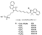
- examples of the near-infrared fluorescent dye that is the main component of the near-infrared fluorescent contrast agent include indocyanine green (ICG), phthalocyanine, squarylium, croconium, and derivatives thereof (ICG-C18).
- the wavelength of the excitation light of the near-infrared fluorescent dye is usually 600 nm or more, preferably 650 nm or more, more preferably 665 nm or more, further preferably 680 nm or more, and particularly preferably 700 nm or more. Most preferably, it is 720 nm or more.
- the fluorescence wavelength is preferably 50 nm or longer, particularly 100 to 200 nm longer than the wavelength of the excitation light.
- composition of the dual-use contrast agent of the present invention includes, as essential components, the above-described liposome particles, gas and near-infrared fluorescent dye, and physiological saline as a dispersion medium.
- a pH adjusting agent for example, An organic solvent such as ethyl alcohol, a viscosity modifier, a surfactant, a stabilizer, an antioxidant, a chelating agent, a colorant, and the like may be contained in a trace amount or an appropriate amount.
- the composition ratio is, but when the weight of the phospholipid constituting the liposome is 1, the near-infrared fluorescent dye is, for example, 0.001 to 0.1, particularly 0.002 to 0.005. preferable.
- the phospholipid is preferably a mixture of saturated fatty acid type phospholipid and unsaturated fatty acid type phospholipid. In that case, saturated fatty acid type phospholipid: unsaturated fatty acid type phospholipid
- the lipid ratio is preferably 1: 5 to 5: 1 by weight, and most preferably 1: 1.
- the amount of gas encapsulated in the liposome particles is preferably 0.1 to 10, and more preferably 0.5 to 1, for example, when the weight of the phospholipid constituting the liposome is 1.
- the particle size (diameter) of the liposome particles is essential in the present invention to be nanometer or micrometer size. In the former case, it is preferably several tens to several hundreds of nm, particularly 50 to 200 nm. 8 to 20 ⁇ m, particularly 1 to 5 ⁇ m is preferable.
- contrast agent of the present invention varies depending on the particle size. In the case of nano size, it is used for imaging (contrast, imaging, observation) of lymphatic system (lymph vessel, lymph node). Used for imaging (contrast, imaging, observation).
- the physiological saline as a dispersion medium is an aqueous solution (saline) of sodium chloride that is almost isotonic with body fluid.
- a pH adjuster an organic solvent such as ethyl alcohol, and a viscosity adjuster are appropriately used.
- a surfactant, a stabilizer, an antioxidant, a chelating agent, a coloring agent, a glucose aqueous solution, and the like may be contained in a trace amount or in an appropriate amount.
- drugs such as anticancer drugs, anti-inflammatory drugs, corticosteroids, antiviral drugs, antibiotics, antituberculosis drugs, various molecular targeted therapeutic drugs (anti-VEGF antibody, anti-EGFR2 antibody), antithrombosis You may include medicine.
- the contrast agent of the present invention can also be used for DDS.
- an external stimulus such as strong ultrasound is irradiated.
- the drug can be released by destroying the liposome particles.
- the method for producing the dual-purpose contrast agent of the present invention may be performed by adding a near-infrared fluorescent dye before or after preparing the liposome-based ultrasonic contrast agent.
- Liposome production apparatuses are also commercially available, and production is not difficult.
- the dual-use contrast medium of the present invention is a dispersion (suspension) in which liposomes are dispersed in physiological saline as a dispersion medium. It is a feature of the present invention that a dye is bound to the liposome.
- This bond may not be a chemical bond but may be an ionic bond, a hydrogen bond, a hydrophobic bond, a protein-substrate bond, a protein-sugar chain bond, or the like.
- Example 1 Preparation of dual-use contrast agent (abbreviated as “fluorescent nanobubble”) First, we measured 22.85 mg of distearyl-phosphatidylcholine (DSPC), 3.48 mg of DSPE-PEG2000, and 0.70 mg of ICG-C18. Was placed in a screw tube. Both are in powder form. DSPE-PEG2000 is “1,2-distearoyl-sn-glycero-3-phosphoethanolamine-N- [cyanur (polyethylene glycol) -2000]”.
- chloroform and methanol are mixed at a volume ratio of 4: 1 to prepare a mixed solvent, 5 ml of this mixed solvent is added to the screw tube, the powder inside is dissolved, and the solution and did.
- the suspension thus obtained was passed through a 400 nm membrane filter using a gas tight syringe holder set for particle size adjustment (Mini-Extruder® Set for liposome production, Avanti Polar Lipids, Inc., USA).
- the particle size was shaped.
- the vial Upon completion of pressurization, the vial was placed in an ultrasonic tank filled with deaerated water and irradiated with ultrasonic waves for 5 minutes.
- fluorescent nanobubble a dual-use contrast agent having an average liposome particle diameter (diameter) of 49.4 nm was obtained.
- reference numeral 1 represents liposome particles
- reference numeral 2 represents gas encapsulated in the liposome particles
- reference numeral 3 represents a pigment
- reference numeral 4 represents a PBS solution (phosphate buffered saline).
- FIG. 2 shows the volume distribution of the liposome particles of this “fluorescent nanobubble” measured by a particle size measuring device (product number “Nanotrac” UPA-UT151, a product of Microtrack Bell Co., Ltd.).
- Comparative Example 1 is nanobubbles (suspension) to which ICG-C18 is not bonded. Except for the fact that ICG-C18 is not bonded, the preparation method is the same as that of the dual-use contrast agent (abbreviated as “fluorescent nanobubble”) of Example 1.
- the average particle diameter (diameter) of the liposome particles is 205.2 nm, and the gas encapsulated in the liposome particles is air.
- FIG. 4 shows a fluorescence image of a flow path observed and photographed from above using the near-infrared fluorescence observation apparatus when Example 1 and Comparative Example 1 are circulated [FIGS. 4 (1), (2), ( 5), (6)] and a tomographic image of the flow path photographed by the ultrasonic diagnostic apparatus [FIGS. 4 (3), (4), (7), (8)].
- Example 4 (1), (3), (5), and (7) were taken immediately after the start of circulation, and (2), (4), (6), and (8) were taken 15 or 19 hours after the start of circulation. It is an image. From the comparison of (1) and (5), in Comparative Example 1 where ICG-C18 is not bonded, the flow path is not fluorescent, in Example 1, the flow path is fluorescent, and Example 1 is near-infrared fluorescence observation. It can be seen that it functions as a contrast medium. Moreover, from the comparison between (3) and (7), it can be confirmed that the inside of the flow path where the ultrasonic contrast agent is present is observed (contrast) in both Comparative Example 1 and Example 1.
- Example 1 retains the function as a dual-purpose contrast agent even after 10 hours or more after circulation.
- the obtained fluorescence image is shown in (2) of FIG.
- this fluorescent image (2) a portion that appears strongly white and triangular is an injection site, and a high-luminance site described linearly from the site is a lymph vessel.
- the appearance photograph is shown in FIG. 5 (3), and the near-infrared fluorescence image is shown in (4). It can be confirmed that a plurality of linear lymphatic vessels flow into the lymph nodes.
- the spatial distribution of fluorescent nanobubbles of Example 1 (that is, the distribution of lymphatic vessels) can be clearly observed as a fluorescent site, and there is no problem in the function as a contrast agent for near-infrared fluorescence observation even in living tissue. It was confirmed.
- Example 1 The moment when the fluorescent nanobubbles of Example 1 flow into the lymph nodes from the body surface of the site where the lymph nodes are present through the lymph vessels is observed with the above-described ultrasonic diagnostic imaging apparatus, and an ultrasonic image shown in FIG. Got.
- photography was performed previously before injecting the fluorescent nanobubble of Example 1 subcutaneously, the imaging
- the fluorescent nanobubble of the present invention enables multi-scale observation with a single contrast agent injection for the same site.
- Example 2 Preparation of dual-use contrast medium (abbreviated as “fluorescent microbubble”) 15 mg of distearyl-phosphatidylcholine (DSPC), 7.6 mg of egg yolk lecithin, and 0.70 mg of ICG-C18 were weighed and transferred to a mill container.
- fluorescent microbubble distearyl-phosphatidylcholine
- DSPC distearyl-phosphatidylcholine
- ICG-C18 0.70 mg
- This mill container was immersed in liquid nitrogen and frozen. This mill container was then set in a mixer mill and the frozen material in the container was pulverized.
- the grinding condition is “20 ⁇ frequency / sec ”for 2 minutes.
- the pulverized product was transferred to a beaker, 5 ml of PBS solution was poured into the beaker, and stirred and mixed using a magnetic stirrer (400 rpm) to prepare a solution (phospholipid solution).
- phospholipid mixed dispersion (dispersion containing liposome particles).
- the dispersion liquid is sucked up with a syringe (syringe), and further, air is put into the syringe at a “dispersion liquid: air volume ratio of 2: 1”, and the syringe is shaken vigorously by hand to remove the dispersion liquid and air. Stirring was performed, thereby producing a bubble dispersion.
- syringe syringe
- air is put into the syringe at a “dispersion liquid: air volume ratio of 2: 1”
- the syringe is shaken vigorously by hand to remove the dispersion liquid and air. Stirring was performed, thereby producing a bubble dispersion.
- the bubble dispersion is stirred with an ultrasonic homogenizer to shape the particle size.
- liposome particle concentration 5.53 ⁇ 10 4 particles / mm 3 of a dual-purpose contrast medium (abbreviation “fluorescent microbubble”) was obtained.
- Comparative Example 2 is a liposome particle (suspension) to which ICG-C18 is not bound. Except for the point that ICG-C18 is not bonded, the preparation method is the same as that of the dual-use contrast agent (abbreviated as “fluorescence microbubble”) in Example 2.
- the main component of this contrast agent is a liposome suspension
- the dispersoid is liposome particles
- the dispersion medium is a PBS solution.
- the average particle diameter (diameter) of the liposome particles is 2.2 ⁇ m (radius 1.1 ⁇ m ⁇ 2)
- the liposome concentration is 6.65 ⁇ 10 4 particles / mm 3 .
- the gas encapsulated in the liposome particles is air.
- FIG. 11 shows an optical microscope in which (1) the contrast agent of Example 2 and (2) the liposome particles (suspension) of Comparative Example 2 are each connected with a 100 ⁇ objective lens (product number UPlansApo, product of Olympus Corporation). It is a bright-field image (photograph) observed using (Product No. BX51, Olympus product). It can be seen that the liposome particles of Example 2 and Comparative Example 2 are spherical.
- the above optical microscope has an epifluorescence system, a xenon light source (part number U-LH75XEAPO, a product of Olympus Corporation), and an ultra-high-sensitivity EMCCD camera to enable near-infrared fluorescence observation in addition to normal bright-field observation.
- a xenon light source part number U-LH75XEAPO, a product of Olympus Corporation
- an ultra-high-sensitivity EMCCD camera to enable near-infrared fluorescence observation in addition to normal bright-field observation.
- Bright field observation visible light observation mode
- fluorescence observation fluorescence observation mode
- FIG. 12 is a fluorescence image (photograph) acquired in the fluorescence observation mode.
- the photograph of Example 2 is bright in the shape of a sphere, whereas (2) the photograph of Comparative Example 2 is black and nothing is shown.
- the image is taken under the same observation (fluorescence imaging) conditions (setting of excitation light intensity, gain, etc.), but the fluorescence image is not confirmed.
- the particle concentration (number density), average particle diameter (radius) ⁇ standard deviation of “fluorescent microbubble” of Example 2 is 5.53 ⁇ 10 4 / mm 3 , 1.2 ⁇ 0.49 ⁇ m, and is a comparative example. 2 is 6.65 ⁇ 10 4 / mm 3 , 1.1 ⁇ 0.53 ⁇ m, which is almost equal.
- Example 2 Phantom experiment The simple flow path system (see FIG. 3) used in Example 1 was used.
- FIG. 3 The result is shown in FIG.
- Example 2 and Comparative Example 2 the flow path in which the contrast agent is present is observed and contrasted as a high-intensity region of ultrasonic contrast.
- the flow path (more precisely, the inside of the flow path) is observed and contrasted as a low-luminance part of ultrasonic contrast.
- the wall of the channel is observed as a high luminance part.
- Example 2 to which ICG-C18 is bonded has the same function as an ultrasonic contrast agent compared to Comparative Example 2 to which ICG-C18 is not bonded.
- FIG. 10 (1) is an appearance photograph of the chicken breast observed and photographed from outside the chicken breast with a digital camera after contrast medium administration
- FIG. 10 (2) is the outside of the chicken breast with the near infrared fluorescence observation apparatus. It is the fluorescence image image
- FIG. 10 (3) is a tomographic image of a chicken breast observed with an ultrasonic diagnostic apparatus (Part No. Aplio500, a product of Toshiba Medical Systems) before contrast medium administration.
- the tip of the syringe can be visually recognized as a bright linear structure at a position 15 to 17 mm deep from the surface.
- the bright area at a depth of 17-20 mm from the surface is the border between the chicken breast and the base.
- FIG. 10 (4) is a tomographic image obtained by observing the same part using the same device after administration of the contrast medium. It turned out that the brightness increased centering on the syringe tip confirmed before contrast agent administration, and it confirmed that the contrast agent "fluorescence microbubble" of a present Example functions as an ultrasonic contrast agent.
- the dual-purpose contrast medium of the present invention itself is deeply injected when it is injected into a living tissue. Therefore, the near-infrared fluorescence imaging has a living tissue depth of about 10 mm, and the ultrasound imaging has a living body. Imaging (observation) is possible up to a tissue depth of about 30 mm.
- a depth of about 10 mm can be observed by both near-infrared fluorescence contrast imaging and ultrasonic contrast imaging, and a depth of about 10 mm to 30 mm can be observed only by ultrasonic contrast.
- a region that can be observed in duplicate specify the location (affected area) that you want to see with near-infrared fluorescence contrast, which is good for wide-range observation, and this location (affected area) It is possible to do convenient operations such as viewing with sonography.
- the conventional near-infrared fluorescent contrast agent has a small molecule, so it is difficult to diffuse in the tissue and the residence time is short.
- the dual-purpose contrast agent of the present invention has a near-infrared fluorescent contrast agent portion in the liposome particle. Since it is bonded, it is large, and therefore has the advantage of a long residence time. The residence time is longer for microbubbles than for nanobubbles.
- the contrast agent capable of both ultrasonic imaging and near-infrared fluorescence imaging of the present invention is a deep part (specifically, liver, kidney, pancreas, gallbladder, stomach, stomach, duodenum, skin, etc.) Since it reaches microvascular systems and lymphatic systems with a depth of 0 to several tens of millimeters, it is possible to obtain images by macroscopic and microscopic multiscale imaging. Therefore, it can be used for pathological diagnosis and research of living tissue (affected area). Therefore, it may be used in the reagent industry, pharmaceutical industry, hospital, university, laboratory, etc.
Landscapes
- Health & Medical Sciences (AREA)
- General Health & Medical Sciences (AREA)
- Epidemiology (AREA)
- Life Sciences & Earth Sciences (AREA)
- Animal Behavior & Ethology (AREA)
- Public Health (AREA)
- Veterinary Medicine (AREA)
- Chemical & Material Sciences (AREA)
- Radiology & Medical Imaging (AREA)
- Nuclear Medicine, Radiotherapy & Molecular Imaging (AREA)
- Dispersion Chemistry (AREA)
- Medicinal Chemistry (AREA)
- Pharmacology & Pharmacy (AREA)
- Medicines Containing Antibodies Or Antigens For Use As Internal Diagnostic Agents (AREA)
Abstract
【課題】比較的広い範囲の浅部領域を近赤外蛍光造影で観察して、見たい局所を発見し(いわばナビゲート)、同じ位置で、今度は超音波造影で深部領域を局所的かつ高分解能で観察する。 【解決手段】分散質としてのリポソーム粒子(1)及び分散媒としての生理食塩水(4)からなり、当該リポソーム粒子の直径がナノサイズ又はマイクロサイズであり、当該リポソーム粒子の内部に気体(2)を含む超音波造影剤(A)及び近赤外蛍光色素(3)からなる近赤外蛍光造影剤(B)からなり、前記リポソーム粒子(1)と前記近赤外蛍光色素(3)とは結合していることを特徴とする、超音波造影及び近赤外蛍光造影の両方が可能な造影剤。
Description
本発明は、超音波造影及び近赤外蛍光造影の両方が可能な造影剤に関するものである。
超音波造影法は、生体内を進んだ超音波パルスが生体組織の変わるところ(正確には音響インピーダンスの変わるところ)で反射され、その反射波を情報処理して画像化(イメージング化)する際、反射を強調させるため、造影剤を用いる方法である。生体組織(例えば、血管やリンパ管)に造影剤を投与して反射を強調し、これまでに得られなかった新たな生体組織の画像(例えば、血管やリンパ管の分布画像)を得るのである。超音波はX線と違って生体組織への損傷がないか極めて少ない利点がある。
最近、超音波造影剤の主役である気泡(気泡は超音波を反射する)の大きさがマイクロやナノのサイズであると、超音波造影剤がサイズによって生体組織(例えば、肝臓、腎臓、膵臓、胆のう、胃、胃、十二指腸、皮膚など)の深部(具体的には皮膚表面から造影の場合は、最大深さ:数十mm、肝臓や心臓等の臓器の表面から造影する場合は、扇状の断層面となるが、最大深さ:100mm程度)の微小血管系やリンパ管系にも届くので、注目されている(非特許文献1、2参照)。
他方、近赤外光造影法は、同様に生体組織に近赤外蛍光造影剤を投与し、外部から近赤外励起光を照射する。励起光を受光した造影剤は励起された後、元の基底状態に戻るとき、励起光と異なる波長の近赤外光を発光する。近赤外光は比較的厚く(具体的には最大厚さ10mm程度)とも生体組織を透過することから、外部から照射及び反射光又は透過光の受光を行うことができる。
そこで、外部から近赤外光カメラを使って発光した光を外部から撮影することで生体組織の状況が低侵襲で判明することから、生体組織(患部)の病理診断法として多用されている。
最近、同様に生体組織(例えば、肝臓、腎臓、膵臓、胆のう、胃、胃、十二指腸、皮膚など)の血管やリンパ管に造影剤を投与して、その深部(具体的には最大深さ10mm程度)の微小血管系やリンパ管系を撮影する近赤外光造影法が注目されている(非特許文献3、4参照)。
なお、非特許文献4のリポソームは、近赤外蛍光造影剤の主役である近赤外蛍光造影色素であるインドシアニングリーン(ICG)又はアルキル鎖で標識されたICG誘導体に結合しているリポソームであり、リポソーム内部に気体を含まないので超音波造影剤ではなく、近赤外蛍光造影剤である。リポソームは色素分子を大きくして色素をリンパ節に滞留・集積させる目的で結合している。結局、非特許文献4のリポソームは、超音波造影とは全く関係なく、本発明と全く異なる。
特許文献1の「蛍光色素を含有するリポソーム」も非特許文献4のリポソームと同じである。基本的には、特許文献1のリポソームも近赤外蛍光造影剤であり、超音波造影とは関係なく、本発明と全く異なる。
特許文献2のリポソーム複合体も、構造的にはインドシアニングリーン(ICG)等が結合したリポソーム粒子であるが、リポソーム粒子内に薬剤を含む。この複合体に強力な近赤外線を照射してICGにそれを吸収させて発熱させることで、リポソームを破壊し、それで薬剤を放出させるのである。ドラッグデリバリーシステム(DDS)に使う薬剤とも言える。結局、特許文献2のリポソーム複合体も、近赤外蛍光造影剤の一種であり、超音波造影とは全く関係なく、本発明と全く異なる。
Y,Matsumura et al., Cancer research, 1986; 46: 6378-6392
平田圭一 他,Drug Delivery System 2010; 25-5: 466-473
JT. Alander et al., International Journal of Biomedical Imaging, 2012; Article ID 940585: 26
Toyota et al., Bioorganic & Medicinal Chemistry 2014; 22: 721-727
超音波造影法は、深部領域を局所的つまり比較的狭い範囲(体表面からの場合、幅:数十mm)を比較的高い分解能(超音波の周波数にもよるが、分解能:凡そ数百μm~1mm)で見る方法であり広い範囲を見ることは困難である(短所)。逆に近赤外蛍光造影法は、浅部領域を比較的広い範囲(幅:数百mm)を高分解能(分解能:凡そ数十~数百μm)でみることができるが、深部領域の観察には適用範囲が狭い(短所)。
そこで、本発明者らは、両者の短所を補完し、比較的広い範囲の浅部領域を近赤外蛍光造影で観察して、見たい局所を発見し(いわばナビゲート)、同じ位置で、今度は超音波造影で深部領域を局所的かつ高分解能で観察できれば、新しい診断法が可能になることを着想した。
この着想を実現可能にするには、両者の造影剤に工夫が必要である。何故ならば、近赤外蛍光造影剤は、拡散が比較的早く、同一領域へ超音波造影剤を注射しても見たい局所の発見が難しくなること、更に同一領域へ2種の注射が必要で、施術的な手間がかかり誤差を生じやすいこと、造影剤を2種用意することの管理および安全上のリスクが高くなることなどの問題がある。
本発明者らは、かかる問題を解決するため、超音波造影剤と近赤外蛍光造影剤とを結合させ1つの造影剤にすることを世界で初めて着想し、鋭意研究の結果、本発明を成すに至った。
超音波造影剤の中には、リポソーム(粒子)を主体とするものがある。そのような造影剤は、分散質としてのリポソーム粒子及び分散媒としての生理食塩水からなる分散体(懸濁液)であり、当該リポソーム粒子の内部に気体を含む。
リポソーム(粒子)は、細胞膜の脂質2層膜を模して人工的に作られたもので、リン脂質(phospholipid)の皮膜と内包物質(後述する)からなる。
リン脂質は、リン原子を含む親水性の部位(フォスフォコリンなど、図1では〇で示す)と疎水性の部位(アシル基、図1では=で示す)を持つ両親媒性の分子である。リン脂質分子が水(分散媒)中で一定濃度以上であると、親水性の部位どうしで近接し、疎水性の部位どうしも近接するように自発的に会合して(寄り集まって)膜を形成し、膜は球形となることで表面積を最小化して安定化する。超音波造影用の気体が共存すると、気体は疎水性であるためにこの球の内部に取り込まれる。これが「マイクロエマルション」または「マクロエマルション」と呼ばれるものである。このときリン脂質は単層膜を形成している。
気体が存在しない場合には、リン脂質の膜は表と裏で2枚が近接するように安定化し、膜は二重構造となり、リポソーム粒子を形成する。リポソーム粒子の内部も親水性であることから、ここに水又は水溶液が入る。
超音波造影用の気体を取り込ませたリポソーム粒子の作製方法によっては、球の一部が単層膜で他部が2層膜というリポソーム粒子もできる。この場合には、単層膜で囲まれた空間に気体が閉じ込められ、残りの空間に水又は水溶液が入る。
このリポソームに近赤外蛍光造影剤の主体である近赤外蛍光色素をリポソーム作製時又は作製後に混合して単層膜又は2層膜(リポソーム粒子)に結合させることに成功し、本発明を成すに至った。
即ち、本発明は、
「分散質としてのリポソーム粒子及び分散媒としての生理食塩水からなり、当該リポソーム粒子の直径がナノサイズ又はマイクロサイズであり、当該リポソーム粒子の内部に気体を含む超音波造影剤(A)及び近赤外蛍光色素からなる近赤外蛍光造影剤(B)からなり、
前記リポソーム粒子と前記近赤外蛍光色素とは結合していることを特徴とする、超音波造影及び近赤外蛍光造影の両方が可能な造影剤」
を提供する。
「分散質としてのリポソーム粒子及び分散媒としての生理食塩水からなり、当該リポソーム粒子の直径がナノサイズ又はマイクロサイズであり、当該リポソーム粒子の内部に気体を含む超音波造影剤(A)及び近赤外蛍光色素からなる近赤外蛍光造影剤(B)からなり、
前記リポソーム粒子と前記近赤外蛍光色素とは結合していることを特徴とする、超音波造影及び近赤外蛍光造影の両方が可能な造影剤」
を提供する。
以下、本発明の超音波造影及び近赤外蛍光造影の両方が可能な造影剤を「両用造影剤」ということがある。
本発明の造影剤は、概して言えば、分散質としてのリポソーム粒子及び分散媒としての生理食塩水からなる懸濁液である。
本発明は、世界で初めて超音波造影及び近赤外蛍光造影の両方が可能な造影剤を提供する。
本発明の造影剤は超音波造影及び近赤外蛍光造影の両方が可能な造影剤で、1度の注射で浅部領域にも深部領域にも届き、剤は分子レベルで見て比較的大きなリポソーム粒子を有するので近赤外蛍光造影時にも、拡散が遅く、時間が経っても鮮明な画像が見られる。そこで、時間をかけて見たい局所を発見し、同じ位置で、今度は超音波造影で深部領域を局所的かつ高分解能で観察できる。そのため、新しい診断法を提供することができる。
そのため、剤も2種用意する必要がなく、1種で良いので、1回注射することで上記の診断法を行うことができるので、施術的手間が減り、その上、剤の管理、安全性のリスクも低減できる。
本発明の造影剤は、リポソームの粒径でナノメートル(nm)サイズ又はマイクロメートル(μm)サイズの2つに大別させることができる。
ナノメートル(nm)サイズの場合は、組織間隙を通過しリンパ管へ吸収される。このような特徴から、皮下注射による投与からリンパ管、リンパ節へのリンパ流の可視化が可能となり、体表、臓器表面近傍におけるリンパ系の機能診断に大きく寄与することが期待される。
マイクロメートル(μm)サイズの場合は、組織間隙や血管壁を通過することができないという特性を逆に利用することで、血管特異的なイメージングが可能となる。静脈注射により投与し、各種臓器内の血管の造影に使用できる。主な用途としては、外科手術において血管位置を三次元的に把握するための手術ナビゲーション手法としての使用が期待される。また、超音波造影法では、細胞によるマクロファージの効果を利用し腫瘍をイメージングするクッパーイメージングと呼ばれる手法が使用されているが、本発明で開発する造影剤を用いることで近赤外蛍光造影でも同様のイメージングを行うことが期待される。
また、本発明の両用造影剤のリポソームの内部に薬剤を内包させれば、近赤外蛍光でドラッグデリバリーシステム(DDS)の体内動態モニタリングを行うことができ、それにより両用造影剤が患部(例えば、癌)に届いたことを確認したあと、強力な超音波を照射してリポソームを破壊(超音波により励起される内部の気体の共鳴振動によりリポソームが崩壊)することにより当該薬剤を放出させ、患部(例えば、癌)だけに直接薬剤を投与することが可能となる。
超音波造影及び近赤外蛍光造影の両方が可能な造影剤(両用造影剤)を構成するリポソーム系超音波造影剤は、それ自体は既に知られており、リポソーム(粒子)の原料(リン脂質、ポリエチレングリコールで修飾した脂質(PEG-脂質)など)は、容易に手に入り、また、リポソーム作製用装置も市販されている。本発明の造影剤は、分散質のリポソーム(粒子)が分散媒としての生理食塩水に分散しており、全体像は分散体(懸濁液)である。そして、当該リポソームはリン脂質(phospholipid)の皮膜と内包物質からなる。
皮膜を構成するリン脂質としては、例えば、ジステアリル-ホスファチジルコリン(DSPC) 、ジミリストイル-ホスファチジルエタノールアミン(DMPE)、ジパルミトイルホスファチジルエタノールアミン(DPPE)、ジオレイルホスファチジルエタノールアミン(DOPE)、ジステアロイルホスファチジルエタノールアミン(DSPE)、ジアラキドイルホスファチジルエタノールアミン(DAPE)又はジリノレイルホスファチジルエタノールアミン(DLPE)等のホスファチジルエタノールアミン;ジラウロイル-ホスファチジルコリン(DLPC)、ジミリストイル-ホスファチジルコリン(DMPC)、ジパルミトイル-ホスファチジルコリン(DPPC)、ジアラキドイル-ホスファチジルコリン(DAPC)、ジオレイル-ホスファチジルコリン(DOPC)等のホスファチジルコリン;ジミリストイルホスファチジルセリン(DMPS)、ジアラキドイルホスファチジルセリン(DAPS)、ジパルミトイルホスファチジルセリン(DPPS)、ジステアロイルホスファチジルセリン(DSPS)、ジオレイルホスファチジルセリン(DOPS)等のホスファチジルセリン;ジパルミトイルホスファチジン酸(DPPA)、ジミリストイルホスファチジン酸(DMPA)、ジステアロイルホスファチジン酸(DSPA)、ジアラキドイルホスファチジン酸(DAPA)及びそのアルカリ金属塩等のホファチジン酸誘導体;ジミリストイルホスファチジルグリセロール(DMPG)及びそのアルカリ金属塩、ジパルミトイルホスファチジルグリセロール(DPPG)及びそのアルカリ金属塩、ジステアロイルホスファチジルグリセロール(DSPG)及びそのアルカリ金属塩、ジオレイル-ホスファチジルグリセロール(DOPG)等のホスファチジルグリセロール;ジラウロイルホスファチジルイノシトール(DLPI)、ジアラキドイルホスファチジルイノシトール(DAPI)、ジミリストイルホスファチジルイノシトール(DMPI)、ジパルミトイルホスファチジルイノシトール(DPPI)、ジステアロイルホスファチジルイノシトール(DSPI)、ジオレイルホスファチジルイノシトール(DOPI)等のホスファチジルイノシトール、カルジオリピン、スフィンゴミエリン脂質などが挙げられ、親水性を増すためのPEG-脂質として、“DSPE”をポリエチレングリコールで修飾した1,2-ジステアロイル-sn-グリセロ-3-ホスホエタノールアミン-N-[シアヌル(ポロエチレングリコール)-2000(1,2-distearoyl-sn-glycero-3-phosphoethanolamine-N-[cyanur(polyethylene glycol)-2000、略称“DSPE-PEG2000”)、ステアリルPEG、パルミトイルPEG、オレイン酸PEG、ミリスチルPEG、ラウロイルPEG等(PEGの後に続く数字はPEG部位の分子量を示す)が挙げられる。PEGの分子量は500~12000が好ましい。DSPE―PEG例えばDSPE-PEG2000、DSPE-PEG3000、DSPE-PEG5000が好ましい。なかでもDSPE-PEG2000が特に好ましい。
次に内包物質について説明する。リポソーム粒子に内包させる気体としては、空気、酸素、二酸化炭素、フッ化硫黄ガス、パーフルオロ炭化水素ガスなどが挙げられる。パーフルオロ炭化水素ガスとして例えば、パーフルオロメタン、パーフルオロエタン、パーフルオロプロパン、パーフルオロブタン、パーフルオロペンタン、パーフルオロヘキサン、パーフルオロヘプタン、パーフルオロオクタン、などを挙げることができる。
気体を内包させる方法としては、当該気体が充満した容器内にリポソーム粒子を入れて超音波処理などを行うことによって、当該気体が内包されたバブルリポソームを調製することができる。
他方、近赤外蛍光造影剤の主体である近赤外蛍光色素としては、インドシアニングリーン(ICG)、フタロシアニン、スクアリリウム、クロコニウムやそれらの誘導体(ICG-C18)が挙げられる。
近赤外蛍光色素の励起光の波長としては、通常、600nm以上であり、好ましくは650nm以上であり、より好ましくは665nm以上であり、さらに好ましくは680nm以上であり、特に好ましくは700nm以上であり、最も好ましくは720nm以上である。そして、蛍光波長としては、励起光の波長より50 nm以上、特に100~200nm長いものが好ましい。
本発明の両用造影剤の組成は、必須成分としては、上述のリポソーム粒子、気体及び近赤外蛍光色素並びに分散媒としての生理食塩水であるが、それら成分のほかに例えば、pH調整剤、エチルアルコール等の有機溶媒、粘度調整剤、界面活性剤、安定化剤、抗酸化剤、キレート剤、着色料などを微量又は適量含んでいてもよい。
また、組成割合であるが、リポソームを構成するリン脂質の重量を1とすると、近赤外蛍光色素は、例えば、0.001~0.1 特に0.002~0.005であることがより好ましい。なお、リポソーム粒子がマイクロメートルサイズの場合は、リン脂質が飽和脂肪酸型リン脂質と不飽和脂肪酸型リン脂質との混合物であることが好ましく、その場合、飽和脂肪酸型リン脂質:不飽和脂肪酸型リン脂質の比は、重量比で1:5~5:1が好ましく、1:1が最も好ましい。
また、リポソーム粒子に内包させる気体の量は、リポソームを構成するリン脂質の重量を1とすると、気体は、例えば、0.1~10 特に0.5~1であることがより好ましい。
リポソーム粒子の粒径(直径)は、ナノメートル又はマイクロメートルサイズであることが本発明では必須であり、前者の場合、数十~数百nm特に50~200nmが好ましく、後者の場合、0.8~20μm特に1~5μmが好ましい。
粒子径により、本発明の造影剤の用途が異なり、ナノサイズの場合はリンパ系(リンパ管、リンパ節)のイメージング(造影、撮影、観察)に、マイクロサイズの場合は、各種臓器の微細血管のイメージング(造影、撮影、観察)などに使われる。
分散媒としての生理食塩水としては、体液とほぼ等張の塩化ナトリウムの水溶液(食塩水)であるが、生理食塩水には、適宜、pH調整剤、エチルアルコール等の有機溶媒、粘度調整剤、界面活性剤、安定化剤、抗酸化剤、キレート剤、着色料、ブドウ糖水溶液などが微量又は適量含まれていても良い。
リポソーム粒子に内包させる気体のほか、薬剤例えば制癌剤、抗炎症薬、副腎皮質ホルモン剤、抗ウィルス薬、抗生物質、抗結核薬、各種分子標的治療薬(抗VEGF抗体、抗EGFR2抗体)、抗血栓薬などを内包させても良い。
薬剤を内包させることで、本発明の造影剤をDDSにも使用することができる。正確な位置に近赤外蛍光造影法で導き、それからその深部にある病変部分(局所)に到達したことを超音波造影法で認識した後、外部からの刺激例えば強力な超音波を照射してリポソーム粒子を破壊することにより当該薬剤を放出させことが可能となる。
本発明の両用造影剤を作製する方法は、リポソーム系超音波造影剤を作製する前(原料準備段階)か、作製後に、近赤外蛍光色素を添加すれば良い。リポソーム作製用装置も市販されており、作製は難しいことではない。
本発明の両用造影剤は、全体像としては、図1や図7に示すように、リポソームが分散媒としての生理食塩水に分散しており、分散体(懸濁液)である。そのリポソームに色素が結合していることが本発明の特徴である。この結合は化学結合でなくとも、イオン結合、水素結合、疎水結合、タンパク質―基質結合、タンパク質-糖鎖結合などでも良い。
実施例1
(1)両用造影剤(略称“蛍光ナノバブル”)の作製
まず、ジステアリル-ホスファチジルコリン(DSPC) を22.85 mg、DSPE-PEG2000を3.48mg、ICG―C18を 0.70mgを計量し、これらをスクリュー管に入れた。いずれも粉末形状である。なお、DSPE-PEG2000とは、「1,2-distearoyl-sn-glycero-3-phosphoethanolamine-N-[cyanur(polyethylene glycol)-2000]」のことである。
(1)両用造影剤(略称“蛍光ナノバブル”)の作製
まず、ジステアリル-ホスファチジルコリン(DSPC) を22.85 mg、DSPE-PEG2000を3.48mg、ICG―C18を 0.70mgを計量し、これらをスクリュー管に入れた。いずれも粉末形状である。なお、DSPE-PEG2000とは、「1,2-distearoyl-sn-glycero-3-phosphoethanolamine-N-[cyanur(polyethylene glycol)-2000]」のことである。
次に、溶解のために、クロロホルム及びメタノールを体積比4 : 1となるように混合して混合溶媒を作製し、この混合溶媒5mlを上記スクリュー管に加え、中の粉末を溶解させ、溶液とした。
この溶液の入った上記スクリュー管をボルテックスミキサーGENIE-2品番G560(米国サイエンティフィックインダストリーズ社の商品)にセットし、撹拌(強度10)を行い、完全に溶解させた時点で、スクリュー管のネジ式のフタを外した後、アルミホイルによりスクリュー管の開口部を覆った。そして、アルミホイルに針で揮発口を開け、スクリュー管をポリデシケーター(300型、アズワン株式会社の商品)に入れ、真空ポンプ(N810FT.18、株式会社ケー・エヌ・エフ・ジャパンの商品)を用いて、300分間、有機溶媒の揮発を行った。
有機溶媒が完全に揮発し、スクリュー管の底部に薄膜が生じたことを確認した後、スクリュー管をポリデシケーターから取り出し、次いで、PBS溶液(リン酸緩衝生理食塩水)が薄膜を剥がさないように注意しながらPBS溶液を5ml加え、そして、90℃に設定した恒温槽内に5時間静置した。
静置が完了後、スクリュー管に優しく振動を加え内部を撹拌することで薄膜の膨潤を行った。
静置が完了後、スクリュー管に優しく振動を加え内部を撹拌することで薄膜の膨潤を行った。
こうして得られた懸濁液を粒径調整用ガスタイトシリンジホルダーセット(リポソーム作製用装置Mini-Extruder Set、米国アバンティポーラリピッド社Avanti Polar Lipids, Inc. の商品)を用いて400nmのメンブレンフィルターに通し、粒径整形を行った。
粒径整形を行った溶液を「10mlバイアル瓶」に4ml充填し、ゴムキャップで栓をした後、アルミキャップをかけ、完全に締め切った。
その後、ルアーロックシリンジ(注射器)を使ってバイアル瓶内に気体としての空気を15mlだけ加圧して入れた。
加圧が終わり次第、脱気水を満たした超音波槽にバイアル瓶を入れ、5分間超音波を照射した。
こうして、リポソーム平均粒径(直径)49.4nmの両用造影剤(略称“蛍光ナノバブル”)を得た。
この“蛍光ナノバブル”のイメージを図1に示す。図1において、符号1はリポソーム粒子を、符号2はリポソーム粒子に内包された気体を、符号3は色素を、符号4はPBS溶液(リン酸緩衝生理食塩水)を示す。
また、この“蛍光ナノバブル”について、粒度測定装置(品番 Nanotrac UPA-UT151、マイクロトラック・ベル株式会社の商品)で測定したリポソーム粒子の体積分布を図2に示す。
(2)比較例1
比較例1は、ICG―C18が結合していないナノバブル(懸濁液)である。ICG-C18が結合されていない点を除けば、実施例1の両用造影剤(略称“蛍光ナノバブル”)と作製方法は同じである。リポソーム粒子の平均粒径(直径)は205.2nmでリポソーム粒子に内包された気体は空気である。
比較例1は、ICG―C18が結合していないナノバブル(懸濁液)である。ICG-C18が結合されていない点を除けば、実施例1の両用造影剤(略称“蛍光ナノバブル”)と作製方法は同じである。リポソーム粒子の平均粒径(直径)は205.2nmでリポソーム粒子に内包された気体は空気である。
(3)評価
(イ)ファントム実験
図3(側面図)に示すような小型水槽に流路を組み合わせた簡易流路システムを構築した。流路は図3では1本しか見えないが奥行き方向にもう1本ある。流路は中空のシリコンチューブ(直径φ=2 mm)である。それぞれの流路内に実施例1の蛍光ナノバブルと比較例1のナノバブル(懸濁液)を循環させ、図3では「近赤外蛍光プローブ」と記す近赤外蛍光観察装置(品番HyperEye Medical Systems II、瑞穂医科工業株式会社の商品)と同じく「超音波プローブ」と記す超音波画像診断装置(品番Aplio500、東芝メディカルシステムズ社の商品)を使用してそれぞれ撮影した。この結果を図4に示す。
(イ)ファントム実験
図3(側面図)に示すような小型水槽に流路を組み合わせた簡易流路システムを構築した。流路は図3では1本しか見えないが奥行き方向にもう1本ある。流路は中空のシリコンチューブ(直径φ=2 mm)である。それぞれの流路内に実施例1の蛍光ナノバブルと比較例1のナノバブル(懸濁液)を循環させ、図3では「近赤外蛍光プローブ」と記す近赤外蛍光観察装置(品番HyperEye Medical Systems II、瑞穂医科工業株式会社の商品)と同じく「超音波プローブ」と記す超音波画像診断装置(品番Aplio500、東芝メディカルシステムズ社の商品)を使用してそれぞれ撮影した。この結果を図4に示す。
図4は、実施例1及び比較例1を循環させたときに、上記近赤外蛍光観察装置を用いて上から観察撮影した流路の蛍光画像[図4(1)、(2)、(5)、(6)]と上記超音波画像診断装置で撮影した流路の断層画像[図4(3)、(4)、(7)、(8)]である。
図4(1)、(3)、(5)、(7)は循環開始直後に、(2)、(4)、(6)、(8)は循環開始から15又は19時間経過後に撮影した画像である。(1)と(5)の比較からICG―C18が結合していない比較例1では流路が蛍光しておらず、実施例1では流路が蛍光し、実施例1が近赤外蛍光観察用造影剤として機能していることが分かる。また、(3)と(7)の比較から、比較例1と実施例1の両者において超音波造影剤が存在する流路内が高輝度部位として観察(造影)されていることが確認できる。ICG―C18が結合した実施例1の超音波造影剤としての機能は、ICG―C18が結合していない比較例1と比べても遜色ないことが分かる。(5)と(6)及び(7)と(8)の比較から、実施例1は循環後10時間以上経過しても両用造影剤としての機能を保持していることがわかる。
(ロ)動物実験
図5の(1)に示す動物実験用のブタの足に、in vivoで実施例1の蛍光ナノバブルを皮下注射した。これにより、蛍光ナノバブルは自然とリンパ管を介してリンパ節に流入する。
図5の(1)に示す動物実験用のブタの足に、in vivoで実施例1の蛍光ナノバブルを皮下注射した。これにより、蛍光ナノバブルは自然とリンパ管を介してリンパ節に流入する。
そこで、上記近赤外蛍光観察装置を使って撮影したので、得られた蛍光画像を図5の(2)に示す。この蛍光画像(2)で強く白く三角形状に見えるところが注射部位であり、該当部位から線状に述べる高輝度部位がリンパ管である。
また、リンパ節が存在する部位を撮影したので、この外観写真を図5の(3)、近赤外蛍光画像を(4)に示す。複数の線状のリンパ管がリンパ節に流入していることが確認できる。このように実施例1の蛍光ナノバブルの空間分布(つまりは、リンパ管の分布)を蛍光部位として明瞭に観察でき、生体組織中においても近赤外蛍光観察用造影剤としての機能に問題がないことを確認した。
リンパ節が存在する部位の体表から、リンパ管を介して実施例1の蛍光ナノバブルがリンパ節に流入する瞬間を上記超音波画像診断装置で観察し、図6(2)に示す超音波画像を得た。なお、予め、実施例1の蛍光ナノバブルを皮下注射する前にも同様の撮影を行ったので、その撮影画像を図6(1)に示す。リンパ節など低エコー強度部位においては、実施例1の体内動態をリアルタイムで確認できることを確かめた。
以上、近赤外蛍光造影の蛍光画像(図5)と超音波造影の画像(図6)から、次のことが分かる。近赤外蛍光造影では、リンパ管の広範囲での走行(マクロな走行)を、超音波造影では、リンパ節の局所的な(ミクロな)内部構造を知ることができる。つまり、本発明の蛍光ナノバブルは、同じ部位について一度の造影剤注射でマルチスケールの観察を可能にする。
実施例2
(1)両用造影剤(略称“蛍光マイクロバブル”)の作製
ジステアリル-ホスファチジルコリン(DSPC)を15mg、卵黄レシチンを7.6mg、ICG-C18を0.70mg計量し、ミル容器に移した。
(1)両用造影剤(略称“蛍光マイクロバブル”)の作製
ジステアリル-ホスファチジルコリン(DSPC)を15mg、卵黄レシチンを7.6mg、ICG-C18を0.70mg計量し、ミル容器に移した。
このミル容器を液体窒素に浸し凍結を行った。このミル容器を今度はミキサーミルにセットし容器内の凍結物の粉砕を行った。粉砕条件は「20 frequency/sec」で2分間である。
そして、再度上記の凍結と粉砕を1回繰り返した。
こうして粉砕したものをビーカーに移し、そのビーカーに5mlのPBS溶液を注ぎ入れ、マグネチックスターラー(400rpm)を用いて撹拌混合し、溶液(リン脂質溶液)を作製した。
このリン脂質溶液にPBS溶液を更に5ml、DSPE-PEG2000を10mg加え、マグネチックスターラー(400rpm)で6時間攪拌し,リン脂質混合分散液(リポソーム粒子を含む分散液)を作製した。
次いで、この分散液をシリンジ(注射器)で吸い取り、更に空気を「分散液:空気の容量比が2:1」にシリンジ内に入れ、そのシリンジを手で激しく振ることにより、分散液と空気を攪拌し、これにより、バブル分散液を作製した。
最後に、このバブル分散液を超音波ホモジナイザーで攪拌することにより、粒径整形を行い、こうして、リポソーム平均粒径(直径):2.4μm(=半径1.2μm×2)、リポソーム粒子濃度:5.53 × 10⁴ 個/mm3の両用造影剤(略称“蛍光マイクロバブル”)を得た。
この“蛍光マイクロバブル”のイメージを図7に示す。
(2)比較例2
比較例2は、ICG―C18が結合していないリポソーム粒子(懸濁液)である。ICG-C18が結合していない点を除けば、実施例2の両用造影剤(略称“蛍光マイクロバブル”)と作製方法は同じである。この造影剤の主体はリポソーム懸濁液であり、分散質はリポソーム粒子、分散媒はPBS溶液である。このリポソーム粒子の平均粒径(直径)は2.2μm(半径1.1μm×2)であり、リポソーム濃度は6.65×10⁴個/mm3である。リポソーム粒子に内包された気体は空気である。
比較例2は、ICG―C18が結合していないリポソーム粒子(懸濁液)である。ICG-C18が結合していない点を除けば、実施例2の両用造影剤(略称“蛍光マイクロバブル”)と作製方法は同じである。この造影剤の主体はリポソーム懸濁液であり、分散質はリポソーム粒子、分散媒はPBS溶液である。このリポソーム粒子の平均粒径(直径)は2.2μm(半径1.1μm×2)であり、リポソーム濃度は6.65×10⁴個/mm3である。リポソーム粒子に内包された気体は空気である。
図11は、(1)実施例2の造影剤及び(2)比較例2のリポソーム粒子(懸濁液)を、それぞれ100倍対物レンズ(品番UPlansApo、オリンパス株式会社の商品)を接続した光学顕微鏡(品番BX51、Olympus社の商品)を用いて観察した明視野像(写真)である。実施例2及び比較例2のリポソーム粒子が球形であることが分かる。
上記光学顕微鏡には、通常の明視野観察に加えて、近赤外蛍光観察も可能とするべく、落射蛍光システム、キセノン光源(品番 U-LH75XEAPO、オリンパス株式会社の商品)及び超高感度EMCCDカメラ(品番 iXons3、ANDOR TECHNOLOGY社の製品)が付加されており、ソフトウェア上で明視野観察(可視光観察モード)と蛍光観察(蛍光観察モード)を切り替えることができる。つまり、同一対象物の明視野と近赤外蛍光の顕微鏡画像を取得できる。
図12は、蛍光観察モードで取得した蛍光画像(写真)である。(1)実施例2の写真は球形のものが明るく映っており、それに対し(2)比較例2の写真は何も写っておらず真っ黒である。図12(2)の写真では、同一観測(蛍光撮影)条件(励起光強度やゲインなどの設定)で撮影しているが、蛍光画像は確認されない。
図12の(1)、(2)の比較から、リポソーム粒子のリン脂質膜にICG-C18が結合していることが確認できる。なお、図12の(1)<2次元写真>を良く見ると、輪帯状に明るく光っているが、輪帯内部では光っていない。これは顕微鏡システムの被写界深度、つまり2次元写真に対し奥行き方向の観察範囲に限界があり、輪帯内部におけるリン脂質膜表面部分のICG-C18の蛍光が観察されていないためである。
明視野観察画像(図11)と近赤外蛍光画像(図12)で僅かにリポソーム粒子の位置が異なるが、これは観察時にリポソーム粒子が動いたためである。
明視野観察画像(図11)と近赤外蛍光画像(図12)で僅かにリポソーム粒子の位置が異なるが、これは観察時にリポソーム粒子が動いたためである。
(3)上記平均粒径、粒子濃度及び粒径分布の測定
実施例2の“蛍光マイクロバブル”と比較例2(リポソーム懸濁液)について、粒子の平均粒径、粒子濃度(数密度)及び粒径分布を測定した。各20μl用意し、血球計算盤(トーマ盤、品番2-5552-31、アズワン株式会社の商品)に注入し、光学顕微鏡により観察した。所定体積内の粒子の数と半径を計測し、その後、計算することで、平均粒径、粒子濃度(数密度)及び粒径分布を求めた。この結果を図13に示す。
実施例2の“蛍光マイクロバブル”の粒子濃度(数密度)、平均粒径(半径)±標準偏差は、5.53×10⁴/mm3、1.2±0.49μmであり、比較例2のそれは、6.65×104/mm3、1.1±0.53μmであり、ほぼ等しい。
実施例2の“蛍光マイクロバブル”と比較例2(リポソーム懸濁液)について、粒子の平均粒径、粒子濃度(数密度)及び粒径分布を測定した。各20μl用意し、血球計算盤(トーマ盤、品番2-5552-31、アズワン株式会社の商品)に注入し、光学顕微鏡により観察した。所定体積内の粒子の数と半径を計測し、その後、計算することで、平均粒径、粒子濃度(数密度)及び粒径分布を求めた。この結果を図13に示す。
実施例2の“蛍光マイクロバブル”の粒子濃度(数密度)、平均粒径(半径)±標準偏差は、5.53×10⁴/mm3、1.2±0.49μmであり、比較例2のそれは、6.65×104/mm3、1.1±0.53μmであり、ほぼ等しい。
(4)評価
(イ)ファントム実験
実施例1で用いた簡易流路システム(図3参照)を使用した。2本の流路(直径φ=2mm)にそれぞれ実施例2の“マイクロバブル”と比較例2を循環させ、近赤外蛍光観察装置(品番HyperEye Medical Systems II、瑞穂医科工業株式会社の商品)と超音波画像診断装置(Aplio500、東芝メディカルシステムズ社の商品)を使用してそれぞれ撮影した。この結果を図8に示す。
(イ)ファントム実験
実施例1で用いた簡易流路システム(図3参照)を使用した。2本の流路(直径φ=2mm)にそれぞれ実施例2の“マイクロバブル”と比較例2を循環させ、近赤外蛍光観察装置(品番HyperEye Medical Systems II、瑞穂医科工業株式会社の商品)と超音波画像診断装置(Aplio500、東芝メディカルシステムズ社の商品)を使用してそれぞれ撮影した。この結果を図8に示す。
図8は、実施例2、比較例2及びPBS(生理食塩水)を、前者2つは第1実験で同時に後者PBSは第2実験で、流路内を循環させ、上記近赤外蛍光観察装置を用いて簡易流路システム(図3=側面図)の上から観察撮影したときの流路の蛍光画像[(2)、(3)]と超音波画像診断装置で撮影したときの流路の断層画像[(5)、(6)]である。なお、図8(2)~(3)から、ICG-C18を結合又は含有していない比較例2及びPBS(生理食塩水)は、流路が全く映って(蛍光して)おらず、それに対し実施例2は流路が映って(蛍光して)おり、実施例2が近赤外蛍光観察用造影剤として機能していることがわかる。
また、図8(6)から、実施例2と比較例2は、造影剤が存在する流路が超音波造影の高輝度部位として観察造影されていることが分かる。それに対し(5)に示されるようにPBS(生理食塩水)では流路(正確には流路の内部)が超音波造影の低輝度部位として観察造影されていることが分かる。但し、流路の壁は高輝度部位として観察されている。
この結果、ICG-C18が結合した実施例2は、超音波造影剤としての機能はICG-C18が結合していない比較例2と比べて同等であることがわかる。
(ロ)動物実験
(a)食肉用のトリ胸肉を用意し、図9に示すように、これの深さ1mm、5mm、15mmの位置に注射器で実施例2の造影剤を注入し、表面から超音波造影撮影及び近赤外蛍光造影撮影を行った。前者は、近赤外蛍光観察装置(品番HyperEye Medical Systems II、瑞穂医科工業株式会社の商品)を用い、後者は超音波画像診断装置(品番Aplio500、東芝メディカルシステムズ社の商品)を用いた。
(a)食肉用のトリ胸肉を用意し、図9に示すように、これの深さ1mm、5mm、15mmの位置に注射器で実施例2の造影剤を注入し、表面から超音波造影撮影及び近赤外蛍光造影撮影を行った。前者は、近赤外蛍光観察装置(品番HyperEye Medical Systems II、瑞穂医科工業株式会社の商品)を用い、後者は超音波画像診断装置(品番Aplio500、東芝メディカルシステムズ社の商品)を用いた。
この結果を図10に示す。図10(1)は造影剤投与後にデジタルカメラでトリ胸肉の外から観察撮影したトリ胸肉の外観写真であり、図10(2)は上記近赤外蛍光観察装置でトリ胸肉の外から撮影取得した蛍光画像である。表面から1mm、5mmの深さに実施例2を投与した部位では投与部位が明るく蛍光していることが分かるが、15mmの深さに投与した部位では明確な蛍光は観察されなかった。生体組織の表面から10mmより浅い部位では、実施例2が近赤外蛍光観察用造影剤として機能することを確認した。これは通常のICGと同程度の性能である。
図10(3)は、造影剤投与前に超音波診断装置(品番Aplio500、東芝メディカルシステムズ社の商品)で観察したトリ胸肉の断層画像である。注射器先端を表面から15~17mm深部の位置に明るい線状の構造体として視認できる。表面から17~20mmの深度に存在する明るい部位はトリ胸肉と土台の境界である。図10(4)は、造影剤投与後に同一部位を同一機器を用いて観察した断層画像である。造影剤投与前に確認された注射器先端を中心として明るさが増していることが分かり、本実施例の造影剤“蛍光マイクロバブル”が超音波造影剤として機能することを確認した。
なお、開腹下のブタ肝臓を対象に動脈又は門脈に直接に実施例2の造影剤を注入し、肝臓表面から内部脈管構造を観察することも可能である。
以上、説明した通り、本発明の両用造影剤は、生体組織に注入した場合、それ自体は深く注入されるので、近赤外蛍光造影では生体組織の深さ10mm程度まで、超音波造影では生体組織の深さ30mm程度まで撮影(観察)可能である。
従って、深さ10mm程度までは近赤外蛍光造影と超音波造影の両方で重複して観察可能であり、深さ10mm程度~30mm程度の間は超音波造影のみで観察可能となる。重複して観察可能な領域(深さ10mm程度まで)では、広範囲観察の得意な近赤外蛍光造影で見たい個所(患部)を特定し、その個所(患部)について、今度は高分解能の超音波造影で見ると言う便利なことが可能となる。この場合、従来の近赤外蛍光造影剤は分子が小さいことから組織内で拡散し易く滞留時間が短いので困るが、本発明の両用造影剤は、近赤外蛍光造影剤部分がリポソーム粒子に結合しているので大きく、そのため、滞留時間が長い利点を持つ。なお、滞留時間はナノバブルに比べマイクロバブルが長い。
本発明の超音波造影及び近赤外蛍光造影の両方が可能な造影剤は、生体組織(例えば、肝臓、腎臓、膵臓、胆のう、胃、胃、十二指腸、皮膚など)の深部(具体的には深さ0~数十mm)の微小血管系やリンパ管系にも届くので、それらの分布状況をマクロ、ミクロなマルチスケールで撮影し、画像を得ることができる。そのため、生体組織(患部)の病理診断、研究などに利用可能である。よって、試薬業、製薬業、病院、大学、研究所などにおいて利用可能性がある。
1・・・・リポソーム粒子
2・・・・気体
3・・・・色素
4・・・・生理食塩水(PBS)
2・・・・気体
3・・・・色素
4・・・・生理食塩水(PBS)
Claims (1)
- 分散質としてのリポソーム粒子及び分散媒としての生理食塩水からなり、当該リポソーム粒子の直径がナノサイズ又はマイクロサイズであり、当該リポソーム粒子の内部に気体を含む超音波造影剤(A)及び近赤外蛍光色素からなる近赤外蛍光造影剤(B)からなり、
前記リポソーム粒子と前記近赤外蛍光色素とは結合していることを特徴とする、超音波造影及び近赤外蛍光造影の両方が可能な造影剤。
Applications Claiming Priority (2)
| Application Number | Priority Date | Filing Date | Title |
|---|---|---|---|
| JP2018075105A JP2019182778A (ja) | 2018-04-10 | 2018-04-10 | 超音波造影及び近赤外蛍光造影の両方が可能な造影剤 |
| JP2018-075105 | 2018-04-10 |
Publications (1)
| Publication Number | Publication Date |
|---|---|
| WO2019198461A1 true WO2019198461A1 (ja) | 2019-10-17 |
Family
ID=68163085
Family Applications (1)
| Application Number | Title | Priority Date | Filing Date |
|---|---|---|---|
| PCT/JP2019/012003 Ceased WO2019198461A1 (ja) | 2018-04-10 | 2019-03-22 | 超音波造影及び近赤外蛍光造影の両方が可能な造影剤 |
Country Status (2)
| Country | Link |
|---|---|
| JP (1) | JP2019182778A (ja) |
| WO (1) | WO2019198461A1 (ja) |
Cited By (2)
| Publication number | Priority date | Publication date | Assignee | Title |
|---|---|---|---|---|
| CN114209829A (zh) * | 2021-12-17 | 2022-03-22 | 中国科学技术大学 | 负载荧光染料的光热脂质体及其制备方法和用途 |
| CN116491983A (zh) * | 2023-04-12 | 2023-07-28 | 岱特智能科技(上海)有限公司 | 人工动静脉手臂血管成像方法及相关装置 |
Families Citing this family (1)
| Publication number | Priority date | Publication date | Assignee | Title |
|---|---|---|---|---|
| JP7076732B2 (ja) * | 2018-03-01 | 2022-05-30 | 公立大学法人広島市立大学 | アデノイド肥大判定装置、アデノイド肥大判定方法及びプログラム |
Citations (1)
| Publication number | Priority date | Publication date | Assignee | Title |
|---|---|---|---|---|
| JPH06508364A (ja) * | 1991-06-18 | 1994-09-22 | イマアーレクス・フアーマシユーチカル・コーポレーシヨン | ガス充填リポソーム及び超音波造影剤としてのそれらの使用 |
Family Cites Families (2)
| Publication number | Priority date | Publication date | Assignee | Title |
|---|---|---|---|---|
| US6123923A (en) * | 1997-12-18 | 2000-09-26 | Imarx Pharmaceutical Corp. | Optoacoustic contrast agents and methods for their use |
| WO2011038043A1 (en) * | 2009-09-22 | 2011-03-31 | Targeson, Inc. | Optical imaging contrast agents and uses therof |
-
2018
- 2018-04-10 JP JP2018075105A patent/JP2019182778A/ja active Pending
-
2019
- 2019-03-22 WO PCT/JP2019/012003 patent/WO2019198461A1/ja not_active Ceased
Patent Citations (1)
| Publication number | Priority date | Publication date | Assignee | Title |
|---|---|---|---|---|
| JPH06508364A (ja) * | 1991-06-18 | 1994-09-22 | イマアーレクス・フアーマシユーチカル・コーポレーシヨン | ガス充填リポソーム及び超音波造影剤としてのそれらの使用 |
Non-Patent Citations (3)
| Title |
|---|
| EBATA, MASAHIKO ET AL.: "Near-infrared fluorescence by ICG derivative-bound microbubbles - Ultrasound imaging simulation", PREPRINTS OF THE ANNUAL MEETING OF THE JAPANESE SOCIETY FOR BIOMATERIALS, 13 November 2017 (2017-11-13) * |
| HE, Y. ET AL.: "Fabrication of Indocyanine Green and 2H, 3H-perfluoropentane loaded microbubbles for fluorescence and ultrasound imaging", PROCEEDING OF SPIE, vol. 9701, 7 March 2016 (2016-03-07), pages 97010T - 1 - 97010T-9, XP060064428 * |
| ZHU Z ET AL.: "Microencapsulation of indocyanine green for potential applications in image-guided drug deli very", LAB. CHIP., vol. 15, no. 3, 7 February 2015 (2015-02-07), pages 646 - 649, XP055643275 * |
Cited By (2)
| Publication number | Priority date | Publication date | Assignee | Title |
|---|---|---|---|---|
| CN114209829A (zh) * | 2021-12-17 | 2022-03-22 | 中国科学技术大学 | 负载荧光染料的光热脂质体及其制备方法和用途 |
| CN116491983A (zh) * | 2023-04-12 | 2023-07-28 | 岱特智能科技(上海)有限公司 | 人工动静脉手臂血管成像方法及相关装置 |
Also Published As
| Publication number | Publication date |
|---|---|
| JP2019182778A (ja) | 2019-10-24 |
Similar Documents
| Publication | Publication Date | Title |
|---|---|---|
| JP5513708B2 (ja) | 造影イメージング用の気体封入マイクロベシクル・アセンブリー | |
| Perera et al. | Contrast enhanced ultrasound imaging by nature-inspired ultrastable echogenic nanobubbles | |
| KR101076053B1 (ko) | 초음파 조영제 및 그것의 제조방법 | |
| US10639009B2 (en) | Apparatus and method for combined photoacoustic and ultrasound diagnosis | |
| RU2204415C2 (ru) | Комбинированный препарат для использования в качестве контрастного агента и способ получения изображения | |
| Son et al. | Echogenic nanoparticles for ultrasound technologies: Evolution from diagnostic imaging modality to multimodal theranostic agent | |
| CN101528268B (zh) | 具有聚合物修饰的脂质的充气微囊泡 | |
| JP2007515471A (ja) | 造影イメージング用の活性成分を有する気体封入マイクロベシクルのアセンブリー | |
| KR101595795B1 (ko) | 약물을 함유한 나노입자가 결합된 이중-목적 pat/초음파 조영제 및 이의 제조방법 | |
| KR102626600B1 (ko) | 동결 건조 제품 및 가스 충전된 미세소포 현탁액 | |
| US20150023881A1 (en) | Contrast agent for combined photoacoustic and ultrasound imaging | |
| EP2253308A1 (en) | Pharmaceutical composition comprising microbubbles for targeted tumor therapy | |
| CN101005858A (zh) | 用于反差成像的充气微泡组合物 | |
| WO2019198461A1 (ja) | 超音波造影及び近赤外蛍光造影の両方が可能な造影剤 | |
| JP6313329B2 (ja) | ガス封入マイクロベシクル | |
| JP2014227338A (ja) | インドシアニングリーン含有粒子およびその製造方法 | |
| KR20190134084A (ko) | 과불화탄소 나노입자, 이를 포함하는 약물 전달용 초음파 조영제 및 그의 제조방법 | |
| CN109568610A (zh) | 一种基于Janus药物共轭体的诊疗微泡的制备方法和用途 | |
| CN113521315A (zh) | 一种多模态微泡造影剂及其制备方法和应用 | |
| US20070110674A1 (en) | Sono-active liposomes and lipid particles and use thereof as contrast agents and active-agent delivery systems | |
| JP2018197208A (ja) | 一酸化窒素内包バブルリポソーム及びその利用 | |
| Kauerová et al. | A prototype ‘Infucon’device for continuous infusion of microbubbles in vivo | |
| CN105536000A (zh) | 一种基于季戊四醇酯的超声造影剂及其制备方法和用途 | |
| Almajed et al. | Liposomal Encapsulation of Chemotherapeutics Agents Combined with the Use of Ultrasound in Cancer Treatment | |
| JPH08310971A (ja) | 超音波診断用造影剤 |
Legal Events
| Date | Code | Title | Description |
|---|---|---|---|
| 121 | Ep: the epo has been informed by wipo that ep was designated in this application |
Ref document number: 19785498 Country of ref document: EP Kind code of ref document: A1 |
|
| NENP | Non-entry into the national phase |
Ref country code: DE |
|
| 122 | Ep: pct application non-entry in european phase |
Ref document number: 19785498 Country of ref document: EP Kind code of ref document: A1 |