WO2019146738A1 - ストレス状態の検出方法、及び、ストレス検出装置 - Google Patents
ストレス状態の検出方法、及び、ストレス検出装置 Download PDFInfo
- Publication number
- WO2019146738A1 WO2019146738A1 PCT/JP2019/002394 JP2019002394W WO2019146738A1 WO 2019146738 A1 WO2019146738 A1 WO 2019146738A1 JP 2019002394 W JP2019002394 W JP 2019002394W WO 2019146738 A1 WO2019146738 A1 WO 2019146738A1
- Authority
- WO
- WIPO (PCT)
- Prior art keywords
- stress
- choroid
- stress state
- choroidal
- detecting
- Prior art date
Links
- 238000001514 detection method Methods 0.000 title claims abstract description 79
- 210000003161 choroid Anatomy 0.000 claims abstract description 170
- 238000004364 calculation method Methods 0.000 claims abstract description 29
- 238000000034 method Methods 0.000 claims description 52
- 230000004323 axial length Effects 0.000 claims description 13
- 210000000873 fovea centralis Anatomy 0.000 claims description 4
- 230000035882 stress Effects 0.000 description 240
- 238000012014 optical coherence tomography Methods 0.000 description 37
- 208000019901 Anxiety disease Diseases 0.000 description 33
- 210000001508 eye Anatomy 0.000 description 33
- 238000005259 measurement Methods 0.000 description 22
- 238000012937 correction Methods 0.000 description 21
- 208000037265 diseases, disorders, signs and symptoms Diseases 0.000 description 20
- 201000010099 disease Diseases 0.000 description 18
- JYGXADMDTFJGBT-VWUMJDOOSA-N hydrocortisone Chemical compound O=C1CC[C@]2(C)[C@H]3[C@@H](O)C[C@](C)([C@@](CC4)(O)C(=O)CO)[C@@H]4[C@@H]3CCC2=C1 JYGXADMDTFJGBT-VWUMJDOOSA-N 0.000 description 18
- 230000036506 anxiety Effects 0.000 description 17
- 210000001525 retina Anatomy 0.000 description 17
- 208000007684 Occupational Stress Diseases 0.000 description 16
- 238000012545 processing Methods 0.000 description 16
- 208000019022 Mood disease Diseases 0.000 description 15
- 230000036651 mood Effects 0.000 description 15
- 238000012795 verification Methods 0.000 description 14
- 230000003340 mental effect Effects 0.000 description 13
- 208000024891 symptom Diseases 0.000 description 13
- 230000036541 health Effects 0.000 description 12
- 210000001519 tissue Anatomy 0.000 description 12
- 230000004044 response Effects 0.000 description 11
- 239000003550 marker Substances 0.000 description 10
- 210000004369 blood Anatomy 0.000 description 9
- 239000008280 blood Substances 0.000 description 9
- 210000004204 blood vessel Anatomy 0.000 description 9
- 229960000890 hydrocortisone Drugs 0.000 description 9
- 210000003296 saliva Anatomy 0.000 description 8
- 230000008859 change Effects 0.000 description 7
- 210000003733 optic disk Anatomy 0.000 description 7
- 210000003786 sclera Anatomy 0.000 description 7
- 230000006870 function Effects 0.000 description 6
- 238000011160 research Methods 0.000 description 6
- 210000003583 retinal pigment epithelium Anatomy 0.000 description 6
- UCTWMZQNUQWSLP-UHFFFAOYSA-N adrenaline Chemical compound CNCC(O)C1=CC=C(O)C(O)=C1 UCTWMZQNUQWSLP-UHFFFAOYSA-N 0.000 description 5
- 230000000875 corresponding effect Effects 0.000 description 5
- 230000006872 improvement Effects 0.000 description 5
- 230000003287 optical effect Effects 0.000 description 5
- 230000002265 prevention Effects 0.000 description 5
- 239000000126 substance Substances 0.000 description 5
- 210000003403 autonomic nervous system Anatomy 0.000 description 4
- 230000017531 blood circulation Effects 0.000 description 4
- 230000036772 blood pressure Effects 0.000 description 4
- 230000037326 chronic stress Effects 0.000 description 4
- 238000009223 counseling Methods 0.000 description 4
- 238000011156 evaluation Methods 0.000 description 4
- 230000003938 response to stress Effects 0.000 description 4
- 230000002889 sympathetic effect Effects 0.000 description 4
- 238000012360 testing method Methods 0.000 description 4
- 206010042209 Stress Diseases 0.000 description 3
- 208000013200 Stress disease Diseases 0.000 description 3
- 230000008901 benefit Effects 0.000 description 3
- 238000006243 chemical reaction Methods 0.000 description 3
- 230000000694 effects Effects 0.000 description 3
- 230000006355 external stress Effects 0.000 description 3
- 230000004424 eye movement Effects 0.000 description 3
- 229940088597 hormone Drugs 0.000 description 3
- 239000005556 hormone Substances 0.000 description 3
- 238000003384 imaging method Methods 0.000 description 3
- 230000004630 mental health Effects 0.000 description 3
- SFLSHLFXELFNJZ-QMMMGPOBSA-N (-)-norepinephrine Chemical compound NC[C@H](O)C1=CC=C(O)C(O)=C1 SFLSHLFXELFNJZ-QMMMGPOBSA-N 0.000 description 2
- UCTWMZQNUQWSLP-VIFPVBQESA-N (R)-adrenaline Chemical compound CNC[C@H](O)C1=CC=C(O)C(O)=C1 UCTWMZQNUQWSLP-VIFPVBQESA-N 0.000 description 2
- 241000557639 Araucaria bidwillii Species 0.000 description 2
- 208000020925 Bipolar disease Diseases 0.000 description 2
- 206010010144 Completed suicide Diseases 0.000 description 2
- LFQSCWFLJHTTHZ-UHFFFAOYSA-N Ethanol Chemical compound CCO LFQSCWFLJHTTHZ-UHFFFAOYSA-N 0.000 description 2
- 241000282326 Felis catus Species 0.000 description 2
- 241000282412 Homo Species 0.000 description 2
- 206010020772 Hypertension Diseases 0.000 description 2
- 241001465754 Metazoa Species 0.000 description 2
- 206010028980 Neoplasm Diseases 0.000 description 2
- 208000012902 Nervous system disease Diseases 0.000 description 2
- 208000025966 Neurological disease Diseases 0.000 description 2
- 208000008589 Obesity Diseases 0.000 description 2
- MUMGGOZAMZWBJJ-DYKIIFRCSA-N Testostosterone Chemical compound O=C1CC[C@]2(C)[C@H]3CC[C@](C)([C@H](CC4)O)[C@@H]4[C@@H]3CCC2=C1 MUMGGOZAMZWBJJ-DYKIIFRCSA-N 0.000 description 2
- 230000005856 abnormality Effects 0.000 description 2
- 230000009471 action Effects 0.000 description 2
- 230000037328 acute stress Effects 0.000 description 2
- 230000003078 antioxidant effect Effects 0.000 description 2
- 201000011510 cancer Diseases 0.000 description 2
- 150000003943 catecholamines Chemical class 0.000 description 2
- 238000010276 construction Methods 0.000 description 2
- 206010012601 diabetes mellitus Diseases 0.000 description 2
- 208000035475 disorder Diseases 0.000 description 2
- 210000000750 endocrine system Anatomy 0.000 description 2
- 238000005516 engineering process Methods 0.000 description 2
- 230000007613 environmental effect Effects 0.000 description 2
- 208000030533 eye disease Diseases 0.000 description 2
- 230000004907 flux Effects 0.000 description 2
- 210000000987 immune system Anatomy 0.000 description 2
- 238000001727 in vivo Methods 0.000 description 2
- 230000001678 irradiating effect Effects 0.000 description 2
- 230000004807 localization Effects 0.000 description 2
- 229960002748 norepinephrine Drugs 0.000 description 2
- SFLSHLFXELFNJZ-UHFFFAOYSA-N norepinephrine Natural products NCC(O)C1=CC=C(O)C(O)=C1 SFLSHLFXELFNJZ-UHFFFAOYSA-N 0.000 description 2
- 235000020824 obesity Nutrition 0.000 description 2
- 210000001328 optic nerve Anatomy 0.000 description 2
- 208000020016 psychiatric disease Diseases 0.000 description 2
- 230000028327 secretion Effects 0.000 description 2
- 230000000391 smoking effect Effects 0.000 description 2
- 208000004998 Abdominal Pain Diseases 0.000 description 1
- 239000004382 Amylase Substances 0.000 description 1
- 102000013142 Amylases Human genes 0.000 description 1
- 108010065511 Amylases Proteins 0.000 description 1
- 206010002383 Angina Pectoris Diseases 0.000 description 1
- 206010002942 Apathy Diseases 0.000 description 1
- 102000009081 Apolipoprotein A-II Human genes 0.000 description 1
- 108010087614 Apolipoprotein A-II Proteins 0.000 description 1
- 206010003210 Arteriosclerosis Diseases 0.000 description 1
- 206010003840 Autonomic nervous system imbalance Diseases 0.000 description 1
- 241000283690 Bos taurus Species 0.000 description 1
- 241000282472 Canis lupus familiaris Species 0.000 description 1
- 241000283707 Capra Species 0.000 description 1
- 208000024172 Cardiovascular disease Diseases 0.000 description 1
- 241000700198 Cavia Species 0.000 description 1
- 206010009900 Colitis ulcerative Diseases 0.000 description 1
- 206010010264 Condition aggravated Diseases 0.000 description 1
- 206010012335 Dependence Diseases 0.000 description 1
- 206010012438 Dermatitis atopic Diseases 0.000 description 1
- 206010012689 Diabetic retinopathy Diseases 0.000 description 1
- 208000003556 Dry Eye Syndromes Diseases 0.000 description 1
- 206010013774 Dry eye Diseases 0.000 description 1
- 208000030814 Eating disease Diseases 0.000 description 1
- 208000017701 Endocrine disease Diseases 0.000 description 1
- 208000008967 Enuresis Diseases 0.000 description 1
- 241000283086 Equidae Species 0.000 description 1
- 208000019454 Feeding and Eating disease Diseases 0.000 description 1
- 208000036993 Frustration Diseases 0.000 description 1
- 241000287828 Gallus gallus Species 0.000 description 1
- 208000022555 Genital disease Diseases 0.000 description 1
- 208000010412 Glaucoma Diseases 0.000 description 1
- WQZGKKKJIJFFOK-GASJEMHNSA-N Glucose Natural products OC[C@H]1OC(O)[C@H](O)[C@@H](O)[C@@H]1O WQZGKKKJIJFFOK-GASJEMHNSA-N 0.000 description 1
- 206010019233 Headaches Diseases 0.000 description 1
- 208000031226 Hyperlipidaemia Diseases 0.000 description 1
- 206010020710 Hyperphagia Diseases 0.000 description 1
- 206010020850 Hyperthyroidism Diseases 0.000 description 1
- 208000027530 Meniere disease Diseases 0.000 description 1
- 241000699670 Mus sp. Species 0.000 description 1
- 208000007101 Muscle Cramp Diseases 0.000 description 1
- 206010052904 Musculoskeletal stiffness Diseases 0.000 description 1
- 206010029216 Nervousness Diseases 0.000 description 1
- 206010029333 Neurosis Diseases 0.000 description 1
- 241000283973 Oryctolagus cuniculus Species 0.000 description 1
- 208000002193 Pain Diseases 0.000 description 1
- 206010033557 Palpitations Diseases 0.000 description 1
- 241001494479 Pecora Species 0.000 description 1
- 206010034912 Phobia Diseases 0.000 description 1
- 201000004681 Psoriasis Diseases 0.000 description 1
- 241000700159 Rattus Species 0.000 description 1
- 208000017442 Retinal disease Diseases 0.000 description 1
- 206010038923 Retinopathy Diseases 0.000 description 1
- 206010039660 School refusal Diseases 0.000 description 1
- 208000013738 Sleep Initiation and Maintenance disease Diseases 0.000 description 1
- 206010041250 Social phobia Diseases 0.000 description 1
- 208000005392 Spasm Diseases 0.000 description 1
- 208000007107 Stomach Ulcer Diseases 0.000 description 1
- 241000282887 Suidae Species 0.000 description 1
- 201000006704 Ulcerative Colitis Diseases 0.000 description 1
- 102000050760 Vitamin D-binding protein Human genes 0.000 description 1
- 101710179590 Vitamin D-binding protein Proteins 0.000 description 1
- 208000025749 Vogt-Koyanagi-Harada disease Diseases 0.000 description 1
- 208000034705 Vogt-Koyanagi-Harada syndrome Diseases 0.000 description 1
- 206010047709 Vomiting psychogenic Diseases 0.000 description 1
- 206010049040 Weight fluctuation Diseases 0.000 description 1
- 210000004404 adrenal cortex Anatomy 0.000 description 1
- 210000004100 adrenal gland Anatomy 0.000 description 1
- 229940102884 adrenalin Drugs 0.000 description 1
- 230000002411 adverse Effects 0.000 description 1
- 206010064930 age-related macular degeneration Diseases 0.000 description 1
- 230000032683 aging Effects 0.000 description 1
- 238000003915 air pollution Methods 0.000 description 1
- 208000004631 alopecia areata Diseases 0.000 description 1
- 235000019418 amylase Nutrition 0.000 description 1
- 238000004458 analytical method Methods 0.000 description 1
- 239000003963 antioxidant agent Substances 0.000 description 1
- 230000004596 appetite loss Effects 0.000 description 1
- 208000037849 arterial hypertension Diseases 0.000 description 1
- 208000011775 arteriosclerosis disease Diseases 0.000 description 1
- 208000003464 asthenopia Diseases 0.000 description 1
- QVGXLLKOCUKJST-UHFFFAOYSA-N atomic oxygen Chemical compound [O] QVGXLLKOCUKJST-UHFFFAOYSA-N 0.000 description 1
- 201000008937 atopic dermatitis Diseases 0.000 description 1
- 210000000467 autonomic pathway Anatomy 0.000 description 1
- WQZGKKKJIJFFOK-VFUOTHLCSA-N beta-D-glucose Chemical compound OC[C@H]1O[C@@H](O)[C@H](O)[C@@H](O)[C@@H]1O WQZGKKKJIJFFOK-VFUOTHLCSA-N 0.000 description 1
- 239000000090 biomarker Substances 0.000 description 1
- 208000028683 bipolar I disease Diseases 0.000 description 1
- 208000025307 bipolar depression Diseases 0.000 description 1
- 206010006451 bronchitis Diseases 0.000 description 1
- 210000001775 bruch membrane Anatomy 0.000 description 1
- 238000004422 calculation algorithm Methods 0.000 description 1
- 235000013330 chicken meat Nutrition 0.000 description 1
- 208000037976 chronic inflammation Diseases 0.000 description 1
- 230000006020 chronic inflammation Effects 0.000 description 1
- 238000004891 communication Methods 0.000 description 1
- 210000004087 cornea Anatomy 0.000 description 1
- 230000002596 correlated effect Effects 0.000 description 1
- 238000010586 diagram Methods 0.000 description 1
- 230000010339 dilation Effects 0.000 description 1
- 235000014632 disordered eating Nutrition 0.000 description 1
- 208000002173 dizziness Diseases 0.000 description 1
- 229940079593 drug Drugs 0.000 description 1
- 239000003814 drug Substances 0.000 description 1
- 208000000718 duodenal ulcer Diseases 0.000 description 1
- 208000024732 dysthymic disease Diseases 0.000 description 1
- 230000002124 endocrine Effects 0.000 description 1
- 230000005713 exacerbation Effects 0.000 description 1
- 210000000744 eyelid Anatomy 0.000 description 1
- 230000004438 eyesight Effects 0.000 description 1
- 239000012530 fluid Substances 0.000 description 1
- 235000013305 food Nutrition 0.000 description 1
- 230000008717 functional decline Effects 0.000 description 1
- 230000002496 gastric effect Effects 0.000 description 1
- 210000001035 gastrointestinal tract Anatomy 0.000 description 1
- 239000008103 glucose Substances 0.000 description 1
- 230000002710 gonadal effect Effects 0.000 description 1
- -1 haptoglobulin Proteins 0.000 description 1
- 231100000869 headache Toxicity 0.000 description 1
- 230000013632 homeostatic process Effects 0.000 description 1
- 208000000122 hyperventilation Diseases 0.000 description 1
- 208000015181 infectious disease Diseases 0.000 description 1
- 208000027866 inflammatory disease Diseases 0.000 description 1
- 208000014674 injury Diseases 0.000 description 1
- 206010022437 insomnia Diseases 0.000 description 1
- 238000007689 inspection Methods 0.000 description 1
- 238000009434 installation Methods 0.000 description 1
- 230000003993 interaction Effects 0.000 description 1
- 230000004410 intraocular pressure Effects 0.000 description 1
- 208000002551 irritable bowel syndrome Diseases 0.000 description 1
- 230000031700 light absorption Effects 0.000 description 1
- 150000002632 lipids Chemical class 0.000 description 1
- 239000004973 liquid crystal related substance Substances 0.000 description 1
- 210000000627 locus coeruleus Anatomy 0.000 description 1
- 230000007774 longterm Effects 0.000 description 1
- 208000019017 loss of appetite Diseases 0.000 description 1
- 235000021266 loss of appetite Nutrition 0.000 description 1
- 208000002780 macular degeneration Diseases 0.000 description 1
- 239000000463 material Substances 0.000 description 1
- 208000030159 metabolic disease Diseases 0.000 description 1
- 239000000203 mixture Substances 0.000 description 1
- 208000010125 myocardial infarction Diseases 0.000 description 1
- 208000001491 myopia Diseases 0.000 description 1
- 230000004379 myopia Effects 0.000 description 1
- 230000008035 nerve activity Effects 0.000 description 1
- 208000015238 neurotic disease Diseases 0.000 description 1
- 235000016709 nutrition Nutrition 0.000 description 1
- 230000035764 nutrition Effects 0.000 description 1
- 235000020830 overeating Nutrition 0.000 description 1
- 230000003647 oxidation Effects 0.000 description 1
- 238000007254 oxidation reaction Methods 0.000 description 1
- 229910052760 oxygen Inorganic materials 0.000 description 1
- 239000001301 oxygen Substances 0.000 description 1
- 208000019906 panic disease Diseases 0.000 description 1
- 210000005037 parasympathetic nerve Anatomy 0.000 description 1
- 230000007170 pathology Effects 0.000 description 1
- 230000035515 penetration Effects 0.000 description 1
- 230000010412 perfusion Effects 0.000 description 1
- 208000019899 phobic disease Diseases 0.000 description 1
- 210000004694 pigment cell Anatomy 0.000 description 1
- 239000002243 precursor Substances 0.000 description 1
- 230000035935 pregnancy Effects 0.000 description 1
- 230000003449 preventive effect Effects 0.000 description 1
- 230000008569 process Effects 0.000 description 1
- 230000001681 protective effect Effects 0.000 description 1
- 230000005180 public health Effects 0.000 description 1
- 239000003642 reactive oxygen metabolite Substances 0.000 description 1
- 208000023504 respiratory system disease Diseases 0.000 description 1
- 239000000790 retinal pigment Substances 0.000 description 1
- 210000001210 retinal vessel Anatomy 0.000 description 1
- 230000033764 rhythmic process Effects 0.000 description 1
- 230000003248 secreting effect Effects 0.000 description 1
- 230000035945 sensitivity Effects 0.000 description 1
- 210000002966 serum Anatomy 0.000 description 1
- 230000001568 sexual effect Effects 0.000 description 1
- 208000017520 skin disease Diseases 0.000 description 1
- 230000003595 spectral effect Effects 0.000 description 1
- 238000007619 statistical method Methods 0.000 description 1
- 208000026843 stiff neck Diseases 0.000 description 1
- 238000010408 sweeping Methods 0.000 description 1
- 208000011580 syndromic disease Diseases 0.000 description 1
- 230000009885 systemic effect Effects 0.000 description 1
- 229960003604 testosterone Drugs 0.000 description 1
- 230000008733 trauma Effects 0.000 description 1
- 230000001960 triggered effect Effects 0.000 description 1
- 230000002485 urinary effect Effects 0.000 description 1
- 208000014001 urinary system disease Diseases 0.000 description 1
- 210000002700 urine Anatomy 0.000 description 1
- 230000006442 vascular tone Effects 0.000 description 1
Images


Classifications
-
- A—HUMAN NECESSITIES
- A61—MEDICAL OR VETERINARY SCIENCE; HYGIENE
- A61B—DIAGNOSIS; SURGERY; IDENTIFICATION
- A61B3/00—Apparatus for testing the eyes; Instruments for examining the eyes
- A61B3/10—Objective types, i.e. instruments for examining the eyes independent of the patients' perceptions or reactions
- A61B3/14—Arrangements specially adapted for eye photography
-
- A—HUMAN NECESSITIES
- A61—MEDICAL OR VETERINARY SCIENCE; HYGIENE
- A61B—DIAGNOSIS; SURGERY; IDENTIFICATION
- A61B5/00—Measuring for diagnostic purposes; Identification of persons
- A61B5/16—Devices for psychotechnics; Testing reaction times ; Devices for evaluating the psychological state
- A61B5/165—Evaluating the state of mind, e.g. depression, anxiety
-
- A—HUMAN NECESSITIES
- A61—MEDICAL OR VETERINARY SCIENCE; HYGIENE
- A61B—DIAGNOSIS; SURGERY; IDENTIFICATION
- A61B3/00—Apparatus for testing the eyes; Instruments for examining the eyes
- A61B3/10—Objective types, i.e. instruments for examining the eyes independent of the patients' perceptions or reactions
- A61B3/102—Objective types, i.e. instruments for examining the eyes independent of the patients' perceptions or reactions for optical coherence tomography [OCT]
-
- A—HUMAN NECESSITIES
- A61—MEDICAL OR VETERINARY SCIENCE; HYGIENE
- A61B—DIAGNOSIS; SURGERY; IDENTIFICATION
- A61B3/00—Apparatus for testing the eyes; Instruments for examining the eyes
- A61B3/10—Objective types, i.e. instruments for examining the eyes independent of the patients' perceptions or reactions
- A61B3/12—Objective types, i.e. instruments for examining the eyes independent of the patients' perceptions or reactions for looking at the eye fundus, e.g. ophthalmoscopes
- A61B3/1225—Objective types, i.e. instruments for examining the eyes independent of the patients' perceptions or reactions for looking at the eye fundus, e.g. ophthalmoscopes using coherent radiation
-
- A—HUMAN NECESSITIES
- A61—MEDICAL OR VETERINARY SCIENCE; HYGIENE
- A61B—DIAGNOSIS; SURGERY; IDENTIFICATION
- A61B5/00—Measuring for diagnostic purposes; Identification of persons
- A61B5/0059—Measuring for diagnostic purposes; Identification of persons using light, e.g. diagnosis by transillumination, diascopy, fluorescence
- A61B5/0073—Measuring for diagnostic purposes; Identification of persons using light, e.g. diagnosis by transillumination, diascopy, fluorescence by tomography, i.e. reconstruction of 3D images from 2D projections
-
- A—HUMAN NECESSITIES
- A61—MEDICAL OR VETERINARY SCIENCE; HYGIENE
- A61B—DIAGNOSIS; SURGERY; IDENTIFICATION
- A61B5/00—Measuring for diagnostic purposes; Identification of persons
- A61B5/0059—Measuring for diagnostic purposes; Identification of persons using light, e.g. diagnosis by transillumination, diascopy, fluorescence
- A61B5/0062—Arrangements for scanning
- A61B5/0066—Optical coherence imaging
Definitions
- the present invention relates to a stress state detection method and a stress detection device.
- stress is the cause of all diseases
- stress adversely affects the mind and body and causes various diseases.
- Stress means a state in which the mind and body are distorted due to mental and physical stress caused by various external stimuli.
- animals such as humans sense external stress such as temporary mental and physical stress, hormones such as cortisol and adrenaline are secreted from the adrenal gland and a moderate stimulus is given to the body.
- the body is strengthened by increasing the resistance to external stress through an increase in heart rate, dilation of the heart and blood vessels, an increase in blood sugar levels, bronchial relaxation and the like.
- the Ministry of Health, Labor and Welfare has introduced a stress check system that defines examinations for grasping the degree of psychological burden on workers and implementation of interview guidance by doctors based on examination results.
- workers respond to questions regarding factors that are considered to be occupational stress, physical and mental responses caused by stress, and other factors that affect stress responses, and the answers to each question are scored Do.
- These results are tabulated and analyzed for each group of a certain size, and highly stressed persons are selected by score based on numerical criteria.
- the stress check is a subjective test in which the workers themselves answer the question items, it is possible to manipulate the answers intentionally so that there is no stress, so it is possible to appropriately select a high stressed person. It may not be possible. Therefore, it is important to select an actual highly stressed person by combining subjective examination and objective examination.
- Patent Document 3 Furthermore, as a method of evaluating chronic stress, a method of using daily time-course changes of cortisol in saliva as an index has been reported (see Patent Document 3). According to the method described in Patent Document 3, a standard cortisol concentration change range obtained from saliva cortisol concentration over a predetermined time range measured in a large number of healthy persons, and in saliva measured in a specific subject It compares with the cortisol concentration change of and evaluates those who deviated from the standard cortisol change range as having the possibility of having chronic stress.
- the stress marker due to the above-mentioned physiological chemical substance is usually decomposed and eliminated in a short time in a living body even if it appears in response to stress.
- the half-life in blood of catecholamines such as adrenalin is as short as 1 to 2 minutes. Therefore, the measured value may not reflect the stress state of the living body, and it is difficult to accurately evaluate the stress state of the subject.
- the in-vivo concentration of the physiological chemical has a large physiological diurnal fluctuation, and also has a large individual difference due to differences in sleep awakening rhythm among individuals. For example, cortisol exhibits daily fluctuation of morning high and low at night, and is less than half of the early morning value after the evening.
- the present inventors have found that the stress state of a subject can be evaluated using a choroid, which is a part of ocular tissue, as an index.
- a choroid which is a part of ocular tissue, as an index.
- the thickness or volume of the choroid accurately reflects the stress state of the subject.
- Such choroidal changes can be objectively detected, and the stress state of the object can be accurately detected.
- optical coherence tomography it can be detected noninvasively and in a short time and easily.
- the present inventors have completed the present invention based on these findings.
- the present invention provides the inventions shown in the following [1] to [7] in order to solve the above-mentioned problems.
- a method of detecting a stress state comprising the step of detecting a stress state of the object based on a tomographic image of a choroid of the object.
- the configuration of the above [1] it is possible to provide a method of detecting a stress state in which the stress state of the object is detected by using the choroid as an index based on the tomographic image of the choroid which is a part of eye tissue of the object.
- the stress state of the object can be objectively detected, and the choroid can accurately reflect the stress state of the living body, so that the stress state of the object can be accurately detected.
- the tomographic image can be acquired by optical coherence tomography in which the eye is irradiated with near-infrared light, and the stress state can be detected non-contactingly, non-invasively, and in a short time and conveniently. And physical burden can be reduced. Therefore, the stress load upon detection can be eliminated, and the stress state of the object can be accurately detected. Furthermore, there is also an advantage that it is not susceptible to eye movement.
- the stress state detection method of this configuration it is possible to detect the object in a stress state with high reliability. For subjects detected as under stress, consultation with a suitable medical institution and counseling can be recommended.
- the method of detecting a stress state of the present configuration it is possible to detect a latent stress state in which stress is not manifested as a specific symptom, disease or the like. Therefore, a stress state can be detected early, which can lead to prevention of symptoms and diseases developed by stress. Therefore, the method of detecting stress conditions according to the present invention can be suitably used for regular health check etc. of workers and can contribute to the physical and mental health management of workers. As a result, it leads to the improvement of the work environment, the work efficiency rises, and it can contribute to the improvement of labor productivity.
- the stress state of the subject is detected using the thickness of the choroid as an index. It can provide a detection method. Since the choroidal thickness which can be clearly and easily calculated from the tomographic image of the choroid is used as the index of the stress state, the stress state of the object can be detected more simply and accurately. In particular, according to the configuration of the above [3], the stress state of the object can be detected based on the clear reference value, and further improvement of simplicity and accuracy in detection of the stress state can be expected.
- the stress state detecting step calculates the volume of the choroid from the tomographic image, and detects the stress state of the subject based on the calculated volume of the choroid. Detection method.
- the configuration of the above [4] it is possible to provide a method of detecting a stress state in which the stress state of the object is detected using the volume of the choroid as an index based on the tomographic image of the choroid which is a part of the eye tissue of the object . Since the volume of the choroid that can be clearly and easily calculated from the tomographic image of the choroid such as a three-dimensional image is used as an index, the stress state of the object can be detected more simply and accurately.
- the step of detecting the stress state includes selecting the calculated choroidal thickness as at least one of a detection time, an age of the subject, a gender, a current medical history, and an index obtained from an eye structure.
- the calculated choroidal thickness of the subject is detected at the detection time that affects the choroidal thickness, the age, sex, current medical history of the subject, and axial length and equivalent sphere area etc.
- the step of detecting the stress state includes selecting at least one of the calculated volume of the choroid from the detection time, the age of the subject, the gender, the current medical history, and the index obtained from the eye structure.
- the calculated volume of the choroid of the subject is detected at the detection time affecting the volume of the choroid, the age, sex, current medical history of the subject, and the axial length and equivalent sphere area
- the correction method using at least one factor selected from the indices obtained from the eye structure and the like can provide a detection method of a stress state that detects the stress state of the object more accurately.
- An image acquisition unit for acquiring a tomographic image of the choroid of the target object, A calculation unit that calculates a choroidal thickness or a choroidal volume from the tomographic image; And a detection unit configured to detect a stress state of the subject based on the choroidal thickness or the choroidal volume.
- a stress detection device for detecting the stress state of the object using the choroidal thickness or the choroidal volume as an index based on the tomographic image of the choroid which is a part of the eye tissue of the object.
- the stress detection device of this configuration uses the choroidal thickness or the choroidal volume that accurately reflects the stress state of the living body as an indicator of the stress state of the object, so it can detect the stress state of the object with high reliability.
- the stress state of the subject can be detected in a non-contact, non-invasive manner, in a short time and easily, so that the mental and physical body of the subject Burden can be reduced. Therefore, the stress load upon detection can be eliminated, and the stress state of the object can be accurately detected.
- the stress detection device of the present configuration can be suitably used for regular health checkup and the like of workers, it can contribute to mental and physical health management of workers through installation in each health checkup center and medical facility.
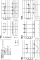
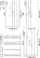
- Example 1 which examined about the relation between choroidal thickness and stress and anxiety is shown, and this figure is a graph showing the result of verifying the relation between choroidal thickness and subjective stress.
- the result of Example 1 which examined about the relation between choroidal thickness and stress and anxiety is shown, and this figure is a graph showing the result of verifying the relation between choroidal thickness and subjective stress.
- the result of Example 1 which examined about the relation between choroidal thickness and stress and anxiety is shown, and this figure is a graph showing the result of verifying the relation between choroidal thickness and subjective stress.
- the result of Example 1 which examined about the relation between choroidal thickness and stress and anxiety is shown, and this figure is a graph showing the result of verifying the relation between choroidal thickness and subjective stress.
- Example 1 The results of Example 1 in which the relationship between choroidal thickness and stress and anxiety were examined are shown, and this figure shows the occupation in the verification of the relationship between choroidal thickness and the result of the occupational stress simple questionnaire (57 items) The results of the sexual stress simple questionnaire (57 items) are shown.
- the results of Example 1 in which the relationship between choroidal thickness and stress and anxiety were examined are shown, and this figure shows the physical and mental responses caused by the stress of choroidal thickness and the occupational stress simple questionnaire (57 items) (region B) It is a graph which shows the verification result of relevancy with.
- Example 1 The result of Example 1 in which the relationship between choroidal thickness and stress and anxiety was examined is shown, and this figure shows the physical and mental caused by the stress of choroidal thickness / axial length and the occupational stress simple questionnaire (57 items) It is a graph which shows the verification result of relevance with reaction (B area
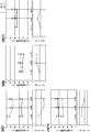
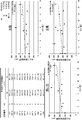
- the results of Example 1 in which the relationship between choroidal thickness and stress and anxiety were examined are shown, and this figure is a verification of the relationship between choroidal thickness and the results of the Japanese version of the mood / anxiety disorder questionnaire (K6).
- K6 The results of the Japanese version of the Mood / Anxiety Disorder Questionnaire (K6) and the correlation with the mind-body response (area B) caused by stress on the Occupational Stress Simple Questionnaire (57 items) are shown.
- Example 1 The results of Example 1 in which the relationship between choroidal thickness and stress and anxiety were examined are shown, and this figure is a verification of the relationship between choroidal thickness and the results of the Japanese version of the mood / anxiety disorder questionnaire (K6). It is a graph which shows a result.
- the results of Example 1 in which the relationship between choroidal thickness and stress and anxiety was examined are shown, and this figure shows the results of choroidal thickness / axial length and the Japanese version of the mood / anxiety disorder questionnaire (K6). It is a graph which shows the verification result of relevance.
- the method of detecting a stress state detects a stress state of a target object by using the choroid of the target body as an index.
- the detection method uses the choroid, which is a part of eye tissue that has not been recognized as a stress marker in the past, as an indicator of the stress state of the subject.
- Subjects in the method for detecting a stress state according to the present embodiment include humans, non-human primates, rabbits, rats, guinea pigs, mice, dogs, cats, cats, horses, cows, pigs, sheep, goats, chickens, etc. Although an animal is exemplified, a human is particularly preferable.
- the stress state in the method of detecting a stress state means a tension state generated by exerting a physical and mental load by various stimuli.
- the stimuli that induce stress are called stressors, and there are stressors due to environmental factors, physical factors, psychological factors, social factors, and the like.
- Environmental factors include, for example, temperature, humidity, light, weather, noise, vibration, exposure to harmful substances, air pollution and the like.
- Physical factors include, for example, trauma, illness, excessive exercise, lack of exercise, physical fatigue, lack of sleep, lack of nutrition, aging, unhealthy lifestyle, obesity and the like.
- reactive oxygen species and the like generated in excess can be included as physical factors.
- Psychological factors include, for example, anxiety, anxiety, frustration, anger, fear, disappointment, conflict and the like.
- Social factors include human relations, employment, career change, unemployment, admission, going to school, poor grades, bullying and so on. However, it is not limited to these.
- the stress state to be detected in the method of detecting a stress state according to the present embodiment is not particularly limited as long as it is a state induced by a stressor, and is any of a short-term acute stress state and a long-term chronic stress state. May be
- the stress state includes stress-related symptoms and diseases (hereinafter, may be abbreviated as “stress symptoms” and “stress diseases”, respectively), and may be a latent condition in which symptoms and diseases do not manifest in the mind and body. Stress conditions can also be targeted for detection. When exposed to stimuli such as stressors, the living body maintains its homeostasis through the interaction of the autonomic nervous system, endocrine system, immune system and the like.
- stress conditions can preferably include sympathetic dominant states, and symptoms and diseases induced by such sympathetic dominant states.
- Stress symptoms include, for example, physical symptoms such as stiff neck, palpitations, dizziness, insomnia, headache, weight fluctuation, loss of appetite, eating disorder, abdominal pain, addiction, etc., psychological symptoms such as anxiety, anger, nervousness, apathy, etc. It includes social symptoms such as overeating, alcohol consumption and withdrawal.
- stress diseases include mental and neurological diseases such as depression, neurosis and autonomic imbalance, cardiovascular diseases such as hypertension, arteriosclerosis, angina, myocardial infarction, obesity, diabetes, hyperlipidemia, Endocrine and metabolic disorders such as hyperthyroidism, respiratory diseases such as secretory bronchitis and hyperpnea syndrome, irritable bowel syndrome, gastric and duodenal ulcers, digestive tract diseases such as ulcerative colitis and psychogenic vomiting, Skin diseases such as atopic dermatitis, psoriasis, alopecia areata, central serous choroidal retinopathy (CSC), eye strain, eyelid spasms, eye diseases such as dry eye, otolaryngologic diseases such as Meniere's disease, enuresis Urinary and genital diseases such as cancer, inflammatory diseases such as cancer and chronic inflammation can be included.
- cardiovascular diseases such as hypertension, arteriosclerosis, angina, myocardial infarction, obesity, diabetes, hyperlipidemia, En
- mental and neurological diseases are considered, for example, anxiety which is a disease that suffers from excessive anxiety and fear caused by excessive anxiety and fear represented by panic disorder, social anxiety disorder, general anxiety disorder, phobia etc.
- Disorders and mood disorders which are disorders that cause an abnormality such as bipolar disorder, depression, and dysthymic disorder, are included.
- Stress conditions include factors such as various symptoms and exacerbation of diseases, so stress symptoms and diseases include not only onset of various symptoms and diseases but also exacerbation.
- the choroid which is an index for detecting a stress state, is a tissue rich in pigment cells and blood vessels between the retina and the sclera, and histologically, from the retina side It can be distinguished into four layers: basal plate (Bruch's membrane), choriocapillaris plate, blood vessel plate, and choroidal plate.
- basal plate Bruch's membrane
- choriocapillaris plate choriocapillaris plate
- blood vessel plate choroidal plate.
- the choroid is a tissue composed mainly of blood vessels, and it has been reported that the choroidal blood flow reaches 85% of the total eye blood flow (see, for example, Experimental Eye Research, 15 (1), 1973, p15-29) about). This leads to strong influence of physiological chemicals.
- choroidal blood vessels unlike retinal blood vessels, have been reported to have low self-regulatory ability, and it has been reported that blood flow through choroidal blood vessels fluctuates due to various physiological stimuli such as blood pressure (for example, Arch Ophthalmol, 83 (1) , 1970, p 95-99), choroidal vessels are strongly affected by changes in the general state of the body. Furthermore, it is known that the choroidal blood vessels express receptors that affect vascular tone, and in particular, it is known that choroidal blood flow is changed in pregnancy and hypertension.
- the method of detecting a stress state uses the choroid, which is a part of ocular tissue as described above, as an index of the stress state.
- the choroid is considered to be able to reflect changes in blood vessels throughout the body, and is considered to properly reflect the stress state of the living body.
- conventionally there has been no knowledge on the relationship between a stress state and ocular tissues including choroid.
- the method of detecting a stress state is preferably to detect a stress state of the object based on a tomographic image of the choroid of the object. From the tomographic image of the choroid, the state including the form of the choroid such as the choroidal thickness and the volume of the choroid can be grasped in detail.
- the tomographic image of the choroid is a tomographic image of an eye including the choroid, preferably a tomographic image of a fundus including the choroid. It is preferable that the boundary between the choroid and the retina and the boundary between the choroid and the sclera can be extracted in order to calculate the characteristic value of the choroid such as the thickness and volume of the choroid. Therefore, it is preferable that the tomographic image of the choroid includes at least a part of the retina and a part of the sclera in addition to the choroid.
- the tomographic image of the choroid may be, for example, a tomographic image of a region including the subfoveal region of the retina or a region including a position separated by a predetermined distance in a predetermined direction from the fovea relative to the fovea of the retina. it can. Further, a tomographic image of a region including the optic papilla, and a region including a position separated by a predetermined distance in a predetermined direction from the optic nerve papilla with respect to the optic nerve papilla can be used. Furthermore, it may be a tomographic image including a plurality of regions.
- the tomographic image of the choroid may be any of a one-dimensional image, a two-dimensional image, and a three-dimensional image.
- Tomographic images of the choroid can be obtained by tomographic imaging of the choroid using techniques known in the art.
- Tomographic imaging of the choroid can be performed preferably using an apparatus based on optical coherence tomography (hereinafter referred to as “OCT”) (hereinafter referred to as “OCT apparatus”).
- OCT apparatus is an interference optical apparatus that branches a light beam emitted from a light source and causes the reflected or scattered light of measurement light incident on a predetermined position of an object to interfere with reference light reflected from a reference object. This is a technology that uses interference light to image the structure of an object in the depth direction of the position where measurement light is incident, and can obtain a tomographic image of the object with high resolution.
- the OCT apparatus can acquire a tomographic image of the fundus including the choroid noninvasively and with high resolution by irradiating the fundus with the measuring light.
- the OCT apparatus is an apparatus capable of objectively inspecting the choroid, and since it is a short-time non-invasive inspection apparatus generally performed daily in ophthalmologic examination, the burden on the object is also small.
- the OCT apparatus is a time domain OCT (hereinafter referred to as "TD-OCT") that performs light wave interference in the time domain, and a Fourier domain OCT (frequency-domain OCT: below) that performs light wave interference in the frequency domain or wavelength domain. Or the like can be used, and preferably FD-OCT.
- TD-OCT time domain OCT
- frequency-domain OCT frequency-domain OCT: below
- FD-OCT Spectral domain OCT
- SD-OCT Spectral-domain OCT
- SS And the like, etc. are exemplified.
- it is SS-OCT.
- EDI-OCT based on enhanced intensity imaging (hereinafter referred to as “EDI”) or high penetration (hereinafter referred to as "HP-OCT”) using a long wavelength light source
- EDI enhanced intensity imaging
- HP-OCT high penetration
- a light source a light source emitting light with a wavelength of 1 ⁇ m band, typically about 950 nm to about 1100 nm, particularly 1050 nm, is preferable.
- 1 ⁇ m band light source it is less susceptible to absorption of light in the retinal pigment epithelium or the like and scattering in the case where there is turbidity in the intermediate translucent material, etc. Images can be acquired.
- a one-dimensional tomographic image (z image) of the fundus including the choroid in the depth direction (z direction) of the irradiation position by irradiating the fundus of the subject with the measurement light, it is possible to acquire a one-dimensional tomographic image (z image) of the fundus including the choroid in the depth direction (z direction) of the irradiation position. Furthermore, a two-dimensional tomographic image (xz image) of the fundus including the choroid can be acquired by one-dimensionally scanning the irradiation position in the direction (x direction) perpendicular to the depth direction with respect to the fundus of the object. One-dimensional scanning can be performed along a predetermined straight line or curve. Thereby, it is possible to obtain a two-dimensional tomographic image in the depth direction along the scanning direction of the measurement light.
- a three-dimensional image (xyz image) including the choroid can be acquired by two-dimensionally scanning the irradiation position in the direction (x, y directions) perpendicular to the depth direction with respect to the fundus. At this time, two-dimensional scanning can be performed within a predetermined area. That is, a three-dimensional tomographic image can be constructed from a plurality of two-dimensional images acquired by repeatedly scanning the measurement light while shifting the position with respect to the fundus of the object in a predetermined area. Thus, the structure of the fundus including the choroid can be captured in three dimensions.
- SS-OCT can scan a wide range of about 3 to 12 mm in the x direction and y direction in a few seconds and acquire an image in the deep direction, so a clear three-dimensional tomographic image of the fundus including the choroid can be constructed. It becomes possible.
- the time required for acquiring a tomographic image of the choroid by the OCT apparatus is about 3 seconds, and there is also an advantage of being less susceptible to eye movement.
- a tomographic image of the choroid it is preferable to acquire a tomographic image of the choroid by avoiding during or immediately after exercise.
- choroidal thickness it has been reported that the choroid has become significantly thickened for at least 5 minutes during exercise with low load and moderate intensity (for example, Sayin N et al., Indian J Ophthalmol. 2015, 63). (5), 445-450).
- acquisition of a tomographic image of the choroid is generally performed in the sitting position, and may be a tomographic image of the choroid of either the left or right eye, or may be a tomographic image of the choroid of both eyes.
- the step of detecting a stress state may include calculating a choroidal thickness from the tomographic image and detecting a stress state of the object based on the calculated choroidal thickness.
- the choroidal thickness is the thickness of the choroid, and can be calculated from the tomographic image of the choroid as, for example, the vertical distance from the border between the retinal pigment epithelium located at the outermost side of the retina and the choroid to the border between the choroid and sclera.
- the localization of the retinal pigment epithelium-choroid interface and the localization of the choroid-sclera interface can be identified using image processing techniques known in the art. For example, the positions of these boundaries can be identified from changes in pixel values (for example, luminance values) in the tomographic image.
- the choroidal thickness may be calculated, for example, by using the calculation function of a computer.
- the calculation of the choroidal thickness is preferably performed at a predetermined position of the choroid.
- the position may be a position of the fovea of the retina or a position separated from the fovea by a predetermined distance in a predetermined direction (for example, upper, lower, nasal side, ear side) with reference to the fovea of the retina .
- the position may be measured at the position of the optic nerve head or at a position away from the optic disk by a predetermined distance in a predetermined direction with reference to the optic disk.
- the choroidal thickness may be measured at a plurality of positions. Particularly preferred is the choroidal thickness below the fovea of the retina.
- the calculation of choroidal thickness may be performed on the choroid of either the left or right eye, or may be performed on the choroid of both eyes. Also, after calculating the choroidal thickness of both eyes, only the thin or thick choroid may be used for detecting the stress state, but the thin one with high correlation of the stress state is used for detection Is preferred.
- the subject in a stressed state exhibits a significantly higher value of choroidal thickness as compared to a healthy non-stressed normal control group.
- choroidal blood vessels dilate and choroids become thick due to overstimulation of the autonomic nerve.
- detection of an object in a stress state is a reference value calculated in advance from a choroidal thickness calculated from a tomogram of the choroid of the object and, for example, a tomogram of a choroid in a healthy object not in a stress state.
- the object is detected as being in a stress state, and if the value is lower than the reference value, the object can be detected not in a stress state.
- selection of healthy body which is not in the stress state used for calculation of standard value such as occupational stress simple questionnaire (57 items) which Japanese Ministry of Health, Labor and Welfare recommends, Japanese version mood / anxiety disorder questionnaire (K6) And other known methods of detecting stress such as a stress marker, and a plurality of methods of detecting stress are preferably performed.
- the stress state can be detected with the reference value.
- a reference value for example, in the case of an adult, it can be set to any value of 250 ⁇ m, 300 ⁇ m or less under the fovea, and preferably can be set to any value from 200 to 300 ⁇ m under the fovea.
- the calculated choroidal thickness of the object is obtained from the detection time, the age, sex, medical history of the object, and an index obtained from the eye structure such as axial length and equivalent sphere area Correction may be included using at least one factor selected from For example, choroidal thickness is negatively correlated with age, and it has been reported that choroidal thinning with age (Wakatsuki Y et al., PLoS One. 2015 Dec 3; 10 (12): e0144156).
- the choroidal thickness is diurnal, and that the choroid is thick in the early morning but becomes gradually thinner in the daytime, and there is no difference in the fluctuation width of the choroidal thickness (for example, See Tan CS et al., Invest Opthalmol Vis Sci, 2012; 53 (1): 261-266 etc.).
- the choroidal layer thickness is affected by some eye diseases and systemic pathologies, and, for example, in central serous chorioretinosis or Harada disease, the choroidal layer thickness is significantly thickened, conversely, strong myopia, age-related macular degeneration It has been reported that glaucoma, diabetic retinopathy, etc. are thinning.
- the eye axis is short, or It has been reported that the thicker the equivalent sphere area, the thicker the choroid. Furthermore, flatter cornea, it has been reported that the choroid is thicker as the correction vision is better.
- the choroidal film thickness calculated from the group with the above factor at the standard value and not in the stress state is used as the standard choroidal film thickness. Determined as Then, when the above factor is not a standard value, a ratio of a standard choroidal film thickness to a choroidal film thickness calculated from a group not in a stress state (standard choroidal film thickness / calculated choroidal film thickness) is used as a correction coefficient. By multiplying the correction film thickness by the correction coefficient, it is possible to correct the correction film thickness calculated from an object having no corresponding standard factor.
- the correction can be made based on the correlation.
- the factor is gender
- the choroidal thickness of the other sex can be corrected based on the choroidal thickness of one sex.
- the choroidal thickness calculated from the group not having any of the above factors and not in the stress state is used as a standard.
- the ratio of standard choroidal film thickness to choroidal film thickness calculated from a group not in a stressed state having any one of the above factors (standard choroidal film thickness / calculated choroidal film thickness) as choroidal film thickness is used as a correction coefficient Based on this, it is possible to correct the choroidal film thickness calculated from the object having the corresponding factor.
- the correction may be performed, for example, by using the calculation function of a computer, as in the calculation of the choroidal film thickness.
- the step of detecting the stress state may include calculating a choroidal volume from the tomographic image and detecting a stress state of the subject based on the calculated volume of the choroid.
- the volume of the choroid can be a volume of a portion surrounded by the retinal pigment epithelium located at the outermost side of the retina and the border of the choroid and the border between the choroid and the sclera, and can be calculated based on the choroidal thickness described above.
- the calculation of the choroidal volume may be performed, for example, by using the computing function of a computer.
- the calculation of the choroidal volume is preferably performed in a predetermined area of the choroid.
- the position of the fovea of the retina, or a direction (x direction) perpendicular to a predetermined direction (deep direction (z direction) from the fovea relative to the fovea of the retina, eg, upper, lower, nasal side It can be calculated as the volume of the choroid in a region whose center point is a position separated by a predetermined distance on the ear side).
- the volume of the choroid may be calculated as the central point of the position of the optic nerve head or the position at a predetermined distance from the optic nerve head in a predetermined direction with reference to the position of the optic nerve head.
- the volume of the choroid in a plurality of regions may be calculated. Particularly preferred is the volume of the choroid in a region centered on the position of the fovea of the retina.
- the size of the area for calculating the volume of the choroid is not particularly limited, but can be, for example, 3 mm ⁇ 3 mm, 6 mm ⁇ 6 mm, 10 mm ⁇ 10 mm.
- the calculation of the choroidal volume may be performed on the choroid of either the left or right eye, or may be performed on the choroid of both eyes. In addition, after calculating the choroidal thickness of both eyes, only one with a large or small choroidal volume may be used for detecting a stress state.
- the choroidal volume shows a significantly higher value as compared to a healthy non-stressed normal control group.
- the stress state of the subject can be detected by comparing the volume of the choroid calculated from the tomogram of the choroid of the subject with, for example, a reference value calculated in advance from the tomogram of the choroid of a healthy subject not in a stress state. If the value of the choroidal volume of the subject is higher than the reference value, the subject is detected as being in a stressed state, and if the value is lower, it can be detected that the subject is not in a stressed state.
- a reference value for example, in the case of an adult, it is set to an arbitrary value of 24.3 mm 3 or less, preferably 20.3 mm 3 under an area of 9 mm ⁇ 9 mm (12 mm ⁇ 9 mm at maximum) centered on the fovea it can.
- the method of detecting a stress state can be configured to measure the stress intensity of the subject based on the volume of the choroid calculated from the tomographic image of the choroid of the object.
- the step of detecting the stress state includes selecting at least one of the calculated choroidal volume of the subject from the time of detection, the age of the subject, the gender, the current medical history, the index obtained from the eye structure, etc.
- a step of correcting with a factor can be included.
- the index obtained from the eye structure the axial length, the equivalent sphere area and the like are exemplified, but it is not limited thereto.
- the choroidal volume is also affected by the factors described above, as well as the choroidal thickness described above. Therefore, by correcting the influence of the factors described above on the choroidal volume, it is possible to detect a more accurate stress state of the object.
- the correction is performed when the volume of the choroid changes due to the numerical value fluctuation of the factor such as the detection time or the age, the volume of the choroid calculated from the group in which the factor is at the standard value and is not in the stress state Determined as a standard volume. Then, when the above factor is not a standard value, the ratio of the standard volume to the choroidal volume calculated from the group not in a stress state (standard volume / calculated choroidal volume) is used as a correction coefficient. By multiplying the calculated volume of the choroid by the correction factor, it is possible to correct the volume of the choroid calculated from the object for which the corresponding factor is not in the standard value.
- correction can be made based on the correlation. For example, if the factor is gender, then the volume of one sex choroid can be corrected based on the volume of the other sex choroid. Also, for example, when the volume of the choroid changes due to the presence or absence of the factor such as the present disease history, the choroid that is calculated from the group that does not have any of the factors described above and is not in a stress state Assuming that the volume is a standard volume, the ratio of the standard volume to the choroidal volume (standard volume / calculated choroidal volume) calculated from the group not in a stressed state having any one of the factors described above is a correction factor. Based on the correction factor, it is possible to correct the choroidal volume calculated from the object having the corresponding above mentioned factor. The correction may be performed, for example, by using the calculation function of a computer, as in the calculation of the choroidal volume and the like.
- the stress state of the object can be objectively detected, and the choroid can accurately reflect the stress state of the living body, so that the stress state of the object can be accurately detected. . Therefore, there is no room for non-detection of the stress state due to the intentional response change of the target object, which is a problem in the conventional stress detection method which is the subjective stress detection method, etc., and highly stressed persons can be selected with high reliability. .
- the stress state can be detected in a non-contact, non-invasive manner, in a short time and easily, and therefore the mental and physical burden on the object can be reduced. Therefore, the stress load upon detection can be eliminated, and the stress state of the object can be accurately detected. Furthermore, there is also an advantage that it is not susceptible to eye movement.
- conventional biomarkers are degraded and disappear in a short time even if they appear in the body in response to stress, and there is a problem that it is difficult to grasp the stress state. According to such a stress state detection method, such a problem does not occur.
- the stress state detection method according to the present embodiment may be configured to automatically detect a stress state by an evaluation unit using a stress detection device described later, a computer, or the like.
- a subject in a stress state can be detected with high reliability.
- consultation with a suitable medical institution and counseling can be recommended.
- the method of detecting a stress state according to the present embodiment it is possible to detect a potential stress state in which stress is not manifested as a specific symptom, disease or the like. Therefore, a stress state can be detected early, which can lead to prevention of symptoms and diseases developed by stress. Therefore, the method of detecting a stress state according to the present embodiment can be suitably used for regular health check and the like of workers, and can contribute to the physical and mental health management and the like of workers. As a result, it leads to the improvement of the work environment, the work efficiency rises, and it can contribute to the improvement of labor productivity.
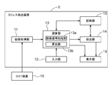
- the stress detection apparatus X can include, for example, an image acquisition unit 11, an input unit 12, an operation unit 13, a detection unit 14, a storage unit 15, a display unit 16 and the like as shown in FIG. .
- the stress detection device X in the present embodiment is configured of a desktop personal computer, a notebook personal computer, a tablet terminal or the like, and can communicate with the OCT device 10 wirelessly or by wire using the image acquisition unit 11 as a communication interface.
- the arithmetic unit 13 and the detection unit 14 are configured by hardware or software or both with the CPU as a core member
- the storage unit 15 is configured by a ROM, a RAM, and the like
- the display unit 16 is a liquid crystal screen or the like. It comprises a known display.
- the stress detection device X may be provided integrally with the OCT device 10, or part of the functions of the calculation unit 13 and the like may be incorporated in the OCT device 10, and is not particularly limited.
- the image acquisition unit 11 acquires tomographic image information of the fundus including the choroid of the object from the OCT apparatus 10 described above.
- the OCT apparatus 10 branches a light source, a measurement optical system that branches a light flux emitted from the light source and irradiates the inside of the eye to be examined and guides measurement light from the fundus of the subject, and branches the light flux emitted from the light source.
- the reference optical system that irradiates the reference object and guides the reference light that is the reflected light, and the interference light that combines the measurement light guided by the measurement optical system and the reference light guided by the reference optical system
- a light receiving element that converts the light into a signal and outputs the signal.
- the input unit 12 receives an operation input by the operator, and outputs an operation input signal corresponding to the operation input to the operation unit 13.
- the input unit 12 is configured of a button, a switch, and a keyboard.
- the input unit 12 can input, for example, information of an object (age, gender, current medical history, axial length, etc.), examination date, and the like.
- the calculation unit 13 processes the tomographic image signal acquired by the image acquisition unit 11.
- the calculation unit 13 includes, for example, an image signal processing unit 13a and a calculation unit 13b.
- the image signal processing unit 13 a performs various types of image processing and the like based on the output signal of the image acquisition unit 11 to generate a tomographic image of the fundus including the choroid.
- the tomographic image generated by the image signal processing unit 13 a is configured to be stored in the storage unit 15 and to be output to the display unit 16.
- the calculating unit 13 b calculates a feature value of the choroid based on the tomographic image of the fundus including the choroid generated by the image signal processing unit 13 a.
- the characteristic value of the choroid can be exemplified by the choroidal thickness of the object, the volume of the choroid, and the like.
- the definition of choroidal thickness and choroidal volume is as described in the section of (Method of detecting stress state) above.
- the choroidal thickness can be calculated by specifying the boundary between the retinal pigment epithelium and the choroid and the boundary between the choroid and the sclera, and measuring the vertical distance between the two boundaries.
- the boundary between the retinal pigment epithelium and the choroid, and the boundary between the choroid and the sclera can be specified by applying known image processing techniques, and automatically performed according to a preset algorithm. You can also.
- a boundary value can be extracted by searching for a pixel value (for example, a luminance value or the like) in the depth direction and detecting a change in the pixel value.
- a boundary line may be specified by
- the measurement of the choroidal thickness is performed by measuring the distance between the two boundaries identified above at a defined measurement position.
- the measurement can be performed, for example, by counting the number of pixels aligned in the depth direction between both boundaries in the tomographic image.
- the specification of the position of the fovea centralis or the optic disc of the retina which is the basis of calculation of the choroidal value, can be set to be automatically performed by applying a known image processing technique in the tomographic image of the choroid. Also, the operator may specify it manually.
- the volume of the choroid can be calculated by adding the thickness information calculated above in the three-dimensional tomographic image of the choroid, and preferably, the volume in the preset specific region is calculated.
- the calculator 13b calculates the characteristic value of the choroid calculated from the tomographic image of the choroid by the calculator 13b, for example, the detection time input by the input unit 12, the age of the object, the sex, the present medical history, the eye axis length, Corrections can be made to eliminate the effects of factors that affect choroidal feature values, such as indices obtained from eye structures such as equivalent sphere areas.
- the correction of the characteristic value of the choroid is the same as the correction content described above, so the description will be omitted.
- the detection unit 14 detects the stress state of the object based on the feature value of the choroid calculated from the tomographic image of the choroid by the calculation unit 13 b.
- the detection unit 14 determines whether the feature value of the choroid calculated by the calculation unit 13 b is equal to or greater than a reference value calculated in advance from the tomographic image of the choroid of a healthy subject not in a stress state stored in the storage unit 15. To detect.
- the comparison between the characteristic value of the choroid of the subject and the reference value can be made based on known statistical methods.
- the detection unit 14 stores the detection result in the storage unit 15 and outputs the detection result to the display unit 16 as a detection result of the stress state.
- the storage unit 15 is calculated by the calculation unit 13 b, the tomographic image of the choroid formed by the image signal processing unit 13 a of the calculation unit 13, the input data such as the information of the object and the examination date and time input by the operator from the input unit 12. Information such as the characteristic value of the choroid of the object and the stress state detected by the detection unit 14 is stored.
- the storage unit 15 also stores a calculation program for calculating the feature value of the choroid, a detection program for detecting a stress state, a reference value used in detection, and the like.
- the storage unit 15 affects the characteristic values of the choroid such as the detection time, the age of the subject, the sex, the current medical history, and the index obtained from the eye structure such as axial length and equivalent sphere area.
- the correction program etc. for correcting the influence of the factor to be stored are stored.
- the display unit 16 displays the information of the object input by the input unit, the examination date, the tomographic image of the choroid formed by the image signal processing unit, and the characteristic value of the choroid calculated from the tomographic image of the choroid by the calculation unit.
- the detection unit displays the detection result of the stress state detected based on the characteristic value of the choroid by comparison with the reference value.
- the display unit 16 can display a warning that it is necessary to have a medical examination or counseling when it is detected that the display device 16 is in a stress state according to the detection result.
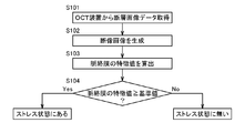
- Step S101 The tomographic image signal (tomographic image data) of the eye of the subject taken by the OCT apparatus 10 is acquired by the image acquisition unit 11.
- Step S102 Then, based on the tomographic image signal acquired by the image signal processing unit 13a, the image signal processing unit 13a performs various types of image processing and the like to generate a tomographic image of the fundus including the choroid.
- the tomographic image of the fundus including the choroid generated by the image signal processing unit 13a may be stored in the storage unit 15, and the tomographic image of the fundus including the choroid may be displayed on the display unit 16.
- the operation of the OCT apparatus 10 the arrangement of the eye of the object, etc. are appropriately corrected, and step S101 is performed again. It may be configured to execute.
- the calculation unit 13b follows the calculation program for calculating the feature value of the choroid stored in the storage unit 15 based on the tomographic image of the fundus including the choroid generated by the image signal processing unit 13a. Alternatively, control is performed to calculate the volume and to output information on the calculated characteristic value of the choroid to the display unit 16. At this time, the calculation unit 13 b may correct the feature value of the choroid according to a correction program for correcting the feature value of the choroid stored in the storage unit 15 as necessary. In addition, the display unit 16 may display an image of the feature value and the correction content of the choroid based on the input information.
- Step S104 The detection unit 14 determines whether the object is in a stress state based on the information on the feature value of the choroid calculated by the calculation unit 13 b and the reference value of the feature value of the choroid stored in the storage unit 15. To detect. Specifically, the detection unit 14 detects that the object is in a stress state when the characteristic value of the choroid is equal to or greater than the reference value, and the object is detected when the characteristic value of the choroid is less than the reference value. The body detects that it is not in stress.
- the detection result of the detection unit 14 is output to the display unit 16, and the display unit 16 displays the stress state of the object on the screen based on the input information.
- a warning screen or the like may be displayed on the assumption that consultation or counseling is necessary.
- the detection unit 14 may detect the stress state in multiple stages, and may display characters such as “high stress”, “medium stress”, “low stress” or the like on the display unit 16 or even by numerical display in multiple stages. Good.
- Example 1 Verification of Relationship between Choroidal Thickness and Stress and Anxiety
- the relationship between stress and anxiety and the choroidal thickness which is a part of eye tissue was verified.
- the relationship with the detection result by the subjective stress detection method was evaluated.
- Method 1-1 Implementation period 1-1-1. Date of implementation: 1-3-2 consecutive days from July to October 2017 1-1-2. Implementation time Evening to night
- the subject profiles are summarized in Table 1 below.
- 1-3 Verification method The stress state of the object was verified by the following three methods. 1-3-1. Verification by Choroidal Film Thickness The choroidal film thickness was measured by SS-OCT (TOPCON (registered trademark) SS-OCT, manufactured by TOPON) on the subject. The choroid was measured by measuring the foveal choroidal thickness. Specifically, a tomographic image of the fundus is acquired so that the fovea retina is as vertical as possible with respect to the tomographic screen, and a ruled line from immediately below the retinal pigment epithelium in the fovea to a position considered to be the lower edge of the choroid. was vertically measured, and the distance was measured as the foveal choroidal thickness.
- the position of the fixation point was finely adjusted so that the fovea was at the center of the tomographic image. Further, the axial length of the subject was measured by IOLmaster (manufactured by ZEISS), and measurement of blood pressure and pulse, and BUT test were performed. At the time of measurement, subjective stress felt by the subject was scored by self-evaluation. Subjective stress was rated on a scale of 5 (5: very, 4: somewhat, 3: slightly, 2: hardly, 1: not at all).
- the method of selecting highly stressed persons using the raw score conversion table was based on the manual for stress check system implementation based on the Industrial Safety and Health Law (http://www.mhlw.go.jp/bunya/roudoukijun/anzeneisei 12 / pdf / See 150803-1.pdf).
- K6 Verification by Japanese version mood / anxiety disorder questionnaire (K6)
- K6 Questionnaire by Japanese version mood / anxiety disorder questionnaire (K6) (http://www.city.noshiro.akita.jp/upload/ (See download / 118779download.pdf), and the questionnaire results were collected.
- K6 is a six-item, five-measure scale that measures mood and anxiety disorders developed by Kessler et al. (See Kessler RC et al., Psychological Medicine, 2002, 32, 959-976). It has been reported that it can be adapted (Sakurai K et al., Psychiatry Clin Neurosci., 2011; 65 (5): 434-441, etc.).
- FIG. Fig. 7 shows the verification results of the relationship between the choroidal thickness and the mind-body response (area B) caused by the stress in the questionnaire for occupational stress (57 items), and Fig. 8 shows the choroidal thickness / axial length and occupationality.
- FIG. Fig. 7 shows the verification results of the relationship between the choroidal thickness and the mind-body response (area B) caused by the stress in the questionnaire for occupational stress (57 items)
- Fig. 8 shows the choroidal thickness / axial length and occupationality.
- the choroidal thickness is the average foveal thickness for 3 days or 4 days.
- FIG. 10 shows the result of examining the relationship between the choroidal thickness and the results of the Japanese version of the mood / anxiety disorder questionnaire (K6), and FIG.
- FIG. 11 shows the choroidal thickness / axial length and the Japanese version of the mood / anxiety disorder questionnaire.
- the verification result of relevance with the result of (K6) is shown.
- the choroidal thickness is the average foveal thickness for 3 days or 4 days.
- the choroid was found to be a stress marker.
- the Japanese version of the Mood Anxiety Disorder Questionnaire (K6) is used worldwide. Therefore, it can be understood that the thickness of the choroid is a stress marker as a matter of course from the data of this research, and it can be understood that if the choroid is thick, the object can be detected with high reliability if it is in a stress state. .
- Tables 3 to 5 show the measurement time of the choroidal thickness and the measurement result of the foveal choroidal thickness (left and right).
- Table 4 shows the measurement results of age and mean foveal choroidal thickness (left and right), and it can be understood that positive correlation is recognized between age and choroidal thickness.
- Table 5 shows the measurement results of gender and mean foveal choroidal thickness (left and right), and it was recognized that the female tended to be thicker in the choroid than the male. Therefore, it can be understood that the stress state of the object can be detected more accurately based on the choroidal film thickness by correcting the measured choroidal film thickness with respect to factors affecting the choroidal film thickness such as age and gender.
- the present invention can be used in all technical fields requiring detection of a stress condition, such as early detection of a stress condition or prevention of a disease or condition induced by a stress condition, and in particular, health management of workers. It can be used for etc.
- OCT apparatus 11 Image acquisition unit 12: Input unit 13: Calculation unit 13a: Image signal processing unit 13b: Calculation unit 14: Detection unit 15: Storage unit 16: Display unit X: Stress detection device
Landscapes
- Health & Medical Sciences (AREA)
- Life Sciences & Earth Sciences (AREA)
- Engineering & Computer Science (AREA)
- General Health & Medical Sciences (AREA)
- Veterinary Medicine (AREA)
- Biophysics (AREA)
- Physics & Mathematics (AREA)
- Public Health (AREA)
- Biomedical Technology (AREA)
- Heart & Thoracic Surgery (AREA)
- Medical Informatics (AREA)
- Molecular Biology (AREA)
- Surgery (AREA)
- Animal Behavior & Ethology (AREA)
- Ophthalmology & Optometry (AREA)
- Psychiatry (AREA)
- Nuclear Medicine, Radiotherapy & Molecular Imaging (AREA)
- Radiology & Medical Imaging (AREA)
- Pathology (AREA)
- Developmental Disabilities (AREA)
- Child & Adolescent Psychology (AREA)
- Educational Technology (AREA)
- Hospice & Palliative Care (AREA)
- Psychology (AREA)
- Social Psychology (AREA)
- Investigating Or Analysing Biological Materials (AREA)
- Measurement Of The Respiration, Hearing Ability, Form, And Blood Characteristics Of Living Organisms (AREA)
- Eye Examination Apparatus (AREA)
Priority Applications (4)
| Application Number | Priority Date | Filing Date | Title |
|---|---|---|---|
| EP19743747.8A EP3744254A4 (en) | 2018-01-25 | 2019-01-25 | STRESS CONDITION DETECTION METHOD AND STRESS DETECTION DEVICE |
| CN201980010385.0A CN111655151B (zh) | 2018-01-25 | 2019-01-25 | 压力状态的检测方法以及压力检测装置 |
| US16/964,029 US20210038077A1 (en) | 2018-01-25 | 2019-01-25 | Method for detecting stressed state and stress detection apparatus |
| JP2019567175A JP6930763B2 (ja) | 2018-01-25 | 2019-01-25 | ストレス状態の検出方法、及び、ストレス検出装置 |
Applications Claiming Priority (2)
| Application Number | Priority Date | Filing Date | Title |
|---|---|---|---|
| JP2018-010822 | 2018-01-25 | ||
| JP2018010822 | 2018-01-25 |
Publications (1)
| Publication Number | Publication Date |
|---|---|
| WO2019146738A1 true WO2019146738A1 (ja) | 2019-08-01 |
Family
ID=67396059
Family Applications (1)
| Application Number | Title | Priority Date | Filing Date |
|---|---|---|---|
| PCT/JP2019/002394 WO2019146738A1 (ja) | 2018-01-25 | 2019-01-25 | ストレス状態の検出方法、及び、ストレス検出装置 |
Country Status (5)
| Country | Link |
|---|---|
| US (1) | US20210038077A1 (zh) |
| EP (1) | EP3744254A4 (zh) |
| JP (1) | JP6930763B2 (zh) |
| CN (1) | CN111655151B (zh) |
| WO (1) | WO2019146738A1 (zh) |
Cited By (1)
| Publication number | Priority date | Publication date | Assignee | Title |
|---|---|---|---|---|
| WO2022208873A1 (ja) * | 2021-04-02 | 2022-10-06 | 日本電気株式会社 | ストレス推定装置、ストレス推定方法及び記憶媒体 |
Families Citing this family (3)
| Publication number | Priority date | Publication date | Assignee | Title |
|---|---|---|---|---|
| CN111954485A (zh) * | 2018-04-18 | 2020-11-17 | 株式会社尼康 | 图像处理方法、程序、图像处理装置及眼科系统 |
| JP7277188B2 (ja) * | 2019-03-14 | 2023-05-18 | 株式会社日立製作所 | 作業場の管理支援システムおよび管理支援方法 |
| CN112826442A (zh) * | 2020-12-31 | 2021-05-25 | 上海鹰瞳医疗科技有限公司 | 基于眼底图像的心理状态识别方法及设备 |
Citations (7)
| Publication number | Priority date | Publication date | Assignee | Title |
|---|---|---|---|---|
| JP2000275248A (ja) | 1999-03-24 | 2000-10-06 | Agency Of Ind Science & Technol | 慢性ストレス判定方法及びその装置、記録媒体、判定シート |
| JP2002544159A (ja) * | 1999-05-07 | 2002-12-24 | ジョンズ ホプキンス ユニヴァーシティ スクール オヴ メディシン | 眼障害の処置におけるプロテインチロシンキナーゼ経路インヒビターの使用 |
| JP2007225606A (ja) | 2006-01-27 | 2007-09-06 | Prima Meat Packers Ltd | 新規ストレスバイオマーカー及びその用途 |
| JP2012251857A (ja) | 2011-06-02 | 2012-12-20 | Nagaoka Univ Of Technology | ストレスの評価方法 |
| JP2013066702A (ja) * | 2011-09-06 | 2013-04-18 | Canon Inc | 画像処理装置および画像処理方法 |
| JP2014104275A (ja) * | 2012-11-29 | 2014-06-09 | Osaka Univ | 眼科装置 |
| JP2017074273A (ja) * | 2015-10-16 | 2017-04-20 | 国立大学法人 鹿児島大学 | 画像処理装置、画像処理方法、診断システム及びプログラム |
Family Cites Families (18)
| Publication number | Priority date | Publication date | Assignee | Title |
|---|---|---|---|---|
| US7575321B2 (en) * | 2003-10-30 | 2009-08-18 | Welch Allyn, Inc. | Apparatus and method of diagnosis of optically identifiable ophthalmic conditions |
| JP5006070B2 (ja) * | 2007-02-28 | 2012-08-22 | 森永製菓株式会社 | ストレス強度演算方法、及びストレス強度演算装置 |
| US7942820B2 (en) * | 2007-08-26 | 2011-05-17 | Philip Chidi Njemanze | Method and system for evaluation of the hemodynamic model in depression for diagnosis and treatment |
| JP5192250B2 (ja) * | 2008-02-04 | 2013-05-08 | 株式会社トプコン | 眼底観察装置 |
| JP2010246787A (ja) * | 2009-04-17 | 2010-11-04 | Sanyo Electric Co Ltd | マッサージ機 |
| EP2470116B1 (en) * | 2009-08-26 | 2014-12-03 | The Bionics Institute of Australia | Apparatus for stimulating and/or monitoring activity in tissue |
| US8882272B2 (en) * | 2010-06-25 | 2014-11-11 | Annidis Health Systems Corp. | Method and apparatus for imaging the choroid |
| JP5836634B2 (ja) * | 2011-05-10 | 2015-12-24 | キヤノン株式会社 | 画像処理装置及び方法 |
| JP6146952B2 (ja) * | 2012-01-27 | 2017-06-14 | キヤノン株式会社 | 画像処理装置、画像処理方法及びプログラム。 |
| JP6278295B2 (ja) * | 2013-06-13 | 2018-02-14 | 国立大学法人 筑波大学 | 脈絡膜の血管網を選択的に可視化し解析する光干渉断層計装置及びその画像処理プログラム |
| JP6322042B2 (ja) * | 2014-04-28 | 2018-05-09 | キヤノン株式会社 | 眼科撮影装置、その制御方法、およびプログラム |
| WO2015188279A1 (en) * | 2014-06-12 | 2015-12-17 | Rsem, Limited Partnership | Non-invasive measurement of choroidal volume change and ocular rigidity through oct imaging |
| NZ773815A (en) * | 2015-03-16 | 2022-07-01 | Magic Leap Inc | Methods and systems for diagnosing and treating health ailments |
| WO2017136696A1 (en) * | 2016-02-05 | 2017-08-10 | Murali Archana | Methods and apparatus for processing opthalmic biomarkers |
| JP2017169974A (ja) * | 2016-03-25 | 2017-09-28 | パナソニックIpマネジメント株式会社 | 生体情報計測装置 |
| KR102355455B1 (ko) * | 2016-06-20 | 2022-01-24 | 매직 립, 인코포레이티드 | 시각적 프로세싱 및 지각 상태들을 포함하는 신경학적 상태들의 평가 및 수정을 위한 증강 현실 디스플레이 시스템 |
| JP6165935B2 (ja) * | 2016-07-08 | 2017-07-19 | 株式会社トプコン | 光画像計測装置 |
| JP6427224B2 (ja) * | 2017-04-27 | 2018-11-21 | 株式会社トプコン | 眼科撮影装置 |
-
2019
- 2019-01-25 WO PCT/JP2019/002394 patent/WO2019146738A1/ja unknown
- 2019-01-25 JP JP2019567175A patent/JP6930763B2/ja active Active
- 2019-01-25 US US16/964,029 patent/US20210038077A1/en active Pending
- 2019-01-25 EP EP19743747.8A patent/EP3744254A4/en active Pending
- 2019-01-25 CN CN201980010385.0A patent/CN111655151B/zh active Active
Patent Citations (7)
| Publication number | Priority date | Publication date | Assignee | Title |
|---|---|---|---|---|
| JP2000275248A (ja) | 1999-03-24 | 2000-10-06 | Agency Of Ind Science & Technol | 慢性ストレス判定方法及びその装置、記録媒体、判定シート |
| JP2002544159A (ja) * | 1999-05-07 | 2002-12-24 | ジョンズ ホプキンス ユニヴァーシティ スクール オヴ メディシン | 眼障害の処置におけるプロテインチロシンキナーゼ経路インヒビターの使用 |
| JP2007225606A (ja) | 2006-01-27 | 2007-09-06 | Prima Meat Packers Ltd | 新規ストレスバイオマーカー及びその用途 |
| JP2012251857A (ja) | 2011-06-02 | 2012-12-20 | Nagaoka Univ Of Technology | ストレスの評価方法 |
| JP2013066702A (ja) * | 2011-09-06 | 2013-04-18 | Canon Inc | 画像処理装置および画像処理方法 |
| JP2014104275A (ja) * | 2012-11-29 | 2014-06-09 | Osaka Univ | 眼科装置 |
| JP2017074273A (ja) * | 2015-10-16 | 2017-04-20 | 国立大学法人 鹿児島大学 | 画像処理装置、画像処理方法、診断システム及びプログラム |
Non-Patent Citations (14)
| Title |
|---|
| ARCH OPHTHALMOL, vol. 83, no. 1, 1970, pages 95 - 99 |
| BERT LR CORNELIUS ET AL., BMC PUBLIC HEALTH, vol. 13, 2013, pages 128 |
| EXPERIMENTAL EYE RESEARCH, vol. 15, no. 1, 1973, pages 15 - 29 |
| HIROHITO NANBU ET AL., KOUSEI NO SHIHYO, vol. 61, no. 5, May 2014 (2014-05-01), pages 23 - 30 |
| IKUNO Y ET AL., INVEST OPHTHALMOL VIS SCI, vol. 51, no. 4, 2010, pages 2173 - 2176 |
| KESSLER RC ET AL., ARCH GEN PSYCHIATRY, vol. 60, 2003, pages 184 - 189 |
| KESSLER RC ET AL., PSYCHOLOGICAL MEDICINE, vol. 32, 2002, pages 959 - 976 |
| NORITO KAWAKAMI ET AL., HEALTH AND LABOR SCIENCES RESEARCH GRANT, 2014 |
| SAKURAI K ET AL., PSYCHIATRY CLIN NEUROSCI., vol. 65, no. 5, 2011, pages 434 - 441 |
| SAYIN N ET AL., INDIAN J OPHTHALMOL., vol. 63, no. 5, 2015, pages 445 - 450 |
| See also references of EP3744254A4 |
| TAN CS ET AL., INVEST OPHTHALMOL VIS SCI, vol. 53, no. 1, 2012, pages 261 - 266 |
| WAKATSUKI Y ET AL., PLOS ONE, vol. 10, no. 12, 3 December 2015 (2015-12-03), pages e0144156 |
| WEN BIN WEI ET AL., OPHTHALMOLOGY, vol. 120, no. 1, 2013, pages 175 - 180 |
Cited By (1)
| Publication number | Priority date | Publication date | Assignee | Title |
|---|---|---|---|---|
| WO2022208873A1 (ja) * | 2021-04-02 | 2022-10-06 | 日本電気株式会社 | ストレス推定装置、ストレス推定方法及び記憶媒体 |
Also Published As
| Publication number | Publication date |
|---|---|
| EP3744254A1 (en) | 2020-12-02 |
| JP6930763B2 (ja) | 2021-09-01 |
| US20210038077A1 (en) | 2021-02-11 |
| EP3744254A4 (en) | 2021-11-03 |
| JPWO2019146738A1 (ja) | 2020-12-03 |
| CN111655151A (zh) | 2020-09-11 |
| CN111655151B (zh) | 2024-03-22 |
Similar Documents
| Publication | Publication Date | Title |
|---|---|---|
| JP6930763B2 (ja) | ストレス状態の検出方法、及び、ストレス検出装置 | |
| Ferguson et al. | Psychophysiological associations with gastrointestinal symptomatology in autism spectrum disorder | |
| Kurysheva et al. | Value of structural and hemodynamic parameters for the early detection of primary open-angle glaucoma | |
| Wu et al. | Intrasession test–retest variability of microperimetry in age-related macular degeneration | |
| Sinclair et al. | Low energy diet and intracranial pressure in women with idiopathic intracranial hypertension: prospective cohort study | |
| Wong et al. | Does refractive error influence the association of blood pressure and retinal vessel diameters? The Blue Mountains Eye Study | |
| Pritchard et al. | Corneal markers of diabetic neuropathy | |
| Pokharel et al. | Macular thickness and macular volume measurements using spectral domain optical coherence tomography in normal Nepalese eyes | |
| Ziolkowski et al. | An evaluation of life satisfaction and health–Quality of life of senior citizens | |
| Almasri et al. | The impact of diabetes mellitus on health-related quality of life in Saudi Arabia | |
| Wong et al. | Intersession test–retest variability of 10-2 MAIA microperimetry in fixation-threatening glaucoma | |
| Aydin Keskinruzgar et al. | Investigation of neurodegenerative and inflammatory processes in sleep bruxism | |
| Leszczynska et al. | The short-term effect of acupuncture on different ocular blood flow parameters in patients with primary open-angle glaucoma: a randomized, clinical study | |
| Kosior-Jarecka et al. | Results of nailfold capillaroscopy in patients with normal-tension glaucoma | |
| Yuki et al. | Evaluation of fear of falling in patients with primary open-angle glaucoma and the importance of inferior visual field damage | |
| Mohammed et al. | Influence of age, gender, and body mass index on balance and mobility performance in Indian community-dwelling older people | |
| Stutzman et al. | Inter-device reliability of the NPi-200 and NPi-300 pupillometers | |
| Pieritz et al. | Chronic stress moderates the impact of social exclusion on pain tolerance: an experimental investigation | |
| Hondur et al. | Age-related changes in the peripheral retinal nerve fiber layer thickness | |
| Asakawa et al. | New approach for the glaucoma detection with pupil perimetry | |
| Takeshima et al. | Central serous chorioretinopathy and heart rate variability analysis with a smartphone application | |
| Fiorentino | The effects of breath alcohol concentration on postural control | |
| Serbest Ceylanoglu et al. | Static and dynamic pupillary features in graves’ ophthalmopathy | |
| van Dijk et al. | Creation and testing of a practical visual function assessment for use in Africa: correlation with visual acuity, contrast sensitivity, and near vision in Malawian adults | |
| US11911373B2 (en) | Attention evaluation and methods for medicating |
Legal Events
| Date | Code | Title | Description |
|---|---|---|---|
| 121 | Ep: the epo has been informed by wipo that ep was designated in this application |
Ref document number: 19743747 Country of ref document: EP Kind code of ref document: A1 |
|
| ENP | Entry into the national phase |
Ref document number: 2019567175 Country of ref document: JP Kind code of ref document: A |
|
| NENP | Non-entry into the national phase |
Ref country code: DE |
|
| ENP | Entry into the national phase |
Ref document number: 2019743747 Country of ref document: EP Effective date: 20200825 |