WO2019039540A1 - 細胞殺傷剤 - Google Patents
細胞殺傷剤 Download PDFInfo
- Publication number
- WO2019039540A1 WO2019039540A1 PCT/JP2018/031136 JP2018031136W WO2019039540A1 WO 2019039540 A1 WO2019039540 A1 WO 2019039540A1 JP 2018031136 W JP2018031136 W JP 2018031136W WO 2019039540 A1 WO2019039540 A1 WO 2019039540A1
- Authority
- WO
- WIPO (PCT)
- Prior art keywords
- peptide
- cngb3
- amino acid
- amino acids
- cells
- Prior art date
Links
- 231100000409 cytocidal Toxicity 0.000 title abstract 3
- 230000000445 cytocidal effect Effects 0.000 title abstract 3
- 108090000765 processed proteins & peptides Proteins 0.000 claims abstract description 396
- 150000001413 amino acids Chemical group 0.000 claims abstract description 72
- 150000008574 D-amino acids Chemical class 0.000 claims abstract description 27
- 150000008575 L-amino acids Chemical class 0.000 claims abstract description 27
- 210000004027 cell Anatomy 0.000 claims description 186
- 102100029140 Cyclic nucleotide-gated cation channel beta-3 Human genes 0.000 claims description 137
- 230000022534 cell killing Effects 0.000 claims description 94
- 206010028980 Neoplasm Diseases 0.000 claims description 70
- 201000010099 disease Diseases 0.000 claims description 64
- 208000037265 diseases, disorders, signs and symptoms Diseases 0.000 claims description 64
- 210000001808 exosome Anatomy 0.000 claims description 62
- 201000009273 Endometriosis Diseases 0.000 claims description 58
- 239000003139 biocide Substances 0.000 claims description 55
- 102000004196 processed proteins & peptides Human genes 0.000 claims description 43
- 201000011510 cancer Diseases 0.000 claims description 42
- 241001465754 Metazoa Species 0.000 claims description 40
- 210000001519 tissue Anatomy 0.000 claims description 39
- 238000005259 measurement Methods 0.000 claims description 38
- 108090000623 proteins and genes Proteins 0.000 claims description 29
- 238000000034 method Methods 0.000 claims description 25
- 238000011156 evaluation Methods 0.000 claims description 24
- 102000004169 proteins and genes Human genes 0.000 claims description 20
- 241000282414 Homo sapiens Species 0.000 claims description 18
- 238000012360 testing method Methods 0.000 claims description 18
- 210000003200 peritoneal cavity Anatomy 0.000 claims description 14
- 102000004145 Annexin A1 Human genes 0.000 claims description 13
- 108090000663 Annexin A1 Proteins 0.000 claims description 13
- 239000003814 drug Substances 0.000 claims description 12
- 229940124597 therapeutic agent Drugs 0.000 claims description 10
- FWMNVWWHGCHHJJ-SKKKGAJSSA-N 4-amino-1-[(2r)-6-amino-2-[[(2r)-2-[[(2r)-2-[[(2r)-2-amino-3-phenylpropanoyl]amino]-3-phenylpropanoyl]amino]-4-methylpentanoyl]amino]hexanoyl]piperidine-4-carboxylic acid Chemical compound C([C@H](C(=O)N[C@H](CC(C)C)C(=O)N[C@H](CCCCN)C(=O)N1CCC(N)(CC1)C(O)=O)NC(=O)[C@H](N)CC=1C=CC=CC=1)C1=CC=CC=C1 FWMNVWWHGCHHJJ-SKKKGAJSSA-N 0.000 claims description 9
- 101710093675 Cyclic nucleotide-gated cation channel beta-3 Proteins 0.000 claims description 9
- 210000004369 blood Anatomy 0.000 claims description 8
- 239000008280 blood Substances 0.000 claims description 8
- 239000000090 biomarker Substances 0.000 claims description 6
- 230000002159 abnormal effect Effects 0.000 claims description 3
- 230000010261 cell growth Effects 0.000 claims description 3
- 239000003795 chemical substances by application Substances 0.000 abstract description 11
- 101000771083 Homo sapiens Cyclic nucleotide-gated cation channel beta-3 Proteins 0.000 description 137
- 230000000694 effects Effects 0.000 description 61
- 125000003275 alpha amino acid group Chemical group 0.000 description 52
- 230000006907 apoptotic process Effects 0.000 description 48
- 239000000243 solution Substances 0.000 description 43
- 241000699666 Mus <mouse, genus> Species 0.000 description 33
- 238000004020 luminiscence type Methods 0.000 description 27
- 238000006243 chemical reaction Methods 0.000 description 26
- 239000012636 effector Substances 0.000 description 23
- 230000001939 inductive effect Effects 0.000 description 23
- 210000002966 serum Anatomy 0.000 description 17
- 241000699670 Mus sp. Species 0.000 description 16
- 210000004303 peritoneum Anatomy 0.000 description 16
- 210000004379 membrane Anatomy 0.000 description 11
- 239000012528 membrane Substances 0.000 description 11
- 102000050037 human CNGB3 Human genes 0.000 description 9
- 238000002054 transplantation Methods 0.000 description 9
- WSFSSNUMVMOOMR-UHFFFAOYSA-N Formaldehyde Chemical compound O=C WSFSSNUMVMOOMR-UHFFFAOYSA-N 0.000 description 8
- -1 amino L- amino acids Chemical class 0.000 description 8
- 101150027878 CNGB3 gene Proteins 0.000 description 7
- 206010033128 Ovarian cancer Diseases 0.000 description 7
- 206010061535 Ovarian neoplasm Diseases 0.000 description 7
- 210000005168 endometrial cell Anatomy 0.000 description 7
- 230000004927 fusion Effects 0.000 description 7
- FBPFZTCFMRRESA-KVTDHHQDSA-N D-Mannitol Chemical compound OC[C@@H](O)[C@@H](O)[C@H](O)[C@H](O)CO FBPFZTCFMRRESA-KVTDHHQDSA-N 0.000 description 6
- 239000002246 antineoplastic agent Substances 0.000 description 6
- 239000000872 buffer Substances 0.000 description 6
- 238000012054 celltiter-glo Methods 0.000 description 6
- 230000003013 cytotoxicity Effects 0.000 description 6
- 231100000135 cytotoxicity Toxicity 0.000 description 6
- 239000006166 lysate Substances 0.000 description 6
- 239000002504 physiological saline solution Substances 0.000 description 6
- 239000000523 sample Substances 0.000 description 6
- 238000010186 staining Methods 0.000 description 6
- 239000000126 substance Substances 0.000 description 6
- 239000006228 supernatant Substances 0.000 description 6
- 210000003556 vascular endothelial cell Anatomy 0.000 description 6
- 206010002091 Anaesthesia Diseases 0.000 description 5
- 206010006187 Breast cancer Diseases 0.000 description 5
- 208000026310 Breast neoplasm Diseases 0.000 description 5
- PIWKPBJCKXDKJR-UHFFFAOYSA-N Isoflurane Chemical compound FC(F)OC(Cl)C(F)(F)F PIWKPBJCKXDKJR-UHFFFAOYSA-N 0.000 description 5
- 230000037005 anaesthesia Effects 0.000 description 5
- 230000008859 change Effects 0.000 description 5
- 210000000805 cytoplasm Anatomy 0.000 description 5
- 238000001727 in vivo Methods 0.000 description 5
- 230000006698 induction Effects 0.000 description 5
- 229960002725 isoflurane Drugs 0.000 description 5
- 239000000203 mixture Substances 0.000 description 5
- 239000002904 solvent Substances 0.000 description 5
- 230000001225 therapeutic effect Effects 0.000 description 5
- 239000013598 vector Substances 0.000 description 5
- YBJHBAHKTGYVGT-ZKWXMUAHSA-N (+)-Biotin Chemical compound N1C(=O)N[C@@H]2[C@H](CCCCC(=O)O)SC[C@@H]21 YBJHBAHKTGYVGT-ZKWXMUAHSA-N 0.000 description 4
- 108090001008 Avidin Proteins 0.000 description 4
- CURLTUGMZLYLDI-UHFFFAOYSA-N Carbon dioxide Chemical compound O=C=O CURLTUGMZLYLDI-UHFFFAOYSA-N 0.000 description 4
- FAPWRFPIFSIZLT-UHFFFAOYSA-M Sodium chloride Chemical compound [Na+].[Cl-] FAPWRFPIFSIZLT-UHFFFAOYSA-M 0.000 description 4
- 208000002495 Uterine Neoplasms Diseases 0.000 description 4
- 210000000683 abdominal cavity Anatomy 0.000 description 4
- 230000009471 action Effects 0.000 description 4
- 238000010171 animal model Methods 0.000 description 4
- 239000012472 biological sample Substances 0.000 description 4
- 230000037396 body weight Effects 0.000 description 4
- 210000004899 c-terminal region Anatomy 0.000 description 4
- 229920002678 cellulose Polymers 0.000 description 4
- 235000010980 cellulose Nutrition 0.000 description 4
- 230000003247 decreasing effect Effects 0.000 description 4
- 238000001514 detection method Methods 0.000 description 4
- 201000009274 endometriosis of uterus Diseases 0.000 description 4
- 238000002474 experimental method Methods 0.000 description 4
- 239000013604 expression vector Substances 0.000 description 4
- 239000000796 flavoring agent Substances 0.000 description 4
- 210000000056 organ Anatomy 0.000 description 4
- 230000001575 pathological effect Effects 0.000 description 4
- 229920001223 polyethylene glycol Polymers 0.000 description 4
- 239000001267 polyvinylpyrrolidone Substances 0.000 description 4
- 229920000036 polyvinylpyrrolidone Polymers 0.000 description 4
- 235000013855 polyvinylpyrrolidone Nutrition 0.000 description 4
- 238000002360 preparation method Methods 0.000 description 4
- 206010046766 uterine cancer Diseases 0.000 description 4
- 210000003462 vein Anatomy 0.000 description 4
- XLYOFNOQVPJJNP-UHFFFAOYSA-N water Chemical compound O XLYOFNOQVPJJNP-UHFFFAOYSA-N 0.000 description 4
- 241000283690 Bos taurus Species 0.000 description 3
- 241000282472 Canis lupus familiaris Species 0.000 description 3
- 241000282693 Cercopithecidae Species 0.000 description 3
- 241000699800 Cricetinae Species 0.000 description 3
- 206010014733 Endometrial cancer Diseases 0.000 description 3
- 206010014759 Endometrial neoplasm Diseases 0.000 description 3
- 241000283074 Equus asinus Species 0.000 description 3
- 241000282326 Felis catus Species 0.000 description 3
- 241000282412 Homo Species 0.000 description 3
- 239000005089 Luciferase Substances 0.000 description 3
- 241000283973 Oryctolagus cuniculus Species 0.000 description 3
- 239000002033 PVDF binder Substances 0.000 description 3
- 241001494479 Pecora Species 0.000 description 3
- 239000002202 Polyethylene glycol Substances 0.000 description 3
- DNIAPMSPPWPWGF-UHFFFAOYSA-N Propylene glycol Chemical compound CC(O)CO DNIAPMSPPWPWGF-UHFFFAOYSA-N 0.000 description 3
- 241000700159 Rattus Species 0.000 description 3
- 239000003963 antioxidant agent Substances 0.000 description 3
- 235000006708 antioxidants Nutrition 0.000 description 3
- 238000003491 array Methods 0.000 description 3
- 239000011230 binding agent Substances 0.000 description 3
- 239000001913 cellulose Substances 0.000 description 3
- 239000003153 chemical reaction reagent Substances 0.000 description 3
- 239000003086 colorant Substances 0.000 description 3
- 239000006059 cover glass Substances 0.000 description 3
- 238000012258 culturing Methods 0.000 description 3
- 239000003995 emulsifying agent Substances 0.000 description 3
- 210000004696 endometrium Anatomy 0.000 description 3
- 235000013355 food flavoring agent Nutrition 0.000 description 3
- 239000001963 growth medium Substances 0.000 description 3
- 238000003384 imaging method Methods 0.000 description 3
- 238000012744 immunostaining Methods 0.000 description 3
- 239000007788 liquid Substances 0.000 description 3
- 239000000314 lubricant Substances 0.000 description 3
- 210000004962 mammalian cell Anatomy 0.000 description 3
- 235000010355 mannitol Nutrition 0.000 description 3
- 210000004400 mucous membrane Anatomy 0.000 description 3
- 230000007935 neutral effect Effects 0.000 description 3
- 210000001672 ovary Anatomy 0.000 description 3
- 239000008194 pharmaceutical composition Substances 0.000 description 3
- 239000000546 pharmaceutical excipient Substances 0.000 description 3
- 210000002381 plasma Anatomy 0.000 description 3
- 229920002981 polyvinylidene fluoride Polymers 0.000 description 3
- 239000003755 preservative agent Substances 0.000 description 3
- 239000000047 product Substances 0.000 description 3
- 238000000926 separation method Methods 0.000 description 3
- 239000011780 sodium chloride Substances 0.000 description 3
- 235000002639 sodium chloride Nutrition 0.000 description 3
- 239000003381 stabilizer Substances 0.000 description 3
- 239000000375 suspending agent Substances 0.000 description 3
- 230000028973 vesicle-mediated transport Effects 0.000 description 3
- 238000005406 washing Methods 0.000 description 3
- WRMNZCZEMHIOCP-UHFFFAOYSA-N 2-phenylethanol Chemical compound OCCC1=CC=CC=C1 WRMNZCZEMHIOCP-UHFFFAOYSA-N 0.000 description 2
- CFKMVGJGLGKFKI-UHFFFAOYSA-N 4-chloro-m-cresol Chemical compound CC1=CC(O)=CC=C1Cl CFKMVGJGLGKFKI-UHFFFAOYSA-N 0.000 description 2
- 244000215068 Acacia senegal Species 0.000 description 2
- GUBGYTABKSRVRQ-XLOQQCSPSA-N Alpha-Lactose Chemical compound O[C@@H]1[C@@H](O)[C@@H](O)[C@@H](CO)O[C@H]1O[C@@H]1[C@@H](CO)O[C@H](O)[C@H](O)[C@H]1O GUBGYTABKSRVRQ-XLOQQCSPSA-N 0.000 description 2
- CIWBSHSKHKDKBQ-JLAZNSOCSA-N Ascorbic acid Chemical compound OC[C@H](O)[C@H]1OC(=O)C(O)=C1O CIWBSHSKHKDKBQ-JLAZNSOCSA-N 0.000 description 2
- IJGRMHOSHXDMSA-UHFFFAOYSA-N Atomic nitrogen Chemical compound N#N IJGRMHOSHXDMSA-UHFFFAOYSA-N 0.000 description 2
- VTYYLEPIZMXCLO-UHFFFAOYSA-L Calcium carbonate Chemical compound [Ca+2].[O-]C([O-])=O VTYYLEPIZMXCLO-UHFFFAOYSA-L 0.000 description 2
- 201000009030 Carcinoma Diseases 0.000 description 2
- 241000700198 Cavia Species 0.000 description 2
- 206010008342 Cervix carcinoma Diseases 0.000 description 2
- 241000725101 Clea Species 0.000 description 2
- 206010009944 Colon cancer Diseases 0.000 description 2
- 208000005171 Dysmenorrhea Diseases 0.000 description 2
- 241000792859 Enema Species 0.000 description 2
- 108090000790 Enzymes Proteins 0.000 description 2
- 102000004190 Enzymes Human genes 0.000 description 2
- 241000283086 Equidae Species 0.000 description 2
- 108010010803 Gelatin Proteins 0.000 description 2
- WQZGKKKJIJFFOK-GASJEMHNSA-N Glucose Natural products OC[C@H]1OC(O)[C@H](O)[C@@H](O)[C@@H]1O WQZGKKKJIJFFOK-GASJEMHNSA-N 0.000 description 2
- PEDCQBHIVMGVHV-UHFFFAOYSA-N Glycerine Chemical compound OCC(O)CO PEDCQBHIVMGVHV-UHFFFAOYSA-N 0.000 description 2
- 229920000084 Gum arabic Polymers 0.000 description 2
- WZUVPPKBWHMQCE-UHFFFAOYSA-N Haematoxylin Chemical compound C12=CC(O)=C(O)C=C2CC2(O)C1C1=CC=C(O)C(O)=C1OC2 WZUVPPKBWHMQCE-UHFFFAOYSA-N 0.000 description 2
- 108010001336 Horseradish Peroxidase Proteins 0.000 description 2
- GUBGYTABKSRVRQ-QKKXKWKRSA-N Lactose Natural products OC[C@H]1O[C@@H](O[C@H]2[C@H](O)[C@@H](O)C(O)O[C@@H]2CO)[C@H](O)[C@@H](O)[C@H]1O GUBGYTABKSRVRQ-QKKXKWKRSA-N 0.000 description 2
- 108091026898 Leader sequence (mRNA) Proteins 0.000 description 2
- 108060001084 Luciferase Proteins 0.000 description 2
- 102000018697 Membrane Proteins Human genes 0.000 description 2
- 108010052285 Membrane Proteins Proteins 0.000 description 2
- DFPAKSUCGFBDDF-UHFFFAOYSA-N Nicotinamide Chemical compound NC(=O)C1=CC=CN=C1 DFPAKSUCGFBDDF-UHFFFAOYSA-N 0.000 description 2
- 229920003171 Poly (ethylene oxide) Polymers 0.000 description 2
- 229920001213 Polysorbate 20 Polymers 0.000 description 2
- 239000004372 Polyvinyl alcohol Substances 0.000 description 2
- WCUXLLCKKVVCTQ-UHFFFAOYSA-M Potassium chloride Chemical compound [Cl-].[K+] WCUXLLCKKVVCTQ-UHFFFAOYSA-M 0.000 description 2
- UIIMBOGNXHQVGW-UHFFFAOYSA-M Sodium bicarbonate Chemical compound [Na+].OC([O-])=O UIIMBOGNXHQVGW-UHFFFAOYSA-M 0.000 description 2
- 229920002472 Starch Polymers 0.000 description 2
- 241000282887 Suidae Species 0.000 description 2
- 108091036066 Three prime untranslated region Proteins 0.000 description 2
- XSQUKJJJFZCRTK-UHFFFAOYSA-N Urea Chemical compound NC(N)=O XSQUKJJJFZCRTK-UHFFFAOYSA-N 0.000 description 2
- 208000006105 Uterine Cervical Neoplasms Diseases 0.000 description 2
- 235000010489 acacia gum Nutrition 0.000 description 2
- 239000000205 acacia gum Substances 0.000 description 2
- DPXJVFZANSGRMM-UHFFFAOYSA-N acetic acid;2,3,4,5,6-pentahydroxyhexanal;sodium Chemical compound [Na].CC(O)=O.OCC(O)C(O)C(O)C(O)C=O DPXJVFZANSGRMM-UHFFFAOYSA-N 0.000 description 2
- 239000000556 agonist Substances 0.000 description 2
- 238000004458 analytical method Methods 0.000 description 2
- 238000003556 assay Methods 0.000 description 2
- 238000003149 assay kit Methods 0.000 description 2
- 239000011324 bead Substances 0.000 description 2
- WQZGKKKJIJFFOK-VFUOTHLCSA-N beta-D-glucose Chemical compound OC[C@H]1O[C@@H](O)[C@H](O)[C@@H](O)[C@@H]1O WQZGKKKJIJFFOK-VFUOTHLCSA-N 0.000 description 2
- 229960002685 biotin Drugs 0.000 description 2
- 235000020958 biotin Nutrition 0.000 description 2
- 239000011616 biotin Substances 0.000 description 2
- 239000001506 calcium phosphate Substances 0.000 description 2
- 150000001720 carbohydrates Chemical class 0.000 description 2
- 239000001569 carbon dioxide Substances 0.000 description 2
- 229910002092 carbon dioxide Inorganic materials 0.000 description 2
- 239000001768 carboxy methyl cellulose Substances 0.000 description 2
- 239000004359 castor oil Substances 0.000 description 2
- 235000019438 castor oil Nutrition 0.000 description 2
- 238000004113 cell culture Methods 0.000 description 2
- 210000000170 cell membrane Anatomy 0.000 description 2
- 238000005119 centrifugation Methods 0.000 description 2
- 201000010881 cervical cancer Diseases 0.000 description 2
- OSASVXMJTNOKOY-UHFFFAOYSA-N chlorobutanol Chemical compound CC(C)(O)C(Cl)(Cl)Cl OSASVXMJTNOKOY-UHFFFAOYSA-N 0.000 description 2
- 208000029742 colonic neoplasm Diseases 0.000 description 2
- 238000012790 confirmation Methods 0.000 description 2
- 238000007796 conventional method Methods 0.000 description 2
- 230000006378 damage Effects 0.000 description 2
- 230000001419 dependent effect Effects 0.000 description 2
- 239000007884 disintegrant Substances 0.000 description 2
- 229940079593 drug Drugs 0.000 description 2
- 210000001163 endosome Anatomy 0.000 description 2
- 210000002889 endothelial cell Anatomy 0.000 description 2
- 239000007920 enema Substances 0.000 description 2
- 229940095399 enema Drugs 0.000 description 2
- 229940011871 estrogen Drugs 0.000 description 2
- 239000000262 estrogen Substances 0.000 description 2
- 239000012091 fetal bovine serum Substances 0.000 description 2
- 235000013305 food Nutrition 0.000 description 2
- 229920000159 gelatin Polymers 0.000 description 2
- 239000008273 gelatin Substances 0.000 description 2
- 235000019322 gelatine Nutrition 0.000 description 2
- 235000011852 gelatine desserts Nutrition 0.000 description 2
- 239000008103 glucose Substances 0.000 description 2
- 235000001727 glucose Nutrition 0.000 description 2
- ZEMPKEQAKRGZGQ-XOQCFJPHSA-N glycerol triricinoleate Natural products CCCCCC[C@@H](O)CC=CCCCCCCCC(=O)OC[C@@H](COC(=O)CCCCCCCC=CC[C@@H](O)CCCCCC)OC(=O)CCCCCCCC=CC[C@H](O)CCCCCC ZEMPKEQAKRGZGQ-XOQCFJPHSA-N 0.000 description 2
- 230000008105 immune reaction Effects 0.000 description 2
- 239000007924 injection Substances 0.000 description 2
- 238000002347 injection Methods 0.000 description 2
- 239000007928 intraperitoneal injection Substances 0.000 description 2
- 238000002372 labelling Methods 0.000 description 2
- 239000008101 lactose Substances 0.000 description 2
- 150000002632 lipids Chemical class 0.000 description 2
- VTHJTEIRLNZDEV-UHFFFAOYSA-L magnesium dihydroxide Chemical compound [OH-].[OH-].[Mg+2] VTHJTEIRLNZDEV-UHFFFAOYSA-L 0.000 description 2
- 239000000347 magnesium hydroxide Substances 0.000 description 2
- 229910001862 magnesium hydroxide Inorganic materials 0.000 description 2
- 230000007246 mechanism Effects 0.000 description 2
- 210000001700 mitochondrial membrane Anatomy 0.000 description 2
- 238000012758 nuclear staining Methods 0.000 description 2
- 108020004707 nucleic acids Proteins 0.000 description 2
- 102000039446 nucleic acids Human genes 0.000 description 2
- 150000007523 nucleic acids Chemical class 0.000 description 2
- 235000010486 polyoxyethylene sorbitan monolaurate Nutrition 0.000 description 2
- 239000000256 polyoxyethylene sorbitan monolaurate Substances 0.000 description 2
- 229920002451 polyvinyl alcohol Polymers 0.000 description 2
- 235000019422 polyvinyl alcohol Nutrition 0.000 description 2
- 230000008569 process Effects 0.000 description 2
- 238000005057 refrigeration Methods 0.000 description 2
- 210000001525 retina Anatomy 0.000 description 2
- RMAQACBXLXPBSY-UHFFFAOYSA-N silicic acid Chemical compound O[Si](O)(O)O RMAQACBXLXPBSY-UHFFFAOYSA-N 0.000 description 2
- 235000012239 silicon dioxide Nutrition 0.000 description 2
- 150000003384 small molecules Chemical class 0.000 description 2
- GEHJYWRUCIMESM-UHFFFAOYSA-L sodium sulfite Chemical compound [Na+].[Na+].[O-]S([O-])=O GEHJYWRUCIMESM-UHFFFAOYSA-L 0.000 description 2
- 239000007787 solid Substances 0.000 description 2
- 241000894007 species Species 0.000 description 2
- 239000008107 starch Substances 0.000 description 2
- 235000019698 starch Nutrition 0.000 description 2
- 238000007619 statistical method Methods 0.000 description 2
- 230000004083 survival effect Effects 0.000 description 2
- 230000002123 temporal effect Effects 0.000 description 2
- 229940126585 therapeutic drug Drugs 0.000 description 2
- 238000011426 transformation method Methods 0.000 description 2
- 210000004881 tumor cell Anatomy 0.000 description 2
- 238000013042 tunel staining Methods 0.000 description 2
- HDTRYLNUVZCQOY-UHFFFAOYSA-N α-D-glucopyranosyl-α-D-glucopyranoside Natural products OC1C(O)C(O)C(CO)OC1OC1C(O)C(O)C(O)C(CO)O1 HDTRYLNUVZCQOY-UHFFFAOYSA-N 0.000 description 1
- IXPNQXFRVYWDDI-UHFFFAOYSA-N 1-methyl-2,4-dioxo-1,3-diazinane-5-carboximidamide Chemical compound CN1CC(C(N)=N)C(=O)NC1=O IXPNQXFRVYWDDI-UHFFFAOYSA-N 0.000 description 1
- VBICKXHEKHSIBG-UHFFFAOYSA-N 1-monostearoylglycerol Chemical compound CCCCCCCCCCCCCCCCCC(=O)OCC(O)CO VBICKXHEKHSIBG-UHFFFAOYSA-N 0.000 description 1
- IIZPXYDJLKNOIY-JXPKJXOSSA-N 1-palmitoyl-2-arachidonoyl-sn-glycero-3-phosphocholine Chemical compound CCCCCCCCCCCCCCCC(=O)OC[C@H](COP([O-])(=O)OCC[N+](C)(C)C)OC(=O)CCC\C=C/C\C=C/C\C=C/C\C=C/CCCCC IIZPXYDJLKNOIY-JXPKJXOSSA-N 0.000 description 1
- PRDFBSVERLRRMY-UHFFFAOYSA-N 2'-(4-ethoxyphenyl)-5-(4-methylpiperazin-1-yl)-2,5'-bibenzimidazole Chemical compound C1=CC(OCC)=CC=C1C1=NC2=CC=C(C=3NC4=CC(=CC=C4N=3)N3CCN(C)CC3)C=C2N1 PRDFBSVERLRRMY-UHFFFAOYSA-N 0.000 description 1
- DLZKEQQWXODGGZ-KCJUWKMLSA-N 2-[[(2r)-2-[[(2s)-2-amino-3-(4-hydroxyphenyl)propanoyl]amino]propanoyl]amino]acetic acid Chemical compound OC(=O)CNC(=O)[C@@H](C)NC(=O)[C@@H](N)CC1=CC=C(O)C=C1 DLZKEQQWXODGGZ-KCJUWKMLSA-N 0.000 description 1
- 239000005995 Aluminium silicate Substances 0.000 description 1
- 206010005003 Bladder cancer Diseases 0.000 description 1
- 208000018084 Bone neoplasm Diseases 0.000 description 1
- 108091003079 Bovine Serum Albumin Proteins 0.000 description 1
- 208000003174 Brain Neoplasms Diseases 0.000 description 1
- OBMZMSLWNNWEJA-XNCRXQDQSA-N C1=CC=2C(C[C@@H]3NC(=O)[C@@H](NC(=O)[C@H](NC(=O)N(CC#CCN(CCCC[C@H](NC(=O)[C@@H](CC4=CC=CC=C4)NC3=O)C(=O)N)CC=C)NC(=O)[C@@H](N)C)CC3=CNC4=C3C=CC=C4)C)=CNC=2C=C1 Chemical compound C1=CC=2C(C[C@@H]3NC(=O)[C@@H](NC(=O)[C@H](NC(=O)N(CC#CCN(CCCC[C@H](NC(=O)[C@@H](CC4=CC=CC=C4)NC3=O)C(=O)N)CC=C)NC(=O)[C@@H](N)C)CC3=CNC4=C3C=CC=C4)C)=CNC=2C=C1 OBMZMSLWNNWEJA-XNCRXQDQSA-N 0.000 description 1
- XAZKFISIRYLAEE-UHFFFAOYSA-N CC1CC(C)CC1 Chemical compound CC1CC(C)CC1 XAZKFISIRYLAEE-UHFFFAOYSA-N 0.000 description 1
- 229920002134 Carboxymethyl cellulose Polymers 0.000 description 1
- 101000957724 Catostomus commersonii Corticoliberin-1 Proteins 0.000 description 1
- 241000700199 Cavia porcellus Species 0.000 description 1
- 108020004705 Codon Proteins 0.000 description 1
- FBPFZTCFMRRESA-FSIIMWSLSA-N D-Glucitol Natural products OC[C@H](O)[C@H](O)[C@@H](O)[C@H](O)CO FBPFZTCFMRRESA-FSIIMWSLSA-N 0.000 description 1
- IGXWBGJHJZYPQS-SSDOTTSWSA-N D-Luciferin Chemical compound OC(=O)[C@H]1CSC(C=2SC3=CC=C(O)C=C3N=2)=N1 IGXWBGJHJZYPQS-SSDOTTSWSA-N 0.000 description 1
- FBPFZTCFMRRESA-JGWLITMVSA-N D-glucitol Chemical compound OC[C@H](O)[C@@H](O)[C@H](O)[C@H](O)CO FBPFZTCFMRRESA-JGWLITMVSA-N 0.000 description 1
- 108020004414 DNA Proteins 0.000 description 1
- 239000004287 Dehydroacetic acid Substances 0.000 description 1
- AHCYMLUZIRLXAA-SHYZEUOFSA-N Deoxyuridine 5'-triphosphate Chemical compound O1[C@H](COP(O)(=O)OP(O)(=O)OP(O)(O)=O)[C@@H](O)C[C@@H]1N1C(=O)NC(=O)C=C1 AHCYMLUZIRLXAA-SHYZEUOFSA-N 0.000 description 1
- 229920002307 Dextran Polymers 0.000 description 1
- 235000019739 Dicalciumphosphate Nutrition 0.000 description 1
- 239000006144 Dulbecco’s modified Eagle's medium Substances 0.000 description 1
- 206010013935 Dysmenorrhoea Diseases 0.000 description 1
- 238000002965 ELISA Methods 0.000 description 1
- 241000283073 Equus caballus Species 0.000 description 1
- 239000004386 Erythritol Substances 0.000 description 1
- UNXHWFMMPAWVPI-UHFFFAOYSA-N Erythritol Natural products OCC(O)C(O)CO UNXHWFMMPAWVPI-UHFFFAOYSA-N 0.000 description 1
- 208000000461 Esophageal Neoplasms Diseases 0.000 description 1
- PIICEJLVQHRZGT-UHFFFAOYSA-N Ethylenediamine Chemical compound NCCN PIICEJLVQHRZGT-UHFFFAOYSA-N 0.000 description 1
- 102000012673 Follicle Stimulating Hormone Human genes 0.000 description 1
- 108010079345 Follicle Stimulating Hormone Proteins 0.000 description 1
- 208000022072 Gallbladder Neoplasms Diseases 0.000 description 1
- NMJREATYWWNIKX-UHFFFAOYSA-N GnRH Chemical compound C1CCC(C(=O)NCC(N)=O)N1C(=O)C(CC(C)C)NC(=O)C(CC=1C2=CC=CC=C2NC=1)NC(=O)CNC(=O)C(NC(=O)C(CO)NC(=O)C(CC=1C2=CC=CC=C2NC=1)NC(=O)C(CC=1NC=NC=1)NC(=O)C1NC(=O)CC1)CC1=CC=C(O)C=C1 NMJREATYWWNIKX-UHFFFAOYSA-N 0.000 description 1
- 229920002153 Hydroxypropyl cellulose Polymers 0.000 description 1
- 208000008839 Kidney Neoplasms Diseases 0.000 description 1
- 239000000232 Lipid Bilayer Substances 0.000 description 1
- 206010058467 Lung neoplasm malignant Diseases 0.000 description 1
- 206010025323 Lymphomas Diseases 0.000 description 1
- 241000124008 Mammalia Species 0.000 description 1
- 208000005410 Mediastinal Neoplasms Diseases 0.000 description 1
- 229920000881 Modified starch Polymers 0.000 description 1
- 101710135898 Myc proto-oncogene protein Proteins 0.000 description 1
- 102100038895 Myc proto-oncogene protein Human genes 0.000 description 1
- WHNWPMSKXPGLAX-UHFFFAOYSA-N N-Vinyl-2-pyrrolidone Chemical compound C=CN1CCCC1=O WHNWPMSKXPGLAX-UHFFFAOYSA-N 0.000 description 1
- 206010029719 Nonspecific reaction Diseases 0.000 description 1
- 208000010505 Nose Neoplasms Diseases 0.000 description 1
- 206010030155 Oesophageal carcinoma Diseases 0.000 description 1
- 108091034117 Oligonucleotide Proteins 0.000 description 1
- 208000002193 Pain Diseases 0.000 description 1
- 206010061902 Pancreatic neoplasm Diseases 0.000 description 1
- 241001504519 Papio ursinus Species 0.000 description 1
- 101710176384 Peptide 1 Proteins 0.000 description 1
- 102000003992 Peroxidases Human genes 0.000 description 1
- 208000009565 Pharyngeal Neoplasms Diseases 0.000 description 1
- 206010034811 Pharyngeal cancer Diseases 0.000 description 1
- ZLMJMSJWJFRBEC-UHFFFAOYSA-N Potassium Chemical compound [K] ZLMJMSJWJFRBEC-UHFFFAOYSA-N 0.000 description 1
- 206010060862 Prostate cancer Diseases 0.000 description 1
- 208000000236 Prostatic Neoplasms Diseases 0.000 description 1
- 239000012979 RPMI medium Substances 0.000 description 1
- 208000015634 Rectal Neoplasms Diseases 0.000 description 1
- 206010038389 Renal cancer Diseases 0.000 description 1
- 238000011579 SCID mouse model Methods 0.000 description 1
- SKZKKFZAGNVIMN-UHFFFAOYSA-N Salicilamide Chemical compound NC(=O)C1=CC=CC=C1O SKZKKFZAGNVIMN-UHFFFAOYSA-N 0.000 description 1
- 208000000453 Skin Neoplasms Diseases 0.000 description 1
- DBMJMQXJHONAFJ-UHFFFAOYSA-M Sodium laurylsulphate Chemical compound [Na+].CCCCCCCCCCCCOS([O-])(=O)=O DBMJMQXJHONAFJ-UHFFFAOYSA-M 0.000 description 1
- 208000000277 Splenic Neoplasms Diseases 0.000 description 1
- 101000857870 Squalus acanthias Gonadoliberin Proteins 0.000 description 1
- 235000021355 Stearic acid Nutrition 0.000 description 1
- 208000005718 Stomach Neoplasms Diseases 0.000 description 1
- 108010090804 Streptavidin Proteins 0.000 description 1
- 229930006000 Sucrose Natural products 0.000 description 1
- LSNNMFCWUKXFEE-UHFFFAOYSA-N Sulfurous acid Chemical compound OS(O)=O LSNNMFCWUKXFEE-UHFFFAOYSA-N 0.000 description 1
- 241000282898 Sus scrofa Species 0.000 description 1
- 208000024313 Testicular Neoplasms Diseases 0.000 description 1
- 206010057644 Testis cancer Diseases 0.000 description 1
- 208000024770 Thyroid neoplasm Diseases 0.000 description 1
- 206010062129 Tongue neoplasm Diseases 0.000 description 1
- 101710150448 Transcriptional regulator Myc Proteins 0.000 description 1
- HDTRYLNUVZCQOY-WSWWMNSNSA-N Trehalose Natural products O[C@@H]1[C@@H](O)[C@@H](O)[C@@H](CO)O[C@@H]1O[C@@H]1[C@H](O)[C@@H](O)[C@@H](O)[C@@H](CO)O1 HDTRYLNUVZCQOY-WSWWMNSNSA-N 0.000 description 1
- 208000007097 Urinary Bladder Neoplasms Diseases 0.000 description 1
- 206010046798 Uterine leiomyoma Diseases 0.000 description 1
- 241000021375 Xenogenes Species 0.000 description 1
- TVXBFESIOXBWNM-UHFFFAOYSA-N Xylitol Natural products OCCC(O)C(O)C(O)CCO TVXBFESIOXBWNM-UHFFFAOYSA-N 0.000 description 1
- YKTSYUJCYHOUJP-UHFFFAOYSA-N [O--].[Al+3].[Al+3].[O-][Si]([O-])([O-])[O-] Chemical compound [O--].[Al+3].[Al+3].[O-][Si]([O-])([O-])[O-] YKTSYUJCYHOUJP-UHFFFAOYSA-N 0.000 description 1
- 210000001015 abdomen Anatomy 0.000 description 1
- 210000003815 abdominal wall Anatomy 0.000 description 1
- 239000002253 acid Substances 0.000 description 1
- 239000004480 active ingredient Substances 0.000 description 1
- 239000008186 active pharmaceutical agent Substances 0.000 description 1
- 208000009956 adenocarcinoma Diseases 0.000 description 1
- 235000010443 alginic acid Nutrition 0.000 description 1
- 239000000783 alginic acid Substances 0.000 description 1
- 229920000615 alginic acid Polymers 0.000 description 1
- 229960001126 alginic acid Drugs 0.000 description 1
- 150000004781 alginic acids Chemical class 0.000 description 1
- HDTRYLNUVZCQOY-LIZSDCNHSA-N alpha,alpha-trehalose Chemical compound O[C@@H]1[C@@H](O)[C@H](O)[C@@H](CO)O[C@@H]1O[C@@H]1[C@H](O)[C@@H](O)[C@H](O)[C@@H](CO)O1 HDTRYLNUVZCQOY-LIZSDCNHSA-N 0.000 description 1
- WNROFYMDJYEPJX-UHFFFAOYSA-K aluminium hydroxide Chemical compound [OH-].[OH-].[OH-].[Al+3] WNROFYMDJYEPJX-UHFFFAOYSA-K 0.000 description 1
- 235000012211 aluminium silicate Nutrition 0.000 description 1
- 229910000323 aluminium silicate Inorganic materials 0.000 description 1
- 230000003078 antioxidant effect Effects 0.000 description 1
- 210000003567 ascitic fluid Anatomy 0.000 description 1
- 235000010323 ascorbic acid Nutrition 0.000 description 1
- 229960005070 ascorbic acid Drugs 0.000 description 1
- 239000011668 ascorbic acid Substances 0.000 description 1
- 238000011888 autopsy Methods 0.000 description 1
- 238000001574 biopsy Methods 0.000 description 1
- 230000015572 biosynthetic process Effects 0.000 description 1
- 239000002981 blocking agent Substances 0.000 description 1
- 230000000903 blocking effect Effects 0.000 description 1
- 210000004204 blood vessel Anatomy 0.000 description 1
- 210000001185 bone marrow Anatomy 0.000 description 1
- 229910021538 borax Inorganic materials 0.000 description 1
- KGBXLFKZBHKPEV-UHFFFAOYSA-N boric acid Chemical compound OB(O)O KGBXLFKZBHKPEV-UHFFFAOYSA-N 0.000 description 1
- 239000004327 boric acid Substances 0.000 description 1
- 235000010338 boric acid Nutrition 0.000 description 1
- 229910000019 calcium carbonate Inorganic materials 0.000 description 1
- 229960003563 calcium carbonate Drugs 0.000 description 1
- 235000010216 calcium carbonate Nutrition 0.000 description 1
- 229910000389 calcium phosphate Inorganic materials 0.000 description 1
- 235000011010 calcium phosphates Nutrition 0.000 description 1
- 239000000378 calcium silicate Substances 0.000 description 1
- 229910052918 calcium silicate Inorganic materials 0.000 description 1
- CJZGTCYPCWQAJB-UHFFFAOYSA-L calcium stearate Chemical compound [Ca+2].CCCCCCCCCCCCCCCCCC([O-])=O.CCCCCCCCCCCCCCCCCC([O-])=O CJZGTCYPCWQAJB-UHFFFAOYSA-L 0.000 description 1
- 239000008116 calcium stearate Substances 0.000 description 1
- 235000013539 calcium stearate Nutrition 0.000 description 1
- OYACROKNLOSFPA-UHFFFAOYSA-N calcium;dioxido(oxo)silane Chemical compound [Ca+2].[O-][Si]([O-])=O OYACROKNLOSFPA-UHFFFAOYSA-N 0.000 description 1
- 238000004364 calculation method Methods 0.000 description 1
- 239000002775 capsule Substances 0.000 description 1
- 239000004202 carbamide Substances 0.000 description 1
- 235000010948 carboxy methyl cellulose Nutrition 0.000 description 1
- 229920003123 carboxymethyl cellulose sodium Polymers 0.000 description 1
- 239000008112 carboxymethyl-cellulose Substances 0.000 description 1
- 229940105329 carboxymethylcellulose Drugs 0.000 description 1
- 229940063834 carboxymethylcellulose sodium Drugs 0.000 description 1
- 201000003961 cecum cancer Diseases 0.000 description 1
- 230000030833 cell death Effects 0.000 description 1
- 230000004663 cell proliferation Effects 0.000 description 1
- 229960004926 chlorobutanol Drugs 0.000 description 1
- 229960002242 chlorocresol Drugs 0.000 description 1
- 210000002987 choroid plexus Anatomy 0.000 description 1
- 210000000349 chromosome Anatomy 0.000 description 1
- 238000010367 cloning Methods 0.000 description 1
- 238000004040 coloring Methods 0.000 description 1
- 239000000470 constituent Substances 0.000 description 1
- 229960000913 crospovidone Drugs 0.000 description 1
- 239000012228 culture supernatant Substances 0.000 description 1
- 210000004748 cultured cell Anatomy 0.000 description 1
- 230000034994 death Effects 0.000 description 1
- 235000019258 dehydroacetic acid Nutrition 0.000 description 1
- 229940061632 dehydroacetic acid Drugs 0.000 description 1
- JEQRBTDTEKWZBW-UHFFFAOYSA-N dehydroacetic acid Chemical compound CC(=O)C1=C(O)OC(C)=CC1=O JEQRBTDTEKWZBW-UHFFFAOYSA-N 0.000 description 1
- PGRHXDWITVMQBC-UHFFFAOYSA-N dehydroacetic acid Natural products CC(=O)C1C(=O)OC(C)=CC1=O PGRHXDWITVMQBC-UHFFFAOYSA-N 0.000 description 1
- 238000012217 deletion Methods 0.000 description 1
- 230000037430 deletion Effects 0.000 description 1
- 238000000326 densiometry Methods 0.000 description 1
- 238000011161 development Methods 0.000 description 1
- 230000018109 developmental process Effects 0.000 description 1
- 229920003045 dextran sodium sulfate Polymers 0.000 description 1
- NEFBYIFKOOEVPA-UHFFFAOYSA-K dicalcium phosphate Chemical compound [Ca+2].[Ca+2].[O-]P([O-])([O-])=O NEFBYIFKOOEVPA-UHFFFAOYSA-K 0.000 description 1
- 229910000390 dicalcium phosphate Inorganic materials 0.000 description 1
- 229940038472 dicalcium phosphate Drugs 0.000 description 1
- 235000014113 dietary fatty acids Nutrition 0.000 description 1
- 239000003085 diluting agent Substances 0.000 description 1
- LVXHNCUCBXIIPE-UHFFFAOYSA-L disodium;hydrogen phosphate;hydrate Chemical compound O.[Na+].[Na+].OP([O-])([O-])=O LVXHNCUCBXIIPE-UHFFFAOYSA-L 0.000 description 1
- 238000004090 dissolution Methods 0.000 description 1
- 239000003651 drinking water Substances 0.000 description 1
- 235000020188 drinking water Nutrition 0.000 description 1
- 238000004520 electroporation Methods 0.000 description 1
- 230000008030 elimination Effects 0.000 description 1
- 238000003379 elimination reaction Methods 0.000 description 1
- 239000012149 elution buffer Substances 0.000 description 1
- 201000003908 endometrial adenocarcinoma Diseases 0.000 description 1
- 230000002357 endometrial effect Effects 0.000 description 1
- 208000029382 endometrium adenocarcinoma Diseases 0.000 description 1
- 238000005516 engineering process Methods 0.000 description 1
- 210000002919 epithelial cell Anatomy 0.000 description 1
- UNXHWFMMPAWVPI-ZXZARUISSA-N erythritol Chemical compound OC[C@H](O)[C@H](O)CO UNXHWFMMPAWVPI-ZXZARUISSA-N 0.000 description 1
- 235000019414 erythritol Nutrition 0.000 description 1
- 229940009714 erythritol Drugs 0.000 description 1
- 201000004101 esophageal cancer Diseases 0.000 description 1
- 150000002148 esters Chemical class 0.000 description 1
- BEFDCLMNVWHSGT-UHFFFAOYSA-N ethenylcyclopentane Chemical compound C=CC1CCCC1 BEFDCLMNVWHSGT-UHFFFAOYSA-N 0.000 description 1
- 229940012017 ethylenediamine Drugs 0.000 description 1
- 210000001508 eye Anatomy 0.000 description 1
- 239000000194 fatty acid Substances 0.000 description 1
- 229930195729 fatty acid Natural products 0.000 description 1
- 235000019634 flavors Nutrition 0.000 description 1
- GNBHRKFJIUUOQI-UHFFFAOYSA-N fluorescein Chemical compound O1C(=O)C2=CC=CC=C2C21C1=CC=C(O)C=C1OC1=CC(O)=CC=C21 GNBHRKFJIUUOQI-UHFFFAOYSA-N 0.000 description 1
- 229940028334 follicle stimulating hormone Drugs 0.000 description 1
- 235000003599 food sweetener Nutrition 0.000 description 1
- 238000009472 formulation Methods 0.000 description 1
- 239000012634 fragment Substances 0.000 description 1
- 201000010175 gallbladder cancer Diseases 0.000 description 1
- 206010017758 gastric cancer Diseases 0.000 description 1
- 210000001035 gastrointestinal tract Anatomy 0.000 description 1
- 239000006481 glucose medium Substances 0.000 description 1
- 235000011187 glycerol Nutrition 0.000 description 1
- 239000008187 granular material Substances 0.000 description 1
- 230000012010 growth Effects 0.000 description 1
- 230000036541 health Effects 0.000 description 1
- 238000007490 hematoxylin and eosin (H&E) staining Methods 0.000 description 1
- 229940125697 hormonal agent Drugs 0.000 description 1
- 239000005556 hormone Substances 0.000 description 1
- 229940088597 hormone Drugs 0.000 description 1
- QOSATHPSBFQAML-UHFFFAOYSA-N hydrogen peroxide;hydrate Chemical compound O.OO QOSATHPSBFQAML-UHFFFAOYSA-N 0.000 description 1
- 235000010977 hydroxypropyl cellulose Nutrition 0.000 description 1
- 239000001863 hydroxypropyl cellulose Substances 0.000 description 1
- 235000010979 hydroxypropyl methyl cellulose Nutrition 0.000 description 1
- 239000001866 hydroxypropyl methyl cellulose Substances 0.000 description 1
- 229920003088 hydroxypropyl methyl cellulose Polymers 0.000 description 1
- UFVKGYZPFZQRLF-UHFFFAOYSA-N hydroxypropyl methyl cellulose Chemical compound OC1C(O)C(OC)OC(CO)C1OC1C(O)C(O)C(OC2C(C(O)C(OC3C(C(O)C(O)C(CO)O3)O)C(CO)O2)O)C(CO)O1 UFVKGYZPFZQRLF-UHFFFAOYSA-N 0.000 description 1
- 230000036039 immunity Effects 0.000 description 1
- 238000011532 immunohistochemical staining Methods 0.000 description 1
- 238000012308 immunohistochemistry method Methods 0.000 description 1
- 230000006872 improvement Effects 0.000 description 1
- 238000011065 in-situ storage Methods 0.000 description 1
- 238000010348 incorporation Methods 0.000 description 1
- 238000011534 incubation Methods 0.000 description 1
- 230000006882 induction of apoptosis Effects 0.000 description 1
- 208000000509 infertility Diseases 0.000 description 1
- 230000036512 infertility Effects 0.000 description 1
- 231100000535 infertility Toxicity 0.000 description 1
- 239000004615 ingredient Substances 0.000 description 1
- 239000003112 inhibitor Substances 0.000 description 1
- 238000007912 intraperitoneal administration Methods 0.000 description 1
- 238000001990 intravenous administration Methods 0.000 description 1
- 238000002955 isolation Methods 0.000 description 1
- NLYAJNPCOHFWQQ-UHFFFAOYSA-N kaolin Chemical compound O.O.O=[Al]O[Si](=O)O[Si](=O)O[Al]=O NLYAJNPCOHFWQQ-UHFFFAOYSA-N 0.000 description 1
- 229960000829 kaolin Drugs 0.000 description 1
- 201000010982 kidney cancer Diseases 0.000 description 1
- 210000002429 large intestine Anatomy 0.000 description 1
- 235000010445 lecithin Nutrition 0.000 description 1
- 239000000787 lecithin Substances 0.000 description 1
- 229940067606 lecithin Drugs 0.000 description 1
- 201000010260 leiomyoma Diseases 0.000 description 1
- 230000003902 lesion Effects 0.000 description 1
- 238000001638 lipofection Methods 0.000 description 1
- XIXADJRWDQXREU-UHFFFAOYSA-M lithium acetate Chemical compound [Li+].CC([O-])=O XIXADJRWDQXREU-UHFFFAOYSA-M 0.000 description 1
- 210000004185 liver Anatomy 0.000 description 1
- 201000007270 liver cancer Diseases 0.000 description 1
- 208000014018 liver neoplasm Diseases 0.000 description 1
- 230000004807 localization Effects 0.000 description 1
- 229940031703 low substituted hydroxypropyl cellulose Drugs 0.000 description 1
- 201000005202 lung cancer Diseases 0.000 description 1
- 208000020816 lung neoplasm Diseases 0.000 description 1
- 208000015486 malignant pancreatic neoplasm Diseases 0.000 description 1
- 238000000691 measurement method Methods 0.000 description 1
- 201000000349 mediastinal cancer Diseases 0.000 description 1
- 230000001404 mediated effect Effects 0.000 description 1
- HEBKCHPVOIAQTA-UHFFFAOYSA-N meso ribitol Natural products OCC(O)C(O)C(O)CO HEBKCHPVOIAQTA-UHFFFAOYSA-N 0.000 description 1
- 229920000609 methyl cellulose Polymers 0.000 description 1
- 239000001923 methylcellulose Substances 0.000 description 1
- 229960002900 methylcellulose Drugs 0.000 description 1
- 235000010981 methylcellulose Nutrition 0.000 description 1
- 229910000403 monosodium phosphate Inorganic materials 0.000 description 1
- 235000019799 monosodium phosphate Nutrition 0.000 description 1
- 208000037830 nasal cancer Diseases 0.000 description 1
- 235000005152 nicotinamide Nutrition 0.000 description 1
- 239000011570 nicotinamide Substances 0.000 description 1
- 229910052757 nitrogen Inorganic materials 0.000 description 1
- 238000010899 nucleation Methods 0.000 description 1
- 210000004940 nucleus Anatomy 0.000 description 1
- QIQXTHQIDYTFRH-UHFFFAOYSA-N octadecanoic acid Chemical compound CCCCCCCCCCCCCCCCCC(O)=O QIQXTHQIDYTFRH-UHFFFAOYSA-N 0.000 description 1
- OQCDKBAXFALNLD-UHFFFAOYSA-N octadecanoic acid Natural products CCCCCCCC(C)CCCCCCCCC(O)=O OQCDKBAXFALNLD-UHFFFAOYSA-N 0.000 description 1
- 239000002674 ointment Substances 0.000 description 1
- 230000002611 ovarian Effects 0.000 description 1
- 210000003101 oviduct Anatomy 0.000 description 1
- TWNQGVIAIRXVLR-UHFFFAOYSA-N oxo(oxoalumanyloxy)alumane Chemical compound O=[Al]O[Al]=O TWNQGVIAIRXVLR-UHFFFAOYSA-N 0.000 description 1
- 230000036407 pain Effects 0.000 description 1
- 201000002528 pancreatic cancer Diseases 0.000 description 1
- 208000008443 pancreatic carcinoma Diseases 0.000 description 1
- 239000013610 patient sample Substances 0.000 description 1
- 210000004197 pelvis Anatomy 0.000 description 1
- 238000010647 peptide synthesis reaction Methods 0.000 description 1
- 108040007629 peroxidase activity proteins Proteins 0.000 description 1
- WVDDGKGOMKODPV-ZQBYOMGUSA-N phenyl(114C)methanol Chemical compound O[14CH2]C1=CC=CC=C1 WVDDGKGOMKODPV-ZQBYOMGUSA-N 0.000 description 1
- 230000001766 physiological effect Effects 0.000 description 1
- 239000006187 pill Substances 0.000 description 1
- 210000004560 pineal gland Anatomy 0.000 description 1
- 239000013600 plasmid vector Substances 0.000 description 1
- 229920000515 polycarbonate Polymers 0.000 description 1
- 239000004417 polycarbonate Substances 0.000 description 1
- 229920000642 polymer Polymers 0.000 description 1
- 229920000098 polyolefin Polymers 0.000 description 1
- 229920000136 polysorbate Polymers 0.000 description 1
- 229950008882 polysorbate Drugs 0.000 description 1
- 235000013809 polyvinylpolypyrrolidone Nutrition 0.000 description 1
- 229920000523 polyvinylpolypyrrolidone Polymers 0.000 description 1
- 239000011591 potassium Substances 0.000 description 1
- 229910052700 potassium Inorganic materials 0.000 description 1
- 239000001103 potassium chloride Substances 0.000 description 1
- 235000011164 potassium chloride Nutrition 0.000 description 1
- 239000000843 powder Substances 0.000 description 1
- 238000001556 precipitation Methods 0.000 description 1
- 102000003998 progesterone receptors Human genes 0.000 description 1
- 108090000468 progesterone receptors Proteins 0.000 description 1
- 235000013772 propylene glycol Nutrition 0.000 description 1
- 239000008213 purified water Substances 0.000 description 1
- 238000004445 quantitative analysis Methods 0.000 description 1
- 206010038038 rectal cancer Diseases 0.000 description 1
- 201000001275 rectum cancer Diseases 0.000 description 1
- 230000009467 reduction Effects 0.000 description 1
- 230000008929 regeneration Effects 0.000 description 1
- 238000011069 regeneration method Methods 0.000 description 1
- 230000001105 regulatory effect Effects 0.000 description 1
- 230000002207 retinal effect Effects 0.000 description 1
- 238000012552 review Methods 0.000 description 1
- 229960000581 salicylamide Drugs 0.000 description 1
- 210000003296 saliva Anatomy 0.000 description 1
- 238000012216 screening Methods 0.000 description 1
- 230000028327 secretion Effects 0.000 description 1
- 230000035945 sensitivity Effects 0.000 description 1
- 201000000849 skin cancer Diseases 0.000 description 1
- 210000000813 small intestine Anatomy 0.000 description 1
- 201000002314 small intestine cancer Diseases 0.000 description 1
- 235000010413 sodium alginate Nutrition 0.000 description 1
- 239000000661 sodium alginate Substances 0.000 description 1
- 229940005550 sodium alginate Drugs 0.000 description 1
- WXMKPNITSTVMEF-UHFFFAOYSA-M sodium benzoate Chemical compound [Na+].[O-]C(=O)C1=CC=CC=C1 WXMKPNITSTVMEF-UHFFFAOYSA-M 0.000 description 1
- 239000004299 sodium benzoate Substances 0.000 description 1
- 235000010234 sodium benzoate Nutrition 0.000 description 1
- 229960003885 sodium benzoate Drugs 0.000 description 1
- 235000017557 sodium bicarbonate Nutrition 0.000 description 1
- 229910000030 sodium bicarbonate Inorganic materials 0.000 description 1
- 235000019812 sodium carboxymethyl cellulose Nutrition 0.000 description 1
- 229920001027 sodium carboxymethylcellulose Polymers 0.000 description 1
- AJPJDKMHJJGVTQ-UHFFFAOYSA-M sodium dihydrogen phosphate Chemical compound [Na+].OP(O)([O-])=O AJPJDKMHJJGVTQ-UHFFFAOYSA-M 0.000 description 1
- 238000002415 sodium dodecyl sulfate polyacrylamide gel electrophoresis Methods 0.000 description 1
- 235000019333 sodium laurylsulphate Nutrition 0.000 description 1
- 235000010265 sodium sulphite Nutrition 0.000 description 1
- 239000004328 sodium tetraborate Substances 0.000 description 1
- 235000010339 sodium tetraborate Nutrition 0.000 description 1
- DGPIGKCOQYBCJH-UHFFFAOYSA-M sodium;acetic acid;hydroxide Chemical compound O.[Na+].CC([O-])=O DGPIGKCOQYBCJH-UHFFFAOYSA-M 0.000 description 1
- 239000004334 sorbic acid Substances 0.000 description 1
- 235000010199 sorbic acid Nutrition 0.000 description 1
- 229940075582 sorbic acid Drugs 0.000 description 1
- 239000000600 sorbitol Substances 0.000 description 1
- 229960002920 sorbitol Drugs 0.000 description 1
- 235000010356 sorbitol Nutrition 0.000 description 1
- 201000002471 spleen cancer Diseases 0.000 description 1
- 239000007921 spray Substances 0.000 description 1
- 229940032147 starch Drugs 0.000 description 1
- 239000008117 stearic acid Substances 0.000 description 1
- 230000000638 stimulation Effects 0.000 description 1
- 210000002784 stomach Anatomy 0.000 description 1
- 201000011549 stomach cancer Diseases 0.000 description 1
- 229960005322 streptomycin Drugs 0.000 description 1
- 238000007920 subcutaneous administration Methods 0.000 description 1
- 238000006467 substitution reaction Methods 0.000 description 1
- 239000005720 sucrose Substances 0.000 description 1
- 150000005846 sugar alcohols Chemical class 0.000 description 1
- 230000000153 supplemental effect Effects 0.000 description 1
- 238000001356 surgical procedure Methods 0.000 description 1
- 238000013268 sustained release Methods 0.000 description 1
- 239000012730 sustained-release form Substances 0.000 description 1
- 239000003765 sweetening agent Substances 0.000 description 1
- 208000024891 symptom Diseases 0.000 description 1
- 206010042772 syncope Diseases 0.000 description 1
- 238000003786 synthesis reaction Methods 0.000 description 1
- 239000006188 syrup Substances 0.000 description 1
- 235000020357 syrup Nutrition 0.000 description 1
- 239000003826 tablet Substances 0.000 description 1
- 239000000454 talc Substances 0.000 description 1
- 229910052623 talc Inorganic materials 0.000 description 1
- 235000012222 talc Nutrition 0.000 description 1
- 239000008399 tap water Substances 0.000 description 1
- 235000020679 tap water Nutrition 0.000 description 1
- 210000001138 tear Anatomy 0.000 description 1
- 201000003120 testicular cancer Diseases 0.000 description 1
- 210000001550 testis Anatomy 0.000 description 1
- 238000010257 thawing Methods 0.000 description 1
- 201000002510 thyroid cancer Diseases 0.000 description 1
- 201000006134 tongue cancer Diseases 0.000 description 1
- 239000012929 tonicity agent Substances 0.000 description 1
- QORWJWZARLRLPR-UHFFFAOYSA-H tricalcium bis(phosphate) Chemical compound [Ca+2].[Ca+2].[Ca+2].[O-]P([O-])([O-])=O.[O-]P([O-])([O-])=O QORWJWZARLRLPR-UHFFFAOYSA-H 0.000 description 1
- LENZDBCJOHFCAS-UHFFFAOYSA-N tris Chemical compound OCC(N)(CO)CO LENZDBCJOHFCAS-UHFFFAOYSA-N 0.000 description 1
- 239000003656 tris buffered saline Substances 0.000 description 1
- HRXKRNGNAMMEHJ-UHFFFAOYSA-K trisodium citrate Chemical compound [Na+].[Na+].[Na+].[O-]C(=O)CC(O)(CC([O-])=O)C([O-])=O HRXKRNGNAMMEHJ-UHFFFAOYSA-K 0.000 description 1
- 229960000281 trometamol Drugs 0.000 description 1
- 241000701161 unidentified adenovirus Species 0.000 description 1
- 201000005112 urinary bladder cancer Diseases 0.000 description 1
- 210000002700 urine Anatomy 0.000 description 1
- 210000004291 uterus Anatomy 0.000 description 1
- 239000013603 viral vector Substances 0.000 description 1
- 230000000007 visual effect Effects 0.000 description 1
- 238000001262 western blot Methods 0.000 description 1
- 239000000811 xylitol Substances 0.000 description 1
- 235000010447 xylitol Nutrition 0.000 description 1
- HEBKCHPVOIAQTA-SCDXWVJYSA-N xylitol Chemical compound OC[C@H](O)[C@@H](O)[C@H](O)CO HEBKCHPVOIAQTA-SCDXWVJYSA-N 0.000 description 1
- 229960002675 xylitol Drugs 0.000 description 1
Images


Classifications
-
- A—HUMAN NECESSITIES
- A61—MEDICAL OR VETERINARY SCIENCE; HYGIENE
- A61K—PREPARATIONS FOR MEDICAL, DENTAL OR TOILETRY PURPOSES
- A61K38/00—Medicinal preparations containing peptides
- A61K38/04—Peptides having up to 20 amino acids in a fully defined sequence; Derivatives thereof
- A61K38/10—Peptides having 12 to 20 amino acids
-
- A—HUMAN NECESSITIES
- A01—AGRICULTURE; FORESTRY; ANIMAL HUSBANDRY; HUNTING; TRAPPING; FISHING
- A01K—ANIMAL HUSBANDRY; AVICULTURE; APICULTURE; PISCICULTURE; FISHING; REARING OR BREEDING ANIMALS, NOT OTHERWISE PROVIDED FOR; NEW BREEDS OF ANIMALS
- A01K67/00—Rearing or breeding animals, not otherwise provided for; New or modified breeds of animals
- A01K67/027—New or modified breeds of vertebrates
-
- A—HUMAN NECESSITIES
- A61—MEDICAL OR VETERINARY SCIENCE; HYGIENE
- A61K—PREPARATIONS FOR MEDICAL, DENTAL OR TOILETRY PURPOSES
- A61K47/00—Medicinal preparations characterised by the non-active ingredients used, e.g. carriers or inert additives; Targeting or modifying agents chemically bound to the active ingredient
- A61K47/50—Medicinal preparations characterised by the non-active ingredients used, e.g. carriers or inert additives; Targeting or modifying agents chemically bound to the active ingredient the non-active ingredient being chemically bound to the active ingredient, e.g. polymer-drug conjugates
- A61K47/51—Medicinal preparations characterised by the non-active ingredients used, e.g. carriers or inert additives; Targeting or modifying agents chemically bound to the active ingredient the non-active ingredient being chemically bound to the active ingredient, e.g. polymer-drug conjugates the non-active ingredient being a modifying agent
- A61K47/62—Medicinal preparations characterised by the non-active ingredients used, e.g. carriers or inert additives; Targeting or modifying agents chemically bound to the active ingredient the non-active ingredient being chemically bound to the active ingredient, e.g. polymer-drug conjugates the non-active ingredient being a modifying agent the modifying agent being a protein, peptide or polyamino acid
- A61K47/66—Medicinal preparations characterised by the non-active ingredients used, e.g. carriers or inert additives; Targeting or modifying agents chemically bound to the active ingredient the non-active ingredient being chemically bound to the active ingredient, e.g. polymer-drug conjugates the non-active ingredient being a modifying agent the modifying agent being a protein, peptide or polyamino acid the modifying agent being a pre-targeting system involving a peptide or protein for targeting specific cells
-
- A—HUMAN NECESSITIES
- A61—MEDICAL OR VETERINARY SCIENCE; HYGIENE
- A61P—SPECIFIC THERAPEUTIC ACTIVITY OF CHEMICAL COMPOUNDS OR MEDICINAL PREPARATIONS
- A61P15/00—Drugs for genital or sexual disorders; Contraceptives
-
- A—HUMAN NECESSITIES
- A61—MEDICAL OR VETERINARY SCIENCE; HYGIENE
- A61P—SPECIFIC THERAPEUTIC ACTIVITY OF CHEMICAL COMPOUNDS OR MEDICINAL PREPARATIONS
- A61P15/00—Drugs for genital or sexual disorders; Contraceptives
- A61P15/02—Drugs for genital or sexual disorders; Contraceptives for disorders of the vagina
-
- A—HUMAN NECESSITIES
- A61—MEDICAL OR VETERINARY SCIENCE; HYGIENE
- A61P—SPECIFIC THERAPEUTIC ACTIVITY OF CHEMICAL COMPOUNDS OR MEDICINAL PREPARATIONS
- A61P35/00—Antineoplastic agents
-
- A—HUMAN NECESSITIES
- A61—MEDICAL OR VETERINARY SCIENCE; HYGIENE
- A61P—SPECIFIC THERAPEUTIC ACTIVITY OF CHEMICAL COMPOUNDS OR MEDICINAL PREPARATIONS
- A61P43/00—Drugs for specific purposes, not provided for in groups A61P1/00-A61P41/00
-
- G—PHYSICS
- G01—MEASURING; TESTING
- G01N—INVESTIGATING OR ANALYSING MATERIALS BY DETERMINING THEIR CHEMICAL OR PHYSICAL PROPERTIES
- G01N33/00—Investigating or analysing materials by specific methods not covered by groups G01N1/00 - G01N31/00
- G01N33/48—Biological material, e.g. blood, urine; Haemocytometers
- G01N33/50—Chemical analysis of biological material, e.g. blood, urine; Testing involving biospecific ligand binding methods; Immunological testing
- G01N33/68—Chemical analysis of biological material, e.g. blood, urine; Testing involving biospecific ligand binding methods; Immunological testing involving proteins, peptides or amino acids
- G01N33/6893—Chemical analysis of biological material, e.g. blood, urine; Testing involving biospecific ligand binding methods; Immunological testing involving proteins, peptides or amino acids related to diseases not provided for elsewhere
-
- C—CHEMISTRY; METALLURGY
- C07—ORGANIC CHEMISTRY
- C07K—PEPTIDES
- C07K19/00—Hybrid peptides, i.e. peptides covalently bound to nucleic acids, or non-covalently bound protein-protein complexes
-
- C—CHEMISTRY; METALLURGY
- C07—ORGANIC CHEMISTRY
- C07K—PEPTIDES
- C07K7/00—Peptides having 5 to 20 amino acids in a fully defined sequence; Derivatives thereof
- C07K7/04—Linear peptides containing only normal peptide links
- C07K7/08—Linear peptides containing only normal peptide links having 12 to 20 amino acids
-
- G—PHYSICS
- G01—MEASURING; TESTING
- G01N—INVESTIGATING OR ANALYSING MATERIALS BY DETERMINING THEIR CHEMICAL OR PHYSICAL PROPERTIES
- G01N2800/00—Detection or diagnosis of diseases
- G01N2800/36—Gynecology or obstetrics
- G01N2800/364—Endometriosis, i.e. non-malignant disorder in which functioning endometrial tissue is present outside the uterine cavity
-
- G—PHYSICS
- G01—MEASURING; TESTING
- G01N—INVESTIGATING OR ANALYSING MATERIALS BY DETERMINING THEIR CHEMICAL OR PHYSICAL PROPERTIES
- G01N2800/00—Detection or diagnosis of diseases
- G01N2800/50—Determining the risk of developing a disease
Definitions
- the present invention relates to peptide agents that can induce apoptosis selectively to target cells.
- Priority is claimed on Japanese Patent Application No. 2017-161556, filed Aug. 24, 2017, the content of which is incorporated herein by reference.
- Apoptosis is a part of cell death of multicellular organisms, which is managed and regulated as a growth control mechanism. In multicellular organisms, elimination of unwanted cells and harmful cells generated during development and regeneration is carried out by inducing apoptosis in these cells. In addition, by inducing and eliminating apoptosis in cells that cause disease, improvement of the pathological condition can be expected. From this, the use of substances having an activity of inducing apoptosis for medical use is also performed. For example, induction of apoptosis in cancer cells can be expected to ameliorate or cure cancer.
- apoptosis induction When apoptosis induction is used to treat a disease, the selectivity for target cells that induce apoptosis is very important. If the target selectivity is low and apoptosis is induced to cells other than the target cells, the side effects exceed the expected therapeutic effects and are unsuitable as therapeutic agents from the viewpoint of safety. If apoptosis can be specifically induced to only target cells, it can be an effective therapeutic agent with sufficiently suppressed side effects.
- Endometriosis is a disease in which endometrial cells proliferate at sites other than the uterine lumen. In many cases, endometrial cells proliferate in the pelvic peritoneum and ovaries. Typical symptoms are menstrual pain (dysmenorrhea) and infertility, and in severe cases severe pain may occur and may cause fainting. Furthermore, cancer may develop with endometriosis as a mother. Endometrial cells proliferate under the stimulation of female hormone (estrogen).
- estrogen female hormone
- hormonal agents that inhibit the secretion of estrogen for example, low-dose pills, selective agonists for progesterone receptor (inhibitors for ovarian function and endometrial cell proliferation) ), GnRH agonists (repressors of follicle stimulating hormone) have been used.
- progesterone receptor inhibitors for ovarian function and endometrial cell proliferation
- GnRH agonists repressors of follicle stimulating hormone
- Patent Document 1 discloses a peptide composition comprising a fusion peptide of a Z13 peptide and an endosomal escape peptide that specifically bind to CNGB 3 (cyclic nucleotide-gated channel beta 3) and a fusion peptide of a Z13 peptide and an apoptosis-inducing peptide.
- CNGB3 is a molecule that is specifically expressed on the cell surface of endometriosis cells (endometrial cells that are present outside the endometrium) and is not expressed on the peritoneal surface.
- Apoptosis can be selectively induced to endometriosis cells by co-incorporation of both peptides into the endometriosis cells by the Z13 peptide moiety.
- a peptide composition comprising a fusion peptide of Z13 peptide and an endosomal escape peptide and a fusion peptide of Z13 peptide and an apoptosis-inducing peptide via laparoscope
- apoptosis was selectively induced only in cells of lesions of endometriosis, and apoptosis was not induced in other adjacent cells (see Non-Patent Document 1).
- the present invention aims to provide a peptide agent capable of inducing apoptosis selectively to target cells.
- a peptide composition containing a fusion peptide of Z13 peptide and an endosomal escape peptide and a fusion peptide of Z13 peptide and an apoptosis-inducing peptide A fusion peptide of an endosomal escape peptide consisting of the peptide sequence of SEQ ID NO: 1 and an apoptosis-inducing peptide consisting of the specific peptide sequence and the Z13 peptide is more efficient and more selective for the endometrial cells expressing CNGB3
- CNGB3 is expressed not only in the endometrium and retina but also in some cancer cells, and completed the present invention.
- the present invention provides the following cell killing agents, endometriosis model animals and the like.
- a cell killing agent having a peptide consisting of the amino acid sequence shown by SEQ ID NO: 1 and a site which selectively binds to a target molecule.
- a peptide comprising the amino acid sequence represented by SEQ ID NO: 1 is Peptides consisting entirely of L-amino acids, Among the amino acid sequences represented by SEQ ID NO: 1, a peptide consisting of D-amino acids from the 1st to 14th amino acids and an L-amino acid from the 15th to 19th amino acids, In the amino acid sequence represented by SEQ ID NO: 1, a peptide consisting of L-amino acids from the 1st to 14th amino acids and a D-amino acid from the 15th to 19th amino acids, or a peptide consisting of all D-amino acids
- the cell killing agent according to [1] or [2] above.
- the site that selectively binds to the target molecule is a peptide or protein, and the peptide consisting of the amino acid sequence represented by SEQ ID NO: 1 and the site that selectively binds to the target molecule are direct or indirect
- a peptide which comprises the amino acid sequence represented by SEQ ID NO: 2 and which selectively binds to the target molecule The cell killing agent according to the above [1], wherein a site which selectively binds to the target molecule is directly or indirectly linked downstream of the peptide consisting of the amino acid sequence represented by SEQ ID NO: 1.
- the peptide which selectively binds to the target molecule is a peptide consisting of the amino acid sequence shown in SEQ ID NO: 3, The cell killing agent according to the above [1], wherein a peptide consisting of the amino acid sequence represented by SEQ ID NO: 1 is directly or indirectly linked downstream of the site that selectively binds to the target molecule.
- [11] A method for evaluating the possibility of developing a disease in which cells causing onset develop CNGB3; Measure CNGB3 in exosomes collected from test animals, The evaluation method of the disease onset possibility which evaluates the possibility that the said test animal has developed the said disease by comparing the obtained measured value with the reference value set beforehand. [12] The evaluation method according to [11], wherein the test animal is evaluated to have a high possibility of developing the disease when the measured value is higher than the reference value. [13] The method for evaluating the possibility of developing the disease of [11] or [12], wherein the exosome is separated from blood collected from the subject animal.
- the cell killing agent according to the present invention can induce apoptosis to target cells very efficiently. Therefore, the cell killing agent is particularly effective as a therapeutic agent for diseases caused by abnormal growth of cells such as endometriosis and cancer.
- the endometriosis model animal according to the present invention is useful for a drug efficacy test for a therapeutic agent candidate for endometriosis.
- the onset possibility of a disease specifically expressing CNGB 3 such as endometriosis and cancer can be simply and efficiently evaluated by the evaluation method and biomarker for disease onset according to the present invention.
- Example 1 it is the figure which showed the measurement result of the luminescence intensity (RLU) of the reaction solution processed with each peptide.
- Example 2 it is the figure which showed the measurement result of the luminescence intensity (RLU) of the reaction solution processed by each peptide of peptide 8 and peptide A1-A6.
- Example 2 it is the figure which showed the measurement result of the luminescence intensity (RLU) of the reaction solution processed by each peptide of peptide 8 and peptide B1-B7.
- Example 3 it is the figure which showed the measurement result of the luminescence intensity (RLU) of the reaction solution processed with each peptide.
- Example 4 it is the figure which showed the measurement result of the viable cell number of A431-CNGB3-myc cell processed by peptide A2.
- Example 4 it is the stained image which performed Apop-Tag assay and nuclear staining with respect to A431-CNGB3-myc cell processed with peptide A2.
- Example 5 it is the figure which showed the measurement result of the luminescence intensity (RLU) of the peritoneum of the endometriosis model mouse which administered the peptide A2 intraperitoneally at a single time.
- Example 5 it is the figure which showed the measurement result of luminescence intensity (RLU) of the peritoneum of the endometriosis model mouse which administered the peptide A2 in multiple times intraperitoneally.
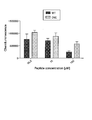
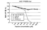
- Example 6 it is the figure which showed the measurement result of the relative ATP amount (%) of A431-CNGB3-myc cell processed by each peptide.
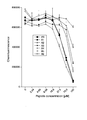
- Example 6 it is the figure which showed the measurement result of the relative ATP amount (%) of the ishikawa cell processed with each peptide.
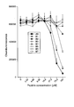
- Example 6 it is the figure which showed the measurement result of the relative ATP amount (%) of the SNG-II cell processed by each peptide.
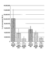
- Example 6 it is the figure which showed the measurement result of the relative ATP amount (%) of Hec-1A cell processed by each peptide. In Example 6, it is the figure which showed the measurement result of the relative ATP amount (%) of RL95-2 cell processed by each peptide. In Example 8, it is the figure which showed the result of having measured the increase rate (%) of the number of photons of the tumor-bearing mouse
- the cell killing agent according to the present invention comprises a peptide consisting of an amino acid sequence (KLAKLAKKLAKLAKHLAHL) represented by SEQ ID NO: 1 (hereinafter sometimes referred to as "effector peptide") and a site which selectively binds to a target molecule.
- the effector peptide is a peptide in which a peptide having apoptosis induction activity and a peptide having endosomal escape activity are linked in tandem.
- the cell killing agent according to the present invention binds to a target molecule through a site that selectively binds to the target molecule, and is endocytosed into target cells.
- the cell killing agent encapsulated in endosomes taken up in the target cells is released into the cytoplasm of the target cells as a result of destruction of the endosomal membrane by the action of the peptide site having endosomal escape activity.
- the cell-killing agent released into the cytoplasm damages the mitochondrial membrane and induces apoptosis of the target cell by the action of the peptide site having apoptosis-inducing activity.
- the cell killing agent according to the present invention contains, in one molecule, a peptide moiety having apoptosis-inducing activity, a peptide moiety having endosomal escape activity, and all sites that selectively bind to a target molecule. Therefore, a peptide that selectively binds to a target molecule and a peptide linked with a peptide having apoptosis-inducing activity, and a peptide that selectively binds to a target molecule and a peptide linked with an endosomal escape activity, Apoptosis can be induced in target cells very efficiently, as compared to peptide compositions containing each independently.
- the "target cell” is a cell of a subject for which it is desired to induce apoptosis.
- a “target molecule” is a molecule present on the surface of a cell of a subject for which apoptosis is to be induced or on the surface of a tissue in which the cell is present, which is a molecule to which the cell killing agent according to the present invention selectively binds.
- a peptide consisting of an amino acid sequence consisting of a repeat of four amino acids of KLAK (hereinafter sometimes referred to as “KLAK sequence”) (hereinafter sometimes referred to as “KLAK peptide”) has an action to induce apoptosis by damaging the mitochondrial membrane (apoptosis inducing activity).
- KLAK sequence an amino acid sequence consisting of a repeat of 4 amino acids of HLAH
- HLAH sequence hereinafter sometimes referred to as "HLAH peptide”
- HLAH sequence has an action of disrupting endosomal membranes. It has (endosomal escape activity).
- a peptide in which a KLAK peptide and an HLAH peptide are linked has endosomal escape activity and apoptosis induction activity.
- the strength of the activity is influenced by the length of the amino acid of each peptide and the order of linkage.
- Both the peptide in which the HLAH peptide is linked downstream of the KLAK peptide (C-terminus) and the peptide in which the KLAK peptide is linked downstream of the HLAH peptide (C-terminus) have endosomal escape activity and apoptosis-inducing activity.
- a peptide having an HLA H peptide linked to the downstream (C-terminal) of is more effective in inducing apoptosis.
- the effector peptide consisting of the amino acid sequence shown by SEQ ID NO: 1 is a peptide in which the HLAH sequence consisting of 5 amino acids is linked downstream of the KLAK sequence consisting of 14 amino acids. That is, in the amino acid sequence of SEQ ID NO: 1, the 1st to 14th amino acids are the sites having apoptosis-inducing activity, and the 15th to 19th amino acids are the sites having endosomal escape activity.
- This effector peptide has the length of the KLAK sequence, the length of the HLAH sequence, and the linkage of the KLAK sequence and the HLAH sequence so as to obtain the highest apoptosis-inducing activity when endocytosed into target cells. It is a peptide whose order has been optimized. Having this effector peptide, the cell killing agent according to the present invention has a very high apoptosis inducing activity.
- the effector peptide possessed by the cell killing agent according to the present invention is not particularly limited as long as it is a peptide consisting of the amino acid sequence represented by SEQ ID NO: 1, and may be a peptide consisting of L-amino acids, D- It may be a peptide consisting of amino acids, or a peptide consisting of L-amino acids and D-amino acids.
- the effector peptide is preferably a peptide containing at least a portion of D-amino acid, because the endosomal stability is high and a higher apoptosis-inducing activity can be obtained, and the KLAK sequence (amino acid represented by SEQ ID NO: 1) Among the sequences, it is more preferable that at least one of the amino acids 1 to 14) and the HLAH sequence (amino acids 15 to 19 of the amino acid sequence represented by SEQ ID NO: 1) be a D-amino acid Particularly preferred is a peptide consisting entirely of D-amino acids.
- the site which selectively binds to the target molecule and the effector peptide consisting of the amino acid sequence represented by SEQ ID NO: 1 may be directly linked, and indirectly linked via a linker. It may be done.
- the linker is not particularly limited, and examples thereof include peptides of about 1 to 20 amino acids, sugar chains, polyethylene glycols, polyolefins, and the like. Since the synthesis is relatively easy, as the cell killing agent according to the present invention, the site which selectively binds to the target molecule is a peptide or a protein, and a peptide directly or with an effector peptide of about 1 to 20 amino acids Those bonded through are preferred.
- the site which selectively binds to the target molecule is the N-terminal side of the effector peptide consisting of the amino acid sequence represented by SEQ ID NO: 1 as long as the binding to the target molecule is not inhibited. And may be linked to the C-terminal side.
- the site which selectively binds to the target molecule in the cell killing agent according to the present invention is not particularly limited, and is determined in accordance with the target molecule.
- the site may be a peptide or a protein, an oligonucleotide or a nucleic acid, a sugar chain, a lipid, or a low molecular weight compound.
- the target molecule to which the cell killing agent according to the present invention selectively binds is not particularly limited as long as it is a molecule present on the surface of a target cell or tissue inducing apoptosis, and it is a protein. It may be a sugar chain or a lipid.
- the selectivity of the cell killing agent according to the present invention is enhanced, and side effects can be reduced when used as a therapeutic agent for various diseases.
- the expression level on the surface of the target cell or the surface of the tissue containing the target cell is the expression level in many other cells and tissues.
- the number of molecules is preferably significantly larger than that of the molecule, and more preferably a molecule specifically expressed on the surface of a target cell or on the surface of a tissue containing the target cell.
- CNGB3 is a membrane protein that is highly expressed only in the endometrium and retina in normal tissues.
- the localization of CNGB3 in normal tissues is highly skewed, with only moderate expression in the pineal body, but weak expression in bone marrow, choroid plexus, oviduct, eye, ovary and testis. Therefore, among the cell killing agents according to the present invention, those in which CNGB3 is the target molecule are cell killing agents capable of specifically inducing apoptosis in endometrial and retinal cells, It is useful as a therapeutic drug for diseases.
- normal cells in the abdominal cavity and pelvis do not induce apoptosis but can selectively induce apoptosis in endometriosis cells, they are very suitable as therapeutic agents for endometriosis.
- CNGB3 is relatively strongly expressed in various cancer cells. Therefore, among the cell killing agents according to the present invention, those using CNGB3 as the target molecule are highly selective for cancer cells expressing CNGB3 and are useful as anticancer agents in which side effects are suppressed.
- Cancers expressing CNGB3 include, for example, uterine cancer, cervical cancer, pelvic cavity cancer, ovarian cancer, breast cancer, abdominal wall tumor, omental tumor, esophageal cancer, gastric cancer, small intestine cancer, colon cancer, rectal cancer, cecum Cancer, gallbladder cancer, pancreatic cancer, liver cancer, spleen cancer, kidney cancer, tongue cancer, pharyngeal cancer, nasal cancer, parotid adenocarcinoma, thyroid cancer, malignant lymphoma, bone tumor, skin cancer, lung cancer, mediastinal cancer, testis Cancer, prostate cancer, bladder cancer, brain tumor and the like can be mentioned.
- the target cancer cells are cells expressing CNGB 3 in advance by biopsy etc. It is better to confirm that.
- the site that selectively binds to the target molecule is, for example, a peptide consisting of an amino acid sequence represented by SEQ ID NO: 2 (VRRAXNXPG; X represents any naturally occurring amino acid) And a peptide consisting of an amino acid sequence obtained by modifying a part of the amino acid sequence represented by SEQ ID NO: 2 (hereinafter, these peptides may be referred to as “CNGB3 binding peptide”) (Patent Document 1).
- a peptide consisting of an amino acid sequence obtained by modifying a part of the amino acid sequence represented by SEQ ID NO: 2 one, two or three amino acids of the amino acid sequence represented by SEQ ID NO: 2 are deleted, substituted or A peptide which is added and which retains the binding ability to CNGB3; having at least 75% or more, preferably 85% or more, more preferably 90% or more of sequence identity with the amino acid sequence shown in SEQ ID NO: 2 And peptides that retain the binding ability to CNGB3.
- the CNGB3 binding peptide is a peptide consisting of an amino acid sequence represented by SEQ ID NO: 3 (VRRADNRPG) (hereinafter sometimes referred to as “Z13 peptide”), SEQ ID NO: 4 (VRRAENRPG) A peptide consisting of the amino acid sequence, a peptide consisting of the amino acid sequence represented by SEQ ID NO: 5 (VRRANNLPG), and a peptide consisting of the amino acid sequence represented by SEQ ID NO: 6 (VRRANNRPG).
- VRRADNRPG amino acid sequence represented by SEQ ID NO: 3
- Z13 peptide amino acid sequence represented by SEQ ID NO: 4
- a peptide consisting of the amino acid sequence a peptide consisting of the amino acid sequence represented by SEQ ID NO: 5 (VRRANNLPG)
- VRRANNLPG a peptide consisting of the amino acid sequence represented by SEQ ID NO: 6 (VRRANNRPG).
- the Z13 peptide and the effector peptide are linked directly or indirectly via a linker
- the peptide is a peptide in which the Z13 peptide and the effector peptide are linked directly or via a peptide consisting of 1 to 20 amino acids, and the Z13 peptide is directly or 1 to the C terminus of the effector peptide.
- the peptide is a peptide linked via a peptide consisting of 20 amino acids, and the peptide (SEQ ID NO: 26: KLAKLAKKKLAKLHAHLVRRADNRPG) in which the C-terminus of the effector peptide is directly linked to the N-terminus of Z13 peptide Rukoto more preferably more, and particularly preferably C-terminal N-terminus directly linked peptides Z13 peptide comprising all L- amino acids of the effector peptide consisting of all D- amino acids.
- annexin I is expressed in the cytoplasm in normal vascular endothelial cells, but is a membrane protein specifically expressed on the bloodstream side of vascular endothelial cells in newly formed blood vessels (tumor neovessels) in malignant tumor tissue It is.
- a molecule that selectively binds to annexin I administered in vivo is, in a malignant tumor tissue, a vascular endothelial cell of a tumor neovessel through binding to annexin I expressed on the bloodstream side of a tumor neovessel Incorporated into Molecules taken up by vascular endothelial cells are transported from the apical side to the basal side by vesicle transport and excreted into the stroma. The excreted molecules can diffuse through the stroma and be taken up into tumor cells.
- those using annexin I as the target molecule are cell killing agents capable of inducing apoptosis in cancer cells, and are useful as anticancer agents.
- the cell killing agent whose annexin I is the target molecule is specifically taken up by the vascular endothelial cells of tumor neovessels, either in the process of vesicle transport in vascular endothelial cells or in vesicle transport in the taken up tumor cells.
- the function of the HLA H peptide part disrupts the endosomal membrane and is released to the cytoplasm.
- the cells that have taken up the cell killing agent are killed by the function of the KLAK peptide peptide moiety.
- the type of cancer to be treated is not particularly limited, and may be used for the same cancers as those listed above. it can.
- a site that selectively binds to the target molecule is, for example, a peptide consisting of an amino acid sequence represented by SEQ ID NO: 7 (IFLLWQR) (hereinafter referred to as "IF7 peptide”). And a peptide consisting of an amino acid sequence obtained by modifying a part of the amino acid sequence represented by SEQ ID NO: 7 (hereinafter, these peptides may be referred to as “annexin I binding peptide”).
- an IF7 peptide and the effector peptide are linked directly or indirectly via a linker, More preferably, it is a peptide in which the IF7 peptide and the effector peptide are linked directly or via a peptide consisting of 1 to 20 amino acids, and the effector peptide is directly or 1 to 20 amino acids at the C terminus of the IF7 peptide.
- the peptide is a peptide linked via a peptide consisting of and further preferably a peptide linked via a peptide consisting of 1 to 5 amino acids of said effector peptide at the C-terminus of IF7 peptide , All of L-amino acids It is particularly preferred C-terminal of the tides is a peptide linked through the N-terminus and a peptide consisting of 1-5 amino L- amino acids at said effector peptide consisting of all D- amino acids.
- Examples of the peptide in which the C-terminus of the IF7 peptide is linked to the N-terminus of the effector peptide via a peptide consisting of 1 to 5 amino acids include, for example, peptides consisting of the amino acid sequence represented by SEQ ID NO: 40 (IFLLWQRRRKLAKKLAKKLAKLAKHLAHL) Be
- the administration route is not particularly limited, and is appropriately determined according to the target cell and the tissue containing it.
- administration routes of the cell killing agent according to the present invention include oral administration, intravenous administration, intraperitoneal administration, enema administration and the like.
- the cell killing agent according to the present invention can be prepared according to a conventional method, oral solid agents such as powders, granules, capsules, tablets, chewable agents, sustained release agents, oral solutions such as solutions, syrups, injections It can be formulated into an enema, a spray, a patch, an ointment and the like.
- the cell killing agent according to the present invention may contain an excipient, a binder, a lubricant, a disintegrant, a fluidizing agent, a solvent, a solubilizing agent, a buffer, a suspending agent, an emulsifying agent according to the need of formulation.
- Tonicity agents, stabilizers, preservatives, antioxidants, flavoring agents, coloring agents, etc. are blended and formulated.
- Excipients include saccharides such as lactose, glucose and D-mannitol, celluloses such as starch and crystalline cellulose, sugar alcohols such as erythritol, sorbitol and xylitol, dicalcium phosphate, calcium carbonate and kaolin .
- As the binder pregelatinized starch, gelatin, gum arabic, methylcellulose, carboxymethylcellulose, carboxymethylcellulose sodium, crystalline cellulose, D-mannitol, trehalose, hydroxypropyl cellulose, hydroxypropyl methylcellulose, polyvinyl pyrrolidone, polyvinyl alcohol and the like can be mentioned.
- stearic acid As the lubricant, stearic acid, calcium stearate, talc, sucrose fatty acid ester, polyethylene glycol and the like can be mentioned.
- Disintegrators include crospovidone (crosslinked polyvinyl pyrrolidone), low-substituted hydroxypropyl cellulose, starch, alginic acid, sodium alginate and the like.
- silicic acid, anhydrous silicic acid, aluminum silicate, calcium silicate, magnesium metasilicate aluminosilicate compound, aluminum oxide, aluminum hydroxide, magnesium hydroxide, magnesium hydroxide and the like As the solvent, purified water, physiological saline and the like can be mentioned.
- the dissolution aid dextran, polyvinyl pyrrolidone, sodium benzoate, ethylene diamine, salicylic acid amide, nicotinic acid amide, polyoxyethylene hydrogenated castor oil derivative and the like can be mentioned.
- the buffer include sodium citrate hydrate, sodium acetate hydrate, sodium hydrogencarbonate, trometamol, boric acid, borax, sodium hydrogenphosphate hydrate, sodium dihydrogenphosphate and the like.
- suspending agent or emulsifying agent sodium lauryl sulfate, gum arabic, gelatin, lecithin, glycerin monostearate, polyvinyl alcohol, polyvinyl pyrrolidone, cellulose such as sodium carboxymethyl cellulose, polysorbate, polyoxyethylene hydrogenated castor oil, etc. It can be mentioned.
- the tonicity agent include saccharides such as lactose, glucose and D-mannitol, sodium chloride, potassium chloride, glycerin, propylene glycol, polyethylene glycol, urea and the like.
- stabilizing agent polyethylene glycol, dextran sodium sulfate, sodium sulfite and the like can be mentioned.
- preservatives examples include p-oxybenzoic acid esters, chlorobutanol, benzyl alcohol, phenethyl alcohol, chlorocresol, dehydroacetic acid, sorbic acid and the like.
- antioxidant sulfite, ascorbic acid and the like can be mentioned.
- Flavoring agents include sweeteners, flavors and the like commonly used in the pharmaceutical and food fields. Coloring agents include those commonly used in the pharmaceutical and food arts.
- the cell killing agent according to the present invention may be used as it is, or may be used as a pharmaceutical composition containing other ingredients.
- the other components contained in the pharmaceutical composition for example, the above-mentioned excipients, binders, lubricants, disintegrants, fluidizers, solvents, solubilizers, buffers, suspending agents, Emulsifiers, tonicity agents, stabilizers, preservatives, antioxidants, flavoring agents, coloring agents and the like can be mentioned.
- the said pharmaceutical composition may contain other active ingredients other than the cell killing agent based on this invention.
- the cell killing agent according to the present invention is preferably one to be administered to mammals, preferably human, mouse, rat, rabbit, guinea pig, hamster, monkey, sheep, horse, cow, pig, donkey, dog, cat Are more preferably administered to domestic animals and experimental animals, and more preferably to humans.
- Endometriosis cells express CNGB3.
- endometriosis cells proliferate in the abdominal cavity. Therefore, an animal in which cells overexpressing CNGB3 are transplanted in the peritoneal cavity can be used as an endometriosis model animal.
- the CNGB3 to be overexpressed is not particularly limited as long as it is a protein that exhibits the same function as wild type human CNGB3 (Gene ID of NCBI: 54714) expressed in endometrial cells.
- it may be human CNGB3 or a homologue protein of human CNGB3 derived from a non-human animal, or a variant of human CNGB3 or a homologue thereof, which retains the same function as human CNGB3. It may be.
- the variant examples include a protein consisting of an amino acid sequence in which one or more amino acids of the amino acid sequence of human CNGB3 or a homologue thereof is deleted, substituted or added, and a protein retaining the function as CNGB3 It can be mentioned.
- the number of amino acids to be deleted is preferably 1 to 50, more preferably 1 to 30, still more preferably 1 to 20, and still more preferably 1 to 10.
- Examples of amino acids added to the N-terminus or C-terminus of human CNGB3 or its homologs include tag peptides such as His tag, Myc tag and Flag tag.
- the sequence identity with the amino acid sequence of human CNGB3 or a homolog thereof is 70% or more, preferably 80% or more, more preferably 85% or more, still more preferably 90% or more, further more Also preferred is a protein consisting of an amino acid sequence of 95% or more and retaining the function as CNGB3.
- deletion of amino acid in a protein means that a part of amino acids constituting a protein is lost (removed).
- substitution of amino acid in protein means that the amino acid constituting the protein is changed to another amino acid.
- an amino acid is added in a protein means that a new amino acid is inserted into a protein.
- a cell (host cell) into which a gene encoding CNGB3 (CNGB3 gene) is introduced to prepare CNGB3 overexpressing cells is not particularly limited, and it may be uterine endothelial cells, and animal tissue It may be a primary culture cell of cells other than uterine endothelial cells collected from U.S. or a cultured cell line.
- the host cell into which the CNGB3 gene is introduced may be a cell of human origin or a cell of non-human animal origin.
- CNGB3 overexpressing cells can be obtained by introducing the CNGB3 gene into a host cell in an expressible state.
- an expression vector incorporating an expression cassette consisting of a combination of DNAs necessary for expressing CNGB3 is introduced into a host cell.
- the expression cassette comprises a gene encoding a protein to be expressed, and a promoter that controls the expression of the gene.
- the expression cassette may further contain any one or more of a terminator, a 5 'untranslated region, and a 3' untranslated region.
- a preferred expression cassette is an expression cassette which comprises all of the gene sequences encoding the protein, a promoter, a terminator, a 5 'untranslated region, a 3' untranslated region.
- the CNGB3 gene to be introduced into the host cell may be any as long as it comprises a base sequence encoding CNGB3 consisting of the target amino acid sequence, and may be modified according to the codon frequency of the host cell.
- the promoter and the terminator may be those which function in the host cell.
- the promoter and the terminator that function in the host cell may be those originally possessed by the host cell, or those not inherently possessed by the host cell.
- any vector usually used for introduction into host cells can be used.
- the vector may be a circular plasmid vector, a linear vector, or a viral vector such as adenovirus.
- An expression vector can also be produced by integrating the CNGB3 gene into the cloning site of a commercially available vector for mammalian cell expression.
- the CNGB3 overexpressing cell may be a transformant in which the expression cassette of the CNGB3 gene is maintained as an extrachromosomal gene in host cell cells, and is a transformant integrated into the host cell chromosome It is also good.
- a transformation method for introducing an expression vector into a mammalian cell serving as a host may be appropriately selected from known transformation methods such as lipofection method, calcium phosphate precipitation method, lithium acetate method, electroporation method and the like. it can.
- the obtained CNGB3 overexpressing cells can be cultured under the same conditions as the host mammalian cells.
- the species of non-human animals to which CNGB3 overexpressing cells are transplanted into the peritoneal cavity is not particularly limited.
- the endometriosis model animal according to the present invention for example, domestic animals or experimental animals such as mice, rats, rabbits, guinea pigs, hamsters, monkeys, sheep, horses, cattle, pigs, donkeys, dogs, cats and the like are preferable.
- Transplantation of CNGB3 overexpressing cells into the peritoneal cavity can be performed by a conventional method.
- the exosome contains a biomolecule expressed on the cell membrane of the original cell from which the exosome was released.
- CNGB3 is abundantly expressed in the cell membrane of endometriosis cells, and exosomes released from endometriosis cells contain CNGB3.
- CNGB3 is also expressed in various cancer cells, and exosomes released from cancer cells contain CNGB3. Therefore, the amount of CNGB3 in exosomes is useful as a biomarker for endometriosis or cancer.
- the evaluation method of the disease onset possibility according to the present invention uses CNGB3 in exosome as a biomarker and the onset possibility of a disease expressing CNGB3. Is a way to evaluate In the body of an animal that has developed a disease in which cells causing the onset express CNGB3 (hereinafter sometimes referred to as “CNGB3 high expression disease”), many CNGB3 can be extracted from the causative cells of the disease. Many exosomes are secreted.
- CNGB3 is expressed only in limited tissues in normal cells, and therefore, there are very few CNGB3-rich exosomes in an animal that has not developed CNGB3 high expression disease. Therefore, it is possible to distinguish the onset group and the onset group of CNGB3 high expression disease based on the amount of CNGB3 in the exosome.
- the evaluation method according to the present invention measures CNGB3 in exosomes collected from a test animal, compares the obtained measurement value with a preset reference value, and the test animal has a CNGB3 high expression disease. Assess the possibility of developing.
- CNGB3 high expression diseases include endometriosis and cancers expressing CNGB3.
- a subject animal to be evaluated by the evaluation method according to the present invention a human or non-human animal may be used.
- the species of non-human animals is not particularly limited.
- domestic animals and experimental animals such as mice, rats, rabbits, guinea pigs, hamsters, monkeys, sheep, horses, cows, pigs, donkeys, dogs, cats and the like can be mentioned.
- the test animals to be evaluated by the evaluation method according to the present invention are preferably humans, domestic animals, and experimental animals, and more preferably humans.
- the reference value is a reference value for identifying the onset and non-occurrence of CNGB3 high expression disease.
- the reference value can be determined experimentally, for example, as a threshold that can be used to measure the amount of CNGB3 in the exosomes of CNGB3 high expression disease patients and non-patients.
- the method of determining the reference value of CNGB3 amount in exosome in the present invention is not particularly limited, and can be determined, for example, by a general statistical method.
- an exosome of a patient diagnosed as having a target CNGB3 high expression disease is collected by another method such as, for example, a commonly performed pathological examination. Measure the amount of CNGB3. After measuring for a plurality of patients, the amount of CNGB3 in these exosomes can be calculated by the mean value or median value, etc., and the numerical value including this can be used as a reference value.
- CNGB3 levels in exosomes are measured for multiple CNGB3 high expression disease patients and multiple non-CNGB3 high expression disease patients, and CNGB3 high expression disease patient group and CNGB3 high expression by average value or median value etc. After calculating CNGB3 amount and variation in the exosomes of the non-diseased patient group, it is possible to obtain a threshold that distinguishes the two values in consideration of the variation and use this as a reference value.
- the reference value can be a detection limit value of CNGB3.
- CNGB3 is detected in the exosome collected from the test animal, it is evaluated that the subject animal is likely to develop the CNGB3 high expression disease, and CNGB3 is not detected. It can be evaluated that the subject animal is highly likely not to develop the CNGB3 high expression disease.
- the method of measuring the amount of CNGB3 in the exosome collected from the test animal is not particularly limited, and various methods generally used in quantitative or semi-quantitative measurement of protein expression It can be measured.
- the method using immune reaction such as ELISA method, an immunohistochemistry method, a western blot method, is mentioned, for example.
- the amount of CNGB3 in the exosome collected from the subject animal may be determined as the amount of exosome containing CNGB3.
- CNGB3 is mainly present on the exosomal lipid bilayer membrane surface. Therefore, the amount of CNGB3 in exosomes can also be measured by applying various methods of measuring exosomes having specific surface molecules.
- Exo Counter manufactured by JVC Kenwood Co., Ltd.
- dNano manufactured by Meiwa Phosis Co., Ltd.
- Neano Sight manufactured by Japan Quantum Design Co., Ltd.
- exosome measurement systems such as "SP6800" Sony Corporation.
- any anti-CNGB3 antibody may be used.
- a molecule that binds to CNGB3 can be used.
- the molecule may be any of a peptide, a protein, a nucleic acid, a small molecule and the like.
- the molecule is a labeled product (a binding substance directly or indirectly bound to a site binding to CNGB3).
- the labeling substance is not particularly limited, and may be, for example, a low molecule such as biotin, may be a fluorescent substance, may be an enzyme, His tag, Myc tag, Flag tag, etc. Or a tag peptide of
- the aforementioned CNGB3 binding peptide or a labeled product thereof is preferable, and the Z13 peptide or a labeled product thereof is particularly preferable.
- a peptide consisting of an amino acid sequence represented by SEQ ID NO: 41 MQRTRAPTPG
- Z24 peptide a peptide consisting of an amino acid sequence represented by SEQ ID NO: 42
- VRSSRSTPQ VRSSRSTPQ
- peptide that binds to CNGB3 one may be used in which all peptide moieties are L-amino acids, or one in which D-amino acids may be used.
- a set of biotinylated Z13 peptide and avidin or streptavidin labeled with an enzyme or fluorescence can be used.
- the exosome to be subjected to the measurement of the amount of CNGB 3 may be any one collected from the test animal, and it may be a biological sample itself collected from the test animal, but it is an exosome purified from the biological sample. Is preferred.
- the separation of exosomes from a biological sample can be performed, for example, using a commercially available kit for exosome separation.
- the biological sample containing exosomes is not particularly limited, and may be blood, plasma, serum, tears, saliva, peritoneal fluid, urine or the like, mucous membranes such as uterine mucous membrane and digestive tract mucous membrane, It may be a piece of tissue collected from tissues such as liver, stomach, small intestine, large intestine and the like. Above all, blood, plasma and serum are widely used as clinical specimens and can be collected relatively less invasively. Therefore, as the exosome to be subjected to the measurement of the amount of CNGB3 in the evaluation method according to the present invention, blood collected from a test animal, in particular, exosome isolated from plasma or serum is preferable. Since the evaluation method according to the present invention can be performed using exosomes separated from serum or the like as a sample, it is also effective for the first screening of CNGB3 high expression diseases such as health checkup.
- the evaluation method according to the present invention can be used to evaluate the therapeutic effect in the treatment of CNGB3 high expression disease.
- the evaluation method according to the present invention is performed on a CNGB3 high expression disease patient before and after the start of the treatment of the CNGB3 high expression disease.
- the treatment causes a decrease in cells causing the CNGB3 high expression disease in the patient's body or its physiological activity is reduced, in the patient's body, an exosome secreted from the cells causing the CNGB3 high expression disease
- the amount of Therefore when the amount of CNGB3 in exosomes is significantly decreased after treatment compared to before treatment, it can be evaluated that the treatment effect is obtained by the treatment.
- the higher the reduction rate of CNGB3 amount in the exosome the higher the therapeutic effect was obtained.
- the therapeutic effect can also be monitored by performing the evaluation method according to the present invention over time also during the treatment period, in addition to before and after the treatment of the CNGB3 high expression disease.
- CNGB3 high expression disease it is possible to monitor the presence or absence of onset of CNGB3 high expression disease of the affected animal by performing the evaluation method according to the present invention sequentially to the animal suffering from CNGB3 high expression disease at least once.
- the evaluation method according to the present invention is sequentially implemented on patients with CNGB3 high expression disease It is possible to monitor the occurrence of recurrence.
- a transformed cell (A431-CNGB3-myc cell) which is forcibly expressed by introducing a gene encoding human CNGB3 having a myc tag fused to the C-terminus into A431 cells (a human epithelial-like cell carcinoma-derived cell line), It was prepared as an endometriosis model cell.
- DMEM High Glucose Medium containing 10% inactivated FBS (fetal bovine serum, Corning) and 1% penicillin-streptomycin (Invitrogen) were used.
- FBS fetal bovine serum, Corning
- penicillin-streptomycin Invitrogen
- the culture medium was used as a culture medium at 37 ° C. in a 5% by volume carbon dioxide environment. Passage was performed every 2-3 days.
- A431-CNGB3-myc cells were transplanted into the peritoneal cavity of an immunodeficient mouse (NOD / ShiJic-scid Jcl strain, supplied by CLEA Japan, Inc.) to prepare an endometriosis model mouse. Specifically, after thawing frozen A431-CNGB3-myc cells, 1 ⁇ 10 7 cells were passaged twice using a 10 cm dish (Thermo Fisher Scientific Co., Ltd., Lot No. F3BAXQ103). The culture solution was added so as to be /0.5 mL / body to prepare a cell solution as an administration solution. A431-CNGB3-myc cells were transplanted by intraperitoneally administering this administration solution to 7-week-old female immunodeficient mice as soon as possible after preparation.
- W ⁇ D ⁇ H 270 ⁇ 440 ⁇ 187 (mm)
- Humid water sterile tap water
- mice after A431-CNGB3-myc cell transplantation As a result of visual confirmation of mice after A431-CNGB3-myc cell transplantation, in the first experiment, although a mass was visually confirmed 1 week after transplantation, no peritoneal dissemination was observed, but 2 Or after 3 weeks, a mass and peritoneal dissemination were confirmed visually. In the second experiment, peritoneal dissemination was visually confirmed even one week after transplantation. In addition, as a result of c-myc staining of the peritoneal tissue fragment of the mouse after transplantation, it was confirmed that one week after the transplantation, peritoneal dissemination was already performed.
- peptides consisting of the amino acid sequences shown in Table 2 were synthesized, and cytotoxicity against A431-CNGB3-myc cells was compared.
- the KLAK sequence and the HLAH sequence part were synthesized from D-amino acids, and the CNGB3 binding peptide part was synthesized with L-amino acids.
- the cell killing activity of each peptide was evaluated by measuring the amount of ATP in A431-CNGB3-myc cells treated with each peptide using CellTiter-Glo (registered trademark) assay kit (manufactured by Promega).
- A431-CNGB3-myc cells after 2 passages were transformed into 96 well plates (Corning / Costar, Lot No. 00515003) so as to be 1 ⁇ 10 4 cells / well.
- each peptide was added to each well to a final concentration of 35.5, 75 or 150 ⁇ M, respectively, and cultured for 24 hours. Thereafter, the culture supernatant was removed from each well, and CellTiter-Glo buffer was added to the remaining cells to homogenize, and then the supernatant was collected by centrifugation and used as a lysate.
- the luminescence intensity (LLU) (RLU: RELATIVE LIGHT UNITS) of the reaction solution after standing at room temperature was measured using a Synergy H1 hybrid multi-mode microplate reader (manufactured by BioTek).
- the luminescence intensity of the reaction solution is an indicator of the amount of ATP. The smaller the luminescence intensity of the reaction solution, the smaller the amount of ATP, and the stronger the cell killing activity of the administered peptide. All trials were measured in triplicate and the average was assessed as the cell killing activity at the concentration of each peptide.
- FIG. 1 shows the measurement results of the luminescence intensity (RLU) of each reaction solution.
- WT shows the result of peptide WT in Table 1
- D5E shows the result of peptide D5E in Table 1. Both peptides showed cell killing activity in a concentration dependent manner.
- the peptide WT in which the CNGB3 binding peptide is the Z13 peptide showed stronger cell killing activity than the peptide D5E in which the CNGB3 binding peptide is the peptide of SEQ ID NO: 4.
- Example 1 For peptides in which the KLAK, HLAH and Z13 peptides were linked, the lengths of the KLAK and HLAH sequences were optimized to maximize cytotoxicity against endometriosis cells.
- 16 kinds of peptides consisting of the amino acid sequences shown in Table 2 were synthesized, and their cytotoxicity against A431-CNGB3-myc cells was compared.
- the KLAK sequence and the HLAH sequence part were synthesized from D-amino acids, and the Z13 peptide part was synthesized from L-amino acids.
- the final concentration of each peptide added to A431-CNGB3-myc cells seeded in a 96-well plate is 2.34, 4.69, 9.38, 18.8, 37.5, 75 or 150 ⁇ M, and the peptide
- the cell killing activity of each peptide was evaluated in the same manner as in Reference Example 1 except that the culture time after the addition was 20 hours.
- the measurement results of the luminescence intensity (RLU) of each reaction solution are shown in FIG. In the figure, “1” to “16” show the results of reaction solutions to which peptides 1 to 16 in Table 2 were added, respectively.
- Peptides 1-8 in which the peptide of HLAH sequence is linked downstream of the peptide of KLAK sequence show stronger cell killing activity than peptides 9-16 in which the peptide of KLAK sequence is linked downstream of the peptide of HLAH sequence.
- peptide 8 which is the shortest among peptides 1 to 8 has a higher cell killing activity than peptide 1 which is the longest
- peptide 16 which is the shortest among peptides 9 to 16 is more than peptide 9 which is the longest Cell killing activity was strong.
- Example 2 The peptide 8 that showed the strongest cell killing activity in Example 1 was further shortened and optimized to maximize the cytotoxicity against endometriotic cells.
- Example 2 Similar to Example 1, the cell killing activity of each peptide was evaluated.
- the measurement results of the luminescence intensity (RLU) of the reaction solution treated with peptide 8 and peptides A1 to A6 are shown in FIG. 3, and peptide 8 and peptides B1 to B7.
- the measurement results of the luminescence intensity (RLU) of the reaction solution treated with 4 are shown in FIG.
- the peptides B1 to B7 in which the HLAH sequence consists of 7 amino acids showed almost no cell killing activity even at a final peptide concentration of 75 ⁇ M (FIG. 4). Also, only peptide A2 showed stronger cell killing activity than peptide 8. From these results, it was found that, as an effector peptide, a peptide in which a peptide having a KLAK sequence consisting of 14 amino acids and a peptide having an HLAH sequence consisting of 5 amino acids is linked shows the highest cell killing activity.
- Example 3 In the peptide in which the KLAK peptide, the HLAH peptide and the Z13 peptide were linked, the strength of the cell killing activity was examined when the constituent amino acids were D-amino acid and L-amino acid.
- peptide A2 listed in Table 3 both the KLAK sequence consisting of 14 amino acids and the HLAH sequence consisting of 5 amino acids consist of D-amino acids. Also referred to as peptide (14D + 5D)) and amino acids of peptide 8 Among them, the peptide (14D + 5L) in which the KLAK sequence consisting of 14 amino acids consists of D-amino acids and the HLAH sequence consisting of 5 amino acids consists of L-amino acids, and the KLAK sequence consisting of 14 amino acids out of L-amino acids
- the strength of cell killing activity of a peptide (14L + 5D) in which the HLAH sequence consisting of 5 amino acids consists of D amino acids and a peptide (14L + 5L) in which all the amino acids of peptide 8 consist of L amino acids is the same as Example 1. I examined it. In addition, all Z13 peptide parts of these peptides consist of L-amino acids.
- Example 4 The cell killing activity of peptide A2, which had the highest cell killing activity in Examples 2 and 3, was examined.
- Example 5 An endometriosis model mouse was administered with peptide A2, which had the highest cell killing activity in Examples 2 and 3, and the therapeutic effect on endometriosis was examined.
- ⁇ Single injection of peptide into the abdominal cavity> Seven days after transplantation of A431-CNGB3-myc cells into the peritoneal cavity, 0.5 mL of physiological saline warmed to 37 ° C. was injected into the peritoneal cavity under isoflurane anesthesia for an endometriosis model mouse under isoflurane anesthesia. Immediately after massaging, a solution of peptide A2 in saline was intraperitoneally administered. Peptide A2 was administered such that the dose per weight of mouse was 0 mg / 10 mL / kg (control), 5 mg / 10 mL / kg, or 10 mg / 10 mL / kg (n 3).
- peritoneum Collection of peritoneum (sample collection)> An endometriosis model mouse administered with peptide A2 was exsanguinated under isoflurane anesthesia 24 hours after or 48 hours after the final administration, and the peritoneum was collected.
- the peritoneal membrane for ATP measurement was frozen in liquid nitrogen and stored in a deep freezer until measurement.
- the peritoneal membrane for pathological specimen preparation was immersed in 10% neutral buffered formalin and fixed, and then stored under refrigeration.
- ⁇ ATP amount measurement> The amount of ATP was measured using CellTiter-Glo (registered trademark) assay kit (manufactured by Promega). The frozen peritoneum was weighed, and 10 volumes of CellTiter-Glo buffer was added to the frozen tissue and homogenized, and then the supernatant was collected by centrifugation and used as a lysate. Two volumes of PBS and the same volume of 2 ⁇ CellTiterGlo Reagent as the lysate were added to this lysate and stirred to obtain a reaction solution, which was allowed to stand at room temperature for 10 minutes.
- CellTiter-Glo registered trademark
- the luminescence intensity (RLU) of the reaction solution after standing at room temperature was measured using a Synergy H1 hybrid multi-mode microplate reader (manufactured by BioTek).
- the measurement results of the luminescence intensity (RLU) of the peritoneum of an endometriosis model mouse to which a single dose of peptide A2 was administered are shown in FIG. Further, the measurement results of the luminescence intensity (RLU) of the peritoneum of the endometriosis model mouse to which peptide A2 was repeatedly administered three times are shown in FIG. As shown in FIGS. 8 and 9, in both single and multiple administration mice, the luminescence intensity (RLU) of the peritoneum is decreased depending on the amount of peptide A2 administered, and the amount of ATP in the peritoneum is It was confirmed that it decreased.
- a decrease in the amount of peritoneal ATP means a decrease in the number of viable cells of A431-CNGB3-myc cells transplanted to the peritoneum. From these results, it can be seen that peptide A2 kills A431-CNGB3-myc cells in the peritoneal cavity, that is, peptide A2 can exert a cell killing effect in the in vivo environment, particularly against endometriosis. It can be said that it is useful as a therapeutic agent.
- Example 6 The cell killing activity of peptide A2 (peptide (14D + 5D)) was examined on cell lines derived from A431-CNGB3-myc cells and various uterine cancer cells.
- cancer cell-derived cell lines include ishikawa cells derived from human endometrial adenocarcinoma, SNG-II cells derived from human endometrial cancer, Hec-1A cells derived from human endometrial cancer, and RL95 derived from human endometrial cancer. -2 cells were used. These cells were cultured in the same manner as A431-CNGB3-myc cells.
- peptides for treating each cell three types of peptides consisting of the amino acid sequences shown in Table 4 were used. In these three types of peptides, the KLAK sequence and the HLAH sequence portion were synthesized from D-amino acids, and the Z13 peptide portion was synthesized with L-amino acids.
- each peptide added to each cell seeded in a 96-well plate is 0 (no peptide added), 2.34, 4.69, 9.38, 18.8, 37.5, 75.0, 150
- the cell killing activity of each peptide was evaluated in the same manner as in Example 1 except that 0.1 or 300.0 ⁇ M was used.
- the relative luminescence intensity (%) of each reaction solution was calculated as the relative amount of ATP (%), where the luminescence intensity (RLU) of the reaction solution with no peptide added was 100%. The calculation results are shown in FIGS.
- A431 cells are cell lines derived from human epithelial cell carcinoma
- the endometriosis model mouse in which A431-CNGB3-myc cells are transplanted in the peritoneal cavity is also a tumor-bearing mouse. Therefore, a peptide (IFLLWQR-RR-KLAKLAK-KLAKLAK-HLAHL, SEQ ID NO: 40) in which an effector peptide consisting of the amino acid sequence represented by SEQ ID NO: 1 and an IF7 peptide are linked to the peritoneal cavity of an endometriosis model mouse "IF7 (RR)-(K + H) peptide" is sometimes administered to examine the cell killing activity against cancer cells.
- the IF7 (RR)-(K + H) peptide was synthesized with D-amino acids from the KLAK and HLAH sequence parts and L-amino acids from the other peptide parts.
- the IF7 (RR)-(K + H) peptide is physiological saline once a day for 6 consecutive days to an endometriosis model mouse 14 days after the intraperitoneal injection of A431-CNGB3-myc cells. 50 ⁇ L of a solution dissolved in water was administered via the tail vein.
- the IF7 (RR)-(K + H) peptide was administered at a dose of 0 ⁇ g / body (control), 10 ⁇ g / body, or 50 ⁇ g / body per weight of mouse.
- the number of dead individuals was 1 until 24 days after the A431-CNGB3-myc cells were implanted intraperitoneally, and the survival rate was 88.9. %Met.
- Example 8 The IF7 (RR)-(K + H) peptide used in Example 7 was intravenously administered to tumor-bearing mice in which a mass was formed on the back, and the cell killing activity against cancer cells was examined.
- ⁇ Luciferase gene-transferred ovarian cancer tumor-bearing subcutaneous tumor mouse OVCAR3-Luc mouse
- the tumor tissue to be transplanted into mice was obtained by culturing a luciferase gene-introduced ovarian cancer cell line (OVCAR3-Luc cells, transferred from another facility). Culturing of OVCAR3-Luc cells was performed at 37 ° C. in a 5 vol% carbon dioxide environment using RPMI medium 1640 (11875-093, gibe by life technologies) as a culture medium.
- ⁇ Tail vein administration of peptide> OVCAR3-Luc mice were administered with 50 ⁇ L of a solution of IF7 (RR)-(K + H) peptide dissolved in physiological saline once a day for 6 consecutive days via the tail vein .
- the IF7 (RR)-(K + H) peptide was administered at a dose of 0 ⁇ g / body (control) or 10 ⁇ g / body per body weight of the mouse.
- the estimated tumor volume (mm 3 ) on the back of each OVCAR3-Luc mouse was determined from the major axis and minor axis of the tumor according to the following equation.
- the major axis (mm) and the minor axis (mm) of the tumor were measured using a caliper.
- FIG. 15 shows the temporal change in the increase rate (%) in the photon number of the mass on the back of each mouse
- FIG. 16 the temporal change in the increase rate (%) of the tumor volume (mm 3 ) on the back of each mouse Indicates a change.
- the increase rate of the photon number and the increase rate of the tumor volume were both based on the value of the peptide solution on the day before the administration start date (100%).
- “treatment” indicates the treatment period in which the peptide solution was intravenously administered. As shown in FIG.
- mice treated with the IF7 (RR)-(K + H) peptide the number of photons hardly increased during the peptide treatment period, and the increase rate after the peptide treatment period was also controlled by the control group ( It was smaller than Saline).
- the control group the IF7 (RR)-(K + H) peptide administration group during the peptide treatment period
- the increase rate after the treatment period is IF 7 (RR)-(K + H)
- the peptide administration group was clearly smaller. From these results, it can be said that administration of IF7 (RR)-(K + H) peptide can kill tumor tissue in vivo, and the peptide is useful as an anticancer agent.
- tissue sections are 2 types (OV20811 and OV2088) of 4 types of commercially available human ovarian cancer tissue array (Ovary cancer tissue array (made by US Biomax)) and cancer tissue arrays (Multiple organ tumor) of various organs. Two types of tissue array (FDA 800a and MC 964a) were used.
- anti-CNGB3 antibodies among commercially available antibodies, include Biorbyt (Catalog No. orb 156415 BRT 100UG) (hereinafter sometimes referred to as “antibody orb”) and Osenses (Code; OSC00253W) (hereinafter, “antibody 253W”.
- Antibody orb Biorbyt
- Osenses Code; OSC00253W
- Immunostaining of tissue sections was performed as follows. First, paraffin-embedded tissue sections are reacted for 30 minutes with a blocking agent for liquid immunity experiment (Immunoblock, manufactured by DS Pharma Biomedical Co., Ltd.) to block nonspecific reactions, and then TBST (Tris buffered saline containing Tween 20) 2 minutes of washing with water). Then, the tissue section was immersed in 0.3% hydrogen peroxide water, reacted for 5 minutes, blocked for endogenous peroxidase activity, and washed twice for 5 minutes with TBST.
- a blocking agent for liquid immunity experiment Immunoblock, manufactured by DS Pharma Biomedical Co., Ltd.
- TBST Tris buffered saline containing Tween 20
- the tissue section is immersed in a primary antibody solution in which anti-CNGB3 antibody is diluted 100 times with REAL Antibody Diluent (Code S2022, manufactured by Dako), reacted at room temperature for 30 minutes, and then reacted for 5 minutes with TBST. The washing process was performed twice. Furthermore, the tissue section was reacted with a labeled secondary antibody (EnVision + System-HRP-labeled Polymer anti-rabbit antibody, manufactured by Dako) for 30 minutes at room temperature, and then washed twice with TBST for 5 minutes. Thereafter, the tissue section was treated with a DAB coloring reagent [DAB + Liquid (large size), RUO (K3468), Dako company] for 5 minutes to develop color. After staining with DAB, the tissue sections were washed with water, treated with Mayer's hematoxylin for 2 minutes to stain nuclei, and then sealed.
- DAB DAB coloring reagent
- the staining results of OV20811 among human ovarian cancer tissue arrays are shown in Tables 5-10, and the staining results of OV2088 are shown in Tables 11-16, respectively.
- the staining results of FDA 800a are shown in Tables 17 and 18, and the staining results of MC 964a are shown in Tables 19 to 21, respectively.
- “+” means the result of immunostaining with the antibody
- “-” means the result of no immunostaining with the antibody.
- Most tissue sections were immunostained with anti-CNGB3 antibody and it was found that CNGB3 was expressed.
- Example 9 The amount of CNGB3 in exosomes isolated from blood was measured to see if it was possible to distinguish between CNGB3 high expression disease patients and those without CNGB3 high expression disease. The following experiments were conducted with the approval of the Hamamatsu Medical University Institutional Review Board.
- the blood collected from 3 patients, 3 breast cancer patients, 1 cervical cancer patient, 2 colon cancer patients, 3 endometriosis patients and 1 uterine adenomyosis patient) is centrifuged and The serum aliquots were stored at -80.degree.
- Uterine adenomyosis patients are patients who develop both endometriosis and fibroids.
- the exosome was isolated from the serum using Exosome Isolation Kit (manufactured by Fujifilm Wako Pure Chemical Industries, Ltd.) according to the manufacturer's instruction. Specifically, first, the serum was centrifuged at 1,000 ⁇ g at 4 ° C. for 20 minutes, and the supernatant was recovered. The collected supernatant was centrifuged at 10,000 ⁇ g, 4 ° C. for 30 minutes, and the supernatant was transferred to a new tube. Then, 1.0 mL of this supernatant was mixed with exosome capture beads on a rotator at 4 ° C. for 180 minutes. Then, after washing the supplemental beads, exosomes were eluted with 100 ⁇ L of elution buffer and stored at -20 ° C.
- Exosome Isolation Kit manufactured by Fujifilm Wako Pure Chemical Industries, Ltd.
- CNGB3 on the film was detected using a CCD camera system (manufactured by ATTO) and ECL prime (manufactured by GE Healthcare). Densitometry analysis was performed using image analysis software "CS Analyzer 4" (manufactured by ATTO).
- the signal intensity of the band of CNGB 3 and the relative value thereof (relative value with the signal intensity of healthy person 1 being 1) obtained in the result of avidin blotting of each measurement sample are shown in Table 22.
- breast cancer patients 1 and 2 could secure only a small amount of serum, they used exosomes isolated from 100 ⁇ L of serum, and breast cancer patient 3 used exosomes isolated from 37.5 ⁇ L of serum. Therefore, the signal intensities of breast cancer patient samples were corrected by multiplying by the reciprocal of serum usage.
- CNGB3 is present in the exosomes of healthy people, though in very small amounts. This is presumed to be due to the fact that CNGB3 of exosomes in serum could be detected with extremely high sensitivity by using a molecule having high binding to CNGB3 such as CNGB3 binding peptide.
- the ability to detect exosomes containing CNGB3 released from extremely small amounts of cancer cells in the body of healthy individuals has made it possible to use advanced CNGB3 binding peptides to make advanced cancer even when using exosomes in serum as specimens. Not only detection but also very early detection of cancer could be expected to be possible.
Landscapes
- Health & Medical Sciences (AREA)
- Life Sciences & Earth Sciences (AREA)
- Chemical & Material Sciences (AREA)
- Engineering & Computer Science (AREA)
- General Health & Medical Sciences (AREA)
- Medicinal Chemistry (AREA)
- Animal Behavior & Ethology (AREA)
- Pharmacology & Pharmacy (AREA)
- Public Health (AREA)
- Veterinary Medicine (AREA)
- Bioinformatics & Cheminformatics (AREA)
- Proteomics, Peptides & Aminoacids (AREA)
- Immunology (AREA)
- Molecular Biology (AREA)
- Organic Chemistry (AREA)
- Chemical Kinetics & Catalysis (AREA)
- General Chemical & Material Sciences (AREA)
- Nuclear Medicine, Radiotherapy & Molecular Imaging (AREA)
- Epidemiology (AREA)
- Cell Biology (AREA)
- Environmental Sciences (AREA)
- Urology & Nephrology (AREA)
- Hematology (AREA)
- Biomedical Technology (AREA)
- Gastroenterology & Hepatology (AREA)
- Biotechnology (AREA)
- Physics & Mathematics (AREA)
- Pathology (AREA)
- Animal Husbandry (AREA)
- Zoology (AREA)
- Reproductive Health (AREA)
- Microbiology (AREA)
- Endocrinology (AREA)
- Food Science & Technology (AREA)
- Biodiversity & Conservation Biology (AREA)
- Analytical Chemistry (AREA)
- Biochemistry (AREA)
- General Physics & Mathematics (AREA)
- Gynecology & Obstetrics (AREA)
- Medicines That Contain Protein Lipid Enzymes And Other Medicines (AREA)
Priority Applications (8)
| Application Number | Priority Date | Filing Date | Title |
|---|---|---|---|
| EP18848077.6A EP3738601A4 (en) | 2017-08-24 | 2018-08-23 | Cytocidal agent |
| AU2018322305A AU2018322305B2 (en) | 2017-08-24 | 2018-08-23 | Cytocidal agent |
| CN201880054701.XA CN111065403B (zh) | 2017-08-24 | 2018-08-23 | 细胞杀伤剂 |
| KR1020207004917A KR102427844B1 (ko) | 2017-08-24 | 2018-08-23 | 세포 살상제 |
| US16/640,248 US11357819B2 (en) | 2017-08-24 | 2018-08-23 | Cytocidal agent |
| CA3073392A CA3073392C (en) | 2017-08-24 | 2018-08-23 | Cytocidal agent |
| SG11202001484QA SG11202001484QA (en) | 2017-08-24 | 2018-08-23 | Cytocidal agent |
| US17/740,864 US20220280596A1 (en) | 2017-08-24 | 2022-05-10 | Cytocidal agent |
Applications Claiming Priority (2)
| Application Number | Priority Date | Filing Date | Title |
|---|---|---|---|
| JP2017-161556 | 2017-08-24 | ||
| JP2017161556A JP6613499B2 (ja) | 2017-08-24 | 2017-08-24 | 細胞殺傷剤 |
Related Child Applications (2)
| Application Number | Title | Priority Date | Filing Date |
|---|---|---|---|
| US16/640,248 A-371-Of-International US11357819B2 (en) | 2017-08-24 | 2018-08-23 | Cytocidal agent |
| US17/740,864 Division US20220280596A1 (en) | 2017-08-24 | 2022-05-10 | Cytocidal agent |
Publications (1)
| Publication Number | Publication Date |
|---|---|
| WO2019039540A1 true WO2019039540A1 (ja) | 2019-02-28 |
Family
ID=65438932
Family Applications (1)
| Application Number | Title | Priority Date | Filing Date |
|---|---|---|---|
| PCT/JP2018/031136 WO2019039540A1 (ja) | 2017-08-24 | 2018-08-23 | 細胞殺傷剤 |
Country Status (9)
Cited By (2)
| Publication number | Priority date | Publication date | Assignee | Title |
|---|---|---|---|---|
| US11191804B2 (en) | 2018-01-09 | 2021-12-07 | Fujita Academy | Cytocidal agent |
| US11357819B2 (en) | 2017-08-24 | 2022-06-14 | Fujita Academy | Cytocidal agent |
Families Citing this family (2)
| Publication number | Priority date | Publication date | Assignee | Title |
|---|---|---|---|---|
| WO2022234346A1 (en) * | 2021-05-07 | 2022-11-10 | Twinpig Biolab, Inc. | Peptides targeting macrophages, and conjugates, compositions, and uses thereof |
| CN115725968A (zh) * | 2022-11-18 | 2023-03-03 | 东莞宜安科技股份有限公司 | 一种生物医用钛合金表面的复合涂层及其制备方法 |
Citations (4)
| Publication number | Priority date | Publication date | Assignee | Title |
|---|---|---|---|---|
| JP2013523824A (ja) * | 2010-04-06 | 2013-06-17 | ジョン ダブリュ. ホラデイ、 | 癌を治療する方法 |
| WO2015041019A1 (ja) * | 2013-09-18 | 2015-03-26 | 国立大学法人 京都大学 | 子宮内膜症様細胞の作製方法および子宮内膜症モデル動物 |
| US20160145308A1 (en) | 2013-06-11 | 2016-05-26 | Sanford-Burnham Medical Research Institute | Compositions and Methods for Targeted Endometriosis Treatment |
| JP2017161556A (ja) | 2017-06-27 | 2017-09-14 | パイオニア株式会社 | 位置認識装置、制御方法、プログラム、及び記憶媒体 |
Family Cites Families (10)
| Publication number | Priority date | Publication date | Assignee | Title |
|---|---|---|---|---|
| US5595756A (en) | 1993-12-22 | 1997-01-21 | Inex Pharmaceuticals Corporation | Liposomal compositions for enhanced retention of bioactive agents |
| JP2009521512A (ja) * | 2005-12-23 | 2009-06-04 | ザ バーナム インスティテュート フォー メディカル リサーチ | 子宮内膜症細胞ターゲティングペプチドおよびその使用 |
| AU2009343754A1 (en) * | 2009-04-01 | 2011-11-03 | Ingo Schmidt-Wolf | Tumor targeting peptides, therapeutic and diagnostic compositions comprising the peptides |
| WO2011027312A1 (en) | 2009-09-03 | 2011-03-10 | Koninklijke Philips Electronics N.V. | NOVEL TUMOR MARKERS SELECTED FROM A GROUP OF cAMP-REGULATORY PATHWAY MEMBERS |
| SG10201704812TA (en) * | 2009-12-23 | 2017-07-28 | Sanford-Burnham Medical Res Inst | Methods and compositions related to annexin 1-binding compounds |
| US20160067357A1 (en) * | 2012-08-17 | 2016-03-10 | Apellis Pharmaceuticals, Inc. | Detection of high risk drusen |
| US10338068B2 (en) * | 2013-07-08 | 2019-07-02 | The Trustees Of Columbia University In The City Of New York | Selection of biological objects |
| CN107406497A (zh) | 2014-12-19 | 2017-11-28 | 埃博灵克斯股份有限公司 | 半胱氨酸连接的纳米抗体二聚体 |
| RU2571551C1 (ru) | 2015-04-16 | 2015-12-20 | Закрытое акционерное общество "ПЕПТЕК" | Специфическая сочетанная терапия злокачественных опухолей цитостатиком и его модификатором |
| JP6613499B2 (ja) | 2017-08-24 | 2019-12-04 | 学校法人藤田学園 | 細胞殺傷剤 |
-
2017
- 2017-08-24 JP JP2017161556A patent/JP6613499B2/ja active Active
-
2018
- 2018-08-23 KR KR1020207004917A patent/KR102427844B1/ko active Active
- 2018-08-23 US US16/640,248 patent/US11357819B2/en active Active
- 2018-08-23 CA CA3073392A patent/CA3073392C/en active Active
- 2018-08-23 WO PCT/JP2018/031136 patent/WO2019039540A1/ja unknown
- 2018-08-23 SG SG11202001484QA patent/SG11202001484QA/en unknown
- 2018-08-23 CN CN201880054701.XA patent/CN111065403B/zh active Active
- 2018-08-23 AU AU2018322305A patent/AU2018322305B2/en active Active
- 2018-08-23 EP EP18848077.6A patent/EP3738601A4/en active Pending
-
2022
- 2022-05-10 US US17/740,864 patent/US20220280596A1/en not_active Abandoned
Patent Citations (4)
| Publication number | Priority date | Publication date | Assignee | Title |
|---|---|---|---|---|
| JP2013523824A (ja) * | 2010-04-06 | 2013-06-17 | ジョン ダブリュ. ホラデイ、 | 癌を治療する方法 |
| US20160145308A1 (en) | 2013-06-11 | 2016-05-26 | Sanford-Burnham Medical Research Institute | Compositions and Methods for Targeted Endometriosis Treatment |
| WO2015041019A1 (ja) * | 2013-09-18 | 2015-03-26 | 国立大学法人 京都大学 | 子宮内膜症様細胞の作製方法および子宮内膜症モデル動物 |
| JP2017161556A (ja) | 2017-06-27 | 2017-09-14 | パイオニア株式会社 | 位置認識装置、制御方法、プログラム、及び記憶媒体 |
Non-Patent Citations (4)
| Title |
|---|
| SHIBATA, TOSHIAKI ET AL: "P3-14-1 Anti-tumor drugs using peptide that mimics sugar chains", ACTA OBSTETRICA ET GYNAECOLOGICA JAPONICA, vol. 68, no. 2, 2016, pages 858, XP009519433, ISSN: 0300-9165 * |
| SUGIHARA ET AL., NATURE COMMUNICATIONS, vol. 5, 2014 |
| SUGIHARA, K. ET AL.: "Development of pro-apoptotic peptides as potential therapy for peritoneal endometriosis", NATURE COMMUNICATIONS, vol. 5, no. 4478, 2014, pages 1 - 9, XP055255332, ISSN: 2041-1723, DOI: 10.1038/ncomms5478 * |
| SUGIHARA, KAZUHIRO ET AL: "Search of peptide therapeutic drugs for endometriosis", PROGRAMS AND ABSTRACTS OF THE 32ND ANNUAL MEETING OF THE JAPAN SOCIETY OF DRUG DELIVERY SYSTEM, 2016, pages 170, XP009519434 * |
Cited By (2)
| Publication number | Priority date | Publication date | Assignee | Title |
|---|---|---|---|---|
| US11357819B2 (en) | 2017-08-24 | 2022-06-14 | Fujita Academy | Cytocidal agent |
| US11191804B2 (en) | 2018-01-09 | 2021-12-07 | Fujita Academy | Cytocidal agent |
Also Published As
| Publication number | Publication date |
|---|---|
| SG11202001484QA (en) | 2020-03-30 |
| US20200289608A1 (en) | 2020-09-17 |
| US20220280596A1 (en) | 2022-09-08 |
| CA3073392A1 (en) | 2019-02-28 |
| AU2018322305B2 (en) | 2021-05-20 |
| EP3738601A4 (en) | 2021-06-16 |
| JP6613499B2 (ja) | 2019-12-04 |
| US11357819B2 (en) | 2022-06-14 |
| CN111065403A (zh) | 2020-04-24 |
| KR102427844B1 (ko) | 2022-07-29 |
| KR20200031670A (ko) | 2020-03-24 |
| CA3073392C (en) | 2023-02-21 |
| EP3738601A1 (en) | 2020-11-18 |
| AU2018322305A1 (en) | 2020-03-05 |
| CN111065403B (zh) | 2023-09-26 |
| JP2019038772A (ja) | 2019-03-14 |
Similar Documents
| Publication | Publication Date | Title |
|---|---|---|
| US20220280596A1 (en) | Cytocidal agent | |
| Gribar et al. | Reciprocal expression and signaling of TLR4 and TLR9 in the pathogenesis and treatment of necrotizing enterocolitis | |
| AU2003216288B2 (en) | Modulation of immune response by non-peptide binding stress response polypeptides | |
| Qi et al. | Endogenous TWEAK is critical for regulating the function of mouse uterine natural killer cells in an immunological model of pregnancy loss | |
| ES2844874T3 (es) | Complejo de proteína de leche y ácido graso | |
| Kim et al. | Clusterin interaction with Bcl-xL is associated with seizure-induced neuronal death | |
| KR20130130562A (ko) | 테모졸로미드 및 발프로산을 함께 이용하는 항암적 용도 | |
| CN113209313A (zh) | Tgfbr2在制备卵巢功能保护药物方面中的应用 | |
| US10751324B2 (en) | Treatment of TNF- alpha cytotoxicity | |
| AU2018402356B2 (en) | Cytocidal agent | |
| HK40023962A (zh) | 细胞杀伤剂 | |
| Yuan et al. | The Human Milk-derived Peptide Drives Rapid Regulation of Macrophage Inflammation Responses in the Neonatal Intestine | |
| HK40032209A (zh) | 细胞杀伤剂 | |
| WO2019154515A1 (en) | Methods and compositions for the treatment of amyloid diseases | |
| Clough et al. | Targeting Replication Stress for Glioma Stem Cell Specific Cytotoxicity | |
| Li | Exploring the function of IGFN1 and MLTK in skeletal muscle | |
| Kohar | The role of microtubule-associated protein 1S (MAP1S) in regulating autophagy in the heart | |
| JP2021534826A (ja) | がんの処置のためのペプチド治療薬およびその使用 | |
| Ni et al. | Dimethyl Malonate Preserves Brain and Neurobehavioral Phenotype Following Neonatal Hypoxia-Ischemia Via Inhibition of Fth1-Mediated Ferritinophagy | |
| Hackam et al. | Reciprocal Expression and Signaling of TLR4 | |
| JP2020030120A (ja) | 疾患発症可能性の評価方法 | |
| Bettenworth et al. | S1642 New Tools for In Vivo Imaging of Murine DSS-Colitis-Mrp14-Directed Fluorescence-Mediated Molecular Tomography (FMT) and High-Resolution Ultrasound Imaging |
Legal Events
| Date | Code | Title | Description |
|---|---|---|---|
| 121 | Ep: the epo has been informed by wipo that ep was designated in this application |
Ref document number: 18848077 Country of ref document: EP Kind code of ref document: A1 |
|
| ENP | Entry into the national phase |
Ref document number: 3073392 Country of ref document: CA |
|
| ENP | Entry into the national phase |
Ref document number: 20207004917 Country of ref document: KR Kind code of ref document: A |
|
| NENP | Non-entry into the national phase |
Ref country code: DE |
|
| ENP | Entry into the national phase |
Ref document number: 2018322305 Country of ref document: AU Date of ref document: 20180823 Kind code of ref document: A |
|
| ENP | Entry into the national phase |
Ref document number: 2018848077 Country of ref document: EP Effective date: 20200324 |