WO2018235915A1 - 測定方法、測定キットおよび測定装置 - Google Patents
測定方法、測定キットおよび測定装置 Download PDFInfo
- Publication number
- WO2018235915A1 WO2018235915A1 PCT/JP2018/023670 JP2018023670W WO2018235915A1 WO 2018235915 A1 WO2018235915 A1 WO 2018235915A1 JP 2018023670 W JP2018023670 W JP 2018023670W WO 2018235915 A1 WO2018235915 A1 WO 2018235915A1
- Authority
- WO
- WIPO (PCT)
- Prior art keywords
- hair
- antibody
- active substance
- physiologically active
- cortisol
- Prior art date
- Legal status (The legal status is an assumption and is not a legal conclusion. Google has not performed a legal analysis and makes no representation as to the accuracy of the status listed.)
- Ceased
Links
Images








Classifications
-
- G—PHYSICS
- G01—MEASURING; TESTING
- G01N—INVESTIGATING OR ANALYSING MATERIALS BY DETERMINING THEIR CHEMICAL OR PHYSICAL PROPERTIES
- G01N33/00—Investigating or analysing materials by specific methods not covered by groups G01N1/00 - G01N31/00
- G01N33/48—Biological material, e.g. blood, urine; Haemocytometers
- G01N33/50—Chemical analysis of biological material, e.g. blood, urine; Testing involving biospecific ligand binding methods; Immunological testing
-
- G—PHYSICS
- G01—MEASURING; TESTING
- G01N—INVESTIGATING OR ANALYSING MATERIALS BY DETERMINING THEIR CHEMICAL OR PHYSICAL PROPERTIES
- G01N33/00—Investigating or analysing materials by specific methods not covered by groups G01N1/00 - G01N31/00
- G01N33/48—Biological material, e.g. blood, urine; Haemocytometers
- G01N33/50—Chemical analysis of biological material, e.g. blood, urine; Testing involving biospecific ligand binding methods; Immunological testing
- G01N33/53—Immunoassay; Biospecific binding assay; Materials therefor
Definitions
- the present disclosure relates to a method, a measurement kit and a measurement device for measuring the accumulation amount of a physiologically active substance in hair.
- Non-Patent Document 1 reports that Cortisol levels were determined by ELISA (enzyme-linked immunosorbent assay) using neonatal hair as a sample.
- One aspect of the present disclosure aims to provide a simple method of measuring the accumulation amount of a physiologically active substance in hair.
- a measuring method concerning one mode of this indication is a method of measuring accumulation amount of physiologically active substance in hair, and the above-mentioned physiologically active substance is peculiar to a section of the above-mentioned hair. And a detection step of detecting the antibody bound to the physiologically active substance in the reaction step.
- the accumulated amount of the physiologically active substance in the hair can be simply measured.
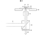
- FIG. 6 is a schematic view showing an optical system used in a detection step of detecting an antibody bound to a physiologically active substance in the method according to Embodiment 1.
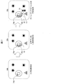
- FIG. 1 In the method which concerns on Embodiment 2, it is a schematic diagram showing each step of the reaction process of making the antibody which specifically recognizes a physiologically active substance react with respect to the cross section of hair. It is a microscope image showing the result of having measured the accumulation amount of cortisol contained in hair by the method concerning Embodiment 2.
- FIG. 3 In the method which concerns on Embodiment 3, it is a schematic diagram showing each step of the reaction process which makes the antibody which specifically recognizes a physiologically active substance react with respect to the cross section of hair. It is a microscope image showing the result of having measured the accumulation amount of cortisol contained in hair by the method concerning Embodiment 3.
- FIG. 1 In the method which concerns on Embodiment 2, it is a schematic diagram showing each step of the reaction process of making the antibody which specifically recognizes a physiologically active substance react with respect to the cross section of hair. It is a microscope image showing the result of having measured the accumulation amount of cortisol contained in hair by the method concerning Embodiment 3.
- FIG. 1 It is a figure showing the result of having measured accumulation amount of cortisol contained in hair by a method concerning Embodiment 5.
- A is a microscope image
- (b) is an image showing fluorescence intensity.
- it is a schematic diagram showing each step of the reaction process which makes the antibody which specifically recognizes a physiologically active substance react with respect to the cross section of hair.
- A is a method in which the first antibody and the second antibody are reacted simultaneously
- (b) is a method in which the first antibody and the second antibody are reacted separately.
- non-human mammals in addition to humans.
- non-human mammals include primates (monkeys, apes, etc.), amphibians (bovines, boars, pigs, sheep, goats, etc.), metamorphosing animals (eg, horses), rodents (mouse, rats, hamsters) And squirrels), lagomorphs (such as rabbits) and carnivores (such as dogs, cats and ferrets).
- the non-human mammal described above may be a domestic animal or a companion animal, and may be a wild animal.
- hair intends "hair that grows on a subject.”
- examples of hair include hair, eyebrows, eyebrows, eyebrows, nose hair, ear hair, eyebrows, pubic hair, body hair.
- hair or a beard it is preferable to use hair or a beard, because the hair can be easily collected.
- the eyebrow is thick hair, it is preferable in that it is easy to detect a physiologically active substance accumulated in the pulp.
- using hair as a biological sample has the following advantages over using blood, saliva and the like as a biological sample.
- Hair collection is non-invasive and easy. Therefore, even a person who is not a doctor or a medical person can take a hair sample.
- the risk of being contaminated with other types of samples is low. For example, if blood contaminates when saliva is collected, the level of the physiologically active substance can not be accurately measured because the level of the physiologically active substance is usually different in saliva and blood. However, the collected hair can be easily prevented from being contaminated with other types of samples (blood, saliva, etc.) by washing.
- bioactive substance intends a substance involved in maintenance, regulation, etc. of biological activity and physiological function.
- physiologically active substances include hormones, neurotransmitters, cytokines, vitamins, enzymes, proteins, peptides and the like.
- the measurement method of the present disclosure is a method of measuring the accumulation amount of a physiologically active substance on hair.
- it may be a method of determining vital activity and physiological function of a subject from whom the hair is collected.
- the stress-related physiologically active substance in the hair, it is possible to know the stress that the subject has experienced chronically.
- stress-related physiologically active substances include cortisol (hydrocortisone; 11 ⁇ , 17 ⁇ , 21-trihydroxypregna-4-ene-3, 20-dione), DHEA (dehydroepiandrosterone; 3 ⁇ -hydroxyandrone St-5-en-17-one), DHEA-S (sulfate-bound DHEA; DHEA-sulfate; DHE-S), adrenocortical hormone such as testosterone, catecholamine (adrenaline, noradrenaline), neurotransmitter such as Neuropeptide Y And acidic proteins such as chromogranin A.
- cortisol, adrenalin and chromogranin A are associated with an increase in accumulated amount and chronic stress.
- DHEA and DHEA-S are associated with increased accumulation and decreased chronic stress.
- a substance having a steroid skeleton is easily accumulated in the hair. For this reason, measuring the accumulated amount of the substance having a steroid skeleton in the hair is one of the preferred embodiments of the method of the present disclosure.
- the substance having a steroid skeleton include cortisol, DHEA, DHEA-S, testosterone and the like.
- antibody as used herein includes immunoglobulins. That is, IgA, IgD, IgE, IgG, IgM, and Fab fragments thereof, F (ab ') 2 fragments, Fc fragments and the like are included. More specifically, all classes of polyclonal antibodies, monoclonal antibodies, single chain antibodies, anti-idiotypic antibodies, humanized antibodies, humanized antibodies by genetic recombination and the like can be mentioned, but are not limited thereto Absent.
- the "antibody” in the present specification also includes antibody fragments and modified antibodies as long as they specifically recognize a specific physiologically active substance. Examples of antibody fragments include Fab fragments and F (ab ') 2 fragments.
- Embodiment 1 is a method of measuring the accumulation amount of a physiologically active substance in hair, which comprises: (1) reacting the antibody that specifically recognizes the physiologically active substance to the cross section of the hair; (2) A measurement method including a detection step of detecting the antibody bound to the physiologically active substance in the reaction step will be described.
- the method for measuring the accumulation amount of a physiologically active substance in hair comprises: (1) a reaction step of reacting an antibody that specifically recognizes the physiologically active substance with respect to the cross section of the hair And (2) a detection step of detecting an antibody bound to the physiologically active substance in the reaction step.
- a reaction step of reacting an antibody that specifically recognizes the physiologically active substance with respect to the cross section of the hair comprises: (1) a detection step of detecting an antibody bound to the physiologically active substance in the reaction step.
- the data regarding the accumulation amount of the physiologically active substance in the said hair can be acquired.
- the tool used to collect the hair is not particularly limited, and known tools (scissors, shaving, etc.) may be used.
- the hair collected from the subject in the collecting step is preferably hair or eyebrows. Hair or eyebrows can be easily collected without using a special tool.
- the number of hairs collected from the subject in the collecting step is not particularly limited as long as it is one or more. From the viewpoint of measuring the accumulated amount of physiologically active substance more accurately, it is preferable to collect a plurality of hairs.
- the number of hairs collected at this time is not particularly limited as long as it can be statistically interpreted. When using hair as hair, it is sufficient for the method of the present disclosure to be implemented if about 20 can be embedded to make a hair sample.
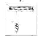
- the collected hair 1 is embedded in an embedding agent 2 after passing through processes such as washing as appropriate ((a) of FIG. 1).
- an embedding agent 2 paraffin, celloidin, gelatin, carbowax, acrylic resin, methacrylic resin, epoxy resin, cyanoacrylate, ice or the like can be used.
- the embedded hair 1 is cut using a microtome, a cutter, a jig or the like to obtain a hair sample 10 (FIG. 1 (b)).
- the thickness of the hair sample 10 is not particularly limited, but may be about 10 nm to 500 ⁇ m as a standard.
- the shape of the hair sample 10 to be produced is not limited to thin pieces, and it is sufficient if the cross section of the hair 1 can be obtained.
- the hair sample 10 may be prepared immediately before measuring the accumulated amount of the physiologically active substance, or may be prepared in advance. However, among the physiologically active substances contained in the hair, those whose amount is likely to change with the passage of time from hair collection, from the viewpoint of accurately measuring the physiologically active substance, the hair sample prepared immediately before the measurement It is preferred to use 10.
- a plurality of hair samples 10 may be prepared by cutting a plurality of places.
- the direction in which the hair 1 is cut is not particularly limited, and may be perpendicular, oblique or parallel to the growth axis of the hair. This will be described based on FIG. FIG. 2 is a diagram showing coordinates defining the cut surface of the hair 1.
- the Y-axis is the growth axis of the hair 1 and the thick arrows are the normal vectors of the cutting plane (ie, the direction vectors perpendicular to the cutting plane).
- the normal vector can be arbitrarily determined in the range of 0 ° ⁇ ⁇ 180 ° and 0 ° ⁇ ⁇ ⁇ 180 °.
- reaction step an antibody that specifically recognizes a physiologically active substance is reacted with the cross section of the hair formed in the cutting step.
- a method (direct method) of detecting a label bound to a primary antibody that recognizes a physiologically active substance is employed.
- the label bound to the primary antibody is, for example, a dye, a fluorescent dye, a radioactive isotope, an enzyme, micro / nano particles (metal particles (gold particles, silver particles, etc.), quantum dots (composition is CdSe) Etc.), polystyrene beads, glass beads etc.).
- a dye or a fluorescent dye is used as a label, light emitted from the dye or the fluorescent dye may be detected.
- a radioactive isotope is used as a label, radiation emitted from the radioactive isotope may be detected.
- an enzyme When using an enzyme as a label, it may be detected using a reaction involving the enzyme.
- a labeling substance a dye, a fluorescent dye, a radioactive isotope, an enzyme, etc.
- the detection may be performed by a method utilizing the physical properties of the above-mentioned fine particles (for example, light scattering by surface plasmon resonance or the like, absorbance, etc. can be detected).
- a fluorescent dye as a label from the viewpoint that miniaturization of a detection device is possible.
- These include light detection elements (photodiodes, phototransistors, complementary metal-oxide semiconductor (CMOS) imaging elements, charge-coupled device (CCD) imaging elements, etc.) and excitation light source elements (semiconductor lasers, This is because small-sized and inexpensive LEDs are widely used in industry.
- CMOS complementary metal-oxide semiconductor
- CCD charge-coupled device
- excitation light source elements semiconductor lasers, This is because small-sized and inexpensive LEDs are widely used in industry.
- Fluorescent dye labels are also preferred in that they differ from radioactive isotopes and do not require special facilities to handle.
- fluorochrome labels are also preferred in that they can also be detected with a fluorescent microscope that is widespread at the laboratory level.
- a method may be used in which a label (conjugate) in which a primary antibody and a labeled antibody are directly bound is produced to stain an antigen.
- the primary antibody used for immunostaining may be appropriately determined depending on the antigen (physiologically active substance) recognized by the primary antibody.
- an anti-cortisol antibody may be used.
- anti-Cortisol antibodies include Anti-Cortisol antibody XM210 ab1949, abcam product and the like.
- anti-DHEA antibodies include Anti-Dehydroepiandrosterone Antibody, Rabbit-Poly and Gene Tex.
- the antibody and the fluorescent dye are linked by known techniques. From the viewpoint of light resistance, it is preferable that the fluorescent dye be contained in the fluorescent particle having a functional group capable of reacting with the antibody to form a bond. At this time, the fluorescent dye is encapsulated by the nanoparticles to form a fluorescent particle, and a bond is formed between the nanoparticle and the antibody.
- the component of the above-mentioned nanoparticles include those capable of stably containing a fluorescent dye, such as polystyrene, polyamide, polylactic acid, polyacrylonitrile, polyglycidyl methacrylate, melamine resin and the like.
- Examples of the fluorescent dye contained in the nanoparticles include rhodamine-based dye molecules, squarylium-based dye molecules, cyanine-based dye molecules, aromatic ring-based dye molecules, oxazine-based dye molecules, carbopyronine-based dye molecules, pyromecene-based dye molecules, etc. Can be mentioned.
- Alexa Fluor registered trademark, manufactured by Invitrogen
- BODIPY registered trademark, manufactured by Invitrogen
- Cy registered trademark, manufactured by GE Healthcare
- DyLight registered trademark, manufactured by Thermo Fisher Scientific
- ATTO registered trademark, manufactured by ATTO-TEC
- pigment molecules MFP (registered trademark, manufactured by Mobitec)
- Dye molecules may be used.
- a known method is used for binding of the fluorescent particle to the antibody. For example, amidation by reaction of amine and carboxylic acid, sulfidation by reaction of maleimide and thiol, imination by reaction of aldehyde and amine, amination by reaction of epoxy and amine, and the like can be used.
- the hair sample 10 includes, in addition to the hair cross section 11 (having the medullary cross section 11a inside), a location 12 where the antibody may bind nonspecifically and a location 13 where the fluorescent dye may bind nonspecifically. (S1).
- the antibody blocking agent 12a is dropped on the hair sample 10 to block the portion 12 where the antibody may nonspecifically bind. This prevents nonspecific binding between the primary antibody 14a and the site 12 where the antibody may bind nonspecifically.
- the fluorescent dye blocking agent 13a is dropped on the hair sample 10 to block the portion 13 where the fluorescent dye can nonspecifically bind. This prevents nonspecific binding between the fluorescent dye 15 and the site 13 where the fluorescent dye can bind nonspecifically.
- Examples of the blocking agent for antibody 12a include artificially synthesized polymers, normal serum, animal serum (rabbit serum, goat serum, rat serum etc.), bovine serum albumin, gelatin, casein and the like.
- Examples of the fluorescent dye blocking agent 13a include reagents that reduce background staining such as FX signal enhancer (manufactured by Thermo Fisher Scientific). Further, if it can be judged that there is no problem even if the treatment with the blocking agent for fluorescent dye is omitted depending on the sample, the treatment with the blocking agent for fluorescent dye may be omitted.
- a primary antibody 14a that specifically recognizes a specific physiologically active substance is dropped on the hair sample 10 and allowed to bind to the physiologically active substance.
- the primary antibody 14a is previously bound to the fluorescent dye 15, the fluorescent dye 15 is bound to the existing location of the physiologically active substance via the primary antibody 14a.
- the hair sample 10 after the reaction step is appropriately subjected to a treatment (for example, a sealing step) necessary for the detection step.
- a treatment for example, a sealing step
- PBS phosphate buffered saline
- the hair sample 10 is placed on a slide glass and the mounting agent is dropped. 3. Place a coverslip on the hair sample.
- the encapsulating agent used in the encapsulating step is not particularly limited as long as the fluorescent dye 15 is not photobleached.
- the cover glass and the slide glass used in the sealing step are also not particularly limited.
- the antibody bound to the physiologically active substance is detected in the reaction step.
- an example of an optical system used in the detection step will be described based on FIG.
- the hair sample 10 enclosed and enclosed between the cover glass 21 and the slide glass 22 is placed on the stage 23.
- fluorescence 32 is emitted from the fluorescent dye 15 contained therein.
- the excitation light 31 is reflected by the dichroic mirror 25 and irradiated to the hair sample 10 via the optical lens 24. Further, the fluorescence 32 passes through the dichroic mirror 25, and only a specific wavelength component is extracted by the filter 26.
- the fluorescence 32 emitted from the fluorescent dye 15 can be detected with less noise. Since the fluorescent dye 15 is bound to the primary antibody 14a and the primary antibody 14a is bound to the physiologically active substance, detecting the fluorescence 32 detects the primary antibody 14a bound to the physiologically active substance It will be.
- the fluorescence 32 may be observed from the column of a fluorescence microscope, or the result detected by a CCD camera or the like may be displayed on a display means such as a monitor.
- image analysis may be performed on an image captured by a CCD camera (image representing the distribution of the primary antibody 14 a in the cross section of the hair by fluorescence) to obtain numerical data indicating the fluorescence distribution.
- a method of analyzing the image will be described in a fifth embodiment.
- FIG. 4 what is described in FIG. 4 is only an example of an optical system that can be used in the detection process. Since the detection step in the method of the present disclosure is sufficient if the antibody bound to the physiologically active substance is detected in the reaction step, detection of the fluorescence 32 is sufficient as the detection step.
- a hair sample 10 which is collected from the same subject and reacted with an antibody that specifically recognizes a physiologically active substance in cross sections having different growth timings.
- this evaluation method can be used to evaluate changes in the degree of stress of a subject.
- the cutting step one hair is cut every 1 cm, and in the reaction step, an antibody that specifically recognizes cortisol in each cut cross section is reacted. Furthermore, the change in the degree of stress of the subject can be evaluated by plotting the intensity (detection result) of the fluorescence 32 of each cross section detected in the detection step in order from the root to the tip of the hair.
- the growth rate of hair varies from subject to subject, but in the case of a human being, it is said that hair grows up to 1 cm in one month. Therefore, in the above example, the change in the degree of stress of the subject can be evaluated at intervals of about one month.
- the intensity of the fluorescence 32 may be an average value of one cross section of the hair sample 10.
- the intensity of the fluorescence 32 may be an average value of the cross sections of the plurality of hair samples 10 at the same growth time.
- the plunger of one syringe was removed and the hair was inserted into the syringe. 3. After the resin for embedding was filled inside the above-mentioned syringe, the plunger was inserted. 4. The plunger of the syringe containing the hair was pushed to push extra bubbles and resin in the syringe to the other connected syringe. 5. The embedding resin was allowed to cure for approximately 1 hour while immediately cooling into ice water.
- the embedded hair was exfoliated using a microtome (or jig (self-made) and a cutter knife).
- the thickness of the flakes was about 300 ⁇ m.
- the obtained slice was placed in a microtube, and 1.0 mL of 1 ⁇ PBS was weighed using a precision pipette and a disposable pipette tip, poured into the above-mentioned microtube, and tapped. The PBS was then removed by aspiration. This process was repeated three times to wash the hair sample.
- the hair sample is placed in a microtube, and 10% gelatin-PBS (w / v) (from Sigma Aldrich gelatin (from cold waterfish)) as a blocking agent for primary antibody is further added to Wako Pure Chemical Industries 10 ⁇ PBS 1 mL of the solution (diluted with ( ⁇ )) was injected into the above-mentioned microtube. After tapping the microtube, the blocking agent for primary antibody was reacted for about 1 hour. Then, 1.0 mL of 1 ⁇ PBS was weighed using a precision pipette and a disposable pipette tip, injected into the above-mentioned microtube, and tapped. The PBS was then removed by aspiration. This process was repeated three times to wash the hair sample.
- FX signal enhancer manufactured by Thermo Fisher Scientific
- a fluorescent dye blocking agent as a fluorescent dye blocking agent
- 1.0 mL of 1 ⁇ PBS was weighed using a precision pipette and a disposable pipette tip, injected into the above-mentioned microtube, and tapped.
- the PBS was then removed by aspiration. This process was repeated three times to wash the hair sample.
- a solution containing the conjugate of the anti-cortisol antibody and the fluorescent dye described above was injected into a microtube for about 200 ⁇ L (the amount in which the hair sample is immersed) and reacted for about 1 hour.
- a hair sample was immersed in 10% gelatin-PBS instead of the above solution.
- 1.0 mL of 1 ⁇ PBS was weighed using a precision pipette and a disposable pipette tip, injected into the above-mentioned microtube, and tapped.
- the PBS was then removed by aspiration. This process was repeated three times to wash the hair sample.
- the same washing operation was performed once with about 1.0 mL of pure water in order to prevent precipitation of the salt contained in PBS at the time of drying and to reduce the background at the time of fluorescence detection.
- the hair sample was then transferred onto a glass slide and allowed to air dry (approximately 30 to 60 minutes) until the hair sample was dry. Finally, an antifading agent (ProLong® Diamond, manufactured by Thermo Fisher Scientific) was dropped onto the hair sample, and a cover glass was covered.
- an antifading agent ProLong® Diamond, manufactured by Thermo Fisher Scientific
- ECLIPSE TE2000-U manufactured by Nikon
- AURA Light Engine manufactured by Lumencor
- the dichroic mirror Chroma, 41008
- a filter Chroma, 41008 was used to block wavelengths below 662 nm.
- EM-CCD manufactured by Hamamatsu Photonics
- a measurement method further including a secondary reaction step of causing a secondary antibody that specifically recognizes the antibody to react with an antibody bound to a physiologically active substance is described.
- the physiologically active substance can be detected with higher sensitivity.
- an indirect method is used as a method of immunostaining.
- the indirect method is a method of performing a first antigen-antibody reaction using an unlabeled primary antibody, and then reacting a labeled secondary antibody that specifically recognizes the primary antibody.
- detection sensitivity is higher in the indirect method than in the direct method described in the first embodiment.
- the measurement method according to the second embodiment will be described below based on FIG.
- the step of collecting hair from a subject (collecting step), the step of forming a cut surface on the hair (cutting step), and the step of detecting an antibody bound to a physiologically active substance (detection step) are described in the first embodiment.
- the description on the antibody and the fluorescent dye according to the step (reaction step) of reacting the antibody that specifically recognizes the physiologically active substance with respect to the cross section of the hair is also as described in the first embodiment.
- the state before reacting the antibody (S1a), the step of allowing the antibody blocking agent to act (S2a), and the step of allowing the fluorescent dye blocking agent to act (S3a) are respectively S1 and S2 in Embodiment 1 (FIG. 3). , Same as S3.
- a primary antibody 14a that specifically recognizes a specific physiologically active substance is dropped on the hair sample 10 and allowed to bind to the physiologically active substance.
- the fluorescent dye is not bound to the primary antibody 14a.
- the secondary antibody 14b that specifically recognizes the primary antibody 14a is dropped onto the hair sample 10 and allowed to bind to the primary antibody 14a.
- the fluorescent dye 15 is bound to the existing site of the physiologically active substance through the primary antibody 14a and the secondary antibody 14b. Become.
- the hair sample is placed in a microtube, and 10% gelatin-PBS (w / v) (from Sigma Aldrich gelatin (from cold water fish)) is added as a blocking agent for the primary antibody, 10 ⁇ PBS (Wako Pure Chemical Industries, Ltd.) 1) was diluted into the above micro tube. After tapping the microtube, the blocking agent for primary antibody was reacted for about 1 hour. Then, 1.0 mL of 1 ⁇ PBS was weighed using a precision pipette and a disposable pipette tip, injected into the above-mentioned microtube, and tapped. The PBS was then removed by aspiration. This process was repeated three times to wash the hair sample.
- FX signal enhancer manufactured by Thermo Fisher Scientific
- a fluorescent dye blocking agent as a fluorescent dye blocking agent
- 1.0 mL of 1 ⁇ PBS was weighed using a precision pipette and a disposable pipette tip, injected into the above-mentioned microtube, and tapped.
- the PBS was then removed by aspiration. This process was repeated three times to wash the hair sample.
- an anti-Cortisol antibody (Anti-Cortisol antibody XM210 ab1949, manufactured by abcam) diluted 200-fold with 10% gelatin-PBS is injected into a microtube for about 200 ⁇ L (the amount in which the hair sample is immersed), and about 1 hour It was made to react.
- a hair sample was immersed in 10% gelatin-PBS instead of the above antibody.
- 1.0 mL of 1 ⁇ PBS was weighed using a precision pipette and a disposable pipette tip, injected into the above-mentioned microtube, and tapped. The PBS was then removed by aspiration. This process was repeated three times to wash the hair sample.
- a fluorescently labeled secondary antibody (goat anti-mouse antibody, attached to Alexa Fluor 488 Goat Anti-Mouse SFX kit (manufactured by Thermo Fisher Scientific)) diluted 100-fold with 10% gelatin-PBS.
- a fluorescently labeled secondary antibody (goat anti-mouse antibody, attached to Alexa Fluor 488 Goat Anti-Mouse SFX kit (manufactured by Thermo Fisher Scientific)) diluted 100-fold with 10% gelatin-PBS.
- 1.0 mL of 1 ⁇ PBS was weighed using a precision pipette and a disposable pipette tip, injected into the above-mentioned microtube, and tapped.
- the PBS was then removed by aspiration. This process was repeated three times to wash the hair sample. Furthermore, the same washing operation was performed once with about 1.0 mL of pure water.
- the hair sample was then transferred onto a glass slide and allowed to air dry (approximately 30 to 60 minutes) until the hair sample was dry. Finally, an antifading agent (ProLong® Diamond, manufactured by Thermo Fisher Scientific) was dropped onto the hair sample, and a cover glass was covered.
- an antifading agent ProLong® Diamond, manufactured by Thermo Fisher Scientific
- the hair sample prepared for speculum observation was observed under a fluorescence microscope to confirm the presence of cortisol.
- BZ-X710 manufactured by Keyence
- ECLIPSE TE2000-U manufactured by Nikon
- a metal halide lamp with a built-in main body was used as an excitation light source.
- the above excitation light source was irradiated to the hair sample through an excitation filter (OP-87763 manufactured by Keyence) having a center wavelength of 470 nm (full width at half maximum 40 nm).
- the dichroic mirror manufactured by Keyence, OP-87763
- a filter manufactured by Keyence, OP-87763
- a cooled CCD incorporated in the main unit was used for fluorescence detection.
- ECLIPSE TE2000-U a light having a center wavelength of 475 nm (full width at half maximum 28 nm) was irradiated using an AURA Light Engine (manufactured by Lumencor) as an excitation light source.
- the dichroic mirror manufactured by Nikon, B-1A
- a filter was used to block wavelengths below 535 nm.
- EM-CCD manufactured by Hamamatsu Photonics
- FIG. 6 A microscopic image representing the results is shown in FIG.
- the upper part of FIG. 6 is a hair sample reacted with the primary antibody, and the lower part is a negative control.
- the left column is a microscope image in bright field
- the right column is a fluorescence microscope image. As shown in FIG. 6, the strongest fluorescence was observed in the medulla. That is, it was shown that cortisol is localized in the medulla.
- Third Embodiment Embodiment 3 describes a measurement method including the reaction with other compounds for the primary antibody and / or the secondary antibody. According to the configuration of the third embodiment, the physiologically active substance can be detected with high sensitivity.
- Examples of measurement methods including reactions with other compounds include ABC (Avidin-Biotin Complex) sensitization and TSA (Thyramide signal amplification) sensitization.
- ABC sensitization is a method of improving the sensitivity of immunostaining by utilizing the formation of a complex between avidin and biotin.
- the TSA sensitization method is a method of improving the sensitivity of immunostaining utilizing the property of tyramide that it is bound to a nearby aromatic molecule by the catalytic action of HRP in the presence of hydrogen peroxide.
- a primary antibody 14a that specifically recognizes a specific physiologically active substance is dropped on the hair sample 10 and allowed to bind to the physiologically active substance.
- the fluorescent dye is not bound to the primary antibody 14a.
- the secondary antibody 14b that specifically recognizes the primary antibody 14a is dropped onto the hair sample 10 and allowed to bind to the primary antibody 14a.
- the secondary antibody 14 b is previously bound to biotin 16.
- biotin 16 that specifically binds to biotin 16 is dropped onto the hair sample 10 and allowed to bind to biotin 16.
- the fluorescent dye 15 is attached to the existing location of the physiologically active substance via the primary antibody 14a, the secondary antibody 14b, the biotin 16 and the avidin 17 It will be combined.
- avidin 17 is a series of proteins (avidins) functionally defined by binding to biotin 16 with high affinity and specificity.
- avidins include avidin, streptavidin, neutravidin.
- an anti-Cortisol antibody (Anti-Cortisol antibody XM210 ab1949, manufactured by abcam) diluted 200-fold with 10% gelatin-PBS was used as a primary antibody, reacted and washed in the same manner as in Experimental Example 2.
- 10% gelatin-PBS was used in the same manner except for using.
- the hair sample was then transferred onto a glass slide and allowed to air dry (approximately 30 to 60 minutes) until the hair sample was dry. Finally, an antifading agent (ProLong® Diamond, manufactured by Thermo Fisher Scientific) was dropped onto the hair sample, and a cover glass was covered.
- an antifading agent ProLong® Diamond, manufactured by Thermo Fisher Scientific
- the hair sample prepared for speculum observation was observed under a fluorescence microscope to confirm the presence of cortisol.
- ECLIPSE TE2000-U manufactured by Nikon
- the excitation light source light with a center wavelength of 475 nm (full width at half maximum 28 nm) was irradiated using AURA Light Engine (manufactured by Lumencor).
- the dichroic mirror manufactured by Nikon, B-1A
- a filter was used to block wavelengths below 535 nm.
- EM-CCD manufactured by Hamamatsu Photonics
- FIG. 8 A microscopic image representing the results is shown in FIG.
- the upper part of FIG. 8 is a hair sample reacted with the primary antibody, and the lower part is a negative control.
- the strongest fluorescence was observed in the medulla. That is, it was shown that cortisol is localized in the medulla.
- Embodiment 4 a measurement method including a quantification step of quantifying the accumulated amount of the physiologically active substance will be described. According to the configuration of the fourth embodiment, the accumulated amount of the physiologically active substance can be quantified according to the detected fluorescence intensity.
- the quantification step is a step of correlating the fluorescence intensity obtained from the immunostained hair sample with the absolute amount of the accumulated amount of the physiologically active substance contained in the hair.
- concentration of the physiologically active substance in another hair (comparative hair) similar to the hair to be measured (the target hair) is previously determined by another method (for example, ELISA) than fluorescent staining using an antibody Measure quantitatively by the method).
- the comparative hair is a hair which is presumed to have accumulated approximately the same amount of physiologically active substance as the target hair, and is, for example, a hair growing near the target hair.
- the fluorescence intensity by the immunostaining is obtained by performing processing of correlating the concentration of the physiologically active substance at the specific site of the comparative hair with the fluorescence intensity at the site corresponding to the specific site of the target hair for a plurality of fluorescent intensities.
- a calibration curve can be created that corresponds to the actual concentration of physiologically active substance.
- an artificial sample containing a physiologically active substance of a known concentration is prepared, and the sample is subjected to the same immunostaining as the immunostaining to be applied to the subject hair.
- a calibration curve showing the relationship between the fluorescence intensity and the concentration of the physiologically active substance can be obtained.
- the dot plot method is mentioned as a method of producing the said artificial sample.
- the calibration curve derived in this manner can also be diverted to the measurement of the amount of physiologically active substance accumulated in the hair thereafter.
- ELISA is a general method that can measure a specific physiologically active substance contained in a sample, and measurement results can be quantified by using a dilution series of known concentration. Specific operating procedures can follow, for example, the protocol attached to the kit for ELISA.
- Embodiment 4 in order to derive a calibration curve representing the relationship between the fluorescence intensity and the accumulated amount (concentration) of the physiologically active substance by ELISA, (1) measuring the accumulated amount of physiologically active substance in the hair by ELISA Then, (2) the correlation with the fluorescence intensity may be determined.
- a physiologically active substance as cortisol.
- Hair is collected from the subject for ELISA (for a more specific method, see Experimental Example 1).
- the hair is preferably grown in the same part as the hair collected from the subject for immunostaining.
- the collected ELISA hair is cut into an appropriate length.
- the portion cut out from the hair for ELISA corresponds to the portion for producing a hair sample from the hair for immunostaining.
- a portion of 0 to 1 cm is cut out from the root side of hair for immunostaining to prepare a hair sample
- a portion of 0 to 1 cm is cut out from the root side of hair for ELISA and measurement by ELISA is performed .
- the hair milled in 4 is placed in a microtube, and 1.5 mL of methanol (manufactured by Wako Pure Chemical Industries, methanol (for LC / MS), product number 138-14521) is added.
- the extract is cortisol extracted by stirring with end-over-end stirring at 10 rpm for 16 to 24 hours at room temperature.
- the resultant is centrifuged at 6500 G for 5 minutes using a centrifuge (Himac K-15, manufactured by Hitachi Koki Co., Ltd.). The supernatant after solid-liquid separation is taken out, and methanol is evaporated using a centrifugal concentrator (manufactured by Tomy Seiko, MV-100). This solidifies cortisol-containing solids.
- a saliva ELISA assay kit (Salimetrics, Cortisol EIA Kit) is diverted and used. Add 25 ⁇ L per well of the following samples, 2 wells each. -Solution obtained by diluting the solid obtained in 6 with 200 ⁇ L of ASSAY diluent-Control solution (cortisol solution of known concentration) ⁇ Cortisol standard solution (kit attachment, 6 series, for preparation of standard curve): • Assay diluent (kit supplied, for zero control and nonspecific binding).
- TMB tetramethyl benzidine
- the fluorescence intensity measured by the method of Experimental Examples 1 to 3 can be associated with the cortisol contained in the hair. That is, since the fluorescence intensity (known amount) measured from the hair sample and the cortisol concentration (known amount, quantified by ELISA) in the portion of the hair corresponding to the hair sample are known, the fluorescence intensity and the hair sample An equation representing the correlation with the cortisol concentration contained in Therefore, once an equation representing such a correlation is prepared, the cortisol concentration can be calculated from the result of immunostaining without the subsequent ELISA.
- the dilution series (the concentration is known) of the target physiologically active substance is dot-plotted on the membrane.
- the dot plot on the membrane is immunostained in the same conditions as the immunostaining applied to the hair sample (eg, in the conditions described in Embodiments 1-3).
- the fluorescence intensity (fluorescence intensity per unit area) of the dot-plotted film is measured.
- the dot plot is a physiologically active substance solution whose concentration is known
- a calibration curve showing the correlation between the fluorescence intensity and the physiologically active substance concentration is obtained. Based on the calibration curve obtained in 4.3, the concentration of the physiologically active substance contained in the hair sample is calculated from the fluorescence intensity emitted from the hair sample.
- membrane known membranes such as PVDF membrane and nitrocellulose membrane can be used. Further, the concentration range of the dilution series of the target physiologically active substance can be determined appropriately.
- the concentration of the physiologically active substance is calculated from the result of the immunostaining without using the dot plot thereafter.
- the distribution of a physiologically active substance in a portion where the fluorescence intensity is weak, which is difficult to distinguish by visual recognition of a fluorescently labeled image, can also be obtained as quantified data.
- the fluorescence intensity per unit area can be calculated, data easy to compare between samples can be obtained.
- the accumulation amount of a physiologically active substance (such as cortisol) localized to the medulla can be quantified without being affected by the difference in the size of the medulla from the subject.
- a plot of fluorescence intensity is generated using an image processing software that can quantify the fluorescence intensity of the image with an image including a fluorescence-labeled image.
- image processing software is not particularly limited, and known image processing software such as ImageJ (free software) and Image Pro Plus (manufactured by Media Cybernetics, registered trademark) may be used.
- the data representing the distribution of the physiologically active substance is preferably in the form of Surface Plot (three-dimensional plot).
- a hair cross section is represented by the X axis and the Y axis
- a fluorescence intensity is represented by the Z axis.
- the data representing the distribution of the physiologically active substance by the above-mentioned Surface Plot can be rotated, tilted and colored as required, and can also be output to the outside.
- Such data have, for example, the advantage that they can be explained in an easy-to-understand manner in the context of explaining the amount of accumulation of physiologically active substances to medical staff.
- a fluorescence intensity distribution represented by a heat map may be mentioned.
- a fluorescence intensity distribution represented by a heat map may be mentioned.
- FIG. (A) of FIG. 9 is a microscopic image showing the result of immunostaining, and is the same as FIG. 6 showing the result of Experimental Example 2.
- (B) of FIG. 9 is a surface plot image representing the luminance in the right column (fluorescent microscope image) of (a) of FIG. The Z axis represents luminance.
- Sixth Embodiment Embodiment 6 is a method of measuring the accumulation amount of a physiologically active substance in hair, which specifically recognizes a second physiologically active substance different from the physiologically active substance with respect to the cross section of the hair.
- the measuring method further includes a second reaction step of reacting the antibody of According to the configuration of Embodiment 6, it is possible to simultaneously measure the accumulation amount of two or more kinds of physiologically active substances.
- the measurement method according to the fifth embodiment will be described below based on FIG.
- the step of collecting hair from a subject (collecting step), the step of forming a cut surface on the hair (cutting step), and the step of detecting an antibody bound to a physiologically active substance (detection step) are described in the first embodiment.
- the description on the antibody and the fluorescent dye according to the step (reaction step) of reacting the antibody that specifically recognizes the physiologically active substance with respect to the cross section of the hair is also as described in the first embodiment. Since the state before reacting the antibody, the step of allowing the antibody blocking agent to act, and the step of allowing the fluorescent dye blocking agent to act are the same as S1 to S3 in the first embodiment (FIG. 3), they are omitted from FIG. Do.
- the step of reacting the antibody includes (1) a method in which two or more types of antibodies are mixed and reacted, and (2) a method in which two types of antibodies are reacted one by one.
- the method (1) is preferable from the viewpoint of reducing the number of work processes.
- the method (2) may be adopted.
- the reaction of the primary antibody is performed by the method of (1)
- the reaction of the secondary antibody is performed by the method of (2), such as (1) and (2) You may use together the method of.
- methods (1) and (2) will be described in order.
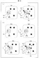
- FIG. 10 is a schematic view showing a method of mixing and reacting (1) two or more types of antibodies.
- the primary antibody 14a that specifically recognizes a physiologically active substance and the second primary antibody 14c that specifically recognizes a second physiologically active substance are dropped onto the hair sample 10 in a mixed state, The substance and the second physiologically active substance are bound respectively.
- the secondary antibody 14b that specifically recognizes the primary antibody 14a and the second secondary antibody 14d that specifically recognizes the second primary antibody 14c are dropped onto the hair sample 10, and the primary antibody 14a and the primary antibody 14a are dropped. It is bound to the second primary antibody 14c.
- the fluorescent dye 15 is bound to the location of the physiologically active substance via the primary antibody 14a and the secondary antibody 14b. Become.
- the second secondary antibody 14 is previously bound to the second fluorescent dye 15a, the second primary antibody 14c and the second The second fluorescent dye 15a is bound via the secondary antibody 14d.
- FIG. 10 is a schematic view showing a method of reacting (2) two types of antibodies one by one.
- the primary antibody 14a that specifically recognizes a physiologically active substance is dropped on the hair sample 10 and allowed to bind to the physiologically active substance.
- the second primary antibody 14c that specifically recognizes the second physiologically active substance is dropped on the hair sample 10 and allowed to bind to the second physiologically active substance.
- the secondary antibody 14b that specifically recognizes the primary antibody 14a is dropped onto the hair sample 10 and allowed to bind to the primary antibody 14a.
- the fluorescent dye 15 is bound to the existing site of the physiologically active substance through the primary antibody 14a and the secondary antibody 14b. Become.
- the second secondary antibody 14d that specifically recognizes the second primary antibody 14c is dropped on the hair sample 10 and allowed to bind to the second primary antibody 14c.
- the second secondary antibody 14d is previously bound to the second fluorescent dye 15a, the second primary antibody 14c and the second The second fluorescent dye 15a is bound via the secondary antibody 14d.
- the same hair sample 10 can be obtained by using fluorescent dyes exhibiting different wavelengths of fluorescence as the fluorescent dye 15 and the second fluorescent dye 15a.
- the accumulated amount of the physiologically active substance and the second physiologically active substance contained in the above can be measured.
- a goat anti-mouse antibody Alexa Fluor (registered trademark) 488 Goat obtained by diluting an anti-Cortisol antibody (Anti-Cortisol antibody XM210 ab1949, abcam) diluted 200-fold as a primary antibody and diluted 100-fold as a secondary antibody.
- Anti-Mouse Antibody manufactured by Thermo Fisher Scientific was used.
- a goat anti-rabbit antibody Anti-Dehydroepiandrosterone Antibody, Rabbit-Poly, Gene Tex, catalog number: GTX10999
- diluted 200-fold as a primary antibody and diluted 100-fold as a secondary antibody Alexa Fluor (registered trademark) 647 Goat Anti-rabbit Antibody (manufactured by Thermo Fisher Scientific) was used.
- NC represents a negative control (Negative Control).
- ECLIPSE TE2000-U (manufactured by Nikon) was used for the speculum.
- AURA Light Engine manufactured by Lumencor
- a ray of central wavelength 475 nm full width at half maximum 28 nm
- a ray of central wavelength 640 nm full width at half maximum 30 nm
- a dichroic mirror (Nikon, B-1A) that reflects a wavelength shorter than 505 nm is used, and for detection of DHEA, a dichroic mirror (a light from Chroma, 41008) that reflects a wavelength shorter than 660 nm is used.
- a filter (Nikon, B-1A) for blocking a wavelength of less than 535 nm was disposed on the detection side of cortisol, and a filter (41008, manufactured by Chroma) for blocking a wavelength of less than 662 nm was disposed on the detection side of DHEA.
- the configuration of the other optical systems is the same as that of Experimental Example 1.
- FIG. 11 is an image showing the results of double immunostaining of cortisol and DHEA.
- the upper row corresponds to the sample reacted with the primary antibody, and the lower row corresponds to the negative control.
- the left column shows the result of detection of 520 nm fluorescence (peak wavelength of the fluorescence emitted by the secondary antibody for cortisol) and the right column shows the fluorescence of 669 nm (peak wavelength of the fluorescence emitted by the secondary antibody for DHEA).
- the hair can be more easily obtained using a known hair removal device.
- the medulla is also expected to be thick, and there is also an advantage that it is easy to detect a physiologically active substance (for example, cortisol) which tends to accumulate in the medulla.
- the washing solution is removed, and after washing with a buffer solution (such as PBS) suitable for immunostaining, embedding is carried out in the same manner as in Embodiment 1 (FIG. 12: ⁇ 1a is embedded in embedding agent 2) ).
- a buffer solution such as PBS
- embedding is carried out in the same manner as in Embodiment 1 (FIG. 12: ⁇ 1a is embedded in embedding agent 2) ).
- the hair sample can be prepared by cutting, and the accumulated amount of the physiologically active substance can be measured in the same manner as in the first embodiment.
- Embodiment 8 describes a kit used in measuring the accumulation amount of the physiologically active substance in the hair in Embodiments 1 to 7.
- a kit according to Embodiment 8 comprises (1) an embedding agent for embedding hair, and (2) an antibody reactive agent containing an antibody that specifically recognizes a physiologically active substance contained in the hair. Have. The following will be described in order.
- An embedding agent is used in cutting the hair to make a hair sample (see FIGS. 1, 12).
- the main component of the embedding agent paraffin, celloidin, gelatin, carbowax, acrylic resin, methacrylic resin, epoxy resin, cyanoacrylate, ice (ice of purified water suitable for freezing and fixing), etc. can be used.
- the antibody reactive agent includes an antibody that specifically recognizes a physiologically active substance (eg, cortisol) contained in hair.
- a physiologically active substance contained in hair can be detected (direct method).
- a labeled antibody as a secondary antibody that specifically recognizes the antibody contained in the above-mentioned antibody reactive agent, the physiologically active substance contained in the hair is higher. It can be detected by sensitivity (indirect method).
- the physiologically active substance contained in the hair can be detected with higher sensitivity also by combining it with the ABC sensitization method, TSA sensitization method or the like.
- the kit according to Embodiment 8 includes, in addition to the embedding agent and the antibody reaction agent, drugs (reagents, solvents, buffers, cleaning agents, etc.) required for use of the kit, devices, containers (bottles, plates, tubes, etc.) Dish etc.), a manual etc. may be provided.
- drugs reagents, solvents, buffers, cleaning agents, etc.
- devices devices, containers (bottles, plates, tubes, etc.) Dish etc.)
- a manual etc. may be provided.
- the medicines, devices, containers, instructions and the like described above may be separately obtained by the user through the market or communication lines.
- Embodiment 9 describes an apparatus used when measuring the accumulation amount of the physiologically active substance in the hair in Embodiments 1 to 7.
- the apparatus according to Embodiment 9 detects the fluorescence generated depending on the antibody bound to the physiologically active substance in the reaction step.
- the above-mentioned device is provided with a member for detecting fluorescence (CCD imaging device, CMOS imaging device, photomultiplier tube, photodiode, phototransistor, etc.).
- the said apparatus may be equipped with the member (Laser, LED, a high pressure mercury lamp, a metal halide lamp etc.) which excites fluorescence as an arbitrary structure.
- FIG. 4 An example of an optical system combining other optical members and elements is shown in FIG. The description of the optical system shown in FIG. 4 is as described in the first embodiment.
- a reaction involving the enzyme for example, a color reaction
- Such devices may be suitably assembled by techniques known in the art.
- a labeling substance a dye, a fluorescent dye, a radioactive isotope, an enzyme or the like
- detection may be performed using physical properties (such as surface plasmon resonance) of the fine particles.
- Such devices may be suitably assembled by techniques known in the art.
- Embodiments 1 to 3 can be arbitrarily combined with Embodiments 4 to 7. That is, the measurement methods described herein can be performed by direct methods or by indirect methods. More specifically, the fluorescent dye may be linked to the primary antibody, linked to the secondary antibody, or linked to another compound such as avidin.
- an antibody to be biotinylated may be a primary antibody or a secondary antibody.
- an antibody to be biotinylated may be a primary antibody or a secondary antibody.
- TSA sensitization may be any of the first and second embodiments.
- the methods of the present disclosure can also be incorporated into radiolabeled antibodies, enzyme labeled antibodies, microparticulate labeled antibodies.
- a radiolabeled antibody When a radiolabeled antibody is used, the radiation emitted by the antibody may be detected in the detection step.
- an enzyme-labeled antibody When an enzyme-labeled antibody is used, a reaction involving the enzyme may be used to detect the antibody bound to the physiologically active substance.
- detection using a phenomenon involving the physical properties of the microparticle, or a labeling substance (a dye, a fluorescent dye, a radioactive isotope, an enzyme, etc.) contained in the microparticle It is sufficient to detect in response.
- the negative control is a cross-section of the hair which has been subjected to treatment in the reaction step with the primary antibody 14a without using the primary antibody 14a (for example, only in the diluted solution of the primary antibody 14a (such as 10% gelatin-PBS)).
- the fluorescence intensity in the hair sample 10 and the background value be measured and processed under the same conditions (for example, the average value of the fluorescence intensity in the pulp of the hair etc. is calculated).
- the method of the present disclosure is easier to align the conditions of the negative control with the conditions of the experimental group than the conventional method.
- the conditions of the negative control and the experimental group can be aligned. 1.
- the hair being embedded is cut along one plane. At this time, two cross sections of the hair are generated, so one is used as an experimental group and the other as a negative control. 2.
- the hair sample is allowed to react under negative control conditions and a first observation is made. The same hair sample is then reacted with the primary antibody (and optionally with the secondary antibody) and subjected to a second observation.
- the method of the present disclosure is characterized by simplicity as compared to the prior art.
- the method of the present disclosure can be analyzed by making a cross-section of the hair, and there is no need to apply a destructive treatment to the hair.
- the method of the present disclosure is different from and superior to methods using mass spectrometry or liquid chromatography.
Landscapes
- Health & Medical Sciences (AREA)
- Life Sciences & Earth Sciences (AREA)
- Engineering & Computer Science (AREA)
- Immunology (AREA)
- Molecular Biology (AREA)
- Biomedical Technology (AREA)
- Chemical & Material Sciences (AREA)
- Hematology (AREA)
- Urology & Nephrology (AREA)
- Biotechnology (AREA)
- Microbiology (AREA)
- Cell Biology (AREA)
- Food Science & Technology (AREA)
- Medicinal Chemistry (AREA)
- Physics & Mathematics (AREA)
- Analytical Chemistry (AREA)
- Biochemistry (AREA)
- General Health & Medical Sciences (AREA)
- General Physics & Mathematics (AREA)
- Pathology (AREA)
- Investigating Or Analysing Biological Materials (AREA)
Abstract
毛髪における生理活性物質の蓄積量を測定する簡便な方法を提供する。毛髪の断面に対して、上記生理活性物質を特異的に認識する抗体を反応させる反応工程と、上記反応工程にて上記生理活性物質と結合した抗体を検出する検出工程と、を含む測定方法とする。
Description
本開示は、毛髪における生理活性物質の蓄積量を測定する方法、測定キットおよび測定装置に関する。
被検体から採取した毛髪中に含まれる物質を測定し、上記被験体の生理状態を判断する技術が存在する。例えば、非特許文献1は、新生児の毛髪をサンプルとして、ELISA(enzyme-linked immunosorbent assay)法によりコルチゾールのレベルを決定したことを報告している。
Yamada J et al (2007) "Hair Cortisol as a Potential BiologicMarker of Chronic Stress in Hospitalized Neonates", Neonatology, Vol.92 (No.1),pp.42-49
しかしながら、上述のような従来技術は、毛髪中の生理活性物質の測定に必要な工程数が多くなり、装置の大型化を招きがちである。
本開示の一態様は、毛髪における生理活性物質の蓄積量を測定する簡便な方法を提供することを目的とする。
上記の課題を解決するために、本開示の一態様に係る測定方法は、毛髪における生理活性物質の蓄積量を測定する方法であって、上記毛髪の断面に対して、上記生理活性物質を特異的に認識する抗体を反応させる反応工程と、上記反応工程にて上記生理活性物質と結合した抗体を検出する検出工程と、を含む。
毛髪における生理活性物質の蓄積量を、簡便に測定することができる。
以下、本開示の実施の形態の一例について詳細に説明する。しかし本開示は、これらに限定されない。
本明細書において特記しない限り、数値範囲を表す「A~B」は、「A以上、B以下」を意味する。
以下、実施形態の説明に先立って、本明細書において広く用いられる用語について説明する。
本明細書における「被検体」の範囲には、ヒトに加えて、非ヒト哺乳類も含まれる。非ヒト哺乳動物の例としては、霊長類(サル、類人猿など)、偶蹄類(ウシ、イノシシ、ブタ、ヒツジ、ヤギなど)、奇蹄類(ウマなど)、齧歯類(マウス、ラット、ハムスター、リスなど)、ウサギ目(ウサギなど)、食肉類(イヌ、ネコ、フェレットなど)などが挙げられる。上述の非ヒト哺乳動物は、家畜または愛玩動物であってもよく、野生動物であってもよい。
本明細書において「毛髪」とは、「被検体に生える毛」を意図する。被検体がヒトの場合、毛髪の例として、頭髪、髭、眉毛、睫毛、鼻毛、耳毛、腋毛、陰毛、体毛が挙げられる。このうち、本開示の方法においては、頭髪または髭を用いることが、毛髪の採取が容易であるため好ましい。さらに、髭は太い毛髪であるため、毛髄質に蓄積している生理活性物質を検出しやすい点においても好ましい。
ここで、生体サンプルとして毛髪を用いることには、生体サンプルとして血液や唾液などを用いることに対して、以下に例示される利点が存在する。
(1)毛髪の採取は非侵襲性であり、容易である。このため、医師または医療関係者でない者でも、毛髪サンプルを採取できる。
(2)他の種類のサンプルに汚染される危険性が低い。例えば、唾液を採取する際に血液が混入すると、唾液中と血液中では生理活性物質のレベルが異なるのが通常であるから、上記生理活性物質のレベルを正確に測定できない。しかし、採取された毛髪は、洗浄により容易に他の種類のサンプル(血液、唾液など)の混入を防ぐことができる。
(3)生理活性物質の「蓄積量」を測定するのに適している。すなわち、血液、唾液などから測定された生理活性物質のレベルは、サーカディアンリズム、急性の生体反応などが原因となり、サンプル採取時の生体条件および環境条件に左右されがちである。これに対し、毛髪の成長速度は頭髪で1cm/月程度であるため、毛髪をサンプルとすることにより、「特定の期間に分泌された生理活性物質の蓄積量」を反映した測定値を得ることができる。
本明細書において、「生理活性物質」とは、生命活動および生理機能の維持、調節などに関わる物質を意図する。生理活性物質の具体例としては、ホルモン、神経伝達物質、サイトカイン、ビタミン、酵素、タンパク質、ペプチドなどが挙げられる。
本開示の測定方法は、毛髪における生理活性物質の蓄積量を測定する方法である。したがって、上記毛髪を採取された被験体の生命活動および生理機能を判断する方法でありうる。
例えば、毛髪におけるストレス関連生理活性物質の蓄積量を測定することにより、上記被験体が慢性的に経験したストレスを知ることができる。このようなストレス関連生理活性物質の例としては、コルチゾール(ヒドロコルチゾン;11β,17α,21-トリヒドロキシプレグナ-4-エン-3,20-ジオン)、DHEA(デヒドロエピアンドロステロン;3β-ヒドロキシアンドロスタ-5-エン-17-オン)、DHEA-S(硫酸結合型DHEA; DHEA-sulfate; DHE-S)、テストステロンなどの副腎皮質ホルモン、カテコールアミン(アドレナリン、ノルアドレナリン)、Neuropeptide Yなどの神経伝達物質、クロモグラニンAなどの酸性タンパク質が挙げられる。このうち、コルチゾール、アドレナリン、クロモグラニンAは、蓄積量の増加と慢性ストレスの増加とが関連付けられる。一方、DHEA、DHEA-Sは、蓄積量の増加と慢性ストレスの減少とが関連付けられる。
また、例えば、ストレス関連疾患である鬱病の患者においては、コルチゾールの蓄積量が高く、DHEAの蓄積量が低いことが判っている。このような場合に、複数の生理活性物質の蓄積量の比(上記の例では、「コルチゾールの蓄積量/DHEAの蓄積量」の値)を求めることも、本開示の範囲に含まれる。
以上に例示した他にも、種々の生理活性物質の蓄積量と、対応する生命活動および生理機能との関係は、医学・生理学分野における技術常識を用いて解釈することができる。
なお、毛髪中にはステロイド骨格を有する物質が蓄積されやすいことが知られている。このため、毛髪におけるステロイド骨格を有する物質の蓄積量を測定することは、本開示の方法の好ましい態様の一つである。ステロイド骨格を有する物質の例としては、コルチゾール、DHEA、DHEA-S、テストステロンなどが挙げられる。
本明細書中における「抗体」には、免疫グロブリンが含まれる。すなわち、IgA、IgD、IgE、IgG、IgM、および、これらのFabフラグメント、F(ab’)2フラグメント、Fcフラグメントなどが含まれる。より具体的には、全てのクラスのポリクローナル抗体、モノクローナル抗体、単鎖抗体、抗イディオタイプ抗体、ヒト化抗体、遺伝子組換えによるヒト型化抗体などが挙げられるが、これらに限定されるものではない。また、本明細書中における「抗体」には、特定の生理活性物質を特異的に認識する限り、抗体断片および抗体修飾物も含まれる。抗体断片の例としては、Fabフラグメント、F(ab’)2フラグメントを挙げることができる。
特定の生理活性物質を特異的に認識する抗体の産生方法は、周知技術である。例えば、[Harlow (Ed.), "Antibodies: a laboratory manual", New York: Cold Spring Harbor Laboratory, 1988]、[岩崎辰夫 他『単クローン抗体: ハイブリドーマとELISA』、講談社、1991年]などを参照することができる。もちろん、市場に流通している抗体を購入して用いてもよい。
〔実施形態1〕
実施形態1では、毛髪における生理活性物質の蓄積量を測定する方法であって、(1)上記毛髪の断面に対して、上記生理活性物質を特異的に認識する抗体を反応させる反応工程と、(2)上記反応工程にて上記生理活性物質と結合した抗体を検出する検出工程と、を含む測定方法について説明する。
実施形態1では、毛髪における生理活性物質の蓄積量を測定する方法であって、(1)上記毛髪の断面に対して、上記生理活性物質を特異的に認識する抗体を反応させる反応工程と、(2)上記反応工程にて上記生理活性物質と結合した抗体を検出する検出工程と、を含む測定方法について説明する。
本開示の態様1に係る、毛髪中における生理活性物質の蓄積量を測定する方法は、(1)上記毛髪の断面に対して、上記生理活性物質を特異的に認識する抗体を反応させる反応工程と、(2)上記反応工程にて上記生理活性物質と結合した抗体を検出する検出工程と、を含む。実施形態1に係る構成によれば、上記毛髪における生理活性物質の蓄積量に関するデータを取得することができる。以下、毛髪サンプルの作製から抗体の検出までを、図1~4に基づいて順に説明する。
まず、反応工程に先立って、被検体から毛髪を採取し(採取工程)、上記毛髪に切断面を形成する(切断工程)。そこで、毛髪の採取および切断について、簡潔に説明する。
毛髪を採取するために用いる道具としては、特に限定されず、公知の道具(ハサミ、髭剃りなど)が用いられうる。
採取工程で被検体から採取される毛髪は、頭髪または髭が好ましい。頭髪または髭は、特殊な器具を用いることなく、容易に採取できるためである。
採取工程で被検体から採取される毛髪の数は、1本以上であれば特に限定されない。より正確な生理活性物質の蓄積量を測定するという観点からは、複数本の毛髪を採取することが好ましい。このときに採取する毛髪の本数は、統計的解釈が可能な本数であれば特に限定されない。毛髪として頭髪を用いる場合、約20本を包埋して毛髪サンプルを作製することができれば、本開示の方法を実施するために充分である。
皮膚や血液などの混入を防ぐ観点から、毛髪の採取に際しては、毛根を含まない部位を採取することが好ましい。一方、時間経過による生理活性物質の蓄積量の変化の影響を小さくするためには、毛髪の根元に近い部位における上記生理活性物質の蓄積量を測定することが好ましく、したがって毛髪の根元に近い部位を採取することが好ましい。以上より、毛髪の採取に際しては、毛髪を根元の近くから採取することが好ましい。
採取された毛髪1は、適宜洗浄などの工程を経た後、包埋剤2に包埋される(図1の(a))。包埋剤の主成分としては、パラフィン、セロイジン、ゼラチン、カーボワックス、アクリル樹脂、メタクリル樹脂、エポキシ樹脂、シアノアクリレート、氷などを用いることができる。
次に、包埋された毛髪1を、ミクロトーム、カッター、治具などを用いて切断し、毛髪サンプル10を得る(図1の(b))。毛髪サンプル10の厚さは特に限定されないが、10nm~500μm程度を目安とすることができる。また、作製される毛髪サンプル10の形状は薄片に限定されず、毛髪1の断面が得られれば充分である。
ここで、毛髪サンプル10は、生理活性物質の蓄積量を測定する直前に作製されたものであってもよいし、事前に作製されたものであってもよい。しかし、毛髪に含まれる生理活性物質のうち、その存在量が毛髪採取からの時間経過により変化しやすいものに関しては、生理活性物質を正確に測定するという観点から、測定の直前に作製した毛髪サンプル10を用いることが好ましい。
毛髪1を切断して毛髪サンプル10を作製する場合、複数箇所を切断することにより、複数の毛髪サンプル10を作製してもよい。
毛髪1を切断するときの方向は特に限定されず、毛髪の成長軸に対して、垂直であってもよく、斜交していてもよく、平行であってもよい。このことを、図2に基づいて説明する。図2は、毛髪1の切断面を規定する座標を表す図である。上記座標において、Y軸は毛髪1の成長軸であり、太い矢印は切断面の法線ベクトル(すなわち、切断面に対して垂直な方向ベクトル)である。このとき、上記法線ベクトルは、0°≦θ≦180°および0°≦φ≦180°の範囲で、任意に定めうる。
反応工程では、切断工程において形成された毛髪の断面に対して、生理活性物質を特異的に認識する抗体を反応させる。上記工程について、まず、使用する抗体に関する一般的な説明を行い、次いで図3に基づいて説明する。
反応工程では、免疫染色に関する公知の手法を用いることができる。このような手法は、例えば、[高田邦昭、斎藤尚亮、川上速人 編『染色・バイオイメージング実験ハンドブック』羊土社、2006年(実験医学別冊)]を参照することができる。
実施形態1では、生理活性物質を認識する一次抗体と結合している標識を検出する方法(直接法)を採用する。上記一次抗体と結合している標識は、例えば、色素、蛍光色素、放射性同位体、酵素、マイクロ・ナノ微粒子(金属微粒子(金微粒子、銀微粒子など)、量子ドット(組成がCdSeである量子ドットなど)、ポリスチレンビーズ、ガラスビーズなど)でありうる。標識として色素または蛍光色素を用いる場合は、当該色素または蛍光色素から放出される光を検出すればよい。標識として放射性同位体を用いる場合は、当該放射性同位体から放出される放射線を検出すればよい。標識として酵素を用いる場合は、当該酵素の関わる反応を用いて検出すればよい。標識としてマイクロ・ナノ微粒子を用いる場合は、当該微粒子の表面に結合している、または当該微粒子に内包されている標識物質(色素、蛍光色素、放射性同位体、酵素など)に応じた検出を行えばよい。あるいは、上記微粒子の物理的性質を利用した方法により検出を行えばよい(例えば、表面プラズモン共鳴などによる光散乱、吸光度等を検出することができる)。
検出機器の小型化が可能であるとの観点からは、標識として蛍光色素を用いることが好ましい。これは、蛍光色素の検出に用いる光検出素子(フォトダイオード、フォトトランジスタ、CMOS(complementary metal-oxide semiconductor)撮像素子、CCD(charge-coupled device)撮像素子など)や励起光源素子(半導体レーザー、高輝度LEDなど)は、小型で安価なものが工業的に広く用いられているためである。また、蛍光色素標識は、放射性同位体とは異なり、取り扱うための特別な施設も必要としない点においても好ましい。さらに、蛍光色素標識は、研究室レベルで普及している蛍光顕微鏡で検出することも可能である点においても好ましい。
なお、実施形態1では、一次抗体と標識抗体とを直接結合した標識(コンジュゲート)を作製し、抗原を染色する方法を用いてもよい。
免疫染色に用いる一次抗体は、当該一次抗体が認識する抗原(生理活性物質)に応じて、適宜決定すればよい。例えば、コルチゾールを抗原とする場合は、抗コルチゾール抗体を用いればよい。抗コルチゾール抗体としては、Anti-Cortisol antibody XM210 ab1949、abcam製などが挙げられる。抗DHEA抗体としては、Anti-Dehydroepiandrosterone Antibody, Rabbit-Poly、Gene Tex製などが挙げられる。
抗体と蛍光色素とは、公知の技術により結合させられる。耐光性の観点からは、抗体と反応して結合を形成しうる官能基を有する蛍光粒子中に、蛍光色素が内包されている状態が好ましい。このとき、上記蛍光色素は、ナノ粒子によって内包されて蛍光粒子を形成し、上記ナノ粒子と抗体との間に結合が形成される。上記ナノ粒子の成分の例としては、ポリスチレン、ポリアミド、ポリ乳酸、ポリアクリロニトリル、ポリグリシジルメタクリレート、メラミン樹脂など、安定的に蛍光色素を内包できるものが挙げられる。
上記ナノ粒子に内包される蛍光色素の例としては、ローダミン系色素分子、スクアリリウム系色素分子、シアニン系色素分子、芳香環系色素分子、オキサジン系色素分子、カルボピロニン系色素分子、ピロメセン系色素分子などが挙げられる。あるいはAlexa Fluor(登録商標、Invitrogen社製)系色素分子、BODIPY(登録商標、Invitrogen製)系色素分子、Cy(登録商標、GE Healthcare製)系色素分子、DY系色素分子(登録商標、DYOMICS製)、HiLyte(登録商標、AnaSpec製)系色素分子、DyLight(登録商標、Thermo Fisher Scientific製)系色素分子、ATTO(登録商標、ATTO-TEC製)系色素分子、MFP(登録商標、Mobitec社製)系色素分子などを用いてもよい。
蛍光粒子と抗体との結合は、公知の手法が用いられる。例えば、アミンとカルボン酸の反応によるアミド化、マレイミドとチオールの反応によるスルフィド化、アルデヒドとアミンの反応によるイミン化、エポキシとアミンの反応によるアミノ化などを用いることができる。
以下、図3に基づいて、反応工程をS1~S4のステップに分けて説明する。S1~S4は、毛髪サンプル10のうち、1つの毛髪の断面近傍を拡大した模式図である。毛髪サンプル10には、毛髪断面11(内部に毛髄質断面11aを有する)の他に、抗体が非特異的に結合しうる箇所12、蛍光色素が非特異的に結合しうる箇所13が含まれている(S1)。
S2では、抗体用ブロッキング剤12aを毛髪サンプル10に滴下し、抗体が非特異的に結合しうる箇所12をブロッキングする。これにより、一次抗体14aと、抗体が非特異的に結合しうる箇所12と、の非特異的な結合を防止する。
S3では、蛍光色素用ブロッキング剤13aを毛髪サンプル10に滴下し、蛍光色素が非特異的に結合しうる箇所13をブロッキングする。これにより、蛍光色素15と、蛍光色素が非特異的に結合しうる箇所13と、の非特異的な結合を防止する。
抗体用ブロッキング剤12aの例としては、人工合成ポリマー、正常血清、動物血清(ウサギ血清、ヤギ血清、ラット血清など)、ウシ血清アルブミン、ゼラチン、カゼインなどが挙げられる。蛍光色素用ブロッキング剤13aの例としては、FX signal enhancer(Thermo Fisher Scientific製)などバックグラウンド染色を低減させる試薬が挙げられる。また、サンプルにより蛍光色素用ブロッキング剤による処理を省略しても問題ないと判断できる場合は、上記の蛍光色素用ブロッキング剤による処理を省略してもよい。
S4では、特定の生理活性物質を特異的に認識する一次抗体14aを、毛髪サンプル10に滴下し、当該生理活性物質と結合させる。ここで、一次抗体14aは、蛍光色素15と予め結合されているため、上記生理活性物質の存在箇所には、一次抗体14aを介して、蛍光色素15が結合することとなる。
なお、反応工程後の毛髪サンプル10には、検出工程に必要な処理(例えば封入工程)を適宜施す。封入工程の具体例を挙げると、以下の通りである。
1.PBS(リン酸緩衝液生理的食塩水)などの水性洗浄液で洗浄し風乾させる。
2.毛髪サンプル10をスライドガラス上に載置し、封入剤を滴下する。
3.毛髪サンプルの上にカバーガラスを載置する。
1.PBS(リン酸緩衝液生理的食塩水)などの水性洗浄液で洗浄し風乾させる。
2.毛髪サンプル10をスライドガラス上に載置し、封入剤を滴下する。
3.毛髪サンプルの上にカバーガラスを載置する。
封入工程で用いられる封入剤は、蛍光色素15を光退色させることがなければ、特に限定されない。封入工程で用いられるカバーガラスおよびスライドガラスも、特に限定されない。
検出工程では、反応工程にて上記生理活性物質と結合した抗体を検出する。以下、検出工程で用いられる光学系の一例について、図4に基づいて説明する。
図4では、カバーグラス21およびスライドグラス22に挟まれて封入されている毛髪サンプル10が、ステージ23上に載置されている。毛髪サンプル10に励起光31が照射されることにより、中に含まれる蛍光色素15から蛍光32が発せられる。
このとき、励起光31は、ダイクロイックミラー25により反射され、光学レンズ24を経由して毛髪サンプル10に照射される。また、蛍光32は、ダイクロイックミラー25を透過して、フィルタ26により特定波長成分のみが取り出される。
以上の構成を取ることにより、蛍光色素15から発せられた蛍光32を、少ないノイズで検出することができる。蛍光色素15は、一次抗体14aに結合しており、一次抗体14aは生理活性物質に結合しているから、蛍光32を検出することは、生理活性物質に結合している一次抗体14aを検出することになる。
蛍光32は、蛍光顕微鏡の鏡筒から観察してもよいし、CCDカメラなどに検出させた結果を、モニターなどの表示手段に表示させてもよい。また、CCDカメラで撮像した画像(毛髪の断面における一次抗体14aの分布を蛍光によって表す画像)に対して画像解析を行い、蛍光分布を示す数値データを得てもよい。当該画像の解析方法については、実施形態5にて説明する。
もっとも、図4で説明されているのは、検出工程に用いることのできる光学系の一例に過ぎない。本開示の方法における検出工程は、反応工程にて上記生理活性物質と結合した抗体を検出すれば足りるのであるから、蛍光32を検出しているならば、検出工程として充分である。
なお、上記の説明は、標識として蛍光色素を用いる場合について成されている。他の種類の標識を用いる場合は、適宜、顕微鏡観察に関する技術常識により、上記の説明を読み換えればよい。
また、切断工程および反応工程において、同一の被検体から採取し、成長時期が異なる断面で生理活性物質を特異的に認識する抗体を反応させた毛髪サンプル10を準備することが好ましい。また、検出工程において抗体から発される蛍光32の強度を、毛髪の成長時期ごとに時系列で比較することが好ましい。蛍光32の強度は被検体の生理活性物質の分泌量に依存するため、蛍光32の強度によって毛髪の成長時期ごとに生理活性物質の分泌量の変化を評価することができる。特に、生理活性物質がストレス関連生理活性物質である場合には、この評価方法を用いて、被検体のストレス度合いの変化を評価することができる。
具体的には、切断工程において1本の毛髪を1cmごとに切断し、反応工程において、切断した各断面においてコルチゾールを特異的に認識する抗体を反応させる。さらに、検出工程で検出した各断面の蛍光32の強度(検出結果)を根元から毛先の順にプロットすることで、被検体のストレス度合いの変化を評価することができる。毛髪の成長速度は被検体によって個体差があるが、人の場合、1箇月で毛髪は1cm成長すると言われている。よって、上記の例では、およそ1箇月間隔で、被検体のストレス度合いの変化を評価することができる。
なお、蛍光32の強度は、毛髪サンプル10の一つの断面の平均値であってもよい。また、蛍光32の強度は、複数の同じ成長時期の毛髪サンプル10の断面の平均値であってもよい。
<実験例1>
実施形態1に係る測定方法により、毛髪に含まれるコルチゾールの蓄積量を測定した。
実施形態1に係る測定方法により、毛髪に含まれるコルチゾールの蓄積量を測定した。
まず、保護具(ゴーグルおよび手袋)を着用し、毛髪採取に用いる器具(実験台、医療用はさみ、クリップ、鏡など)に対し、非感染性処理を実施した。上記非感染性処理には、70%(v/v)エタノール(自家調製)を用いた。
その後、被験者自らが、頭髪を根元付近から採取した(50~150本程度)。
得られた毛髪のうち20本程度を50mL遠沈管へ入れ、該遠沈管へ2-プロパノールを約40mL注入した。次に、上記遠沈管をローテーター(Thermo Fisher Scientific製)10rpmにて3分間、転倒撹拌した。撹拌後、2-プロパノールを除去した。その後、上記と同じ条件で、再度毛髪を洗浄した。こうして処理された毛髪を、ペーパータオル上に置き、30~60分間ほど風乾させた。
次に、包埋・切断工程について説明する。実験の前日に、LR White Acrylic Resin(Sigma Aldrich製)19.8gと過酸化ベンゾイル(Sigma Aldrich製)8.0gとを混合し、室温にて一晩転倒撹拌させた。以下、上記の操作により調製された樹脂を、包埋用樹脂と称する。
25mLチューブへ、上記包埋用樹脂を5mL注入し、LR accelerator(Sigma Aldrich製)を約100μL加え、即座に転倒撹拌した。毛髪をシリンジへセットし、そこへ上記包埋用樹脂を流し込み、包埋した。具体的には、以下の手順で行った。
1.2mLシリンジ(テルモ製、テルモシリンジ)を2本用意し、これらの先端を樹脂製チューブ(Saint-Gobain製、TYGON(登録商標)チューブ)で連結した。
2.一方のシリンジのプランジャーを抜き去り、シリンジ内部に毛髪を挿入した。
3.上記シリンジ内部に包埋用樹脂を充填させた後、プランジャーを挿入した。
4.毛髪が入っているシリンジのプランジャーを押し、シリンジ内の余分な気泡や樹脂を、連結された他方のシリンジへ押し出した。
5.即座に氷水内へつけて冷却しながら、1時間ほどかけて上記包埋樹脂を硬化させた。
1.2mLシリンジ(テルモ製、テルモシリンジ)を2本用意し、これらの先端を樹脂製チューブ(Saint-Gobain製、TYGON(登録商標)チューブ)で連結した。
2.一方のシリンジのプランジャーを抜き去り、シリンジ内部に毛髪を挿入した。
3.上記シリンジ内部に包埋用樹脂を充填させた後、プランジャーを挿入した。
4.毛髪が入っているシリンジのプランジャーを押し、シリンジ内の余分な気泡や樹脂を、連結された他方のシリンジへ押し出した。
5.即座に氷水内へつけて冷却しながら、1時間ほどかけて上記包埋樹脂を硬化させた。
なお、包埋用樹脂を注入した際に気泡が混入した場合は、以下のように対処すればよい。
1.包埋用樹脂が注入されているシリンジの先端が上になるように、上記シリンジを鉛直方向に立てる。これにより、気泡をシリンジ先端へ移動する。
2.1の状態で、さらにプランジャーを押し込むことにより、もう一方のシリンジ側へ気泡を移動させる。
1.包埋用樹脂が注入されているシリンジの先端が上になるように、上記シリンジを鉛直方向に立てる。これにより、気泡をシリンジ先端へ移動する。
2.1の状態で、さらにプランジャーを押し込むことにより、もう一方のシリンジ側へ気泡を移動させる。
続いて、包埋された毛髪を、ミクロトーム(または治具(自作)およびカッターナイフ)を用いて薄片化した。上記薄片の厚さは、約300μmであった。得られた薄片をマイクロチューブに入れ、1×PBSを1.0mL、精密ピペットおよびディスポーザブルピペットチップを用いて量り取り、上記マイクロチューブ内へ注入、タッピングした。その後、吸引によりPBSを除いた。この工程を3回繰り返し、毛髪サンプルを洗浄した。
次に、反応工程について説明する。毛髪サンプルとの反応に先立って、抗コルチゾール抗体(Anti-Cortisol antibody XM210 ab1949、abcam製)と蛍光色素(Alexa Fluor(登録商標)647、Thermo Fisher Scientific製)とのコンジュゲートをAlexa Fluor(登録商標)647 Monoclonal Antibody Labeling Kit(Thermo Fisher Scientific製)を用いて調製した。具体的な調製方法は、上記蛍光色素標識キット添付のプロトコールに従った。最終的には、上記抗体は200倍に希釈された。
次に、毛髪サンプルをマイクロチューブ内へ入れ、さらに一次抗体用ブロッキング剤として、10%ゼラチン-PBS(w/v)(Sigma Aldrich製ゼラチン(from cold waterfish)を、和光純薬工業製10×PBS(-)により希釈したもの)を1mL、上記マ
イクロチューブへ注入した。上記マイクロチューブをタッピングした後、約1時間かけて上記一次抗体用ブロッキング剤を反応させた。そして、1×PBSを1.0mL、精密ピペットおよびディスポーザブルピペットチップを用いて量り取り、上記マイクロチューブ内へ注入、タッピングした。その後、吸引によりPBSを除いた。この工程を3回繰り返し、毛髪サンプルを洗浄した。
イクロチューブへ注入した。上記マイクロチューブをタッピングした後、約1時間かけて上記一次抗体用ブロッキング剤を反応させた。そして、1×PBSを1.0mL、精密ピペットおよびディスポーザブルピペットチップを用いて量り取り、上記マイクロチューブ内へ注入、タッピングした。その後、吸引によりPBSを除いた。この工程を3回繰り返し、毛髪サンプルを洗浄した。
次に、蛍光色素用ブロッキング剤として、FX signal enhancer(Thermo Fisher Scientific製)を3滴程度(毛髪サンプルが浸漬する量)上記マイクロチューブへ注入し、約1時間かけて反応させた。そして、1×PBSを1.0mL、精密ピペットおよびディスポーザブルピペットチップを用いて量り取り、上記マイクロチューブ内へ注入、タッピングした。その後、吸引によりPBSを除いた。この工程を3回繰り返し、毛髪サンプルを洗浄した。
次に、上記抗コルチゾール抗体と上記蛍光色素とのコンジュゲートを含む溶液を200μL程度(毛髪サンプルが浸漬する量)、マイクロチューブへ注入し、約1時間かけて反応させた。また、ネガティブコントロールとしては、上記溶液の代わりに、10%ゼラチン-PBSに毛髪サンプルを浸漬させた。そして、1×PBSを1.0mL、精密ピペットおよびディスポーザブルピペットチップを用いて量り取り、上記マイクロチューブ内へ注入、タッピングした。その後、吸引によりPBSを除いた。この工程を3回繰り返し、毛髪サンプルを洗浄した。さらに、乾燥時にPBSに含まれる塩が析出するのを防ぐため、および、蛍光検出の際のバックグラウンドを低減させるために、同様の洗浄操作を1.0mL程度の純水で1回行った。
次に、スライドガラス上へ毛髪サンプルを移し、毛髪サンプルが乾燥するまで風乾させた(30~60分程度)。最後に、毛髪サンプル上へ褪色防止剤(ProLong(登録商標)Diamond、Thermo Fisher Scientific製)を滴下し、カバーガラスを被せた。
検鏡用に調製した毛髪サンプルを、蛍光顕微鏡下で観察した(データなし)。このとき、蛍光顕微鏡として、ECLIPSE TE2000-U(ニコン製)を用いた。励起光源として、AURA Light Engine(Lumencor製)を用い、中心波長640nm(半値全幅30nm)の光線を照射した。ダイクロイックミラー(Chroma製、41008)は、660nmよりも短波長を反射させる設計とした。検出側に、フィルタ(Chroma製、41008)を使用し、662nm未満の波長を遮断した。蛍光の検出にはEM-CCD(浜松ホトニクス製)を使用した。
〔実施形態2〕
実施形態2では、生理活性物質と結合している抗体に対して、当該抗体を特異的に認識する二次抗体を反応させる二次反応工程をさらに含んでいる測定方法について説明する。実施形態2に係る構成によれば、生理活性物質をより高感度に検出することができる。
実施形態2では、生理活性物質と結合している抗体に対して、当該抗体を特異的に認識する二次抗体を反応させる二次反応工程をさらに含んでいる測定方法について説明する。実施形態2に係る構成によれば、生理活性物質をより高感度に検出することができる。
実施形態2では、免疫染色の方法として間接法を用いる。間接法とは、標識していない一次抗体を用いて1度目の抗原抗体反応を行い、次に一次抗体を特異的に認識する標識二次抗体を反応させる方法である。一般的に、実施形態1で説明した直接法に比べて、間接法の方が検出感度は高い。
以下、図5に基づいて、実施形態2に係る測定方法を説明する。なお、被検体から毛髪を採取する工程(採取工程)、上記毛髪に切断面を形成する工程(切断工程)および生理活性物質と結合した抗体を検出する工程(検出工程)については、実施形態1に記載の通りである。また、上記毛髪の断面に対して生理活性物質を特異的に認識する抗体を反応させる工程(反応工程)に係る抗体および蛍光色素に関する説明も、実施形態1に記載の通りである。
抗体を反応させる前の状態(S1a)、抗体用ブロッキング剤を作用させる工程(S2a)、蛍光色素用ブロッキング剤を作用させる工程(S3a)は、それぞれ、実施形態1(図3)におけるS1、S2、S3と同じである。
S4aでは、特定の生理活性物質を特異的に認識する一次抗体14aを、毛髪サンプル10に滴下し、当該生理活性物質と結合させる。実施形態1と異なり、実施形態2では、一次抗体14aに蛍光色素が結合されていない。
S5aでは、一次抗体14aを特異的に認識する二次抗体14bを、毛髪サンプル10に滴下し、一次抗体14aと結合させる。ここで、二次抗体14bは、蛍光色素15と予め結合されているため、上記生理活性物質の存在箇所には、一次抗体14aおよび二次抗体14bを介して、蛍光色素15が結合することとなる。
<実験例2>
実施形態2に係る測定方法により、毛髪に含まれるコルチゾールの蓄積量を測定した。
実施形態2に係る測定方法により、毛髪に含まれるコルチゾールの蓄積量を測定した。
毛髪を採取、包埋、切断して毛髪サンプルを作製する手順は、実験例1に記載の通りである。
毛髪サンプルをマイクロチューブ内へ入れ、さらに一次抗体用ブロッキング剤として、10%ゼラチン-PBS(w/v)(Sigma Aldrich製ゼラチン(from cold water fish)を、和光純薬工業製10×PBS(-)により希釈したもの)を1mL、上記マイクロチューブへ注入した。上記マイクロチューブをタッピングした後、約1時間かけて上記一次抗体用ブロッキング剤を反応させた。そして、1×PBSを1.0mL、精密ピペットおよびディスポーザブルピペットチップを用いて量り取り、上記マイクロチューブ内へ注入、タッピングした。その後、吸引によりPBSを除いた。この工程を3回繰り返し、毛髪サンプルを洗浄した。
続いて、蛍光色素用ブロッキング剤として、FX signal enhancer(Thermo Fisher Scientific製)を3滴程度(毛髪サンプルが浸漬する量)上記マイクロチューブへ注入し、約1時間かけて反応させた。そして、1×PBSを1.0mL、精密ピペットおよびディスポーザブルピペットチップを用いて量り取り、上記マイクロチューブ内へ注入、タッピングした。その後、吸引によりPBSを除いた。この工程を3回繰り返し、毛髪サンプルを洗浄した。
次に、10%ゼラチン-PBSにて200倍に希釈した抗コルチゾール抗体(Anti-Cortisol antibody XM210 ab1949、abcam製)を200μL程度(毛髪サンプルが浸漬する量)、マイクロチューブへ注入し、約1時間かけて反応させた。また、ネガティブコントロールとしては、上記抗体の代わりに、10%ゼラチン-PBSに毛髪サンプルを浸漬させた。そして、1×PBSを1.0mL、精密ピペットおよびディスポーザブルピペットチップを用いて量り取り、上記マイクロチューブ内へ注入、タッピングした。その後、吸引によりPBSを除いた。この工程を3回繰り返し、毛髪サンプルを洗浄した。
次に、10%ゼラチン-PBSにて100倍に希釈した蛍光標識二次抗体(ヤギ抗マウス抗体、Alexa Fluor(登録商標)488 Goat Anti-Mouse SFX kit(Thermo Fisher Scientific製)に附属)を200μL程度(毛髪サンプルが浸漬する量)、マイクロチューブへ注入し、約1時間かけて反応させた。そして、1×PBSを1.0mL、精密ピペットおよびディスポーザブルピペットチップを用いて量り取り、上記マイクロチューブ内へ注入、タッピングした。その後、吸引によりPBSを除いた。この工程を3回繰り返し、毛髪サンプルを洗浄した。さらに、同様の洗浄操作を1.0mL程度の純水で1回行った。
次に、スライドガラス上へ毛髪サンプルを移し、毛髪サンプルが乾燥するまで風乾させた(30~60分程度)。最後に、毛髪サンプル上へ褪色防止剤(ProLong(登録商標)Diamond、Thermo Fisher Scientific製)を滴下し、カバーガラスを被せた。
検鏡用に調製した毛髪サンプルを、蛍光顕微鏡下で観察し、コルチゾールの存在を確認した。このとき、蛍光顕微鏡として、BZ-X710(キーエンス製)およびECLIPSE TE2000-U(ニコン製)を用いた。
BZ-X710による観察においては、励起光源として、本体内蔵のメタルハライドランプを用いた。上記励起光源を、中心波長470nm(半値全幅40nm)の励起フィルタ(キーエンス製、OP-87763)を介して、毛髪サンプルに照射した。ダイクロイックミラー(キーエンス製、OP-87763)は、495nmよりも短波長を反射させる設計とした。検出側に、フィルタ(キーエンス製、OP-87763)を使用し、500nm未満の波長を遮断した。蛍光の検出には本体内蔵の冷却CCDを使用した。
ECLIPSE TE2000-Uによる観察においては、励起光源として、AURA Light Engine(Lumencor製)を用い、中心波長475nm(半値全幅28nm)の光線を照射した。ダイクロイックミラー(ニコン製、B-1A)は、505nmよりも短波長を反射させる設計とした。検出側に、フィルタ(ニコン製、B-1A)を使用し、535nm未満の波長を遮断した。蛍光の検出にはEM-CCD(浜松ホトニクス製)を使用した。
結果を表す顕微鏡像を、図6に示す。図6の上段は一次抗体による反応を行った毛髪サンプル、下段はネガティブコントロールである。また、左列は明視野での顕微鏡像、右列は蛍光顕微鏡像である。図6に示されている通り、毛髄質において最も強い蛍光が認められた。すなわち、毛髄質にコルチゾールが局在していることが示された。
〔実施形態3〕
実施形態3では、一次抗体および/または二次抗体に対する、他の化合物との反応を含む測定方法について説明する。実施形態3に係る構成によれば、さらに生理活性物質を高感度に検出することができる。
実施形態3では、一次抗体および/または二次抗体に対する、他の化合物との反応を含む測定方法について説明する。実施形態3に係る構成によれば、さらに生理活性物質を高感度に検出することができる。
他の化合物との反応を含む測定方法の例としては、ABC(Avidin-Biotin Complex)増感法、TSA(Thyramide signal amplification)増感法が挙げられる。ABC増感法は、アビジンとビオチンとが複合体を形成することを利用して、免疫染色の感度を向上させる方法である。TSA増感法は、過酸化水素存在下にてHRPの触媒作用により近傍の芳香族分子に結合する、というチラミドの性質を利用して、免疫染色の感度を向上させる方法である。以下、例として、ABC増感法による免疫染色の工程を、図7に基づいて説明する。
S4bより前の工程(抗体用ブロッキングおよび蛍光色素用ブロッキング)までは、実施形態1と同様である。
S4bでは、まず、特定の生理活性物質を特異的に認識する一次抗体14aを、毛髪サンプル10に滴下し、当該生理活性物質と結合させる。このとき、実施形態2と同様に、一次抗体14aには蛍光色素が結合されていない。
S5bでは、一次抗体14aを特異的に認識する二次抗体14bを、毛髪サンプル10に滴下し、一次抗体14aと結合させる。ここで、二次抗体14bは、ビオチン16と予め結合されている。
S6bでは、ビオチン16と特異的に結合するアビジン17を、毛髪サンプル10に滴下し、ビオチン16と結合させる。ここで、ビオチン16は、蛍光色素15と予め結合されているため、上記生理活性物質の存在箇所には、一次抗体14a、二次抗体14b、ビオチン16およびアビジン17を介して、蛍光色素15が結合することとなる。
ここで、アビジン17は、ビオチン16に対して高度の親和性および特異性をもって結合することによって機能的に定義される、一連のタンパク質(アビジン類)である。アビジン類の例としては、アビジン、ストレプトアビジン、ニュートラアビジンが挙げられる。
<実験例3>
実施形態3に係る測定方法(ABC増感法)により、毛髪に含まれるコルチゾールの蓄積量を測定した。
実施形態3に係る測定方法(ABC増感法)により、毛髪に含まれるコルチゾールの蓄積量を測定した。
毛髪を採取、包埋、切断して毛髪サンプルを作製する手順は、実験例1に記載の通りである。
また、一次抗体として10%ゼラチン-PBSにて200倍に希釈した抗コルチゾール抗体(Anti-Cortisol antibody XM210 ab1949、abcam製)を使用し、実験例2と同様の手順で反応させ、洗浄した。ネガティブコントロールとしては、上記抗体の代わりに、10%ゼラチン-PBSを使用した以外は、同様に処理した。
次に、10%ゼラチン-PBSにて200倍に希釈したビオチン化抗体(Thermo Fisher Scientific製、Biotin-XX goat anti-mouse IgG(H+L))を150μL程度(毛髪サンプルが浸漬する量)、マイクロチューブへ注入し、約1時間かけて反応させた。そして、1×PBSを1.0mL、精密ピペットおよびディスポーザブルピペットチップを用いて量り取り、上記マイクロチューブ内へ注入、タッピングした。その後、吸引によりPBSを除いた。この工程を3回繰り返し、毛髪サンプルを洗浄した。
次に、10%ゼラチン-PBSにて10μg/mLに希釈した蛍光標識ストレプトアビジン(Thermo Fisher Scientific製、Streptavidin, Alexa Fluor(登録商標)488 conjugate)を100μL程度(毛髪サンプルが浸漬する量)、マイクロチューブへ注入し、約1時間かけて反応させた。そして、1×PBSを1.0mL、精密ピペットおよびディスポーザブルピペットチップを用いて量り取り、上記マイクロチューブ内へ注入、タッピングした。その後、吸引によりPBSを除いた。この工程を3回繰り返し、毛髪サンプルを洗浄した。さらに、同様の洗浄操作を1.0mL程度の純水で行った。
次に、スライドガラス上へ毛髪サンプルを移し、毛髪サンプルが乾燥するまで風乾させた(30~60分程度)。最後に、毛髪サンプル上へ褪色防止剤(ProLong(登録商標)Diamond、Thermo Fisher Scientific製)を滴下し、カバーガラスを被せた。
検鏡用に調製した毛髪サンプルを、蛍光顕微鏡下で観察し、コルチゾールの存在を確認した。このとき、蛍光顕微鏡として、ECLIPSE TE2000-U(ニコン製)を用いた。励起光源として、AURA Light Engine(Lumencor製)を用い、中心波長475nm(半値全幅28nm)の光線を照射した。ダイクロイックミラー(ニコン製、B-1A)は、505nmよりも短波長を反射させる設計とした。検出側に、フィルタ(ニコン製、B-1A)を使用し、535nm未満の波長を遮断した。蛍光の検出にはEM-CCD(浜松ホトニクス製)を使用した。
結果を表す顕微鏡像を、図8に示す。図8の上段は一次抗体による反応を行った毛髪サンプル、下段はネガティブコントロールである。図8に示されている通り、毛髄質において最も強い蛍光が認められた。すなわち、毛髄質にコルチゾールが局在していることが示された。
〔実施形態4〕
実施形態4では、生理活性物質の蓄積量を定量する定量工程を含む測定方法について説明する。実施形態4に係る構成によれば、検出された蛍光強度に応じて生理活性物質の蓄積量を定量することができる。
実施形態4では、生理活性物質の蓄積量を定量する定量工程を含む測定方法について説明する。実施形態4に係る構成によれば、検出された蛍光強度に応じて生理活性物質の蓄積量を定量することができる。
定量工程とは、免疫染色された毛髪サンプルから得られる蛍光強度と、上記毛髪に含まれる生理活性物質の蓄積量の絶対量とを、関連付ける工程である。この関連付けのために、予め、測定対象となる毛髪(対象毛髪)と類似した別の毛髪(比較毛髪)における生理活性物質の濃度を、抗体を用いた蛍光染色とは別の方法(例えば、ELISA法)で定量的に測定しておく。上記比較毛髪とは、対象毛髪とほぼ同じ量の生理活性物質を蓄積していると推定される毛髪であり、例えば、対象毛髪の近傍に生えている毛髪である。
比較毛髪の特定部位における生理活性物質の濃度と、対象毛髪の、上記特定部位に対応する部位における蛍光強度とを対応付けるという処理を、複数の蛍光強度について行うことにより、免疫染色による蛍光強度と、実際の生理活性物質の濃度とを対応付ける検量線を作成することができる。
または、比較毛髪の代わりに、既知の濃度の生理活性物質を含む人工的なサンプルを作製し、当該サンプルに対して、対象毛髪に施す免疫染色と同様の免疫染色を行うことにより、免疫染色における蛍光強度と生理活性物質の濃度との関係を示す検量線を得ることができる。上記人工的なサンプルを作製する方法として、ドットプロット法が挙げられる。
このように導出された検量線は、以降の毛髪における生理活性物質の蓄積量の測定に流用することもできる。
以下、ELISA法およびドットプロット法について順に説明する。
ELISAは、サンプルに含まれる特定の生理活性物質を測定することができる一般的な手法であり、濃度既知の希釈系列を用いることによって、測定結果を定量することができる。具体態的な操作手順は、例えば、ELISA用キットに添付のプロトコールに従うことができる。
実施形態4において、蛍光強度と生理活性物質の蓄積量(濃度)との関係を表す検量線を、ELISAによって導出するためは、(1)毛髪中の生理活性物質の蓄積量をELISAで測定し、次いで(2)蛍光強度との相関を求めればよい。以下、生理活性物質をコルチゾールとして、具体的な操作手順の一例を示す。
1.ELISA用に被検体から毛髪を採取する(より具体的な方法は、実験例1を参照)。上記毛髪は、免疫染色用に被検体から採取した毛髪と、同じ部分に生えているものとすることが好ましい。
2.採取したELISA用の毛髪を、適当な長さに切り出す。このとき、ELISA用の毛髪から切り出す部分は、免疫染色用の毛髪から毛髪サンプルを作製する部分に相当している。例えば、免疫染色用の毛髪の根元側から0~1cmの部分を切り出して、毛髪サンプルを作製する場合は、ELISA用の毛髪の根元側から0~1cmの部分を切り出して、ELISAによる測定を行う。
3.切り出した毛髪をマイクロチューブに(トミー精工製、TM-625S)3mg入れ、イソプロピルアルコール(キシダ化学製、2-プロパノール 高速液体クロマト用、製品番号140-64781)1mLを加える。攪拌機(アズワン製、ACR-100)にセットし、10rpmにて3分間攪拌した後、イソプロピルアルコールを除く。以上の工程を3回程度繰り返し、毛髪を洗浄する。
4.ビーズ式細胞破砕機(トミー精工製、MS-100R)およびジルコニア製ビーズ(トミー精工製、ZB-20、直径2.0mm)を用いて、回転数4000rpm、破砕時間60秒間の条件で毛髪を粉砕する。冷却時間を1分間以上空けて、毛髪が充分に粉砕されるまで、上記の条件による粉砕を繰り返す。
5.マイクロチューブに、4で粉砕された毛髪を入れ、メタノール(和光純薬工業製、メタノール(LC/MS用)、製品番号138-14521)1.5mLを加える。これを、室温において10rpmにて16~24時間、転倒攪拌しながらコルチゾールを抽出する。
6.コルチゾールの抽出工程の終了後、遠心分離機(日立工機製、Himac CF-15RX)により、6500Gにて5分間遠心分離する。固液分離後の上澄み液を取り出し、遠心濃縮機(トミー精工製、MV-100)を用いて、メタノールを蒸発させる。これにより、コルチゾールを含む固形物が乾固する。
7.コルチゾール濃度の決定には、唾液用ELISA測定キット(Salimetrics製、Cortisol EIA Kit)を転用して用いる。以下のサンプルを、2ウェルずつ、1ウェルあたり25μLを加える。
・6で得た固形物を、200μLのASSAY diluentに希釈した溶液
・コントロール溶液(既知濃度のコルチゾール溶液)
・コルチゾール標準溶液(キット附属、6系列、検量線作成用):
・Assay diluent(キット附属、ゼロコントロールおよび非特異的結合用)。
・6で得た固形物を、200μLのASSAY diluentに希釈した溶液
・コントロール溶液(既知濃度のコルチゾール溶液)
・コルチゾール標準溶液(キット附属、6系列、検量線作成用):
・Assay diluent(キット附属、ゼロコントロールおよび非特異的結合用)。
8.最終濃度に希釈した酵素コンジュゲート(HRPとコルチゾールの複合体)溶液を200μL、各ウェルに加える。5分間攪拌し、室温にて1時間かけて反応させる。このとき、サンプル溶液、コントロール溶液および標準溶液中のコルチゾールは、上記酵素コンジュゲートと競争的に抗コルチゾール抗体に結合する。
9.300μLの洗浄バッファー(PBSを主成分とする)でウェルを4回洗浄する。抗コルチゾール抗体に結合していないコルチゾールは、この段階で洗い流される。
10.テトラメチルベンジジン(TMB)を各ウェルに200μLずつ加える。TMBは酵素コンジュゲートに含まれるHRPと反応し、呈色する。その後、STOP solution(キット附属のメタンスルホン酸)50μLを滴下し、酵素反応を停止させる。
11.プレートリーダー(PerkinElmer製、ARVO-MX)を用いて、450nmおよび490nmの吸光度を測定する。
12.以下の手順により、毛髪中のコルチゾール濃度を算出する。
・同じサンプルを含んでいる、2つのウェルの平均光強度を算出する。
・ゼロコントロール、標準溶液、コントロール溶液、毛髪サンプルの各光学強度から、非特異的結合の光強度を差し引く。
・各ウェル光学強度(B)を、ゼロコントロールの平均光学強度(B0)で除す。これにより、規格化された抗コルチゾール抗体に対する酵素コンジュゲートの結合率(B/B0)を算出する。
・得られた標準溶液の吸光度から検量線を作成する。4-パラメータ非線形回帰曲線を推奨されるため、逆シグモイド関数(4-パラメータ)でフィットする。
・得られた検量線から、毛髪サンプルのコルチゾール濃度を定量する。コントロール溶液の濃度も定量し、測定の精度を確認する。
・同じサンプルを含んでいる、2つのウェルの平均光強度を算出する。
・ゼロコントロール、標準溶液、コントロール溶液、毛髪サンプルの各光学強度から、非特異的結合の光強度を差し引く。
・各ウェル光学強度(B)を、ゼロコントロールの平均光学強度(B0)で除す。これにより、規格化された抗コルチゾール抗体に対する酵素コンジュゲートの結合率(B/B0)を算出する。
・得られた標準溶液の吸光度から検量線を作成する。4-パラメータ非線形回帰曲線を推奨されるため、逆シグモイド関数(4-パラメータ)でフィットする。
・得られた検量線から、毛髪サンプルのコルチゾール濃度を定量する。コントロール溶液の濃度も定量し、測定の精度を確認する。
以上の過程を経ることにより、実験例1~3の方法により測定された蛍光強度と、毛髪に含まれるコルチゾールとを関連付けることができる。すなわち、毛髪サンプルから測定された蛍光強度(既知量)と、上記毛髪サンプルに対応する毛髪の部分におけるコルチゾール濃度(既知量、ELISAにて定量)が判明しているため、蛍光強度と上記毛髪サンプルに含まれるコルチゾール濃度との相関を表す式が得られる。したがって、このような相関を表す式を一度作成したならば、以降はELISAを伴わなくとも、免疫染色の結果からコルチゾール濃度を算出することができる。
次に、ドットプロット法により、毛髪サンプルの発する蛍光強度から、毛髪サンプルに含まれる生理活性物質の蓄積量(濃度)を定量する方法について、以下に一例を示す。
1.目的とする生理活性物質の希釈系列(濃度既知)を、膜上にドットプロットする。
2.毛髪サンプルに施す免疫染色と同条件で(例えば、実施形態1~3に記載の条件で)、上記膜上のドットプロットを免疫染色する。
3.ドットプロットした膜の蛍光強度(単位面積あたりの蛍光強度)を測定する。ドットプロットは濃度既知の生理活性物質溶液であるから、蛍光強度と生理活性物質濃度との相関を示す検量線が得られる。
4.3で得られた検量線に基づいて、毛髪サンプルの発する蛍光強度から、毛髪サンプルに含まれる生理活性物質の濃度を算出する。
1.目的とする生理活性物質の希釈系列(濃度既知)を、膜上にドットプロットする。
2.毛髪サンプルに施す免疫染色と同条件で(例えば、実施形態1~3に記載の条件で)、上記膜上のドットプロットを免疫染色する。
3.ドットプロットした膜の蛍光強度(単位面積あたりの蛍光強度)を測定する。ドットプロットは濃度既知の生理活性物質溶液であるから、蛍光強度と生理活性物質濃度との相関を示す検量線が得られる。
4.3で得られた検量線に基づいて、毛髪サンプルの発する蛍光強度から、毛髪サンプルに含まれる生理活性物質の濃度を算出する。
上記膜はPVDF膜、ニトロセルロース膜など公知のものが使用可能である。また、目的の生理活性物質の希釈系列の濃度範囲は、適宜定めうる。
ELISAを用いる方法と同様に、蛍光強度と生理活性物質濃度との相関を示す検量線が一度得られれば、以降はドットプロットを伴わなくとも、免疫染色の結果から生理活性物質濃度を算出することができる。
〔実施形態5〕
実施形態5では、上記毛髪の断面における上記抗体の分布を表す画像を解析する方法について説明する。実施形態5に係る構成によれば、毛髪断面において生理活性物質が存在している位置に関するデータを得ることができる。また、毛髪断面において生理活性物質が存在している位置と、当該位置における当該生理活性物質の蓄積量とが関連付けられたデータを得ることもできる。
実施形態5では、上記毛髪の断面における上記抗体の分布を表す画像を解析する方法について説明する。実施形態5に係る構成によれば、毛髪断面において生理活性物質が存在している位置に関するデータを得ることができる。また、毛髪断面において生理活性物質が存在している位置と、当該位置における当該生理活性物質の蓄積量とが関連付けられたデータを得ることもできる。
その他、実施形態5に係る測定方法が奏しうる効果としては、例えば、以下のものが挙げられる。
1.蛍光標識された像を肉眼で視認するのみでは識別することが難しい、蛍光強度の弱い部分における生理活性物質の分布をも、数値化されたデータとして得ることができる。
2.単位面積当たりの蛍光強度を算出することができるため、サンプル間で比較しやすいデータが得られる。例えば、被検体による毛髄質の大きさの差の影響を受けることなく、毛髄質に局在する生理活性物質(コルチゾールなど)の蓄積量を定量できる。
1.蛍光標識された像を肉眼で視認するのみでは識別することが難しい、蛍光強度の弱い部分における生理活性物質の分布をも、数値化されたデータとして得ることができる。
2.単位面積当たりの蛍光強度を算出することができるため、サンプル間で比較しやすいデータが得られる。例えば、被検体による毛髄質の大きさの差の影響を受けることなく、毛髄質に局在する生理活性物質(コルチゾールなど)の蓄積量を定量できる。
生理活性物質の分布を表すデータを取得するには、蛍光標識された像を含む画像に対して、当該像の蛍光強度を数値化できる画像処理ソフトウェアを用いて、蛍光強度のプロット図を生成すればよい。このような画像処理ソフトウェアは、特に限定されないが、ImageJ(フリーソフト)、Image Pro Plus(Media Cybernetics製。登録商標)などの公知の画像処理ソフトが用いられる。
生理活性物質の分布を表すデータは、Surface Plot(3次元プロット)の形式であることが好ましい。Surface Plotによる生理活性物質の分布を表すデータの表示例としては、X軸およびY軸で毛髪断面を表し、Z軸で蛍光強度(輝度)を表すものである。上記Surface Plotによる生理活性物質の分布を表すデータは、必要に応じて回転、傾斜、色づけが可能であり、また外部への出力も可能である。このようなデータには、例えば、医療従事者が患者に対して生理活性物質の蓄積量を説明する場面において、視覚的に判りやすく説明することができるという利点がある。
他の種類の分布表現方法としては、例えば、蛍光強度分布をヒートマップで表したものなどが挙げられる。このような技術を用いる場合は、表面分布画像の表現法に関する技術常識により、上記の説明を適宜読み換えればよい。
<実験例4>
実施形態5に係る測定方法により、実験例2と同様に処理を行った毛髪サンプルに対して、画像処理ソフトImageJを用い、生理活性物質の分布を表すデータを得た。
実施形態5に係る測定方法により、実験例2と同様に処理を行った毛髪サンプルに対して、画像処理ソフトImageJを用い、生理活性物質の分布を表すデータを得た。
結果を図9に表す。図9の(a)は、免疫染色の結果を表す顕微鏡像で、実験例2の結果を表す図6と同じものである。図9の(b)は、図9の(a)の右列(蛍光顕微鏡像)における、輝度を表したSurface Plot像である。Z軸に輝度を表している。
一次抗体による反応を行った毛髪サンプルに由来するSurface plot像(上段)によると、毛髄質および毛小皮において強い蛍光強度が示された。一方、ネガティブコントロールに由来するSurface plot像では、毛髪の断面全体において低い蛍光強度が見られた。
ここで、一次抗体による反応を行った毛髪サンプルの輝度分布から、ネガティブコントロールの輝度分布を減算した結果を基にSurface Plot像を生成すれば、より正確な毛髪断面におけるコルチゾール分布が判る。
〔実施形態6〕
実施形態6では、毛髪における生理活性物質の蓄積量を測定する方法であって、上記毛髪の断面に対して、上記生理活性物質とは異なる第2の生理活性物質を特異的に認識する第2の抗体を反応させる第2反応工程をさらに含む測定方法について、説明する。実施形態6の構成によれば、2種類以上の生理活性物質の蓄積量を同時に測定することが可能である。
実施形態6では、毛髪における生理活性物質の蓄積量を測定する方法であって、上記毛髪の断面に対して、上記生理活性物質とは異なる第2の生理活性物質を特異的に認識する第2の抗体を反応させる第2反応工程をさらに含む測定方法について、説明する。実施形態6の構成によれば、2種類以上の生理活性物質の蓄積量を同時に測定することが可能である。
以下、図10に基づいて、実施形態5に係る測定方法を説明する。なお、被検体から毛髪を採取する工程(採取工程)、上記毛髪に切断面を形成する工程(切断工程)および生理活性物質と結合した抗体を検出する工程(検出工程)については、実施形態1に記載の通りである。また、上記毛髪の断面に対して生理活性物質を特異的に認識する抗体を反応させる工程(反応工程)に係る抗体および蛍光色素に関する説明も、実施形態1に記載の通りである。抗体を反応させる前の状態、抗体用ブロッキング剤を作用させる工程、蛍光色素用ブロッキング剤を作用させる工程は、実施形態1(図3)におけるS1~S3と同じであるため、図10からは省略する。
抗体を反応させる工程には、(1)2種類以上の抗体を混合して反応させる方法と、(2)2種類の抗体を1種類ずつ反応させる方法とがある。作業工程数を減らせるという観点からは、(1)の方法が好ましい。一方、(1)では染色が弱い場合または染色にむらが生じる場合などには、(2)の方法を採用するとよい。なお、例えば間接法により免疫染色を行う場合に、一次抗体の反応は(1)の方法で行い、二次抗体の反応は(2)の方法で行というように、(1)および(2)の方法を併用してもよい。以下、(1)および(2)の方法について、順に説明する。
図10の(a)は、(1)2種類以上の抗体を混合して反応させる方法を示す模式図である。S4cでは、生理活性物質を特異的に認識する一次抗体14a、および第2の生理活性物質を特異的に認識する第2の一次抗体14cを、毛髪サンプル10に混合状態で滴下し、当該生理活性物質および当該第2の生理活性物質とそれぞれ結合させる。
S5cでは、一次抗体14aを特異的に認識する二次抗体14b、および第2の一次抗体14cを特異的に認識する第2の二次抗体14dを、毛髪サンプル10に滴下し、一次抗体14aおよび第2の一次抗体14cと結合させる。ここで、二次抗体14bは、蛍光色素15と予め結合されているため、上記理活性物質の存在箇所には、一次抗体14aおよび二次抗体14bを介して、蛍光色素15が結合することとなる。同様に、第2の二次抗体14は、第2の蛍光色素15aと予め結合されているため、上記第2の生理活性物質の存在箇所には、第2の一次抗体14cおよび第2の二次抗体14dを介して、第2の蛍光色素15aが結合することとなる。
図10の(b)は、(2)2種類の抗体を1種類ずつ反応させる方法を示す模式図である。S4d1では、生理活性物質を特異的に認識する一次抗体14aを、毛髪サンプル10に滴下し、当該生理活性物質と結合させる。
S4d2では、第2の生理活性物質を特異的に認識する第2の一次抗体14cを、毛髪サンプル10に滴下し、当該第2の生理活性物質と結合させる。
S5d1では、一次抗体14aを特異的に認識する二次抗体14bを、毛髪サンプル10に滴下し、一次抗体14aと結合させる。ここで、二次抗体14bは、蛍光色素15と予め結合されているため、上記生理活性物質の存在箇所には、一次抗体14aおよび二次抗体14bを介して、蛍光色素15が結合することとなる。
S5d2では、第2の一次抗体14cを特異的に認識する第2の二次抗体14dを、毛髪サンプル10に滴下し、第2の一次抗体14cと結合させる。ここで、第2の二次抗体14dは、第2の蛍光色素15aと予め結合されているため、上記第2の生理活性物質の存在箇所には、第2の一次抗体14cおよび第2の二次抗体14dを介して、第2の蛍光色素15aが結合することとなる。
このようにして2種類の蛍光色素で異なる生理活性物質を免疫染色した場合、蛍光色素15と第2の蛍光色素15aとして、互いに異なる蛍光の波長を呈する蛍光色素を用いることにより、同じ毛髪サンプル10に含まれる生理活性物質および第2の生理活性物質の蓄積量を測定することができる。
<実験例5>
実施形態6に係る測定方法により、毛髪中に含まれるコルチゾールおよびDHEAの蓄積量を測定した。
実施形態6に係る測定方法により、毛髪中に含まれるコルチゾールおよびDHEAの蓄積量を測定した。
コルチゾールの染色には、一次抗体として200倍希釈した抗コルチゾール抗体(Anti-Cortisol antibody XM210 ab1949、abcam製)を、二次抗体として100倍希釈したヤギ抗マウス抗体(Alexa Fluor(登録商標)488 Goat Anti-Mouse Antibody、Thermo Fisher Scientific製)を用いた。
DHEAの染色には、一次抗体として200倍希釈した抗DHEA抗体(Anti-Dehydroepiandrosterone Antibody, Rabbit-Poly、Gene Tex製、カタログ番号:GTX10999)を、二次抗体として100倍希釈したヤギ抗ウサギ抗体(Alexa Fluor(登録商標)647 GoatAnti-rabbit Antibody、Thermo Fisher Scientific製)を用いた。
その他に用いた試薬は実験例1に準じており、下記表1に記載の通りである。なお、表中、「NC」はネガティブコントロール(Negative Control)を表す。

免疫染色の結果を図11に示す。図11は、コルチゾールおよびDHEAの二重免疫染色の結果を示す像である。それぞれ、上段は一次抗体を反応させたサンプル、下段はネガティブコントロールに対応する。また、左列は520nmの蛍光(コルチゾール用二次抗体が発する蛍光のピーク波長)、右列は669nmの蛍光(DHEA用二次抗体が発する蛍光のピーク波長)を検出した結果である。
上段の顕微鏡像から、コルチゾールに起因する蛍光およびDHEAに起因する蛍光が確認された。すなわち、同一のサンプルから、多種類の生理活性物質を同時に検出できることが示された。
〔実施形態7〕
実施形態7では、毛髪として特に髭を用いる場合の測定方法について説明する。実施形態7に係る構成によれば、公知の脱毛用機器を用いて、毛髪をより容易に入手することができる。また、一般的に髭は太い毛髪であるため、毛髄質も太いと予想され、毛髄質に蓄積する傾向のある生理活性物質(例えば、コルチゾール)を検出しやすいという利点もある。
実施形態7では、毛髪として特に髭を用いる場合の測定方法について説明する。実施形態7に係る構成によれば、公知の脱毛用機器を用いて、毛髪をより容易に入手することができる。また、一般的に髭は太い毛髪であるため、毛髄質も太いと予想され、毛髄質に蓄積する傾向のある生理活性物質(例えば、コルチゾール)を検出しやすいという利点もある。
毛髪として髭を用いる場合、髭の採取には電動髭剃り、手動髭剃り(剃刀)などを用いることができる。電動髭剃りには、脱毛処理後に当該電動髭剃りの刃を洗浄するものがある。この場合、脱毛処理後に電動髭剃りの刃を市販の電動髭剃り用洗浄液で洗浄することにより、髭を含む粉末が得られる。
その後、洗浄液を除き、免疫染色に適した緩衝液(PBSなど)による洗浄を経た後は、実施形態1と同様に包埋する(図12。包埋剤2に髭1aが包埋されている)。図12に示される包埋状態の髭からは、切断して毛髪サンプルを作製することができ、実施形態1と同様にして生理活性物質の蓄積量を測定することができる。
〔実施形態8〕
実施形態8では、実施形態1~7において、毛髪中の生理活性物質の蓄積量を測定するときに用いられる、キットについて説明する。
実施形態8では、実施形態1~7において、毛髪中の生理活性物質の蓄積量を測定するときに用いられる、キットについて説明する。
実施形態8に係るキットは、(1)毛髪を包埋するための包埋剤、および(2)上記毛髪に含まれる生理活性物質を特異的に認識する抗体を含んでいる抗体反応剤、を備えている。以下、順に説明する。
包埋剤は、毛髪を切断して毛髪サンプルを作製する際に用いられる(図1、12を参照)。包埋剤の主成分としては、パラフィン、セロイジン、ゼラチン、カーボワックス、アクリル樹脂、メタクリル樹脂、エポキシ樹脂、シアノアクリレート、氷(凍結固定に適した精製水の氷)などを用いることができる。
抗体反応剤は、毛髪に含まれる生理活性物質(例えば、コルチゾール)を特異的に認識する抗体を含んでいる。例えば、実施形態1のように、上記抗体として標識付けされた抗体を用いることにより、毛髪中に含まれる生理活性物質を検出することができる(直接法)。また、実施形態2のように、上記抗体反応剤に含まれている抗体を特異的に認識する二次抗体として標識付けされた抗体を用いることにより、毛髪中に含まれる生理活性物質をより高い感度で検出することができる(間接法)。さらに、実施形態3のように、ABC増感法やTSA増感法などと組み合わせることによっても、毛髪中に含まれる生理活性物質をより高い感度で検出することができる。
実施形態8に係るキットは、包埋剤および抗体反応剤の他に、キットの使用に必要となる薬剤(試薬、溶媒、緩衝液、洗浄剤など)、器具、容器(ボトル、プレート、チューブ、ディッシュなど)、説明書などを備えていてよい。上述した薬剤、器具、容器、説明書などは、市場または通信回線などを通じて、別途使用者に入手させる態様を取ってもよい。
〔実施形態9〕
実施形態9では、実施形態1~7において、毛髪中の生理活性物質の蓄積量を測定するときに用いられる、装置について説明する。
実施形態9では、実施形態1~7において、毛髪中の生理活性物質の蓄積量を測定するときに用いられる、装置について説明する。
実施形態9に係る装置は、反応工程にて生理活性物質と結合した抗体に依存して発生した蛍光を検出する。このため、上記装置は、蛍光を検出する部材(CCD撮像素子、CMOS撮像素子、光電子増倍管、フォトダイオード、フォトトランジスタなど)を備えている。また、上記装置は、任意構成として、蛍光を励起する部材(レーザー、LED、高圧水銀ランプ、メタルハライドランプなど)を備えていてもよい。
その他の光学部材・素子を組み合わせた光学系の一例が、図4に示されている。図4に示されている光学系に関する説明は、実施形態1に記載の通りである。
なお、放射線標識された抗体を用いて免疫染色を行う場合は、反応工程にて生理活性物質と結合した抗体に依存して発生した放射線を検出する構成とすればよい。このような装置は、当該分野における公知技術によって、適宜組み立てることができる。
また、酵素標識された抗体を用いて免疫染色を行う場合は、当該酵素が関わる反応(例えば呈色反応)を検出する構成とすればよい。このような装置は、当該分野における公知技術によって、適宜組み立てることができる。
さらに、微粒子標識された抗体を用いて免疫染色を行う場合は、当該微粒子に含まれる標識物質(色素、蛍光色素、放射性同位体、酵素など)を検出する構成とすればよい。あるいは、上記微粒子の物理的性質(表面プラズモン共鳴など)を利用する検出を行う構成とすればよい。このような装置は、当該分野における公知技術によって、適宜組み立てることができる。
〔変形例1〕
実施形態1~3は、実施形態4~7と任意に組み合わせうる。すなわち、本明細書に記載されている測定方法は、直接法によっても、間接法によっても、実施することができる。より具体的には、蛍光色素は、一次抗体と連結されていてもよいし、二次抗体と連結されていてもよいし、アビジンなど他の化合物に連結されていてもよい。
実施形態1~3は、実施形態4~7と任意に組み合わせうる。すなわち、本明細書に記載されている測定方法は、直接法によっても、間接法によっても、実施することができる。より具体的には、蛍光色素は、一次抗体と連結されていてもよいし、二次抗体と連結されていてもよいし、アビジンなど他の化合物に連結されていてもよい。
また、実施形態3は、実施形態1、2のいずれとも組み合わせうる。すなわち、例えばABC増感法において、ビオチン化される抗体(ビオチンと結合している抗体)は、一次抗体であってもよいし、二次抗体であってもよい。TSA増感法など他の方法に関しても同様である。
さらに、本開示の方法は、放射線標識された抗体、酵素標識された抗体、微粒子標識された抗体にも援用可能である。放射線標識された抗体を用いる場合は、検出工程で当該抗体が発する放射線を検出すればよい。酵素標識された抗体を用いる場合は、当該酵素が関わる反応を利用して、生理活性物質と結合した抗体を検出すればよい。微粒子標識された抗体を用いる場合には、当該微粒子が有する物理的な性質が関わる現象を利用した検出、または、当該微粒子に含まれる標識物質(色素、蛍光色素、放射性同位体、酵素など)に応じた検出をすればよい。
〔変形例2〕
より精密な測定のためには、各毛髪サンプル10に対してネガティブコントロールを用意し、毛髪サンプル10における蛍光強度からネガティブコントロールにおける蛍光強度(バックグラウンド値)を減じた値を、測定値とすることができる。ここで、ネガティブコントロールは、一次抗体14aとの反応工程において、一次抗体14aを用いずに処理(例えば、一次抗体14aの希釈液(10%ゼラチン-PBSなど)のみに、切断工程を経た毛髪断面を浸漬させる)する以外は、上述の毛髪サンプル10の作製工程と同じ工程によって作製される。毛髪サンプル10における蛍光強度と、バックグラウンド値とは、同じ条件で測定・処理される(例えば、いずれも毛髄質における蛍光強度の平均値を算出する、など)ことが好ましい。
より精密な測定のためには、各毛髪サンプル10に対してネガティブコントロールを用意し、毛髪サンプル10における蛍光強度からネガティブコントロールにおける蛍光強度(バックグラウンド値)を減じた値を、測定値とすることができる。ここで、ネガティブコントロールは、一次抗体14aとの反応工程において、一次抗体14aを用いずに処理(例えば、一次抗体14aの希釈液(10%ゼラチン-PBSなど)のみに、切断工程を経た毛髪断面を浸漬させる)する以外は、上述の毛髪サンプル10の作製工程と同じ工程によって作製される。毛髪サンプル10における蛍光強度と、バックグラウンド値とは、同じ条件で測定・処理される(例えば、いずれも毛髄質における蛍光強度の平均値を算出する、など)ことが好ましい。
本開示の方法は、従来法よりも、ネガティブコントロールの条件を、実験群の条件と揃えやすい。例えば、以下の方法によれば、ネガティブコントロールと実験群との条件を揃えることができる。
1.包埋されている毛髪を、1つの平面に沿って切断する。このとき、毛髪の断面が2つ生じるため、一方を実験群、他方をネガティブコントロールとして使用する。
2.毛髪サンプルに対して、ネガティブコントロールの条件で反応させ、1回目の観察を行う。次に、同じ毛髪サンプルに対して、一次抗体との反応(および、任意で二次抗体との反応)を施し、2回目の観察を行う。
1.包埋されている毛髪を、1つの平面に沿って切断する。このとき、毛髪の断面が2つ生じるため、一方を実験群、他方をネガティブコントロールとして使用する。
2.毛髪サンプルに対して、ネガティブコントロールの条件で反応させ、1回目の観察を行う。次に、同じ毛髪サンプルに対して、一次抗体との反応(および、任意で二次抗体との反応)を施し、2回目の観察を行う。
なお、上述したネガティブコントロールを用いる測定は、上述したいずれの実施形態および変形例と組み合わせることができる。
〔従来技術との比較〕
上記に説明した通り、本開示の方法は、従来技術と比較して簡便であることを特徴とする。一例を挙げると、本開示の方法は、毛髪の断面を作製すれば分析が可能なのであって、毛髪に対して破壊的な処理を加える必要がない。この点において、本開示の方法は、質量分析や液体クロマトグラフィを用いる方法とは異なっており、また優れている。
上記に説明した通り、本開示の方法は、従来技術と比較して簡便であることを特徴とする。一例を挙げると、本開示の方法は、毛髪の断面を作製すれば分析が可能なのであって、毛髪に対して破壊的な処理を加える必要がない。この点において、本開示の方法は、質量分析や液体クロマトグラフィを用いる方法とは異なっており、また優れている。
本開示は上述した各実施形態に限定されるものではなく、請求項に示した範囲で種々の変更が可能であり、異なる実施形態にそれぞれ開示された技術的手段を適宜組み合わせて得られる実施形態についても本開示の技術的範囲に含まれる。さらに、各実施形態にそれぞれ開示された技術的手段を組み合わせることにより、新しい技術的特徴を形成することができる。
本開示は、2017年6月23日に日本国特許庁に出願された特願2017-123625号に基づく優先権を主張するものであり、特願2017-123625号の全内容を日本国出願に援用する。
1:包埋された毛髪
1a:包埋された髭
10:毛髪サンプル
11:毛髪断面
11a:毛髄質断面
12:抗体が非特異的に結合しうる箇所
12a:抗体用ブロッキング剤
13:蛍光色素が非特異的に結合しうる箇所
13a:蛍光色素用ブロッキング剤
14a:一次抗体
14b:二次抗体
14c:第2の一次抗体
14d:第2の二次抗体
15:蛍光色素
15a:第2の蛍光色素
16:ビオチン
17:アビジン
21:カバーガラス
22:スライドガラス
23:ステージ
24:光学レンズ
25:ダイクロイックミラー
26:フィルタ
31:励起光
32:蛍光
1a:包埋された髭
10:毛髪サンプル
11:毛髪断面
11a:毛髄質断面
12:抗体が非特異的に結合しうる箇所
12a:抗体用ブロッキング剤
13:蛍光色素が非特異的に結合しうる箇所
13a:蛍光色素用ブロッキング剤
14a:一次抗体
14b:二次抗体
14c:第2の一次抗体
14d:第2の二次抗体
15:蛍光色素
15a:第2の蛍光色素
16:ビオチン
17:アビジン
21:カバーガラス
22:スライドガラス
23:ステージ
24:光学レンズ
25:ダイクロイックミラー
26:フィルタ
31:励起光
32:蛍光
Claims (10)
- 毛髪における生理活性物質の蓄積量を測定する方法であって、
上記毛髪の断面に対して、上記生理活性物質を特異的に認識する抗体を反応させる反応工程と、
上記反応工程にて上記生理活性物質と結合した抗体を検出する検出工程と、
を含む測定方法。 - 上記検出工程において、上記毛髪の断面における上記抗体の分布を表す画像を取得する、請求項1に記載の測定方法。
- 上記毛髪の断面に対して、上記生理活性物質とは異なる第2の生理活性物質を特異的に認識する第2の抗体を反応させる第2反応工程をさらに含む、請求項1または2に記載の測定方法。
- 上記生理活性物質と結合した上記抗体に対して、当該抗体を特異的に認識する二次抗体を反応させる二次反応工程をさらに含む、請求項1~3のいずれか1項に記載の方法。
- 上記毛髪は、頭髪または髭である、請求項1~4のいずれか1項に記載の方法。
- 上記抗体は、コルチゾールを特異的に認識する抗体である、請求項1~5のいずれか1項に記載の方法。
- 上記反応工程では、同一の被験体の成長時期が異なる複数の毛髪の断面に対して、上記生理活性物質を特異的に認識する抗体を反応させ、
さらに、上記検出工程の検出結果を、上記複数の毛髪の断面を成長時期ごとに並べる評価工程を含む、請求項1~6のいずれか1項に記載の方法。 - 請求項1~7のいずれか1項に記載の測定方法において用いられる測定キットであって、
毛髪を包埋するための包埋剤と、
上記毛髪に含まれる生理活性物質を特異的に認識する抗体を含んでいる抗体反応剤と、を備えている、測定キット。 - 上記抗体は、コルチゾールを特異的に認識する抗体である、請求項8に記載のキット。
- 請求項1~7のいずれか1項に記載の測定方法において用いられる測定装置であって、
上記反応工程にて上記生理活性物質と結合した抗体に依存して発生した蛍光を検出する測定装置。
Applications Claiming Priority (2)
| Application Number | Priority Date | Filing Date | Title |
|---|---|---|---|
| JP2017123625A JP2019007835A (ja) | 2017-06-23 | 2017-06-23 | 測定方法、測定キットおよび測定装置 |
| JP2017-123625 | 2017-06-23 |
Publications (1)
| Publication Number | Publication Date |
|---|---|
| WO2018235915A1 true WO2018235915A1 (ja) | 2018-12-27 |
Family
ID=64737604
Family Applications (1)
| Application Number | Title | Priority Date | Filing Date |
|---|---|---|---|
| PCT/JP2018/023670 Ceased WO2018235915A1 (ja) | 2017-06-23 | 2018-06-21 | 測定方法、測定キットおよび測定装置 |
Country Status (2)
| Country | Link |
|---|---|
| JP (1) | JP2019007835A (ja) |
| WO (1) | WO2018235915A1 (ja) |
Citations (3)
| Publication number | Priority date | Publication date | Assignee | Title |
|---|---|---|---|---|
| JP2011133371A (ja) * | 2009-12-24 | 2011-07-07 | Shiseido Co Ltd | 毛髪の糖化度の特定方法、及び、これを用いる評価方法 |
| WO2013032905A1 (en) * | 2011-08-26 | 2013-03-07 | University Of Rochester | Modulating wnt activity by targeting gpr177 |
| WO2016037016A1 (en) * | 2014-09-03 | 2016-03-10 | The Brigham And Women's Hospital, Inc. | Compositions, systems, and methods for generating inner ear hair cells for treatment of hearing loss |
-
2017
- 2017-06-23 JP JP2017123625A patent/JP2019007835A/ja active Pending
-
2018
- 2018-06-21 WO PCT/JP2018/023670 patent/WO2018235915A1/ja not_active Ceased
Patent Citations (3)
| Publication number | Priority date | Publication date | Assignee | Title |
|---|---|---|---|---|
| JP2011133371A (ja) * | 2009-12-24 | 2011-07-07 | Shiseido Co Ltd | 毛髪の糖化度の特定方法、及び、これを用いる評価方法 |
| WO2013032905A1 (en) * | 2011-08-26 | 2013-03-07 | University Of Rochester | Modulating wnt activity by targeting gpr177 |
| WO2016037016A1 (en) * | 2014-09-03 | 2016-03-10 | The Brigham And Women's Hospital, Inc. | Compositions, systems, and methods for generating inner ear hair cells for treatment of hearing loss |
Non-Patent Citations (2)
| Title |
|---|
| IZAWA, SYUHEI: "Evaluation of stress hormone using sample of hair and nail", OCCUPATIONAL HEALTH 21, no. 88, 21 April 2017 (2017-04-21), pages 26 * |
| NISSIMOV, J. ET AL.: "Method for chronological recording of antigen appearance in human head-hair shafts and its use for monitoring glycation products in diabetes", JOURNAL OF IMMUNOLOGICAL METHODS, vol. 320, 27 November 2006 (2006-11-27), pages 1 - 17, XP055568973 * |
Also Published As
| Publication number | Publication date |
|---|---|
| JP2019007835A (ja) | 2019-01-17 |
Similar Documents
| Publication | Publication Date | Title |
|---|---|---|
| JP5915531B2 (ja) | 組織評価方法 | |
| JP2021035372A (ja) | サンプル分析のための方法、機器、及びシステム | |
| JP6099108B2 (ja) | 被検出物質の検出装置および方法 | |
| EP3441142A1 (en) | Methods and systems for detecting an analyte in a sample | |
| US20130034863A1 (en) | Apparatus and Methods for Detecting Inflammation Using Quantum Dots | |
| CN102089418A (zh) | 用于对细胞和生物分子进行计数的系统和方法 | |
| WO2012051206A1 (en) | System and method for cell analysis | |
| JP2016516195A (ja) | 長期的なアッセイを使用する分析物の測定 | |
| JP2010281595A (ja) | リガンド分子の検出方法 | |
| CN115052997A (zh) | 用于确定病毒例如冠状病毒的系统和方法 | |
| US20230243816A1 (en) | Fluorescence counting system for quantifying viruses or antibodies on an immobilized metal substrate by using an antigen-antibody reaction | |
| Probst et al. | Characterization of protein particles in therapeutic formulations using imaging flow cytometry | |
| JP6272327B2 (ja) | 血液試料内の標的分子を検出するための方法及び手段における抗酸化物質の使用 | |
| EP3312606B1 (en) | Pathological specimen, method for producing pathological specimen, and method for acquiring fluorescence image | |
| WO2018235914A1 (ja) | 測定方法、測定キットおよび測定装置 | |
| JP5863057B2 (ja) | 組織評価方法 | |
| WO2018235915A1 (ja) | 測定方法、測定キットおよび測定装置 | |
| US20230221319A1 (en) | A Method, A System, An Article, A Kit And Use Thereof For Biomolecule, Bioorganelle, Bioparticle, Cell And Microorganism Detection | |
| CN114217073A (zh) | 一种基于数字等离子免疫吸附测定法的蛋白质结合动力学超灵敏动态成像分析方法 | |
| Lee et al. | Discrimination between target and non-target interactions on the viral surface by merging fluorescence emission into Rayleigh scattering | |
| WO2018235913A1 (ja) | 測定方法、評価方法および測定キット | |
| WO2020166469A1 (ja) | 情報提供方法、情報提供装置及びプログラム | |
| WO2020262374A1 (ja) | エクソソームの測定方法およびエクソソームの測定キット | |
| EP4249889B1 (en) | Methods for analyzing biopsies and biological samples | |
| JP7644958B2 (ja) | 試料中の被検出物質を検出する方法 |
Legal Events
| Date | Code | Title | Description |
|---|---|---|---|
| 121 | Ep: the epo has been informed by wipo that ep was designated in this application |
Ref document number: 18819664 Country of ref document: EP Kind code of ref document: A1 |
|
| NENP | Non-entry into the national phase |
Ref country code: DE |
|
| 122 | Ep: pct application non-entry in european phase |
Ref document number: 18819664 Country of ref document: EP Kind code of ref document: A1 |