WO2018043303A1 - セロトニン神経細胞の製造方法 - Google Patents
セロトニン神経細胞の製造方法 Download PDFInfo
- Publication number
- WO2018043303A1 WO2018043303A1 PCT/JP2017/030426 JP2017030426W WO2018043303A1 WO 2018043303 A1 WO2018043303 A1 WO 2018043303A1 JP 2017030426 W JP2017030426 W JP 2017030426W WO 2018043303 A1 WO2018043303 A1 WO 2018043303A1
- Authority
- WO
- WIPO (PCT)
- Prior art keywords
- pluripotent stem
- cells
- serotonin
- neurons
- cell
- Prior art date
- Legal status (The legal status is an assumption and is not a legal conclusion. Google has not performed a legal analysis and makes no representation as to the accuracy of the status listed.)
- Ceased
Links
Images













Classifications
-
- C—CHEMISTRY; METALLURGY
- C12—BIOCHEMISTRY; BEER; SPIRITS; WINE; VINEGAR; MICROBIOLOGY; ENZYMOLOGY; MUTATION OR GENETIC ENGINEERING
- C12N—MICROORGANISMS OR ENZYMES; COMPOSITIONS THEREOF; PROPAGATING, PRESERVING, OR MAINTAINING MICROORGANISMS; MUTATION OR GENETIC ENGINEERING; CULTURE MEDIA
- C12N15/00—Mutation or genetic engineering; DNA or RNA concerning genetic engineering, vectors, e.g. plasmids, or their isolation, preparation or purification; Use of hosts therefor
- C12N15/09—Recombinant DNA-technology
Definitions
- the serotonin nervous system is one of the important nervous systems related to mental disorders such as depression and schizophrenia. Since compounds that modulate serotonin neurotransmission are effective as antidepressants and therapeutic agents for schizophrenia, it is suggested that serotonin nervous system dysfunction exists in the background of these diseases. Therefore, analysis using human serotonin neurons is important from the practical viewpoint of basic medical knowledge such as elucidation of the mechanism of depression and schizophrenia, and the search for effective substances for the treatment and prevention of these diseases. It is. However, access to human serotonin neurons is difficult, and attempts have been made to obtain patient-derived human serotonin neurons using disease-specific human iPS cells. It takes a long time such as a week (Non-Patent Document 1).
- serotonin was expressed in a shorter period of time by expressing a specific combination of transcription factors involved in the development of serotonin neurons rather than induction of differentiation into serotonin neurons by low molecular weight compounds. It was found that nerve cells can be obtained. Based on such knowledge, further research was repeated and the invention represented by the following was completed.
- Item 1 A method for producing a serotonin neuron comprising expressing ASCL1 and LMX1B and / or FEV in a pluripotent stem cell.
- Item 2. The method according to Item 1, comprising expressing ASCL1 and LMX1B in pluripotent stem cells.
- Item 3. Item 3.
- Item 4. Item 3.
- Item 5. Item 5.
- Item 6. An expression vector for producing serotonin neurons, comprising genes encoding ASCL1 and LMX1B and / or FEV.
- Item 7. A kit for producing serotonin neurons comprising the expression vector according to Item 6.
- Serotonin neurons can be produced in a short period of time.
- Lentiviral vector LVV505 shows the base sequence of pFUW-M2rtTA. The capital letter is an area encoding M2rtTA.
- Lentiviral vector LVV543 shows the base sequence of pFUW-TetO-hASCL1. The uppercase letters are the area that encodes hASCL1.
- Lentiviral vector LVV544 shows the base sequence of pFUW-TetO-hFEV. The capital letter is an area that encodes hFEV.
- Lentiviral vector LVV545 shows the base sequence of pFUW-TetO-hFOXA2. The capital letter is the area encoding hFOXA2.
- Lentiviral vector LVV546 Shows the base sequence of pFUW-TetO-hGATA2. The capital letter is an area encoding hGATA2.
- Lentiviral vector LVV547 shows the base sequence of pFUW-TetO-hLMX1B. The capital letter is the area that encodes hLMX1B.
- Lentiviral vector LVV548 shows the base sequence of pFUW-TetO-hNKX2-2. The upper case is the area that encodes hNKX2-2
- Lentiviral vector LVV549 shows the base sequence of pFUW-TetO-hNKX2-8. The capital letter is the area that encodes hNKX2-8.
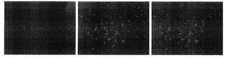
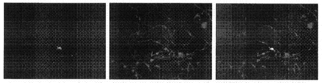
- the left is an image in which the expression of 5HT (serotonin) is detected.
- the center is an image in which Tuj1 (nerve cell marker) is detected.
- the image on the right is a fusion of 5HT and Tuj1 and cell nucleus detection results.
- the result of test No. 2 is shown.
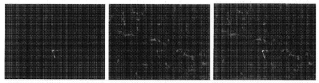
- the left is an image in which the expression of 5HT (serotonin) is detected.
- the center is an image in which Tuj1 (nerve cell marker) is detected.
- the image on the right is a fusion of 5HT and Tuj1 and cell nucleus detection results.
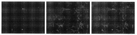
- the result of test No. 3 is shown.
- the left is an image in which the expression of 5HT (serotonin) is detected.
- the center is an image in which Tuj1 (nerve cell marker) is detected.
- the image on the right is a fusion of 5HT and Tuj1 and cell nucleus detection results.
- the result of test No. 4 is shown.
- the left is an image in which the expression of 5HT (serotonin) is detected.
- the center is an image in which Tuj1 (nerve cell marker) is detected.
- the image on the right is a fusion of 5HT and Tuj1 and cell nucleus detection results.
- the result of test No. 5 is shown.
- the left is an image in which the expression of 5HT (serotonin) is detected.
- the center is an image in which Tuj1 (nerve cell marker) is detected.
- the image on the right is a fusion of 5HT and Tuj1 and cell nucleus detection results.
- the result of test No. 6 is shown.
- the left is an image in which the expression of 5HT (serotonin) is detected.
- the center is an image in which Tuj1 (nerve cell marker) is detected.
- the image on the right is a fusion of 5HT and Tuj1 and cell nucleus detection results.
- the result of test No. 7 is shown.
- the left is an image in which the expression of 5HT (serotonin) is detected.
- the center is an image in which Tuj1 (nerve cell marker) is detected.
- the image on the right is a fusion of 5HT and Tuj1 and cell nucleus detection results.
- the result of test No. 8 is shown.
- the left is an image in which the expression of 5HT (serotonin) is detected.
- the center is an image in which Tuj1 (nerve cell marker) is detected.
- the image on the right is a fusion of 5HT and Tuj1 and cell nucleus detection results.
- the result of test No. 9 is shown.
- the left is an image in which the expression of 5HT (serotonin) is detected.
- the center is an image in which Tuj1 (nerve cell marker) is detected.
- the image on the right is a fusion of 5HT and Tuj1 and cell nucleus detection results.
- the result of test No. 10 is shown.
- the left is an image in which the expression of 5HT (serotonin) is detected.
- the center is an image in which Tuj1 (nerve cell marker) is detected.
- the image on the right is a fusion of 5HT and Tuj1 and cell nucleus detection results.
- the result of test No. 11 is shown.
- the left is an image in which the expression of 5HT (serotonin) is detected.
- the center is an image in which Tuj1 (nerve cell marker) is detected.
- the image on the right is a fusion of 5HT and Tuj1 and cell nucleus detection results.
- the method for producing serotonin neurons preferably includes expressing ASCL1, LMX1B and / or FEV in pluripotent stem cells.
- “to express” means that a nucleic acid encoding a transcription factor is introduced into a pluripotent stem cell, and the transcription factor that is the product is produced in the cell.
- the nucleic acid may be either DNA or RNA.
- the nucleic acid is preferably DNA, and in another embodiment, the nucleic acid is preferably RNA.
- pluripotent stem cell is not particularly limited as long as it can be induced in serotonin neurons.
- pluripotent stem cells include inducible pluripotent stem cells (iPS cells), embryonic stem cells (ES cells), somatic stem cells, fertilized egg inner cell mass cells, and early embryonic cells. These pluripotent stem cells may be used alone or in combination of two or more as necessary.
- iPS cells include cells described in Nature, 2007, July 19; Vol.448, pp.313-317; Cell, 2006, August 25; Vol.126 (4), pp.663-676, or this Similar cells are included.
- the pluripotent stem cell is preferably an iPS cell.
- the type (origin) of pluripotent stem cells is not particularly limited.
- the pluripotent stem cell can be a pluripotent stem cell such as a human, monkey, gorilla, mouse, rat, pig, or dog.
- the pluripotent stem cell is preferably a human pluripotent stem cell, and in another embodiment, the pluripotent stem cell is preferably a mouse or monkey pluripotent stem cell.
- the pluripotent stem cells are preferably pluripotent stem cells (eg, iPS cells) that have been passaged until no colonies spontaneously differentiate in feeder-free culture.
- ASCL1 is one of the important transcription factors in the development of the central nervous system and is classified as a basic helix loop helix transcription factor.
- An example of a base sequence encoding human ASCL1 is shown in SEQ ID NO: 9.
- the nucleotide sequence that can be used to express human ASCL1 is not particularly limited as long as it encodes human ASCL1, and is, for example, 80% or more, 85% or more, 90% or more, 95% or more, 96 and the nucleotide sequence of SEQ ID NO: 9. %, 97% or more, 98% or more, or 99% or more of a nucleotide sequence that encodes human ASCL1 can be used.
- ASCL1 is not limited to wild-type ASCL1 and may be mutant ASCL1 as long as it can induce serotonin neurons.
- pluripotent stem cells derived from animals other than humans are used as pluripotent stem cells, it is preferable to use ASCL1 that is derived from the cells.
- LMX1B is one of transcription factors important for the formation of the ventral dorsal axis in histogenesis and is classified as a LIM homeobox transcription factor.
- An example of a base sequence encoding human LMX1B is shown in SEQ ID NO: 10.
- the nucleotide sequence that can be used to express human LMX1B is not particularly limited as long as it encodes human ASCL1.
- the nucleotide sequence of SEQ ID NO: 10 is 80% or more, 85% or more, 90% or more, 95% or more, 96 %, 97% or more, 98% or more, or 99% or more of a nucleotide sequence that encodes human LMX1B can be used.
- LMX1B is not limited to wild-type LMX1B as long as it can induce serotonin neurons, and may be mutant LMX1B.
- pluripotent stem cells derived from animals other than humans are used as pluripotent stem cells, it is preferable to use LMX1B that is derived from the cells.
- FEV is a transcription factor that has been suggested to be important for regulation of nicotinic acetylcholine receptor expression and serotonin neurogenesis, and is classified as an ETS transcription factor.
- An example of a base sequence encoding human FEV is shown in SEQ ID NO: 11.
- the nucleotide sequence that can be used to express human FEV is not particularly limited as long as it encodes human FEV.
- the nucleotide sequence of SEQ ID NO: 11 is 80% or more, 85% or more, 90% or more, 95% or more, 96 %, 97% or more, 98% or more, or 99% or more of a nucleotide sequence that encodes human FEV can be used.
- the FEV is not limited to the wild type FEV and may be a mutant FEV as long as it can induce serotonin neurons.
- pluripotent stem cells derived from animals other than humans are used as pluripotent stem cells, it is preferable to use FEV that is derived from the cells.
- the transcription factor expressed in pluripotent stem cells preferably contains ASCL1, LMX1B and / or FEV.
- ASCL1 and LMX1B and / or FEV are expressed in pluripotent stem cells
- other transcription factors may be expressed in the pluripotent stem cells.
- Such other transcription factors may include one or more selected from the group consisting of FOXA2, NKX2.2, NKX2.8, and GATA2.
- the other transcription factors are preferably not expressed in pluripotent stem cells, and FEV, FOXA2, NKX2.2, NKX2.8, More preferably, only ASCL1 and LMX1B are expressed without expressing GATA2.
- “not expressed” means that a nucleic acid encoding a predetermined transcription factor is not introduced into a pluripotent stem cell in a state where it can be expressed.
- Transcription factors are a group of proteins that specifically bind to DNA, bind to a transcriptional control region called a promoter or enhancer on DNA, and promote or suppress the process of transcription of DNA genetic information into RNA. . Transcription factors exert this function alone or by forming a complex with other proteins.
- transcription factors in pluripotent stem cells can be performed by any method known in the art.
- nucleic acids encoding the above specific combinations of transcription factors can be expressed by introducing them into pluripotent stem cells using an expression vector. It is also possible to directly introduce a transcription factor encoding mRNA or protein in a pluripotent stem cell.
- mRNA can be introduced into pluripotent stem cells in the form of mRNA using, for example, mMESSAGE mMACHINE (manufactured by Thermofisher) and introduced into cells using Lipofectamine MessengerMAX.
- a transcription factor in a protein state is possible, for example, by adding a tag (for example, a tat-peptide) having intracellular transferability.
- a tag for example, a tat-peptide
- the type of expression vector is not particularly limited, and can be appropriately selected from, for example, a viral vector and a plasmid vector according to the type of pluripotent stem cell.
- a viral vector as the expression vector.
- virus vectors include lentivirus vectors, retrovirus vectors, adeno-associated virus vectors, adenovirus vectors, and the like.
- preferred viral vectors are lentiviral vectors and retroviral vectors, more preferably lentiviral vectors.
- a transcription factor can be stably expressed by using a lentiviral vector or a retroviral vector. Transcription factors can be transiently expressed by using adeno-associated virus vectors, adenovirus vectors, and plasmid vectors.
- the expression vector can be prepared according to a conventional method. For example, since kits and reagents for producing an expression vector for expressing a foreign gene in pluripotent stem cells are commercially available, they can be produced using them.
- a viral vector can be prepared by a production method including the following steps (A) and (B): (A) an expression construct containing a promoter sequence at the 5 ′ side of the transgene translation region. A step of constructing; and (B) a step of cleaving a vector containing a signal sequence for incorporation into a virus with a restriction enzyme and ligating the expression construct prepared in (A) thereto.
- the expression vector may incorporate only DNA encoding one type of transcription factor, or may incorporate DNA encoding two or three types of transcription factors.
- the transcription factor expression vector (or a combination thereof) obtained as described above can be used as a kit for producing serotonin neurons.
- the kit can include any reagent used in producing serotonin neurons from pluripotent stem cells and / or instructions for handling.
- Introduction of a nucleic acid encoding a transcription factor into a pluripotent stem cell using an expression vector can be performed according to a conventional method.
- the target transcription factor can be expressed in the pluripotent stem cell by infecting the pluripotent stem cell with the viral vector.
- transfection with cationic polymer, cationic lipid, or calcium phosphate, or electroporation or microinjection is used to introduce the plasmid vector into pluripotent stem cells to express transcription factors. Can do.
- Pluripotent stem cells into which a nucleic acid encoding a transcription factor has been introduced are preferably cultured under conditions suitable for differentiation into serotonin neurons.
- Conditions suitable for differentiation into nerve cells are not particularly limited and can be arbitrarily selected.
- the composition of [DMEM / F12 (1: 1) plus 2% B27, 1% N2 as an additional factor] is widely used as the basic composition of a medium used for differentiation into nerve cells.
- B27 and N2 are medium additives for culturing nerve cells, and are used in place of serum when culturing nerve cells.
- Various other basic compositions are also known.
- a substance useful for inducing differentiation of a nerve cell may be appropriately added to a medium having such a basic composition.
- retinoic acid is often used in inducing differentiation into nerve cells, and is usually added to the medium at a concentration of about 1 ⁇ M.
- Doxycycline can be added to the basic medium for 1 to 2 days after the start of culture, and SB-431542 and LDN can be further added for 2 to 6 days after the start of culture.
- -193189 can be added.
- a medium excluding Doxycycline, SB-431542 and LDN-193189 can be used for 7 to 11 days after the start of culture.
- SB-431542 and LDN193189 are ALK inhibitors, and in one embodiment, ALK inhibitors of the same type as each of these ALK inhibitors can be used. In one embodiment, it is preferable to add a Wnt signal activator (eg, CHIR-99021) and / or an inhibitor (eg, IWP-2) to the differentiation medium.
- a Wnt signal activator eg, CHIR-99021
- an inhibitor eg, IWP-2
- the culture of pluripotent stem cells can be usually performed on a plate (for example, 24-well plate, 6-well plate) used for cell culture.
- the amount (concentration) of pluripotent stem cells in the culture solution is not particularly limited, and can be, for example, 10 2 to 10 8 cells / ml or 10 3 to 10 6 cells / ml.
- the culture temperature is not particularly limited, and can be about 37 ° C., for example.
- the culture period is not particularly limited as long as differentiation into serotonin neurons is induced.
- the culture period is 4 days or more, 5 days or more, 6 days or more, 1 week or more, 2 weeks or more, 3 weeks or more, 4 weeks or more. can do.
- the culture period is preferably within 5 weeks, within 4 weeks, within 3 weeks, within 2 weeks, within 12 days, or within 11 days. is there.
- pluripotent stem cells have been differentiated into serotonin neurons can be determined using an arbitrary index. For example, as employed in Examples described later, cells after culture can be confirmed by staining with an anti-serotonin antibody and an antibody that specifically recognizes nerve cells (for example, an anti-Tuj1 antibody).
- Neuronal cells that have been induced to differentiate on the cell culture substrate of the present invention can be recovered by pipetting, physical detachment with a cell scraper, or by enzymatic treatment (trypsin, dispase, etc.) and used for a desired application. .
- Lentiviral Vector for Differentiation Induction was performed according to the method of Takahashi et al., (Takahashi et al., PLoS One.2014 Apr 16; 9 (4): e94657.).
- Lenti-X 293T cells (Clontech) were seeded in a 10 cm dish at 25% confluency and cultured overnight at 37 ° C. in a CO 2 5% environment.
- pNHP pNHP
- pHEF-VSVG 0.43 ⁇ g of pCEP4-tat
- 2.6 ⁇ g of shuttle vector pFUW-M2rtTA or pFUW-TetO-GeneX
- DMEM DMEM
- 7.5 ⁇ L of polyethyleneimine solution manufactured by polysciences
- the suspension was added to 5 mL of 10% FBS-added DMEM preheated at 37 ° C. and mixed by inversion.
- Differentiation medium Differentiation medium A (DMEM / F12 + N2 + NEAA (1: 100) + B27 (1:50) + Y (1: 1000) +20 ng / mL hBDNF + 20 ng / mL hGDNF + 1 ⁇ M PD + 1 ⁇ g / mL Doxycycline)
- Differentiation medium B (DMEM / F12 + N2 + NEAA (1: 100) + B27 (1:50) + Y (1: 1000) +20 ng / mL hBDNF + 20 ng / mL hGDNF + 1 ⁇ M PD + 1 ⁇ g / mL Doxycycline + 0.1 ⁇ M LDN + 2.5 ⁇ M SB)
- Differentiation medium C (DMEM / F12 + N2 + NEAA (1: 100) + B27 (1:50) + Y (1: 1000) +20 ng / mL hBDNF + 20 ng
- the 24-well plate was coated with Matrigel (BD Biosciences) diluted 30 times.
- the culture supernatant was removed from the hiPSC culture in a subconfluent state in a 6-well plate.
- the well plate was washed twice with 2 mL / well PBS.
- preheated Accutase was added at 1 mL / well and incubated at 37 ° C. for 5 minutes.
- 1 mL / well PBS was added, and the cells were detached while pipetting with a 1 mL pipetman.
- the whole culture solution for 2 wells was transferred to a 15 mL centrifuge tube, and a part was collected for cell concentration measurement.
- Day-2 The culture solution in the well plate was replaced with 500 ⁇ L of Essential 8 + Sup + Y.
- Lentiviral vector (LVV505: pFUW-M2rtTA (manufactured by Addgene)) is added at 40 ⁇ L / well, lentiviral vectors LVV543 to LVV 550 are added at 40 ⁇ L / well in the combinations shown in Table 1 below, and suspended by pipetting. After becoming cloudy, the cells were cultured overnight at 37 ° C. in a CO 2 5% environment.
- Day-1 The culture solution was replaced with 500 ⁇ L of Essential 8 + Sup + Y, and cultured overnight at 37 ° C. in a CO 2 5% environment.
- Day 0 The culture solution was replaced with 500 ⁇ L of differentiation medium A, and cultured for 2 days at 37 ° C. in a CO 2 5% environment.
- Day 2 The culture medium was replaced with 500 ⁇ L of differentiation medium B, and cultured for 2 days at 37 ° C. in a CO 2 5% environment.
- Day 4 The culture solution was replaced with 500 ⁇ L of differentiation medium B, and cultured for 2 days at 37 ° C. in a CO 2 5% environment.
- Day 6 The culture medium was replaced with 500 ⁇ L of differentiation medium B and cultured overnight at 37 ° C.
- Day 7 The culture solution was replaced with 500 ⁇ L of differentiation medium C, and cultured for 2 days at 37 ° C. in a CO 2 5% environment.
- Day 9 The culture solution was replaced with 500 ⁇ L of differentiation medium C, and cultured for 2 days at 37 ° C. in a CO 2 5% environment.
- Day 11 The culture solution was replaced with 500 ⁇ L of differentiation medium C, and cultured overnight at 37 ° C. in a CO 2 5% environment.
- the cells were fixed and immunostained by the following procedure.
- (2) The cells were washed twice with 500 ⁇ L / well of PBS.
- (3) 4% paraformaldehyde solution 500 ⁇ L / well was added.
- (4) The cells were fixed by allowing to stand at room temperature for 10 minutes.
- the cells were washed 3 times with 500 ⁇ L / well of PBS.
- (6) 0.1% Triton X-100 solution was added (200 ⁇ L / well).
- the membrane permeation treatment was performed by allowing to stand at room temperature for 10 minutes.
- (8) The cells were washed 3 times with 500 ⁇ L / well of PBS.
Landscapes
- Health & Medical Sciences (AREA)
- Life Sciences & Earth Sciences (AREA)
- Engineering & Computer Science (AREA)
- Genetics & Genomics (AREA)
- Wood Science & Technology (AREA)
- Biomedical Technology (AREA)
- Organic Chemistry (AREA)
- Biotechnology (AREA)
- General Engineering & Computer Science (AREA)
- Chemical & Material Sciences (AREA)
- Zoology (AREA)
- Bioinformatics & Cheminformatics (AREA)
- Biophysics (AREA)
- Microbiology (AREA)
- Plant Pathology (AREA)
- Molecular Biology (AREA)
- Biochemistry (AREA)
- General Health & Medical Sciences (AREA)
- Physics & Mathematics (AREA)
- Micro-Organisms Or Cultivation Processes Thereof (AREA)
Abstract
短時間でセロトニン神経細胞を製造する手段を提供すること 多能性幹細胞において、ASCL1とLMX1B及び/又はFEVとを発現させることを含む、セロトニン神経細胞を製造する方法。
Description
神経細胞の製造に関する技術が開示される。
セロトニン神経系は、うつ病や統合失調症などの精神疾患に係わる重要な神経系の一つである。セロトニン神経伝達を調節する化合物が、抗うつ薬や統合失調症の治療薬として有効であることから、これらの疾患の背景に、セロトニン神経系の機能異常が存在することが示唆されている。従って、ヒトのセロトニン神経を用いた解析は、うつ病や統合失調症のメカニズムの解明といった基礎医学的知見、及び、それらの疾患の治療及び予防に有効な物質の探索という実用的な見地から重要である。しかし、ヒトセロトニン神経へのアクセスは困難であり、疾患特異的なヒトiPS細胞をもちいて患者由来のヒトセロトニン神経細胞を得る試みがなされているが、十分量のセロトニン神経の取得には約6週間といった長期間を要する(非特許文献1)。
Lu et al., Nature Biotechnology, 34, 89-94, 2016
上記の現状の下、より短時間でセロトニン神経細胞を製造する手段を提供することが1つの課題である。
上記課題を解決すべく鋭意研究を重ねた結果、低分子化合物によるセロトニン神経への分化誘導ではなく、セロトニン神経の発生に関与する転写因子の特定の組み合わせを発現させることにより、より短期間でセロトニン神経細胞が得られることが判明した。斯かる知見に基づき、更なる研究を重ね、下記に代表される発明を完成するに至った。
項1.
多能性幹細胞において、ASCL1とLMX1B及び/又はFEVとを発現させることを含む、セロトニン神経細胞を製造する方法。
項2.
多能性幹細胞において、ASCL1とLMX1Bとを発現させることを含む、項1に記載の方法。
項3.
更に、多能性幹細胞において、FEVを発現させることを含む、項2に記載の方法。
項4.
多能性幹細胞においてFEVを発現させない、項2に記載の方法。
項5.
多能性幹細胞がiPS細胞である、項1~4のいずれかに記載の方法。
項6.
ASCL1とLMX1B及び/又はFEVをコードする遺伝子を含む、セロトニン神経細胞製造用発現ベクター。
項7.
項6に記載の発現ベクターを含む、セロトニン神経細胞製造用キット。
多能性幹細胞において、ASCL1とLMX1B及び/又はFEVとを発現させることを含む、セロトニン神経細胞を製造する方法。
項2.
多能性幹細胞において、ASCL1とLMX1Bとを発現させることを含む、項1に記載の方法。
項3.
更に、多能性幹細胞において、FEVを発現させることを含む、項2に記載の方法。
項4.
多能性幹細胞においてFEVを発現させない、項2に記載の方法。
項5.
多能性幹細胞がiPS細胞である、項1~4のいずれかに記載の方法。
項6.
ASCL1とLMX1B及び/又はFEVをコードする遺伝子を含む、セロトニン神経細胞製造用発現ベクター。
項7.
項6に記載の発現ベクターを含む、セロトニン神経細胞製造用キット。
短期間でセロトニン神経細胞を製造することができる。
セロトニン神経細胞の製造方法は、多能性幹細胞において、ASCL1とLMX1B及び/又はFEVとを発現させることを含むことが好ましい。ここで、「発現させる」とは、転写因子をコードする核酸を多能性幹細胞に導入し、その産物である転写因子を細胞内で生産させることを意味する。核酸は、DNA及びRNAのいずれでもよい。一実施形態において核酸はDNAであることが好ましく、他の実施形態において核酸はRNAであることが好ましい。
多能性幹細胞の種類はセロトニン神経細胞に誘導可能である限り特に制限されない。多能性幹細胞には、例えば、誘導型多能性幹細胞(iPS細胞)、胚性幹細胞(ES細胞)、及び体性幹細胞、受精卵内部細胞塊細胞、及び初期胚細胞等が包含される。これらの多能性幹細胞は、1種のみを用いてもよく、又は必要に応じて2種以上を混合して用いてもよい。iPS細胞には、Nature, 2007, July 19; Vol.448, pp.313-317;Cell, 2006, August 25; Vol.126(4), pp.663-676に記載されている細胞、又はこれに類する細胞が包含される。一実施形態において、多能性幹細胞は、iPS細胞であることが好ましい。
多能性幹細胞の種類(由来)は特に制限されない。例えば、多能性幹細胞は、ヒト、サル、ゴリラ、マウス、ラット、ブタ、又はイヌ等の多能性幹細胞であり得る。一実施形態において、多能性幹細胞は、ヒトの多能性幹細胞であることが好ましく、他の実施形態において多能性幹細胞はマウス又はサルの多能性幹細胞であることが好ましい。一実施形態において、多能性幹細胞は、フィーダーフリー培養において自発的に分化するコロニーがなくなるまで継代した多能性幹細胞(例えば、iPS細胞)であることが好ましい。
ASCL1とは、中枢神経系の発生における重要な転写因子の一つであり、塩基性ヘリックスループヘリックス転写因子に分類される。ヒトASCL1をコードする塩基配列の一例を配列番号9に示す。ヒトASCL1を発現させるために使用できる塩基配列は、ヒトASCL1をコードする限り特に制限されず、例えば、配列番号9の塩基配列と80%以上、85%以上、90%以上、95%以上、96%以上、97%以上、98%以上、又は99%以上の同一性を有する塩基配列であって、ヒトASCL1をコードするものを使用することができる。ASCL1は、セロトニン神経細胞を誘導可能である限り、野生型ASCL1に限らず変異型ASCL1であってもよい。多能性幹細胞としてヒト以外の動物由来の多能性幹細胞を使用する場合は、細胞の由来にあったASCL1を使用することが好ましい。
LMX1Bとは、組織発生における腹側背側軸の形成に重要な転写因子の一つであり、LIMホメオボックス転写因子に分類される。ヒトLMX1Bをコードする塩基配列の一例を配列番号10に示す。ヒトLMX1Bを発現させるために使用できる塩基配列は、ヒトASCL1をコードする限り特に制限されず、例えば、配列番号10の塩基配列と80%以上、85%以上、90%以上、95%以上、96%以上、97%以上、98%以上、又は99%以上の同一性を有する塩基配列であって、ヒトLMX1Bをコードするものを使用することができる。LMX1Bは、セロトニン神経細胞を誘導可能である限り、野生型LMX1Bに限らず変異型LMX1Bであってもよい。多能性幹細胞としてヒト以外の動物由来の多能性幹細胞を使用する場合は、細胞の由来にあったLMX1Bを使用することが好ましい。
FEVとは、ニコチン性アセチルコリン受容体の発現制御およびセロトニン神経発生における重要性が示唆されている転写因子であり、ETS転写因子に分類される。ヒトFEVをコードする塩基配列の一例を配列番号11に示す。ヒトFEVを発現させるために使用できる塩基配列は、ヒトFEVをコードする限り特に制限されず、例えば、配列番号11の塩基配列と80%以上、85%以上、90%以上、95%以上、96%以上、97%以上、98%以上、又は99%以上の同一性を有する塩基配列であって、ヒトFEVをコードするものを使用することができる。FEVは、セロトニン神経細胞を誘導可能である限り、野生型FEVに限らず変異型FEVであってもよい。多能性幹細胞としてヒト以外の動物由来の多能性幹細胞を使用する場合は、細胞の由来にあったFEVを使用することが好ましい。
多能性幹細胞において発現させる転写因子はASCL1とLMX1B及び/又はFEVを含むことが好ましい。多能性幹細胞においてASCL1とLMX1B及び/又はFEVを発現させる場合、更に他の転写因子を当該多能性幹細胞において発現させてもよい。そのような他の転写因子としては、FOXA2、NKX2.2、NKX2.8、及びGATA2から成る群より選択される一種以上を挙げることができる。好適な一実施形態において、上記他の転写因子(特に、FOXA2、NKX2.2、及びNKX2.8)は多能性幹細胞において発現させないことが好ましく、FEV、FOXA2、NKX2.2、NKX2.8、及びGATA2を発現させず、ASCL1及びLMX1Bだけを発現させることがより好ましい。ここで「発現させない」とは、所定の転写因子をコードする核酸をそれが発現可能な状態で多能性幹細胞に導入しないことを意味する。
転写因子とは、DNAに特異的に結合するタンパク質の一群であり、DNA上のプロモーターやエンハンサーと呼ばれる転写を制御する領域に結合し、DNAの遺伝情報をRNAに転写する過程を促進又は抑制する。転写因子はこの機能を単独で、または他のタンパク質と複合体を形成することによって発揮する。
多能性幹細胞における転写因子の発現は、当該技術分野において知られる任意の手法で行うことができる。例えば、上記特定の組み合わせの転写因子をコードする核酸を発現ベクターを用いて多能性幹細胞に導入することにより、それらを発現させることができる。また、転写因子をコードするmRNA又はタンパク質の状態の転写因子を直接多能性幹細胞に導入することも可能である。mRNAの状態で多能性幹細胞に導入することは、例えば、mMESSAGE mMACHINE (Thermofisher製)を用いてmRNAを作製し、それをLipofectamine MessengerMAXを用いて細胞内に導入することができる。タンパク質の状態の転写因子の導入は、例えば、細胞内移行性を有するタグ(例えば、tat-ペプチド)を付加することによって可能である。一実施形態において、発現ベクターを用いて転写因子をコードする核酸を導入することが好ましい。
発現ベクターの種類は特に制限されず、多能性幹細胞の種類等に応じて、例えば、ウイルスベクター、及びプラスミドベクター等から適宜選択することができる。一実施形態において、発現ベクターとしてウイルスベクターを用いることが好ましい。ウイルスベクターとしては、例えば、レンチウイルスベクター、レトロウイルスベクター、アデノ随伴ウイルスベクター、及びアデノウイルスベクター等を挙げることができる。一実施形態において好ましいウイルスベクターは、レンチウイルスベクター及びレトロウイルスベクターであり、より好ましくはレンチウイルスベクターである。レンチウイルスベクター又はレトロウイルスベクターを用いることにより転写因子を安定的に発現させることができる。アデノ随伴ウイルスベクター、及びアデノウイルスベクター、並びにプラスミドベクターを用いることにより転写因子を一過的に発現させることができる。
発現ベクターの作製は常法に従って行うことができる。例えば、多能性幹細胞において外来遺伝子を発現させるための発現ベクターを作製するためのキット及び試薬が市販されているため、それらを用いて作製することができる。また、例えば、ウイルスベクターは、次の(A)および(B)の工程を含む製造方法により作製することができる:(A)導入遺伝子の翻訳領域の5'側にプロモーター配列を含む発現コンストラクトを構築する工程;及び(B)ウイルスへの取り込みシグナル配列を含有したベクターを制限酵素で切断し、それに(A)で作製した発現コンストラクトをライゲーションする工程。発現ベクターは、1種類の転写因子をコードするDNAのみを組み込んでいてもよく、2種又は3種の転写因子をコードするDNAを組み込んでいてもよい。
上記のようにして得られる転写因子の発現ベクター(又はその組み合わせ)は、セロトニン神経細胞製造用キットとすることができる。当該キットには、多能性幹細胞からセロトニン神経細胞を製造する際に使用する任意の試薬、及び/又は取扱に関する説明書を含めることができる。
発現ベクターを用いた多能性幹細胞への転写因子をコードする核酸の導入は常法に従って行うことができる。例えば、ウイルスベクターを用いる場合は、ウイルスベクターで多能性幹細胞を感染させることにより、目的の転写因子を多能性幹細胞内で発現させることができる。プラスミドベクターを用いる場合は、カチオンポリマー、カチオン脂質、又はリン酸カルシウムを用いたトランスフェクション、或いは、エレクトロポレーション、又はマイクロインジェクションを用いてプラスミドベクターを多能性幹細胞に導入し、転写因子を発現させることができる。
転写因子をコードする核酸を導入した多能性幹細胞は、セロトニン神経細胞への分化に適した条件にて培養することが好ましい。神経細胞への分化に適した条件は、特に制限されず、任意に選択することができる。例えば、[DMEM/F12(1:1)に添加因子として2%B27,1%N2を加えたもの]の組成が神経細胞への分化に用いられる培地の基本組成として広く用いられている。B27およびN2は神経細胞培養用の培地添加物であり、神経細胞の培養時に血清の代わりに使用される。これ以外の基本組成も各種知られている。そのような基本組成の培地に、神経細胞の分化誘導に有用な物質(細胞培養基材に固定化されたタンパク質以外の物質)を適宜添加して用いてもよい。例えば、神経細胞への分化誘導においてはレチノイン酸がしばしば用いられており、通常1μM程度の濃度で培地に添加して使用される。また、後述する実施例で採用するように、培養開始後1~2日間は、基本培地にDoxycyclineを添加することができ、培養開始後2日~6日の間は、更にSB-431542及びLDN-193189添加することができる。更に培養開始後7日~11日の間は、Doxycycline、SB-431542及びLDN-193189を除いた培地を使用することができる。SB-431542及びLDN193189はALK阻害薬であり、一実施形態において、これらのALK阻害剤のそれぞれと同種のALK阻害剤を用いることもできる。一実施形態において、分化用培地にWntシグナルの活性化剤(例えば、CHIR-99021)及び/又は阻害薬(例えば、IWP-2)を添加することが好ましい。
多能性幹細胞の培養は、通常、細胞の培養に使用されるプレート(例えば、24ウェルプレート、6ウェルプレート)上で行うことができる。培養液中の多能性幹細胞の量(濃度)は特に制限されないが、例えば、102~108cells/ml、又は103~106cells/mlとすることができる。培養温度は、特に制限されず、例えば、約37℃とすることができる。また、CO2細胞培養装置などを利用して、5%程度のCO2濃度雰囲気下で培養することが好ましい。
培養期間は、セロトニン神経細胞への分化が誘導される限り特に制限されず、例えば、4日以上、5日以上、6日以上、1週間以上、2週間以上、3週間以上、4週間以上培養することができる。一実施形態において、セロトニン神経細胞を短期間で製造するという観点から、培養期間は、好ましくは、5週間以内、4週間以内、3週間以内、2週間以内、12日以内、又は11日以内である。
多能性幹細胞がセロトニン神経細胞に分化されたか否かは、任意の指標を用いて行うことができる。例えば、後述する実施例で採用したように、抗セロトニン抗体及び神経細胞を特異的に認識する抗体(例えば、抗Tuj1抗体)で培養後の細胞を染色して確認することができる。
本発明の細胞培養基材上で分化誘導した神経細胞は、ピペッティング、セルスクレーパーによる物理的な剥離により、または酵素処理(トリプシン、ディスパーゼ等)により回収して所望の用途に使用することができる。
以下、実施例により本発明についてさらに詳細に説明するが、本発明はこれらに制限されるものではない。
1.分化誘導用レンチウイルスベクター作製
分化誘導用レンチウイルスベクターは、Takahashi et al., (Takahashi et al., PLoS One.2014 Apr 16;9(4):e94657.)の方法に準拠して行った。10cm dishに25% confluencyでLenti-X 293T細胞(Clontech)を播種し、37℃、CO25%環境下で終夜培養した。一方、300μLのDMEM (Life technologies)に5.2μgのpNHP、2.1μgのpHEF-VSVG、0.43μgのpCEP4-tat、及び2.6μgのshuttle vector(pFUW-M2rtTA or pFUW-TetO-GeneX)を加えた。これをボルテックスで懸濁し、7.5μLのpolyethyleneimine溶液(polysciences社製)を加え、再度ボルテックスで懸濁して常温で20分放置した。懸濁液を37℃で予熱した5mLの10%FBS添加DMEMに加え転倒混和した。これを、培地を除去した培養細胞(Lenti-X 293T細胞)に加え、37℃、CO25%環境下で終夜培養した。培養上清を除去し、PBSで2回洗浄し、6 mLのEssential 8 basal medium(Life technologies社製)を加え、37℃、CO2 5%環境下で終夜培養した。培養上清を回収し、0.45μm孔径のフィルターを通し清澄化した。これを500μLづつ分注し、-80度で保存し、セロトニン神経誘導試験において各転写因子を強制発現させるためのウイルスベクターとして使用した。
分化誘導用レンチウイルスベクターは、Takahashi et al., (Takahashi et al., PLoS One.2014 Apr 16;9(4):e94657.)の方法に準拠して行った。10cm dishに25% confluencyでLenti-X 293T細胞(Clontech)を播種し、37℃、CO25%環境下で終夜培養した。一方、300μLのDMEM (Life technologies)に5.2μgのpNHP、2.1μgのpHEF-VSVG、0.43μgのpCEP4-tat、及び2.6μgのshuttle vector(pFUW-M2rtTA or pFUW-TetO-GeneX)を加えた。これをボルテックスで懸濁し、7.5μLのpolyethyleneimine溶液(polysciences社製)を加え、再度ボルテックスで懸濁して常温で20分放置した。懸濁液を37℃で予熱した5mLの10%FBS添加DMEMに加え転倒混和した。これを、培地を除去した培養細胞(Lenti-X 293T細胞)に加え、37℃、CO25%環境下で終夜培養した。培養上清を除去し、PBSで2回洗浄し、6 mLのEssential 8 basal medium(Life technologies社製)を加え、37℃、CO2 5%環境下で終夜培養した。培養上清を回収し、0.45μm孔径のフィルターを通し清澄化した。これを500μLづつ分注し、-80度で保存し、セロトニン神経誘導試験において各転写因子を強制発現させるためのウイルスベクターとして使用した。
2.セロトニン神経細胞の誘導
2-1.試薬等
本試験では下記の試薬等を使用した。
・hiPS cells (454E2):RIKENから提供され、フィーダーフリー培養へ移行後、自発的に分化するコロニーがなくなるまで継代したhiPS cellsを使用した。
・Essential 8 + Supplement(Life technologies)
・Matrigel growth factor reduced (BD Biosciences)
・Accutase (Stempro)
・Y-27632 (Y: 10mM stock. use 1 μL/1 mL media)
・DMEM/F12 (Life technologies 11330-032, HEPES+, Gln+, sodium bicarbonate+)
・N2 supplement (Wako, アポトランスフェリンタイプ)
・NEAA (MEM, Life technologies)
・B27 supplement (Life technologies)
・PD-0332991 (PD: 10 mM)
・hBDNF (100 μg/mL, 10 μL/tube, -20degC)
・hGDNF (100 μg/mL, 10 μL/tube, -20degC)
・SB-431542 (SB: 10 mM, 100 μL/tube, -20degC)
・LDN-193189 (LDN: 10 mM)
・Doxycycline (Clontech) dissolved in ddH2O (20 mg/mL), フィルターで滅菌
・Lentiviral vectors(いずれも上記1で作製したもの)
LVV505:pFUW-M2rtTA(Addgene#20342)
LVV543:pFUW-TetO-hASCL1
LVV544:pFUW-TetO-hFEV
LVV545:pFUW-TetO-hFOXA2
LVV546:pFUW-TetO-hGATA2
LVV547:pFUW-TetO-hLMX1B
LVV548:pFUW-TetO-hNKX2-2
LVV549:pFUW-TetO-hNKX2-8
これらのベクターの配列を図1に示す。各配列において大文字は転写因子のコーディング領域を示す。
・分化培地
分化培地A(DMEM/F12+N2+NEAA(1:100)+B27(1:50)+Y (1:1000)+20 ng/mL hBDNF+20 ng/mL
hGDNF+1 μM PD+1 μg/mL Doxycycline)
分化培地B(DMEM/F12+N2+NEAA(1:100)+B27(1:50)+Y (1:1000)+20 ng/mL hBDNF+20 ng/mL
hGDNF+1 μM PD+1 μg/mL Doxycycline+0.1 μM LDN+2.5 μM SB)
分化培地C(DMEM/F12+N2+NEAA(1:100)+B27(1:50)+Y (1:1000)+20 ng/mL hBDNF+20 ng/mL hGDNF+1 μM PD)
2-1.試薬等
本試験では下記の試薬等を使用した。
・hiPS cells (454E2):RIKENから提供され、フィーダーフリー培養へ移行後、自発的に分化するコロニーがなくなるまで継代したhiPS cellsを使用した。
・Essential 8 + Supplement(Life technologies)
・Matrigel growth factor reduced (BD Biosciences)
・Accutase (Stempro)
・Y-27632 (Y: 10mM stock. use 1 μL/1 mL media)
・DMEM/F12 (Life technologies 11330-032, HEPES+, Gln+, sodium bicarbonate+)
・N2 supplement (Wako, アポトランスフェリンタイプ)
・NEAA (MEM, Life technologies)
・B27 supplement (Life technologies)
・PD-0332991 (PD: 10 mM)
・hBDNF (100 μg/mL, 10 μL/tube, -20degC)
・hGDNF (100 μg/mL, 10 μL/tube, -20degC)
・SB-431542 (SB: 10 mM, 100 μL/tube, -20degC)
・LDN-193189 (LDN: 10 mM)
・Doxycycline (Clontech) dissolved in ddH2O (20 mg/mL), フィルターで滅菌
・Lentiviral vectors(いずれも上記1で作製したもの)
LVV505:pFUW-M2rtTA(Addgene#20342)
LVV543:pFUW-TetO-hASCL1
LVV544:pFUW-TetO-hFEV
LVV545:pFUW-TetO-hFOXA2
LVV546:pFUW-TetO-hGATA2
LVV547:pFUW-TetO-hLMX1B
LVV548:pFUW-TetO-hNKX2-2
LVV549:pFUW-TetO-hNKX2-8
これらのベクターの配列を図1に示す。各配列において大文字は転写因子のコーディング領域を示す。
・分化培地
分化培地A(DMEM/F12+N2+NEAA(1:100)+B27(1:50)+Y (1:1000)+20 ng/mL hBDNF+20 ng/mL
hGDNF+1 μM PD+1 μg/mL Doxycycline)
分化培地B(DMEM/F12+N2+NEAA(1:100)+B27(1:50)+Y (1:1000)+20 ng/mL hBDNF+20 ng/mL
hGDNF+1 μM PD+1 μg/mL Doxycycline+0.1 μM LDN+2.5 μM SB)
分化培地C(DMEM/F12+N2+NEAA(1:100)+B27(1:50)+Y (1:1000)+20 ng/mL hBDNF+20 ng/mL hGDNF+1 μM PD)
Day -3:細胞を接着させるため、24-well plateを30倍稀釈したMatrigel(BD Biosciences社製)でコートした。6-wellプレートにおいてsubconfluentな状態のhiPSC培養から、培養上清を除去した。2mL/wellのPBSでwell plateを2回洗浄した。細胞を剥離するため、予熱したAccutaseを1mL/wellで加え、37℃で5分間インキュベートした。1mL/wellのPBSを加え、1mLのピペットマンでピペッティングしながら細胞を剥がした。2well分の培養液全量を15mLの遠沈管に移し、一部を細胞濃度測定用に採取した。遠沈管に6mlのPBSを加え、300g、4分、4℃で遠心分離し、Accutaseを除去した。遠心中に細胞数をカウントした。遠心上清を吸引除去し、沈殿をEssential 8 +Sup+ Y 1mLに懸濁した。8x10⁴cells/mLになるようにE8+Sup+Yで薄めた。24-well plateからマトリゲルを吸引除去し、そこに500μL/well (= 4x10⁴ cells/well)で希釈した細胞を播種した。
Day -2:well plateの培養液を500μLのEssential 8 + Sup + Yに交換した。レンチウイルスベクター(LVV505:pFUW-M2rtTA(Addgene社製)を40 μL/wellで添加し、レンチウイルスベクターLVV543~LVV 550を下記の表1に示す組み合わせで40 μL/well添加し、ピペッティングで懸濁した後、37℃、CO25%環境下で終夜培養した。

Day -1:培養液を500μLのEssential 8 + Sup + Yに交換し、37℃、CO25%環境下で終夜培養した。
Day 0:培養液を500μLの分化培地Aに交換し、37℃、CO2 5%環境下で2日間培養した。Day 2:培養液を500μLの分化培地Bに交換し、37℃、CO2 5%環境下で2日間培養した。Day 4:培養液を500μLの分化培地Bに交換し、37℃、CO2 5%環境下で2日間培養した。Day 6:培養液を500μLの分化培地Bに交換し、37℃、CO2 5%環境下で終夜培養した。Day 7:培養液を500μLの分化培地Cに交換し、37℃、CO2 5%環境下で2日間培養した。Day 9:培養液を500μLの分化培地Cに交換し、37℃、CO2 5%環境下で2日間培養した。Day 11:培養液を500μLの分化培地Cに交換し、37℃、CO2 5%環境下で終夜培養した。
Day 0:培養液を500μLの分化培地Aに交換し、37℃、CO2 5%環境下で2日間培養した。Day 2:培養液を500μLの分化培地Bに交換し、37℃、CO2 5%環境下で2日間培養した。Day 4:培養液を500μLの分化培地Bに交換し、37℃、CO2 5%環境下で2日間培養した。Day 6:培養液を500μLの分化培地Bに交換し、37℃、CO2 5%環境下で終夜培養した。Day 7:培養液を500μLの分化培地Cに交換し、37℃、CO2 5%環境下で2日間培養した。Day 9:培養液を500μLの分化培地Cに交換し、37℃、CO2 5%環境下で2日間培養した。Day 11:培養液を500μLの分化培地Cに交換し、37℃、CO2 5%環境下で終夜培養した。
その後、下記の手順で細胞を固定し、免疫染色した。
(1)培地を吸引除去した。
(2)細胞を500 μL/wellのPBSで2回洗浄した。
(3)4% パラホルムアルデヒド溶液 500μL/wellを加えた。
(4)室温で10分間静置し、細胞を固定した。
(5)細胞を500 μL/wellのPBSで3回洗浄した。
(6)0.1% Triton X-100溶液を加えた(200μL/well)。
(7)室温で10分間静置することで、膜透過処理を行った。
(8)細胞を500 μL/wellのPBSで3回洗浄した。
(9)1% BSA/PBS溶液を加えた(200μL/well)。
(10)室温で30分間静置することで、抗体の非特異的吸着を防ぐためのブロッキングを行った。
(11)1% BSA/PBS溶液を除去した。
(12)一次抗体液 (0.4 μL/well 5-HT 抗体 + 0.4 μL/well TUJ1 抗体を加えた(200 μL/well 1% BSA/PBS)。
(13)4℃で終夜静置した。
(14)翌日、一次抗体液を除去した
(15)細胞を500 μL/wellのPBSで3回洗浄した
(16)二次抗体液を加えた(1μL/well Alexa FluorR 488 Goat Anti-Rabbit IgG (H+L) antibody + 1μL/well Alexa FluorR 594 Donkey Anti-Mouse IgG (H+L) Antibody + 1μL/well Hoechst 33342 in 200μL/well 1% BSA/PBS)。
(17)室温で2時間静置した。
(18)細胞を500 μL/wellのPBSで3回洗浄した。
(19)200μL/well PBSを加えて蛍光顕微鏡(オリンパス社製BZ-9000)で観察した。結果(図2)は、下記のとおりである。
(1)培地を吸引除去した。
(2)細胞を500 μL/wellのPBSで2回洗浄した。
(3)4% パラホルムアルデヒド溶液 500μL/wellを加えた。
(4)室温で10分間静置し、細胞を固定した。
(5)細胞を500 μL/wellのPBSで3回洗浄した。
(6)0.1% Triton X-100溶液を加えた(200μL/well)。
(7)室温で10分間静置することで、膜透過処理を行った。
(8)細胞を500 μL/wellのPBSで3回洗浄した。
(9)1% BSA/PBS溶液を加えた(200μL/well)。
(10)室温で30分間静置することで、抗体の非特異的吸着を防ぐためのブロッキングを行った。
(11)1% BSA/PBS溶液を除去した。
(12)一次抗体液 (0.4 μL/well 5-HT 抗体 + 0.4 μL/well TUJ1 抗体を加えた(200 μL/well 1% BSA/PBS)。
(13)4℃で終夜静置した。
(14)翌日、一次抗体液を除去した
(15)細胞を500 μL/wellのPBSで3回洗浄した
(16)二次抗体液を加えた(1μL/well Alexa FluorR 488 Goat Anti-Rabbit IgG (H+L) antibody + 1μL/well Alexa FluorR 594 Donkey Anti-Mouse IgG (H+L) Antibody + 1μL/well Hoechst 33342 in 200μL/well 1% BSA/PBS)。
(17)室温で2時間静置した。
(18)細胞を500 μL/wellのPBSで3回洗浄した。
(19)200μL/well PBSを加えて蛍光顕微鏡(オリンパス社製BZ-9000)で観察した。結果(図2)は、下記のとおりである。
試験No.1
Tuj1陽性の神経細胞数は少なく、その突起の長さも極めて短く、神経細胞への分化が不十分であることが示唆された。また、5-HT(セロトニン)陽性の神経細胞は観察されなかった。
試験No.2
Tuj1陽性の神経細胞が多数観察され、その突起長も長く、試験No.1と比較すると神経細胞への分化が誘導されていることが示唆された。しかし、5-HT陽性の神経細胞は観察されなかった。
試験No.3
Tuj1陽性の神経細胞が観察され、神経細胞への分化が誘導されていることが示唆された。また、5-HT陽性の神経細胞が観察された。
試験No.4
Tuj1陽性の神経細胞が観察され、その突起長も長く、神経細胞への分化が誘導されていることが示唆された。また、5-HT陽性の神経細胞が試験No.3よりも更に多く観察された。
試験No.5
Tuj1陽性の神経細胞が観察され、神経細胞への分化が誘導されていることが示唆された。また、5-HT陽性の神経細胞が観察された。
試験No.6
数は少ないもののTuj1陽性の神経細胞が観察された。5-HT陽性の神経細胞がわずかに観察された。
試験No.7
試験No.5の結果と同等の結果であった。
試験No.8
試験No.5の結果と同等の結果であった。
試験No.9
試験No.5の結果と同等の結果であった。
試験No.10
数は少ないもののTuj1陽性の神経細胞が観察された。一方、5-HT陽性の神経細胞は観察されなかった。
試験No.11
試験No.10の結果と同様の結果であった。
Tuj1陽性の神経細胞数は少なく、その突起の長さも極めて短く、神経細胞への分化が不十分であることが示唆された。また、5-HT(セロトニン)陽性の神経細胞は観察されなかった。
試験No.2
Tuj1陽性の神経細胞が多数観察され、その突起長も長く、試験No.1と比較すると神経細胞への分化が誘導されていることが示唆された。しかし、5-HT陽性の神経細胞は観察されなかった。
試験No.3
Tuj1陽性の神経細胞が観察され、神経細胞への分化が誘導されていることが示唆された。また、5-HT陽性の神経細胞が観察された。
試験No.4
Tuj1陽性の神経細胞が観察され、その突起長も長く、神経細胞への分化が誘導されていることが示唆された。また、5-HT陽性の神経細胞が試験No.3よりも更に多く観察された。
試験No.5
Tuj1陽性の神経細胞が観察され、神経細胞への分化が誘導されていることが示唆された。また、5-HT陽性の神経細胞が観察された。
試験No.6
数は少ないもののTuj1陽性の神経細胞が観察された。5-HT陽性の神経細胞がわずかに観察された。
試験No.7
試験No.5の結果と同等の結果であった。
試験No.8
試験No.5の結果と同等の結果であった。
試験No.9
試験No.5の結果と同等の結果であった。
試験No.10
数は少ないもののTuj1陽性の神経細胞が観察された。一方、5-HT陽性の神経細胞は観察されなかった。
試験No.11
試験No.10の結果と同様の結果であった。
Claims (7)
- 多能性幹細胞において、ASCL1とLMX1B及び/又はFEVとを発現させることを含む、セロトニン神経細胞を製造する方法。
- 多能性幹細胞において、ASCL1とLMX1Bとを発現させることを含む、請求項1に記載の方法。
- 更に、多能性幹細胞において、FEVを発現させることを含む、請求項2に記載の方法。
- 多能性幹細胞においてFEVを発現させない、請求項2に記載の方法。
- 多能性幹細胞がiPS細胞である、請求項1~4のいずれかに記載の方法。
- ASCL1とLMX1B及び/又はFEVをコードする遺伝子を含む、セロトニン神経細胞製造用発現ベクター。
- 請求項6に記載の発現ベクターを含む、セロトニン神経細胞製造用キット。
Applications Claiming Priority (2)
| Application Number | Priority Date | Filing Date | Title |
|---|---|---|---|
| JP2016167263A JP2018033330A (ja) | 2016-08-29 | 2016-08-29 | セロトニン神経細胞の製造方法 |
| JP2016-167263 | 2016-08-29 |
Publications (1)
| Publication Number | Publication Date |
|---|---|
| WO2018043303A1 true WO2018043303A1 (ja) | 2018-03-08 |
Family
ID=61300984
Family Applications (1)
| Application Number | Title | Priority Date | Filing Date |
|---|---|---|---|
| PCT/JP2017/030426 Ceased WO2018043303A1 (ja) | 2016-08-29 | 2017-08-24 | セロトニン神経細胞の製造方法 |
Country Status (2)
| Country | Link |
|---|---|
| JP (1) | JP2018033330A (ja) |
| WO (1) | WO2018043303A1 (ja) |
-
2016
- 2016-08-29 JP JP2016167263A patent/JP2018033330A/ja active Pending
-
2017
- 2017-08-24 WO PCT/JP2017/030426 patent/WO2018043303A1/ja not_active Ceased
Non-Patent Citations (7)
| Title |
|---|
| DOLMAZON, V. ET AL.: "Forced expression of LIM homeodomain transcription factor lb enhances differentiation of mouse embryonic stem cells into serotonergic neurons", STEM CELLS AND DEVELOPMENT, vol. 20, no. 2, 2011, pages 301 - 311, XP055474040 * |
| LU , J. F. ET AL.: "Generation of serotonin neurons from human pluripotent stem cells", NATURE BIOTECHNOLOGY, vol. 34, no. 1, 14 December 2015 (2015-12-14), pages 89 - 94, XP055474042 * |
| MERTENS, J. ET AL.: "Evaluating cell reprogramming, differentiation and conversion technologies in neuroscience", NATURE REVIEWS NEUROSCIENCE, vol. 17, July 2016 (2016-07-01), pages 424 - 437, XP055474046 * |
| PANG, P. Z. ET AL.: "Induction of human neuronal cells by defined transcription factors", NATURE, vol. 476, 2011, pages 220 - 223, XP055087834 * |
| SHIMADA, TAKESHI ET AL.: "A simplified method to generate serotonergic neurons from mouse embryonic stem and induced pluripotent stem cells", JOURNAL OF NEUROCHEMISTRY, vol. 122, 2012, pages 81 - 93, XP055474044 * |
| VADODARIA, K. C. ET AL.: "Generation of functional human serotonergic neurons from fibroblasts", MOLECULAR PSYCHIATRY, vol. 21, 14 July 2016 (2016-07-14), pages 49 - 61, XP055474037 * |
| XU, Z. ET AL.: "Direct conversion of human fibroblasts to induce serotonergic neurons", MOLECULAR PSYCHIATRY, vol. 21, 28 July 2015 (2015-07-28), pages 62 - 70, XP055474034 * |
Also Published As
| Publication number | Publication date |
|---|---|
| JP2018033330A (ja) | 2018-03-08 |
Similar Documents
| Publication | Publication Date | Title |
|---|---|---|
| Lai et al. | Correction of Hirschsprung-associated mutations in human induced pluripotent stem cells via clustered regularly interspaced short palindromic repeats/Cas9, restores neural crest cell function | |
| US12098392B2 (en) | Compositions and methods for differentiation of human pluripotent stem cells into desired cell types | |
| JP2022095796A (ja) | ヒト腸神経堤系統由来多能性幹細胞によって可能にされるヒルシュスプルング病における細胞ベースの処置および薬物発見 | |
| Pacary et al. | Synergistic effects of CoCl2 and ROCK inhibition on mesenchymal stem cell differentiation into neuron-like cells | |
| Zeng et al. | Functional impacts of NRXN1 knockdown on neurodevelopment in stem cell models | |
| Malchenko et al. | Onset of rosette formation during spontaneous neural differentiation of hESC and hiPSC colonies | |
| WO2013127293A1 (zh) | 用于制备神经干细胞的培养基及其用途 | |
| JP2019502407A (ja) | 神経オルガノイド組成物および使用方法 | |
| JP7236738B2 (ja) | 神経前駆細胞の選別方法 | |
| JP2022180634A (ja) | 幹細胞由来星状細胞、作製方法および使用方法 | |
| CN103028119B (zh) | 一种miR-132在制备治疗帕金森病药物中的应用 | |
| US20190322981A1 (en) | Means and methods for the generation of oligodendrocytes | |
| Cooke et al. | Propensity of patient-derived iPSCs for retinal differentiation: implications for autologous cell replacement | |
| JP6985293B2 (ja) | 分化細胞からの腎細胞の作製方法 | |
| Choi et al. | Efficient generation human induced pluripotent stem cells from human somatic cells with Sendai-virus | |
| Niu et al. | A conserved transcription factor regulatory program promotes tendon fate | |
| Domingo-Muelas et al. | The rates of adult neurogenesis and oligodendrogenesis are linked to cell cycle regulation through p27-dependent gene repression of SOX2 | |
| CN108431212A (zh) | 胰内分泌细胞的制造方法和转分化剂 | |
| JP2019103393A (ja) | 骨格筋幹細胞誘導方法 | |
| US20220333070A1 (en) | Induction of functional astrocytes from pluripotent stem cells | |
| US20230089966A1 (en) | Method for differentiation of brain mural cells from human pluripotent stem cells | |
| JP2018033330A (ja) | セロトニン神経細胞の製造方法 | |
| WO2019078263A1 (ja) | 多能性幹細胞から人工神経筋接合部を得る方法 | |
| US20220204925A1 (en) | Generation of norepinephrine neurons from human stem cells | |
| US20240309319A1 (en) | Compositions and methods for the generation of neurons and uses thereof |
Legal Events
| Date | Code | Title | Description |
|---|---|---|---|
| DPE2 | Request for preliminary examination filed before expiration of 19th month from priority date (pct application filed from 20040101) | ||
| 121 | Ep: the epo has been informed by wipo that ep was designated in this application |
Ref document number: 17846308 Country of ref document: EP Kind code of ref document: A1 |
|
| NENP | Non-entry into the national phase |
Ref country code: DE |
|
| 122 | Ep: pct application non-entry in european phase |
Ref document number: 17846308 Country of ref document: EP Kind code of ref document: A1 |