WO2018034097A1 - 細胞の内包化方法および内包化細胞 - Google Patents
細胞の内包化方法および内包化細胞 Download PDFInfo
- Publication number
- WO2018034097A1 WO2018034097A1 PCT/JP2017/025901 JP2017025901W WO2018034097A1 WO 2018034097 A1 WO2018034097 A1 WO 2018034097A1 JP 2017025901 W JP2017025901 W JP 2017025901W WO 2018034097 A1 WO2018034097 A1 WO 2018034097A1
- Authority
- WO
- WIPO (PCT)
- Prior art keywords
- cell
- cells
- encapsulated
- encapsulating
- cell membrane
- Prior art date
- Legal status (The legal status is an assumption and is not a legal conclusion. Google has not performed a legal analysis and makes no representation as to the accuracy of the status listed.)
- Ceased
Links
Images



Classifications
-
- C—CHEMISTRY; METALLURGY
- C12—BIOCHEMISTRY; BEER; SPIRITS; WINE; VINEGAR; MICROBIOLOGY; ENZYMOLOGY; MUTATION OR GENETIC ENGINEERING
- C12N—MICROORGANISMS OR ENZYMES; COMPOSITIONS THEREOF; PROPAGATING, PRESERVING, OR MAINTAINING MICROORGANISMS; MUTATION OR GENETIC ENGINEERING; CULTURE MEDIA
- C12N1/00—Microorganisms; Compositions thereof; Processes of propagating, maintaining or preserving microorganisms or compositions thereof; Processes of preparing or isolating a composition containing a microorganism; Culture media therefor
Definitions
- the present invention relates to a method for encapsulating cells and an encapsulated cell.
- a single encapsulated cell is micro-sized, and thus is useful for application in the biomedical field.
- single cells encapsulated in durable and functional materials are expected to expand applications in biotechnology and bioelectronics such as cell-based biosensors.
- Non-Patent Document 1 a technique relating to the encapsulation of microbial cells such as yeast, chlorella, and E. coli is known (for example, Non-Patent Document 1). Because it has a strong cell wall, it can withstand capsule conditions that are not desirable for cells.
- Non-patent Documents 2-4 As a technique for encapsulating a single mammalian cell, a droplet forming method using a microchannel is known (Non-patent Documents 2-4). As another technique, a method of forming a thin polymer layer on the outside of a mammalian cell by physical interaction such as hydrophobic interaction or electrostatic interaction is known (for example, non-patent literature). 5-7).
- Non-Patent Document 2-4 there is a problem that the efficiency of encapsulating a single cell is low. In addition, this method has a problem that the size of the capsule is increased (for example, 70 ⁇ m or more), and the encapsulated cells are poor in mass transfer.
- the stability of the polymer layer cannot be said to be sufficient.
- the polymer layer is dissociated, so that there is a problem that the time for maintaining the capsule (encapsulated state) is short.
- the polymer layer is dynamically Due to the adsorbed and sparse structure, in most cases, there is a problem that it is difficult to satisfy the requirements for protective action and immunosuppressive action.
- the present invention has been made in view of the circumstances as described above, and encapsulates cells in a capsule (polymer layer) having excellent stability and permeability and low toxicity under easy operation and mild conditions.
- An object of the present invention is to provide a method for encapsulating cells.
- Another object of the present invention is to provide an encapsulated cell coated with a capsule (polymer layer) having excellent stability and permeability and low toxicity.
- the cell encapsulation method of the present invention is a method of encapsulating a cell membrane of a single cell with a polymer layer, The following steps: An acryloylation step in which a solution containing an acryloylating agent dissolved in an organic solvent is mixed with a cell suspension to introduce an acryloyl group on the surface of the cell membrane; and (A) a cell suspension containing acryloylated cells.
- the acryloylating agent is preferably N-acryloxysuccinimide.
- the organic solvent is preferably dimethyl sulfoxide.
- the crosslinking agent is preferably glycerol dimethacrylate or N ′, NN′-methylenebisacrylamide.
- the polymerization initiator is a mixed solution containing ammonium persulfate and N, N, N ', N'-tetramethylethylenediamine, or potassium persulfate and N, N, N', N'-tetra A mixed solution containing methylethylenediamine is preferred.
- the cells are preferably animal cells.
- a polymer layer having a crosslinked network structure containing a water-soluble monomer and a crosslinking agent is formed on the surface of the cell membrane via an acryloyl group bonded to the amino group of the cell membrane of a single cell. It is characterized by being.
- the cross-linking agent is preferably glycerol dimethacrylate or N ′, N′-methylenebisacrylamide.
- the cell is preferably an animal cell.
- cells can be encapsulated in a capsule (polymer layer) having excellent stability and permeability and low toxicity under easy operation and mild conditions.
- the encapsulated cell of the present invention is coated with a capsule (polymer layer) having excellent stability and permeability and low toxicity.
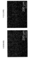
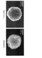
- a and c are photographs of low magnification observation, and b and d are photographs of high magnification observation. It is a scanning electron micrograph of unmodified MSC cells (left) and encapsulated MSC cells (right). It is the photograph of the fluorescence microscope which dye
- the cell encapsulation method of the present invention is a method of coating a cell membrane of a single cell with a polymer layer in vitro. That is, in the present invention, “encapsulation” refers to a state in which a polymer layer is formed over the entire outer periphery of a cell (cell membrane).
- the cells used in the method for encapsulating cells of the invention may be microbial cells (yeast, E. coli, etc.) or animal cells, such as humans, monkeys, cows, pigs, sheep, dogs, cats, rabbits, mice, etc. Mammalian cells can be preferably exemplified.
- the cells may be various cells such as stem cells, somatic cells, germ cells, cancer cells, and are not particularly limited.
- stem cells include embryonic stem cells, iPS cells (artificial pluripotent stem cells), and tissue-derived stem cells.
- tissue-derived stem cells include adipose-derived mesenchymal stem cells, bone marrow-derived mesenchymal stem cells, skin-derived stem cells, neural stem cells, cartilage stem cells, muscle stem cells, hematopoietic stem cells, dental pulp stem cells, cancer stem cells, and the like.
- normal somatic cells examples include fibroblasts, epithelial cells, adipocytes, chondrocytes, osteoblasts, osteoclasts, nerve cells, muscle cells, endothelial cells, hepatocytes, kidney cells, stromal cells, etc. Can be illustrated.
- germ cells such as eggs and sperm, cells of diseased or dead cells, cells isolated from cancer tissue, and the like can also be used.
- not only primary cells but also established cell lines can be used.
- the cell encapsulation method of the present invention includes an acryloylation step and a polymerization step.
- an acryloylation step a solution containing an acryloylating agent dissolved in an organic solvent and a cell suspension are mixed to introduce an acryloyl group on the surface of the cell membrane.
- the acryloyl group is represented by the following structural formula, for example.
- a mixed solution in which an acryloylating agent is dissolved in an organic solvent and mixed with a phosphate buffer can be used.
- An acryloylating agent is a compound that contains an acryloyl group and can bind to an amino group on the surface of a cell membrane.
- examples of the acryloylating agent include N-acryloxysuccinimide, acryloyl chloride and diacrylic anhydride.
- N-acryloxysuccinimide can be preferably used because it has low reactivity with water and hardly loses reactivity with amino groups of cell membranes.
- the organic solvent is not particularly limited as long as it can dissolve the acryloylating agent, and examples thereof include dimethyl sulfoxide, dimethylformamide, tetrahydrofuran, and dioxane. Among them, dimethyl sulfoxide has a low cytotoxicity and can be particularly preferably used when encapsulating living cells.
- the concentration of the phosphate buffer is preferably 0.1 to 0.001M, and more preferably 0.05 to 0.005M.
- the pH of the phosphate buffer is preferably 6.8 to 7.8, more preferably 7.0 to 7.6.
- N-acryloxysuccinimide when N-acryloxysuccinimide is used as an acryloylating agent to prepare a dimethylsulfoxide solution of N-acryloxysuccinimide, N-acryloxysuccinimide
- the ratio between the weight (mg) and the volume ( ⁇ L) of dimethyl sulfoxide is preferably 1: 500 to 1: 5, and more preferably 1: 250 to 1:10.
- N-acryloxy succinimide dimethyl sulfoxide and phosphate buffer by mixing dimethyl sulfoxide solution of N-acryloxy succinimide and phosphate buffer, dimethyl sulfoxide of N-acryloxy succinimide
- the volume ratio of the solution to the phosphate buffer is preferably 1: 400 to 1: 4, more preferably 1: 200 to 1:40.
- N-acryloxysuccinimide can be surely dissolved in dimethylsulfoxide, and the amino acid of the cell membrane by N-acryloxysuccinimide can be dissolved. Reactivity with the group is maintained. Furthermore, cytotoxicity due to dimethyl sulfoxide can also be suppressed.
- a desired cell suspension is prepared.
- the method and conditions for preparing the cell suspension are not particularly limited.
- the cell suspension can be prepared by suspending cells in a serum-free medium, phosphate buffer, or saline.
- the cell concentration of the cell suspension is preferably 1 ⁇ 10 4 to 1 ⁇ 10 8 cells / mL, and preferably 1 ⁇ 10 5 to 1 ⁇ 10 7 cells / mL. More preferred. When the concentration of the cells in the cell suspension is within this range, single cells can be reliably encapsulated by the steps described below.
- a solution containing an acryloylating agent dissolved in an organic solvent (for example, dimethyl sulfoxide) prepared as described above is mixed with a cell suspension.
- an organic solvent for example, dimethyl sulfoxide
- a cell suspension can be added to a solution containing an acryloylating agent dissolved in an organic solvent, or a solution containing an acryloylating agent dissolved in an organic solvent can be added to a cell. It can also be added to the suspension.
- the volume ratio of the mixed solution and the cell suspension is used. Is preferably 1: 5 to 80: 1, more preferably 1: 2 to 20: 1.
- an acryloyl group can be introduced by binding an acryloylating agent to an amino group on the surface of the cell membrane (acryloylation).
- the reaction temperature is preferably 1 to 37 ° C, more preferably 4 to 8 ° C.
- the reaction time is preferably 1 minute to 24 hours, and more preferably 10 minutes to 6 hours.
- acryloylated cells can be collected by centrifugation, suspended in a phosphate buffer and centrifuged again.
- This centrifugation / suspension / centrifugation operation excess unreacted acryloylating agent and organic solvent can be removed.
- This centrifugation / suspension / centrifugation (washing) can be performed, for example, once to five times.
- the centrifugal speed in the centrifugation is preferably 300 to 3000 rpm, more preferably 800 to 1500 rpm.
- the temperature for centrifugation is preferably 1 to 40 ° C., more preferably 4 to 8 ° C.
- the centrifugation time is preferably 10 seconds to 30 minutes, more preferably 1 to 10 minutes. Under such conditions, excess unreacted acryloylating agent and organic solvent can be more reliably removed.
- the collected acryloylated cells can be resuspended in a serum-free medium, phosphate buffer, or saline to prepare a cell suspension containing acryloylated cells.
- the cell concentration of this cell suspension is preferably 1 ⁇ 10 4 to 1 ⁇ 10 8 cells / mL, more preferably 1 ⁇ 10 5 to 1 ⁇ 10 7 cells / mL.
- the cell suspension containing acryloylated cells those obtained by the acryloylation step described above are used.
- an acryloyl group is introduced on the surface of the cell membrane, more specifically, on the amino group on the surface of the cell membrane.
- water-soluble monomer examples include acrylamide, sodium vinyl sulfonate, N-vinyl pyrrolidone, N-vinyl acetamide, 2-hydroxyethyl methacrylate, methyl methacrylate, polyethylene glycol monomethacrylate, and the like.
- acrylamide is particularly preferable because it is a monomer that is easily available and easily polymerized. A part of the water-soluble monomer binds to the acryloyl group introduced on the surface of the cell membrane.
- Crosslinkers include glycerol dimethacrylate, N ', N'-methylenebisacrylamide, N, N'-trimethylenebis (vinylsulfonylacetamide), ethylene glycol dimethacrylate, diethylene glycol dimethacrylate, triethylene glycol dimethacrylate.
- Examples thereof include Rato, tetraethylene glycol dimethacrylate, polyethylene glycol dimethacrylate, methylene bisacrylamide, N ′, N ′-(1,2-dihydroxyethylene) bisacrylamide, and the like.
- N ′, N′-methylenebisacrylamide is preferable because it is easily available and the efficiency of the crosslinking reaction is high.
- the polymerization initiator is a redox system in which peroxides such as ammonium persulfate and potassium persulfate are combined with a reducing agent such as N, N, N ′, N′-tetramethylethylenediamine, potassium persulfate / N, N, N ′.
- 2,2'-Azobis [2- (2-imidazolin-2-yl) propane] dihydrochloride, 2,2'-Azobis (2-methylpropionamidine) dihydrochloride, composed of N, -tetramethylethylenediamine
- An azo compound such as
- the polymerization initiator is a redox system in which peroxides such as ammonium persulfate and potassium persulfate are combined with a reducing agent such as N, N, N ′, N′-tetramethylethylenediamine, etc.
- the polymerization initiator is mild.
- the water-soluble monomer and the crosslinking agent can be polymerized at a temperature (for example, 4 ° C. to 37 ° C.), but it is particularly preferable to carry out at 4 ° C. to 8 ° C.
- the acrylamide monomer and glycerol dimethacrylate are dissolved in the phosphate buffer.
- the ratio of the weight of acrylamide (mg) to the volume of glycerol dimethacrylate ( ⁇ L) is preferably 30: 1 to 3:40, more preferably 10: 1 to 3:10.
- the ratio between the volume of glycerol dimethacrylate ( ⁇ L) and the volume of phosphate buffer ( ⁇ L) is preferably 3: 5000-3: 50, and 3: 2000-3: 100. Is more preferable.
- acrylamide monomer / glycerol dimethacrylate / acryloyl A cell suspension can be prepared.
- the ratio of the volume of the acrylamide monomer / glycerol dimethacrylate phosphate buffer solution (mL) to the volume of the acryloylated cell suspension (mL) is preferably 60: 1 to 3:20. 20: 1 to 3:10 is more preferable.
- ammonium persulfate and N, N, N ', N'-tetramethylethylenediamine can be dissolved in phosphate buffer.
- the ratio of the weight of ammonium persulfate ⁇ (mg) to the volume of N, N, N ', N'-tetramethylethylenediamine ( ⁇ L) is preferably 8: 1 to 1:50, 2: 1 to 2:25 It is more preferable that The ratio of the volume ( ⁇ L) of N, N, N ′, N′-tetramethylethylenediamine to the volume of the phosphate buffer ( ⁇ L) is preferably 1:10 to 1: 4000, and 1:20 More preferably, it is ⁇ 1: 500.
- a mixture of ammonium persulfate and N, N, N ', N'-tetramethylethylenediamine is dropped onto an acryloylated cell surface on an acrylamide monomer / glycerol dimethacrylate / acryloylated cell suspension.
- Polymerization reaction of acrylamide monomer and glycerol dimethacrylate can be performed.
- the ratio of the volume (mL) of acrylamide monomer / glycerol dimethacrylate / acryloylated cell suspension to the volume (mL) of the mixture of ammonium persulfate and N, N, N ', ′ N'-tetramethylethylenediamine is 80: 1 to 1: 5 is preferable, and 40: 1 to 2: 5 is more preferable.
- the polymerization reaction can be performed by shaking for a certain time while maintaining a certain reaction temperature.
- the reaction temperature in the polymerization reaction is preferably 1 to 40 ° C, more preferably 4 to 8 ° C.
- the reaction time is preferably 1 minute to 24 hours, more preferably 10 minutes to 6 hours.
- the surface of the cell membrane is covered with a polymer thin layer (polymer layer) formed by a polymerization reaction of a water-soluble monomer and a crosslinking agent, and the cells are encapsulated.
- the encapsulated cells can be collected, for example, by centrifugation and washed with a phosphate buffer. This cleaning operation can be performed about 1 to 5 times.
- the centrifugal speed in the centrifugation is preferably 300 to 3000 rpm, more preferably 800 to 1500 rpm.
- the temperature for centrifugation is preferably 1 to 40 ° C, more preferably 4 to 8 ° C.
- the centrifugation time is preferably 10 seconds to 30 minutes, and more preferably 1 to 10 minutes.
- the method for encapsulating cells of the present invention involves encapsulating cells with a polymer layer (polymer cross-linking network) through the above-described acryloylation step and polymerization step, with easy operation and mild conditions.
- a polymer layer polymer cross-linking network
- a polymer layer having a crosslinked network structure containing a water-soluble monomer and a crosslinking agent is formed on the surface of the cell membrane via an acryloyl group bonded to the amino group of the cell membrane of a single cell.
- the cross-linked network structure refers to a structure in which a water-soluble monomer and a cross-linking agent are continuously bonded in a network by cross-linking with a cross-linking agent as illustrated in FIG.
- the polymer layer formed on the surface of the cell membrane is thin and stable, and has a high molecular weight (for example, a molecular weight of 50 kDa or more) or nanoparticles (for example, a diameter of It has a permeability that allows a molecule having a low molecular weight (for example, a molecule having a molecular weight of less than 50 kDa) to permeate freely.
- the polymer layer has no toxicity.
- the encapsulated single cells can be transplanted into the patient's body by injection and can be circulated in the patient's body.
- the encapsulated cells are free from the attack of the immune system from the transplant, and can freely diffuse oxygen, nutrients, metabolites, and smaller physiologically active molecules having a low molecular weight.
- cells encapsulated by the cell encapsulation method of the present invention can be applied to the fields of biosensors and bioelectronics.
- the cell encapsulation method of the present invention is not limited to the above embodiment, and may include other preparation steps, washing steps, and the like.
- Example 1 Encapsulation of HeLa cells The acryloylation of the cell membrane of HeLa cells, which is a human cervical cancer cell line, was performed (acryloylation step).
- N-acryloxysuccinimide was dissolved in 100 ⁇ L of dimethyl sulfoxide and then mixed with 4 mL of phosphate buffer (0.01 M, pH 7.4). Add 1 mL of a suspension of HeLa cells suspended in serum-free medium (1 ⁇ 10 6 cells / mL) to the above mixture and react for 2 hours with shaking at 4-8 ° C. Acrylylated. Thereafter, acryloylated HeLa cells were collected by centrifugation at 4 ° C., 1100 rpm, 5 minutes, suspended in a phosphate buffer, and again collected by centrifugation at 4 ° C., 1100 rpm, 5 minutes. This is a washing step for removing excess unreacted N-acryloxysuccinimide and dimethyl sulfoxide. Thereafter, the collected acryloylated HeLa cells were resuspended in 1 mL of serum-free medium.
- the above mixture of ammonium persulfate and N, ⁇ N, N ', N'-tetramethylethylenediamine is added dropwise to the mixture of acryloylated HeLa cells, acrylamide monomer and glycerol dimethacrylate, and the reaction temperature is 4-8.
- the polymerization reaction was carried out on the surface of acryloylated HeLa cells by shaking for 2 hours while maintaining the temperature. As a result, the surface of the HeLa cell was encapsulated so as to be covered with the thin polymer layer formed by the polymerization reaction.
- the encapsulated HeLa cells were collected by centrifugation at 4 ° C., 1100 rpm for 5 minutes, and washed with phosphate buffer.
- HeLa cells and encapsulated HeLa cells were fixed with 2.5% (v / v) glutaraldehyde phosphate buffer at room temperature for 15 minutes. Next, the fixed cells were immersed in 50%, 70%, 80%, 90%, 100%, 100% (v / v) ethanol aqueous solution for 10 minutes each and dehydrated. Thereafter, ethanol was gradually replaced with t-butanol, and the cells were dried with a tert-butyl alcohol lyophilizer. Dry HeLa cells and encapsulated HeLa cells were coated with platinum by sputtering, and then observed with a scanning electron microscope.
- a life-and-death determination staining reagent containing calcein acetoxymethyl (calcein AM) and propidium iodide (PI) was used.
- a cell phosphate buffer suspension (200 ⁇ L) was seeded in a 24-well plate at a density of 1 ⁇ 10 5 cells / well.
- 100 ⁇ L of the mixed stock reagent of the cell viability determination kit was mixed with the cell suspension. After incubating at 37 ° C. for 15 minutes, the cells were visualized with an inverted fluorescence microscope.
- the activity of unmodified HeLa cells and encapsulated HeLa cells was quantitatively analyzed by the WST-1 assay.
- a 200- ⁇ L suspension of the cell phosphate buffer was seeded in a 96-well plate at a density of 2 ⁇ 10 4 cells / well.
- 20 ⁇ L of WST-1 reagent was added to each well.
- absorbance at a wavelength of 440 nm was measured with a microplate reader.
- the absorbance of the unmodified HeLa cells was assumed to be 100% viability, the viability of the encapsulated HeLa cells showed a high cell activity of 78.2 ⁇ 2.9%.
- AuNPs gold nanoparticles
- FITC-PEG polyethylene glycol PEG
- FITC-PEG polyethylene glycol PEG
- the solution was then cooled to room temperature and AuNPs were collected by centrifugation.
- the mixture was treated with ultrasound for 30 minutes, and then stirred at room temperature for 24 hours under light shielding to prepare FINP-PEG-coated AuNPs (FITC-PEG-AuNPs).
- the prepared FITC-PEG-AuNPs were washed by centrifugation to remove residual reagents.
- the size of the prepared FITC-PEG-AuNPs was 52.7 ⁇ 4.9 nm.
- FITC-PEG-AuNPs Place 2 mL of each suspension of unmodified HeLa cells and encapsulated HeLa cells (concentration: 1 ⁇ 10 5 cells / mL) into a centrifuge tube, and add FITC-PEG-AuNPs to a concentration of 50 ⁇ M.
- FITC-PEG-AuNPs was added to and incubated at 37 ° C. for 6 hours. Subsequently, the medium was removed and the cells were washed with phosphate buffer. The FITC-PEG-AuNPs on the cell membrane were quenched by incubating with 0.4% trypan blue for 10 minutes. The cells were then fixed in a 2.5% (v / v) glutaraldehyde solution at room temperature for 15 minutes.
- cell membranes and nuclei were stained by incubating with Cellmask TM -Deep Red and DAPI solution at 37 ° C. for 10 minutes. After washing the cells with phosphate buffer, the cells were enclosed between two cover glasses and observed with a confocal laser microscope.
- CD44 antigen is a cell surface glycoprotein involved in cell-cell interactions, cell adhesion, and migration, and is distributed on the surface of HeLa cells.
- Anti-CD44 antibody has a molecular weight of 85,000 Da and a hydrodynamic diameter of approximately 10 nm. 1 ⁇ 10 6 unmodified HeLa cells and encapsulated HeLa cells were fixed with 2.5% (v / v) glutaraldehyde PBS solution for 15 minutes. Cells were then blocked with 2% BSA for 2 hours.
- the cells were then incubated for 2 hours with 20 ⁇ g / mL primary antibody, anti-CD44 antibody, and the secondary antibody was added to the cell suspension and incubated for an additional hour.
- Cell nuclei were stained with DAPI. All experimental processes were performed at room temperature, and at least three phosphate buffer washes were performed between each step. Thereafter, observation with a fluorescence microscope and a confocal laser microscope was performed.
- Example 2 Encapsulation of MSC cells The acryloylation of the cell membrane of MSC cells, which are human bone marrow-derived mesenchymal stem cells, was performed (acryloylation step).
- N-acryloxysuccinimide was dissolved in 100 ⁇ L of dimethyl sulfoxide and then mixed with 4 mL of phosphate buffer (0.01 M, pH 7.4). Add 1 mL of a suspension of MSC cells suspended in serum-free medium (1 ⁇ 10 6 cells / mL) to the above mixture and react for 2 hours with shaking at 4-8 ° C. Acrylylated. Thereafter, acryloylated MSC cells were collected by centrifugation at 4 ° C., 1100 rpm, 5 minutes, suspended in a phosphate buffer, and again collected by centrifugation at 4 ° C., 1100 rpm, 5 minutes. This is a washing step for removing unreacted N-acryloxy succinimide and dimethyl sulfoxide. Thereafter, the collected acryloylated MSC cells were resuspended in 1 mL of serum-free medium.
- the above mixture of ammonium persulfate and N, N, N ', N'-tetramethylethylenediamine is added dropwise to the mixture of acryloylated MSC cells, acrylamide monomer and glycerol dimethacrylate, and the reaction temperature is 4-8.
- the polymerization reaction was performed on the surface of acryloylated MSC cells by shaking for 2 hours while maintaining the temperature. As a result, the surface of the MSC cells was encapsulated so as to be covered with the thin polymer layer formed by the polymerization reaction.
- the encapsulated MSC cells were recovered by centrifugation at 4 ° C., 1100 rpm for 5 minutes, and washed with phosphate buffer.
- MSC cells and encapsulated MSC cells were fixed for 15 minutes at room temperature using 2.5% (v / v) glutaraldehyde phosphate buffer. Next, the fixed cells were immersed in 50%, 70%, 80%, 90%, 100%, 100% (v / v) ethanol aqueous solution for 10 minutes each and dehydrated. Thereafter, ethanol was gradually replaced with t-butanol, and the cells were dried with a tert-butyl alcohol lyophilizer. Dry MSC cells and encapsulated MSC cells were coated with platinum by sputtering and then observed with a scanning electron microscope.
- the surface of the unmodified MSC cells was more uneven, but it was confirmed that the surface of the encapsulated MSC cells was smooth.
- a viability determination staining reagent containing calcein acetoxymethyl (calcein AM) and propidium iodide (PI) was used.
- a cell phosphate buffer suspension (200 ⁇ L) was seeded in a 24-well plate at a density of 1 ⁇ 10 5 cells / well.
- 100 ⁇ L of the mixed stock reagent of the cell viability determination kit was mixed with the cell suspension. After incubating at 37 ° C. for 15 minutes, the cells were visualized with an inverted fluorescence microscope.
- the activity of unmodified MSC cells and encapsulated MSC cells was quantitatively analyzed by the WST-1 assay.
- a 200- ⁇ L suspension of the cell phosphate buffer was seeded in a 96-well plate at a density of 2 ⁇ 10 4 cells / well.
- 20 ⁇ L of WST-1 reagent was added to each well.
- absorbance at a wavelength of 440 nm was measured with a microplate reader.
- the absorbance of unmodified MSC cells was assumed to be 100% viability, the viability of the encapsulated MSC cells showed a high cell activity of 74.7 ⁇ 3.6%.
- the acryloylated chondrocytes were then collected by centrifugation at 4 ° C, 1100 rpm, 5 minutes, suspended in phosphate buffer, and again collected by centrifugation at 4 ° C, 1100 rpm, 5 minutes (this was extra This is a cleaning step for removing unreacted N-acryloxysuccinimide and dimethyl sulfoxide). Thereafter, the collected acryloylated chondrocytes were resuspended in 1 mL of serum-free medium.
- the above mixture of ammonium persulfate and N, N, N ', ⁇ ⁇ ⁇ ⁇ ⁇ N'-tetramethylethylenediamine is added dropwise to the mixture of acryloylated chondrocytes, acrylamide monomer and glycerol dimethacrylate, and the reaction temperature is set to 4-8.
- the polymerization reaction was carried out on the surface of acryloylated chondrocytes by shaking for 2 hours while maintaining the temperature. As a result, the surface of the chondrocytes was encapsulated so as to be covered with the thin polymer layer formed by the polymerization reaction.
- the encapsulated chondrocytes were collected by centrifugation at 4 ° C., 1100 rpm for 5 minutes, and washed with phosphate buffer.
- chondrocytes not encapsulated and encapsulated chondrocytes were fixed with 2.5% (v / v) glutaraldehyde phosphate buffer at room temperature for 15 minutes. Next, the fixed cells were immersed in 50%, 70%, 80%, 90%, 100%, 100% (v / v) ethanol aqueous solution for 10 minutes each and dehydrated. Thereafter, ethanol was gradually replaced with t-butanol, and the cells were dried with a tert-butyl alcohol lyophilizer. Dry chondrocytes and encapsulated chondrocytes were coated with platinum by sputtering, and then observed with a scanning electron microscope.
- the surface of the unmodified chondrocytes was more uneven, but it was confirmed that the surface of the encapsulated chondrocytes was smooth.
- a viability determination staining reagent containing calcein acetoxymethyl (calcein AM) and propidium iodide (PI) was used.
- a cell phosphate buffer suspension (200 ⁇ L) was seeded in a 24-well plate at a density of 1 ⁇ 10 5 cells / well.
- 100 ⁇ L of the mixed stock reagent of the cell viability determination kit was mixed with the cell suspension. After incubating at 37 ° C. for 15 minutes, the cells were visualized with an inverted fluorescence microscope.
- the activity of unmodified chondrocytes and encapsulated chondrocytes was quantitatively analyzed by WST-1 assay.
- a 200- ⁇ L suspension of the cell phosphate buffer was seeded in a 96-well plate at a density of 2 ⁇ 10 4 cells / well.
- 20 ⁇ L of WST-1 reagent was added to each well.
- absorbance at a wavelength of 440 nm was measured with a microplate reader. Assuming that the absorbance of unmodified chondrocytes is 100% viability, the viability of the encapsulated chondrocytes was 75.3 ⁇ 3.2%, indicating a high cellular activity.
Landscapes
- Life Sciences & Earth Sciences (AREA)
- Chemical & Material Sciences (AREA)
- Engineering & Computer Science (AREA)
- Health & Medical Sciences (AREA)
- Genetics & Genomics (AREA)
- Biotechnology (AREA)
- Organic Chemistry (AREA)
- Zoology (AREA)
- Bioinformatics & Cheminformatics (AREA)
- Wood Science & Technology (AREA)
- Medicinal Chemistry (AREA)
- Microbiology (AREA)
- Biomedical Technology (AREA)
- Virology (AREA)
- Biochemistry (AREA)
- General Engineering & Computer Science (AREA)
- General Health & Medical Sciences (AREA)
- Tropical Medicine & Parasitology (AREA)
- Micro-Organisms Or Cultivation Processes Thereof (AREA)
- Apparatus Associated With Microorganisms And Enzymes (AREA)
Abstract
容易な操作かつ温和な条件で、安定性、浸透性に優れ、かつ毒性の低いカプセル(高分子層)で細胞を内包化することができる細胞の内包化方法を提供する。本発明の単一細胞の細胞膜を高分子層で被覆して内包化する方法は、以下の工程:有機溶剤に溶解させたアクリロイル化剤を含む溶液と、細胞懸濁液とを混合して、細胞膜の表面にアクリロイル基を導入するアクリロイル化工程;および、(A)アクリロイル化された細胞を含む細胞懸濁液と、(B)水溶性モノマー、架橋剤および重合開始剤を含む溶液とを混合して、細胞膜の表面のアクリロイル基を介して水溶性モノマーと架橋剤を重合させる重合工程を含む。
Description
本発明は、細胞の内包化方法および内包化細胞に関する。
従来より、細胞を半透過性の高分子膜やハイドロゲルによって被覆して内包化する技術が知られている。このような内包化細胞は、例えば、炎症性細胞、抗体、酵素、その他の高分子等との接触から保護される一方で、酸素、栄養分、代謝物質やより小さな生理活性分子を自由に相互拡散させることもできる。このため、細胞の内包化は、移植した細胞が免疫システムによって拒絶されるのを防ぐ目的で、細胞治療、および遺伝子治療の分野で広く利用されている。
そして、近年、単一細胞を内包化する技術が注目を集めている。従来の多細胞内包化と比較して、単一の内包化細胞はマイクロサイズであるので、バイオメディカル分野への応用に有用である。特に、耐久性かつ機能性をもつ材料で内包化した単一細胞によって、細胞ベースのバイオセンサーなど、バイオテクノロジーやバイオエレクトロニクスへの応用を拡大させることが期待されている。
単一細胞を内包化する技術としては、例えば、酵母菌、クロレラ菌、大腸菌などの微生物細胞の内包化に関する技術が知られているが(例えば、非特許文献1)、微生物細胞は、厚くて丈夫な細胞壁を持つので、細胞にとって好ましいとはいえないカプセル条件にも耐えることができる。
一方で、単一哺乳動物細胞の内包化に関する研究は少ない。これは、哺乳動物の細胞は環境に非常に敏感であり、細胞の活性を保つために、中性pH、特定のイオン強度、温度、カプセル化剤の生体適合性といった内包化条件が厳格に求められるからである。
単一哺乳動物細胞を内包化する技術として、マイクロ流路による液滴形成手法が知られている(非特許文献2-4)。また、別の手法としては、哺乳動物細胞の外側に疎水性相互作用や静電相互作用などの物理的な相互作用により薄い高分子層を形成する方法が知られている(例えば、非特許文献5-7)。
Biomimetic Encapsulation of Individual Cells with Silica, Angew. Chem. Int. Ed., 2009, 48, 9160-9163. S. H. Yang, K. B. Lee, B. Kong, J. H. Kim, H. S. Kim, I. S. Choi,
Microfluidic high-throughput encapsulation and hydrodynamic self-sorting of single cells, Proc. Natl. Acad. Sci. USA 2008, 105, 3191-3196. M. Chabert, J. L. Viovy,
Drop-based microfluidic devices for encapsulation of single cells, Lab Chip 2008, 8, 1110-1115. S. Koster, F. E. Angile, H. Duan, J. J. Agresti, A. Wintner, C. Schmitz, A. C. Rowat, C. A. Merten, D. Pisignano, A. D. Griffiths, D. A. Weitz,
The Poisson distribution and beyond: methods for microfluidic droplet production and single cell encapsulation", Lab Chip 2015, 15, 3439-3459. D. J. Collins, A. Neild, A. deMello, A. Q. Liu, Y. Ai,
Control of cell attachment through polyDNA hybridization, Biomaterials 2010, 31, 2229-2235 Y. Teramura, H. Chen, T. Kawamoto
Cell surface modification with ssDNA-PEG-lipid for analysing intercellular interactions between different cells, Biomaterials 2015, 48, 119-128 Y. Teramura
The Effect of Layer-by-Layer Assembly Coating on the Proliferation and Differentiation of Neural Stem Cells, ACS Appl. Mater. Inter. 2015, 7, 3018-3029. W. Li, T. Guan, X. Zhang, Z. Wang, M. Wang, W. Zhong, H. Feng, M. Xing, J. Kong
しかしながら、非特許文献2-4のようなマイクロ流路による液滴形成手法の場合、単一細胞を内包化する効率が低いという問題がある。また、この手法では、カプセルのサイズが大きくなってしまうために(例えば70 μm以上)、内包化した細胞では物質移動に乏しいという問題がある。
一方、非特許文献5-7のような物理的な相互作用により薄い高分子層を形成する方法の場合、高分子層の安定性が十分とは言えない。その一例として、疎水的相互作用を利用する方法の場合、高分子層が解離するために、カプセル(内包化した状態)を維持できる時間が短いという問題がある。別の例として、静電相互作用を利用する方法では、内包化に用いられる高分子電解質の大部分は非常に強い細胞毒性を持っているという問題があり、また、高分子層が動的に吸着し、しかも疎な構造のため、大部分の場合、保護作用や免疫抑制作用に対する要件を満たすのが難しいという問題がある。
本発明は、以上のとおりの事情に鑑みてなされたものであり、容易な操作かつ温和な条件で、安定性、浸透性に優れ、かつ毒性の低いカプセル(高分子層)で細胞を内包化することができる細胞の内包化方法を提供することを課題としている。また、安定性、浸透性に優れ、かつ毒性の低いカプセル(高分子層)で被覆された内包化細胞を提供することを課題としている。
上記の課題を解決するために、本発明の細胞の内包化方法は、単一細胞の細胞膜を高分子層で被覆して内包化する方法であって、
以下の工程:
有機溶剤に溶解させたアクリロイル化剤を含む溶液と、細胞懸濁液とを混合して、細胞膜の表面にアクリロイル基を導入するアクリロイル化工程;および
(A)アクリロイル化された細胞を含む細胞懸濁液と、(B)水溶性モノマー、架橋剤および重合開始剤を含む溶液とを混合して、細胞膜の表面のアクリロイル基を介して水溶性モノマーと架橋剤を重合させる重合工程
を含むことを特徴としている。
以下の工程:
有機溶剤に溶解させたアクリロイル化剤を含む溶液と、細胞懸濁液とを混合して、細胞膜の表面にアクリロイル基を導入するアクリロイル化工程;および
(A)アクリロイル化された細胞を含む細胞懸濁液と、(B)水溶性モノマー、架橋剤および重合開始剤を含む溶液とを混合して、細胞膜の表面のアクリロイル基を介して水溶性モノマーと架橋剤を重合させる重合工程
を含むことを特徴としている。
この内包化方法では、前記アクリロイル化剤は、N-アクリルオキシスクシンイミドであることが好ましい。
この内包化方法では、前記有機溶剤は、ジメチルスルホキシドであることが好ましい。
この内包化方法では、前記架橋剤は、グリセロールジメタクリル酸塩、または、N′, N′-メチレンビスアクリルアミドであるが好ましい。
この内包化方法では、前記重合開始剤は、過硫酸アンモニウムとN, N, N′, N′-テトラメチルエチレンジアミンを含む混合液、または、過硫酸カリウムとN, N, N′, N′-テトラメチルエチレンジアミンとを含む混合液であることが好ましい。
この内包化方法では、細胞が、動物細胞であることが好ましい。
本発明の内包化細胞は、単一細胞の細胞膜のアミノ基に結合したアクリロイル基を介して、細胞膜の表面に、水溶性モノマーと架橋剤を含む架橋ネットワーク構造を有する高分子層が形成されていることを特徴としている。
この内包化細胞では、前記架橋剤は、グリセロールジメタクリル酸塩、または、N′, N′-メチレンビスアクリルアミドであることが好ましい。
この内包化細胞では、細胞が、動物細胞であることが好ましい。
本発明の細胞の内包化方法によれば、容易な操作かつ温和な条件で、安定性、浸透性に優れ、かつ毒性の低いカプセル(高分子層)で細胞を内包化することができる。本発明の内包化細胞は、安定性、浸透性に優れ、かつ毒性の低いカプセル(高分子層)で被覆されている。
本発明の細胞の内包化方法は、in vitroにおいて、単一細胞の細胞膜を高分子層で被覆する方法である。すなわち、本発明において、「内包化」とは、細胞(細胞膜)の外周囲全体に亘って高分子層が形成されている状態をいう。
発明の細胞の内包化方法に使用される細胞は、微生物細胞(酵母、大腸菌など)または動物細胞であってよく、例えば、ヒト、サル、ウシ、ブタ、ヒツジ、イヌ、ネコ、ウサギ、ネズミなど哺乳動物細胞を好ましく例示することができる。
細胞は、幹細胞、体細胞、生殖細胞、がん細胞などの各種の細胞であってよく、特に限定されない。具体的には、例えば、幹細胞としては、胚性幹細胞、iPS細胞(人工多能性幹細胞)、組織由来幹細胞を例示することができる。組織由来幹細胞としては、脂肪由来間葉系幹細胞、骨髄由来間葉系幹細胞、皮膚由来幹細胞、神経幹細胞、軟骨幹細胞、筋肉幹細胞、造血幹細胞、歯髄幹細胞、がん幹細胞などを例示することができる。正常な体細胞としては、例えば、繊維芽細胞、上皮細胞、脂肪細胞、軟骨細胞、骨芽細胞、破骨細胞、神経細胞、筋肉細胞、内皮細胞、肝実質細胞、腎臓細胞、間質細胞などを例示することができる。さらに、体細胞の他、卵、精子などの生殖細胞や、病気にかかった組織の細胞や死細胞、がん組織から単離した細胞なども利用することができる。また、初代細胞だけでなく株化細胞も利用することができる。
そして、本発明の細胞の内包化方法は、アクリロイル化工程および重合工程を含んでいる。以下、各工程について説明する。
(アクリロイル化工程)
アクリロイル化工程では、有機溶剤に溶解させたアクリロイル化剤を含む溶液と、細胞懸濁液とを混合して、細胞膜の表面にアクリロイル基を導入する。アクリロイル基は、例えば以下の構造式で表される。
(アクリロイル化工程)
アクリロイル化工程では、有機溶剤に溶解させたアクリロイル化剤を含む溶液と、細胞懸濁液とを混合して、細胞膜の表面にアクリロイル基を導入する。アクリロイル基は、例えば以下の構造式で表される。

本発明の細胞の内包化方法の一実施形態では、アクリロイル化剤を有機溶剤に溶解させ、リン酸緩衝液と混合した混合溶液を使用することができる。
アクリロイル化剤は、アクリロイル基を含み、細胞膜の表面のアミノ基に結合可能な化合物である。具体的には、アクリロイル化剤は、N-アクリルオキシスクシンイミド、塩化アクリロイルやジアクリル酸無水物を例示することができる。なかでも、N-アクリルオキシスクシンイミドは、水との反応性が低く、細胞膜のアミノ基との反応性が失われ難いため、好ましく使用することができる。
有機溶剤は、アクリロイル化剤を溶解させるものであれば特に限定されず、例えば、ジメチルスルホキシド、ジメチルホルムアミド、テトラヒドロフラン、ジオキサンなどを例示することができる。なかでも、ジメチルスルホキシドは細胞毒性が低いため、生きた細胞を内包化する場合には、特に好ましく使用することができる。
また、リン酸緩衝液の濃度は、0.1~0.001Mであることが好ましく、0.05~0.005Mであることがより好ましい。リン酸緩衝液のpHは、6.8~7.8であることが好ましく、7.0~7.6であることがより好ましい。
例えば、本発明の細胞の内包化方法の一実施形態において、アクリロイル化剤としてN-アクリルオキシスクシンイミドを使用して、N-アクリルオキシスクシンイミドのジメチルスルホキシド溶液を調製する場合、N-アクリルオキシスクシンイミドの重量(mg)とジメチルスルホキシドの体積(μL)の比は、1:500~1:5であることが好ましく、1:250~1:10であることがより好ましい。そして、N-アクリルオキシスクシンイミドのジメチルスルホキシド溶液とリン酸緩衝液を混合し、N-アクリルオキシスクシンイミドとジメチルスルホキシドとリン酸緩衝液の混合溶液を作製する場合は、N-アクリルオキシスクシンイミドのジメチルスルホキシド溶液とリン酸緩衝液との体積比は1:400~1:4であることが好ましく、1:200~1:40であることがより好ましい。N-アクリルオキシスクシンイミド、ジメチルスルホキシドおよびリン酸緩衝液の配合比率がこの範囲であると、N-アクリルオキシスクシンイミドをジメチルスルホキシドに確実に溶解させることができるとともに、N-アクリルオキシスクシンイミドによる細胞膜のアミノ基との反応性が維持される。さらに、ジメチルスルホキシドによる細胞毒性も抑制することができる。
また、本発明の細胞の内包化方法の一実施形態では、所望の細胞懸濁液を調製する。細胞懸濁液の調製方法、条件は特に限定されないが、例えば、細胞を無血清培地やリン酸緩衝液、食塩水に懸濁し、細胞懸濁液を調製することができる。具体的には、例えば、細胞懸濁液の細胞の濃度は1×10⁴~1×10⁸cells/mLであることが好ましく、1×105~1×107cells/mLであることがより好ましい。細胞懸濁液の細胞の濃度がこの範囲であると、後述する工程によって確実に単一細胞を内包化することができる。
本発明の細胞の内包化方法では、例えば、上記のように調製した有機溶剤(例えばジメチルスルホキシド)に溶解させたアクリロイル化剤を含む溶液と、細胞懸濁液とを混合する。
具体的には、例えば、細胞懸濁液を、有機溶剤に溶解させたアクリロイル化剤を含む溶液に添加することもできるし、あるいは、有機溶剤に溶解させたアクリロイル化剤を含む溶液を、細胞懸濁液に添加することもできる。
そして、例えば、アクリロイル化剤としてのN-アクリルオキシスクシンイミドと、有機溶剤としてのジメチルスルホキシドと、リン酸緩衝液との混合溶液を使用する場合は、この混合溶液と細胞懸濁液との体積比は、1:5~80:1であることが好ましく、1:2~20:1であることがより好ましい。
例えば、細胞懸濁液と、有機溶剤に溶解させたアクリロイル化剤を含む溶液を混合した後、所定の温度で振とうしながら所定の時間反応させることで、図1に示したように、細胞の細胞膜の表面のアミノ基にアクリロイル化剤を結合させてアクリロイル基を導入することができる(アクリロイル化)。
反応温度は、1~37℃であることが好ましく、4~8℃であることがより好ましい。反応時間は、1分間~24時間であることが好ましく、10分間~6時間であることがより好ましい。
アクリロイル化工程では、例えば、アクリロイル化した細胞を遠心分離によって回収、リン酸緩衝液で懸濁して再度、遠心分離することによって回収することができる。この遠心・懸濁・遠心作業により、余分の未反応のアクリロイル化剤と有機溶剤を除去することができる。この遠心・懸濁・遠心作業(洗浄作業)は、例えば、1回~5回行うことができる。遠心分離における遠心速度は、300~3000 rpmであることが好ましく、800~1500 rpmであることがより好ましい。遠心分離の温度は、1~40℃であることが好ましく、4~8℃であることがより好ましい。遠心分離の時間は10秒~30分間であることが好ましく、1~10分間であることがより好ましい。このような条件であると、余分の未反応のアクリロイル化剤と有機溶剤をより確実に除去することができる。
そして、回収したアクリロイル化した細胞を無血清培地やリン酸緩衝液、食塩水に再懸濁し、アクリロイル化した細胞を含む細胞懸濁液を調製することができる。この細胞懸濁液の細胞の濃度は1×10⁴~1×10⁸cells/mLであることが好ましく、1×105~1×107cells/mLであることがより好ましい。
(重合工程)
重合工程では、(A)アクリロイル化された細胞を含む細胞懸濁液と、(B)水溶性モノマー、架橋剤および重合開始剤を含む溶液とを混合して、細胞膜の表面のアクリロイル基を介して水溶性モノマーと架橋剤を重合させる。
重合工程では、(A)アクリロイル化された細胞を含む細胞懸濁液と、(B)水溶性モノマー、架橋剤および重合開始剤を含む溶液とを混合して、細胞膜の表面のアクリロイル基を介して水溶性モノマーと架橋剤を重合させる。
アクリロイル化された細胞を含む細胞懸濁液は、上述したアクリロイル化工程によって得たものが使用される。アクリロイル化された細胞は、細胞膜の表面に、より具体的には細胞膜の表面のアミノ基に、アクリロイル基が導入されている。
水溶性モノマーは、アクリルアミド、ビニルスルホン酸ナトリウム、N-ビニルピロリドン、N-ビニルアセトアミド、メタクリル酸2-ヒドロキシエチル、メチルメタクリレート、ポリエチレングリコールモノメタクリレートなどを例示することができる。なかでも、アクリルアミドは入手が容易で重合しやすいモノマーであるため、特に好ましい。水溶性モノマーの一部は、細胞膜の表面に導入されたアクリロイル基に結合する。
架橋剤は、グリセロールジメタクリル酸塩、N′, N′-メチレンビスアクリルアミド、N, N′-トリメチレンビス(ビニルスルホニルアセトアミド)、エチレングリコールジメタクリラート、ジエチレングリコールジメタクリラート、トリエチレングリコールジメタクリラート、ジメタクリル酸テトラエチレングリコール、ポリエチレングリコールジメタクリレート、メチレンビスアクリルアミド、N′, N′-(1,2-ジヒドロキシエチレン)ビスアクリルアミドなどを例示することができる。なかでも、N′, N′-メチレンビスアクリルアミドは、入手が容易で架橋反応の効率が高いため好ましい。
重合開始剤は、過硫酸アンモニウム、過硫酸カリウム等の過酸化物とN, N, N′, N′-テトラメチルエチレンジアミン等の還元剤を組み合わせたレドックス系、過硫酸カリウム/N, N, N′, N′-テトラメチルエチレンジアミンからなる、2,2'-アゾビス[2-(2-イミダゾリン-2-イル)プロパン]二塩酸塩、2,2'-アゾビス(2-メチルプロピオンアミジン)ニ塩酸塩などのアゾ化合物などを例示することができる。なかでも、重合開始剤が、過硫酸アンモニウム、過硫酸カリウム等の過酸化物とN, N, N′, N′-テトラメチルエチレンジアミン等の還元剤を組み合わせたレドックス系であると、細胞にとって温和な温度(例えば、4℃~37℃)で水溶性モノマーと架橋剤を重合させることができるが、4℃~8℃で行うのが特に好ましい。
例えば、図1に示したように、水溶性モノマーがアクリルアミドであり、架橋剤がグリセロールジメタクリル酸塩である場合、アクリルアミドモノマーとグリセロールジメタクリル酸塩をリン酸緩衝液に溶解させる。アクリルアミドの重量(mg)とグリセロールジメタクリル酸塩の体積(μL)の比率は30:1~3:40であることが好ましく、10:1~3:10であることがより好ましい。また、グリセロールジメタクリル酸塩の体積(μL)と、リン酸緩衝液の体積(μL)の比率は、3:5000~3:50であることが好ましく、3:2000~3:100であることがより好ましい。
そして、アクリルアミドモノマーと、グリセロールジメタクリル酸塩と、リン酸緩衝液との混合液に、アクリロイル化された細胞を含む細胞懸濁液を添加することで、アクリルアミドモノマー/グリセロールジメタクリル酸塩/アクリロイル化細胞懸濁液を調製することができる。アクリルアミドモノマー/グリセロールジメタクリル酸塩のリン酸緩衝液の混合液の体積(mL)と、アクリロイル化細胞懸濁液の体積(mL)の比率は、60:1~3:20であることが好ましく、20:1~3:10であることがより好ましい。
また、例えば、重合開始剤として、ラジカル重合開始剤である過硫酸アンモニウムと触媒であるN, N, N′, N′-テトラメチルエチレンジアミンの混合液を使用する場合、過硫酸アンモニウムとN, N, N′, N′-テトラメチルエチレンジアミンをリン酸緩衝液に溶解させることができる。
過硫酸アンモニウムの重量 (mg)とN, N, N′, N′-テトラメチルエチレンジアミンの体積(μL)の比率は、8:1~1:50であることが好ましく、2:1~2:25であることがより好ましい。また、N, N, N′, N′-テトラメチルエチレンジアミンの体積(μL)とリン酸緩衝液の体積(μL)の比率は、1:10~1:4000であることが好ましく、1:20~1:500であることがより好ましい。
例えば、アクリルアミドモノマー/グリセロールジメタクリル酸塩/アクリロイル化細胞懸濁液に、過硫酸アンモニウムとN, N, N′, N′-テトラメチルエチレンジアミンの混合液を滴下することで、アクリロイル化細胞の表面でアクリルアミドモノマーとグリセロールジメタクリル酸塩の重合反応を行うことができる。
アクリルアミドモノマー/グリセロールジメタクリル酸塩/アクリロイル化細胞懸濁液の体積(mL)と、過硫酸アンモニウムとN, N, N′, N′-テトラメチルエチレンジアミンの混合液の体積(mL)の比率は、80:1~1:5であることが好ましく、40:1~2:5であることがより好ましい。
また、重合反応は、一定の反応温度を保ちながら一定の時間振とうすることによって行うことができる。重合反応における反応温度は、1~40℃であることが好ましく、4~8℃であることがより好ましい。反応時間は1分間~24時間であることが好ましく、10分間~6時間であることがより好ましい。
この重合工程によって、細胞膜の表面が、水溶性モノマーと架橋剤の重合反応により形成された高分子の薄層(高分子層)で被覆され、細胞が内包化される。内包化された細胞は、例えば、遠心分離によって回収し、リン酸緩衝液で洗浄することができる。この洗浄作業は1回~5回程度行うことができる。また、遠心分離における遠心速度は、300~3000rpmであることが好ましく、800~1500rpmであることがより好ましい。遠心分離の温度は、1~40℃であることが好ましく、4~8℃であることがより好ましい。遠心分離の時間は、10秒~30分間であることが好ましく、1~10分間であることがより好ましい。このような洗浄によって、重合開始剤や未反応のモノマーなどの分子量の小さい化合物を取り除くことができる。
以上の通り、本発明の細胞の内包化方法は、上述したアクリロイル化工程および重合工程を経ることで、容易な操作かつ温和な条件によって、高分子層(高分子の架橋ネットワーク)で細胞を内包化することができる。
そして、本発明の内包化細胞は、単一細胞の細胞膜のアミノ基に結合したアクリロイル基を介して、細胞膜の表面に、水溶性モノマーと架橋剤を含む架橋ネットワーク構造を有する高分子層が形成されている。ここで、架橋ネットワーク構造とは、図1に例示したように、架橋剤による架橋によって、水溶性モノマーと架橋剤とが網目状に連続して結合している構造をいう。
本発明の内包化細胞では、細胞膜の表面に形成された高分子層は、薄層かつ安定であり、分子量の高い分子(例えば、分子量が50 kDa以上の分子)やナノ粒子(例えば、直径が5nm以上の粒子)を透過させず、分子量の低い分子(例えば、分子量が50 kDa未満の分子)を自由に透過させることができる浸透性を有している。また、高分子層は毒性を有していない。
このため、例えば、内包化された単一細胞を注射で患者の体内に移植することができ、患者の体内で循環させることができる。この場合、内包化細胞は、移植体からの免疫システムの攻撃を受けず、分子量が低い酸素、栄養分、代謝物質やより小さな生理活性分子を自由に拡散させることができる。また、例えば、本発明の細胞の内包化方法によって内包化された細胞は、バイオセンサーやバイオエレクトロニクスの分野にも応用することができる。
本発明の細胞の内包化方法は、以上の実施形態に限定されることはなく、その他の調製工程や洗浄工程などを含むことができる。
以下、本発明の細胞の内包化方法について実施例とともに説明するが、本発明の細胞の内包化方法は、以下の実施例に何ら限定されるものではない。
<実施例1>HeLa細胞の内包化
ヒト子宮頸部がん細胞株であるHeLa細胞の細胞膜のアクリロイル化を行った(アクリロイル化工程)。
<実施例1>HeLa細胞の内包化
ヒト子宮頸部がん細胞株であるHeLa細胞の細胞膜のアクリロイル化を行った(アクリロイル化工程)。
HeLa細胞膜のアクリロイル化を行うため、2 mgのN-アクリルオキシスクシンイミドを100 μLのジメチルスルホキシドに溶解させ、その後、4 mLのリン酸緩衝液(0.01 M、pH 7.4)と混合した。HeLa細胞を無血清培地に懸濁した懸濁液(1×10⁶ cells/mL )1 mLを上記混合物に添加し、4~8℃で振とうしながら2時間反応し、HeLa細胞の表面をアクリロイル化した。その後、アクリロイル化したHeLa細胞を4℃、1100 rpm、5分間の遠心分離によって回収、リン酸緩衝液で懸濁して再度4℃、1100 rpm、5分間の遠心分離によって回収した。これは余分の未反応のN-アクリルオキシスクシンイミドとジメチルスルホキシドを除去するための洗浄工程である。その後、回収したアクリロイル化したHeLa細胞を無血清培地1 mLに再懸濁させた。
次に、HeLa細胞表面におけるin situ重合を行った(重合工程)。
アクリルアミドモノマー9 mgとグリセロールジメタクリル酸塩6 μLをリン酸緩衝液3 mLに溶解させた。そして、アクリルアミドモノマー/グリセロールジメタクリル酸塩の混合液にアクリロイル化HeLa細胞懸濁液1 mLを添加した。一方、ラジカル重合開始剤である過硫酸アンモニウム 2 mgと触媒であるN, N, N′, N′-テトラメチルエチレンジアミン 5 μLをリン酸緩衝液1 mLに添加し、過硫酸アンモニウムとN, N, N′, N′-テトラメチルエチレンジアミンのリン酸緩衝液の混合液を調製した。上記のアクリロイル化HeLa細胞とアクリルアミドモノマーとグリセロールジメタクリル酸塩の混合液に上記の過硫酸アンモニウムとN, N, N′, N′-テトラメチルエチレンジアミンの混合液を滴下し、反応温度を4~8℃に保ちながら2時間振とうすることによって、アクリロイル化HeLa細胞の表面で重合反応を行った。これにより、HeLa細胞の表面が、上記の重合反応により形成した高分子の薄層によって覆われるようにして内包化された。内包化されたHeLa細胞を4℃、1100 rpm、5分間の遠心分離によって回収し、リン酸緩衝液で洗浄した。
内包化されていない未修飾のHeLa細胞および内包化したHeLa細胞の表面モルフォロジーを走査電子顕微鏡により観察した。HeLa細胞および内包化したHeLa細胞を2.5%(v/v)グルタルアルデヒドのリン酸緩衝液を用いて室温で15分間固定した。次に、固定した細胞を50%、70%、80%、90%、100%、100%(v/v)のエタノール水溶液で10分間ずつ浸漬し、脱水処理した。その後、エタノールを段階的にt-ブタノールで置換し、細胞をtert-ブチルアルコール凍結乾燥機で乾燥させた。乾燥したHeLa細胞および内包化したHeLa細胞をスパッターにより白金でコーティングした後、走査電子顕微鏡による観察を行った。
図2に示したように、未修飾のHeLa細胞と内包化したHeLa細胞では、異なる表面モルフォロジーが観察された。内包化したHeLa細胞の表面は滑らかであったが、未修飾のHeLa細胞は起伏の多い表面を示した。この結果から、内包化したHeLa細胞が全体の形状を保ち、またより滑らかな表面を持っていることが確認された。
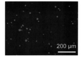
未修飾のHeLa細胞と内包化したHeLa細胞の生細胞と死細胞を可視化するため、カルセインアセトキシメチル(カルセインAM)とヨウ化プロピジウム(PI)を含む生死判定染色試薬を用いた。細胞のリン酸緩衝液の懸濁液200 μLを、1×10⁵ cells/wellの密度で24-ウェルプレートに播種した。その後ただちに、細胞生死判別キットの混合ストック試薬100 μLを細胞懸濁液と混合した。37℃で15分間インキュベートした後、細胞を倒立型蛍光顕微鏡で可視化した。
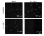
図3に例示したように、未修飾のHeLa細胞のほとんどが生存していた。内包化したHeLa細胞の場合、死細胞の数はわずかに増加したが、それでも細胞は高い細胞生存性を示していた。
また、未修飾のHeLa細胞と内包化したHeLa細胞の活性をWST-1アッセイ法によって定量的に分析した。細胞のリン酸緩衝液の懸濁液200 μL を2×10⁴ cells/wellの密度で96穴プレートに播種した。次に、WST-1試薬 20 μLを各々のウェルに添加した。37℃で3時間インキュベーションを行った後、440 nm波長での吸光度をマイクロプレートリーダーで測定した。未修飾のHeLa細胞の吸光度を100%の生存性とすると、内包化したHeLa細胞の細胞生存性は78.2±2.9%と高い細胞活性を示した。
さらに、内包化したHeLa細胞の表面に形成した高分子の薄層によるナノ粒子および高分子の侵入を阻止する効果を調べた。まず、金ナノ粒子(AuNPs)の侵入を阻止する効果を確認した。蛍光色素FITCでラベルしたポリエチレングリコールPEG(FITC-PEG)でコーティングしたAuNPsを用いた。FITC-PEGでコーティングしたAuNPsを作製するために、100 mLのHAuCl4溶液(0.1 mg/mL)を撹拌しながら110℃まで加熱し、20分間還流した。1 mLのクエン酸三ナトリウム溶液(10 mg/mL)をすばやく添加し、撹拌しながら15分間反応を行った。その後、溶液を室温まで冷却し、AuNPsを遠心分離によって回収した。AuNPsは適切な量のPEGと混合し、モル比はmPEG-SH: FITC-PEG-SH = 3:2であった。混合物を超音波で30分間処理した後、室温、遮光下で24時間撹拌し、FITC-PEGでコーティングしたAuNPs(FITC-PEG-AuNPs)を作製した。作製したFITC-PEG-AuNPsは、残留試薬を除去するために、遠心法で洗浄した。作製したFITC-PEG-AuNPsのサイズは52.7 ± 4.9 nmであった。未修飾HeLa細胞と内包化したHeLa細胞のそれぞれの懸濁液(濃度は1×10⁵ cells/mL)2 mLを遠心分離チューブに入れ、さらにFITC-PEG-AuNPsの濃度が50 μMになるようにFITC-PEG-AuNPsを添加して37℃、6時間インキュベートした。続いて、培地を除去し、細胞をリン酸緩衝液で洗浄した。細胞膜上のFITC-PEG-AuNPsを0.4%トリパンブルーで10分間インキュベートすることによってクエンチした。その後、細胞を2.5%(v/v)グルタルアルデヒド溶液中で室温、15分間固定した。続いて、CellmaskTM-Deep RedとDAPI溶液を用いて37℃で10分間インキュベートすることにより、細胞膜および核を染色した。細胞をリン酸緩衝液で洗浄した後、細胞を2枚のカバーグラスの間に封入し、共焦点レーザー顕微鏡で観察した。
図4に示したように、共焦点レーザー顕微鏡の画像より、FITC-PEG-AuNPsに由来する緑色蛍光が未修飾HeLa細胞内で観察されたが、内包化したHeLa細胞内では観察されないことが分かった。この結果は、内包化したHeLa細胞の表面で形成した高分子の薄層は金ナノ粒子の侵入を防げることを示している。
続いて、分子量が高い抗‐CD44抗体の侵入を防ぐ効果を確認した。CD44抗原は、細胞-細胞間相互作用、細胞接着、移動に関係する、細胞表面の糖タンパク質で、HeLa細胞表面に分布している。抗-CD44抗体の分子量は85,000 Daで、流体力学径はおよそ10 nmと推定される。1×10⁶個の未修飾のHeLa細胞及び内包化したHeLa細胞を2.5%(v/v)のグルタルアルデヒドPBS溶液で15分間固定した。次に、細胞を2%BSAで2時間ブロッキングした。その後、細胞を20 μg/mLの一次抗体である抗‐CD44抗体で2時間インキュベートし、二次抗体を細胞懸濁液に添加してさらに1時間インキュベートした。細胞核は、DAPIで染色した。全ての実験プロセスは室温で行い、各々の工程間で少なくともリン酸緩衝液による洗浄を3回行った。その後、蛍光顕微鏡および共焦点レーザー顕微鏡による観察を行った。
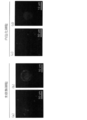
図5に示したように、未修飾のHeLa細胞の細胞膜に沿ってFITC-抗CD44の緑色の蛍光が観察された(図5a、図5b)。しかし、内包化したHeLa細胞は細胞膜表面で緑色の蛍光を示さなかった(図5c、図5d)。これらの結果から、内包化したHeLa細胞の表面で形成した高分子の薄層は抗体などの分子量の高い分子の侵入を阻止できることが確認された。
<実施例2>MSC細胞の内包化
ヒト骨髄由来間葉系幹細胞であるMSC細胞の細胞膜のアクリロイル化を行った(アクリロイル化工程)。
ヒト骨髄由来間葉系幹細胞であるMSC細胞の細胞膜のアクリロイル化を行った(アクリロイル化工程)。
MSC細胞膜のアクリロイル化を行うため、2 mgのN-アクリルオキシスクシンイミドを100 μLのジメチルスルホキシドに溶解させ、その後、4 mLのリン酸緩衝液(0.01 M、pH 7.4)と混合した。MSC細胞を無血清培地に懸濁した懸濁液(1×10⁶ cells/mL)1 mLを上記混合物に添加し、4~8℃で振とうしながら2時間反応し、MSC細胞の表面をアクリロイル化した。その後、アクリロイル化したMSC細胞を4℃、1100 rpm、5分間の遠心分離によって回収、リン酸緩衝液で懸濁して再度4℃、1100 rpm、5分間の遠心分離によって回収した。これは未反応のN-アクリルオキシスクシンイミドとジメチルスルホキシドを除去するための洗浄工程である。その後、回収したアクリロイル化したMSC細胞を無血清培地1 mLに再懸濁させた。
次に、MSC細胞表面におけるin situ重合を行った(重合工程)。
アクリルアミドモノマー9 mgとグリセロールジメタクリル酸塩6 μLをリン酸緩衝液3 mLに溶解させた。そして、アクリルアミドモノマー/グリセロールジメタクリル酸塩の混合液にアクリロイル化MSC細胞懸濁液1 mLを添加した。一方、ラジカル重合開始剤である過硫酸アンモニウム 2 mgと触媒であるN, N, N′, N′-テトラメチルエチレンジアミン 5 μLをリン酸緩衝液1 mLに添加し、過硫酸アンモニウムとN, N, N′, N′-テトラメチルエチレンジアミンのリン酸緩衝液の混合液を調製した。上記のアクリロイル化MSC細胞とアクリルアミドモノマーとグリセロールジメタクリル酸塩の混合液に上記の過硫酸アンモニウムとN, N, N′, N′-テトラメチルエチレンジアミンの混合液を滴下し、反応温度を4~8℃に保ちながら2時間振とうすることによって、アクリロイル化MSC細胞の表面で重合反応を行った。これにより、MSC細胞の表面が、上記の重合反応により形成した高分子の薄層によって覆われるようにして内包化された。内包化されたMSC細胞を4℃、1100 rpm、5分間の遠心分離によって回収し、リン酸緩衝液で洗浄した。
内包化されていない未修飾のMSC細胞および内包化したMSC細胞の表面モルフォロジーを走査電子顕微鏡により観察した。MSC細胞および内包化したMSC細胞を2.5%(v/v)グルタルアルデヒドのリン酸緩衝液を用いて室温で15分間固定した。次に、固定した細胞を50%、70%、80%、90%、100%、100%(v/v)のエタノール水溶液で10分間ずつ浸漬し、脱水処理した。その後、エタノールを段階的にt-ブタノールで置換し、細胞をtert-ブチルアルコール凍結乾燥機で乾燥させた。乾燥したMSC細胞および内包化したMSC細胞をスパッターにより白金でコーティングした後、走査電子顕微鏡による観察を行った。
図6に示したように、未修飾のMSC細胞の表面はより起伏が多かったが、内包化したMSC細胞の表面は滑らかであることが確認された。
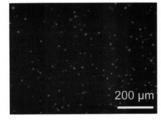
内包化したMSC細胞の生細胞と死細胞を可視化するため、カルセインアセトキシメチル(カルセインAM)とヨウ化プロピジウム(PI)を含む生死判定染色試薬を用いた。細胞のリン酸緩衝液の懸濁液200 μLを、1×10⁵ cells/wellの密度で24-ウェルプレートに播種した。その後ただちに、細胞生死判別キットの混合ストック試薬100 μLを細胞懸濁液と混合した。37℃で15分間インキュベートした後、細胞を倒立型蛍光顕微鏡で可視化した。
図7に示したように、内包化したMSC細胞の大部分は生きていることが確認された。
また、未修飾のMSC細胞と内包化したMSC細胞の活性をWST-1アッセイ法によって定量的に分析した。細胞のリン酸緩衝液の懸濁液200 μL を2×10⁴ cells/wellの密度で96穴プレートに播種した。次に、WST-1試薬 20 μLを各々のウェルに添加した。37℃で3時間インキュベーションを行った後、440 nm波長での吸光度をマイクロプレートリーダーで測定した。未修飾のMSC細胞の吸光度を100%の生存性とすると、内包化したMSC細胞の生存率は74.7±3.6%と高い細胞活性を示した。
<実施例3>軟骨細胞の内包化
ウシ膝間接軟骨細胞の細胞膜のアクリロイル化を行った(アクリロイル化工程)。
軟骨細胞膜のアクリロイル化を行うため、2 mgのN-アクリルオキシスクシンイミドを100 μLのジメチルスルホキシドに溶解させ、その後、4 mLのリン酸緩衝液(0.01 M、pH 7.4)と混合した。軟骨細胞を無血清培地に懸濁した懸濁液(1×10⁶ cells/mL )1 mLを上記混合物に添加し、4~8℃で振とうしながら2時間反応し、軟骨細胞の表面をアクリロイル化した。その後、アクリロイル化した軟骨細胞を4℃、1100 rpm、5分間の遠心分離によって回収、リン酸緩衝液で懸濁して再度4℃、1100 rpm、5分間の遠心分離によって回収した(これは余分の未反応のN-アクリルオキシスクシンイミドとジメチルスルホキシドを除去するための洗浄工程である)。その後、回収したアクリロイル化した軟骨細胞を無血清培地1 mLに再懸濁させた。
ウシ膝間接軟骨細胞の細胞膜のアクリロイル化を行った(アクリロイル化工程)。
軟骨細胞膜のアクリロイル化を行うため、2 mgのN-アクリルオキシスクシンイミドを100 μLのジメチルスルホキシドに溶解させ、その後、4 mLのリン酸緩衝液(0.01 M、pH 7.4)と混合した。軟骨細胞を無血清培地に懸濁した懸濁液(1×10⁶ cells/mL )1 mLを上記混合物に添加し、4~8℃で振とうしながら2時間反応し、軟骨細胞の表面をアクリロイル化した。その後、アクリロイル化した軟骨細胞を4℃、1100 rpm、5分間の遠心分離によって回収、リン酸緩衝液で懸濁して再度4℃、1100 rpm、5分間の遠心分離によって回収した(これは余分の未反応のN-アクリルオキシスクシンイミドとジメチルスルホキシドを除去するための洗浄工程である)。その後、回収したアクリロイル化した軟骨細胞を無血清培地1 mLに再懸濁させた。
次に、軟骨細胞表面におけるin situ重合を行った(重合工程)。
アクリルアミドモノマー9 mgとグリセロールジメタクリル酸塩6 μLをリン酸緩衝液3mLに溶解させた。そして、アクリルアミドモノマー/グリセロールジメタクリル酸塩の混合液にアクリロイル化軟骨細胞懸濁液1 mLを添加した。一方、ラジカル重合開始剤である過硫酸アンモニウム 2 mgと触媒であるN, N, N′, N′-テトラメチルエチレンジアミン 5 μLをリン酸緩衝液1 mLに添加し、過硫酸アンモニウムとN, N, N′, N′-テトラメチルエチレンジアミンのリン酸緩衝液の混合液を調製した。上記のアクリロイル化軟骨細胞とアクリルアミドモノマーとグリセロールジメタクリル酸塩の混合液に上記の過硫酸アンモニウムとN, N, N′, N′-テトラメチルエチレンジアミンの混合液を滴下し、反応温度を4~8℃に保ちながら2時間振とうすることによって、アクリロイル化軟骨細胞の表面で重合反応を行った。これにより、軟骨細胞の表面が、上記の重合反応により形成した高分子の薄層によって覆われるようにして内包化された。内包化された軟骨細胞を4℃、1100 rpm、5分間の遠心分離によって回収し、リン酸緩衝液で洗浄した。
内包化されていない未修飾の軟骨細胞および内包化した軟骨細胞の表面モルフォロジーを走査電子顕微鏡により観察した。軟骨細胞および内包化した軟骨細胞を2.5%(v/v)グルタルアルデヒドのリン酸緩衝液を用いて室温で15分間固定した。次に、固定した細胞を50%、70%、80%、90%、100%、100%(v/v)のエタノール水溶液で10分間ずつ浸漬し、脱水処理した。その後、エタノールを段階的にt-ブタノールで置換し、細胞をtert-ブチルアルコール凍結乾燥機で乾燥させた。乾燥した軟骨細胞および内包化した軟骨細胞をスパッターにより白金でコーティングした後、走査電子顕微鏡による観察を行った。
図8に示したように、未修飾の軟骨細胞の表面はより起伏が多かったが、内包化した軟骨細胞の表面は滑らかであることが確認された。
内包化した軟骨細胞の生細胞と死細胞を可視化するため、カルセインアセトキシメチル(カルセインAM)とヨウ化プロピジウム(PI)を含む生死判定染色試薬を用いた。細胞のリン酸緩衝液の懸濁液200 μLを、1×10⁵ cells/wellの密度で24-ウェルプレートに播種した。その後ただちに、細胞生死判別キットの混合ストック試薬100 μLを細胞懸濁液と混合した。37℃で15分間インキュベートした後、細胞を倒立型蛍光顕微鏡で可視化した。
図9に示したように、内包化した軟骨細胞の大部分は生きていることが確認された。
また、未修飾の軟骨細胞と内包化した軟骨細胞の活性をWST-1アッセイ法によって定量的に分析した。細胞のリン酸緩衝液の懸濁液200 μL を2×10⁴ cells/wellの密度で96穴プレートに播種した。次に、WST-1試薬 20 μLを各々のウェルに添加した。37℃で3時間インキュベーションを行った後、440 nm波長での吸光度をマイクロプレートリーダーで測定した。未修飾の軟骨細胞の吸光度を100%の生存性とすると、内包化した軟骨細胞の生存率は75.3 ±3.2%と高い細胞活性を示した。
Claims (9)
- 単一細胞の細胞膜を高分子層で被覆して内包化する方法であって、
以下の工程:
有機溶剤に溶解させたアクリロイル化剤を含む溶液と、細胞懸濁液とを混合して、細胞膜の表面にアクリロイル基を導入するアクリロイル化工程;および
(A)アクリロイル化された細胞を含む細胞懸濁液と、(B)水溶性モノマー、架橋剤および重合開始剤を含む溶液とを混合して、細胞膜の表面のアクリロイル基を介して水溶性モノマーと架橋剤を重合させる重合工程
を含むことを特徴とする細胞の内包化方法。 - 前記アクリロイル化剤は、N-アクリルオキシスクシンイミドであることを特徴とする請求項1の細胞の内包化方法。
- 前記有機溶剤は、ジメチルスルホキシドであることを特徴とする請求項1または2の細胞の内包化方法。
- 前記架橋剤は、グリセロールジメタクリル酸塩、または、N′, N′-メチレンビスアクリルアミドであることを特徴とする請求項1から3のいずれかの細胞の内包化方法。
- 前記重合開始剤は、過硫酸アンモニウムとN, N, N′, N′-テトラメチルエチレンジアミンを含む混合液、または、過硫酸カリウムとN, N, N′, N′-テトラメチルエチレンジアミンとを含む混合液であることを特徴とする請求項1から4のいずれかの細胞の内包化方法。
- 細胞が、動物細胞であることを特徴とする請求項1から5のいずれかの細胞の内包化方法。
- 単一細胞の細胞膜のアミノ基に結合したアクリロイル基を介して、細胞膜の表面に、水溶性モノマーと架橋剤を含む架橋ネットワーク構造を有する高分子層が形成されていることを特徴とする内包化細胞。
- 前記架橋剤は、グリセロールジメタクリル酸塩、または、N′, N′-メチレンビスアクリルアミドであることを特徴とする請求項7の内包化細胞。
- 細胞が、動物細胞であることを特徴とする請求項7または8の内包化細胞。
Priority Applications (1)
| Application Number | Priority Date | Filing Date | Title |
|---|---|---|---|
| JP2018534307A JP6786120B2 (ja) | 2016-08-17 | 2017-07-18 | 細胞の内包化方法および内包化細胞 |
Applications Claiming Priority (2)
| Application Number | Priority Date | Filing Date | Title |
|---|---|---|---|
| JP2016160199 | 2016-08-17 | ||
| JP2016-160199 | 2016-08-17 |
Publications (1)
| Publication Number | Publication Date |
|---|---|
| WO2018034097A1 true WO2018034097A1 (ja) | 2018-02-22 |
Family
ID=61196749
Family Applications (1)
| Application Number | Title | Priority Date | Filing Date |
|---|---|---|---|
| PCT/JP2017/025901 Ceased WO2018034097A1 (ja) | 2016-08-17 | 2017-07-18 | 細胞の内包化方法および内包化細胞 |
Country Status (2)
| Country | Link |
|---|---|
| JP (1) | JP6786120B2 (ja) |
| WO (1) | WO2018034097A1 (ja) |
Citations (4)
| Publication number | Priority date | Publication date | Assignee | Title |
|---|---|---|---|---|
| JP2000507849A (ja) * | 1996-03-07 | 2000-06-27 | ユニヴァーシティ オブ ピッツバーグ | 生体適合性を向上させるためのポリマーによる表面の共有結合修飾 |
| JP2003116529A (ja) * | 2001-08-09 | 2003-04-22 | Teruyuki Nagamune | 細胞膜が修飾された細胞 |
| JP2013234155A (ja) * | 2012-05-09 | 2013-11-21 | Kansai Univ | 化合物連結糖タンパク質 |
| JP2015510896A (ja) * | 2012-03-16 | 2015-04-13 | ザ リージェンツ オブ ザ ユニバーシティ オブ カリフォルニア | 単一のsiRNAおよびshRNAナノカプセルに基づいた新規RNAi分子送達プラットホーム |
Family Cites Families (2)
| Publication number | Priority date | Publication date | Assignee | Title |
|---|---|---|---|---|
| JP5451385B2 (ja) * | 2006-06-23 | 2014-03-26 | ザ・ジョンズ・ホプキンス・ユニバーシティー | 自己組織化され、マイクロパターン化され、高周波(rf)遮断がなされたバイオコンテナと、遠隔から空間的に制御された薬品のデリバリーのためのその使用 |
| JP2015040276A (ja) * | 2013-08-23 | 2015-03-02 | 旭化成株式会社 | 生分解性ポリマーと粘土鉱物とを複合してなるヒドロゲル化剤 |
-
2017
- 2017-07-18 WO PCT/JP2017/025901 patent/WO2018034097A1/ja not_active Ceased
- 2017-07-18 JP JP2018534307A patent/JP6786120B2/ja active Active
Patent Citations (4)
| Publication number | Priority date | Publication date | Assignee | Title |
|---|---|---|---|---|
| JP2000507849A (ja) * | 1996-03-07 | 2000-06-27 | ユニヴァーシティ オブ ピッツバーグ | 生体適合性を向上させるためのポリマーによる表面の共有結合修飾 |
| JP2003116529A (ja) * | 2001-08-09 | 2003-04-22 | Teruyuki Nagamune | 細胞膜が修飾された細胞 |
| JP2015510896A (ja) * | 2012-03-16 | 2015-04-13 | ザ リージェンツ オブ ザ ユニバーシティ オブ カリフォルニア | 単一のsiRNAおよびshRNAナノカプセルに基づいた新規RNAi分子送達プラットホーム |
| JP2013234155A (ja) * | 2012-05-09 | 2013-11-21 | Kansai Univ | 化合物連結糖タンパク質 |
Non-Patent Citations (1)
| Title |
|---|
| JIANMIN YANG ET AL.: "Single mammalian cell encapsulation by in situ polymerization", JOURNAL OF MATERIALS CHEMISTRY B, vol. 4, 1 November 2016 (2016-11-01), pages 7662 - 7668, XP055469872 * |
Also Published As
| Publication number | Publication date |
|---|---|
| JPWO2018034097A1 (ja) | 2019-06-13 |
| JP6786120B2 (ja) | 2020-11-18 |
Similar Documents
| Publication | Publication Date | Title |
|---|---|---|
| Hu et al. | Carbon-nanotube reinforcement of DNA-silica nanocomposites yields programmable and cell-instructive biocoatings | |
| Cooperstein et al. | Biological cell detachment from poly (N-isopropyl acrylamide) and its applications | |
| Stanton et al. | Bioprinting of 3D hydrogels | |
| Drachuk et al. | pH-responsive layer-by-layer nanoshells for direct regulation of cell activity | |
| Yang et al. | Nanoencapsulation of individual mammalian cells with cytoprotective polymer shell | |
| Guerrero et al. | Cell interactions between human progenitor-derived endothelial cells and human mesenchymal stem cells in a three-dimensional macroporous polysaccharide-based scaffold promote osteogenesis | |
| CN101679948A (zh) | 细胞培养系统 | |
| US10131874B2 (en) | Cell culture support and associated method for cell growth and release | |
| JPWO2016208777A1 (ja) | 細胞培養器 | |
| US12403221B2 (en) | Preparation of composite gels, polymer scaffolds, aggregates and films comprising soluble cross-linked chitosan and uses thereof | |
| Wu et al. | Two-in-one platform for high-efficiency intracellular delivery and cell harvest: when a photothermal agent meets a thermoresponsive polymer | |
| US12364791B2 (en) | Conformal coating of biological surfaces | |
| Yang et al. | Single mammalian cell encapsulation by in situ polymerization | |
| KR20200103762A (ko) | 줄기 세포 배양용 스캐폴드 재료 및 그것을 사용한 줄기 세포의 배양 방법 | |
| JP6375358B2 (ja) | 細胞培養用組成物及び細胞培養器 | |
| JP4284412B2 (ja) | 細胞およびリポソームの固定化体とその固定化方法 | |
| Deng et al. | Programming colloidal self-assembled patterns (cSAPs) into thermo-responsible hybrid surfaces for controlling human stem cells and macrophages | |
| Wang et al. | Yolk–shell encapsulation of cells by biomimetic mineralization and visible light-induced surface graft polymerization | |
| Hastings et al. | Crosslinked hydrogel capsules for cell encapsulation formed using amino/betaine dual-functional semibatch copolymers | |
| JP6786120B2 (ja) | 細胞の内包化方法および内包化細胞 | |
| US11447733B2 (en) | Transparent disc-shaped microparticles coated with polydopamine | |
| Barroso et al. | A combined strategy to surface-graft stimuli-responsive hydrogels using plasma activation and supercritical carbon dioxide | |
| EP3831909A1 (en) | Luminescent zwitterionic polymeric nanoparticles | |
| Wang et al. | Thermoresponsive poly (2-propyl-2-oxazoline) surfaces of glass for nonenzymatic cell harvesting | |
| JP3428133B2 (ja) | 細胞培養材料、製造および培養方法 |
Legal Events
| Date | Code | Title | Description |
|---|---|---|---|
| 121 | Ep: the epo has been informed by wipo that ep was designated in this application |
Ref document number: 17841325 Country of ref document: EP Kind code of ref document: A1 |
|
| NENP | Non-entry into the national phase |
Ref country code: DE |
|
| ENP | Entry into the national phase |
Ref document number: 2018534307 Country of ref document: JP Kind code of ref document: A |
|
| 122 | Ep: pct application non-entry in european phase |
Ref document number: 17841325 Country of ref document: EP Kind code of ref document: A1 |