WO2017195638A1 - 抗体またはκ鎖可変領域含有抗体断片の精製方法 - Google Patents
抗体またはκ鎖可変領域含有抗体断片の精製方法 Download PDFInfo
- Publication number
- WO2017195638A1 WO2017195638A1 PCT/JP2017/016812 JP2017016812W WO2017195638A1 WO 2017195638 A1 WO2017195638 A1 WO 2017195638A1 JP 2017016812 W JP2017016812 W JP 2017016812W WO 2017195638 A1 WO2017195638 A1 WO 2017195638A1
- Authority
- WO
- WIPO (PCT)
- Prior art keywords
- antibody
- separation matrix
- antibody fragment
- affinity separation
- buffer
- Prior art date
- Legal status (The legal status is an assumption and is not a legal conclusion. Google has not performed a legal analysis and makes no representation as to the accuracy of the status listed.)
- Ceased
Links
Images


Classifications
-
- C—CHEMISTRY; METALLURGY
- C07—ORGANIC CHEMISTRY
- C07K—PEPTIDES
- C07K16/00—Immunoglobulins [IG], e.g. monoclonal or polyclonal antibodies
-
- B—PERFORMING OPERATIONS; TRANSPORTING
- B01—PHYSICAL OR CHEMICAL PROCESSES OR APPARATUS IN GENERAL
- B01D—SEPARATION
- B01D15/00—Separating processes involving the treatment of liquids with solid sorbents; Apparatus therefor
- B01D15/08—Selective adsorption, e.g. chromatography
- B01D15/26—Selective adsorption, e.g. chromatography characterised by the separation mechanism
- B01D15/38—Selective adsorption, e.g. chromatography characterised by the separation mechanism involving specific interaction not covered by one or more of groups B01D15/265 and B01D15/30 - B01D15/36, e.g. affinity, ligand exchange or chiral chromatography
- B01D15/3804—Affinity chromatography
-
- C—CHEMISTRY; METALLURGY
- C07—ORGANIC CHEMISTRY
- C07K—PEPTIDES
- C07K1/00—General methods for the preparation of peptides, i.e. processes for the organic chemical preparation of peptides or proteins of any length
- C07K1/14—Extraction; Separation; Purification
- C07K1/16—Extraction; Separation; Purification by chromatography
- C07K1/22—Affinity chromatography or related techniques based upon selective absorption processes
-
- C—CHEMISTRY; METALLURGY
- C07—ORGANIC CHEMISTRY
- C07K—PEPTIDES
- C07K1/00—General methods for the preparation of peptides, i.e. processes for the organic chemical preparation of peptides or proteins of any length
- C07K1/14—Extraction; Separation; Purification
- C07K1/36—Extraction; Separation; Purification by a combination of two or more processes of different types
-
- C—CHEMISTRY; METALLURGY
- C07—ORGANIC CHEMISTRY
- C07K—PEPTIDES
- C07K16/00—Immunoglobulins [IG], e.g. monoclonal or polyclonal antibodies
- C07K16/06—Immunoglobulins [IG], e.g. monoclonal or polyclonal antibodies from serum
- C07K16/065—Purification, fragmentation
-
- C—CHEMISTRY; METALLURGY
- C07—ORGANIC CHEMISTRY
- C07K—PEPTIDES
- C07K16/00—Immunoglobulins [IG], e.g. monoclonal or polyclonal antibodies
- C07K16/12—Immunoglobulins [IG], e.g. monoclonal or polyclonal antibodies against material from bacteria
- C07K16/1267—Gram-positive bacteria
- C07K16/1275—Streptococcus (G)
-
- C—CHEMISTRY; METALLURGY
- C07—ORGANIC CHEMISTRY
- C07K—PEPTIDES
- C07K2317/00—Immunoglobulins specific features
- C07K2317/50—Immunoglobulins specific features characterized by immunoglobulin fragments
- C07K2317/515—Complete light chain, i.e. VL + CL
-
- C—CHEMISTRY; METALLURGY
- C07—ORGANIC CHEMISTRY
- C07K—PEPTIDES
- C07K2317/00—Immunoglobulins specific features
- C07K2317/50—Immunoglobulins specific features characterized by immunoglobulin fragments
- C07K2317/55—Fab or Fab'
-
- C—CHEMISTRY; METALLURGY
- C07—ORGANIC CHEMISTRY
- C07K—PEPTIDES
- C07K2317/00—Immunoglobulins specific features
- C07K2317/60—Immunoglobulins specific features characterized by non-natural combinations of immunoglobulin fragments
- C07K2317/62—Immunoglobulins specific features characterized by non-natural combinations of immunoglobulin fragments comprising only variable region components
- C07K2317/622—Single chain antibody (scFv)
-
- C—CHEMISTRY; METALLURGY
- C07—ORGANIC CHEMISTRY
- C07K—PEPTIDES
- C07K2317/00—Immunoglobulins specific features
- C07K2317/90—Immunoglobulins specific features characterized by (pharmaco)kinetic aspects or by stability of the immunoglobulin
- C07K2317/92—Affinity (KD), association rate (Ka), dissociation rate (Kd) or EC50 value
Definitions
- the present invention relates to a method capable of successfully purifying an antibody or an antibody fragment containing a ⁇ chain variable region without causing damage.
- Protein A affinity separation matrix (hereinafter referred to as “SpA”) is used to capture and purify antibody drugs from animal cell cultures at a high purity at a time.
- an antibody when an antibody is purified using an affinity separation matrix, the antibody is selectively bound to the ligand of the affinity separation matrix, washed to remove impurities, and then separated from the affinity separation matrix. It has been broken.
- fractions of monomeric monoclonal antibodies are collected from an SpA affinity chromatography column to form an SpA product pool, the pH of the product pool is about 3.5 to about 4.5, and antibody aggregation Citrate and acetate are exemplified as the buffer for eluting the carrier, which describes a method for suppressing the above.
- Patent Documents 2 and 3 describe that an aqueous solution such as citrate or acetate is used as a washing solution for washing SpA on which the antibody is adsorbed, although it is not for eluting the antibody. ing.
- Monoclonal antibodies are basically developed as antibody drugs and are produced in large quantities using recombinant cultured cell technology.
- “Monoclonal antibody” refers to an antibody obtained from a clone derived from a single antibody-producing cell.
- Most antibody drugs currently on the market are immunoglobulin G (IgG) subclass in terms of molecular structure.
- antibody drugs composed of fragment antibodies, which are antibody derivatives having a molecular structure obtained by fragmenting immunoglobulin have been actively developed, and clinical development of various fragment antibody drugs is progressing (Non-patent Document 3).
- SpA affinity separation matrix is used for the initial purification step in the antibody drug manufacturing process.
- SpA is basically a protein that specifically binds to the Fc region of IgG. Therefore, a fragment antibody that does not contain an Fc region cannot be captured using the SpA affinity separation matrix. Therefore, from the viewpoint of developing a platform for antibody drug purification process, there is a great industrial need for an affinity separation matrix capable of capturing a fragment antibody that does not contain the Fc region of IgG.
- Non-Patent Document 4 A plurality of peptides that bind to regions other than the Fc region of IgG are already known (Non-Patent Document 4). Among these, from the viewpoint of the variety of fragment antibody formats that can be bound and the ability to bind to IgM and IgA, peptides that can bind to the variable region that is the antigen binding domain are most preferred.
- protein L is sometimes abbreviated as “PpL”).
- PpL is a protein containing a plurality of ⁇ chain variable region binding domains (hereinafter, the ⁇ chain variable region may be abbreviated as “VL- ⁇ ”), and the amino acid sequences of individual VL- ⁇ binding domains are different. .
- the number of VL- ⁇ binding domains and individual amino acid sequences differ depending on the type of strain. For example, the number of VL- ⁇ binding domains contained in PpL of the Peptostreptococcus magnus 312 strain is 5, and the VL- ⁇ binding domains contained in the PpL of the Peptostreptococcus magnus strain 3316 are five. Is four (Non-Patent Documents 5 to 7, Patent Documents 4 and 5). Among these nine VL- ⁇ binding domains, there are no domains having the same amino acid sequence.
- the present invention can make the pH of the eluate relatively high, so that the antibody fragment containing the antibody or ⁇ chain variable region does not damage the antibody fragment containing the antibody or ⁇ chain variable region and has a sharp elution peak.
- An object of the present invention is to provide a method capable of purifying the water well.
- the inventors of the present invention have made extensive studies to solve the above problems.
- an affinity separation matrix containing PpL to purify an antibody or a VL- ⁇ -containing peptide
- an acetate buffer is used as the eluent, and the antibody has a sharp elution peak even if the pH of the eluate is relatively high.
- the present invention has been completed by finding that the antibody fragments can be purified well. Hereinafter, the present invention will be described.
- a method for purifying an antibody or an antibody fragment containing a ⁇ chain variable region By contacting a liquid sample containing at least one of the antibody and antibody fragment with an affinity separation matrix in which protein L, a domain thereof, or a variant thereof is immobilized on an insoluble carrier as a ligand, the antibody and / or Adsorbing antibody fragments to an affinity separation matrix; Washing the affinity separation matrix to remove impurities other than the antibody and / or antibody fragment; and Separating the antibody and / or antibody fragment from the affinity separation matrix adsorbed with the antibody and / or antibody fragment using an acetate buffer.
- the pH of the eluate can be made relatively high. it can. Moreover, since the elution peak of the antibody or antibody fragment becomes sharp and the amount of the eluted fraction is reduced, further purification, concentration, and drying of the antibody or antibody fragment from the eluted fraction are easy. As a result, the purification cost of the antibody or VL- ⁇ -containing antibody fragment can be suppressed.
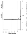
- FIG. 1 shows a case where a commercially available affinity separation matrix on which protein L is immobilized is used to purify a Fab fragment containing VL- ⁇ , and an acetic acid buffer solution or a citrate buffer solution having a pH of 3.0 is used as an eluent. Elution profile. A pH 2.4 citrate buffer is also used to dissociate the remaining Fab fragments in the matrix.
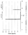
- FIG. 2 shows a case where a pH-3.0 acetate buffer or a citrate buffer is used as an eluent when purifying a Fab fragment containing VL- ⁇ using a prototype affinity separation matrix on which protein L is immobilized. Elution profile. A pH 2.4 citrate buffer is also used to dissociate the remaining Fab fragments in the matrix.
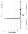
- FIG. 3 shows the case of using an acetate buffer or citrate buffer at pH 3.0 as an eluent when purifying Fab fragments containing VL- ⁇ using a prototype affinity separation matrix to which mutant protein L is immobilized. This is an elution profile.
- a pH 2.4 citrate buffer is also used to dissociate the remaining Fab fragments in the matrix.
- FIG. 4 shows a case where a commercially available affinity separation matrix on which protein L is immobilized is used to purify a Fab fragment containing VL- ⁇ , and an acetate buffer or a citrate buffer having a pH of 3.5 is used as an eluent. It is the figure which compared the elution profile.
- FIG. 5 shows a comparison of elution profiles when using a commercially available affinity separation matrix on which protein A is immobilized, and using an acetate buffer or citrate buffer having a pH of 3.0 as the eluent when purifying IgG. It is.
- a pH 2.4 citrate buffer is also used to dissociate the remaining IgG in the matrix.
- FIG. 6 is a diagram comparing elution profiles when using a commercially available affinity separation matrix on which protein A is immobilized and purifying IgG, using an acetic acid buffer solution or a citrate buffer solution at pH 3.5 as an eluent. It is.
- a pH 2.4 citrate buffer is also used to dissociate the remaining IgG in the matrix.
- the method of the present invention is a method for purifying antibodies and antibody fragments containing VL- ⁇ well without damaging them using an affinity separation matrix in which protein L, its domain, or a variant thereof is immobilized.
- the method of the present invention will be described step by step.
- Step 1 Adsorption step In this step, an affinity separation in which a liquid sample containing at least one of an antibody and a VL- ⁇ -containing antibody fragment is immobilized on an insoluble carrier as a protein L, its domain, or a variant thereof is used as a ligand.
- the antibody and / or antibody fragment is adsorbed to the affinity separation matrix by contacting with the matrix.
- Immunoglobulin (Ig) is a glycoprotein produced by B cells of lymphocytes and has a function of recognizing and binding molecules such as specific proteins.
- An immunoglobulin has a function of specifically binding to a specific molecule called an antigen and a function of detoxifying or removing a factor having the antigen in cooperation with other biomolecules or cells.
- Immunoglobulin is generally called “antibody”, which is a name that focuses on such a function.
- All immunoglobulins basically have the same molecular structure, and have a “Y” -shaped four-chain structure as a basic structure.
- the four-chain structure is composed of two polypeptide chains each called a light chain and a heavy chain.
- Immunoglobulin G is a monomeric immunoglobulin and is composed of two ⁇ chains and two light chains, and has two antigen-binding sites.
- the place corresponding to the vertical bar of the lower half of the “Y” of immunoglobulin is called the Fc region, and the “V” of the upper half is called the Fab region.
- the Fc region has an effector function that induces a reaction after the antibody binds to the antigen, and the Fab region has a function of binding to the antigen.
- the heavy chain Fab region and the Fc region are connected by a hinge part, and the proteolytic enzyme papain contained in papaya decomposes this hinge part and cleaves it into two Fab regions and one Fc region.
- the portion near the tip of the “Y” in the Fab region is called a variable region (V region) because various changes in the amino acid sequence are seen so that it can bind to various antigens.
- the variable region of the light chain is called the VL region, and the variable region of the heavy chain is called the VH region.
- the Fab region and the Fc region other than the V region are regions with relatively little change, and are called constant regions (C regions).
- the constant region of the light chain is referred to as the CL region, and the constant region of the heavy chain is referred to as the CH region.
- the CH region is further divided into three, CH1 to CH3.
- the heavy chain Fab region consists of a VH region and CH1, and the heavy chain Fc region consists of CH2 and CH3.
- the hinge part is located between CH1 and CH2.
- PpL binds to a variable region (VL- ⁇ ) in which the light chain is a ⁇ chain (Non-Patent Documents 5 to 7).
- the ligand of the affinity separation matrix according to the present invention is based on the sequence of protein L (PpL) and binds to the ⁇ chain variable region (VL- ⁇ ) of immunoglobulin.
- the antibody to be bound by the affinity separation matrix according to the present invention is not limited as long as it contains VL- ⁇ , and may be IgG containing the Fab region and the Fc region without deficiency, such as IgM, IgD, and IgA. Other Igs may also be used, or they may be derivatives of immunoglobulin molecules that have been modified by protein engineering.
- the antibody fragment to which the affinity separation matrix used in the present invention binds is not particularly limited as long as it is an antibody fragment having VL- ⁇ .
- Fab fragments fragmented only in the Fab region of immunoglobulin G, scFv and diabody consisting of only the variable region of immunoglobulin G, and partial domains of human immunoglobulin G as immunoglobulin G domains of other species examples thereof include chimeric immunoglobulin G fused by replacement, immunoglobulin G obtained by molecular modification of the sugar chain of the Fc region, and scFv fragment covalently bound to a drug.
- the liquid sample is not particularly limited as long as it contains at least one of the antibody to be purified and the antibody fragment containing VL- ⁇ , but the antibody and / or the antibody fragment containing VL- ⁇ is dissolved in an aqueous solvent.
- liquid samples include serum samples containing antibodies and / or VL- ⁇ -containing antibody fragments, bacterial cultures or homogenate supernatants containing VL- ⁇ -containing antibody fragments, and homogenates of monoclonal antibody-producing hybridomas.
- the pH of the liquid sample is preferably in the vicinity of neutrality of about 6 or more and 8 or less.
- the solvent of the body sample may be water alone, or may contain a water-miscible organic solvent such as C 1-4 alcohol as long as water is the main component, and the pH is 6 or more. A buffer solution of about 8 or less may be used.
- the affinity separation matrix used in the present invention is a protein L, its domain, or a variant thereof immobilized on an insoluble carrier as a ligand.
- protein L, a domain thereof, or a variant thereof may be collectively referred to as “VL- ⁇ binding peptide”.
- peptide includes all molecules having a polypeptide structure, and includes not only so-called proteins, but also fragments and those in which other peptides are linked by peptide bonds.
- a “domain” is a unit of protein conformation, consisting of a sequence of tens to hundreds of amino acid residues, sufficient to express some physicochemical or biochemical function.
- Protein L domain in the present invention refers to a protein showing affinity for VL- ⁇ .
- the “mutant” of protein L or domain is a protein or peptide in which at least one substitution, addition or deletion is introduced at the amino acid level with respect to the wild type protein L or domain sequence.
- the affinity to VL- ⁇ is at least maintained and preferably improved.
- the number of mutations is preferably 10 or less, more preferably 5 or less.
- Protein L is a protein derived from the cell wall of anaerobic gram-positive cocci belonging to the genus Peptostreptococcus.
- PpL derived from Peptostreptococcus magnus is preferable, and two types of PpL derived from Peptostreptococcus magnus 312 and Peptostreptococcus magnus 3316 are preferable, but are not limited thereto.
- PpL contains a plurality of VL- ⁇ binding domains consisting of 70 to 80 residues in a protein.
- the number of VL- ⁇ binding domains contained in PpL312 is five, and the number of VL- ⁇ binding domains contained in PpL3316 is four.
- the VL- ⁇ binding domain of PpL312 is called B1-5 domain in order from the N terminus, and the VL- ⁇ binding domain of PpL3316 is called C1-4 domain in order from the N terminus (Non-patent Documents 5-6).
- Non-patent Document 7 Studies have shown that about 20 residues at the N-terminus of the VL- ⁇ -binding domain of PpL do not have a specific secondary structure. Even when the N-terminus is deleted, VL- ⁇ binding It retains a three-dimensional structure as a sex domain and exhibits VL- ⁇ binding.
- the VL- ⁇ binding peptide according to the present invention also includes, as one embodiment, two or more, preferably three or more, more preferably 4 VL- ⁇ binding peptides that are monomers or single domains. It may be a multimer of multiple domains linked by one or more, more preferably five or more. The upper limit of the number of domains to be linked includes 10 or less, preferably 8 or less, more preferably 6 or less. These multimers may be homodimers such as homodimers and homotrimers that are linked to a single VL- ⁇ -binding peptide, or are linked to multiple types of VL- ⁇ -binding peptides. Heteromultimers such as heterodimers and heterotrimers may be used.
- a method of linking the monomeric VL- ⁇ binding peptide includes a method of linking with one or a plurality of amino acid residues, but is not limited to this method.
- the number of amino acid residues to be linked is not particularly limited, but is preferably 20 residues or less, and more preferably 15 residues or less.
- the ligand of the affinity separation matrix of the present invention includes a VL- ⁇ binding peptide or a multimer in which two or more VL- ⁇ binding domains are linked as one constituent component.
- a fusion peptide characterized in that it is fused with another peptide having a different function.
- fusion peptides include, but are not limited to, peptides fused with albumin or GST (glutathione S-transferase).
- a nucleic acid such as a DNA aptamer, a drug such as an antibiotic, and a polymer such as PEG (polyethylene glycol) are fused
- the present invention can be used if the affinity separation matrix obtained in the present invention is useful. Included in the invention.
- the VL- ⁇ binding peptide used in the present invention can be prepared by a conventional method. That is, a DNA encoding the amino acid sequence of a desired VL- ⁇ binding peptide or a fragment thereof is chemically synthesized, and the DNA encoding the VL- ⁇ binding peptide is amplified by PCR and incorporated into a vector such as a plasmid. The obtained vector is cultured after infecting Escherichia coli or the like, and a desired VL- ⁇ binding peptide may be purified from the cultured cells or culture solution by chromatography or the like.
- the affinity separation matrix used in the present invention is obtained by immobilizing the above VL- ⁇ binding peptide on an insoluble carrier.
- the “insoluble carrier” used in the present invention is insoluble in an aqueous solvent that is a solvent of a liquid sample containing a VL- ⁇ binding peptide, and specifically binds to the ligand by supporting the ligand. Those that can be used to purify antibodies and antibody fragments.
- insoluble carrier used in the present invention examples include inorganic carriers such as glass beads and silica gel; synthetic polymers such as crosslinked polyvinyl alcohol, crosslinked polyacrylate, crosslinked polyacrylamide, and crosslinked polystyrene; crystalline cellulose, crosslinked cellulose, crosslinked agarose, crosslinked Examples thereof include organic carriers composed of polysaccharides such as dextran; and organic-organic and organic-inorganic composite carriers obtained by combining these.
- GCL2000 a porous cellulose gel
- Sephacryl S-1000 in which allyl dextran and methylene bisacrylamide are covalently crosslinked
- Toyopearl an acrylate carrier
- Sepharose CL4B an agarose crosslinking carrier
- Cellufine which is a cellulosic crosslinking carrier.
- the water-insoluble carrier in the present invention is not limited to these exemplified carriers.
- the insoluble carrier used in the present invention preferably has a large surface area in view of the purpose and method of use of the affinity separation matrix, and is preferably a porous material having a large number of pores of an appropriate size.
- the form of the carrier can be any of beads, monoliths, fibers, membranes (including hollow fibers), and any form can be selected.
- a conventional method may be used as a method for immobilizing a VL- ⁇ binding peptide, which is a ligand, on an insoluble carrier.
- the reactive groups present on the surface of the insoluble carrier are used for immobilization.
- reactive groups such as amino groups, hydroxyl groups, and carboxy groups exist on the surface of general insoluble carriers, and these are activated, substituted with other reactive groups, or reacted with them.
- a linker group having a functional group may be introduced. For example, epichlorohydrin, diglycidyl ether, 1,4-bis (2,3-epoxypropoxy) butane etc.
- the linker group is not particularly limited.
- An ester group (—C ( ⁇ O) —O— or —O—C ( ⁇ O) —), an amide group (—C ( ⁇ O) —NH— or —NH—C ( ⁇ O) —), Sulfoxide group (—S ( ⁇ O) —), sulfonyl group (—S ( ⁇ O) 2 —), sulfonylamide group (—NH—S ( ⁇ O) 2 — and —S ( ⁇ O) 2 —NH— ), And a group in which
- a spacer molecule composed of a plurality of atoms may be introduced between the ligand and the carrier, or the ligand may be directly immobilized on the carrier.
- the VL- ⁇ binding peptide according to the present invention may be chemically modified for immobilization.
- the liquid sample and the affinity separation matrix are contacted to selectively bind the antibody and / or antibody fragment containing VL- ⁇ to the VL- ⁇ binding peptide as a ligand.
- the specific embodiment is not particularly limited, and the liquid sample and the affinity separation matrix may only be mixed.
- the affinity separation matrix according to the present invention is packed in a column and affinity is used.
- a liquid sample is passed through the affinity column, and the antibody and / or antibody fragment is selectively adsorbed to the VL- ⁇ binding peptide.
- the conditions for this step may be appropriately adjusted within a range in which the antibody and / or antibody fragment contained in the liquid sample is sufficiently adsorbed to the affinity separation matrix.
- the temperature in this step may be adjusted to 4 ° C. or higher and 40 ° C. or lower.
- the temperature is 4 ° C. or higher, the liquid sample having an appropriate fluidity can be passed through the matrix without freezing the liquid sample.
- the temperature is 40 ° C. or lower, the antibody and / or antibody fragment contained in the liquid sample can be purified with a low possibility of undergoing heat denaturation.
- the said temperature can be easily adjusted by utilizing a column jacket etc., when a chromatography system is used.
- Step 2 Washing step of affinity separation matrix
- the affinity separation matrix on which the antibody and / or antibody fragment is adsorbed and retained in Step 1 is washed to remove impurities other than the antibody and / or antibody fragment.
- the antibody and / or antibody fragment is adsorbed to the affinity separation matrix.
- washing solution used for washing the affinity separation matrix in this step a washing solution that does not interfere with the interaction between the antibody and / or antibody fragment and the VL- ⁇ binding peptide is used.
- a washing solution that does not interfere with the interaction between the antibody and / or antibody fragment and the VL- ⁇ binding peptide
- water or a buffer solution having a pH of 6 or more and 8 or less can be used as the cleaning solution.
- What is necessary is just to adjust the usage-amount of a washing
- Step 3 Separation of antibody and / or antibody fragment
- the antibody and / or antibody fragment is separated from the affinity separation matrix adsorbed with the antibody and / or antibody fragment using an acetate buffer as an eluent. To do.
- the purified antibody and / or antibody fragment can be obtained.
- an acetate buffer is used as an eluent for eluting the antibody and / or antibody fragment containing VL- ⁇ .
- the acetate buffer can efficiently elute the antibody and / or antibody fragment even when the pH is relatively high, and can reduce damage to the antibody and / or antibody fragment due to the eluate.
- an acetate buffer as an eluent, the above antibody and / or antibody fragment is favorably dissociated from the VL- ⁇ binding peptide.
- the above antibody and / or antibody fragment The elution peak of becomes sharp. As a result, the amount of the solution containing the antibody and / or antibody fragment is reduced, and further purification and drying of the antibody and / or antibody fragment is facilitated.
- the acetic acid buffer is obtained, for example, by mixing an aqueous solution of a salt of acetic acid and a weak base such as an aqueous sodium acetate solution and an aqueous acetic acid solution at a ratio that provides a desired pH.
- the pH of the acetate buffer is preferably 2.5 or more and 4.0 or less. If the said pH is 2.5 or more, the chemical change of the said antibody and / or antibody fragment, etc. can be suppressed more reliably. On the other hand, if the pH is 4.0 or less, the elution of the antibody and / or antibody fragment can be made more reliable.
- the pH is preferably 2.8 or more, more preferably 3.0 or more, more preferably 3.8 or less, and even more preferably 3.5 or less.
- the ion concentration of the acetate buffer also affects the elution of the antibody and / or antibody fragment.
- concentration of acetate ions in the acetate buffer is preferably 10 mM or more and 500 mM or less.
- the acetate ion concentration is 10 mM or more, the effect of sharpening the elution peak of the antibody and / or antibody fragment is easily obtained.
- the acetate ion concentration is 500 mM or less, pH adjustment after obtaining the above-obtained antibody and / or antibody fragment elution fraction becomes easier.
- the acetate ion concentration is more preferably 50 mM or more, still more preferably 80 mM or more, more preferably 400 mM or less or 300 mM or less, still more preferably 200 mM or less, and particularly preferably 150 mM or less.
- the conditions of this step may be appropriately adjusted within a range in which the antibody and / or antibody fragment adsorbed on the affinity separation matrix is sufficiently separated and eluted.
- the temperature in this step may be adjusted to 4 ° C. or higher and 40 ° C. or lower.
- the eluate or the like can be passed through the matrix with appropriate fluidity without freezing.
- the temperature is 40 ° C. or lower, the antibody and / or antibody fragment can be eluted with a low possibility of undergoing heat denaturation.
- the elution peak of the antibody and / or antibody fragment becomes sharp, and the amount of acetate buffer used as the eluate can be reduced.
- the amount of the acetate buffer used may be adjusted as appropriate. For example, it can be set to 2 times or more and 20 times or less the volume of the affinity separation matrix. If the said ratio is 2 times volume or more, the said antibody and / or antibody fragment can be eluted more reliably. On the other hand, when the ratio is 20 times or less, the amount of the fraction containing the antibody and / or antibody fragment can be reduced, and further purification is facilitated.
- the reference volume of the affinity separation matrix is the volume of the affinity separation matrix in a gel state that is in a suspended state and is tapped or allowed to stand until the volume does not decrease.
- Step 4 Post-treatment step In step 3 above, an acetate buffer solution of the antibody and / or antibody fragment is obtained.
- the antibody and / or antibody fragment may be further purified by salting out, chromatography, recrystallization or the like, or may be dried by freeze drying, spray drying, thin film drying or the like.
- Step 5 Regeneration step of affinity separation matrix
- the affinity separation matrix from which the antibody and / or antibody fragment has been separated in Step 3 above is regenerated by washing with an alkaline aqueous solution.
- this step does not necessarily have to be performed after the above step 3, and may be performed once in 3 times, once in 5 times, or once in 10 times. Absent. That is, this step is not necessarily performed in a state where the performance of the affinity separation matrix such as the binding capacity is maintained, and the frequency and conditions of the execution are also determined depending on the liquid sample containing the antibody and / or antibody fragment to be purified. Different.
- the “alkaline aqueous solution” used for the regeneration of the affinity separation matrix is an aqueous solution showing alkalinity to such an extent that the purpose of washing or sterilization can be achieved. More specifically, a sodium hydroxide aqueous solution of 0.01 M or more and 1.0 M or less or 0.01 N or more and 1.0 N or less is applicable, but is not limited thereto.
- the lower limit of the concentration is preferably 0.01M, more preferably 0.02M, and even more preferably 0.05M.
- the upper limit of the concentration of sodium hydroxide is preferably 1.0M, more preferably 0.5M, even more preferably 0.3M, still more preferably 0.2M, and even more preferably 0.1M.
- the alkaline aqueous solution is not necessarily a sodium hydroxide aqueous solution, but the pH is preferably 12 or more and 14 or less. Regarding the lower limit of pH, 12.0 or more is preferable, and 12.5 or more is more preferable. Regarding the upper limit of the pH, it is preferably 14 or less, more preferably 13.5 or less, and even more preferably 13.0 or less.
- the time for treating the affinity separation matrix that has undergone Step 3 with an alkaline aqueous solution is not particularly limited and may be adjusted as appropriate because the damage to the peptide varies depending on the concentration of the alkaline aqueous solution and the temperature during the treatment.
- the concentration of sodium hydroxide is 0.05M and the temperature at the time of immersion is room temperature
- the lower limit of the time for immersion in the alkaline aqueous solution is preferably 1 hour, more preferably 2 hours, more preferably 4 hours.
- the time is more preferable, and 20 hours is more preferable, but there is no particular limitation as long as the affinity separation matrix can be regenerated.
- the affinity separation matrix regenerated through this step can be used again in the above steps 1 to 3.
- Example 1 Preparation of commercially available affinity separation matrix As an affinity separation matrix capable of adsorbing antibody fragments containing the ⁇ chain variable region (VL- ⁇ ), HI Healthcare's “HiTrap Protein L 1 mL-gel” was obtained and a chromatography system Installed. Note that “1 mL-gel” means that the volume of the affinity separation matrix in the gel state in which the affinity separation matrix in the suspended state is tapped or allowed to stand until the volume does not decrease is 1 mL.
- a base sequence encoding the peptide is designed, and an artificial synthetic gene of DNA having a PstI recognition site at the 5 ′ end and an XbaI recognition site at the 3 ′ end is supplied by Eurofin Genomics Total synthesis by outsourcing to
- the expression plasmid after this subcloning was digested with restriction enzymes PstI and XbaI (Takara Bio), and the obtained DNA fragment was ligated to Brevibacillus expression vector pNCMO2 (Takara Bio) digested with the same restriction enzymes, Expression plasmids in which DNA encoding the amino acid sequence of the VL- ⁇ binding peptide was inserted into the Brevibacillus expression vector pNCMO2 were prepared.
- the ligation reaction was performed using a ligation reagent ("Ligation high" manufactured by TOYOBO) according to the protocol attached to the product, and Escherichia coli JM109 strain (Takara Bio) was used for plasmid preparation. .
- a ligation reagent ("Ligation high” manufactured by TOYOBO) according to the protocol attached to the product, and Escherichia coli JM109 strain (Takara Bio) was used for plasmid preparation. .
- Each expression plasmid DNA base sequence was confirmed using a DNA sequencer 3130xl Genetic Analyzer (Applied Biosystems). BigDye Terminator v. 1.1 Using a Cycling Sequencing Kit (Applied Biosystems) according to the attached protocol, each plasmid DNA was subjected to a sequencing PCR reaction, and the sequencing product was converted into a plasmid purification kit (Applied Biosystems, “BigDye XT Terminator Kit”). )) According to
- Brevibacillus choshinensis strain SP3 (Takara Bio Inc.) was transformed with the obtained plasmid, and a gene recombinant that secreted and produced the VL- ⁇ binding peptide was bred.
- This gene recombinant was added to 30 mL of A medium (polypeptone 3.0%, yeast extract 0.5%, glucose 3%, magnesium sulfate 0.01%, iron sulfate 0.001%, containing 60 ⁇ g / mL neomycin.
- the cells were cultured with shaking in manganese chloride (0.001%, zinc chloride 0.0001%) at 30 ° C. for 3 days. After the culture, the cells were separated by centrifuging the culture solution at 15,000 rpm and 25 ° C. for 5 minutes.
- VL- ⁇ binding peptide was purified by cation exchange chromatography using a cation exchange carrier (“UnoSphere S” manufactured by Bio-Rad). UnoSphere S was packed into a column (“Tricorn 10/200” manufactured by GE Healthcare Bioscience) and used.
- cation exchange buffer A 50 mM CH 3 COOH—CH 3 COONa, pH 4 0.0
- cation exchange buffer B 50 mM CH 3 COOH—CH 3 COONa, 1M
- the VL- ⁇ binding peptide was purified by anion exchange chromatography using an anion exchange carrier (“Nuvia Q” manufactured by Bio-Rad). Nuvia Q was packed into a column (“Tricorn 10/200” manufactured by GE Healthcare Bioscience) and used. Specifically, the fractionated VL- ⁇ binding peptide solution is dialyzed against anion exchange buffer A (50 mM Tris-HCl, pH 8.0) and equilibrated with anion exchange buffer A. The anion exchange buffer A and the anion exchange buffer B (50 mM Tris-HCl, 1.0 M NaCl, pH 8.0) were used after adding to the Nuvia Q column and washing with anion exchange buffer A.
- anion exchange buffer A 50 mM Tris-HCl, 1.0 M NaCl, pH 8.0
- VL- ⁇ -binding peptide eluted in the middle was fractionated with a salt concentration gradient.
- the separated VL- ⁇ binding peptide was dialyzed against ultrapure water, and an aqueous solution containing only the VL- ⁇ binding peptide was used as a final purified sample.
- protein purification by chromatography using the above-described column was performed using a chromatography system (“AKTA york 25 system” manufactured by GE Healthcare Bioscience).
- the prepared VL- ⁇ binding peptide was immobilized on a commercially available agarose carrier.
- N- [ ⁇ -Maleimidocaic acid] hydrazide.TFA (EMCH, Thermo Fisher Scientific) solution adjusted with a coupling buffer and adjusted to a concentration of 10 mM is added to a centrifuge tube containing a carrier, and the solution is mixed with 1 Reacted for hours. Thereafter, the carrier is transferred to a glass filter, 10 mL of washing buffer A (0.5 M ethanolamine, 0.5 M sodium chloride, pH 7.2), 10 mL of coupling buffer, and 10 mL of washing buffer A in this order. Was washed and allowed to stand at 25 ° C. for 15 minutes. Further, the carrier was washed with a coupling buffer (10 mL). The maleimide was provided to the support by the above operations.
- washing buffer A 0.5 M ethanolamine, 0.5 M sodium chloride, pH 7.2
- the carrier provided with maleimide was transferred to a centrifuge tube, a VL- ⁇ binding peptide solution was further added, and the carrier was reacted at 25 ° C. for 2 hours. Thereafter, the reacted carrier was transferred to a glass filter, and washed with 7 mL of coupling buffer to recover the unreacted VL- ⁇ binding peptide. Thereafter, 10 mL of washing buffer B (50 mM L-cysteine, 100 mM NaH 2 PO 4 -Na 2 HPO 4 , 0.5 M sodium chloride, pH 7.2), 10 mL of coupling buffer, and 10 mL of washing buffer B After washing the carrier in order, it was allowed to stand at 25 ° C. for 15 minutes.
- washing buffer B 50 mM L-cysteine, 100 mM NaH 2 PO 4 -Na 2 HPO 4 , 0.5 M sodium chloride, pH 7.2
- the absorbance at 280 nm of the collected unreacted VL- ⁇ binding peptide was measured with a spectrometer, and the amount of unreacted VL- ⁇ binding peptide was calculated from the extinction coefficient calculated from the amino acid sequence.
- the amount of immobilized VL- ⁇ -binding peptide was calculated from the difference between the charged amount of VL- ⁇ -binding peptide and the amount of unreacted VL- ⁇ -binding peptide quantified, and the ligand density was calculated from the volume of the carrier.
- the ligand density of prototype A was 10 mg / mL-gel.
- VL- ⁇ binding domain variants having the amino acid sequence of SEQ ID NO: 3 were linked with the interdomain connecting peptide contained in SEQ ID NO: 1, and a carrier-immobilizing active group was added to the C-terminus.
- a VL- ⁇ binding peptide was designed and prepared in the same manner as described above to prepare an affinity separation matrix (prototype B) immobilized on a carrier.
- the ligand density of prototype B was 12.5 mg / mL-gel.
- a 1 mL-gel portion of each of the prototype A and B affinity separation matrices was packed into a commercially available column (“Tricorn 5/50” manufactured by GE Healthcare).
- IgG-derived Fab fragment Fab fragment (IgG-Fab) was selected as a VL- ⁇ -containing peptide.
- a humanized monoclonal IgG preparation having VL- ⁇ was used as a raw material, and this was fragmented into a Fab fragment and an Fc fragment with papain, and only the Fab fragment was separated and purified.
- a preparation method of Fab derived from an anti-IgE monoclonal antibody (generic name “omalizumab”) is shown, but basically other monoclonal Fabs can be prepared by the same method.
- a humanized monoclonal IgG preparation (in the case of an anti-IgE monoclonal antibody, “Zolea” manufactured by Novartis Pharma) was added to a papain digestion buffer (0.1 M AcOH-AcONa, 2 mM EDTA, 1 mM cysteine, The solution was dissolved in pH 5.5), papain-immobilized agarose (“Papain Agarose from papalatex” manufactured by SIGMA) was added, and the mixture was incubated at 37 ° C. for about 8 hours while mixing with a rotator.
- a papain digestion buffer 0.1 M AcOH-AcONa, 2 mM EDTA, 1 mM cysteine
- papain-immobilized agarose (“Papain Agarose from papalatex” manufactured by SIGMA) was added, and the mixture was incubated at 37 ° C. for about 8 hours while mixing with a rotator.
- the Fab solution was recovered as a flow-through fraction by affinity chromatography using KANEK KanCapA TM column (Kaneka).
- the fractionated Fab solution was purified by gel filtration chromatography using a Superdex 75 10/300 GL column (standard buffer was used for equilibration and separation) to obtain a Fab solution. Chromatographic protein purification was performed using the AKTA york 25 system.
- Fab was eluted with a 100 mM acetate buffer with a linear pH gradient from pH 5.0 to pH 3.0.
- acetate buffer A 100 mM acetate buffer, pH 5.0
- acetate buffer B 100 mM acetate buffer
- the elution pH was evaluated from the Fab elution position in the step of increasing the concentration of the buffer solution (pH 3.0) linearly from 0% to 100%.
- the Fab elution pH when using a citrate buffer as a reference was adjusted to 20 CV after equilibrating the column with 5 CV citrate buffer A (100 mM citrate buffer, pH 5.0) as described above.
- prototype A As shown in Table 1, in any of the commercially available products, prototype A, and prototype B, 0.6 or more in the case of using the acetate buffer compared to the case of using the citrate buffer, It was found that the Fab elution pH was significantly increased. Since both the commercial product, prototype A and prototype B have the same tendency, the effect of improving the Fab elution pH by the acetate buffer is effective for the affinity separation matrix having both protein L and protein L variants as ligands. It is believed that there is.
- Example 2 (1) Elution experiment using pH 3.0 eluate An experiment was carried out in which Fab was adsorbed on an affinity separation matrix capable of adsorbing antibody fragments containing VL- ⁇ and then eluted.
- an affinity separation matrix capable of adsorbing antibody fragments containing VL- ⁇ as in Example 1 above, commercially available products (“HiTrap Protein L” manufactured by GE Healthcare) and prototype A and prototype B columns were used as AKTA school. 25. The following experiment was conducted at 25 ° C.
- FIG. 1 shows a chromatographic chart when an acetate buffer solution and a citrate buffer solution are used as eluents in a commercially available affinity separation matrix.
- citrate buffer When citrate buffer is used, Fab is almost completely eluted with 7.6 CV eluate, whereas when acetate buffer is used, Fab is almost completely eluted with 1.2 CV eluate. Eluted.
- Fig. 2 shows the results of the affinity separation matrix of prototype A.
- citrate buffer was used, Fab was not completely eluted even when the eluate was flowed at 10 CV, and Fab was eluted in the strongly washed fraction.
- Fab was eluted almost completely by flowing 1.5 CV of the eluate.
- Fig. 3 shows the result of the affinity separation matrix of prototype B.
- citrate buffer was used, Fab was not almost completely eluted even when the eluate was flowed at 10 CV, and Fab was eluted in the strongly washed fraction.
- Fab was almost completely eluted by flowing 1.2 CV of the eluate.
- Comparative Example 1 As a comparative experimental example, an antibody elution test was conducted using an acetate buffer solution and a citrate buffer solution as an eluent in another affinity separation matrix.
- affinity separation matrix a commercially available Protein A carrier (“HiTrap MabSelect SuRe” manufactured by GE Healthcare) (1 mL) generally used in antibody purification was used. Since Protein A carrier hardly adsorbs Fab, IgG was used as an antibody for the test.
- the aforementioned humanized monoclonal IgG preparation (anti-IgE monoclonal antibody, “Zolea” manufactured by Novartis Pharma) was used.
- Example 1 an antibody elution test was conducted in the same manner as in Example 1 (4) except that the above commercially available Protein A carrier was used instead of the Protein L carrier. Specifically, an AKTA york 25 system (GE Healthcare) was used as a chromatographic system, and a column packed with a commercially available Protein A carrier was connected. 1 mg of humanized monoclonal IgG was added to a column equilibrated with an equilibration buffer (20 mM NaH 2 PO 4 -Na 2 HPO 4 , 150 mM NaCl, pH 7.4).
- an equilibration buffer (20 mM NaH 2 PO 4 -Na 2 HPO 4 , 150 mM NaCl, pH 7.4
- IgG was eluted with a 100 mM acetate buffer with a pH linear gradient from pH 5.0 to pH 3.0. More specifically, after equilibrating the column with 5 CV of acetate buffer A (100 mM acetate buffer, pH 5.0), when passing 20 CV of buffer solution, acetate buffer B (100 mM acetate buffer). The elution pH was evaluated from the IgG elution position in the step of increasing the concentration of the buffer solution (pH 3.0) linearly from 0% to 100%.
- the IgG elution pH when using a reference citrate buffer was 20 CV after equilibrating the column with 5 CV citrate buffer A (100 mM citrate buffer, pH 5.0) as described above. Elution pH from the IgG elution position in the step of linearly increasing the concentration of citrate buffer B (100 mM citrate buffer, pH 2.4) from 0% to 100%. Evaluated. All the above operations were performed at a flow rate of 0.3 mL / min. The results of elution pH of the obtained humanized monoclonal IgG are summarized in Table 2.
- FIG. 5 shows a chromatograph chart in the case where an acetate buffer and a citrate buffer are used as eluents in a commercially available Protein A carrier. As shown in the results of FIG. 5, the elution curves were almost completely the same when citrate buffer was used and when acetate buffer was used. In either case, the elution solution was 1.2 CV. IgG was almost completely eluted, and there was no difference in the amount of eluate due to the difference between acetate buffer and citrate buffer.
- Comparative Example 3 After IgG was adsorbed on a commercially available Protein A carrier, an experiment was conducted to elute IgG. The pH of the eluate was changed from that in Comparative Example 2 above. A column packed with a commercially available Protein A carrier was connected to AKTA van 25. The following experiment was conducted at 25 ° C. As in Comparative Example 2, 3 mg of humanized monoclonal IgG was added to a column equilibrated with an equilibration buffer (20 mM NaH 2 PO 4 -Na 2 HPO 4 , 150 mM NaCl, pH 7.4).
- an equilibration buffer (20 mM NaH 2 PO 4 -Na 2 HPO 4 , 150 mM NaCl, pH 7.4
- the equilibration buffer was allowed to flow through the column at 10 CV (column volume), and then 100 mM acetate buffer (pH 3.5) was allowed to flow at 10 CV as the eluent. Further, 3 CV of equilibration buffer was flowed, and then 10 CV of strong washing solution (100 mM citrate buffer, pH 2.4) was flowed to elute IgG remaining on the column. Finally, 5 CV of equilibration buffer was flowed to complete the experiment. As a reference, an experiment using 100 mM citrate buffer pH 3.5 as an eluent was also conducted. FIG.
- FIG. 6 shows a chromatographic chart when an acetate buffer and a citrate buffer are used as eluents in a commercially available Protein A carrier.
- acetate buffer was used, IgG was almost completely eluted with 1.3 CV eluate, and when citrate buffer was used, IgG was almost completely eluted with 1.9 CV.
- citrate buffer was used, IgG was almost completely eluted with 1.9 CV.
- the amount of eluate was different between the acetate buffer and the citrate buffer, but the difference was small.
- the elution fraction amount is 0.6 CV when the acetate buffer is used as the eluate compared to the case where the citrate buffer is used as the eluate.
- the amount of elution fraction was reduced by only 32%.
Landscapes
- Chemical & Material Sciences (AREA)
- Organic Chemistry (AREA)
- Health & Medical Sciences (AREA)
- General Health & Medical Sciences (AREA)
- Biochemistry (AREA)
- Biophysics (AREA)
- Life Sciences & Earth Sciences (AREA)
- Genetics & Genomics (AREA)
- Medicinal Chemistry (AREA)
- Molecular Biology (AREA)
- Proteomics, Peptides & Aminoacids (AREA)
- Analytical Chemistry (AREA)
- Immunology (AREA)
- Chemical Kinetics & Catalysis (AREA)
- Peptides Or Proteins (AREA)
Abstract
本発明は、溶出液のpHを比較的高くできることから、抗体またはκ鎖可変領域を含む抗体断片にダメージを与えることなく、且つシャープな溶出ピークをもって、抗体またはκ鎖可変領域を含む抗体断片を良好に精製することができる方法を提供することを目的とする。本発明に係る抗体、または、κ鎖可変領域を含む抗体断片の精製方法は、上記抗体および抗体断片の少なくとも一方を含む液体試料を、プロテインL、そのドメイン、またはそれらの変異体がリガンドとして不溶性担体に固定化されているアフィニティ分離マトリックスに接触させることにより、上記抗体および/または抗体断片をアフィニティ分離マトリックスに吸着させる工程、上記アフィニティ分離マトリックスを洗浄し、上記抗体および/または抗体断片以外の不純物を除去する工程、および、酢酸緩衝液を使って、上記抗体および/または抗体断片が吸着された上記アフィニティ分離マトリックスから上記抗体および/または抗体断片を分離する工程を含むことを特徴とする。
Description
本発明は、抗体、または、κ鎖可変領域を含む抗体断片を、ダメージを与えることなく良好に精製することができる方法に関するものである。
タンパク質の重要な機能の一つとして、特定の分子に特異的に結合する機能が挙げられる。この機能は、生体内における免疫反応やシグナル伝達に重要な役割を果たす。この機能を有用物質の分離精製に利用する技術開発も盛んになされている。実際に産業的に利用されている一例として、抗体医薬を動物細胞培養物から一度に高い純度でキャプチャリングして精製するために利用されるプロテインAアフィニティ分離マトリックス(以下、プロテインAを「SpA」と略記する場合がある)が挙げられる(非特許文献1,2)。
一般的に、アフィニティ分離マトリックスを用いて抗体を精製する場合には、アフィニティ分離マトリックスのリガンドに抗体を選択的に結合させ、洗浄して不純物を除去した後、アフィニティ分離マトリックスから分離することが行われている。例えば特許文献1には、SpAアフィニティクロマトグラフィカラムから単量体モノクローナル抗体の画分を集めてSpA産物プールを形成させ、当該産物プールのpHを約3.5~約4.5とし、抗体の凝集を抑制する方法が記載されている、担体を溶出するためのバッファーとしては、シトラートとアセタートが例示されている。また、特許文献2,3には、抗体を溶出させるためのものではないが、抗体が吸着されたSpAを洗浄するための洗浄液として、クエン酸塩や酢酸塩などの水溶液を用いることが記載されている。
抗体医薬として開発されているのは基本的にモノクローナル抗体であり、組換え培養細胞技術などを用いて大量に生産されている。「モノクローナル抗体」とは、単一の抗体産生細胞に由来するクローンから得られた抗体を指す。現在上市されている抗体医薬のほとんどは、分子構造的には免疫グロブリンG(IgG)サブクラスである。また、免疫グロブリンを断片化した分子構造を有する抗体誘導体である断片抗体からなる抗体医薬も盛んに臨床開発されており、様々な断片抗体医薬の臨床開発が進んでいる(非特許文献3)。
抗体医薬製造工程における初期精製工程には、先述のSpAアフィニティ分離マトリックスが利用されている。しかし、SpAは基本的にIgGのFc領域に特異的に結合するタンパク質である。よって、Fc領域を含まない断片抗体は、SpAアフィニティ分離マトリックスを利用したキャプチャリングができない。従って、抗体医薬精製プロセスのプラットフォーム開発の観点から、IgGのFc領域を含まない断片抗体をキャプチャリング可能なアフィニティ分離マトリックスに対する産業的なニーズは高い。
IgGのFc領域以外に結合するペプチドはすでに複数知られている(非特許文献4)。それらの中でも、結合できる断片抗体フォーマットの種類の多さ、および、IgMやIgAなどにも結合可能という観点からは、抗原結合ドメインである可変領域に結合できるペプチドが最も好ましく、例えば、プロテインL(以下、プロテインLを「PpL」と略記する場合がある)がよく知られている。PpLは、複数のκ鎖可変領域結合ドメイン(以下、κ鎖可変領域を「VL-κ」と略記する場合がある)を含むタンパク質であり、個々のVL-κ結合性ドメインのアミノ酸配列は異なる。また、菌株の種類によっても、VL-κ結合性ドメインの数、および個々のアミノ酸配列は異なる。例えば、ペプトストレプトコッカス・マグヌス(Peptostreptococcus magnus)312株のPpLに含まれるVL-κ結合性ドメインの数は5個であり、ペプトストレプトコッカス・マグヌス株3316のPpLに含まれるVL-κ結合性ドメインの数は4個である(非特許文献5~7,特許文献4,5)。そして、それら計9個のVL-κ結合ドメインの中に、互いに同じアミノ酸配列であるドメインは無い。
Hober S.ら,J.Chromatogr.B,2007,848巻,40-47頁
Shukla A.A.ら,Trends Biotechnol.,2010,28巻,253-261頁
Nelson A.N.ら,Nat.Biotechnol.,2009,27巻,331-337頁
Bouvet P.J.,Int.J.Immunopharmac.,1994,16巻,419-424頁
Kastern W.ら,J.Biol.Chem.,1992,267巻,12820-12825頁
Murphy J.P.ら,Mol.Microbiol.,1994,12巻,911-920頁
Housden N.G.ら,Biochemical Society Transactions,2003,31巻,716-718頁
上述したように、SpAを含むアフィニティ分離マトリックスを用いた抗体の精製に関しては、リガンドや溶出条件などの検討が進んでいる。一方、PpLを含むアフィニティ分離マトリックスについては、十分な検討が為されているとは言い難い。
例えば、SpAを含むアフィニティ分離マトリックスの場合には、比較的高いpHで抗体を溶出することが検討されているが、PpLを含むアフィニティ分離マトリックスの場合には十分な検討はされておらず、低pHの溶出液により抗体やVL-κを含む抗体断片が化学変化を受けたり切断されたり、また、凝集し易くなるおそれがあった。その他、抗体や抗体断片をアフィニティ分離マトリックスから分離する場合、溶出のピークがシャープなものでなければ、他のピークと重複して純度が低下するのみならず、溶出画分の溶液量が増加し、溶液からの精製や、濃縮、乾燥に手間や時間がかかる。
そこで本発明は、溶出液のpHを比較的高くできることから、抗体またはκ鎖可変領域を含む抗体断片にダメージを与えることなく、且つシャープな溶出ピークをもって、抗体またはκ鎖可変領域を含む抗体断片を良好に精製することができる方法を提供することを目的とする。
本発明者らは、上記課題を解決するために鋭意研究を進めた。その結果、PpLを含むアフィニティ分離マトリックスを使って抗体またはVL-κ含有ペプチドを精製するに当たり溶出液として酢酸緩衝液を用いれば、溶出液のpHを比較的高くしてもシャープな溶出ピークをもって抗体や抗体断片を良好に精製できることを見出して、本発明を完成した。
以下、本発明を示す。
以下、本発明を示す。
[1] 抗体、または、κ鎖可変領域を含む抗体断片を精製するための方法であって、
上記抗体および抗体断片の少なくとも一方を含む液体試料を、プロテインL、そのドメイン、またはそれらの変異体がリガンドとして不溶性担体に固定化されているアフィニティ分離マトリックスに接触させることにより、上記抗体および/または抗体断片をアフィニティ分離マトリックスに吸着させる工程、
上記アフィニティ分離マトリックスを洗浄し、上記抗体および/または抗体断片以外の不純物を除去する工程、および、
酢酸緩衝液を使って、上記抗体および/または抗体断片が吸着された上記アフィニティ分離マトリックスから上記抗体および/または抗体断片を分離する工程を含むことを特徴とする方法。
上記抗体および抗体断片の少なくとも一方を含む液体試料を、プロテインL、そのドメイン、またはそれらの変異体がリガンドとして不溶性担体に固定化されているアフィニティ分離マトリックスに接触させることにより、上記抗体および/または抗体断片をアフィニティ分離マトリックスに吸着させる工程、
上記アフィニティ分離マトリックスを洗浄し、上記抗体および/または抗体断片以外の不純物を除去する工程、および、
酢酸緩衝液を使って、上記抗体および/または抗体断片が吸着された上記アフィニティ分離マトリックスから上記抗体および/または抗体断片を分離する工程を含むことを特徴とする方法。
[2] 上記酢酸緩衝液のpHを2.5以上、4.0以下とする上記[1]に記載の方法。
[3] 上記酢酸緩衝液における酢酸イオンの濃度を10mM以上、500mM以下とする上記[1]または[2]に記載の方法。
[4] 上記液体試料を上記アフィニティ分離マトリックスに接触させる際の温度を4℃以上、40℃以下にする上記[1]~[3]のいずれかに記載の方法。
[5] 上記アフィニティ分離マトリックスから上記抗体および/または抗体断片を分離する際の温度を4℃以上、40℃以下にする上記[1]~[4]のいずれかに記載の方法。
本発明方法においては、PpLを含むアフィニティ分離マトリックスから抗体またはVL-κ含有ペプチドを分離するに当たり、溶出液のpHを比較的高くできるために、上記抗体や抗体断片などに対するダメージを低減することができる。また、上記抗体や抗体断片の溶出ピークがシャープなものとなり、溶出画分の溶液量が低減されるので、溶出画分からの上記抗体や抗体断片のさらなる精製や、濃縮、乾燥が容易である。その結果、抗体またはVL-κ含有抗体断片の精製コストを抑制することが可能になる。
本発明方法は、プロテインL、そのドメイン、またはそれらの変異体を固定化したアフィニティ分離マトリックスを用い、VL-κを含む抗体や抗体断片を、ダメージを与えることなく良好に精製する方法である。以下、本発明方法を工程毎に説明する。
工程1: 吸着工程
本工程では、抗体およびVL-κ含有抗体断片の少なくとも一方を含む液体試料を、プロテインL、そのドメイン、またはそれらの変異体がリガンドとして不溶性担体に固定化されているアフィニティ分離マトリックスに接触させることにより、上記抗体および/または抗体断片をアフィニティ分離マトリックスに吸着させる。
本工程では、抗体およびVL-κ含有抗体断片の少なくとも一方を含む液体試料を、プロテインL、そのドメイン、またはそれらの変異体がリガンドとして不溶性担体に固定化されているアフィニティ分離マトリックスに接触させることにより、上記抗体および/または抗体断片をアフィニティ分離マトリックスに吸着させる。
「免疫グロブリン(Ig)」は、リンパ球のB細胞が産生する糖タンパク質であり、特定のタンパク質などの分子を認識して結合する働きを持つ。免疫グロブリンは、抗原と呼ばれるかかる特定分子に特異的に結合する機能と、他の生体分子や細胞と協同して抗原を有する因子を無毒化したり除去する機能を有する。免疫グロブリンは、一般的に「抗体」と呼ばれるが、それはこのような機能に着目した名称である。
全ての免疫グロブリンは、基本的には同じ分子構造からなり、“Y”字型の4本鎖構造を基本構造としている。当該4本鎖構造は、軽鎖および重鎖と呼ばれるポリペプチド鎖それぞれ2本ずつから構成される。軽鎖(L鎖)にはλ鎖とκ鎖の2種類があり、すべての免疫グロブリンはこのどちらかを持つ。重鎖(H鎖)には、γ鎖、μ鎖、α鎖、δ鎖、ε鎖という構造の異なる5種類があり、この重鎖の違いによって免疫グロブリンの種類(アイソタイプ)が変わる。免疫グロブリンG(IgG)は、単量体型の免疫グロブリンで、2本のγ鎖と2本の軽鎖から構成され、2箇所の抗原結合部位を持っている。
免疫グロブリンの“Y”字の下半分の縦棒部分にあたる場所をFc領域と呼び、上半分の“V”字の部分をFab領域と呼ぶ。Fc領域は抗体が抗原に結合した後の反応を惹起するエフェクター機能を有し、Fab領域は抗原と結合する機能を有する。重鎖のFab領域とFc領域はヒンジ部でつながっており、パパイヤに含まれるタンパク分解酵素パパインは、このヒンジ部を分解して2つのFab領域と1つのFc領域に切断する。Fab領域のうち“Y”字の先端に近い部分は、多様な抗原に結合できるように、アミノ酸配列に多彩な変化が見られるため、可変領域(V領域)と呼ばれている。軽鎖の可変領域をVL領域、重鎖の可変領域をVH領域と呼ぶ。V領域以外のFab領域とFc領域は、比較的変化の少ない領域であり、定常領域(C領域)と呼ばれる。軽鎖の定常領域をCL領域と呼び、重鎖の定常領域をCH領域と呼ぶが、CH領域はさらにCH1~CH3の3つに分けられる。重鎖のFab領域はVH領域とCH1からなり、重鎖のFc領域はCH2とCH3からなる。ヒンジ部はCH1とCH2の間に位置する。PpLは、軽鎖がκ鎖である可変領域(VL-κ)に結合する(非特許文献5~7)。
本発明に係るアフィニティ分離マトリックスのリガンドはプロテインL(PpL)の配列をベースとしており、免疫グロブリンのκ鎖可変領域(VL-κ)に結合する。本発明に係るアフィニティ分離マトリックスが結合すべき抗体は、VL-κを含むものであればよく、Fab領域とFc領域を不足なく含有するIgGであってもよいし、IgM、IgDおよびIgAなどの他のIg類であってもよいし、それらをタンパク質工学的に改変した免疫グロブリン分子の誘導体であってもよい。本発明で用いるアフィニティ分離マトリックスが結合する抗体断片は、VL-κを有する抗体断片であれば特に制限されない。例えば、免疫グロブリンGのFab領域のみに断片化されたFabフラグメント、免疫グロブリンGの可変領域のみからなるscFvやdaiabody、ヒト免疫グロブリンGの一部のドメインを他生物種の免疫グロブリンGのドメインに置き換えて融合させたキメラ型免疫グロブリンG、Fc領域の糖鎖に分子改変を加えた免疫グロブリンG、薬剤を共有結合したscFv断片などを挙げることができる。
上記液体試料は、精製すべき抗体およびVL-κ含有抗体断片の少なくとも一方を含むものであれば特に制限されないが、抗体および/またはVL-κ含有抗体断片が水系溶媒に溶解されているものであることが好ましい。液体試料としては、例えば、抗体および/またはVL-κ含有抗体断片を含む血清試料や、VL-κ含有抗体断片を含む菌体の培養液またはホモジェネートの上清、モノクローナル抗体産生ハイブリドーマのホモジェネートなどを挙げることができる。また、当該液体試料のpHは6以上8以下程度の中性付近であることが好ましい。当該体試料の溶媒は水のみでもよいし、また、水を主成分とするものであればC1-4アルコールなどの水混和性有機溶媒を含むものであってもよいし、pHが6以上8以下程度の緩衝液であってもよい。
本発明で用いるアフィニティ分離マトリックスは、プロテインL、そのドメイン、またはそれらの変異体がリガンドとして不溶性担体に固定化されているものである。以下、プロテインL、そのドメイン、またはそれらの変異体を、まとめて「VL-κ結合性ペプチド」という場合がある。
本発明において「ペプチド」とは、ポリペプチド構造を有するあらゆる分子を含むものであって、いわゆるタンパク質のみならず、断片化されたものや、ペプチド結合によって他のペプチドが連結されたものも包含されるものとする。「ドメイン」とは、タンパク質の高次構造上の単位であり、数十から数百のアミノ酸残基配列から構成され、なんらかの物理化学的または生物化学的な機能を発現するに十分なタンパク質の単位をいう。本発明における「プロテインLのドメイン」は、VL-κに対する親和性を示すものをいう。また、本発明においてプロテインLやドメインの「変異体」は、野生型のプロテインLやドメインの配列に対し、アミノ酸レベルで、少なくとも1つ以上の置換、付加または欠失が導入されたタンパク質またはペプチドであって、VL-κに対する親和性が少なくとも維持されており、好ましくは向上しているものをいう。変異の数としては、10以下が好ましく、5以下がさらに好ましい。
「プロテインL」(PpL)は、ペプトストレプトコッカス属(Peptostreptococcus)に属する嫌気性グラム陽性球菌の細胞壁に由来するタンパク質である。好ましくは、ペプトストレプトコッカス・マグヌス(Peptostreptococcus magnus)に由来するPpLであり、ペプトストレプトコッカス・マグヌス312株、および、ペプトストレプトコッカス・マグヌス3316株に由来する2種類のPpLが好ましいが、これらに限定されない(非特許文献4~6)。
PpLは、タンパク質中に70~80残基からなる複数のVL-κ結合性ドメインを含有する。PpL312に含まれるVL-κ結合性ドメインの数は5個であり、PpL3316に含まれるVL-κ結合性ドメインの数は4個である。PpL312のVL-κ結合性ドメインは、N末端から順にB1~5ドメインと呼び、PpL3316のVL-κ結合性ドメインは、N末端から順にC1~4ドメインと呼ぶ(非特許文献5~6)。
また、PpLのVL-κ結合性ドメインのN末端の約20残基は特定の二次構造を取らないことが研究によって分かっており、N末端を欠失させた場合にも、VL-κ結合性ドメインとして三次元構造を保持し、VL-κ結合性を示す(非特許文献7)。
PpLは、上述した通り、VL-κ結合性ドメインが4個または5個タンデムに並んだ形で含まれるタンパク質である。従って、本発明に係るVL-κ結合性ペプチドも、実施形態の1つとして、単量体または単ドメインであるVL-κ結合性ペプチドが2個以上、好ましくは3個以上、より好ましくは4個以上、より好ましくは5個以上連結された複数ドメインの多量体であってもよい。連結されるドメイン数の上限としては、10個以下が挙げられ、好ましくは8個以下、より好ましくは6個以下である。これらの多量体は、単一のVL-κ結合性ペプチドの連結体であるホモダイマー、ホモトリマー等のホモ多量体であってもよいし、複数種類のVL-κ結合性ペプチドの連結体であるヘテロダイマー、ヘテロトリマー等のヘテロ多量体であってもよい。
上記多量体において、単量体VL-κ結合性ペプチドの連結のされ方としては、1または複数のアミノ酸残基で連結する方法が挙げられるが、この方法に限定されるものではない。連結するアミノ酸残基数に特に制限は無いが、好ましくは20残基以下であり、より好ましくは15残基以下である。好ましくは、野生型PpLのB1とB2の間、B2とB3の間、B3とB4の間、B4とB5の間、または、C1とC2の間、C2とC3の間、C3とC4の間を連結している配列を利用するのがよい。また、別の観点からは、単量体VL-κ結合性ペプチドの3次元立体構造を不安定化しないものが好ましい。
また、実施形態の1つとして、本発明のアフィニティ分離マトリックスのリガンドとしては、VL-κ結合性ペプチド、または、VL-κ結合性ドメインが2個以上連結された多量体が、1つの構成成分として、機能の異なる他のペプチドと融合されていることを特徴とする融合ペプチドも挙げられる。融合ペプチドの例としては、アルブミンやGST(グルタチオンS-トランスフェラーゼ)が融合したペプチドを例として挙げることができるが、これに限定されるものではない。また、DNAアプタマーなどの核酸、抗生物質などの薬物、PEG(ポリエチレングリコール)などの高分子が融合されている場合も、本発明で得られたアフィニティ分離マトリックスに対して有用性があれば、本発明に包含される。
本発明で用いるVL-κ結合性ペプチドは、常法により調製することが可能である。すなわち、所望のVL-κ結合性ペプチドのアミノ酸配列またはその断片をコードするDNAを化学的に合成し、VL-κ結合性ペプチドをコードするDNAをPCRにより増幅し、プラスミドなどのベクターに組み込む。得られたベクターを大腸菌などに感染させた上で培養し、培養された菌体または培養液から所望のVL-κ結合性ペプチドをクロマトグラフィなどで精製すればよい。
本発明で用いるアフィニティ分離マトリックスは、上記VL-κ結合性ペプチドが不溶性担体に固定化されたものである。本発明で用いる「不溶性担体」とは、VL-κ結合性ペプチドを含む液体試料の溶媒である水系溶媒に対して不溶性を示し、且つリガンドを担持することにより、リガンドへ特異的に結合する上記抗体および抗体断片の精製に用いることができるものをいう。本発明で用いる不溶性担体としては、ガラスビーズ、シリカゲルなどの無機担体;架橋ポリビニルアルコール、架橋ポリアクリレート、架橋ポリアクリルアミド、架橋ポリスチレンなどの合成高分子や;結晶性セルロース、架橋セルロース、架橋アガロース、架橋デキストランなどの多糖類からなる有機担体;さらにはこれらの組み合わせによって得られる有機-有機、有機-無機などの複合担体などが挙げられる。市販品としては、多孔質セルロースゲルであるGCL2000、アリルデキストランとメチレンビスアクリルアミドを共有結合で架橋したSephacryl S-1000、アクリレート系の担体であるToyopearl、アガロース系の架橋担体であるSepharose CL4B、および、セルロース系の架橋担体であるCellufineなどを例示することができる。但し、本発明における水不溶性担体は、例示したこれらの担体のみに限定されるものではない。
また、本発明に用いる不溶性担体は、アフィニティ分離マトリックスの使用目的および方法からみて、表面積が大きいことが望ましく、適当な大きさの細孔を多数有する多孔質であることが好ましい。担体の形態としては、ビーズ状、モノリス状、繊維状、膜状(中空糸を含む)などいずれも可能であり、任意の形態を選ぶことができる。
本発明においてリガンドであるVL-κ結合性ペプチドを不溶性担体に固定化する方法としては常法を用いればよい。例えば、不溶性担体の表面に存在する反応性基を利用して固定化する。具体的には、一般的な不溶性担体の表面には、アミノ基、水酸基、カルボキシ基などの反応性基が存在し、これらを活性化したり、別の反応性基に置換したり、これらに反応性基を有するリンカー基を導入してもよい。例えば、エピクロロヒドリン、ジグリシジルエーテル、1,4-ビス(2,3-エポキシプロポキシ)ブタンなどを用いて水不溶性担体の表面にエポキシ基を導入したり、ヨードアセチル基、ブロモアセチル基、マレイミド基、N-ヒドロキシスクシンイミドエステル基などを導入すれば、VL-κ結合性ペプチドの反応性基との間でカップリング反応が容易に進行する。
水不溶性担体へのリガンドの固定化にリンカー基を用いる場合、当該リンカー基は特に制限されるものではないが、例えば、C1-6アルキレン基、アミノ基(-NH-)、イミノ基(>C=N-または-N=C<)、エーテル基(-O-)、チオエーテル基(-S-)、カルボニル基(-C(=O)-)、チオニル基(-C(=S)-)、エステル基(-C(=O)-O-または-O-C(=O)-)、アミド基(-C(=O)-NH-または-NH-C(=O)-)、スルホキシド基(-S(=O)-)、スルホニル基(-S(=O)2-)、スルホニルアミド基(-NH-S(=O)2-および-S(=O)2-NH-)、並びにこれら2以上が結合した基を挙げることができる。2以上のこれら基が結合して上記リンカー基が構成されている場合、当該結合数としては、10以下または5以下が好ましく、3以下がより好ましい。
また、リガンドと担体の間に複数の原子からなるスペーサー分子を導入してもよいし、担体にリガンドを直接固定化してもよい。また、固定化のために、本発明に係るVL-κ結合性ペプチドを化学修飾してもよい。
本工程では、上記液体試料と上記アフィニティ分離マトリックスとを接触させることにより、VL-κを含む抗体および/または抗体断片を、リガンドである上記VL-κ結合性ペプチドに選択的に結合させる。その具体的な態様は特に制限されず、上記液体試料と上記アフィニティ分離マトリックスとを混合するのみでもよいが、例えば、利便性の観点から、本発明に係るアフィニティ分離マトリックスをカラムに充填してアフィニティカラムとし、当該アフィニティカラムに液体試料を通過させ、VL-κ結合性ペプチドに上記抗体および/または抗体断片を選択的に吸着させることが好ましい。
本工程の条件は、上記液体試料に含まれる上記抗体および/または抗体断片が上記アフィニティ分離マトリックスに十分に吸着される範囲で適宜調整すればよい。例えば、本工程の温度は4℃以上、40℃以下に調整すればよい。当該温度が4℃以上であれば、上記液体試料が凍結することなく、適度な流動性をもった液体試料をマトリックス内に通液することが可能となる。一方、当該温度が40℃以下であれば、上記液体試料に含まれる上記抗体および/または抗体断片が熱変性を受ける可能性が低い状態で精製することが可能となる。なお、上記温度は、クロマトグラフィシステムを用いた場合、カラムジャケットなどを利用することによって容易に調整可能である。
工程2: アフィニティ分離マトリックスの洗浄工程
本工程では、上記工程1により上記抗体および/または抗体断片が吸着保持されたアフィニティ分離マトリックスを洗浄し、上記抗体および/または抗体断片以外の不純物を除去する。なお、この時点では、上記抗体および/または抗体断片はアフィニティ分離マトリックスに吸着されている。
本工程では、上記工程1により上記抗体および/または抗体断片が吸着保持されたアフィニティ分離マトリックスを洗浄し、上記抗体および/または抗体断片以外の不純物を除去する。なお、この時点では、上記抗体および/または抗体断片はアフィニティ分離マトリックスに吸着されている。
本工程においてアフィニティ分離マトリックスの洗浄に用いられる洗浄液としては、上記抗体および/または抗体断片とVL-κ結合性ペプチドとの相互作用を妨げないものを使用する。例えば、pHが6以上8以下の水や緩衝液を洗浄液として用いることができる。洗浄液の使用量は、アフィニティ分離マトリックスから不純物を十分に除去できる範囲で適宜調整すればよい。不純物が十分に除去できたか否かは、例えば、クロマトグラフィシステムを用いる場合、溶出プロファイルのモニターにより容易に判断することが可能である。
工程3: 抗体および/または抗体断片の分離工程
本工程では、溶出液として酢酸緩衝液を使って、上記抗体および/または抗体断片が吸着されたアフィニティ分離マトリックスから上記抗体および/または抗体断片を分離する。本工程によって、精製された上記抗体および/または抗体断片が得られる。
本工程では、溶出液として酢酸緩衝液を使って、上記抗体および/または抗体断片が吸着されたアフィニティ分離マトリックスから上記抗体および/または抗体断片を分離する。本工程によって、精製された上記抗体および/または抗体断片が得られる。
本発明では、VL-κを含む抗体および/または抗体断片を溶出するための溶出液として、酢酸緩衝液を用いる。当該酢酸緩衝液は、そのpHを比較的高くしても上記抗体および/または抗体断片の効率的な溶出が可能であり、上記抗体および/または抗体断片に対する溶出液によるダメージを軽減することができる。また、溶出液として酢酸緩衝液を用いることにより、上記抗体および/または抗体断片の上記VL-κ結合性ペプチドからの解離が良好になり、例えばカラムを用いた場合、上記抗体および/または抗体断片の溶出ピークがシャープなものとなる。その結果、上記抗体および/または抗体断片を含む画分の溶液量が低減され、上記抗体および/または抗体断片のさらなる精製や乾燥が容易になる。
酢酸緩衝液は、例えば、酢酸ナトリウム水溶液など酢酸と弱塩基との塩の水溶液と酢酸水溶液を、所望のpHが得られる割合で混合することにより得られる。酢酸緩衝液のpHとしては、2.5以上、4.0以下が好ましい。当該pHが2.5以上であれば、上記抗体および/または抗体断片の化学変化などをより確実に抑制することができる。一方、当該pHが4.0以下であれば、上記抗体および/または抗体断片の溶出をより確実なものとすることができる。当該pHとしては、2.8以上がより好ましく、3.0以上がよりさらに好ましく、また、3.8以下がより好ましく、3.5以下がよりさらに好ましい。
酢酸緩衝液のイオン濃度も、上記抗体および/または抗体断片の溶出に影響を与える。例えば、酢酸緩衝液の酢酸イオンの濃度としては、10mM以上、500mM以下が好ましい。当該酢酸イオン濃度が10mM以上であれば、上記抗体および/または抗体断片の溶出ピークがシャープとなる効果が得られやすくなる。一方、当該酢酸イオン濃度が500mM以下であれば、得られる上記抗体および/または抗体断片の溶出画分取得後のpH調整がより容易になる。当該酢酸イオン濃度としては、50mM以上がより好ましく、80mM以上がよりさらに好ましく、また、400mM以下または300mM以下がより好ましく、200mM以下がよりさらに好ましく、150mM以下が特に好ましい。
本工程の条件は、上記アフィニティ分離マトリックスに吸着された上記抗体および/または抗体断片が十分に分離して溶出される範囲で適宜調整すればよい。例えば、本工程の温度は4℃以上、40℃以下に調整すればよい。当該温度が4℃以上であれば、溶出液などが凍結することなく、適度な流動性をもってマトリックス内に通液することが可能となる。一方、当該温度が40℃以下であれば、上記抗体および/または抗体断片が熱変性を受ける可能性が低い状態で溶出することが可能となる。
上述した通り、本発明においては、上記抗体および/または抗体断片の溶出ピークがシャープなものとなり、溶出液である酢酸緩衝液の使用量を低減することができる。当該酢酸緩衝液の使用量は適宜調整すればよいが、例えば、アフィニティ分離マトリックスの体積の2倍体積以上、20倍体積以下とすることができる。当該割合が2倍体積以上であれば、上記抗体および/または抗体断片をより確実に溶出することができる。一方、当該割合が20倍体積以下であれば、上記抗体および/または抗体断片を含む画分の量を低減することができ、さらなる精製が容易となる。上記割合としては、15倍体積以下がより好ましく、10倍体積以下または8倍体積以下がよりさらに好ましく、5倍体積以下が特に好ましい。なお、基準となるアフィニティ分離マトリックスの体積は、懸濁状態であり、その体積が減少しなくなるまでタッピングまたは静置したゲル状態のアフィニティ分離マトリックスの体積とする。
工程4: 後処理工程
上記工程3により、上記抗体および/または抗体断片の酢酸緩衝液溶液が得られる。上記抗体および/または抗体断片は、塩析、クロマトグラフィ、再結晶などによりさらに精製してもよいし、凍結乾燥、スプレードライ、薄膜乾燥法などにより乾燥してもよい。
上記工程3により、上記抗体および/または抗体断片の酢酸緩衝液溶液が得られる。上記抗体および/または抗体断片は、塩析、クロマトグラフィ、再結晶などによりさらに精製してもよいし、凍結乾燥、スプレードライ、薄膜乾燥法などにより乾燥してもよい。
工程5: アフィニティ分離マトリックスの再生工程
本工程では、上記工程3において上記抗体および/または抗体断片を分離したアフィニティ分離マトリックスをアルカリ性水溶液で洗浄することによって再生する。但し、本工程は、上記工程3の後に必須的に実施する必要はなく、上記工程1~3の繰り返しの3回に1回、5回に1回、または10回に1回の実施でも構わない。すなわち、結合容量などアフィニティ分離マトリックスの性能が維持されている状態では本工程は必ずしも実施する必要はなく、精製対象である上記抗体および/または抗体断片を含む液体試料によってもその実施頻度や条件が異なる。
本工程では、上記工程3において上記抗体および/または抗体断片を分離したアフィニティ分離マトリックスをアルカリ性水溶液で洗浄することによって再生する。但し、本工程は、上記工程3の後に必須的に実施する必要はなく、上記工程1~3の繰り返しの3回に1回、5回に1回、または10回に1回の実施でも構わない。すなわち、結合容量などアフィニティ分離マトリックスの性能が維持されている状態では本工程は必ずしも実施する必要はなく、精製対象である上記抗体および/または抗体断片を含む液体試料によってもその実施頻度や条件が異なる。
アフィニティ分離マトリックスの再生に用いる「アルカリ性水溶液」は、洗浄や殺菌などの目的を達成し得る程度のアルカリ性を示す水溶液である。より具体的には、0.01M以上1.0M以下、または、0.01N以上1.0N以下の水酸化ナトリウム水溶液などが該当するが、これに限定されるものではない。水酸化ナトリウムを例とした場合、その濃度の下限は、0.01Mが好ましく、0.02Mがより好ましく、0.05Mがさらにより好ましい。一方、水酸化ナトリウムの濃度の上限は、1.0Mが好ましく、0.5Mがより好ましく、0.3Mがさらにより好ましく、0.2Mがさらにより好ましく、0.1Mがさらにより好ましい。アルカリ性水溶液としては、水酸化ナトリウム水溶液である必要はないが、そのpHは12以上14以下が好ましい。pHの下限に関し、12.0以上が好ましく、12.5以上がより好ましい。pHの上限に関し、14以下が好ましく、13.5以下がさらにより好ましく、13.0以下がさらにより好ましい。
上記工程3を経たアフィニティ分離マトリックスをアルカリ性水溶液により処理する時間は、アルカリ性水溶液の濃度や処理時の温度によってペプチドの受けるダメージは異なるので、特に限定はされず、適宜調整すればよい。例えば、水酸化ナトリウムの濃度が0.05Mで、浸漬時の温度が室温の場合、アルカリ性水溶液に浸漬する時間の下限は、1時間が好ましく、2時間がより好ましく、4時間がより好ましく、10時間がより好ましく、20時間がより好ましいが、アフィニティ分離マトリックスの再生が可能な条件であれば特に限定はされない。
本工程を経て再生されたアフィニティ分離マトリックスは、再び上記工程1~3で使用し得る。
本願は、2016年5月9日に出願された日本国特許出願第2016-94178号に基づく優先権の利益を主張するものである。2016年5月9日に出願された日本国特許出願第2016-94178号の明細書の全内容が、本願に参考のため援用される。
以下、実施例に基づいて本発明をより詳細に説明するが、本発明はこれらの実施例に限定されるものではない。
実施例1
(1) 市販アフィニティ分離マトリックスの準備
κ鎖可変領域(VL-κ)を含む抗体断片を吸着可能なアフィニティ分離マトリックスとして、GEヘルスケア社の「HiTrap Protein L 1mL-gel」を入手し、クロマトグラフィシステムに設置した。なお、「1mL-gel」とは、懸濁状態のアフィニティ分離マトリックスを体積が減少しなくなるまでタッピングまたは静置したゲル状態のアフィニティ分離マトリックスの体積が1mLであることをいう。
(1) 市販アフィニティ分離マトリックスの準備
κ鎖可変領域(VL-κ)を含む抗体断片を吸着可能なアフィニティ分離マトリックスとして、GEヘルスケア社の「HiTrap Protein L 1mL-gel」を入手し、クロマトグラフィシステムに設置した。なお、「1mL-gel」とは、懸濁状態のアフィニティ分離マトリックスを体積が減少しなくなるまでタッピングまたは静置したゲル状態のアフィニティ分離マトリックスの体積が1mLであることをいう。
(2) アフィニティ分離マトリックスの試作
上記市販アフィニティ分離マトリックス以外のアフィニティ分離マトリックスにおける酢酸緩衝液の効果を確認するため、VL-κを含む抗体断片を吸着可能なアフィニティ分離マトリックスを試作した。具体的には、ペプトストレプトコッカス・マグヌス312株由来の野生型プロテインL(配列番号1)に含まれるVL-κ結合性ドメイン(配列番号2)4個を、配列番号1に含まれるドメイン間連結ペプチドで連結し、さらにC末端に担体への固定化活性基を付与したVL-κ結合性ペプチドを設計した。設計したアミノ酸配列から逆翻訳を行い、当該ペプチドをコードする塩基配列を設計し、5’末端にPstI認識サイト、3’末端にXbaI認識サイトを付与したDNAの人工合成遺伝子を、ユーロフィンジェノミクス社への外注によって全合成した。このサブクローニング後の発現プラスミドを、制限酵素PstIおよびXbaI(タカラバイオ社)で消化し、取得したDNA断片を、同じ制限酵素で消化したブレビバチルス発現用ベクターpNCMO2(タカラバイオ社)へライゲーションし、上記VL-κ結合性ペプチドのアミノ酸配列をコードするDNAがブレビバチルス発現用ベクターpNCMO2に挿入された発現プラスミドをそれぞれ調製した。なおライゲーション反応は、ライゲーション試薬(TOYOBO社製「Ligation high」)を用いて、製品に添付のプロトコルに準ずる形で実施し、プラスミドの調製にはエシェリヒア・コリJM109株(タカラバイオ社)を用いた。各々の発現プラスミドDNA塩基配列の確認は、DNAシークエンサー3130xl Genetic Analyzer(Applied Biosystems社)を用いて行った。BigDye Terminator v.1.1 Cycle Sequencing Kit(Applied Biosystems社)を用いて、付属のプロトコルに従い、各々のプラスミドDNAのシークエンシングPCR反応を行い、そのシークエンシング産物をプラスミド精製キット(Applied Biosystems社,「BigDye XTerminator Purification Kit」)を用いて添付のプロトコルに従い精製し、配列解析に用いた。
上記市販アフィニティ分離マトリックス以外のアフィニティ分離マトリックスにおける酢酸緩衝液の効果を確認するため、VL-κを含む抗体断片を吸着可能なアフィニティ分離マトリックスを試作した。具体的には、ペプトストレプトコッカス・マグヌス312株由来の野生型プロテインL(配列番号1)に含まれるVL-κ結合性ドメイン(配列番号2)4個を、配列番号1に含まれるドメイン間連結ペプチドで連結し、さらにC末端に担体への固定化活性基を付与したVL-κ結合性ペプチドを設計した。設計したアミノ酸配列から逆翻訳を行い、当該ペプチドをコードする塩基配列を設計し、5’末端にPstI認識サイト、3’末端にXbaI認識サイトを付与したDNAの人工合成遺伝子を、ユーロフィンジェノミクス社への外注によって全合成した。このサブクローニング後の発現プラスミドを、制限酵素PstIおよびXbaI(タカラバイオ社)で消化し、取得したDNA断片を、同じ制限酵素で消化したブレビバチルス発現用ベクターpNCMO2(タカラバイオ社)へライゲーションし、上記VL-κ結合性ペプチドのアミノ酸配列をコードするDNAがブレビバチルス発現用ベクターpNCMO2に挿入された発現プラスミドをそれぞれ調製した。なおライゲーション反応は、ライゲーション試薬(TOYOBO社製「Ligation high」)を用いて、製品に添付のプロトコルに準ずる形で実施し、プラスミドの調製にはエシェリヒア・コリJM109株(タカラバイオ社)を用いた。各々の発現プラスミドDNA塩基配列の確認は、DNAシークエンサー3130xl Genetic Analyzer(Applied Biosystems社)を用いて行った。BigDye Terminator v.1.1 Cycle Sequencing Kit(Applied Biosystems社)を用いて、付属のプロトコルに従い、各々のプラスミドDNAのシークエンシングPCR反応を行い、そのシークエンシング産物をプラスミド精製キット(Applied Biosystems社,「BigDye XTerminator Purification Kit」)を用いて添付のプロトコルに従い精製し、配列解析に用いた。
得られたプラスミドによりブレビバチルス・チョウシネンシスSP3株(タカラバイオ社)を形質転換し、上記VL-κ結合性ペプチドを分泌生産する遺伝子組換え体を育種した。この遺伝子組換え体を、60μg/mLのネオマイシンを含む30mLのA培地(ポリペプトン3.0%,酵母エキス0.5%,グルコース3%,硫酸マグネシウム0.01%,硫酸鉄0.001%,塩化マンガン0.001%,塩化亜鉛0.0001%)にて、30℃で3日間振盪培養した。培養後、培養液を15,000rpm、25℃で5分間遠心分離することにより菌体を分離した。
得られた培養上清から、陽イオン交換担体(バイオラッド社製「UnoSphere S」)を利用した陽イオン交換クロマトグラフィにて上記VL-κ結合性ペプチドを精製した。UnoSphere Sはカラム(GEヘルスケアバイオサイエンス社製「Tricorn 10/200」)に充填して使用した。具体的には、酢酸ナトリウムを終濃度50mMになるように添加し、さらに酢酸でpH4.0に調整した培養上清を、陽イオン交換用緩衝液A(50mM CH3COOH-CH3COONa,pH4.0)にて平衡化したUnoSphere Sカラムに添加し、陽イオン交換用緩衝液Aで洗浄後、陽イオン交換緩衝液Aと陽イオン交換緩衝液B(50mM CH3COOH-CH3COONa,1M NaCl,pH4.0)を利用した塩濃度勾配にて、途中に溶出されるVL-κ結合性ペプチドを分取した。次に、陰イオン交換担体(バイオラッド社製「Nuvia Q」)を利用した陰イオン交換クロマトグラフィにて上記VL-κ結合性ペプチドを精製した。Nuvia Qはカラム(GEヘルスケアバイオサイエンス社製「Tricorn 10/200」)に充填して使用した。具体的には、分取した上記VL-κ結合性ペプチド溶液を、陰イオン交換用緩衝液A(50mM Tris-HCl,pH8.0)に透析し、陰イオン交換用緩衝液Aにて平衡化したNuvia Qカラムに添加し、陰イオン交換用緩衝液Aで洗浄後、陰イオン交換緩衝液Aと陰イオン交換緩衝液B(50mM Tris-HCl,1.0M NaCl,pH8.0)を利用した塩濃度勾配にて、途中に溶出される上記VL-κ結合性ペプチドを分取した。分取した上記VL-κ結合性ペプチドを超純水に透析し、上記VL-κ結合性ペプチドのみを含む水溶液を最終精製サンプルとした。なお、上記のカラムを用いたクロマトグラフィによるタンパク質精製は、クロマトグラフィシステム(GEヘルスケアバイオサイエンス社製「AKTAavant 25システム」)を利用して実施した。調製したVL-κ結合性ペプチドを、市販のアガロース担体へ固定化した。
具体的には、まず、市販のNHS活性化担体(GEヘルスケアバイオサイエンス社「NHS Activated Sepharose 4 Fast Flow」)1.5mL-gelをガラスフィルターに移し、保存溶液であるイソプロパノールを吸引除去した後、氷冷した1mM塩酸(5mL)で洗浄した。続いて、カップリング緩衝液(20mM NaH2PO4-Na2HPO4,150mM塩化ナトリウム,pH7.2)5mLで担体を洗浄した後、カップリング緩衝液に懸濁させながら担体を回収し遠沈管に移した。カップリング緩衝液で溶解し、10mMの濃度に調整したN-[ε-Maleimidocaproic acid]hydrazide・TFA(EMCH,サーモフィッシャーサイエンフィティック社)溶液を担体の入った遠沈管に加え、25℃で1時間反応させた。その後、担体をガラスフィルターに移し、洗浄緩衝液A(0.5Mエタノールアミン,0.5M塩化ナトリウム,pH7.2)を10mL、カップリング緩衝液を10mL、洗浄緩衝液Aを10mLの順で担体を洗浄し、25℃で15分間静置した。さらにカップリング緩衝液(10mL)で担体を洗浄した。ここまでの操作で担体にマレイミドを付与した。
次に、マレイミドを付与した担体を遠沈管に移し、さらにVL-κ結合性ペプチド溶液を加えて担体を25℃で2時間反応させた。その後、反応させた担体をガラスフィルターに移し、7mLのカップリング緩衝液で洗浄することで未反応の上記VL-κ結合性ペプチドを回収した。その後、洗浄緩衝液B(50mM L-システイン,100mM NaH2PO4-Na2HPO4,0.5M塩化ナトリウム,pH7.2)を10mL、カップリング緩衝液を10mL,洗浄緩衝液Bを10mLの順で担体を洗浄した後、25℃で15分間静置した。さらに、カップリング緩衝液10mL、超純水10mL、20%エタノール10mLで担体を洗浄した後、20%エタノール担体を懸濁、回収することにより、上記VL-κ結合性ペプチドをリガンドとするアフィニティ分離マトリックス(試作A)を得た。
回収した未反応VL-κ結合性ペプチドの280nmの吸光度を分光計で測定し、アミノ酸配列から算出した吸光係数から未反応VL-κ結合性ペプチドの量を算出した。VL-κ結合性ペプチドの仕込み量と定量した未反応VL-κ結合性ペプチドの量の差からVL-κ結合性ペプチドの固定化量を算出し、さらに担体の体積からリガンド密度を算出した。その結果、試作Aのリガンド密度は10mg/mL-gelであった。
また、配列番号3のアミノ酸配列を有するVL-κ結合性ドメイン変異体4個を、配列番号1に含まれるドメイン間連結ペプチドで連結し、さらにC末端に担体への固定化活性基を付与したVL-κ結合性ペプチドを設計し、上記と同様に調製して担体に固定化したアフィニティ分離マトリックス(試作B)を作製した。試作Bのリガンド密度は12.5mg/mL-gelであった。
試作Aおよび試作Bのアフィニティ分離マトリックスのそれぞれ1mL-gel分を市販のカラム(GEヘルスケア社製「Tricorn5/50」)に充填した。
(3) IgG由来Fabフラグメントの調製
VL-κ含有ペプチドとしてFabフラグメント(IgG-Fab)を選択した。VL-κを有するヒト化モノクローナルIgG製剤を原料として、これをパパインによってFabフラグメントとFcフラグメントに断片化し、Fabフラグメントのみを分離精製することで調製した。ここでは、抗IgEモノクローナル抗体(一般名「オマリズマブ」)由来のFabの調製方法を示すが、基本的には他のモノクローナルFabも同様の方法で調製が可能である。
VL-κ含有ペプチドとしてFabフラグメント(IgG-Fab)を選択した。VL-κを有するヒト化モノクローナルIgG製剤を原料として、これをパパインによってFabフラグメントとFcフラグメントに断片化し、Fabフラグメントのみを分離精製することで調製した。ここでは、抗IgEモノクローナル抗体(一般名「オマリズマブ」)由来のFabの調製方法を示すが、基本的には他のモノクローナルFabも同様の方法で調製が可能である。
具体的には、ヒト化モノクローナルIgG製剤(抗IgEモノクローナル抗体の場合には、ノバルティスファーマ社製の「ゾレア」)を、パパイン消化用緩衝液(0.1M AcOH-AcONa,2mM EDTA,1mMシステイン,pH5.5)に溶解し、パパイン固定化アガロース(SIGMA社製「Papain Agarose from papaya latex」)を添加し、ローテーターで混和させながら、37℃で約8時間インキュベートした。パパイン固定化アガロースから分離した反応溶液(FabフラグメントとFcフラグメントが混在)から、KANEKA KanCapATMカラム(カネカ社)を利用したアフィニティクロマトグラフィにより、素通り画分としてFab溶液を回収した。分取したFab溶液を、Superdex 75 10/300 GLカラム(平衡化および分離には標準緩衝液を使用)を用いたゲルろ過クロマトグラフィにて精製し、Fab溶液を得た。クロマトグラフィによるタンパク質精製は、AKTAavant 25システムを利用して実施した。
(4) 溶出試験
VL-κを含む抗体断片を吸着可能なアフィニティ分離マトリックスにFabを吸着した後、pHグラジエントによりFabの溶出ピークトップにおけるpHを解析した。VL-κを含む抗体断片を吸着可能なアフィニティ分離マトリックスとしては、(1)で準備をした市販品(GEヘルスケア社製「HiTrap Protein L)と、(2)で作製した試作Aおよび試作Bを使用した。以下の操作は25℃で行った。
VL-κを含む抗体断片を吸着可能なアフィニティ分離マトリックスにFabを吸着した後、pHグラジエントによりFabの溶出ピークトップにおけるpHを解析した。VL-κを含む抗体断片を吸着可能なアフィニティ分離マトリックスとしては、(1)で準備をした市販品(GEヘルスケア社製「HiTrap Protein L)と、(2)で作製した試作Aおよび試作Bを使用した。以下の操作は25℃で行った。
具体的には、クロマトシステムとしては、AKTAavant 25システム(GEヘルスケア社)を利用し、市販品および試作A、試作Bのカラムを接続した。平衡化緩衝液(20mM NaH2PO4-Na2HPO4,150mM NaCl,pH7.4)で平衡化したカラムに、(3)で調製したFabを1mg添加した。平衡化緩衝液を3CV(カラムボリューム)流した後、100mMの酢酸緩衝液でpH5.0からpH3.0へのpH直線勾配にてFabを溶出させた。より具体的には、カラムを5CVの酢酸緩衝液A(100mM 酢酸緩衝液,pH5.0)で平衡化させた後、20CV分の緩衝液を通液する際に、酢酸緩衝液B(100mM 酢酸緩衝液,pH3.0)の濃度を0%から100%に直線的に上げていく工程にて、Fab溶出位置から溶出pHを評価した。参照となるクエン酸緩衝液を用いた場合のFab溶出pHは、前述と同様に、5CVのクエン酸緩衝液A(100mM クエン酸緩衝液,pH5.0)でカラムを平衡化させた後、20CV分の緩衝液を通液する際に、クエン酸緩衝液B(100mM クエン酸緩衝液,pH2.4)の濃度を0%から100%に直線的に上げる工程にて、Fab溶出位置から溶出pHを評価した。上記の操作はすべて流速0.3mL/minで行った。得られたFabの溶出pHの結果について表1にまとめた。

表1に示す結果の通り、市販品、試作A、試作Bのいずれにおいても、クエン酸緩衝液を使用した場合と比較して、酢酸緩衝液を用いた場合の方が0.6以上と、大幅にFab溶出pHが高くなることが判明した。市販品、試作A、試作Bのいずれにおいても同様の傾向があるため、酢酸緩衝液によるFab溶出pHの向上効果は、プロテインLおよびプロテインL変異体の両方をリガンドとするアフィニティ分離マトリックスについて有効であると考えられる。
実施例2
(1) pH3.0の溶出液を用いた溶出実験
VL-κを含む抗体断片を吸着可能なアフィニティ分離マトリックスにFabを吸着した後、Fabを溶出させる実験を行った。VL-κを含む抗体断片を吸着可能なアフィニティ分離マトリックスとしては、上記実施例1と同様に、市販品(GEヘルスケア社製「HiTrap Protein L」)と、試作Aおよび試作BのカラムをAKTAavant 25に接続した。以下の実験は25℃で行った。
(1) pH3.0の溶出液を用いた溶出実験
VL-κを含む抗体断片を吸着可能なアフィニティ分離マトリックスにFabを吸着した後、Fabを溶出させる実験を行った。VL-κを含む抗体断片を吸着可能なアフィニティ分離マトリックスとしては、上記実施例1と同様に、市販品(GEヘルスケア社製「HiTrap Protein L」)と、試作Aおよび試作BのカラムをAKTAavant 25に接続した。以下の実験は25℃で行った。
平衡化緩衝液(20mM NaH2PO4-Na2HPO4,150mM NaCl,pH7.4)で平衡化したカラムに、実施例1(3)で調製したFabを3mg添加した。平衡化緩衝液をカラムに10CV(カラムボリューム)流した後、溶出液として100mMの酢酸緩衝液(pH3.0)を10CV流した。さらに平衡化緩衝液を3CV流した後、強洗浄液(100mM クエン酸緩衝液,pH2.4)を10CV流し、カラムに残存しているFabを溶出させた。最後に平衡化緩衝液を5CV流して実験を終了した。参照として、溶出液として100mM クエン酸緩衝液 pH3.0を用いた実験も行った。
図1に、市販品のアフィニティ分離マトリックスにおいて酢酸緩衝液およびクエン酸緩衝液を溶出液として使用した場合のクロマトチャートを示す。クエン酸緩衝液を使用した場合は、7.6CVの溶出液でFabがほぼ完全に溶出するのに対して、酢酸緩衝液を使用した場合は、1.2CVの溶出液でFabがほぼ完全に溶出した。
図2に試作Aのアフィニティ分離マトリックスの結果を示す。クエン酸緩衝液を使用した場合は、溶出液を10CV流してもFabは完全には溶出せず、強洗浄の画分でFabが溶出した。一方で、酢酸緩衝液を使用した場合は、溶出液を1.5CV流すことによりFabはほぼ完全に溶出した。
図3に試作Bのアフィニティ分離マトリックスの結果を示す。クエン酸緩衝液を使用した場合は、溶出液を10CV流してもFabがほぼ完全に溶出せず、強洗浄の画分でFabが溶出した。一方で、酢酸緩衝液を使用した場合は、溶出液を1.2CV流すことによりFabがほぼ完全に溶出した。
これらの結果から、いずれのマトリックスにおいても、酢酸緩衝液を用いた場合の方が溶出ピークがシャープになっていることが分かった。また、酢酸緩衝液を使用することで、クエン酸緩衝液と比較して大幅にVL-κ含有ペプチドの溶出画分の容量を低減することが可能となり、精製がより容易になるといえる。
(2) pH3.5の溶出液を用いた溶出実験
市販品について、pH3.5の溶出液を使用した場合の評価を(1)と同様に行った。結果を図4に示す。図4のクロマトチャートの比較から、クエン酸緩衝液を使用した場合は、溶出液を流してもFabは全く溶出せず、カラム内に残存していたFabは強洗浄画分により溶出した。一方で、酢酸緩衝液を使用した場合は、溶出液を2.9CV流すことによりFabが完全に溶出した。これらの結果から、クエン酸緩衝液ではFabの溶出が困難なpHにおいても、酢酸緩衝液を使用することで、溶出が可能となることが分かる。
市販品について、pH3.5の溶出液を使用した場合の評価を(1)と同様に行った。結果を図4に示す。図4のクロマトチャートの比較から、クエン酸緩衝液を使用した場合は、溶出液を流してもFabは全く溶出せず、カラム内に残存していたFabは強洗浄画分により溶出した。一方で、酢酸緩衝液を使用した場合は、溶出液を2.9CV流すことによりFabが完全に溶出した。これらの結果から、クエン酸緩衝液ではFabの溶出が困難なpHにおいても、酢酸緩衝液を使用することで、溶出が可能となることが分かる。
比較例1
比較実験例として、他のアフィニティ分離マトリックスにおいて、抗体の酢酸緩衝液およびクエン酸緩衝液を溶出液とした場合の抗体溶出試験を行った。アフィニティ分離マトリックスとしては抗体精製において一般的に使用されている市販のProtein A担体(「HiTrap MabSelect SuRe」GEヘルスケア社製)(1mL)を使用した。Protein A担体は、Fabはほとんど吸着しないので、試験用の抗体としてはIgGを使用した。IgGとしては、先述のヒト化モノクローナルIgG製剤(抗IgEモノクローナル抗体,ノバルティスファーマ社製の「ゾレア」)を使用した。
比較実験例として、他のアフィニティ分離マトリックスにおいて、抗体の酢酸緩衝液およびクエン酸緩衝液を溶出液とした場合の抗体溶出試験を行った。アフィニティ分離マトリックスとしては抗体精製において一般的に使用されている市販のProtein A担体(「HiTrap MabSelect SuRe」GEヘルスケア社製)(1mL)を使用した。Protein A担体は、Fabはほとんど吸着しないので、試験用の抗体としてはIgGを使用した。IgGとしては、先述のヒト化モノクローナルIgG製剤(抗IgEモノクローナル抗体,ノバルティスファーマ社製の「ゾレア」)を使用した。
まず始めに、Protein L担体の代わりに上記市販Protein A担体を使用した以外は上記実施例1(4)と同様にして、抗体溶出試験を行った。具体的には、クロマトシステムとしてAKTAavant 25システム(GEヘルスケア社)を利用し、市販品Protein A担体を充填したカラムを接続した。平衡化緩衝液(20mM NaH2PO4-Na2HPO4,150mM NaCl,pH7.4)で平衡化したカラムに、ヒト化モノクローナルIgGを1mg添加した。平衡化緩衝液を3CV(カラムボリューム)流した後、100mMの酢酸緩衝液でpH5.0からpH3.0へのpH直線勾配にてIgGを溶出させた。より具体的には、カラムを5CVの酢酸緩衝液A(100mM 酢酸緩衝液,pH5.0)で平衡化させた後、20CV分の緩衝液を通液する際に、酢酸緩衝液B(100mM 酢酸緩衝液,pH3.0)の濃度を0%から100%に直線的に上げていく工程にて、IgG溶出位置から溶出pHを評価した。参照となるクエン酸緩衝液を用いた場合のIgG溶出pHは、前述と同様に、5CVのクエン酸緩衝液A(100mM クエン酸緩衝液,pH5.0)でカラムを平衡化させた後、20CV分の緩衝液を通液する際に、クエン酸緩衝液B(100mM クエン酸緩衝液,pH2.4)の濃度を0%から100%に直線的に上げる工程にて、IgG溶出位置から溶出pHを評価した。上記の操作はすべて流速0.3mL/minで行った。得られたヒト化モノクローナルIgGの溶出pHの結果について表2にまとめた。

表2に示す結果の通り、Protein Aをリガンドとしたアフィニティ分離マトリックスを用いた場合にはΔpHが0.11となり、表1のProtein Lをリガンドとしたアフィニティ分離マトリックスのΔpH(0.67~0.76)と比較して大幅に小さかった。つまり、酢酸緩衝液による抗体溶出pHの向上効果は、Protein Lをリガンドとしたアフィニティ分離マトリックスにおいて、非常に大きいということが分かった。
比較例2
上記市販Protein A担体にIgGを吸着した後、IgGを溶出させる実験を行った。市販Protein A担体を充填したカラムをAKTAavant 25に接続した。以下の実験は25℃で行った。上記実施例2(1)と同様に、平衡化緩衝液(20mM NaH2PO4-Na2HPO4,150mM NaCl,pH7.4)で平衡化したカラムに、ヒト化モノクローナルIgGを3mg添加した。平衡化緩衝液をカラムに10CV(カラムボリューム)流した後、溶出液として100mMの酢酸緩衝液(pH3.0)を10CV流した。さらに平衡化緩衝液を3CV流した後、強洗浄液(100mM クエン酸緩衝液,pH2.4)を10CV流し、カラムに残存しているIgGを溶出させた。最後に平衡化緩衝液を5CV流して実験を終了した。参照として、溶出液として100mM クエン酸緩衝液 pH3.0を用いた実験も行った。
図5に、市販Protein A担体において酢酸緩衝液およびクエン酸緩衝液を溶出液として使用した場合のクロマトチャートを示す。図5に示す結果の通り、クエン酸緩衝液を使用した場合と酢酸緩衝液を使用した場合では溶出曲線がほぼ完全に一致してしまっており、いずれの場合でも、1.2CVの溶出液でIgGがほぼ完全に溶出し、酢酸緩衝液とクエン酸緩衝液の違いによる溶出液量に差異はなかった。
上記市販Protein A担体にIgGを吸着した後、IgGを溶出させる実験を行った。市販Protein A担体を充填したカラムをAKTAavant 25に接続した。以下の実験は25℃で行った。上記実施例2(1)と同様に、平衡化緩衝液(20mM NaH2PO4-Na2HPO4,150mM NaCl,pH7.4)で平衡化したカラムに、ヒト化モノクローナルIgGを3mg添加した。平衡化緩衝液をカラムに10CV(カラムボリューム)流した後、溶出液として100mMの酢酸緩衝液(pH3.0)を10CV流した。さらに平衡化緩衝液を3CV流した後、強洗浄液(100mM クエン酸緩衝液,pH2.4)を10CV流し、カラムに残存しているIgGを溶出させた。最後に平衡化緩衝液を5CV流して実験を終了した。参照として、溶出液として100mM クエン酸緩衝液 pH3.0を用いた実験も行った。
図5に、市販Protein A担体において酢酸緩衝液およびクエン酸緩衝液を溶出液として使用した場合のクロマトチャートを示す。図5に示す結果の通り、クエン酸緩衝液を使用した場合と酢酸緩衝液を使用した場合では溶出曲線がほぼ完全に一致してしまっており、いずれの場合でも、1.2CVの溶出液でIgGがほぼ完全に溶出し、酢酸緩衝液とクエン酸緩衝液の違いによる溶出液量に差異はなかった。
比較例3
市販Protein A担体にIgGを吸着した後、IgGを溶出させる実験を行った。上記比較例2とは溶出液のpHを変えた。市販Protein A担体を充填したカラムをAKTAavant 25に接続した。以下の実験は25℃で行った。上記比較例2と同様に、平衡化緩衝液(20mM NaH2PO4-Na2HPO4,150mM NaCl,pH7.4)で平衡化したカラムに、ヒト化モノクローナルIgGを3mg添加した。平衡化緩衝液をカラムに10CV(カラムボリューム)流した後、溶出液として100mMの酢酸緩衝液(pH3.5)を10CV流した。さらに平衡化緩衝液を3CV流した後、強洗浄液(100mM クエン酸緩衝液,pH2.4)を10CV流し、カラムに残存しているIgGを溶出させた。最後に平衡化緩衝液を5CV流して実験を終了した。参照として、溶出液として100mM クエン酸緩衝液 pH3.5を用いた実験も行った。
図6に、市販Protein A担体において酢酸緩衝液およびクエン酸緩衝液を溶出液として使用した場合のクロマトチャートを示す。酢酸緩衝液を使用した場合は1.3CVの溶出液でIgGがほぼ完全に溶出し、クエン酸緩衝液を使用した場合は1.9CVの溶出液でIgGがほぼ完全に溶出した。比較的穏やかな酸性pH条件において、酢酸緩衝液とクエン酸緩衝液で溶出液量は異なったがその違いは小さかった。
市販Protein A担体にIgGを吸着した後、IgGを溶出させる実験を行った。上記比較例2とは溶出液のpHを変えた。市販Protein A担体を充填したカラムをAKTAavant 25に接続した。以下の実験は25℃で行った。上記比較例2と同様に、平衡化緩衝液(20mM NaH2PO4-Na2HPO4,150mM NaCl,pH7.4)で平衡化したカラムに、ヒト化モノクローナルIgGを3mg添加した。平衡化緩衝液をカラムに10CV(カラムボリューム)流した後、溶出液として100mMの酢酸緩衝液(pH3.5)を10CV流した。さらに平衡化緩衝液を3CV流した後、強洗浄液(100mM クエン酸緩衝液,pH2.4)を10CV流し、カラムに残存しているIgGを溶出させた。最後に平衡化緩衝液を5CV流して実験を終了した。参照として、溶出液として100mM クエン酸緩衝液 pH3.5を用いた実験も行った。
図6に、市販Protein A担体において酢酸緩衝液およびクエン酸緩衝液を溶出液として使用した場合のクロマトチャートを示す。酢酸緩衝液を使用した場合は1.3CVの溶出液でIgGがほぼ完全に溶出し、クエン酸緩衝液を使用した場合は1.9CVの溶出液でIgGがほぼ完全に溶出した。比較的穏やかな酸性pH条件において、酢酸緩衝液とクエン酸緩衝液で溶出液量は異なったがその違いは小さかった。
比較例2および比較例3の結果から、市販Protein A担体において、酢酸緩衝液を溶出液として使用した場合の溶出画分量(抗体溶出液量)削減効果は、Protein L担体に比べて小さいことが確認された。具体的には、Protein L担体を用いた実施例2では、クエン酸緩衝液を溶出液として使用した場合と比較して、酢酸緩衝液を溶出液として使用した場合は、溶出画分量が6.4CV分低減され、溶出画分量が84%削減された。一方で比較例2では、市販Protein A担体を用いたクエン酸緩衝液と酢酸緩衝液で溶出液量はほとんど変化していない。また、市販Protein A担体を用いた比較例3においては、クエン酸緩衝液を溶出液として使用した場合と比較して、酢酸緩衝液を溶出液として使用した場合は、溶出画分量が0.6CV分低減されたのみであり、溶出画分量の削減は僅か32%だった。このように、Protein L担体に吸着した抗体溶出において、酢酸緩衝液の効果は大きいということがわかる。
Claims (5)
- 抗体、または、κ鎖可変領域を含む抗体断片を精製するための方法であって、
上記抗体および抗体断片の少なくとも一方を含む液体試料を、プロテインL、そのドメイン、またはそれらの変異体がリガンドとして不溶性担体に固定化されているアフィニティ分離マトリックスに接触させることにより、上記抗体および/または抗体断片をアフィニティ分離マトリックスに吸着させる工程、
上記アフィニティ分離マトリックスを洗浄し、上記抗体および/または抗体断片以外の不純物を除去する工程、および、
酢酸緩衝液を使って、上記抗体および/または抗体断片が吸着された上記アフィニティ分離マトリックスから上記抗体および/または抗体断片を分離する工程を含むことを特徴とする方法。 - 上記酢酸緩衝液のpHを2.5以上、4.0以下とする請求項1に記載の方法。
- 上記酢酸緩衝液における酢酸イオンの濃度を10mM以上、500mM以下とする請求項1または2に記載の方法。
- 上記液体試料を上記アフィニティ分離マトリックスに接触させる際の温度を4℃以上、40℃以下にする請求項1~3のいずれかに記載の方法。
- 上記アフィニティ分離マトリックスから上記抗体および/または抗体断片を分離する際の温度を4℃以上、40℃以下にする請求項1~4のいずれかに記載の方法。
Priority Applications (1)
| Application Number | Priority Date | Filing Date | Title |
|---|---|---|---|
| US16/183,308 US10844112B2 (en) | 2016-05-09 | 2018-11-07 | Method for purifying antibody or antibody fragment containing κ-chain variable region |
Applications Claiming Priority (2)
| Application Number | Priority Date | Filing Date | Title |
|---|---|---|---|
| JP2016-094178 | 2016-05-09 | ||
| JP2016094178 | 2016-05-09 |
Related Child Applications (1)
| Application Number | Title | Priority Date | Filing Date |
|---|---|---|---|
| US16/183,308 Continuation US10844112B2 (en) | 2016-05-09 | 2018-11-07 | Method for purifying antibody or antibody fragment containing κ-chain variable region |
Publications (1)
| Publication Number | Publication Date |
|---|---|
| WO2017195638A1 true WO2017195638A1 (ja) | 2017-11-16 |
Family
ID=60266999
Family Applications (1)
| Application Number | Title | Priority Date | Filing Date |
|---|---|---|---|
| PCT/JP2017/016812 Ceased WO2017195638A1 (ja) | 2016-05-09 | 2017-04-27 | 抗体またはκ鎖可変領域含有抗体断片の精製方法 |
Country Status (2)
| Country | Link |
|---|---|
| US (1) | US10844112B2 (ja) |
| WO (1) | WO2017195638A1 (ja) |
Families Citing this family (3)
| Publication number | Priority date | Publication date | Assignee | Title |
|---|---|---|---|---|
| WO2024165378A2 (en) | 2023-02-06 | 2024-08-15 | Cytiva Bioprocess R&D Ab | Optimization of elution conditions |
| KR20260054701A (ko) | 2023-08-14 | 2026-04-22 | 씨티바 바이오프로세스 알&디 에이비 | Vh3 결합 분리 매트릭스 및 이를 사용하는 방법 |
| WO2025131519A1 (en) | 2023-12-21 | 2025-06-26 | Cytiva Bioprocess R&D Ab | Antibody separation with a vh3 binding separation matrix |
Citations (2)
| Publication number | Priority date | Publication date | Assignee | Title |
|---|---|---|---|---|
| WO2000015803A1 (en) * | 1998-09-14 | 2000-03-23 | Affitech As | Immunoglobulin binding protein |
| WO2014141150A1 (en) * | 2013-03-15 | 2014-09-18 | Glaxosmithkline Intellectual Property (No.2) Limited | Methods for purifying antibodies |
Family Cites Families (32)
| Publication number | Priority date | Publication date | Assignee | Title |
|---|---|---|---|---|
| SE8505922D0 (sv) | 1985-12-13 | 1985-12-13 | Kabigen Ab | Construction of an igg binding protein to facilitate downstream processing using protein engineering |
| SE9201331D0 (sv) | 1992-04-28 | 1992-04-28 | Hightech Receptor C O Active | Protein l och hybridproteiner daerav |
| JPH07507682A (ja) | 1992-05-07 | 1995-08-31 | アクティノバ・リミテッド | Lプロテインに由来する免疫グロブリン結合性タンパク質およびその用途 |
| SE9503925D0 (sv) | 1995-11-07 | 1995-11-07 | Pharmacia Biotech Ab | Separationsmedium för IgG |
| GB9823071D0 (en) | 1998-10-21 | 1998-12-16 | Affibody Technology Ab | A method |
| CA2473144C (en) | 2002-02-05 | 2013-05-28 | Genentech, Inc. | Protein purification |
| SE0200943D0 (sv) | 2002-03-25 | 2002-03-25 | Amersham Biosciences Ab | Mutant protein |
| US7709209B2 (en) | 2002-03-25 | 2010-05-04 | Ge Healthcare Bio-Sciences Ab | Protein ligands |
| ES2626268T3 (es) | 2002-09-11 | 2017-07-24 | Chugai Seiyaku Kabushiki Kaisha | Método de purificación de proteínas |
| AU2003903317A0 (en) | 2003-06-27 | 2003-07-10 | Proteome Systems Intellectual Property Pty Ltd | Method of isolating a protein |
| GB0323238D0 (en) | 2003-10-03 | 2003-11-05 | Domantis Ltd | Synthetic LG binding domains of protein L |
| US20070275873A1 (en) | 2004-04-29 | 2007-11-29 | Heidner Hans W | Methods and Compositions Comprising Protein L Immunoglobulin Binding Domains for Cell-Specific Targeting |
| CN1319988C (zh) | 2004-10-14 | 2007-06-06 | 上海润龙生物科技有限公司 | 一种高亲和力免疫球蛋白结合分子及其制备方法 |
| WO2006065208A1 (en) | 2004-12-14 | 2006-06-22 | Ge Healthcare Bio-Sciences Ab | Purification of immunoglobulins |
| US8728828B2 (en) | 2004-12-22 | 2014-05-20 | Ge Healthcare Bio-Sciences Ab | Purification of immunoglobulins |
| JP2006304633A (ja) | 2005-04-26 | 2006-11-09 | Apro Life Science Institute Inc | イムノグロブリン結合タンパク質 |
| AU2006250120A1 (en) | 2005-05-24 | 2006-11-30 | Ge Healthcare Bio-Sciences Ab | Regeneration of a chromatography matrix |
| EP1933883B1 (en) * | 2005-08-03 | 2015-10-07 | Rq Bioscience, Inc. | Methods and compositions for diagnosis of iga-and igm-mediated kidney diseases |
| JP4179517B2 (ja) | 2006-02-21 | 2008-11-12 | プロテノバ株式会社 | イムノグロブリン親和性リガンド |
| WO2007097361A1 (ja) | 2006-02-21 | 2007-08-30 | Protenova Co., Ltd. | イムノグロブリン親和性リガンド |
| WO2010019493A1 (en) | 2008-08-14 | 2010-02-18 | Merck Sharp & Dohme Corp. | Methods for purifying antibodies using protein a affinity chromatography |
| AU2011230313B2 (en) | 2010-03-24 | 2015-01-22 | Kaneka Corporation | Protein capable of binding specifically to immunoglobulin, and immunoglobulin-binding affinity ligand |
| JP6147726B2 (ja) | 2011-03-29 | 2017-06-14 | グラクソスミスクライン・リミテッド・ライアビリティ・カンパニーGlaxoSmithKline LLC | タンパク質精製のためのバッファー系 |
| JPWO2015041218A1 (ja) | 2013-09-17 | 2017-03-02 | 株式会社カネカ | 新規抗体精製方法及びそれから得られる抗体(NovelAntibodyPurificationMethodandAntibodyobtainedtherefrom)、並びに陽イオン交換基を用いた新規抗体精製法及びそれから得られる抗体(NovelAntibodyPurificationmethodusingCationExchangerandAntibodyobtainedtherefrom) |
| EP3074036B1 (en) * | 2013-11-25 | 2019-09-18 | Seattle Genetics, Inc. | Preparing antibodies from cho cell cultures for conjugation |
| EP3153581B1 (en) | 2014-06-09 | 2019-08-28 | Kaneka Corporation | Method for producing recombinant proteins using recombinant brevibacillus |
| WO2015190458A1 (ja) | 2014-06-09 | 2015-12-17 | 株式会社カネカ | 組換えブレビバチルス属細菌による組換え蛋白質の製造方法 |
| WO2016031902A1 (ja) | 2014-08-28 | 2016-03-03 | 株式会社カネカ | Fab領域含有ペプチド用アフィニティー分離マトリックス |
| JP6369807B2 (ja) | 2014-10-21 | 2018-08-08 | 株式会社プロテイン・エクスプレス | プロテインl変異体 |
| WO2016096644A1 (en) | 2014-12-17 | 2016-06-23 | Ge Healthcare Bioprocess R&D Ab | Modified kappa light chain-binding polypeptides |
| WO2016121701A1 (ja) | 2015-01-26 | 2016-08-04 | 株式会社カネカ | 免疫グロブリンκ鎖可変領域含有タンパク質精製用アフィニティー分離マトリックス |
| GB201503578D0 (en) | 2015-03-03 | 2015-04-15 | Ge Healthcare Bio Sciences Ab | Sanitization method for affinity chromatography matrices |
-
2017
- 2017-04-27 WO PCT/JP2017/016812 patent/WO2017195638A1/ja not_active Ceased
-
2018
- 2018-11-07 US US16/183,308 patent/US10844112B2/en active Active
Patent Citations (2)
| Publication number | Priority date | Publication date | Assignee | Title |
|---|---|---|---|---|
| WO2000015803A1 (en) * | 1998-09-14 | 2000-03-23 | Affitech As | Immunoglobulin binding protein |
| WO2014141150A1 (en) * | 2013-03-15 | 2014-09-18 | Glaxosmithkline Intellectual Property (No.2) Limited | Methods for purifying antibodies |
Non-Patent Citations (1)
| Title |
|---|
| GE HEALTHCARE BIO- SCIENCE AB, August 2014 (2014-08-01), pages 1, Retrieved from the Internet <URL:https://www.gelifesciences.com/gehcls-images/GELS/Related%20Content/Files/1346418936586/litdoc29010008_20161015023003.pdf> [retrieved on 20170713] * |
Also Published As
| Publication number | Publication date |
|---|---|
| US20190211082A1 (en) | 2019-07-11 |
| US10844112B2 (en) | 2020-11-24 |
Similar Documents
| Publication | Publication Date | Title |
|---|---|---|
| CN103269761B (zh) | 亲和色谱基质 | |
| EP2831096B1 (en) | Affinity chromatography matrix | |
| CN102895960B (zh) | 包括基于金黄色葡萄球菌a蛋白的新配体的层析基质 | |
| US8859726B2 (en) | Affinity chromatography matrix | |
| CN103269762B (zh) | 亲和色谱基质 | |
| CN104640983A (zh) | 亲和分离基质用蛋白质配体 | |
| WO2015041218A1 (ja) | 新規抗体精製方法及びそれから得られる抗体(Novel Antibody Purification Method and Antibody obtained therefrom)、並びに陽イオン交換基を用いた新規抗体精製法及びそれから得られる抗体(Novel Antibody Purification method using Cation Exchanger and Antibody obtained therefrom) | |
| JP6663360B2 (ja) | 変異型免疫グロブリンκ鎖可変領域結合性ペプチド | |
| CN107849088A (zh) | 抗体样蛋白质的纯化方法 | |
| US20190134606A1 (en) | Method for producing affinity separation matrix, and affinity separation matrix | |
| WO2016031902A1 (ja) | Fab領域含有ペプチド用アフィニティー分離マトリックス | |
| WO2016121701A1 (ja) | 免疫グロブリンκ鎖可変領域含有タンパク質精製用アフィニティー分離マトリックス | |
| US10844112B2 (en) | Method for purifying antibody or antibody fragment containing κ-chain variable region | |
| US20190153072A1 (en) | Method for producing antibody fragment | |
| JP7620456B2 (ja) | κ鎖を含む抗体の製造方法 | |
| US20180215796A1 (en) | ANTIBODY-BINDING PROTEIN HAVING REDUCED ANTIBODY BINDING CAPACITY IN ACIDIC pH REGION | |
| WO2020066270A1 (ja) | κ鎖可変領域を含む抗体および/または抗体断片の製造方法 | |
| WO2016136910A1 (ja) | 改変型Fab領域結合性ペプチド | |
| US20180215795A1 (en) | ANTIBODY-BINDING PROTEIN HAVING REDUCED ANTIBODY-BINDING CAPACITY IN ACIDIC pH REGIONS | |
| WO2017191747A1 (ja) | κ鎖可変領域を含むタンパク質の製造方法 | |
| US20180215835A1 (en) | ANTIBODY-BINDING PROTEIN HAVING REDUCED ANTIBODY-BINDING CAPACITY IN ACIDIC pH REGION |
Legal Events
| Date | Code | Title | Description |
|---|---|---|---|
| NENP | Non-entry into the national phase |
Ref country code: DE |
|
| 121 | Ep: the epo has been informed by wipo that ep was designated in this application |
Ref document number: 17795999 Country of ref document: EP Kind code of ref document: A1 |
|
| 122 | Ep: pct application non-entry in european phase |
Ref document number: 17795999 Country of ref document: EP Kind code of ref document: A1 |
|
| NENP | Non-entry into the national phase |
Ref country code: JP |