WO2017170776A1 - High-speed in vitro screening method - Google Patents
High-speed in vitro screening method Download PDFInfo
- Publication number
- WO2017170776A1 WO2017170776A1 PCT/JP2017/013076 JP2017013076W WO2017170776A1 WO 2017170776 A1 WO2017170776 A1 WO 2017170776A1 JP 2017013076 W JP2017013076 W JP 2017013076W WO 2017170776 A1 WO2017170776 A1 WO 2017170776A1
- Authority
- WO
- WIPO (PCT)
- Prior art keywords
- conjugate
- spherical
- mrna
- linker
- molecule
- Prior art date
Links
Images


Classifications
-
- C—CHEMISTRY; METALLURGY
- C12—BIOCHEMISTRY; BEER; SPIRITS; WINE; VINEGAR; MICROBIOLOGY; ENZYMOLOGY; MUTATION OR GENETIC ENGINEERING
- C12N—MICROORGANISMS OR ENZYMES; COMPOSITIONS THEREOF; PROPAGATING, PRESERVING, OR MAINTAINING MICROORGANISMS; MUTATION OR GENETIC ENGINEERING; CULTURE MEDIA
- C12N15/00—Mutation or genetic engineering; DNA or RNA concerning genetic engineering, vectors, e.g. plasmids, or their isolation, preparation or purification; Use of hosts therefor
- C12N15/09—Recombinant DNA-technology
- C12N15/10—Processes for the isolation, preparation or purification of DNA or RNA
- C12N15/1034—Isolating an individual clone by screening libraries
- C12N15/1062—Isolating an individual clone by screening libraries mRNA-Display, e.g. polypeptide and encoding template are connected covalently
-
- C—CHEMISTRY; METALLURGY
- C12—BIOCHEMISTRY; BEER; SPIRITS; WINE; VINEGAR; MICROBIOLOGY; ENZYMOLOGY; MUTATION OR GENETIC ENGINEERING
- C12N—MICROORGANISMS OR ENZYMES; COMPOSITIONS THEREOF; PROPAGATING, PRESERVING, OR MAINTAINING MICROORGANISMS; MUTATION OR GENETIC ENGINEERING; CULTURE MEDIA
- C12N15/00—Mutation or genetic engineering; DNA or RNA concerning genetic engineering, vectors, e.g. plasmids, or their isolation, preparation or purification; Use of hosts therefor
- C12N15/09—Recombinant DNA-technology
- C12N15/10—Processes for the isolation, preparation or purification of DNA or RNA
- C12N15/1034—Isolating an individual clone by screening libraries
- C12N15/1037—Screening libraries presented on the surface of microorganisms, e.g. phage display, E. coli display
-
- C—CHEMISTRY; METALLURGY
- C12—BIOCHEMISTRY; BEER; SPIRITS; WINE; VINEGAR; MICROBIOLOGY; ENZYMOLOGY; MUTATION OR GENETIC ENGINEERING
- C12N—MICROORGANISMS OR ENZYMES; COMPOSITIONS THEREOF; PROPAGATING, PRESERVING, OR MAINTAINING MICROORGANISMS; MUTATION OR GENETIC ENGINEERING; CULTURE MEDIA
- C12N15/00—Mutation or genetic engineering; DNA or RNA concerning genetic engineering, vectors, e.g. plasmids, or their isolation, preparation or purification; Use of hosts therefor
- C12N15/09—Recombinant DNA-technology
- C12N15/11—DNA or RNA fragments; Modified forms thereof; Non-coding nucleic acids having a biological activity
- C12N15/115—Aptamers, i.e. nucleic acids binding a target molecule specifically and with high affinity without hybridising therewith ; Nucleic acids binding to non-nucleic acids, e.g. aptamers
-
- C—CHEMISTRY; METALLURGY
- C12—BIOCHEMISTRY; BEER; SPIRITS; WINE; VINEGAR; MICROBIOLOGY; ENZYMOLOGY; MUTATION OR GENETIC ENGINEERING
- C12N—MICROORGANISMS OR ENZYMES; COMPOSITIONS THEREOF; PROPAGATING, PRESERVING, OR MAINTAINING MICROORGANISMS; MUTATION OR GENETIC ENGINEERING; CULTURE MEDIA
- C12N2310/00—Structure or type of the nucleic acid
- C12N2310/10—Type of nucleic acid
- C12N2310/16—Aptamers
-
- C—CHEMISTRY; METALLURGY
- C12—BIOCHEMISTRY; BEER; SPIRITS; WINE; VINEGAR; MICROBIOLOGY; ENZYMOLOGY; MUTATION OR GENETIC ENGINEERING
- C12Q—MEASURING OR TESTING PROCESSES INVOLVING ENZYMES, NUCLEIC ACIDS OR MICROORGANISMS; COMPOSITIONS OR TEST PAPERS THEREFOR; PROCESSES OF PREPARING SUCH COMPOSITIONS; CONDITION-RESPONSIVE CONTROL IN MICROBIOLOGICAL OR ENZYMOLOGICAL PROCESSES
- C12Q1/00—Measuring or testing processes involving enzymes, nucleic acids or microorganisms; Compositions therefor; Processes of preparing such compositions
- C12Q1/68—Measuring or testing processes involving enzymes, nucleic acids or microorganisms; Compositions therefor; Processes of preparing such compositions involving nucleic acids
-
- C—CHEMISTRY; METALLURGY
- C40—COMBINATORIAL TECHNOLOGY
- C40B—COMBINATORIAL CHEMISTRY; LIBRARIES, e.g. CHEMICAL LIBRARIES
- C40B30/00—Methods of screening libraries
- C40B30/04—Methods of screening libraries by measuring the ability to specifically bind a target molecule, e.g. antibody-antigen binding, receptor-ligand binding
-
- C—CHEMISTRY; METALLURGY
- C40—COMBINATORIAL TECHNOLOGY
- C40B—COMBINATORIAL CHEMISTRY; LIBRARIES, e.g. CHEMICAL LIBRARIES
- C40B40/00—Libraries per se, e.g. arrays, mixtures
- C40B40/02—Libraries contained in or displayed by microorganisms, e.g. bacteria or animal cells; Libraries contained in or displayed by vectors, e.g. plasmids; Libraries containing only microorganisms or vectors
-
- C—CHEMISTRY; METALLURGY
- C40—COMBINATORIAL TECHNOLOGY
- C40B—COMBINATORIAL CHEMISTRY; LIBRARIES, e.g. CHEMICAL LIBRARIES
- C40B40/00—Libraries per se, e.g. arrays, mixtures
- C40B40/04—Libraries containing only organic compounds
- C40B40/06—Libraries containing nucleotides or polynucleotides, or derivatives thereof
- C40B40/08—Libraries containing RNA or DNA which encodes proteins, e.g. gene libraries
Definitions
- the present invention relates to a high-speed in vitro screening method for cDNA display and nucleic acid aptamer.
- the research orientation is now shifting toward creating new functional molecules such as proteins and peptides from artificially synthesized DNA without exploring functional molecules from natural products including conventional organisms.
- the antibody is a monoclonal antibody
- mutations occur during subculture and large lot differences are problematic in the field of diagnostics and research reagents.
- next-generation antibodies and peptide aptamers are synthesized in E. coli or peptide synthesized based on DNA sequences, making them less susceptible to antibody-like mutations, cost-effective, and thermal stability. High is also an advantageous feature.
- Various display techniques have been developed as evolutionary engineering techniques for obtaining such next-generation antibodies. A typical example is a phage display method.
- This is a method in which a desired protein is fused to a coat protein localized in the outermost shell of bacteriophage and presented, and bound to a biotinylated ligand for screening.
- This method is used for screening for antibodies, DNA-binding proteins, protease inhibitors, and the like, and proteins and peptide molecules that function as bioactive ligands and drugs have been obtained.
- a method for displaying on the surface of microorganisms has also been developed.
- yeast display method has been established in which proteins and peptides having a higher molecular weight than phage display and having specific activities are displayed on the cell surface of yeast cells that are eukaryotes. Since yeast display cannot be modified after translation, a method has been proposed that can display eukaryotic proteins that could not be applied by phage display (see Non-Patent Document 1).
- This method displays the target protein in the following procedure.
- yeast cells are transformed with this construct DNA to express the fusion protein.
- the expressed fusion protein is exposed in the lumen of the endoplasmic reticulum and is transported to the cell membrane via the Golgi apparatus by exocytosis via the secretory vesicle.
- the anchor protein and the cell membrane are fused, the target protein is displayed on the cell membrane.
- the anchor protein is cleaved by an enzyme, the target protein and the cell wall localized protein are carried to the cell wall, and the target protein is displayed on the cell surface.
- Prior art 1 a highly efficient and rapid method for transforming yeast cells has been proposed to enable the production of a library having a maximum size of 2 ⁇ 10 10 using electroporation.
- Prior Art 2 a highly efficient and rapid method for transforming yeast cells has been proposed to enable the production of a library having a maximum size of 2 ⁇ 10 10 using electroporation.
- Non-patent Document 3 hereinafter referred to as “Prior Art 3”.
- nucleic acid aptamers are characterized in that they can be produced without using living organisms, and can be produced at low cost by chemical synthesis.
- a nucleic acid aptamer is prepared as a molecule corresponding to an arbitrary target molecule by the SELEX (systematic evolution of ligand by exponential enrichment) method or the like.
- an initial library consisting of a mixture of 10 10 to 10 14 single-stranded oligo DNAs with different sequences is passed through an affinity column to which the target molecule is immobilized, and there is no binding activity to the target molecule. Sort out things. Thereafter, DNA showing affinity with the target molecule is amplified by PCR, and the double strand after amplification is made into single-stranded DNA by a method using ⁇ exonuclease or the like. Thereafter, the sample is again subjected to selection using an affinity column or the like and amplified by PCR. By repeating this selection and amplification, a DNA sequence (DNA library) showing affinity for the target molecule is concentrated. Thereafter, DNA aptamers are isolated from the enriched DNA library by a cloning method.
- Non-Patent Document 4 hereinafter referred to as “Prior Art 4”.
- nucleic acid aptamer screening method using an atomic force microscope that does not use any of the above-described affinity column or capillary zone electrophoresis (see Patent Document 3; hereinafter referred to as “Prior Art 5”). .
- Non-Patent Literature a streptavidin-binding RNA aptamer that can be used as an affinity tag by selecting an RNA aptamer that binds to streptavidin from an RNA library and using biotin as a competitive eluent is known (Non-Patent Literature). 5: hereinafter referred to as “prior art 6”).
- Prior Art 1 is that a library having a size of 1 ⁇ 10 10 can be obtained by a screening method using a Fluorescence Activated Cell Sorting (FACS) apparatus in yeast display, and a plurality of antibodies against a plurality of antigens can be obtained in two to three. It is an excellent technique in that it can be screened simultaneously in a week. However, only 40-80% of the scFv introduced for transformation is expressed on the cell surface, and the expressed scFv also binds to multiple different peptides having the same amino acid sequence, so it has high specificity. There was a problem that molecules could not be obtained.
- FACS Fluorescence Activated Cell Sorting
- the prior art 2 is an excellent technique in that a library having a maximum size of 2 ⁇ 10 10 larger than the prior art 1 can be constructed by increasing the transformation efficiency of yeast itself using the electroporation method.
- each yeast display method has a problem that the library size remains on the order of 10 10 .
- yeast Furthermore, it is necessary to cultivate yeast into which protein or peptide gene sequences have been introduced, and in the case of cell sorting using FACS, the yeast must be labeled with an antibody, etc. It was difficult.
- Conventional technology 3 is superior in that screening can be performed in a shorter time than conventional technologies 1 and 2.
- the concentration or selection of the binding protein is performed manually, so it takes time and about half a day to several days, and there is a problem of variation in work associated with human operation. It was.
- Prior art 3 can construct a library with a size of 10 14, nearly 1,000 times that of yeast display, but the screening process is the rate-determining step in the cDNA display method work process, and molecules with new functions can be searched quickly. Had become an obstacle. For this reason, there has been a strong social demand for screening a cDNA display at a high speed using a large-size library and obtaining a molecule having high specificity.
- Conventional technique 4 is an excellent technique in that both the target molecule and the library are present in the liquid phase, and the complex of the target molecule and the active sequence can be highly separated from the inactive sequence that does not bind to the target.
- the inactive sequence is not mixed in the active sequence fraction, and it can be screened with high accuracy. The problem remained.
- Conventional technology 5 is excellent in that it can efficiently screen for nucleic acid aptamers having a high specific binding force to small molecules such as amino acids.
- it is necessary to fix the target substance on the cantilever of the atomic force microscope and to fix the nucleic acid on the surface of the flat plate scanned with the probe. was left.
- Conventional technology 6 is an excellent technology in that RNA aptamers that bind to streptavidin can be selected from 10 16 RNA libraries by performing 9 selection cycles.
- the selection performed here is excision purification and column purification, the concentration efficiency remains in the low range of 60 times. For this reason, there has been a strong social demand for obtaining molecules with high specificity in screening nucleic acid aptamers at high speed.
- the embodiment of the present invention includes (i) a spherical structure preparation step of preparing a positive spherical structure in which a target molecule is immobilized on a spherical molecule and a negative spherical structure in which the target molecule is not immobilized; (ii) 10 A target detection molecule capable of binding to the target molecule selected from any one of the libraries having a library size of 10 or more is bound to the positive spherical structure or the negative spherical structure, and the positive spherical structure or negative A spherical conjugate forming step for forming a spherical conjugate;
- a spherical conjugate selection step of selecting the positive spherical conjugate and the negative spherical conjugate with a cell sorter;
- the positive spherical conjugate and the negative spherical conjugate selected in the spherical conjugate step ;
- An amplification step of subjecting the nucleic acid molecules bound to the target molecules on each surface to amplification by PCR;
- steps (i) to (iv) using all the double-stranded DNAs obtained in the amplification step A high-speed in vitro screening method for any library selected from the group consisting of a cDNA display library and a nucleic acid aptamer library.
- the spherical structure may be any one selected from the group consisting of liposomes, sepharose beads, silica beads, and latex beads, and liposome-coated sepharose beads, silica beads, and latex beads. It is preferable from the viewpoint of fixation. Further, it is preferable that the spherical structure has a particle size of 0.5 ⁇ m to 20 ⁇ m because sorting by FACS can be performed accurately.
- the target detection molecule is preferably a nucleic acid-linker conjugate obtained from a cDNA display library by a cDNA display method (FIG. 1A), or a nucleic acid aptamer obtained from the nucleic acid-linker conjugate. It is preferable for selecting a molecule that specifically binds to the spherical structure to be bonded directly to the functional group or via the target molecule immobilized on the surface of the spherical structure.
- the target molecule may be any compound selected from the group consisting of biotin, streptavidin, azide by click chemistry, and N-hydroxysuccinimide ester (NHS). This is preferable because of the strong interaction. It is preferable that the functional group is any one selected from the group consisting of a carboxyl group, an amino group, a hydroxyl group, and a thiol group because the interaction between the spherical structure and the target detection molecule is strong.
- the target detection molecule is obtained by the cDNA display method including the following steps in order to enable high-speed screening of a cDNA display library: (a) mRNA preparation step for preparing a desired mRNA (B) a linker binding step for binding the desired mRNA to a linker to obtain an mRNA-linker conjugate; (c) mRNA for translating the mRNA-linker conjugate to form an mRNA-linker-protein conjugate; A linker-protein conjugate forming step; and (d) a reverse transcription step in which the mRNA-linker-protein conjugate is reverse-transcribed to obtain an mRNA / cDNA-protein conjugate.
- the number of times of repeating steps (i) to (iv) is 10 times or less from the viewpoint of rapid screening.
- the linker has a main chain and a side chain
- the main chain is (p1) a solid phase binding site for binding the linker to a solid phase and (p2) a cleavage site for separating the linker from the solid phase
- the side chains are (s1) main chain binding site and (s2) fluorescent label binding site for main chain (S3) a main-chain fluorescent label bonded to the main-chain fluorescent label binding site, and (s4) puromycin which is a peptide-binding site.
- the cleavage site is preferably provided in the solid-phase binding site because the linker and mRNA can be non-enzymatically linked by photocrosslinking, and the linker synthesis time can be shortened.
- the spherical conjugate includes the following combinations in that it can be fluorescently labeled and sorted according to the purpose: (1) the positive spherical conjugate that is fluorescently labeled, and the negative sphere that is not fluorescently labeled A conjugate and the unlabeled target detection molecule; (2) the fluorescently labeled positive spherical conjugate, the negative spherical conjugate that is fluorescently labeled with a fluorescence different from the positive spherical conjugate, and a labeled (3) the target detection molecule labeled with a non-fluorescent labeled positive sphere conjugate, a fluorescently labeled negative sphere conjugate, and a label emitting fluorescence different from the fluorescent label And (4) the positive spherical conjugate that is not fluorescently labeled, and the negative that is not fluorescently labeled Spherical conjugate and the target detection molecule labeled with a label emit different fluorescent from said fluorescent label.
- the fluorescently labeled positive spherical conjugate is provided with a target molecule capable of binding to the mRNA-protein conjugate or the mRNA / cDNA-protein conjugate on the surface, and includes a fluorescent label. It is preferable in that it can increase the accuracy of sorting and can efficiently screen cDNA display molecules or nucleic acid aptamers.
- the fluorescent label is preferably selected from the group consisting of Alexa-Fluora 594, Fluorescein-Amine, FITC, Rhodamin, mCherry2, and Quantum-Dot because it can be used for gene expression and multiple labels can be used.
- the label that emits fluorescence different from such a fluorescent label is a molecule selected from the group consisting of SYBRSYGold, SYBR Green, and Quantum Dot, which are fluorescent intercalators. It is preferable because it is easy and suitable for cell sorting.
- the screening may be performed by exciting the fluorescent label at two different wavelengths and using a cell sorter only for the spherical conjugate having the label excited at both of the two different wavelengths. Is preferable. Further, it is preferable to further select by applying a selective pressure in the presence of a specific peptide because the accuracy of screening can be further increased.
- screening a cDNA display library or nucleic acid aptamer library requires concentration and selection using columns, beads, capillary zone electrophoresis, or atomic force microscopy, which is cumbersome and requires time and effort.
- concentration and selection using columns, beads, capillary zone electrophoresis, or atomic force microscopy, which is cumbersome and requires time and effort.
- a cell sorter by combining a fluorescent label and a spherical structure and performing selection at high speed using a cell sorter, from a cDNA display library or a nucleic acid aptamer library having a library size of 10 10 or more, The target cDNA display molecule or nucleic acid aptamer can be screened at high speed simply and in a short time.
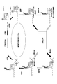
- FIG. 1A is a diagram showing a procedure for in vitro screening of a cDNA display method.
- FIG. 1B is a diagram showing a screening procedure when RNA is used as a target detection molecule.
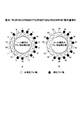
- FIG. 2A is a diagram showing a negative spherical structure (hereinafter sometimes referred to as “spherical structure N”).
- FIG. 2B is a diagram showing a positive spherical structure (hereinafter sometimes referred to as “spherical structure P”).
- FIG. 2C is a diagram (1) showing a high-speed screening procedure using the spherical structures P and N.
- FIG. 2D is a diagram (2) showing the procedure of high-speed screening using the spherical structures P and N.
- FIG. 3 is a microscopic photograph of a fluorescent substance-containing liposome.
- A1 is a differential interference image
- B1 is a fluorescence image using FITC
- C1 is a fluorescence image using Alexa-Fluore®594.
- A2 to C2 are schematic views of A1 to C1, respectively.
- FIG. 4A is a diagram showing an analysis result by FACS of fluorescent liposomes not treated with Maginin 2.
- FIG. 4B is a diagram showing the FACS analysis results of fluorescent liposomes treated with 10 ⁇ M Maginin 2.
- FIG. 4C is a diagram showing a result of FACS analysis of fluorescent liposomes treated with 30 ⁇ M Ma Maginin 2.
- FIG. 5 shows the predicted peptide sequence translated by the design DNA library, and the two ⁇ -helix wheels A and B (positions of amino acids 1 to 18) and B (positions of amino acids 19 to 35).
- FIG. 6 is a schematic diagram of extension PCR when preparing a promoter-DNA conjugate (244mer).
- FIG. 7 is a schematic diagram of a photocrosslinking linker.
- FIG. 8 is a diagram schematically showing a photocrosslinking linker and mRNA bound to this linker by photocrosslinking.
- FIG. 9 is a diagram schematically showing a conventional linker and mRNA enzymatically bound to the conventional linker by T4RNA ligase.
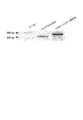
- FIG. 10 is a photograph of the result of electrophoresis of the ligation product of mRNA and puromycin linker.
- FIG. 11A is a diagram showing the FACS analysis result of the first fluorescent liposome of the high-speed screening cycle of the present invention.
- FIG. 11B is a diagram showing the FACS analysis result of the second fluorescent liposome of the high-speed screening cycle of the present invention.
- FIG. 11C is a diagram showing the results of FACS analysis of fluorescent liposomes in the third rapid screening cycle of the present invention.
- FIG. 12 is a photograph showing the results of electrophoresis of PCR products of cDNA display molecules after the rapid screening cycles 1 to 3 of the present invention.
- FIG. 13 shows the amino acid sequence of the peptide obtained from the cloning sequence and the sequence, which is represented by two ⁇ helices A (position of the 1st to 18th amino acids) and B (position of the 19th to 35th amino acids). It is the figure arranged on the wheel figure.
- FIG. 14A is a schematic diagram of a gene sequence of a fusion protein to be introduced into a vector.
- FIG. 14B is a schematic diagram showing the structure of a target recognition molecule.
- A1 is a fluorescence microscopic image when mCherry-LB-1 fusion protein is bound to 2 ⁇ M outside the liposome membrane.
- B1 is a fluorescence microscope image when only 2 ⁇ M of mCherry is bound to the outside of the liposome membrane.
- A2 and B2 are schematic views of A1 and B1, respectively.
- X represents a liposome membrane that emits bright red fluorescence
- Y represents a liposome membrane that emits dark red fluorescence.
- FIG. 16 is a photograph of DNA electrophoresis of a streptavidin-binding RNA aptamer.
- FIG. 17 is a photograph of electrophoresis of a streptavidin-binding RNA aptamer.
- FIG. 18 is a photograph of electrophoresis of a streptavidin-binding RNA aptamer to which streptavidin is bound.
- FIG. 19 is a photograph showing the FITC fluorescence intensity of the reaction product of labeled silica beads and fluorescein-labeled biotin with rhodamine B immobilized with streptavidin.
- FIG. 20 is a graph showing the sorting area of rhodamine B by FACS.
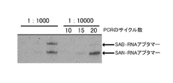
- FIG. 21 shows the results obtained by mixing the streptavidin-bound RNA aptamer and the streptavidin-unbound RNA aptamer at a ratio of 1:10 to 1: 10,000 and performing PCR for 15 cycles under the predetermined conditions for sorting by FACS. It is a gel electrophoresis image which shows the provided result.
- R indicates nucleic acid before FACS
- P indicates nucleic acid eluted from plus beads
- N indicates nucleic acid eluted from minus beads.
- FIG. 22 shows the result of mixing streptavidin-binding RNA aptamer and streptavidin non-binding RNA aptamer at 1: 1,000 or 1: 10,000, and performing PCR for 10 cycles, 15 cycles or 20 cycles under the same conditions as FIG. It is a gel electrophoresis image which shows the result of having used for the sorting by FACS.
- the present invention is a high-speed in vitro screening method for any library selected from the group consisting of a cDNA display library and a nucleic acid aptamer library, and includes the following steps (FIGS. 1A and B).
- the spherical molecule used in this step is not particularly limited as long as it can be used for selection using a cell sorter.
- liposomes composed of lipid bilayers, sepharose beads, silica beads, latex beads, dextran beads, A molecule
- the particle size of the spherical molecule is preferably 0.5 ⁇ m to 20 ⁇ m, since it enables highly accurate measurement. Although it is difficult to control the particle size of the liposome, spherical particles with a uniform particle size distribution can be prepared by coating the surface of each bead as described above with a liposome in accordance with a conventional method. Can be improved.
- the spherical molecule may contain a label such as Alexa Fluora 594, Fluorescein Amine, FITC, Rhodamin, mCherry2, and Quantum Dot, or may contain magnetite or other magnetized material. Further, the outside of these beads may be colored with a pigment such as blue, red, green, and black.
- the spherical particles are not usually porous, but may be porous.
- the surface of the spherical particle is modified with a functional group such as a carboxyl group, amino group, hydroxyl group, thiol group, etc., so that it binds to the protein on the mRNA-protein conjugate or mRNA / cDNA-protein conjugate. It is preferable at the point which becomes easy.
- the spherical particles may be purchased from commercial products.
- those prepared as follows may be used.
- the method for preparing liposomes is mainly the W / O emulsion method, and is prepared as follows. First, phospholipids, which are the raw materials for liposomes, are dissolved in alcohol, chloroform or other organic solvent to form a lipid solution, poured into test tubes or other glassware, and thinned on the inner wall surfaces while volatilizing the organic solvent. Form a film.
- oil is added to dissolve the phospholipid film formed on the inner wall, and an aqueous solution that is the internal solution of liposomes is added to the oil containing phospholipid and mixed well to obtain an oil-water mixture.
- the oil / water mixture is gently poured onto an aqueous solution that is an external solution of the vesicle, left to stand for a while, and then centrifuged.
- the droplets covered with the phospholipid monolayer pass through the phospholipid monolayer formed at the interface between the oil and the aqueous solution, and thus liposomes can be obtained.
- the size of cells and other substances that can be selected with a cell sorter is said to be about 0.5 ⁇ m to 20 ⁇ m. Therefore, it is preferable to prepare liposomes having such a size.
- aqueous solution containing fluorescently labeled molecules as the internal solution of the liposomes in order to select the target liposome with high accuracy in the selection process using a cell sorter described later.
- a fluorescent label is not particularly limited as long as it is a fluorescent label that can be used in a cell sorter, but it can be any one selected from the group consisting of Alexa Fluora 594, FITC, Rhodamin, mCherry2 and Quantum Dot to facilitate gene expression. It is further preferable from the viewpoints that it can be confirmed and multiple labels can be formed.
- the liposome When the liposome is labeled with an encapsulated fluorescent label or the like, it is possible to modify the liposome membrane with the following protein or peptide in the sorting step using a cell sorter described later with high accuracy. Is preferable because it becomes possible.
- modified peptides include peptide molecules that can be anchored on the liposome membrane, fluorescent proteins, various antibodies, disease marker proteins, fusion proteins with target molecules having sequences encoding other biomolecules, and the like.
- the liposome when the liposome is not labeled with a fluorescent label or the like, it is possible to bind a labeled target molecule that can bind to the mRNA / cDNA-protein conjugate to the surface of the liposome.
- the sorting process used is preferable because accurate sorting is possible.
- it may be labeled with a fluorescent label as described above, or may be labeled with an intercalator.
- an intercalator is not particularly limited as long as it is generally used, but it is any fluorescent molecule selected from the group consisting of SYBR Gold, SYBR Green, and Quantum Dot. It is preferable from the viewpoint of availability.
- the target molecule immobilized on the spherical molecule is selected and prepared in consideration of the combination with the target detection molecule that binds to the target molecule.
- the target detection molecule refers to a molecule that can bind to the target molecule fixed to the spherical molecule, and the spherical particle to which the target molecule is bound is defined as a positive spherical structure (spherical structure P; FIG. 2A),
- the spherical particles to which the target molecule is not bound are referred to as negative spherical structures (spherical structures N; FIG. 2B).
- the target detection molecule is an RNA aptamer that binds to streptavidin, or when biotin is immobilized on the target detection molecule, streptavidin is selected as the target molecule, and the spherical conjugate P or Spherical conjugate N is prepared.
- streptavidin is selected as the target molecule, and the spherical conjugate P or Spherical conjugate N is prepared.
- the carboxyl group is activated with N-hydroxysuccinimide (NHS) to form an NHS ester.
- NHS N-hydroxysuccinimide
- the target detection molecule refers to a nucleic acid, a nucleic acid derivative, a peptide obtained by cDNA display method, a molecule in which a linker for cDNA display is linked thereto, or a nucleic acid aptamer created based on these (see FIG. 1A).
- Nucleic acids include single stranded oligo DNA, double stranded DNA and RNA.
- Nucleic acid derivatives also include RNA aptamers that bind to target molecules such as streptavidin.
- the nucleic acid and linker linked to each other include, for example, mRNA-protein conjugates and mRNA / cDNA-protein conjugates described later.
- an mRNA preparation step for preparing mRNA for use in the cDNA display method (a) an mRNA preparation step for preparing mRNA for use in the cDNA display method, (b) an mRNA-linker binding step for binding this mRNA to a linker by photocrosslinking, ( c) an mRNA-linker-protein conjugate formation step of binding an mRNA-translated protein to the mRNA-linker conjugate, and (d) reverse transcription of the mRNA of the mRNA-linker-protein conjugate to obtain an mRNA / cDNA. It is preferable to further include a reverse transcription step for forming a linker-protein conjugate in that a desired mRNA, cDNA, and the like can be obtained using a cDNA display method.
- the spherical molecule and the target detection molecule are prepared in a predetermined binding buffer so that the concentration of these molecules is a predetermined concentration, for example, at about 4 ° C. to about 30 ° C., for about 0.5 hour to about 3 hours. It can be combined by reaction.
- the spherical conjugate P and the spherical conjugate N obtained in the above step are prepared in a predetermined solution so as to have a predetermined concentration.
- sorting is performed using a commercially available cell sorter according to the instruction manual of the product to select the target spherical conjugate (FIG. 2D).
- the spherical molecule used is a liposome, it is fluorescently labeled based on the fluorescence intensity of the fluorescent label in the liposome or the fluorescent label (for example, intercalator) bound to the target detection molecule (for example, mRNA / cDNA-protein conjugate).
- Fluorescent liposomes (hereinafter sometimes referred to as “fluorescent liposomes”) are selectively collected.
- a spherical conjugate having a fluorescent label excited at both of two different excitation wavelengths by labeling the liposome with a fluorescent label that labels the liposome and a label that emits fluorescence at different wavelengths. Can be sorted using a cell sorter. Since such sorting is possible, the efficiency of sorting is improved, and a fraction containing a spherical conjugate with a high content of desired target molecules can be obtained. Also, by applying a selective pressure by coexisting a molecule that causes competitive inhibition with the target binding molecule in the above binding solution, a fraction containing a spherical conjugate having a higher ratio of the desired target molecule can be obtained. become able to.
- the combinations of the spherical structure and the label of the target detection molecule are shown in Table 1 below.
- “spherical structure P” and “spherical structure N” are as described above.
- Labels 1 and 2 each represent a different fluorescent label.
- the target detection molecule is a linker-nucleic acid molecule (C) selected from a cDNA library by a cDNA display method, or an aptamer molecule (A) contained in a nucleic acid aptamer library.
- the phosphorous substance lump that has not been used for liposome formation is removed, for example, by centrifugation from the fluorescent liposome fraction prepared and collected according to a conventional method, and the fluorescent substance-containing liposome is purified. be able to.
- purified liposomes positive liposome conjugates (hereinafter referred to as LiBP) in which these are bound to target molecules and negative liposome conjugates (hereinafter referred to as LiBN) in which target molecules are not bound are prepared.
- LiBP positive liposome conjugates
- LiBN negative liposome conjugates
- the target detection molecule is bound by the method as described above, and then selected with a cell sorter. Such selection of liposomes can be completed in about 0.1 hours to about 6 hours, although there are some differences depending on the cell sorter used.
- mRNA / cDNA-protein conjugate may be obtained by reverse transcription of mRNA in the mRNA-protein conjugate according to a conventional method. Specifically, for example, the mRNA-protein conjugate obtained as described above is fixed on styrene beads or other magnetic beads, and then reverse transcription is performed. It is preferable to perform such reverse transcription before the amplification step described later in view of efficient purification of the mRNA / cDNA-protein conjugate.
- the magnetic beads are washed with a desired washing buffer, and then an enzyme or other mRNA / cDNA-protein conjugate release agent is added thereto and incubated, whereby mRNA / cDNA- cleaved from the cleavage site in the linker is obtained. Protein conjugates can be released. Thereafter, the mRNA / cDNA-protein conjugate is preferably purified using a His-tag or other protein purification tag.
- the DNA contained in the mRNA / cDNA-protein conjugate (target detection molecule) on the spherical conjugate obtained as described above is amplified by PCR according to a conventional method. If the target detection molecule is RNA, reverse transcription is performed before PCR to prepare cDNA. For example, first, the mRNA / cDNA-protein conjugate is precipitated using a coprecipitation agent such as ethanol to obtain cDNA. The obtained cDNA is added to a desired PCR reaction solution and amplified by a desired PCR program. Using the amplified DNA, the steps (i) to (iv) are repeated, and DNA having a desired sequence can be obtained from the original DNA library by high-speed screening.
- a coprecipitation agent such as ethanol
- the number of times the steps (i) to (iv) are repeated is preferably within 10 times for high-speed DNA screening. This is because the DNA contained in the original library having diversity of 10 11 order can be converged to several tens order.
- the number of repetitions is more preferably within 5 times, and further preferably within 3 times from the viewpoint of cost performance.
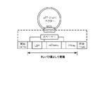
- a promoter and other sequences necessary for producing a full construct are added to the selected number of DNAs.
- the full construct DNA is constructed by binding, and mRNA is prepared from the full construct DNA (FIG. 6).
- examples of the “promoter” include T7 promoter, SP6 and T3 promoter, and the use of T7 promoter is preferable from the viewpoint of versatility.
- examples of “other sequences necessary for producing a full construct” include a liposome-binding sequence, His tag, Strep-tag, and other purification tag sequences when a liposome is used as a spherical structure.
- a DNA fragment containing the sequence as described above is prepared, and the DNA is amplified by extension PCR to obtain a double-stranded full construct DNA (hereinafter sometimes referred to as “promoter-DNA conjugate”).
- the obtained promoter-DNA conjugate can be transcribed according to a conventional method to obtain mRNA.
- ScripTMAX R
- Thermo T7 Transcription Kit Toyobo Co., Ltd.
- RiboMAX TM Large Scale RNA Production System-T7 Promega
- T7 Transcription Kit Cosmo Bio Co., Ltd.
- mRNA can be prepared by performing transcription quickly, simply and accurately.
- the mRNA obtained above is bound to a cDNA display linker to prepare a linker-mRNA conjugate.
- a linker that can be ligated in a short time.
- Such connection in a short time is preferably performed by photocrosslinking, and it is preferable to use a photocrosslinking linker.
- a photocrosslinkable linker a photocrosslinkable linker containing a high-speed photocrosslinkable artificial nucleic acid (3-cyanovinylcarbazole) (hereinafter sometimes referred to as “cnvk”) in the main chain is quickly used.
- cnvk high-speed photocrosslinkable artificial nucleic acid
- the main chain of the linker includes (p1) a solid phase binding site for binding the linker to the solid phase, (p2) a side chain binding site for binding the side chain, and (p3) an mRNA binding site. And (p4) a cleavage site for separating the linker from the solid phase, the side chain comprising: (s1) a main chain binding site that binds to the main chain; and (s2) an mRNA-linker conjugate. It comprises a label binding site for binding a detection label and (s3) a peptide binding site to which a peptide having a sequence synthesized corresponding to mRNA binds. Cnvk is contained between the main chain cleavage site and the side chain binding site.
- the label binding site of the side chain may be composed of one or more spacer sequences, and a fluorescent label is bound thereto.
- the peptide binding site may be composed of puromycin or an analog thereof.
- the linker has the structure as described above, mRNA and the linker can be linked in a short time of 1/30 or less compared to the case where T4 RNA ligase is used. And since these can be connected in such a short time, the risk of degradation of mRNA by RNAse can be significantly reduced.
- the linker-mRNA conjugate can be easily selected by using a label bonded to the side chain of the linker.
- the linker-mRNA conjugate as described above can also be prepared by an enzymatic ligation technique using a conventionally used linker and the mRNA obtained as described above using an enzyme such as T4RNA ligase.
- the linker-mRNA conjugate obtained above is translated using a cell-free translation system, and the sequence corresponding to mRNA is translated into the peptide binding site of the linker. Peptides are displayed and mRNA-linker-protein conjugates are formed.
- cell-free translation it is preferable to use a rabbit reticulocyte lysate or other commercially available reticulocyte lysate since translation can be performed quickly and stable results can be obtained.
- Peptide bound to the peptide binding site of the linker in the mRNA-protein conjugate (target detection molecule) obtained in the step (c) or the mRNA / cDNA-protein conjugate obtained in the step (d) described later Binds to a target molecule of the spherical structure or directly binds to a functional group on the surface of the spherical structure. Thereby, a spherical conjugate can be obtained.
- the step of reverse transcription of the mRNA-protein conjugate to obtain the mRNA / cDNA-protein conjugate may be performed before or after the selection step (iii). This is preferably performed before the selection step (iii) because the mRNA bound to the linker can be stabilized.
- the mRNA-protein conjugate in the spherical conjugate is reverse-transcribed under predetermined conditions. That is, the conditions for this reverse transcription reaction can be arbitrarily set. For example, according to a conventional method, dNTP mixed solution, DTT, reverse transcriptase, and water from which RNAse has been removed (hereinafter referred to as “RNAse-free water”).
- a DNA fragment having a desired sequence can be synthesized according to a conventional method. For example, first, a PCR reaction solution containing a DNA fragment of a sequence encoding a peptide sequence and a sequence having a His-tag is prepared, and extension PCR is performed under desired conditions. Thereafter, a PCR reaction solution containing a sequence consisting of T7 promoter sequence-Cap sequence- ⁇ sequence-Kozak sequence is added to this PCR product, and extension PCR is performed under desired conditions to obtain DNA of the desired sequence.
- PCR reaction solution 1 ⁇ PrimeSTAR buffer (Mg 2+ ), 0.1-0.4 mM dNTPs, 0.01-0.04 U /%, containing a sequence encoding the desired peptide and a sequence having a His-tag.
- PrimeSTAR HS DNA polymerase manufactured by Takara Bio Inc.
- overlap extension PCR1 is performed using the following PCR program.
- the PCR program at this time is, for example, (a) 92-96 ° C (1-3 minutes), (b) 92-96 ° C (5-45 seconds), (c) 50-70 ° C (2-30 seconds) (D) 65 to 80 ° C (20 to 40 seconds), (e) 65 to 80 ° C (1 to 3 minutes), and steps (b) to (d) are performed for 6 to 10 cycles. This is preferable because the product can be obtained.
- the PCR product obtained by the above overlap extension PCR1 is added to 25 to 75 ⁇ L of PCR reaction solution (including a sequence consisting of T7 promoter sequence-Cap sequence- ⁇ sequence-Kozak sequence, further including 0.6 ⁇ M F1).
- PCR reaction solution including a sequence consisting of T7 promoter sequence-Cap sequence- ⁇ sequence-Kozak sequence, further including 0.6 ⁇ M F1.
- overlap extension PCR2 is performed using the following PCR program. It is preferable that the PCR program at this time is the same as that described above from the viewpoint of obtaining a sufficient amplification product.
- the double-stranded full-construct DNA obtained by the above overlap extension PCR2 is precipitated using, for example, a coprecipitation agent (Quick-Precip® Plus Solution, manufactured by EdgeBio), and then, for example, FavorPrep® PCR® Clean-Up It is preferable to purify using Mini Kit (manufactured by Favogen).
- a coprecipitation agent Quick-Precip® Plus Solution, manufactured by EdgeBio
- FavorPrep® PCR® Clean-Up It is preferable to purify using Mini Kit (manufactured by Favogen).
- double-stranded full-construct DNA can be obtained by performing extension PCR using a random sequence including DNA designed to encode a desired peptide, a sequence including a T7 promoter and an SD sequence, and a Ytag sequence.
- a PCR reaction solution containing a random sequence and a Ytag sequence is prepared, and extension PCR is performed under desired conditions.
- the PCR program is preferably the same as described above except that steps (b) to (d) are performed for 4 to 10 cycles from the viewpoint of obtaining sufficient amplification products.
- steps (b) to (d) are preferably performed for 4 to 10 cycles as described above.
- RNA Transcription is performed according to a conventional method using the DNA obtained as described above as a template DNA.
- a reaction solution containing a predetermined concentration of T7 transcription buffer, rNTPs (each containing 25 mM ATP, CTP, UTP, GTP), enzyme mixture and template DNA is reacted at about 37 ° C. for about 2 to about 6 hours.
- RQ-1 RNAse-Free DNAse (Promega) is added and further reacted at about 37 ° C. for about 15-30 minutes.
- purification can be performed using, for example, Rneasy minElute Cleanup kit (manufactured by QIAGEN) to obtain purified mRNA.
- RNA for such transcription a commercially available transcription kit may be used.
- RiboMAX Large Scale RNA Production Systems T-7 manufactured by Promega
- transcription may be performed on a scale of 10 to 30 ⁇ L using, for example, 0.5 to 5 ⁇ g of dsDNA according to the protocol attached to the kit.
- Incubation is performed under desired conditions using a thermostat that is widely used, and mRNA can be obtained by adding a desired amount of DNAse and incubating again.
- RNA Clean-Up Kit manufactured by Favogen
- linker refers to a linker-mRNA conjugate, linker-mRNA-protein conjugate used in cDNA display method, Alternatively, it refers to a linker used for producing any of a linker-mRNA / cDNA-protein conjugate (hereinafter sometimes referred to as “IVV”).
- the “mRNA-protein conjugate” is a linker-mRNA-peptide complex in which the linker-mRNA conjugate is translated and a peptide having a sequence corresponding to the mRNA is bound to a peptide binding site of the linker described later. It means the body.
- the “mRNA / cDNA-protein conjugate” refers to a linker-mRNA / cDNA-peptide complex in which the mRNA-protein conjugate is reverse-transcribed and cDNA is bound onto the linker main chain.
- the linker is mainly composed of DNA, but preferably contains cnvK because it can be photocrosslinked with mRNA in a short time. Moreover, it may contain DNA analogs such as deoxyinosine, biotin-modified deoxythymine, and fluorescein-modified deoxythymine, and is preferably designed so as to have flexibility and hydrophilicity as a whole.
- Examples of molecules that form a bond with a solid phase include biotin or its analog when avidin and streptavidin are bound to the solid phase, maltose when a maltose binding protein is bound, G A guanine nucleotide is preferred when the protein is bound, a metal such as Ni or Co when the polyhistidine peptide is bound, and a glutathione when glutathione-S-transferase is bound.
- DNA or RNA having a specific sequence is bound to them, and when antibodies or aptamers are bound, antigen or epitope peptide, calmodulin is bound.
- calmodulin binding peptide, ATP when ATP binding protein is bound, and estradiol when estradiol receptor protein is bound it is preferable to use a metal such as biotin, maltose, Ni or Co, glutathione, an antigen molecule or an epitope peptide, and it is preferable to use biotin or an analog thereof from the viewpoint of ease of linker synthesis. .
- the solid phase binding site (p1) is a site for binding the mRNA-protein conjugate or mRNA / cDNA-protein conjugate described above to a solid phase via a linker, and the solid phase binding site (p1) Consists of at least 1 to 10 bases.
- the poly A it is preferable that at least 10 adenines are bound to each other so that an appropriate distance from the solid phase can be maintained, and separation from the solid phase described later can be successfully performed, and about 20 adenines are included. More preferably, they are bonded.
- the side chain binding site (p2) located in the vicinity of the 3 'end of the linker is a site to which a side chain described later binds. Further, for example, when the side chain binding site (p2) of the linker is composed of Amino-Modifier C6 dT, the 5 ′ end of the side chain is 5′-Thiol-Modifier C6, and EMCS is used.
- the main chain and the side chain can be bonded by crosslinking.
- the primer region (PR) is a region located on the 3 ′ end side of the linker and functions as a primer for reverse transcription when reverse transcription is performed on the linker, and 3 ′ side of the side chain binding site Adjacent to.
- the primer region (PR) is a region that functions as a primer for reverse transcription when reverse transcription is performed on the linker.
- This region preferably consists of about 1 to 15 bases, particularly 3 to 5 bases. When the number of bases exceeds 15, the binding efficiency as a linker is deteriorated. Therefore, from the viewpoint of the binding efficiency with a linker and the reaction efficiency as a primer, the above base number is preferable.
- the peptide binding site is preferably composed of puromycin or an analogous compound thereof.
- puromycin-related compounds 3'-N-aminoacylpuromycin (PANS-amino acid) and 3'-N-aminoacyl adenosine amino acid nucleoside (AANS-amino acid) can be used, and the amino acid part of PANS PANS-Gly, which is glycine, PANS-Val, which is valine, PANS-Ala, which is alanine, a mixture of PANS amino acids, AANS-Gly, in which the amino acid part of AANS is glycine, AANS-Val, which is valine, AANS that is alanine More preferably, it is any compound selected from the group consisting of a mixture of -Ala and AANS amino acids.
- PANS-amino acids examples include PANS-Gly, PANS-Val, and PANS-Ala.
- AANS-amino acids include AANS-Gly, AANS-Val, and AANS-Ala.
- an ester-linked nucleoside and an amino acid can be used, but it is particularly preferable to use puromycin because of the high stability of peptide binding at the peptide binding site.
- the side chain has a fluorescent group between the peptide binding site and the side chain binding site, the presence or absence of binding to the linker can be easily detected in each step of the cDNA display method described later. It becomes possible.
- the fluorescent group has, for example, a free functional group such as a carboxyl group that can be converted into an active ester, a hydroxyl group that can be converted into a phosphoramidide, or an amino group, and can be bonded to a linker as a labeled base. It is preferable to use fluorescent compounds that can be used. Examples of such fluorescent compounds include fluorescein isothiocyanate (FITC), rhodamine, CyCdye, AlexaR Fluor, and the use of FITC is preferable from the viewpoint of cost.
- FITC fluorescein isothiocyanate
- CyCdye CyCdye
- AlexaR Fluor AlexaR Fluor
- a photocrosslinking linker (FIG. 7).
- an enzyme such as T4 RNA ligase
- T4 RNA ligase it is difficult to completely remove RNAse, and it takes a long time to ligate, so mRNA may be degraded.
- the linker of the present invention does not use an enzyme, there is no concern about the degradation of mRNA, and because it contains cnvK, it is not in a buffer, but crosslinks between the mRNA and the linker in a very short time by UV irradiation in water. This is because it can be formed.
- the structure of this photocrosslinking linker is as described above.
- the solid phase cleavage site (p2) is composed of deoxyinosine for the following reason.
- the two conjugates described later, ie, the linker of the present invention, the conjugate of mRNA and cDNA, or the linker of the present invention, the conjugate of mRNA, cDNA and peptide (hereinafter, both are simply fused together.
- Endonuclease V is used to separate the body from the solid phase.
- the solid-phase cleavage site is composed of deoxyinosine, the solid phase can be specifically separated from the linker together with the solid-phase binding site.
- the photocrosslinking linker as described above can be prepared as follows. First, the linker main chain (poly A + cnvK segment) of the present invention is designed so that cnvK is in the desired position between the solid-phase binding site and the site where the main chain and the side chain are bonded. Chemical synthesis is performed according to conventional methods. Such chemical synthesis of DNA strands may be outsourced to a company that performs the synthesis.
- Such a main chain includes, for example, a reverse transcription initiation site as shown in FIG. 7, a side chain linking site, a high-speed photocrosslinking site comprising 3-cyanovinylcarbazole, and a solid phase binding site.
- a reverse transcription initiation site as shown in FIG. 7
- a side chain linking site a high-speed photocrosslinking site comprising 3-cyanovinylcarbazole
- a solid phase binding site can be designed.
- the base sequence of the main chain excluding the modified site is shown in the following sequence (SEQ ID NO: 1).
- the following main chain has BioTEG added to the 5 ′ end.
- R represents inosine
- Y represents amino C6-dT.
- the side chain of the linker (hereinafter sometimes referred to as “puromycin-segment”) is also designed to have the desired sequence, and the DNA is chemically synthesized in the same manner as the PolyA + cnvK segment. Such chemical synthesis of DNA strands may be outsourced to a company that performs the synthesis.
- Such a side chain is preferably designed to include a side chain linking site, a fluorescent label, and a peptide binding site, for example, as shown in FIG.
- the base sequence (SEQ ID NO: 2) excluding the modified site is as follows.
- P (K in the following sequence) serving as a free end is puromycin as a peptide binding site.
- R represents 5'ThiolhiC6
- Y represents FITC-dT
- M represents Spacer18.
- the main chain having the above sequence is transferred to 0.1 to 0.3 M sodium phosphate (pH 7.0 to 7.4) containing 10 to 20 nmol (final concentration 100 to 200 ⁇ M) to EMCS (Dojindo Laboratories). Prepared at a final concentration of 15-18 mM, and incubated at about 37 ° C. for 20-40 minutes, followed by ethanol precipitation.
- EMCS is added to a 0.1-0.3M sodium phosphate solution (about pH 7.2) containing about 15 nmol of the main chain (final concentration about 150 ⁇ M) at a final concentration of about 16.7 mM at about 37 ° C. Incubate for about 30 minutes, and then modify by ethanol precipitation using, for example, Quick-Precip Plus Solution (Edge BioSystems).
- the solution containing the reduced side chain subjected to the buffer exchange as described above is mixed with the above-mentioned EMCS-modified main chain ethanol precipitation product and left at 2 to 6 ° C. overnight. Thereafter, DTT is added to the reaction solution so that the final concentration is 40 to 60 mM, and the mixture is stirred at room temperature for 15 to 60 minutes. Thereafter, ethanol precipitation is performed, and the obtained ethanol precipitate is dissolved in 50 to 200 ⁇ L of nuclease-free water for purification.
- the solution containing the reduced side chain exchanged in the buffer is mixed with the ethanol-precipitated product of the EMCS-modified main chain in the above-mentioned buffer and left at about 4 ° C. overnight. Then, add DTT to the above reaction solution so that the final concentration is about 50 mM, stir at room temperature for about 30 minutes, and then ethanol precipitation using, for example, Quick-Precip Plus Solution (manufactured by Edge BioSystems). To do.
- the obtained ethanol precipitation product is dissolved in about 100 ⁇ L of nucleotide-free water and purified by HPLC by gradient elution using a C18 column under the following conditions to obtain a photocrosslinkable linker (FIG. 8). ).
- the eluent used for gradient elution is, for example, 0.05 to 0.2M trimethylammonium acetate (ultra pure water) for solution A, 75 to 85% acetonitrile for solution B, and the ratio of solution A during the elution period at the start. It may be reduced by about 20% over 40 to 50 minutes.
- the flow rate can be 0.5-1.5 ml / min and the fraction can be 0.5-1.5 mL.
- liquid A is about 0.1M trimethylammonium acetate (ultra pure water)
- liquid B is about 80% acetonitrile
- the ratio of liquid A during the starting elution period (about 85%) is 40 to 50 minutes. And reduce it to about 65%.
- the flow rate is about 1.0 ml / min and the fraction is about 1.0 mL.
- the components in the above fractions are confirmed by fluorescence and ultraviolet absorption (for example, 280 nm), the fractions in which peaks are observed by both detection means are collected, the solvent is evaporated using a vacuum evaporator, and then ethanol precipitation is performed.
- the linker of the present invention can be produced by dissolving in nucleotide-free water. For example, when peaks are observed in both fluorescence and UV in fractions from 30 to 32 minutes, fractions from 30 to 32 minutes are collected and the solvent is evaporated using a vacuum evaporator. Thereafter, for example, ethanol precipitation is performed using Quick-Precip Plus Plus Solution to obtain a photocrosslinking linker.
- the obtained photocrosslinking linker may be dissolved in nuclease-free water and stored at about ⁇ 20 ° C.
- a conventional enzyme-type linker can also be used.
- DNA is synthesized according to a conventional method so as to obtain a desired sequence, and a single-stranded oligomer for use as a main chain is prepared.
- the single-stranded oligomer synthesized in this way includes a solid phase binding site, two or more cleavage sites, an mRNA binding site, a side chain binding site, and a primer region.
- the length of the single-stranded oligomer serving as the main chain is appropriately determined depending on the size of the two or more cleavage sites and the position in the main chain.
- a side chain of the desired length is then synthesized and bound to a side chain binding site on the main chain.
- puromycin can be introduced into the free end of the side chain, and the above-described Fluorescein-dT can be introduced into the fluorescent labeling site to obtain the linker for preparing the mRNA / cDNA-peptide conjugate of the present invention.
- mRNA coding sequences can be referred to.
- mRNA encoding various receptor proteins with known sequences mRNA encoding various antibodies or fragments thereof, and other mRNAs can be mentioned.
- the 3 'terminal analog of aminoacyl-tRNA such as puromycin and its analogs is incorporated into the C-terminal of the polypeptide chain generated by translation from the mRNA coding sequence, and the polypeptide chain and the linker-mRNA conjugate are combined. To do so, select a sequence that does not contain a stop codon.
- Such mRNA can be obtained by using in vitro transcription reaction, chemical synthesis, extraction from living organisms, cells, and microorganisms, and other various methods. High cell translation reaction efficiency.
- RNA RNA RNA RNA RNA RNA RNA RNA RNA RNA . RNA RNA RNA RNA RNA RNA RNA RNA RNA RNA RNA RNA RNA RNA RNA RNA RNA RNA RNA RNA RNA RNA RNA RNA RNA RNA RNA RNA RNA RNA RNA RNA RNA RNA RNA RNA RNA RNA RNA RNA RNA RNA RNA RNA RNA RNA RNA RNA RNA RNA RNA RNA RNA RNA RNA RNA RNA RNA RNA RNA RNA RNA RNA RNA RNA RNA RNA RNA RNA RNA RNA RNA RNA RNA RNA RNA RNA RNA RNA RNA RNA RNA RNA RNA RNA RNA RNA RNA RNA RNA RNA RNA RNA RNA RNA RNA RNA RNA RNA RNA RNA RNA RNA RNA RNA RNA RNA RNA RNA RNA RNA RNA RNA RNA RNA RNA
- (3-2) Ligation of mRNA and linker Ligation is performed by irradiating UV of a long wavelength of 300 to 400 nm for 0.5 to 5 minutes with respect to photocrosslinking of the mRNA obtained above and the linker of the present invention. Since it has a long wavelength and a short irradiation time, there is an advantage that a desired peptide corresponding to the used mRNA can be obtained without causing a problem that a thymine dimer is formed in the synthesized cDNA. .
- mRNA / cDNA-protein conjugate by cell-free translation of mRNA-linker conjugate
- a lysate of mammalian reticulocyte cells More preferably, lysates of reticulocytes obtained from rabbit blood are used.
- acetylphenylhydrazine is preliminarily administered to the mammal to induce hemolytic anemia and blood is collected after several days, the ratio of reticulocytes in the blood can be increased.
- a cell-derived mRNA is degraded with micro-coccal nuclease, and calcium ether is added with glycol ether diamine tetraacetic acid (EGTA) to inactivate the nuclease (hereinafter referred to as “micro-coccal”).
- EGTA glycol ether diamine tetraacetic acid
- a rabbit reticulocyte lysate that has been treated with micrococcal nuclease and the above-mentioned conjugate can be added to carry out a translation reaction.
- the amount of rabbit reticulocyte lysate is about 8.5 to about 17 ⁇ L
- the amount of the above conjugate is about 1.2 to about 2 pmol
- the reaction system size is about 12.5 to about 25 ⁇ L
- the reaction solution used in this case contains about 80 mM potassium acetate, about 0.5 mM magnesium acetate, about 10 mM creatine phosphate, about 0.025 mM methionine and leucine, respectively, and about 0.05 mM methionine and amino acids other than leucine.
- the translation product peptide and the linker-mRNA conjugate are, for example, in the presence of about 0.3 to about 1.6 M potassium chloride and about 40 to about 170 mM magnesium chloride (the concentrations are both final concentrations).
- the reaction is performed at about 27 to about 47 ° C. for about 30 minutes to about 1.5 hours, the peptide can be efficiently bound to the above conjugate.
- dioleoylphosphatidylcholine DOPC
- dioleoylphosphatidylglycerol DOPG
- dipalmitoyl phosphatidylcholine DPPC
- egg phosphatidylcholine eggPC
- lipid is dissolved in the liquid paraffin by applying ultrasonic vibration for one hour or more at 55 to 65 ° C.
- a solution of a desired amount of liposome in the membrane is added to this solution, and the mixture is vigorously mixed for 30 seconds or more by applying ultrasonic vibration.
- the liposome membrane solution 0.1 to 0.4 M glucose containing 25 to 75 mM NaCl and 27 to 75 mM Tris buffer (pH 7.5) can be used.
- the liposome can be labeled by adding a fluorescent label usable in flow cytometry to the solution in the liposome membrane.
- fluorescent labels include AMCA, Pacific Blur, Alex Flur 405, Pacfic Orange, Krome Orange, Brilliant Violet 421, Brilliant Violet 510, Brilliant Violet 650, Brilliant Violet 711, Brilliant Violet 711, Brilliant Violet ⁇ 711, Dot, FITC, PE / RD1, ECD, PE-TexasRed, PC5, SPRD, PE-Cy5, PC5.5, PE-Cy5.5, PerCP, PerCP-5.5, PE-Alex Fluor 700, PE-Alex Fluor 750, PC7, PE-Cy7, TRITC, Cy3, Alex Fluor 594, TexasRed, Alex Flur 594, Alex Flur 700, Cy5, Cy5.5, APC, APC7, APC-Cy7, APC Alexa Fluor 700, APC Alexa Fluor 750, Hoechst33342, DAPI, DyeCycle Violet, Chromomycin A3, PI, Flu
- fluorescent liposomes it is preferable to select a plurality of fluorescent labels having different fluorescence wavelengths from the above fluorescent labels to label the liposomes in that the target fluorescent liposome can be selected with high accuracy.
- select Fluorescein Amine that emits green fluorescence and Alexa Fluora 594 that emits red fluorescence or select FITC that emits green fluorescence and mCherry2 that emits red fluorescence.
- Selected liposomes (hereinafter sometimes referred to as “fluorescent liposomes”) can be selected.
- Tris buffer (pH 7.5) or 0.1-0.5M glucose solution containing NaCl is used as the liposome membrane outer solution.
- the liposome membrane solution is allowed to stand on the outer solution and centrifuged at room temperature. Do. Thereby, the droplet of the liquid mixture in the liposome membrane covered with the phospholipid monolayer passes through the phospholipid monolayer formed at the interface between the oil and the liposome outer membrane, thereby forming the liposome. Then, the liquid paraffin which exists in the upper part is removed, and the solution containing a liposome can be obtained.
- the liposome membrane obtained above can be fluorescently labeled according to a conventional method.
- the liposome membrane may be fluorescently labeled with a carbocyanine dye that emits green fluorescence using a commercially available kit such as Green-fluorescent Cytoplasmic Membrane Staining Kit (Takara Bio Inc.).
- Orange-fluorescent Cytoplasmic Membrane Staining Kit (manufactured by Takara Bio Inc.) is used in orange
- Red-fluorescent Cytoplasmic Membrane Staining Kit (manufactured by Takara Bio Inc.) is used in red
- Blue-fluorescent It can also be labeled blue using Cytoplasmic® Membrane® Staining® Kit (Takara Bio Inc.). With such a label, fluorescent liposomes can also be identified with a cell sorter.
- the label molecule can be modified on the surface by using a fusion protein comprising a protein that binds to the liposome membrane and a label molecule.
- a fusion protein comprising a protein that binds to the liposome membrane and a label molecule.
- the LB-1 protein binds to the liposome membrane, and the labeled molecule is exposed on the surface of the liposome membrane.
- the target molecule is a fluorescent protein
- an image in which the liposome membrane is labeled with a fluorescent label can be obtained by microscopic observation.
- the mRNA-protein conjugate or mRNA / cDNA-protein conjugate obtained as described above is a peptide bond of a linker.
- the peptide bound to the site is bound to the target molecule of the liposome membrane, or the peptide on the linker is bound directly to the functional group of the liposome membrane to bind to the liposome.
- antibacterial peptides such as Maginin2, PGLa, and Melittin bind to bacterial cell membranes. These peptides have 15 to 40 amino acid residues and are amphipathic consisting of a cluster of basic amino acids and hydrophobic amino acids.
- Reverse transcription Reverse transcription may be performed before or after the sorting step by the cell sorter.
- mRNA / protein-protein is synthesized by synthesizing a cDNA strand according to a conventional method under a predetermined condition using the 3 ′ end of the main chain on the linker of the mRNA-protein conjugate as a reaction start point and using the mRNA as a template. A linked body is obtained.
- the reverse transcription reaction system can be arbitrarily selected and is not particularly limited. However, the linker-mRNA conjugate, dNTP mixture, DTT, reverse transcriptase, standard solution, and RNAse-free water (hereinafter referred to as “RNAse-free”).
- reverse transcription it is preferable to prepare a reaction system by adding “water”), and to carry out reverse transcription in this system for 5 to 20 minutes at 30 to 50 ° C. Further, reverse transcription may be performed using a commercially available kit. For example, reverse transcription can be performed using PrimeScript RT-PCR Kit (Takara Bio Inc.) and ReverTra Ase (Toyobo Co., Ltd.) according to the attached protocol.
- Sorting With a general-purpose cell sorter, only fluorescent liposomes are cell-sorted according to the manual. When a plurality of fluorescent dyes are used, the fluorescence is corrected as appropriate, and the target fluorescent liposome region is specified from the fluorescence intensity distribution of the labeled fluorescent dye. Specifically, a control sample having a function similar to that of the target peptide is allowed to react with liposomes, a fluorescent region is identified on the histogram, and fluorescent liposomes can be selectively fractionated from the same region in the target sample.
- the solution of the liposome conjugate after sorting is vortexed to release the cDNA-display molecule (mRNA-protein conjugate, mRNA / cDNA-protein conjugate) from the liposome conjugate, and the solution is centrifuged to obtain an organic solution containing lipids.
- the phase and the aqueous phase containing the cDNA display molecule are separated. From cDNA display molecules dissolved in the aqueous phase, mRNA-protein conjugates or mRNA / cDNA-protein conjugates can be purified using ethanol precipitation or nucleic acid column purification methods.
- the mRNA-protein conjugate obtained by the above purification can be immobilized on a desired solid phase during reverse transcription.
- the solid phase include beads such as styrene beads, glass beads, agarose beads, sepharose beads, and magnetic beads; substrates such as glass substrates, silicon (quartz) substrates, plastic substrates, and metal substrates (for example, gold foil substrates); Examples thereof include containers such as glass containers and plastic containers; membranes made of materials such as nitrocellulose and polyvinylidene fluoride (PVDF).
- the linker may be directly covalently bonded to the solid phase using a known method, if necessary (See Qiagen, LiquidChip Applications Handbook, etc.)
- a known method See Qiagen, LiquidChip Applications Handbook, etc.
- biotin or an analog thereof is bound to the mRNA / cDNA-protein conjugate, the conjugate can be easily bound to the solid phase by binding avidin to the solid phase.
- the magnetic beads are removed from the mRNA / cDNA-protein conjugate obtained by the immobilization and the linker-mRNA / cDNA conjugate is recovered.
- the magnetic beads are washed with a desired washing buffer, and then a release agent is added and incubated to release the cleaved linker-mRNA / cDNA conjugate from the cleavage site in the linker.
- a washing buffer include 1 ⁇ His-tag washing buffer (including 10 to 30 mM sodium phosphate (pH 7.4), 0.25 to 0.75 M NaCl, 10 to 30 mM imidazole, and 0.025 to 0.1% Tween-20), 1 ⁇ NEB 4 buffer can be used.
- RNA-degrading enzyme for example, 1 ⁇ His-tag washing buffer containing 500 to 1,500 U RNAse T1, 1 ⁇ NE buffer containing 10 U EndoNucleaseV (New England Biolabs), etc. is used. can do.
- the linker-mRNA / cDNA conjugate can be purified using His-tag protein purification beads or the like.
- His-tagged protein purification beads HisHiMag Sepharose Ni (manufactured by GE healthcare) or the like can be used.
- the beads for purification of His-tag protein are previously washed with the His-tag washing buffer.
- the recovered linker-mRNA / cDNA conjugate is added to the washed His-tag protein purification beads, incubated under desired conditions, and the beads are washed with the His-tag washing buffer.
- a purified linker-mRNA / cDNA conjugate can be obtained by adding His-tag protein elution buffer and stirring under desired conditions.
- the purified linker-mRNA / cDNA conjugate can be amplified using PCR with a desired primer to prepare a DNA library containing the desired coding sequence.
- a solution containing the purified linker-mRNA / cDNA conjugate is precipitated using, for example, a coprecipitation agent (Quick-Precip Plus Solution, manufactured by EdgeBio), a PCR reaction solution is added, and a desired PCR program is performed.
- RNA streptavidin-binding RNA aptamers can be used as RNA aptamers that bind to the target molecule, streptavidin.
- the DNA of SAB-RNA aptamer and RNA aptamer to which streptavidin does not bind (hereinafter referred to as “SAN-RNA aptamer”) is T7 on the 5 ′ side of both RNA aptamers so that the target RNA can be prepared after transcription. It is constructed as a sequence having a promoter sequence.
- primers having the following sequences shown in Table 2 below are prepared and used.
- the third to the 22nd are T7 promoter sequences.
- a primer having the following sequence (SEQ ID NO: 12) is prepared and used together with the primer of SEQ ID NO: 11.
- the third to the 22nd are T7 promoter sequences.
- a DNA synthesis reaction solution containing a DNA polymerase such as Taq DNA polymerase and dNTP is prepared, each of the above two primers is added to a predetermined concentration, and each DNA is synthesized by the overlap extension method.
- a DNA synthesis reaction solution a commercially available kit such as PrimeSTAR (Takara Bio Inc.) can be used. Overlap extension conditions are 95 to 98 ° C for 30 seconds to 1 minute and 30 seconds, 60 to 62 ° C for 5 to 10 seconds, 71 to 73 ° C for 20 to 40 seconds, and then cooled to 10 ° C.
- DNA is purified according to a conventional method. Such purification can be performed, for example, using a commercially available kit such as FavorPrep PCR Clean-Up Mini Kit (manufactured by Favorgen) according to the attached manual.
- RNA polymerase such as T7 RNA polymerase and rNTP
- a transcription reaction is performed by incubating at 37 ° C. for 20 minutes to 1 hour.
- excision purification and kits such as Rneasy minElute Cleanup kit (manufactured by QIAGEN) can be used and purification can be performed according to the attached manual.
- the obtained SA-bound RNA aptamer and non-bound RNA aptamer are used for binding to the globular structure.
- a spherical structure modified with a functional group such as a carboxyl group can be selected.
- a spherical structure modified with a functional group such as a carboxyl group can be selected.
- Commercially available spherical structures can be used, for example, silica beads such as Sicastar (registered trademark: manufactured by Micromod), and latex beads such as Micormer (registered trademark: manufactured by Micromod). Can be purchased and used.
- the carboxyl group is activated with an imide compound to form NHS, and then bound with streptavidin, which is a protein.
- streptavidin which is a protein.
- silica beads modified with carboxyl groups are used in a spherical structure, first, about 10 to about 30 ⁇ L of silica beads are added to a centrifuge tube, and about 15,000 rpm (about 20,000 ⁇ g) for 3 to 7 minutes at room temperature. And then remove the supernatant.
- N, N-dimethylformamide hereinafter sometimes abbreviated as “DMF”
- DMF N, N-dimethylformamide
- a spherical structure having a label such as red, blue and green can be used in addition to those having a functional group modified on the surface, thereby increasing the efficiency of sorting. be able to.
- the spherical structure having these labels commercially available products can be used.
- the label includes a fluorescent protein, a fluorescent dye, and the like.
- sorting is performed using FACS. Sorting is performed according to the sign and size of the spherical structure used, and according to the manual of the machine used.
- Example 1 Preparation of labeled spherical structure
- liposomes can be used to coat the surface of various beads, liposomes were used as spherical molecules.
- reagent (1-1) Phospholipid-containing chloroform solution As phospholipids constituting the liposome, dioleoylphosphatidylcholine and dioleoylphosphatidylglycerol (both manufactured by Avanti) were used as final concentrations of each phospholipid.
- Liposomes were prepared by the water / oil emulsion method as follows. First, 20 ⁇ L of the phospholipid-containing chloroform solution prepared in (1) above was placed in a Durham test tube having a total length of 30 mm (manufactured by Maruemu Co., Ltd.), and nitrogen gas was blown into the chloroform solution to evaporate the chloroform. A thin film of phospholipid was formed on the inner wall. The Durham test tube on which a thin film of phospholipid was formed was left in a desiccator equipped with a vacuum pump, and the remaining chloroform was removed over 1 hour.
- liquid paraffin manufactured by Wako Pure Chemical Industries, Ltd.
- the phospholipid is dissolved in the liquid paraffin by placing it in a sonicator at 60 ° C for 1 hour or longer. A paraffin solution was obtained.
- liposome encapsulating fluorescent label recovered as described above (hereinafter sometimes referred to as “fluorescent liposome”) is centrifuged at 1,000 ⁇ g for 10 minutes at room temperature. Floating matters such as phospholipid masses that did not lead to liposome formation were removed, and fluorescent liposomes were purified.
- the fluorescent liposome prepared as described above was observed with a microscope as follows. First, a silicon sheet having a thickness of 200 ⁇ m (manufactured by AS ONE Co., Ltd.) was cut into a size of 20 ⁇ 20 mm (cover glass size), and an opening having a diameter of 5 mm was formed at the center by a commercially available punch. The silicon sheet having the opening thus formed was placed on the cover glass, about 5 ⁇ L of the fluorescent liposome-containing solution was dropped into the recess formed by the opening, and the sample was covered with another cover glass.
- the sample prepared in this manner is placed on a sample stage and purified with a confocal laser microscope FV-1000D (manufactured by Olympus Co., Ltd.) to obtain a fluorescent liposome solution (hereinafter sometimes referred to as “fluorescent liposome suspension”). The state of was observed.
- encapsulating fluoresceinamine and transferrin-Alexa Fluor 594 has no effect on liposome formation (FIG. 3A), and the particle size of the formed liposome may be several ⁇ m to several tens ⁇ m. It was revealed. It was also confirmed that the two fluorescent dyes had sufficient fluorescence intensity to observe with a confocal microscope (FIGS. 3B and 3C).
- the two types of fluorescence intensities are both high (hereinafter sometimes referred to as “high regions”) and low in both regions (hereinafter sometimes referred to as “low regions”). It was confirmed that there are two regions (FIG. 4A). On the other hand, the low region disappeared in the sample added with 10 ⁇ M Maginin 2 (FIG. 4B), and the high region and the low region disappeared in the sample added 30 ⁇ M Magainin 2 (FIG. 4C). From this, it was confirmed that fluorescent liposomes exist in the high region and the low region. In addition, since the low region is widely distributed compared to the high region, considering the reproducibility of the region where fluorescent liposomes are collected, the liposomes are separated from the high region when performing the screening of the present invention. Decided to take.
- Example 2 Preparation of Fluorescent Liposome Conjugate (1) Preparation of DNA Library Peptides that act on phospholipid membranes contain a large amount of lysine and arginine, and easily take an amphiphilic ⁇ -helix structure in the phospholipid membrane. Must be an array. For this reason, using the ⁇ -helix wheel diagram, the appearance position is adjusted so that the right half (A) of the wheel diagram is a polar amino acid residue containing lysine and arginine and the left half (B) is a nonpolar amino acid.
- the DNA library was designed as the 5′-3 ′ line shown above A and B in FIG.
- the above DNA library was prepared as follows. First, the full length 244mer sequence is divided into three regions, a random region (SEQ ID NO: 3), an R-Ytag region (SEQ ID NO: 4) and a T7-PRO-SD region (SEQ ID NO: 5) so as to include an overlap region. Separately, DNA sequence fragments were prepared, and these were extended by extension PCR to obtain a full-length sequence. The random region, R-Ytag region and T7-PRO-SD region were obtained by requesting DNA synthesis from Tsukuba Oligo Service Co., Ltd., respectively. Using these, extension PCR was performed according to the procedure shown in the schematic diagram of FIG. 6, and the above three DNA fragments having the sequences shown below were prepared.
- the frequency (A: T: G: C) in which bases other than ATCG appear in place of ATGC in the above sequence was as shown in Table 6 below.
- the extension PCR1 in FIG. 6 is PrimeSTAR (registered trademark) HS DNA Polymerase. (Takara Bio Inc.) was used.
- a reaction solution having the composition shown in Table 7 below was prepared to 25 ⁇ L with ultrapure water, and random regions were amplified with the following PCR program.
- the PCR program consists of (a) 96 ° C (2 minutes), (b) 94 ° C (20 seconds), (c) 51 ° C (5 seconds), (d) 72 ° C (30 seconds), and (e) 72 ° C. (2 minutes), and steps (b) to (d) were set to 5 cycles.
- a reaction solution having the composition shown in Table 8 below was prepared in 25 ⁇ L with ultrapure water, and the T7-PRO-SD region was amplified with the following PCR program.
- the PCR program consists of (a) 96 ° C (2 minutes), (b) 94 ° C (20 seconds), (c) 58 ° C (5 seconds), (d) 72 ° C (30 seconds) and (e) 72 ° C ( 2 minutes), and steps (b) to (d) were performed for 5 cycles.
- the puromycin linker (FIG. 9) was prepared by the method described in the following cited document. Cited references: Mochizuki Y, Biyani M, Tsuji-Ueno S, Suzuki M, Nishigaki K, Husimi Y, and Nemoto N. (2011) One-pot preparation of mRNA / cDNA display by a novel and versatile puromycin-linker DNA. ACS Comb. Sci., 13, 478-485
- (S) represents 5'-thiol modifier C6, and (F) represents fluorescein-dT.
- (Puro) represents puromycin CPG, and (Spc18) represents spacer phosphoramidite 18.
- the SBS has the following structure.
- (T) represents amino modifier C6 dT
- (TB) represents biotin-dT
- (rG) represents riboG.
- EMCS was purchased from Dojindo Laboratories (Kumamoto, Japan). Two segments (puromycin segment (PS) and short biotin segment (SBS)) were synthesized by chemically cross-linking the puromycin-linker using EMCS (N- (6-maleimidocaproyloxy) succinimide).
- the ligation reaction product was confirmed by electrophoresis using 6% PAGE.
- the results are shown in FIG.
- FIG. 10 from the position of the band of the library mRNA obtained by transcription of the DNA library and the position of the band of the mRNA bound to the puromycin linker, the transcription of the DNA library to the mRNA and the mRNA and the puro. Ligation with the mycin linker was successfully performed, indicating that the mRNA-linker conjugate was obtained.
- Sorting of liposomes by FACS After filling a specified tank of FACS (Cell Sorter SH800: manufactured by Sony Business Solutions Co.) with ultrapure water to a specified amount, the software was started. After setting the sorting chip, the operation was performed according to the instructions of the software. During this time, bubbles generated in each filter were removed. Subsequently, the sample flow path and the droplet formation conditions at the time of sorting were set up using dedicated automatic setup adjustment beads.
- the liposome suspension solution was filtered in advance using a filter having a pore size of 35 ⁇ m so that the liposome was not clogged in the sample channel. From the analysis result of the obtained histogram, the high region was selected and the target fluorescent liposome conjugate was fractionated.
- Example 3 Purification of mRNA / cDNA-Protein Conjugate and cDNA (1) Removal of Fluorescent Liposomes The fluorescent liposomes separated in Example 2 were removed by centrifugation to obtain mRNA-linker-protein. A conjugate was obtained.
- Example 4 Various analyzes after screening (1) Analysis by FACS Analysis by FACS of fluorescent liposome conjugates obtained in Examples 1 to 3 from the first to third screening in the in-vitro sputum cycle of FIG. 1A The results are shown in FIGS. 11A to 11C. Although these histograms show differences in the size and density of the fluorescent liposome conjugates scheduled to be collected, there are no changes in the region characteristics themselves, and even when screening in the in vitro scissor cycle, fluorescent liposomes are used. It was shown that fractionation of the conjugate is possible.
- Cloning sequence analysis Cloning sequence analysis using Escherichia coli was performed in order to confirm in detail the degree of convergence of the DNA library by the in-vitro sputum cycle.
- the experimental procedure is as follows. As E. coli colony culture plate and E. coli culture solution, mix 5g BectoTrypton (Invitrogen), 2.5g Becto Yeast extract (Invitrogen), 5g NaCl, and make up to 500mL with MilliQ water. Up and autoclaved. When the solution temperature after sterilization reached 40 ° C. or lower, 50 mg of ampicillin was added, and 7.5 g of agar was further added to the solution for the E. coli colony culture plate to obtain an agar medium.
- agar medium Before the agar medium hardened, it was quickly poured onto the plate to a thickness of about 0.5 cm. Immediately before inoculating E. coli on the culture plate, 100 ⁇ L of 100 mM IPTG and 20 ⁇ L of 50 mg / ml X-Gal were applied to each plate.
- This DNA was ligated to plasmid DNA in order to express the DNA obtained in the test tube in vitro in E. coli.
- a solution having the composition shown in the following Table 13 prepared to 10 ⁇ L with MilliQ water was reacted at room temperature for 1 hour.
- Example 5 Modification of Liposome Membrane with Liposome Membrane-Binding Fusion Protein
- a fusion protein that binds to the liposome membrane was prepared, and the surface of the liposome was modified with a target molecule.
- the above vector was inserted into Escherichia coli to express a fusion protein of LB-1 and mcherry2. Thereafter, purification with a His tag was performed according to a conventional method to obtain a fusion protein solution.
- the obtained fusion protein is schematically shown in FIG. 14B.
- Example 2 In the same manner as in Example 1, a liposome solution containing no fluorescent dye was prepared, and a fusion protein solution was added thereto so that the concentration of mCherry2 was 2 ⁇ M. Excitation at 587 nm and microscopic observation. As a result, it was observed that red fluorescence of mCherry2 was present on the liposome surface to which the fusion protein was added (FIG. 15A). On the other hand, the liposome in which only mCherr2 was added to the liposome solution had weak fluorescence intensity and could not sufficiently label the liposome surface (FIG. 15B). This indicates that the LB-1 peptide can bind to the liposome membrane and modify the labeled molecule called mCherry2.
- Example 6 Screening using RNA aptamer Silica beads (manufactured by Micromod) as a spherical molecule and streptavidin-binding RNA aptamer (hereinafter sometimes referred to as “SAB-RNA aptamer”) or streptavidin as a target detection molecule Screening was performed using a non-binding RNA aptamer (hereinafter sometimes referred to as “SAN-RNA aptamer”).
- SAB-RNA aptamer streptavidin-binding RNA aptamer
- SAN-RNA aptamer non-binding RNA aptamer
- a primer (5 ′ primer) having the following sequence was prepared.
- the 3 ′ primer was the same as that used for the preparation of DNA for SAB-RNA aptamer. 5 'primer: AGTAATACGACTCACTATAGGGAGTCGACCGACCAGAAATGGATAACAAATTCAACAAAGAACAACAATATGTGCGTCTACATCTAGACTCAT (SEQ ID NO: 12)
- reaction solutions shown in Table 14 below were prepared, and SAB-RNA aptamer DNA and SAN were obtained by using PrimeSTAR HS DNA Polymerase (manufactured by TAKARA BIO INC.) With an overlap extension.
- -DNA for RNA aptamer was prepared. The overlap-extension was performed under the condition of cooling to 10 ° C. after reacting at 98 ° C. for 1 minute, 61 ° C. for 5 seconds, and 72 ° C. for 30 seconds. After the reaction, purification was performed using FavorPrep PCR ⁇ ⁇ Clean-Up Mini Kit (manufactured by Favorgen) according to the manual attached to the kit.
- the DNA obtained by purification as described above was subjected to electrophoresis at 200 V for 30 minutes. As a result, a 103 bp SAB-RNA aptamer DNA band was confirmed in the rightmost lane (FIG. 16).
- M represents a marker.
- binding buffer (pH 7.4) was prepared.
- HEPES, MgCl 2 and NaCl were all purchased from Wako Pure Chemical Industries.
- the composition of the binding assay buffer was as shown in Table 16 below, and ultrapure water was added to make 200 ml.
- a 30% glycerol binding buffer (pH 7.4) was prepared by adding ultrapure water to a solution having the composition shown in Table 16 to 800 ml.
- Streptavidin with concentrations of 50 ⁇ m, 200 ⁇ m and 456 pmol were added to 1.25 ⁇ L of the above binding buffer, and the amount of the reaction solution was adjusted with ultrapure water so that the final concentration of SAB-RNA aptamer was 1 ⁇ M.
- the reaction solution was reacted at 25 ° C. for 1 hour, 2 ⁇ L of the 30% glycerol binding buffer was added, and non-denaturing polyacrylamide gel electrophoresis was performed at 200 V for 30 minutes.
- a band having lower mobility (a band indicated by an arrow in the figure) appeared than when streptavidin was not added (FIG. 18). This is because streptavidin is bound to the SAB-RNA aptamer and thus mobility is reduced. From the above results, it was confirmed that the SAB-RNA aptamer binds to streptavidin.
- the measurement of the amount of immobilized streptavidin was calculated from the fluorescence intensity of FITC by reacting the obtained spherical conjugate with biotin labeled with fluorescein as follows. First, 1 ⁇ mol of biotin-4-fluorescein (manufactured by SIGMA-ALDRICH) was added to 1 ⁇ L of the dispersion solution, and 20 ⁇ L of binding buffer was added. After reacting at 25 ° C. for 1 hour, the mixture was centrifuged at 15,000 rpm for 5 minutes, and the supernatant was removed. The process of adding 20 ⁇ L of binding buffer to this tube, centrifuging at 15,000 rpm for 5 minutes, and removing the supernatant was repeated three times. Finally, 20 ⁇ L of binding buffer was added to prepare a sample for measuring fluorescence intensity.
- Sup + Wash The sum of the fluorescence intensity of Sup and Wash (hereinafter sometimes referred to as “Sup + Wash”) and the fluorescence intensity of biotin-4-fluorescein (hereinafter sometimes referred to as “B4F”).
- Sup + Wash: B4F was 23.82: 76.18 when the whole value was 100.
- the amount of measurement sample was 20 ⁇ L each and that the added biotin-4-fluorescein was 1 pmol
- the total amount of biotin-4-fluorescein released in Sup and Wash was 0.31 pmol.
- biotin-4-fluorescein bound to streptavidin immobilized on the 1 ⁇ L silica beads used was 0.69 pmol.
- a PCR reaction solution having the composition shown in Table 18 below was prepared using PrimeSTAR HS DNA Polymerase (manufactured by Takara Bio Inc.) and added to carry out PCR. .
- the reaction conditions were 98 ° C. for 10 seconds, 68 ° C. for 5 seconds and 72 ° C. for 7 seconds for 5 to 20 cycles.
- the mixture was reacted at 72 ° C. for 1 minute and then cooled to 10 ° C.
- the T7 primer having the sequence of SEQ ID NO: 13 and the 3 'primer having the sequence of SEQ ID NO: 11 were used.
- the obtained PCR product was subjected to 8% acrylamide gel electrophoresis under conditions of 8M urea.
- the concentration ratio of SAB-RNA aptamer and SAN-RNA aptamer is 1:10 or 1: 100
- the reference lane before sorting indicated as R in FIG. 21.
- a band that could not be confirmed in the lane) was confirmed in the positive lane (the lane labeled P in FIG. 21). From this, it was confirmed that most of the RNA eluted from the plus beads was a SAB-RNA aptamer, and a candidate sequence with high affinity could be obtained (FIG. 21).
- the concentration rate during screening was estimated to be 100 to 1,000 times.
- the concentration rate using FACS was estimated to be 600 times or more. Since the concentration rate of conventional technology 6 is about 60 times, it is shown that the concentration rate is 10 times higher than the concentration rate of the conventional method using column and cutting as the selection technology, and it is high speed and high. It has been shown that molecules with specificity can be obtained.
- the present invention is useful in the field of medicine, particularly in the field of diagnostic agents.
- SEQ ID NO: 1 Base sequence of main chain of photocrosslinkable linker
- SEQ ID NO: 2 Base sequence of side chain of photocrosslinkable linker
- SEQ ID NO: 3 Base sequence of DNA fragment for DNA library synthesis
- SEQ ID NO: 4 For DNA library synthesis
- SEQ ID NO: 5 DNA fragment sequence for DNA library synthesis
- SEQ ID NO: 6 Forward primer for PCR
- SEQ ID NO: 7 Reverse primer for PCR
- SEQ ID NO: 8 Amino acid sequence of peptide obtained from design DNA
- SEQ ID NO: 9 amino acid sequence of LB-1 peptide
- SEQ ID NO: 10 5 ′ primer for DNA synthesis of SA-binding RNA aptamer
- SEQ ID NO: 11 3 ′ primer for DNA synthesis of RNA aptamer
- SEQ ID NO: 12 DNA synthesis of unbound RNA aptamer 5 'primer
- SEQ ID NO: 13 T7 primer for PCR
Abstract
Description
このため、cDNAディスプレイのスクリーニングを、大きなサイズのライブラリを用いて、高速で行い、高い特異性を有する分子を取得することに対する強い社会的要請があった。
For this reason, there has been a strong social demand for screening a cDNA display at a high speed using a large-size library and obtaining a molecule having high specificity.
このため、核酸アプタマーのスクリーニングにおいても、高速で行い、高い特異性を有する分子を取得することに対する強い社会的要請があった。
For this reason, there has been a strong social demand for obtaining molecules with high specificity in screening nucleic acid aptamers at high speed.
すなわち、本発明の態様は、(i)球状分子上に標的分子を固定したポジティブ球状構造体と、標的分子を固定していないネガティブ球状構造体を調製する球状構造体調製工程と;(ii)1010以上のライブラリサイズを有する前記いずれかのライブラリから選択した前記標的分子と結合し得る標的検出分子を、前記ポジティブ球状構造体又は前記ネガティブ球状構造体に結合させて、ポジティブ球状結合体又はネガティブ球状結合体を形成させる球状結合体形成工程と; Under such circumstances, the inventors of the present invention have completed the present invention through extensive research.
That is, the embodiment of the present invention includes (i) a spherical structure preparation step of preparing a positive spherical structure in which a target molecule is immobilized on a spherical molecule and a negative spherical structure in which the target molecule is not immobilized; (ii) 10 A target detection molecule capable of binding to the target molecule selected from any one of the libraries having a library size of 10 or more is bound to the positive spherical structure or the negative spherical structure, and the positive spherical structure or negative A spherical conjugate forming step for forming a spherical conjugate;
本発明の高速試験管内スクリーニング方法によれば、蛍光標識と球状構造体を結合させ、セルソーターを用いて高速で選択を行なうことにより、1010以上のライブラリサイズのcDNAディスプレイライブラリ又は核酸アプタマーライブラリから、目的のcDNAディスプレイ分子又は核酸アプタマーを、簡便かつ短時間のうちに、高速スクリーニングすることができる。 Conventionally, screening a cDNA display library or nucleic acid aptamer library requires concentration and selection using columns, beads, capillary zone electrophoresis, or atomic force microscopy, which is cumbersome and requires time and effort. Was.
According to the high-speed in vitro screening method of the present invention, by combining a fluorescent label and a spherical structure and performing selection at high speed using a cell sorter, from a cDNA display library or a nucleic acid aptamer library having a library size of 10 10 or more, The target cDNA display molecule or nucleic acid aptamer can be screened at high speed simply and in a short time.
本発明は、上述したように、cDNAディスプレイライブラリ及び核酸アプタマーライブラリとからなる群から選ばれるいずれかのライブラリの高速試験管内スクリーニング方法であり、以下の工程を備えている(図1A及びB)。 In the following, the present invention will be described in detail with reference to FIGS. 1A-2D and 6-9.
As described above, the present invention is a high-speed in vitro screening method for any library selected from the group consisting of a cDNA display library and a nucleic acid aptamer library, and includes the following steps (FIGS. 1A and B).
(ii)上記工程で得られた上記球状構造体P又は前記球状構造体Nから、標的検出分子を結合させた球状結合体P又は球状結合体Nを調製する球状結合体形成工程
(iii)上記工程で得られた球状結合体を選別する選別工程
(iv)上記各球状結合体の表面の標的分子と結合した核酸分子を増幅する増幅工程
(v)上記(i)~(iv)を繰り返す反復工程。 (i) Spherical structure preparation step of preparing a spherical structure P or spherical structure N from a spherical molecule (ii) Binding a target detection molecule from the spherical structure P or the spherical structure N obtained in the above step Spherical conjugate forming step for preparing the spherical conjugate P or spherical conjugate N (iii) a screening step for selecting the spherical conjugate obtained in the above step (iv) a target molecule on the surface of each spherical conjugate Amplifying step of amplifying the bound nucleic acid molecule (v) Repeating step (i) to (iv) above.
また、標的結合分子と競合阻害を起こす分子等を上記の結合溶液中に共存させて選択圧をかけることにより、所望の標的分子をより高い割合で有する球状結合体を含む画分を得ることができるようになる。 At this time, a spherical conjugate having a fluorescent label excited at both of two different excitation wavelengths by labeling the liposome with a fluorescent label that labels the liposome and a label that emits fluorescence at different wavelengths. Can be sorted using a cell sorter. Since such sorting is possible, the efficiency of sorting is improved, and a fraction containing a spherical conjugate with a high content of desired target molecules can be obtained.
Also, by applying a selective pressure by coexisting a molecule that causes competitive inhibition with the target binding molecule in the above binding solution, a fraction containing a spherical conjugate having a higher ratio of the desired target molecule can be obtained. become able to.

精製されたリポソームを用いて、これらと標的分子とを結合させたポジティブリポソーム結合体(以下、LiBP)及び標的分子を結合させていないネガティブリポソーム結合体(以下、LiBN)をそれぞれ調製し、これらを標的検出分子と上記のような方法で結合させ、その後、セルソーターで選別する。このようなリポソームの選別は、使用するセルソーターの機器によって若干の相違はあるが、約0.1時間~約6時間程度で終えることができる。 When using liposomes, the phosphorous substance lump that has not been used for liposome formation is removed, for example, by centrifugation from the fluorescent liposome fraction prepared and collected according to a conventional method, and the fluorescent substance-containing liposome is purified. be able to.
Using purified liposomes, positive liposome conjugates (hereinafter referred to as LiBP) in which these are bound to target molecules and negative liposome conjugates (hereinafter referred to as LiBN) in which target molecules are not bound are prepared. The target detection molecule is bound by the method as described above, and then selected with a cell sorter. Such selection of liposomes can be completed in about 0.1 hours to about 6 hours, although there are some differences depending on the cell sorter used.
(1)プロモーター-DNA結合体の調製
所望の配列を有するDNA断片は、常法に従って合成することができる。例えば、まず、ペプチド配列をコードする配列とHis-tagを有する配列のDNAフラグメントを含むPCR反応液を調製し、所望の条件で伸長PCRを行う。その後、このPCR産物にT7プロモーター配列-Cap配列-Ω配列-Kozak配列からなる配列を含むPCR反応液を加えて、所望の条件で伸長PCRを行い、目的の配列のDNAを得ることが出来る。 In the following, using liposomes for the spherical structure, T7 promoter, Cap sequence, Ω sequence, Kozak sequence, and His-tag sequence, and a DNA sequence encoding a desired peptide are used as examples. The present invention will be described in further detail.
(1) Preparation of promoter-DNA conjugate A DNA fragment having a desired sequence can be synthesized according to a conventional method. For example, first, a PCR reaction solution containing a DNA fragment of a sequence encoding a peptide sequence and a sequence having a His-tag is prepared, and extension PCR is performed under desired conditions. Thereafter, a PCR reaction solution containing a sequence consisting of T7 promoter sequence-Cap sequence-Ω sequence-Kozak sequence is added to this PCR product, and extension PCR is performed under desired conditions to obtain DNA of the desired sequence.
以上のようにして得たDNAを鋳型DNAとして用いて、常法に従って転写を行う。例えば、所定の濃度のT7転写バッファー、rNTPs(各々25 mMのATP,CTP,UTP,GTPを含む混合物)、酵素混合物及び鋳型DNAを含む反応液を、約37℃で約2~約6時間反応させ、その後、RQ-1 RNAse-Free DNAse(プロメガ社製)を加えて、更に約37℃で約15~30分間反応させる。反応終了後、例えばRneasy minElute Cleanup kit (QIAGEN社製)を用いて精製を行い、精製mRNAを得ることができる。 (2) Preparation of mRNA Transcription is performed according to a conventional method using the DNA obtained as described above as a template DNA. For example, a reaction solution containing a predetermined concentration of T7 transcription buffer, rNTPs (each containing 25 mM ATP, CTP, UTP, GTP), enzyme mixture and template DNA is reacted at about 37 ° C. for about 2 to about 6 hours. After that, RQ-1 RNAse-Free DNAse (Promega) is added and further reacted at about 37 ° C. for about 15-30 minutes. After completion of the reaction, purification can be performed using, for example, Rneasy minElute Cleanup kit (manufactured by QIAGEN) to obtain purified mRNA.
(3-1)リンカーの調製
本明細書において、「リンカー」とは、cDNAディスプレイ法において用いられる、リンカー-mRNA連結体、リンカー-mRNA-タンパク質連結体、又はリンカー-mRNA/cDNA-タンパク質連結体(以下、「IVV」ということがある。)のいずれかを生成する際に使用するリンカーのことをいう。また、「mRNA-タンパク質連結体」とは、前記リンカー-mRNA連結体を翻訳し、後述するリンカーのペプチド結合部位に、上記mRNAに対応する配列のペプチドを結合させた、リンカー-mRNA-ペプチド複合体のことをいう。さらに、「mRNA/cDNA-タンパク質連結体」とは、前記mRNA-タンパク質連結体を逆転写して、リンカー主鎖上にcDNAを結合させた、リンカー-mRNA/cDNA-ペプチド複合体のことをいう。 (3) Preparation of mRNA-linker conjugate (3-1) Preparation of linker In this specification, “linker” refers to a linker-mRNA conjugate, linker-mRNA-protein conjugate used in cDNA display method, Alternatively, it refers to a linker used for producing any of a linker-mRNA / cDNA-protein conjugate (hereinafter sometimes referred to as “IVV”). The “mRNA-protein conjugate” is a linker-mRNA-peptide complex in which the linker-mRNA conjugate is translated and a peptide having a sequence corresponding to the mRNA is bound to a peptide binding site of the linker described later. It means the body. Furthermore, the “mRNA / cDNA-protein conjugate” refers to a linker-mRNA / cDNA-peptide complex in which the mRNA-protein conjugate is reverse-transcribed and cDNA is bound onto the linker main chain.
まず、cnvKが固相結合部位と、主鎖と側鎖が結合する部位との間の所望の位置になるように本発明のリンカーの主鎖(poly A+cnvKセグメント)を設計し、DNAの化学合成を常法に従って行う。このようなDNA鎖の化学合成は、合成を行う会社に委託してもよい。 The photocrosslinking linker as described above can be prepared as follows.
First, the linker main chain (poly A + cnvK segment) of the present invention is designed so that cnvK is in the desired position between the solid-phase binding site and the site where the main chain and the side chain are bonded. Chemical synthesis is performed according to conventional methods. Such chemical synthesis of DNA strands may be outsourced to a company that performs the synthesis.
5'AAAAAAAAAAAAAAAAAAAARTTCCAGCCGCCCCCCGYCCT 3' [SEQ ID NO: 1]
5'AAAAAAAAAAAAAAAAAAAARTTCCAGCCGCCCCCCGYCCT 3 '
5' RTCTYMMCCK [SEQ ID NO: 2]
5 'RTCTYMMCCK
上記で得られたmRNAと本発明のリンカーの光架橋を300~400nmの長波長のUVを0.5~5分間照射してライゲーションを行う。長波長でしかも照射時間も短いことから、合成されたcDNA中でチミンダイマーが形成されるといった障害が発生することもなく、使用したmRNAに対応する所望のペプチドを得ることができるという利点がある。 (3-2) Ligation of mRNA and linker Ligation is performed by irradiating UV of a long wavelength of 300 to 400 nm for 0.5 to 5 minutes with respect to photocrosslinking of the mRNA obtained above and the linker of the present invention. Since it has a long wavelength and a short irradiation time, there is an advantage that a desired peptide corresponding to the used mRNA can be obtained without causing a problem that a thymine dimer is formed in the synthesized cDNA. .
mRNA-リンカー連結体の無細胞翻訳には、哺乳類の網状赤血球細胞のライセートを利用することが好ましく、ウサギの血液から得られた網状赤血球細胞のライセートを利用することがさらに好ましい。また、前記哺乳動物に予めアセチルフェニルヒドラジンを投与して溶血性貧血等を誘導し、数日間経過した後に採血をすると、血中の網状赤血球の割合を高めることができる。例えば、前記ライセートとして、マイクロコッカルヌクレアーゼによって細胞由来のmRNAを分解し、グリコールエーテルジアミン四酢酸(EGTA)を加えてカルシウムをキレートし、前記ヌクレアーゼを不活化処理したもの(以下、「マイクロコッカルヌクレアーゼ処理済」という。)を使用する。 (3-3) Preparation of mRNA / cDNA-protein conjugate by cell-free translation of mRNA-linker conjugate For cell-free translation of mRNA-linker conjugate, it is preferable to use a lysate of mammalian reticulocyte cells, More preferably, lysates of reticulocytes obtained from rabbit blood are used. In addition, when acetylphenylhydrazine is preliminarily administered to the mammal to induce hemolytic anemia and blood is collected after several days, the ratio of reticulocytes in the blood can be increased. For example, as the lysate, a cell-derived mRNA is degraded with micro-coccal nuclease, and calcium ether is added with glycol ether diamine tetraacetic acid (EGTA) to inactivate the nuclease (hereinafter referred to as “micro-coccal”). Nuclease-treated ”).
(4-1)球状分子の調製
球状分子として、リポソームを使用する場合を例に挙げて説明する。光学顕微鏡で確認でき、セルソーターで使用可能な巨大リポソームは、W/Oエマルジョン法により調製することができる。先ず、リン脂質をクロロホルムに対して5~15mMとなるように調製する。この溶液をガラス製の試験管内に入れ、この溶液に窒素ガスを吹き付けてクロロホルムを除去し、試験管の内壁にリン脂質の薄いフィルムを形成させる。ここで、リン脂質としては、ジオレオイルホスファチジルコリン(DOPC)、ジオレオイルホスファチジルグリセロール(DOPG)、ジパルミトイルホスファチジルコリン(DPPC)、卵フォスファチジルコリン(eggPC)等を用いることができる。 (4) Preparation of Spherical Structure (4-1) Preparation of Spherical Molecule A case where liposome is used as the spherical molecule will be described as an example. Giant liposomes that can be confirmed with an optical microscope and can be used with a cell sorter can be prepared by the W / O emulsion method. First, phospholipid is prepared so as to be 5 to 15 mM with respect to chloroform. This solution is put in a glass test tube, and nitrogen gas is blown into the solution to remove chloroform, thereby forming a thin film of phospholipid on the inner wall of the test tube. Here, as the phospholipid, dioleoylphosphatidylcholine (DOPC), dioleoylphosphatidylglycerol (DOPG), dipalmitoyl phosphatidylcholine (DPPC), egg phosphatidylcholine (eggPC) or the like can be used.
上記のようにして得られたmRNA-タンパク質連結体又はmRNA/cDNA-タンパク質連結体は、リンカーのペプチド結合部位に結合したペプチドとリポソーム膜の標的分子とが結合するか、又は、リンカー上のペプチドがリポソーム膜の官能基と直接結合することによって、リポソームと結合する。例えば、Magainin2、PGLa、Melittin等の抗菌ペプチドは、細菌の細胞膜に結合する。これらのペプチドは、アミノ酸残基が15~40ほどで、塩基性アミノ酸と疎水性アミノ酸のクラスターからなる両親媒性であることから、その多くが細菌細胞膜表面に結合するとαヘリックス構造をとるという性質を有している。この性質を利用して、リポソーム膜結合タンパク質をコードするDNAをデザインし、mRNA-タンパク質連結体又はmRNA/cDNA-タンパク質連結体とリポソームを結合させ、球状結合体としてのリポソーム結合体を得ることができる。 (4-2) Binding of mRNA-protein conjugate or mRNA / cDNA-protein conjugate and liposomes The mRNA-protein conjugate or mRNA / cDNA-protein conjugate obtained as described above is a peptide bond of a linker. The peptide bound to the site is bound to the target molecule of the liposome membrane, or the peptide on the linker is bound directly to the functional group of the liposome membrane to bind to the liposome. For example, antibacterial peptides such as Maginin2, PGLa, and Melittin bind to bacterial cell membranes. These peptides have 15 to 40 amino acid residues and are amphipathic consisting of a cluster of basic amino acids and hydrophobic amino acids. Many of these peptides have an α-helical structure when bound to the bacterial cell membrane surface. have. Utilizing this property, it is possible to design a DNA encoding a liposome membrane-bound protein and bind the mRNA-protein conjugate or mRNA / cDNA-protein conjugate to the liposome to obtain a liposome conjugate as a spherical conjugate. it can.
逆転写は、セルソーターによる選別工程の前に行ってもよく、又は後に行ってもよい。mRNA-タンパク質連結体のリンカー上の主鎖の3’末端を反応開始点とし、前記mRNAを鋳型として、所定の条件の下で、常法に従ってcDNA鎖を合成することにより、mRNA/cDNA-タンパク質連結体が得られる。逆転写反応系は任意に選択でき、特に限定されないが、上記リンカー-mRNA連結体と、dNTP混合物と、DTTと、逆転写酵素と、標準溶液と、RNAseを除去した水(以下、「RNAseフリー水」という。)とを加えて反応系を調製し、この系中にて、5~20分間、30~50℃の条件で逆転写を行わせることが好ましい。また、市販のキットを使用して逆転写を行ってもよい。例えば、PrimeScript RT-PCR Kit(タカラバイオ(株)製)やReverTra Ase(東洋紡(株)製)を用いて、付属のプロトコルに従って逆転写を行うことができる。 (5) Reverse transcription Reverse transcription may be performed before or after the sorting step by the cell sorter. mRNA / protein-protein is synthesized by synthesizing a cDNA strand according to a conventional method under a predetermined condition using the 3 ′ end of the main chain on the linker of the mRNA-protein conjugate as a reaction start point and using the mRNA as a template. A linked body is obtained. The reverse transcription reaction system can be arbitrarily selected and is not particularly limited. However, the linker-mRNA conjugate, dNTP mixture, DTT, reverse transcriptase, standard solution, and RNAse-free water (hereinafter referred to as “RNAse-free”). It is preferable to prepare a reaction system by adding “water”), and to carry out reverse transcription in this system for 5 to 20 minutes at 30 to 50 ° C. Further, reverse transcription may be performed using a commercially available kit. For example, reverse transcription can be performed using PrimeScript RT-PCR Kit (Takara Bio Inc.) and ReverTra Ase (Toyobo Co., Ltd.) according to the attached protocol.
汎用のセルソーターで、マニュアルに従って蛍光リポソームのみをセルソーティングする。複数の蛍光色素を使用した場合は、適宜蛍光補正を行い、標識した蛍光色素の蛍光強度分布から、目的の蛍光リポソームの領域を特定する。具体的には、目的のペプチドと類似の機能を有する対照サンプルをリポソームとを反応させ、ヒストグラム上で蛍光領域を特定し、目的サンプルで同様の領域から蛍光リポソームを選択分取することもできる。 (6) Sorting With a general-purpose cell sorter, only fluorescent liposomes are cell-sorted according to the manual. When a plurality of fluorescent dyes are used, the fluorescence is corrected as appropriate, and the target fluorescent liposome region is specified from the fluorescence intensity distribution of the labeled fluorescent dye. Specifically, a control sample having a function similar to that of the target peptide is allowed to react with liposomes, a fluorescent region is identified on the histogram, and fluorescent liposomes can be selectively fractionated from the same region in the target sample.
上記の精製によって得られたmRNA-タンパク質連結体は、逆転写を行う際に、所望の固相に固定化することができる。固相としては、例えば、スチレンビーズ、ガラスビーズ、アガロースビーズ、セファロースビーズ、磁性体ビーズ等のビーズ;ガラス基板、シリコン(石英)基板、プラスチック基板、金属基板(例えば、金箔基板)等の基板;ガラス容器、プラスチック容器等の容器;ニトロセルロース、ポリビニリデンフロリド(PVDF)等の材料からなるメンブレンなどが挙げられる。 (7) Preparation of PCR sample The mRNA-protein conjugate obtained by the above purification can be immobilized on a desired solid phase during reverse transcription. Examples of the solid phase include beads such as styrene beads, glass beads, agarose beads, sepharose beads, and magnetic beads; substrates such as glass substrates, silicon (quartz) substrates, plastic substrates, and metal substrates (for example, gold foil substrates); Examples thereof include containers such as glass containers and plastic containers; membranes made of materials such as nitrocellulose and polyvinylidene fluoride (PVDF).
標的分子であるストレプトアビジンに結合するRNAアプタマーとして、公知のストレプトアビジン結合RNAアプタマー(以下、「SAB-RNAアプタマー」という)を用いることが出来る。例えば、SAB-RNAアプタマー及びストレプトアビジンが結合しないRNAアプタマー(以下、「SAN-RNAアプタマー」という)のDNAは、転写後に目的のRNAを作製できるように、両者のRNAアプタマーの5’側にT7プロモーター配列を有する配列として構築する。 (1) Preparation of DNA and RNA aptamers Known RNA streptavidin-binding RNA aptamers (hereinafter referred to as “SAB-RNA aptamers”) can be used as RNA aptamers that bind to the target molecule, streptavidin. For example, the DNA of SAB-RNA aptamer and RNA aptamer to which streptavidin does not bind (hereinafter referred to as “SAN-RNA aptamer”) is T7 on the 5 ′ side of both RNA aptamers so that the target RNA can be prepared after transcription. It is constructed as a sequence having a promoter sequence.

標的分子であるストレプトアビジンを球状構造体に固定するため、例えばカルボキシル基等の官能基で修飾された球状構造体を選択することができる。こうした球状構造体は、市販されているものを使用することができ、例えば、Sicastar(登録商標:マイクロモッド社製)等のシリカビーズ、及びMicormer(登録商標:マイクロモッド社製)等のラテックスビーズを購入して使用することができる。 (2) Immobilization of target molecule on spherical structure In order to immobilize the target molecule streptavidin on the spherical structure, for example, a spherical structure modified with a functional group such as a carboxyl group can be selected. Commercially available spherical structures can be used, for example, silica beads such as Sicastar (registered trademark: manufactured by Micromod), and latex beads such as Micormer (registered trademark: manufactured by Micromod). Can be purchased and used.
上記のようにして得られた球状構造体に、標的分子(ストレプトアビジン)を固定したポジティブ球状構造体(球状構造体P)、標的分子が固定されていないネガティブ球状構造体(球状構造体N)、SAB-RNAアプタマー及びSAN-RNAアプタマーを、30%グリセロールを配合したHEPES-Naバッファー (pH約7.2~7.4)に所定の濃度になるように加え、室温で約30分~約1.5時間反応させ、球状構造体Pに主としてSAB-RNAアプタマーが結合したポジティブ球状結合体(球状結合体P)、ネガティブ球状構造体に主としてSAN-RNAアプタマーが結合したネガティブ球状結合(球状結合体N)体を得ることができる。 (3) Formation and sorting of spherical conjugates Positive spherical structure (spherical structure P) in which the target molecule (streptavidin) is immobilized on the spherical structure obtained as described above, negative in which the target molecule is not immobilized Add the globular structure (spherical structure N), SAB-RNA aptamer and SAN-RNA aptamer to HEPES-Na buffer (pH approx. React for about 30 minutes to about 1.5 hours. Positive spherical binding (spherical binding P) with mainly SAB-RNA aptamer bound to spherical structure P, negative spherical binding with mainly SAN-RNA aptamer bound to negative spherical structure A (spherical conjugate N) body can be obtained.
(実施例1)標識された球状構造体の調製
上述した通り、リポソームは各種ビーズの表面をコートするためにも使用できるため、球状分子として、リポソームを使用した。
(1)試薬の調製
(1-1)リン脂質含有クロロホルム溶液
リポソームを構成するリン脂質として、ジオレオイルホスファチジルコリン及びジオレオイルホスファチジルグリセロール(いずれも、Avanti社製)を、各リン脂質の終濃度が10mMとなるようにクロロホルム(和光純薬工業(株)製)に溶解させ、下記表3に示す組成のリン脂質含有クロロホルム溶液を調製した。 Hereinafter, the present invention will be described in more detail with reference to examples. The following examples are merely illustrative and do not limit the scope of the present invention.
(Example 1) Preparation of labeled spherical structure As described above, since liposomes can be used to coat the surface of various beads, liposomes were used as spherical molecules.
(1) Preparation of reagent (1-1) Phospholipid-containing chloroform solution As phospholipids constituting the liposome, dioleoylphosphatidylcholine and dioleoylphosphatidylglycerol (both manufactured by Avanti) were used as final concentrations of each phospholipid. Was dissolved in chloroform (manufactured by Wako Pure Chemical Industries, Ltd.) so as to be 10 mM to prepare a phospholipid-containing chloroform solution having the composition shown in Table 3 below.

蛍光標識を含有するリポソーム(以下、「蛍光リポソーム」ということがある。)を調製するために、下記表4に示す配合の蛍光物質を含んだリポソーム膜内溶液及び下記表5に示すリポソーム膜外溶液をそれぞれ調製した。表に示すスクロース、グルコース、NaCl、Tris-塩酸はいずれも和光純薬工業(株)より、フルオレセインアミンはシグマ-アルドリッチ社より、また、トランスフェリン-アレクサFluor 594(以下、単に「アレクサ」ということがある。)はライフテクノロジー社よりそれぞれ購入した。 (1-2) Preparation of fluorescent liposome in-membrane solution and extra-membrane solution In order to prepare a liposome containing a fluorescent label (hereinafter sometimes referred to as “fluorescent liposome”), the fluorescent substance having the composition shown in Table 4 below. And a liposome outer membrane solution shown in Table 5 below were prepared. Sucrose, glucose, NaCl, and Tris-hydrochloric acid shown in the table are all from Wako Pure Chemical Industries, Ltd., fluoresceinamine is from Sigma-Aldrich, and transferrin-Alexa Fluor 594 (hereinafter simply referred to as “Alexa”) Were purchased from Life Technologies.

リポソームの調製は、以下のようにwater/oil Emulsion法により行った。先ず、全長30mmのダーラム試験管((株)マルエム製)に上記(1)で調製したリン脂質含有クロロホルム溶液を20μL入れ、このクロロホルム溶液に窒素ガスを吹き付けてクロロホルムを蒸発させ、ダーラム試験管の内壁にリン脂質の薄いフィルムを形成させた。リン脂質の薄いフィルムが形成されたダーラム試験管を真空ポンプ付きデシケーター内に静置し、1時間以上かけて残存するクロロホルムを除去した。デシケーターから取り出した後、このダーラム試験管内に流動パラフィン(和光純薬工業(株)製)を150mL加え、60℃で1時間以上ソニケーター中に置いてリン脂質を流動パラフィンに溶解させ、リン脂質含有パラフィン溶液とした。 (2) Preparation of liposomes Liposomes were prepared by the water / oil emulsion method as follows. First, 20 μL of the phospholipid-containing chloroform solution prepared in (1) above was placed in a Durham test tube having a total length of 30 mm (manufactured by Maruemu Co., Ltd.), and nitrogen gas was blown into the chloroform solution to evaporate the chloroform. A thin film of phospholipid was formed on the inner wall. The Durham test tube on which a thin film of phospholipid was formed was left in a desiccator equipped with a vacuum pump, and the remaining chloroform was removed over 1 hour. After removing from the desiccator, 150 mL of liquid paraffin (manufactured by Wako Pure Chemical Industries, Ltd.) is added to the Durham test tube, and the phospholipid is dissolved in the liquid paraffin by placing it in a sonicator at 60 ° C for 1 hour or longer. A paraffin solution was obtained.
以上のようにして回収した蛍光標識を内包するリポソーム(以下、「蛍光リポソーム」ということがある。)溶液を、室温にて1,000xgで10分間遠心し、リポソーム形成に至らなかったリン脂質の塊等の浮遊物を除去し、蛍光リポソームを精製した。 (3) Purification of liposome encapsulating fluorescent label The liposome encapsulating the fluorescent label recovered as described above (hereinafter sometimes referred to as “fluorescent liposome”) is centrifuged at 1,000 × g for 10 minutes at room temperature. Floating matters such as phospholipid masses that did not lead to liposome formation were removed, and fluorescent liposomes were purified.
上記のようにして調製した蛍光リポソームを、以下のようにして顕微鏡で観察した。先ず、厚さ200μmのシリコンシート(アズワン(株)製)を20×20mmサイズ(カバーガラスサイズ)に切り分け、その中心に市販の穴あけパンチで直径5mmの開口部を形成した。カバーガラスにこのように開口部を形成したシリコンシートを載せ、上記開口部が形成するくぼみに蛍光リポソーム含有溶液を5μLほど滴下し、別のカバーガラスで蓋をして試料とした。このようにして作製した試料をサンプル台に載せ、共焦点レーザー顕微鏡FV-1000D(オリンパス(株)製)で、精製された蛍光リポソーム溶液(以下、「蛍光リポソーム浮遊液」ということがある。)の状態を観察した。 (4) Observation with confocal laser microscope The fluorescent liposome prepared as described above was observed with a microscope as follows. First, a silicon sheet having a thickness of 200 μm (manufactured by AS ONE Co., Ltd.) was cut into a size of 20 × 20 mm (cover glass size), and an opening having a diameter of 5 mm was formed at the center by a commercially available punch. The silicon sheet having the opening thus formed was placed on the cover glass, about 5 μL of the fluorescent liposome-containing solution was dropped into the recess formed by the opening, and the sample was covered with another cover glass. The sample prepared in this manner is placed on a sample stage and purified with a confocal laser microscope FV-1000D (manufactured by Olympus Co., Ltd.) to obtain a fluorescent liposome solution (hereinafter sometimes referred to as “fluorescent liposome suspension”). The state of was observed.
セルソーターとしてFACSを使用した。FACSによる蛍光リポソーム浮遊液の分取するに先立って、実験の再現性に大きく影響するリポソームの安定性を確認した。リポソームの安定性の確認のために、脂質膜に結合して膜を破壊する性質を有するMagainin 2(SIGMA-ALDRICH社製)を使用した。
Magainin 2の添加量を変化させると、FACSのヒストグラム上に変化が現れるため、その結果からリポソームを分取すべき領域の同定を行った。先ず、蛍光リポソーム浮遊液のみをFACSで解析した。続いて、400μLの上記蛍光リポソーム浮遊液に、400μMのMagainin 2(終濃度10μM又は30μM)を加え、室温で1時間反応させた。その後、孔径40μmのフィルターを用いてろ過し、FACS(Cell Sorter SH800:ソニービジネスソリューション(株)製)を用いてろ液の蛍光強度解析を行った。 (5) Identification of fluorescent liposome region by FACS FACS was used as a cell sorter. Prior to the separation of the fluorescent liposome suspension by FACS, the stability of the liposome, which greatly affects the reproducibility of the experiment, was confirmed. In order to confirm the stability of the liposome, Magainin 2 (manufactured by SIGMA-ALDRICH) having a property of binding to a lipid membrane and breaking the membrane was used.
When the amount of
(1)DNAライブラリの作製
リン脂質膜に作用するペプチドは、リシンとアルギニンとを多く含み、リン脂質膜において両親媒性のαへリックス構造をとりやすい配列とする必要がある。このため、αへリックス車輪図を用いて、当該車輪図の右半分(A)にリシン及びアルギニンを含む極性アミノ酸残基、左半分(B)に非極性アミノ酸となるように出現位置を調整し、DNAライブラリを図5のA及びBの上に示された5’-3’の線のようにデザインした。 Example 2 Preparation of Fluorescent Liposome Conjugate (1) Preparation of DNA Library Peptides that act on phospholipid membranes contain a large amount of lysine and arginine, and easily take an amphiphilic α-helix structure in the phospholipid membrane. Must be an array. For this reason, using the α-helix wheel diagram, the appearance position is adjusted so that the right half (A) of the wheel diagram is a polar amino acid residue containing lysine and arginine and the left half (B) is a nonpolar amino acid. The DNA library was designed as the 5′-3 ′ line shown above A and B in FIG.
5' ATTCCACCATGGGCGGTBDHBDHKSWRYMKSWKSWRYMRYMKSWKSWRYMKSWKSWRYMRYMKSWBDHBDHBDHBDHKSWRYMKSWKSWRYMRYMKSWKSWRYMKSWKSWRYMRYMKSWBDBGGGGGAGGCAGCCA 3' [SEQ ID NO: 3]
5 'ATTCCACCATGGGCGGTBDHBDHKSWRYMKSWKSWRYMRYMKSWKSWRYMKSWKSWRYMRYMKSWBDHBDHBDHBDHKSWRYMKSWKSWRYMRYMKSWKSWRYMKSWKSWRYMRYMKSWBDBGGGGGAGGCAGCCA 3'
5' TTTCCCCGCCGCCCCCCGTCCTATGGCTGCCTCCCCC 3' [SEQ ID NO: 4]
5 'TTTCCCCGCCGCCCCCCGTCCTATGGCTGCCTCCCCC 3'
5'- GATCCCGCGAAATTAATACGACTCACTATAGGGAGACCACAACGGTTTCCCTCTAGAAATAATTTTGTTTAACTTTAAGAAGGAGATTCCACCATGGGCGG 3' [SEQ ID NO: 5]
5'- GATCCCGCGAAATTAATACGACTCACTATAGGGAGACCACAACGGTTTCCCTCTAGAAATAATTTTGTTTAACTTTAAGAAGGAGATTCCACCATGGGCGG 3 '
(タカラバイオ(株)製)を用いて行った。下記表7に示す組成の反応液を超純水で25μLに調製し、ランダム領域を以下のPCRプログラムで増幅させた。PCRプログラムは、(a)96℃(2分)、(b)94℃(20秒)、(c)51℃(5秒)、(d)72℃(30秒)、及び(e)72℃(2分)とし、ステップ(b)~(d)を5サイクルとした。 The extension PCR1 in FIG. 6 is PrimeSTAR (registered trademark) HS DNA Polymerase.
(Takara Bio Inc.) was used. A reaction solution having the composition shown in Table 7 below was prepared to 25 μL with ultrapure water, and random regions were amplified with the following PCR program. The PCR program consists of (a) 96 ° C (2 minutes), (b) 94 ° C (20 seconds), (c) 51 ° C (5 seconds), (d) 72 ° C (30 seconds), and (e) 72 ° C. (2 minutes), and steps (b) to (d) were set to 5 cycles.


(2-1)mRNAの調製
まず、上記で得られたDNAライブラリからmRNAへの転写を以下のようにして行った。
下記表9に示す組成の反応液を超純水で20μLに調製し、37℃で4時間反応させた。その後、1μLのRQ-1 RNAse-Free DNAse(プロメガ社製)をこの反応液に加え、更に37℃で20分間反応させた。反応終了後、速やかにRneasy minElute Cleanup kit (QIAGEN社製) を用い、添付のマニュアルに従って精製を行った。 (2) Preparation of labeled binding molecule (ligation of mRNA and linker)
(2-1) Preparation of mRNA First, transcription from the DNA library obtained above to mRNA was performed as follows.
A reaction solution having the composition shown in Table 9 below was prepared to 20 μL with ultrapure water and reacted at 37 ° C. for 4 hours. Thereafter, 1 μL of RQ-1 RNAse-Free DNAse (manufactured by Promega) was added to the reaction solution, and further reacted at 37 ° C. for 20 minutes. Immediately after the reaction, purification was performed using an Rneasy minElute Cleanup kit (QIAGEN) according to the attached manual.

次いで、得られたmRNAと後述するピューロマイシンリンカーとのライゲーションを、以下のようにして行った。まず、下記表10に示す組成の反応液を超純水で20μLに調製し、反応液を90℃で2分及び70℃で1分インキュベートした後、4℃まで降温させた。最後に25℃で1時間アニーリングさせた。その後、この反応溶液中に、1μLのT4 ポリヌクレオチドレオキナーゼ、1μLのT4 RNA リガーゼをそれぞれ加え、更に25℃で1時間反応させた。 (2-2) Binding of mRNA and Conventional Linker Next, ligation of the obtained mRNA and the puromycin linker described below was performed as follows. First, a reaction solution having the composition shown in Table 10 below was prepared to 20 μL with ultrapure water, and the reaction solution was incubated at 90 ° C. for 2 minutes and at 70 ° C. for 1 minute, and then cooled to 4 ° C. Finally, annealing was performed at 25 ° C. for 1 hour. Thereafter, 1 μL of T4 polynucleotide reokinase and 1 μL of T4 RNA ligase were added to the reaction solution, and the mixture was further reacted at 25 ° C. for 1 hour.

引用文献:Mochizuki Y, Biyani M, Tsuji-Ueno S, Suzuki M, Nishigaki K, Husimi Y, and Nemoto N. (2011) One-pot preparation of mRNA/cDNA display by a novel and versatile puromycin-linker DNA. ACS Comb. Sci., 13, 478-485 The puromycin linker (FIG. 9) was prepared by the method described in the following cited document.
Cited references: Mochizuki Y, Biyani M, Tsuji-Ueno S, Suzuki M, Nishigaki K, Husimi Y, and Nemoto N. (2011) One-pot preparation of mRNA / cDNA display by a novel and versatile puromycin-linker DNA. ACS Comb. Sci., 13, 478-485
(rG)はリボGを表す。EMCSは、(株)同仁化学研究所(熊本、日本国)より購入した。2つのセグメント(ピューロマイシンセグメント(PS)と短いビオチンセグメント(SBS))をピューロマイシン-リンカーは、EMCS(N-(6-マレイミドカプロイルオキシ)スクシンイミド)を用いて、化学架橋させて合成した。 Here, (T) represents amino modifier C6 dT, and (TB) represents biotin-dT.
(rG) represents riboG. EMCS was purchased from Dojindo Laboratories (Kumamoto, Japan). Two segments (puromycin segment (PS) and short biotin segment (SBS)) were synthesized by chemically cross-linking the puromycin-linker using EMCS (N- (6-maleimidocaproyloxy) succinimide).
上記のようにFACSによって分取した、mRNA-リンカー連結体を無細胞翻訳系により以下のように翻訳した。下記表11に示す組成の反応液を超純水で25μLに調製し、37℃で15分間反応させて、この反応液に、5μLの3M KCl及び1.5μLの1M MgCl2を加えた。その後、この溶液を更に37℃で40分間反応させ、mRNA-タンパク質連結体(標的結合分子)を得た。 (3) Formation of mRNA-protein conjugate The mRNA-linker conjugate fractionated by FACS as described above was translated as follows using a cell-free translation system. A reaction solution having the composition shown in Table 11 below was prepared to 25 μL with ultrapure water, reacted at 37 ° C. for 15 minutes, and 5 μL of 3M KCl and 1.5 μL of 1M MgCl 2 were added to the reaction solution. Thereafter, this solution was further reacted at 37 ° C. for 40 minutes to obtain an mRNA-protein conjugate (target binding molecule).

上記実施例1で得られた蛍光リポソームと、上記のようにして得られたmRNA-タンパク質連結体とを室温下で混合処理して、蛍光リポソーム結合体を得た。 (4) Preparation of fluorescent liposome conjugate The fluorescent liposome obtained in Example 1 and the mRNA-protein conjugate obtained as described above were mixed at room temperature to obtain a fluorescent liposome conjugate. It was.
FACS(Cell Sorter SH800:ソニービジネスソリューション(株)製)の指定されたタンクに超純水を規定量になるまで満たした後、ソフトウェアを起動させた。ソーティングチップをセットした後、ソフトウェアの指示に従い操作を行った。この間、各フィルターに発生した気泡は除去した。引き続き、専用の自動セットアップ調整用ビーズ用いてサンプル流路、分取時のドロップレット形成条件のセットアップを行った。 (5) Sorting of liposomes by FACS After filling a specified tank of FACS (Cell Sorter SH800: manufactured by Sony Business Solutions Co.) with ultrapure water to a specified amount, the software was started. After setting the sorting chip, the operation was performed according to the instructions of the software. During this time, bubbles generated in each filter were removed. Subsequently, the sample flow path and the droplet formation conditions at the time of sorting were set up using dedicated automatic setup adjustment beads.
(1)蛍光リポソームの除去
実施例2で分取した蛍光リポソーム結合体から、遠心分離により蛍光リポソームを除去し、mRNA-リンカー-タンパク質連結体を得た。 Example 3 Purification of mRNA / cDNA-Protein Conjugate and cDNA (1) Removal of Fluorescent Liposomes The fluorescent liposomes separated in Example 2 were removed by centrifugation to obtain mRNA-linker-protein. A conjugate was obtained.
ストレプトアビジン(SA)磁性粒子(Dynabeads MyOne Streptavidin C1:Invitrogen社製)を説明書に従って洗浄し、上記mRNA-タンパク質連結体を固定するのに必要な量をエッペンドルフチューブにとり、磁気スタンド上に1分間静置した。その後、上清を除去し、溶液A(100mM NaOH, 50mM NaCl)で再懸濁した。タッピングを1~2分間行った後に、磁気スタンド上に1分間静置した。その後、溶液Aでもう1回、同様の操作を行い、溶液B(100mM NaCl)で1回、同様の操作を行った。 (2) Purification with magnetic beads Streptavidin (SA) magnetic particles (Dynabeads MyOne Streptavidin C1: manufactured by Invitrogen) were washed according to the instructions, and the amount necessary to immobilize the mRNA-protein conjugate was placed in an Eppendorf tube. And left on a magnetic stand for 1 minute. The supernatant was then removed and resuspended in solution A (100 mM NaOH, 50 mM NaCl). After tapping for 1-2 minutes, the sample was left on a magnetic stand for 1 minute. Thereafter, the same operation was performed once again with the solution A, and the same operation was performed once with the solution B (100 mM NaCl).
上記固定化mRNA-タンパク質連結体に、下記表12に示す組成の反応液を入れ、42℃で30分間インキュベートして逆転写を行い、ストレプトアビジン(SA)磁性粒子に固定化された状態のまま、mRNA/cDNA-タンパク質連結体を調製した。 (3) Synthesis of cDNA by reverse transcription reaction The reaction mixture having the composition shown in Table 12 below is put into the above-mentioned immobilized mRNA-protein conjugate, followed by incubation at 42 ° C for 30 minutes for reverse transcription, and streptavidin (SA) An mRNA / cDNA-protein conjugate was prepared while immobilized on the magnetic particles.
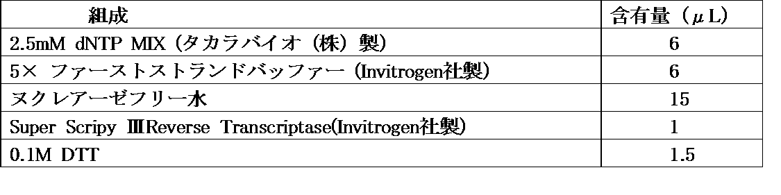
上記ストレプトアビジン(SA)磁性ビーズに固定化されたmRNA/cDNA-タンパク質連結体を、1× NEB 4バッファーで1回洗浄したのち、10UのエンドヌクレアーV(New England Biolabs社製)を含む40μLの1× NEB 4バッファーを加え、37℃で60分間インキュベートし、ストレプトアビジン(SA)磁性ビーズ結合cDNAディスプレイ分子を溶出させた。 (4) Purification of cDNA The mRNA / cDNA-protein conjugate immobilized on the above streptavidin (SA) magnetic beads was washed once with 1 × NEB 4 buffer, and then 10 U of endonuclease V (New England Biolabs). 40 μL of 1 × NEB 4 buffer was added and incubated at 37 ° C. for 60 minutes to elute streptavidin (SA) magnetic bead-bound cDNA display molecules.
(1)FACSによる解析
実施例1~3で得られた、図1Aの試験管内淘汰サイクル1回目~3回目までのスクリーニングの蛍光リポソーム結合体のFACSによる解析結果を図11A~図11Cに示す。これらのヒストグラムから、分取予定の蛍光リポソーム結合体の大きさや密度に差異が生じているものの、領域の特徴自体には変化がなく、試験管内淘汰サイクルでのスクリーニングを行なった際にも蛍光リポソーム結合体の分取が可能であることが示された。 (Example 4) Various analyzes after screening (1) Analysis by FACS Analysis by FACS of fluorescent liposome conjugates obtained in Examples 1 to 3 from the first to third screening in the in-vitro sputum cycle of FIG. 1A The results are shown in FIGS. 11A to 11C. Although these histograms show differences in the size and density of the fluorescent liposome conjugates scheduled to be collected, there are no changes in the region characteristics themselves, and even when screening in the in vitro scissor cycle, fluorescent liposomes are used. It was shown that fractionation of the conjugate is possible.
実施例3で得られたcDNAディスプレイ分子を溶出させ、以下のプライマーを用いて、常法に従ってPCRを行った。その後、常法に従って、PCR産物を6%PAGE電気泳動に供した。当該電気泳動の結果より、スクリーニングを進める(試験管内淘汰サイクルを重ねる)につれて、リポソームPCR産物のバンドが濃くなることから、FACSによるリポソームの領域を分取した際に取得できる分子数が増加していることが示された。また、対照的に、夾雑物PCR産物のバンドが薄くなっていることから、DNAライブラリでのリポソームに作用する性質を有しない翻訳産物の淘汰が進んでいることが示された(図12)。
フォワードプライマー: GATCCCGCGAAATTAATACGACTCACTATAGGGAGACCACAACGGTTTCCC(配列番号6)
リバースプライマー :TTTCCCCGCCGCCCCCCGTCCT(配列番号7) (2) Electrophoresis of PCR product of cDNA display molecule The cDNA display molecule obtained in Example 3 was eluted, and PCR was performed according to a conventional method using the following primers. Thereafter, the PCR product was subjected to 6% PAGE electrophoresis according to a conventional method. From the results of the electrophoresis, as the screening progresses (the in-vitro sputum cycle is repeated), the liposome PCR product band becomes darker, so the number of molecules that can be obtained when the liposome region by FACS is sorted increases. It was shown that In contrast, since the band of the contaminant PCR product was thin, it was shown that the translation product that does not act on the liposome in the DNA library has progressed (FIG. 12).
Forward primer: GATCCCGCGAAATTAATACGACTCACTATAGGGAGACCACAACGGTTTCCC (SEQ ID NO: 6)
Reverse primer: TTTCCCCGCCGCCCCCCGTCCT (SEQ ID NO: 7)
試験管内淘汰サイクルによる、DNAライブラリの収束度を詳細に確かめるために、大腸菌を用いたクローニングシーケンス解析を行った。実験手順は以下の通りである。
大腸菌コロニー培養プレート、及び大腸菌用の培養液としては、いずれも5gのBectoTrypton(Invitrogen社製)、2.5gのBecto Yeast extract(Invitrogen社製)、5gのNaClを混合し、MilliQ 水で500mLまでメスアップし、オートクレーブしたものを使用した。滅菌処理後の溶液温度が40℃以下になったところで、50mgのアンピシリンを加え、大腸菌コロニー培養プレート用の溶液には更に7.5gの寒天を加えて寒天培地とした。寒天培地が固まる前に、速やかにプレート上に厚さ約0.5cmになるよう流し込んだ。培養プレートに大腸菌を播種する直前に、プレート1枚につき100μLの100mM IPTG及び20μL 50mg/ml X-Galを塗りこんだ。 (3) Cloning sequence analysis Cloning sequence analysis using Escherichia coli was performed in order to confirm in detail the degree of convergence of the DNA library by the in-vitro sputum cycle. The experimental procedure is as follows.
As E. coli colony culture plate and E. coli culture solution, mix 5g BectoTrypton (Invitrogen), 2.5g Becto Yeast extract (Invitrogen), 5g NaCl, and make up to 500mL with MilliQ water. Up and autoclaved. When the solution temperature after sterilization reached 40 ° C. or lower, 50 mg of ampicillin was added, and 7.5 g of agar was further added to the solution for the E. coli colony culture plate to obtain an agar medium. Before the agar medium hardened, it was quickly poured onto the plate to a thickness of about 0.5 cm. Immediately before inoculating E. coli on the culture plate, 100 μL of 100 mM IPTG and 20 μL of 50 mg / ml X-Gal were applied to each plate.

Thr Asp Gln Phe Lys Arg Cys Ala Arg Thr Met Glu Lys Val Thr Gln Cys Pro Met Ile Glu Thr Lys Glu Gly Ala Thr Lys Ile Glu Ser Pro Pro Gln Arg [SEQ ID NO: 8]
Thr Asp Gln Phe Lys Arg Cys Ala Arg Thr Met Glu Lys Val Thr Gln Cys Pro Met Ile Glu Thr Lys Glu Gly Ala Thr Lys Ile Glu Ser Pro Pro Gln Arg
リポソームの膜に結合する融合タンパク質を調製し、リポソームの表面を標的分子で修飾した。
まず、リポソーム膜に対するアンカーとして機能する、LB-1ペプチド(配列番号9)をコードする配列と、標識分子である赤色蛍光色素mCherry2をコードする配列を含む遺伝子配列とを含む配列を、Eurofins社に依頼して合成した。常法に従って、この配列をpET-21a(+)ベクターのマルチクローニングサイトに導入した(図14A)。次に、常法に従って、上記ベクターを大腸菌に挿入し、LB-1とmcherry2との融合タンパク質を発現させた。その後、常法に従ってHisタグによる精製を行い、融合タンパク質溶液を得た。得られた融合タンパク質を模式的に図14Bに示す。 Example 5 Modification of Liposome Membrane with Liposome Membrane-Binding Fusion Protein A fusion protein that binds to the liposome membrane was prepared, and the surface of the liposome was modified with a target molecule.
First, a sequence containing a sequence encoding the LB-1 peptide (SEQ ID NO: 9), which functions as an anchor for the liposome membrane, and a gene sequence including a sequence encoding the red fluorescent dye mCherry2, which is a labeling molecule, was transferred to Eurofins. Requested and synthesized. This sequence was introduced into the multicloning site of the pET-21a (+) vector according to a conventional method (FIG. 14A). Next, according to a conventional method, the above vector was inserted into Escherichia coli to express a fusion protein of LB-1 and mcherry2. Thereafter, purification with a His tag was performed according to a conventional method to obtain a fusion protein solution. The obtained fusion protein is schematically shown in FIG. 14B.
Arg His Ser Lys Ser Leu Pro Ser Arg Val Ile Pro Arg Ala Asp Pro Arg Thr Lys Thr Arg Arg Arg Arg Arg Arg Lys Arg Thr Leu -Cys(F) [SEQ ID NO: 9]
Arg His Ser Lys Ser Leu Pro Ser Arg Val Ile Pro Arg Ala Asp Pro Arg Thr Lys Thr Arg Arg Arg Arg Arg Arg Lys Arg Thr Leu -Cys (F)
球状分子としてシリカビーズ(Micromod社製)を、標的検出分子としてストレプトアビジン結合RNAアプタマー(以下、「SAB-RNAアプタマー」ということがある。)又はストレプトアビジン非結合RNAアプタマー(以下、「SAN-RNAアプタマー」ということがある。)を用いて、スクリーニングを行なった。 (Example 6) Screening using RNA aptamer Silica beads (manufactured by Micromod) as a spherical molecule and streptavidin-binding RNA aptamer (hereinafter sometimes referred to as “SAB-RNA aptamer”) or streptavidin as a target detection molecule Screening was performed using a non-binding RNA aptamer (hereinafter sometimes referred to as “SAN-RNA aptamer”).
SAB-RNAアプタマー調製用DNAを作製するためのに、下記配列のプライマー(5’プライマー及び3’プライマー)を作成した。 (1) Preparation of DNA and RNA aptamers In order to prepare DNA for SAB-RNA aptamer preparation, primers (5 ′ primer and 3 ′ primer) having the following sequences were prepared.
3’プライマー: ATGAGTCTAGATGTAGACGCACATA(配列番号11) 5 'primer: AGTAATACGACTCACTATAGGGAGTCGACCGACCAGAATCATGCAAGTGCGTAAGATAGTCGCGGGCCGGGGGCGTATTATGTGCGTCTACATCTAGACTCAT (SEQ ID NO: 10)
3 'primer: ATGAGTCTAGATGTAGACGCACATA (SEQ ID NO: 11)
5’プライマー: AGTAATACGACTCACTATAGGGAGTCGACCGACCAGAAATGGATAACAAATTCAACAAAGAACAACAATATGTGCGTCTACATCTAGACTCAT(配列番号12) In addition, in order to prepare SAN-RNA aptamer DNA, a primer (5 ′ primer) having the following sequence was prepared. The 3 ′ primer was the same as that used for the preparation of DNA for SAB-RNA aptamer.
5 'primer: AGTAATACGACTCACTATAGGGAGTCGACCGACCAGAAATGGATAACAAATTCAACAAAGAACAACAATATGTGCGTCTACATCTAGACTCAT (SEQ ID NO: 12)


上記で得られたSAB-RNAアプタマーとストレプトアビジンとの結合アッセイを行うため、結合バッファー(pH7.4)調製した。HEPES、MgCl2及びNaClはいずれも和光純薬工業(株)から購入した。上記結合アッセイ用バッファーの組成は、下記表16に示す通りとし、超純水を加えて200mlとした。表16に示す組成の溶液に超純水を加えて800mlになるようにして30%グリセロール結合バッファー(pH7.4)を調製した。 (2) Binding assay of SAB-RNA aptamer In order to perform a binding assay between the SAB-RNA aptamer obtained above and streptavidin, a binding buffer (pH 7.4) was prepared. HEPES, MgCl 2 and NaCl were all purchased from Wako Pure Chemical Industries. The composition of the binding assay buffer was as shown in Table 16 below, and ultrapure water was added to make 200 ml. A 30% glycerol binding buffer (pH 7.4) was prepared by adding ultrapure water to a solution having the composition shown in Table 16 to 800 ml.

標的分子であるストレプトアビジンを固定する球状構造体として、粒径1.5μmでローダミンBを内包し、表面をCOOH基で修飾したシリカビーズ(製品名:Sicaster-redF、Micromod社製)を使用した。上記シリカビーズ上のCOOH基をNHS化し、NHS化されたカルボキシル基に標的分子(タンパク質)であるストレプトアビジンを、以下の手順で固定化した。 (3) Immobilization of streptavidin on the spherical structure Silica beads with a particle size of 1.5μm encapsulating rhodamine B and modified with COOH groups on the surface as a spherical structure for immobilizing the target molecule streptavidin (product name : Sicaster-redF, manufactured by Micromod). The COOH group on the silica beads was NHS, and the target molecule (protein) streptavidin was immobilized on the NHS carboxyl group by the following procedure.
ストレプトアビジン固定-ローダミンB内包シリカビーズ(以下、「プラスビーズ」ということがある。)、及びストレプトアビジンが固定化されておらず蛍光標識も含まないSicastar(Micromod社製、以下、「マイナスビーズ」ということがある)の2種類のシリカビーズ、SAB-RNAアプタマー及びSAN-RNAアプタマーを用いて、下記表17の組成物に超純水を加えて5μLとなるように各ソーティング用溶液を調製した。 (4) Sorting by FACS Streptavidin fixation—Rhodamine B-encapsulated silica beads (hereinafter sometimes referred to as “plus beads”), and Sicastar (manufactured by Micromod, with no streptavidin immobilized and no fluorescent label) Hereinafter, each of the two types of silica beads, SAB-RNA aptamer and SAN-RNA aptamer may be added to the composition shown in Table 17 to give 5 μL. A sorting solution was prepared.

プラスビーズ又はマイナスビーズを含むソーティング溶液を、それぞれセルソーターでソーティングした。これらのソーティングされた溶液200μLに、1μLの24μM ビオチン-4-フルオレセインを加えて、25℃で1時間反応させ、プラスビーズ又はマイナスビーズからSAB-RNAアプタマー又はSAN-RNAアプタマーを、ビオチンとの競合溶出によって遊離させた。その後、この溶液を入れたチューブを15,000rpmで5分間遠心し、エタノール沈殿法によって、遠心上清からRNAを回収した。 (5) Examination of sputum Each of the sorting solutions containing plus beads or minus beads was sorted with a cell sorter. To 200 μL of these sorted solutions, add 1 μL of 24 μM biotin-4-fluorescein and react at 25 ° C. for 1 hour to compete SAB-RNA aptamer or SAN-RNA aptamer with biotin from plus or minus beads. Released by elution. Thereafter, the tube containing this solution was centrifuged at 15,000 rpm for 5 minutes, and RNA was collected from the centrifuged supernatant by ethanol precipitation.
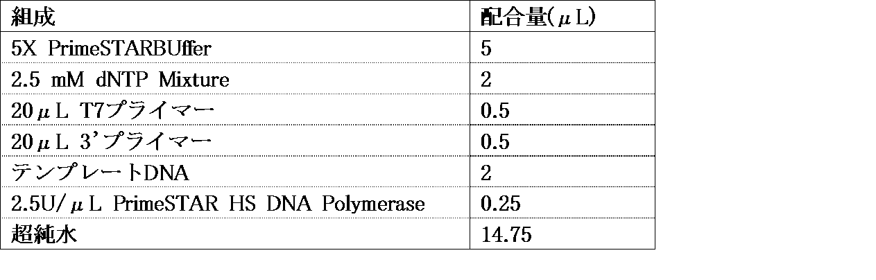
配列番号2:光架橋型リンカーの側鎖の塩基配列
配列番号3:DNAライブラリ合成用のDNA断片の塩基配列
配列番号4:DNAライブラリ合成用のDNA断片の塩基配列
配列番号5:DNAライブラリ合成用のDNA断片の塩基配列
配列番号6:PCR用フォワードプライマー
配列番号7:PCR用リバースプライマー
配列番号8:デザインDNAから得られたペプチドのアミノ酸配列
配列番号9:LB-1ペプチドのアミノ酸配列
配列番号10:SA結合RNAアプタマーのDNA合成用5’プライマー
配列番号11:RNAアプタマーのDNA合成用3’プライマー
配列番号12:非結合RNAアプタマーのDNA合成用5’プライマー
配列番号13:PCR用T7プライマー SEQ ID NO: 1: Base sequence of main chain of photocrosslinkable linker SEQ ID NO: 2: Base sequence of side chain of photocrosslinkable linker SEQ ID NO: 3: Base sequence of DNA fragment for DNA library synthesis SEQ ID NO: 4: For DNA library synthesis SEQ ID NO: 5: DNA fragment sequence for DNA library synthesis SEQ ID NO: 6: Forward primer for PCR SEQ ID NO: 7: Reverse primer for PCR SEQ ID NO: 8: Amino acid sequence of peptide obtained from design DNA SEQ ID NO: 9: amino acid sequence of LB-1 peptide SEQ ID NO: 10: 5 ′ primer for DNA synthesis of SA-binding RNA aptamer SEQ ID NO: 11: 3 ′ primer for DNA synthesis of RNA aptamer SEQ ID NO: 12: DNA synthesis of unbound
Claims (15)
- cDNAディスプレイライブラリ及び核酸アプタマーライブラリからなる群から選ばれるいずれかのライブラリの高速試験管内スクリーニング方法であって、
(i)球状分子上に標的分子を固定したポジティブ球状構造体と、標的分子を固定していないネガティブ球状構造体を調製する球状構造体調製工程と;
(ii)1010以上のライブラリサイズを有する前記いずれかのライブラリから選択した前記標的分子と結合し得る標的検出分子を、前記ポジティブ球状構造体又は前記ネガティブ球状構造体に結合させて、ポジティブ球状結合体又はネガティブ球状結合体を形成させる球状結合体形成工程と;
(iii)前記ポジティブ球状結合体と前記ネガティブ球状結合体とをセルソーターで選別する球状結合体選別工程と;
(iv)前記球状結合体工程で選別された前記ポジティブ球状結合体及び前記ネガティブ球状結合体の各表面の標的分子と結合した核酸分子をPCRに供して増幅させる増幅工程と;
(v)前記増幅工程で得られたすべての二本鎖DNAを用いて前記(i)~(iv)工程を繰り返す反復工程と;
を備える、高速試験管内スクリーニング方法。 A high-speed in vitro screening method for any library selected from the group consisting of a cDNA display library and a nucleic acid aptamer library,
(I) a positive spherical structure in which the target molecule is immobilized on the spherical molecule, and a spherical structure preparation step for preparing a negative spherical structure in which the target molecule is not immobilized;
(Ii) Positive spherical binding by binding a target detection molecule capable of binding to the target molecule selected from any one of the libraries having a library size of 10 10 or more to the positive spherical structure or the negative spherical structure A spherical conjugate forming step of forming a negative sphere conjugate or a negative spherical conjugate;
(Iii) a spherical conjugate selection step of selecting the positive spherical conjugate and the negative spherical conjugate with a cell sorter;
(Iv) an amplification step in which a nucleic acid molecule bound to a target molecule on each surface of the positive spherical conjugate and the negative spherical conjugate selected in the spherical conjugate step is subjected to amplification by PCR;
(V) an iterative step of repeating the steps (i) to (iv) using all the double-stranded DNAs obtained in the amplification step;
A high-speed in vitro screening method comprising: - 前記球状構造体は、リポソーム、セファロースビーズ、シリカビーズ及びラテックスビーズ、並びにリポソームでコートされた、セファロースビーズ、シリカビーズ、及びラテックスビーズからなる群から選ばれるいずれかであることを特徴とする、請求項1に記載の高速試験管内スクリーニング方法。 The spherical structure is any one selected from the group consisting of liposomes, sepharose beads, silica beads and latex beads, and liposome-coated sepharose beads, silica beads, and latex beads. Item 6. The high-speed in vitro screening method according to Item 1.
- 前記球状構造体は、粒径が0.5μm~20μmであることを特徴とする、請求項2に記載の高速試験管内スクリーニング方法。 3. The high-speed in vitro screening method according to claim 2, wherein the spherical structure has a particle size of 0.5 μm to 20 μm.
- 前記標的検出分子は、cDNAディスプレイ法によってcDNAディスプレイライブラリから得られる核酸-リンカー連結体、又は前記核酸-リンカー連結体から得られる核酸アプタマーである、ことを特徴とする請求項1に記載の高速試験管内スクリーニング方法。 The high-speed test according to claim 1, wherein the target detection molecule is a nucleic acid-linker conjugate obtained from a cDNA display library by a cDNA display method, or a nucleic acid aptamer obtained from the nucleic acid-linker conjugate. Intraductal screening method.
- 前記標的検出分子は、前記球状構造体表面の官能基と直接的に結合されるか、又は前記球状構造体表面に固定された前記標的分子を介して結合される、ことを特徴とする請求項1に記載の高速試験管内スクリーニング方法。 The target detection molecule is directly bonded to a functional group on the surface of the spherical structure, or is bonded through the target molecule immobilized on the surface of the spherical structure. 2. The high-speed in vitro screening method according to 1.
- 前記官能基は、カルボキシル基、アミノ基、ヒドロキシル基又はチオール基からなる群から選ばれるいずれかであることを特徴とする、請求項5に記載の高速試験管内スクリーニング方法。 The high-speed in vitro screening method according to claim 5, wherein the functional group is any one selected from the group consisting of a carboxyl group, an amino group, a hydroxyl group or a thiol group.
- 前記標的分子は、ビオチン、ストレプトアビジン、クリックケミストリーによるアジ化物及びN-ヒドロキシスクシンイミドエステル(NHS)からなる群から選ばれるいずれかの化合物であることを特徴とする、請求項5に記載の高速試験管内スクリーニング方法。 The high-speed test according to claim 5, wherein the target molecule is any compound selected from the group consisting of biotin, streptavidin, azide by click chemistry, and N-hydroxysuccinimide ester (NHS). Intraductal screening method.
- 前記標的検出分子は、以下の工程を備える前記cDNAディスプレイ法によって得られるものである、ことを特徴とする請求項1に記載の高速試験管内スクリーニング方法:
(a)所望のmRNAを調製するmRNA調製工程;
(b)前記所望のmRNAをリンカーと結合させてmRNA-リンカー連結体を得るリンカー結合工程;
(c)前記mRNA-リンカー連結体を翻訳してmRNA-リンカー-タンパク質連結体を形成させるmRNA-リンカー-タンパク質連結体形成工程;及び
(d)前記mRNA-リンカー-タンパク質連結体を逆転写してmRNA/cDNA-タンパク質連結体を得る逆転写工程。 The high-speed in vitro screening method according to claim 1, wherein the target detection molecule is obtained by the cDNA display method including the following steps:
(A) an mRNA preparation step for preparing a desired mRNA;
(B) a linker binding step of binding the desired mRNA with a linker to obtain an mRNA-linker conjugate;
(C) an mRNA-linker-protein conjugate forming step of translating the mRNA-linker conjugate to form an mRNA-linker-protein conjugate; and (d) reverse transcription of the mRNA-linker-protein conjugate to produce an mRNA. A reverse transcription step to obtain a / cDNA-protein conjugate. - 前記リンカーは、主鎖と側鎖とを有し、
前記主鎖は、(p1)前記リンカーを固相に結合する固相結合部位と(p2)前記固相から前記リンカーを切り離す切断部位と、(p3)3’末端近傍に3-シアノビニルカルバゾールで構成されたmRNA結合部位とを含み、
前記側鎖は、(s1)主鎖結合部位と、(s2)主鎖用蛍光標識結合部位を有するスペーサーと、(s3)前記主鎖用蛍光標識結合部位に結合した主鎖用蛍光標識と、(s4)ペプチド結合部位であるピューロマイシンとを備えており、
前記切断部位は前記固相結合部位内に設けられている、ことを特徴とする請求項8に記載の高速試験管内スクリーニング方法。 The linker has a main chain and a side chain,
The main chain includes (p1) a solid phase binding site for binding the linker to the solid phase, (p2) a cleavage site for separating the linker from the solid phase, and (p3) 3-cyanovinylcarbazole in the vicinity of the 3 ′ end. A configured mRNA binding site,
The side chain includes (s1) a main chain binding site, (s2) a spacer having a main chain fluorescent label binding site, and (s3) a main chain fluorescent label bound to the main chain fluorescent label binding site; (S4) comprising puromycin which is a peptide binding site,
The high-speed in vitro screening method according to claim 8, wherein the cleavage site is provided in the solid-phase binding site. - 前記(i)~(iv)工程を繰り返す回数が10回以内であることを特徴とする、請求項1に記載の高速試験管内スクリーニング方法。 2. The high-speed in vitro screening method according to claim 1, wherein the number of times the steps (i) to (iv) are repeated is 10 times or less.
- 前記球状結合体は、以下の(1)~(4)からなる群から選ばれるいずれかの組み合わせを含むことを特徴とする、請求項1に記載の高速試験管内スクリーニング方法:
(1)蛍光標識されている前記ポジティブ球状結合体、蛍光標識されていない前記ネガティブ球状結合体及び標識されていない前記標的検出分子;
(2)蛍光標識されている前記ポジティブ球状結合体、前記ポジティブ球状結合体とは異なる蛍光で蛍光標識されている前記ネガティブ球状結合体及び標識されていない前記標的検出分子体;
(3)蛍光標識されていない前記ポジティブ球状結合体、蛍光標識されている前記ネガティブ球状結合体、及び前記蛍光標識とは異なる蛍光を発する標識で標識された前記標的検出分子;及び
(4)蛍光標識されていない前記ポジティブ球状結合体、蛍光標識されていない前記ネガティブ球状結合体、及び前記蛍光標識とは異なる蛍光を発する標識で標識された前記標的検出分子。 2. The rapid in vitro screening method according to claim 1, wherein the spherical conjugate includes any combination selected from the group consisting of the following (1) to (4):
(1) The positive spherical conjugate that is fluorescently labeled, the negative spherical conjugate that is not fluorescently labeled, and the target detection molecule that is not labeled;
(2) the positive spherical conjugate that is fluorescently labeled, the negative spherical conjugate that is fluorescently labeled with a fluorescence different from that of the positive spherical conjugate, and the target detection molecule that is not labeled;
(3) the positive spherical conjugate that is not fluorescently labeled, the negative spherical conjugate that is fluorescently labeled, and the target detection molecule that is labeled with a label that emits fluorescence different from the fluorescent label; and (4) fluorescence. The positive spherical conjugate that is not labeled, the negative spherical conjugate that is not fluorescently labeled, and the target detection molecule that is labeled with a label that emits fluorescence different from the fluorescent label. - 前記蛍光標識は、Alexa Fluora 594、Fluorescein Amine、FITC、Rhodamin、mCherry2及びQuantum Dotからなる群から選ばれるいずれかであることを特徴とする、請求項11に記載の高速試験管内スクリーニング方法。 The high-speed in vitro screening method according to claim 11, wherein the fluorescent label is any one selected from the group consisting of Alexa-Fluora 594, Fluorescein-Amine, FITC, Rhodamin, mCherry2, and Quantum-Dot.
- 前記蛍光標識と異なる蛍光を発する標識は、SYBR Gold、SYBR Green及びQuantum Dotの蛍光分子からなる群から選ばれるいずれかであることを特徴とする、請求項11に記載の高速試験管内スクリーニング方法。 The high-speed in vitro screening method according to claim 11, wherein the fluorescent label different from the fluorescent label is selected from the group consisting of fluorescent molecules of SYBR® Gold, SYBR® Green, and Quantum® Dot.
- 前記選別は、2つの異なる波長で蛍光標識を励起し、前記2つの異なる波長双方で励起された前記球状結合体のみをセルソーターを用いて行うことを特徴とする、請求項1~12のいずれかに記載の高速試験管内スクリーニング方法。 The method according to any one of claims 1 to 12, wherein the selection is performed by exciting a fluorescent label at two different wavelengths and using a cell sorter only for the spherical conjugate excited at both of the two different wavelengths. 4. A high-speed in vitro screening method according to 1.
- 特定のポリペプチドを共存させて選択圧をかけることにより、さらに前記選別を行なう請求項13に記載の高速試験管内スクリーニング方法。 The high-speed in vitro screening method according to claim 13, wherein the screening is further performed by applying a selective pressure in the presence of a specific polypeptide.
Priority Applications (5)
| Application Number | Priority Date | Filing Date | Title |
|---|---|---|---|
| EP17775310.0A EP3450554B1 (en) | 2016-03-30 | 2017-03-29 | High-speed in vitro screening method |
| ES17775310T ES2929650T3 (en) | 2016-03-30 | 2017-03-29 | Method for high speed in vitro screening |
| JP2018509384A JP6995367B2 (en) | 2016-03-30 | 2017-03-29 | High-speed in vitro screening method |
| US16/145,292 US10781442B2 (en) | 2016-03-30 | 2018-09-28 | High-speed in vitro screening method |
| US17/025,954 US11634705B2 (en) | 2016-03-30 | 2020-09-18 | High-speed in vitro screening method |
Applications Claiming Priority (2)
| Application Number | Priority Date | Filing Date | Title |
|---|---|---|---|
| JP2016-069064 | 2016-03-30 | ||
| JP2016069064 | 2016-03-30 |
Related Child Applications (1)
| Application Number | Title | Priority Date | Filing Date |
|---|---|---|---|
| US16/145,292 Continuation US10781442B2 (en) | 2016-03-30 | 2018-09-28 | High-speed in vitro screening method |
Publications (1)
| Publication Number | Publication Date |
|---|---|
| WO2017170776A1 true WO2017170776A1 (en) | 2017-10-05 |
Family
ID=59964718
Family Applications (1)
| Application Number | Title | Priority Date | Filing Date |
|---|---|---|---|
| PCT/JP2017/013076 WO2017170776A1 (en) | 2016-03-30 | 2017-03-29 | High-speed in vitro screening method |
Country Status (5)
| Country | Link |
|---|---|
| US (1) | US10781442B2 (en) |
| EP (1) | EP3450554B1 (en) |
| JP (1) | JP6995367B2 (en) |
| ES (1) | ES2929650T3 (en) |
| WO (1) | WO2017170776A1 (en) |
Cited By (4)
| Publication number | Priority date | Publication date | Assignee | Title |
|---|---|---|---|---|
| JP2019146516A (en) * | 2018-02-27 | 2019-09-05 | 学校法人甲南学園 | Screening method of rna aptamer |
| WO2022102791A1 (en) * | 2020-11-16 | 2022-05-19 | 国立大学法人埼玉大学 | Novel linker for antigen detection and antigen detection method using same |
| JP2022527351A (en) * | 2019-04-02 | 2022-06-01 | イーライ リリー アンド カンパニー | Methods for selecting and detecting bound peptides |
| WO2022270396A1 (en) * | 2021-06-21 | 2022-12-29 | 株式会社Epsilon Molecular Engineering | Double-screening method |
Families Citing this family (2)
| Publication number | Priority date | Publication date | Assignee | Title |
|---|---|---|---|---|
| JP6506838B2 (en) * | 2015-03-31 | 2019-05-08 | 国立大学法人埼玉大学 | High-speed photocrosslinkable co-linker for in-vitro sputum and intermolecular interaction analysis, in-vitro sputum method using the linker |
| CN114585736B (en) * | 2020-10-27 | 2023-05-02 | 北京寻因生物科技有限公司 | Puromycin linker and application thereof in-vitro nucleic acid display peptide synthesis |
Citations (4)
| Publication number | Priority date | Publication date | Assignee | Title |
|---|---|---|---|---|
| JP2006211984A (en) * | 2005-02-04 | 2006-08-17 | Univ Nagoya | Method for amplifying nucleic acid utilizing emulsion, and kit for nucleic acid amplification reaction |
| JP2010006745A (en) * | 2008-06-27 | 2010-01-14 | Sekisui Chem Co Ltd | Fused protein, fused protein-immobilized carrier, method for screening compound, screening composition and screening kit |
| JP2012531887A (en) * | 2009-04-01 | 2012-12-13 | ディクステリティー ダイアグノーティクス インコーポレイテッド | Chemical ligation-dependent probe amplification (CLPA) |
| JP2014515749A (en) * | 2011-04-21 | 2014-07-03 | ガーバン インスティテュート オブ メディカル リサーチ | Modified variable domain molecules and methods for their production and use B |
Family Cites Families (8)
| Publication number | Priority date | Publication date | Assignee | Title |
|---|---|---|---|---|
| US6699658B1 (en) | 1996-05-31 | 2004-03-02 | Board Of Trustees Of The University Of Illinois | Yeast cell surface display of proteins and uses thereof |
| US7115396B2 (en) * | 1998-12-10 | 2006-10-03 | Compound Therapeutics, Inc. | Protein scaffolds for antibody mimics and other binding proteins |
| WO2002032925A2 (en) * | 2000-10-16 | 2002-04-25 | Phylos, Inc. | Protein scaffolds for antibody mimics and other binding proteins |
| JP5495088B2 (en) * | 2007-11-19 | 2014-05-21 | 国立大学法人東京農工大学 | PQQGDH controlled aptamer and its use |
| CN102084002B (en) | 2008-03-03 | 2014-09-10 | Abbvie公司 | Methods for transforming yeast |
| JP2011055770A (en) | 2009-09-11 | 2011-03-24 | Kobe Univ | Nucleic acid aptamer, and method for screening the same |
| US9994616B2 (en) | 2013-10-15 | 2018-06-12 | The University Of Tokyo | c-Met protein agonist |
| JP2016002041A (en) * | 2014-06-17 | 2016-01-12 | キリン株式会社 | Dna molecules which bind to caffeine and method for detecting caffeine |
-
2017
- 2017-03-29 ES ES17775310T patent/ES2929650T3/en active Active
- 2017-03-29 WO PCT/JP2017/013076 patent/WO2017170776A1/en active Application Filing
- 2017-03-29 EP EP17775310.0A patent/EP3450554B1/en active Active
- 2017-03-29 JP JP2018509384A patent/JP6995367B2/en active Active
-
2018
- 2018-09-28 US US16/145,292 patent/US10781442B2/en active Active
Patent Citations (4)
| Publication number | Priority date | Publication date | Assignee | Title |
|---|---|---|---|---|
| JP2006211984A (en) * | 2005-02-04 | 2006-08-17 | Univ Nagoya | Method for amplifying nucleic acid utilizing emulsion, and kit for nucleic acid amplification reaction |
| JP2010006745A (en) * | 2008-06-27 | 2010-01-14 | Sekisui Chem Co Ltd | Fused protein, fused protein-immobilized carrier, method for screening compound, screening composition and screening kit |
| JP2012531887A (en) * | 2009-04-01 | 2012-12-13 | ディクステリティー ダイアグノーティクス インコーポレイテッド | Chemical ligation-dependent probe amplification (CLPA) |
| JP2014515749A (en) * | 2011-04-21 | 2014-07-03 | ガーバン インスティテュート オブ メディカル リサーチ | Modified variable domain molecules and methods for their production and use B |
Non-Patent Citations (4)
| Title |
|---|
| ERI HAMADA ET AL.: "Development of a high- throughput screening system using FACS for peptide aptamers", THE 39TH ANNUAL MEETING OF THE MOLECULAR BIOLOGY SOCIETY OF JAPAN PROGRAM YOSHISHU, vol. 39, 16 November 2016 (2016-11-16), XP009513279 * |
| HIROSHI YANAGAWA ET AL.: "Development of IVV method into drug discovery", DRUG DELIVERY SYSTEM, vol. 26, no. 6, 2011, pages 571 - 583, 573, XP055550493 * |
| MOCHIZUKI Y. ET AL.: "A versatile puromycin- linker using cnvK for high-throughput in vitro selection by cDNA display", JOURNAL OF BIOTECHNOLOGY, vol. 212, 28 August 2015 (2015-08-28), pages 174 - 180, XP029284068 * |
| See also references of EP3450554A4 * |
Cited By (5)
| Publication number | Priority date | Publication date | Assignee | Title |
|---|---|---|---|---|
| JP2019146516A (en) * | 2018-02-27 | 2019-09-05 | 学校法人甲南学園 | Screening method of rna aptamer |
| JP7053009B2 (en) | 2018-02-27 | 2022-04-12 | 玉樹 遠藤 | RNA aptamer screening method |
| JP2022527351A (en) * | 2019-04-02 | 2022-06-01 | イーライ リリー アンド カンパニー | Methods for selecting and detecting bound peptides |
| WO2022102791A1 (en) * | 2020-11-16 | 2022-05-19 | 国立大学法人埼玉大学 | Novel linker for antigen detection and antigen detection method using same |
| WO2022270396A1 (en) * | 2021-06-21 | 2022-12-29 | 株式会社Epsilon Molecular Engineering | Double-screening method |
Also Published As
| Publication number | Publication date |
|---|---|
| US10781442B2 (en) | 2020-09-22 |
| EP3450554B1 (en) | 2022-09-21 |
| JPWO2017170776A1 (en) | 2019-03-28 |
| EP3450554A1 (en) | 2019-03-06 |
| EP3450554A4 (en) | 2020-01-08 |
| JP6995367B2 (en) | 2022-02-17 |
| ES2929650T3 (en) | 2022-11-30 |
| US20190085322A1 (en) | 2019-03-21 |
Similar Documents
| Publication | Publication Date | Title |
|---|---|---|
| WO2017170776A1 (en) | High-speed in vitro screening method | |
| US11300572B2 (en) | Methods and systems for producing nanolipoprotein particles | |
| US20020072087A1 (en) | Molecule assigning genotype to phenotype and use thereof | |
| JP3750020B2 (en) | C-terminal modified protein and production method thereof, modifying agent and translation template used for production of C-terminal modified protein, and protein interaction detection method using C-terminal modified protein | |
| JP6506838B2 (en) | High-speed photocrosslinkable co-linker for in-vitro sputum and intermolecular interaction analysis, in-vitro sputum method using the linker | |
| JP5733784B2 (en) | Efficient synthesis of cDNA / mRNA-protein conjugates | |
| US11634705B2 (en) | High-speed in vitro screening method | |
| JP2006211984A (en) | Method for amplifying nucleic acid utilizing emulsion, and kit for nucleic acid amplification reaction | |
| JP6762001B2 (en) | Unnatural amino acid-containing peptide library | |
| JP5049136B2 (en) | Efficient synthesis method of protein labeled with N-terminal amino acid | |
| JP7153996B2 (en) | Method for producing crosslinked peptide, and crosslinked peptide produced using the method | |
| Doi et al. | Photocleavable linkage between genotype and phenotype for rapid and efficient recovery of nucleic acids encoding affinity-selected proteins | |
| JP3706942B2 (en) | Method for detecting interaction between substance and protein, method for screening protein interacting with substance, and method for forming complex of substance and protein interacting with the substance | |
| JP6478392B2 (en) | Nucleic acid linker | |
| JP2020007309A (en) | Method for producing chemically cross-linked peptide, chemically cross-linked peptide prepared using the method, and peptide library by cdna display method using the constructed peptide | |
| JP5587528B2 (en) | Protein that forms a complex with c-Jun protein, nucleic acid encoding the same, and method of using the same | |
| WO2022102791A1 (en) | Novel linker for antigen detection and antigen detection method using same | |
| JP2015227806A (en) | High sensitivity detection method of target molecule using magnetic material gel bead, and detection kit | |
| JPWO2005001086A1 (en) | Immobilized mRNA-puromycin conjugate and uses thereof | |
| JP4437193B2 (en) | Nucleic acid library and protein library | |
| JP6590474B2 (en) | Method for producing chemically cross-linked peptide, chemically cross-linked peptide produced using the method, and peptide library by cDNA display method constructed using the peptide | |
| WO2020171236A1 (en) | A NOVEL IMMUNO-PCR METHOD USING cDNA DISPLAY | |
| WO2021182587A1 (en) | Target module for use in search for novel peptide aptamer utilizing photocrosslinkable base, linker for use in search for novel peptide aptamer, and method for searching for novel peptide aptamer using said target module or said linker | |
| JP4747292B2 (en) | Translation templates and libraries thereof, proteins and protein libraries synthesized from them, elements constituting them, and methods for producing and using them | |
| JP6194894B2 (en) | Nucleic acid linker |
Legal Events
| Date | Code | Title | Description |
|---|---|---|---|
| DPE1 | Request for preliminary examination filed after expiration of 19th month from priority date (pct application filed from 20040101) | ||
| WWE | Wipo information: entry into national phase |
Ref document number: 2018509384 Country of ref document: JP |
|
| NENP | Non-entry into the national phase |
Ref country code: DE |
|
| WWE | Wipo information: entry into national phase |
Ref document number: 2017775310 Country of ref document: EP |
|
| ENP | Entry into the national phase |
Ref document number: 2017775310 Country of ref document: EP Effective date: 20181030 |
|
| 121 | Ep: the epo has been informed by wipo that ep was designated in this application |
Ref document number: 17775310 Country of ref document: EP Kind code of ref document: A1 |
|
| ENP | Entry into the national phase |
Ref document number: 2017775310 Country of ref document: EP Effective date: 20181030 |