WO2017135048A1 - 光線力学療法用組成物、治療方法、殺菌システムおよび殺菌システムの作動方法 - Google Patents
光線力学療法用組成物、治療方法、殺菌システムおよび殺菌システムの作動方法 Download PDFInfo
- Publication number
- WO2017135048A1 WO2017135048A1 PCT/JP2017/001727 JP2017001727W WO2017135048A1 WO 2017135048 A1 WO2017135048 A1 WO 2017135048A1 JP 2017001727 W JP2017001727 W JP 2017001727W WO 2017135048 A1 WO2017135048 A1 WO 2017135048A1
- Authority
- WO
- WIPO (PCT)
- Prior art keywords
- composition
- control unit
- light
- calculation
- imaging
- Prior art date
- Legal status (The legal status is an assumption and is not a legal conclusion. Google has not performed a legal analysis and makes no representation as to the accuracy of the status listed.)
- Ceased
Links
Images


Classifications
-
- A—HUMAN NECESSITIES
- A61—MEDICAL OR VETERINARY SCIENCE; HYGIENE
- A61K—PREPARATIONS FOR MEDICAL, DENTAL OR TOILETRY PURPOSES
- A61K41/00—Medicinal preparations obtained by treating materials with wave energy or particle radiation ; Therapies using these preparations
- A61K41/0057—Photodynamic therapy with a photosensitizer, i.e. agent able to produce reactive oxygen species upon exposure to light or radiation, e.g. UV or visible light; photocleavage of nucleic acids with an agent
-
- A—HUMAN NECESSITIES
- A61—MEDICAL OR VETERINARY SCIENCE; HYGIENE
- A61B—DIAGNOSIS; SURGERY; IDENTIFICATION
- A61B1/00—Instruments for performing medical examinations of the interior of cavities or tubes of the body by visual or photographical inspection, e.g. endoscopes; Illuminating arrangements therefor
-
- A—HUMAN NECESSITIES
- A61—MEDICAL OR VETERINARY SCIENCE; HYGIENE
- A61K—PREPARATIONS FOR MEDICAL, DENTAL OR TOILETRY PURPOSES
- A61K31/00—Medicinal preparations containing organic active ingredients
- A61K31/33—Heterocyclic compounds
- A61K31/395—Heterocyclic compounds having nitrogen as a ring hetero atom, e.g. guanethidine or rifamycins
- A61K31/54—Heterocyclic compounds having nitrogen as a ring hetero atom, e.g. guanethidine or rifamycins having six-membered rings with at least one nitrogen and one sulfur as the ring hetero atoms, e.g. sulthiame
- A61K31/5415—Heterocyclic compounds having nitrogen as a ring hetero atom, e.g. guanethidine or rifamycins having six-membered rings with at least one nitrogen and one sulfur as the ring hetero atoms, e.g. sulthiame ortho- or peri-condensed with carbocyclic ring systems, e.g. phenothiazine, chlorpromazine, piroxicam
-
- A—HUMAN NECESSITIES
- A61—MEDICAL OR VETERINARY SCIENCE; HYGIENE
- A61K—PREPARATIONS FOR MEDICAL, DENTAL OR TOILETRY PURPOSES
- A61K41/00—Medicinal preparations obtained by treating materials with wave energy or particle radiation ; Therapies using these preparations
-
- A—HUMAN NECESSITIES
- A61—MEDICAL OR VETERINARY SCIENCE; HYGIENE
- A61K—PREPARATIONS FOR MEDICAL, DENTAL OR TOILETRY PURPOSES
- A61K47/00—Medicinal preparations characterised by the non-active ingredients used, e.g. carriers or inert additives; Targeting or modifying agents chemically bound to the active ingredient
- A61K47/02—Inorganic compounds
-
- A—HUMAN NECESSITIES
- A61—MEDICAL OR VETERINARY SCIENCE; HYGIENE
- A61P—SPECIFIC THERAPEUTIC ACTIVITY OF CHEMICAL COMPOUNDS OR MEDICINAL PREPARATIONS
- A61P17/00—Drugs for dermatological disorders
-
- A—HUMAN NECESSITIES
- A61—MEDICAL OR VETERINARY SCIENCE; HYGIENE
- A61P—SPECIFIC THERAPEUTIC ACTIVITY OF CHEMICAL COMPOUNDS OR MEDICINAL PREPARATIONS
- A61P31/00—Antiinfectives, i.e. antibiotics, antiseptics, chemotherapeutics
- A61P31/04—Antibacterial agents
-
- A—HUMAN NECESSITIES
- A61—MEDICAL OR VETERINARY SCIENCE; HYGIENE
- A61P—SPECIFIC THERAPEUTIC ACTIVITY OF CHEMICAL COMPOUNDS OR MEDICINAL PREPARATIONS
- A61P31/00—Antiinfectives, i.e. antibiotics, antiseptics, chemotherapeutics
- A61P31/10—Antimycotics
-
- Y—GENERAL TAGGING OF NEW TECHNOLOGICAL DEVELOPMENTS; GENERAL TAGGING OF CROSS-SECTIONAL TECHNOLOGIES SPANNING OVER SEVERAL SECTIONS OF THE IPC; TECHNICAL SUBJECTS COVERED BY FORMER USPC CROSS-REFERENCE ART COLLECTIONS [XRACs] AND DIGESTS
- Y02—TECHNOLOGIES OR APPLICATIONS FOR MITIGATION OR ADAPTATION AGAINST CLIMATE CHANGE
- Y02A—TECHNOLOGIES FOR ADAPTATION TO CLIMATE CHANGE
- Y02A50/00—TECHNOLOGIES FOR ADAPTATION TO CLIMATE CHANGE in human health protection, e.g. against extreme weather
- Y02A50/30—Against vector-borne diseases, e.g. mosquito-borne, fly-borne, tick-borne or waterborne diseases whose impact is exacerbated by climate change
Definitions
- the present invention relates to a composition for photodynamic therapy, a treatment method, a sterilization system, and a method for operating the sterilization system. More particularly, the present invention relates to a composition for photodynamic therapy for treating H. pylori infection, a method for treating H. pylori infection, a sterilization system for lesions caused by H. pylori infection, and H. pylori infection.
- Method of operating lesion sterilization system, photodynamic therapy composition for treating mucocutaneous infection, method of treating mucocutaneous infection, sterilization system of lesion due to mucocutaneous infection, and mucocutaneous infection The present invention relates to a method for operating a sterilization system for a lesion caused by a disease.
- photosensitizers photosensitive substances
- pathogenic microorganisms present in lesions of infectious diseases such as skin and mucous membranes, and light is emitted by irradiating light of a wavelength corresponding to the photosensitizer. It is a method for treating cutaneous mucosal infection by generating active oxygen from a sensitizer and sterilizing pathogenic microorganisms.
- Methylene blue has been put to practical use in clinical settings and daily life as a drug for methemoglobinemia, a dye in contrast methods for chromoendoscopy, a stain in dyeing methods, and a fish disease drug. It is.
- Patent Document 1 discloses the use of methylene blue as an antibacterial agent in the dental field by inducing cell death by singlet oxygen generated by light absorption.
- methylene blue alone has a weak antibacterial activity and requires a large amount of application with an ointment, which limits its application to oral infections.
- Non-Patent Document 1 reports that a hydrophobic derivative of methylene blue showed a stronger killing effect than MRA against MRSA (methicillin-resistant Staphylococcus aureus) and VRSA (vancomycin-resistant Staphylococcus aureus). ing.
- MRSA methicillin-resistant Staphylococcus aureus
- VRSA vancomycin-resistant Staphylococcus aureus
- the present invention provides a photodynamic therapy composition for treating H. pylori infection, a photosensitizing effect strong and safe, a method for treating H. pylori infection, and a sterilization system for lesions caused by H. pylori infection And a method for operating a system for sterilizing a lesion caused by Helicobacter pylori infection, and a photodynamic therapy composition for treating skin mucosal infection, which has a strong photosensitizing effect and is safe, and mucosal mucosa It is an object of the present invention to provide a method for treating an infection, a sterilization system for a lesion caused by a mucocutaneous infection, and a method for operating the sterilization system for a lesion caused by a skin mucosal infection.
- the present invention provides the following [1] to [22].
- [1] A composition for photodynamic therapy for treating H. pylori infection comprising methylene blue and having a pH of 7.6 to 9.5.
- composition for photodynamic therapy for treating a H. pylori infection according to any one of [5].
- composition for photodynamic therapy for treating a Helicobacter pylori infection according to any one of [1] to [6] described above is applied endoscopically to a lesioned part of a Helicobacter pylori infection.
- a method for treating H. pylori infection which is applied and irradiated with white light or LED light or laser light having a wavelength of 660 ⁇ 10 nm.
- a composition for photodynamic therapy for treating cutaneous mucosal infection comprising methylene blue and having a pH of 7.6 to 9.5.
- a composition for photodynamic therapy for treating infectious diseases comprising methylene blue and having a pH of 7.6 to 9.5.
- the pathogenic microorganism is selected from the group consisting of Candida albicans, methicillin-resistant Staphylococcus aureus, Pseudomonas aeruginosa, and Escherichia coli.
- the composition for photodynamic therapy for treating a mucocutaneous infection according to the above [13] which is at least one.
- the mucocutaneous infection is at least one selected from the group consisting of candidiasis, methicillin-resistant Staphylococcus aureus infection, Pseudomonas aeruginosa infection, and E. coli infection.
- the composition for photodynamic therapy for treating skin mucosal infection as described in any one of these.
- the composition for photodynamic therapy for treating cutaneous mucosal infection according to any one of the above [9] to [15] is applied to a lesion site of the cutaneous mucosal infection, and white light or A method for treating a mucocutaneous infection, which is irradiated with LED light or laser light having a wavelength of 660 ⁇ 10 nm.
- Imaging means for controlling the imaging direction of the imaging means, irradiation direction control means for controlling the light irradiation direction of the light irradiation means, An injection direction control means for controlling the injection direction of the composition injection means, the imaging means, the light irradiation means, the composition injection means, the imaging direction control means, the irradiation direction control means, and the injection direction control means.
- a sterilization system for lesions caused by Helicobacter pylori infection comprising a calculation / control unit for controlling, (a) The imaging direction control means and the irradiation direction control means operate based on a control signal from the calculation / control unit so that the light irradiation means illuminates the observation area of the imaging means, (b) The imaging means images the mucous membrane based on the control signal from the calculation / control unit, and transmits a video signal to the calculation / control unit, (c) The calculation / control unit receives the video signal transmitted from the imaging means, performs video processing so as to emphasize the color difference between the normal region and the abnormal region of the mucous membrane, Identify the location of the lesion, (d) The injection direction control means operates based on a control signal from the calculation / control unit so as to match the injection direction of the composition injection means with the lesioned part, (e) The composition injection means comprises a photodynamic for treating the Helicobacter pylori infection according to any one of [1]
- the irradiation direction control means operates to match the irradiation direction of the light irradiation means with the lesioned part based on a control signal from the calculation / control unit, and (g) The light irradiation means emits white light or LED light or laser light having a wavelength of 660 ⁇ 10 nm based on a control signal from the calculation / control unit. Sterilization system for lesions caused by Helicobacter pylori infection.
- Imaging means for controlling the imaging direction of the imaging means, irradiation direction control means for controlling the light irradiation direction of the light irradiation means, An injection direction control means for controlling the injection direction of the composition injection means, the imaging means, the light irradiation means, the composition injection means, the imaging direction control means, the irradiation direction control means, and the injection direction control means.
- An operation method of a sterilization system for a lesion caused by Helicobacter pylori infection comprising a calculation / control unit for controlling, (a) actuating the imaging direction control unit and the irradiation direction control unit based on a control signal from the calculation / control unit so that the light irradiation unit illuminates the observation region of the imaging unit; (b) a step of imaging the mucous membrane based on a control signal from the calculation / control unit, and transmitting a video signal to the calculation / control unit, the imaging means; (c) The calculation / control unit receives the video signal transmitted from the imaging means, performs video processing so as to emphasize the color difference between the normal region and the abnormal region of the mucous membrane, And identifying the location of the lesion, (d) a step of operating the injection direction control unit based on a control signal from the calculation / control unit so as to match the injection direction of the composition injection unit with a lesioned part, (e) Photodynamics for treating the Helico
- the light irradiation means includes a step of emitting white light or LED light or laser light having a wavelength of 660 ⁇ 10 nm based on a control signal from the calculation / control unit. How to operate the sterilization system.
- Imaging means for controlling the imaging direction of the imaging means, irradiation direction control means for controlling the light irradiation direction of the light irradiation means, An injection direction control means for controlling the injection direction of the composition injection means, the imaging means, the light irradiation means, the composition injection means, the imaging direction control means, the irradiation direction control means, and the injection direction control means.
- a sterilization system for lesions caused by mucocutaneous infections comprising a calculation / control unit for controlling, (a) The imaging direction control means and the irradiation direction control means operate based on a control signal from the calculation / control unit so that the light irradiation means illuminates the observation area of the imaging means, (b) The imaging means, based on a control signal from the calculation / control unit, images the skin or mucous membrane, transmits a video signal to the calculation / control unit, (c) The calculation / control unit receives the video signal transmitted from the imaging means, performs video processing so as to emphasize the color difference between the normal region and the abnormal region of the skin or mucous membrane, Identify the lesion, locate the lesion, (d) The injection direction control means operates based on a control signal from the calculation / control unit so as to match the injection direction of the composition injection means with the lesioned part, (e) The composition ejecting means is a photodynamic for treating the mucocutaneous infection according to any one of [9
- the irradiation direction control means operates to match the irradiation direction of the light irradiation means with the lesioned part based on a control signal from the calculation / control unit, and (g) The light irradiation means emits white light or LED light or laser light having a wavelength of 660 ⁇ 10 nm based on a control signal from the calculation / control unit. Sterilization system for lesions caused by mucocutaneous infections.
- Imaging means for controlling the imaging direction of the imaging means, irradiation direction control means for controlling the light irradiation direction of the light irradiation means, An injection direction control means for controlling the injection direction of the composition injection means, the imaging means, the light irradiation means, the composition injection means, the imaging direction control means, the irradiation direction control means, and the injection direction control means.
- An operation method of a sterilization system for a lesion caused by a mucocutaneous infection comprising a calculation / control unit for controlling, (a) actuating the imaging direction control unit and the irradiation direction control unit based on a control signal from the calculation / control unit so that the light irradiation unit illuminates the observation region of the imaging unit; (b) a step of imaging the skin or mucous membrane based on a control signal from the calculation / control unit and transmitting a video signal to the calculation / control unit, (c) The calculation / control unit receives the video signal transmitted from the imaging means, performs video processing so as to emphasize the color difference between the normal region and abnormal region of the skin or mucous membrane, Identifying the lesion and identifying the location of the lesion, (d) a step of operating the injection direction control unit based on a control signal from the calculation / control unit so as to match the injection direction of the composition injection unit with a lesioned part, (e) Photodynamics for treating the mu
- the light irradiation means includes a step of emitting white light, LED light having a wavelength of 660 ⁇ 10 nm or laser light based on a control signal from the calculation / control unit, and How to operate the sterilization system.
- a photodynamic therapy composition for treating H. pylori infection, a photosensitizing effect strong and safe a method for treating H. pylori infection, and a sterilization system for lesions caused by H. pylori infection
- a method for operating a system for sterilizing a lesion caused by Helicobacter pylori infection and a photodynamic therapy composition for treating skin mucosal infection, which has a strong photosensitizing effect and is safe, and mucosal mucosa
- FIG. 1 is a conceptual diagram showing a sterilization system according to the present invention.
- FIG. 2 is a perspective view showing a specific example of the sterilization system according to the present invention.
- FIG. 3A is a front view showing a distal end cap of the endoscope.
- FIG. 3B is a front view showing another aspect of the distal end cap of the endoscope.
- FIG. 4A is a cross-sectional view showing a flexible tube portion of an endoscope.
- FIG. 4B is a cross-sectional view showing another aspect of the flexible tube portion of the endoscope.
- the characteristic point of the present invention is that it is a composition for photodynamic therapy in a specific pH range containing methylene blue whose safety has already been established. Even when adjusting to a specific pH range, a composition for photodynamic therapy having low toxicity and extremely high safety can be obtained by using a pH adjuster whose safety has already been established.
- the range when a range is expressed using “ ⁇ ”, the range includes both sides of “ ⁇ ”.
- the range represented by “A to B” includes “A” and “B”.
- composition for photodynamic therapy for treating H. pylori infection contains methylene blue and has a pH of 7.6 to 9.5.
- Helicobacter pylori infection is an infection of the mucous membrane caused by Helicobacter pylori, and particularly refers to an infection of the gastric mucosa caused by Helicobacter pylori.
- Helicobacter pylori, Helicobacter mustelae, Helicobacter mustelae, Helicobacter mustelae, Helicobacter mustelae and Helicobacter ferris are not particularly limited as long as they are bacteria belonging to the genus Helicobacter having urease working under acidic conditions. It is at least one selected from the group consisting of (Helicobacter felis), more preferably Helicobacter pylori.
- Methylene blue (3,7-bis (dimethylamino) phenothiazinium chloride) contained in the composition for photodynamic therapy for treating H. pylori infection of the present invention is not particularly limited, and for example, an anhydride It may be a hydrate. Examples of the hydrate include, but are not limited to, dihydrate, trihydrate, and tetrahydrate.
- methylene blue may be sold as a pharmaceutical or may be sold as a reagent. A reagent having a high purity is preferable, and for example, a reagent specified in JIS K 8897: 2012 is more preferable.
- Methylene blue is safe for the human body to be used for staining during chromoendoscopy of the gastrointestinal tract, intravenously or orally administered in the treatment of drug-induced methemoglobinemia, treatment of ifosfamide encephalopathy It has been established because it is used by intravenous injection. It is low toxic to animals other than humans, and the safety of methylene blue has been established.
- the concentration of methylene blue when the composition for photodynamic therapy for treating H. pylori infection of the present invention is brought into contact with H. pylori is not particularly limited, but is preferably 0.0007 to 4.0 w / v%, more preferably Is 0.001 to 3.0 w / v%, more preferably 0.003 to 2.0 w / v%, still more preferably 0.005 to 1.0 w / v%, and still more preferably 0.007 to 0. 80 w / v%.
- the antibacterial activity against H. pylori is further improved.
- the lowest digit of the concentration is rounded off by one digit below that. For example, “0.001” includes 0.00095 or more and less than 0.0015, and “4.0” includes 3.95 or more and less than 4.05.
- the pH of the photodynamic therapy composition for treating H. pylori infection of the present invention is 7.6 to 9.5, preferably 8.3 to 9.2, and more preferably 8.5. Is 9.0.
- the pH when the composition for photodynamic therapy for treating H. pylori infection of the present invention is brought into contact with H. pylori is within this range, the antibacterial activity against H. pylori is excellent.
- the pH is rounded off to the second decimal place.
- “7.6” is 7.55 or more and less than 7.65
- “9.5” is 9.45 or more and less than 9.55. Respectively.
- the pH was measured with a pH meter using a glass electrode specified in JIS Z 8802: 2011 “pH measurement method”. 25 PH value at ° C.
- the method for adjusting the pH of the composition for photodynamic therapy for treating H. pylori infection of the present invention is not particularly limited, and examples thereof include a method of adding a pH adjuster.
- a pH adjuster those having low toxicity to humans or animals other than humans are preferable.
- citric acid, gluconic acid, succinic acid, lactic acid, sodium hydrogen carbonate (also referred to as “bicarbonate”) examples thereof include sodium carbonate, potassium carbonate, potassium dihydrogen phosphate, dipotassium hydrogen phosphate, sodium dihydrogen phosphate, and disodium hydrogen phosphate.
- sodium bicarbonate is particularly preferred.
- sodium hydrogen carbonate When sodium hydrogen carbonate is used as a pH adjuster, the antibacterial activity is further improved.
- sodium bicarbonate is used as a pH adjuster and when potassium dihydrogen phosphate (KH 2 PO 4 ) and disodium hydrogen phosphate (Na 2 HPO 4 ) are used, the same pH is obtained. Even so, the use of sodium hydrogen carbonate shows superior antibacterial activity.
- potassium dihydrogen phosphate (KH 2 PO 4 ) and disodium hydrogen phosphate (Na 2 HPO 4 ) the same pH is obtained. Even so, the use of sodium hydrogen carbonate shows superior antibacterial activity.
- KH 2 PO 4 potassium dihydrogen phosphate
- Na 2 HPO 4 disodium hydrogen phosphate
- the addition amount of the pH adjuster is not particularly limited as long as it is added so that the pH of the composition for photodynamic therapy for treating H. pylori infection of the present invention falls within the above-described pH range.
- sodium bicarbonate sodium bicarbonate
- the concentration cannot be generally stated, but is preferably, for example, 0.007 to 0.80 w / v%, more preferably 0.01 to 0.70 w / v%, more preferably 0.05 to 0.65 w / v%, even more preferably 0.10 to 0.60 w / v%, and even more preferably 0.15 to 0.40 w. / V%.
- the antibacterial activity against H. pylori is further improved.
- the photodynamic therapy composition for treating H. pylori infection of the present invention may further contain a pharmaceutically acceptable additive.
- Pharmaceutically acceptable additives include pH adjusters, buffers, wetting agents / solubilizers, antibacterial preservatives, chelating agents, complexing agents, antioxidants, sweeteners, and excipients other than those mentioned above Agents, binders, disintegrants, lubricants, fluidizers / anti-caking agents, and suspending agents / adhesives. These additives can be included in the composition for photodynamic therapy for treating H. pylori infections of the present invention alone or in combination of two or more.
- composition for photodynamic therapy for treating H. pylori infection of the present invention is preferably brought into contact with H. pylori present in a lesion of H. pylori infection and irradiated with light to H. pylori. Used in photodynamic therapy to treat fungal infections.
- the composition for photodynamic therapy for treating Helicobacter pylori infection of the present invention can be used for humans as well as animals other than humans.
- non-human animals include, without limitation, non-human mammals including monkeys, cats, pigs, dogs and the like.
- animals other than humans include, in addition to mammals, vertebrates other than mammals such as birds, reptiles, amphibians, and fish, but are not limited thereto.
- animals other than humans include domestic animals such as dogs, cats, cows, horses, pigs, sheep, goats, donkeys and camels, and dogs, cats, foxes, raccoons, monkeys, donkeys, and horses.
- the dosage form of the photodynamic therapy composition for treating H. pylori infection of the present invention is not particularly limited, but is preferably locally administered, particularly preferably endoscopically in the lesioned part of the gastric mucosa. Administration by nebulization directly through a mirror. Specifically, for example, methylene blue and, if necessary, a pH adjusting agent are dissolved in pure water or physiological saline and filled into a syringe having a tube at the tip, and the tube is inserted into the forceps port of the endoscope. The drug can be sprayed onto the stomach wall by pressing.
- the method for producing the composition for photodynamic therapy for treating H. pylori infection of the present invention is not particularly limited.
- methylene blue is dissolved in water to produce a methylene blue solution having a desired concentration. Can do. If the pH of the methylene blue solution is not within the range of 7.6 to 9.5, a pH adjuster is added to adjust the pH within the range of 7.6 to 9.5.
- methylene blue can be dissolved in water or ethanol to prepare a stock solution having a concentration of about 1.0 to 5.0 w / v%, and this stock solution can be diluted with water to produce a methylene blue solution having a desired concentration. Good. Commercially available products may be used as the methylene blue stock solution.
- stock solutions examples include methylene blue stock solution (manufactured by Kishida Chemical Co., Ltd .; ethanol solution, concentration 5.0 w / v%). .
- physiological saline sodium chloride concentration: 0.9 w / v%)
- sterilized purified water sterilized purified water
- distilled water or the like can be used.
- the composition for photodynamic therapy for treating H. pylori infection described above is applied endoscopically to a lesioned part of H. pylori infection and white light is applied.
- LED light or laser light with a wavelength of 660 ⁇ 10 nm is irradiated.
- H. pylori is a bacterium that infects the gastric mucosa
- the photodynamic therapy composition for treating H. pylori infection of the present invention is applied to the lesion of H. pylori infection by performing endoscopically. It can be applied more reliably, and it can be applied as little as possible to the non-lesioned part.
- Differentiation of lesioned and non-lesioned parts includes a method that utilizes the tendency of redness in the lesioned part. This method has an advantage that the load on the gastric mucosa is extremely small because the lesioned part and the non-lesioned part can be distinguished by image processing.
- the light to be irradiated is white light, LED light having a wavelength of 660 ⁇ 10 nm, or laser light.
- the light source is not particularly limited as long as it can emit white light or light having the above wavelength.
- the light source may be a coherent light source or an incoherent light source.
- the coherent light source include a laser and a laser diode, and an AlGaInP (aluminum gallium indium phosphide) quantum well laser diode or an aluminum gallium arsenide laser having a wavelength of 660 nm is particularly preferable.
- Examples of incoherent light sources include LEDs (Light Emitting Diodes), incandescent lamps, fluorescent lamps, and xenon lamps.
- the white light white light that is usually used for observation can be used.
- the white light may be white light obtained by combining blue laser light having a wavelength of 445 nm and fluorescence excited and emitted from the phosphor by the blue laser light.
- the phosphors are a plurality of kinds of phosphors that absorb a part of blue laser light and emit light with excitation from green to yellow (for example, YAG (yttrium, aluminum, garnet) phosphors, BAM (barium / aluminum)). It is preferable to use a material including an oxide (phosphor such as BaMgAl 10 O 17 : Eu 2+ ).
- the light projection amount is not particularly limited, but is preferably 1 to 200 J / m 2 , more preferably 1 to 100 J / m 2 , and further preferably 5 to 100 J / m 2 , and more It is preferably 3 to 30 J / m 2 , and more preferably 5 to 15 J / m 2 . Within this range, a safer and higher sterilizing effect can be expected.
- the light irradiation time is not particularly limited and can be appropriately set so as to be the above light projection amount. However, for example, it is preferably 30 to 600 seconds, more preferably 30 to 300 seconds, and still more preferably 60. It is ⁇ 300 seconds, more preferably 80 to 240 seconds.
- the composition for photodynamic therapy for treating H. pylori infection of the present invention can exhibit sufficient antibacterial activity even at a relatively low light projection amount. For example, even a 1400 lux white light source used for normal observation shows antibacterial activity.
- the method for treating H. pylori infection of the present invention can be applied not only to humans but also to animals other than humans.
- non-human animals include, without limitation, non-human mammals including monkeys, cats, pigs, dogs and the like.
- animals other than humans include, in addition to mammals, vertebrates other than mammals such as birds, reptiles, amphibians, and fish, but are not limited thereto.
- animals other than humans include domestic animals such as dogs, cats, cows, horses, pigs, sheep, goats, donkeys and camels, and dogs, cats, foxes, raccoons, monkeys, donkeys, and horses.
- the present invention relates to a sterilization system for lesions caused by Helicobacter pylori infection (hereinafter sometimes simply referred to as “the Helicobacter pylori sterilization system of the present invention”) and a method for operating a sterilization system for lesions caused by Helicobacter pylori infection (hereinafter simply referred to as “pylori sterilization system of the present invention”). "It may be referred to as” the method of operating the H. pylori sterilization system of the present invention ").
- the Helicobacter pylori sterilization system of the present invention ⁇ Disinfection system for lesions caused by Helicobacter pylori infection>
- the composition for photodynamic therapy for treating H. pylori infection of the present invention may be referred to as “the composition for photodynamic therapy of the present invention”.
- the Helicobacter pylori sterilization system of the present invention is composed of a sterilization system 100 shown in FIG.
- the sterilization system 100 includes a calculation / control unit 101, an imaging unit 102, an imaging direction control unit 103, a light irradiation unit 104, an irradiation direction control unit 105, and a composition injection unit 106.
- the injection direction control means 107 and the transmission paths 108 to 113 are provided.
- the image pickup means 102 includes an image pickup optical system 102a including an image pickup device (not shown) for picking up an observation region, a signal processing unit for performing analog / digital conversion processing of a video signal from the image pickup device, and an arithmetic / control unit 101. With an interface.
- the imaging unit 102 is controlled based on a control signal from the calculation / control unit 101. Communication is performed between the imaging unit 102 and the calculation / control unit 101 via the transmission path 108.
- the image sensor is a color image sensor, which captures a reflected image to be photographed and outputs a video signal.
- the imaging element is preferably a CCD (Charge Coupled Device) image sensor or a CMOS (Complementary Metal-Oxide Semiconductor: complementary metal oxide semiconductor) image sensor.
- the imaging direction control means 103 includes a drive mechanism for changing the imaging direction of the imaging means 102 and an interface with the calculation / control unit 101.
- the imaging direction control means 103 is controlled based on a control signal from the calculation / control unit 101. Communication is performed between the imaging direction control unit 103 and the calculation / control unit 101 via the transmission path 109.
- the light irradiation unit 104 includes an illumination optical system 104a that emits illumination light and / or excitation light, a light source unit that supplies light to the illumination optical system 104a, and an interface with the calculation / control unit 101.
- the light irradiation unit 104 is controlled based on a control signal from the calculation / control unit 101. Communication is performed between the light irradiation unit 104 and the calculation / control unit 101 via the transmission path 110.
- the light irradiation means 104 may be an integrated type in which the illumination optical system 104a and the light source unit are built in the same housing, or a separate type in which the illumination optical system 104a and the light source unit are built in separate housings. It may be.
- the illumination light and / or excitation light emitted by the light irradiation means 104 can be appropriately selected depending on the application.
- white light, daylight white light, daylight color light, light bulb color light, or the like is usually used as light for illuminating the observation region.
- white light is preferable because a natural color tone of the observation region can be obtained.
- light that emphasizes the color difference is used. It is preferable.
- the excitation light for the composition for photodynamic therapy of the present invention it is preferable to irradiate white light, LED light having a wavelength of 660 ⁇ 10 nm, or laser light because methylene blue can be efficiently excited.
- the irradiation direction control unit 105 includes a drive mechanism for changing the irradiation direction of the light irradiation unit 104 and an interface with the calculation / control unit 101.
- the irradiation direction control means 105 is controlled based on a control signal from the calculation / control unit 101. Communication is performed between the irradiation direction control unit 105 and the calculation / control unit 101 via the transmission path 111.
- the composition injection means 106 includes an injection unit 106a having an opening for injecting the photodynamic therapy composition of the present invention, a liquid supply tank (not shown) in which the photodynamic therapy composition of the present invention is stored, The liquid feed pump (not shown) for injecting the composition for photodynamic therapy of the present invention and an interface with the calculation / control unit 101 are provided.
- the composition injection unit 106 is controlled based on a control signal from the calculation / control unit 101. Communication is performed between the composition injection unit 106 and the calculation / control unit 101 via the transmission path 112.
- the composition injection means 106 may be an integrated type in which the injection unit 106a, the liquid supply tank, and the liquid supply pump are built in the same housing, or only the liquid supply tank may be disposed outside.
- the liquid feeding tank and the liquid feeding pump may be arranged outside, and the liquid feeding tank and the liquid feeding pump may be separated.
- the opening of the injection unit 106a may be a simple hole or a nozzle capable of spraying the composition for photodynamic therapy of the present invention in a spray form.
- the injection direction control unit 107 includes a drive mechanism for changing the injection direction of the injection direction control unit 107 and an interface with the calculation / control unit 101.
- the injection direction control means 107 is controlled based on a control signal from the calculation / control unit 101. Communication is performed between the ejection direction control means 107 and the calculation / control unit 101 via the transmission path 113.
- the transmission paths 108 to 113 may be wired or wireless. In the case of a wired connection, either a metal cable or an optical cable may be used.
- the imaging direction control unit 103 When the imaging unit 102, the light irradiation unit 104, and the composition ejecting unit 106 move together, the imaging direction control unit 103, the irradiation direction control unit 105, and the ejection direction control unit 107 are integrated into one direction control unit. It may be.
- the photodynamic therapy composition for treating H. pylori infection of the present invention may be referred to as “the photodynamic therapy composition of the present invention”. is there.
- the imaging direction control unit 103 and the irradiation direction control unit 105 are operated based on a control signal from the calculation / control unit 101 so that the light irradiation unit 104 illuminates the observation region of the imaging unit 102.
- the imaging unit 102 images the skin or mucous membrane 200 based on the control signal from the calculation / control unit 101 and transmits a video signal to the calculation / control unit 101.
- the calculation / control unit 101 receives the video signal transmitted from the imaging unit 102, performs video processing so as to emphasize the color difference between the normal region and the abnormal region of the mucous membrane, and the normal unit 201 and the lesion
- the part 202 is identified, and the position of the lesioned part 202 is specified.
- the injection direction control means 107 is operated based on a control signal from the calculation / control unit 101 so that the injection direction of the composition injection means 106 is matched with the lesioned part 202.
- the composition ejecting means 106 ejects the composition for photodynamic therapy of the present invention based on the control signal from the calculation / control unit 101.
- the irradiation direction control unit 105 is operated based on a control signal from the calculation / control unit 101 so that the irradiation direction of the light irradiation unit 104 is aligned with the lesioned part 202.
- the light irradiation means 104 emits white light, LED light having a wavelength of 660 ⁇ 10 nm or laser light based on a control signal from the calculation / control unit 101.
- light irradiation to the composition for photodynamic therapy is performed as follows instead of (f) and (g) above.
- (F1) to (g1) may be used.
- the imaging direction control unit 103 and the irradiation direction control unit 105 are operated based on a control signal from the calculation / control unit 101 so that the light irradiation unit 104 illuminates the observation region of the imaging unit 102.
- the imaging unit 102 images the skin or mucous membrane 200 based on the control signal from the calculation / control unit 101 and transmits a video signal to the calculation / control unit 101.
- the calculation / control unit 101 receives the video signal transmitted from the imaging means 102, identifies the normal region and the abnormal region of the mucous membrane, and identifies the presence or absence of adhesion of the photodynamic therapy composition of the present invention. Image processing is performed so as to be able to be performed, and the position of the region where the lesioned part 202 and the attachment part 203 of the composition for photodynamic therapy overlap is specified. (f4) Irradiation direction control means based on a control signal from the calculation / control unit 101 so that the irradiation direction of the light irradiation means 104 matches the region where the lesioned part 202 and the photodynamic therapy composition attachment part 203 overlap. 105 is activated. (g1) The light irradiation unit 104 emits white light, LED light having a wavelength of 660 ⁇ 10 nm, or laser light based on a control signal from the calculation / control unit 101.
- the sterilization system 10 shown in FIG. 2 includes an endoscope 11, a calculation / control device 12, a light source device 13, an air / water supply device 14, and a liquid supply device 15.
- the air / water supply device 14 is built in the light source device 13 and is provided outside the light source device 13 and a known air supply pump 14a that generates a delivery pressure of fluid such as air and cleaning water, and stores cleaning water.
- the cleaning water tank 14b is constituted.
- the difference from conventional electron microscope systems is that, generally speaking, the calculation / control device 12 shown in FIG.
- the liquid processing device 15 can supply the composition for photodynamic therapy of the present invention to the endoscope 11, and the endoscope 11 has a video processing unit that identifies the position of the abnormal part.
- a point that can be discharged from the WJ outlet 24 (see FIGS. 3A and 3B) provided at the distal end portion 16a toward the lesioned portion of the mucous membrane, and illumination windows 22a and 22b provided at the distal end portion 16a of the endoscope 11 ( 3A and FIG. 3B) that white light or LED light or laser light having a wavelength of 660 ⁇ 10 nm can be irradiated to a mucosal lesion or an area where the composition for photodynamic therapy of the present invention is attached. It is in
- the endoscope 11 shown in FIG. 2 includes an insertion unit 16 to be inserted into a subject, an operation unit 17 connected to a proximal end (rear end) portion of the insertion unit 16, an arithmetic / control device 12, And a universal cord 18 connected to the light source device 13.
- the insertion portion 16 shown in FIG. 2 is provided at the distal end thereof, and is connected to a distal end portion 16a in which an imaging element (not shown) for in-subject imaging is incorporated, and a proximal end of the distal end portion 16a.
- the portion 16a is rotatably supported and includes a bendable bendable portion 16b and a flexible flexible tube portion 16c that is connected to the base end of the bendable portion 16b.
- a tip cap 20 shown in FIGS. 3A and 3B is attached to the tip of the tip portion 16a shown in FIG.
- the tip cap 20 is provided with an observation window 21, illumination windows 22a and 22b, and a forceps outlet 23 from which the tip of the forceps protrudes.
- the tip cap 20 has a composition for photodynamic therapy of the present invention, washing water, a chemical solution, and the like for a portion to be observed in the subject (hereinafter sometimes referred to as “observed portion”).
- a water jet outlet (hereinafter sometimes referred to as “WJ outlet”) 24 for jetting a liquid and an jet nozzle 25 for jetting air or washing water toward the observation window 21 are provided.
- the observation window 21 is formed in front of the image sensor and allows photographing light incident on the image sensor to pass therethrough.
- Two illumination windows 22a and 22b are arranged at symmetrical positions with respect to the observation window 21, and irradiate the observation part in the subject with the illumination light from the light source device 13.
- the forceps outlet 23 shown in FIGS. 3A and 3B communicates with a forceps inlet 26 provided in the operation unit 17 shown in FIG.
- Various kinds of treatment tools having an injection needle, a high-frequency knife or the like arranged at the tip are inserted into the forceps inlet 26.
- an air / water button 30 for feeding cleaning water may be provided.
- the air / water supply button 30 is operated so as to supply air
- the air supplied from the air supply pump 14 a is sent to the endoscope 11.
- the air / water supply button 30 is operated so as to perform water supply, the air supplied from the air supply pump 14a is sent to the wash water tank 14b, and the wash water is supplied from the wash water tank 14b by this air pressure. And sent to the endoscope 11.
- the operation portion 17 receives a control signal from the arithmetic / control device 12, and drives the bending portion up / down bending mechanism and / or the bending portion left / right bending mechanism built in the endoscope 11. Then, the bending portion 16b is bent up and down / left and right.
- the air supply pump 14a receives a control signal from the arithmetic / control device 12, and the air supply pump 14a operates to supply air to the endoscope 11.
- the supplied air is sent to the endoscope 11.
- the air supply pump 14a receives a control signal from the arithmetic / control device 12, operates so that the air supply pump 14a supplies air to the cleaning water tank 14b, and supplies air from the air supply pump 14a.
- the air that has been aired is sent to the washing water tank 14 b, and the washing water is sent from the washing water tank 14 b by this air pressure and sent to the endoscope 11.
- the operation unit 17 may be provided with a mode switch 27 and a zoom operation unit 31 in addition.
- the mode changeover switch 27 is used for a switching operation between two types of modes, a normal observation mode and a special observation mode.
- the normal observation mode is a mode in which white light is used for illumination of the observation area.
- the special observation mode is a mode that uses bluish light to illuminate the area to be observed, and emphasizes changes in the color of the mucous membrane and blood see-through to distinguish between normal and abnormal areas of the mucosa This mode makes it easy to distinguish.
- the zoom operation unit 31 is used for a zoom operation for driving the zooming mechanism in the endoscope 11 to enlarge or reduce the observation image. In the normal observation mode, daylight white light, daylight color light, light bulb color light, or the like may be used instead of white light, and in the special observation mode, white light may be used instead of the special light.
- a connector 32 is attached to one end of the universal cord 18 shown in FIG.
- the connector 32 is a composite type connector and is connected to the calculation / control device 12, the light source device 13, and the liquid feeding device 15, respectively.
- the arithmetic / control apparatus 12 shown in FIG. 2 performs various video processes on the video signal input from the image sensor via the universal code 18 and the connector 32, and generates an endoscopic image.
- the endoscopic image generated by the arithmetic / control device 12 is displayed on a monitor 33 connected to the arithmetic / control device 12 by a cable.
- the arithmetic / control device 12 is connected to the light source device 13 via a communication cable, and communicates various control information with the light source device 13.
- the liquid delivery device 15 shown in FIG. 2 includes a composition for photodynamic therapy according to the present invention, washing water or washing liquid for washing the inside of a subject, or a liquid feeding tank 35 in which a chemical solution is stored, a motor and a control circuit. And a liquid feed pump 37 that is disposed on the front surface of the liquid feed body 36 and that feeds the cleaning liquid stored in the liquid feed tank 35.
- the liquid feeding device 15 includes a liquid feeding pipe 39 that connects the liquid feeding pump 37 and the connector 32, and a connecting pipe 40 that connects the liquid feeding tank 35 and the liquid feeding pump.
- the liquid feeding device 15 may include a foot switch 38 for operating the liquid feeding pump 37 to perform a liquid feeding operation. Instead of the foot switch 38, a manually operable switch or a remote controller may be used.
- the liquid delivery device 15 normally operates based on a control signal from the arithmetic / control device 12.
- the light guides 41 a and 41 b have one end fixed to the tip cap 20 and the other end connected to the light source device 13 via the universal cord 18 and the connector 32.
- An illumination optical system (not shown) including an illumination lens (not shown) is incorporated behind the illumination windows 22a and 22b.
- the light guides 41a and 41b have their emission ends facing illumination lenses disposed behind the illumination windows 22a and 22b, and guide the light from the light source device 13 to the illumination windows 22a and 22b.
- One end of the forceps tube 42 is fixed to the tip cap 20 and connected to the forceps outlet 23, and the other end is connected to the forceps inlet 26 through the inside of the bending portion 16 b, the flexible tube portion 16 c, the operation portion 17, and the like.
- the forceps outlet 23 and the forceps inlet 26 communicate with each other.
- One end of the air / water supply tube 43 is connected to the injection nozzle 25, and the other end is connected to the air / water supply device 14 via the universal cord 18 and the connector 32.
- the air / water supply tube 43 sends the air and the wash water supplied from the air / water supply device 14 to the injection nozzle 25.
- the injection nozzle 25 injects air and cleaning water supplied from the air / water supply device 14 toward the observation window 21 to wipe away dirt adhering to the observation window 21.
- the multi-core cable 44 electrically connects the arithmetic / control device 12 and the image sensor.
- the multi-core cable 44 includes a plurality of signal cables 44a, and the plurality of signal cables 44a are covered with an outer cover 44b that functions as an electric shield layer.
- the flexible tube portion 16c includes a screw tube 51 called a flex that protects the inside while maintaining flexibility in order from the inside, and a net 52 called a blade that covers the screw tube 51 and prevents the screw tube 51 from extending. And a flexible rubber 53 covered on the net 52.
- the outer layer of the curved portion 16 b is also composed of rubber 53.
- the WJ tube 45 includes a soft rubber WJ soft tube (not shown) that passes through the curved portion 16b and the flexible tube portion 16c, and a hard metal WJ that passes through the tip portion 16a. It consists of a hard tube (not shown), and the photodynamic therapy composition, cleaning solution or chemical solution of the present invention fed by the solution feeding device 15 is sent to the WJ outlet 24.
- One end of the WJ soft tube passes through the operation unit 17 and is connected to the liquid feeding device 15 via the universal cord 18, the connector 32, and the liquid feeding pipe 39. The other end of the WJ soft tube is connected to the WJ hard tube.
- the WJ hard tube (not shown) extends in the photographic optical axis direction, and its distal end is bent in a direction substantially perpendicular to the photographic optical axis direction, and an image sensor (not shown) is formed in the radial direction of the distal end portion 16a.
- a first WJ path (not shown) extending in the photographic optical axis direction at a position away from the optical axis), one end communicating with the WJ outlet 24, and the other end extending in a direction substantially perpendicular to the photographic optical axis direction.
- a second WJ path 47b in communication.
- the first WJ path is connected to a WJ soft tube (not shown).
- the second WJ path is linearly formed on the orthogonal plane orthogonal to the photographing optical axis.
- the second WJ path may be bent or curved. Also good.
- the tip end portion 16a is made of a metal cylindrical tip portion main body (not shown) and a metal tip pipe (not shown) covering the tip portion main body.
- the WJ hard tube (not shown) is fixed to the tip body with the tip inserted into the tip body.
- the light guides 41a and 41b, the forceps tube 42, and the air / water supply tube 43 are also fixed to the distal end portion main body in a state where the distal end portions are inserted into the distal end portion main body.
- the distal end pipe has an outer peripheral surface covered with rubber 53, and a distal end cap 20 is attached to the distal end.
- an imaging optical system including an imaging lens (not shown) and an imaging element (not shown) is arranged.
- the imaging optical system is fixed to a tip body (not shown).
- the imaging device is preferably a CCD (Charge Coupled Device) image sensor or a CMOS (Complementary Metal-Oxide Semiconductor) image sensor.
- the imaging optical system makes the image light of the observation part incident from the observation window 21 enter the imaging lens, and the imaging lens forms an image on the imaging surface of the imaging element.
- the calculation / control device 12 and the light source device 13 are turned on, and the insertion portion 16 of the endoscope 11 is inserted into the subject.
- the light from the light source device 13 is irradiated to the observation part in the subject through the light guides 41a and 41b, the illumination optical system (not shown), and the illumination windows 22a and 22b.
- the light may be white light, but bluish light may be used in order to emphasize the color difference between the normal area and the abnormal area (lesioned area) of the mucous membrane.
- An imaging element (not shown) built in the distal end portion 16a of the insertion portion 16 images the inside of the subject and outputs a video signal.
- This video signal is input to the arithmetic / control device 12 via the universal cord 18 and the connector 32.
- the arithmetic / control device 12 performs image processing that emphasizes the color difference between the normal area and abnormal area (lesion) of the mucous membrane, and identifies the position of the abnormal area (lesion area) of the mucosa based on the color difference. To do. Further, the arithmetic / control device 12 can perform various video processes on the input video signal to generate an image in the subject, and display the image in the subject on the monitor 33.
- the calculation / control device 12 When observing the inside of the subject, the calculation / control device 12 outputs a control signal to the operation unit 17 so as to bend the bending portion 16b in the vertical direction and the horizontal direction.
- This control signal is input to the operation unit 17 via the universal cord 18 and the connector 32, and the operation unit 17 operates the bending portion up-and-down bending mechanism and the bending portion right and left bending mechanism built in the endoscope 11, so that the distal end The direction of the part 16a is changed, and different places of the mucous membrane in the subject are observed.
- the calculation / control device 12 gives an instruction to the operator, and the operation unit 17 is bent up and down.
- the angle knob 28 for bending and / or the angle knob 29 for bending left and right may be operated to bend the bending portion 16b in the vertical direction and the horizontal direction.
- the calculation / control device 12 When the calculation / control device 12 specifies the position of the abnormal region (lesion) in the mucous membrane of the subject, the calculation / control device 12 transmits a control signal to the operation unit 17 to bend the bending portion 16b. The jet direction of the WJ outlet 24 is adjusted to the abnormal region (lesioned portion). Then, a control signal is transmitted to the liquid delivery device 15, the liquid delivery pump 37 is operated, and the composition for photodynamic therapy of the present invention stored in the liquid delivery tank 35 is connected to the connecting tube 40, the liquid delivery pump 37, It is sent to the WJ tube via the liquid feed pipe 39 and the connector 32.
- composition for photodynamic therapy of the present invention sent to the WJ tube is ejected from the WJ outlet 24 through the WJ soft tube and the WJ hard tube, and includes at least a part of an abnormal region (lesion) of the mucous membrane. Adhere to.
- the arithmetic / control device 12 transmits a control signal to the light source device 13, and the light from the light source device 13 passes through the light guides 41a and 41b, the illumination optical system (not shown), and the illumination windows 22a and 22b. Then, irradiation is performed toward the region of the mucous membrane of the subject to which the composition for photodynamic therapy of the present invention is attached.
- the irradiated light is white light, LED light having a wavelength of 660 ⁇ 10 nm, or laser light.
- the position of the part of the abnormal region (lesion) of the mucous membrane to which the composition for photodynamic therapy of the present invention is attached may be specified. This is preferable because the risk of light irradiation other than the part to which the composition for photodynamic therapy of the present invention is attached in the abnormal region (lesion) of the mucous membrane is reduced.
- the light source device 13 is activated, emits white light or bluish light from the illumination windows 22a and 22b, and irradiates the observed portion in the subject.
- a video signal from the imaging device is input to the arithmetic / control device 12, and the arithmetic / control device 12 is a portion of the abnormal region (lesion) of the mucous membrane of the subject to which the composition for photodynamic therapy of the present invention is attached. Specify the position of.
- the arithmetic / control device 12 transmits a control signal to the light source device 13, and the light from the light source device 13 passes through the illumination windows 22a and 22b, and the composition for photodynamic therapy of the present invention on the mucous membrane of the subject.
- the irradiated light is white light, LED light having a wavelength of 660 ⁇ 10 nm, or laser light. This light excites methylene blue contained in the composition for photodynamic therapy of the present invention to sterilize abnormal areas (lesions) of the mucosa.
- composition for photodynamic therapy for treating mucocutaneous infection contains methylene blue and has a pH of 7.6 to 9.5.
- a mucocutaneous infection is an infection of the skin or mucous membrane caused by pathogenic microorganisms. Neither skin nor mucous membrane is particularly limited. Examples of the mucous membrane include oral mucosa, esophageal mucosa, gastric mucosa, intestinal mucosa, nostril, lips, ears, genital organs, and anus.
- pathogenic microorganisms for mucocutaneous infection include Staphylococcus aureus, MRSA (Methicillin Resistant Staphylococcus aureus), Streptococcus sp., Mycobacteria Includes Gram-positive bacteria such as Mycobacterium sp., Gram-negative bacteria such as Pseudomonas aeruginosa, Escherichia coli, and Treponema sp.
- Pathogenic bacteria including Candida albicans, Candida sp., Cryptococcus neoformans, Cryptococcus sp., Malassezia pachydermatis, and Malassezia pachydermatis Diseases such as (Malassezia sp.) Examples include protozoan fungi, parasites such as striatum, amoeba such as acanthamoeba, and viruses having an envelope such as herpes simplex virus.
- the pathogenic microorganism is preferably at least one selected from the group consisting of Candida albicans, MRSA (Methicillin Resistant Staphylococcus aureus), Pseudomonas aeruginosa, and Escherichia coli.
- the methylene blue (3,7-bis (dimethylamino) phenothiazinium chloride) contained in the photodynamic therapy composition for treating dermal mucosal infection of the present invention is not particularly limited. It may be a hydrate. Examples of the hydrate include, but are not limited to, dihydrate, trihydrate, and tetrahydrate.
- methylene blue may be sold as a pharmaceutical or may be sold as a reagent. A reagent having a high purity is preferable, and for example, a reagent specified in JIS K 8897: 2012 is more preferable.
- Methylene blue is safe for the human body to be used for staining during chromoendoscopy of the gastrointestinal tract, intravenously or orally administered in the treatment of drug-induced methemoglobinemia, treatment of ifosfamide encephalopathy It has been established because it is used by intravenous injection. It is low toxic to animals other than humans, and the safety of methylene blue has been established.
- the concentration of methylene blue when the composition for photodynamic therapy for treating dermal mucosal infection of the present invention is contacted with a pathogenic microorganism is not particularly limited, but is preferably 0.0007 to 4.0 w / v%, more preferably Is 0.001 to 3.0 w / v%, more preferably 0.003 to 2.0 w / v%, still more preferably 0.005 to 1.0 w / v%, and still more preferably 0.007 to 0. 80 w / v%.
- the antibacterial activity against pathogenic microorganisms is further improved.
- the lowest digit of the concentration is rounded off by one digit below that. For example, “0.001” includes 0.00095 or more and less than 0.0015, and “4.0” includes 3.95 or more and less than 4.05.
- the pH of the composition when the composition for photodynamic therapy for treating mucocutaneous infection of the present invention is contacted with a pathogenic microorganism is 7.6 to 9.5, preferably 8.3 to 9. 2, more preferably 8.5 to 9.0.
- the pH when the composition for photodynamic therapy for treating a mucocutaneous infection of the present invention is brought into contact with a pathogenic microorganism of the mucocutaneous infection is within this range, the antibacterial activity against the pathogenic microorganism is excellent.
- pH is rounded off to the second decimal place.
- “7.6” includes 7.55 or more and less than 7.65
- “9.5” includes 9.45 or more and less than 9.55.
- pH was measured with a pH meter using a glass electrode specified in JIS Z 8802: 2011 “pH measurement method”. 25 PH value at ° C.
- the method for adjusting the pH of the composition for photodynamic therapy for treating the mucocutaneous infection of the present invention is not particularly limited, and examples thereof include a method of adding a pH adjuster.
- a pH adjuster those having low toxicity to humans or animals other than humans are preferable.
- citric acid, gluconic acid, succinic acid, lactic acid, sodium hydrogen carbonate (also referred to as “bicarbonate”) examples thereof include sodium carbonate, potassium carbonate, potassium dihydrogen phosphate, dipotassium hydrogen phosphate, sodium dihydrogen phosphate, and disodium hydrogen phosphate.
- sodium bicarbonate is particularly preferred.
- sodium hydrogen carbonate When sodium hydrogen carbonate is used as a pH adjuster, the antibacterial activity is further improved.
- sodium bicarbonate is used as a pH adjuster and when potassium dihydrogen phosphate (KH 2 PO 4 ) and disodium hydrogen phosphate (Na 2 HPO 4 ) are used, the same pH is obtained. Even so, the use of sodium hydrogen carbonate shows superior antibacterial activity.
- potassium dihydrogen phosphate (KH 2 PO 4 ) and disodium hydrogen phosphate (Na 2 HPO 4 ) the same pH is obtained. Even so, the use of sodium hydrogen carbonate shows superior antibacterial activity.
- KH 2 PO 4 potassium dihydrogen phosphate
- Na 2 HPO 4 disodium hydrogen phosphate
- the addition amount of the pH adjuster is not particularly limited as long as the pH of the composition for photodynamic therapy for treating the mucocutaneous infection of the present invention is within the above-described pH range.
- sodium bicarbonate sodium bicarbonate
- the concentration cannot be generally stated, but is preferably, for example, 0.007 to 0.80 w / v%, more preferably 0.01 to 0.70 w / v%, more preferably 0.05 to 0.55 w / v%, still more preferably 0.07 to 0.50 w / v%, still more preferably 0.10 to 0.40 w. / V%.
- the antibacterial activity against pathogenic microorganisms of dermal mucosal infection is further improved.
- composition for photodynamic therapy for treating mucocutaneous infection of the present invention may further contain a pharmaceutically acceptable additive.
- Pharmaceutically acceptable additives include pH adjusters, buffers, wetting agents / solubilizers, antibacterial preservatives, chelating agents, complexing agents, antioxidants, sweeteners, and excipients other than those mentioned above Agents, binders, disintegrants, lubricants, fluidizers / anti-caking agents, and suspending agents / adhesives. These additives can be included in the composition for photodynamic therapy for treating H. pylori infections of the present invention alone or in combination of two or more.
- the photodynamic therapy composition for treating dermal mucosal infection of the present invention may further comprise liposomes, nanoparticles, colloidal suspensions, micelles, microemulsions, vesicles and nanospheres.
- the photodynamic therapy composition for treating dermal mucosal infections of the present invention may also contain further components, such as conventionally known delivery vehicles and excipients, which may include solvents such as alcohol ( For example, ethanol, polyethylene glycol, glycerol or n-butanol), dimethyl sulfoxide, water, saline, solubilizer, pH adjuster, gelling agent, thickener, buffer and combinations thereof.
- the composition for photodynamic therapy for treating mucocutaneous infection of the present invention may be a dry composition that can be reconstituted before use, or is in the form of a prefill that is pre-sterilized and sealed. Also good.
- methylene blue and, if desired, a pH adjuster may be aseptically dissolved in pure water or physiological saline and aseptically filled into a syringe having a tube at the tip.
- composition for photodynamic therapy for treating a mucocutaneous infection of the present invention is preferably brought into contact with a pathogenic microorganism present in a lesion of a mucocutaneous infection and irradiated with light to the skin. Used in photodynamic therapy to treat mucosal infections.
- composition for photodynamic therapy for treating dermal mucosal infection of the present invention can also be used as an antibacterial agent, antifungal agent or antiviral agent.
- photodynamic therapy compositions for treating mucocutaneous infections of the present invention include use as antimicrobial and antifungal treatments for skin and wound infections such as burns; parasitic infections, gastric infections, Use for malaria, leprosy; Use for bacterial and fungal spore inactivation; Use for prion and viral infections, eg treatment of HIV; Use for ear, nose and throat infection, tuberculosis; Sexual infection Use for treatment of local infections of the hair, nails and epidermis, such as foot and body tinea and genital candidiasis; and infection prevention agents such as surgical wounds Use for sterilization, skin sterilization, stem cell sterilization, graft-versus-host rejection disease; skin diseases such as psoriasis, acne, vitiligo and eczema and other skin conditions such as multiple Use for infectious and sunburn damage, other benign
- the administration form of the composition for photodynamic therapy for treating the mucocutaneous infection of the present invention is not particularly limited, but for example, intravenous administration, oral administration, transdermal administration, transmucosal administration, muscle Internal administration etc. are mentioned, Preferably it is local administration.
- a method of administering by direct spraying to the lesion part of the upper gastrointestinal tract or the lower gastrointestinal tract through an endoscope can be mentioned.
- methylene blue and, if necessary, a pH adjusting agent are dissolved in pure water or physiological saline and filled into a syringe having a tube at the tip, and the tube is inserted into the forceps port of the endoscope.
- the drug can be sprayed onto the stomach wall by pressing.
- the composition When administered topically, the composition is delivered via various means, for example via sprays, lotions, suspensions, emulsions, gels, ointments, salves, sticks, soaps, liquid aerosols, powder aerosols, drops or pastes. can do.
- the method for producing the composition for photodynamic therapy for treating mucocutaneous infection of the present invention is not particularly limited.
- methylene blue is dissolved in water to produce a methylene blue solution having a desired concentration. Can do. If the pH of the methylene blue solution is not within the range of 7.6 to 9.5, a pH adjuster is added to adjust the pH within the range of 7.6 to 9.5.
- methylene blue can be dissolved in water or ethanol to prepare a stock solution having a concentration of about 1.0 to 5.0 w / v%, and this stock solution can be diluted with water to produce a methylene blue solution having a desired concentration. Good. Commercially available products may be used as the methylene blue stock solution.
- Such stock solutions include methylene blue stock solution (manufactured by Kishida Chemical Co., Ltd .; ethanol solution, concentration 5.0 w / v%). .
- physiological saline sodium chloride concentration: 0.9 w / v%)
- sterilized purified water sterilized purified water
- distilled water and the like can be used.
- the photodynamic therapy composition for treating a mucocutaneous infection of the present invention and one or more pharmaceutically acceptable carriers are mixed at a suitable temperature and a suitable pH.
- the composition for photodynamic therapy for treating mucocutaneous infection of the present invention can be used for humans and can also be used for animals other than humans.
- non-human animals include, without limitation, non-human mammals including monkeys, cats, pigs, dogs and the like.
- animals other than humans include, but are not limited to, vertebrates other than mammals such as birds, reptiles, amphibians, and fish.
- non-human animals include domestic animals such as dogs, cats, cows, horses, pigs, sheep, goats, donkeys, camels, etc., dogs, cats, foxes, raccoons, monkeys, donkeys, horses, etc.
- pets include, but are not limited to.
- the composition for photodynamic therapy for treating skin mucosal infection of the present invention can be applied according to the dosage form.
- the composition for photodynamic therapy for treating mucocutaneous infection described above is applied to a lesion of mucocutaneous infection and white light or an LED having a wavelength of 660 ⁇ 10 nm is applied. Irradiate light or laser light. The application and / or light irradiation is preferably performed endoscopically.
- Transendoscopically is used to mean “using an endoscope”.
- pathogenic microorganisms that infect mucosa such as esophagus, duodenum, small intestine, large intestine, pharynx, bronchi, etc.
- photodynamic therapy for treating mucocutaneous infection of the present invention by performing endoscopically
- the composition can be more reliably applied to the lesioned part of the dermal mucosal infection, and can be applied to the non-lesioned part as much as possible.
- Differentiation of lesioned and non-lesioned parts includes a method that utilizes the tendency of redness in the lesioned part. This method has an advantage that the load on the skin mucous membrane is extremely small because the lesioned part and the non-lesioned part can be differentiated by image processing.
- the light to be irradiated is white light, LED light having a wavelength of 660 ⁇ 10 nm, or laser light.
- the light source is not particularly limited as long as it can emit white light or light having the above wavelength.
- the light source may be a coherent light source or an incoherent light source.
- the coherent light source include a laser and a laser diode, and an AlGaInP (aluminum gallium indium phosphide) quantum well laser diode or an aluminum gallium arsenide laser having a wavelength of 660 nm is particularly preferable.
- Examples of incoherent light sources include LEDs (Light Emitting Diodes), incandescent lamps, fluorescent lamps, and xenon lamps.
- the white light white light that is usually used for observation can be used.
- the white light may be white light obtained by combining blue laser light having a wavelength of 445 nm and fluorescence excited and emitted from the phosphor by the blue laser light.
- the phosphors are a plurality of kinds of phosphors that absorb a part of blue laser light and emit light with excitation from green to yellow (for example, YAG (yttrium, aluminum, garnet) phosphors, BAM (barium / aluminum)). It is preferable to use a material including an oxide (phosphor such as BaMgAl 10 O 17 : Eu 2+ ).
- the light projection amount is not particularly limited, but is preferably 1 to 200 J / m 2 , more preferably 1 to 100 J / m 2 , and further preferably 5 to 100 J / m 2 , and more It is preferably 3 to 30 J / m 2 , and more preferably 5 to 15 J / m 2 . Within this range, a safer and higher sterilizing effect can be expected.
- the light irradiation time is not particularly limited and can be appropriately set so as to be the above light projection amount. However, for example, it is preferably 30 to 600 seconds, more preferably 30 to 300 seconds, and still more preferably 60. It is ⁇ 300 seconds, more preferably 80 to 240 seconds.
- the composition for photodynamic therapy for treating dermal mucosal infection of the present invention can exhibit sufficient antibacterial activity even at a relatively low light projection amount. For example, even a 1400 lux white light source used for normal observation shows antibacterial activity.
- the method for treating mucocutaneous infection of the present invention can be applied to humans as well as animals other than humans.
- non-human animals include, without limitation, non-human mammals including monkeys, cats, pigs, dogs and the like.
- animals other than humans include, but are not limited to, vertebrates other than mammals such as birds, reptiles, amphibians, and fish.
- non-human animals include domestic animals such as dogs, cats, cows, horses, pigs, sheep, goats, donkeys, camels, etc., dogs, cats, foxes, raccoons, monkeys, donkeys, horses, etc.
- pets include, but are not limited to.
- the present invention relates to a sterilization system for lesions caused by skin mucosal infection (hereinafter sometimes simply referred to as “the skin mucosa sterilization system of the present invention”) and a method for operating a sterilization system for lesions caused by skin mucosal infection (hereinafter referred to as “mucosal mucosa infection system”).
- the skin mucosa sterilization system of the present invention a sterilization system for lesions caused by skin mucosal infection
- mucosal mucosa infection system simply referred to as “mucosal mucosa infection system”.
- the skin mucosa sterilization system of the present invention will be described below with reference to the drawings as appropriate.
- the composition for photodynamic therapy for treating a mucocutaneous infection of the present invention may be referred to as “the composition for photodynamic therapy of the present invention”.
- the skin mucous membrane sterilization system of the present invention includes a sterilization system 100 shown in FIG.
- the sterilization system 100 includes a calculation / control unit 101, an imaging unit 102, an imaging direction control unit 103, a light irradiation unit 104, an irradiation direction control unit 105, and a composition injection unit 106.
- the injection direction control means 107 and the transmission paths 108 to 113 are provided.
- the image pickup means 102 includes an image pickup optical system 102a including an image pickup device (not shown) for picking up an observation region, a signal processing unit for performing analog / digital conversion processing of a video signal from the image pickup device, and an arithmetic / control unit 101. With an interface.
- the imaging unit 102 is controlled based on a control signal from the calculation / control unit 101. Communication is performed between the imaging unit 102 and the calculation / control unit 101 via the transmission path 108.
- the image sensor is a color image sensor, which captures a reflected image to be photographed and outputs a video signal.
- the imaging element is preferably a CCD (Charge Coupled Device) image sensor or a CMOS (Complementary Metal-Oxide Semiconductor: complementary metal oxide semiconductor) image sensor.
- the imaging direction control means 103 includes a drive mechanism for changing the imaging direction of the imaging means 102 and an interface with the calculation / control unit 101.
- the imaging direction control means 103 is controlled based on a control signal from the calculation / control unit 101. Communication is performed between the imaging direction control unit 103 and the calculation / control unit 101 via the transmission path 109.
- the light irradiation unit 104 includes an illumination optical system 104a that emits illumination light and / or excitation light, a light source unit that supplies light to the illumination optical system 104a, and an interface with the calculation / control unit 101.
- the light irradiation unit 104 is controlled based on a control signal from the calculation / control unit 101. Communication is performed between the light irradiation unit 104 and the calculation / control unit 101 via the transmission path 110.
- the light irradiation means 104 may be an integrated type in which the illumination optical system 104a and the light source unit are built in the same housing, or a separate type in which the illumination optical system 104a and the light source unit are built in separate housings. It may be.
- the illumination light and / or excitation light emitted by the light irradiation means 104 can be appropriately selected depending on the application.
- white light, daylight white light, daylight color light, light bulb color light, or the like is usually used as light for illuminating the observation region.
- white light is preferable because a natural color tone of the observation region can be obtained.
- light that emphasizes the color difference is used. It is preferable.
- the excitation light for the composition for photodynamic therapy of the present invention it is preferable to irradiate white light, LED light having a wavelength of 660 ⁇ 10 nm, or laser light because methylene blue can be efficiently excited.
- the irradiation direction control unit 105 includes a drive mechanism for changing the irradiation direction of the light irradiation unit 104 and an interface with the calculation / control unit 101.
- the irradiation direction control means 105 is controlled based on a control signal from the calculation / control unit 101. Communication is performed between the irradiation direction control unit 105 and the calculation / control unit 101 via the transmission path 111.
- the composition injection means 106 includes an injection unit 106a having an opening for injecting the photodynamic therapy composition of the present invention, a liquid supply tank (not shown) in which the photodynamic therapy composition of the present invention is stored, The liquid feed pump (not shown) for injecting the composition for photodynamic therapy of the present invention and an interface with the calculation / control unit 101 are provided.
- the composition injection unit 106 is controlled based on a control signal from the calculation / control unit 101. Communication is performed between the composition injection unit 106 and the calculation / control unit 101 via the transmission path 112.
- the composition injection means 106 may be an integrated type in which the injection unit 106a, the liquid supply tank, and the liquid supply pump are built in the same housing, or only the liquid supply tank may be disposed outside.
- the liquid feeding tank and the liquid feeding pump may be arranged outside, and the liquid feeding tank and the liquid feeding pump may be separated.
- the opening of the injection unit 106a may be a simple hole or a nozzle capable of spraying the composition for photodynamic therapy of the present invention in a spray form.
- the injection direction control unit 107 includes a drive mechanism for changing the injection direction of the injection direction control unit 107 and an interface with the calculation / control unit 101.
- the injection direction control means 107 is controlled based on a control signal from the calculation / control unit 101. Communication is performed between the ejection direction control means 107 and the calculation / control unit 101 via the transmission path 113.
- the transmission paths 108 to 113 may be wired or wireless. In the case of a wired connection, either a metal cable or an optical cable may be used.
- the imaging direction control unit 103 When the imaging unit 102, the light irradiation unit 104, and the composition ejecting unit 106 move together, the imaging direction control unit 103, the irradiation direction control unit 105, and the ejection direction control unit 107 are integrated into one direction control unit. It may be.
- the composition for photodynamic therapy for treating mucocutaneous infection of the present invention may be referred to as “the composition for photodynamic therapy of the present invention”. is there.
- the imaging direction control unit 103 and the irradiation direction control unit 105 are operated based on a control signal from the calculation / control unit 101 so that the light irradiation unit 104 illuminates the observation region of the imaging unit 102.
- the imaging unit 102 images the skin or mucous membrane 200 based on the control signal from the calculation / control unit 101 and transmits a video signal to the calculation / control unit 101.
- the calculation / control unit 101 receives the video signal transmitted from the imaging unit 102, performs video processing so as to emphasize the color difference between the normal region and the abnormal region of the mucous membrane, and the normal unit 201 and the lesion
- the part 202 is identified, and the position of the lesioned part 202 is specified.
- the injection direction control means 107 is operated based on a control signal from the calculation / control unit 101 so that the injection direction of the composition injection means 106 is matched with the lesioned part 202.
- the composition ejecting means 106 ejects the composition for photodynamic therapy of the present invention based on the control signal from the calculation / control unit 101.
- the irradiation direction control unit 105 is operated based on a control signal from the calculation / control unit 101 so that the irradiation direction of the light irradiation unit 104 is aligned with the lesioned part 202.
- the light irradiation means 104 emits white light, LED light having a wavelength of 660 ⁇ 10 nm or laser light based on a control signal from the calculation / control unit 101.
- light irradiation to the composition for photodynamic therapy is performed as follows instead of (f) and (g) above.
- (F1) to (g1) may be used.
- the imaging direction control unit 103 and the irradiation direction control unit 105 are operated based on a control signal from the calculation / control unit 101 so that the light irradiation unit 104 illuminates the observation region of the imaging unit 102.
- the imaging unit 102 images the skin or mucous membrane 200 based on the control signal from the calculation / control unit 101 and transmits a video signal to the calculation / control unit 101.
- the calculation / control unit 101 receives the video signal transmitted from the imaging means 102, identifies the normal region and the abnormal region of the mucous membrane, and identifies the presence or absence of adhesion of the photodynamic therapy composition of the present invention. Image processing is performed so as to be able to be performed, and the position of the region where the lesioned part 202 and the attachment part 203 of the composition for photodynamic therapy overlap is specified. (f4) Irradiation direction control means based on a control signal from the calculation / control unit 101 so that the irradiation direction of the light irradiation means 104 matches the region where the lesioned part 202 and the photodynamic therapy composition attachment part 203 overlap. 105 is activated. (g1) The light irradiation unit 104 emits white light, LED light having a wavelength of 660 ⁇ 10 nm, or laser light based on a control signal from the calculation / control unit 101.
- the sterilization system 10 shown in FIG. 2 includes an endoscope 11, a calculation / control device 12, a light source device 13, an air / water supply device 14, and a liquid supply device 15.
- the air / water supply device 14 is built in the light source device 13 and is provided outside the light source device 13 and a known air supply pump 14a that generates a delivery pressure of fluid such as air and cleaning water, and stores cleaning water.
- the cleaning water tank 14b is constituted.
- the difference from conventional electron microscope systems is that, generally speaking, the calculation / control device 12 shown in FIG.
- the liquid processing device 15 can supply the composition for photodynamic therapy of the present invention to the endoscope 11, and the endoscope 11 has a video processing unit that identifies the position of the abnormal part.
- a point that can be discharged from the WJ outlet 24 (see FIGS. 3A and 3B) provided at the distal end portion 16a toward the lesioned portion of the mucous membrane, and illumination windows 22a and 22b provided at the distal end portion 16a of the endoscope 11 ( 3A and FIG. 3B) that white light or LED light or laser light having a wavelength of 660 ⁇ 10 nm can be irradiated to a mucosal lesion or an area where the composition for photodynamic therapy of the present invention is attached. It is in
- the endoscope 11 shown in FIG. 2 includes an insertion unit 16 to be inserted into a subject, an operation unit 17 connected to a proximal end (rear end) portion of the insertion unit 16, an arithmetic / control device 12, And a universal cord 18 connected to the light source device 13.
- the insertion portion 16 shown in FIG. 2 is provided at the distal end thereof, and is connected to a distal end portion 16a in which an imaging element (not shown) for in-subject imaging is incorporated, and a proximal end of the distal end portion 16a.
- the portion 16a is rotatably supported and includes a bendable bendable portion 16b and a flexible flexible tube portion 16c that is connected to the base end of the bendable portion 16b.
- a tip cap 20 shown in FIGS. 3A and 3B is attached to the tip of the tip portion 16a shown in FIG.
- the tip cap 20 is provided with an observation window 21, illumination windows 22a and 22b, and a forceps outlet 23 from which the tip of the forceps protrudes.
- the tip cap 20 has a composition for photodynamic therapy of the present invention, washing water, a chemical solution, and the like for a portion to be observed in the subject (hereinafter sometimes referred to as “observed portion”).
- a water jet outlet (hereinafter sometimes referred to as “WJ outlet”) 24 for jetting a liquid and an jet nozzle 25 for jetting air or washing water toward the observation window 21 are provided.
- the observation window 21 is formed in front of the image sensor and allows photographing light incident on the image sensor to pass therethrough.
- Two illumination windows 22a and 22b are arranged at symmetrical positions with respect to the observation window 21, and irradiate the observation part in the subject with the illumination light from the light source device 13.
- the forceps outlet 23 shown in FIGS. 3A and 3B communicates with a forceps inlet 26 provided in the operation unit 17 shown in FIG.
- Various kinds of treatment tools having an injection needle, a high-frequency knife or the like arranged at the tip are inserted into the forceps inlet 26.
- an air / water button 30 for feeding cleaning water may be provided.
- the air / water supply button 30 is operated so as to supply air
- the air supplied from the air supply pump 14 a is sent to the endoscope 11.
- the air / water supply button 30 is operated so as to perform water supply, the air supplied from the air supply pump 14a is sent to the wash water tank 14b, and the wash water is supplied from the wash water tank 14b by this air pressure. And sent to the endoscope 11.
- the operation portion 17 receives a control signal from the arithmetic / control device 12, and drives the bending portion up / down bending mechanism and / or the bending portion left / right bending mechanism built in the endoscope 11. Then, the bending portion 16b is bent up and down / left and right.
- the air supply pump 14a receives a control signal from the arithmetic / control device 12, and the air supply pump 14a operates to supply air to the endoscope 11.
- the supplied air is sent to the endoscope 11.
- the air supply pump 14a receives a control signal from the arithmetic / control device 12, operates so that the air supply pump 14a supplies air to the cleaning water tank 14b, and supplies air from the air supply pump 14a.
- the air that has been aired is sent to the washing water tank 14 b, and the washing water is sent from the washing water tank 14 b by this air pressure and sent to the endoscope 11.
- the operation unit 17 may be provided with a mode switch 27 and a zoom operation unit 31 in addition.
- the mode changeover switch 27 is used for a switching operation between two types of modes, a normal observation mode and a special observation mode.
- the normal observation mode is a mode in which white light is used for illumination of the observation area.
- the special observation mode is a mode that uses bluish light to illuminate the area to be observed, and emphasizes changes in the color of the mucous membrane and blood see-through to distinguish between normal and abnormal areas of the mucosa This mode makes it easy to distinguish.
- the zoom operation unit 31 is used for a zoom operation for driving the zooming mechanism in the endoscope 11 to enlarge or reduce the observation image. In the normal observation mode, daylight white light, daylight color light, light bulb color light, or the like may be used instead of white light, and in the special observation mode, white light may be used instead of the special light.
- a connector 32 is attached to one end of the universal cord 18 shown in FIG.
- the connector 32 is a composite type connector and is connected to the calculation / control device 12, the light source device 13, and the liquid feeding device 15, respectively.
- the arithmetic / control apparatus 12 shown in FIG. 2 performs various video processes on the video signal input from the image sensor via the universal code 18 and the connector 32, and generates an endoscopic image.
- the endoscopic image generated by the arithmetic / control device 12 is displayed on a monitor 33 connected to the arithmetic / control device 12 by a cable.
- the arithmetic / control device 12 is connected to the light source device 13 via a communication cable, and communicates various control information with the light source device 13.
- the liquid delivery device 15 shown in FIG. 2 includes a composition for photodynamic therapy according to the present invention, washing water or washing liquid for washing the inside of a subject, or a liquid feeding tank 35 in which a chemical solution is stored, a motor and a control circuit. And a liquid feed pump 37 that is disposed on the front surface of the liquid feed body 36 and that feeds the cleaning liquid stored in the liquid feed tank 35.
- the liquid feeding device 15 includes a liquid feeding pipe 39 that connects the liquid feeding pump 37 and the connector 32, and a connecting pipe 40 that connects the liquid feeding tank 35 and the liquid feeding pump.
- the liquid feeding device 15 may include a foot switch 38 for operating the liquid feeding pump 37 to perform a liquid feeding operation. Instead of the foot switch 38, a manually operable switch or a remote controller may be used.
- the liquid delivery device 15 normally operates based on a control signal from the arithmetic / control device 12.
- the light guides 41 a and 41 b have one end fixed to the tip cap 20 and the other end connected to the light source device 13 via the universal cord 18 and the connector 32.
- An illumination optical system (not shown) including an illumination lens (not shown) is incorporated behind the illumination windows 22a and 22b.
- the light guides 41a and 41b have their emission ends facing illumination lenses disposed behind the illumination windows 22a and 22b, and guide the light from the light source device 13 to the illumination windows 22a and 22b.
- One end of the forceps tube 42 is fixed to the tip cap 20 and connected to the forceps outlet 23, and the other end is connected to the forceps inlet 26 through the inside of the bending portion 16 b, the flexible tube portion 16 c, the operation portion 17, and the like.
- the forceps outlet 23 and the forceps inlet 26 communicate with each other.
- One end of the air / water supply tube 43 is connected to the injection nozzle 25, and the other end is connected to the air / water supply device 14 via the universal cord 18 and the connector 32.
- the air / water supply tube 43 sends the air and the wash water supplied from the air / water supply device 14 to the injection nozzle 25.
- the injection nozzle 25 injects air and cleaning water supplied from the air / water supply device 14 toward the observation window 21 to wipe away dirt adhering to the observation window 21.
- the multi-core cable 44 electrically connects the arithmetic / control device 12 and the image sensor.
- the multi-core cable 44 includes a plurality of signal cables 44a, and the plurality of signal cables 44a are covered with an outer cover 44b that functions as an electric shield layer.
- the flexible tube portion 16c includes a screw tube 51 called a flex that protects the inside while maintaining flexibility in order from the inside, and a net 52 called a blade that covers the screw tube 51 and prevents the screw tube 51 from extending. And a flexible rubber 53 covered on the net 52.
- the outer layer of the curved portion 16 b is also composed of rubber 53.
- the WJ tube 45 includes a soft rubber WJ soft tube (not shown) that passes through the curved portion 16b and the flexible tube portion 16c, and a hard metal WJ that passes through the tip portion 16a. It consists of a hard tube (not shown), and the photodynamic therapy composition, cleaning solution or chemical solution of the present invention fed by the solution feeding device 15 is sent to the WJ outlet 24.
- One end of the WJ soft tube passes through the operation unit 17 and is connected to the liquid feeding device 15 via the universal cord 18, the connector 32, and the liquid feeding pipe 39. The other end of the WJ soft tube is connected to the WJ hard tube.
- the WJ hard tube (not shown) extends in the photographic optical axis direction, and its distal end is bent in a direction substantially perpendicular to the photographic optical axis direction, and an image sensor (not shown) is formed in the radial direction of the distal end portion 16a.
- a first WJ path (not shown) extending in the photographic optical axis direction at a position away from the optical axis), one end communicating with the WJ outlet 24, and the other end extending in a direction substantially perpendicular to the photographic optical axis direction.
- a second WJ path 47b in communication.
- the first WJ path is connected to a WJ soft tube (not shown).
- the second WJ path is linearly formed on the orthogonal plane orthogonal to the photographing optical axis.
- the second WJ path may be bent or curved. Also good.
- the tip end portion 16a is made of a metal cylindrical tip portion main body (not shown) and a metal tip pipe (not shown) covering the tip portion main body.
- the WJ hard tube (not shown) is fixed to the tip body with the tip inserted into the tip body.
- the light guides 41a and 41b, the forceps tube 42, and the air / water supply tube 43 are also fixed to the distal end portion main body in a state where the distal end portions are inserted into the distal end portion main body.
- the distal end pipe has an outer peripheral surface covered with rubber 53, and a distal end cap 20 is attached to the distal end portion.
- an imaging optical system including an imaging lens (not shown) and an imaging element (not shown) is arranged.
- the imaging optical system is fixed to a tip body (not shown).
- the imaging device is preferably a CCD (Charge Coupled Device) image sensor or a CMOS (Complementary Metal-Oxide Semiconductor) image sensor.
- the imaging optical system makes the image light of the observation part incident from the observation window 21 enter the imaging lens, and the imaging lens forms an image on the imaging surface of the imaging element.
- the calculation / control device 12 and the light source device 13 are turned on, and the insertion portion 16 of the endoscope 11 is inserted into the subject.
- the light from the light source device 13 is irradiated to the observation part in the subject through the light guides 41a and 41b, the illumination optical system (not shown), and the illumination windows 22a and 22b.
- the light may be white light, but bluish light may be used in order to emphasize the color difference between the normal area and the abnormal area (lesioned area) of the mucous membrane.
- An imaging element (not shown) built in the distal end portion 16a of the insertion portion 16 images the inside of the subject and outputs a video signal.
- This video signal is input to the arithmetic / control device 12 via the universal cord 18 and the connector 32.
- the arithmetic / control device 12 performs image processing that emphasizes the color difference between the normal area and abnormal area (lesion) of the mucous membrane, and identifies the position of the abnormal area (lesion area) of the mucosa based on the color difference. To do. Further, the arithmetic / control device 12 can perform various video processes on the input video signal to generate an image in the subject, and display the image in the subject on the monitor 33.
- the calculation / control device 12 When observing the inside of the subject, the calculation / control device 12 outputs a control signal to the operation unit 17 so as to bend the bending portion 16b in the vertical direction and the horizontal direction.
- This control signal is input to the operation unit 17 via the universal cord 18 and the connector 32, and the operation unit 17 operates the bending portion up-and-down bending mechanism and the bending portion right and left bending mechanism built in the endoscope 11, so that the distal end The direction of the part 16a is changed, and different places of the mucous membrane in the subject are observed.
- the calculation / control device 12 gives an instruction to the operator, and the operation unit 17 is bent up and down.
- the angle knob 28 for bending and / or the angle knob 29 for bending left and right may be operated to bend the bending portion 16b in the vertical direction and the horizontal direction.
- the calculation / control device 12 When the calculation / control device 12 specifies the position of the abnormal region (lesion) in the mucous membrane of the subject, the calculation / control device 12 transmits a control signal to the operation unit 17 to bend the bending portion 16b. The jet direction of the WJ outlet 24 is adjusted to the abnormal region (lesioned portion). Then, a control signal is transmitted to the liquid delivery device 15, the liquid delivery pump 37 is operated, and the composition for photodynamic therapy of the present invention stored in the liquid delivery tank 35 is connected to the connecting tube 40, the liquid delivery pump 37, It is sent to the WJ tube via the liquid feed pipe 39 and the connector 32.
- composition for photodynamic therapy of the present invention sent to the WJ tube is ejected from the WJ outlet 24 through the WJ soft tube and the WJ hard tube, and includes at least a part of an abnormal region (lesion) of the mucous membrane. Adhere to.
- the arithmetic / control device 12 transmits a control signal to the light source device 13, and the light from the light source device 13 passes through the light guides 41a and 41b, the illumination optical system (not shown), and the illumination windows 22a and 22b. Then, irradiation is performed toward the region of the mucous membrane of the subject to which the composition for photodynamic therapy of the present invention is attached.
- the irradiated light is white light, LED light having a wavelength of 660 ⁇ 10 nm, or laser light.
- the position of the part of the abnormal region (lesion) of the mucous membrane to which the composition for photodynamic therapy of the present invention is attached may be specified. This is preferable because the risk of light irradiation other than the part to which the composition for photodynamic therapy of the present invention is attached in the abnormal region (lesion) of the mucosa is reduced.
- the light source device 13 is activated, emits white light or bluish light from the illumination windows 22a and 22b, and irradiates the observed portion in the subject.
- a video signal from the imaging device is input to the arithmetic / control device 12, and the arithmetic / control device 12 is a portion of the abnormal region (lesion) of the mucous membrane of the subject to which the composition for photodynamic therapy of the present invention is attached. Specify the position of.
- the arithmetic / control device 12 transmits a control signal to the light source device 13, and the light from the light source device 13 passes through the illumination windows 22a and 22b, and the composition for photodynamic therapy of the present invention on the mucous membrane of the subject.
- the irradiated light is white light, LED light having a wavelength of 660 ⁇ 10 nm, or laser light. This light excites methylene blue contained in the composition for photodynamic therapy of the present invention to sterilize abnormal areas (lesions) of the mucosa.
- Example 1 ⁇ Antimicrobial activity test against Helicobacter pylori> (1) Acquisition of Helicobacter pylori Helicobacter pylori JCM 12093 strain was purchased from the Bioresource Center of RIKEN (JCM: Japan Collection of Microorganisms).
- 0.20 mL, 1.0 mL, 2.0 mL, 4.0 mL, 10.0 mL, and 20.0 mL of the prepared 5.0 w / v% methylene blue aqueous solution are weighed and transferred to 100 mL volumetric flasks, respectively.
- the methylene blue concentration is 0.01 w / v%, 0.05 w / v%, 0.10 w / v%, 0.20 w / v%, 0.50 w / v%, and 1. 100 mL of 0 w / v% methylene blue aqueous solution was prepared.
- Antibacterial activity test 130 mL of the antibacterial activity test sample was added to a Helicobacter agar medium (manufactured by Nissui Pharmaceutical Co., Ltd.), and evenly applied to the medium with a congeal bar, and left in the dark for 5 minutes.
- LED light beam projector amount (wavelength 660 nm) is 0J / m 2, 5J / m 2, 10J / m 2 or 15J / m 2,, and the light irradiation so.
- the light projection amount of 0 J / m 2 means that no light irradiation was performed.
- the cells were cultured for 4 days at 37 ° C. in an incubator.
- the blank sample was not irradiated with light.
- a blank sample that was not irradiated with light was used as a blank. After culturing for 4 days, the number of viable colonies in each medium was counted, and compared with the blank, antibacterial activity was evaluated according to the following criteria.
- the methylene blue concentration [w / v%], sodium bicarbonate concentration [w / v%] and pH of the antibacterial activity test sample are set in the “antibacterial activity test”.
- the column shows the evaluation results of antibacterial activity.
- Example 2 ⁇ Antimicrobial activity test against Candida> (1) Obtaining Candida Candida albicans was purchased from a strain distribution agency.
- Candida suspension (bacterial solution)
- Candida albicans was cultured, the obtained culture was suspended in sterile physiological saline, the number of bacteria was adjusted, and a bacterial solution was prepared.
- sample Methylene blue solution 10.0 mL, sodium hydrogen carbonate aqueous solution 1.0 mL, and bacterial solution 2.0 mL were mixed, and methylene blue shown in the column of “Composition for photodynamic therapy” in Table 2 Samples for antibacterial activity testing of concentration, sodium bicarbonate concentration, and pH were prepared. In addition, a blank sample was prepared by mixing 11.0 mL of physiological saline and 2.0 mL of bacterial solution.
- Antibacterial activity test was conducted in the same manner as in Example 1.
- the methylene blue concentration [w / v%], sodium bicarbonate concentration [w / v%] and pH of the antibacterial activity test sample are set in the “antibacterial activity test”.
- the column shows the evaluation results of antibacterial activity.
- Examples other than the examples that is, the case where the pH of the antibacterial activity test sample is outside the range of 7.6 to 9.5 and / or the light projection amount is 0 J / m 2 , that is, the case where no light irradiation was performed. It is a comparative example of the present invention. In the comparative examples, the evaluation results of the antibacterial activity test were all (-), and no antibacterial activity was observed.
- Example 3 ⁇ Antimicrobial activity test against Pseudomonas aeruginosa> (1) Obtaining Pseudomonas aeruginosa Pseudomonas aeruginosa was purchased from a strain distribution agency.
- Antibacterial activity test An antibacterial activity test was conducted in the same manner as in Example 2.
- the column of “Photodynamic therapy composition” in Table 3 the methylene blue concentration [w / v%], sodium bicarbonate concentration [w / v%] and pH of the antibacterial activity test sample are set in the “antibacterial activity test”.
- the column shows the evaluation results of antibacterial activity.
- Examples other than the examples that is, the case where the pH of the antibacterial activity test sample is outside the range of 7.6 to 9.5 and / or the light projection amount is 0 J / m 2 , that is, the case where no light irradiation was performed. It is a comparative example of the present invention. In the comparative examples, the evaluation results of the antibacterial activity test were all (-), and no antibacterial activity was observed.
- Example 4 ⁇ Antimicrobial activity test against MRSA> (1) Obtaining MRSA A methicillin resistant strain (MRSA, Methicillin Resistant Staphylococcus aureus) of Staphylococcus aureus was purchased from a strain distribution agency.
- MRSA Methicillin Resistant Staphylococcus aureus
- MRSA suspension MRSA was cultured, the obtained culture was suspended in sterile physiological saline, and the number of bacteria was adjusted to prepare a bacterial solution.
- Sample Methylene blue solution 10.0 mL, sodium hydrogen carbonate aqueous solution 1.0 mL, and bacterial solution 2.0 mL were mixed, and methylene blue shown in the column of “Composition for photodynamic therapy” in Table 4 Samples for antibacterial activity testing of concentration, sodium bicarbonate concentration, and pH were prepared. In addition, a blank sample was prepared by mixing 11.0 mL of physiological saline and 2.0 mL of bacterial solution.
- Antibacterial activity test An antibacterial activity test was conducted in the same manner as in Example 2. In the column of “Composition for photodynamic therapy” in Table 4, the methylene blue concentration [w / v%], sodium bicarbonate concentration [w / v%] and pH of the antibacterial activity test sample are listed in the “antibacterial activity test”. The column shows the evaluation results of antibacterial activity.
- Examples other than the examples that is, the case where the pH of the antibacterial activity test sample is outside the range of 7.6 to 9.5 and / or the light projection amount is 0 J / m 2 , that is, the case where no light irradiation was performed. It is a comparative example of the present invention. In the comparative examples, the evaluation results of the antibacterial activity test were all (-), and no antibacterial activity was observed.
- Example 5 ⁇ Antimicrobial activity test against E. coli> (1) Obtaining Escherichia coli Escherichia coli was purchased from a strain distribution agency.
- Antibacterial activity test An antibacterial activity test was conducted in the same manner as in Example 2. In the column of “Composition for photodynamic therapy” in Table 4, the methylene blue concentration [w / v%], sodium bicarbonate concentration [w / v%] and pH of the antibacterial activity test sample are listed in the “antibacterial activity test”. The column shows the evaluation results of antibacterial activity.
- Examples other than the examples that is, the case where the pH of the antibacterial activity test sample is outside the range of 7.6 to 9.5 and / or the light projection amount is 0 J / m 2 , that is, the case where no light irradiation was performed. It is a comparative example of the present invention. In the comparative examples, the evaluation results of the antibacterial activity test were all (-), and no antibacterial activity was observed.
Landscapes
- Health & Medical Sciences (AREA)
- Life Sciences & Earth Sciences (AREA)
- Chemical & Material Sciences (AREA)
- General Health & Medical Sciences (AREA)
- Veterinary Medicine (AREA)
- Public Health (AREA)
- Animal Behavior & Ethology (AREA)
- Medicinal Chemistry (AREA)
- Pharmacology & Pharmacy (AREA)
- Epidemiology (AREA)
- Nuclear Medicine, Radiotherapy & Molecular Imaging (AREA)
- Surgery (AREA)
- Chemical Kinetics & Catalysis (AREA)
- Organic Chemistry (AREA)
- General Chemical & Material Sciences (AREA)
- Molecular Biology (AREA)
- Engineering & Computer Science (AREA)
- Oncology (AREA)
- Communicable Diseases (AREA)
- Pathology (AREA)
- Radiology & Medical Imaging (AREA)
- Optics & Photonics (AREA)
- Biomedical Technology (AREA)
- Heart & Thoracic Surgery (AREA)
- Medical Informatics (AREA)
- Biophysics (AREA)
- Physics & Mathematics (AREA)
- Inorganic Chemistry (AREA)
- Bioinformatics & Cheminformatics (AREA)
- Dermatology (AREA)
- Biochemistry (AREA)
- Pharmaceuticals Containing Other Organic And Inorganic Compounds (AREA)
- Radiation-Therapy Devices (AREA)
- Medicinal Preparation (AREA)
- Medicines That Contain Protein Lipid Enzymes And Other Medicines (AREA)
- Endoscopes (AREA)
Abstract
Description
[1] メチレンブルーを含み、pHが7.6~9.5である、ピロリ菌感染症を治療するための光線力学療法用組成物。
[2] 上記pHが8.3~9.2である、上記[1]に記載のピロリ菌感染症を治療するための光線力学療法用組成物。
[3] さらに炭酸水素ナトリウムを含む、上記[1]または[2]に記載のピロリ菌感染症を治療するための光線力学療法用組成物。
[4] 上記メチレンブルーの濃度が0.0007~4.0w/v%であり、上記炭酸水素ナトリウムの濃度が0.007~0.50w/v%である、上記[3]に記載のピロリ菌感染症を治療するための光線力学療法用組成物。
[5] 上記炭酸水素ナトリウムの濃度が0.15~0.40w/v%である、上記[3]または[4]に記載のピロリ菌感染症を治療するための光線力学療法用組成物。
[6] ピロリ菌感染症の病変部に存在するピロリ菌に接触させ、上記ピロリ菌に光を照射することによりピロリ菌感染症を治療する光線力学療法において使用するための、上記[1]~[5]のいずれか1つに記載のピロリ菌感染症を治療するための光線力学療法用組成物。
[7] 経内視鏡的に、上記[1]~[6]のいずれか1つに記載のピロリ菌感染症を治療するための光線力学療法用組成物をピロリ菌感染症の病変部に塗布し、白色光または波長660±10nmのLED光もしくはレーザー光を照射する、ピロリ菌感染症の治療方法。
[8] 光線投射量が1~200J/cm2である、上記[7]に記載のピロリ菌感染症の治療方法。
[9] メチレンブルーを含み、pHが7.6~9.5である、皮膚粘膜感染症を治療するための光線力学療法用組成物。
[10] 上記pHが8.3~9.2である、上記[9]に記載の皮膚粘膜感染症を治療するための光線力学療法用組成物。
[11] さらに炭酸水素ナトリウムを含む、上記[9]または[10]に記載の皮膚粘膜感染症を治療するための光線力学療法用組成物。
[12] 上記メチレンブルーの濃度が0.0007~4.0w/v%であり、上記炭酸水素ナトリウムの濃度が0.007~0.50w/v%である、上記[11]に記載の皮膚粘膜感染症を治療するための光線力学療法用組成物。
[13] 皮膚粘膜感染症の病変部に存在する病原微生物に接触させ、上記病原微生物に光を照射することにより上記皮膚粘膜感染症を治療する光線力学療法において使用するための、上記[9]~[12]のいずれか1つに記載の皮膚粘膜感染症を治療するための光線力学療法用組成物。
[14] 上記病原微生物が、カンジダ・アルビカンス(Candida albicans)、メチシリン耐性スタフィロコッカス・アウレウス(Staphylococcus aureus)、シュードモナス・エルギノーサ(Pseudomonas aeruginosa)およびエシェリヒア・コリ(Escherichia coli)からなる群から選択される少なくとも1つである、上記[13]に記載の皮膚粘膜感染症を治療するための光線力学療法用組成物。
[15] 上記皮膚粘膜感染症が、カンジダ症、メチシリン耐性黄色ブドウ球菌感染症、緑膿菌感染症および大腸菌感染症からなる群から選択される少なくとも1つである、上記[9]~[13]のいずれか1つに記載の皮膚粘膜感染症を治療するための光線力学療法用組成物。
[16] 上記[9]~[15]のいずれか1つに記載の皮膚粘膜感染症を治療するための光線力学療法用組成物を上記皮膚粘膜感染症の病変部に塗布し、白色光または波長660±10nmのLED光もしくはレーザー光を照射する、皮膚粘膜感染症の治療方法。
[17] 経内視鏡的に、上記皮膚粘膜感染症を治療するための光線力学療法用組成物を塗布し、上記白色光または上記レーザー光を照射する、上記[16]に記載の皮膚粘膜感染症の治療方法。
[18] 光線投射量が1~200J/cm2である、上記[16]または[17]に記載の皮膚粘膜感染症の治療方法。
[19] 撮像手段と、光照射手段と、組成物噴射手段と、上記撮像手段の撮像方向を制御する撮像方向制御手段と、上記光照射手段の光照射方向を制御する照射方向制御手段と、上記組成物噴射手段の噴射方向を制御する噴射方向制御手段と、上記撮像手段、上記光照射手段、上記組成物噴射手段、上記撮像方向制御手段、上記照射方向制御手段および上記噴射方向制御手段を制御する演算・制御部とを備える、ピロリ菌感染症による病変部の殺菌システムであって、
(a)上記撮像方向制御手段および上記照射方向制御手段は、上記演算・制御部からの制御信号に基づいて、上記撮像手段の被観察領域を上記光照射手段が照明するように作動し、
(b)上記撮像手段は、上記演算・制御部からの制御信号に基づいて、粘膜を撮像し、映像信号を上記演算・制御部に送信し、
(c)上記演算・制御部は、上記撮像手段から送信された映像信号を受信し、粘膜の正常領域と異常領域との色の差を強調するように映像処理を行い、正常部と病変部とを識別し、病変部の位置を特定し、
(d)上記噴射方向制御手段は、上記演算・制御部からの制御信号に基づいて、上記組成物噴射手段の噴射方向を病変部に合わせるように作動し、
(e)上記組成物噴射手段は、上記演算・制御部からの制御信号に基づいて、上記[1]~[6]のいずれか1つに記載のピロリ菌感染症を治療するための光線力学療法用組成物を噴射し、
(f)上記照射方向制御手段は、上記演算・制御部からの制御信号に基づいて、上記光照射手段の照射方向を病変部に合わせるように作動し、および
(g)上記光照射手段は、上記演算・制御部からの制御信号に基づいて、白色光または波長660±10nmのLED光もしくはレーザー光を発射する、
ピロリ菌感染症による病変部の殺菌システム。
[20] 撮像手段と、光照射手段と、組成物噴射手段と、上記撮像手段の撮像方向を制御する撮像方向制御手段と、上記光照射手段の光照射方向を制御する照射方向制御手段と、上記組成物噴射手段の噴射方向を制御する噴射方向制御手段と、上記撮像手段、上記光照射手段、上記組成物噴射手段、上記撮像方向制御手段、上記照射方向制御手段および上記噴射方向制御手段を制御する演算・制御部とを備える、ピロリ菌感染症による病変部の殺菌システムの作動方法であって、
(a)上記撮像手段の被観察領域を上記光照射手段が照明するように、上記演算・制御部からの制御信号に基づいて、上記撮像方向制御手段および上記照射方向制御手段を作動させる工程、
(b)上記撮像手段が、上記演算・制御部からの制御信号に基づいて、粘膜を撮像し、映像信号を上記演算・制御部に送信する工程、
(c)上記演算・制御部が、上記撮像手段から送信された映像信号を受信し、粘膜の正常領域と異常領域との色の差を強調するように映像処理を行い、正常部と病変部とを識別し、病変部の位置を特定する工程、
(d)上記組成物噴射手段の噴射方向を病変部に合わせるように、上記演算・制御部からの制御信号に基づいて、上記噴射方向制御手段を作動させる工程、
(e)上記組成物噴射手段が、上記演算・制御部からの制御信号に基づいて、上記[1]~[6]のいずれか1つに記載のピロリ菌感染症を治療するための光線力学療法用組成物を噴射する工程、
(f)上記光照射手段の照射方向を病変部に合わせるように、上記演算・制御部からの制御信号に基づいて、上記照射方向制御手段を作動させる工程、および
(g)上記光照射手段が、上記演算・制御部からの制御信号に基づいて、白色光または波長660±10nmのLED光もしくはレーザー光を発射する工程
を含む、ピロリ菌感染症による病変部の殺菌システムの作動方法。
[21] 撮像手段と、光照射手段と、組成物噴射手段と、上記撮像手段の撮像方向を制御する撮像方向制御手段と、上記光照射手段の光照射方向を制御する照射方向制御手段と、上記組成物噴射手段の噴射方向を制御する噴射方向制御手段と、上記撮像手段、上記光照射手段、上記組成物噴射手段、上記撮像方向制御手段、上記照射方向制御手段および上記噴射方向制御手段を制御する演算・制御部とを備える、皮膚粘膜感染症による病変部の殺菌システムであって、
(a)上記撮像方向制御手段および上記照射方向制御手段は、上記演算・制御部からの制御信号に基づいて、上記撮像手段の被観察領域を上記光照射手段が照明するように作動し、
(b)上記撮像手段は、上記演算・制御部からの制御信号に基づいて、皮膚または粘膜を撮像し、映像信号を上記演算・制御部に送信し、
(c)上記演算・制御部は、上記撮像手段から送信された映像信号を受信し、皮膚または粘膜の正常領域と異常領域との色の差を強調するように映像処理を行い、正常部と病変部とを識別し、病変部の位置を特定し、
(d)上記噴射方向制御手段は、上記演算・制御部からの制御信号に基づいて、上記組成物噴射手段の噴射方向を病変部に合わせるように作動し、
(e)上記組成物噴射手段は、上記演算・制御部からの制御信号に基づいて、上記[9]~[15]のいずれか1つに記載の皮膚粘膜感染症を治療するための光線力学療法用組成物を噴射し、
(f)上記照射方向制御手段は、上記演算・制御部からの制御信号に基づいて、上記光照射手段の照射方向を病変部に合わせるように作動し、および
(g)上記光照射手段は、上記演算・制御部からの制御信号に基づいて、白色光または波長660±10nmのLED光もしくはレーザー光を発射する、
皮膚粘膜感染症による病変部の殺菌システム。
[22] 撮像手段と、光照射手段と、組成物噴射手段と、上記撮像手段の撮像方向を制御する撮像方向制御手段と、上記光照射手段の光照射方向を制御する照射方向制御手段と、上記組成物噴射手段の噴射方向を制御する噴射方向制御手段と、上記撮像手段、上記光照射手段、上記組成物噴射手段、上記撮像方向制御手段、上記照射方向制御手段および上記噴射方向制御手段を制御する演算・制御部とを備える、皮膚粘膜感染症による病変部の殺菌システムの作動方法であって、
(a)上記撮像手段の被観察領域を上記光照射手段が照明するように、上記演算・制御部からの制御信号に基づいて、上記撮像方向制御手段および上記照射方向制御手段を作動させる工程、
(b)上記撮像手段が、上記演算・制御部からの制御信号に基づいて、皮膚または粘膜を撮像し、映像信号を上記演算・制御部に送信する工程、
(c)上記演算・制御部が、上記撮像手段から送信された映像信号を受信し、皮膚または粘膜の正常領域と異常領域との色の差を強調するように映像処理を行い、正常部と病変部とを識別し、病変部の位置を特定する工程、
(d)上記組成物噴射手段の噴射方向を病変部に合わせるように、上記演算・制御部からの制御信号に基づいて、上記噴射方向制御手段を作動させる工程、
(e)上記組成物噴射手段が、上記演算・制御部からの制御信号に基づいて、上記[9]~[15]のいずれか1つに記載の皮膚粘膜感染症を治療するための光線力学療法用組成物を噴射する工程、
(f)上記光照射手段の照射方向を病変部に合わせるように、上記演算・制御部からの制御信号に基づいて、上記照射方向制御手段を作動させる工程、および
(g)上記光照射手段が、上記演算・制御部からの制御信号に基づいて、白色光または波長660±10nmのLED光もしくはレーザー光を発射する工程
を含む、皮膚粘膜感染症による病変部の殺菌システムの作動方法。
〈概要〉
本発明のピロリ菌感染症を治療するための光線力学療法用組成物は、メチレンブルーを含み、pHが7.6~9.5である。
ピロリ菌感染症はピロリ菌による粘膜の感染症であり、特にはピロリ菌による胃粘膜の感染症をいう。本発明において、ピロリ菌は酸性条件下で働くウレアーゼを持つヘリコバクター(Helicobacter)属細菌であれば特に限定されないが、好ましくはヘリコバクター・ピロリ(Helicobacter pylori)、ヘリコバクター・ムステラエ(Helicobacter mustelae)およびヘリコバクター・フェリス(Helicobacter felis)からなる群から選択される少なくとも1種であり、より好ましくはヘリコバクター・ピロリである。
本発明のピロリ菌感染症を治療するための光線力学療法用組成物に含まれるメチレンブルー(3,7-ビス(ジメチルアミノ)フェノチアジニウムクロリド)は、特に限定されず、例えば、無水物であってもよいし、水和物であってもよい。水和物としては、例えば、二水和物、三水和物、および四水和物等が挙げられるが、これらに限定されるものではない。また、メチレンブルーは、医薬品として販売されているものであってもよいし、試薬として販売されているものでもよい。試薬としては高純度のものが好ましく、例えば、JIS K 8897:2012に規定するものがより好ましい。また、メチレンブルーをエタノール等の溶媒に溶解し、濃度5.0w/v%程度の溶液としたものも好ましい。なお、「w/v%」は重量/体積パーセント濃度であり、溶液100mL中に含まれる溶質の重量(g)をパーセントで表したものである。溶質の重量をW(g)、溶液の体積をV(mL)とすると、
w/v%=W(g)/V(mL)×100
で表される。
本発明のピロリ菌感染症を治療するための光線力学療法用組成物をピロリ菌に接触させる際のメチレンブルーの濃度は特に限定されないが、好ましくは0.0007~4.0w/v%、より好ましくは0.001~3.0w/v%、さらに好ましくは0.003~2.0w/v%、いっそう好ましくは0.005~1.0w/v%、よりいっそう好ましくは0.007~0.80w/v%である。この範囲内であると、ピロリ菌に対する抗菌活性がさらに優れたものとなる。ここで、濃度の最も下の位はそれよりも1桁下の位を四捨五入したものである。例えば、「0.001」は0.00095以上0.0015未満を、「4.0」は3.95以上4.05未満を、それぞれ含む。
本発明のピロリ菌感染症を治療するための光線力学療法用組成物のpHは、7.6~9.5であり、好ましくは8.3~9.2であり、より好ましくは8.5~9.0である。
本発明のピロリ菌感染症を治療するための光線力学療法用組成物をピロリ菌に接触させる際のpHがこの範囲内であると、ピロリ菌に対する抗菌活性が優れる。
ここで、pHは、小数点以下第2位を四捨五入したものであり、例えば、「7.6」は7.55以上7.65未満を、「9.5」は9.45以上9.55未満を、それぞれ含む。
本発明のピロリ菌感染症を治療するための光線力学療法用組成物のpHを調整する方法は、特に限定されないが、例えば、pH調整剤を添加する方法が挙げられる。
pH調整剤としては、ヒトまたはヒトを除く動物に対する毒性が低いものが好ましく、具体的には、例えば、クエン酸、グルコン酸、コハク酸、乳酸、炭酸水素ナトリウム(「重曹」ともいう。)、炭酸ナトリウム、炭酸カリウム、リン酸二水素カリウム、リン酸水素二カリウム、リン酸二水素ナトリウム、およびリン酸水素二ナトリウム等が挙げられる。
これらのpH調整剤のなかでは、炭酸水素ナトリウムが特に好ましい。pH調整剤として炭酸水素ナトリウムを用いると、抗菌活性がさらに優れたものとなる。例えば、pH調整剤として炭酸水素ナトリウムを用いた場合と、リン酸二水素カリウム(KH2PO4)およびリン酸水素二ナトリウム(Na2HPO4)を用いた場合とを比較すると、同一のpHであっても、炭酸水素ナトリウムを用いた方がより優れた抗菌活性を示す。炭酸水素ナトリウムが他のpH調整剤よりも抗菌活性を向上させるメカニズムは明らかではないが、炭酸水素ナトリウムに特異的な作用によるものと推定される。
pH調整剤の添加量は、本発明のピロリ菌感染症を治療するための光線力学療法用組成物のpHが上述したpH範囲内となるように添加すればよく、特に限定はされない。
pH調整剤として炭酸水素ナトリウム(重曹)を用いる場合、その濃度は、一概にいうことはできないが、例えば、好ましくは0.007~0.80w/v%であり、より好ましくは0.01~0.70w/v%であり、さらに好ましくは0.05~0.65w/v%であり、いっそう好ましくは0.10~0.60w/v%、よりいっそう好ましくは0.15~0.40w/v%である。この範囲内であると、ピロリ菌に対する抗菌活性がさらに優れたものとなる。
本発明のピロリ菌感染症を治療するための光線力学療法用組成物は、さらに、製薬学的に許容しうる添加剤を含んでもよい。製薬学的に許容しうる添加剤としては、上記以外のpH調整剤、緩衝剤、湿潤剤・可溶化剤、抗菌性保存剤、キレート剤、錯化剤、抗酸化剤、甘味剤、賦形剤、結合剤、崩壊剤、滑沢剤、流動化剤・固化防止剤、および懸濁化剤・粘着剤などが挙げられる。これらの添加剤は、1種類を単独で、または2種類以上を組み合わせて、本発明のピロリ菌感染症を治療するための光線力学療法用組成物に含むことができる。
本発明のピロリ菌感染症を治療するための光線力学療法用組成物は、好ましくは、ピロリ菌感染症の病変部に存在するピロリ菌に接触させ、上記ピロリ菌に光を照射することによりピロリ菌感染症を治療する光線力学療法において使用される。
本発明のピロリ菌感染症を治療するための光線力学療法用組成物は、ヒトに用いることができるほか、ヒト以外の動物にも用いることができる。ヒト以外の動物としては、例えば、サル、ネコ、ブタ、およびイヌ等を含むヒト以外の哺乳動物を、限定することなく、挙げることができる。また、ヒト以外の動物としては、哺乳動物のほかに、例えば、鳥類、爬虫類、両生類、または魚類等の哺乳類以外の脊椎動物を挙げることができるが、これらに限定されるものではない。また、ヒト以外の動物としては、イヌ、ネコ、ウシ、ウマ、ブタ、ヒツジ、ヤギ、ロバ、およびラクダ等に例示される家畜など、ならびにイヌ、ネコ、キツネ、タヌキ、サル、ロバ、およびウマ等に例示されるペットなども挙げられるが、これらに限定されるものではない。
本発明のピロリ菌感染症を治療するための光線力学療法用組成物の投与形態は、特に限定されるものではないが、好ましくは局所的投与、特に好ましくは、胃粘膜の病変部に内視鏡を介して直接噴霧による投与である。具体的には、例えば、メチレンブルーと、所望によりpH調整剤とを純水または生理食塩水に溶解させ先端部にチューブを有するシリンジに充填し、内視鏡の鉗子口にチューブを挿入し、シリンジを押して薬剤を胃壁などに噴霧することができる。
本発明のピロリ菌感染症を治療するための光線力学療法用組成物の製造方法は特に限定されるものではないが、例えば、メチレンブルーを水に溶解し、所望の濃度のメチレンブルー溶液を製造することができる。メチレンブルー溶液のpHが7.6~9.5の範囲内でない場合には、pH調整剤を添加してpHを7.6~9.5の範囲内に調整する。
また、メチレンブルーを水またはエタノールに溶解して濃度1.0~5.0w/v%程度のストック溶液を調製し、このストック溶液を水で希釈して所望の濃度のメチレンブルー溶液を製造してもよい。メチレンブルーのストック溶液としては、市販品を使用してもよく、このようなストック溶液としては、例えば、メチレンブルー原液(キシダ化学株式会社製;エタノール溶液、濃度5.0w/v%)等が挙げられる。
なお、水としては、生理食塩水(塩化ナトリウム濃度0.9w/v%)、滅菌精製水(精製水を滅菌したもの)、または蒸留水等を使用することができる。
〈概要〉
本発明のピロリ菌感染症の治療方法は、経内視鏡的に、上述したピロリ菌感染症を治療するための光線力学療法用組成物をピロリ菌感染症の病変部に塗布し、白色光または波長660±10nmのLED光もしくはレーザー光を照射するものである。
「経内視鏡的に」とは「内視鏡を用いて」という意味で使用される。
ピロリ菌は胃粘膜に感染する細菌であるため、経内視鏡的に行うことで、本発明のピロリ菌感染症を治療するための光線力学療法用組成物をピロリ菌感染症の病変部に、より確実に塗布することができ、非病変部には可能な限り塗布しないようにすることができる。
照射する光は、白色光または波長660±10nmのLED光もしくはレーザー光である。
光源は、白色光、または上記波長の光を発光することができれば特に限定されない。光源は、コヒーレント光源であってもよいし、インコヒーレント光源であってもよい。コヒーレント光源としては、レーザー、レーザダイオード等が挙げられ、波長660nmのAlGaInP(Aluminum Gallium Indium Phosphide;アルミニウムガリウムインジウムリン)量子井戸構造レーザダイオードまたは砒化アルミニウムガリウムレーザーが特に好ましい。インコヒーレント光源としては、LED(Light Emitting Diode;発光ダイオード)、白熱灯、蛍光灯、キセノンランプ等が挙げられる。
光線投射量は、特に限定されるものではないが、好ましくは1~200J/m2であり、より好ましくは1~100J/m2であり、さらに好ましくは5~100J/m2であり、いっそう好ましくは3~30J/m2であり、よりいっそう好ましくは5~15J/m2である。この範囲内であると、より安全で高い殺菌効果を期待することができる。
光照射時間は、特に限定されず、上記光線投射量となるように適宜設定することができるが、例を挙げれば、好ましくは30~600秒、より好ましくは30~300秒、さらに好ましくは60~300秒、いっそう好ましくは80~240秒である。
本発明のピロリ菌感染症を治療するための光線力学療法用組成物は、比較的低い光線投射量であっても、十分な抗菌活性を発揮することができる。例えば、通常観察の用いられる1400ルクスの白色光源でも抗菌活性を示す。
本発明のピロリ菌感染症の治療方法は、ヒトに適用できるほか、ヒト以外の動物にも適用することができる。ヒト以外の動物としては、例えば、サル、ネコ、ブタ、およびイヌ等を含むヒト以外の哺乳動物を、限定することなく、挙げることができる。また、ヒト以外の動物としては、哺乳動物のほかに、例えば、鳥類、爬虫類、両生類、または魚類等の哺乳類以外の脊椎動物を挙げることができるが、これらに限定されるものではない。また、ヒト以外の動物としては、イヌ、ネコ、ウシ、ウマ、ブタ、ヒツジ、ヤギ、ロバ、およびラクダ等に例示される家畜など、ならびにイヌ、ネコ、キツネ、タヌキ、サル、ロバ、およびウマ等に例示されるペットなども挙げられるが、これらに限定されるものではない。
本発明は、ピロリ菌感染症による病変部の殺菌システム(以下、単に「本発明のピロリ菌殺菌システム」という場合がある。)およびピロリ菌感染症による病変部の殺菌システム作動方法(以下、単に「本発明のピロリ菌殺菌システムの作動方法」という場合がある。)を提供する。
以下、図を適宜参照しながら、本発明のピロリ菌殺菌システムを説明する。
なお、本発明のピロリ菌殺菌システムの説明において、本発明のピロリ菌感染症を治療するための光線力学療法用組成物を、「本発明の光線力学療法用組成物」という場合がある。
本発明のピロリ菌殺菌システムは、図1に示す殺菌システム100から構成される。
さらに、本発明の光線力学療法用組成物に対する励起光としては、メチレンブルーを効率よく励起させられることから、白色光または波長660±10nmのLED光もしくはレーザー光を照射することが好ましい。
以下、図を適宜参照しながら、本発明のピロリ菌殺菌システムの作動方法を説明する。
なお、本発明のピロリ菌殺菌システムの作動方法の説明において、本発明のピロリ菌感染症を治療するための光線力学療法用組成物を、「本発明の光線力学療法用組成物」という場合がある。
(a)撮像手段102の被観察領域を光照射手段104が照明するように、演算・制御部101からの制御信号に基づいて、撮像方向制御手段103および照射方向制御手段105を作動させる。
(b)撮像手段102が、演算・制御部101からの制御信号に基づいて、皮膚または粘膜200を撮像し、映像信号を演算・制御部101に送信する。
(c)演算・制御部101が、撮像手段102から送信された映像信号を受信し、粘膜の正常領域と異常領域との色の差を強調するように映像処理を行い、正常部201と病変部202とを識別し、病変部202の位置を特定する。
(d)組成物噴射手段106の噴射方向を病変部202に合わせるように、演算・制御部101からの制御信号に基づいて、噴射方向制御手段107を作動させる。
(e)組成物噴射手段106が、演算・制御部101からの制御信号に基づいて、本発明の光線力学療法用組成物を噴射する。
(f)光照射手段104の照射方向を病変部202に合わせるように、演算・制御部101からの制御信号に基づいて、照射方向制御手段105を作動させる。
(g)光照射手段104が、演算・制御部101からの制御信号に基づいて、白色光または波長660±10nmのLED光もしくはレーザー光を発射する。
(f1)撮像手段102の被観察領域を光照射手段104が照明するように、演算・制御部101からの制御信号に基づいて、撮像方向制御手段103および照射方向制御手段105を作動させる。
(f2)撮像手段102が、演算・制御部101からの制御信号に基づいて、皮膚または粘膜200を撮像し、映像信号を演算・制御部101に送信する。
(f3)演算・制御部101が、撮像手段102から送信された映像信号を受信し、粘膜の正常領域と異常領域との識別および本発明の光線力学療法用組成物の付着の有無の識別を行えるように映像処理を行い、病変部202と光線力学療法用組成物の付着部203が重なる領域の位置を特定する。
(f4)光照射手段104の照射方向を病変部202と光線力学療法用組成物の付着部203が重なる領域に合わせるように、演算・制御部101からの制御信号に基づいて、照射方向制御手段105を作動させる。
(g1)光照射手段104が、演算・制御部101からの制御信号に基づいて、白色光または波長660±10nmのLED光もしくはレーザー光を発射する。
本発明のピロリ菌殺菌システムおよびその作動方法について、電子内視鏡システムを用いた場合を説明する。
送気・送水装置14は、光源装置13に内蔵され、空気、洗浄水などの流体の送出圧を発生する周知の送気ポンプ14aと、光源装置13の外部に設けられ、洗浄水を貯留する洗浄水タンク14bとから構成されている。
送気を行うように、送気・送水ボタン30が操作されると、送気ポンプ14aから送気された空気が内視鏡11に送られる。送水を行うように、送気・送水ボタン30が操作されると、送気ポンプ14aから送気された空気が洗浄水タンク14bに送られ、この空気圧により、洗浄水タンク14bから洗浄水が送水されて、内視鏡11に送られる。
湾曲部16bを湾曲させるには、演算・制御装置12からの制御信号を操作部17が受信し、内視鏡11に内蔵されている湾曲部上下湾曲機構および/または湾曲部左右湾曲機構を駆動して、湾曲部16bを上下/左右に湾曲させる。
送気を行わせるには、演算・制御装置12からの制御信号を送気ポンプ14aが受信して、送気ポンプ14aが内視鏡11に送気するように作動し、送気ポンプ14aから送気された空気が内視鏡11に送られる。送水を行わせるには、演算・制御装置12からの制御信号を送気ポンプ14aが受信して、送気ポンプ14aが洗浄水タンク14bに送気するように作動し、送気ポンプ14aから送気された空気が洗浄水タンク14bに送られ、この空気圧により、洗浄水タンク14bから洗浄水が送水されて、内視鏡11に送られる。
モード切替スイッチ27は、通常観察モードと、特殊観察モードの2種類のモード間の切替え操作に用いられる。通常観察モードは、被観察領域の照明に白色光を用いるモードである。特殊観察モードは、被観察領域の照明に青味を帯びた光を用いるモードであり、粘膜の色の変化や血管の透見を強調して粘膜の正常領域と異常領域(病変部)とを見分けやすくするモードである。ズーム操作部31は、内視鏡11内のズーミング機構を駆動させて、観察画像を拡大縮小させるズーム操作に用いられる。なお、通常観察モードでは、白色光に代えて、昼白色光、昼光色光、電球色光などを用いてもよく、特殊観察モードでは、特殊光に代えて、白色光を用いてもよい。
なお、上記実施形態では、第2WJ路を、撮影光軸に直交する直交面上において直線状に形成しているが、上記直交面上であれば、折り曲げたり、曲線状に形成したりしてもよい。
演算・制御装置12および光源装置13の電源をオンにして、内視鏡11の挿入部16を被検体内に挿入する。
また、演算・制御装置12は、入力された映像信号に各種映像処理を施して、被検体内の画像を生成し、この被検体内の画像をモニタ33に表示することができる。
次いで、演算・制御装置12は、光源装置13に制御信号を送信して、光源装置13からの光が、照明窓22a,22bを通って、被検体の粘膜の本発明の光線力学療法用組成物が付着している領域に向けて照射される。照射される光は、白色光または波長660±10nmのLED光もしくはレーザー光である。
この光により、本発明の光線力学療法用組成物に含まれるメチレンブルーが励起させられ、粘膜の異常領域(病変部)の殺菌が行われる。
〈概要〉
本発明の皮膚粘膜感染症を治療するための光線力学療法用組成物は、メチレンブルーを含み、pHが7.6~9.5である。
皮膚粘膜感染症は病原微生物による皮膚または粘膜の感染症である。皮膚および粘膜はいずれも特に限定されない。粘膜としては、例えば、口腔粘膜、食道粘膜、胃粘膜、腸粘膜、鼻孔、唇、耳、生殖器、または肛門等が挙げられる。
皮膚粘膜感染症の病原微生物としては、MRSA(Methicillin Resistant Staphylococcus aureus,メチシリン耐性黄色ブドウ球菌)を含む黄色ブドウ球菌(スタフィロコッカス・アウレウス(Staphylococcus aureus))、レンサ球菌(Streptococcus sp.)、マイコバクテリウム(Mycobacterium sp.)等のグラム陽性細菌、緑膿菌(シュードモナス・エルギノーサ(Pseudomonas aeruginosa))、大腸菌(エシェリヒア・コリ(Escherichia coli))、トレポネマ(Treponema sp.)等のグラム陰性細菌などを含む病原性細菌、カンジダ・アルビカンス(Candida albicans)を含むカンジダ(Candida sp.)、クリプトコッカス・ネオフォルマンス(Cryptococcus neoformans)を含むクリプトコッカス(Cryptococcus sp.)、マラセチア・パキデルマティス(Malassezia pachydermatis)を含むマラセチア(Malassezia sp.)等の病原性真菌、線条虫等の寄生虫、アカントアメーバ等のアメーバ、単純ヘルペスウイルス等のエンベロープを有するウイルスなどが挙げられる。
上記病原微生物としては、カンジダ・アルビカンス(Candida albicans)、MRSA(Methicillin Resistant Staphylococcus aureus)、シュードモナス・エルギノーサ(Pseudomonas aeruginosa)およびエシェリヒア・コリ(Escherichia coli)からなる群から選択される少なくとも1つが好ましい。
本発明の皮膚粘膜感染症を治療するための光線力学療法用組成物に含まれるメチレンブルー(3,7-ビス(ジメチルアミノ)フェノチアジニウムクロリド)は、特に限定されず、例えば、無水物であってもよいし、水和物であってもよい。水和物としては、例えば、二水和物、三水和物、および四水和物等が挙げられるが、これらに限定されるものではない。また、メチレンブルーは、医薬品として販売されているものであってもよいし、試薬として販売されているものでもよい。試薬としては高純度のものが好ましく、例えば、JIS K 8897:2012に規定するものがより好ましい。また、メチレンブルーをエタノール等の溶媒に溶解し、濃度5.0w/v%程度の溶液としたものも好ましい。なお、「w/v%」は重量/体積パーセント濃度であり、溶液100mL中に含まれる溶質の重量(g)をパーセントで表したものである。溶質の重量をW(g)、溶液の体積をV(mL)とすると、
w/v%=W(g)/V(mL)×100
で表される。
本発明の皮膚粘膜感染症を治療するための光線力学療法用組成物を病原微生物に接触させる際のメチレンブルーの濃度は特に限定されないが、好ましくは0.0007~4.0w/v%、より好ましくは0.001~3.0w/v%、さらに好ましくは0.003~2.0w/v%、いっそう好ましくは0.005~1.0w/v%、よりいっそう好ましくは0.007~0.80w/v%である。この範囲内であると、病原微生物に対する抗菌活性がさらに優れたものとなる。ここで、濃度の最も下の位はそれよりも1桁下の位を四捨五入したものである。例えば、「0.001」は0.00095以上0.0015未満を、「4.0」は3.95以上4.05未満を、それぞれ含む。
本発明の皮膚粘膜感染症を治療するための光線力学療法用組成物を病原微生物に接触させる際の組成物のpHは、7.6~9.5であり、好ましくは8.3~9.2であり、より好ましくは8.5~9.0である。
本発明の皮膚粘膜感染症を治療するための光線力学療法用組成物を皮膚粘膜感染症の病原微生物に接触させる際のpHがこの範囲内であると、病原微生物に対する抗菌活性が優れる。
ここで、pHは、小数点以下第2位を四捨五入したものである。例えば、「7.6」は7.55以上7.65未満を、「9.5」は9.45以上9.55未満を、それぞれ含む。
本発明の皮膚粘膜感染症を治療するための光線力学療法用組成物のpHを調整する方法は、特に限定されないが、例えば、pH調整剤を添加する方法が挙げられる。
pH調整剤としては、ヒトまたはヒトを除く動物に対する毒性が低いものが好ましく、具体的には、例えば、クエン酸、グルコン酸、コハク酸、乳酸、炭酸水素ナトリウム(「重曹」ともいう。)、炭酸ナトリウム、炭酸カリウム、リン酸二水素カリウム、リン酸水素二カリウム、リン酸二水素ナトリウム、およびリン酸水素二ナトリウム等が挙げられる。
これらのpH調整剤のなかでは、炭酸水素ナトリウムが特に好ましい。pH調整剤として炭酸水素ナトリウムを用いると、抗菌活性がさらに優れたものとなる。例えば、pH調整剤として炭酸水素ナトリウムを用いた場合と、リン酸二水素カリウム(KH2PO4)およびリン酸水素二ナトリウム(Na2HPO4)を用いた場合とを比較すると、同一のpHであっても、炭酸水素ナトリウムを用いた方がより優れた抗菌活性を示す。炭酸水素ナトリウムが他のpH調整剤よりも抗菌活性を向上させるメカニズムは明らかではないが、炭酸水素ナトリウムに特異的な作用によるものと推定される。
pH調整剤の添加量は、本発明の皮膚粘膜感染症を治療するための光線力学療法用組成物のpHが上述したpH範囲内となるように添加すればよく、特に限定はされない。
pH調整剤として炭酸水素ナトリウム(重曹)を用いる場合、その濃度は、一概にいうことはできないが、例えば、好ましくは0.007~0.80w/v%であり、より好ましくは0.01~0.70w/v%であり、さらに好ましくは0.05~0.55w/v%であり、いっそう好ましくは0.07~0.50w/v%、よりいっそう好ましくは0.10~0.40w/v%である。この範囲内であると、皮膚粘膜感染症の病原微生物に対する抗菌活性がさらに優れたものとなる。
本発明の皮膚粘膜感染症を治療するための光線力学療法用組成物は、さらに、製薬学的に許容しうる添加剤を含んでもよい。製薬学的に許容しうる添加剤としては、上記以外のpH調整剤、緩衝剤、湿潤剤・可溶化剤、抗菌性保存剤、キレート剤、錯化剤、抗酸化剤、甘味剤、賦形剤、結合剤、崩壊剤、滑沢剤、流動化剤・固化防止剤、および懸濁化剤・粘着剤などが挙げられる。これらの添加剤は、1種類を単独で、または2種類以上を組み合わせて、本発明のピロリ菌感染症を治療するための光線力学療法用組成物に含むことができる。
本発明の皮膚粘膜感染症を治療するための光線力学療法用組成物は、さらに、リポソーム、ナノ粒子、コロイド懸濁液、ミセル、マイクロエマルジョン、ベシクル(vesicle)およびナノスフェアを含んでもよい。本発明の皮膚粘膜感染症を治療するための光線力学療法用組成物は、また、更なる成分、例えば従来公知のデリバリービヒクルや賦形剤を含んでいてもよく、それは、溶媒、例えばアルコール(例えばエタノール、ポリエチレングリコール、グリセロールまたはn-ブタノール)、ジメチルスルホキシド、水、生理食塩水、可溶化剤、pH調整剤、ゲル化剤、増粘剤、緩衝剤およびそれらの組み合わせを含む。
本発明の皮膚粘膜感染症を治療するための光線力学療法用組成物は、使用前に再構成することができる乾燥組成物であってもよいし、あらかじめ滅菌密封されたプレフィルドの形態であってもよい。また、メチレンブルーと、所望によりpH調整剤とを純水または生理食塩水に無菌的に溶解させ、先端部にチューブを有するシリンジに無菌的に充填したものでもよい。
本発明の皮膚粘膜感染症を治療するための光線力学療法用組成物は、好ましくは、皮膚粘膜感染症の病変部に存在する病原微生物に接触させ、上記病原微生物に光を照射することにより皮膚粘膜感染症を治療する光線力学療法において使用される。
本発明の皮膚粘膜感染症を治療するための光線力学療法用組成物の使用例は、皮膚および損傷感染、例えば火傷のための抗微生物および抗真菌治療としての使用;寄生虫感染、胃感染、マラリア、ハンセン病のための使用;バクテリアおよび真菌胞子不活性化のための使用;プリオンおよびウイルス感染、例えばHIVの治療のための使用;耳、鼻および咽喉頭感染、結核のための使用;性感染症(STD)、疱疹のための使用;例えば毛髪、爪および表皮のカンジダ局部感染、例えば足白癬・体部白癬および外陰部カンジダ症の治療のための使用;および感染予防薬、例えば外科創傷の殺菌、植皮殺菌、幹細胞殺菌、移植片対宿主拒絶反応疾患のための使用;皮膚疾患、例えば乾癬、にきび、白斑および湿疹および他の皮膚状態、例えば多毛症および日焼けによる損傷、他の良性状態、例えば子宮内膜症および月経過多のための使用;口腔内細菌症、例えば歯肉膿瘍、歯周病、歯肉炎、およびプラークバイオフィルムの除去、不活性化または死滅のために使用される。
本発明の皮膚粘膜感染症を治療するための光線力学療法用組成物の投与形態は、特に限定されるものではないが、例えば、静脈内投与、経口投与、経皮投与、経粘膜投与、筋肉内投与等が挙げられ、好ましくは局所的投与である。局所的投与としては、例えば、上部消化管または下部消化管の病巣部に内視鏡を介して直接噴霧により投与する方法が挙げられる。具体的には、例えば、メチレンブルーと、所望によりpH調整剤とを純水または生理食塩水に溶解させ先端部にチューブを有するシリンジに充填し、内視鏡の鉗子口にチューブを挿入し、シリンジを押して薬剤を胃壁などに噴霧することができる。
本発明の皮膚粘膜感染症を治療するための光線力学療法用組成物の製造方法は特に限定されるものではないが、例えば、メチレンブルーを水に溶解し、所望の濃度のメチレンブルー溶液を製造することができる。メチレンブルー溶液のpHが7.6~9.5の範囲内でない場合には、pH調整剤を添加してpHを7.6~9.5の範囲内に調整する。
また、メチレンブルーを水またはエタノールに溶解して濃度1.0~5.0w/v%程度のストック溶液を調製し、このストック溶液を水で希釈して所望の濃度のメチレンブルー溶液を製造してもよい。メチレンブルーのストック溶液としては、市販品を使用してもよく、このようなストック溶液としては、例えば、メチレンブルー原液(キシダ化学株式会社製;エタノール溶液、濃度5.0w/v%)等が挙げられる。
なお、水としては、生理食塩水(塩化ナトリウム濃度0.9w/v%)、滅菌精製水(精製水を滅菌したもの)、蒸留水等を使用することができる。
本発明の皮膚粘膜感染症を治療するための光線力学療法用組成物は、ヒトに用いることができるほか、ヒト以外の動物にも用いることができる。ヒト以外の動物としては、例えば、サル、ネコ、ブタ、イヌ等を含むヒト以外の哺乳動物を、限定することなく、挙げることができる。また、ヒト以外の動物としては、哺乳動物のほかに、例えば、鳥類、爬虫類、両生類、魚類等の哺乳類以外の脊椎動物を挙げることができるが、これらに限定されるものではない。また、ヒト以外の動物としては、イヌ、ネコ、ウシ、ウマ、ブタ、ヒツジ、ヤギ、ロバ、ラクダ等に例示される家畜など、イヌ、ネコ、キツネ、タヌキ、サル、ロバ、ウマ等に例示されるペットなども挙げられるが、これらに限定されるものではない。
さらに、本発明の皮膚粘膜感染症を治療するための光線力学療法用組成物は、その剤形に応じて適用することができる。
〈概要〉
本発明の皮膚粘膜感染症の治療方法は、上述した皮膚粘膜感染症を治療するための光線力学療法用組成物を皮膚粘膜感染症の病変部に塗布し、白色光または波長660±10nmのLED光もしくはレーザー光を照射するものである。塗布および/または光照射は、経内視鏡的に行うことが好ましい。
「経内視鏡的に」とは「内視鏡を用いて」という意味で使用される。
食道、十二指腸、小腸、大腸、咽頭、気管支等の粘膜に感染する病原微生物に対しては、経内視鏡的に行うことで、本発明の皮膚粘膜感染症を治療するための光線力学療法用組成物を、皮膚粘膜感染症の病変部に、より確実に塗布することができ、非病変部には可能な限り塗布しないようにすることができる。
照射する光は、白色光または波長660±10nmのLED光もしくはレーザー光である。
光源は、白色光、または上記波長の光を発光することができれば特に限定されない。光源は、コヒーレント光源であってもよいし、インコヒーレント光源であってもよい。コヒーレント光源としては、レーザー、レーザダイオード等が挙げられ、波長660nmのAlGaInP(Aluminum Gallium Indium Phosphide;アルミニウムガリウムインジウムリン)量子井戸構造レーザダイオードまたは砒化アルミニウムガリウムレーザーが特に好ましい。インコヒーレント光源としては、LED(Light Emitting Diode;発光ダイオード)、白熱灯、蛍光灯、キセノンランプ等が挙げられる。
光線投射量は、特に限定されるものではないが、好ましくは1~200J/m2であり、より好ましくは1~100J/m2であり、さらに好ましくは5~100J/m2であり、いっそう好ましくは3~30J/m2であり、よりいっそう好ましくは5~15J/m2である。この範囲内であると、より安全で高い殺菌効果を期待することができる。
光照射時間は、特に限定されず、上記光線投射量となるように適宜設定することができるが、例を挙げれば、好ましくは30~600秒、より好ましくは30~300秒、さらに好ましくは60~300秒、いっそう好ましくは80~240秒である。
本発明の皮膚粘膜感染症を治療するための光線力学療法用組成物は、比較的低い光線投射量であっても、十分な抗菌活性を発揮することができる。例えば、通常観察の用いられる1400ルクスの白色光源でも抗菌活性を示す。
本発明の皮膚粘膜感染症の治療方法は、ヒトに適用できるほか、ヒト以外の動物にも適用することができる。ヒト以外の動物としては、例えば、サル、ネコ、ブタ、イヌ等を含むヒト以外の哺乳動物を、限定することなく、挙げることができる。また、ヒト以外の動物としては、哺乳動物のほかに、例えば、鳥類、爬虫類、両生類、魚類等の哺乳類以外の脊椎動物を挙げることができるが、これらに限定されるものではない。また、ヒト以外の動物としては、イヌ、ネコ、ウシ、ウマ、ブタ、ヒツジ、ヤギ、ロバ、ラクダ等に例示される家畜など、イヌ、ネコ、キツネ、タヌキ、サル、ロバ、ウマ等に例示されるペットなども挙げられるが、これらに限定されるものではない。
本発明は、皮膚粘膜感染症による病変部の殺菌システム(以下、単に「本発明の皮膚粘膜殺菌システム」という場合がある。)および皮膚粘膜感染症による病変部の殺菌システムの作動方法(以下、単に「本発明の皮膚粘膜殺菌システム」という場合がある。)を提供する。
以下、図を適宜参照しながら、本発明の皮膚粘膜殺菌システムを説明する。
なお、本発明の皮膚粘膜殺菌システムの説明において、本発明の皮膚粘膜感染症を治療するための光線力学療法用組成物を、「本発明の光線力学療法用組成物」という場合がある。
本発明の皮膚粘膜殺菌システムは、図1に示す殺菌システム100から構成される。
さらに、本発明の光線力学療法用組成物に対する励起光としては、メチレンブルーを効率よく励起させられることから、白色光または波長660±10nmのLED光もしくはレーザー光を照射することが好ましい。
以下、図を適宜参照しながら、本発明の皮膚粘膜殺菌システムの作動方法を説明する。
なお、本発明の皮膚粘膜殺菌システムの作動方法の説明において、本発明の皮膚粘膜感染症を治療するための光線力学療法用組成物を、「本発明の光線力学療法用組成物」という場合がある。
(a)撮像手段102の被観察領域を光照射手段104が照明するように、演算・制御部101からの制御信号に基づいて、撮像方向制御手段103および照射方向制御手段105を作動させる。
(b)撮像手段102が、演算・制御部101からの制御信号に基づいて、皮膚または粘膜200を撮像し、映像信号を演算・制御部101に送信する。
(c)演算・制御部101が、撮像手段102から送信された映像信号を受信し、粘膜の正常領域と異常領域との色の差を強調するように映像処理を行い、正常部201と病変部202とを識別し、病変部202の位置を特定する。
(d)組成物噴射手段106の噴射方向を病変部202に合わせるように、演算・制御部101からの制御信号に基づいて、噴射方向制御手段107を作動させる。
(e)組成物噴射手段106が、演算・制御部101からの制御信号に基づいて、本発明の光線力学療法用組成物を噴射する。
(f)光照射手段104の照射方向を病変部202に合わせるように、演算・制御部101からの制御信号に基づいて、照射方向制御手段105を作動させる。
(g)光照射手段104が、演算・制御部101からの制御信号に基づいて、白色光または波長660±10nmのLED光もしくはレーザー光を発射する。
(f1)撮像手段102の被観察領域を光照射手段104が照明するように、演算・制御部101からの制御信号に基づいて、撮像方向制御手段103および照射方向制御手段105を作動させる。
(f2)撮像手段102が、演算・制御部101からの制御信号に基づいて、皮膚または粘膜200を撮像し、映像信号を演算・制御部101に送信する。
(f3)演算・制御部101が、撮像手段102から送信された映像信号を受信し、粘膜の正常領域と異常領域との識別および本発明の光線力学療法用組成物の付着の有無の識別を行えるように映像処理を行い、病変部202と光線力学療法用組成物の付着部203が重なる領域の位置を特定する。
(f4)光照射手段104の照射方向を病変部202と光線力学療法用組成物の付着部203が重なる領域に合わせるように、演算・制御部101からの制御信号に基づいて、照射方向制御手段105を作動させる。
(g1)光照射手段104が、演算・制御部101からの制御信号に基づいて、白色光または波長660±10nmのLED光もしくはレーザー光を発射する。
本発明の皮膚粘膜殺菌システムおよびその作動方法について、電子内視鏡システムを用いた場合を説明する。
送気・送水装置14は、光源装置13に内蔵され、空気、洗浄水などの流体の送出圧を発生する周知の送気ポンプ14aと、光源装置13の外部に設けられ、洗浄水を貯留する洗浄水タンク14bとから構成されている。
送気を行うように、送気・送水ボタン30が操作されると、送気ポンプ14aから送気された空気が内視鏡11に送られる。送水を行うように、送気・送水ボタン30が操作されると、送気ポンプ14aから送気された空気が洗浄水タンク14bに送られ、この空気圧により、洗浄水タンク14bから洗浄水が送水されて、内視鏡11に送られる。
湾曲部16bを湾曲させるには、演算・制御装置12からの制御信号を操作部17が受信し、内視鏡11に内蔵されている湾曲部上下湾曲機構および/または湾曲部左右湾曲機構を駆動して、湾曲部16bを上下/左右に湾曲させる。
送気を行わせるには、演算・制御装置12からの制御信号を送気ポンプ14aが受信して、送気ポンプ14aが内視鏡11に送気するように作動し、送気ポンプ14aから送気された空気が内視鏡11に送られる。送水を行わせるには、演算・制御装置12からの制御信号を送気ポンプ14aが受信して、送気ポンプ14aが洗浄水タンク14bに送気するように作動し、送気ポンプ14aから送気された空気が洗浄水タンク14bに送られ、この空気圧により、洗浄水タンク14bから洗浄水が送水されて、内視鏡11に送られる。
モード切替スイッチ27は、通常観察モードと、特殊観察モードの2種類のモード間の切替え操作に用いられる。通常観察モードは、被観察領域の照明に白色光を用いるモードである。特殊観察モードは、被観察領域の照明に青味を帯びた光を用いるモードであり、粘膜の色の変化や血管の透見を強調して粘膜の正常領域と異常領域(病変部)とを見分けやすくするモードである。ズーム操作部31は、内視鏡11内のズーミング機構を駆動させて、観察画像を拡大縮小させるズーム操作に用いられる。なお、通常観察モードでは、白色光に代えて、昼白色光、昼光色光、電球色光などを用いてもよく、特殊観察モードでは、特殊光に代えて、白色光を用いてもよい。
なお、上記実施形態では、第2WJ路を、撮影光軸に直交する直交面上において直線状
に形成しているが、上記直交面上であれば、折り曲げたり、曲線状に形成したりしてもよい。
演算・制御装置12および光源装置13の電源をオンにして、内視鏡11の挿入部16を被検体内に挿入する。
また、演算・制御装置12は、入力された映像信号に各種映像処理を施して、被検体内の画像を生成し、この被検体内の画像をモニタ33に表示することができる。
次いで、演算・制御装置12は、光源装置13に制御信号を送信して、光源装置13からの光が、照明窓22a,22bを通って、被検体の粘膜の本発明の光線力学療法用組成物が付着している領域に向けて照射される。照射される光は、白色光または波長660±10nmのLED光もしくはレーザー光である。
この光により、本発明の光線力学療法用組成物に含まれるメチレンブルーが励起させられ、粘膜の異常領域(病変部)の殺菌が行われる。
〈ピロリ菌に対する抗菌活性試験〉
(1)ピロリ菌の入手
ヘリコバクター・ピロリ(Helicobacter pylori) JCM 12093株を、独立行政法人理化学研究所 バイオリソースセンターから購入した(JCM:Japan Collection of Microorganisms)。
(2.1)メチレンブルー溶液の調製
メチレンブルー(キシダ化学株式会社製;三水和物,特級)5.0gを秤量し、100mL容メスフラスコに全量移し、生理食塩水(大塚生食注,大塚製薬株式会社;9.0w/v%塩化ナトリウム水溶液)で100mLにメスアップして、メチレンブルー濃度が5.0w/v%のメチレンブルー水溶液を100mL調製した。調製した5.0w/v%メチレンブルー水溶液を0.20mL、1.0mL、2.0mL、4.0mL、10.0mL、20.0mL量りとり、それぞれ100mL容メスフラスコに全量移し、生理食塩水(前述)で100mLにフィルアップして、メチレンブルー濃度が0.01w/v%、0.05w/v%、0.10w/v%、0.20w/v%、0.50w/v%、1.0w/v%のメチレンブルー水溶液を100mLずつ調製した。
炭酸水素ナトリウム(和光純薬工業株式会社製;純度99.5%以上,JIS試薬特級)を滅菌精製水(滅菌精製水「ヨシダ」,吉田製薬株式会社製)に溶解し、濃度2.0w/v%、3.0w/v%、4.0w/v%、5.0w/v%、6.0w/v%、6.5w/v%の炭酸水素ナトリウム水溶液を調製した。
ピロリ菌 JCM 12093株を、アピ ヘリコ培養同定キット(シスメックス・ビオメリュー株式会社製)を用いて菌種を同定・確認した培養株を使用した。
培養したピロリ菌を、滅菌生理食塩水に懸濁し、菌数を2.0×10⁸個/mLに調整した。
メチレンブルー溶液10.0mLと、炭酸水素ナトリウム水溶液1.0mLと、菌液2.0mLとを混合し、表1の「光線力学療法用組成物」の欄に示すメチレンブルー濃度、炭酸水素ナトリウム濃度およびpHの抗菌活性試験用試料を調製した。
また、生理食塩水11.0mLと、菌液2.0mLとを混合したブランク用試料を調製した。
抗菌活性試験用試料130mLを、ヘリコバクター寒天培地(日水製薬株式会社製)に添加し、コンラージ棒で培地に均等塗布した後、5分間暗所に放置した。
5分間放置の後、LED光(波長 660nm)を光線投射量が0J/m2、5J/m2、10J/m2、または15J/m2、となるように光照射した。ここで、光線投射量が0J/m2とは、光照射を行わなかったことを意味する。その後、インキュベータ内で、37℃において、4日間培養した。
ブランク用試料には光照射を行わなかった。光照射を行わなかったブランク用試料をブランクとして用いた。
4日間培養した後、各培地の生存コロニー数を計測し、ブランクと比較して、以下の基準に従って抗菌活性を評価した。
(-):効果なし ブランクに比べて生存コロニー数の減少率が20%未満である
(+):効果あり ブランクに比べて生存コロニー数の減少率が20%以上40%未満である
(2+):効果あり ブランクに比べて生存コロニー数の減少率が40%以上60%未満である
(3+):効果あり ブランクに比べて生存コロニー数の減少率が60%以上80%未満である
(4+):効果あり ブランクに比べて生存コロニー数の減少率が80%以上である
(5+):効果あり 生存コロニー数が0である

抗菌活性試験用試料のpHが7.6~9.5の範囲内であり、かつ、光照射を行った例が実施例である。
実施例は、抗菌活性試験の評価結果がすべて(+)以上であり、抗菌活性が認められた。驚くべきことに、メチレンブルー濃度が0.008w/v%と低濃度であっても、光線投射量5~15J/m2の範囲で、抗菌活性試験の評価結果が(2+)以上と、優れていた。
光照射を行わなかった例(光線投射量=0J/m2)が比較例である。光線投射量5~15J/m2で(2+)以上の抗菌活性試験の評価結果が得られたメチレンブルー濃度が0.008w/v%の光線力学療法用組成物でも、抗菌活性が認められなかった。
〈カンジダに対する抗菌活性試験〉
(1)カンジダの入手
カンジダ・アルビカンス(Candida albicans)を菌株分譲機関から購入した。
(2.1)メチレンブルー溶液の調製
実施例1と同様にして、メチレンブルー濃度が0.01w/v%、0.05w/v%、0.10w/v%、0.20w/v%、0.50w/v%、1.0w/v%のメチレンブルー溶液を調製した。
(2.2)炭酸水素ナトリウム水溶液の調製
実施例1と同様にして、濃度1.0w/v%、2.0w/v%、3.0w/v%、4.0w/v%、5.0w/v%、6.0w/v%の炭酸水素ナトリウム水溶液を調製した。
(2.3)カンジダ懸濁液(菌液)の調製
カンジダ・アルビカンスを培養し、得られた培養物を滅菌生理食塩水に懸濁し、菌数を調整して、菌液を調製した。
(2.4)試料の調製
メチレンブルー溶液10.0mLと、炭酸水素ナトリウム水溶液1.0mLと、菌液2.0mLとを混合し、表2の「光線力学療法用組成物」の欄に示すメチレンブルー濃度、炭酸水素ナトリウム濃度、およびpHの抗菌活性試験用試料を調製した。
また、生理食塩水11.0mLと、菌液2.0mLとを混合したブランク用試料を調製した。
実施例1と同様にして抗菌活性試験を行った。
表2の「光線力学療法用組成物」の欄に、抗菌活性試験用試料のメチレンブルー濃度[w/v%]、炭酸水素ナトリウム濃度[w/v%]およびpHを、「抗菌活性試験」の欄に抗菌活性の評価結果を、それぞれ示す。
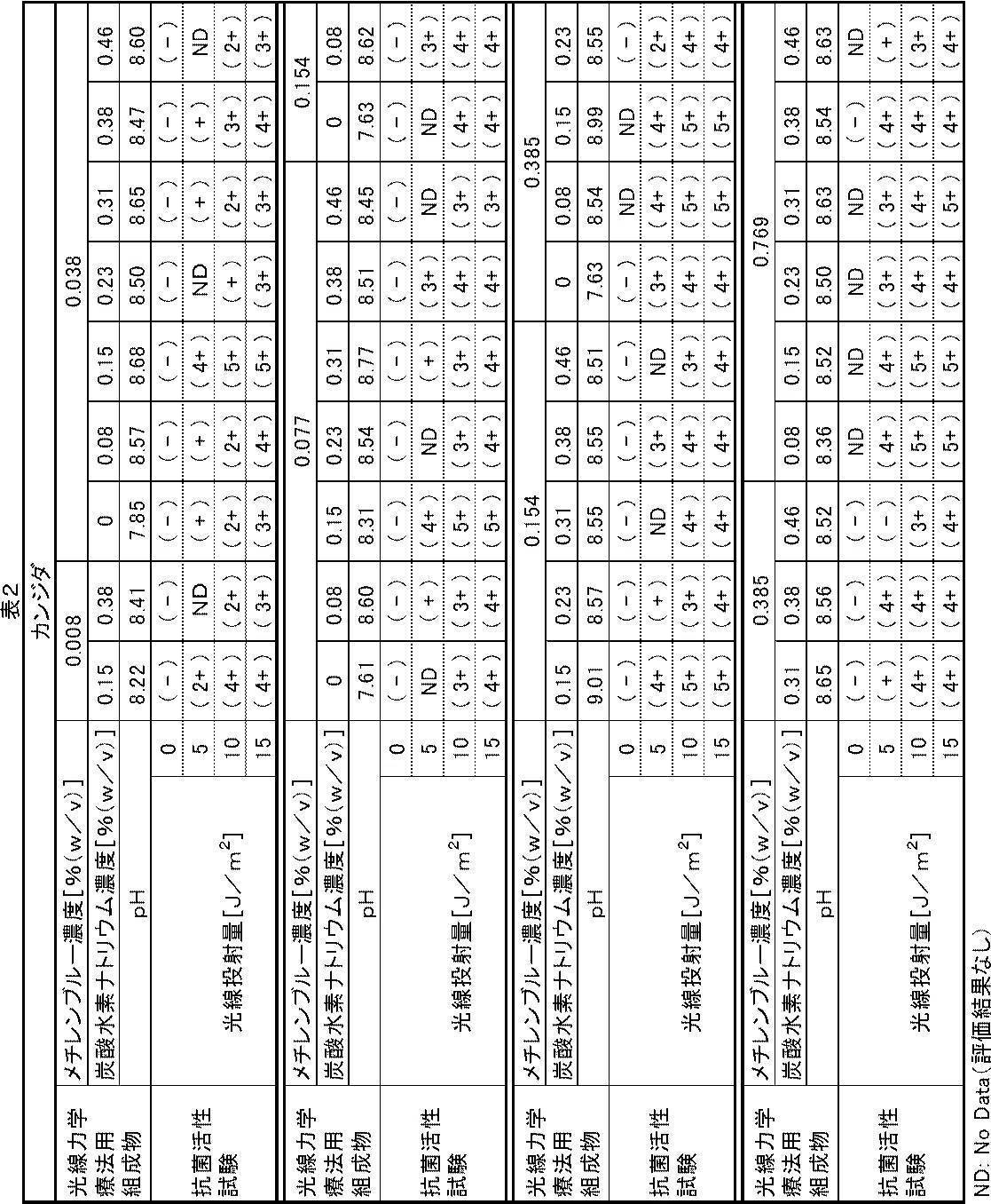
抗菌活性試験用試料のpHが7.6~9.5の範囲内であり、かつ、光照射を行った例が、本発明の実施例である。
実施例は、抗菌活性試験の評価結果がすべて(+)以上であり、抗菌活性が認められた。驚くべきことに、メチレンブルー濃度が0.008w/v%と低濃度であっても、光線投射量5~15J/m2の範囲で、抗菌活性の評価が(2+)以上と、優れていた。
実施例以外の例、すなわち、抗菌活性試験用試料のpHが7.6~9.5の範囲外、および/または、光線投射量が0J/m2、すなわち、光照射を行わなかった例は本発明の比較例である。
比較例は、抗菌活性試験の評価結果がすべて(-)であり、抗菌活性が認められなかった。
〈緑膿菌に対する抗菌活性試験〉
(1)緑膿菌の入手
シュードモナス・エルギノーサ(Pseudomonas aeruginosa)を菌株分譲機関から購入した。
(2.1)メチレンブルー溶液の調製
実施例2と同様にして、メチレンブルー濃度が0.01w/v%、0.05w/v%、0.10w/v%、0.20w/v%、0.50w/v%、1.0w/v%のメチレンブルー溶液を調製した。
(2.2)炭酸水素ナトリウム水溶液の調製
実施例2と同様にして、濃度1.0w/v%、2.0w/v%、3.0w/v%、4.0w/v%、5.0w/v%、6.0w/v%の炭酸水素ナトリウム水溶液を調製した。
(2.3)緑膿菌懸濁液(菌液)の調製
シュードモナス・エルギノーサを培養し、得られた培養物を滅菌生理食塩水に懸濁し、菌数を調整して、菌液を調製した。
(2.4)試料の調製
メチレンブルー溶液10.0mLと、炭酸水素ナトリウム水溶液1.0mLと、菌液2.0mLとを混合し、表3の「光線力学療法用組成物」の欄に示すメチレンブルー濃度、炭酸水素ナトリウム濃度、およびpHの抗菌活性試験用試料を調製した。
また、生理食塩水11.0mLと、菌液2.0mLとを混合したブランク用試料を調製した。
実施例2と同様にして抗菌活性試験を行った。
表3の「光線力学療法用組成物」の欄に、抗菌活性試験用試料のメチレンブルー濃度[w/v%]、炭酸水素ナトリウム濃度[w/v%]およびpHを、「抗菌活性試験」の欄に抗菌活性の評価結果を、それぞれ示す。

抗菌活性試験用試料のpHが7.6~9.5の範囲内であり、かつ、光照射を行った例が、本発明の実施例である。
実施例は、抗菌活性試験の評価結果がすべて(+)以上であり、抗菌活性が認められた。驚くべきことに、メチレンブルー濃度が0.008w/v%と低濃度であっても、光線投射量5~15J/m2の範囲で、抗菌活性の評価が(2+)以上と、優れていた。
実施例以外の例、すなわち、抗菌活性試験用試料のpHが7.6~9.5の範囲外、および/または、光線投射量が0J/m2、すなわち、光照射を行わなかった例は本発明の比較例である。
比較例は、抗菌活性試験の評価結果がすべて(-)であり、抗菌活性が認められなかった。
〈MRSAに対する抗菌活性試験〉
(1)MRSAの入手
スタフィロコッカス・アウレウス(Staphylococcus aureus)のメチシリン耐性株(MRSA,Methicillin Resistant Staphylococcus aureus)を菌株分譲機関から購入した。
(2.1)メチレンブルー溶液の調製
実施例2と同様にして、メチレンブルー濃度が0.01w/v%、0.05w/v%、0.10w/v%、0.20w/v%、0.50w/v%、1.0w/v%のメチレンブルー溶液を調製した。
(2.2)炭酸水素ナトリウム水溶液の調製
実施例2と同様にして、濃度1.0w/v%、2.0w/v%、3.0w/v%、4.0w/v%、5.0w/v%、6.0w/v%の炭酸水素ナトリウム水溶液を調製した。
(2.3)MRSA懸濁液(菌液)の調製
MRSAを培養し、得られた培養物を滅菌生理食塩水に懸濁し、菌数調整して、菌液を調製した。
(2.4)試料の調製
メチレンブルー溶液10.0mLと、炭酸水素ナトリウム水溶液1.0mLと、菌液2.0mLとを混合し、表4の「光線力学療法用組成物」の欄に示すメチレンブルー濃度、炭酸水素ナトリウム濃度、およびpHの抗菌活性試験用試料を調製した。
また、生理食塩水11.0mLと、菌液2.0mLとを混合したブランク用試料を調製した。
実施例2と同様にして抗菌活性試験を行った。
表4の「光線力学療法用組成物」の欄に、抗菌活性試験用試料のメチレンブルー濃度[w/v%]、炭酸水素ナトリウム濃度[w/v%]およびpHを、「抗菌活性試験」の欄に抗菌活性の評価結果を、それぞれ示す。
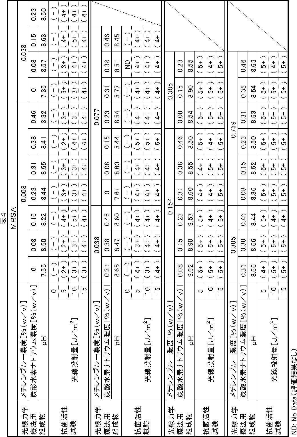
抗菌活性試験用試料のpHが7.6~9.5の範囲内であり、かつ、光照射を行った例が、本発明の実施例である。
実施例は、抗菌活性試験の評価結果がすべて(+)以上であり、抗菌活性が認められた。驚くべきことに、メチレンブルー濃度が0.008w/v%と低濃度であっても、光線投射量5~15J/m2の範囲で、抗菌活性の評価が(2+)以上と、優れていた。
実施例以外の例、すなわち、抗菌活性試験用試料のpHが7.6~9.5の範囲外、および/または、光線投射量が0J/m2、すなわち、光照射を行わなかった例は本発明の比較例である。
比較例は、抗菌活性試験の評価結果がすべて(-)であり、抗菌活性が認められなかった。
〈大腸菌に対する抗菌活性試験〉
(1)大腸菌の入手
エシェリヒア・コリ(Escherichia coli)を菌株分譲機関から購入した。
(2.1)メチレンブルー溶液の調製
実施例2と同様にして、メチレンブルー濃度が0.01w/v%、0.05w/v%、0.10w/v%、0.20w/v%、0.50w/v%、1.0w/v%のメチレンブルー溶液を調製した。
(2.2)炭酸水素ナトリウム水溶液の調製
実施例2と同様にして、濃度1.0w/v%、2.0w/v%、3.0w/v%、4.0w/v%、5.0w/v%、6.0w/v%の炭酸水素ナトリウム水溶液を調製した。
(2.3)大腸菌懸濁液(菌液)の調製
エシェリヒア・コリを培養し、得られた培養物を滅菌生理食塩水に懸濁し、菌数を調整して、菌液を調製した。
(2.4)試料の調製
メチレンブルー溶液10.0mLと、炭酸水素ナトリウム水溶液1.0mLと、菌液2.0mLとを混合し、表4の「光線力学療法用組成物」の欄に示すメチレンブルー濃度、炭酸水素ナトリウム濃度、およびpHの抗菌活性試験用試料を調製した。
また、生理食塩水11.0mLと、菌液2.0mLとを混合したブランク用試料を調製した。
実施例2と同様にして抗菌活性試験を行った。
表4の「光線力学療法用組成物」の欄に、抗菌活性試験用試料のメチレンブルー濃度[w/v%]、炭酸水素ナトリウム濃度[w/v%]およびpHを、「抗菌活性試験」の欄に抗菌活性の評価結果を、それぞれ示す。

抗菌活性試験用試料のpHが7.6~9.5の範囲内であり、かつ、光照射を行った例が、本発明の実施例である。
実施例は、抗菌活性試験の評価結果がすべて(+)以上であり、抗菌活性が認められた。驚くべきことに、メチレンブルー濃度が0.008w/v%と低濃度であっても、光線投射量5~15J/m2の範囲で、抗菌活性の評価が(+)以上であり、抗菌活性が認められた。また、光線投射量が多くなるほど抗菌活性も高くなる傾向が認められた。
実施例以外の例、すなわち、抗菌活性試験用試料のpHが7.6~9.5の範囲外、および/または、光線投射量が0J/m2、すなわち、光照射を行わなかった例は本発明の比較例である。
比較例は、抗菌活性試験の評価結果がすべて(-)であり、抗菌活性が認められなかった。
11 内視鏡
12 演算・制御装置
13 光源装置
14 送気・送水装置
14a 送気ポンプ
14b 洗浄水タンク
15 送液装置
16 挿入部
16a 先端部
16b 湾曲部
16c 可撓管部
17 操作部
18 ユニバーサルコード
20 先端キャップ
21 観察窓
22a 照明窓
22b 照明窓
23 鉗子出口
24 ウォータージェット出口(WJ出口)
25 噴射ノズル
26 鉗子入口
27 モード切替スイッチ
28 上下湾曲用アングルノブ
29 左右湾曲用アングルノブ
30 送気・送水ボタン
31 ズーム操作部
32 コネクタ
33 モニタ
35 送液タンク
36 送液装置本体
37 送液ポンプ
38 フットスイッチ
39 送液管
40 連結管
41a,41b ライトガイド
42 鉗子チューブ
43 送気・送水チューブ
44 多芯ケーブル
44a 信号ケーブル
44b 外皮
45 ウォータージェットチューブ(WJチューブ)
47b 第2WJ路
51 螺管
52 ネット
53 ゴム
100 殺菌システム
101 演算・制御部
102 撮像手段
102a 撮像光学系
103 撮像方向制御手段
104 光照射手段
104a 照明光学系
105 照射方向制御手段
106 組成物噴射手段
106a 噴射部
107 噴射方向制御手段
108,109,110,111,112,113 伝送路
200 皮膚または粘膜
201 正常部
202 病変部
203 付着部
Claims (22)
- メチレンブルーを含み、pHが7.6~9.5である、ピロリ菌感染症を治療するための光線力学療法用組成物。
- 前記pHが8.3~9.2である、請求項1に記載のピロリ菌感染症を治療するための光線力学療法用組成物。
- さらに炭酸水素ナトリウムを含む、請求項1または2に記載のピロリ菌感染症を治療するための光線力学療法用組成物。
- 前記メチレンブルーの濃度が0.0007~4.0w/v%であり、前記炭酸水素ナトリウムの濃度が0.007~0.50w/v%である、請求項3に記載のピロリ菌感染症を治療するための光線力学療法用組成物。
- 前記炭酸水素ナトリウムの濃度が0.15~0.40w/v%である、請求項3または4に記載のピロリ菌感染症を治療するための光線力学療法用組成物。
- ピロリ菌感染症の病変部に存在するピロリ菌に接触させ、前記ピロリ菌に光を照射することによりピロリ菌感染症を治療する光線力学療法において使用するための、請求項1~5のいずれか1項に記載のピロリ菌感染症を治療するための光線力学療法用組成物。
- 経内視鏡的に、請求項1~6のいずれか1項に記載のピロリ菌感染症を治療するための光線力学療法用組成物をピロリ菌感染症の病変部に塗布し、白色光または波長660±10nmのLED光もしくはレーザー光を照射する、ピロリ菌感染症の治療方法。
- 光線投射量が1~200J/cm2である、請求項7に記載のピロリ菌感染症の治療方法。
- メチレンブルーを含み、pHが7.6~9.5である、皮膚粘膜感染症を治療するための光線力学療法用組成物。
- 前記pHが8.3~9.2である、請求項9に記載の皮膚粘膜感染症を治療するための光線力学療法用組成物。
- さらに炭酸水素ナトリウムを含む、請求項9または10に記載の皮膚粘膜感染症を治療するための光線力学療法用組成物。
- 前記メチレンブルーの濃度が0.0007~4.0w/v%であり、前記炭酸水素ナトリウムの濃度が0.007~0.50w/v%である、請求項11に記載の皮膚粘膜感染症を治療するための光線力学療法用組成物。
- 皮膚粘膜感染症の病変部に存在する病原微生物に接触させ、前記病原微生物に光を照射することにより前記皮膚粘膜感染症を治療する光線力学療法において使用するための、請求項9~12のいずれか1項に記載の皮膚粘膜感染症を治療するための光線力学療法用組成物。
- 前記病原微生物が、カンジダ・アルビカンス(Candida albicans)、メチシリン耐性スタフィロコッカス・アウレウス(Staphylococcus aureus)、シュードモナス・エルギノーサ(Pseudomonas aeruginosa)およびエシェリヒア・コリ(Escherichia coli)からなる群から選択される少なくとも1つである、請求項13に記載の皮膚粘膜感染症を治療するための光線力学療法用組成物。
- 前記皮膚粘膜感染症が、カンジダ症、メチシリン耐性黄色ブドウ球菌感染症、緑膿菌感染症および大腸菌感染症からなる群から選択される少なくとも1つである、請求項9~13のいずれか1項に記載の皮膚粘膜感染症を治療するための光線力学療法用組成物。
- 請求項9~15のいずれか1項に記載の皮膚粘膜感染症を治療するための光線力学療法用組成物を前記皮膚粘膜感染症の病変部に塗布し、白色光または波長660±10nmのLED光もしくはレーザー光を照射する、皮膚粘膜感染症の治療方法。
- 経内視鏡的に、前記皮膚粘膜感染症を治療するための光線力学療法用組成物を塗布し、前記白色光または前記レーザー光を照射する、請求項16に記載の皮膚粘膜感染症の治療方法。
- 光線投射量が1~200J/cm2である、請求項16または17に記載の皮膚粘膜感染症の治療方法。
- 撮像手段と、光照射手段と、組成物噴射手段と、前記撮像手段の撮像方向を制御する撮像方向制御手段と、前記光照射手段の光照射方向を制御する照射方向制御手段と、前記組成物噴射手段の噴射方向を制御する噴射方向制御手段と、前記撮像手段、前記光照射手段、前記組成物噴射手段、前記撮像方向制御手段、前記照射方向制御手段および前記噴射方向制御手段を制御する演算・制御部とを備える、ピロリ菌感染症による病変部の殺菌システムであって、
(a)前記撮像方向制御手段および前記照射方向制御手段は、前記演算・制御部からの制御信号に基づいて、前記撮像手段の被観察領域を前記光照射手段が照明するように作動し、
(b)前記撮像手段は、前記演算・制御部からの制御信号に基づいて、粘膜を撮像し、映像信号を前記演算・制御部に送信し、
(c)前記演算・制御部は、前記撮像手段から送信された映像信号を受信し、粘膜の正常領域と異常領域との色の差を強調するように映像処理を行い、正常部と病変部とを識別し、病変部の位置を特定し、
(d)前記噴射方向制御手段は、前記演算・制御部からの制御信号に基づいて、前記組成物噴射手段の噴射方向を病変部に合わせるように作動し、
(e)前記組成物噴射手段は、前記演算・制御部からの制御信号に基づいて、請求項1~6のいずれか1項に記載のピロリ菌感染症を治療するための光線力学療法用組成物を噴射し、
(f)前記照射方向制御手段は、前記演算・制御部からの制御信号に基づいて、前記光照射手段の照射方向を病変部に合わせるように作動し、および
(g)前記光照射手段は、前記演算・制御部からの制御信号に基づいて、白色光または波長660±10nmのLED光もしくはレーザー光を発射する、
ピロリ菌感染症による病変部の殺菌システム。 - 撮像手段と、光照射手段と、組成物噴射手段と、前記撮像手段の撮像方向を制御する撮像方向制御手段と、前記光照射手段の光照射方向を制御する照射方向制御手段と、前記組成物噴射手段の噴射方向を制御する噴射方向制御手段と、前記撮像手段、前記光照射手段、前記組成物噴射手段、前記撮像方向制御手段、前記照射方向制御手段および前記噴射方向制御手段を制御する演算・制御部とを備える、ピロリ菌感染症による病変部の殺菌システムの作動方法であって、
(a)前記撮像手段の被観察領域を前記光照射手段が照明するように、前記演算・制御部からの制御信号に基づいて、前記撮像方向制御手段および前記照射方向制御手段を作動させる工程、
(b)前記撮像手段が、前記演算・制御部からの制御信号に基づいて、粘膜を撮像し、映像信号を前記演算・制御部に送信する工程、
(c)前記演算・制御部が、前記撮像手段から送信された映像信号を受信し、粘膜の正常領域と異常領域との色の差を強調するように映像処理を行い、正常部と病変部とを識別し、病変部の位置を特定する工程、
(d)前記組成物噴射手段の噴射方向を病変部に合わせるように、前記演算・制御部からの制御信号に基づいて、前記噴射方向制御手段を作動させる工程、
(e)前記組成物噴射手段が、前記演算・制御部からの制御信号に基づいて、請求項1~6のいずれか1項に記載のピロリ菌感染症を治療するための光線力学療法用組成物を噴射する工程、
(f)前記光照射手段の照射方向を病変部に合わせるように、前記演算・制御部からの制御信号に基づいて、前記照射方向制御手段を作動させる工程、および
(g)前記光照射手段が、前記演算・制御部からの制御信号に基づいて、白色光または波長660±10nmのLED光もしくはレーザー光を発射する工程
を含む、ピロリ菌感染症による病変部の殺菌システムの作動方法。 - 撮像手段と、光照射手段と、組成物噴射手段と、前記撮像手段の撮像方向を制御する撮像方向制御手段と、前記光照射手段の光照射方向を制御する照射方向制御手段と、前記組成物噴射手段の噴射方向を制御する噴射方向制御手段と、前記撮像手段、前記光照射手段、前記組成物噴射手段、前記撮像方向制御手段、前記照射方向制御手段および前記噴射方向制御手段を制御する演算・制御部とを備える、皮膚粘膜感染症による病変部の殺菌システムであって、
(a)前記撮像方向制御手段および前記照射方向制御手段は、前記演算・制御部からの制御信号に基づいて、前記撮像手段の被観察領域を前記光照射手段が照明するように作動し、
(b)前記撮像手段は、前記演算・制御部からの制御信号に基づいて、皮膚または粘膜を撮像し、映像信号を前記演算・制御部に送信し、
(c)前記演算・制御部は、前記撮像手段から送信された映像信号を受信し、皮膚または粘膜の正常領域と異常領域との色の差を強調するように映像処理を行い、正常部と病変部とを識別し、病変部の位置を特定し、
(d)前記噴射方向制御手段は、前記演算・制御部からの制御信号に基づいて、前記組成物噴射手段の噴射方向を病変部に合わせるように作動し、
(e)前記組成物噴射手段は、前記演算・制御部からの制御信号に基づいて、請求項9~15のいずれか1項に記載の皮膚粘膜感染症を治療するための光線力学療法用組成物を噴射し、
(f)前記照射方向制御手段は、前記演算・制御部からの制御信号に基づいて、前記光照射手段の照射方向を病変部に合わせるように作動し、および
(g)前記光照射手段は、前記演算・制御部からの制御信号に基づいて、白色光または波長660±10nmのLED光もしくはレーザー光を発射する、
皮膚粘膜感染症による病変部の殺菌システム。 - 撮像手段と、光照射手段と、組成物噴射手段と、前記撮像手段の撮像方向を制御する撮像方向制御手段と、前記光照射手段の光照射方向を制御する照射方向制御手段と、前記組成物噴射手段の噴射方向を制御する噴射方向制御手段と、前記撮像手段、前記光照射手段、前記組成物噴射手段、前記撮像方向制御手段、前記照射方向制御手段および前記噴射方向制御手段を制御する演算・制御部とを備える、皮膚粘膜感染症による病変部の殺菌システムの作動方法であって、
(a)前記撮像手段の被観察領域を前記光照射手段が照明するように、前記演算・制御部からの制御信号に基づいて、前記撮像方向制御手段および前記照射方向制御手段を作動させる工程、
(b)前記撮像手段が、前記演算・制御部からの制御信号に基づいて、皮膚または粘膜を撮像し、映像信号を前記演算・制御部に送信する工程、
(c)前記演算・制御部が、前記撮像手段から送信された映像信号を受信し、皮膚または粘膜の正常領域と異常領域との色の差を強調するように映像処理を行い、正常部と病変部とを識別し、病変部の位置を特定する工程、
(d)前記組成物噴射手段の噴射方向を病変部に合わせるように、前記演算・制御部からの制御信号に基づいて、前記噴射方向制御手段を作動させる工程、
(e)前記組成物噴射手段が、前記演算・制御部からの制御信号に基づいて、請求項9~15のいずれか1項に記載の皮膚粘膜感染症を治療するための光線力学療法用組成物を噴射する工程、
(f)前記光照射手段の照射方向を病変部に合わせるように、前記演算・制御部からの制御信号に基づいて、前記照射方向制御手段を作動させる工程、および
(g)前記光照射手段が、前記演算・制御部からの制御信号に基づいて、白色光または波長660±10nmのLED光もしくはレーザー光を発射する工程
を含む、皮膚粘膜感染症による病変部の殺菌システムの作動方法。
Priority Applications (3)
| Application Number | Priority Date | Filing Date | Title |
|---|---|---|---|
| CN201780005825.4A CN108463249A (zh) | 2016-02-04 | 2017-01-19 | 光线力学疗法用组合物、治疗方法、杀菌系统及杀菌系统的工作方法 |
| KR1020187019978A KR102203314B1 (ko) | 2016-02-04 | 2017-01-19 | 광선역학 요법용 조성물, 치료 방법, 살균 시스템 및 살균 시스템의 작동 방법 |
| JP2017565467A JP6602400B2 (ja) | 2016-02-04 | 2017-01-19 | 光線力学療法用組成物、殺菌システムおよび殺菌システムの作動方法 |
Applications Claiming Priority (2)
| Application Number | Priority Date | Filing Date | Title |
|---|---|---|---|
| JP2016019728 | 2016-02-04 | ||
| JP2016-019728 | 2016-02-04 |
Publications (1)
| Publication Number | Publication Date |
|---|---|
| WO2017135048A1 true WO2017135048A1 (ja) | 2017-08-10 |
Family
ID=59500748
Family Applications (1)
| Application Number | Title | Priority Date | Filing Date |
|---|---|---|---|
| PCT/JP2017/001727 Ceased WO2017135048A1 (ja) | 2016-02-04 | 2017-01-19 | 光線力学療法用組成物、治療方法、殺菌システムおよび殺菌システムの作動方法 |
Country Status (4)
| Country | Link |
|---|---|
| JP (1) | JP6602400B2 (ja) |
| KR (1) | KR102203314B1 (ja) |
| CN (1) | CN108463249A (ja) |
| WO (1) | WO2017135048A1 (ja) |
Cited By (1)
| Publication number | Priority date | Publication date | Assignee | Title |
|---|---|---|---|---|
| JP2020156902A (ja) * | 2019-03-27 | 2020-10-01 | Hoya株式会社 | 内視鏡 |
Citations (5)
| Publication number | Priority date | Publication date | Assignee | Title |
|---|---|---|---|---|
| JP2011529898A (ja) * | 2008-08-01 | 2011-12-15 | オンディーヌ インターナショナル リミテッド | Mrsaの治療用の組成物および方法 |
| JP2012520876A (ja) * | 2009-03-16 | 2012-09-10 | オンディーヌ インターナショナル リミテッド | 光線力学殺菌用の組成物 |
| JP2014004329A (ja) * | 2012-06-01 | 2014-01-16 | Sony Corp | 歯用装置、医療用装置及び算出方法 |
| JP2015066129A (ja) * | 2013-09-27 | 2015-04-13 | 富士フイルム株式会社 | 蛍光観察装置、内視鏡システム及びプロセッサ装置並びに作動方法 |
| WO2016031875A1 (ja) * | 2014-08-27 | 2016-03-03 | 富士フイルム株式会社 | 光線力学療法用組成物、殺菌方法、殺菌システムおよび殺菌システムの作動方法 |
Family Cites Families (2)
| Publication number | Priority date | Publication date | Assignee | Title |
|---|---|---|---|---|
| ATE159661T1 (de) | 1992-04-30 | 1997-11-15 | Eastman Dental Inst | Medikament zur desinfektion der mundhöhle |
| CN1329398C (zh) * | 2006-02-25 | 2007-08-01 | 贵州法澳医疗器械科技有限公司 | 二氢卟吩e6锌合物光敏剂及其制备方法与应用 |
-
2017
- 2017-01-19 KR KR1020187019978A patent/KR102203314B1/ko active Active
- 2017-01-19 JP JP2017565467A patent/JP6602400B2/ja active Active
- 2017-01-19 CN CN201780005825.4A patent/CN108463249A/zh active Pending
- 2017-01-19 WO PCT/JP2017/001727 patent/WO2017135048A1/ja not_active Ceased
Patent Citations (5)
| Publication number | Priority date | Publication date | Assignee | Title |
|---|---|---|---|---|
| JP2011529898A (ja) * | 2008-08-01 | 2011-12-15 | オンディーヌ インターナショナル リミテッド | Mrsaの治療用の組成物および方法 |
| JP2012520876A (ja) * | 2009-03-16 | 2012-09-10 | オンディーヌ インターナショナル リミテッド | 光線力学殺菌用の組成物 |
| JP2014004329A (ja) * | 2012-06-01 | 2014-01-16 | Sony Corp | 歯用装置、医療用装置及び算出方法 |
| JP2015066129A (ja) * | 2013-09-27 | 2015-04-13 | 富士フイルム株式会社 | 蛍光観察装置、内視鏡システム及びプロセッサ装置並びに作動方法 |
| WO2016031875A1 (ja) * | 2014-08-27 | 2016-03-03 | 富士フイルム株式会社 | 光線力学療法用組成物、殺菌方法、殺菌システムおよび殺菌システムの作動方法 |
Non-Patent Citations (2)
| Title |
|---|
| CHOI SS. ET AL.: "Synergistic in vitro photodynamic antimicrobial activity of methylene blue and chitosan against Helicobacter pylori 26695", PHOTODIAGNOSIS AND PHOTODYNAMIC THERAPY, vol. 11, 2014, pages 526 - 532, XP029105737 * |
| MORIMOTO, YUJI ET AL.: "Kosen Rikigaku Ryoho de Kansensho o Gekitai", OPTICAL ALLIANCE, vol. 24, no. 3, 2013, pages 1 - 5 * |
Cited By (3)
| Publication number | Priority date | Publication date | Assignee | Title |
|---|---|---|---|---|
| JP2020156902A (ja) * | 2019-03-27 | 2020-10-01 | Hoya株式会社 | 内視鏡 |
| WO2020195518A1 (ja) * | 2019-03-27 | 2020-10-01 | Hoya株式会社 | 内視鏡 |
| US12262876B2 (en) | 2019-03-27 | 2025-04-01 | Hoya Corporation | Endoscope |
Also Published As
| Publication number | Publication date |
|---|---|
| CN108463249A (zh) | 2018-08-28 |
| JP6602400B2 (ja) | 2019-11-06 |
| KR102203314B1 (ko) | 2021-01-15 |
| JPWO2017135048A1 (ja) | 2018-09-27 |
| KR20180086505A (ko) | 2018-07-31 |
Similar Documents
| Publication | Publication Date | Title |
|---|---|---|
| JP6412142B2 (ja) | 光線力学療法用組成物、殺菌方法、殺菌システムおよび殺菌システムの作動方法 | |
| EP2054030B1 (en) | Antimicrobials | |
| KR101453011B1 (ko) | 세포외 다당류 용매화 시스템 | |
| US7919761B2 (en) | Fluorescence observation apparatus | |
| US20070135874A1 (en) | Endoscope for therapeutic light delivery | |
| US20030187427A1 (en) | Method and apparatus for in VIVO treatment of mammary ducts by light induced fluorescence | |
| US20210338854A1 (en) | Remote pathogen eradication | |
| US20200406052A1 (en) | Digestive Tract Capsule Endoscopy Integrated With Photodynamic Diagnosis And Therapy | |
| JP2023532262A (ja) | 蛍光薬剤用内視鏡照明システム | |
| CN110461211A (zh) | 内窥镜和治疗方法 | |
| JP2025061094A (ja) | 体内紫外線療法 | |
| Astuti et al. | Photodynamic effectiveness of laser diode combined with ozone to reduce Staphylicoccus aureus biofilm with exogenous chlorophyll of Dracaena angustifolia leaves | |
| JP4366528B2 (ja) | 室内消毒方法 | |
| JP6602400B2 (ja) | 光線力学療法用組成物、殺菌システムおよび殺菌システムの作動方法 | |
| Ramaswamy et al. | Photodynamic diagnosis of a human nasopharyngeal carcinoma xenograft model using the novel Chlorin e6 photosensitizer Fotolon® | |
| EP2698097A1 (en) | Two-way photodynamic therapy stereo colposcope for diagnosing and treating diseases of the female genital tract | |
| US10448817B2 (en) | Endoscopic device incorporating diode laser for PDD, PDT, and AF applications | |
| US20140357950A1 (en) | Photodynamic diagnosis stereo colposcope (pdd) for female genital tract diseases and early detection of neoplastic lesion | |
| JP7659800B2 (ja) | 癌治療装置 | |
| CN219270872U (zh) | 一种治疗幽门螺杆菌感染的光疗型电子内窥镜 | |
| JP2012050513A (ja) | 内視鏡用フード | |
| JP2006527609A (ja) | 内視鏡カプセル | |
| JP2025507760A5 (ja) | ||
| RU2731312C2 (ru) | Способ профилактики инфекционного поражения голосовых протезов у пациентов после ларингэктомии | |
| CN113877066A (zh) | 黏膜层检查消杀装置及方法 |
Legal Events
| Date | Code | Title | Description |
|---|---|---|---|
| 121 | Ep: the epo has been informed by wipo that ep was designated in this application |
Ref document number: 17747207 Country of ref document: EP Kind code of ref document: A1 |
|
| ENP | Entry into the national phase |
Ref document number: 2017565467 Country of ref document: JP Kind code of ref document: A |
|
| ENP | Entry into the national phase |
Ref document number: 20187019978 Country of ref document: KR Kind code of ref document: A |
|
| WWE | Wipo information: entry into national phase |
Ref document number: 1020187019978 Country of ref document: KR |
|
| NENP | Non-entry into the national phase |
Ref country code: DE |
|
| 122 | Ep: pct application non-entry in european phase |
Ref document number: 17747207 Country of ref document: EP Kind code of ref document: A1 |