WO2017130563A1 - 希少細胞を捕捉する方法 - Google Patents
希少細胞を捕捉する方法 Download PDFInfo
- Publication number
- WO2017130563A1 WO2017130563A1 PCT/JP2016/085775 JP2016085775W WO2017130563A1 WO 2017130563 A1 WO2017130563 A1 WO 2017130563A1 JP 2016085775 W JP2016085775 W JP 2016085775W WO 2017130563 A1 WO2017130563 A1 WO 2017130563A1
- Authority
- WO
- WIPO (PCT)
- Prior art keywords
- cells
- filter
- beads
- rare
- rare cells
- Prior art date
- Legal status (The legal status is an assumption and is not a legal conclusion. Google has not performed a legal analysis and makes no representation as to the accuracy of the status listed.)
- Ceased
Links
Images

Classifications
-
- G—PHYSICS
- G01—MEASURING; TESTING
- G01N—INVESTIGATING OR ANALYSING MATERIALS BY DETERMINING THEIR CHEMICAL OR PHYSICAL PROPERTIES
- G01N1/00—Sampling; Preparing specimens for investigation
- G01N1/28—Preparing specimens for investigation including physical details of (bio-)chemical methods covered elsewhere, e.g. G01N33/50, C12Q
- G01N1/40—Concentrating samples
- G01N1/4077—Concentrating samples by other techniques involving separation of suspended solids
-
- G—PHYSICS
- G01—MEASURING; TESTING
- G01N—INVESTIGATING OR ANALYSING MATERIALS BY DETERMINING THEIR CHEMICAL OR PHYSICAL PROPERTIES
- G01N33/00—Investigating or analysing materials by specific methods not covered by groups G01N1/00 - G01N31/00
- G01N33/48—Biological material, e.g. blood, urine; Haemocytometers
- G01N33/50—Chemical analysis of biological material, e.g. blood, urine; Testing involving biospecific ligand binding methods; Immunological testing
- G01N33/53—Immunoassay; Biospecific binding assay; Materials therefor
- G01N33/575—Immunoassay; Biospecific binding assay; Materials therefor for cancer
-
- G—PHYSICS
- G01—MEASURING; TESTING
- G01N—INVESTIGATING OR ANALYSING MATERIALS BY DETERMINING THEIR CHEMICAL OR PHYSICAL PROPERTIES
- G01N35/00—Automatic analysis not limited to methods or materials provided for in any single one of groups G01N1/00 - G01N33/00; Handling materials therefor
- G01N35/0098—Automatic analysis not limited to methods or materials provided for in any single one of groups G01N1/00 - G01N33/00; Handling materials therefor involving analyte bound to insoluble magnetic carrier, e.g. using magnetic separation
-
- G—PHYSICS
- G01—MEASURING; TESTING
- G01N—INVESTIGATING OR ANALYSING MATERIALS BY DETERMINING THEIR CHEMICAL OR PHYSICAL PROPERTIES
- G01N1/00—Sampling; Preparing specimens for investigation
- G01N1/28—Preparing specimens for investigation including physical details of (bio-)chemical methods covered elsewhere, e.g. G01N33/50, C12Q
- G01N1/40—Concentrating samples
- G01N1/4077—Concentrating samples by other techniques involving separation of suspended solids
- G01N2001/4088—Concentrating samples by other techniques involving separation of suspended solids filtration
-
- G—PHYSICS
- G01—MEASURING; TESTING
- G01N—INVESTIGATING OR ANALYSING MATERIALS BY DETERMINING THEIR CHEMICAL OR PHYSICAL PROPERTIES
- G01N35/00—Automatic analysis not limited to methods or materials provided for in any single one of groups G01N1/00 - G01N33/00; Handling materials therefor
- G01N2035/00465—Separating and mixing arrangements
- G01N2035/00475—Filters
-
- G—PHYSICS
- G01—MEASURING; TESTING
- G01N—INVESTIGATING OR ANALYSING MATERIALS BY DETERMINING THEIR CHEMICAL OR PHYSICAL PROPERTIES
- G01N35/00—Automatic analysis not limited to methods or materials provided for in any single one of groups G01N1/00 - G01N33/00; Handling materials therefor
- G01N2035/00465—Separating and mixing arrangements
- G01N2035/00564—Handling or washing solid phase elements, e.g. beads
Definitions
- the present invention relates to a method for capturing rare cells.
- cancer cell enrichment is extremely large, and if cancer cells in blood can be enriched, that is, cancer cells can be selectively captured, it can be applied to cancer diagnosis.
- the most important factor for the prognosis and treatment of cancer is the presence or absence of cancer cell metastasis at the first visit and treatment.
- detecting circulating tumor cells is useful for determining the progression of cancer pathology. Means.
- blood components such as red blood cells and white blood cells are predominantly present in blood, it is difficult to detect an extremely small amount of CTC.
- Circulating Endothelial Cell (hereinafter also referred to as “CEC”) is also used as a biomarker for cancer prognosis and treatment, but it is present in the blood only in a very small amount and is detected in the same manner as CTC. It is difficult.
- the filter method is a method of concentrating rare cells based on differences in cell size and deformability.
- Patent Document 1 a method of detecting a small amount of CTC by using a resin filter using parylene has been proposed.
- Patent Document 2 a method of improving the strength of the filter by using a metal filter and separating the leukocytes and the cancer cells depending on the deformability is proposed (Patent Document 2). ⁇ 4).
- the present invention has been made in view of such problems, and provides a method for concentrating rare cells while selectively capturing rare cells from a liquid containing rare cells and contaminated cells.
- the present inventors have found that when a liquid containing rare cells and contaminated cells is filtered through a filter in the presence of beads, the rare cells can be selectively captured and concentrated.
- the headline and the present invention were completed.
- the method for capturing rare cells of the present invention includes a step of adding beads to a liquid containing rare cells and contaminated cells, and a step of filtering the liquid to which the beads are added with a filter.
- the present inventors presume the action of beads in the present invention as follows. Contaminant cells remaining on the filter overlap in layers to block the pores of the filter, resulting in a decrease in filtration efficiency. The beads extrude contaminating cells stacked in layers, thereby eliminating clogging of the filter and preventing a decrease in filtration efficiency. As a result, it is possible to selectively capture rare cells.
- the median diameter of the beads can be 0.5 ⁇ m to 8 ⁇ m.
- the addition amount of the beads may be 1 ⁇ 10 4 to 9 ⁇ 10 6 per mL of the liquid.
- the beads may be at least one of resin beads or magnetic beads.
- the contaminating cells can be leukocytes.
- the rare cell can be a blood circulating cancer cell or a blood circulating endothelial cell.
- the present invention it is possible to concentrate rare cells while selectively capturing rare cells from a liquid containing rare cells and contaminated cells.
- Random cells refers to cells to be captured, and the ratio of the number of cells is extremely small relative to the total number of cells contained in the liquid containing rare cells and contaminating cells.
- Rare cells are, for example, cancer cells such as CTC or endothelial cells such as CEC.
- “Contaminating cells” refers to cells other than rare cells contained in a liquid containing rare cells and contaminated cells. Usually, the ratio of the number of contaminating cells is extremely large relative to the number of rare cells.
- a “contaminating cell” may refer to a cell having the same diameter as a rare cell and having deformability among such cells, and more specifically, a leukocyte. There is a case.
- the “diameter” of the cell is the length of the longest line segment connecting two arbitrary points on the outline of the cell when observed with a microscope.
- the liquid containing rare cells and contaminating cells can be, for example, blood, ascites or pleural effusion.
- Capture means that the liquid containing the cells is filtered through a filter, and the cells remain on the filter. Further, “selectively capture” means that the ratio of the predetermined cells in the cell population remaining on the filter is higher than the ratio in the liquid containing the cells before filtration.
- the method for capturing rare cells of the present invention includes a step of adding beads to a liquid containing rare cells and contaminated cells, and a step of filtering the liquid to which the beads are added with a filter.
- the shape of the bead is not particularly limited, and can be, for example, a sphere, a rectangular parallelepiped, a triangular pyramid, or a cone. From the viewpoint of reducing local impact when a bead collides with a rare cell and minimizing damage to the rare cell, the bead is preferably approximately a sphere or a sphere, and more preferably a sphere. .
- the median diameter of the beads can be appropriately selected according to the diameters of rare cells and contaminated cells and the pore size of the filter.
- the median diameter of the beads is from the viewpoint of preventing clogging of the filter, the viewpoint of pushing out the leukocyte, and the observation of the rare cell after capture.
- 0.5 ⁇ m to 8 ⁇ m is preferable, 1 ⁇ m to 7.5 ⁇ m is more preferable, and 3 ⁇ m to 7 ⁇ m is even more preferable.
- the median diameter of the beads refers to a particle diameter at which the integrated value in the particle size distribution measured with a laser diffraction particle size distribution measuring device is 50%.
- the material of the beads is not particularly limited, and examples thereof include resins (polystyrene, polymethacrylate, polylactic acid, etc.), metals or metal oxides (iron oxide, etc.), polysaccharides (chitosan, etc.), silica, or combinations thereof. Good. Of these, resin beads are preferred.
- the beads are preferably magnetic beads.
- a magnetic bead is a bead containing a magnetic substance.
- the magnetic beads may be ones in which a magnetic material is coated with a resin, a metal or metal oxide, a polysaccharide, silica, or a combination thereof.
- the surface of the beads can be coated with a material having an effect of suppressing the adsorption of DNA, RNA and protein, for example.
- a material having an effect of suppressing the adsorption of DNA, RNA and protein for example.
- coating materials include, for example, polyethylene glycol (PEG), bovine serum albumin (BSA), parylene, and 2-methacryloyloxyethyl phosphorylcholine monomer (MPC monomer).
- the amount of beads added is preferably 1 ⁇ 10 4 to 9 ⁇ 10 6 with respect to 1 mL of liquid containing rare cells and contaminated cells, and is 1 ⁇ 10 5 to 7 ⁇ 10 6. More preferably, the number is 5 ⁇ 10 5 to 5 ⁇ 10 6 . If the amount of beads added is greater than or equal to the lower limit, it is possible to more effectively extrude contaminating cells. If the added amount of beads is less than or equal to the above upper limit, the beads will not clog the filter, and it will be easier to observe the captured rare cells. Moreover, the addition amount of beads is appropriately adjusted according to the number of cells in the liquid containing rare cells and contaminating cells.
- the amount of beads added is preferably 0.002 to 100 times, more preferably 0.02 to 75 times the number of contaminating cells in the liquid containing rare cells and contaminating cells, More preferably, it is 0.2 to 50 times. If the added amount of beads is within the above numerical range, it is possible to more effectively extrude contaminating cells.
- a blood sample can be obtained by collecting blood from a subject by a normal method.
- additives conventionally added to blood samples such as anticoagulants and fixatives can be further added.
- the anticoagulant include ethylenediaminetetraacetic acid dihydrogen disodium dihydrate (EDTA-2Na), ethylenediaminetetraacetic acid dipotassium dihydrate (EDTA-2K), sodium citrate, sodium fluoride, heparin.
- CTAD solvent mixed solution of citric acid, theophylline, adenosine, and dipyridamole.
- the fixing agent examples include paraformaldehyde (PFA), glutaraldehyde, and ethanol. These additives can be put in a blood collection tube before blood collection in advance, or can be added after blood collection.
- the blood sample can also be diluted with, for example, a buffer.
- This step removes the contaminating cells from the liquid containing the rare cells and the contaminated cells, and selectively captures the rare cells.
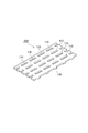
- the filter 200 has a structure in which a large number of through holes 110 are formed in a thin film 120 such as plastic or metal.
- the opening shape of the through hole 110 can be, for example, a circle, an ellipse, a square, a rectangle, a rounded rectangle, or a polygon. From the viewpoint of efficiently capturing rare cells, the opening shape is preferably a circle, a rectangle or a rounded rectangle, and more preferably a rectangle or a rounded rectangle.
- a rounded rectangle is a shape having a rectangle and two semicircles that have the same radius as the short side of the rectangle and that are adjacent to each other and are adjacent to the two short sides of the rectangle. If the opening shape is a rectangle or a rounded rectangle, the through hole 110 is less likely to be clogged, and rare cells can be captured more selectively.
- the through holes 110 may be arranged as shown in FIG. 1, may be arranged in a staggered manner in which the arrangement is shifted for each column, or may be arranged randomly.
- the hole diameter of the through hole 110 is set according to the diameter of the rare cell to be captured.
- the diameter of the through hole 110 is preferably 5 ⁇ m to 15 ⁇ m, more preferably 6 ⁇ m to 12 ⁇ m, and even more preferably 7 ⁇ m to 10 ⁇ m.
- the hole diameter of the through hole 110 (also simply referred to as “filter hole diameter”) refers to the maximum value of the diameter of a sphere that can pass through the through hole 110.
- the diameter of the through hole 110 is the length of the short side of the rectangle.
- the hole diameter of the through hole 110 is the diameter of the inscribed circle of the polygon.
- the opening shape is a rectangle or a rounded rectangle, a gap is formed in the long axis direction of the opening shape in the through-hole 110 even when rare cells are captured in the through-hole 110. Since contaminant cells can pass through the gap, the filter 200 can be prevented from being clogged.
- the aperture ratio of the filter 200 is preferably 5 to 50%, more preferably 10 to 40%, more preferably 10 to 30% from the viewpoint of maintaining a balance between the strength of the filter 200 and prevention of clogging of the filter 200. % Is more preferable.
- the aperture ratio refers to the area occupied by the through hole 110 with respect to the entire area of the filter 200.
- the thickness of the filter 200 is preferably 3 ⁇ m to 50 ⁇ m, more preferably 5 ⁇ m to 40 ⁇ m, and more preferably 5 ⁇ m to 30 ⁇ m from the viewpoint of maintaining a balance between the strength of the filter 200 and prevention of clogging of the filter 200. Is more preferable.
- the filter 200 is preferably made of metal. Since metal is excellent in processability, the processing accuracy of the filter 200 can be increased, and the capture rate of rare cells can be further improved. In addition, since metal is rigid compared to other materials, its size and shape are maintained even when a force is applied from the outside. For this reason, even if a contaminated cell is slightly larger than the through-hole 110, the filter 200 does not deform
- the metal examples include nickel, silver, palladium, copper, iridium, ruthenium, chromium, and alloys thereof.
- Palladium and iridium have a high redox potential, are hardly soluble and have good characteristics, but are expensive.
- Nickel has a lower oxidation-reduction potential than hydrogen, so it is easily dissolved but is inexpensive.
- Silver and palladium are noble metals and are less expensive than palladium and iridium.
- the manufacturing method of the filter 200 is not particularly limited, but for example, it can be manufactured as follows. First, a photoresist is placed on the substrate at a position that will later become the through-hole 110 of the filter 200, and a metal layer (thin film 120) having the through-hole 110 is formed by plating the substrate with a metal that becomes the filter 200. Is formed on the substrate. Then, the filter 200 is obtained by removing the substrate and the photoresist from the metal layer.
- the substrate for example, a substrate whose surface is plated with copper can be used. Copper is preferable as a substrate material because it can be easily removed by chemical dissolution with a chemical solution and has excellent adhesion to a photoresist.
- the method for filtering a liquid containing rare cells and contaminated cells and the apparatus used for the filtration are not particularly limited, but it is preferable to control the flow rate during liquid passage.
- the flow rate during liquid passage is preferably 50 ⁇ L / min to 3000 ⁇ L / min, preferably 100 ⁇ L / min. Minute to 1000 ⁇ L / min is more preferable, and 200 ⁇ L / min to 600 ⁇ L / min is more preferable.
- the captured rare cells can be collected to obtain a liquid containing the rare cells.
- Examples of the method for collecting rare cells include backwashing, micromanipulation, and pipetting.
- Backwashing refers to removing the cells trapped on the filter by flowing liquid from the surface of the filter opposite to the surface where the cells are trapped.
- Micromanipulation refers to collecting individual cells from a filter using a pipette. Pipetting means that the liquid on the filter is mixed using a micropipette, and the cells captured on the filter are peeled off.
- Photosensitive resin composition PHOTEC RD-1225: thickness 25 ⁇ m, manufactured by Hitachi Chemical Co., Ltd.
- 250 mm square substrate MCL-E679F: substrate in which peelable copper foil is bonded to the surface of MCL, manufactured by Hitachi Chemical Co., Ltd.
- Lamination was performed at a roll temperature of 90 ° C., a pressure of 0.3 MPa, and a conveyor speed of 2.0 m / min.
- a glass mask having an opening with an aperture ratio of 6.7% was left on the surface of the substrate laminated with the photosensitive resin composition.
- the opening of the glass mask is a light transmitting part, and the rounded rectangular holes of 7.8 ⁇ m ⁇ 100 ⁇ m are arranged in the same direction at a constant pitch along the major axis and minor axis directions of the glass mask. Aligned facing.
- the size of the rounded rectangle is defined by the short side length (7.8 ⁇ m) and the long side length (100 ⁇ m) of the rectangle forming the rounded rectangle.
- the substrate on which the glass mask was placed was irradiated with ultraviolet rays at an exposure amount of 30 mJ / cm 2 using an ultraviolet irradiation device.
- the substrate after the ultraviolet irradiation was developed with a 1.0% aqueous sodium carbonate solution to form a resist layer in which rounded rectangular photoresists were arranged on the substrate.
- This substrate with a resist layer was treated with a nickel plating solution having a pH of 4.5 at a temperature of 55 ° C. for about 20 minutes to form a nickel plating having a thickness of about 20 ⁇ m on the exposed copper foil.
- Table 1 shows the composition of the nickel plating solution.
- the obtained nickel plating layer was peeled from the substrate together with the peelable copper foil.
- the copper foil with nickel plating layer is stirred for about 120 minutes in an etching chemical solution (MEC BRIGHT SF-5420B, manufactured by MEC Co., Ltd.) at a temperature of 40 ° C., and the copper foil portion is removed by chemical dissolution, thereby removing the metal filter.
- a nickel self-supporting film (size 20 mm ⁇ 20 mm) was obtained.
- the completed metal filter was free from damage such as wrinkles, breaks, scratches and curls, and had through holes with sufficient accuracy.
- the metal filter was immersed in an acidic degreasing solution Z-200 (manufactured by World Metal Co., Ltd.) at a temperature of 40 ° C. for 3 minutes to remove organic substances on the metal filter.
- Z-200 manufactured by World Metal Co., Ltd.
- the metal filter After washing the metal filter with water, remove the gold filter from the HGS-100 (Hitachi Chemical Co., Ltd.), a non-cyan substitutional electroless gold plating solution. It was immersed for a minute and a pretreatment for displacement gold plating was performed. Next, the metal filter was immersed in HGS-100 at 80 ° C. for 20 minutes to perform displacement gold plating. The thickness of the displacement gold plating was 0.05 ⁇ m.
- the metal filter was immersed in HGS-5400 (manufactured by Hitachi Chemical Co., Ltd.), which is a non-cyan reduced electroless gold plating solution, at 65 ° C. for 10 minutes to perform gold plating. Thereafter, the metal filter was washed with water and dried. The total thickness of the gold plating was 0.2 ⁇ m.
- HGS-5400 manufactured by Hitachi Chemical Co., Ltd.
- a CTC capturing device 100 shown in FIG. 2 is a device that captures rare cells contained in a sample by filtering a sample (blood sample or the like) to be a test solution using a filter.
- the captured rare cells can be stained with a staining solution to identify the rare cells and count the number of the rare cells.
- the CTC capturing device 100 is provided with a filter unit 1 having a filter therein, a processing liquid channel 3 for supplying a processing liquid to the filter unit 1, and a sample channel 4 for supplying a sample to the filter unit 1.
- a plurality of processing liquid storage containers 5 containing different processing liquids are provided on the upstream side of the processing liquid flow path 3.
- Examples of the processing liquid that is put into the processing liquid storage container 5 include a staining liquid for staining rare cells, a fixative, and a cleaning liquid for cleaning rare cells captured by a filter.
- a soft tube is inserted into each processing liquid storage container 5 to form individual processing liquid flow paths 6. These flow paths are connected to the selection valve 8, and the processing liquid connected to the processing liquid flow path 3 is selected by rotating the selection valve 8.
- a reservoir 10 is connected to the sample flow path 4, and the sample is supplied to the reservoir 10.
- the filter unit 1 is configured to supply either the processing liquid or the sample, and control of which liquid of the processing liquid and the sample is supplied is attached to each of the flow paths 3 and 4.
- the pinch valves 12 and 13 are used for switching.
- the treatment liquid and the sample are supplied by being sucked by a peristaltic pump 14 provided downstream of the filter unit 1.
- the sample or the processing liquid flows through the processing liquid channel 3 or the sample channel 4 and is supplied to the filter unit 1, and then flows into the waste liquid tank 16.
- Rare cells in the sample are captured by a filter provided on the flow path in the filter unit 1 and stained with a staining solution.
- the above units are controlled by the control unit 48.
- the selection valve 8 is controlled by the selection valve driver 49 based on an instruction from the control unit 48.
- the pinch valves 12 and 13 are controlled by two valve drivers 50 connected to each other.
- the driving of the peristaltic pump 14 is controlled by a pump driver 51.
- NCI-H358 cells a non-small cell carcinoma cell line, were statically cultured at 37 ° C. in 5% CO 2 condition in RPMI-1640 medium containing 10% fetal bovine serum (FBS) in a culture flask. . Cells are detached and collected by trypsin treatment, and the cells are washed with phosphate buffered saline (PBS), and then PBS containing 2 mM EDTA and 0.5% BSA (hereinafter also referred to as “washing solution”). To obtain a suspension of NCI-H358 cells. As the PBS, product code 166-23555 manufactured by Wako Pure Chemical Industries, Ltd. was used.
- Jurkat cells a leukocyte-derived cell line, were statically cultured in a culture flask at 37 ° C. under 5% CO 2 in RPMI-1640 medium containing 10% fetal bovine serum (FBS). Next, the culture solution was collected from the culture flask, and the concentration of Jurkat cells was measured using a hemocytometer.
- FBS fetal bovine serum
- Example 1 A cell suspension of NCI-H358 cells and cultured Jurkat cells in such an amount that about 1 ⁇ 10 3 cancer cells and about 1 ⁇ 10 5 cells of a leukocyte-derived cell line are added to the washing solution, 1 mL of cell suspension was prepared. Samples were prepared by adding 5 ⁇ 10 6 beads having a diameter of 1 ⁇ m (beads coated with polystyrene with PEG300; product code 01-54-103, product name micromer, manufactured by micromod) to the cell suspension. .
- the following experiment was conducted using the CTC capture device 100.
- the cleaning liquid was put into the reservoir 10 and the inside of the filter unit 1 was filled with the cleaning liquid.
- the sample was introduced into the reservoir 10 and fed at a flow rate of 600 ⁇ L / min for about 5 minutes.
- the filter was washed by feeding 3 mL of the cleaning liquid from the processing liquid storage container 5.
- a solution in which 4% PFA as a fixing agent was dissolved in PBS was sent to the filter unit 1, and the filter in which the cells were captured was immersed for 15 minutes. After the filter was washed with a washing solution, a solution obtained by dissolving 0.2% Triton X-100 (manufactured by Sigma-Aldrich) in PBS was fed to the filter unit 1 and the filter was immersed for 10 minutes.
- FITC fluorescein isothiocyanate
- PE R-phycoerythrin
- the filter was observed with a fluorescence microscope (Axio Imager M2, Carl Zeiss Microscopy) equipped with a computer-controlled electric stage and a cooled digital camera (AxioCam 506 mono, manufactured by Carl Zeiss Microscopy).
- a fluorescence microscope (Axio Imager M2, Carl Zeiss Microscopy) equipped with a computer-controlled electric stage and a cooled digital camera (AxioCam 506 mono, manufactured by Carl Zeiss Microscopy).
- the fluorescence microscope uses Filter set 38, Filter set 43HE, and Filter set 34 (all manufactured by Carl Zeiss Microscopy, Inc.). Acquired.
- ZEN manufactured by Carl Zeiss Microscopy
- cancer cell purity number of captured cancer cells / captured in Comparative Example 1
- the total number of cancer cells and leukocytes ⁇ 100 was calculated.
- Example 2 Experiments were performed in the same manner as in Example 1 except that the beads added to the cell suspension were changed to resin beads having a diameter of 3 ⁇ m (product code 01-54-303, product name: micromer, manufactured by micromod). The purity of the cancer cells was determined.
- Example 3 Experiments were conducted in the same manner as in Example 1 except that the beads added to the cell suspension were changed to resin beads having a diameter of 5 ⁇ m (product code 01-54-503, product name: micromer, manufactured by micromod). The purity of the cancer cells was determined.
- Example 4 Experiments were conducted in the same manner as in Example 1 except that the beads added to the cell suspension were changed to resin beads having a diameter of 7 ⁇ m (product code 01-54-703, product name: micromer, manufactured by micromod). The purity of the cancer cells was determined.
- Example 1 Experiments were performed in the same manner as in Example 1 except that beads were not added to the cell suspension, and the purity of the captured cancer cells was determined.
- Example 5 Blood of a healthy person is collected in a blood collection tube (Cell-Free DNA BCT, manufactured by Streck), and after 72 hours or more, a suspension of NCI-H358 cells in an amount that makes 1 ⁇ 10 3 cancer cells is added. A 1 mL blood sample was obtained. A blood sample was used instead of the cell suspension, and the beads to be added were resin beads having a diameter of 5 ⁇ m (product code 01-54-503, trade name: micromer, micromod), and the amount added was 1 ⁇ 10 The experiment was performed in the same manner as in Example 1 except that the number was five , and the purity of the captured cancer cells was determined.
- Example 6 Experiments were performed in the same manner as in Example 5 except that the amount of beads added to the blood sample was changed to 1 ⁇ 10 6 , and the purity of the captured cancer cells was determined.
Landscapes
- Health & Medical Sciences (AREA)
- Immunology (AREA)
- Life Sciences & Earth Sciences (AREA)
- Chemical & Material Sciences (AREA)
- Analytical Chemistry (AREA)
- Physics & Mathematics (AREA)
- Pathology (AREA)
- Biochemistry (AREA)
- General Physics & Mathematics (AREA)
- General Health & Medical Sciences (AREA)
- Engineering & Computer Science (AREA)
- Urology & Nephrology (AREA)
- Molecular Biology (AREA)
- Hematology (AREA)
- Biomedical Technology (AREA)
- Cell Biology (AREA)
- Medicinal Chemistry (AREA)
- Food Science & Technology (AREA)
- Microbiology (AREA)
- Biotechnology (AREA)
- Apparatus Associated With Microorganisms And Enzymes (AREA)
- Hospice & Palliative Care (AREA)
- Oncology (AREA)
- Micro-Organisms Or Cultivation Processes Thereof (AREA)
Abstract
本発明は、希少細胞及び夾雑細胞を含有する液体にビーズを添加する工程と、該ビーズを添加した上記液体をフィルターでろ過する工程と、を含む、希少細胞を捕捉する方法を提供する。かかる方法によれば、希少細胞及び夾雑細胞を含有する液体から希少細胞を選択的に捕捉しつつ、希少細胞を濃縮することができる。
Description
本発明は、希少細胞を捕捉する方法に関する。
癌細胞濃縮の研究及び臨床的意義は極めて大きく、血液中の癌細胞を濃縮、すなわち癌細胞を選択的に捕捉、することができれば、癌の診断に応用することができる。例えば、癌の予後及び治療に最も重要な要素は、初診時及び処置時における癌細胞の転移の有無である。癌細胞の初期の拡散が末梢血中に及んだ場合、血中循環癌細胞(Circulating Tumor Cell、以下、「CTC」ともいう。)を検出することは、癌の病状進行を判断する有用な手段である。しかしながら、血液中には、赤血球及び白血球等の血液成分が圧倒的に多く存在するため、極めて少量のCTCの検出は困難である。血中循環内皮細胞(Circulating Endothelial Cell、以下、「CEC」ともいう。)も癌の予後及び治療のバイオマーカーとして利用されるが、血中には極めて少量しか存在せず、CTCと同様に検出は困難である。
血液からCTC及びCEC等の希少細胞を濃縮する方法として、フィルターを用いる方法(以下、「フィルター法」ともいう。)がある。フィルター法は、細胞のサイズ及び変形能の違いで希少細胞を濃縮する方法である。
フィルター法の一例として、パリレンを用いた樹脂フィルターを使用することで、少量のCTCを検出する方法が提案されている(特許文献1)。
フィルター法の他の例として、金属を用いたフィルターを使用することでフィルターの強度を向上させ、白血球と癌細胞の変形能の違いにより、これらを分離する方法が提案されている(特許文献2~4)。
近年、少数の細胞から遺伝子解析が可能な次世代シークエンサー、デジタルPCR及び定量的逆転写PCR(qRT-PCR)の技術が向上したことにより、希少細胞の遺伝子変異やタンパク質発現量を検出する「質的評価」に研究が移行してきている。これは、希少細胞の遺伝子解析結果を用いて、新規マーカータンパク質及び遺伝子のスクリーニングが望まれているためである。
白血球に代表されるような夾雑細胞が存在する状況下で遺伝子解析を行った場合、希少細胞の遺伝子情報が狭雑細胞の遺伝子情報に埋もれ、希少細胞の遺伝子情報が検出されない場合、又は偽陰性の結果を生じる場合がある。しかしながら、特許文献1~4に記載されるようなフィルター法では、分離された希少細胞の純度が、上記遺伝子解析等には充分でなかった。
本発明は、このような課題に鑑みてなされたものであり、希少細胞及び夾雑細胞を含有する液体から希少細胞を選択的に捕捉しつつ、希少細胞を濃縮する方法を提供する。
本発明者らは鋭意検討した結果、希少細胞及び夾雑細胞を含有する液体を、ビーズの存在下、フィルターでろ過すると、希少細胞を選択的に捕捉しつつ、希少細胞を濃縮することができることを見出し、本発明を完成するに至った。
本発明の希少細胞を捕捉する方法は、希少細胞及び夾雑細胞を含有する液体にビーズを添加する工程と、該ビーズを添加した上記液体をフィルターでろ過する工程と、を含む。特定の理論に拘束されるものではないが、本発明者らは本発明におけるビーズの作用は以下のように推定している。フィルター上に残留した夾雑細胞は、層状に重なってフィルターの孔を塞ぐため、ろ過効率が低下する。ビーズは層状に重なった夾雑細胞を押し出すことにより、フィルターの詰まりを解消し、ろ過効率の低下を防ぎ、その結果、希少細胞を選択的に捕捉することを可能としている。
上記ビーズのメディアン径は0.5μm~8μmであることができる。
上記ビーズの添加量は、上記液体1mLあたり1×10⁴個~9×10⁶個であることができる。
上記ビーズは、樹脂ビーズ又は磁気ビーズのうち少なくとも一方であることができる。
上記夾雑細胞は白血球であることができる。
上記希少細胞は、血中循環癌細胞又は血中循環内皮細胞であることができる。
本発明によれば、希少細胞及び夾雑細胞を含有する液体から希少細胞を選択的に捕捉しつつ、希少細胞を濃縮することができる。
「希少細胞」とは、捕捉の対象となる細胞のことであり、その細胞数の割合が希少細胞及び夾雑細胞を含有する液体に含有される全細胞の総数に対して極めて小さいものをいう。希少細胞は、例えば、CTC等の癌細胞又はCEC等の内皮細胞である。
「夾雑細胞」とは、希少細胞及び夾雑細胞を含有する液体に含有される、希少細胞以外の細胞である。通常、夾雑細胞の細胞数の割合は、希少細胞の細胞数に対して極めて大きい。本明細書において、「夾雑細胞」は、そのような細胞のうち、希少細胞と同程度の直径を有し、かつ、変形能を有する細胞を指す場合があり、より具体的には白血球を指す場合がある。
ここで、細胞の「直径」とは、顕微鏡で観察した場合の細胞の輪郭上の任意の2点を結ぶ線分のうち最も長い線分の長さのことである。
希少細胞及び夾雑細胞を含有する液体は、例えば、血液、腹水又は胸水であることができる。
「捕捉」とは、細胞を含有する液体をフィルターでろ過して、細胞をフィルター上に残留させることを意味する。また、「選択的に捕捉する」とは、フィルター上に残留した細胞集団における所定の細胞の割合が、ろ過前の細胞を含有する液体における割合よりも高いことを意味する。
本発明の希少細胞を捕捉する方法は、希少細胞及び夾雑細胞を含有する液体にビーズを添加する工程と、該ビーズを添加した上記液体をフィルターでろ過する工程と、を含む。以下、これらの工程について図1及び図2を参照しながら、詳細に説明していく。
まず、希少細胞及び夾雑細胞を含有する液体にビーズを添加する工程について説明する。
ビーズの形状は、特に制限がなく、例えば、球形、直方体、三角錐又は円錐であることができる。ビーズが希少細胞に衝突した際の局所的な衝撃を低減し、希少細胞へのダメージを最小限に抑える観点から、ビーズは、略球体又は球体であることが好ましく、球体であることがより好ましい。
ビーズのメディアン径は、希少細胞及び夾雑細胞の直径並びにフィルターの孔径に応じて、適宜選択可能である。例えば、希少細胞がCTC又はCECであり、夾雑細胞が白血球である場合、フィルターの目詰まりを防止する観点、白血球を押し出す観点、及び捕捉後の希少細胞を観察する観点から、ビーズのメディアン径は、0.5μm~8μmであることが好ましく、1μm~7.5μmであることがより好ましく、3μm~7μmであることがさらに好ましい。ここで、ビーズのメディアン径は、レーザー回折式粒度分布測定装置で測定された粒度分布における積算値が50%となる粒子径を指す。
ビーズの材質は、特に制限がなく、例えば、樹脂(ポリスチレン、ポリメタクリレート、ポリ乳酸等)、金属若しくは金属酸化物(酸化鉄等)、多糖類(キトサン等)、シリカ又はこれらの組み合わせであってよい。これらのうち、樹脂ビーズが好ましい。
ビーズは、磁気ビーズであることが好ましい。磁気ビーズは、磁性体を含むビーズである。磁気ビーズは、例えば、磁性体が、樹脂、金属若しくは金属酸化物、多糖類、シリカ又はこれらの組み合わせによってコーティングされているものであってもよい。
ビーズの表面は、例えば、DNA、RNA及びタンパク質の吸着を抑える効果を有する材料でコーティングされていることができる。このようなコーティング材料として、例えば、ポリエチレングルコール(PEG)、ウシ血清アルブミン(BSA)、パリレン、及び2-メタクリロイルオキシエチルホスホリルコリンモノマー(MPCモノマー)がある。
ビーズの添加量は、希少細胞及び夾雑細胞を含有する液体1mLに対して、1×10⁴個~9×10⁶個であることが好ましく、1×10⁵個~7×10⁶個であることがより好ましく、5×10⁵個~5×10⁶個であることがさらに好ましい。ビーズの添加量が上記下限値以上であれば、より効果的に夾雑細胞を押し出すことができる。ビーズの添加量が上記上限値以下であれば、ビーズがフィルターに目詰まりせず、捕捉した希少細胞の観察がより容易である。また、ビーズの添加量は、希少細胞及び夾雑細胞を含有する液体中の細胞数に応じて、適宜調整される。ビーズの添加量は、希少細胞及び夾雑細胞を含有する液体中の夾雑細胞の数の、0.002倍~100倍であることが好ましく、0.02倍~75倍であることがより好ましく、0.2倍~50倍であることがさらに好ましい。ビーズの添加量が上記数値範囲内であれば、より効果的に夾雑細胞を押し出すことができる。
希少細胞及び夾雑細胞を含有する液体が血液である場合、通常の方法により被験者から採血することで血液サンプルを得られる。この場合、抗凝固剤及び固定剤等、血液サンプルに従来添加される添加剤がさらに添加されることができる。抗凝固剤としては、例えば、エチレンジアミン四酢酸二水素二ナトリウム二水和物(EDTA-2Na)、エチレンジアミン四酢酸二カリウム塩二水和物(EDTA-2K)、クエン酸ナトリウム、フッ化ナトリウム、ヘパリン及びCTAD溶剤(クエン酸、テオフィリン、アデノシン、及びジピリダモールの混合溶液)が挙げられる。固定剤としては、例えば、パラホルムアルデヒド(PFA)、グルタルアルデヒド及びエタノールが挙げられる。これらの添加剤は、採血する前の採血管に予め入れておくこともできるし、採血後に添加することもできる。血液サンプルは、例えば、緩衝液で希釈されることもできる。
次に、ビーズを添加した希少細胞及び夾雑細胞を含有する液体をフィルターでろ過する工程について説明する。
この工程により、希少細胞及び夾雑細胞を含有する液体から夾雑細胞を取り除き、希少細胞を選択的に捕捉する。
以下、図1を参照しながら、この工程で使用し得るフィルターの構造について説明する。フィルター200は、プラスチック又は金属等の薄膜120に、多数の貫通孔110が形成されている構造を有する。
貫通孔110の開口形状は、例えば、円、楕円、正方形、長方形、角丸長方形又は多角形であることができる。希少細胞を効率よく捕捉する観点から、開口形状は、円、長方形又は角丸長方形が好ましく、長方形又は角丸長方形がより好ましい。角丸長方形とは、長方形と、長方形の短辺と同じ長さの半径を有し長方形の2つの短辺それぞれに隣接して結合している2つの半円形と、を有する形状である。開口形状が長方形又は角丸長方形であると、貫通孔110が目詰まりしにくく、希少細胞をより選択的に捕捉できる。貫通孔110は、図1に示すような整列配置でもよく、列毎に配置がずれた千鳥配置でもよく、任意に配置されたランダム配置であってもよい。
貫通孔110の孔径は、捕捉対象である希少細胞の直径に応じて設定される。希少細胞がCTCのように直径10μm以上である場合は、貫通孔110の孔径は、5μm~15μmであることが好ましく、6μm~12μmであることがより好ましく、7μm~10μmであることがさらに好ましい。なお、本明細書において貫通孔110の孔径(単に「フィルターの孔径」ともいう。)とは、貫通孔110を通過できる球の直径の最大値をいう。例えば、開口形状が長方形の場合、その貫通孔110の孔径はその長方形の短辺の長さである。開口形状が多角形の場合には、貫通孔110の孔径はその多角形の内接円の直径となる。開口形状が長方形又は角丸長方形の場合、希少細胞が貫通孔110に捕捉された状態であっても、貫通孔110において開口形状の長軸方向に隙間ができる。この隙間を通して夾雑細胞が通過できるため、フィルター200の目詰まりを防止することができる。
フィルター200の開口率は、フィルター200の強度とフィルター200の目詰まり防止とのバランスを保つ観点から、5~50%であることが好ましく、10~40%であることがより好ましく、10~30%であることがさらに好ましい。なお、本明細書において、開口率とは、フィルター200全体の面積に対する貫通孔110が占める面積をいう。
フィルター200の厚みは、フィルター200の強度とフィルター200の目詰まり防止とのバランスを保つ観点から、3μm~50μmであることが好ましく、5μm~40μmであることがより好ましく、5μm~30μmであることがさらに好ましい。
フィルター200は金属からなることが好ましい。金属は加工性に優れるため、フィルター200の加工精度を高めることができ、希少細胞の捕捉率をさらに向上させることができる。また、金属はその他の材料と比べて剛直であるため、外部から力が加わった場合でも、そのサイズ及び形状が維持される。このため、夾雑細胞が貫通孔110よりやや大きくても、フィルター200は変形せず、夾雑細胞が変形して貫通孔110を通過することができ、希少細胞をより選択的に捕捉できる。
金属としては、例えば、ニッケル、銀、パラジウム、銅、イリジウム、ルテニウム、クロム及びこれらの合金が挙げられる。パラジウム及びイリジウムは酸化還元電位が高く、難溶性で特性が良好だが、高価である。ニッケルは水素よりも酸化還元電位が低いため、溶解しやすいが、安価である。銀及びパラジウムは貴金属であり、パラジウム及びイリジウムに比べると安価である。
フィルター200の作製方法は、特に限定されないが、例えば、次のように作製することができる。まず、基板上の、後にフィルター200の貫通孔110となる位置にフォトレジストを配置し、この基板に、フィルター200となる金属のめっきを施すことで、貫通孔110を有する金属層(薄膜120)を基板上に形成する。その後、金属層から基板及びフォトレジストを除去することで、フィルター200が得られる。基板としては、例えば、表面に銅めっきが施されたものを用いることができる。銅は、薬液による化学的溶解で容易に除去可能であり、フォトレジストとの密着力も優れているため、基板の材料として好ましい。
希少細胞及び夾雑細胞を含有する液体をろ過する方法及びろ過に用いる装置は特に限定されないが、液体の通液時の流速は制御されることが好ましい。希少細胞へのダメージを最小限に抑える観点、夾雑細胞を押し出す観点及びフィルター200の目詰まりを防止する観点から、液体の通液時の流速は、50μL/分~3000μL/分が好ましく、100μL/分~1000μL/分がより好ましく、200μL/分~600μL/分がさらに好ましい。
希少細胞を捕捉後、捕捉した希少細胞を回収して、希少細胞を含有する液体を得ることができる。希少細胞の回収方法としては、例えば、逆洗、マイクロマニピュレーション及びピペッティングが挙げられる。逆洗とは、フィルターの、細胞が捕捉された面とは反対の面から液を流し、フィルター上に捕捉された細胞を取ることをいう。マイクロマニピュレーションとは、ピコピペットを用いて細胞1つ1つをフィルター上から回収することをいう。ピペッティングとは、フィルター上の液体をマイクロピペットを用いて混合し、フィルター上に捕捉された細胞をはがし取ることをいう。
以下、実施例に基づき発明を具体的に説明するが、本発明は以下の実施例に限定されるものではない。
(フィルターの作製)
感光性樹脂組成物(PHOTEC RD-1225:厚さ25μm、日立化成株式会社製)を250mm角の基板(MCL-E679F:MCLの表面にピーラブル銅箔を貼り合わせた基板、日立化成株式会社製)の片面にラミネートした。ラミネートは、ロール温度90℃、圧力0.3MPa及びコンベア速度2.0m/分で行った。
感光性樹脂組成物(PHOTEC RD-1225:厚さ25μm、日立化成株式会社製)を250mm角の基板(MCL-E679F:MCLの表面にピーラブル銅箔を貼り合わせた基板、日立化成株式会社製)の片面にラミネートした。ラミネートは、ロール温度90℃、圧力0.3MPa及びコンベア速度2.0m/分で行った。
基板の感光性樹脂組成物をラミネートした面に、開口率6.7%で開口部を有するガラスマスクを静置した。ガラスマスクの開口部は、光の透過部であり、7.8μm×100μm大の角丸長方形状の貫通孔が、ガラスマスクの長軸及び短軸方向に沿って、一定のピッチで同一方向を向いて整列している。なお、上記角丸長方形のサイズは、角丸長方形を形成する長方形の短辺の長さ(7.8μm)と長辺の長さ(100μm)によって規定されている。
600mmHg以下の真空下において、紫外線照射装置を用いて、ガラスマスクを載置した基板に露光量30mJ/cm2で紫外線を照射した。
紫外線照射後の基板を1.0%炭酸ナトリウム水溶液で現像し、基板上に角丸長方形のフォトレジストが並んだレジスト層を形成した。このレジスト層付き基板を、pH4.5のニッケルめっき液で、液中温度55℃で約20分処理し、露出した銅箔上に厚み約20μmのニッケルめっきを形成した。ニッケルめっき液の組成を表1に示す。

得られたニッケルめっき層をピーラブル銅箔とともに基板から剥離した。このニッケルめっき層付き銅箔を、温度40℃のエッチング薬液(メックブライトSF-5420B、メック株式会社製)中、約120分間撹拌し、銅箔部分を化学的溶解によって除去することで、金属フィルターとなるニッケルの自立膜(サイズ20mm×20mm)を得た。
自立膜を、温度60℃のレジスト剥離液(P3 Poleve、Henkel社製)中、約40分間超音波処理することで、自立膜内に残ったフォトレジストを除去し、微細貫通孔を有する金属フィルターを得た。完成した金属フィルターは、シワ、折れ、キズ及びカール等のダメージはなく、充分な精度の貫通孔を有していた。
温度40℃の酸性脱脂液Z-200(株式会社ワールドメタル製)に金属フィルターを3分間浸漬し、金属フィルター上の有機物を除去した。
金属フィルターを水洗後、非シアン系の置換型無電解金めっき液であるHGS-100(日立化成株式会社製)から金供給源である亜硫酸金を抜いた液に、金属フィルターを80℃で10分浸漬し、置換金めっき前処理を行った。次いで、金属フィルターを80℃のHGS-100に20分浸漬し、置換金めっきを行った。置換金めっきの厚みは0.05μmであった。
めっき処理した金属フィルターを水洗後、非シアン系還元型無電解金めっき液であるHGS-5400(日立化成株式会社製)に金属フィルターを65℃で10分浸漬し、金めっきを行った。その後、金属フィルターを水洗し、乾燥させた。金めっきの厚みの合計は0.2μmであった。
(CTC捕捉装置)
以下、本実施例で使用するCTC捕捉装置について、図2を参照しながら説明する。図2に示すCTC捕捉装置100は、被検液となるサンプル(血液サンプル等)をフィルターによってろ過することで、サンプル中に含まれる希少細胞を捕捉する装置である。また、捕捉された希少細胞を染色液で染色し、希少細胞の特定及び希少細胞の個体数のカウント等も行うことができる。
以下、本実施例で使用するCTC捕捉装置について、図2を参照しながら説明する。図2に示すCTC捕捉装置100は、被検液となるサンプル(血液サンプル等)をフィルターによってろ過することで、サンプル中に含まれる希少細胞を捕捉する装置である。また、捕捉された希少細胞を染色液で染色し、希少細胞の特定及び希少細胞の個体数のカウント等も行うことができる。
CTC捕捉装置100には、フィルターを内部に備えるフィルターユニット1、フィルターユニット1に処理液を供給する処理液流路3、フィルターユニット1にサンプルを供給するサンプル流路4が設けられている。
処理液流路3の上流側には、それぞれ異なる処理液が入った複数の処理液収納容器5が設けられている。処理液収納容器5に投入される処理液としては、例えば、希少細胞を染色するための染色液、固定剤、及びフィルターに捕捉された希少細胞等を洗浄するための洗浄液が挙げられる。各処理液収納容器5にはそれぞれ軟質チューブが挿入されて個別の処理液流路6を形成している。これらの流路は選択バルブ8に接続されており、選択バルブ8を回転させることにより処理液流路3に接続する処理液が選択される。
サンプル流路4には、リザーバー10が接続されており、サンプルはこのリザーバー10に供給される。フィルターユニット1には、処理液及びサンプルのいずれか一方を供給する構成となっており、処理液及びサンプルのうちいずれの液体を供給するかの制御は流路3,4のそれぞれに取り付けられたピンチバルブ12,13よって切り替える。
処理液及びサンプルの供給は、フィルターユニット1の下流に設けられたペリスタルティックポンプ14によって吸引することで行われる。サンプル又は処理液は、処理液流路3又はサンプル流路4の内部を流れてフィルターユニット1に供給され、その後廃液タンク16に流れ込む。サンプル中の希少細胞は、フィルターユニット1内の流路上に設けられたフィルターによって捕捉され、染色液によって染色される。
上記の各部は制御部48により制御される。具体的には、選択バルブ8の制御は、制御部48からの指示に基づいて選択バルブドライバ49により行われる。ピンチバルブ12,13は、それぞれに接続された2つのバルブドライバ50により制御される。ペリスタルティックポンプ14の駆動は、ポンプドライバ51により制御される。
(癌細胞懸濁液の調製)
非小細胞癌細胞株であるNCI-H358細胞を、培養フラスコ中で、10%ウシ胎児血清(FBS)を含むRPMI-1640培地にて、5%CO2条件下、37℃で静置培養した。トリプシン処理により細胞を剥離させて回収し、細胞をリン酸緩衝生理食塩水(Phosphate buffered saline、PBS)で洗浄後、2mM EDTA及び0.5%BSAを含むPBS(以下、「洗浄液」ともいう。)に懸濁し、NCI-H358細胞の懸濁液を得た。PBSは、和光純薬工業株式会社製の製品コード166-23555のものを用いた。BSAは、Albumin from bovine serum-Lyophilized powder,Bio Reagent for cell culture(Sigma-Aldrich社製)を用いた。EDTA-2Naは、和光純薬工業株式会社製の製品コード345-01865のものを用いた。次いで、血球計算盤を用いてNCI-H358細胞の懸濁液中のNCI-H358細胞の濃度を計測した。
非小細胞癌細胞株であるNCI-H358細胞を、培養フラスコ中で、10%ウシ胎児血清(FBS)を含むRPMI-1640培地にて、5%CO2条件下、37℃で静置培養した。トリプシン処理により細胞を剥離させて回収し、細胞をリン酸緩衝生理食塩水(Phosphate buffered saline、PBS)で洗浄後、2mM EDTA及び0.5%BSAを含むPBS(以下、「洗浄液」ともいう。)に懸濁し、NCI-H358細胞の懸濁液を得た。PBSは、和光純薬工業株式会社製の製品コード166-23555のものを用いた。BSAは、Albumin from bovine serum-Lyophilized powder,Bio Reagent for cell culture(Sigma-Aldrich社製)を用いた。EDTA-2Naは、和光純薬工業株式会社製の製品コード345-01865のものを用いた。次いで、血球計算盤を用いてNCI-H358細胞の懸濁液中のNCI-H358細胞の濃度を計測した。
(白血球由来細胞の調製)
白血球由来細胞株であるJurkat細胞を、培養フラスコ中で、10%ウシ胎児血清(FBS)を含むRPMI-1640培地にて、5%CO2条件下、37℃で静置培養した。次いで、培養フラスコから培養液を回収し、血球計算盤を用いてJurkat細胞の濃度を計測した。
白血球由来細胞株であるJurkat細胞を、培養フラスコ中で、10%ウシ胎児血清(FBS)を含むRPMI-1640培地にて、5%CO2条件下、37℃で静置培養した。次いで、培養フラスコから培養液を回収し、血球計算盤を用いてJurkat細胞の濃度を計測した。
(実施例1)
上記洗浄液に、癌細胞が1×10³個程度、白血球由来細胞株の細胞が1×10⁵個程度となる量のNCI-H358細胞の細胞懸濁液及び培養したJurkat細胞を加えることで、細胞懸濁液1mLを調製した。該細胞懸濁液に、直径1μmのビーズ(ポリスチレンをPEG300でコーティングしたビーズ;商品コード01-54-103、商品名micromer、micromod社製)を5×10⁶個添加することでサンプルを調製した。
上記洗浄液に、癌細胞が1×10³個程度、白血球由来細胞株の細胞が1×10⁵個程度となる量のNCI-H358細胞の細胞懸濁液及び培養したJurkat細胞を加えることで、細胞懸濁液1mLを調製した。該細胞懸濁液に、直径1μmのビーズ(ポリスチレンをPEG300でコーティングしたビーズ;商品コード01-54-103、商品名micromer、micromod社製)を5×10⁶個添加することでサンプルを調製した。
上記CTC捕捉装置100を用いて、以下の実験を行った。まず、リザーバー10に洗浄液を入れ、フィルターユニット1の内部を洗浄液で満たした。続いて、サンプルをリザーバー10に導入し、流速600μL/分で約5分間送液した。処理液収納容器5から洗浄液3mLを送液してフィルターを洗浄した。
PBSに固定剤である4%PFAを溶かした液をフィルターユニット1に送液し、細胞が捕捉されたフィルターを15分浸した。フィルターを洗浄液で洗浄後、PBSに0.2%Triton X-100(Sigma-Aldrich社製)を溶かした液をフィルターユニット1に送液し、フィルターを10分浸した。
洗浄液でフィルターを洗浄後、2mLの細胞染色液(FITC(フルオレセインイソチオシアネート)標識抗Cytokeratin抗体、PE(R-フィコエリスリン)標識抗CD45抗体、1μg/mL Hoechst33342)をフィルターユニット1に送液し、フィルター上の細胞を30分間蛍光染色した。その後、洗浄液3mLで細胞を洗浄した。
コンピューター制御式電気ステージ及び冷却デジタルカメラ(AxioCam 506 mono、カールツァイスマイクロスコピー社製)を装備した蛍光顕微鏡(Axio Imager M2、カールツァイスマイクロスコピー社製)でフィルターを観察した。FITC、PE、及びHoechst 33342由来の蛍光を観察するために、蛍光顕微鏡にそれぞれFilter set 38、Filter set 43HE、及びFilter set 34(いずれもカールツァイスマイクロスコピー社製)を使用し、観察視野の画像を取得した。画像の取得及び解析にはZEN(カールツァイスマイクロスコピー社製)を用いた。FITC陽性細胞を癌細胞、PE陽性細胞を白血球としてそれぞれの細胞数を計測し、捕捉された癌細胞の純度(%)を、癌細胞純度=捕捉された癌細胞数/比較例1で捕捉された癌細胞と白血球の合計細胞数×100、として算出した。
(実施例2)
細胞懸濁液に添加するビーズを、直径3μmの樹脂ビーズ(商品コード01-54-303、商品名micromer、micromod社製)に変更した以外は実施例1と同様にして実験を行い、捕捉された癌細胞の純度を求めた。
細胞懸濁液に添加するビーズを、直径3μmの樹脂ビーズ(商品コード01-54-303、商品名micromer、micromod社製)に変更した以外は実施例1と同様にして実験を行い、捕捉された癌細胞の純度を求めた。
(実施例3)
細胞懸濁液に添加するビーズを、直径5μmの樹脂ビーズ(商品コード01-54-503、商品名micromer、micromod社製)に変更した以外は実施例1と同様にして実験を行い、捕捉された癌細胞の純度を求めた。
細胞懸濁液に添加するビーズを、直径5μmの樹脂ビーズ(商品コード01-54-503、商品名micromer、micromod社製)に変更した以外は実施例1と同様にして実験を行い、捕捉された癌細胞の純度を求めた。
(実施例4)
細胞懸濁液に添加するビーズを、直径7μmの樹脂ビーズ(商品コード01-54-703、商品名micromer、micromod社製)に変更した以外は実施例1と同様にして実験を行い、捕捉された癌細胞の純度を求めた。
細胞懸濁液に添加するビーズを、直径7μmの樹脂ビーズ(商品コード01-54-703、商品名micromer、micromod社製)に変更した以外は実施例1と同様にして実験を行い、捕捉された癌細胞の純度を求めた。
(比較例1)
細胞懸濁液にビーズを添加しなかったこと以外は実施例1と同様にして実験を行い、捕捉された癌細胞の純度を求めた。
細胞懸濁液にビーズを添加しなかったこと以外は実施例1と同様にして実験を行い、捕捉された癌細胞の純度を求めた。
(評価)
各実施例及び比較例の結果を表2に示す。ビーズを添加した実施例1~4では、ビーズを添加しなかった比較例1と比べて、より選択的に癌細胞を捕捉することができた。
各実施例及び比較例の結果を表2に示す。ビーズを添加した実施例1~4では、ビーズを添加しなかった比較例1と比べて、より選択的に癌細胞を捕捉することができた。

(実施例5)
健常者の血液を採血チューブ(Cell-Free DNA BCT、Streck社製)に採血し、72時間以上経過後、癌細胞が1×10³個となる量のNCI-H358細胞の懸濁液を加え、血液サンプル1mLを得た。細胞懸濁液の代わりに血液サンプルを用いたこと、及び、添加するビーズを直径5μmの樹脂ビーズ(商品コード01-54-503、商品名micromer、micromod社)とし、その添加量を1×10⁵個としたこと以外は実施例1と同様にして実験を行い、捕捉された癌細胞の純度を求めた。
健常者の血液を採血チューブ(Cell-Free DNA BCT、Streck社製)に採血し、72時間以上経過後、癌細胞が1×10³個となる量のNCI-H358細胞の懸濁液を加え、血液サンプル1mLを得た。細胞懸濁液の代わりに血液サンプルを用いたこと、及び、添加するビーズを直径5μmの樹脂ビーズ(商品コード01-54-503、商品名micromer、micromod社)とし、その添加量を1×10⁵個としたこと以外は実施例1と同様にして実験を行い、捕捉された癌細胞の純度を求めた。
(実施例6)
血液サンプルに添加するビーズの添加量を1×10⁶個に変更した以外は実施例5と同様にして実験を行い、捕捉された癌細胞の純度を求めた。
血液サンプルに添加するビーズの添加量を1×10⁶個に変更した以外は実施例5と同様にして実験を行い、捕捉された癌細胞の純度を求めた。
(比較例2)
血液サンプルにビーズを添加しなかったこと以外は実施例5と同様にして実験を行い、捕捉された癌細胞の純度を求めた。
血液サンプルにビーズを添加しなかったこと以外は実施例5と同様にして実験を行い、捕捉された癌細胞の純度を求めた。
(評価)
各実施例及び比較例の結果を表3に示す。血液サンプル1mLあたり1×10⁵個又は1×10⁶個のビーズを添加した実施例5~6では、ビーズを添加しなかった比較例2と比べて、より選択的に癌細胞を捕捉することができた。
各実施例及び比較例の結果を表3に示す。血液サンプル1mLあたり1×10⁵個又は1×10⁶個のビーズを添加した実施例5~6では、ビーズを添加しなかった比較例2と比べて、より選択的に癌細胞を捕捉することができた。

1…フィルターユニット、3、6…処理液流路、4…サンプル流路、5…処理液収納容器、8…選択バルブ、10…リザーバー、12,13…ピンチバルブ、14…ペリスタルティックポンプ、16…廃液タンク、48…制御部、49…選択バルブドライバ、50…バルブドライバ、51…ポンプドライバ、100…CTC捕捉装置、110…貫通孔、120…薄膜、200…フィルター。
Claims (6)
- 希少細胞及び夾雑細胞を含有する液体にビーズを添加する工程と、
該ビーズを添加した前記液体をフィルターでろ過する工程と、
を含む、希少細胞を捕捉する方法。 - 前記ビーズのメディアン径が0.5μm~8μmである、請求項1に記載の方法。
- 前記ビーズの添加量が、前記液体1mLあたり1×10⁴個~9×10⁶個である、請求項1又は2に記載の方法。
- 前記ビーズが、樹脂ビーズ又は磁気ビーズのうち少なくとも一方である、請求項1~3のいずれか一項に記載の方法。
- 前記夾雑細胞が白血球である、請求項1~4のいずれか一項に記載の方法。
- 前記希少細胞が、血中循環癌細胞又は血中循環内皮細胞である、請求項1~5のいずれか一項に記載の方法。
Priority Applications (2)
| Application Number | Priority Date | Filing Date | Title |
|---|---|---|---|
| JP2017563722A JPWO2017130563A1 (ja) | 2016-01-25 | 2016-12-01 | 希少細胞を捕捉する方法 |
| US16/071,988 US20190033186A1 (en) | 2016-01-25 | 2016-12-01 | Method for capturing rare cells |
Applications Claiming Priority (2)
| Application Number | Priority Date | Filing Date | Title |
|---|---|---|---|
| JP2016011427 | 2016-01-25 | ||
| JP2016-011427 | 2016-01-25 |
Publications (1)
| Publication Number | Publication Date |
|---|---|
| WO2017130563A1 true WO2017130563A1 (ja) | 2017-08-03 |
Family
ID=59398068
Family Applications (1)
| Application Number | Title | Priority Date | Filing Date |
|---|---|---|---|
| PCT/JP2016/085775 Ceased WO2017130563A1 (ja) | 2016-01-25 | 2016-12-01 | 希少細胞を捕捉する方法 |
Country Status (3)
| Country | Link |
|---|---|
| US (1) | US20190033186A1 (ja) |
| JP (1) | JPWO2017130563A1 (ja) |
| WO (1) | WO2017130563A1 (ja) |
Families Citing this family (1)
| Publication number | Priority date | Publication date | Assignee | Title |
|---|---|---|---|---|
| JP6619271B2 (ja) * | 2015-03-23 | 2019-12-11 | アークレイ株式会社 | 稀少細胞を分離又は検出する方法 |
Citations (1)
| Publication number | Priority date | Publication date | Assignee | Title |
|---|---|---|---|---|
| WO2015012315A1 (ja) * | 2013-07-24 | 2015-01-29 | 愛知県 | 末梢循環腫瘍細胞又は希少細胞分離用デバイス、及び末梢循環腫瘍細胞又は希少細胞分離方法 |
Family Cites Families (1)
| Publication number | Priority date | Publication date | Assignee | Title |
|---|---|---|---|---|
| US20080057505A1 (en) * | 2006-07-14 | 2008-03-06 | Ping Lin | Methods and compositions for detecting rare cells from a biological sample |
-
2016
- 2016-12-01 US US16/071,988 patent/US20190033186A1/en not_active Abandoned
- 2016-12-01 WO PCT/JP2016/085775 patent/WO2017130563A1/ja not_active Ceased
- 2016-12-01 JP JP2017563722A patent/JPWO2017130563A1/ja active Pending
Patent Citations (1)
| Publication number | Priority date | Publication date | Assignee | Title |
|---|---|---|---|---|
| WO2015012315A1 (ja) * | 2013-07-24 | 2015-01-29 | 愛知県 | 末梢循環腫瘍細胞又は希少細胞分離用デバイス、及び末梢循環腫瘍細胞又は希少細胞分離方法 |
Non-Patent Citations (3)
| Title |
|---|
| KIM Y. J. ET AL.: "A microchip filter device incorporating slit arrays and 3-D flow for detection of circulating tumor cells using CAVl-EpCAM conjugated microbeads", BIOMATERIALS, vol. 35, no. 26, 7 June 2014 (2014-06-07), pages 7501 - 7510, XP028874887, ISSN: 0142-9612, DOI: 10.1016/j.biomaterials.2014.05.039 * |
| LEE H. J. ET AL.: "Efficient isolation and acuurate in situ analysis of circulating tumor cells using detachable beads and a high-pore- density filter", ANGEW. CHEM. INT. ED., vol. 52, no. 32, 5 August 2013 (2013-08-05), pages 8337 - 8340, XP002768440, ISSN: 1521-3773, DOI: doi:10.1002/anie.201302278 * |
| LIN M. X. ET AL.: "Continuous labeling of circulating tumor cells with microbeads using a vortex micromixer for highly selective isolation", BIOSENSORS AND BIOELECTRONICS, vol. 40, no. 1, 15 February 2013 (2013-02-15), pages 63 - 67, XP055598415, ISSN: 0956-5663, DOI: 10.1016/j.bios.2012.06.016 * |
Also Published As
| Publication number | Publication date |
|---|---|
| US20190033186A1 (en) | 2019-01-31 |
| JPWO2017130563A1 (ja) | 2018-11-15 |
Similar Documents
| Publication | Publication Date | Title |
|---|---|---|
| CN202730123U (zh) | 癌细胞浓缩过滤器 | |
| JP5704590B2 (ja) | サイズ選択マイクロキャビティアレイを用いた循環腫瘍細胞の検出 | |
| Sun et al. | High-purity capture of CTCs based on micro-beads enhanced isolation by size of epithelial tumor cells (ISET) method | |
| Kim et al. | SSA-MOA: a novel CTC isolation platform using selective size amplification (SSA) and a multi-obstacle architecture (MOA) filter | |
| JP7074148B2 (ja) | 有核細胞の濾過用フィルターおよびそれを用いた濾過方法 | |
| EP2853893B1 (en) | Method for processing blood sample | |
| CN104073428A (zh) | 一种细胞分离微结构系统 | |
| JPWO2016136273A1 (ja) | 細胞を分離する方法、およびそのためのデバイス | |
| JP6176406B2 (ja) | 血中循環癌細胞捕獲方法 | |
| JP2018183100A (ja) | 有核細胞の濾過用フィルターおよびそれを用いた濾過方法 | |
| WO2017154951A1 (ja) | 細胞外小胞の回収方法 | |
| KR101275744B1 (ko) | 금속 스크린필터 | |
| JP2018528391A (ja) | マイクロ流体デバイス | |
| JP6619271B2 (ja) | 稀少細胞を分離又は検出する方法 | |
| JP2016086736A (ja) | 血中希少細胞含有液の製造方法 | |
| WO2017130563A1 (ja) | 希少細胞を捕捉する方法 | |
| JP2017181096A (ja) | 希少細胞を捕捉する方法 | |
| CN108441471B (zh) | 一种血液中稀有细胞的分离方法 | |
| WO2018211706A1 (ja) | 希少細胞の捕捉方法 | |
| JP6815307B2 (ja) | 有核細胞の濾過用フィルターおよびそれを用いた濾過方法 | |
| JP2018061446A (ja) | 希少細胞回収方法 | |
| JP2018061447A (ja) | 希少細胞回収方法 | |
| WO2018221496A1 (ja) | 血液試料中の希少細胞を分離する方法及び該細胞の遺伝子を解析する方法 | |
| JP2017163905A (ja) | 希少細胞の単離方法及び遺伝子解析方法 | |
| WO2016017756A1 (ja) | 血中希少細胞捕獲方法 |
Legal Events
| Date | Code | Title | Description |
|---|---|---|---|
| 121 | Ep: the epo has been informed by wipo that ep was designated in this application |
Ref document number: 16888147 Country of ref document: EP Kind code of ref document: A1 |
|
| ENP | Entry into the national phase |
Ref document number: 2017563722 Country of ref document: JP Kind code of ref document: A |
|
| NENP | Non-entry into the national phase |
Ref country code: DE |
|
| 122 | Ep: pct application non-entry in european phase |
Ref document number: 16888147 Country of ref document: EP Kind code of ref document: A1 |