WO2016195247A2 - 전기·기계적 자극을 가하여 줄기세포를 심근세포로 분화시키는 방법 - Google Patents
전기·기계적 자극을 가하여 줄기세포를 심근세포로 분화시키는 방법 Download PDFInfo
- Publication number
- WO2016195247A2 WO2016195247A2 PCT/KR2016/004341 KR2016004341W WO2016195247A2 WO 2016195247 A2 WO2016195247 A2 WO 2016195247A2 KR 2016004341 W KR2016004341 W KR 2016004341W WO 2016195247 A2 WO2016195247 A2 WO 2016195247A2
- Authority
- WO
- WIPO (PCT)
- Prior art keywords
- stem cells
- support
- piezoelectric material
- differentiating
- differentiating stem
- Prior art date
- Legal status (The legal status is an assumption and is not a legal conclusion. Google has not performed a legal analysis and makes no representation as to the accuracy of the status listed.)
- Ceased
Links
Images




Definitions
- the present invention relates to a method for differentiating stem cells into cardiomyocytes by applying electromechanical stimulation.
- Stem cells remain differentiated into specific cells and then differentiate into individual cells that have the potential to divide and replicate as needed to differentiate into all kinds of cells constituting the body such as nerves, blood, cartilage, and muscles.
- the undifferentiated cells of the stage before becoming are collectively referred to.
- Stem cells unlike differentiated cells that cease cell division, have the characteristics of self-renewal and proliferation by cell division, and also, when differentiation stimulation (microenvironment) is applied, Differentiation is progressed into cells, which can be differentiated into other cells by different environments or differentiation stimuli, and thus have plasticity in differentiation.
- heart disease is the leading cause of death in developed countries, and the prevalence rate is rapidly increasing in Korea.
- various procedures are being performed to treat myocardial infarction and abnormal heart development, but there are many difficulties in curing due to the low regenerative capacity of cardiomyocytes.
- cardiomyocytes To solve this problem, many research groups are working on how to restore the heart function through the transplantation of cardiomyocytes. As a method for obtaining cells to be used for transplantation of cardiomyocytes, it is most common to obtain cardiomyocytes by differentiating adult stem cells, embryonic stem cells and induced pluripotent stem cells (iPSC). Way. However, embryonic stem cells and induced pluripotent stem cells have difficulty in clinical application due to the risk of teratoma formation in common.
- iPSC induced pluripotent stem cells
- an object of the present invention is to provide a method of inducing differentiation of stem cells into cardiomyocytes more efficiently.
- It provides a method of differentiating stem cells, comprising the step of differentiating stem cells into cardiomyocytes by applying a kinetic force to the support inoculated with the stem cells.
- the present invention also provides a cardiomyocyte-support complex containing cardiomyocytes differentiated from stem cells by the above method.
- the method of the present invention which differentiates stem cells into cardiomyocytes by inoculating stem cells into a piezoelectric and elastic substrate (PES) at the same time and inducing electrical and mechanical stimulation by applying kinetic force to
- the differentiation efficiency can be further improved by simulating in vivo microenvironment. Therefore, the present invention can be used in various studies such as the treatment of cardiovascular diseases including myocardial infarction.
- 1 is a schematic diagram showing two microenvironments of the heart, electrical signals and mechanical signals.
- Figure 2 is a schematic diagram of the process of myocardial differentiation of mesenchymal stem cells using PES according to the present invention.
- FIG. 3 is a schematic configuration diagram of a cell culture apparatus capable of flexing and stretching of PES according to an embodiment of the present invention.
- FIG. 4 is a schematic diagram showing a manufacturing process of the PES.
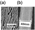
- FIG 5 is an SEM image of (a) zinc oxide (ZnO) nanorods used in the examples, and (b) SEM images of single zinc oxide nanorods used in PES.
- FIG. 10 shows the arrangement of ZnO nanorods in PES for mechanical stimulation in Test Example 1.
- FIG. 11 shows the permanent strain profile when flexing and mechanical stimulation of PES and PDMS were given for 10 days in Test Example 1.
- FIG. 11 shows the permanent strain profile when flexing and mechanical stimulation of PES and PDMS were given for 10 days in Test Example 1.
- FIG. 12 to 14 show the cytotoxicity when flexion, electrical stimulation and mechanical stimulation were applied to PES in Test Example 1.
- FIG. 12 to 14 show the cytotoxicity when flexion, electrical stimulation and mechanical stimulation were applied to PES in Test Example 1.
- FIG. 19 is a schematic diagram of the myocardial differentiation mechanism of hMSC according to electrical and mechanical stimulation.
- Figure 21 shows the expression of p38, SMAD, FAK, ERK1 / 2 by Western blot method of Test Example 3.
- 'elasticity' refers to a property that is deformed when a force is applied but returns to its original form when the force is removed.
- 'kinetic force' or 'kinetic energy' refers to a stimulus that may cause physical changes such as bending, tension, stretching, and the like, and does not select any means or method.
- the exercise force may be one or both of flexing exercise and stretching exercise or both are applied at the same time.
- Flexing movement refers to the movement of bending and stretching, stretching movement to increase or return to the original state. This movement is possible because PES is elastic.
- the stem cell differentiation method according to the present invention promotes differentiation by simultaneously applying electrical energy and mechanical energy to the stem cells, the stretching or stretching movement applied to the support using the piezoelectricity of the support electrical and mechanical It may be to generate energy.
- the electrical energy may be to generate a voltage of 0.1 ⁇ 10V by the bending and unfolding movement of the support at a frequency of 0.1 ⁇ 10 Hz.
- a voltage of 0.5 to 5V may be generated at a frequency of 0.1 to 5 Hz.
- the mechanical stimulus may be caused by the movement of the support to increase the length of 1 to 20% compared to the original length at a frequency of 0.1 to 10 Hz and then contract.
- the voltage may be generated by the movement of increasing the length of 1 to 10% relative to the original length of the support at a frequency of 0.1 to 5 Hz and then contracting it.
- the inoculated stem cells may be arranged in a direction perpendicular to the direction of the kinetic force applied to the support.
- the stem cells may be selected from the group consisting of adipose stem cells, mesenchymal stem cells, bone marrow stem cells, cord blood stem cells, neural stem cells and induced pluripotent stem cells.
- mesenchymal stem cells are pluripotent, undifferentiated cells, connective tissue derived from the mesoderm of the fetus, morphologically stromal cells containing coarse fibers and nonspecific cells in the microenvironment. Therefore, it is possible to differentiate into organs such as connective tissue, bone, cartilage, lymphatic vessels, and blood vessels.
- FIG. 1 is a schematic diagram showing two microenvironments of the heart, electrical signals and mechanical signals.
- the heart is accelerated by differentiation of electric field and cyclic strain.
- the differentiation process of cardiomyocytes is described in detail in the Examples.
- PES can deliver electrical stimulation and mechanical stimulation to stem cells inoculated into the support by, for example, repeated stretching or bending using the device shown in FIG. 3.
- the motion of the motor of the apparatus of FIG. 3 is transmitted to the PES located in the cell culture unit, whereby the PES performs a bending motion, stimulates the piezoelectric material of the PES, generates an electrical signal, and expands and contracts the PES. It can generate mechanical signals by elastic materials through stretching.
- the PES may have a structure in which an elastic layer and a piezoelectric material layer are alternately stacked one or more times, preferably two or more times. At this time, it is preferable that the outermost layer is an elastic layer.
- the support is
- It may have a unit structure including a first elastic layer, a second elastic layer, and a piezoelectric material layer arranged between the first elastic layer and the second elastic layer.
- the piezoelectric material may be one or more of piezoelectric materials including ZnO, BaTiO 3 and NaNBO 3 , but is not limited thereto. That is, ZnO is one example of a representative piezoelectric material, and the present invention is not limited thereto, and may be applied to the present invention as long as it is a piezoelectric material capable of converting an external mechanical force into a potential.
- piezoelectric materials such as ZnO, ZnSnO 3 , GaN, Te, CdTe, CdSe, KNbO 3 , NaNbO 3 , InN, PVDF, and PVDF-TrFE may be used.
- the piezoelectric material layer may include a plurality of piezoelectric material nanorods.
- the piezoelectric material nanorods may be biaxially grown piezoelectric material nanorods.
- the piezoelectric material nanorods may be arranged in one direction to form a piezoelectric material layer.
- the piezoelectric material nanorods may be arranged in one direction to form a piezoelectric material layer.
- nanorods is a term commonly used by those skilled in the art, and may refer to a rod having an aspect ratio of usually 10 or less, and in some cases, in the art, nano It may also refer to about 100 nm or less with respect to the size. As such, it should be noted that nanorods are interpreted as having a technical meaning commonly used by those skilled in the art.
- ZnO nanorods used in one embodiment of the present invention was about 2.5 ⁇ 3 ⁇ m in length, about 200 ⁇ 250 nm in diameter.
- the small sized one-dimensional nanomaterial has a feature that the lattice deformation can be easily induced by small mechanical energy (bending or shaking) from the outside.
- the piezoelectric potential was generated using nanorods, specifically biaxially grown nanorods, but the present invention is not limited thereto. That is, uniaxially grown nanorods may also be employed, and as a material having piezoelectric properties, it may be possible to adopt a nano-sized film, a nanowire or the like.
- the biaxially-grown nanorods employed in the examples have been exemplified by hydrothermal synthesis, the present invention is not limited thereto. In other words, the hydrothermal synthesis method is used to synthesize a large amount, but in order to form a nanorod having a small amount of high crystallinity, the CVD method may be used.
- the elastic layer is preferably made of a material capable of transferring mechanical energy applied from the outside to the piezoelectric material layer.
- the elastic layer is preferably made of a material capable of transferring the piezoelectric potential generated from the piezoelectric material layer to the inoculated stem cells.
- the elastic layer may be made of a material containing PDMS (polydimethylsiloxane), but is not limited thereto, and may be used without limitation as long as it is an elastic material having cell compatibility or dielectric constant. That is, a dielectric material having a dielectric constant and easy to transfer the piezoelectric potential generated from the piezoelectric material can be applied to the present invention, and it is preferable that the mechanical energy applied from the outside can be transferred to the piezoelectric material without large loss.
- PDMS polydimethylsiloxane
- differentiation efficiency into cardiomyocytes is increased when both electrical and mechanical stimulation are given to mesenchymal stem cells.
- Cardiomyocytes differentiated from stem cells by the above-described method can be utilized in various applications as a cardiomyocyte-support complex.
- Zinc oxide nanorod powder was prepared using a wet chemical method (wet chemical method).
- nano-rods flocculated After centrifugation, to separate the agglomerated nano-rods (nanorods flocculated) from the suspension, washed with deionized water three times to thereby remove the unreacted Zn 2 + and other ions. The final precipitate was dried at 80 ° C., annealed at 400 ° C. for 2 hours in vacuo to improve crystallinity, and finally a biaxially grown ZnO nanorod powder was prepared by synthesis.
- the piezoelectric and elastic material (PES) disclosed in FIG. 4 has a plurality of structures in which a plurality of zinc oxide nanorods (ZnO NRs) having piezoelectricity are arranged between polydimethyl-siloxane (PDMS) having a thickness of about 3 mm (Elastic). Laminated, the manufacturing process can refer to the Republic of Korea Patent Registration No. 10-1497338.
- Morphological characteristics of the zinc oxide nanorods were analyzed using JEOL JSM-7000F field emission scanning electron microscope (FESEM, Jeol Ltd, Akishima, Tokyo, Japan; 15 kV) (see FIG. 5).
- the PDMS block and the substrate for the rubbing process were prepared with a curing agent in a weight ratio of 10: 1 (Sylgard 184, Dow corning Chemicals). Form a single layer of zinc oxide nanorods through a unidirectional rubbing process with a PDMS block on the PDMS substrate (see FIG. 5 (a)) and hexane to treat untreated PDMS to coat the PDMS on the zinc oxide nanorods single layer. Diluted in a weight ratio of 1: 1.
- the diluted PDMS was added to a PDMS substrate with a zinc oxide nanorod monolayer, spin-coated for 30 seconds at 3,000 rpm, and treated for 30 minutes on an 85 ° C. hot plate in air.
- a device obtained by depositing a silver electrode (200 nm) on the top and bottom of the PES by thermal evaporation (thermal evaporation) is mounted on a 3 mm thick PDMS substrate After that, the voltage and current signals were measured.
- 6 to 9 show electrical characteristics when bending a PES having a length of 5 cm with a curvature radius of 20 mm, and current and voltage are respectively picoammeters (picoammeter, Keithley 6485, Keithely Instruments, Cleveland, OH, USA). ) And an electrometer / high resistance meter (Keithley 6517, Keithley Instruments).
- FIG. 6 is an open circuit voltage generated at the PES when forward connected
- FIG. 7 is an open circuit voltage generated at the PES when reverse connected
- FIG. 8 is a short circuit current density generated at the PES when forward connected
- FIG. 9 shows the short-circuit current density produced by the PES when reversed.
- FIG. 10 shows the arrangement of ZnO NRs in PES for flexural and mechanical stimulation on days 0 and 10.
- FIG. 11 shows the permanent strain profile when PES and PDMS were subjected to flexion and mechanical stimulation for 10 days.
- hMSCs Human mesenchymal stem cells (Lonza, Walkerxville, MD, USA) were 10% (v / v) etal bovine serum (FBS) (Gibco BRL) and 1% (v / v) penicillin / Medium consisting of DMEM low glucose (Dulbecco's Modified Eagle Medium low glucose) (Gibco BRL, Gaithersburg, MD, USA) containing streptomycin (Gibco BRL) in a 37 ° C, 5% (v / v) CO 2 constant temperature and humidity incubator The cells were cultured, and only hMSCs cells of 6 passages or less were used.
- FBS etal bovine serum
- penicillin / Medium consisting of DMEM low glucose (Dulbecco's Modified Eagle Medium low glucose) (Gibco BRL, Gaithersburg, MD, USA) containing streptomycin (Gibco BRL) in a 37 ° C, 5% (v / v
- the electrical stimulus generates an electrical signal of about 3V through a 20 mm curvature radius at a frequency of 1 Hz, and the mechanical stimulus increases the contraction length by 3% compared to the existing length at a frequency of 1 Hz and contracts the mechanical stimulus.
- the cell culture medium of the cell culture part was exchanged every three days.
- the biocompatibility and stability of zinc oxide was not cytotoxic at the zinc oxide nanowire concentration of 100 ⁇ g / ml or less.
- CS bending and stretching
- F-actin filamentous actin
- filamentous actin is one of the fibers that make up the cytoskeleton, which is responsible for maintaining the shape of the cell, changing its shape, and transporting substances within the cell.
- the cell line array was confirmed through F-actin staining of the mesenchymal stem cells of Comparative Example and Example of Table 1 using Phalloidin staining as being directly involved in muscle contraction.
- Paloidin staining of F-actin of mesenchymal stem cells was performed using Actin Cytoskeleton and Focal adhesion staining kit (FAK100, Millipore). The arrangement was confirmed using Image J software (National Institute of Health).
- Figure 15 shows the F-actin array of the mesenchymal stem cells of the Examples and Comparative Examples stained by the paloidine staining method, according to the results of Figure 15 cells tend to be arranged in a direction perpendicular to the direction to increase and shrink the PES Showed. This is because the cell is arranged in a direction that minimizes the stimulation when the cell receives a certain mechanical signal, such a cell array is a very important factor in myocardial differentiation. Cells lined up are more likely to be present and distributed in cells than in randomly arranged cells, with more interstitial binding proteins (eg connexin 43) in the cells that play an important role in intercellular signal transduction. In particular, in the myocardium, such a gap binding protein is a key factor for the delivery and beat of calcium ions.
- interstitial binding proteins eg connexin 43
- the mesenchymal stem cells myocardial differentiation by mechanical signal in the above results can be said to have a condition that can perform the function as a cardiomyocytes better.
- Cardiomyogenic differentiation in the mesenchymal stem cells and molecular signals related to myocardial differentiation were analyzed using the relevant protein markers using the Western blot method.
- Proteins in the buffer are electrophoresed on 4-10% SDS-PAGE (SDS-polyacrylamide gel), transcribed into membranes (Millipore, Bedford, MA, USA), and Cx43, NKX2.5 as primary antibodies against , MEF-2, GATA4, sarcomeric ⁇ -actinin, ⁇ -MHC, p38, pp38, SMAD, pSMAD, FAK, pFAK, ERK1 / 2, pERK1 / 2, and ⁇ -actin (Abcam, Cambridge, MA, USA) After treatment, the reaction was reacted overnight at 4 ° C., followed by washing. The secondary antibody combined with horseradish peroxidase (HRP) was treated and reacted at room temperature for 50 minutes. Blots were developed using enhanced chemiluminescence (ECL) (LumiGLO, KPL Europe, Guildford, UK).
- ECL enhanced chemiluminescence
- Figure 16 shows the improvement of expression of NKX 2.5, MEF-2, GATA4, ⁇ -MHC, sarcomeric ⁇ -actinin and Cx43 by Western blot method
- Figure 21 is p38, SMAD, FAK, ERK1 / 2 by Western blot method Indicates expression.
- sarcomeric ⁇ -actinin and connexin 43 which are late differentiation factors, were analyzed by fluorescence immunostaining (Immunocytochemistry).
- qRT-PCR was used to quantify the relative gene expression levels of cyclic nucleotide-gated potassium channel 2 (HCN2) and calcium channel, voltage-dependent, L type, and alpha 1C subunit (CACNA1C).
- FIG. 19 is a schematic diagram of the myocardial differentiation mechanism of hMSC according to electrical and mechanical stimulation.
- an electrical signal or mechanical stimulus including an electric field is applied, autologous or near-secretory proteins (BMP-4, TGF- ⁇ , vascular endothelial growth factor (VEGF)) in mesenchymal stem cells are applied.
- BMP-4 autologous or near-secretory proteins
- TGF- ⁇ TGF- ⁇
- VEGF vascular endothelial growth factor
- IGF IGF
- the phosphorylation reaction of SMAD-1, 4, 5, 8 occurs by BMP-4 expressed by electrical stimulation, and the phosphorylation reaction increases the expression levels of NKX 2.5, MEF-2 and ⁇ -MHC.
- VEGF and TGF- ⁇ are known to increase the expression level of connexin43, a type of gap-binding protein. IGF causes myocardial differentiation by enhancing the phosphorylation of p38 and increasing the expression level of MEF-2.
- BMP-4, TGF- ⁇ , VEGF, and IGF which are related to myocardial differentiation by electrical stimulation, was compared using reverse transcriptase polymerase chain reaction.
- RNA samples were lysed by treatment with TRIZOL reagent (TRIzol reagent, Invitrogen Carlsbad, CA, USA), total RNA was extracted with chloroform (Sigma) and 80% (v / v) isopropanol (Sigma). After precipitation, the supernatant was removed and the precipitated RNA pellet was washed with 75% (v / v) ethanol and dried, and then dried with 0.1% (v / v) DEPC pyrocarbonate-treated water. The pellet was melted to obtain pure total RNA.
- TRIZOL reagent Trizol reagent, Invitrogen Carlsbad, CA, USA
- RNA extracted by the above method was synthesized cDNA using SuperScript TM II reverse transcriptase (Invitrogen).
- the synthesized cDNA was subjected to 35 cycles of thermal denaturation at 94 ° C. for 30 seconds, annealing at 58 ° C. for 45 seconds, and stretching at 72 ° C. for 45 seconds, and finally at 72 ° C. for 10 minutes.
- electrophoresis on 2% (w / v) agarose gel and stained with Et-Br (ethidium bromide), the gel documentation system (Gel Doc 100, Bio-Rad, Hercules, CA, USA) and analyzed using an imaging densitometer (Imaging densitometer, Bio-Rad).
- ⁇ -actin was used as the internal control.
- Example 20 shows the results of expression evaluation of BMP-4, IGF, VEGF and TGF- ⁇ by RT-PCR method.
- the expression levels of BMP-4, IFG, VEGF, and TGF- ⁇ which are related to myocardial differentiation, were significantly higher in Example 1.
- FIG. 21 shows expression of p38, SMAD, FAK, and ERK1 / 2 by Western blot method described in Test Example 3.
- FIG. 21 Since mesenchymal stem cells are stimulated by self-secreting factors, as shown in FIG. 21, it can be seen that electrical and mechanical stimulation further increased protein expression of intercellular signaling molecules for myocardial differentiation (pp38 and pSMAD).
- phosphorylation of focal adhesion kinase (FAK) and extracellular signal-regulated kinases 1/2 (ERK1 / 2) was enhanced by mechanical and electrical stimulation.
- Phosphorylation of ERK1 / 2 causes myocardial differentiation by enhancing GATA4 expression.
- pp38, pSMAD, pFAK, and pERK1 / 2 were further enhanced compared to p38, SMAD, FAK, and ERK1 / 2.
Landscapes
- Health & Medical Sciences (AREA)
- Engineering & Computer Science (AREA)
- Life Sciences & Earth Sciences (AREA)
- Micro-Organisms Or Cultivation Processes Thereof (AREA)
- Biomedical Technology (AREA)
- Biotechnology (AREA)
- Chemical & Material Sciences (AREA)
- Organic Chemistry (AREA)
- Bioinformatics & Cheminformatics (AREA)
- Genetics & Genomics (AREA)
- Medicines Containing Material From Animals Or Micro-Organisms (AREA)
- Wood Science & Technology (AREA)
- Zoology (AREA)
- Microbiology (AREA)
- Biochemistry (AREA)
- General Engineering & Computer Science (AREA)
- General Health & Medical Sciences (AREA)
- Cell Biology (AREA)
- Rheumatology (AREA)
- Cardiology (AREA)
Abstract
압전성과 탄성을 동시에 가지는 지지체에 접종된 줄기세포에 전기적 자극과 기계적 자극을 동시에 가하여 줄기세포를 분화하는 생체 내 심근세포의 미세환경을 모사하여 줄기세포 분화 효율을 더욱 높일 수 있다. 따라서, 본 발명에 다른 줄기세포 분화방법은 다양한 연구에 활용 할 수 있을 뿐만 아니라, 심근경색을 포함하여 심근세포와 관련된 질환의 치료에 활용 할 수 있다.
Description
본 발명은 전기·기계적 자극을 가하여 줄기세포를 심근세포로 분화시키는 방법에 관한 것이다.
줄기 세포는 특정한 세포로 분화가 진행되지 않은 채 유지되다가 필요할 경우 분열, 복제하여 신경, 혈액, 연골, 근육 등 몸을 구성하는 모든 종류의 세포로 분화할 가능성을 가진, 각 세포로 분화(differentiation)되기 전 단계의 미분화 세포들을 총칭한다. 줄기세포는 세포분열이 정지된 분화된 세포와는 달리 세포분열에 의해 자신과 동일한 세포를 생산(self-renewal)하며 증식(proliferation)하는 특성이 있으며, 또한 분화 자극(미세환경)이 가해지면 특정 세포로 분화가 진행되는데 다른 환경 또는 다른 분화 자극에 의해 다른 세포로도 분화될 수 있어 분화에 유연성(plasticity)을 가지고 있는 것이 특징이다.
최근에는 상기와 같은 줄기세포의 특징을 활용하여 전기적 자극(electric field) 혹은 기계적 자극(cyclic strain)을 이용한 심장의 미세환경 모사를 통한 중간엽 줄기세포의 심근분화 방법이 많이 사용되고 있다. 그러나, 전기장 혹은 전류를 사용한 전기적 자극만을 가하는 방법과, 기계적 자극만을 가하는 연구는 각각 존재하지만 분화 효율에서 한계가 있어, 개선의 여지가 있다.
한편, 심장질환은 선진국들에서 주된 사망원인으로 자리 잡고 있으며, 우리나라에서도 유병률이 빠른 속도로 증가하고 있는 추세이다. 현재 심근경색 및 비정상적인 심장발달을 치료하기 위한 다양한 시술들이 시행되고 있으나, 심근세포의 낮은 재생능력 때문에 완치에 많은 어려움이 있다.
현재 심장질환 치료를 위해 가장 많이 시행되고 있는 시술은 장기 이식 및 동물 유래 조직으로 심장조직 일부를 치환하는 시술이다. 하지만 이러한 시술들은 장기 기증자의 확보 및 이종간 면역거부 반응 등의 문제가 있다.
이러한 문제점을 해결하기 위해 많은 연구 그룹들이 심근세포의 이식을 통하여 심장의 기능을 회복시키는 방법에 관한 연구를 진행하고 있다. 심근세포의 이식에 이용될 세포를 획득하기 위한 방법으로는 성체줄기세포, 배아줄기세포 및 유도 만능줄기세포(Induced pluripotent stem cells: iPSC)를 분화시켜서 심근세포를 획득하는 것이 가장 일반적으로 제시되고 있는 방법이다. 그러나 배아줄기세포와 유도만능줄기세포는 공통적으로 양성종양(teratoma) 형성 위험이 문제가 되어 임상적용에 어려움을 겪고 있다.
본 발명은 상기와 같은 종래 기술의 한계를 해결하기 위해, 보다 효율적으로 줄기세포를 심근세포로 분화 유도시키는 방법을 제공하는 것을 목적으로 한다.
상기 목적을 달성하기 위하여, 본 발명은
압전성과 탄성을 동시에 갖는 지지체 상에 줄기세포를 접종하는 단계 및
상기 줄기세포가 접종된 지지체에 운동력을 가하여 줄기세포를 심근세포로 분화시키는 단계를 포함하는 줄기세포의 분화 방법을 제공한다.
또한 본 발명은 상기 방법에 의해 줄기세포로부터 분화된 심근세포를 함유하는 심근세포-지지체 복합체를 제공한다.
기타 본 발명의 구현 예들의 구체적인 사항은 이하의 상세한 설명에 포함되어 있다.
압전성과 탄성을 동시에 가지는 지지체(Piezoelectric and Elastic Substrate: PES)에 줄기세포를 접종한 후 운동력을 가하여 전기적 자극과 기계적 자극을 동시에 발생시킴으로써 줄기세포를 심근세포로 분화시키는 본 발명의 방법은 심근세포의 생체 내 미세환경을 모사하여 분화 효율을 더욱 높일 수 있다. 따라서, 본 발명은 심근경색을 포함한 심혈관 질환의 치료 등 다양한 연구에 활용될 수 있다.
도 1은 심장의 두 가지 미세환경, 전기적 신호와 기계적 신호를 보여주는 모식도이다.
도 2는 본 발명에 따라 PES를 이용하여 중간엽 줄기세포를 심근분화시키는 과정에 대한 모식도이다.
도 3은 본 발명에 일 실시예에 따라 PES의 굴신(屈伸) 및 신축(伸縮)운동이 가능한 세포 배양장치의 개략적인 구성도이다.
도 4는 PES의 제조과정을 보여주는 모식도이다.
도 5는 실시예에서 사용된 (a)산화아연(ZnO) 나노로드의 SEM 이미지, (b)PES에 사용되는 단일 산화 아연나노로드의 SEM 이미지이다.
도 6 내지 도 9는 시험예 1에서 PES 벤딩시(굴신운동시) 전기적 특성을 나타낸다.
도 10은 시험예 1에서 기계적 자극에 대한 PES 내 ZnO 나노로드의 배열을 나타낸다.
도 11은 시험예 1에서 10일 동안 PES와 PDMS를 굴신운동 및 기계적 자극을 주었을 때의 영구 변형 프로필을 나타낸다.
도 12 내지 도 14는 시험예 1에서 PES에 굴신, 전기적 자극 및 기계적 자극을 가하였을 때의 세포독성에 대한 것이다.
도 15는 시험예 2에서 기계적 자극에 의한 hMSC의 배열을 F-action 염색으로 나타낸 것이다(청색 = 세포 핵, 스케일 바 = 30 μm).
도 16 내지 18은 시험예 3 내지 5에 따라 전기적 자극과 기계적 자극에 의한 hMSC의 심근분화 향상을 평가한 것이다.
도 19는 전기적 자극 및 기계적 자극에 따른 hMSC의 심근분화 메커니즘의 모식도이다.
도 20은 시험예 6의 RT-PCR 방법에 의한 BMP-4, IGF, VEGF 및 TGF-β의 발현 평가 결과이다.
도 21은 시험예 3의 웨스턴 블롯 방법에 의한 p38, SMAD, FAK, ERK1/2의 발현을 나타낸다.
본 발명은 다양한 변환을 가할 수 있고 여러 가지 실시예를 가질 수 있는 바, 특정 실시예들을 도면에 예시하고 상세한 설명에 상세하게 설명하고자 한다. 그러나, 이는 본 발명을 특정한 실시 형태에 대해 한정하려는 것이 아니며, 본 발명의 사상 및 기술 범위에 포함되는 모든 변환, 균등물 내지 대체물을 포함하는 것으로 이해되어야 한다. 본 발명을 설명함에 있어서 관련된 공지 기술에 대한 구체적인 설명이 본 발명의 요지를 흐릴 수 있다고 판단되는 경우 그 상세한 설명을 생략한다.
본 명세서에서 층, 막, 필름, 기판 등의 부분이 다른 부분 '위에' 또는 '상에' 있다고 할 때, 이는 다른 부분 '바로 위에' 있는 경우뿐 아니라 그 중간에 또 다른 부분이 있는 경우도 포함한다. 반대로 층, 막, 필름, 기판 등의 부분이 다른 부분 '아래에' 있다고 할 때, 이는 다른 부분 '바로 아래에' 있는 경우뿐 아니라 그 중간에 또 다른 부분이 있는 경우도 포함한다.
이하에서는 본 발명을 구체적으로 설명한다.
본 발명에 따른 줄기세포 분화 방법은
압전성과 탄성을 동시에 갖는 지지체(PES) 상에 줄기세포를 접종하는 단계 및
상기 줄기세포가 접종된 지지체에 운동력을 가하여 줄기세포를 심근세포로 분화시키는 단계를 포함하는 것을 특징으로 한다.
본 명세서에서 '탄성'은 힘을 가하면 형태가 변형되지만 힘을 제거하면 원래 형태로 돌아오는 성질을 의미한다.
또, 본 명세서에서 '운동력' 또는 '운동에너지'는 굴곡, 인장, 신축 등과 같이 물리적 변화를 야기할 수 있는 자극을 의미하며, 그 수단이나 방식을 가리지 않는다.
바람직한 구현예에 따르면, 상기 운동력은 굴신(屈伸) 운동 및 신축(伸縮) 운동 중 어느 하나이거나 두 가지가 동시에 가해지는 것일 수 있다. 굴신운동은 굽혔다 펴지는 운동, 신축운동은 늘어났다가 원래 상태로 복귀하거나 수축하는 운동을 의미한다. PES는 탄성을 보유하기 때문에 이러한 운동이 가능하다.
또한, 본 발명에 따른 줄기세포 분화방법은 전기적 에너지와 기계적 에너지를 줄기세포에 동시에 가함으로써 분화를 촉진하는바, 상기 지지체에 가해지는 굴신 운동 또는 신축 운동은 지지체의 압전성을 이용하여 전기적 에너지 및 기계적 에너지를 발생시키는 것일 수 있다.
상기 전기적 에너지는 상기 지지체를 0.1 ~ 10 Hz 빈도로 굽혔다 펴주는 운동에 의해 0.1 ~ 10 V의 전압을 발생시키는 것일 수 있다. 바람직하게는 0.1 ~ 5 Hz의 빈도로 0.5 ~ 5V의 전압을 발생시킬 수 있다.
상기 기계적 자극은 상기 지지체를 0.1 ~ 10 Hz 빈도로 원래 길이 대비 1 ~ 20%의 길이를 늘렸다가 수축시키는 운동에 의해 발생시키는 것일 수 있다. 바람직하게는 0.1 내지 5 Hz의 빈도로 지지체 원래 길이 대비 1 ~ 10%의 길이를 늘렸다가 수축시키는 운동에 의해 전압을 발생시킬 수 있다.
바람직한 실시예에 따르면, 상기 접종된 줄기세포는 지지체에 가해지는 운동력의 방향과 수직한 방향으로 배열될 수 있다.
상기 줄기세포는 지방 줄기세포, 중간엽 줄기세포, 골수 줄기세포, 제대혈 줄기세포, 신경줄기세포 및 유도만능 줄기세포로 구성된 군으로부터 선택되는 것일 수 있다.
예를 들어, 중간엽 줄기세포(mesenchymal stem cell, MSC)는 다능성 미분화 세포로 태아의 중배엽에서 유래된 결합조직이며, 형태적으로 망상섬유와 비특이적 세포의 성긴 집합체를 포함한 기질세포로 미세환경에 따라서, 결합조직, 골, 연골, 림프관, 혈관 등의 기관으로 분화 가능하다.
도 1은 심장의 두 가지 미세환경, 전기적 신호와 기계적 신호를 보여주는 모식도이다. 즉 심장은 전기적 자극(electric field)과 기계적 자극(cyclic strain) 이 가해짐으로써 심근세포 분화가 촉진된다. 심근 세포의 분화 과정은 실시예에서 구체적으로 설명한다.
이에 본 발명에서는, 중간엽 줄기세포를 심근세포로 분화시키기 위한 배양 지지체로서 도 2에 도시된 바와 같이 전기적 신호 및 기계적 신호를 동시에 발생시키거나 인가할 수 있는 PES를 사용하였다.
PES는 예를 들어 도 3에 도시된 장치를 이용하여 반복적인 스트레칭이나 벤딩됨으로써 전기적 자극 및 기계적 자극을 지지체에 접종된 줄기세포에 전달할 수 있다.
구체적으로, 도 3 장치의 모터의 운동이 세포 배양부에 위치한 PES에 전해지고, 이에 의하여 PES가 굴신(屈伸, bending) 운동을 하며 PES의 압전 물질을 자극하여 전기적 신호를 발생시키고, PES의 신축(伸縮, stretching) 운동을 통한 탄성 물질에 의한 기계적 신호를 발생시 킬 수 있다.
바람직한 구현예에 따르면, 도 4에 도시된 바와 같이, PES는 탄성층과 압전물질층이 1회 이상, 바람직하게는 2회 이상 교대로 적층된 구조를 가질 수 있다. 이때, 최외곽층은 탄성층이 되도록 하는 것이 바람직하다.
일 구현예에 따르면, 상기 지지체는
제 1 탄성층, 제 2 탄성층, 및 상기 제 1 탄성층과 제 2 탄성층 사이에 배열된 압전물질층을 포함하는 단위 구조를 갖는 것일 수 있다.
상기 압전물질은 ZnO, BaTiO3 및 NaNBO3 를 포함하는 압전 물질 중 하나 이상일 수 있으나, 이에 한정되는 것은 아니다. 즉 ZnO는 대표적인 압전 재료의 한 가지 예로서, 본 발명은 이에 제한되지 않으며, 외부의 기계적인 힘을 전위로 바꿀 수 있는 압전 재료이기만 하면, 본 발명에 적용될 수 있다. 예컨대, ZnO, ZnSnO3, GaN, Te, CdTe, CdSe, KNbO3, NaNbO3, InN, PVDF, PVDF-TrFE 등의 압전재료를 이용할 수도 있다.
또한, 상기 압전물질층은 복수의 압전물질 나노로드를 포함하여 이루어질 수 있다. 또, 상기 압전물질 나노로드는 이축 성장 압전물질 나노로드일 수 있다.
또, 상기 압전물질 나노로드는 일방향으로 배열되어 압전물질층을 형성할 수 있으며, 특히 일방향 단일층으로 배열되어 압전물질층을 형성할 수 있다.
본 명세서에서 나노로드라는 용어는 당업계에서 당업자가 통상적으로 사용하고 있는 용어로서, 보통 종횡비(aspect ratio)가 10 이하인 상태의 로드를 의미할 수 있으며, 또한, 경우에 따라서, 당업계에서는 나노는 그 크기와 관련하여 약 100 nm 이하인 것을 지칭하기도 한다. 이와 같이, 나노로드는 당업계에서 당업자가 통상적으로 사용하고 있는 기술적 의미를 갖고 있는 물질로서 해석된다는 점에 유의하여야 한다. 본 발명의 일 실시예에서 사용된 ZnO 나노로드는 길이가 약 2.5~3 ㎛이고, 직경은 약 200~250 nm 이었다. 이러한 작은 크기의 1차원 나노물질은 외부로부터의 작은 기계적 에너지(굽힘이나 흔들림)에 의해 쉽게 격자 변형이 유도될 수 있는 특징을 갖고 있다.
이후 설명되는 실시예에서는 나노로드, 구체적으로 이축 성장 나노로드를 사용하여 압전전위를 생성하였지만, 본 발명은 이에 제한되지 않는다. 즉, 일축 성장 나노로드 역시 채용할 수 있으며, 압전 특성을 갖는 물질로서, 나노크기를 갖는 필름의 형태, 나노와이어 형태 등의 것을 채용할 수도 있다. 또한, 실시예에서 채용한 이축 성장 나노로드를 수열합성법에 의해 제조하는 것을 예시하였지만, 본 발명은 이에 제한되지 않는다. 즉, 대량으로 합성하는 경우에는 수열합성법을 이용하지만, 소량의 높은 결정화도를 갖는 나노로드를 형성하기 위하여, CVD법을 이용할 수도 있다.
상기 탄성층은 외부로부터 인가되는 기계적 에너지를 상기 압전물질층에 전달할 수 있는 재료로 이루어지는 것이 바람직하다.
또한, 상기 탄성층은 상기 압전물질층으로부터 발생되는 압전 전위를 접종된 줄기세포에 전달할 수 있는 재료로 이루어진 것이 바람직하다.
바람직한 실시예에 따르면, 탄성층은 PDMS(polydimethylsiloxane)를 포함하는 재료로 이루어질 수 있으나, 이에 한정되지 않으며 세포 적합성이나 유전상수를 갖고 있는 탄성 재료라면 제한없이 사용할 수 있다. 즉, 유전 상수를 갖고 있어 압전 재료로부터 발생되는 압전 전위를 전달하기 쉬운 유전물질을 본 발명에 적용할 수 있으며, 외부로부터 인가되는 기계적 에너지를 압전 재료에 큰 손실 없이 전달할 수 있으면 바람직하다.
본 발명의 바람직한 실시예에 따르면, 중간엽 줄기세포에 전기적 자극과 기계적 자극을 동시에 주었을 때 심근세포로의 분화효율이 증가함을 알 수 있다.
전술한 방법에 의해 줄기세포로부터 분화된 심근세포는 심근세포-지지체 복합체로서 다양한 용도에 활용될 수 있다.
이하에서는, 첨부 도면을 참조하여 본 발명의 실시예를 설명한다. 이하의 설명에 있어서, 당업계에 이미 알려진 기술적 구성에 대한 설명은 생략한다. 예컨대, 일축 나노로드, 이축 성장 나노로드를 합성/제조하는 방법, 나노로드를 러빙(rubbing) 공정을 통해 정렬하는 공정은 액정 정렬과 관련하여 이미 널리 알려진 공정이므로, 그 자세한 설명은 생략한다. 이러한 설명을 생략하더라도, 당업자라면 이하의 설명을 통해 본 발명에 따라 제시되는 세포 지지체의 구성, 그 기능 등을 쉽게 이해할 수 있을 것이다.
<
제조예
1> 산화아연
나노로드
(
ZnO
NRs
) 분말의 준비
산화아연 나노로드 분말은 습식화학방법(wet chemical method)을 이용하여 준비하였다.
0.42g의 징크 나이트레이트 헥사하이드레이트(zinc nitrate hexahydrate)(Zn(NO3)2ㆍ6H2O, ≥99.0%)(Sigma Aldrich)를 100 mL의 탈이온수에 용해하고, 0.24g의 HMTA(헥사메틸렌테트라민, Sigma Aldrich)를 100 mL의 탈이온수에 별도로 용해하여(모두 실온에서), 두 개의 전구체 용액을 준비하였다. 85℃에서 교반하면서, 징크 전구체 용액을 25분 동안 2 mL/h의 주입 속도로 시린지 펌프를 통해 HMTA 용액 내로 연속하여 주입하였으며, 상기 프로세스를 5분 후에 종결하였다. 원심 분리 후에, 응집된 나노로드(flocculated nanorods)를 부유물로부터 분리하여 탈이온수로 3번 세정하여, 미반응 Zn2
+및 다른 이온들을 제거하였다. 최종 석출물을 80℃에서 건조하였고, 진공 중에서 2시간 동안 400℃에서 어닐링 처리하여 결정성을 향상시켜, 최종적으로 이축 성장 ZnO 나노로드 파우더를 합성하여 준비하였다.
<
제조예
2> 압전성과 탄성을 가진 재료(Piezoelectric and Elastic Substrate: PES)의 제작
도 4에 개시된 압전성과 탄성을 가진 재료(PES)는 탄성을 가지는 PDMS(polydimethyl-siloxane)(두께 약 3 mm) 사이에 압전성을 가지는 복수의 산화아연 나노로드(ZnO NRs)가 정렬된 구조가 복수개 적층되어 있으며, 그 제조과정은 대한민국 특허등록 10-1497338호를 참조할 수 있다.
산화 아연 나노로드의 형태학적 특징을 JEOL JSM-7000F 전계 방출형 주사전자현미경(FESEM, Jeol Ltd, Akishima, Tokyo, Japan; 15 kV)을 이용하여 분석하였다(도 5 참조).
PDMS 블록과 러빙 공정(rubbing process)용 기판은 10:1의 중량비로 경화제와 같이 준비하였다(Sylgard 184, Dow corning Chemicals). PDMS 기판상에 PDMS 블록과 함께 단방향 러빙 공정을 통하여 산화 아연 나노로드 단일 층을 형성하고(도 5(a) 참조), 산화 아연 나노로드 단일 층 상에 PDMS를 코팅하기 위하여 처리되지 않은 PDMS를 헥산에 1:1의 중량비로 희석하였다.
상기 희석된 PDMS를 산화아연 나노로드 단일 층을 가지는 PDMS 기판에 가하여 3,000 rpm으로 30 초간 스핀코팅(spin-coating)하고, 공기 중의 85℃ 핫 플레이트 에서 30분간 처리하였다.
상기와 같은 공정을 5번 반복하여 3V의 전기적 신호를 내는 5겹의 산화아연 나노로드층(압전물질층)을 가지는 PES(Piezoelectric and Elastic Substrate)를 제작하였다.
<
시험예
1>
PES
(Piezoelectric and Elastic Substrate)의 특징 분석
상기 제조예 1에서 제조된 PES의 전기적 특성을 평가하기 위하여, 은 전극(200 nm)을 열증착법(thermal evaporation)으로 PES의 상부 및 하부에 증착하여 얻은 소자를 3 mm 두께의 PDMS 기판 상에 장착한 후, 전압 및 전류 신호를 측정하였다.
도 6 내지 도 9는 길이 5 cm의 PES 를 20 mm 곡률반경으로 벤딩(굴신운동)시 전기적 특성을 나타내며, 전류와 전압은 각각 피코암메터(picoammeter, Keithley 6485, Keithely Instruments, Cleveland, OH, USA)와 일렉트로미터/고저항미터(electrometer/high resistance meter, Keithley 6517, Keithley Instruments)를 이용하여 측정하였다.
구체적으로, 도 6은 포워드 연결했을 때 PES에서 생성되는 개방회로 전압, 도 7은 리버스 연결했을 때 PES에서 생성되는 개방회로 전압, 도 8은 포워드 연결했을 때 PES에서 생성되는 단락회로 전류 밀도, 도 9는 리버스 연결했을 때 PES에서 생성되는 단락회로 전류 밀도를 나타낸다.
PES의 산화아연 나노로드의 정렬은 5개의 랜덤 사이트에 대하여 굴신자극 0일과 10일 후의 사진을 광학현미경(BX41, Olympus, Tokyo, Japan)을 이용하여 촬영하고 평가하고, 이미지 제이(Image J software, National Institute of Health, Bethesda, MD, USA)를 이용하여 분석하였다. 도 10은 0일과 10일의 굴신운동 및 기계적 자극에 대한 PES 안의 ZnO NRs의 배열을 보여준다.
PES 와 PDMS의 영구변형을 알아보기 위하여, 굴신자극 또는 굴신/신축 자극을 주기 전과 후의 길이를 측정하여 비교하였다. 도 11은 10일 동안 PES와 PDMS를 굴신운동 및 기계적 자극을 주었을 때의 영구 변형 프로필을 나타낸다.
시험예 1의 결과 ZnO NRs가 PDMS 위에 단일 방향, 모노레이어로 배열되어 있는 것을 확인할 수 있었으며, 5 cm 길이의 PES를 20 mm 곡률 반경으로 굴신 운동 시켰을 때 3V의 전기적 신호를 내는 것을 확인할 수 있었다.
<
실시예
1>
PES를
이용한
중간엽
줄기세포의 심근분화 유도
사람 중간엽 줄기세포(Human mesenchymal stem cells: hMSCs)(Lonza, Walkerxville, MD, USA)는 10%(v/v) FBS(etal bovine serum)(Gibco BRL)와 1%(v/v) penicillin/streptomycin(Gibco BRL)이 포함된 DMEM low glucose(Dulbecco's Modified Eagle Medium low glucose)(Gibco BRL, Gaithersburg, MD, USA)로 구성된 배지로 37℃, 5%(v/v)CO2 항온·항습 배양기에서 배양하였으며, 계대배양(passage) 6 회 이하인 hMSCs 세포만을 사용하였다.
중간엽 줄기세포를 PES로 이루어진 세포 부착부에 약 5 x 10⁴ cells/cm2
의 세포 농도로 씨딩(seeding)하고, 5-azacytidine(5-azaC )(6 μmol/L)을 처리하고 1일 동안 배양하였다. 도 3에 도시된 장치에, 줄기세포가 접종된 PES를 배치시킨 후, 약 10일간 굽혔다 펴주는 자극을 통해 전기적 신호를 발생시키고, 늘렸다 수축시키는 자극을 통해 기계적 신호를 발생시켜 PES에 부착된 중간엽 줄기세포에 자극을 주었다. 이때, 상기 전기적 자극은 1 Hz 빈도로 20 mm 곡률반경을 통해서 약 3V의 전기적 신호를 발생시키고, 상기 기계적 자극은 1 Hz 빈도로 기존의 길이대비 3 %의 길이를 늘렸다가 수축시켜줌으로써 기계적 자극을 발생 시켰으며, 세포 배양부의 세포 배양배지는 3일 간격으로 교환하였다.
한편, 산화아연의 생체적합성 및 안정성은 산화아연 나노와이어 농도 100μg/ml 이하에서 세포 독성이 없는 것으로 나타났다.
다양한 자극에 대한 세포분화 정도를 알아보기 위하여 실험은 하기 표 1에 나타낸 것과 같이 자극을 통제하였다.
| 5-asacytidine(6μmol)5-azaC | 굴신자극(屈伸刺戟)Bending | 신축자극(伸縮刺戟)Cyclic Strain | PES(PZ) | PDMS(PD) | TCP(세포배양접시) | |
| 비교예 1 | - | - | - | - | - | o |
| 비교예 2 | o | - | - | - | - | o |
| 비교예 3 | o | - | - | - | o | - |
| 비교예 4 | o | - | - | o | - | - |
| 비교예 5 | o | o | - | - | o | - |
| 비교예 6 | o | o | - | o | - | - |
| 비교예 7 | o | o | o | - | o | - |
| 실시예 1 | o | o | o | o | - | - |
도 12 내지 도 14는 PES에 굴신(bending), 전기적 자극 및 기계적 자극을 가하였을 때의 세포독성에 대한 것으로, 구체적으로 도 12는 caspase-3(화살표로 표시된 붉은점)의 형광면역염색법에 의한 hMSC의 세포 사멸 기작에 대한 평가(청색=핵, 스케일 바 = 100 μm, PD=PDMS, PZ=PES), 도 13은 caspase-3에 대한 RT-PCR 결과, 도 14는 BCL-2 및 p53에 대한 RT-PCR 결과를 나타낸다. 이러한 결과에 따르면 PES 자체, PES 굴신에 의해 발생되는 전기적 자극, 굴신운동(Bending) 및 신축운동(CS)에 의한 세포독성효과가 나타나지 않음을 알 수 있다.
<
시험예
2>
팔로이딘
염색법에 의한 심근분화 유도
중간엽
줄기세포의 F-
액틴
면역 염색
F-액틴(filamentous actin)은 세포의 형태를 유지하거나 형태를 변화시키거나 세포내의 물질 이동을 담당하고 있는 세포골격을 구성하는 섬유 중 하나로, 근원섬유의 구조를 형성할 뿐만 아니라 미오신과 상호반응하여 근수축에 직접 관여하는 것으로 팔로이딘 염색법(Phalloidin staining)을 이용하여 상기 표 1의 비교예 및 실시예의 중간엽 줄기세포의 F-액틴 염색을 통하여 세포의 일렬 배열을 확인하였다.
중간엽 줄기세포의 F-액틴의 팔로이딘 염색은 액틴사이토스캘레톤(Actin Cytoskeleton)과 포칼어드히젼 염색 키트(Focal adhesion staining kit)(FAK100, Millipore)을 이용하여 하였으며, 염색된 F-액틴의 배열은 Image J software(National Institute of Health)를 사용하여 확인하였다.
도 15는 팔로이딘 염색법으로 염색한 상기 실시예와 비교예의 중간엽 줄기세포의 F-액틴 배열을 나타낸 것으로, 도 15의 결과에 의하면 세포는 PES를 늘리고 수축 시키는 방향과 수직한 방향으로 배열되는 경향성을 보였다. 이는 세포가 특정 기계적 신호를 받았을 때 그 자극을 최소화 시킬 수 있는 뱡향으로 배열되려는 성질이 때문으로, 이러한 세포의 일렬 배열은 심근분화에 있어서 매우 중요한 요소이다. 일렬로 배열된 세포는 무작위로 배열된 세포에 비해, 세포간 신호 전달에 매우 중요한 역할을 하는 세포의 간극결합 단백질(예를 들어 connexin 43)이 더 많이 존재 하고, 분포하게 되기 때문이다. 특히, 심근의 경우 이러한 간극결합 단백질은 칼슘 이온의 전달 및 박동을 위한 핵심적인 요소이다.
상기 결과에서 기계적 신호에 의해 심근 분화된 중간엽 줄기세포는 심근 세포로서의 기능을 더 잘 수행할 수 있는 조건을 갖추었다고 할 수 있다.
<
시험예
3>
웨스턴
블롯에
의한 심근분화 유도
중간엽
줄기세포의 심근분화 관련 단백질의 발현량 비교
웨스턴 블롯 방법을 이용하여 중간엽 줄기세포에서의 심근분화(cardiomyogenic differentiation), 심근분화에 관련된 분자신호(molecular signaling)를 관련된 단백질 마커를 통해 분석하였다.
세포는 PBS(phosphate buffered saline, Gibco-BRL)을 이용하여 3번 세척한 후, SDS 샘플 버퍼(sodium dodecyl sulfate sample buffer)(62.5 mM Tris-HCl (pH 6.8), 2% SDS, 10% glycerol, 50 mM dithiothreitol, 0.1% Bromophenol Blue)을 첨가하여 라이시스시킨 후에 수집하였다. 버퍼 속의 단백질은 4-10% SDS-PAGE (SDS-polyacrylamide gel)에 전기영동하고, 맴브레인(Millipore, Bedford, MA, USA)으로 전사하고, 1차 항체(primary antibodies against)로 Cx43, NKX2.5, MEF-2, GATA4, sarcomeric α-actinin, β-MHC, p38, pp38, SMAD, pSMAD, FAK, pFAK, ERK1/2, pERK1/2, 및 β-actin (Abcam, Cambridge, MA, USA)를 처리한 후 4℃에서 하룻밤 동안 반응시킨 후 세척하고, HRP(horseradish peroxidase)와 결합된 2차 항체를 처리하고 상온에서 50분동안 반응시켰다. 블롯은 ECL(enhanced chemiluminescence)(LumiGLO, KPL Europe, Guildford, UK)을 이용하여 디벨롭하였다.
도 16은 웨스턴 블롯 방법에 의한 NKX 2.5, MEF-2, GATA4, β-MHC, sarcomeric α-actinin 및 Cx43의 발현 향상을 나타내고, 도 21은 웨스턴 블롯 방법에 의한 p38, SMAD, FAK, ERK1/2의 발현을 나타낸다.
도 16에서 볼 수 있듯이, 전사 인자들로 구성되어 있는 초기 분화 인자인 NKX 2.5, MEF-2, GATA 4 및 후기 분화 인자인 β-MHC, sarcomeric α-actinin, connexin 43(Cx 43)을 비교한 결과, 5-azacytidine(5-azaC), 전기적 자극 및 기계적 자극을 주지 않고 TCP에서 세포 배양한 비교예 1에서는 단백질 발현이 거의 일어나지 않았고, 전기적 자극 및 기계적 자극 없이 5-azacytidine 처리만 한 비교예 2 내지 비교예 5에서는 단백질 발현이 아주 약간만 일어난 것을 확인할 수 있었다. 반면, 전기적 자극만 가한 비교예 6 및 기계적 자극만 가한 비교예 7의 경우 그렇지 않은 경우보다 더 많은 단백질 발현이 관찰되었으며, 전기적 자극과 기계적 자극을 동시에 가한 실시예 1에서는 비교예들과 비교하여 월등히 많은 심근분화 단백질 마커가 발현된 것을 관찰할 수 있었다.
이는 도 21에 나타낸 심근분화에 관련된 분자신호에 의해 발현되는 단백질 마커들의 발현량과 비슷한 경향성을 보이는 것을 알 수 있다.
상기 결과에서 중간엽 줄기세포에 한가지 자극만 가해졌을 경우 보다, 전기적 자극과 기계적 자극이 동시에 가해진 경우에 심근분화가 더욱 촉진됨을 확인할 수 있었다.
<
시험예
4> 형광면역염색법에 의한 심근분화 유도
중간엽
줄기세포의 심근분화 관련 단백질의 발현량
상기 시험예 3에서 확인한 심근분화 관련 단백질 중 후기 분화 인자인 sarcomeric α-actinin, connexin 43을 형광면역염색법(Immunocytochemistry)으로 분석 하였다.
세포를 4% 파라포름알데하이드(paraformaldehyde)로 상온에서 10분간 고정시킨 후 PBS로 씻어내어 준비하였다. 1차 항체로 Cx43 및 α-actinin을 사용하여 반응시킨 후, TRITC(tetramethyl rhodamine isothiocyanate) 또는 FITC(fluorescein isothiocyanate)와 결합된 2차 항체(Jackson-Immunoreseaarch, West Grove, PA, USA)를 사용하여 상온에서 1시간 동안 반응시켰다. 모든 샘플은 DAPI(4,6-diamidino-2-phenylindole, Vector Laboratories, Burlingame, CA, USA)가 포함된 마운팅 솔루션을 이용하여 봉입(mounting)하고, 형광현미경(Olympus, Tokyo, Japan)을 사용하여 관찰하였다.
도 17a 및 17b 는 형광면역염색법에 의한 Cx43(녹색)의 발현 향상 및 sarcomeric α-actin(적색)의 발현 향상(청색 = 세포 핵, 스케일 바 = 50 μm)을 나타낸다.
도 17a 및 17b에 나타낸 실험 결과에서도 상기 시험예 3의 결과와 같이 전기적 자극과 기계적 자극이 동시해 가해진 실시예 1에서 월등하게 많은 심근분화 단백질 마커가 발현되는 것을 관찰할 수 있었다.
<
시험예
5> 정량분석
역전사
중합효소 연쇄반응(
qRT
-
PCR
)
qRT-PCR 을 사용하여 이온 채널 마커인 cyclic nucleotide-gated potassium channel 2(HCN2)와 calcium channel, voltage-dependent, L type, alpha 1C subunit(CACNA1C) 의 상대적인 유전자 발현량을 정량하였다.
샘플로부터 1 mL 트리졸 시약 (Invitrogen) 과 200 μL 클로로포름을 사용하여 토탈 RNA 를 추출하였다. 라이시스된 샘플을 12,000 rpm 의 속도로 4 ℃에서 10분간 원심분리하였다. RNA 펠렛을 75 %(v/v) 에탄올을 이용하여 씻어주고 건조시킨 후, RNase-프리 워터에 용해시켰다. qRT-PCR 을 위해 iQ™ SYBR Green Supermix kit (Bio-Rad) 와 MyiQ™ single color Real-Time PCR Detection System (Bio-Rad)을 사용하였다. β-actin을 인터널 컨트롤로 사용하였다.
도 18은 qRT-PCR에 의한 HCN2 및 CACNA1C의 발현 평가 결과를 나타낸다.
상기 결과에서도 중간엽 줄기세포에 한가지 자극만 가해졌을 경우 보다, 전기적 자극과 기계적 자극이 동시에 가해진 경우에 심근분화가 더욱 촉진됨을 확인 할 수 있다.
<
시험예
6>
중간엽
줄기세포의 심근분화 메커니즘과 관련된 인자들의 발현량 비교-역전사 중합효소 연쇄반응(RT-PCR)
도 19는 전기적 자극 및 기계적 자극에 따른 hMSC의 심근분화 메커니즘의 모식도이다. 도 19에 나타낸 바와 같이, 심근분화 메커니즘에서, 전기장을 포함한 전기적 신호 혹은 기계적 자극을 가했을 경우 중간엽 줄기세포에서는 자가분비 혹은 근거리분비 단백질 (BMP-4, TGF-β, vascular endothelial growth factor (VEGF), 및 IGF)의 발현량이 늘어날 수 있다.
구체적으로, 전기적 자극에 의하여 발현된 BMP-4에 의해서 SMAD-1,4,5,8 의 인산화 반응이 일어나고, 이러한 인산화 반응에 의해서 NKX 2.5, MEF-2 및 β-MHC의 발현량을 높여주며, VEGF와 TGF-β에 의해서 간극결합 단백질의 일종인 connexin43의 발현량이 높아진다고 알려져 있다. IGF는 p38의 인산화 반응을 증대시키고 MEF-2의 발현량을 높여줌으로써 심근분화를 야기한다.
전기적 자극에 의한 심근분화와 관련된 자가분비 인자인 BMP-4, TGF-β, VEGF, 및 IGF에 대하여 역전사 중합효소 연쇄반응법을 이용하여 발현을 비교하였다.
샘플을 트리졸 시약(TRIzol reagent, Invitrogen Carlsbad, CA, USA)처리하여 라이시스시키고, 토탈 RNA는 클로로폼(chloroform, Sigma)으로 추출하고, 80 %(v/v) 이소프로판올(isopropanol, Sigma)로 침전시킨 후, 상층액을 제거하고 침전된 RNA 펠렛을 75 %(v/v) 에탄올을 이용하여 씻어주고 건조시킨 후, 0.1 %(v/v) DEPC 처리된 물(diethyl pyrocarbonate-treated water)로 펠렛을 녹여 순수한 토탈 RNA를 얻었다.
상기의 방법으로 추출한 토탈 RNA를 SuperScriptTM II reverse transcriptase (Invitrogen)을 이용하여 cDNA 합성하였다.
합성한 cDNA는 94℃에서 30초간 열변성(denaturing), 58℃에서 45초간 풀림(annealing), 72℃에서 45초간 신축(extending)의 사이클을 35회 반복하고, 마지막으로 72℃에서 10분간 최종 신축(final extension)의 PCR 증폭 조건으로 증폭하고, 2%(w/v) 아가로즈 겔에 전기영동하고 Et-Br(ethidium bromide) 염색한 후, 겔 도큐멘테이션 시스템(Gel Doc 100, Bio-Rad, Hercules, CA, USA)를 이용하여 분석하고, 이미징 농도계(Imaging densitometer, Bio-Rad)를 이용하여 정량 하였다. 또한, 상기 시험에 있어서 β-actin을 인터널 컨트롤로 사용하였다.
도 20은 RT-PCR 방법에 의한 BMP-4, IGF, VEGF 및 TGF-β의 발현 평가 결과이다. 도 20에 나타낸 실험결과에서 심근분화와 관련된 자가 분비 인자인 BMP-4, IFG, VEGF 및 TGF-β의 발현량이 실시예 1의 경우 월등히 높은 것을 관찰할 수 있었다.
도 21은 시험예 3에서 설명한 웨스턴 블롯 방법에 의한 p38, SMAD, FAK, ERK1/2의 발현을 나타낸다. 중간엽 줄기세포는 자가 분비 인자에 의해 자극되기 때문에 도 21에서 보는 바와 같이, 전기적 및 기계적 자극은 심근분화용 세포간 시그널링 분자(pp38 및 pSMAD)의 단백질 발현을 더욱 증대시켰음을 알 수 있다. 또한, focal adhesion kinase (FAK) 및 extracellular signal-regulated kinases 1/2 (ERK1/2)의 인산화가 기계적/전기적 자극에 의해 강화되었다. ERK1/2의 인산화는 GATA4 발현을 강화시킴으로써 심근분화를 야기한다. 전기적 및 기계적 신호 중 어느 하나만 가해졌을 때 보다 동시에 가해졌을 때 p38, SMAD, FAK, ERK1/2 와 비교하여 pp38, pSMAD, pFAK, pERK1/2가 더욱 강화되었다.
이상과 같은 결과로부터 중간엽 줄기세포에 한가지 자극만 가해졌을 경우 보다, 전기적 자극과 기계적 자극이 동시에 가해진 경우에 심근분화가 더욱 촉진됨을 확인 할 수 있었다.
Claims (16)
- 압전성과 탄성을 동시에 갖는 지지체 상에 줄기세포를 접종하는 단계 및상기 줄기세포가 접종된 지지체에 운동력을 가하여 줄기세포를 심근세포로 분화시키는 단계를 포함하는 줄기세포를 분화시키는 방법.
- 제 1항에 있어서,상기 운동력은 굴신(屈伸) 운동 및 신축(伸縮) 운동 중 어느 하나이거나 두 가지가 동시에 가해지는 것인, 줄기세포를 분화시키는 방법.
- 제 2항에 있어서,상기 지지체에 가해지는 굴신 운동 또는 신축 운동은 지지체의 압전성을 이용하여 전기적 에너지 및 기계적 에너지를 발생시키는 것인, 줄기세포를 분화시키는 방법.
- 제 3항에 있어서,상기 전기적 에너지는 상기 지지체를 0.1 ~ 10 Hz 빈도로 굽혔다 펴주는 운동에 의해 0.1 ~ 10 V의 전압을 발생시키는 것인, 줄기세포를 분화시키는 방법.
- 제 3항에 있어서,상기 기계적 자극은 상기 지지체를 0.1 ~ 10 Hz 빈도로 원래 길이 대비 1 ~ 20%의 길이를 늘렸다가 수축시키는 운동에 의해 발생시키는 것인, 줄기세포를 분화시키는 방법.
- 제 1항에 있어서,상기 줄기세포는 지지체에 가해지는 운동력의 방향과 수직한 방향으로 배열되는 것인, 줄기세포를 분화시키는 방법.
- 제 1항에 있어서,상기 줄기세포는 지방 줄기세포, 중간엽 줄기세포, 골수 줄기세포, 제대혈 줄기세포, 신경줄기세포 및 유도만능 줄기세포로 구성된 군으로부터 선택되는 것인, 줄기세포를 분화시키는 방법.
- 제 1항에 있어서,상기 지지체는 탄성층과 압전물질층이 1회 이상 교대로 적층된 구조를 가지되, 탄성층이 최외곽층을 형성하는 것인, 줄기세포를 분화시키는 방법.
- 제 8항에 있어서,상기 압전물질층은 복수의 압전물질 나노로드를 포함하는 것인, 줄기세포를 분화시키는 방법.
- 제 9항에 있어서,상기 압전물질 나노로드는 이축 성장 압전물질 나노로드인 것인, 줄기세포를 분화시키는 방법.
- 제 9항에 있어서,상기 압전물질 나노로드는 사이에서 일방향으로 배열되어 압전물질층을 형성하는 것인, 줄기세포를 분화시키는 방법.
- 제 9항에 있어서,상기 압전물질 나노로드는 일방향 단일층으로 배열된 것인, 줄기세포를 분화시키는 방법.
- 제 8항에 있어서,상기 탄성층은 외부로부터 인가되는 기계적 에너지를 상기 압전물질층에 전달할 수 있는 재료로 이루어진 것인, 줄기세포를 분화시키는 방법.
- 제 8항에 있어서,상기 탄성층은 상기 압전물질층으로부터 발생되는 압전 전위를 접종된 줄기세포에 전달할 수 있는 재료로 이루어진 것인, 줄기세포를 분화시키는 방법.
- 제 8항에 있어서,상기 탄성층은 유전상수를 갖는 유전물질을 포함하는 재료로 이루어진 것인, 줄기세포를 분화시키는 방법.
- 제1항 내지 제15항 중 어느 한 항의 방법에 의해 줄기세포로부터 분화된 심근세포를 함유하는 심근세포-지지체 복합체.
Applications Claiming Priority (2)
| Application Number | Priority Date | Filing Date | Title |
|---|---|---|---|
| KR1020150078511A KR20160142587A (ko) | 2015-06-03 | 2015-06-03 | 전기·기계적 자극을 가하여 줄기세포를 심근세포로 분화시키는 방법 |
| KR10-2015-0078511 | 2015-06-03 |
Publications (2)
| Publication Number | Publication Date |
|---|---|
| WO2016195247A2 true WO2016195247A2 (ko) | 2016-12-08 |
| WO2016195247A3 WO2016195247A3 (ko) | 2017-01-26 |
Family
ID=57440640
Family Applications (1)
| Application Number | Title | Priority Date | Filing Date |
|---|---|---|---|
| PCT/KR2016/004341 Ceased WO2016195247A2 (ko) | 2015-06-03 | 2016-04-27 | 전기·기계적 자극을 가하여 줄기세포를 심근세포로 분화시키는 방법 |
Country Status (2)
| Country | Link |
|---|---|
| KR (1) | KR20160142587A (ko) |
| WO (1) | WO2016195247A2 (ko) |
Cited By (1)
| Publication number | Priority date | Publication date | Assignee | Title |
|---|---|---|---|---|
| EP3351291A1 (en) * | 2017-01-20 | 2018-07-25 | Consejo Superior De Investigaciones Científicas (CSIC) | Self-generating voltage device for electrical cell stimulation, and method thereof |
Families Citing this family (2)
| Publication number | Priority date | Publication date | Assignee | Title |
|---|---|---|---|---|
| KR102473087B1 (ko) * | 2019-11-25 | 2022-12-01 | 한국생산기술연구원 | 세포 배양 장치 및 그를 이용한 세포 배양 방법 |
| KR102283340B1 (ko) * | 2020-03-27 | 2021-07-30 | 서울대학교산학협력단 | 심근 직분화를 위한 심장 모사 세포 배양장치 및 이를 이용한 세포 분화 방법 |
Family Cites Families (5)
| Publication number | Priority date | Publication date | Assignee | Title |
|---|---|---|---|---|
| AU2004278634B2 (en) * | 2003-10-03 | 2009-10-01 | Keiichi Fukuda | Method of inducing the differentiation of stem cells into cardiomyocytes |
| IT1394977B1 (it) * | 2009-04-14 | 2012-08-07 | Fond Istituto Italiano Di Tecnologia | Stimolazione elettrica cellulare mediata da nanotubi piezoelettrici |
| KR101344719B1 (ko) * | 2011-07-19 | 2013-12-26 | 한국과학기술연구원 | 응력을 가하여 줄기세포를 평활근세포로 분화시키는 방법 및 평활근세포를 함유하는 조직공학용 복합체 |
| KR101497338B1 (ko) * | 2013-12-31 | 2015-03-25 | 연세대학교 산학협력단 | 의료용 패치 |
| CN104388311B (zh) * | 2014-11-20 | 2016-09-21 | 杭州电子科技大学 | 组织工程干细胞诱导心肌细胞培养与测量生物反应器 |
-
2015
- 2015-06-03 KR KR1020150078511A patent/KR20160142587A/ko not_active Ceased
-
2016
- 2016-04-27 WO PCT/KR2016/004341 patent/WO2016195247A2/ko not_active Ceased
Cited By (2)
| Publication number | Priority date | Publication date | Assignee | Title |
|---|---|---|---|---|
| EP3351291A1 (en) * | 2017-01-20 | 2018-07-25 | Consejo Superior De Investigaciones Científicas (CSIC) | Self-generating voltage device for electrical cell stimulation, and method thereof |
| WO2018134366A1 (en) * | 2017-01-20 | 2018-07-26 | Consejo Superior De Investigaciones Científicas (Csic) | Self-generating voltage device for electrical cell stimulation, and method thereof |
Also Published As
| Publication number | Publication date |
|---|---|
| KR20160142587A (ko) | 2016-12-13 |
| WO2016195247A3 (ko) | 2017-01-26 |
Similar Documents
| Publication | Publication Date | Title |
|---|---|---|
| Alexander et al. | Electric field-induced astrocyte alignment directs neurite outgrowth | |
| US7410797B2 (en) | Meningeal-derived stem cells | |
| JP6635383B2 (ja) | 細胞膜電位の操作のためのグラフェンおよびグラフェン関連材料 | |
| WO2011105724A2 (ko) | 관절연골 재생용 지지체 및 이의 제조방법 | |
| Zhang et al. | Harnessing 3D collagen hydrogel-directed conversion of human GMSCs into SCP-like cells to generate functionalized nerve conduits | |
| WO2016195247A2 (ko) | 전기·기계적 자극을 가하여 줄기세포를 심근세포로 분화시키는 방법 | |
| Yoon et al. | Stretchable piezoelectric substrate providing pulsatile mechanoelectric cues for cardiomyogenic differentiation of mesenchymal stem cells | |
| WO2018221893A1 (ko) | 피부 조직의 탈세포화 방법, 인공 피부 제작 방법, 탈세포화된 피부 조직의 하이드로젤 제조 방법, 동결 건조한 탈세화된 피부 조직 및 바이오 잉크 | |
| CN106701824B (zh) | 基于人iPS细胞获取脊髓运动神经元及其功能性细胞的方法 | |
| WO2019117444A1 (ko) | 전분화능 줄기세포를 이용한 면역세포 분화 | |
| Panda et al. | Tunable substrate functionalities direct stem cell fate toward electrophysiologically distinguishable neuron-like and glial-like cells | |
| CN110464506B (zh) | 可以原位导入药物的电子血管、其制备方法及应用 | |
| CN107475200B (zh) | 一种背根神经节源的神经嵴干细胞的分离、培养与分化方法 | |
| CN106047802B (zh) | 一种促进间充质干细胞成骨分化的材料及其制备方法和应用 | |
| CN111254108B (zh) | 丝蛋白水凝胶及其制备方法、应用 | |
| WO2016032152A1 (ko) | 성상세포의 생산방법 | |
| WO2016208879A1 (ko) | 인 비트로 섬유증 모델, 그의 제조방법 및 용도 | |
| Sifringer et al. | An implantable biohybrid nerve model towards synaptic deep brain stimulation | |
| CN114369571B (zh) | 一种基于电磁感应诱导间充质干细胞神经分化的方法 | |
| WO2010008157A2 (ko) | 줄기세포의 외배엽성 세포로의 분화 방법 | |
| WO2020159025A1 (ko) | 세포의 부착, 증식과 분화를 촉진하는 신규 펩티드 및 이의 용도 | |
| WO2019221477A1 (ko) | 전구세포 배양액 및 다층 그래핀 필름을 포함하는 줄기세포 분화 촉진용 조성물 및 이의 용도 | |
| Amirabad et al. | Enhanced cardiac differentiation of human CVD patient specific iPS cells by applying unidirectional electrical pulses using aligned electroactive nanofibrous scaffolds | |
| CN1673356A (zh) | 一种体外诱导神经干细胞定向分化为胆碱能神经元的方法 | |
| WANG et al. | Effects of Mechanical Stimulation on Biological Behavior of Periodontal Ligament Stem Cells |
Legal Events
| Date | Code | Title | Description |
|---|---|---|---|
| 121 | Ep: the epo has been informed by wipo that ep was designated in this application |
Ref document number: 16803602 Country of ref document: EP Kind code of ref document: A2 |
|
| NENP | Non-entry into the national phase in: |
Ref country code: DE |
|
| 122 | Ep: pct application non-entry in european phase |
Ref document number: 16803602 Country of ref document: EP Kind code of ref document: A2 |