WO2016186204A1 - 温度感受性重合体を用いた核酸の送達用ミセル組成物およびその製造方法 - Google Patents
温度感受性重合体を用いた核酸の送達用ミセル組成物およびその製造方法 Download PDFInfo
- Publication number
- WO2016186204A1 WO2016186204A1 PCT/JP2016/065052 JP2016065052W WO2016186204A1 WO 2016186204 A1 WO2016186204 A1 WO 2016186204A1 JP 2016065052 W JP2016065052 W JP 2016065052W WO 2016186204 A1 WO2016186204 A1 WO 2016186204A1
- Authority
- WO
- WIPO (PCT)
- Prior art keywords
- micelle
- temperature
- peg
- brain
- glucose
- Prior art date
- Legal status (The legal status is an assumption and is not a legal conclusion. Google has not performed a legal analysis and makes no representation as to the accuracy of the status listed.)
- Ceased
Links
Images










Classifications
-
- A—HUMAN NECESSITIES
- A61—MEDICAL OR VETERINARY SCIENCE; HYGIENE
- A61K—PREPARATIONS FOR MEDICAL, DENTAL OR TOILETRY PURPOSES
- A61K47/00—Medicinal preparations characterised by the non-active ingredients used, e.g. carriers or inert additives; Targeting or modifying agents chemically bound to the active ingredient
- A61K47/50—Medicinal preparations characterised by the non-active ingredients used, e.g. carriers or inert additives; Targeting or modifying agents chemically bound to the active ingredient the non-active ingredient being chemically bound to the active ingredient, e.g. polymer-drug conjugates
- A61K47/69—Medicinal preparations characterised by the non-active ingredients used, e.g. carriers or inert additives; Targeting or modifying agents chemically bound to the active ingredient the non-active ingredient being chemically bound to the active ingredient, e.g. polymer-drug conjugates the conjugate being characterised by physical or galenical forms, e.g. emulsion, particle, inclusion complex, stent or kit
- A61K47/6905—Medicinal preparations characterised by the non-active ingredients used, e.g. carriers or inert additives; Targeting or modifying agents chemically bound to the active ingredient the non-active ingredient being chemically bound to the active ingredient, e.g. polymer-drug conjugates the conjugate being characterised by physical or galenical forms, e.g. emulsion, particle, inclusion complex, stent or kit the form being a colloid or an emulsion
- A61K47/6907—Medicinal preparations characterised by the non-active ingredients used, e.g. carriers or inert additives; Targeting or modifying agents chemically bound to the active ingredient the non-active ingredient being chemically bound to the active ingredient, e.g. polymer-drug conjugates the conjugate being characterised by physical or galenical forms, e.g. emulsion, particle, inclusion complex, stent or kit the form being a colloid or an emulsion the form being a microemulsion, nanoemulsion or micelle
-
- A—HUMAN NECESSITIES
- A61—MEDICAL OR VETERINARY SCIENCE; HYGIENE
- A61K—PREPARATIONS FOR MEDICAL, DENTAL OR TOILETRY PURPOSES
- A61K47/00—Medicinal preparations characterised by the non-active ingredients used, e.g. carriers or inert additives; Targeting or modifying agents chemically bound to the active ingredient
- A61K47/30—Macromolecular organic or inorganic compounds, e.g. inorganic polyphosphates
- A61K47/34—Macromolecular compounds obtained otherwise than by reactions only involving carbon-to-carbon unsaturated bonds, e.g. polyesters, polyamino acids, polysiloxanes, polyphosphazines, copolymers of polyalkylene glycol or poloxamers
-
- A—HUMAN NECESSITIES
- A61—MEDICAL OR VETERINARY SCIENCE; HYGIENE
- A61K—PREPARATIONS FOR MEDICAL, DENTAL OR TOILETRY PURPOSES
- A61K31/00—Medicinal preparations containing organic active ingredients
- A61K31/70—Carbohydrates; Sugars; Derivatives thereof
- A61K31/7088—Compounds having three or more nucleosides or nucleotides
- A61K31/7105—Natural ribonucleic acids, i.e. containing only riboses attached to adenine, guanine, cytosine or uracil and having 3'-5' phosphodiester links
-
- A—HUMAN NECESSITIES
- A61—MEDICAL OR VETERINARY SCIENCE; HYGIENE
- A61K—PREPARATIONS FOR MEDICAL, DENTAL OR TOILETRY PURPOSES
- A61K31/00—Medicinal preparations containing organic active ingredients
- A61K31/70—Carbohydrates; Sugars; Derivatives thereof
- A61K31/7088—Compounds having three or more nucleosides or nucleotides
- A61K31/713—Double-stranded nucleic acids or oligonucleotides
-
- A—HUMAN NECESSITIES
- A61—MEDICAL OR VETERINARY SCIENCE; HYGIENE
- A61K—PREPARATIONS FOR MEDICAL, DENTAL OR TOILETRY PURPOSES
- A61K47/00—Medicinal preparations characterised by the non-active ingredients used, e.g. carriers or inert additives; Targeting or modifying agents chemically bound to the active ingredient
- A61K47/30—Macromolecular organic or inorganic compounds, e.g. inorganic polyphosphates
- A61K47/32—Macromolecular compounds obtained by reactions only involving carbon-to-carbon unsaturated bonds, e.g. carbomers, poly(meth)acrylates, or polyvinyl pyrrolidone
-
- A—HUMAN NECESSITIES
- A61—MEDICAL OR VETERINARY SCIENCE; HYGIENE
- A61K—PREPARATIONS FOR MEDICAL, DENTAL OR TOILETRY PURPOSES
- A61K47/00—Medicinal preparations characterised by the non-active ingredients used, e.g. carriers or inert additives; Targeting or modifying agents chemically bound to the active ingredient
- A61K47/30—Macromolecular organic or inorganic compounds, e.g. inorganic polyphosphates
- A61K47/42—Proteins; Polypeptides; Degradation products thereof; Derivatives thereof, e.g. albumin, gelatin or zein
-
- A—HUMAN NECESSITIES
- A61—MEDICAL OR VETERINARY SCIENCE; HYGIENE
- A61K—PREPARATIONS FOR MEDICAL, DENTAL OR TOILETRY PURPOSES
- A61K47/00—Medicinal preparations characterised by the non-active ingredients used, e.g. carriers or inert additives; Targeting or modifying agents chemically bound to the active ingredient
- A61K47/50—Medicinal preparations characterised by the non-active ingredients used, e.g. carriers or inert additives; Targeting or modifying agents chemically bound to the active ingredient the non-active ingredient being chemically bound to the active ingredient, e.g. polymer-drug conjugates
-
- A—HUMAN NECESSITIES
- A61—MEDICAL OR VETERINARY SCIENCE; HYGIENE
- A61K—PREPARATIONS FOR MEDICAL, DENTAL OR TOILETRY PURPOSES
- A61K47/00—Medicinal preparations characterised by the non-active ingredients used, e.g. carriers or inert additives; Targeting or modifying agents chemically bound to the active ingredient
- A61K47/50—Medicinal preparations characterised by the non-active ingredients used, e.g. carriers or inert additives; Targeting or modifying agents chemically bound to the active ingredient the non-active ingredient being chemically bound to the active ingredient, e.g. polymer-drug conjugates
- A61K47/51—Medicinal preparations characterised by the non-active ingredients used, e.g. carriers or inert additives; Targeting or modifying agents chemically bound to the active ingredient the non-active ingredient being chemically bound to the active ingredient, e.g. polymer-drug conjugates the non-active ingredient being a modifying agent
- A61K47/54—Medicinal preparations characterised by the non-active ingredients used, e.g. carriers or inert additives; Targeting or modifying agents chemically bound to the active ingredient the non-active ingredient being chemically bound to the active ingredient, e.g. polymer-drug conjugates the non-active ingredient being a modifying agent the modifying agent being an organic compound
- A61K47/549—Sugars, nucleosides, nucleotides or nucleic acids
-
- A—HUMAN NECESSITIES
- A61—MEDICAL OR VETERINARY SCIENCE; HYGIENE
- A61K—PREPARATIONS FOR MEDICAL, DENTAL OR TOILETRY PURPOSES
- A61K47/00—Medicinal preparations characterised by the non-active ingredients used, e.g. carriers or inert additives; Targeting or modifying agents chemically bound to the active ingredient
- A61K47/50—Medicinal preparations characterised by the non-active ingredients used, e.g. carriers or inert additives; Targeting or modifying agents chemically bound to the active ingredient the non-active ingredient being chemically bound to the active ingredient, e.g. polymer-drug conjugates
- A61K47/51—Medicinal preparations characterised by the non-active ingredients used, e.g. carriers or inert additives; Targeting or modifying agents chemically bound to the active ingredient the non-active ingredient being chemically bound to the active ingredient, e.g. polymer-drug conjugates the non-active ingredient being a modifying agent
- A61K47/56—Medicinal preparations characterised by the non-active ingredients used, e.g. carriers or inert additives; Targeting or modifying agents chemically bound to the active ingredient the non-active ingredient being chemically bound to the active ingredient, e.g. polymer-drug conjugates the non-active ingredient being a modifying agent the modifying agent being an organic macromolecular compound, e.g. an oligomeric, polymeric or dendrimeric molecule
- A61K47/59—Medicinal preparations characterised by the non-active ingredients used, e.g. carriers or inert additives; Targeting or modifying agents chemically bound to the active ingredient the non-active ingredient being chemically bound to the active ingredient, e.g. polymer-drug conjugates the non-active ingredient being a modifying agent the modifying agent being an organic macromolecular compound, e.g. an oligomeric, polymeric or dendrimeric molecule obtained otherwise than by reactions only involving carbon-to-carbon unsaturated bonds, e.g. polyureas or polyurethanes
-
- A—HUMAN NECESSITIES
- A61—MEDICAL OR VETERINARY SCIENCE; HYGIENE
- A61K—PREPARATIONS FOR MEDICAL, DENTAL OR TOILETRY PURPOSES
- A61K47/00—Medicinal preparations characterised by the non-active ingredients used, e.g. carriers or inert additives; Targeting or modifying agents chemically bound to the active ingredient
- A61K47/50—Medicinal preparations characterised by the non-active ingredients used, e.g. carriers or inert additives; Targeting or modifying agents chemically bound to the active ingredient the non-active ingredient being chemically bound to the active ingredient, e.g. polymer-drug conjugates
- A61K47/51—Medicinal preparations characterised by the non-active ingredients used, e.g. carriers or inert additives; Targeting or modifying agents chemically bound to the active ingredient the non-active ingredient being chemically bound to the active ingredient, e.g. polymer-drug conjugates the non-active ingredient being a modifying agent
- A61K47/56—Medicinal preparations characterised by the non-active ingredients used, e.g. carriers or inert additives; Targeting or modifying agents chemically bound to the active ingredient the non-active ingredient being chemically bound to the active ingredient, e.g. polymer-drug conjugates the non-active ingredient being a modifying agent the modifying agent being an organic macromolecular compound, e.g. an oligomeric, polymeric or dendrimeric molecule
- A61K47/59—Medicinal preparations characterised by the non-active ingredients used, e.g. carriers or inert additives; Targeting or modifying agents chemically bound to the active ingredient the non-active ingredient being chemically bound to the active ingredient, e.g. polymer-drug conjugates the non-active ingredient being a modifying agent the modifying agent being an organic macromolecular compound, e.g. an oligomeric, polymeric or dendrimeric molecule obtained otherwise than by reactions only involving carbon-to-carbon unsaturated bonds, e.g. polyureas or polyurethanes
- A61K47/60—Medicinal preparations characterised by the non-active ingredients used, e.g. carriers or inert additives; Targeting or modifying agents chemically bound to the active ingredient the non-active ingredient being chemically bound to the active ingredient, e.g. polymer-drug conjugates the non-active ingredient being a modifying agent the modifying agent being an organic macromolecular compound, e.g. an oligomeric, polymeric or dendrimeric molecule obtained otherwise than by reactions only involving carbon-to-carbon unsaturated bonds, e.g. polyureas or polyurethanes the organic macromolecular compound being a polyoxyalkylene oligomer, polymer or dendrimer, e.g. PEG, PPG, PEO or polyglycerol
-
- A—HUMAN NECESSITIES
- A61—MEDICAL OR VETERINARY SCIENCE; HYGIENE
- A61K—PREPARATIONS FOR MEDICAL, DENTAL OR TOILETRY PURPOSES
- A61K47/00—Medicinal preparations characterised by the non-active ingredients used, e.g. carriers or inert additives; Targeting or modifying agents chemically bound to the active ingredient
- A61K47/50—Medicinal preparations characterised by the non-active ingredients used, e.g. carriers or inert additives; Targeting or modifying agents chemically bound to the active ingredient the non-active ingredient being chemically bound to the active ingredient, e.g. polymer-drug conjugates
- A61K47/51—Medicinal preparations characterised by the non-active ingredients used, e.g. carriers or inert additives; Targeting or modifying agents chemically bound to the active ingredient the non-active ingredient being chemically bound to the active ingredient, e.g. polymer-drug conjugates the non-active ingredient being a modifying agent
- A61K47/62—Medicinal preparations characterised by the non-active ingredients used, e.g. carriers or inert additives; Targeting or modifying agents chemically bound to the active ingredient the non-active ingredient being chemically bound to the active ingredient, e.g. polymer-drug conjugates the non-active ingredient being a modifying agent the modifying agent being a protein, peptide or polyamino acid
- A61K47/64—Drug-peptide, drug-protein or drug-polyamino acid conjugates, i.e. the modifying agent being a peptide, protein or polyamino acid which is covalently bonded or complexed to a therapeutically active agent
- A61K47/645—Polycationic or polyanionic oligopeptides, polypeptides or polyamino acids, e.g. polylysine, polyarginine, polyglutamic acid or peptide TAT
- A61K47/6455—Polycationic oligopeptides, polypeptides or polyamino acids, e.g. for complexing nucleic acids
-
- A—HUMAN NECESSITIES
- A61—MEDICAL OR VETERINARY SCIENCE; HYGIENE
- A61K—PREPARATIONS FOR MEDICAL, DENTAL OR TOILETRY PURPOSES
- A61K9/00—Medicinal preparations characterised by special physical form
- A61K9/0012—Galenical forms characterised by the site of application
- A61K9/0019—Injectable compositions; Intramuscular, intravenous, arterial, subcutaneous administration; Compositions to be administered through the skin in an invasive manner
-
- A—HUMAN NECESSITIES
- A61—MEDICAL OR VETERINARY SCIENCE; HYGIENE
- A61K—PREPARATIONS FOR MEDICAL, DENTAL OR TOILETRY PURPOSES
- A61K9/00—Medicinal preparations characterised by special physical form
- A61K9/10—Dispersions; Emulsions
- A61K9/107—Emulsions ; Emulsion preconcentrates; Micelles
- A61K9/1075—Microemulsions or submicron emulsions; Preconcentrates or solids thereof; Micelles, e.g. made of phospholipids or block copolymers
-
- A—HUMAN NECESSITIES
- A61—MEDICAL OR VETERINARY SCIENCE; HYGIENE
- A61P—SPECIFIC THERAPEUTIC ACTIVITY OF CHEMICAL COMPOUNDS OR MEDICINAL PREPARATIONS
- A61P25/00—Drugs for disorders of the nervous system
-
- A—HUMAN NECESSITIES
- A61—MEDICAL OR VETERINARY SCIENCE; HYGIENE
- A61P—SPECIFIC THERAPEUTIC ACTIVITY OF CHEMICAL COMPOUNDS OR MEDICINAL PREPARATIONS
- A61P25/00—Drugs for disorders of the nervous system
- A61P25/02—Drugs for disorders of the nervous system for peripheral neuropathies
-
- A—HUMAN NECESSITIES
- A61—MEDICAL OR VETERINARY SCIENCE; HYGIENE
- A61P—SPECIFIC THERAPEUTIC ACTIVITY OF CHEMICAL COMPOUNDS OR MEDICINAL PREPARATIONS
- A61P25/00—Drugs for disorders of the nervous system
- A61P25/14—Drugs for disorders of the nervous system for treating abnormal movements, e.g. chorea, dyskinesia
- A61P25/16—Anti-Parkinson drugs
-
- A—HUMAN NECESSITIES
- A61—MEDICAL OR VETERINARY SCIENCE; HYGIENE
- A61P—SPECIFIC THERAPEUTIC ACTIVITY OF CHEMICAL COMPOUNDS OR MEDICINAL PREPARATIONS
- A61P25/00—Drugs for disorders of the nervous system
- A61P25/18—Antipsychotics, i.e. neuroleptics; Drugs for mania or schizophrenia
-
- A—HUMAN NECESSITIES
- A61—MEDICAL OR VETERINARY SCIENCE; HYGIENE
- A61P—SPECIFIC THERAPEUTIC ACTIVITY OF CHEMICAL COMPOUNDS OR MEDICINAL PREPARATIONS
- A61P25/00—Drugs for disorders of the nervous system
- A61P25/20—Hypnotics; Sedatives
-
- A—HUMAN NECESSITIES
- A61—MEDICAL OR VETERINARY SCIENCE; HYGIENE
- A61P—SPECIFIC THERAPEUTIC ACTIVITY OF CHEMICAL COMPOUNDS OR MEDICINAL PREPARATIONS
- A61P25/00—Drugs for disorders of the nervous system
- A61P25/22—Anxiolytics
-
- A—HUMAN NECESSITIES
- A61—MEDICAL OR VETERINARY SCIENCE; HYGIENE
- A61P—SPECIFIC THERAPEUTIC ACTIVITY OF CHEMICAL COMPOUNDS OR MEDICINAL PREPARATIONS
- A61P25/00—Drugs for disorders of the nervous system
- A61P25/24—Antidepressants
-
- A—HUMAN NECESSITIES
- A61—MEDICAL OR VETERINARY SCIENCE; HYGIENE
- A61P—SPECIFIC THERAPEUTIC ACTIVITY OF CHEMICAL COMPOUNDS OR MEDICINAL PREPARATIONS
- A61P25/00—Drugs for disorders of the nervous system
- A61P25/28—Drugs for disorders of the nervous system for treating neurodegenerative disorders of the central nervous system, e.g. nootropic agents, cognition enhancers, drugs for treating Alzheimer's disease or other forms of dementia
-
- A—HUMAN NECESSITIES
- A61—MEDICAL OR VETERINARY SCIENCE; HYGIENE
- A61P—SPECIFIC THERAPEUTIC ACTIVITY OF CHEMICAL COMPOUNDS OR MEDICINAL PREPARATIONS
- A61P25/00—Drugs for disorders of the nervous system
- A61P25/30—Drugs for disorders of the nervous system for treating abuse or dependence
-
- A—HUMAN NECESSITIES
- A61—MEDICAL OR VETERINARY SCIENCE; HYGIENE
- A61P—SPECIFIC THERAPEUTIC ACTIVITY OF CHEMICAL COMPOUNDS OR MEDICINAL PREPARATIONS
- A61P27/00—Drugs for disorders of the senses
- A61P27/02—Ophthalmic agents
-
- A—HUMAN NECESSITIES
- A61—MEDICAL OR VETERINARY SCIENCE; HYGIENE
- A61P—SPECIFIC THERAPEUTIC ACTIVITY OF CHEMICAL COMPOUNDS OR MEDICINAL PREPARATIONS
- A61P9/00—Drugs for disorders of the cardiovascular system
- A61P9/10—Drugs for disorders of the cardiovascular system for treating ischaemic or atherosclerotic diseases, e.g. antianginal drugs, coronary vasodilators, drugs for myocardial infarction, retinopathy, cerebrovascula insufficiency, renal arteriosclerosis
-
- C—CHEMISTRY; METALLURGY
- C12—BIOCHEMISTRY; BEER; SPIRITS; WINE; VINEGAR; MICROBIOLOGY; ENZYMOLOGY; MUTATION OR GENETIC ENGINEERING
- C12N—MICROORGANISMS OR ENZYMES; COMPOSITIONS THEREOF; PROPAGATING, PRESERVING, OR MAINTAINING MICROORGANISMS; MUTATION OR GENETIC ENGINEERING; CULTURE MEDIA
- C12N15/00—Mutation or genetic engineering; DNA or RNA concerning genetic engineering, vectors, e.g. plasmids, or their isolation, preparation or purification; Use of hosts therefor
- C12N15/09—Recombinant DNA-technology
- C12N15/11—DNA or RNA fragments; Modified forms thereof; Non-coding nucleic acids having a biological activity
- C12N15/113—Non-coding nucleic acids modulating the expression of genes, e.g. antisense oligonucleotides; Antisense DNA or RNA; Triplex- forming oligonucleotides; Catalytic nucleic acids, e.g. ribozymes; Nucleic acids used in co-suppression or gene silencing
-
- C—CHEMISTRY; METALLURGY
- C12—BIOCHEMISTRY; BEER; SPIRITS; WINE; VINEGAR; MICROBIOLOGY; ENZYMOLOGY; MUTATION OR GENETIC ENGINEERING
- C12N—MICROORGANISMS OR ENZYMES; COMPOSITIONS THEREOF; PROPAGATING, PRESERVING, OR MAINTAINING MICROORGANISMS; MUTATION OR GENETIC ENGINEERING; CULTURE MEDIA
- C12N2310/00—Structure or type of the nucleic acid
- C12N2310/10—Type of nucleic acid
- C12N2310/14—Type of nucleic acid interfering nucleic acids [NA]
-
- C—CHEMISTRY; METALLURGY
- C12—BIOCHEMISTRY; BEER; SPIRITS; WINE; VINEGAR; MICROBIOLOGY; ENZYMOLOGY; MUTATION OR GENETIC ENGINEERING
- C12N—MICROORGANISMS OR ENZYMES; COMPOSITIONS THEREOF; PROPAGATING, PRESERVING, OR MAINTAINING MICROORGANISMS; MUTATION OR GENETIC ENGINEERING; CULTURE MEDIA
- C12N2310/00—Structure or type of the nucleic acid
- C12N2310/30—Chemical structure
- C12N2310/35—Nature of the modification
- C12N2310/351—Conjugate
-
- C—CHEMISTRY; METALLURGY
- C12—BIOCHEMISTRY; BEER; SPIRITS; WINE; VINEGAR; MICROBIOLOGY; ENZYMOLOGY; MUTATION OR GENETIC ENGINEERING
- C12N—MICROORGANISMS OR ENZYMES; COMPOSITIONS THEREOF; PROPAGATING, PRESERVING, OR MAINTAINING MICROORGANISMS; MUTATION OR GENETIC ENGINEERING; CULTURE MEDIA
- C12N2310/00—Structure or type of the nucleic acid
- C12N2310/30—Chemical structure
- C12N2310/35—Nature of the modification
- C12N2310/351—Conjugate
- C12N2310/3513—Protein; Peptide
Definitions
- the present invention relates to a micelle composition for nucleic acid delivery using a temperature-sensitive polymer and a method for producing the same.
- a micelle for delivering a nucleic acid a micelle obtained by forming a polyion complex with a copolymer containing PEG and a cationic polyamino acid and a nucleic acid is known (Patent Document 1). These micelles have a certain utility in delivering biochemically stable molecules such as DNA. However, for example, the micelles have a problem in the delivery efficiency of molecules having low stability such as RNA due to low stability in vivo.
- the present invention provides a micelle for delivery of a nucleic acid (for example, siRNA) using a temperature-sensitive polymer and a method for producing the same.
- a nucleic acid for example, siRNA
- the inventors bonded a temperature-sensitive polymer having a cationic block and a nucleic acid having a biocompatible hydrophobic group at a temperature lower than the lower critical solution temperature (LCST) to form a unit PIC, and then the temperature was changed to LCST.
- LCST lower critical solution temperature
- the obtained micelles showed high biostability.
- the nucleic acid can be significantly delivered into the brain, and the expression of the target gene in the brain cell can be knocked down using siRNA as the nucleic acid.
- the present invention is based on this finding.
- a polyion complex of a temperature sensitive copolymer and a nucleic acid The temperature sensitive copolymer has a cationic block and a temperature sensitive block; A polyion complex that can be obtained by mixing a temperature-sensitive copolymer and a nucleic acid under a temperature condition equal to or lower than the lower critical solution temperature (LCST) of the temperature-sensitive copolymer.
- LCST lower critical solution temperature
- LCST critical solution temperature
- a nucleic acid delivery composition comprising the micelle described in (11) above.
- a composition for nucleic acid delivery to the brain comprising the micelle according to (13) or (14) above.
- a composition for nucleic acid delivery to cerebral vascular endothelial cells comprising the micelle according to (13) or (15).
- composition for nucleic acid delivery to the brain is a composition for administration to a subject according to a dosing schedule, A dosing regimen comprising administering the composition to a subject that has been fasted or has induced hypoglycemia and inducing an increase in blood glucose level in the subject.
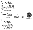
- FIG. 1 shows a polyion complex micelle (PIC micelle) whose outer surface is modified with glucose and a preparation method thereof.
- FIG. 2 shows the result of dynamic light scattering measurement (DLS) of the particle size distribution of the Glc (6) -Cy5-PIC micelle obtained in Example 1 and the particle image obtained by a transmission electron microscope (TEM).
- TEM transmission electron microscope
- Glc (6) indicates that it is bound to a polymer that forms a micelle at the 6th carbon of glucose.
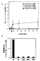
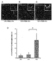
- FIG. 3 shows the selective and effective accumulation of Glc (6) -Cy5-PIC micelles obtained in Example 1 in the brain.
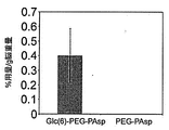
- FIG. 4 is a graph showing accumulation of micelles in the brain obtained by binding glucose to a polymer at the 3rd or 6th carbon.
- FIG. 5 is a diagram showing a fluorescence microscopic image (FIG. 5A) of the brain parenchyma when micelles are taken into the brain, and changes in blood glucose level and brain uptake amount (FIG. 5B).
- FIG. 6 is a diagram showing accumulation of PICsome having a diameter of 100 nm in the brain.
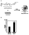
- FIG. 7 shows accumulation of siRNA micelles whose outer surface is modified with glucose in the brain.
- FIG. 7A shows siRNA micelles and preparation methods thereof, and
- FIG. 7B shows the transition of the amount accumulated in the brain.
- FIG. 8 is a fluorescence microscopic image showing the accumulation of siRNA micelles in brain cells.
- FIG. 9 is a diagram showing accumulation in the brain of a polymer conjugated with one glucose molecule.
- FIG. 10 is a diagram showing accumulation in the brain of an IgG antibody in which glucose is linked via a linker.
- G-IgG indicates IgG linked to glucose.
- FIG. 11 is a graph showing changes in fluorescence intensity in the brain parenchyma when Glc (6) -Cy5-PIC micelles are administered intravenously (iv) 30 minutes after glucose is administered intraperitoneally (ip). It is.
- FIG. 12 is a diagram showing that a part of Glc (6) -Cy5-PIC micelles can accumulate in cerebrovascular endothelial cells.
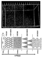
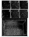
- FIG. 13 shows the localization of PIC micelles after intravenous administration in mouse cerebral cortex.
- FIG. 14 is a diagram showing the localization of PIC micelles after intravenous administration in a section of mouse cerebral cortex.
- FIG. 15 is a diagram showing the temporal localization change of PIC micelles after intravenous administration in mouse cerebral cortex.
- FIG. 16 shows a gel permeation chromatogram (a) and an NMR chart (b) of PEG-PLys-PnPrOx.
- FIG. 16 (a) shows a GPC chart of PnPrOx and PEG-PLys before coupling and PEG-PLys-PnPrOx as a final product.
- FIG. 16B shows a 1 H-NMR chart of PEG-PLys-PnPrOx which is the final product.
- FIG. 17 shows a scheme for micelle formation with PEG-PLys-PnPrOx.
- FIG. 17 (1) shows that the PnPrOx portion of PEG-PLys-PnPrOx is made hydrophobic at a temperature equal to or higher than the lower critical solution temperature (LCST), and after the formation of micelles, siRNA is added to form polyion complex micelles (PIC micelles).
- FIG. 17 (2) shows a scheme for forming a PIC micelle according to the present invention.
- LCST lower critical solution temperature
- FIG. 18 is an agarose electrophoresis photograph of the prepared cPIC micelle and uPIC micelle.
- FIG. 18A is an electrophoresis photograph of a micelle containing siRNA
- FIG. 18B is an electrophoresis photograph of a micelle containing cholesterol-modified siRNA (Chol-siRNA).
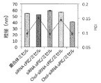
- FIG. 19 is a diagram showing the average particle size and particle size distribution (PDI) of various micelles.
- FIG. 19 the particle size is shown by a bar graph, and PDI is shown by a broken line.
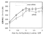
- FIG. 20 is a diagram showing the relationship between the diffusion time by fluorescence correlation spectroscopy and the mixing ratio of the temperature-sensitive copolymer of the present invention and siRNA. In fluorescence correlation spectroscopy, the diffusion time reflects the apparent particle size of the molecule.
- FIG. 21 is a diagram showing the resistance of the prepared cPIC micelle and uPIC to polyanion molecule (sodium heparin).
- FIG. 22 is a diagram showing NMR analysis results of Chol-siRNA and cPIC and uPIC containing the chol-siRNA.
- FIG. 20 is a diagram showing the relationship between the diffusion time by fluorescence correlation spectroscopy and the mixing ratio of the temperature-sensitive copolymer of the present invention and siRNA. In fluorescence correlation spectroscopy, the diffusion time reflects the apparent particle size of the molecule.
- FIG. 21 is
- FIG. 22 (a) shows a 1 H-NMR chart of Chol-siRNA
- (b) shows a 1 H-NMR chart of Chol-siRNA-cPIC / micelles
- (c) is, Chol-siRNA-uPIC / 1 shows a 1 H-NMR chart of micelles.
- FIG. 22 (d) shows the structure of Chol-siRNA-cPIC / micelle estimated from the chemical shift
- FIG. 22 (e) shows the structure of Chol-siRNA-uPIC / micelle estimated from the chemical shift.
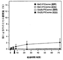
- FIG. 23 is a view showing the retention in blood of the prepared uPIC and cPIC.
- FIG. 24 is a graph showing that siRNA fluorescently labeled with Cy5 accumulates in brain cells by glucose-coated Chol-siRNA-uPIC / micelles.
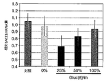
- FIG. 25 shows that siRNA delivered to brain cells by Chol-siRNA-uPIC / micelle coated with glucose can knock down the target BACE1 gene.
- micelle means a spherical aggregate formed by a single layer of molecular film.
- examples of micelles include micelles formed from amphiphilic molecules such as surfactants, and micelles formed from polyion complexes (PIC micelles). It is known that the micelle is preferably modified on the outer surface with polyethylene glycol from the viewpoint of residence time in blood.
- the “liposome” means a vesicle formed by a bilayer molecular film.
- the molecular membrane is usually a bilayer membrane made of phospholipids.
- polyion complex (hereinafter also referred to as “PIC”) means an ionic complex of a polymer having an anionic block and a polymer having a cationic block, and PEG and anionic It is known that it is formed by mixing a copolymer of a block and a copolymer of PEG and a cationic block so as to neutralize the charge in an aqueous solution.
- PIC micelle (hereinafter, also referred to as “PIC micelle”) means a micelle formed by PIC.
- the significance of combining PEG and the above-mentioned charged chain is to suppress the aggregation and precipitation of the polyion complex, and thereby the polyion complex has a monodisperse core with a particle size of several tens of nm.
- -Forming nanoparticles having a shell structure since PEG covers the outer shell (shell) of the nanoparticle, it is known that it has high biocompatibility and is advantageous in improving the residence time in blood. Also, in forming a polyion complex, one charged block copolymer does not require a PEG moiety.
- one charged block copolymer may be replaced with a homopolymer or a nucleic acid.
- PIC micelles formed by nucleic acids and copolymers of PEG and cationic blocks can be used for drug delivery of nucleic acids.
- unit polyion complex means an anionic polymer (for example, nucleic acid), a hydrophilic block such as polyethylene glycol (PEG), and a cation. It means a composite obtained by mixing a temperature-sensitive copolymer having a functional block in an aqueous solution at a temperature lower than the lower critical solution temperature (LCST). Since this complex is formed by the formation of an ionic bond between the cationic block of the block copolymer and the anionic polymer (for example, nucleic acid), a polyion complex (hereinafter referred to as “PIC”). Also called).
- PIC polyion complex
- Such PIC is considered to be in the form of a complex of a cationic molecule and an anionic molecule, but is not considered to be in a micelle form.
- micelles are obtained.
- a micelle obtained by setting the temperature of a solution containing uPIC to LCST or higher may be referred to as “uPIC / micelle”.
- temperature sensitivity means a property that changes from water-soluble to poorly water-soluble (or changes from poorly water-soluble to water-soluble) depending on the temperature.
- the temperature-sensitive copolymer that is the polymer of the present invention for example, a temperature-sensitive terpolymer (that is, a terpolymer of a hydrophilic block, a temperature-sensitive block, and a polycationic block) )
- LCST lower critical solution temperature
- the polymer of the present invention is a copolymer of a temperature-sensitive polymer part and other parts, and is therefore a copolymer exhibiting temperature sensitivity. Therefore, in the present specification, the polymer of the present invention may be referred to as the temperature-sensitive copolymer of the present invention.
- inducing hypoglycemia means lowering the blood glucose level in the subject than the blood glucose that should have been shown if the treatment was not performed.
- examples of a method for inducing hypoglycemia include administration of a diabetic drug.
- in inducing hypoglycemia as long as the purpose of inducing hypoglycemia is achieved, for example, taking other drugs or drinking a drink such as water is acceptable.
- Inducing hypoglycemia may involve other treatments that do not substantially affect blood glucose.
- fasting means fasting a subject, for example, 3 hours or more, 5 hours or more, 10 hours or more, 15 hours or more, 20 hours or more, 25 hours or more, 30 hours or more, 35 hours or more, Meaning fasting for 40 hours or more, 45 hours or more or 48 hours or more.
- Fasting causes the subject to hypoglycemia.
- the fasting period is determined by a doctor or the like in view of the health condition of the subject, and may be, for example, sufficient time for the subject to reach fasting blood glucose (for example, within 72 hours, within 48 hours, or within 24 hours). can do).
- the fasting period may be, for example, a period of time beyond which the expression of GLUT1 on the intravascular surface of cerebral vascular endothelial cells increases or reaches a plateau.
- the fasting period can be, for example, the above period that is 12 hours or more, 24 hours or more, or 36 hours or more. Fasting may also involve other treatments that do not substantially affect blood glucose levels or the expression of GLUT1 on the intravascular surface.
- “inducing an increase in blood glucose level” means increasing the blood glucose level in a subject in which hypoglycemia is induced or in a subject in which a hypoglycemic state is maintained.
- the blood glucose level can be increased by various methods well known to those skilled in the art.
- administration of an agent that induces an increase in blood glucose level for example, an increase in blood glucose level such as glucose, fructose (fructose), galactose, etc.
- administration of a simple sugar administration of a polysaccharide that induces an increase in blood sugar level such as maltose, intake of a carbohydrate that induces an increase in blood sugar level such as starch, or diet.
- blood glucose manipulation refers to inducing hypoglycemia in a subject and then raising the blood glucose level. After inducing hypoglycemia in the subject, the subject's blood glucose level can be maintained at the hypoglycemia.
- the time for maintaining the target blood glucose level at low blood sugar is, for example, 0 hour or more, 1 hour or more, 5 hours or more, 10 hours or more, 15 hours or more, 20 hours or more, 30 hours or more, 40 hours or more, 48 hours or more It can be. Thereafter, the blood sugar level can be raised.
- “maintaining blood glucose” is permitted to take other drugs or drink beverages such as water, for example, as long as the purpose of maintaining hypoglycemia in the subject is achieved. Inducing hypoglycemia may involve other treatments that do not substantially affect blood glucose.
- subject refers to mammals including humans.
- the subject may be a healthy subject or a subject suffering from some disease.
- diseases include cranial nerve diseases such as psychotic disorders, depression, mood disorders, anxiety, sleep disorders, dementia and substance-related disorders.
- dementia include, but are not limited to, Alzheimer's disease and Creutzfeldt-Jakob disease.
- blood-brain barrier refers to a functional barrier that exists between the blood circulation and the brain and has selectivity for the permeation of substances.
- the actual state of the blood-brain barrier is considered to be cerebrovascular endothelial cells.
- substance permeability of the blood brain barrier there are many unclear points, but glucose, alcohol and oxygen are known to easily pass through the blood brain barrier, and fat-soluble substances and small molecules (for example, molecular weight less than 500) It is considered that there is a tendency to pass through more easily than water-soluble molecules and polymers (for example, molecular weight of 500 or more).
- Many therapeutic agents and diagnostic agents for brain diseases do not pass through the blood-brain barrier, which is a major obstacle for treatment of brain diseases and brain analysis.
- blood nerve barrier refers to a functional barrier that exists between the blood circulation and peripheral nerves and has selectivity for the permeation of substances.
- blood cerebrospinal fluid barrier refers to a functional barrier that exists between blood circulation and cerebrospinal fluid and has selectivity for the permeation of substances.
- blood retinal barrier refers to a functional barrier that exists between blood circulation and retinal tissue and has selectivity for the permeation of substances.
- the entities of the blood nerve barrier, blood cerebrospinal fluid barrier, and blood retinal barrier are considered to be vascular endothelial cells existing at the respective barriers, and the functions thereof are considered to be the same as the blood brain barrier.
- GLUT1 ligand means a substance that binds to GLUT1.
- Various ligands are known as GLUT1 ligands, including, but not limited to, molecules such as glucose and hexose, both of which are used in the present invention for the preparation of carriers or conjugates instead of glucose. be able to.
- the GLUT1 ligand preferably has an affinity for GLUT1 that is equal to or greater than that of glucose.
- the temperature of the present invention is determined.
- a complex (unit polyion complex) of a sensitive copolymer and an anionic polymer (for example, nucleic acid) is formed (see the unit PIC in FIG. 17B).
- the unit PIC is under this temperature condition. It was found that micelles were not formed, and micelles in which uPIC incorporated siRNA into the core were formed when the temperature condition was changed to LCST or higher thereafter. The obtained micelle was predicted to be a micelle having a three-layer structure by analysis (FIG.
- the uPIC / micelle obtained in this manner has high stability, for example, has resistance to decay induced by polyanions present in a large amount in the living body, and exhibits high blood retention. Moreover, because uPIC micelles whose micelle surface was coated with glucose were intravenously administered together with blood glucose manipulation because of their stability, delivery of nucleic acids to the brain was achieved. Furthermore, the present inventors succeeded in significantly reducing the expression level of target mRNA (knockdown) by an experiment of delivering siRNA to the brain parenchyma. The case of delivering such a large amount of siRNA molecules to a body tissue by intravenous bolus administration has not been known so far, and can be said to be a very surprising result.
- a copolymer having a cationic block and a temperature sensitive block can be used as the temperature sensitive copolymer.
- a temperature-sensitive terpolymer that is, a copolymer having a hydrophilic block, a cationic block, and a temperature-sensitive block can be used.
- a hydrophilic block eg, PEG
- PEG polycation of a temperature sensitive copolymer
- a cationic block is a polymer of cationic units, for example, cationic amino acids such as cationic unnatural amino acids or cationic natural amino acids such as cationic natural amino acids such as histidine, tryptophan, ornithine, arginine and lysine. And / or a group represented by — (NH— (CH 2 ) 2 ) p —NH 2 , wherein p is an integer of 1 to 5.
- a side chain for example, a cationic non-natural amino acid polymer block having the cationic side chain, for example, a cationic non-natural amino acid such as aspartic acid or glutamic acid having the cationic side chain. Examples include polymer blocks.
- the polycation block is a group represented by — (NH— (CH 2 ) 2 ) p —NH 2 , where p is an integer of 1 to 5.
- p is an integer of 1 to 5.
- the cationic natural amino acid is preferably histidine, tryptophan, ornithine, arginine and lysine, more preferably arginine, ornithine and lysine, still more preferably ornithine and lysine, and still more preferably. Includes lysine.
- the temperature-sensitive block is not particularly limited as long as the LCST is present in a temperature range in which siRNA is stable.
- polymers and copolymers of N-isopropylacrylamide, polymers and copolymers of 2-isopropyl-2-oxazoline And polymers and copolymers of 2-n-propyl-2-oxazoline can be appropriately prepared by those skilled in the art and can be used in the present invention.
- Poly (2-n-propyl-2-oxazoline) has the following chemical structure (I): ⁇ Wherein, l is an integer of 10 to 5000, 10 to 1000, 50 to 500, 100 to 300, or 150 to 200. ⁇
- Poly (2-isopropyl-2-oxazoline) has the following chemical structure (II). ⁇ Wherein y is an integer of 10 to 5000, 10 to 1000, 50 to 500, 100 to 300, or 150 to 200. ⁇
- Poly (N-isopropylacrylamide) has the following chemical structure (III). ⁇ Wherein x is an integer of 10 to 5000, 10 to 1000, 50 to 500, 100 to 300, or 150 to 200. ⁇
- the cationic block and the temperature sensitive block can be linked directly or via a linker.
- a linker is used for the convenience of synthesis, it is preferable to use dibenzylcyclooctyne NHS ester (DBCO-NHS) because the synthesis procedure is simplified.
- the temperature-sensitive copolymer may further have a hydrophilic block.
- a copolymer in which a hydrophilic block-polycationic block-temperature-sensitive block are connected in this order can be used.
- hydrophilic block a biocompatible hydrophilic polymer can be used.
- PEG polyethylene glycol
- a GLUT1 ligand eg, glucose
- the surface of the uPIC / micelle of the present invention is coated with the GLUT1 ligand.
- the temperature-sensitive copolymer may be a copolymer in which PEG-polycation-temperature-sensitive blocks are linked in this order.
- the temperature sensitive copolymer is PEG-polycation-poly (2-n-propyl-2-oxazoline), PEG-polycation-poly (2-isopropyl-2-oxazoline), or PEG- It can be polycation-poly (N-isopropylacrylamide).
- the temperature-sensitive copolymer can be a copolymer in which GLUT1 ligand-PEG-polycation-temperature-sensitive block is linked in this order.
- the temperature sensitive copolymer comprises GLUT1 ligand-PEG-polycation-poly (2-n-propyl-2-oxazoline), GLUT1 ligand-PEG-polycation-poly (2-isopropyl-2- Oxazoline), or GLUT1 ligand-PEG-polycation-poly (N-isopropylacrylamide).
- GLUT1 ligand-PEG-polylysine-poly (2-n-propyl-2-oxazoline), GLUT1 ligand-PEG-polylysine-poly (2-n-propyl-2-oxazoline), or GLUT1 ligand-PEG- Polylysine-poly (N-isopropylacrylamide) can be used.
- the GLUT1 ligand is glucose.
- the temperature sensitive copolymer of the present invention can be used to make a unit PIC.
- a composition for making a unit PIC comprising the temperature sensitive copolymer of the present invention.
- the above composition comprising a temperature sensitive copolymer is used for mixing with nucleic acid at a temperature below the LCST of the temperature sensitive copolymer.
- nucleic acids can be used as polyanions.
- siRNA means double-stranded RNA (nucleic acid) capable of inducing RNA interference (RNAi).
- the siRNA is not particularly limited, but is a double-stranded RNA consisting of 20 to 30 bp, preferably 21 to 23 bp, 25 bp, and 27 bp, and having a sequence homologous to the sequence of the target gene.
- short hairpin RNA shRNA
- Other RNA may be used as the nucleic acid. Since siRNA has the same properties as a polyanion regardless of its sequence, the sequence may be freely designed based on the target sequence.
- the siRNA may be a chimeric RNA with DNA.
- the uPIC / micelle used in the present invention is not particularly limited.
- the diameter is 400 nm or less, 200 nm or less, 150 nm or less, 100 nm or less, or 80 nm or less, for example, 20 nm or more, 30 nm or more, or 40 nm or more.
- the uPIC / micelle used in the present invention has a diameter of, for example, 30 nm to 150 nm, or of 30 nm to 100 nm, for example.
- the present invention provides a pharmaceutical composition for use in treating tumors comprising uPIC / micelle comprising a nucleic acid capable of inhibiting tumors.
- UPIC / micelles coated with glucose show accumulation in the brain even when administered to a subject. Therefore, a dosing regime according to the present invention may not induce fasting or hypoglycemia and / or may not induce an increase in blood glucose level.
- uPIC / micelle whose outer surface is modified with glucose so that glucose is exposed on the surface is administered according to a certain administration plan, uPIC is significantly delivered into the brain (brain parenchyma) across the blood-brain barrier. .
- a dosing schedule according to the present invention preferably comprises administering the uPIC / micelle to a subject that has been fasted or induced hypoglycemia, but more preferably the dosing schedule according to the present invention is fasting Or administering the uPIC / micelle to a subject that has induced hypoglycemia and inducing an increase in blood glucose level in the subject.
- the uPIC / micelle may be administered to the subject simultaneously, sequentially or sequentially with the induction of elevated blood glucose levels in the subject.
- the dosing regime may or may not have an interval between administration of uPIC / micelle to the subject and induction of an increase in blood glucose level in the subject.
- the uPIC / micelle When the uPIC / micelle is administered simultaneously with the induction of an increase in blood glucose level in the subject, the uPIC / micelle may be administered to the subject in a form mixed with an agent that causes the increase in blood glucose level. Alternatively, it may be administered in a form other than the agent that causes the increase in blood glucose level in the subject. In addition, when the uPIC / micelle induces an increase in blood glucose level in the subject and when administered to the subject continuously or sequentially, the uPIC / micelle induces an increase in blood glucose level in the subject. Although it may be administered to the subject earlier or later, preferably the uPIC / micelle can be administered to the subject prior to inducing an increase in blood glucose levels in the subject.
- the increase in blood glucose level is induced in the subject prior to the administration of the uPIC / micelle to the subject
- the increase in blood glucose level is induced in the subject within 1 hour, within 45 minutes, 30 minutes
- the uPIC / micelle is administered to the subject within minutes, within 15 minutes or within 10 minutes.
- inducing an increase in blood glucose level in the subject after administration of the uPIC / micelle to the subject within 6 hours, within 4 hours after the administration of the uPIC / micelle to the subject, Within 2 hours, within 1 hour, within 45 minutes, within 30 minutes, within 15 minutes or within 10 minutes, it is preferred to induce an increase in blood glucose level in the subject.
- the above regimen cycle may be performed more than once.
- the context between glucose administration and micelle administration can be determined by the timing of passing the blood brain barrier.
- the cerebral cortex is composed of six layers. From the surface layer, the molecular layer (first layer), outer granule layer (second layer), outer cone cell layer (third layer), inner granule layer (fourth layer), inner layer There are pyramidal cell layers (fifth layer) and polymorphic cell layers (sixth layer), and any of these layers can deliver carriers to the brain parenchyma. Among these layers, carrier delivery is particularly effective in the outer cone cell layer (third layer) and the inner granule layer (fourth layer).
- GLUT1 is a glucose transporter expressed on the intravascular surface of brain vascular endothelial cells. Therefore, in the present invention, GLUT1 ligand can also play the same role as glucose. Further, in the present invention, the GLUT1 ligand can be bound so as to be exposed on the outer surface so that it can bind to the glucose transporter expressed on the inner blood vessel surface of the vascular endothelial cell of the brain.
- molecules, complexes, micelles and others that can present GLUT1 ligands to GLUT1 can bind to GLUT1, and together with GLUT1 are taken up into the vascular endothelial cells when glucose is taken up by the glucose after binding. It is thought that it is taken in.
- the taken micelles pass through the blood-brain barrier and migrate to the brain parenchyma.
- the proportion of micelles that reached the brain parenchyma was slightly reduced.
- uPIC / micelles coated with the GLUT1 ligand of the present invention can be used for delivery to cerebrovascular endothelial cells.
- the role of glucose in the present invention is also the same in the blood nerve barrier, blood retinal barrier and blood cerebrospinal fluid barrier.
- GLUT1 is also expressed in vascular endothelial cells during hypoglycemia at the blood nerve barrier, blood retina barrier and blood cerebrospinal fluid barrier.
- uPIC / micelles coated with the GLUT1 ligand of the present invention can be used to cross the blood nerve barrier, blood retinal barrier and blood cerebrospinal fluid barrier.
- the uPIC / micelles coated with the GLUT1 ligand of the present invention can also be used to deliver to vascular endothelial cells present at the blood nerve barrier, blood retinal barrier and blood cerebrospinal fluid barrier.
- a polymer in which glucose is conjugated through the carbon at the 6-position a polymer in which glucose is conjugated through the carbon at the 6-position (for example, see FIG. 1 (a)).
- the micelle obtained by use has higher uptake efficiency into the brain. It is known that OH groups, which are substituents of carbon atoms at the 1-position, 3-position and 4-position, are strongly involved in the bond between GLUT1 and glucose. The fact that micelles obtained by modifying the polymer via the 6-position carbon atom that is not used for binding to GLUT1 tend to accumulate more effectively in the brain, indicating the involvement of GLUT1 in brain accumulation.
- micelles obtained by modifying a polymer via a carbon atom at the 3-position which is said to be important for recognition with GLUT1 also showed accumulation in the brain. This indicates that more micelles accumulate in the brain in micelles obtained by modifying the polymer via a carbon atom at the 2-position that is less involved in binding to GLUT1.
- glucose is conjugated to a polymer or drug via any one of its 1, 3 and 4 carbon atoms, preferably via its 2 or 6 carbon atom. Can be gated.
- at least the 1-, 3- and 4-position OH groups of glucose conjugated to the temperature sensitive copolymer are reducing ends.
- the GLUT1 ligand can modify the temperature-sensitive copolymer so as not to lose its function as a ligand, and those skilled in the art can easily interact with a drug based on the binding mode with GLUT1. You can decide where to join.
- glucose bonded through the carbon atom at the n-position may be expressed as “Glc (n)” (where n is an integer of 1 to 4 and 6).
- n is an integer of 1 to 4 and 6
- glucose bonded through the carbon atom at the 6-position is expressed as “Glc (6)”
- glucose bonded through the carbon atom at the 2-position is expressed as “Glc (2)”.
- Glucose bonded through the 3-position carbon atom may be referred to as “Glc (3)”.
- a glucose derivative that binds to GLUT1 may be used instead of glucose.
- the temperature sensitive copolymer may be further modified with a GLUT1 ligand and the GLUT1 ligand-hydrophilic block-polycationic block-temperature sensitive block linked in this order.
- a GLUT1 ligand for example, glucose can be used. By doing so, the GLUT1 ligand can be exposed to the micelle surface after micelle formation.
- the temperature-sensitive copolymer modified with GLUT1 ligand and the temperature-sensitive copolymer not modified with GLUT1 ligand may be mixed in uPIC / micelle.
- the content (mol%) of the temperature-sensitive copolymer modified with the GLUT1 ligand to the total temperature-sensitive copolymer can be, for example, 10 to 100%, %, And can be 20 to 40%.
- the temperature-sensitive copolymer modified with GLUT1 ligand and the temperature-sensitive copolymer not modified with GLUT1 ligand are used in uPIC / micelle. It may be mixed.
- the content (mol%) of the temperature-sensitive copolymer modified with the GLUT1 ligand to the total temperature-sensitive copolymer can be, for example, 10 to 100%, and 50 to 100 %, And 80-100%.
- the temperature-sensitive copolymer modified with the GLUT1 ligand and the temperature-sensitive copolymer not modified with the GLUT1 ligand may be mixed in the uPIC / micelle.
- the content (mol%) of the temperature-sensitive copolymer modified with the GLUT1 ligand to the total temperature-sensitive copolymer can be, for example, 10 to 100%, %, And can be 20 to 40%.
- the temperature-sensitive copolymer modified with a GLUT1 ligand is used in the uPIC / micelle.
- the content (mol%) with respect to the total temperature-sensitive copolymer can be, for example, 10 to 100%, 50 to 100%, or 80 to 100%.
- the nucleic acid is delivered to both brain parenchyma and cerebral vascular endothelial cells whatever the content (mol%) of the temperature-sensitive copolymer modified with the GLUT1 ligand relative to the total temperature-sensitive copolymer. As the content increases, the amount of accumulation in the cerebral vascular endothelial cells tends to increase. Therefore, those skilled in the art can freely determine the desired content according to the purpose.
- the nucleic acid may be substituted with a biocompatible hydrophobic group.
- the nucleic acid is siRNA and the biocompatible hydrophobic group is a cholesteryl group. That is, in one aspect, siRNA micelles are prepared by mixing siRNA conjugated with cholesterol and the temperature-sensitive copolymer of the present invention or a salt thereof under a temperature condition of LCST or lower, and combining the temperature-sensitive copolymer and siRNA. After the unit PIC is formed, the temperature is obtained at a temperature equal to or higher than LCST.
- the mixing ratio of siRNA and temperature-sensitive copolymer is from 1:10 to 10: 1, preferably from 1: 5 to 5: 1, more preferably from 1: 2 to 2: 1 in the ratio of the total amount of charge each has. More preferably, it can be 1: 1.5 to 1.5: 1, or about 1: 1. In this specification, “about” means that an error of less than 50%, 40% or less, 30% or less, 20% or less, 10% or less, or 5% or less may be included.
- the mixing ratio can be, for example, a stoichiometric ratio such that a positive charge and a negative charge are neutralized.
- the salt is preferably a pharmaceutically acceptable salt.
- the siRNA conjugated with cholesterol is not particularly limited, and is an siRNA conjugated with cholesterol at the 5 ′ end or 3 ′ end of the RNA strand, but these can be appropriately synthesized by those skilled in the art, or It is commercially available through custom synthesis and can be used in the present invention.
- the siRNA is not particularly limited, but preferably cholesterol can be conjugated to the 3 'end of the sense strand or the 5' end or 3 'end of the antisense strand.
- cholesterol can be introduced into siRNA via carbamate linkages by well-known techniques.
- P-Toluenesulfonate methyl can be dissolved in acetonitrile and chlorobenzene to polymerize n-propyloxazoline (nPrOx).
- the polymerization can be terminated with, for example, sodium azide.
- PEG-NH 2 polyethylene glycol
- DMF N, N′-dimethylformamide
- N6-trifluoroacetyl-L-lysine-N-carboxylic anhydride (Lys (TFA) -NCA) is dissolved in DMF containing 1M thiourea, and PEG-NH 2 is polymerized with Lys (TFA) -NCA.
- PEG-PLys (TFA) can be obtained.
- n can be any integer from 2 to 20000, for example, any integer from 10 to 5000, for example, any integer from 40 to 500, and m is any from 2 to 5000.
- m is any from 2 to 5000.
- l can be from 10 to 5000.
- PnPrOx—N 3 is soluble in acetone, unreacted PnPrOx—N 3 can be dissolved in acetone and removed. The desired product can be present in the fraction insoluble in acetone. Therefore, after collecting components insoluble in acetone and dissolving in pure water, dialysis can be performed using pure water as an external solution.
- PEG-polylysine-PnPrOx can be generated using the particle forming ability.
- the PnPrOx moiety becomes hydrophobic and PEG-polylysine-PnPrOx forms micelles.
- Micelles can be collected using ultrafiltration tubes. For example, when a tube with a molecular weight cut off of 300 kDa is used as the ultrafiltration tube, it is possible to pass only the unreacted PEG-PLys (TFA) -DBCO while keeping the micelle in the tube by centrifugation or the like. it can. This operation can be repeated.
- TFA unreacted PEG-PLys
- PEG-PLys-PnPrOx The resulting solution in the tube is dialyzed using pure water as an external solution to obtain PEG-PLys-PnPrOx. Thereafter, it may be freeze-dried.
- PEG-PLys-PnPrOx can be identified by molecular weight analysis by aqueous gel filtration chromatography or structural analysis by 1 H-NMR.
- a temperature-sensitive copolymer modified with a GLUT1 ligand can be appropriately prepared by those skilled in the art.
- the introduction of a protecting group for glucose in a temperature-sensitive copolymer modified with glucose is, for example, 1,2-O-isopropylidene-3, It is achieved by 5-O-benzylidene- ⁇ -D-glucofuranose (hereinafter referred to as “BIG”).
- BIG 5-O-benzylidene- ⁇ -D-glucofuranose
- BIG for example, protects the OH group, which is a substituent of the 3- and 5-position carbons of 1,2-O-isopropylidene- ⁇ -D-glucofuranose (hereinafter referred to as “MIG”) with a benzyl group. Is obtained. Specifically, BIG is obtained by reacting MIG with benzaldehyde and extracting with ethyl acetate. Next, from the viewpoint of keeping the molecular weight of PEG constant, before the polymerization reaction, before the polymerization reaction, before the polymerization reaction, before the polymerization reaction, before the polymerization reaction, before the polymerization reaction, before the polymerization reaction, before the polymerization reaction, before the polymerization reaction, before the polymerization reaction, before the polymerization reaction, before the polymerization reaction, before the polymerization reaction, before the polymerization reaction, before the polymerization reaction, before the polymerization reaction, before the polymerization reaction, before the polymerization reaction, before the polymerization reaction,
- the degree of polymerization can be appropriately adjusted depending on the amount of ethylene oxide to be added.
- the OH group of BIG-PEG-OH is aminated to obtain BIG-PEG-NH 2
- the OH group of BIG-PEG-OH is aminated to obtain BIG-PEG-NH 2
- the OH group of BIG-PEG-OH is aminated to obtain BIG-PEG-NH 2
- the OH group of BIG-PEG-OH is aminated to obtain BIG-PEG-NH 2
- BIG-PEG-NH 2 is used in place of PEG-NH 2 in Scheme 2 to obtain PEG A copolymer having Glc (6) introduced on the side can be obtained.
- Deprotection of Glc (6) can be performed in the final step of the process.
- a temperature-sensitive copolymer modified with Glc (3) can be used in place of BIG, for example, 1,2,5,6-di-O-isopropylidene- ⁇ -D-glucofuranose (DIG).
- DIG 1,2,5,6-di-O-isopropylidene- ⁇ -D-glucofuranose
- a temperature-sensitive copolymer modified with Glc (2) can be appropriately synthesized by those skilled in the art.
- the unit PIC / micelle coated with the GLUT1 ligand of the present invention can be administered to a subject as it is, or can be administered based on the administration plan according to the present invention (for example, accompanied by blood glucose manipulation).
- the dosing schedule according to the present invention preferably the subject is first fasted or the subject is induced with hypoglycemia, but then the unit is administered unit PIC / micelle. More preferably, in the dosing regime according to the invention, the subject is first fasted or the subject is induced to hypoglycemia, but then the subject is administered unit PIC / micelle and the subject has an increased blood glucose level. To trigger.
- the administration of the unit PIC / micelle to the subject is performed simultaneously, continuously or sequentially with the induction of an increase in blood glucose level in the subject.
- Induction of a hypoglycemic state is thought to be useful for expressing GLUT1 on the inner surface of vascular endothelial cells (eg, cerebral vascular endothelial cells).
- vascular endothelial cells eg, cerebral vascular endothelial cells.
- an increase in blood glucose level in the administration subject is extremely effective for delivery to these brains.
- Unit PIC / micelle by increasing the blood glucose level when the blood concentration of the unit PIC / micelle of the present invention in a subject that has been fasted or induced hypoglycemia is above a certain level, Unit PIC / micelle can be delivered very effectively into the brain. Also, according to this example, the unit PIC / micelle of the present invention is delivered into the subject's brain for some time after inducing a rise in blood glucose level in the subject.
- the unit PIC / micelle of the present invention it is preferable to administer the unit PIC / micelle of the present invention as an infusion. By doing in this way, it is easy to ensure a constant blood concentration even if the unit PIC / micelle has a short residence time in the blood.
- siRNA micelles containing siRNA with a short residence time in blood are likely to be effective when administered to a subject by infusion.
- Infusion administration can be preferably performed for 30 minutes or more, 45 minutes or more, 60 minutes or more, 90 minutes or more, or 2 hours or more. Infusion administration is preferably performed at a constant infusion rate.
- infusion administration may be done at the same time as inducing an increase in blood glucose in the subject, and inducing an increase in blood glucose in the subject during infusion administration You may let them.
- uPIC / micelle can be administered as a bolus.
- the bolus administration can be preferably performed in less than 30 minutes, 20 minutes or less, 10 minutes or less, or 5 minutes or less.
- a unit PIC / micelle coated with a GLUT1 ligand of the present invention can be used to deliver nucleic acids to the brain.
- Unit PIC / micelles coated with a GLUT1 ligand of the present invention can also allow nucleic acids to cross the blood brain barrier. Therefore, the unit PIC / micelle coated with the GLUT1 ligand of the present invention can be used to deliver nucleic acids to the brain parenchyma, which has heretofore been difficult to deliver.
- compositions or conjugates of the invention can also cause nucleic acids to accumulate in cerebral vascular endothelial cells. Therefore, the unit PIC / micelle coated with the GLUT1 ligand of the present invention can be used to deliver nucleic acids to cerebral vascular endothelial cells, which were conventionally difficult to deliver. The unit PIC / micelle coated with the GLUT1 ligand of the present invention can also be used to deliver a nucleic acid that weakens or destroys adhesion between cerebral vascular endothelial cells to cerebral vascular endothelial cells.
- unit PIC / micelles coated with GLUT1 ligands of the invention can be used to deliver nucleic acids to the retina, peripheral nerves and / or cerebrospinal fluid.
- the unit PIC / micelle coated with the GLUT1 ligand of the present invention can also be used to deliver nucleic acids to vascular endothelial cells present at the blood nerve barrier, blood retinal barrier or blood cerebrospinal fluid barrier, respectively.
- the unit PIC / micelle coated with the GLUT1 ligand of the present invention has a nucleic acid that weakens or destroys adhesion between vascular endothelial cells existing at the blood nerve barrier, blood retinal barrier or blood cerebrospinal fluid barrier, respectively, to brain vascular endothelial cells. It can also be used for delivery. By weakening or destroying the adhesion between vascular endothelial cells, the function of the barrier is weakened, and various barriers can be passed through.
- the unit PIC / micelle of the present invention can be administered by oral administration and parenteral administration (for example, intravenous administration or intraperitoneal administration).
- a method of targeting brain tissue comprising administering to a subject a unit PIC / micelle coated with a GLUT1 ligand according to a dosing schedule.
- the present invention also provides a method of targeting cerebrovascular endothelial cells comprising administering to a subject a unit PIC / micelle coated with a GLUT1 ligand according to a dosing regimen.
- the dosing schedule according to the present invention preferably comprises administering a unit PIC / micelle coated with GLUT1 ligand to a subject that has been fasted or induced hypoglycemia, but more preferably the dosing schedule according to the present invention Includes administering a unit PIC / micelle coated with GLUT1 ligand to a subject that has fasted or induced hypoglycemia and induces an increase in blood glucose in the subject.
- a method of targeting peripheral nerve tissue, retina and / or cerebrospinal fluid comprising administering to a subject a unit PIC / micelle coated with a GLUT1 ligand according to a dosing regimen.
- the vascular endothelial cells respectively present in the blood nerve barrier, blood retinal barrier or blood cerebrospinal fluid barrier, comprising administering to the subject a unit PIC / micelle coated with a GLUT1 ligand according to the administration plan
- a method of targeting is provided.
- the nucleic acid can be effectively delivered to the brain, peripheral nerve tissue, retina and / or spinal fluid by forming a complex with the temperature-sensitive copolymer of the present invention.
- a nucleic acid used for treatment or prevention of brain disease can be used as the nucleic acid.
- a unit PIC / micelle coated with a GLUT1 ligand and containing as a nucleic acid a nucleic acid used for treatment or prevention of brain disease is administered to a subject in need thereof according to a dosage plan.
- a method of treating or preventing brain disease is provided.
- a unit PIC / micelle coated with a GLUT1 ligand and containing as a nucleic acid a nucleic acid used for the treatment or prevention of peripheral nerve disease is administered to a subject in need thereof according to a dosing schedule
- a method of treating or preventing brain disease comprising the above.
- a unit PIC / micelle coated with a GLUT1 ligand comprising a nucleic acid used as a nucleic acid for treatment or prevention of retinal disease is administered to a subject in need thereof according to a dosage plan.
- a method of treating or preventing brain disease is provided.
- a dosing schedule according to the present invention preferably comprises administering the micelle to a subject that has been fasted or induced hypoglycemia, but more preferably, the dosing schedule according to the present invention is fasted or low. Administering the micelles to a subject who has induced blood glucose and inducing an increase in blood glucose level in the subject.
- Brain diseases include brain diseases that can be treated by passing nucleic acid through the blood brain barrier, such as anxiety, depression, sleep disorders, Alzheimer's disease, Parkinson's disease, and multiple sclerosis. Therefore, in the present invention, in order to treat these brain diseases, therapeutic agents for brain diseases such as anti-anxiety agents, antidepressants, sleep-inducing agents, Alzheimer's therapeutic agents, Parkinson's disease therapeutic agents, and multiple sclerosis therapeutic agents, Prophylactic drugs can be used.
- Alzheimer's disease therapeutic agent for example, A ⁇ antibody is well known
- Parkinson's disease therapeutic agent for example, dopamine receptor agonist and L-dopa are well known
- multiple sclerosis therapeutic agent for example, adrenal gland Steroid drugs, interferon ⁇ (IFN ⁇ ), and immunosuppressants are well known and these therapeutic agents can be used in the present invention.
- Peripheral neurological diseases include peripheral neurological diseases that can be treated by passing a blood brain barrier through a therapeutic agent for peripheral neurological diseases, such as Guillain-Barre syndrome, Fisher syndrome, and chronic inflammatory demyelinating polyneuropathy.
- Retinal diseases include retinal diseases that can be treated by passing retinal disease drugs through the blood-brain barrier, such as retinitis pigmentosa, cerebral reticular retinal atrophy, choroideremia, crystallin retinopathy, congenital cataract, congenital arrest.
- retinal diseases include retinal diseases that can be treated by passing retinal disease drugs through the blood-brain barrier, such as retinitis pigmentosa, cerebral reticular retinal atrophy, choroideremia, crystallin retinopathy, congenital cataract, congenital arrest.
- Superficial night blindness small mouth disease, white spotted fundus, white spotted retinopathy, pigmented paravenous choroidal atrophy, Stargardt disease, yolk macular dystrophy, juvenile retinal seizures, central ring-shaped choroidal dystrophy, occult macular Examples include dystrophies, familial exudative vitreoretinopathy and retinal pigment streaks.
- Example 1 Production of Glc (6) -PIC micelle In Example 1, a polymer necessary for micelle formation was synthesized.
- BIG-PEG-OH tetrahydrofuran
- the OH group of the obtained BIG-PEG-OH was aminated to synthesize BIG-PEG-NH 2 having an aminoethyl group.
- 2.0 g of benzene freeze-dried BIG-PEG-OH is dissolved in 20 mL of THF solution in which 0.8 mL of triethylamine is dissolved.
- the precipitated salt was removed by filtration, and the filtrate was reprecipitated with 500 mL of a cryogen containing diethyl ether containing 10% methanol, filtered and dried under reduced pressure.
- the obtained powder was dissolved in 100 mL of 25% aqueous ammonia solution and reacted at room temperature for 2 days. It dialyzed with the ammonium aqueous solution diluted 2000 times using the dialysis membrane (fraction molecular weight 1,000), and dialyzed with the pure water after that. Thereafter, the fraction in which amination did not proceed was removed with Sephadex C-25 (GE healthcare), and freeze-dried to recover 1.6 g of BIG-PEG-NH 2 (yield 85%). No peaks due to impurities were observed in the H 1 NMR spectrum of BIG-PEG-NH 2 after purification (data not shown).
- BIG-PEG-PBLA ⁇ -benzyl-L-aspartate
- BIG-PEG-PBLA ⁇ -benzyl-L-aspartate-N-carboxylic acid anhydride
- BLA-NCA ⁇ -benzyl-L-aspartate-N-carboxylic acid anhydride
- BIG-PEG-polyaspartic acid (hereinafter referred to as “BIG-PEG-P (Asp.)”) was synthesized from the obtained BIG-PEG-PBLA.
- the benzyl ester is hydrolyzed at room temperature while suspending 500 mg of BIG-PEG-PBLA in 0.5N sodium hydroxide. After the copolymer was dissolved, it was dialyzed in water using a dialysis membrane (fractionated molecular weight 1,000). The solution in the membrane was lyophilized to obtain 132 mg (yield 68%) of BIG-PEG-P (Asp.).
- Glc (6) -PEG-P (Asp.) was synthesized from BIG-PEG-P (Asp.).
- Glc (6) means that glucose is bonded to PEG at its 6th carbon.
- 100 mg of BIG-PEG-P (Asp.) was dissolved in 10 mL of trifluoroacetic acid / pure water (8: 2) and reacted for 1 hour.
- Dialysis was performed in the order of 0.01N NaOH and pure water using a dialysis membrane (fractionated molecular weight: 1,000). The solution in the membrane was lyophilized to obtain 70 mg (yield 70%) of Glc (6) -PEG-P (Asp.).
- PEG-P polyethylene glycol-polyaspartic acid block copolymer
- PEG-P polyethylene glycol-poly ((5-aminopentyl) -aspartic acid) block copolymer
- DAP 1,5-diaminopentane
- reaction solution was added to 15.2 mL of a 20% by weight acetic acid aqueous solution, and dialyzed in water using a dialysis membrane (fractionated molecular weight: 6,000-8,000).
- the solution in the membrane was lyophilized to obtain 954 mg (yield 81%) of PEG-P (Asp.-AP).
- DIG-PEG-OH was obtained from benzene lyophilized 1,2,5,6-di-O-isopropylidene- ⁇ -D-glucofuranose (DIG). Obtained. Specifically, 0.72 g of DIG (manufactured by TCI) was dissolved in 5 mL of THF to obtain a DIG-OH solution. Thereafter, 3.5 mL of a THF solution containing 0.3 M potassium naphthalene was added dropwise to the obtained DIG-OH solution, and 2.5 mL of ethylene oxide (EO) was added in an argon atmosphere and reacted at room temperature for 48 hours.
- EO ethylene oxide
- DIG-PEG-OH was aminated to obtain DIG-PEG-NH 2 .
- 3.2 g of benzene lyophilized DIG-PEG-OH is dissolved in 32 mL of THF solution in which 0.8 mL of triethylamine is dissolved.
- a solution of 912 mg of methanesulfonyl chloride dissolved in 32 mL of cold THF was added to the above DIG-PEG-OH solution and allowed to react overnight at room temperature.
- the precipitated salt was removed by filtration, and the filtrate was reprecipitated with 500 mL of a cryogen containing diethyl ether containing 10% methanol, filtered and dried under reduced pressure.
- the obtained powder was dissolved in 100 mL of 25% aqueous ammonia solution and reacted at room temperature for 2 days. It dialyzed in order of the pure water in the ammonium aqueous solution diluted 2000 times using the dialysis membrane (fraction molecular weight 1,000). Subsequently, the fraction not aminated with Sephadex C-25 (GE healthcare) was removed and freeze-dried to recover 2.95 g of DIG-PEG-NH 2 (yield 89%).
- DIG-PEG-PBLA was synthesized from the obtained DIG-PEG-NH 2 .
- BLA-NCA was dissolved in 3.5 mL of DMF and diluted with 30 mL of dichloromethane.
- 200 mg of DIG-PEG-NH 2 after lyophilization of benzene was dissolved in 4 mL of dichloromethane, and the solution was added to the BLA-NCA solution and polymerized at 35 ° C. for 40 hours in the presence of argon.
- DIG-PEG-polyaspartic acid (DIG-PEG-P (Asp.)) was synthesized from the obtained DIG-PEG-PBLA. Specifically, benzyl ester is hydrolyzed at room temperature while suspending 500 mg of DIG-PEG-PBLA in 0.5N sodium hydroxide. After the copolymer was dissolved, it was dialyzed in water using a dialysis membrane (fractionated molecular weight 1,000). The solution in the membrane was lyophilized to obtain 145 mg (yield 54%) of DIG-PEG-P (Asp.).
- Glc (3) -PEG-P (Asp.) was synthesized from the obtained DIG-PEG-P (Asp.).
- Glc (3) means that glucose is bonded to PEG at the 3rd carbon.
- Dialysis was performed in the order of 0.01N NaOH and pure water using a dialysis membrane (fractionated molecular weight: 1,000). The solution in the membrane was lyophilized to obtain 75 mg (yield 86%) of Glc (3) -PEG-P (Asp.).
- Cy5-PIC micelle 50 mg of Cy5-PEG-P (Asp.) was dissolved in 50 mL of 10 mM phosphate buffer (PB, pH 7.4, 0 mM NaCl), and 1 mg / mL of Cy5-PEG-P (Asp.) Was dissolved. ) A solution was prepared. Similarly, 50 mg of PEG-P (Asp.-AP) was dissolved in 50 mL of PB to prepare a 1 mg / mL PEG-P (Asp.-AP) solution.
- the size (Z average particle diameter) and polydispersity index (PDI) of the obtained Cy5-PIC micelles were measured with a Zetasizer (Malvern). Size was measured by the diffusion of particles moving by Brownian motion, and the measurement results were converted to particle size and particle size distribution using Stokes-Einstein equation. The micelle shape was evaluated using a transmission electron microscope (TEM, JEM-1400).
- TEM transmission electron microscope
- the Z average particle diameter is data obtained by analyzing measurement data of a dynamic light scattering method such as a particle dispersion using a cumulant analysis method. In the cumulant analysis, an average value of the particle diameter and a polydispersity index (PDI) are obtained.
- this average particle diameter is defined as the Z average particle diameter.
- the work of fitting a polynomial to the logarithm of the G1 correlation function obtained by measurement is called cumulant analysis.
- a constant b in LN (G1) a + bt + ct 2 + dt 3 + et 4 +... Is called a second-order cumulant or Z-average diffusion coefficient.
- the value obtained by converting the value of the Z average diffusion coefficient into the particle size using the viscosity of the dispersion medium and some device constants is the Z average particle size, and is a value suitable for quality control purposes as an index of dispersion stability.
- Glc (6) -Cy5-PIC micelle 20 mg of Glc (6) -PEG- P (Asp.) And 40 mg of Cy5-PEG-P (Asp.) Were mixed with 10 mM phosphate buffer (PB, pH 7.4, 0 mM NaCl). ) Dissolved in 60 mL to prepare a mixed solution of 1 mg / mL Cy5-Glc (6) -PEG-P (Asp.) And PEG-P (Asp.).
- PEG-P (Asp.-AP) was dissolved in 50 mL of PB to prepare a 1 mg / mL PEG-P (Asp.-AP) solution.
- Two kinds of aqueous solutions that is, a mixture of Cy5-PEG-P (Asp.) And PEG-P (Asp.) And a PEG-P (Asp.-AP) solution, 4 mL and 7.0 mL, respectively, were added to 50 mL of conical. Added to the tube and vortexed for 2 minutes (2000 rpm).
- Glc (6) -Cy5-PIC micelles Characterization of Glc (6) -Cy5-PIC micelles The size (Z average particle diameter) and polydispersity index (PDI) of the obtained Glc (6) -Cy5-PIC micelles were measured with a Zetasizer (Malvern). As a result, it was revealed that micelles having an average particle diameter of 40 nm and a uniform particle diameter were obtained (FIG. 2A). The micelle shape was observed after staining with uranyl acetate using a transmission electron microscope (TEM, JEM-1400) (FIG. 2B).
- TEM transmission electron microscope
- Glc (3) -Cy5-PIC micelle 20 mg of Glc (3) -PEG- P (Asp.) And 40 mg of Cy5-PEG-P (Asp. ) Were added to pH 7.4 10 mM phosphate buffer (PB, 0 mM NaCl). Dissolved in 60 mL, a mixed solution of 1 mg / mL Cy5-Glc (3) -PEG-P (Asp.) And PEG-P (Asp.) was prepared.
- PEG-P (Asp.-AP) was dissolved in 50 mL of PB to prepare a 1 mg / mL PEG-P (Asp.-AP) solution.
- Example 2 Evaluation of pharmacokinetics of PIC micelles
- the micelles prepared in Example 1 were intravenously administered to mice and their pharmacokinetics were examined. When micelles were administered, the effect of blood glucose manipulation was also evaluated.
- accumulation in the brain was evaluated based on the amount (%) accumulated per gram of brain with respect to the total dose.
- the mouse After waiting for a predetermined time, the mouse was anesthetized, and after laparotomy, blood was collected from the abdominal aorta, and the brain, liver, spleen, kidney, heart, lung and thigh muscle were taken out.
- the collected blood is centrifuged at 15,000 rpm for 5 minutes at 4 ° C. to prepare plasma, dispensed into a 96-well plate (Thermo Fisher, USA), and the fluorescence intensity of the plasma by fluorescence measurement using Tecan Infinite M1000 PRO The micelle concentration in the blood was quantified.
- the blood of a mouse to which no sample was administered was used as a control.
- the pharmacokinetics of the drug was evaluated on the assumption that the total blood of the mouse was 2 mL, and the amount of plasma was 55%.
- the accumulation efficiency (%) of micelles in each organ was quantified by fluorescence measurement using PRO.
- Glc (6) -PIC micelles in the brain is important to reduce the blood glucose level of mice by fasting and to increase the blood glucose level of mice before and after micelle administration. It became clear. However, some micelles are taken up into the brain even after micelle administration and before resumption of feeding in fasted mice (black squares in FIG. 3A). In mice not fasted, some micelles were taken into the brain after micelle administration (open squares in FIG. 3A). Moreover, when the amount of micelles accumulated in each organ was evaluated, the amount accumulated in the brain was selectively increased by the blood glucose manipulation (FIG. 3B). Therefore, it can be understood that the increase in accumulated amount due to blood glucose manipulation is brain-specific.
- the liver and kidney showed accumulation of about 8% and 4%, respectively, irrespective of the presence or absence of blood glucose manipulation (data not shown).
- all of the cationic polymers used for preparing micelles (Glc (6) -PIC micelles) whose outer surface is modified via carbon at the 6-position of glucose are converted to Glc (6) -PEG-P.
- Glc (6) -PIC micelles whose outer surface is modified via carbon at the 6-position of glucose
- Micelles with a glucose introduction rate of 50% can be obtained, and if half of them are Glc (6) -PEG-P (Asp.)
- Micelles with a glucose introduction rate of 25% can be obtained.
- micelles with a glucose introduction rate of 25% showed accumulation in the brain exceeding 3%
- micelles with a glucose introduction rate of 50% showed a brain with about 1.3%. Showed accumulation.
- Glc (3) -PIC micelle and Glc (6) -PIC micelle whose outer surface was modified via the carbon at the 3rd position of glucose was compared.
- Glc (6) -Cy5-PIC micelles and Glc (3) -Cy5-PIC micelles were i. v. After 6 hours of administration, feeding was resumed, and 8 hours after administration (2 hours after resumption of feeding), the brain was removed, and the accumulation amount of each sample in the brain was calculated by the method described above. Then, more Glc (6) -PIC micelles showed accumulation in the brain than Glc (3) -PIC micelles (FIG. 4B).
- FIG. 5B (0 minutes in the graph FIG. 5B is the timing of sample administration). Then, 30 minutes after sample administration, a 20 v / v% glucose solution was injected i. p. Administered.
- a behavior of the sample in the brain was observed in real time for about 3 hours using a laser having an excitation wavelength of 638 nm (fluorescence wavelength: 662 to 737 nm). Then, it was observed that the fluorescence observed only in the blood vessel oozes out into the brain parenchyma (for example, the dotted line portion) with time (FIG. 5A).
- the horizontal axis is the observation elapsed time, and the vertical axis is in the ROI (region of interest) of five regions that do not overlap with the cerebral blood vessels (dotted line portion of the brain parenchyma shown in FIG. 5A)
- the average fluorescence intensity was plotted.
- the uptake of micelles into the brain parenchyma increased following the increase in blood glucose level (FIG. 5B).
- the blood glucose level of the mouse is determined by i. p.
- 20 minutes, 30 minutes, 50 minutes, or 90 minutes after administration 5 ⁇ L of blood was collected from each venule, and the blood glucose level was measured using a laboratory animal blood glucose meter.
- FIG. 5B Since uptake of micelles into the brain occurred with a decrease in blood glucose level after an increase in blood glucose level, it is considered that micelle administration may be after an increase in blood glucose level.
- composition of the present invention can effectively reach the brain parenchyma through the blood-brain barrier by blood glucose manipulation.
- Example 3 Production of PICsome and pharmacokinetic evaluation experiment PICsome was produced as a hollow carrier having a diameter of about 100 nm, and the targeting effect on the brain was verified by pharmacokinetic evaluation.
- poly ( ⁇ -benzyl-L-aspartate) (homo PBLA polymer) was obtained by polymerization of BLA-NCA.
- BLA-NCA ⁇ -benzyl-L-aspartate-N-carboxylic anhydride
- DMF N, N′-dimethylformamide
- DMF dichloromethane
- poly ((5-aminopentyl) -aspartic acid) (homo P (Asp.-AP)
- homo P Asp.-AP
- poly ((5-aminopentyl) -aspartic acid) was synthesized from the obtained homo PBLA polymer.
- 1 g of benzene freeze-dried homo-PBLA is dissolved in 10 mL of N-methyl-2-pyrrolidone (NMP).
- NMP N-methyl-2-pyrrolidone
- Dissolve 17.2 mL of DAP in 17.2 mL of NMP and add to the homo PBLA solution.
- the mixed solution was reacted for 40 minutes while maintaining at 5 ° C.
- homo-P (Asp.-AP) was similarly dissolved in 50 mL of PB to prepare a 1 mg / mL homo-P (Asp.-AP) solution.
- two types of aqueous solutions that is, a mixed solution of Glc (6) -PEG-P (Asp.) And Cy5-PEG-P (Asp.) And a homo P (Asp.-AP) solution, respectively.
- 0 mL and 5.0 mL were mixed in a 50 mL conical tube and vortexed for 2 minutes (2000 rpm).
- Fluorescence images of the sections at the positions of 0 ⁇ m, 60 ⁇ m, 200 ⁇ m, 300 ⁇ m, 500 ⁇ m or 600 ⁇ m were observed from the surface layer of the brain. Then, the migration of PIC micelles to the brain parenchyma was confirmed at any depth, but a large amount of fluorescence was localized in the brain parenchyma particularly at 200 ⁇ m to 500 ⁇ m (FIG. 14).
- PIC micelles whose surface is modified with glucose can be accumulated in the brain parenchyma even in the deep part of the brain (for example, 60 ⁇ m to 600 ⁇ m) when administered to a subject with the blood glucose manipulation of the present invention. It became clear that. In addition, the accumulation continued even 1 week after administration.
- the brain has a molecular layer, an outer granule layer, an outer cone cell layer, an inner granule layer, an inner cone cell layer, and a polymorphic cell layer from the surface layer (see, for example, FIGS. 13 to 15).
- the carrier could be delivered to the brain parenchyma.
- carrier delivery was particularly effective in the outer cone cell layer and the inner granule layer.
- Example 4 Production of siRNA micelle and evaluation of pharmacokinetics
- pharmacokinetic evaluation was performed in the same manner as in Examples 2 and 3 using siRNA with a short residence time in blood and low delivery efficiency. More specifically, in this example, accumulation of siRNA in the brain was evaluated using micelles composed of glucose-conjugated PEG-polycation and fluorescently labeled siRNA.
- Glc (6) -PEG-P (Asp-TEP) -Chol was obtained from BIG-PEG-P (Asp-TEP) -Chol.
- 56 mg of BIG-PEG-P (Asp-TEP) -Chol was dissolved in 8 mL of a trifluoroacetic acid / pure water (8: 2) solution and reacted for 1 hour.
- Dialysis was performed using a dialysis membrane (fractionated molecular weight: 1,000) as 0.01N NaOH as an external dialysis solution, and then dialyzed against pure water. The obtained solution was freeze-dried to obtain 67 mg of Glc (6) -PEG-P (Asp-TEP) -Chol (yield 82%).
- Glc (6) -siRNA micelles were prepared according to the scheme of FIG. 7A. Specifically, 261.5 ⁇ L of Glc (6) -PEG-P (Asp.-TEP) -Chol (2 mg / mL) dissolved in 10 mM HEPES buffer was diluted with 437.5 ⁇ L of HEPES buffer. 279 ⁇ L of Cy5-siRNA-chol (75 ⁇ M), a scrambled siRNA manufactured by Hokkaido System Science, was diluted with 1121 ⁇ L of HEPES buffer. The obtained two liquids were mixed and pipetted 10 times to obtain Glc (6) -siRNA micelles.
- Glc (6) -Cy5-siRNA micelles obtained by pharmacokinetic evaluation 200 ⁇ L / 2 hours were intravenously administered for 30 minutes or 2 hours using a syringe pump (Harvard). % Glucose solution was administered intraperitoneally.
- the brain was removed, ground with a multi-bead shocker, and then evaluated for brightness using an IVIS imaging system (Xenogen). Then, as the time for intravenous administration increased, the brain brightness increased, and the accumulation in the brain by intravenous administration for 2 hours was more than the accumulation in the brain by 30 minutes intravenous administration (FIG. 7B).
- siRNA micelles were able to deliver 1.3% of the dose to the brain (per gram) after 2 hours of intravenous administration. . Furthermore, 6 hours after the end of administration, the brain was removed and observed with a confocal microscope (LSM510), and the fluorescence intensity in the brain parenchyma was measured. Then, it became clear that siRNA micelles accumulated in brain cells (FIG. 8).
- siRNA micelles can be delivered to the brain parenchyma even by rapid intravenous injection.
- the amount of siRNA micelle delivered to the brain parenchyma is greatly increased by continuous intravenous injection. It became clear that it could be improved.
- Example 5 Evaluation of Pharmacokinetics of Glucose-Modified Block Copolymer
- Glc (6) -PEG-polyaspartic acid was administered to mice without forming micelles, and the pharmacokinetics thereof were evaluated.
- Glc (6) -PEG-polyaspartic acid As Glc (6) -PEG-polyaspartic acid, Glc (6) -PEG-polyaspartic acid synthesized in Example 1 was used. As a control, PEG-polyaspartic acid was used.
- Example 6 Preparation of glucose-conjugated antibody and evaluation of pharmacokinetics
- glucose was conjugated to an antibody to evaluate pharmacokinetics.
- the antibody also accumulated in the brain due to blood glucose manipulation.
- antibody commercially available mouse IgG, isotype control (Southern Biotechnology Associates Inc.) was used.
- the conjugate of antibody and glucose was prepared as follows.
- THP-PEG-OH was synthesized. Specifically, 0.104 mL of 2- (2-hydroxyethoxy) tetrahydropyran (THP) was dissolved in 100 mL of tetrahydrofuran (THF). 2.8 mL of a THF solution containing 0.3 M potassium naphthalene was added dropwise to the THP solution, and 8.9 mL of ethylene oxide (EO) was added in an argon atmosphere and reacted at 40 ° C. for 1 day.
- THP 2- (2-hydroxyethoxy) tetrahydropyran
- THF tetrahydrofuran
- EO ethylene oxide
- reaction solution was reprecipitated with diethyl ether, and one end tetrahydropyranyl group one end 3-hydroxypropyl group polyethylene glycol (THP-PEG-OH) (molecular weight 12,000) 8.56 g (yield 95%) Got.
- the OH group of the obtained THP-PEG-OH was mesylated. Specifically, 19.7 ⁇ L of methanesulfonic acid chloride (MsCl) was dissolved in 20 mL of THF. Further, 1.4 g of THP-PEG-OH (molecular weight 12,000) was dissolved in 10 mL of tetrahydrofuran (THF), and 89 ⁇ L of triethylamine was added thereto. To the MsCl solution cooled in the water bath, the THP-PEG-OH solution was added dropwise and stirred for 3 hours 30 minutes.
- MsCl methanesulfonic acid chloride
- THF tetrahydrofuran
- the reaction mixture was dropped into 200 mL of diethyl ether, and the precipitated polymer was collected by suction filtration, washed with diethyl ether, and then vacuum-dried, whereby one end 3-methanesulfonyl group and one end tetrahydropyranyl group of polyethylene glycol (MsO -PEG-THP) (yield 100%) 1.50 g was obtained.
- MsO -PEG-THP polyethylene glycol
- N 3 -PEG-THP was synthesized from the obtained MsO-PEG-THP. Specifically, 15 g of MsO-PEG-THP (molecular weight 12,000) was dissolved in 100 mL of N, N′-dimethylformamide (DMF). While stirring the reaction solution at room temperature, 1.63 g of sodium azide was added. While maintaining the mixed solution at 45 ° C., the mixture was stirred for 71 hours. After returning the mixed solution to room temperature, 200 mL of pure water was added. The mixed solution was extracted six times with 200 mL of methylene chloride using a separatory funnel, and the obtained organic layer was concentrated to 150 mL with a rotary evaporator.
- DMF N, N′-dimethylformamide
- N 3 -PEG-THP was deprotected to obtain N 3 -PEG-THP.
- 14.1 g of N 3 -PEG-THP (molecular weight 12,000) was dissolved in 200 mL of methanol.
- 24 mL of 1N HCl aqueous solution was added to the mixed solution.
- the mixture was stirred for 4 hours while maintaining the reaction temperature at 25 ° C.
- the reaction mixture was dropped into 2.5 L of diethyl ether, and the precipitated polymer was collected by suction filtration, washed with diethyl ether, and then vacuum-dried, whereby one end azide group and one end 3-hydroxypropyl group of polyethylene glycol (N 1- PEG-OH) 13.7 g (yield 96%) was obtained.
- N 3 -PEG-OH was aminated to obtain N 3 -PEG-NH 2 .
- N 3 -PEG-OH molecular weight 12,000
- THF tetrahydrofuran
- 33.4 ⁇ L of triethylamine was added thereto.
- 19.7 ⁇ L of methanesulfonic acid chloride was dissolved in 20 mL of THF, and the N 3 -PEG-OH solution was added to the N 3 -PEG-OH solution while being cooled in a room temperature water bath. The mixed solution was stirred at room temperature for 6 hours.
- the precipitated salt was removed by filtration, the reaction mixture was added dropwise to a mixed solution of 950 mL of diethyl ether and 50 mL of 2-propanol, and the precipitated polymer was collected by suction filtration, washed with diethyl ether, and then vacuum dried.
- the obtained powder was dissolved in 8 mL of 28% aqueous ammonia solution and reacted at room temperature for 3 days. Dialyzed with pure water using a dialysis membrane (fraction molecular weight: 6000-8000).
- N 3 -PEG-PBLA was synthesized from N 3 -PEG-NH 2 .
- 150 mg of N 3 -PEG-NH 2 (molecular weight 12,000) after lyophilization of benzene was dissolved in 5.4 mL of dichloromethane.
- 218 mg of ⁇ -benzyl-L-aspartate-N-carboxylic anhydride was dissolved in 0.6 mL of DMF, and the solution was added to N 3 -PEG-NH 2 solution and polymerized at 35 ° C. for 2 days in the presence of argon. .
- the reaction mixture was dropped into 150 mL of diethyl ether, and the precipitated polymer was collected by suction filtration, dried in vacuo, and polyethylene glycol-poly ( ⁇ - 250 mg (yield 91%) of benzyl-L-aspartate) block copolymer (N 3 -PEG-PBLA) (molecular weight 12,000) were obtained.
- N 3 -PEG-P (Asp) was obtained from N 3 -PEG-PBLA. Specifically, 250 mg of N 3 -PEG-PBLA was dissolved in 4 mL of acetonitrile, and 5.5 mL of 0.5N aqueous sodium hydroxide solution was added thereto, followed by stirring at room temperature for 1 hour. The reaction solution was dialyzed in water using a dialysis membrane (fraction molecular weight: 6000 to 8000).
- 6-amino-6-deoxy-1,2 3,5-di-O-isopropylidene- ⁇ -D-glucofuranose (P-aminoglucose) was synthesized.
- P-aminoglucose was synthesized based on the description of Carbohydr. Res. 19, 197-210 (1971).
- Polyethylenecricol-polyaspartic acid block copolymer into which protected glucose was introduced was synthesized. Specifically, 137 mg of 6-amino-6-deoxy-1,2: 3,5-di-O-isopropylidene- ⁇ -D-glucofuranose (P-aminoglucose) was added to N, N′-dimethylformamide ( DMF) was dissolved in 4 mL.
- the glucose protecting group was deprotected to obtain a glucose-introduced polyethylene glycol-polyaspartic acid block copolymer.
- the copolymer obtained with DyLight 488 was labeled as a fluorescent dye. Specifically, 40 mg of glucose-introduced polyethylene glycol-polyaspartic acid block copolymer was dissolved in 10 mL of dimethyl sulfoxide (DMSO). Further, DyLight 488 N-succinimide ester was dissolved in 5 mL of DMSO, and the solution was added to the glucose-introduced polyethylene glycol-polyaspartic acid block copolymer solution. The mixed solution was stirred at room temperature for 48 hours. Next, the mixed solution was dialyzed in water using a dialysis membrane (fractionated molecular weight: 6,000-8,000).
- DMSO dimethyl sulfoxide
- the solution in the membrane was lyophilized to give a yellow solid.
- the obtained solid was purified with a PD-10 column (GE Healthcare).
- the eluate was dialyzed in water using a dialysis membrane (fractionated molecular weight: 6,000-8,000).
- the solution in the membrane was lyophilized to obtain 33 mg of DyLight 488 fluorescently labeled glucose-introduced polyethylene glycol-polyaspartic acid block copolymer.
- the IgG antibody was labeled with Cy5. Specifically, 5 mL of a commercially available mouse IgG, isotype control (Southern Biotechnology Associates Inc.) (5 mg / mL) solution was placed in the upper part of VIVASPIN (fractionated molecular weight 10,000). Here, after adding 0.1 M phosphate buffer (pH 8.4), the operation of centrifuging at 4 ° C. and 2000 rpm was repeated to replace the solvent with 0.1 M phosphate buffer (pH 8.4). The solution was concentrated until the solution volume became 2.5 mL.
- dibenzylcyclooctyne DBCO
- Cy5-IgG 0.9 mg / mL solution was placed in the upper part of VIVASPIN (fractionated molecular weight 10,000).
- 0.1 M phosphate buffer pH 8.4
- ultrafiltration at 4 ° C. and 2000 rpm to replace the solvent with 0.1 M phosphate buffer (pH 8.4), then It concentrated until the amount of solutions was set to 2 mL.
- DBCO and the azide group of the copolymer obtained in 6-1 were reacted to obtain a conjugate of the antibody and the copolymer.
- 3.5 mg of glucose-introduced DyLight 488 fluorescently labeled polyethylene glycol-polyaspartic acid block copolymer was dissolved in 800 ⁇ L of D-PBS ( ⁇ ).
- the resulting solution was added to 2 mL of Cy5-labeled DBCO-IgG solution.
- the mixed solution was allowed to stand at ⁇ 30 ° C. for 36 hours, and then allowed to stand at 4 ° C. for 4 hours to slowly melt.
- the obtained reaction liquid was put into the upper part of VIVASPIN (fraction molecular weight 50,000).
- D-PBS (-) was added to the upper part, and ultrafiltration was repeated at 4 ° C and 2000 rpm to purify the IgG solution.
- DyLight488 fluorescently labeled polyethylene glycol-polyaspartic acid block copolymer was converted into one antibody molecule.
- 3 mL of a Cy5-labeled IgG (Glc-polymer conjugated IgG) solution (0.11 mg / mL) in which two molecules were bonded on average was obtained.
- mice were divided into A group, B group and control group (3, 3 and 2 respectively).
- the mice in group A were intravenously injected with 200 ⁇ L of Glc-polymer conjugated IgG and the mice of group B with Cy5-IgG at 750 nM, and 5 minutes later, 200 ⁇ L of 20% glucose solution was intraperitoneally administered.
- the fluorescence intensity derived from Cy5 of each antibody was equivalent.
- mice each of group A and group B were subjected to diethyl ether anesthesia 57 minutes after antibody administration, and blood collection and organ excision (brain, liver, kidney, lung, heart, spleen and thigh muscle) were performed 3 minutes later.
- two control groups also collected blood and removed organs.
- the blood obtained by blood collection was centrifuged at 4 ° C. and 15,000 rpm, respectively, and the supernatant was collected.
- the organs extracted from 8 mice were first weighed, then cut out half of the brain and approximately 200 mg of the liver, and obtained one side of the kidney and weighed the multibead shocker. The organ was placed in a tube with a metal cone.
- 1 ⁇ Passive Lysis Buffer was 600 ⁇ L for 7 brain and liver samples excluding 1 of the control group, 300 ⁇ L for spleen, heart and thigh muscle samples, and 400 ⁇ L for kidney and lung samples. Added one by one. On the other hand, for the sample of the remaining one mouse (100% control) in the control group, the amount of sample was calculated assuming that all the intravenously injected samples were accumulated in the corresponding organ. An amount of Cy5-IgG solution was added to the organ sample, and 1 ⁇ Passive Lysis Buffer was added so that the total amount of solution added was the same as the other seven mice.
- All organ samples were homogenized with a multi-bead shocker by repeating the operation at 2000 rpm for 30 seconds 5 times. After removing the cone from the organ tube, 100 ⁇ L of each sample was placed in each well of the multiplate, and the fluorescence intensity was measured with an excitation wavelength of 643 nm and a fluorescence wavelength of 667 nm using a multiplate reader. In the control group, the non-Cy-IgG solution was added as a blank, and the added one was calculated as 100%, and the antibody accumulation rate in each organ was calculated. ) Was calculated.
- the glucose-conjugated antibody broke through the blood-brain barrier and reached the brain parenchyma.
- the amount of antibody reaching the brain parenchyma was twice that of the control (Cy5-IgG) (FIG. 10).
- PIC micelle (Example 2), PICsome (Example 3), siRNA micelle (Example 4), glucose conjugate polymer (Example 5) whose outer surface is modified with glucose And glucose-conjugated antibody (Example 6), when administered to mice with blood glucose manipulation, crossed the blood-brain barrier and accumulated significantly in the brain.
- the substance permeability of the blood-brain barrier is limited, and many drugs cannot cross the blood-brain barrier and cannot demonstrate their original effects.
- a drug when a drug is modified with glucose, or a micelle encapsulating a drug is modified with glucose and administered with a blood glucose operation, even a huge micelle such as a micelle or PICsome has a blood-brain barrier. was able to pass.
- This achievement provides an innovative method for delivering molecules that did not cross the blood-brain barrier to the brain, and can be applied to various existing or future brain diseases and diagnostic imaging agents. It opens up new avenues in brain disease treatment or brain imaging.
- Example 7 Delivery to Vascular Endothelial Cells
- micelles with a glucose introduction rate of 25% showed more than 3% accumulation in the brain, whereas micelles with a glucose introduction rate of 50% were approximately 1.3%. % Accumulation in the brain. This was considered to mean that the dissociation between the micelles taken up by the cerebral vascular endothelial cells and the cerebral vascular endothelial cells was decreased by increasing the introduction rate of glucose. Therefore, in this example, the relationship between the glucose introduction rate and the accumulation of micelles on the cerebrovascular endothelial cells was confirmed.
- Example 1-7 and Example 2 the mixing amount of Glc (6) -PEG-P (Asp.) And PEG-P (Asp.) was adjusted, and the glucose introduction rate was 10%. Micelles, micelles with a glucose introduction rate of 25%, or micelles with a glucose introduction rate of 50%.
- brain tissue sections were prepared by a conventional method, brain vascular endothelial cells were stained by immunofluorescence staining, and the localization of micelle fluorescence was observed.
- Cerebrovascular endothelial cells use anti-PECAM-1 antibody (manufactured by Santa Cruz, product number: SC18916, Rat monoclonal) as the primary antibody, and Alexa488 conjugate-goat anti-rat IgG (H + L) antibody (Invitrogen) as the secondary antibody. And product number: A11006).
- the micelles were detected by Cy5 fluorescence.
- FIG. 12A co-localization of cerebral vascular endothelial cells and micelles was observed particularly frequently in the brains of mice administered with micelles with a glucose introduction rate of 50% (arrowheads in FIG. 12A).
- FIG. 12B micelle co-localization to cerebral vascular endothelial cells was observed at glucose introduction rates of 10%, 25%, and 50%. The frequency of localization to vascular endothelial cells was significantly increased.
- Examples 1 to 6 show that vesicles such as micelles whose surfaces are covered with glucose and compounds such as antibodies conjugated with glucose can be delivered very efficiently through the brain vascular endothelial cells and into the brain parenchyma.
- some vesicles and compounds such as micelles may accumulate in cerebral vascular endothelial cells.
- the amount of micelles that escape from the cerebral vascular endothelial cells to the brain parenchyma decreases as the glucose introduction rate increases, so some of the micelles become cerebral vascular endothelial cells.
- Example 7 it was shown that micelles also accumulate in cerebrovascular endothelial cells. It was also shown that micelles accumulated markedly in cerebral vascular endothelial cells when the glucose introduction rate was 50%.
- Example 1B Synthesis of Temperature Sensitive Triblock Copolymer
- a temperature sensitive copolymer causing micellization in response to temperature was synthesized.
- the temperature sensitive copolymer was a ternary block copolymer in which a hydrophilic chain-cationic chain-temperature sensitive chain were linked.
- the copolymer is a copolymer in which polyethylene glycol (PEG) as a hydrophilic chain, polycation as a cationic chain, and poly (2-n-propyl-2-oxazoline) (PnPrOx) as a temperature-sensitive chain are linked.
- PEG polyethylene glycol
- PnPrOx poly (2-n-propyl-2-oxazoline)
- LCST lower critical solution temperature
- nPrOx Normal propyl oxazoline
- DBCO-NHS dibenzylcyclooctyne-N-hydroxysuccinate
- MeOTs methyl p-toluenesulfonate
- This monomer solution was added to the initiator solution, and a polymerization reaction was performed at 35 ° C. for 3 days. After confirming the completion of the polymerization reaction by infrared spectroscopic (IR) analysis, the reaction mixture was dropped into 2 L of diethyl ether, and the precipitated polymer was collected by suction filtration, washed with diethyl ether, and then vacuum-dried to obtain polyethylene. 2.39 g (yield 85.1%) of a glycol-poly (N6-trifluoroacetyl-L-lysine) block copolymer (PEG-PLys (TFA)) was obtained. The degree of polymerization was confirmed by 1 H-NMR to be 40 (degree of polymerization having the same number of charges as one molecule of siRNA after TFA group deprotection).
- IR infrared spectroscopic
- the polymer sample is considered to be PEG-PLys, PEG-PLys-PnPrOx.
- PEG-PLys-PnPrOx In order to obtain only PEG-PLys-PnPrOx, an attempt was made to purify using PnPrOx hydrophobization by temperature response and subsequent particle formation.
- the polymer sample was dissolved in about 1 mL of 500 mM NaCl aqueous solution per 20 mg and allowed to stand at 40 ° C. for about 30 minutes. Thereafter, centrifugation was performed at 40 ° C. and 3,000 rpm for 20 minutes using an ultrafiltration tube having a membrane with a molecular weight cutoff of 300,000 Da.
- the washing solution dropped under the filter was removed, the tube was again filled with a 500 mM NaCl aqueous solution at 40 ° C., and then the operation of centrifuging at 40 ° C. and 3,000 rpm for 20 minutes was repeated 8 times. Finally, pure water was added to the tube, and it was allowed to stand at 4 ° C. for 1 hour to dissolve and collect the components trapped on the filter. The collected solution was dialyzed several times with pure water as the outer solution, and then collected by freeze-drying. The yield was 72 mg (yield 29.3%).
- the collected sample was identified as PEG-PLys-PnPrOx by the difference in molecular weight with respect to PnPrOx and PEG-PLys by aqueous gel permeation chromatography (aqueous GPC) and by structural analysis by 1 H-NMR (FIG. 16).
- the number average molecular weight of the PnPrOx portion was about 20,000, and the average degree of polymerization was 177.
- Example 2B Preparation of polyion complex (PIC) micelle of PEG-PLys-PnPrOx and siRNA PEG-PLys-PnPrOx copolymer can control the progress of micelle formation in a temperature-dependent manner.
- PIC polyion complex
- siRNA siRNA
- SiRNA was purchased from Gene Design Co., Ltd. as cholesterol-binding and non-binding types, as well as fluorescent molecule-binding and non-binding types. Specifically, oligo RNA of SEQ ID NO: 1 with 5 'end cholesterolated and oligo RNA of SEQ ID NO: 2 were purchased from Gene Design.
- siRNA delivery carriers that have been reported in large numbers using hydrophilic / hydrophobic micelles include a method in which hydrophilic / hydrophobic micelles are first formed and then siRNA is supported thereon.
- the polymer solution is allowed to stand at 37 ° C., and a hydrophobic / hydrophobic micelle is formed by PnPrOx temperature response, and then siRNA is brought into contact with the micelle to support the siRNA (control).
- the micelle of the present invention is obtained by contacting a temperature-sensitive copolymer with siRNA at a temperature of LCST or lower, and then forming a micelle under a temperature condition of LCST or higher (unit PIC). Called micelle or uPIC / micelle).
- control micelle Preparation of control micelle (siRNA-cPIC / micelle) Specifically, first, PEG-PLys-PnPrOx was dissolved in 10 mM HEPES buffer (pH 7.4, 150 mM NaCl) at a concentration of 15 ⁇ M, and allowed to stand at 37 ° C. for 30 minutes. I let you. Thereafter, this polymer solution was mixed with a 15 ⁇ M siRNA solution at 37 ° C. in a similar volume (polymer / siRNA charge ratio of 1) to form a complex.
- the polymeric micelle prepared by this method is named siRNA-cPIC / micelle.
- polymeric micelles, chol-siRNA-cPIC / micelles were prepared using siRNA into which cholesterol, a hydrophobic molecule, was introduced (chol-siRNA).
- uPIC / micelle Production of unit PIC / micelle (uPIC / micelle)
- uPIC unit PIC / micelle
- uPIC unit PIC / micelle
- PnPrOx temperature-responsive hydrophobized.
- the PIC formation is not performed after the higher order structure is formed as in the prior art, but the higher order structure is formed after the PIC formation, so that the final structure is more ordered than the conventional type. It is expected to be a stable structure.
- the polymer is dissolved in 10 mM HEPES buffer (pH 7.4, 150 mM NaCl) at a concentration of 15 ⁇ M, and 15 ⁇ M siRNA solution is homogenously mixed with the polymer solution at 4 ° C. (charge ratio of polymer to siRNA is 1) to form uPIC. I let you. Thereafter, the polymer micelle was formed by allowing to stand at 37 ° C. for 30 minutes.
- the polymer micelle prepared by this method is named uPIC / micelle.
- polymeric micelles, chol-siRNA-uPIC / micelles were prepared using siRNA into which cholesterol, which is a hydrophobic molecule, was introduced (chol-siRNA).
- Example 3B Characterization of the obtained uPIC / micelle and cPIC / micelle
- the physical properties and physiological stability of the uPIC / micelle and cPIC / micelle obtained in Example 2 were analyzed.
- siRNA-cPIC / micelle Load 20 ⁇ L each of siRNA-cPIC / micelle, siRNA-uPIC / micelle, chol-siRNA-cPIC / micelle and chol-siRNA-uPIC / micelle solutions at a concentration of 7.5 ⁇ M onto an agarose gel and perform electrophoresis at 100 V for 30 minutes. Went. After electrophoresis, siRNA was stained with ethidium bromide. Since siRNA incorporated into PIC has neutralized charge, the siRNA band does not move from the starting point. On the other hand, siRNA in which no aggregate is formed has a negative charge, and the band of siRNA should move by electrophoresis.
- uPIC Fluorescence correlation spectroscopy measurement
- SiRNA and chol-siRNA solutions carrying the fluorescent molecule Alexa-647 were each prepared at 20 nM.
- concentration of the PEG-PLys-PnPrOx solution was adjusted so that various positive charge / negative charge ratios (+/ ⁇ ratio) were obtained with the copolymer and siRNA, and 150 ⁇ L each was added to 150 ⁇ L of the siRNA solution.
- the diffusion time of Alexa-647 carried by siRNA or chol-siRNA was examined by fluorescence correlation spectroscopy (FCS).
- FCS fluorescence correlation spectroscopy
- the copolymer and the siRNA are associated, the apparent particle size increases, so that the diffusion time of Alexa-647 observed by FCS increases.
- the copolymer was actually added, the diffusion time increased, and the formation of an ion complex was confirmed (FIG. 20). Further, since the diffusion time is constant when the copolymer / siRNA +/ ⁇ ratio is 1 or more, it can be seen that the copolymer and the siRNA are associated at a ratio of 1: 1.
- the diffusion time is about 300 ⁇ sec, whereas the diffusion time when micelle formation is promoted under a temperature condition exceeding LCST is about 1300 ⁇ sec.
- a temperature condition exceeding LCST is about 1300 ⁇ sec.
- uPIC unit PIC which is an association of a temperature-sensitive terpolymer and siRNA is formed, Subsequent exposure to temperature conditions above LCST is considered to cause uPIC to form micelles.
- siRNA-cPIC / micelle Using siRNA and chol-siRNA labeled with Alexa-647, siRNA-cPIC / micelle, chol-siRNA-cPIC / micelle, siRNA-uPIC / micelle and chol-siRNA-uPIC / micelle were prepared by the method described above. .
- siRNA-cPIC / micelle and siRNA-uPIC / micelle the siRNA concentration was adjusted to 100 nM, and for chol-siRNA-cPIC / micelle and chol-siRNA-uPIC / micelle, the concentration of chol-siRNA was 10 nM.
- concentration of sodium heparin was added to these solutions to be 0, 3.8, 7.5, 15, 30, or 60 ⁇ g / L, and the mixture was incubated at 37 ° C. for 30 minutes. The result of observing the particles by FCS was as shown in FIG.
- PIC micelles containing no cholesterol have a dramatic decrease in diffusion time when heparin sodium is added in an amount of 7.5 ⁇ g / L or more, and it can be understood that the PIC micelles are collapsed by heparin. .
- the heparin concentration was increased to 15 ⁇ g / L.
- diffusion time FIG. 21.
- cholesterol linked to siRNA dramatically enhances resistance to polyanions. Resistance to polyanions was particularly high in uPIC / micelle, and chol-uPIC / micelle was stable to 30 ⁇ g / L heparin sodium.
- siRNA-cPIC / micelle linked with cholesterol also showed a relatively high stability.
- cholesterol is hydrophobic during micelle formation via its hydrophobic interaction. It is thought that this structural difference was brought about by being incorporated into the core, and showed higher stability.
- the combination of siRNA into which cholesterol has been introduced and the uPIC of the present invention produces a characteristic structure that has not existed in the past, thereby providing a micelle that is more stable than in the past.
- Example 4B Evaluation of micelle stability in vivo
- the stability of cPIC / micelle and uPIC / micelle in vivo was examined.
- siRNA-cPIC / micelle In order to confirm whether the uPIC / micelle of the present invention has high stability in vivo, it was evaluated by observing the retention of uPIC / micelle in blood using an in vivo confocal laser microscope.
- siRNA-cPIC / micelle, chol-siRNA-cPIC / micelle, siRNA-uPIC / micelle and chol-siRNA-uPIC / micelle are prepared as described above. did. 200 ⁇ L of a polymeric micelle solution prepared to a siRNA concentration of 7.5 ⁇ M was administered to the mouse via the tail vein. The mouse ear vein was observed with an in vivo confocal laser microscope, and the residual amount of polymer micelles in the blood was evaluated from the fluorescence amount of Alexa-647.
- the retention of blood in siRNA linked with cholesterol was relatively good, and in particular, chol-siRNA-uPIC / micelle showed the longest retention in blood.
- the Chol-siRNA-uPIC / micelle of the present invention is expected to realize efficient siRNA delivery.
- Example 5B Delivery of siRNA targeting the brain
- the target tissue is not particularly limited, but in this example, delivery of siRNA targeting the brain was attempted.
- the brain is believed to be the most difficult organ to deliver drugs due to the presence of the blood brain barrier (BBB).
- BBB blood brain barrier
- siRNA-uPIC / micelle Delivery of siRNA-uPIC / micelle to the brain was carried out by administering glucose solution to fasted mice after glucose was carried on the micelle surface, and then administering siRNA-uPIC / micelle 30 minutes later.
- 1,2-O-isopropylidene-3,5-O— is used as a starting material for the temperature-sensitive copolymer described in Example 1 at the PEG side end of PEG-NH 2.
- BIG-PEG-HN 2 linked with benzylidene- ⁇ -D-glucofuranose (hereinafter referred to as “BIG”) was used.
- siRNA labeled with a fluorescent dye Cy5 and having a cholesterol group introduced was used as siRNA.
- the siRNA was designed to knock down the expression of the ⁇ -secretase BACE1 gene, and the specific sequences were as shown in SEQ ID NOs: 1 and 2.
- SiRNA-uPIC / micelle was prepared as described in Example 2.
- the temperature-sensitive copolymer combined with glucose and the temperature-sensitive polymer synthesized in Example 1 were mixed in a ratio of 1: 9, 1: 3 or 1: 1, and 10% Glc ( 6) -Cy5-uPIC / micelle, 25% Glc (6) -Cy5-uPIC / micelle and 50% Glc (6) -Cy5-uPIC / micelle were prepared.
- the excised brain was fixed and stained by the following procedure.
- the brain is fixed by standing overnight in phosphate buffered saline (PBS) containing 1.4% 4% paraformaldehyde. 2.
- the whole brain is embedded in OCT compound TM and frozen in cold hexane. Store at -80 ° C. 5. Brains frozen with a freezing microtome are sliced to 10-100 ⁇ m. 6). Wash slices by immersing in PBS-T (PBS + 0.1% tween 20) for 5 minutes. 7).
- the slice is contacted with blocking solution (PBS-T containing 2% BSA) for 1 hour. 8).
- the blocking solution is removed, the primary antibody solution is added without washing, and the mixture is allowed to stand at room temperature for 2 hours. 9.
- the primary antibody used for immunohistochemical staining was a rabbit polyclonal anti-NeuN antibody (Millipore® ABN78), and the secondary antibody was Alexa Fluor 488 conjugated goat anti-rabbit IgG (Invitrogen® A11034).
- Example 6B Inhibition of expression of ⁇ -secretase gene in brain using siRNA-uPIC / micelle
- delivered siRNA suppresses expression of target ⁇ -secretase gene (BACE1) in brain cells. It was confirmed.
- a siRNA-uPIC / micelle for BACE1 was prepared in the same manner as in Example 5, and a 20 v / v% glucose solution was intraperitoneally administered (ip administration) to fasted mice (Balb / c female 6 weeks old) that had not been fed for 24 hours.
- each micelle solution encapsulating Cy5-siRNA-Chol (BACE1) (uPIC / micelle (control), 0% Glc (6) -Cy5-uPIC / micelle, 25% Glc (6)- Cy5-uPIC / micelle, 50% Glc (6) -Cy5-uPIC / micelle, 100% Glc (6) -Cy5-uPIC / micelle 50 ⁇ g / mouse, 200 ⁇ L) were administered 200 ⁇ L tail vein (iv administration). Two days after i.v. administration, the brain was removed and the brain was homogenized.
- BACE1 Cy5-siRNA-Chol
- RNA in the brain was extracted with RNeasy Mini Mini kit (Qiagen, Valencia, CA, USA), and the extracted RNA concentration was normalized by UV absorption at 260 nm.
- Reverse transcription PCR was performed using QuantiTect Reverse Transcription kit (Qiagen, Valencia, CA, USA).
- BACE1 and ⁇ -actin levels were quantified using real-time PCR (ABI-7500 Fast-Real-Time PCR System, Applied Biosystems, Foster City, CA, USA).
- the amount of BACE1 mRNA was normalized using the amount of ⁇ -actin mRNA (see the relative BACE1 mRNA amount in FIG. 25). As shown in FIG. 25, it was shown that the siRNA-uPIC / micelle of the present invention significantly knocks down the NeuN gene in nerve cells in the brain.
- the surface of uPIC / micelle is covered with glucose in order to break through the BBB, and the glucose transporter (GLUT1) presented by the vascular endothelial cells to the blood vessel wall is promoted by blood glucose manipulation, together with this Glucose-coated micelles bound to GLUT1 were taken up into cells. That is, it is considered that the efficiency of micelle incorporation into vascular endothelial cells increases as the modification rate with glucose increases.
- siRNA-uPIC / micelle having a high glucose modification rate can be used.
Landscapes
- Health & Medical Sciences (AREA)
- Life Sciences & Earth Sciences (AREA)
- Chemical & Material Sciences (AREA)
- General Health & Medical Sciences (AREA)
- Animal Behavior & Ethology (AREA)
- Pharmacology & Pharmacy (AREA)
- Veterinary Medicine (AREA)
- Public Health (AREA)
- Medicinal Chemistry (AREA)
- Engineering & Computer Science (AREA)
- Bioinformatics & Cheminformatics (AREA)
- Epidemiology (AREA)
- Biomedical Technology (AREA)
- Organic Chemistry (AREA)
- Molecular Biology (AREA)
- Chemical Kinetics & Catalysis (AREA)
- General Chemical & Material Sciences (AREA)
- Nuclear Medicine, Radiotherapy & Molecular Imaging (AREA)
- Neurology (AREA)
- Neurosurgery (AREA)
- Biochemistry (AREA)
- Proteomics, Peptides & Aminoacids (AREA)
- Inorganic Chemistry (AREA)
- Genetics & Genomics (AREA)
- Psychiatry (AREA)
- Biophysics (AREA)
- Dispersion Chemistry (AREA)
- Zoology (AREA)
- Biotechnology (AREA)
- Wood Science & Technology (AREA)
- General Engineering & Computer Science (AREA)
- Dermatology (AREA)
- Physics & Mathematics (AREA)
- Nanotechnology (AREA)
- Microbiology (AREA)
- Plant Pathology (AREA)
- Pain & Pain Management (AREA)
- Anesthesiology (AREA)
- Ophthalmology & Optometry (AREA)
- Addiction (AREA)
Abstract
Description
(1)温度感受性共重合体と核酸とのポリイオンコンプレックスであって、
温度感受性共重合体は、カチオン性ブロックと温度感受性ブロックとを有し、
温度感受性共重合体の下限臨界溶液温度(LCST)以下の温度条件下で温度感受性共重合体と核酸とを混合して得ることができる、ポリイオンコンプレックス。
(2)カチオン性ブロックが、カチオン性アミノ酸ポリマーブロックである、上記(1)に記載のポリイオンコンプレックス。
(3)温度感受性共重合体が親水性ブロックを有し、親水性ブロックが、ポリエチレングリコールである、上記(1)または(2)に記載のポリイオンコンプレックス。
(4)温度感受性共重合体が、GLUT1リガンドで修飾された、上記(1)~(3)のいずれかに記載のポリイオンコンプレックス。
(5)核酸が、生体適合性の疎水基で修飾されている、上記(1)~(4)のいずれかに記載のポリイオンコンプレックス。
(6)核酸が、siRNAである、上記(1)~(5)のいずれかに記載のポリイオンコンプレックス。
(7)カチオン性ブロックと温度感受性ブロックとを有する温度感受性共重合体を含んでなる、ポリイオンコンプレックスを作製するための組成物。
(8)カチオン性ブロックが、カチオン性アミノ酸ポリマーブロックである、上記(7)に記載の組成物。
(9)温度感受性共重合体が親水性ブロックを有し、親水性ブロックが、ポリエチレングリコールである、上記(7)または(8)に記載の組成物。
(10)温度感受性共重合体が、グルコースで修飾された、上記(7)~(9)のいずれかに記載の組成物。
(11)上記(1)~(6)のいずれかに記載のポリイオンコンプレクスを該ポリイオンコンプレックスの下限臨界溶液温度(LCST)以上の温度条件下にさらして得られる、核酸を含有するミセル。
(12)上記(11)に記載のミセルを含む、核酸送達用組成物。
(13)上記(11)に記載のミセルであって、温度感受性共重合体がグルコースで修飾された、ミセル。
(14)上記(13)に記載のミセルであって、ミセル中の温度感受性共重合体のグルコース修飾率が、15~40%である、ミセル。
(15)上記(13)に記載のミセルであって、ミセル中の温度感受性共重合体のグルコース修飾率が、50~100%である、ミセル。
(16)上記(13)または(14)に記載のミセルを含む、脳への核酸送達用組成物。
(17)上記(13)または(15)に記載のミセルを含む、脳血管内皮細胞への核酸送達用組成物。
(18)上記(16)または(17)に記載の脳への核酸送達用組成物であって、
組成物は、投与計画に従って対象に投与するための組成物であり、
投与計画は、絶食させるか、または低血糖を誘発させた対象に該組成物を投与することと、該対象において血糖値の上昇を誘発させることとを含む、組成物。


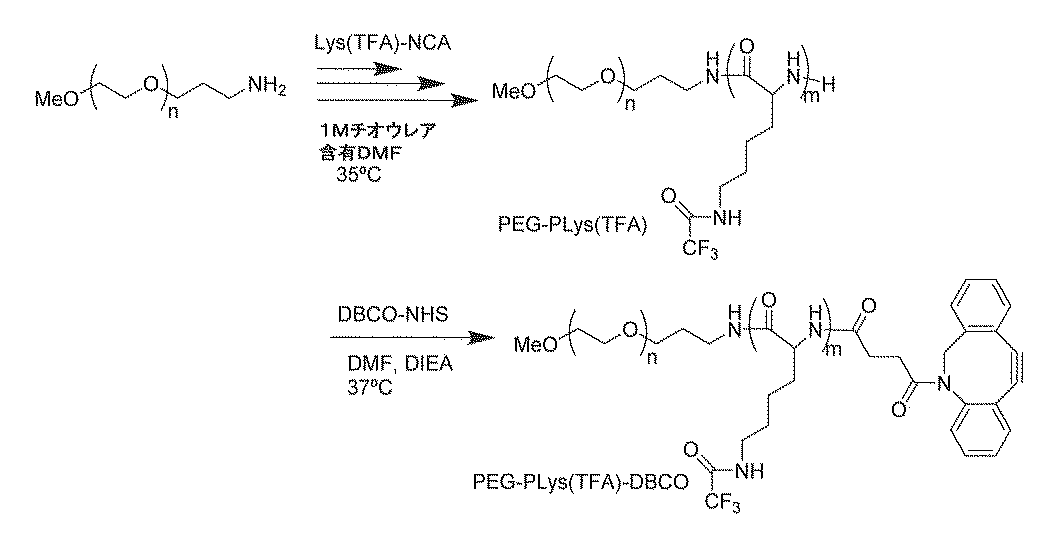

実施例1では、ミセル形成に必要な高分子の合成を行なった。
まず、1,2-O-イソプロピリデン-3,5-O-ベンジリデン-α-D-グルコフラノース(以下、「BIG-OH」という)を合成した。具体的には、1,2-O-イソプロピリデン-α-D-グルコフラノース(以下、「MIG」という)(和光純薬工業社製)10g、ベンズアルデヒド40mLをフラスコ中で混合し、ロータリーエバポレーターで4時間回転させながら混合し、反応させた。反応後、酢酸エチル 66mLを加え、蒸留水 120mLで洗浄し、有機層(酢酸エチル層)のみを回収し、ヘキサン 500mLに加えて0℃で再結晶し、BIG-OH 9.2g(収率85%)を得た。
まず、ポリエチレングリコール-ポリ(β-ベンジル-L-アスパルテート)ブロック共重合体(PEG-PBLA)をβ-ベンジル-L-アスパルテート-N-カルボン酸無水物(BLA-NCA)(中央化製品社に製造委託して得た)の重合により得た。具体的には、BLA-NCA 18.9gをN,N'-ジメチルホルムアミド(DMF)20mLに溶解する。メトキシ基の末端とアミノエチル基の末端を有するポリエチレングリコール(PEG-NH2)(分子量2,000)2.0gをDMF 20mLに溶解し、その溶液をBLA-NCA溶液に加える。混合溶液を35℃に保ちながら40時間重合した。赤外分光(IR)分析で重合反応が終了したことを確認した後、反応混合物をジエチルエーテル2Lに滴下して沈澱したポリマーを吸引濾過により回収し、ジエチルエーテルで洗浄した後に真空乾燥してPEG-PBLA 15.51g(収率79%)を得た。
上記で得られたPEG-PBLA 500mgをジメチルスルフォオキシド(DMSO) 20mLに溶解した。スルホ型Cy5-N-ヒドロキシスクシイミドエステル(Lumiprobe社製、製品番号:43320)25mgを、PEG-PBLA溶液に加え、常温で2日間反応させた。その後、0.5N水酸化ナトリウムを75mL添加し、室温でベンジルエステルを加水分解した。透析膜(分画分子量6,000-8,000)を用いてエタノール、水の順で透析した。膜内の溶液を凍結乾燥してCy5-PEG-P(Asp.) 456mg(収率86%)を得た。
まず、ベンゼン凍結乾燥した1,2,5,6-ジ-O-イソプロピリデン-α-D-グルコフラノース(DIG)からDIG-PEG-OHを得た。具体的には、DIG(TCI社製) 0.72gをTHF 5mLに溶解して、DIG-OH溶液を得た。その後、0.3Mのナフタレンカリウムを含んだTHF溶液 3.5mLを得られたDIG-OH溶液に滴下し、エチレンオキシド(EO)2.5mLをアルゴン雰囲気下で添加し常温で48時間反応させた。その後、1mLのメタノールを反応液に添加し、10%メタノールを含むエーテルを寒剤でよく冷やしたもので再沈殿させDIG-PEG-OH 3.2g(収率86%)を回収した。
Cy5-PEG-P(Asp.)50mgを10mM リン酸緩衝液(PB、pH 7.4、0mM NaCl)50mLに溶解し、1mg/mLのCy5-PEG-P(Asp.)溶液を調製した。PEG-P(Asp.-AP) 50mgも同様にPB 50mLに溶解し、1mg/mLのPEG-P(Asp.-AP)溶液を調製した。2種類の水溶液Cy5-PEG-P(Asp.)とPEG-P(Asp.-AP)それぞれ4mLと7.0mLを50mLのコニカルチューブに添加し、ボルテックスで2分間撹拌した(2000rpm)。その後、水溶性の縮合剤である1-エチル-3-(3-ジメチルアミノプロピル)カルボジイミド塩酸塩(EDC)(10mg/mL)を含有するPB溶液5.6mLを加え、一晩静置しポリイオンコンプレクスのコアを架橋した。その後、分画分子量100,000の膜のついた限外濾過チューブを用いて、ミセル形成に関与していないポリマーおよびEDCの副生成物などを除去した。
得られたCy5-PICミセルのサイズ(Z平均粒子径)および多分散指数(PDI)は、ゼータサイザー(Malvern)で測定した。サイズはブラウン運動により移動している粒子の拡散を測定し、その測定結果をストークス・アインシュタインの式を用いて粒子径と粒度分布に変換した。またミセルの形状は、透過型電子顕微鏡(TEM、JEM-1400)を用いて評価した。ここで、Z平均粒子径とは、粒子分散 物等の動的光散乱法の測定データを、キュムラント解析法を用いて解析したデータである。キュムラント解析においては、粒子径の平均値と多分散指数(PDI)が得られ、本発明においては、この平均粒子径をZ平均粒子径と定義する。厳密には、測定で得られたG1相関関数の対数に、多項式をフィットさせる作業を、キュムラント解析といい、下式:
LN(G1)=a+bt+ct2+dt3+et4+・・・における定数bが、二次キュムラントまたは、Z平均拡散係数と呼ばれる。Z平均拡散係数の値を分散媒の粘度と幾つかの装置定数を用いて粒子径に換算した値がZ平均粒子径であり、分散安定性の指標として品質管理目的に適した値である。
Glc(6)-PEG-P(Asp.)20mgとCy5-PEG-P(Asp.)40mgを10mM リン酸緩衝液(PB、pH7.4、0mM NaCl)60mLに溶解し、1mg/mLのCy5-Glc(6)-PEG-P(Asp.)とPEG-P(Asp.)混合溶液を調製した。PEG-P(Asp.-AP)50mgも同様にPB 50mLに溶解し、1mg/mLのPEG-P(Asp.-AP)溶液を調製した。2種類の水溶液、すなわち、Cy5-PEG-P(Asp.)とPEG-P(Asp.)との混合液とPEG-P(Asp.-AP)溶液のそれぞれ4mLと7.0mLを50mLのコニカルチューブに添加して、ボルテックスにより2分間撹拌した(2000rpm)。その後、水溶性の縮合剤であるEDC(10mg/mL)を含有するPB溶液 5.6mLを加え、一晩静置しポリイオンコンプレクスのコアを架橋した。その後、分画分子量100,000の膜のついた限外濾過チューブを用いて、ミセル形成に関与していないポリマー、EDCの副生成物などを除去した。
得られたGlc(6)-Cy5-PICミセルのサイズ(Z平均粒子径)および多分散指数(PDI)は、ゼータサイザー(Malvern)で測定した。その結果、平均粒径40nmであり、粒径が均一なミセルを得たことが明らかとなった(図2A)。また、ミセルの形状は、透過型電子顕微鏡(TEM、JEM-1400)を用いて、酢酸ウラニルで染色後に観察した(図2B)。
Glc(3)-PEG-P(Asp.)20mgとCy5-PEG-P(Asp.)40mgをpH7.4 10mMリン酸緩衝液 (PB、0mM NaCl)60mLに溶解し、1mg/mLのCy5-Glc(3)-PEG-P(Asp.)とPEG-P(Asp.)混合溶液を調製した。PEG-P(Asp.-AP) 50mgも同様にPB 50mLに溶解し、1mg/mLのPEG-P(Asp.-AP)溶液を調製した。2種類の水溶液Cy5-PEG-P(Asp.)、PEG-P(Asp.)混合液とPEG-P(Asp.-AP)溶液のそれぞれ4mLと4.3mLを50mLのコニカルチューブに添加して、ボルテックスで2分間撹拌した(2000rpm)。その後、水溶性の縮合剤であるEDC(10mg/mL)を含有するPB溶液5.6mLを加え、一晩静置しポリイオンコンプレクスのコアを架橋した。その後、分画分子量100,000の膜のついた限外濾過チューブを用いて、ミセル形成に関与していないポリマー、EDCの副生成物などを除去した。得られたGlc(6)-Cy5-PICミセルのサイズ(Z平均粒子径)および多分散指数(PDI)は、ゼータサイザー(Malvern)で測定した。またミセルの形状は、透過型電子顕微鏡(TEM、JEM-1400)を用いて評価した。得られたミセルは、直径32nm(PDI=0.043)であった(データ非掲載)。
実施例1で作製したミセルをマウスに静脈投与してその体内動態を調べた。ミセルを投与する際には、血糖操作の効果も加えて評価した。
直径約100nmの中空キャリアとしてPICsomeを作製し、体内動態評価により脳への標的化効果を検証した。
まず、ポリ(β-ベンジル-L-アスパルテート)(ホモPBLAポリマー)をBLA-NCAの重合により得た。具体的には、β-ベンジル-L-アスパルテート-N-カルボン酸無水物(BLA-NCA)20gをN,N'-ジメチルホルムアミド(DMF)33.3mL、ジクロロメタン300mLに溶解する。N-ブチルアミン89.0μLを上記BLA-NCA溶液に加える。混合溶液を35℃に保ちながら40時間重合した。赤外分光(IR)分析で重合反応が終了したことを確認したのち、反応混合物をヘキサン/酢酸エチル溶液(ヘキサン:酢酸エチル=6:4)1Lに滴下して沈澱したポリマーを吸引濾過により回収し、ジエチルエーテルで洗浄した後に真空乾燥してホモPBLAポリマー 14.82g(79%)を得た。
実施例1で得たGlc(6)-PEG-P(Asp.)20mgとCy5-PEG-P(Asp.)40mgを10mM リン酸緩衝液(PB、pH7.4、0mM NaCl)60mLに溶解し、1mg/mLのGlc(6)-PEG-P(Asp.)とCy5-PEG-P(Asp.)との混合溶液を調製した。また、ホモP(Asp.-AP)50mgも同様にPB 50mLに溶解し、1mg/mLのホモP(Asp.-AP)溶液を調製した。次に、2種類の水溶液、すなわち、Glc(6)-PEG-P(Asp.)とCy5-PEG-P(Asp.)との混合液およびホモP(Asp.-AP)溶液のそれぞれ4.0mLおよび5.0mLを50mLのコニカルチューブ中で混合し、ボルテックスにより2分間撹拌した(2000rpm)。その後、水溶性の縮合剤であるEDC(10mg/mL)を含有するPB溶液5.6mLを加え、一晩静置してポリイオンコンプレクスのコアを架橋した。その後、分画分子量100,000の膜のついた限外濾過チューブを用いて、PICsome形成に関与していないポリマー、EDCの副生成物などを除去した。得られたGlc(6)-Cy5-PICsomeのサイズ(Z平均粒子径)および多分散指数(PDI)は、ゼータサイザー(Malvern)で測定した。またミセルの形状は、透過型電子顕微鏡(TEM、JEM-1400)を用いて、酢酸ウラニルで染色後に観察した。すると、直径100nm(PDI=0.086)のPICsomeを得たことが明らかとなった(データ非掲載)。
PICミセルの代わりに得られたPICsomeを投与する以外は実施例2と全く同じ方法で、PICsomeをマウスに投与し、脳へのPICsomeの蓄積を観察した。すると、グルコースでその外表面が修飾されたPICsomeのみが給餌後、急激に脳内に蓄積する様子が観察された(図6)。グルコースでその外表面が修飾されたPICsomeの脳1g当りの蓄積量は約2%であった(図6)。
本実施例では、血中滞留時間が短く送達効率の低いsiRNAを用いて実施例2および3と同様に体内動態評価を行なった。より具体的には、本実施例では、グルコースをコンジュゲートしたPEG-ポリカチオンと蛍光標識siRNAからなるミセルを用いて、siRNAの脳への蓄積を評価した。
まず、実施例1に記載の方法により得たBIG-PEG-PBLAからBIG-PEG-PBLA-Cholを合成した。具体的には、BIG-PEG-PBLA 120mgをNMP 10mLに溶解し、PBLAの末端のアミノ基に対し10等量の4-コレステリルアミノ-4-ブタン酸および触媒量のジメチルアミノピリジンを添加し、その後、室温にて6時間撹拌した。反応溶液をジエチルエーテル/2-プロパノール(9:1)溶液に滴下し、目的物を沈殿させた。沈殿物をろ過後減圧乾燥させてBIG-PEG-PBLA-Cholを130mg得た(収率95%)。
Glc(6)-siRNAミセルは、図7Aのスキームに従って調製した。具体的には、10mM HEPES緩衝液中に溶解させたGlc(6)-PEG-P(Asp.-TEP)-Chol(2mg/mL) 262.5μLをHEPES緩衝液437.5μLで希釈した。北海道システム・サイエンス社製のスクランブルsiRNAであるCy5-siRNA-chol(75μM) 279μLをHEPES緩衝液1121μLで希釈した。得られた2液を混合して10回ピペッティングし、Glc(6)-siRNAミセルを得た。In vivo実験直前に2.1mLのミセル溶液に65μLの5M NaCl溶液を加え、ピペッティングすることで等張液にしてから投与に用いた。得られたGlc(6)-Cy5-siRNAミセルのサイズ(Z平均粒子径)および多分散指数(PDI)は、ゼータサイザー(Malvern)で測定した。またミセルの形状は、透過型電子顕微鏡(TEM、JEM-1400)を用いて、酢酸ウラニルで染色後に観察した。結果、直径80nm(PDI=0.104)のsiRNAミセルが得られたことが明らかとなった。
得られたGlc(6)-Cy5-siRNAミセル 200μL/2時間を静脈投与により30分または2時間にわたりシリンジポンプ(Harvard社)を用いて精密持続静注し、投与開始5分後に20%グルコース溶液を腹腔内投与した。siRNAミセルの投与終了から1時間後に脳を摘出し、マルチビーズショッカーで粉砕後に、IVISイメージングシステム(Xenogen社)をもちいてそれぞれの輝度を評価した。すると、静脈投与の時間が長くなると、脳の輝度が上昇し、2時間の静脈投与による脳への蓄積は、30分の静脈投与による脳への蓄積よりも多かった(図7B)。また、脳への蓄積量を算出すると、siRNAミセルでは、2時間の静脈投与において、その投与量の1.3%を脳(1g当り)に送達することができていることが明らかとなった。さらに、投与終了から6時間後に脳を摘出し、共焦点顕微鏡(LSM510)で観察を行い、脳実質における蛍光強度を測定した。すると、siRNAミセルは、脳細胞内に蓄積していることが明らかとなった(図8)。
本実施例では、Glc(6)-PEG-ポリアスパラギン酸をミセルを形成させずにマウスに投与してその体内動態を評価した。
本実施例では、抗体にグルコースをコンジュゲートさせ、体内動態を評価した。すると、抗体も血糖操作により脳への蓄積を示した。
次に、得られたDyLight488蛍光標識化グルコース導入ポリエチレングリコール-ポリアスパラギン酸ブロック共重合体と抗体とをコンジュゲートさせてグルコース導入抗体を得た。具体的には以下の通りである。
6週齢のBalb/C雌マウス8匹の給餌を24時間止めた。8匹のマウスをA群、B群およびコントロール群(それぞれ3匹、3匹および2匹)に分けた。A群のマウスにはGlc-polymer conjugated IgGを、B群のマウスにはCy5-IgGをそれぞれ750nMで200μLずつ静脈内注射し、その5分後に20%グルコース溶液200μLを腹腔内投与した。なお、各抗体のCy5に由来する蛍光強度は同等であった。A群およびB群のマウス3匹ずつを抗体投与から57分後にジエチルエーテル麻酔にかけ、その3分後に採血と臓器摘出(脳、肝臓、腎臓、肺、心臓、脾臓および大腿筋)を行った。コントロール群2匹も別途、採血と臓器摘出を行った。採血により得られた血液は、それぞれ4℃、15,000rpmで遠心をし、上清を回収した。8匹のマウスの摘出した臓器はまず全体の重量の測定を行った後、脳は半分、肝臓はおよそ200mgを切りとり、また、腎臓は片側を取得して、重量の測定を行い、マルチビーズショッカー用のチューブに臓器を金属のコーンとともに入れた。また、コントロール群のうちの1匹を除いた7匹の脳および肝臓のサンプルには1×Passive Lysis Bufferを600μL、脾臓、心臓および大腿筋のサンプルには300μL、腎臓および肺のサンプルには400μLずつ加えた。一方、コントロール群のうちの残った1匹のマウス(100%コントロール)の臓器のサンプルには、静脈内注射したサンプル全てが該当する臓器に集積したと仮定した時のサンプル量を計算し、その量のCy5-IgG溶液を臓器のサンプルに加え、さらに加える溶液量の合計が他の7匹のマウスと同じになるように1×Passive Lysis Bufferを加えた。全ての臓器サンプルをマルチビーズショッカーにより、2000rpmで30秒動作を5回繰り返し、臓器をホモシナイズした。臓器のチューブからコーンを除いた後、それぞれのサンプルを100μLずつマルチプレートの各ウェルに入れ、マルチプレートリーダーで励起波長643nm、蛍光波長667nmとして蛍光強度の測定を行った。コントロール群のうちCy5-IgG溶液を加えていない方をブランクとし、加えた方を100%として各臓器の抗体の集積率を計算し、血液以外は、得られた集積率を臓器の重量(g)で除した値を算出した。
上記実施例2によれば、グルコース導入率25%のミセルは3%を超える脳への蓄積を示したのに対して、グルコース導入率50%のミセルは約1.3%の脳への蓄積を示した。このことは、グルコースの導入率が増加することにより、脳血管内皮細胞に取り込まれたミセルと脳血管内皮細胞との解離が低下することを意味すると考えられた。そこで、本実施例では、グルコース導入率と脳血管内皮細胞へのミセルの蓄積との関係を確認した。
本実施例では、温度に応答してミセル化を引き起こす温度感受性共重合体を合成した。温度感受性共重合体は、親水性鎖-カチオン性鎖-温度感受性鎖を連結した三元型ブロック共重合体とした。
N,N-ジメチルホルムアミド(DMF)、ジメチルスルホキシド(DMSO)、アセトニトリル、クロロベンゼン、N,N-ジイソプロピルエチルアミン(DIEA)、メタノール、アジ化ナトリウム、p-トルエンスルホン酸メチル(MeOTs)、N6-トリフルオロアセチル-L-リシン-N-カルボン酸無水物(Lys(TFA)-NCA)は、一般グレードのものを購入して適宜精製作業を行った後使用した。ノルマルプロピルオキサゾリン(nPrOx)は東京化成工業株式会社に委託大量合成したもの、またジベンジルシクロオクチン-N-ヒドロキシコハク酸エステル(DBCO-NHS)はClick Chemistry Toolsから購入したものを用いた。
開始剤p-トルエンスルホン酸メチル(MeOTs)を50μL(61.7μg)測りとり、アセトニトリル10mL、クロロベンゼン10mLを混合した溶媒に溶かした。氷冷しながらこの開始剤溶液にモノマーであるn-プロピルオキサゾリン(nPrOx)を8.5mL(8.76g,モノマー/開始剤=234)加え、42℃の水浴で12日間重合した。マトリックス支援レーザー脱離イオン化飛行時間型質量分析法(MALDI-TOF-MS)で目標の分子量に達したことを確認し、重合停止剤としてアジ化ナトリウムを430mg(停止剤/開始剤=20)加え、70℃で2日間撹拌して重合停止を行った。全ての反応はアルゴン雰囲気下で行った。なお、アセトニトリルとnPrOxモノマーは脱水剤の水素化カルシウムを加えて蒸留、クロロベンゼンと開始剤MeOTsは脱水剤の五酸化二リンを加えて蒸留したものを用いた。反応後のサンプルは、メタノールに対して透析を3回、水に対して透析を4回行った後、凍結乾燥により回収した(収率58%)。
ベンゼン凍結乾燥をした片末端メトキシ基片末端3-アミノプロピル基のポリエチレングリコール(PEG-NH2)(分子量2,200)433mgを、1Mチオウレアを含むN,N’-ジメチルホルムアミド(DMF)10mLに溶解し、開始剤溶液とした。N6-トリフルオロアセチル-L-リシン-N-カルボン酸無水物(Lys(TFA)-NCA)2.37gを、1Mチオウレアを含むDMF20mLに溶解させモノマー溶液とした。このモノマー溶液を開始剤溶液に加え、35℃で3日間重合反応を行った。赤外分光(IR)分析で重合反応が終了したことを確認したのち、反応混合物をジエチルエーテル2Lに滴下して沈澱したポリマーを吸引濾過により回収し、ジエチルエーテルで洗浄した後に真空乾燥してポリエチレングリコール-ポリ(N6-トリフルオロアセチル-L-リシン)ブロック共重合体(PEG-PLys(TFA))2.39g(収率85.1%)を得た。重合度は1H-NMRにより40(TFA基脱保護後、1分子のsiRNAと同等の電荷数を持つ重合度)であることを確認した。
PEG-PLys(TFA)120mgを5mLのDMFに溶解した。これにジベンジルシクロオクチンNHSエステル(DBCO-NHS)を34mg加え、4時間撹拌した。その後N,N-ジイソプロピルアミン(DIEA)を11μL加え、37℃で24時間撹拌した。反応後のサンプルは、ジエチルエーテル150mLに滴下して沈澱したポリマーを吸引濾過に供し、ジエチルエーテルで洗浄した後に真空乾燥して回収した。PEG-PLys(TFA)-DBCOを105mg(収率84.6%)得た。
まず、PEG-PLys(TFA)-DBCOとPnPrOx-N3のカップリング反応を行った。100mgのPEG-PLys(TFA)-DBCOと550mgのPnPrOx-N3を10mLのジメチルスルホキシド(DMSO)に溶解し、4℃で一晩かけてポリマー溶液を凍結させた。その後室温で凍結させたポリマー溶液を融解させた。続いてTFA保護基の脱保護を行うため、この反応溶液に40mLのメタノールと0.84mLの6M NaOH水溶液を加え、35℃で12時間撹拌した。反応後のポリマー溶液は、0.01M塩酸水溶液を外液として3回透析し、さらに純水を外液として4回透析した後に凍結乾燥により回収した。
PEG-PLys-PnPrOx共重合体は、温度依存的にミセル形成の進行を制御できる。本実施例ではこの温度感受性共重合体を用いてsiRNAとのポリイオンコンプレックスを作製した。
具体的には、まず、PEG-PLys-PnPrOxを15μMの濃度で10mM HEPESバッファー(pH7.4、150mM NaCl)に溶解させ、37℃で30分間静置させた。その後このポリマー溶液と37℃とした15μMのsiRNA溶液を同様の体積だけ混合して(ポリマーとsiRNAの電荷比1)、コンプレックス形成を行った。この方法で調製した高分子ミセルをsiRNA-cPIC/ミセルと名付ける。同様に、疎水的な分子であるコレステロールが導入されたsiRNA(chol-siRNA)を用いて高分子ミセル、chol-siRNA-cPIC/ミセルを調製した。
本実施例では、PLysとsiRNAが1対1会合体(uPIC)を形成することに着目して、まずuPICを形成させた後にPnPrOxの温度応答疎水化によりuPICを束ねる形で高分子ミセルを形成させることを思いついた。この方法では、従来のように高次構造を作った後にPIC形成を行うのではなく、PIC形成を行った後に高次構造を形成するため、最終的な構造が従来型より秩序だったものになっていると予想され、安定な構造体であると期待される。
本実施例では、実施例2で得られたuPIC/ミセルとcPIC/ミセルの物性や生理的安定性を分析した。
まず、siRNAが高分子ミセルに封入されているかをアガロースゲル電気泳動により確認した。7.5μMの濃度のsiRNA-cPIC/ミセル、siRNA-uPIC/ミセル、chol-siRNA-cPIC/ミセルおよびchol-siRNA-uPIC/ミセル溶液をそれぞれ20μLずつアガロースゲルにロードし、100Vで30分間電気泳動を行った。泳動後はエチジウムブロマイドによりsiRNAを染色した。PIC中に取り込まれたsiRNAは、電荷が中和されているため、siRNAのバンドは開始点より動かない。一方で、会合体の形成が起きていないsiRNAは、マイナスの電荷を有し、siRNAのバンドは電気泳動により移動するはずである。
続いて、得られたuPIC/ミセルおよびcPIC/ミセルの粒径と粒径分布(PDI)を動的光散乱(DLS)測定で調べた。その結果、図19に示されるように、いずれのミセルも50nm程度の粒径を有しており、PDIもすべて0.20以下となった。特筆すべき点として、uPIC/ミセルの方が小さいPDI(図19中の折れ線)を示した。PDIが小さいことは得られたミセルが均一な粒径を有することを意味し、本発明のuPIC/ミセルは形状において均一であると言える。おそらく、温度感受性共重合体とsiRNAとの複合体を形成させてからミセルを形成させると、秩序だった構造を形成しやすいからであると思われる。
uPIC/ミセルについては、37℃での静置前にuPIC(すなわち、温度感受性共重合体とsiRNAとのイオン性結合による会合分子)を形成していることの確認実験を行った。蛍光分子Alexa-647を担持したsiRNAおよびchol-siRNA溶液をそれぞれ20nMで用意した。共重合体とsiRNAでさまざまな正電荷/負電荷比(+/-比)となるよう、PEG-PLys-PnPrOx溶液の濃度を調節し150μLずつsiRNA溶液150μLに加えた。調製したそれぞれのサンプルについて、蛍光相関分光法(FCS)でsiRNAまたはchol-siRNAが担持しているAlexa-647の拡散時間を調べた。共重合体とsiRNAとが会合すると、見かけの粒径が大きくなるため、FCSで観測されるAlexa-647の拡散時間は大きくなる。実際に共重合体を添加すると拡散時間が大きくなり、イオンコンプレックスの形成が確認された(図20)。また、拡散時間は共重合体とsiRNAの+/-比が1以上では一定であることから、共重合体とsiRNAは1対1の比で会合していることが分かる。
生体内には数多くのポリアニオン性分子が存在する。これらのポリアニオン性分子は、PICミセルを形成しているポリアニオン性分子と置き換わりPICのミセル構造を崩壊させ、ミセルの標的への送達効率を低下させるため、PICミセルにおいては、ポリアニオンに対する安定性は重要な因子である。ここでは、ポリアニオンとして、細胞膜表面に多く存在する生体由来成分であるヘパリンのナトリウム塩を用いた。
cPIC/ミセルとuPIC/ミセルとの構造の違いを1H-NMR測定により明らかにした。図22(a)に示されるようにChol-siRNAは、2ppm付近にコレステロール基に由来するピークを有する。cPICでは、図22(b)に示されるように、このコレステロール基に由来するピークが見いだされたが、uPIC/ミセルでは、コレステロール基に由来するピークが見いだされなかった。このことは、cPIC/ミセルでは、コレステロール基がPIC/ミセルの表面の露出している(図22(d))一方で、uPIC/ミセルでは、コレステロール基がミセルのコアの内部に格納されている(図22(e))ことを意味する。
本実施例では、生体内におけるcPIC/ミセルとuPIC/ミセルの安定性を調べた。
標的組織は、特に限定されないが、本実施例では脳を標的としたsiRNAの送達を試みた。脳は、血液脳関門(BBB)の存在により、最も薬剤の送達が困難な臓器であると考えられている。
実施例1におけるPEG-PLys(TFA)の合成と同様に行った。149mgのBIG-PEG-HN2と764mgのLys(TFA)-NCAから、BIG-PEG-PLys(TFA)を得た。
実施例1と同様の手順で、200mgのBIG-PEG-PLys(TFA)と30mgのDBCO-NHSからBIG-PEG-PLys(TFA)-DBCOを180mg(収率87.0%)得た。
170mgのBIG-PEG-PLys(TFA)-DBCOと、実施例1で合成した840gmのPnPrOx-N3から、実施例1と同様の手順で、BIG-PEG-PLys-PnPrOxを得た。その後、BIG-PEG-PLys-PnPrOxのBIG部分の保護基をトリフルオロ酢酸を用いて脱保護して、Glc(6)-PEG-PLys-PnPrOxを90mg(収率25.0%)得た。
24時間給餌しなかった絶食マウス(Balb/c雌6週齢)に20v/v%のグルコース溶液を腹腔内投与(i.p.投与)し、i.p.投与から30分後にCy5-siRNA-Chol(BACE1)を封入した各ミセル溶液(uPIC/ミセル、10%Glc(6)-Cy5-uPIC/ミセル、25%Glc(6)-Cy5-uPIC/ミセル、50%Glc(6)-Cy5-uPIC/ミセル 50μg/マウス、200μL)を200μL微静脈投与(i.v.投与)した。i.v.投与から6時間後に脳を摘出し、脳の固定および染色を行なった。
1.4℃、4%パラホルムアルデヒドを含有するリン酸緩衝生理食塩水(PBS)中で一晩静置して脳を固定する。
2.4℃、20%スクロース溶液を含有するPBS中で一晩静置(置換1)する。
3.4℃、30%スクロース溶液を含有するPBS中で一晩静置(置換2)する。
4.全脳をOCT compound(商標)に包埋し、低温ヘキサンにて凍結する。-80℃にて保存する。
5.凍結ミクロトームで凍結した脳を10~100μmにスライスする。
6.PBS-T(PBS+0.1%tween20)に5分間浸してスライスを洗浄する。
7.スライスにブロッキング液(2%BSAを含有するPBS-T)を1時間接触させる。
8.ブロッキング液を除去し、洗わずに1次抗体溶液を加え室温で2時間静置する。
9.PBS-Tでスライスを10分間洗浄する。この洗浄を3回繰り返す。
10.2次抗体溶液を加え、スライスを室温で1時間処理する。
11.PBS-Tでスライスを10分間洗浄する。この洗浄を3回繰り返す。
本実施例では、送達したsiRNAが脳細胞内で標的であるβセクレターゼ遺伝子(BACE1)の発現を抑制しているかを確認した。
Claims (18)
- 温度感受性共重合体と核酸とのポリイオンコンプレックスであって、
温度感受性共重合体は、カチオン性ブロックと温度感受性ブロックとを有し、
温度感受性共重合体の下限臨界溶液温度(LCST)以下の温度条件下で温度感受性共重合体と核酸とを混合して得られる、ポリイオンコンプレックス。 - カチオン性ブロックが、カチオン性アミノ酸ポリマーブロックである、請求項1に記載のポリイオンコンプレックス。
- 温度感受性共重合体が親水性ブロックを有し、親水性ブロックが、ポリエチレングリコールである、請求項1または2に記載のポリイオンコンプレックス。
- 温度感受性共重合体が、GLUT1リガンドで修飾された、請求項1~3のいずれか一項に記載のポリイオンコンプレックス。
- 核酸が、生体適合性の疎水基で修飾されている、請求項1~4のいずれか一項に記載のポリイオンコンプレックス。
- 核酸が、siRNAである、請求項1~5のいずれか一項に記載のポリイオンコンプレックス。
- カチオン性ブロックと温度感受性ブロックとを有する温度感受性共重合体を含んでなる、ポリイオンコンプレックスを作製するための組成物。
- カチオン性ブロックが、カチオン性アミノ酸ポリマーブロックである、請求項7に記載の組成物。
- 温度感受性共重合体が親水性ブロックを有し、親水性ブロックが、ポリエチレングリコールである、請求項7または8に記載の組成物。
- 温度感受性共重合体が、グルコースで修飾された、請求項7~9のいずれか一項に記載の組成物。
- 請求項1~6のいずれか一項に記載のポリイオンコンプレクスを該ポリイオンコンプレックスの下限臨界溶液温度(LCST)以上の温度条件下にさらして得られる、核酸を含有するミセル。
- 請求項11に記載のミセルを含む、核酸送達用組成物。
- 請求項11に記載のミセルであって、温度感受性共重合体がグルコースで修飾された、ミセル。
- 請求項13に記載のミセルであって、ミセル中の温度感受性共重合体のグルコース修飾率が、15~40%である、ミセル。
- 請求項13に記載のミセルであって、ミセル中の温度感受性共重合体のグルコース修飾率が、50~100%である、ミセル。
- 請求項13または14に記載のミセルを含む、脳実質への核酸送達用組成物。
- 請求項13または15に記載のミセルを含む、脳血管内皮細胞への核酸送達用組成物。
- 請求項16または17に記載の脳への核酸送達用組成物であって、
組成物は、投与計画に従って対象に投与するための組成物であり、
投与計画は、絶食させるか、または低血糖を誘発させた対象に該組成物を投与することと、該対象において血糖値の上昇を誘発させることとを含む、組成物。
Priority Applications (3)
| Application Number | Priority Date | Filing Date | Title |
|---|---|---|---|
| US15/575,716 US10668169B2 (en) | 2015-05-21 | 2016-05-20 | Micelle composition for nucleic acid delivery using temperature-sensitive polymer and method for producing same |
| JP2017519417A JP6853924B2 (ja) | 2015-05-21 | 2016-05-20 | 温度感受性重合体を用いた核酸の送達用ミセル組成物およびその製造方法 |
| EP16796597.9A EP3299020A4 (en) | 2015-05-21 | 2016-05-20 | MICROM COMPOSITION FOR NUCLEIC ACID RELIEF WITH TEMPERATURE-PERMANENT POLYMER AND METHOD FOR THE PRODUCTION THEREOF |
Applications Claiming Priority (2)
| Application Number | Priority Date | Filing Date | Title |
|---|---|---|---|
| JP2015-104028 | 2015-05-21 | ||
| JP2015104028 | 2015-05-21 |
Publications (1)
| Publication Number | Publication Date |
|---|---|
| WO2016186204A1 true WO2016186204A1 (ja) | 2016-11-24 |
Family
ID=57320438
Family Applications (1)
| Application Number | Title | Priority Date | Filing Date |
|---|---|---|---|
| PCT/JP2016/065052 Ceased WO2016186204A1 (ja) | 2015-05-21 | 2016-05-20 | 温度感受性重合体を用いた核酸の送達用ミセル組成物およびその製造方法 |
Country Status (4)
| Country | Link |
|---|---|
| US (1) | US10668169B2 (ja) |
| EP (1) | EP3299020A4 (ja) |
| JP (1) | JP6853924B2 (ja) |
| WO (1) | WO2016186204A1 (ja) |
Cited By (6)
| Publication number | Priority date | Publication date | Assignee | Title |
|---|---|---|---|---|
| WO2018216792A1 (ja) * | 2017-05-26 | 2018-11-29 | 公益財団法人川崎市産業振興財団 | 血中におけるrnaの安定性の改善剤および投与方法 |
| WO2018216793A1 (ja) * | 2017-05-26 | 2018-11-29 | 公益財団法人川崎市産業振興財団 | 血中で安定なmRNA内包ミセル |
| US20210038634A1 (en) * | 2017-10-05 | 2021-02-11 | Kawasaki Institute Of Industrial Promotion | Composition controlling pharmacokinetics in the body |
| WO2022122872A1 (en) | 2020-12-09 | 2022-06-16 | Ucl Business Ltd | Therapeutics for the treatment of neurodegenerative disorders |
| JPWO2022191107A1 (ja) * | 2021-03-10 | 2022-09-15 | ||
| WO2023104964A1 (en) | 2021-12-09 | 2023-06-15 | Ucl Business Ltd | Therapeutics for the treatment of neurodegenerative disorders |
Families Citing this family (1)
| Publication number | Priority date | Publication date | Assignee | Title |
|---|---|---|---|---|
| WO2023230099A1 (en) * | 2022-05-23 | 2023-11-30 | Sirnagen Therapeutics Incorporated | Compositions comprising oligonucleotide and uses thereof |
Citations (1)
| Publication number | Priority date | Publication date | Assignee | Title |
|---|---|---|---|---|
| JP2013505200A (ja) * | 2008-09-18 | 2013-02-14 | ウニフェルジテイト・ユトレヒト・ホールディング・ベスローテン・フェンノートシャップ | 制御放出系の調製方法 |
Family Cites Families (2)
| Publication number | Priority date | Publication date | Assignee | Title |
|---|---|---|---|---|
| JP4655298B1 (ja) | 2010-02-23 | 2011-03-23 | ナノキャリア株式会社 | 短鎖のカチオン性ポリアミノ酸およびその使用 |
| US9320814B2 (en) * | 2011-11-01 | 2016-04-26 | Board Of Regents Of The University Of Nebraska | Polyplexes of hydrophobically-modified siRNA for delivery of siRNA |
-
2016
- 2016-05-20 WO PCT/JP2016/065052 patent/WO2016186204A1/ja not_active Ceased
- 2016-05-20 EP EP16796597.9A patent/EP3299020A4/en not_active Withdrawn
- 2016-05-20 JP JP2017519417A patent/JP6853924B2/ja active Active
- 2016-05-20 US US15/575,716 patent/US10668169B2/en active Active
Patent Citations (1)
| Publication number | Priority date | Publication date | Assignee | Title |
|---|---|---|---|---|
| JP2013505200A (ja) * | 2008-09-18 | 2013-02-14 | ウニフェルジテイト・ユトレヒト・ホールディング・ベスローテン・フェンノートシャップ | 制御放出系の調製方法 |
Non-Patent Citations (5)
| Title |
|---|
| HALADJOVA E. ET AL.: "Polymeric Nanoparticle Engineering: From Temperature-Responsive Polymer Mesoglobules to Gene Delivery Systems", BIOMACROMOLECULES, vol. 15, no. 12, 2014, pages 4377 - 4395, XP055330509, ISSN: 1525-7797 * |
| QIN Y. ET AL.: "In vitro and in vivo investigation of glucose-mediated brain- targeting liposomes", JOURNAL OF DRUG TARGETING, vol. 18, no. 7, 2010, pages 536 - 549, XP009195259, ISSN: 1029-2330 * |
| See also references of EP3299020A4 * |
| SHIGEHITO OSAWA: "Sosuisei Hogoso o Core-Shell Kaimen ni Yusuru Kobunshi Micelle-gata Idenshi Carrier no Sosei", SYMPOSIUM ON POLYMERS AND BIOSCIENCES KOEN YOSHISHU, vol. 24th, 2014, pages 15 - 16, XP009507628 * |
| YANG Z. ET AL.: "Thermo-sensitive nanoparticles for triggered release of siRNA", JOURNAL OF BIOMATERIALS SCIENCE , POLYMER EDITION, vol. 26, no. 4, 8 January 2015 (2015-01-08), pages 264 - 276, XP055330518, ISSN: 1568-5624 * |
Cited By (17)
| Publication number | Priority date | Publication date | Assignee | Title |
|---|---|---|---|---|
| JP7471373B2 (ja) | 2017-05-26 | 2024-04-19 | 公益財団法人川崎市産業振興財団 | 血中で安定なmRNA内包ミセル |
| WO2018216793A1 (ja) * | 2017-05-26 | 2018-11-29 | 公益財団法人川崎市産業振興財団 | 血中で安定なmRNA内包ミセル |
| JPWO2018216792A1 (ja) * | 2017-05-26 | 2020-03-26 | 公益財団法人川崎市産業振興財団 | 血中におけるrnaの安定性の改善剤および投与方法 |
| JPWO2018216793A1 (ja) * | 2017-05-26 | 2020-05-21 | 公益財団法人川崎市産業振興財団 | 血中で安定なmRNA内包ミセル |
| WO2018216792A1 (ja) * | 2017-05-26 | 2018-11-29 | 公益財団法人川崎市産業振興財団 | 血中におけるrnaの安定性の改善剤および投与方法 |
| JP2022191527A (ja) * | 2017-05-26 | 2022-12-27 | 公益財団法人川崎市産業振興財団 | 血中で安定なmRNA内包ミセル |
| JP7237825B2 (ja) | 2017-05-26 | 2023-03-13 | 公益財団法人川崎市産業振興財団 | 血中におけるrnaの安定性の改善剤および投与方法 |
| EP3766519A4 (en) * | 2017-10-05 | 2021-12-01 | Kawasaki Institute of Industrial Promotion | COMPOSITION FOR THE CONTROL OF PHARMACOKINETICS IN THE BODY |
| US11957708B2 (en) | 2017-10-05 | 2024-04-16 | Kawasaki Institute Of Industrial Promotion | Composition controlling pharmacokinetics in the body |
| US20210038634A1 (en) * | 2017-10-05 | 2021-02-11 | Kawasaki Institute Of Industrial Promotion | Composition controlling pharmacokinetics in the body |
| US12582669B2 (en) | 2017-10-05 | 2026-03-24 | Kawasaki Institute Of Industrial Promotion | Composition controlling pharmacokinetics in the body |
| WO2022122872A1 (en) | 2020-12-09 | 2022-06-16 | Ucl Business Ltd | Therapeutics for the treatment of neurodegenerative disorders |
| EP4647124A2 (en) | 2020-12-09 | 2025-11-12 | UCL Business Ltd | Therapeutics for the treatment of neurodegenerative disorders |
| US12522826B2 (en) | 2020-12-09 | 2026-01-13 | The United States Of America, As Represented By The Secretary, Department Of Health And Human Services | Therapeutics for the treatment of neurodegenerative disorders |
| JPWO2022191107A1 (ja) * | 2021-03-10 | 2022-09-15 | ||
| JP7739406B2 (ja) | 2021-03-10 | 2025-09-16 | Agc株式会社 | 含ハロゲン部位を有するポリマーおよび該ポリマーを用いた医薬組成物 |
| WO2023104964A1 (en) | 2021-12-09 | 2023-06-15 | Ucl Business Ltd | Therapeutics for the treatment of neurodegenerative disorders |
Also Published As
| Publication number | Publication date |
|---|---|
| EP3299020A1 (en) | 2018-03-28 |
| JP6853924B2 (ja) | 2021-04-07 |
| US20180289835A1 (en) | 2018-10-11 |
| US10668169B2 (en) | 2020-06-02 |
| EP3299020A4 (en) | 2019-01-23 |
| JPWO2016186204A1 (ja) | 2018-03-08 |
Similar Documents
| Publication | Publication Date | Title |
|---|---|---|
| JP6086566B2 (ja) | 薬剤送達用のキャリア、コンジュゲートおよびこれらを含んでなる組成物並びにこれらの投与方法 | |
| JP6853924B2 (ja) | 温度感受性重合体を用いた核酸の送達用ミセル組成物およびその製造方法 | |
| Savin et al. | Chitosan grafted-poly (ethylene glycol) methacrylate nanoparticles as carrier for controlled release of bevacizumab | |
| JP6797415B2 (ja) | 薬剤送達用キャリア及びこれを含む組成物 | |
| KR20140098739A (ko) | Sirna에 대한 나노 전달 시스템 | |
| Bukchin et al. | Amphiphilic polymeric nanoparticles modified with a retro-enantio peptide shuttle target the brain of mice | |
| US20210393799A1 (en) | Dendritic polymers complexed with immune checkpoint inhibitors for enhanced cancer immunotherapy | |
| US12472151B2 (en) | Delivery compositions, and methods of making and using same | |
| KR20220009417A (ko) | 혼합 중합체 미셀을 함유하는 제약 조성물 | |
| Hye Myung et al. | Chemical structure and surface modification of dendritic nanomaterials tailored for therapeutic and diagnostic applications | |
| JP7320448B2 (ja) | 脳への薬剤送達用のキャリアおよびこれを含んでなる組成物 | |
| JPWO2018216792A1 (ja) | 血中におけるrnaの安定性の改善剤および投与方法 | |
| JP2022191527A (ja) | 血中で安定なmRNA内包ミセル | |
| Shi | Self-assembled Polymeric Nanoparticles for Targeted Delivery of Anticancer Drugs |
Legal Events
| Date | Code | Title | Description |
|---|---|---|---|
| 121 | Ep: the epo has been informed by wipo that ep was designated in this application |
Ref document number: 16796597 Country of ref document: EP Kind code of ref document: A1 |
|
| ENP | Entry into the national phase |
Ref document number: 2017519417 Country of ref document: JP Kind code of ref document: A |
|
| WWE | Wipo information: entry into national phase |
Ref document number: 15575716 Country of ref document: US |
|
| NENP | Non-entry into the national phase |
Ref country code: DE |
|
| WWE | Wipo information: entry into national phase |
Ref document number: 2016796597 Country of ref document: EP |