WO2016182019A1 - 蛍光蛋白質 - Google Patents
蛍光蛋白質 Download PDFInfo
- Publication number
- WO2016182019A1 WO2016182019A1 PCT/JP2016/064132 JP2016064132W WO2016182019A1 WO 2016182019 A1 WO2016182019 A1 WO 2016182019A1 JP 2016064132 W JP2016064132 W JP 2016064132W WO 2016182019 A1 WO2016182019 A1 WO 2016182019A1
- Authority
- WO
- WIPO (PCT)
- Prior art keywords
- fluorescence
- amino acid
- fluorescent protein
- acid sequence
- seq
- Prior art date
- Legal status (The legal status is an assumption and is not a legal conclusion. Google has not performed a legal analysis and makes no representation as to the accuracy of the status listed.)
- Ceased
Links
Images




Classifications
-
- C—CHEMISTRY; METALLURGY
- C07—ORGANIC CHEMISTRY
- C07K—PEPTIDES
- C07K14/00—Peptides having more than 20 amino acids; Gastrins; Somatostatins; Melanotropins; Derivatives thereof
- C07K14/435—Peptides having more than 20 amino acids; Gastrins; Somatostatins; Melanotropins; Derivatives thereof from animals; from humans
-
- C—CHEMISTRY; METALLURGY
- C07—ORGANIC CHEMISTRY
- C07K—PEPTIDES
- C07K14/00—Peptides having more than 20 amino acids; Gastrins; Somatostatins; Melanotropins; Derivatives thereof
- C07K14/435—Peptides having more than 20 amino acids; Gastrins; Somatostatins; Melanotropins; Derivatives thereof from animals; from humans
- C07K14/43504—Peptides having more than 20 amino acids; Gastrins; Somatostatins; Melanotropins; Derivatives thereof from animals; from humans from invertebrates
- C07K14/43595—Peptides having more than 20 amino acids; Gastrins; Somatostatins; Melanotropins; Derivatives thereof from animals; from humans from invertebrates from coelenteratae, e.g. medusae
-
- C—CHEMISTRY; METALLURGY
- C12—BIOCHEMISTRY; BEER; SPIRITS; WINE; VINEGAR; MICROBIOLOGY; ENZYMOLOGY; MUTATION OR GENETIC ENGINEERING
- C12N—MICROORGANISMS OR ENZYMES; COMPOSITIONS THEREOF; PROPAGATING, PRESERVING, OR MAINTAINING MICROORGANISMS; MUTATION OR GENETIC ENGINEERING; CULTURE MEDIA
- C12N15/00—Mutation or genetic engineering; DNA or RNA concerning genetic engineering, vectors, e.g. plasmids, or their isolation, preparation or purification; Use of hosts therefor
- C12N15/09—Recombinant DNA-technology
- C12N15/10—Processes for the isolation, preparation or purification of DNA or RNA
- C12N15/102—Mutagenizing nucleic acids
-
- C—CHEMISTRY; METALLURGY
- C12—BIOCHEMISTRY; BEER; SPIRITS; WINE; VINEGAR; MICROBIOLOGY; ENZYMOLOGY; MUTATION OR GENETIC ENGINEERING
- C12N—MICROORGANISMS OR ENZYMES; COMPOSITIONS THEREOF; PROPAGATING, PRESERVING, OR MAINTAINING MICROORGANISMS; MUTATION OR GENETIC ENGINEERING; CULTURE MEDIA
- C12N15/00—Mutation or genetic engineering; DNA or RNA concerning genetic engineering, vectors, e.g. plasmids, or their isolation, preparation or purification; Use of hosts therefor
- C12N15/09—Recombinant DNA-technology
- C12N15/63—Introduction of foreign genetic material using vectors; Vectors; Use of hosts therefor; Regulation of expression
- C12N15/70—Vectors or expression systems specially adapted for E. coli
-
- C—CHEMISTRY; METALLURGY
- C12—BIOCHEMISTRY; BEER; SPIRITS; WINE; VINEGAR; MICROBIOLOGY; ENZYMOLOGY; MUTATION OR GENETIC ENGINEERING
- C12N—MICROORGANISMS OR ENZYMES; COMPOSITIONS THEREOF; PROPAGATING, PRESERVING, OR MAINTAINING MICROORGANISMS; MUTATION OR GENETIC ENGINEERING; CULTURE MEDIA
- C12N15/00—Mutation or genetic engineering; DNA or RNA concerning genetic engineering, vectors, e.g. plasmids, or their isolation, preparation or purification; Use of hosts therefor
- C12N15/09—Recombinant DNA-technology
- C12N15/63—Introduction of foreign genetic material using vectors; Vectors; Use of hosts therefor; Regulation of expression
- C12N15/79—Vectors or expression systems specially adapted for eukaryotic hosts
- C12N15/80—Vectors or expression systems specially adapted for eukaryotic hosts for fungi
- C12N15/81—Vectors or expression systems specially adapted for eukaryotic hosts for fungi for yeasts
-
- C—CHEMISTRY; METALLURGY
- C12—BIOCHEMISTRY; BEER; SPIRITS; WINE; VINEGAR; MICROBIOLOGY; ENZYMOLOGY; MUTATION OR GENETIC ENGINEERING
- C12N—MICROORGANISMS OR ENZYMES; COMPOSITIONS THEREOF; PROPAGATING, PRESERVING, OR MAINTAINING MICROORGANISMS; MUTATION OR GENETIC ENGINEERING; CULTURE MEDIA
- C12N15/00—Mutation or genetic engineering; DNA or RNA concerning genetic engineering, vectors, e.g. plasmids, or their isolation, preparation or purification; Use of hosts therefor
- C12N15/09—Recombinant DNA-technology
- C12N15/63—Introduction of foreign genetic material using vectors; Vectors; Use of hosts therefor; Regulation of expression
- C12N15/79—Vectors or expression systems specially adapted for eukaryotic hosts
- C12N15/85—Vectors or expression systems specially adapted for eukaryotic hosts for animal cells
-
- C—CHEMISTRY; METALLURGY
- C12—BIOCHEMISTRY; BEER; SPIRITS; WINE; VINEGAR; MICROBIOLOGY; ENZYMOLOGY; MUTATION OR GENETIC ENGINEERING
- C12N—MICROORGANISMS OR ENZYMES; COMPOSITIONS THEREOF; PROPAGATING, PRESERVING, OR MAINTAINING MICROORGANISMS; MUTATION OR GENETIC ENGINEERING; CULTURE MEDIA
- C12N5/00—Undifferentiated human, animal or plant cells, e.g. cell lines; Tissues; Cultivation or maintenance thereof; Culture media therefor
- C12N5/10—Cells modified by introduction of foreign genetic material
-
- C—CHEMISTRY; METALLURGY
- C12—BIOCHEMISTRY; BEER; SPIRITS; WINE; VINEGAR; MICROBIOLOGY; ENZYMOLOGY; MUTATION OR GENETIC ENGINEERING
- C12N—MICROORGANISMS OR ENZYMES; COMPOSITIONS THEREOF; PROPAGATING, PRESERVING, OR MAINTAINING MICROORGANISMS; MUTATION OR GENETIC ENGINEERING; CULTURE MEDIA
- C12N5/00—Undifferentiated human, animal or plant cells, e.g. cell lines; Tissues; Cultivation or maintenance thereof; Culture media therefor
- C12N5/10—Cells modified by introduction of foreign genetic material
- C12N5/12—Fused cells, e.g. hybridomas
- C12N5/16—Animal cells
-
- G—PHYSICS
- G01—MEASURING; TESTING
- G01N—INVESTIGATING OR ANALYSING MATERIALS BY DETERMINING THEIR CHEMICAL OR PHYSICAL PROPERTIES
- G01N21/00—Investigating or analysing materials by the use of optical means, i.e. using sub-millimetre waves, infrared, visible or ultraviolet light
- G01N21/62—Systems in which the material investigated is excited whereby it emits light or causes a change in wavelength of the incident light
- G01N21/63—Systems in which the material investigated is excited whereby it emits light or causes a change in wavelength of the incident light optically excited
- G01N21/64—Fluorescence; Phosphorescence
-
- G—PHYSICS
- G01—MEASURING; TESTING
- G01N—INVESTIGATING OR ANALYSING MATERIALS BY DETERMINING THEIR CHEMICAL OR PHYSICAL PROPERTIES
- G01N21/00—Investigating or analysing materials by the use of optical means, i.e. using sub-millimetre waves, infrared, visible or ultraviolet light
- G01N21/62—Systems in which the material investigated is excited whereby it emits light or causes a change in wavelength of the incident light
- G01N21/63—Systems in which the material investigated is excited whereby it emits light or causes a change in wavelength of the incident light optically excited
- G01N21/64—Fluorescence; Phosphorescence
- G01N21/645—Specially adapted constructive features of fluorimeters
- G01N21/6456—Spatial resolved fluorescence measurements; Imaging
- G01N21/6458—Fluorescence microscopy
-
- C—CHEMISTRY; METALLURGY
- C07—ORGANIC CHEMISTRY
- C07K—PEPTIDES
- C07K2319/00—Fusion polypeptide
- C07K2319/60—Fusion polypeptide containing spectroscopic/fluorescent detection, e.g. green fluorescent protein [GFP]
-
- C—CHEMISTRY; METALLURGY
- C12—BIOCHEMISTRY; BEER; SPIRITS; WINE; VINEGAR; MICROBIOLOGY; ENZYMOLOGY; MUTATION OR GENETIC ENGINEERING
- C12N—MICROORGANISMS OR ENZYMES; COMPOSITIONS THEREOF; PROPAGATING, PRESERVING, OR MAINTAINING MICROORGANISMS; MUTATION OR GENETIC ENGINEERING; CULTURE MEDIA
- C12N2750/00—MICROORGANISMS OR ENZYMES; COMPOSITIONS THEREOF; PROPAGATING, PRESERVING, OR MAINTAINING MICROORGANISMS; MUTATION OR GENETIC ENGINEERING; CULTURE MEDIA ssDNA viruses
- C12N2750/00011—Details
- C12N2750/14011—Parvoviridae
- C12N2750/14111—Dependovirus, e.g. adenoassociated viruses
- C12N2750/14141—Use of virus, viral particle or viral elements as a vector
- C12N2750/14143—Use of virus, viral particle or viral elements as a vector viral genome or elements thereof as genetic vector
Definitions
- the present disclosure relates to a fluorescent protein, a DNA of the protein, a vector, a transformant, and an imaging method using them.
- Fluorescent proteins allow live cell imaging to be easily performed, and various structures and functions in the cell have been elucidated.
- a super-resolution method using a fluorescent protein Reversibly Photo-Switchable Fluorescent Protein, ⁇ ⁇ RSFP
- ⁇ ⁇ RSFP Reversibly Photo-Switchable Fluorescent Protein
- RSFP for example, 1) a negative light switching type that changes from non-fluorescence to fluorescence by light irradiation of a specific wavelength that does not excite fluorescence, and changes from fluorescence to non-fluorescence by light irradiation for fluorescence excitation (for example, Dronpa (Patent Document 1), rsEGFP) 2) Positive light switching from non-fluorescence to fluorescence by light irradiation for fluorescence excitation, and from fluorescence to non-fluorescence by light irradiation of a specific wavelength that does not excite fluorescence
- types for example, Padron (Non-Patent Document 1)
- 3) types in which fluorescence on / off and fluorescence excitation wavelength are all independent for example, Dreiklang (Non-Patent Document 2)).
- Dreiklang In the light-switching fluorescent protein of type 3), no modified type has been reported since Dreiklang was reported. Dreiklang can be applied to super-resolution microscopy called PALM (Photo-Activated-Localization-Microscopy) and other super-resolution microscopy methods other than PALM. The slow time resolution was a problem.
- PALM Photo-Activated-Localization-Microscopy
- the present disclosure provides, in one or a plurality of embodiments, a light-switched fluorescent protein variant in which fluorescence on / off and fluorescence excitation wavelengths are all independent.
- the present disclosure relates to a fluorescent protein having an amino acid sequence in which at least a mutation of S208G is introduced into the amino acid sequence of SEQ ID NO: 1 in one or a plurality of embodiments.
- the present disclosure relates to a fluorescent protein having an amino acid sequence in which at least three mutations of I47V, M153T, and S208G are introduced into the amino acid sequence of SEQ ID NO: 1.
- the present disclosure relates to a fluorescent protein having an amino acid sequence of SEQ ID NO: 1 having at least five amino acid sequences of I47V, T59S, M153T, S208G, and M233T.
- the present disclosure relates to a fluorescent protein having at least six mutated amino acid sequences of I47V, T59S, M69Q, M153T, S208G, and M233T in the amino acid sequence of SEQ ID NO: 1.
- the present disclosure relates to a fusion protein including the fluorescent protein according to the present disclosure in one or a plurality of other embodiments.
- the present disclosure relates to an imaging method using the fluorescent protein or the fusion protein according to the present disclosure in one or a plurality of other embodiments.
- the fluorescence on / off and the fluorescence excitation wavelength are all independent type light-switching fluorescent proteins, and the fluorescence is changed from non-fluorescence due to thermal equilibrium to fluorescence.
- a fluorescent protein with improved recovery speed can be provided.
- all of the fluorescence on / off and fluorescence excitation wavelengths are independent types of light-switching fluorescent proteins, and are fluorescent from non-fluorescence due to thermal equilibrium. It is possible to provide a fluorescent protein having a higher recovery rate than Dreiklang (SEQ ID NO: 1).
- the spatial resolution and temporal resolution in super-resolution imaging can be improved.
- FIG. 1 is an alignment of amino acid sequences of Dreiklang (SEQ ID NO: 1), PSFP2 (SEQ ID NO: 2), PSFP3 (SEQ ID NO: 3), and PSFP4 (SEQ ID NO: 4).
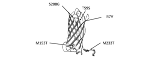
- FIG. 2 is a diagram showing five mutation sites from Dreiklang in PSFP3.
- FIG. 3 is an example of a graph of PSFP2, PSFP3, PSFP4, and Dreiklang, in which the recovery rate is measured until the fluorescence is turned on by thermal equilibrium after the fluorescence is turned off by light irradiation.
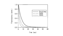
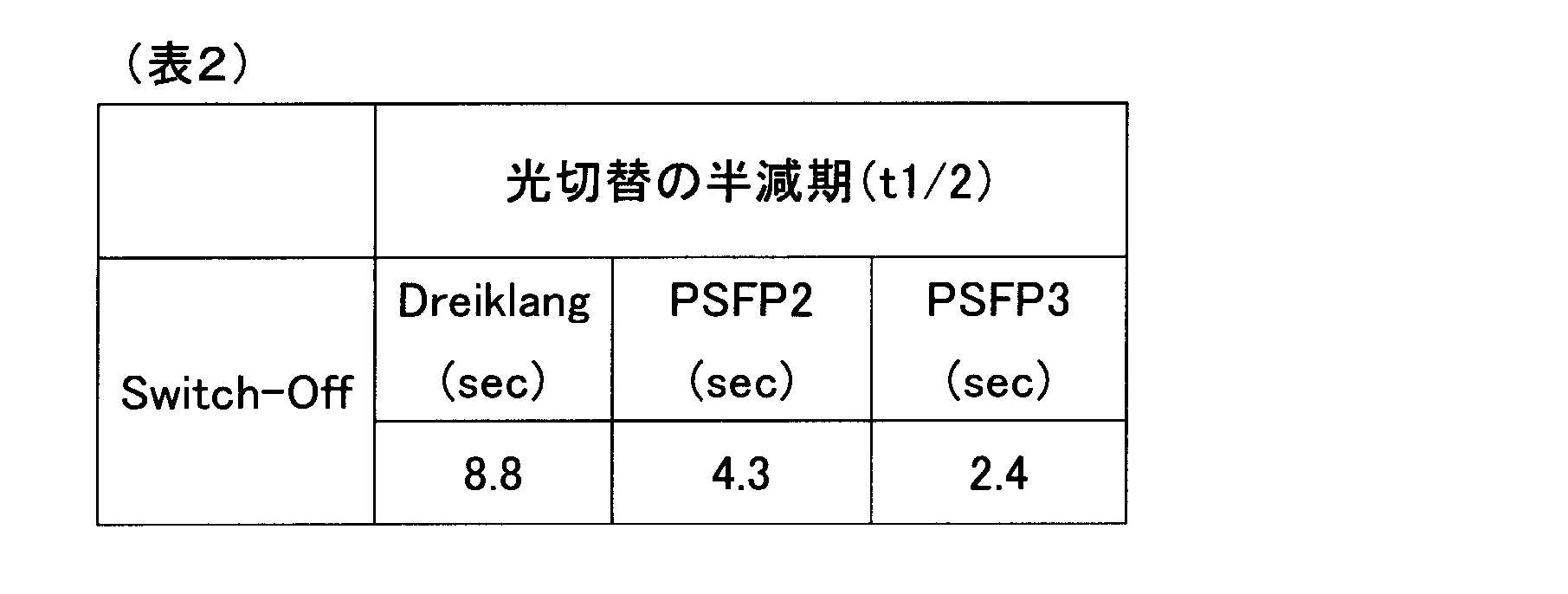
- FIG. 4 is an example of a graph in which the speed of light switching from fluorescence on to off is measured for PSFP2, PSFP3, and Dreiklang.
- FIG. 1 is an alignment of amino acid sequences of Dreiklang (SEQ ID NO: 1), PSFP2 (SEQ ID NO: 2), PSFP3 (SEQ ID NO: 3), and PSFP4 (SEQ ID NO: 4).
- FIG. 2 is a diagram showing five mutation sites from Dreik
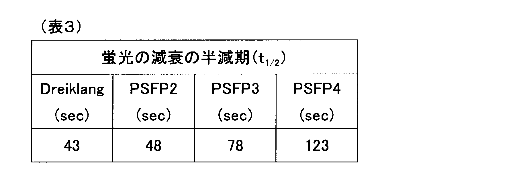
- FIG. 5 is an example of a graph in which changes over time in fluorescence due to bleaching are measured for PSFP2, PSFP3, PSFP4, and Dreiklang.
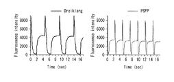
- FIG. 6 is a graph showing an example of continuous light switching for PSFP3 and Dreiklang.
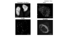
- FIG. 7 is an image showing an example of observing the localization of the fusion protein of PSFP3.
- FIG. 8 is a diagram for explaining the measurement principle of DSSM (Decoupled / stochastic / switching / microscopy).
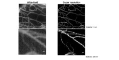
- FIG. 9 shows an example of a vimentin full-field microscope image using PSFP3 (left) and an example of a super-resolution microscope observation (DSSM) image (right).
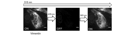
- FIG. 10 is an image showing an example of observation of fluorescence photoswitching of a localized PSFP3 fusion protein.
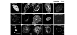
- FIG. 11 is an image showing another example of observing the localization of the fusion protein of PSFP3.
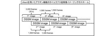
- FIG. 12 is a diagram illustrating an outline of an example of an observation scheme of time-lapse super-resolution imaging.
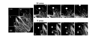
- FIG. 13 is an image showing an example of observation of time-lapse super-resolution imaging.
- the present disclosure relates to Dreiklang, a light-switching fluorescent protein of a type in which the wavelength for changing from non-fluorescence to fluorescence, the wavelength for changing from fluorescence to non-fluorescence, and the wavelength for fluorescence excitation are all different.
- the present disclosure also shows that the fluorescence protein (PSFP2) in which a total of three mutations of I47V and M153T are introduced into Dreiklang has a recovery rate from non-fluorescence to fluorescence due to thermal equilibrium is 3 compared to Dreiklang. This is based on the finding that the speed from fluorescence to non-fluorescence due to light irradiation is doubled compared to Dreiklang, and that the brightness in the fluorescent state is increased compared to the single mutation introduction of S208G.
- PSFP2 fluorescence protein
- the present disclosure further provides a fluorescent protein in which two mutations of T59S and M233T are introduced into the PSFP2 (that is, a fluorescent protein in which five mutations of S208G, I47V, M153T, T59S, and M233T are introduced into Dreiklang) (PSFP3) Is based on the finding that the speed from fluorescence to non-fluorescence by light irradiation is improved 3.7 times compared to Dreiklang.
- the five mutation sites in PSFP3 are located in the site shown in Fig. 2 in Dreiklang. These mutations are all located away from the ⁇ -barrel structure where the Dreiklang chromophore is located, and the mutations in these parts improve the recovery rate from non-fluorescence to fluorescence due to thermal equilibrium and light irradiation. It can be said that bringing about the improvement of the speed
- the scope of the present invention and the effects of the present invention are not construed as being limited to the numerical value “x times” related to the effects described above or described later.
- the present disclosure further provides a fluorescent protein in which mutation of M69Q is introduced into PSFP3 (that is, fluorescent protein in which six mutations of S208G, I47V, M153T, T59S, M233T, and M69Q are introduced into Dreiklang) (PSFP4), This is based on the finding that fluorescence light stability (suppression of fluorescence decay by bleaching) is improved by 2.8 times compared to Dreiklang.
- the fluorescent protein according to the present disclosure is a light-switching fluorescent protein of a type in which the wavelength for switching from non-fluorescence to fluorescence, the wavelength for switching from fluorescence to non-fluorescence, and the wavelength for fluorescence excitation are all different.
- Dreiklang which is a protein consisting of the amino acid sequence of SEQ ID NO: 1, is this type of light-switching fluorescent protein.
- the “light-switching fluorescent protein” means that in one or a plurality of embodiments, on and off of the fluorescent state (that is, fluorescence and non-fluorescence) can be controlled by two light irradiations having different wavelengths. And a protein that can be repeatedly switched between on and off.
- Mutations represented by "X 1 nX 2" in the present disclosure a notation of the general variant, n th amino acid residue X 1 (amino acid residues of single letter code) of the amino acid sequence, amino acid residue Represents a mutation substituted in the group X 2 (single letter amino acid residue).
- the present disclosure relates to a fluorescent protein having an amino acid sequence in which at least a mutation of S208G is introduced into the amino acid sequence of SEQ ID NO: 1.
- the recovery rate from non-fluorescence to fluorescence due to thermal equilibrium is improved compared to before the introduction of the mutation.
- the recovery rate from non-fluorescence to fluorescence due to thermal equilibrium is improved compared to before the introduction of the mutation, and The speed from fluorescence to non-fluorescence can be improved by light irradiation.
- the amino acid sequence of SEQ ID NO: 1 is selected from the group consisting of S208G mutation and I47V, T59S, M69Q, M153T, and M233T.
- a fluorescent protein having an amino acid sequence into which at least one mutation is introduced can be a light-switching fluorescent protein with an improved recovery rate from non-fluorescence to fluorescence due to thermal equilibrium as compared to before the introduction of the mutation.
- mutations of I47V or M153T, or I47V and M153T can contribute to an improvement in fluorescence brightness.
- mutations of T59S or M233T, or T59S and M233T can contribute to an improvement in the speed from fluorescence to non-fluorescence upon irradiation with light.
- the M69Q mutation can contribute to the improvement of fluorescence light stability.
- One or more embodiments of the fluorescent protein according to the present disclosure include a fluorescent protein having an amino acid sequence in which at least three mutations of I47V, M153T, and S208G are introduced into the amino acid sequence of SEQ ID NO: 1.
- the amino acid sequence in which the three mutations are introduced into SEQ ID NO: 1 is SEQ ID NO: 2 (PSFP2).
- PSFP2 SEQ ID NO: 2
- the luminance in the fluorescent state can be improved by introducing the mutations of I47V and M153T in addition to the mutation of S208G, as compared to the case where only S208G is introduced.
- the amino acid sequence of SEQ ID NO: 1 has an amino acid sequence in which at least five mutations of I47V, T59S, M153T, S208G, and M233T are introduced.
- the amino acid sequence in which the five mutations are introduced into SEQ ID NO: 1 is SEQ ID NO: 3 (PSFP3).
- PSFP3 SEQ ID NO: 3
- the mutation of T59S and M233T is introduced in addition to the mutation of I47V, M153T, and S208G. The speed of becoming sex can be further improved.
- Examples include a fluorescent protein having at least six mutated amino acid sequences of I47V, T59S, M69Q, M153T, S208G, and M233T in the amino acid sequence of SEQ ID NO: 1.
- the amino acid sequence in which the six mutations are introduced into SEQ ID NO: 1 is SEQ ID NO: 4 (PSFP4).
- the M69Q mutation is introduced in addition to the five mutations of I47V, T59S, M153T, S208G, and M233T, so that the photostability of fluorescence is improved as compared with that before the introduction of the M69Q mutation. It can be improved.
- the “recovery speed from non-fluorescence to fluorescence by thermal equilibrium”, “speed from fluorescence to non-fluorescence by light irradiation”, and “photostability of fluorescence” are the methods described in the examples. Can be measured and evaluated.
- the fluorescent protein according to the present disclosure preferably has a faster recovery rate from non-fluorescence to fluorescence due to thermal equilibrium than that of Dreiklang (SEQ ID NO: 1). In one or a plurality of embodiments, the fluorescent protein according to the present disclosure preferably has a faster rate of becoming fluorescent to non-fluorescent when irradiated with light than that of Dreiklang (SEQ ID NO: 1). In one or a plurality of embodiments, the fluorescent protein according to the present disclosure preferably has higher fluorescence light stability than that of Dreiklang (SEQ ID NO: 1).
- the wavelength for changing from non-fluorescence to fluorescence is all different. It may have mutations other than the six mutations (I47V, T59S, M69Q, M153T, S208G, and M233T) as long as the function of the type of light-switching fluorescent protein can be maintained. Examples of the mutations other than the six mutations include deletion, substitution, and / or addition of 1 to several amino acids. “One to several” refers to 1 to 4 in one or more embodiments. 1 to 3, 1 to 2, or 1.
- the fluorescent protein according to the present disclosure has a recovery rate from non-fluorescence to fluorescence due to thermal equilibrium even when it contains a mutation other than the six mutations described above (Dreiklang (SEQ ID NO: 1)). It is preferably faster than that. In one or a plurality of embodiments, the fluorescent protein according to the present disclosure has a speed that changes from fluorescence to non-fluorescence upon irradiation with light even when it contains mutations other than the six mutations (SEQ ID NO: 1). It is preferably faster than that. In one or a plurality of embodiments, the fluorescent protein according to the present disclosure has higher fluorescence photostability than that of Dreiklang (SEQ ID NO: 1) even when it contains a mutation other than the six mutations. preferable.
- the fluorescent protein according to the present disclosure may be a protein synthesized by chemical synthesis or may be a recombinant protein produced by a gene recombination technique.
- preparation of a recombinant protein by a genetic recombination technique includes a method of using a host transformed with an expression vector containing a gene encoding the fluorescent protein according to the present disclosure.
- the fluorescent protein according to the present disclosure is a fusion protein in which the fluorescent protein according to the present disclosure described above is fused with another protein or peptide, and the fluorescent protein portion is non-fluorescent. It is a fusion protein that can function as a light-switching fluorescent protein in which the wavelength for making it fluorescent, the wavelength for making it fluorescent to non-fluorescent, and the wavelength for making fluorescence excitation all differ.
- the fluorescent protein part preferably has a faster recovery rate from non-fluorescence to fluorescence due to thermal equilibrium than that of Dreiklang (SEQ ID NO: 1). .
- the fluorescent protein portion preferably has a faster rate of becoming fluorescent to non-fluorescent when irradiated with light than that of Dreiklang (SEQ ID NO: 1). . In one or a plurality of embodiments of the fusion protein according to the present disclosure, the fluorescent protein portion preferably has higher fluorescence light stability than that of Dreiklang (SEQ ID NO: 1).
- the protein to be bound (fused) to the fluorescent protein according to the present disclosure may be a signal sequence, an expression tag, or a protein (optionally a linker sequence) in one or a plurality of non-limiting embodiments. ).
- the signal sequence and the protein may be an intracellular cytoskeleton (microfilament, intermediate filament, microtubule) or organelle (nucleus, endoplasmic reticulum, Golgi) from the viewpoint of imaging. Body, mitochondria, endosome, lysosome, etc.) and signal sequences and proteins that can be localized.
- the fluorescent protein according to the present disclosure has a high light switching speed and can repeatedly read fluorescence, in one or a plurality of embodiments, super-resolution imaging, ultra-high density optical memory, and ultra-sensitive fluorescence imaging Etc. can be used. In one or a plurality of embodiments, the fluorescent protein according to the present disclosure can be used as a fluorescent function indicator, a photoprotein, or a luminescent function indicator using a light-switching fluorescent protein.
- the present disclosure relates to DNA encoding the fluorescent protein according to the present disclosure or the fusion protein according to the present disclosure.
- the fluorescent protein according to the present disclosure can be expressed and produced by introducing a recombinant vector containing the DNA of the present disclosure into a host.
- the DNA of the present disclosure can be produced by PCR using a specific primer, the phosphoramidite method, or the like.
- the present disclosure relates to a vector capable of expressing the fluorescent protein according to the present disclosure or the fusion protein according to the present disclosure.
- the vector of the present disclosure is a vector having the DNA of the present disclosure.
- the vector of the present disclosure can be obtained by inserting the DNA of the present disclosure into an appropriate vector.
- the vector for inserting the DNA of the present disclosure is not particularly limited as long as it can replicate in a host in one or a plurality of embodiments, and includes a plasmid, a phage, and the like.
- Examples of the plasmid include, in one or a plurality of embodiments, a plasmid derived from E. coli, a plasmid derived from Bacillus subtilis, and a plasmid derived from yeast.
- the present disclosure relates to a transformant expressing the fluorescent protein according to the present disclosure or the fusion protein according to the present disclosure.
- the transformant of the present disclosure is a cell that expresses the fluorescent protein according to the present disclosure or the fusion protein according to the present disclosure, or a tissue, an organ, or a living body including the cell.
- this indication is related with the transformant which has the DNA or recombinant vector of this indication in one or some embodiment.
- the transformant of the present disclosure can be produced by introducing the DNA or recombinant vector of the present disclosure into a host.
- Examples of the host include commonly used microorganisms and cultured cells in one or more embodiments.
- Examples of the microorganism include Escherichia coli or yeast in one or more embodiments.
- Examples of the cultured cells include animal cells (for example, CHO cells, HEK-293 cells, or COS cells) or insect cells (for example, BmN4 cells) in one or more embodiments.
- the present disclosure relates to an imaging method using the fluorescent protein according to the present disclosure or the fusion protein according to the present disclosure, the DNA of the present disclosure, or the vector of the present disclosure.
- the imaging method of the present disclosure includes introducing the fluorescent protein according to the present disclosure or the fusion protein according to the present disclosure into a cell, the fluorescent protein according to the present disclosure, or the fusion protein according to the present disclosure. Switching light to turn fluorescence on and / or off, and / or detecting a fluorescent signal of the fluorescent protein according to the present disclosure or the fusion protein according to the present disclosure.
- the imaging method of the present disclosure is, in one or more embodiments, super-resolution imaging, and in one or more embodiments, DSSM (Decoupled stochastic switching microscopy), PALM (photoactivated localization microscopy), STORM (stochastic optical microscopy). reconstruction microscopy), RESOLFT (reversible saturable optical fluorescence transition), or SOFI (superresolution optical fluctuation imaging).
- DSSM Decoupled stochastic switching microscopy
- PALM photoactivated localization microscopy
- STORM stochastic optical microscopy
- reconstruction microscopy reconstruction microscopy
- RESOLFT reversible saturable optical fluorescence transition
- SOFI superresolution optical fluctuation imaging
- the fluorescent protein according to the present disclosure exhibits a photochromic effect, in one or a plurality of embodiments, the use of an optical recording medium such as a CD, a DVD, a holographic recording medium, a smart card, an advertising board, a fluorescent board, a TV, a computer monitor, etc. It can be set as the photochromic material applicable to the use of a display element, or uses, such as a lens, a biosensor, a biochip, a photochromic fiber material.
- the fluorescent protein according to [1] which has an amino acid sequence in which at least three mutations of I47V, M153T, and S208G are introduced into the amino acid sequence of SEQ ID NO: 1.
- the amino acid sequence of the protein according to any one of [1] to [5] has an amino acid sequence in which one to several amino acids are deleted, substituted, and / or added, and is non-fluorescent to fluorescent And the wavelength for fluorescence to non-fluorescence and the wavelength for fluorescence excitation are all different, and the recovery rate from non-fluorescence to fluorescence due to thermal equilibrium is different from the amino acid sequence of SEQ ID NO: 1.
- DNA having a base sequence encoding the protein according to any one of [1] to [7].
- a photochromic material comprising the protein according to any one of [1] to [7].
- the bacterial expression vector of PSFP was prepared by introducing the genes (SEQ ID NOs: 5, 6 and 7 respectively) encoding PSFP (PSFP2, PSFP3 and PSFP4) into the bacterial expression vector pRSET B. Similarly, a bacterial expression vector was also prepared for Dreiklang.
- the mammalian expression vector of PSFP was prepared by introducing a gene encoding PSFP (PSFP2, PSFP3 and PSFP4) into the mammalian expression vector pcDNA3.
- a mammalian expression vector of a fusion fluorescent protein in which the following signal sequence or signal protein was fused with PSFP was also prepared.
- overlapping mitochondrial targeting signal derived from precursor of human cytochrome c oxidase subunit VIII (COX-VIII)
- COX-VIII human cytochrome c oxidase subunit VIII
- Golgi of ⁇ -N-acetylglucosanyl-glycopeptide ⁇ -1,4-galactosyltransferase Body localization signal sequences 3) DNA binding protein H2B, and 4) nucleolar protein fibrillarin were fused to PSFP to target mitochondria, Golgi apparatus, nucleus, and nucleolus, respectively.
- a mammalian expression vector of a fusion protein was also prepared by fusing ⁇ -actin, vimentin, paxillin, zyxin, and clathrin through PSFP and a 17 amino acid long linker sequence (GGSGGSGGSGGSGGQFQ: SEQ ID NO: 8).
- PSFPs PSFP2, PSFP3 and PSFP4 having a polyhistidine tag at the N-terminus were introduced into the bacterial expression vector pRSET B and expressed in E. coli. After culturing in LB medium at 23 ° C. for 65 hours, the cells were crushed with a French press, and the supernatant was subjected to gel filtration using a Ni-NTA agarose affinity column (Qiagen) and a PD-10 column (GE Healthcare). The purified product was further purified with an AKTA 10S (GE Healthcare) Hi-load 20/60 Superdex 200 pg column.
- AKTA 10S GE Healthcare
- PSFP characterization Fluorescence excitation of PSFP (PSFP2, PSFP3, and PSFP4) and on and off of fluorescence light switching were performed with LED light sources of 475 ⁇ 28 nm, 360 ⁇ 20 nm, and 410 ⁇ 10 nm, respectively.
- the absorption spectrum was measured using a V-630 BIO spectrophotometer (manufactured by JASCO).
- the fluorescence excitation and fluorescence emission spectra were measured using an F-7000 fluorescence spectrophotometer (manufactured by Hitachi).
- the molar extinction coefficient was calculated using the absorbance of purified protein of known concentration measured by Bradford assay.
- the fluorescence quantum yield was measured using QuantaurusQY-C11347 (manufactured by Hama Photonics). In this measurement, the protein absorbance was adjusted to less than 0.05. All of the above measurements were performed under physiological pH conditions using proteins in 20 mM HEPES buffer. The quantum yield of photoinduced on / off was calculated by measuring the irradiation-dependent change in absorbance with a V-630 BIO spectrophotometer (manufactured by JASCO). The specific method followed the method described in Gayda, S., Nienhaus, K. & Nienhaus, GU Biophysical journal 103, 2521-31 (2012).
- Measurement of thermal relaxation time from fluorescence off to on is performed by measuring fluorescence spectrum after 10 ml of 20 ⁇ M protein solution (20 mM HEPES buffer, pH 7.4) is turned off with 438 ⁇ 20 nm wavelength light before measurement. did.
- PSFP2, PSFP3, and PSFP4 were measured for the recovery rate at which the fluorescence was turned on by thermal equilibrium from the fluorescence turned off, and compared with Dreiklang, which is its counterpart. Specifically, the recovery of fluorescence was measured by an absorption spectrum at 511 nm. As a result, the recovery time constants due to thermal equilibrium were 218 seconds for PSFP2, 298 seconds for PSFP3, 526 seconds for PSFP4, and 603 seconds for Dreiklang (Table 1, FIG. 3).
- Example of fluorescent light switching of localized fusion fluorescent protein This was observed in live HeLa cells in which the vimentin-PSFP3 fusion protein prepared above was expressed by switching the fluorescence light. Specifically, while irradiating excitation light of 515 nm, light switching was performed by turning off fluorescence with 405 nm light and then turning on fluorescence with 365 nm light. The results are shown in FIG. As shown in the figure, photoswitching was observed in the fusion fluorescent protein in which live HeLa cells were localized.
- AAV vector expressing PSFP An AAV vector was prepared using pAAV-CAG-PSFP3 and pAAV2-hSyn-PSFP3 having PSFP3 genes linked to two types of promoters (CAG and hSyn). When these AAV vectors were infected with primary cultured cells of HEK293T cells and hippocampal neurons, respectively, expression and fluorescence of PSFP3 were confirmed.
- time-lapse super-resolution imaging The LifeAct-PSFP3 fusion protein was expressed in living HeLa cells, and time-lapse super-resolution imaging was performed in which DSSM imaging was repeated as shown in FIG. The result is shown in FIG. As shown in FIG. 13, morphological changes of the actin network could be observed by time-lapse super-resolution imaging.
- SEQ ID NO: 1 amino acid sequence of Dreiklang SEQ ID NO: 2: amino acid sequence of PSFP2 SEQ ID NO: 3: amino acid sequence of PSFP3 SEQ ID NO: 4: amino acid sequence of PSFP4 SEQ ID NO: 5: nucleotide sequence of PSFP2 SEQ ID NO: 6: nucleotide sequence of SEQ ID NO: 3 7: base sequence of PSFP4 SEQ ID NO: 8: amino acid sequence of linker
Landscapes
- Health & Medical Sciences (AREA)
- Life Sciences & Earth Sciences (AREA)
- Genetics & Genomics (AREA)
- Chemical & Material Sciences (AREA)
- Engineering & Computer Science (AREA)
- Organic Chemistry (AREA)
- Zoology (AREA)
- Bioinformatics & Cheminformatics (AREA)
- Biotechnology (AREA)
- Biomedical Technology (AREA)
- Wood Science & Technology (AREA)
- Biochemistry (AREA)
- General Health & Medical Sciences (AREA)
- General Engineering & Computer Science (AREA)
- Biophysics (AREA)
- Molecular Biology (AREA)
- Microbiology (AREA)
- Physics & Mathematics (AREA)
- Plant Pathology (AREA)
- Proteomics, Peptides & Aminoacids (AREA)
- Medicinal Chemistry (AREA)
- Gastroenterology & Hepatology (AREA)
- Toxicology (AREA)
- Mycology (AREA)
- Cell Biology (AREA)
- Nuclear Medicine, Radiotherapy & Molecular Imaging (AREA)
- Analytical Chemistry (AREA)
- General Physics & Mathematics (AREA)
- Immunology (AREA)
- Pathology (AREA)
- Tropical Medicine & Parasitology (AREA)
- Crystallography & Structural Chemistry (AREA)
- Peptides Or Proteins (AREA)
- Investigating, Analyzing Materials By Fluorescence Or Luminescence (AREA)
- Micro-Organisms Or Cultivation Processes Thereof (AREA)
Abstract
蛍光性のon/off及び蛍光励起波長の全てが独立したタイプの光切替型蛍光蛋白質であって、熱平衡による無蛍光性から蛍光性への回復速度が向上した蛍光蛋白質の提供。 一実施形態において、配列番号1に少なくともS208Gの変異が導入されたアミノ酸配列を有する蛍光蛋白質。その他の実施形態において、配列番号1に少なくともI47V、M153T、及びS208Gの3つの変異が導入されたアミノ酸配列を有する蛍光蛋白質。さらにその他の実施形態において、配列番号1に少なくともI47V、T59S、M153T、S208G、及びM233Tの5つの変異導入されたアミノ酸配列を有する蛍光蛋白質。さらにその他の実施形態において、配列番号1に少なくともI47V、T59S、M69Q、M153T、S208G、及びM233Tの6つの変異導入されたアミノ酸配列を有する蛍光蛋白質。
Description
本開示は、蛍光蛋白質、該蛋白質のDNA、ベクター、及び形質転換体、並びにそれらを用いたイメージング方法に関する。
蛍光蛋白質によってライブセルイメージングが簡便に実施可能となり、細胞内の様々な構造や機能が解明されてきている。近年では、可逆的に蛍光性を光切替可能な蛍光蛋白質(Reversibly Photo-Switchable Fluorescent Protein, RSFP)を利用した超解像法が開発され、光学顕微鏡の回折限界を超えたイメージングが可能となっている。RSFPとしては、例えば、1)蛍光励起しない特定の波長の光照射によって無蛍光性から蛍光性になり、蛍光励起のための光照射によって蛍光性から無蛍光性になるネガティブ光切替型(例えば、Dronpa(特許文献1)、rsEGFP)、2)蛍光励起のための光照射によって無蛍光性から蛍光性になり、蛍光励起しない特定の波長の光照射によって蛍光性から無蛍光性になるポジティブ光切替型(例えば、Padron(非特許文献1))、及び、3)蛍光性のon/off及び蛍光励起波長の全てが独立したタイプ(例えば、Dreiklang(非特許文献2))がある。
Tanja Brakemann et al, J. Biol. Chem. 2010, 285:14603-14609
Tanja Brakemann et al, Nat Biotechnol. 29 (2011):942-947
前記3)のタイプの光切替型蛍光蛋白質では、Dreiklangが報告されて以降、改変型は報告されていない。DreiklangはPALM(Photo Activated Localization Microscopy)と呼ばれる超解像顕微鏡法や、PALM以外の他の超解像顕微鏡法にも適用可能であるといった長所があるが、光スイッチング速度が遅く、超解像時の時間分解能の遅さが問題となっていた。
本開示は、一又は複数の実施形態において、蛍光性のon/off及び蛍光励起波長の全てが独立したタイプの光切替型蛍光蛋白質の改変体を提供する。
本開示は、一又は複数の実施形態において、配列番号1のアミノ酸配列に少なくともS208Gの変異が導入されたアミノ酸配列を有する蛍光蛋白質に関する。
本開示は、その他の一又は複数の実施形態において、配列番号1のアミノ酸配列に少なくともI47V、M153T、及びS208Gの3つの変異が導入されたアミノ酸配列を有する蛍光蛋白質に関する。
本開示は、その他の一又は複数の実施形態において、配列番号1のアミノ酸配列に少なくともI47V、T59S、M153T、S208G、及びM233Tの5つの変異導入されたアミノ酸配列を有する蛍光蛋白質に関する。
本開示は、その他の一又は複数の実施形態において、配列番号1のアミノ酸配列に少なくともI47V、T59S、M69Q、M153T、S208G、及びM233Tの6つの変異導入されたアミノ酸配列を有する蛍光蛋白質に関する。
本開示は、その他の一又は複数の実施形態において、配列番号1のアミノ酸配列に少なくともI47V、T59S、M153T、S208G、及びM233Tの5つの変異導入されたアミノ酸配列を有する蛍光蛋白質に関する。
本開示は、その他の一又は複数の実施形態において、配列番号1のアミノ酸配列に少なくともI47V、T59S、M69Q、M153T、S208G、及びM233Tの6つの変異導入されたアミノ酸配列を有する蛍光蛋白質に関する。
本開示は、その他の一又は複数の実施形態において、本開示に係る蛍光蛋白質を含む融合蛋白質に関する。
本開示は、その他の一又は複数の実施形態において、本開示に係る蛍光蛋白質又は融合蛋白質を用いるイメージング方法に関する。
本開示によれば、一又は複数の実施形態において、蛍光性のon/off及び蛍光励起波長の全てが独立したタイプの光切替型蛍光蛋白質であって、熱平衡による無蛍光性から蛍光性への回復速度が向上した蛍光蛋白質を提供できる。その他の一又は複数の実施形態において、本開示によれば、蛍光性のon/off及び蛍光励起波長の全てが独立したタイプの光切替型蛍光蛋白質であって、熱平衡による無蛍光性から蛍光性への回復速度がDreiklang(配列番号1)よりも向上した蛍光蛋白質を提供できる。
本開示に係る蛍光蛋白質によれば、一又は複数の実施形態において、超解像イメージングにおける空間解像能及び時間解像能を向上できる。
本開示は、無蛍光性から蛍光性とするための波長と蛍光性から無蛍光性とするための波長と蛍光励起のための波長とが全て異なるタイプの光切替型蛍光蛋白質であるDreiklang(配列番号1)にS208Gの変異を導入する事で、熱平衡による無蛍光性から蛍光性への回復速度(蛍光性がOffから自発的にOnになる速度)がDreiklangに比べて3倍となり、光照射により蛍光性から無蛍光性となる速度(OnからOffへの光切替速度)がDreiklangに比べて2倍になる、という知見に基づく。
本開示は、また、S208Gの変異に加えてI47VとM153Tの合計3つの変異をDreiklangに導入した蛍光蛋白質(PSFP2)は、熱平衡による無蛍光性から蛍光性への回復速度がDreiklangに比べて3倍となり、光照射により蛍光性から無蛍光性となる速度がDreiklangに比べて2倍になり、さらに、S208Gの単独変異導入時に比べて蛍光状態における輝度が上昇するという知見に基づく。
本開示は、さらにまた、前記PSFP2にT59SとM233Tの2つ変異を導入した蛍光蛋白質(すなわち、DreiklangにS208G、I47V、M153T、T59S、及びM233Tの5つ変異を導入した蛍光蛋白質)(PSFP3)は、光照射により蛍光性から無蛍光性となる速度がDreiklangに比べて3.7倍に向上する、という知見に基づく。
PSFP3における5つの変異の箇所は、Dreiklangにおいて図2に示すような部位に位置する。これらの変異の箇所はいずれもDreiklangの発色団があるβバレル構造から離れた箇所であり、これらの箇所の変異が、熱平衡による無蛍光性から蛍光性への回復速度の向上、及び、光照射により蛍光性から無蛍光性となる速度の向上をもたらすことは技術常識から予測できない効果といえる。なお、本発明の範囲及び本発明の効果は、上述した又は後述する効果に関する「x倍」という数値に限定して解釈されない。
本開示は、さらにまた、前記PSFP3にM69Qの変異を導入した蛍光蛋白質(すなわち、DreiklangにS208G、I47V、M153T、T59S、M233T、及びM69Qの6つ変異を導入した蛍光蛋白質)(PSFP4)は、蛍光の光安定性(ブリーチングによる蛍光の減衰抑制)がDreiklangに比べて2.8倍に向上する、という知見に基づく。
[蛍光蛋白質]
本開示に係る蛍光蛋白質は、無蛍光性から蛍光性とするための波長と蛍光性から無蛍光性とするための波長と蛍光励起のための波長とが全て異なるタイプの光切替型蛍光蛋白質である。配列番号1のアミノ酸配列からなるタンパク質であるDreiklangは、このタイプの光切替型蛍光蛋白質である。
本開示に係る蛍光蛋白質は、無蛍光性から蛍光性とするための波長と蛍光性から無蛍光性とするための波長と蛍光励起のための波長とが全て異なるタイプの光切替型蛍光蛋白質である。配列番号1のアミノ酸配列からなるタンパク質であるDreiklangは、このタイプの光切替型蛍光蛋白質である。
本開示において「光切替型蛍光蛋白質」とは、一又は複数の実施形態において、蛍光性状態のonとoff(すなわち、蛍光性と無蛍光性)を波長の異なる2つの光照射によって制御ができ、かつ、onとoffの切り替えが繰り返し行うことができる蛋白質をいう。
本開示において“X1nX2”で表される変異は、一般的な変異の表記方法であって、アミノ酸配列のn番目のアミノ酸残基X1(一文字表記のアミノ酸残基)が、アミノ酸残基X2(一文字表記のアミノ酸残基)に置換される変異を表す。
本開示は、一態様として、配列番号1のアミノ酸配列に、少なくともS208Gの変異が導入されたアミノ酸配列を有する蛍光蛋白質に関する。
上記S208Gの変異が導入されることで、該変異の導入前と比較して、熱平衡による無蛍光性から蛍光性への回復速度が向上する。その他の一又は複数の実施形態においては、上記S208Gの変異が導入されることで、該変異の導入前と比較して、熱平衡による無蛍光性から蛍光性への回復速度が向上し、かつ、光照射により蛍光性から無蛍光性となる速度が向上されうる。
上記S208Gの変異が導入されることで、該変異の導入前と比較して、熱平衡による無蛍光性から蛍光性への回復速度が向上する。その他の一又は複数の実施形態においては、上記S208Gの変異が導入されることで、該変異の導入前と比較して、熱平衡による無蛍光性から蛍光性への回復速度が向上し、かつ、光照射により蛍光性から無蛍光性となる速度が向上されうる。
本開示に係る蛍光蛋白質の一又は複数の実施形態として、配列番号1のアミノ酸配列に、S208Gの変異と、I47V、T59S、M69Q、M153T、及びM233Tからなる群から選択される1又は2~5つの変異とが少なくとも導入されたアミノ酸配列を有する蛍光蛋白質が挙げられる。
本実施形態の蛍光蛋白質は、該変異の導入前と比較して、熱平衡による無蛍光性から蛍光性への回復速度が向上した光切替型蛍光蛋白質となりうる。
前記変異のうち、I47V若しくはM153T、又は、I47V及びM153Tの変異は、蛍光の輝度の向上に貢献しうる。
前記変異のうち、T59S若しくはM233T、又は、T59S及びM233Tの変異は、光照射により蛍光性から無蛍光性となる速度の向上に貢献し得る。
前記変異のうち、M69Qの変異は、蛍光の光安定性の向上に寄与しうる。
本実施形態の蛍光蛋白質は、該変異の導入前と比較して、熱平衡による無蛍光性から蛍光性への回復速度が向上した光切替型蛍光蛋白質となりうる。
前記変異のうち、I47V若しくはM153T、又は、I47V及びM153Tの変異は、蛍光の輝度の向上に貢献しうる。
前記変異のうち、T59S若しくはM233T、又は、T59S及びM233Tの変異は、光照射により蛍光性から無蛍光性となる速度の向上に貢献し得る。
前記変異のうち、M69Qの変異は、蛍光の光安定性の向上に寄与しうる。
本開示に係る蛍光蛋白質の一又は複数の実施形態として、配列番号1のアミノ酸配列に、少なくともI47V、M153T、及びS208Gの3つの変異が導入されたアミノ酸配列を有する蛍光蛋白質が挙げられる。配列番号1に前記3つの変異が導入されたアミノ酸配列が配列番号2(PSFP2)である。
本実施形態の蛍光蛋白質では、S208Gの変異に加えてI47V及びM153Tの変異が導入されることで、S208Gのみが導入された場合に比べて、蛍光状態における輝度が向上されうる。
本実施形態の蛍光蛋白質では、S208Gの変異に加えてI47V及びM153Tの変異が導入されることで、S208Gのみが導入された場合に比べて、蛍光状態における輝度が向上されうる。
本開示に係る蛍光蛋白質の一又は複数の実施形態として、配列番号1のアミノ酸配列に少なくともI47V、T59S、M153T、S208G、及びM233Tの5つの変異導入されたアミノ酸配列を有するが挙げられる。配列番号1に前記5つの変異が導入されたアミノ酸配列が配列番号3(PSFP3)である。
本実施形態の蛍光蛋白質では、I47V、M153T、及びS208Gの変異に加えてT59S及びM233Tの変異が導入されることで、T59S及びM233Tの変異導入前に比べて、光照射により蛍光性から無蛍光性となる速度がいっそう向上されうる。
本実施形態の蛍光蛋白質では、I47V、M153T、及びS208Gの変異に加えてT59S及びM233Tの変異が導入されることで、T59S及びM233Tの変異導入前に比べて、光照射により蛍光性から無蛍光性となる速度がいっそう向上されうる。
配列番号1のアミノ酸配列に少なくともI47V、T59S、M69Q、M153T、S208G、及びM233Tの6つの変異導入されたアミノ酸配列を有する蛍光蛋白質が挙げられる。配列番号1に前記6つの変異が導入されたアミノ酸配列が配列番号4(PSFP4)である。
本実施形態の蛍光蛋白質では、I47V、T59S、M153T、S208G、及びM233Tの5つの変異に加えてM69Qの変異が導入されることで、M69Qの変異導入前に比べて、蛍光の光安定性が向上されうる。
本実施形態の蛍光蛋白質では、I47V、T59S、M153T、S208G、及びM233Tの5つの変異に加えてM69Qの変異が導入されることで、M69Qの変異導入前に比べて、蛍光の光安定性が向上されうる。
本開示において、「熱平衡による無蛍光性から蛍光性への回復速度」、「光照射により蛍光性から無蛍光性となる速度」、及び「蛍光の光安定性」は、実施例に記載の方法で測定及び評価されうる。
本開示に係る蛍光蛋白質は、一又は複数の実施形態において、熱平衡による無蛍光性から蛍光性への回復速度がDreiklang(配列番号1)のそれよりも速いことが好ましい。
本開示に係る蛍光蛋白質は、一又は複数の実施形態において、光照射により蛍光性から無蛍光性となる速度がDreiklang(配列番号1)のそれよりも速いことが好ましい。
本開示に係る蛍光蛋白質は、一又は複数の実施形態において、蛍光の光安定性がDreiklang(配列番号1)のそれよりも高いことが好ましい。
本開示に係る蛍光蛋白質は、一又は複数の実施形態において、光照射により蛍光性から無蛍光性となる速度がDreiklang(配列番号1)のそれよりも速いことが好ましい。
本開示に係る蛍光蛋白質は、一又は複数の実施形態において、蛍光の光安定性がDreiklang(配列番号1)のそれよりも高いことが好ましい。
本開示に係る蛍光蛋白質は、一又は複数の実施形態において、無蛍光性から蛍光性とするための波長と蛍光性から無蛍光性とするための波長と蛍光励起のための波長とが全て異なるタイプの光切替型蛍光蛋白質の機能を維持できる範囲で、前記6つの変異(I47V、T59S、M69Q、M153T、S208G、及びM233T)以外の変異を有していてもよい。前記6つの変異以外の変異としては、1から数個のアミノ酸の欠失、置換、及び/又は付加が挙げられ、「1から数個」とは、一又は複数の実施形態において、1~4、1~3、1~2、又は1個を含む。
本開示に係る蛍光蛋白質は、一又は複数の実施形態において、前記6つの変異以外の変異を含む場合であっても、熱平衡による無蛍光性から蛍光性への回復速度がDreiklang(配列番号1)のそれよりも速いことが好ましい。
本開示に係る蛍光蛋白質は、一又は複数の実施形態において、前記6つの変異以外の変異を含む場合であっても、光照射により蛍光性から無蛍光性となる速度がDreiklang(配列番号1)のそれよりも速いことが好ましい。
本開示に係る蛍光蛋白質は、一又は複数の実施形態において、前記6つの変異以外の変異を含む場合であっても、蛍光の光安定性がDreiklang(配列番号1)のそれよりも高いことが好ましい。
本開示に係る蛍光蛋白質は、一又は複数の実施形態において、前記6つの変異以外の変異を含む場合であっても、熱平衡による無蛍光性から蛍光性への回復速度がDreiklang(配列番号1)のそれよりも速いことが好ましい。
本開示に係る蛍光蛋白質は、一又は複数の実施形態において、前記6つの変異以外の変異を含む場合であっても、光照射により蛍光性から無蛍光性となる速度がDreiklang(配列番号1)のそれよりも速いことが好ましい。
本開示に係る蛍光蛋白質は、一又は複数の実施形態において、前記6つの変異以外の変異を含む場合であっても、蛍光の光安定性がDreiklang(配列番号1)のそれよりも高いことが好ましい。
本開示に係る蛍光蛋白質は、一又は複数の実施形態において、化学合成により合成した蛋白質であってもよいし、遺伝子組み換え技術により作製した組み換え蛋白質であってもよい。遺伝子組み換え技術による組み換え蛋白質の作製としては、一又は複数の実施形態において、本開示に係る蛍光蛋白質をコードする遺伝子を含有する発現ベクターで形質転換した宿主を用いて作製する方法が挙げられる。
本開示に係る蛍光蛋白質は、一又は複数の実施形態において、上述した本開示に係る蛍光蛋白質が他の蛋白質又はペプチドと融合した融合蛋白質であって、該蛍光蛋白質の部分が、無蛍光性から蛍光性とするための波長と蛍光性から無蛍光性とするための波長と蛍光励起のための波長とが全て異なる光切替型蛍光蛋白質として機能可能な融合蛋白質である。
本開示に係る融合蛋白質は、一又は複数の実施形態において、該蛍光蛋白質の部分は、熱平衡による無蛍光性から蛍光性への回復速度がDreiklang(配列番号1)のそれよりも速いことが好ましい。
本開示に係る融合蛋白質は、一又は複数の実施形態において、該蛍光蛋白質の部分は、光照射により蛍光性から無蛍光性となる速度がDreiklang(配列番号1)のそれよりも速いことが好ましい。
本開示に係る融合蛋白質は、一又は複数の実施形態において、該蛍光蛋白質の部分は、蛍光の光安定性がDreiklang(配列番号1)のそれよりも高いことが好ましい。
本開示に係る融合蛋白質は、一又は複数の実施形態において、該蛍光蛋白質の部分は、熱平衡による無蛍光性から蛍光性への回復速度がDreiklang(配列番号1)のそれよりも速いことが好ましい。
本開示に係る融合蛋白質は、一又は複数の実施形態において、該蛍光蛋白質の部分は、光照射により蛍光性から無蛍光性となる速度がDreiklang(配列番号1)のそれよりも速いことが好ましい。
本開示に係る融合蛋白質は、一又は複数の実施形態において、該蛍光蛋白質の部分は、蛍光の光安定性がDreiklang(配列番号1)のそれよりも高いことが好ましい。
本開示に係る融合蛋白質において、本開示に係る蛍光蛋白質に結合(融合)される蛋白質は、限定されない一又は複数の実施形態において、シグナル配列、発現タグ、又は、蛋白質(必要に応じてリンカー配列)が挙げられる。前記シグナル配列及び前記蛋白質としては、一又は複数の実施形態において、イメージングをする観点から、細胞内の細胞骨格(マイクロフィラメント、中間径フィラメント、微小管)や細胞小器官(核、小胞体、ゴルジ体、ミトコンドリア、エンドソーム、リソソーム等)に局在化できるシグナル配列や蛋白質が挙げられる。
本開示に係る蛍光蛋白質は、光切替速度が速く、また繰り返しの蛍光の読み出しが可能であるため、一又は複数の実施形態において、超解像イメージング、超高密度光メモリー及び超高感度蛍光イメージング等に用いることができる。また、本開示に係る蛍光蛋白質は、一又は複数の実施形態において、光切替型蛍光蛋白質を利用した蛍光性機能指示薬、発光蛋白質、発光性機能指示薬として使用することができる。
[DNA]
本開示は、一態様において、本開示に係る蛍光蛋白質又は本開示に係る融合蛋白質をコードするDNAに関する。本開示のDNAによれば、一又は複数の実施形態において、本開示のDNAを含む組み換えベクターを宿主に導入することによって、本開示に係る蛍光蛋白質を発現し、産生することができる。本開示のDNAは、一又は複数の実施形態において、特異的プライマーを用いたPCRや、ホスホアミダイト法等によって製造することができる。
本開示は、一態様において、本開示に係る蛍光蛋白質又は本開示に係る融合蛋白質をコードするDNAに関する。本開示のDNAによれば、一又は複数の実施形態において、本開示のDNAを含む組み換えベクターを宿主に導入することによって、本開示に係る蛍光蛋白質を発現し、産生することができる。本開示のDNAは、一又は複数の実施形態において、特異的プライマーを用いたPCRや、ホスホアミダイト法等によって製造することができる。
[ベクター]
本開示は、一態様において、本開示に係る蛍光蛋白質又は本開示に係る融合蛋白質を発現可能なベクターに関する。本開示のベクターは、一又は複数の実施形態において、本開示のDNAを有するベクターである。本開示のベクターは、一又は複数の実施形態において、本開示のDNAを適当なベクターに挿入することによって得ることができる。本開示のDNAを挿入するベクターとしては、一又は複数の実施形態において、宿主中で複製可能なものであれば特に制限されず、プラスミド、及びファージ等が挙げられる。
本開示は、一態様において、本開示に係る蛍光蛋白質又は本開示に係る融合蛋白質を発現可能なベクターに関する。本開示のベクターは、一又は複数の実施形態において、本開示のDNAを有するベクターである。本開示のベクターは、一又は複数の実施形態において、本開示のDNAを適当なベクターに挿入することによって得ることができる。本開示のDNAを挿入するベクターとしては、一又は複数の実施形態において、宿主中で複製可能なものであれば特に制限されず、プラスミド、及びファージ等が挙げられる。
プラスミドとしては、一又は複数の実施形態において、大腸菌由来のプラスミド、枯菌草由来のプラスミド、及び酵母菌由来のプラスミド等が挙げられる。
[形質転換体]
本開示は、一態様において、本開示に係る蛍光蛋白質又は本開示に係る融合蛋白質を発現する形質転換体に関する。本開示の形質転換体は、一又は複数の実施形態において、本開示に係る蛍光蛋白質又は本開示に係る融合蛋白質を発現する細胞、又は、該細胞を含む組織、器官、生体である。また、本開示は、一又は複数の実施形態において、本開示のDNA又は組み換えベクターを有する形質転換体に関する。本開示の形質転換体は、一又は複数の実施形態において、本開示のDNA又は組み換えベクターを宿主に導入することによって作成することができる。
本開示は、一態様において、本開示に係る蛍光蛋白質又は本開示に係る融合蛋白質を発現する形質転換体に関する。本開示の形質転換体は、一又は複数の実施形態において、本開示に係る蛍光蛋白質又は本開示に係る融合蛋白質を発現する細胞、又は、該細胞を含む組織、器官、生体である。また、本開示は、一又は複数の実施形態において、本開示のDNA又は組み換えベクターを有する形質転換体に関する。本開示の形質転換体は、一又は複数の実施形態において、本開示のDNA又は組み換えベクターを宿主に導入することによって作成することができる。
宿主としては、一又は複数の実施形態において、通常使用される公知の微生物、及び培養細胞等が挙げられる。微生物としては、一又は複数の実施形態において、大腸菌又は酵母等が挙げられる。培養細胞としては、一又は複数の実施形態において、動物細胞(例えば、CHO細胞、HEK-293細胞、又はCOS細胞)又は昆虫細胞(例えば、BmN4細胞)等が挙げられる。
[イメージング方法]
本開示は、一態様において、本開示に係る蛍光蛋白質若しくは本開示に係る融合蛋白質、本開示のDNA、又は、本開示のベクターを用いるイメージング方法に関する。本開示のイメージング方法は、一又は複数の実施形態において、本開示に係る蛍光蛋白質又は本開示に係る融合蛋白質を細胞等に導入すること、本開示に係る蛍光蛋白質又は本開示に係る融合蛋白質の光切替を行い蛍光性をon及び/又はoffすること、及び/又は、本開示に係る蛍光蛋白質又は本開示に係る融合蛋白質の蛍光シグナルを検出することを含む。本開示のイメージング方法は、一又は複数の実施形態において、超解像イメージングであって、一又は複数の実施形態において、DSSM(Decoupled stochastic switching microscopy)、PALM(photoactivated localization microscopy)、STORM(stochastic optical reconstruction microscopy)、RESOLFT(reversible saturable optical fluorescence transition)、又は、SOFI(superresolution optical fluctuation imaging)が挙げられる。本開示に係る蛍光蛋白質は、自発的に蛍光性がOnになる速度(熱平衡による無蛍光性から蛍光性への回復速度)が向上しているため、一又は複数の実施形態において、簡便な超解像イメージング法であるDSSMに好ましく利用できる。
本開示は、一態様において、本開示に係る蛍光蛋白質若しくは本開示に係る融合蛋白質、本開示のDNA、又は、本開示のベクターを用いるイメージング方法に関する。本開示のイメージング方法は、一又は複数の実施形態において、本開示に係る蛍光蛋白質又は本開示に係る融合蛋白質を細胞等に導入すること、本開示に係る蛍光蛋白質又は本開示に係る融合蛋白質の光切替を行い蛍光性をon及び/又はoffすること、及び/又は、本開示に係る蛍光蛋白質又は本開示に係る融合蛋白質の蛍光シグナルを検出することを含む。本開示のイメージング方法は、一又は複数の実施形態において、超解像イメージングであって、一又は複数の実施形態において、DSSM(Decoupled stochastic switching microscopy)、PALM(photoactivated localization microscopy)、STORM(stochastic optical reconstruction microscopy)、RESOLFT(reversible saturable optical fluorescence transition)、又は、SOFI(superresolution optical fluctuation imaging)が挙げられる。本開示に係る蛍光蛋白質は、自発的に蛍光性がOnになる速度(熱平衡による無蛍光性から蛍光性への回復速度)が向上しているため、一又は複数の実施形態において、簡便な超解像イメージング法であるDSSMに好ましく利用できる。
[フォトクロミック材料]
本開示に係る蛍光蛋白質は、フォトクロミズム効果を示すため、一又は複数の実施形態において、CD、DVD、ホログラフィー記録媒体、スマートカードなどの光記録媒体の用途、広告板、蛍光板、TV、コンピュータモニターなどの表示素子の用途、或いは、レンズ、バイオセンサー、バイオチップ、フォトクロミック繊維素材などの用途に適用できるフォトクロミック材料とすることができる。
本開示に係る蛍光蛋白質は、フォトクロミズム効果を示すため、一又は複数の実施形態において、CD、DVD、ホログラフィー記録媒体、スマートカードなどの光記録媒体の用途、広告板、蛍光板、TV、コンピュータモニターなどの表示素子の用途、或いは、レンズ、バイオセンサー、バイオチップ、フォトクロミック繊維素材などの用途に適用できるフォトクロミック材料とすることができる。
本開示はさらに以下の限定されない一又は複数の実施形態に関する。
〔1〕 配列番号1のアミノ酸配列に少なくともS208Gの変異が導入されたアミノ酸配列を有する蛍光蛋白質。
〔2〕 配列番号1のアミノ酸配列に少なくともI47V、M153T、及びS208Gの3つの変異が導入されたアミノ酸配列を有する、〔1〕に記載の蛍光蛋白質。
〔3〕 配列番号1のアミノ酸配列に少なくともI47V、T59S、M153T、S208G、及びM233Tの5つの変異導入されたアミノ酸配列を有する、〔1〕又は〔2〕に記載の蛍光蛋白質。
〔4〕 配列番号1のアミノ酸配列に少なくともI47V、T59S、M69Q、M153T、S208G、及びM233Tの6つの変異導入されたアミノ酸配列を有する、〔1〕から〔3〕のいずれかに記載の蛍光蛋白質。
〔5〕 配列番号2から4のいずれかのアミノ酸配列を有する、〔1〕から〔4〕のいずれかに記載の蛍光蛋白質。
〔6〕 〔1〕から〔5〕のいずれかに記載の蛋白質のアミノ酸配列において1から数個のアミノ酸が欠失、置換、及び/又は付加されたアミノ酸配列を有し、無蛍光性から蛍光性とするための波長と蛍光性から無蛍光性とするための波長と蛍光励起のための波長とが全て異なり、熱平衡による無蛍光性から蛍光性への回復速度が配列番号1のアミノ酸配列からなる蛋白質のそれよりも速い、蛍光蛋白質。
〔7〕 〔1〕から〔6〕のいずれかに記載の蛍光蛋白質が融合された融合蛋白質であって、該蛍光蛋白質の部分は、無蛍光性から蛍光性とするための波長と蛍光性から無蛍光性とするための波長と蛍光励起のための波長とが全て異なり、熱平衡による無蛍光性から蛍光性への回復速度が配列番号1のアミノ酸配列からなる蛋白質のそれよりも速い、融合蛋白質。
〔8〕 〔1〕から〔7〕のいずれかに記載の蛋白質をコードする塩基配列を有するDNA。
〔9〕 〔1〕から〔7〕のいずれかに記載の蛋白質を発現可能な、或いは、〔8〕に記載のDNAを有するベクター。
〔10〕 〔1〕から〔7〕のいずれかに記載の蛋白質を発現する形質転換体。
〔11〕 〔1〕から〔7〕のいずれかに記載の蛋白質、〔8〕に記載のDNA、〔9〕に記載のベクター、又は、〔10〕に記載の形質転換体を用いるイメージング方法。
〔12〕 前記イメージング方法が、超解像蛍光顕微鏡観察法である、〔11〕に記載のイメージング方法。
〔13〕 〔1〕から〔7〕のいずれかに記載の蛋白質を含むフォトクロミック材料。
〔1〕 配列番号1のアミノ酸配列に少なくともS208Gの変異が導入されたアミノ酸配列を有する蛍光蛋白質。
〔2〕 配列番号1のアミノ酸配列に少なくともI47V、M153T、及びS208Gの3つの変異が導入されたアミノ酸配列を有する、〔1〕に記載の蛍光蛋白質。
〔3〕 配列番号1のアミノ酸配列に少なくともI47V、T59S、M153T、S208G、及びM233Tの5つの変異導入されたアミノ酸配列を有する、〔1〕又は〔2〕に記載の蛍光蛋白質。
〔4〕 配列番号1のアミノ酸配列に少なくともI47V、T59S、M69Q、M153T、S208G、及びM233Tの6つの変異導入されたアミノ酸配列を有する、〔1〕から〔3〕のいずれかに記載の蛍光蛋白質。
〔5〕 配列番号2から4のいずれかのアミノ酸配列を有する、〔1〕から〔4〕のいずれかに記載の蛍光蛋白質。
〔6〕 〔1〕から〔5〕のいずれかに記載の蛋白質のアミノ酸配列において1から数個のアミノ酸が欠失、置換、及び/又は付加されたアミノ酸配列を有し、無蛍光性から蛍光性とするための波長と蛍光性から無蛍光性とするための波長と蛍光励起のための波長とが全て異なり、熱平衡による無蛍光性から蛍光性への回復速度が配列番号1のアミノ酸配列からなる蛋白質のそれよりも速い、蛍光蛋白質。
〔7〕 〔1〕から〔6〕のいずれかに記載の蛍光蛋白質が融合された融合蛋白質であって、該蛍光蛋白質の部分は、無蛍光性から蛍光性とするための波長と蛍光性から無蛍光性とするための波長と蛍光励起のための波長とが全て異なり、熱平衡による無蛍光性から蛍光性への回復速度が配列番号1のアミノ酸配列からなる蛋白質のそれよりも速い、融合蛋白質。
〔8〕 〔1〕から〔7〕のいずれかに記載の蛋白質をコードする塩基配列を有するDNA。
〔9〕 〔1〕から〔7〕のいずれかに記載の蛋白質を発現可能な、或いは、〔8〕に記載のDNAを有するベクター。
〔10〕 〔1〕から〔7〕のいずれかに記載の蛋白質を発現する形質転換体。
〔11〕 〔1〕から〔7〕のいずれかに記載の蛋白質、〔8〕に記載のDNA、〔9〕に記載のベクター、又は、〔10〕に記載の形質転換体を用いるイメージング方法。
〔12〕 前記イメージング方法が、超解像蛍光顕微鏡観察法である、〔11〕に記載のイメージング方法。
〔13〕 〔1〕から〔7〕のいずれかに記載の蛋白質を含むフォトクロミック材料。
以下、実施例により本開示をさらに詳細に説明するが、これらは例示的なものであって、本開示はこれら実施例に制限されるものではない。
[光切替型蛍光蛋白質(PSFP)の作製]
無蛍光性から蛍光性とするための波長と蛍光性から無蛍光性とするための波長と蛍光励起のための波長とが全て異なる光切替型蛍光蛋白質であるDreiklang(配列番号4のアミノ酸配列からなる蛋白質)に下記変異1、3及び4の3つの変異を導入して光切替型蛍光蛋白質PSFP2を作成した(配列番号2のアミノ酸配列からなる蛋白質)。
PSFP2に、さらに、下記変異2及び5の2つの変異を導入して光切替型蛍光蛋白質PSFP3を作成した(配列番号3のアミノ酸配列からなる蛋白質)。変異1~5の5つの変異箇所を図1及び図2に示す。
PSFP3に、さらに、M69Qの変異を導入して光切替型蛍光蛋白質PSFP4を作成した(配列番号4のアミノ酸配列からなる蛋白質)。
変異1:I47V
変異2:T59S
変異3:M153T
変異4:S208G
変異5:M233T
無蛍光性から蛍光性とするための波長と蛍光性から無蛍光性とするための波長と蛍光励起のための波長とが全て異なる光切替型蛍光蛋白質であるDreiklang(配列番号4のアミノ酸配列からなる蛋白質)に下記変異1、3及び4の3つの変異を導入して光切替型蛍光蛋白質PSFP2を作成した(配列番号2のアミノ酸配列からなる蛋白質)。
PSFP2に、さらに、下記変異2及び5の2つの変異を導入して光切替型蛍光蛋白質PSFP3を作成した(配列番号3のアミノ酸配列からなる蛋白質)。変異1~5の5つの変異箇所を図1及び図2に示す。
PSFP3に、さらに、M69Qの変異を導入して光切替型蛍光蛋白質PSFP4を作成した(配列番号4のアミノ酸配列からなる蛋白質)。
変異1:I47V
変異2:T59S
変異3:M153T
変異4:S208G
変異5:M233T
[バクテリア発現ベクターの作製]
PSFPのバクテリア発現ベクターは、PSFP(PSFP2,PSFP3及びPSFP4)をコードする遺伝子(それぞれ、配列番号5,6及び7)をバクテリア発現ベクターpRSETBに導入して作製した。同様にDreiklangについても同様にバクテリア発現ベクターを作製した。
PSFPのバクテリア発現ベクターは、PSFP(PSFP2,PSFP3及びPSFP4)をコードする遺伝子(それぞれ、配列番号5,6及び7)をバクテリア発現ベクターpRSETBに導入して作製した。同様にDreiklangについても同様にバクテリア発現ベクターを作製した。
[哺乳類発現ベクターの作製]
PSFPの哺乳類発現ベクターは、PSFP(PSFP2,PSFP3及びPSFP4)をコードする遺伝子を哺乳類発現ベクターpcDNA3に導入して作製した。
また、下記のシグナル配列又はシグナル蛋白質をPSFP(PSFP2,PSFP3及びPSFP4)と融合させた融合蛍光蛋白質の哺乳類発現ベクターも作製した。すなわち、1)ヒトシトクロムcオキシダーゼのサブユニットVIII(COX-VIII)の前駆体由来の重複ミトコンドリア標的シグナル、2)β-N-アセチルグルコサニル-グリコペプチドβ-1,4-ガラクトシルトランスフェラーゼのゴルジ体局在シグナル配列、3)DNA結合蛋白質H2B、及び4)核小体蛋白質フィブリラリンを、それぞれ、ミトコンドリア、ゴルジ体、核、及び核小体を標的とするため、PSFPに融合させた。
さらに、βアクチン、ビメンチン、パキシリン、ザイキシン、及びクラスリンをそれぞれPSFPと17アミノ酸長のリンカー配列(GGSGGSGGSGGSGGQFQ:配列番号8)を介して融合させた融合蛋白質の哺乳類発現ベクターも作製した。
PSFPの哺乳類発現ベクターは、PSFP(PSFP2,PSFP3及びPSFP4)をコードする遺伝子を哺乳類発現ベクターpcDNA3に導入して作製した。
また、下記のシグナル配列又はシグナル蛋白質をPSFP(PSFP2,PSFP3及びPSFP4)と融合させた融合蛍光蛋白質の哺乳類発現ベクターも作製した。すなわち、1)ヒトシトクロムcオキシダーゼのサブユニットVIII(COX-VIII)の前駆体由来の重複ミトコンドリア標的シグナル、2)β-N-アセチルグルコサニル-グリコペプチドβ-1,4-ガラクトシルトランスフェラーゼのゴルジ体局在シグナル配列、3)DNA結合蛋白質H2B、及び4)核小体蛋白質フィブリラリンを、それぞれ、ミトコンドリア、ゴルジ体、核、及び核小体を標的とするため、PSFPに融合させた。
さらに、βアクチン、ビメンチン、パキシリン、ザイキシン、及びクラスリンをそれぞれPSFPと17アミノ酸長のリンカー配列(GGSGGSGGSGGSGGQFQ:配列番号8)を介して融合させた融合蛋白質の哺乳類発現ベクターも作製した。
[PSFPの精製]
N末にポリヒスチジンタグを持つPSFP(PSFP2,PSFP3及びPSFP4)をバクテリア発現ベクターpRSETBに導入し、大腸菌で発現させた。23℃65時間LB培地で培養した後、菌体をフレンチプレスで破砕し、上清をNi-NTAアガロースアフィニティカラム(Qiagen社製)、及びPD-10カラム(GE Healthcare社製)によるゲルろ過で精製し、さらに、AKTA 10S (GE Healthcare)Hi-load 20/60 Superdex 200 pgカラムで再精製した。
N末にポリヒスチジンタグを持つPSFP(PSFP2,PSFP3及びPSFP4)をバクテリア発現ベクターpRSETBに導入し、大腸菌で発現させた。23℃65時間LB培地で培養した後、菌体をフレンチプレスで破砕し、上清をNi-NTAアガロースアフィニティカラム(Qiagen社製)、及びPD-10カラム(GE Healthcare社製)によるゲルろ過で精製し、さらに、AKTA 10S (GE Healthcare)Hi-load 20/60 Superdex 200 pgカラムで再精製した。
[PSFPのキャラクタライゼーション]
PSFP(PSFP2,PSFP3及びPSFP4)の蛍光励起、並びに、蛍光性の光切替のon及びoffは、それぞれ、475±28nm、360±20nm、及び410±10nmのLED光源で行った。吸収スペクトルは、V-630 BIO スペクトロフォトメーター(JASCO社製)を用いて測定した。蛍光励起及び蛍光発光スペクトルはF-7000蛍光スペクトロフォトメーター(Hitachi社製)を用いて測定した。モル吸光係数は、ブラッドフォードアッセイにより測定した既知濃度の精製蛋白質の吸光度を使用して計算した。蛍光量子収率は、QuantaurusQY-C11347(浜ホトニクス社製)を使用して測定した。この測定において蛋白質吸光度を0.05未満に調節した。上記全ての測定は、20 mM HEPESバッファー中の蛋白質を用い、生理学的pH条件下で行った。
光誘導on/offの量子収率は、吸光度における照射依存変化をV-630 BIO スペクトロフォトメーター(JASCO社製)にて測定することで算出した。具体的な方法は、Gayda, S., Nienhaus, K. &Nienhaus, G. U. Biophysical journal 103, 2521-31 (2012).に記載される方法に従った。
蛍光性offからonへの熱緩和時間の測定は、10 mlの20μM蛋白質溶液(20 mM HEPESバッファー, pH 7.4)を測定前に438±20nm波長光で蛍光性offしてから、蛍光スペクトルを測定した。
PSFP(PSFP2,PSFP3及びPSFP4)の蛍光励起、並びに、蛍光性の光切替のon及びoffは、それぞれ、475±28nm、360±20nm、及び410±10nmのLED光源で行った。吸収スペクトルは、V-630 BIO スペクトロフォトメーター(JASCO社製)を用いて測定した。蛍光励起及び蛍光発光スペクトルはF-7000蛍光スペクトロフォトメーター(Hitachi社製)を用いて測定した。モル吸光係数は、ブラッドフォードアッセイにより測定した既知濃度の精製蛋白質の吸光度を使用して計算した。蛍光量子収率は、QuantaurusQY-C11347(浜ホトニクス社製)を使用して測定した。この測定において蛋白質吸光度を0.05未満に調節した。上記全ての測定は、20 mM HEPESバッファー中の蛋白質を用い、生理学的pH条件下で行った。
光誘導on/offの量子収率は、吸光度における照射依存変化をV-630 BIO スペクトロフォトメーター(JASCO社製)にて測定することで算出した。具体的な方法は、Gayda, S., Nienhaus, K. &Nienhaus, G. U. Biophysical journal 103, 2521-31 (2012).に記載される方法に従った。
蛍光性offからonへの熱緩和時間の測定は、10 mlの20μM蛋白質溶液(20 mM HEPESバッファー, pH 7.4)を測定前に438±20nm波長光で蛍光性offしてから、蛍光スペクトルを測定した。
[熱平衡によるOff状態からの回復速度]
蛍光性がOffの状態からの熱平衡により蛍光性がOnの状態となる回復速度を、PSFP2,PSFP3及びPSFP4について測定し、そのカウンターパートであるDreiklangと比較した。具体的には、蛍光性の回復は511nmの吸収スペクトルを測定した。その結果、熱平衡による回復速度の時定数は、PSFP2が218秒、PSFP3が298秒、PSFP4が526秒、Dreiklangが603秒であった(表1、図3)。
蛍光性がOffの状態からの熱平衡により蛍光性がOnの状態となる回復速度を、PSFP2,PSFP3及びPSFP4について測定し、そのカウンターパートであるDreiklangと比較した。具体的には、蛍光性の回復は511nmの吸収スペクトルを測定した。その結果、熱平衡による回復速度の時定数は、PSFP2が218秒、PSFP3が298秒、PSFP4が526秒、Dreiklangが603秒であった(表1、図3)。

[光切替速度]
PSFP(PSFP2及びPSFP3)の光切替特性をそのカウンターパートであるDreiklangと比較した。蛍光性onからoffへの光切替の半減期(t1/2)は、PSFP2が4.3秒、PSFP3が2.4秒、Dreiklangが8.8秒であった(表2、図4)。
PSFP(PSFP2及びPSFP3)の光切替特性をそのカウンターパートであるDreiklangと比較した。蛍光性onからoffへの光切替の半減期(t1/2)は、PSFP2が4.3秒、PSFP3が2.4秒、Dreiklangが8.8秒であった(表2、図4)。
[光安定性]
PSFP(PSFP2,PSFP3及びPSFP4)の蛍光性のブリーチングによる蛍光の減衰を、そのカウンターパートであるDreiklangと比較した。具体的には、それぞれの試料に励起光を照射し、蛍光強度の変化を測定した。その結果、蛍光の減衰の半減期(t1/2)は、PSFP2が48秒、PSFP3が78秒、PSFP4が123秒、Dreiklangが43秒であった(表3、図5)。
PSFP(PSFP2,PSFP3及びPSFP4)の蛍光性のブリーチングによる蛍光の減衰を、そのカウンターパートであるDreiklangと比較した。具体的には、それぞれの試料に励起光を照射し、蛍光強度の変化を測定した。その結果、蛍光の減衰の半減期(t1/2)は、PSFP2が48秒、PSFP3が78秒、PSFP4が123秒、Dreiklangが43秒であった(表3、図5)。
[光切替サイクル数]
実施例1の蛍光蛋白質における蛍光性on/offのスピードの向上と光安定性の向上により、光切替回数の向上をもたらした(図6)。すなわち、蛍光強度が50%減少するまでのサイクル数は、PSFP3が230サイクル、Dreiklangが195サイクルであった。
実施例1の蛍光蛋白質における蛍光性on/offのスピードの向上と光安定性の向上により、光切替回数の向上をもたらした(図6)。すなわち、蛍光強度が50%減少するまでのサイクル数は、PSFP3が230サイクル、Dreiklangが195サイクルであった。
[融合蛍光蛋白質の局在]
HeLa細胞で、PSFP3単独、又は、PSFP3とβアクチン、パキシリン若しくはビメンチンとの融合蛋白質を発現させて蛍光顕微鏡観察した。その結果を図7に示す。同図に示す通り、PSFP3単独では細胞質と核に局在し、βアクチン、パキシリン若しくはビメンチンとの融合蛋白質は、それぞれ、アクチン、パキシリン、及びビメンチンに局在し、かつ、蛍光性を示した。
HeLa細胞で、PSFP3単独、又は、PSFP3とβアクチン、パキシリン若しくはビメンチンとの融合蛋白質を発現させて蛍光顕微鏡観察した。その結果を図7に示す。同図に示す通り、PSFP3単独では細胞質と核に局在し、βアクチン、パキシリン若しくはビメンチンとの融合蛋白質は、それぞれ、アクチン、パキシリン、及びビメンチンに局在し、かつ、蛍光性を示した。
[DSSMによる超解像イメージング]
PSFP3とビメンチンとの融合蛋白質をHeLa細胞で発現させ、DSSM(Decoupled stochastic switching microscopy)による超解像イメージングを行った。DSSMイメージングの概要は図8に示す。すなわち、初めに、蛍光性がon状態であるPSFP3に、405 nmの光を照射する事で蛍光性をOffにする。PSPF3は熱平衡により自発的に蛍光性がonになる。次に、488 nmの励起光を照射し、1分子蛍光観察を行うと同時に蛍光分子を褪色させることにより、画面上の1分子輝点をまばらに計測した。その結果を広視野イメージ(左)とともに図9に示す。本実施例のDSSMによる超解像イメージングにおいて、空間分解能34 nm、及び時間分解能20秒という従来報告されてきた値より高い空間分解能値、及び時間分解能での取得に成功した。
PSFP3とビメンチンとの融合蛋白質をHeLa細胞で発現させ、DSSM(Decoupled stochastic switching microscopy)による超解像イメージングを行った。DSSMイメージングの概要は図8に示す。すなわち、初めに、蛍光性がon状態であるPSFP3に、405 nmの光を照射する事で蛍光性をOffにする。PSPF3は熱平衡により自発的に蛍光性がonになる。次に、488 nmの励起光を照射し、1分子蛍光観察を行うと同時に蛍光分子を褪色させることにより、画面上の1分子輝点をまばらに計測した。その結果を広視野イメージ(左)とともに図9に示す。本実施例のDSSMによる超解像イメージングにおいて、空間分解能34 nm、及び時間分解能20秒という従来報告されてきた値より高い空間分解能値、及び時間分解能での取得に成功した。
[局在した融合蛍光蛋白質の蛍光光切替の一例]
上記で作製したビメンチン-PSFP3融合蛋白質を発現させた、生きたHeLa細胞で、蛍光の光切替を行って観察した。具体的には、515 nmの励起光を照射しつつ、405 nmの光で蛍光性をOffにし、その後、365 nmの光で蛍光性をonにする光切替を行った。結果を図10に示す。
同図に示す通り、生きたHeLa細胞の局在した融合蛍光蛋白質において、光切替が観察された。
上記で作製したビメンチン-PSFP3融合蛋白質を発現させた、生きたHeLa細胞で、蛍光の光切替を行って観察した。具体的には、515 nmの励起光を照射しつつ、405 nmの光で蛍光性をOffにし、その後、365 nmの光で蛍光性をonにする光切替を行った。結果を図10に示す。
同図に示す通り、生きたHeLa細胞の局在した融合蛍光蛋白質において、光切替が観察された。
[融合蛍光蛋白質の局在(追加)]
βアクチン、パキシリン、及びビメンチンに加え、チューブリン、ザイキシン、Src型チロシンキナーゼLyn、ヒストンH2B、Smac/DIABLO、フィブリラリン、クラスリン、ゴルジ体、小胞体(ER)、ミトコンドリア、核膜孔(Nucleopore)のタンパク質又は局在化シグナルとのPSFP3融合蛋白質を作製し、HeLa細胞内で発現させて蛍光顕微鏡観察した。その結果を図11に示す。同図に示す通り、各融合蛋白質は、それぞれ局在し、かつ、蛍光性を示した。
βアクチン、パキシリン、及びビメンチンに加え、チューブリン、ザイキシン、Src型チロシンキナーゼLyn、ヒストンH2B、Smac/DIABLO、フィブリラリン、クラスリン、ゴルジ体、小胞体(ER)、ミトコンドリア、核膜孔(Nucleopore)のタンパク質又は局在化シグナルとのPSFP3融合蛋白質を作製し、HeLa細胞内で発現させて蛍光顕微鏡観察した。その結果を図11に示す。同図に示す通り、各融合蛋白質は、それぞれ局在し、かつ、蛍光性を示した。
[PSFPを発現するアデノ随伴ウイルス(AAV)ベクター]
2種類のプロモータ(CAGとhSyn)に結合したPSFP3遺伝子を有するpAAV-CAG-PSFP3及びpAAV2-hSyn-PSFP3を用い、AAVベクターを作製した。これらのAAVベクターを、それぞれ、HEK293T細胞及び海馬神経細胞の初代培養細胞に感染させたところ、PSFP3の発現と蛍光が確認された。
2種類のプロモータ(CAGとhSyn)に結合したPSFP3遺伝子を有するpAAV-CAG-PSFP3及びpAAV2-hSyn-PSFP3を用い、AAVベクターを作製した。これらのAAVベクターを、それぞれ、HEK293T細胞及び海馬神経細胞の初代培養細胞に感染させたところ、PSFP3の発現と蛍光が確認された。
[LifeAct-PSFP3融合蛋白質]
F-アクチン(G-アクチンの糸状重合体)へ結合するペプチドLifeAct(J. Riedlら、Nature Methods 5, 605-607, 2008)とのPSFP3融合蛋白質をコードする遺伝子を有する発現ベクターを作製した(LifeAct-PSFP3-pcDNA3)。
F-アクチン(G-アクチンの糸状重合体)へ結合するペプチドLifeAct(J. Riedlら、Nature Methods 5, 605-607, 2008)とのPSFP3融合蛋白質をコードする遺伝子を有する発現ベクターを作製した(LifeAct-PSFP3-pcDNA3)。
[タイムラプス超解像イメージング]
上記LifeAct-PSFP3融合蛋白を生きたHeLa細胞で発現させ、図12に示すような、DSSMイメージングを繰り返し行うタイムラプス超解像イメージングを行った。その結果を図13に示す。
図13に示す通り、タイムラプス超解像イメージングにより、アクチンネットワークの形態変化が観察できた。
上記LifeAct-PSFP3融合蛋白を生きたHeLa細胞で発現させ、図12に示すような、DSSMイメージングを繰り返し行うタイムラプス超解像イメージングを行った。その結果を図13に示す。
図13に示す通り、タイムラプス超解像イメージングにより、アクチンネットワークの形態変化が観察できた。
配列番号1:Dreiklangのアミノ酸配列
配列番号2:PSFP2のアミノ酸配列
配列番号3:PSFP3のアミノ酸配列
配列番号4:PSFP4のアミノ酸配列
配列番号5:PSFP2の塩基配列
配列番号6:PSFP3の塩基配列
配列番号7:PSFP4の塩基配列
配列番号8:リンカーのアミノ酸配列
配列番号2:PSFP2のアミノ酸配列
配列番号3:PSFP3のアミノ酸配列
配列番号4:PSFP4のアミノ酸配列
配列番号5:PSFP2の塩基配列
配列番号6:PSFP3の塩基配列
配列番号7:PSFP4の塩基配列
配列番号8:リンカーのアミノ酸配列
Claims (13)
- 配列番号1のアミノ酸配列に少なくともS208Gの変異が導入されたアミノ酸配列を有する蛍光蛋白質。
- 配列番号1のアミノ酸配列に少なくともI47V、M153T、及びS208Gの3つの変異が導入されたアミノ酸配列を有する、請求項1に記載の蛍光蛋白質。
- 配列番号1のアミノ酸配列に少なくともI47V、T59S、M153T、S208G、及びM233Tの5つの変異導入されたアミノ酸配列を有する、請求項1又は2に記載の蛍光蛋白質。
- 配列番号1のアミノ酸配列に少なくともI47V、T59S、M69Q、M153T、S208G、及びM233Tの6つの変異導入されたアミノ酸配列を有する、請求項1から3のいずれかに記載の蛍光蛋白質。
- 配列番号2から4のいずれかのアミノ酸配列を有する、請求項1から4のいずれかに記載の蛍光蛋白質。
- 請求項1から5のいずれかに記載の蛋白質のアミノ酸配列において1から数個のアミノ酸が欠失、置換、及び/又は付加されたアミノ酸配列を有し、無蛍光性から蛍光性とするための波長と蛍光性から無蛍光性とするための波長と蛍光励起のための波長とが全て異なり、熱平衡による無蛍光性から蛍光性への回復速度が配列番号1のアミノ酸配列からなる蛋白質のそれよりも速い、蛍光蛋白質。
- 請求項1から6のいずれかに記載の蛍光蛋白質が融合された融合蛋白質であって、該蛍光蛋白質の部分は、無蛍光性から蛍光性とするための波長と蛍光性から無蛍光性とするための波長と蛍光励起のための波長とが全て異なり、熱平衡による無蛍光性から蛍光性への回復速度が配列番号1のアミノ酸配列からなる蛋白質のそれよりも速い、融合蛋白質。
- 請求項1から7のいずれかに記載の蛋白質をコードする塩基配列を有するDNA。
- 請求項1から7のいずれかに記載の蛋白質を発現可能な、或いは、請求項8に記載のDNAを有するベクター。
- 請求項1から7のいずれかに記載の蛋白質を発現する形質転換体。
- 請求項1から7のいずれかに記載の蛋白質、請求項8に記載のDNA、請求項9に記載のベクター、又は、請求項10に記載の形質転換体を用いるイメージング方法。
- 前記イメージング方法が、超解像蛍光顕微鏡観察法である、請求項11に記載のイメージング方法。
- 請求項1から7のいずれかに記載の蛋白質を含むフォトクロミック材料。
Priority Applications (2)
| Application Number | Priority Date | Filing Date | Title |
|---|---|---|---|
| US15/572,886 US20180194816A1 (en) | 2015-05-12 | 2016-05-12 | Fluorescent protein |
| JP2017517979A JPWO2016182019A1 (ja) | 2015-05-12 | 2016-05-12 | 蛍光蛋白質 |
Applications Claiming Priority (2)
| Application Number | Priority Date | Filing Date | Title |
|---|---|---|---|
| JP2015-097655 | 2015-05-12 | ||
| JP2015097655 | 2015-05-12 |
Publications (1)
| Publication Number | Publication Date |
|---|---|
| WO2016182019A1 true WO2016182019A1 (ja) | 2016-11-17 |
Family
ID=57248821
Family Applications (1)
| Application Number | Title | Priority Date | Filing Date |
|---|---|---|---|
| PCT/JP2016/064132 Ceased WO2016182019A1 (ja) | 2015-05-12 | 2016-05-12 | 蛍光蛋白質 |
Country Status (3)
| Country | Link |
|---|---|
| US (1) | US20180194816A1 (ja) |
| JP (1) | JPWO2016182019A1 (ja) |
| WO (1) | WO2016182019A1 (ja) |
Families Citing this family (1)
| Publication number | Priority date | Publication date | Assignee | Title |
|---|---|---|---|---|
| CN109608525B (zh) * | 2018-12-17 | 2022-07-05 | 深圳先进技术研究院 | 大斯托克斯橙色荧光蛋白LSSmKO1及其应用 |
Citations (2)
| Publication number | Priority date | Publication date | Assignee | Title |
|---|---|---|---|---|
| WO2012045733A1 (en) * | 2010-10-04 | 2012-04-12 | MAX-PLANCK-Gesellschaft zur Förderung der Wissenschaften e.V. | Reversibly photoswitchable polypeptides |
| WO2015037674A1 (ja) * | 2013-09-13 | 2015-03-19 | 国立大学法人大阪大学 | 蛍光蛋白質 |
-
2016
- 2016-05-12 US US15/572,886 patent/US20180194816A1/en not_active Abandoned
- 2016-05-12 JP JP2017517979A patent/JPWO2016182019A1/ja active Pending
- 2016-05-12 WO PCT/JP2016/064132 patent/WO2016182019A1/ja not_active Ceased
Patent Citations (2)
| Publication number | Priority date | Publication date | Assignee | Title |
|---|---|---|---|---|
| WO2012045733A1 (en) * | 2010-10-04 | 2012-04-12 | MAX-PLANCK-Gesellschaft zur Förderung der Wissenschaften e.V. | Reversibly photoswitchable polypeptides |
| WO2015037674A1 (ja) * | 2013-09-13 | 2015-03-19 | 国立大学法人大阪大学 | 蛍光蛋白質 |
Non-Patent Citations (2)
| Title |
|---|
| BRAKEMANN T ET AL.: "A reversibly photoswitchable GFP-like protein with fluorescence excitation decoupled from switching", NAT. BIOTECHNOL., vol. 29, no. 10, 2011, pages 942 - 947, XP055016082, ISSN: 1087-0156 * |
| JENSEN NA ET AL.: "Coordinate-targeted and coordinate-stochastic super-resolution microscopy with the reversibly switchable fluorescent protein Dreiklang", CHEMPHYSCHEM., vol. 15, no. 4, 2014, pages 756 - 762, XP055329123, ISSN: 1439-4235 * |
Also Published As
| Publication number | Publication date |
|---|---|
| JPWO2016182019A1 (ja) | 2018-05-24 |
| US20180194816A1 (en) | 2018-07-12 |
Similar Documents
| Publication | Publication Date | Title |
|---|---|---|
| Campbell et al. | Chemically stable fluorescent proteins for advanced microscopy | |
| Ando et al. | StayGold variants for molecular fusion and membrane-targeting applications | |
| Bajar et al. | Improving brightness and photostability of green and red fluorescent proteins for live cell imaging and FRET reporting | |
| Ai et al. | Directed evolution of a monomeric, bright and photostable version of Clavularia cyan fluorescent protein: structural characterization and applications in fluorescence imaging | |
| Luedtke et al. | Surveying polypeptide and protein domain conformation and association with FlAsH and ReAsH | |
| Ptacin et al. | A spindle-like apparatus guides bacterial chromosome segregation | |
| Lim et al. | Stable, high‐affinity streptavidin monomer for protein labeling and monovalent biotin detection | |
| Stiel et al. | 1.8 Å bright-state structure of the reversibly switchable fluorescent protein Dronpa guides the generation of fast switching variants | |
| Xia et al. | Superresolution imaging reveals structural features of EB1 in microtubule plus-end tracking | |
| Hoi et al. | An engineered monomeric Zoanthus sp. yellow fluorescent protein | |
| CN108503701B (zh) | 一种荧光蛋白、融合蛋白、分离的核酸、载体和应用 | |
| Doh et al. | MiniVIPER is a peptide tag for imaging and translocating proteins in cells | |
| WO2022215532A1 (ja) | 蛍光特性を示す新規なポリペプチド、およびその利用 | |
| Liu et al. | A bright monomeric near-infrared fluorescent protein with an excitation peak at 633 nm for labeling cellular protein and reporting protein–protein interaction | |
| JP4505439B2 (ja) | 糖濃度に対するシグナル強度の向上した蛍光標識蛋白質及びその用途 | |
| JP6115923B2 (ja) | 蛍光蛋白質 | |
| JP5182671B2 (ja) | コイルドコイルを利用した膜タンパク質標識方法 | |
| JPWO2016182019A1 (ja) | 蛍光蛋白質 | |
| US10899804B2 (en) | Fluorescent protein | |
| EP2436690B1 (en) | Reversibly photoswitchable polypeptides | |
| Ganapathy et al. | Expanding the family of genetically encoded voltage indicators with a candidate Heliorhodopsin exhibiting near-infrared fluorescence | |
| JP6535483B2 (ja) | 幼若ホルモンセンサー | |
| Chelykhova et al. | icBTnC2, a new green calcium indicator based on troponin C and mBaoJin, with enhanced photostability | |
| Kaestner et al. | Optogenetic tools in the microscopy of cardiac excitation-contraction coupling | |
| CN121159473B (zh) | 一种量化蛋白质动态的标记用探针 |
Legal Events
| Date | Code | Title | Description |
|---|---|---|---|
| 121 | Ep: the epo has been informed by wipo that ep was designated in this application |
Ref document number: 16792744 Country of ref document: EP Kind code of ref document: A1 |
|
| ENP | Entry into the national phase |
Ref document number: 2017517979 Country of ref document: JP Kind code of ref document: A |
|
| NENP | Non-entry into the national phase |
Ref country code: DE |
|
| 122 | Ep: pct application non-entry in european phase |
Ref document number: 16792744 Country of ref document: EP Kind code of ref document: A1 |