WO2016104777A1 - がんの処置のための方法 - Google Patents
がんの処置のための方法 Download PDFInfo
- Publication number
- WO2016104777A1 WO2016104777A1 PCT/JP2015/086383 JP2015086383W WO2016104777A1 WO 2016104777 A1 WO2016104777 A1 WO 2016104777A1 JP 2015086383 W JP2015086383 W JP 2015086383W WO 2016104777 A1 WO2016104777 A1 WO 2016104777A1
- Authority
- WO
- WIPO (PCT)
- Prior art keywords
- fbxw7
- cancer
- cells
- mice
- ccl2
- Prior art date
- Legal status (The legal status is an assumption and is not a legal conclusion. Google has not performed a legal analysis and makes no representation as to the accuracy of the status listed.)
- Ceased
Links
Images


Classifications
-
- A—HUMAN NECESSITIES
- A61—MEDICAL OR VETERINARY SCIENCE; HYGIENE
- A61K—PREPARATIONS FOR MEDICAL, DENTAL OR TOILETRY PURPOSES
- A61K31/00—Medicinal preparations containing organic active ingredients
- A61K31/28—Compounds containing heavy metals
-
- A—HUMAN NECESSITIES
- A61—MEDICAL OR VETERINARY SCIENCE; HYGIENE
- A61K—PREPARATIONS FOR MEDICAL, DENTAL OR TOILETRY PURPOSES
- A61K45/00—Medicinal preparations containing active ingredients not provided for in groups A61K31/00 - A61K41/00
-
- C—CHEMISTRY; METALLURGY
- C12—BIOCHEMISTRY; BEER; SPIRITS; WINE; VINEGAR; MICROBIOLOGY; ENZYMOLOGY; MUTATION OR GENETIC ENGINEERING
- C12Q—MEASURING OR TESTING PROCESSES INVOLVING ENZYMES, NUCLEIC ACIDS OR MICROORGANISMS; COMPOSITIONS OR TEST PAPERS THEREFOR; PROCESSES OF PREPARING SUCH COMPOSITIONS; CONDITION-RESPONSIVE CONTROL IN MICROBIOLOGICAL OR ENZYMOLOGICAL PROCESSES
- C12Q1/00—Measuring or testing processes involving enzymes, nucleic acids or microorganisms; Compositions therefor; Processes of preparing such compositions
- C12Q1/68—Measuring or testing processes involving enzymes, nucleic acids or microorganisms; Compositions therefor; Processes of preparing such compositions involving nucleic acids
-
- C—CHEMISTRY; METALLURGY
- C12—BIOCHEMISTRY; BEER; SPIRITS; WINE; VINEGAR; MICROBIOLOGY; ENZYMOLOGY; MUTATION OR GENETIC ENGINEERING
- C12N—MICROORGANISMS OR ENZYMES; COMPOSITIONS THEREOF; PROPAGATING, PRESERVING, OR MAINTAINING MICROORGANISMS; MUTATION OR GENETIC ENGINEERING; CULTURE MEDIA
- C12N15/00—Mutation or genetic engineering; DNA or RNA concerning genetic engineering, vectors, e.g. plasmids, or their isolation, preparation or purification; Use of hosts therefor
- C12N15/09—Recombinant DNA-technology
Definitions
- the present invention relates to cancer treatment.
- Metastasis is a major cause of death in cancer patients, and elucidation of the genes and mechanisms underlying this process is expected to provide the basis for the development of new cancer therapies. Because metastases are complex, including the detachment of cancer cells from the primary tumor, followed by infiltration of the cells into the surrounding tissue, entry into the circulatory system, and invasion and proliferation in distal organs, An understanding of these mechanisms remains unsatisfactory. In addition to genomic variation between malignant tumor cells, recent research has focused on the link between cancer and the host environment.
- T cells B cells, granulocytes and monocytic bone marrow derived suppressor cells (G- and Mo-MDSC, respectively), macrophages, bone marrow derived stromal cells (BMSC), hematopoietic progenitor cells (HPC) and endothelial progenitor cells (EPC) Bone marrow derived cells (BMDC), including), play a central role in promoting metastasis, including promoting tumor cell growth and invasion and angiogenesis.
- G- and Mo-MDSC monocytic bone marrow derived suppressor cells
- macrophages macrophages
- BMSC bone marrow derived stromal cells
- HPC hematopoietic progenitor cells
- EPC endothelial progenitor cells
- BMDC bone marrow derived cells
- Non-Patent Documents 1 and 2 Tumor cells and surrounding stromal cells secrete various growth factors, cytokines, and chemokines that promote cancer development.
- Chemokines promote tumor development and progression in addition to recruitment of immune cells to the tumor site.
- Chemokine CC motif ligand 2 (CCL2) [also known as monocyte chemotactic protein (MCP) -1], through its interaction with its receptor CCR2, monocytes, macrophages and other inflammatory cells The replenishment to the inflammatory site is controlled (Non-patent Document 3).
- CCL2 also contributes to monocyte-macrophage recruitment to the site of lung metastases and then promotes tumor growth in breast cancer mice (4).
- Systemic administration of neutralizing antibodies against CCL2 in mouse cancer models has resulted in marked attenuation of tumor growth, decreased tumor vessel density, and inhibition of metastasis (Non-Patent Documents 4-8).
- Fbxw7 (also known as Fbw7, Sel-10, hCdc4, or hAgo) is the F box protein component of the SCF (Skp1-Cul1-F box protein) type ubiquitin ligase, in which ligase It functions as a receptor involved.
- SCF Skp1-Cul1-F box protein
- Most of the substrates of Fbxw7 are growth promoting factors, which include c-Myc (Non-Patent Documents 9 and 10), Notch (Non-Patent Documents 11 to 13), and Cyclin E (Non-patent Documents 14 to Non-Patent Documents).
- Non-Patent Document 16 Analysis of FBXW7 in many primary human tumors revealed that ⁇ 6% of tumors have mutations in this gene (Non-Patent Document 22). Mutations were most frequently detected in cholangiocarcinoma (35%) and T cell acute lymphocytic leukemia (T-ALL, 31%). Of note, about half (43%) of the identified mutations are missense mutations that result in amino acid substitutions at key arginine residues (Arg 465 and Arg 479 ) within the WD40 domain involved in substrate recognition. It was found to be a mutation.
- Fbxw7-null mice (Fbxw7 ⁇ / ⁇ / Apc min / + ) having a mutation in the colorectal adenomatous polyposis (APC) gene show an increase in both the number and size of intestinal tumors, compared to Apc min / + mice Resulted in decreased survival (Non-patent Document 26).
- Chemokine (C-C motif) ligand 2 engages CCR2 + stromal cells of monocytic origin to promote breast cancer metastasis to lung and bone. J Biol Chem. 2009; 284 (42): 2908796. Welcker M, et al. The Fbw7 tumor suppressor regulates glycogen synthase kinase 3 phosphorylation-dependent c-Myc protein degradation. Proc Natl Acad Sci U S A. 2004; 101 (24): 9085-90. Yada M, et al. Phosphorylation-dependent degradation of c-Myc is mediated by the F-box protein Fbw7. EMBO J. 2004; 23 (10): 2116-25.
- Fbxw7 is a potent tumor suppressor in mice and in humans.
- the combined role of Fbxw7 in mice with conditional deletion of Fbxw7 in various tissues confirms the central role of Fbxw7 in suppressing tumor development in vivo.
- Fbxw7 expression in the host environment is an important determinant of cancer metastasis.
- metastasis was found to be enhanced in mice lacking Fbxw7 in the bone marrow compared to control mice.
- deletion of Fbxw7 is the accumulation of Notch in bone marrow mesenchymal stromal cells (BMSC) and consequently the activity of CCL2 gene transcription Has occurred.
- BMSC bone marrow mesenchymal stromal cells
- CCL2 gene transcription Has occurred Increased production of CCL2 by these cells appeared to promote metastatic niche formation through Mo-MDSC and macrophage recruitment.
- Inhibition of CCL2-CCR2 signaling reduced the frequency of metastasis in Fbxw7-deficient mice.
- Our results therefore suggest that the Fbxw7-Notch-CCL2 pathway plays a central role in cancer metastasis control.
- the present invention provides the following.
- [1] A method for predicting the prognosis of cancer or for early detection of cancer, comprising a step of detecting the expression level of FBXW7 in a subject.
- [2] A method for selecting a method for treating cancer in a cancer patient, comprising a step of detecting the expression level of FBXW7 in the cancer patient.
- [3] The method according to 1 or 2, wherein the cancer is breast cancer.
- the CCR2 antagonist has the following general formula (I) [(O 1/2 ) 3 Ge-A-COOH] n (I) (In the formula, n represents an integer of 100 to 1000, and A represents a lower alkylene group.) 6.
- the pharmaceutical composition according to 8, wherein the cancer is breast cancer.
- a CCR2 antagonist is represented by the following general formula (I): [(O 1/2 ) 3 Ge-A-COOH] n (I) (In the formula, n represents an integer of 100 to 1000, and A represents a lower alkylene group.) 11.
- a method for selecting a patient to be treated with a pharmaceutical composition comprising a CCR2 antagonist comprising a step of detecting the FBXW7 gene or an expression product thereof.
- the CCR2 antagonist has the following general formula (I) [(O 1/2 ) 3 Ge-A-COOH] n (I) (In the formula, n represents an integer of 100 to 1000, and A represents a lower alkylene group.) 16.
- the present invention also provides the following.
- a method for predicting a prognosis after administering an anticancer drug to a patient or for early detection of cancer comprising a step of detecting the expression level of FBXW7 in a cancer patient.
- a method for determining whether or not to administer an anticancer agent to a patient comprising the step of detecting the expression level of FBXW7 in a cancer patient.
- the method according to 1 or 2 wherein the cancer patient is a patient with breast cancer.
- a CCR2 antagonist is represented by the following general formula (I): [(O 1/2 ) 3 Ge-A-COOH] n (I) (In the formula, n represents an integer of 100 to 1000, and A represents a lower alkylene group.) 6.
- a custom-made medicine for the treatment of cancer wherein the presence or absence and / or the amount of at least one anticancer agent is adjusted according to the expression level of FBXW7 detected in cancer patients Product.
- the anticancer agent is a pharmaceutical composition containing a CCR2 antagonist.
- a CCR2 antagonist is represented by the following general formula (I): [(O 1/2 ) 3 Ge-A-COOH] n (I) (In the formula, n represents an integer of 100 to 1000, and A represents a lower alkylene group.) 11.
- the product according to 10 which is a pharmaceutical composition comprising the organic acid polymer represented by the formula: [12] The product according to 11, wherein the organic acid polymer represented by the general formula (I) is propagermanium. [13] Use of the FBXW7 gene or an expression product thereof as a marker for predicting a prognosis after administration of an anticancer drug to a patient or for early detection of cancer. [14] The use according to 13, wherein the cancer patient is a patient with breast cancer. [15] The use according to any one of 12 to 14, wherein the anticancer agent is a pharmaceutical composition comprising a CCR2 antagonist.
- a CCR2 antagonist is represented by the following general formula (I): [(O 1/2 ) 3 Ge-A-COOH] n (I) (In the formula, n represents an integer of 100 to 1000, and A represents a lower alkylene group.) 15.
- Fbxw7 deletion in bone marrow (BM) promotes cancer metastasis in an intravenous tumor cell transplant model.
- Gross lung findings (A) and occupancy with tumor colonies (B) were examined. Horizontal bars in B show mean values.
- E Total tumor area
- F number of tumor nodules
- G tumor mean area
- HJ WT mice were reconstituted with the mouse BM cells shown and with tdTomato labeled E0771 cells.
- tumor cells and cell nuclei stained with Hoechst 33238
- lungs were subjected to fluorescence microscopy (H)
- the box plot in J shows the minimum, lower quartile, median, upper quadrant.
- TCR (A), B220 (B), Ly6G (C), Ly6C (D), F4 / 80 (E), and FSP (F)
- Representative immunohistofluorescence staining (left panel) is shown, intrinsic fluorescence of EGFP, tdTomato, and Hoechst 33258 was also imaged.
- Be. Scale bar 100 [mu] m. In both peritumoral area and the non-peripheral region, to quantify the percentage of EGFP + BMDC positive for each marker (right panel).
- * P ⁇ 0.05, *** P ⁇ 0.001 one-way ANOVA and Bonferroni test).
- C Luciferase assay of CCL2 gene in BMSCs infected with NICD1, c-Myc, or KLF5 retroviruses.
- D CAG-Cre-ER T2 / Fbxw7 ( / (Relative abundance of CCL2 mRNA in BMSC.
- E WT and mutant schematics of mouse CCL2 gene promoter fused to firefly luciferase gene) The consensus binding sequence of Notch-RBP-J ⁇ is shown in bold.In G, the proximal and distal amplicons are shown.
- Bone marrow cells, HPC, and EPC in the microenvironment of metastatic tumors in mouse lung reconstituted with Fbxw7-deficient bone marrow cells.
- Representative immunohistofluorescence staining (left panel) for Mac1 (A), c-Kit (B), and VE-cadherin (C) is shown for lung sections from WT mice.
- Serum was mixed with a biotinylated detection antibody cocktail and then incubated with panel A (ARY006) (A) and panel B (ARY015) (B). The array was then incubated with streptavidin labeled horseradish peroxidase and subjected to chemiluminescence based detection. Image J was used to quantify the optical density of cytokines in two different exposures of a representative array. Solid circles indicate points analyzed with respect to FIG. 4A. The average relative intensity in Panel A and Panel B is shown in FIG. 4A for CCL2, CCL3, CXCL1, CXCL10, CXCL12, GM-CSF, IL-1 ⁇ , IL-10, and TIMP-1.
- the position of the capture antibody for each analyte is shown in the table on the right. Fbxw7 deficiency in bone marrow cells, T cells, or B cells does not affect the metastasis of intravenously injected B16F10 melanoma cells.
- treatment includes diagnosis, prevention, treatment, suppression of progression, and suppression of metastasis, unless otherwise specified.
- treatment includes diagnosis, prevention, treatment, suppression of progression, and suppression of metastasis, unless otherwise specified.
- the present invention relates to a method for treating cancer, comprising a step of detecting the expression level of FBXW7 in a cancer patient.
- Cancer treatment methods include diagnostic methods (including companion diagnostic methods), early detection methods, prognosis prediction methods, cancer metastasis suppression methods, treatment methods (including individual treatment methods), and the like.
- the present invention relates to a prognosis of cancer comprising the step of detecting the expression level of FBXW7 (F-box / WD repeat-containing protein 7, also known as Fbw7, Sel-10, hCdc4, or hAgo) in a subject.
- a method for selecting a method for treating cancer in a cancer patient comprising: a method for predicting cancer, or for early detection of cancer, and detecting the expression level of FBXW7 in the cancer patient provide.
- Fbxw7 is a molecule with many mutations found in cancer.
- the present inventors examined the blood cells of breast cancer patients and found that people with low expression levels of Fbxw7 tend to relapse cancer. It was also found that when a gene is artificially disrupted in a mouse, metastasis of cancer cells is likely to occur.
- CCL2 chemokine CC motif ligand 2. also known as monocyte chemotactic protein (MCP) -1
- MCP monocyte chemotactic protein
- the expression level of Fbxw7 refers to the amount of mRNA, which is a transcription product from the FBXW7 gene, or a polypeptide, which is its translation product, unless otherwise specified. The amount may be expressed as a concentration. Expression can occur in cancer cells, in a niche, or in peripheral blood. According to the study by the present inventors, in the present invention, the expression level of Fbxw7 in a niche or peripheral blood can be used as an index. In a particularly preferred embodiment, the expression level of Fbxw7 in peripheral blood is used as an index. And
- Whether the expression level of Fbxw7 is low or high may be determined by setting a standard in advance and comparing it with the standard.
- the standard can be, for example, a median value obtained from statistical data of FBXW7 expression levels of a plurality of cancer patients.
- the expression level of a protein that is constitutively expressed for example, Glyceraldehyde (3-phosphate dehydrogenase, GAPDH) may be determined as a relative amount.
- the reference may be a value based on a quantitative ratio with a constitutively expressed protein.
- the expression level of Fbxw7 when it is referred to as low FBXW7 expression, unless otherwise specified, when the expression level of Fbxw7 is 1 or less with respect to the expression level of GAPDH, it is preferably 0.7 or less More preferably, it is when it is 0.5 or less.
- selection of cancer treatment method for cancer patients In the method for selecting a cancer treatment method in a specific cancer patient of the present invention, for example, administration of a pharmaceutical composition containing a CCR2 antagonist is selected as the treatment method.
- CCR2 antagonists include CCR2 antagonists, CCR2 antagonists, CCR2 antagonists, CCR2 blockers, CCR2 blockers, MCP-1 receptor antagonists, MCP-1 receptor antagonists (JP 2000-136139 A), MCP- 1 receptor antagonist, MCP-1 receptor blocker, MCP-1 receptor blocker, etc.
- a CCR antagonist binds to CCR2 or inhibits binding of CCR to a ligand of CCR2, and thereby inhibits the biological function of CCR2.
- Antagonists include antibodies, synthetic or native sequence peptides, and small molecule antagonists.
- antagonists are those that bind to the receptor (eg, antibodies, natural ligand variants, small molecular weight organic molecules, other competitive inhibitors of ligand binding), and receptor function without binding to the receptor. It refers to a substance that includes an inhibitory substance (eg, an anti-idiotype antibody).
- CCR2 antagonist examples include the following general formula (I) [(O 1/2 ) 3 Ge-A-COOH] n (I) (Wherein n represents an integer of 100 to 1000, and A represents a lower alkylene group) It is an organic acid polymer shown by.
- the lower alkylene group of A is preferably a lower alkylene group having 1 to 3 carbon atoms.
- Particularly preferred is a 3-oxygermylpropionic acid polymer in which A is an ethylene group.
- n representing the degree of polymerization is preferably an integer of 200 to 900.
- the 3-oxygermylpropionic acid polymer a 3-oxygermylpropionic acid polymer having a polymerization degree n of 200 to 900 is well known. Its three-dimensional structure is
- R represents —CH 2 CH 2 COOH
- m represents a weight average degree of polymerization converted from the weight average molecular weight of the 3-oxygermylpropionic acid propyl ester polymer, 137 ⁇ 84 (average ⁇ standard
- the minimum structural unit is (O 1/2 ) 3 GeCH 2 CH 2 COOH, and the empirical formula is C 6 H 10 Ge 2 O 7 , Presumed to be a structure.
- the 3-oxygermylpropionic acid polymer can be produced by the method described in JP-A-2003-81843. Further, in JP-A-2003-81843, the 3-oxygermylpropionic acid polymer is described as having physical property values shown in Tables 1 and 2 below. Table 1 shows the results of molecular weight measurement by the light scattering method, and Table 2 shows the lattice constants determined by powder X-ray analysis.
- a 3-oxygermylpropionic acid polymer can be specified as a compound having the following physical property values from FIGS. 1 to 3 of JP-A No. 54-115324. That is, there is a large diffraction peak around 6.5 °, and relatively large diffraction peaks around 11.6 °, 13.8 °, 18.4 °, 21.2 °, and 22.4 °. A characteristic powder X-ray diffraction spectrum is shown.
- IR infrared absorption
- a relatively large absorption band 560cm -1, 705cm -1, 760cm -1 , 780cm -1 , 1250 cm ⁇ 1 , 1350 cm ⁇ 1 , and 1400 cm ⁇ 1 are present in the vicinity (however, the absorption band near 1400 cm ⁇ 1 is a doublet).
- a preferred embodiment of the 3-oxygermylpropionic acid polymer has the following general formula (II) (C 3 H 5 GeO 3.5 ) n (II) (In the formula, n represents a weight average degree of polymerization of 548 ⁇ 337.) And 3-oxygermylpropionic acid polymer having a weight average molecular weight of 9.29 ⁇ 10 4 ⁇ 5.72 ⁇ 10 4 (average value ⁇ standard error).
- the 3-oxygermylpropionic acid polymer may be referred to as propagermanium, bis (2-carboxyethylgermanium) sesquioxide, or 2-carboxyethylgermane sesquioxide.
- the pharmaceutical composition containing a CCR2 antagonist may optionally contain formulation adjuvants such as pharmaceutically acceptable excipients, carriers and diluents.
- the pharmaceutical composition is in accordance with conventional methods, tablets, capsules, powders, syrups, granules, pills, suspensions, emulsions, solutions, powder formulations, suppositories, eye drops, nasal drops, ear drops. It can be in a dosage form such as a patch, ointment or injection, and can be administered orally or parenterally. Preferably it is administered orally.
- the administration method, the dosage, and the number of administrations can be appropriately selected according to the age, weight and symptoms of the patient. For example, for an adult, 1 to 1000 mg / day, preferably 10 to 500 mg / day may be divided into 1 to several times by oral administration. When administration is combined with surgery, it can be performed before that, for example, 1 day to several weeks before surgery.
- a pharmaceutical composition comprising a CCR2 antagonist can be performed in combination with other therapies for the treatment of cancer, such as surgery, chemotherapy, and / or radiation therapy.
- anticancer agents that can be used in combination are not particularly limited as long as the desired effect is obtained.
- alkylating agents, platinum compounds, antimetabolites, topoisomerase inhibitors, microtubule inhibitors, Any of antibiotics, hormone agents, antibodies and the like may be used. More specific examples include gemcitabine, tegafur uracil, and fluorouracil.
- a pharmaceutical composition comprising a CCR2 antagonist can be for the treatment of cancer in a cancer patient with low FBXW7 expression.
- it is preferably used for the treatment of cancer in cancer patients with low FBXW7 expression, and more preferably used for the treatment of triple negative breast cancer in cancer patients with low FBXW7 expression.
- the present invention provides the use of the FBXW7 gene or its expression product as a marker for predicting the prognosis of cancer or for early detection of cancer.
- the present invention is intended for those who are in a disease or condition associated with FBXW7 expression levels or associated with suppression of cancer niche formation, a cancer patient, or a person who may have cancer, Can be used to predict whether or not to benefit from treatment with the above-described pharmaceutical composition) and to predict the response or prognosis of the subject receiving / receiving the drug treatment.
- the present invention can be used for selection of a patient to be treated with a specific drug (for example, the above-described pharmaceutical composition) including a step of detecting the FBXW7 gene or an expression product thereof. That is, according to the present invention, a patient can be selected for treatment of a specific drug (for example, the above-described pharmaceutical composition). As such, the present invention provides for the use of certain drugs in novel indications. For example, the present invention proposes the use of the above-described pharmaceutical composition for the treatment of cancer in cancer patients with low expression levels of FBXW7.
- a specific drug for example, the above-described pharmaceutical composition
- Cancers to which the present invention can be applied include various solid cancers.
- solid cancers are pancreatic cancer, glioblastoma, esophageal cancer, stomach cancer, colon cancer, lung cancer, kidney cancer, thyroid cancer, parotid gland cancer, head and neck cancer, bone / soft tissue sarcoma Ureteral cancer, bladder cancer, uterine cancer, liver cancer, breast cancer, prostate cancer, ovarian cancer, fallopian tube cancer and the like.
- the diseases listed here are exemplifications, and in addition to these, the effects of the present invention can be expected for various cancers related to the expression level of FBXW7 or related to suppression of cancer niche formation.
- cancers for which the effect of the present application can be particularly expected are breast cancer, melanoma, and lung cancer, preferably breast cancer.
- Breast cancer mainly includes three types of hormone receptor positive breast cancer, HER2 positive breast cancer, and triple negative breast cancer. Of these, hormone therapy is used for hormone receptor-positive breast cancer and Herceptin is used for HER2-positive breast cancer, which is very effective. However, a definitive treatment for triple-negative breast cancer has been established. There wasn't. The present inventors have found that patients with higher malignancy can be selected among triple negative breast cancers by measuring the expression level of Fbxw7 in blood cells. In addition, for the patient thus selected, the present invention proposes administration of a pharmaceutical composition containing, for example, a CCR2 antagonist.
- the present invention also provides a companion diagnostic method and a diagnostic product, and a companion for a person having a disease or condition associated with the expression level of FBXW7, a cancer patient, or a person who may have cancer
- a companion diagnostic method and a diagnostic product for a person having a disease or condition associated with the expression level of FBXW7, a cancer patient, or a person who may have cancer
- products for use in personalized therapy custom-made therapy
- Products for diagnosis are, for example, probes, probe and primer sets, kits, and the like.
- Products for use in personalized therapy (custom-made therapy) include a specific drug (for example, the above-described pharmaceutical composition) as a result of companion diagnosis, and the drug is effective. If not, the drug is not included.
- a product can be a combination of multiple drugs.
- Fbxw7 F / F mice are also RbpJ ⁇ F / F (44) or c-Myc F / F (45) mice, or Lck-Cre (46), LysM-Cre (47), CD19-Cre (48), CAG -Cre-ER T2 (49) or CAG-EGFP (50) transgenic mice were also crossed.
- C57BL / 6 or other recipient mice (8 weeks old) are irradiated with a lethal dose (11 Gy) of gamma radiation and through the tail vein, 8 weeks old CAG-EGFP, CAG-EGFP / Fbxw7 F / F , or BM cells (2.0 ⁇ 10 6 in 100 ⁇ l phosphate buffered saline) isolated from CAg-EGFP / Mx1-Cre / Fbxw7 F / F mice were injected.
- BM cells 2.0 ⁇ 10 6 in 100 ⁇ l phosphate buffered saline
- recipients Two months after transplantation, recipients were injected with poly (I: C) as described above to delete the loxP-introduced Fbxw7 allele, and 3 days after the last poly (I: C) injection, they were B16F10, LLC, or E0771 cells were injected as described below.
- the peripheral blood cells of the recipients were examined for chimerism by flow cytometry.
- Tumor volume (mm 3 ) is measured using calipers and calculated as (W 2 ⁇ L) / 2, where W is the width and L is the length.
- B16F10 (provided by Tohoku University Cell Resource Center), LLC (provided by Tohoku University Cell Resource Center), B16F1 (donated by Kyushu University, S. Okano), and E0771 cells (purchased from CH3 BioSystems) 10% fetal bovine serum (Invitrogen) in RPMI 1640 medium (Sigma) supplemented with 1 mM sodium pyruvate, penicillin (100 U / ml), streptomycin (100 ⁇ g / ml), 2 mM L-glutamine, and non-essential amino acids (10 ml / l, Gibco). Maintained.
- BMSCs were isolated from BM collected from the tibia and femur of 8-10 week old mice and these were isolated from 10% fetal calf serum, 10% horse serum, 2 mM L-glutamine, penicillin (100 U / ml), And cultured in RPMI 1640 medium supplemented with streptomycin (100 ⁇ g / ml). After 24 hours, non-adherent cells were removed and adherent cells were maintained with medium replenishment every 3 days. In order to delete the loxP-introduced Fbxw7 allele, BMSCs were treated with 10 ⁇ M tamoxifen (Sigma) for 2 days. They were also treated with DAPT (Calbiochem) for 2 days to inhibit Notch signaling.
- MEFs were prepared and maintained from embryos at embryonic stage 13.5 (51) and these were treated with 2 ⁇ M tamoxifen (Sigma) to delete the loxP-introduced Fbxw7 allele. Treated for 2 days.
- erythrocytes were lysed with hemolysis buffer (0.14M NH 4 Cl and 0.01M Tris-HCl, pH 7.5) and an antibody against F4 / 80 (BM8, eBioscience), The remaining cells were stained with an antibody against CD115 (AFS98, eBioscience), an antibody against Mac1 (M1 / 70, eBioscience), an antibody against Ly6G (1A8, BD Biosciences), and an antibody against Ly6C (AL-21, BD Biosciences). Stained cells were analyzed on a FACSCalibur flow cytometer (Becton Dickinson).
- the tissue was fixed with 4% paraformaldehyde in 0.1M phosphate buffer, embedded in 30% sucrose overnight, and cryostated as described above. Sectioned at 15 ⁇ m thickness and stained (52).
- Antibodies against TCR ⁇ H57-597
- antibodies against B220 RA3-6B2
- antibodies against Mac1 M1 / 70
- antibodies against c-Kit (2B8) were obtained from eBioscience; against Ly6C (AL-21) For Ly6G (1A8) and for VE-cadherin (11D4.1) from BD Biosciences; for F4 / 80 (A3-1) from Sertec; and for FSP (D9F9D) Obtained from Cell Signaling Technology.
- Immune complexes were detected with secondary antibodies labeled with 1: 2000 dilutions of Alexa 633 or Alexa 405 (Molecular Probes), respectively. Sections were mounted in Fluoromount (Diagnostic BioSystems) and examined with a laser scanning confocal microscope (LSM700, Carl Zeiss).
- Lysis buffer [50 mM Tris-HCl (pH 7.5), 150 mM NaCl, 0.5% Triton X-100, 10 mM NaF, 10 mM Na 4 P 2 O 7 , 0.4 mM Na 3 VO 4 , 0.4 mM EDTA, Total protein extract was prepared from BMSC using leupeptin (20 ⁇ g / ml), aprotinin (10 ⁇ g / ml), 1 mM phenylmethylsulfonyl fluoride]. The extract (20 ⁇ g protein) was subjected to immunoblot analysis as previously described (53).
- Antibodies against Notch3 M-20 and antibodies against KLF5 (BTEB2, H-300) were obtained from Santa Cruz Biotechnology; one against cleaved Notch1 (D1E11), one against Notch2 (D76A6), one against c-Jun (60A8) , For p100-p52 (catalog number 4882), and for mTOR (7C10) are from Cell Signaling Technology; for c-Myc are from Abcam; and glyceraldehyde-3-phosphate dehydrogenase ( For GAPDH, loading control) was from BD Biosciences.
- Retrovirus expression system Complementary DNA encoding hemagglutinin epitope-tagged mouse NICD1, mouse c-Myc, mouse KLF5, or FLAG epitope-tagged Fbxw7 ⁇ (or its ⁇ F mutant) was donated by pMX-puro (University of Tokyo, T. Kitamura) ) And the resulting vector was used to transfect Plat E cells (54), thereby producing recombinant retroviruses.
- BMSCs were infected with recombinant retrovirus and subjected to selection in medium containing puromycin (10 ⁇ g / ml). Cells stably expressing each recombinant protein were pooled for experiments.
- RNAi 1.10 RNAi.
- shRNA vector construction and RNAi were performed as previously described (55).
- the sequence targeting mouse CCL2 was 5'-GGTTATTCCCTTTCATGAATAC-3 '(SEQ ID NO: 1).
- An EGFP RNAi vector was used as a control.
- RT and real-time PCR analysis Total RNA (1 ⁇ g) isolated from mouse cells using Isogen (Nippon Gene) was subjected to reverse transcription (RT) using QuantTect reverse transcription kit (Qiagen), and the resulting cDNA was transferred to the StepOnePlus real-time PCR system (Applied). Biosystems) was subjected to real-time PCR analysis using SYBR green PCR master mix and specific primers.
- sequences of the PCR primers are 5′-CAGCAGCAGGTGTCCCCAAAG-3 ′ (SEQ ID NO: 2) and 5′-TGTCTGGACCCATTCCTTCTG-3 ′ (SEQ ID NO: 3), and Rps18 for CCL2.
- 5′-GAGGACCTGGAGAGGCTGAAG-3 ′ SEQ ID NO: 4
- 5′-CTGCGGCAGTGGTCTTG-3 ′ SEQ ID NO: 5
- the amount of CCL2 mRNA was normalized by the amount of Rps18 mRNA.
- RNA 2.7 ⁇ g isolated from cells using Isogen-LS (Nippon Gene) was subjected to RT using Moloney leukemia virus reverse transcriptase (BRL) and the resulting cDNA was , Subjected to real-time PCR using SYBR-green I dye and specific primers in the LightCycler system (Roche Applied Science).
- sequences of the PCR primers are 5′-CCTCCAGGAATGGCTAAAA-3 ′ (SEQ ID NO: 6) and 5′-AAGAGTTCATCATAAGCAAGCAA-3 ′ (SEQ ID NO: 7) for Fbxw7, and GAPDH
- 5′-AGCCACATCGCTCAGACAC-3 ′ SEQ ID NO: 8
- 5′-GCCCAATACGACCAAATCC-3 ′ SEQ ID NO: 9
- the amount of Fbxw7 mRNA was normalized by the amount of GAPDH mRNA.
- Luciferase reporter assay 1.12 Luciferase reporter assay.
- the promoter region of the mouse CCL2 gene and deletion mutants thereof were subcloned into pGL2-Basic (Promega) encoding firefly luciferase.
- FuGENE HD reagent Promega
- BMSCs were transfected 24 hours before transfection with the promoter construct for Renilla luciferase (0.25 ⁇ g) and the internal control vector pRL-TK (0.25 ⁇ g, Promega). Planted at a density of 2 ⁇ 10 4 per well in a 24-well plate.
- luciferase activity was measured using a dual luciferase reporter assay system (Promega) and a Lumat LB9507 luminometer (EG & G Berthold). Firefly luciferase activity was normalized by that of Renilla luciferase.
- Nucleosomes were incubated with 2 ⁇ g of antibody against Notch1 (C-20, Santa Cruz Biotechnology) conjugated to 20 ⁇ l of Dynabeads-Protein G (Veritas, Japan) at 4 ° C. for 6 hours. The immunoprecipitated material was washed, the chromatin was eluted and the cross-linking was reversed. DNA fragments were purified by phenol-chloroform extraction followed by precipitation with isopropanol, and these were then used as templates for real-time PCR analysis using the SYBR select PCR system (Applied Biosystems).
- sequences of the PCR primers are 5′-GCTCACATCCAGCTAAATACTCT-3 ′ (SEQ ID NO: 10) and 5′-GAGTATTGTTCGTTTCCCTCTCA-3 ′ (SEQ ID NO: 11) for CCL2 (distal) And for CCL2 (proximal), 5'-TTACTGGGGGTCCTTTCCCA-3 '(SEQ ID NO: 12) and 5'-GGAGTGGCTCTGCTTTCACT-3' (SEQ ID NO: 13).
- the degree of chromatin concentration was normalized by the input.
- T cell receptor ⁇ for T cells (FIG. 3A), B220 for B cells (FIG. 3B), Ly6G for G-MDSC (FIG. 3C), Ly6C for Mo-MDSC (FIG. 3D), for monocyte-macrophages F4 / 80 (FIG. 3E), fibroblast specific protein (FSP) for stromal cells (FIG. 3F), Mac1 for bone marrow cells (FIG. 11A), c-Kit for HPC (FIG.
- CCL2 and CCL12 induce monocyte-macrophage migration by binding to the common receptor CCR2.
- CCL12 is secreted mainly from macrophages, but disappearance of Fbxw7 in these cells of LysM-Cre / Fbxw7 F / F mice has no effect on metastasis frequency (FIGS. 13, A and B). It was suggested that it is not greatly involved in the promotion of metastasis. We have therefore concentrated on CCL2 and found that the serum concentration of CCL2 is greater in Mx1-Cre / Fbxw7 ⁇ / ⁇ mice than in control mice, both before and after transplantation of E0771 cells. (FIG. 4B).
- the CCL2 gene is activated in Fbxw7-deficient BMSCs.
- Fbxw7-deficient BMSCs We next examined which cells of BM are responsible for promoting metastasis and increasing CCL2 serum levels in Fbxw7-deficient mice.
- the abundance of CCL2 mRNA was increased in CAG-Cre-ER T2 / Fbxw7 ⁇ / ⁇ BMSC compared to control cells (FIG. 5C).
- the amount of CCL2 released from the CAG-Cre-ER T2 / Fbxw7 ⁇ / ⁇ BMSC into the medium was also substantially higher than that released from the control cells (FIG. 5D).
- WT Fbxw7 ⁇ cDNA is introduced into CAG-Cre-ER T2 / Fbxw7 ⁇ / ⁇ BMSC
- the abundance of CCL2 mRNA is significantly reduced, while a mutant ( ⁇ F) cDNA of Fbxw7 ⁇ lacking the F box domain is generated. There was no such effect when introduced (FIG. 5E), suggesting that Fbxw7 negatively regulates CCL2 production in BMSC.
- BMSCs small hairpin RNA (shRNA) -mediated RNA interference (RNAi).
- shRNA small hairpin RNA
- RNAi RNA interference
- -ER T2 / Fbxw7 ⁇ / ⁇ BCL was depleted of CCL2 (FIG. 5F) and these cells were then transferred into the recipient mice through the tail vein along with B16F10 melanoma cells.
- BMSC has been shown to be detected in many tissues such as bone marrow and lung even after 4 months of transplantation (33).
- the degree of lung metastasis in recipient mice was increased by co-infusion of melanoma cells and Fbxw7 ⁇ / ⁇ BMSC compared to that seen after co-infusion of control BMSC (FIGS. 5, G and H).
- Fbxw7 deletion was abolished by CCL2 depletion in BMSC, suggesting that increased CCL2 production by Fbxw7-deficient BMSC contributed to the promotion of metastasis.
- Notch accumulation in Fbxw7-deficient BMSCs promotes metastasis by increasing CCL2 production.
- Immunoblot analysis revealed that, among the Fbxw7 substrates examined, Notch1 intracellular domain (NICD1), c-Myc, and KLF5 accumulate at high levels in Fbxw7-deficient BMSCs (FIG. 6A).
- Notch1 intracellular domain NICD1
- c-Myc c-Myc
- FIG. 7F Immunohistochemical analysis of Fbxw7 in primary tumor lesions
- FIG. 7G shows that the abundance of Fbxw7 mRNA in peripheral blood is not significantly correlated with Fbxw7 expression in tumor cells (FIG. 7G), but surrounding stromal cells was found to be highly correlated with (Fig. 7H).
- FIG. 7I shows that the abundance of Fbxw7 mRNA in peripheral blood is a marker of Fbxw7 expression in stromal cells surrounding tumor cells.
- the inventors have also found that a negative correlation is apparent between the abundance of Fbxw7 mRNA in peripheral blood (FIG. 7I).
- High serum levels of CCL2 correlate with poor prognosis in breast cancer patients (SA and KM, unpublished data).
- the Fbxw7 gene is due to a number of corresponding mutations associated with human cancer (noted 22), as well as tumor formation observed in conditional knockout mice (noted 23, 24, 26). ) As demonstrated, it is a powerful tumor suppressor.
- the anti-cancer function of Fbxw7 is a growth-promoting oncoprotein such as c-Myc (Non-Patent Documents 9 and 10), Notch (Non-Patent Documents 11, 12, and 35), Cyclin E (Non-Patent Documents).
- CCL2 is thought to be secreted from both cancer cells and non-cancer cells in the tumor environment.
- administration of an antibody specific for human CCL2 or an antibody specific for mouse CCL2 inhibits tumor growth and metastasis and is secreted from both the tumor and host environment.
- CCL2 plays an important role in tumor development (Non-Patent Documents 4 and 6).
- the Fbxw7-Notch1-CCL2 axis in cancer cells also appears to play an important role in metastasis.
- FBXW7 expression levels in peripheral blood are associated with prognosis in breast cancer patients.
- CXCL13 also known as trigger receptor 1, expressed on bone marrow cells, TREM-1
- CXCL2 epidermal growth factor
- platelet derived growth factor-AA Suggests an effect on tumor growth. Serum levels of these proteins increased or decreased more than 2-fold in Mx1-Cre / Fbxw7 ⁇ / ⁇ mice after E0771 cell transplantation compared to controls. Further studies are needed to elucidate the molecular mechanisms responsible for increased tumor cell engraftment in Fbxw7-deficient mice.
- chemokines such as CCL2 in Fbxw7-deficient mice.
- BMSC may be a major source of CCL2 production, the possible contribution of other BMDCs cannot be excluded.
- CCL2 produced from other cell types such as fibroblasts, endothelial cells, and smooth muscle cells is also thought to promote cancer metastasis (39-42).
- Fbxw7 and Notch as upstream regulators of CCL2 expression, thereby providing both important insights into the mechanisms by which this chemokine production is controlled, as well as the development of new strategies for cancer treatment. Provided.
- SCFFBW7 regulates cellular apoptosis by targeting MCL1 for ubiquitylation and destruction.Nature. 2011; 471 (7336): 104-9. 37. Wertz IE, et al. Sensitivity to antitubulin chemotherapeutics is regulated by MCL1 and FBW7. Nature. 2011; 471 (7336): 110-4. 38. Balamurugan K, et al. The tumour suppressor C / EBP ⁇ inhibits FBXW7 expr ession and promotes mammary tumour metastasis.EMBO J. 2010; 29 (24): 4106-17. 39. Cushing SD, et al.
- Minimally modified low density lipoprotein induces monocyte chemotactic protein 1 in human endothelial cells and smooth muscle cells.Proc Natl Acad Sci U S A. 1990; 87 (13): 5134-8. 40. Standiford TJ, Kunkel SL, Phan SH, Rollins BJ, Strieter RM.Alveolar macrophage-derived cytokines induce monocyte chemoattractant protein-1 expression from human pulmonary type II-like epithelial cells.J Biol Chem. 1991; 266 (15): 9912-8. 41. Lu Y, et al.
- Kamura T et al. Degradation of p57Kip2 mediated by SCFSkp2-dependent ubiquitylation. Proc Natl Acad Sci U S A. 2003; 100 (18): 10231-6. 54. Morita S, Kojima T, Kitamura T. Plat-E: an efficient and stable system for transient packaging of retroviruses. Gene Ther. 2000; 7 (12): 1063-6. 55. Kamura T, et al. Cytoplasmic ubiquitin ligase KPC regulates proteolysis of p27Kip1 at G1 phase. Nat Cell Biol. 2004; 6 (12): 1229-35.
Landscapes
- Health & Medical Sciences (AREA)
- Chemical & Material Sciences (AREA)
- Life Sciences & Earth Sciences (AREA)
- Organic Chemistry (AREA)
- General Health & Medical Sciences (AREA)
- Engineering & Computer Science (AREA)
- Medicinal Chemistry (AREA)
- Epidemiology (AREA)
- Public Health (AREA)
- Veterinary Medicine (AREA)
- Zoology (AREA)
- Animal Behavior & Ethology (AREA)
- Wood Science & Technology (AREA)
- Proteomics, Peptides & Aminoacids (AREA)
- Pharmacology & Pharmacy (AREA)
- Analytical Chemistry (AREA)
- Immunology (AREA)
- Microbiology (AREA)
- Molecular Biology (AREA)
- Biotechnology (AREA)
- Biophysics (AREA)
- Physics & Mathematics (AREA)
- Biochemistry (AREA)
- Bioinformatics & Cheminformatics (AREA)
- General Engineering & Computer Science (AREA)
- Genetics & Genomics (AREA)
- Medicines That Contain Protein Lipid Enzymes And Other Medicines (AREA)
- Measuring Or Testing Involving Enzymes Or Micro-Organisms (AREA)
Abstract
本発明は、対象においてFBXW7の発現レベルを検出する工程を含む、がんの予後を予測するための、またはがんの早期発見のための方法、およびがん患者においてFBXW7の発現レベルを検出する工程を含む、該がん患者におけるがんの処置方法を選択するための方法を提供する。癌の処置方法の好適な例は、プロパゲルマニウムを含む医薬組成物の投与である。本発明はまた、低FBXW7発現性のがん患者におけるがんの処置のための、CCR2アンタゴニストを含む医薬組成物、およびFBXW7遺伝子またはその発現産物を検出する工程を含む、CCR2アンタゴニストを含む医薬組成物による処置の対象となる患者の選択方法も提供する。
Description
本発明は、がん治療に関する。
転移は、がん患者における主な死因であり、そしてこのプロセスの根底にある遺伝子および機構の解明は、新規がん治療の発展に関する基礎を提供すると期待される。転移は、原発性腫瘍からのがん細胞の剥離、その後、該細胞の周囲組織内への浸潤、循環系への進入、ならびに遠位臓器における浸潤および増殖を含む、複雑なものであるため、こうした機構の理解は十分になされないままである。悪性腫瘍細胞間でのゲノム変動に加えて、近年の研究は、がんおよび宿主環境の間の関連に重点を置いてきている。T細胞、B細胞、顆粒球および単球性骨髄由来サプレッサー細胞(それぞれ、G-およびMo-MDSC)、マクロファージ、骨髄由来間質細胞(BMSC)、造血前駆細胞(HPC)および内皮前駆細胞(EPC)を含む、骨髄由来細胞(BMDC)は、腫瘍細胞増殖および浸潤ならびに血管新生の促進を含めて、転移促進において中心的な役割を果たす。
腫瘍細胞および周囲の間質細胞は、がん発展を促進する多様な増殖因子、サイトカインおよびケモカインを分泌する(非特許文献1、2)。ケモカインは、免疫細胞の腫瘍部位への補充に加えて、腫瘍発展および悪化を促進する。ケモカインC-Cモチーフリガンド2(CCL2)[単球走化性タンパク質(MCP)-1としてもまた知られる]は、その受容体CCR2との相互作用を通じて、単球、マクロファージおよび他の炎症性細胞の炎症部位への補充を制御する(非特許文献3)。CCL2はまた、乳がんのマウスにおいて、肺転移の部位への単球-マクロファージの補充にも寄与し、そして次いで、腫瘍増殖を促進する(非特許文献4)。マウスがんモデルにおける、CCL2に対する中和抗体の全身投与は、腫瘍増殖の顕著な減弱、腫瘍血管密度の減少、および転移の阻害を生じてきている(非特許文献4~8)。
Fbxw7(Fbw7、Sel-10、hCdc4、またはhAgoとしてもまた知られる)は、SCF(Skp1-Cul1-Fボックスタンパク質)型ユビキチン・リガーゼのFボックスタンパク質構成要素であり、該リガーゼにおいて、基質認識に関与する受容体として機能する。Fbxw7の基質の大部分は、増殖促進因子であり、これには、c-Myc(非特許文献9、10)、Notch(非特許文献11~13)、サイクリンE(非特許文献14~非特許文献16)、c-Jun(非特許文献17、18)、KLF5(非特許文献19、20)、およびmTOR(非特許文献21)が含まれる。多くの原発性ヒト腫瘍におけるFBXW7の分析によって、腫瘍の~6%がこの遺伝子において突然変異を有することが明らかになった(非特許文献22)。突然変異は、胆管細胞がん(35%)およびT細胞急性リンパ球性白血病(T-ALL、31%)において、最も頻繁に検出された。注目すべきことに、同定された突然変異のうち約半分(43%)は、基質認識に関与するWD40ドメイン内の重要なアルギニン残基(Arg465およびArg479)でのアミノ酸置換を生じるミスセンス突然変異であることが見出された。
マウスのT細胞系譜におけるFbxw7の条件付き不活性化は、c-Mycの過剰な集積の結果として胸腺リンパ腫の発展を誘導した(非特許文献23)。半数より多くの骨髄特異的Fbxw7欠損マウスが、16週以内にT-ALLを発展させ、Notch1およびc-Mycタンパク質の顕著な集積を示した(非特許文献24、25)。大腸腺腫様ポリポーシス(APC)遺伝子に突然変異を有するFbxw7-ヌルマウス(Fbxw7Δ/Δ/Apcmin/+)は、腸腫瘍の数およびサイズの両方に増加を示し、Apcmin/+マウスに比較して、減少した生存率を生じた(非特許文献26)。
Balkwill F. Cancer and the chemokine network. Nat Rev Cancer. 2004;4(7):540-50.
Mukaida N, Baba T. Chemokines in tumor development and progression. Exp Cell Res. 2011;318(2):95-102.
Van Coillie E, Van Damme J, Opdenakker G. The MCP/eotaxin subfamily of CC chemokines. Cytokine Growth Factor Rev. 1999;10(1):61-86.
Qian BZ, et al. CCL2 recruits inflammatory monocytes to facilitate breast-tumour metastasis. Nature. 2011;475(7355):222-5.
Salcedo R, et al. Human endothelial cells express CCR2 and respond to MCP-1: direct role of MCP-1 in angiogenesis and tumor progression. Blood. 2000;96(1):34-40.
Loberg RD, et al. Targeting CCL2 with systemic delivery of neutralizing antibodies induces prostate cancer tumor regression in vivo. Cancer Res. 2007;67(19):9417-24.
Zhu X, Fujita M, Snyder LA, Okada H. Systemic delivery of neutralizing antibody targeting CCL2 for glioma therapy. J Neurooncol. 2011;104(1):83-92.
Lu X, Kang Y. Chemokine (C-C motif) ligand 2 engages CCR2+ stromal cells of monocytic origin to promote breast cancer metastasis to lung and bone. J Biol Chem. 2009;284(42):29087-96.
Welcker M, et al. The Fbw7 tumor suppressor regulates glycogen synthase kinase 3 phosphorylation-dependent c-Myc protein degradation. Proc Natl Acad Sci U S A. 2004;101(24):9085-90.
Yada M, et al. Phosphorylation-dependent degradation of c-Myc is mediated by the F-box protein Fbw7. EMBO J. 2004;23(10):2116-25.
Hubbard EJ, Wu G, Kitajewski J, Greenwald I. sel-10, a negative regulator of lin-12 activity in Caenorhabditis elegans, encodes a member of the CDC4 family of proteins. Genes Dev. 1997;11(23):3182-93.
Gupta-Rossi N, et al. Functional interaction between SEL-10, an F-box protein, and the nuclear form of activated Notch1 receptor. J Biol Chem. 2001;276(37):34371-8.
Oberg C, et al. The Notch intracellular domain is ubiquitinated and negatively regulated by the mammalian Sel-10 homolog. J Biol Chem. 2001;276(38):35847-53.
Koepp DM, et al. Phosphorylation-dependent ubiquitination of cyclin E by the SCFFbw7 ubiquitin ligase. Science. 2001;294(5540):173-7.
Moberg KH, Bell DW, Wahrer DC, Haber DA, Hariharan IK. Archipelago regulates Cyclin E levels in Drosophila and is mutated in human cancer cell lines. Nature. 2001;413(6853):311-6.
Strohmaier H, et al. Human F-box protein hCdc4 targets cyclin E for proteolysis and is mutated in a breast cancer cell line. Nature. 2001;413(6853):316-22.
Nateri AS, Riera-Sans L, Da Costa C, Behrens A. The ubiquitin ligase SCFFbw7 antagonizes apoptotic JNK signaling. Science. 2004;303(5662):1374-8.
Wei W, Jin J, Schlisio S, Harper JW, Kaelin WG, Jr. The v-Jun point mutation allows c-Jun to escape GSK3-dependent recognition and destruction by the Fbw7 ubiquitin ligase. Cancer Cell. 2005;8(1):25-33.
Liu N, et al. The Fbw7/human CDC4 tumor suppressor targets proproliferative factor KLF5 for ubiquitination and degradation through multiple phosphodegron motifs. J Biol Chem. 2010;285(24):18858-67.
Zhao D, Zheng HQ, Zhou Z, Chen C. The Fbw7 tumor suppressor targets KLF5 for ubiquitin-mediated degradation and suppresses breast cell proliferation. Cancer Res. 2010;70(11):4728-38.
Mao JH, et al. FBXW7 targets mTOR for degradation and cooperates with PTEN in tumor suppression. Science. 2008;321(5895):1499-502.
Akhoondi S, et al. FBXW7/hCDC4 is a general tumor suppressor in human cancer. Cancer Res. 2007;67(19):9006-12.
Onoyama I, et al. Conditional inactivation of Fbxw7 impairs cell-cycle exit during T cell differentiation and results in lymphomatogenesis. J Exp Med. 2007;204(12):2875-88.
Matsuoka S, et al. Fbxw7 acts as a critical fail-safe against premature loss of hematopoietic stem cells and development of T-ALL. Genes Dev. 2008;22(8):986-91.
Thompson BJ, et al. The SCFFBW7 ubiquitin ligase complex as a tumor suppressor in T cell leukemia. J Exp Med. 2007;204(8):1825-35.
Babaei-Jadidi R, et al. FBXW7 influences murine intestinal homeostasis and cancer, targeting Notch, Jun, and DEK for degradation. J Exp Med. 2011;208(2):295-312.
上述のような多様な観察は、Fbxw7が、マウスにおいて、およびヒトにおいて、強力な腫瘍サプレッサーであることを示唆する。また、多様な組織において、Fbxw7が条件付きで欠失されているマウスの遺伝子分析を合わせると、in vivoでの腫瘍発生の抑制におけるFbxw7の中枢的な役割が裏付けられる。
本発明者らは本願で、宿主環境におけるFbxw7発現が、がん転移の重要な決定要因であることを示す。こうして、転移は、対照マウスに比較して、骨髄においてFbxw7を欠くマウスにおいて、増進していることが見出された。本発明者らは、転移のこの増進の根底にある機構を特徴付けた:Fbxw7の欠失は、骨髄間葉系間質細胞(BMSC)におけるNotchの集積、そしてその結果としてCCL2遺伝子転写の活性化を生じた。これらの細胞によるCCL2の産生増加は、Mo-MDSCおよびマクロファージの補充を通じて、転移ニッチの形成を促進するようであった。CCL2-CCR2シグナル伝達の阻害は、Fbxw7欠損マウスにおいて、転移頻度を減少させた。本発明者らの結果は、したがって、Fbxw7-Notch-CCL2経路が、がん転移制御において、中心的な役割を果たすことを示唆する。
本発明は、以下を提供する。
[1]対象においてFBXW7の発現レベルを検出する工程を含む、がんの予後を予測するための、またはがんの早期発見のための方法。
[2]がん患者においてFBXW7の発現レベルを検出する工程を含む、該がん患者におけるがんの処置方法を選択するための方法。
[3]がんが、乳がんである、1または2に記載の方法。
[4]対象またはがん患者においてFBXW7の発現レベルを検出する工程が、対象またはがん患者の末梢血におけるFBXW7の発現レベルを検出することによって行われる、1~3のいずれか1項に記載の方法。
[5]治療方法が、CCR2アンタゴニストを含む医薬組成物の投与である、2~4のいずれか1項に記載の方法。
[6]CCR2アンタゴニストが、以下の一般式(I)
[(O1/2)3Ge-A-COOH]n (I)
(式中、nは100~1000の整数を表し、およびAは低級アルキレン基を表す。)
で示される有機酸重合体である、5に記載の方法。
[7]一般式(I)で示される有機酸重合体が、プロパゲルマニウムである、6に記載の方法。
[8]低FBXW7発現性のがん患者におけるがんの処置のための、CCR2アンタゴニストを含む医薬組成物。
[9]がんが、乳がんである、8に記載の医薬組成物。
[10]乳がんが、トリプルネガティブ乳がんである、8または9に記載の医薬組成物。
[11]CCR2アンタゴニストが、以下の一般式(I)
[(O1/2)3Ge-A-COOH]n (I)
(式中、nは100~1000の整数を表し、およびAは低級アルキレン基を表す。)
で示される有機酸重合体である、10に記載の医薬組成物。
[12]一般式(I)で示される有機酸重合体が、プロパゲルマニウムである、11に記載の医薬組成物。
[13]FBXW7遺伝子またはその発現産物の、がんの予後を予測するための、またはがんの早期発見のための、マーカーとしての使用。
[14]がんが、乳がんである、13に記載の使用。
[15]FBXW7遺伝子またはその発現産物を検出する工程を含む、CCR2アンタゴニストを含む医薬組成物による処置の対象となる患者の選択方法。
[16]CCR2アンタゴニストが、以下の一般式(I)
[(O1/2)3Ge-A-COOH]n (I)
(式中、nは100~1000の整数を表し、およびAは低級アルキレン基を表す。)
で示される有機酸重合体である、15に記載の方法。
[1]対象においてFBXW7の発現レベルを検出する工程を含む、がんの予後を予測するための、またはがんの早期発見のための方法。
[2]がん患者においてFBXW7の発現レベルを検出する工程を含む、該がん患者におけるがんの処置方法を選択するための方法。
[3]がんが、乳がんである、1または2に記載の方法。
[4]対象またはがん患者においてFBXW7の発現レベルを検出する工程が、対象またはがん患者の末梢血におけるFBXW7の発現レベルを検出することによって行われる、1~3のいずれか1項に記載の方法。
[5]治療方法が、CCR2アンタゴニストを含む医薬組成物の投与である、2~4のいずれか1項に記載の方法。
[6]CCR2アンタゴニストが、以下の一般式(I)
[(O1/2)3Ge-A-COOH]n (I)
(式中、nは100~1000の整数を表し、およびAは低級アルキレン基を表す。)
で示される有機酸重合体である、5に記載の方法。
[7]一般式(I)で示される有機酸重合体が、プロパゲルマニウムである、6に記載の方法。
[8]低FBXW7発現性のがん患者におけるがんの処置のための、CCR2アンタゴニストを含む医薬組成物。
[9]がんが、乳がんである、8に記載の医薬組成物。
[10]乳がんが、トリプルネガティブ乳がんである、8または9に記載の医薬組成物。
[11]CCR2アンタゴニストが、以下の一般式(I)
[(O1/2)3Ge-A-COOH]n (I)
(式中、nは100~1000の整数を表し、およびAは低級アルキレン基を表す。)
で示される有機酸重合体である、10に記載の医薬組成物。
[12]一般式(I)で示される有機酸重合体が、プロパゲルマニウムである、11に記載の医薬組成物。
[13]FBXW7遺伝子またはその発現産物の、がんの予後を予測するための、またはがんの早期発見のための、マーカーとしての使用。
[14]がんが、乳がんである、13に記載の使用。
[15]FBXW7遺伝子またはその発現産物を検出する工程を含む、CCR2アンタゴニストを含む医薬組成物による処置の対象となる患者の選択方法。
[16]CCR2アンタゴニストが、以下の一般式(I)
[(O1/2)3Ge-A-COOH]n (I)
(式中、nは100~1000の整数を表し、およびAは低級アルキレン基を表す。)
で示される有機酸重合体である、15に記載の方法。
本発明は、また以下を提供する。
[1] がん患者においてFBXW7の発現レベルを検出する工程を含む、抗がん剤を患者に投与した後の予後を予測するための、またはがんの早期発見のための方法。
[2] がん患者においてFBXW7の発現レベルを検出する工程を含む、抗がん剤を患者へ投与する/投与しないを決定するための方法。
[3] がん患者が、乳がんの患者である、1または2に記載の方法。
[4] がん患者においてFBXW7の発現レベルを検出する工程が、患者の末梢血におけるFBXW7の発現レベルを検出することによって行われる、1~3のいずれか1項に記載の方法。
[5] 抗がん剤が、CCR2アンタゴニストを含む医薬組成物である、1~4のいずれか1項に記載の方法。
[6] CCR2アンタゴニストが、以下の一般式(I)
[(O1/2)3Ge-A-COOH]n (I)
(式中、nは100~1000の整数を表し、およびAは低級アルキレン基を表す。)
で示される有機酸重合体を含む医薬組成物である、5に記載の方法。
[7] 一般式(I)で示される有機酸重合体が、プロパゲルマニウムである、6に記載の方法。
[8] がん患者において検出されたFBXW7の発現レベルに応じて少なくとも1つの抗がん剤の含有・非含有および/またはその量が調整される、がんの処置のための、オーダーメイド医薬製品。
[9] がん患者が、乳がんの患者である、8に記載の製品。
[10] 抗がん剤が、CCR2アンタゴニストを含む医薬組成物である、8または9に記載の製品。
[11] CCR2アンタゴニストが、以下の一般式(I)
[(O1/2)3Ge-A-COOH]n (I)
(式中、nは100~1000の整数を表し、およびAは低級アルキレン基を表す。)
で示される有機酸重合体を含む医薬組成物である、10に記載の製品。
[12] 一般式(I)で示される有機酸重合体が、プロパゲルマニウムである、11に記載の製品。
[13] FBXW7遺伝子またはその発現産物の、抗がん剤を患者に投与した後の予後を予測するための、またはがんの早期発見のための、マーカーとしての使用。
[14] がん患者が、乳がんの患者である、13に記載の使用。
[15]
抗がん剤が、CCR2アンタゴニストを含む医薬組成物である、12~14のいずれか1項に記載の使用。
[16] CCR2アンタゴニストが、以下の一般式(I)
[(O1/2)3Ge-A-COOH]n (I)
(式中、nは100~1000の整数を表し、およびAは低級アルキレン基を表す。)
で示される有機酸重合体を含む医薬組成物である、15に記載の使用。
[17] 一般式(I)で示される有機酸重合体が、プロパゲルマニウムである、16に記載の使用。
[18] 検出結果を、予め決定した基準と対比する工程を含む、1~5のいずれか1項に記載の方法。
[1] がん患者においてFBXW7の発現レベルを検出する工程を含む、抗がん剤を患者に投与した後の予後を予測するための、またはがんの早期発見のための方法。
[2] がん患者においてFBXW7の発現レベルを検出する工程を含む、抗がん剤を患者へ投与する/投与しないを決定するための方法。
[3] がん患者が、乳がんの患者である、1または2に記載の方法。
[4] がん患者においてFBXW7の発現レベルを検出する工程が、患者の末梢血におけるFBXW7の発現レベルを検出することによって行われる、1~3のいずれか1項に記載の方法。
[5] 抗がん剤が、CCR2アンタゴニストを含む医薬組成物である、1~4のいずれか1項に記載の方法。
[6] CCR2アンタゴニストが、以下の一般式(I)
[(O1/2)3Ge-A-COOH]n (I)
(式中、nは100~1000の整数を表し、およびAは低級アルキレン基を表す。)
で示される有機酸重合体を含む医薬組成物である、5に記載の方法。
[7] 一般式(I)で示される有機酸重合体が、プロパゲルマニウムである、6に記載の方法。
[8] がん患者において検出されたFBXW7の発現レベルに応じて少なくとも1つの抗がん剤の含有・非含有および/またはその量が調整される、がんの処置のための、オーダーメイド医薬製品。
[9] がん患者が、乳がんの患者である、8に記載の製品。
[10] 抗がん剤が、CCR2アンタゴニストを含む医薬組成物である、8または9に記載の製品。
[11] CCR2アンタゴニストが、以下の一般式(I)
[(O1/2)3Ge-A-COOH]n (I)
(式中、nは100~1000の整数を表し、およびAは低級アルキレン基を表す。)
で示される有機酸重合体を含む医薬組成物である、10に記載の製品。
[12] 一般式(I)で示される有機酸重合体が、プロパゲルマニウムである、11に記載の製品。
[13] FBXW7遺伝子またはその発現産物の、抗がん剤を患者に投与した後の予後を予測するための、またはがんの早期発見のための、マーカーとしての使用。
[14] がん患者が、乳がんの患者である、13に記載の使用。
[15]
抗がん剤が、CCR2アンタゴニストを含む医薬組成物である、12~14のいずれか1項に記載の使用。
[16] CCR2アンタゴニストが、以下の一般式(I)
[(O1/2)3Ge-A-COOH]n (I)
(式中、nは100~1000の整数を表し、およびAは低級アルキレン基を表す。)
で示される有機酸重合体を含む医薬組成物である、15に記載の使用。
[17] 一般式(I)で示される有機酸重合体が、プロパゲルマニウムである、16に記載の使用。
[18] 検出結果を、予め決定した基準と対比する工程を含む、1~5のいずれか1項に記載の方法。
数値範囲「X~Y」は、特に記載した場合を除き、両端の値を含む。Aおよび/またはBというときは、特に記載した場合を除き、A、Bのうち少なくとも一方を含むことを指す。疾患または状態に関し、処置というときは、特に記載した場合を除き、診断、予防、治療、進行の抑制および転移の抑制を含む。本発明は、がん患者においてFBXW7の発現レベルを検出する工程を含む、がんの処置方法に関する。がんの処置方法には、診断方法(コンパニオン診断方法を含む。)、早期発見方法、予後の予測方法、がん転移の抑制方法、治療方法(個別治療方法を含む。)等が含まれる。
[FBXW7]
本発明は、対象においてFBXW7(F-box/WD repeat-containing protein 7。Fbw7、Sel-10、hCdc4、またはhAgoとしてもまた知られる。)の発現レベルを検出する工程を含む、がんの予後を予測するための、またはがんの早期発見のための方法、およびがん患者においてFBXW7の発現レベルを検出する工程を含む、該がん患者におけるがんの処置方法を選択するための方法を提供する。Fbxw7はがんで多く変異が見つかっている分子である。本発明者らは、乳がん患者の血液細胞を調べ、Fbxw7の発現量が低い人はがんが再発しやすくなることを見出した。またマウスで人工的に遺伝子を破壊すると、がん細胞の転移が起こりやすくなることを見出した。
本発明は、対象においてFBXW7(F-box/WD repeat-containing protein 7。Fbw7、Sel-10、hCdc4、またはhAgoとしてもまた知られる。)の発現レベルを検出する工程を含む、がんの予後を予測するための、またはがんの早期発見のための方法、およびがん患者においてFBXW7の発現レベルを検出する工程を含む、該がん患者におけるがんの処置方法を選択するための方法を提供する。Fbxw7はがんで多く変異が見つかっている分子である。本発明者らは、乳がん患者の血液細胞を調べ、Fbxw7の発現量が低い人はがんが再発しやすくなることを見出した。またマウスで人工的に遺伝子を破壊すると、がん細胞の転移が起こりやすくなることを見出した。
Fbxw7の発現レベルが低い状態では、がん周囲にいる線維芽細胞からCCL2(ケモカインC-Cモチーフリガンド2。単球走化性タンパク質(MCP)-1としてもまた知られる。)が過剰に分泌され、それががん細胞の周りに単球細胞を異常に呼び寄せ、がんニッチを作り上げる。そこでマウスに抗がん剤(具体的には、CCL2アンタゴニスト。より特定すると、プロパゲルマニウム)を投与すると、単球細胞の集積がみられなくなり、転移先でのがん細胞の増殖が抑制された。本願は、Fbxw7ががんニッチ形成抑制に重要な役割を担うことを示した初めての報告でもある。
Fbxw7の発現量というときは、特に記載した場合を除き、FBXW7遺伝子からの転写産物であるmRNA、またはその翻訳産物であるポリペプチドの、全部または一部の、量をいう。量は濃度で表されることもある。発現は、がん細胞中、ニッチ中または末梢血中で生じうる。本発明者らの検討によると、本発明においては、ニッチ中または末梢血中でのFbxw7の発現量を指標として用いることができ、特に好ましい態様では、末梢血中でのFbxw7の発現量を指標とする。
Fbxw7の発現レベルが低いか高いかは、予め基準を設けておき、その基準との比較により、決定してもよい。基準は、例えば、複数のがん患者のFBXW7発現量の統計データから得られる中央値とすることができる。Fbxw7の発現量を求める際には、構成的に発現しているタンパク質(例えば、Glyceraldehyde 3-phosphate dehydrogenase、GAPDH))の発現量を基準に、相対量として求めてもよい。あるいは、基準は、構成的に発現しているタンパク質との量比に基づく値としてもよい。より具体的には、Fbxw7の発現量が、GAPDHの発現量に対して一定値以下であるとき、Fbxw7の発現レベルが低い、すなわち「低FBXW7発現性」と判断することができる。本発明に関し、ある対象において、低FBXW7発現性というときは、特に記載した場合を除き、Fbxw7の発現量が、GAPDHの発現量に対して1以下であるとき、好ましくは0.7以下であるとき、より好ましくは0.5以下であるときをいう。
[がん患者におけるがんの処置方法の選択]
本発明の、特定のがん患者におけるがんの処置方法を選択するための方法においては、処置方法として、例えばCCR2アンタゴニストを含む医薬組成物の投与が選択される。
本発明の、特定のがん患者におけるがんの処置方法を選択するための方法においては、処置方法として、例えばCCR2アンタゴニストを含む医薬組成物の投与が選択される。
CCR2アンタゴニストは、CCR2拮抗薬、CCR2拮抗剤、CCR2拮抗物質、CCR2遮断薬、CCR2ブロッカー、MCP-1受容体拮抗薬、MCP-1受容体拮抗剤(特開2000-136139号公報)、MCP-1受容体拮抗物質、MCP-1受容体遮断薬、MCP-1受容体ブロッカー等ということもある。CCRアンタゴニストは、CCR2と結合するか、またはCCRとCCR2のリガンドとの結合を阻害し、そしてそれによりCCR2の生物学的機能を阻害する。アンタゴニストは、抗体、合成または天然の配列のペプチド、および小分子アンタゴニストを含む。したがって、アンタゴニストは、受容体に結合するもの(例えば抗体、天然のリガンドの変異体、小分子量有機分子、リガンド結合の他の競合的阻害剤)、および受容体に結合することなく受容体機能を阻害する物質(例えば抗イディオタイプ抗体)を包含する物質を指す。
CCR2アンタゴニストの好ましい例は、以下の一般式(I)
[(O1/2)3Ge-A-COOH]n (I)
(式中、nは100~1000の整数を表わし、およびAは低級アルキレン基を表わす)
で示される有機酸重合体である。
[(O1/2)3Ge-A-COOH]n (I)
(式中、nは100~1000の整数を表わし、およびAは低級アルキレン基を表わす)
で示される有機酸重合体である。
ここで、Aの低級アルキレン基としては、炭素数1から3の低級アルキレン基が好ましい。特に、Aがエチレン基である3-オキシゲルミルプロピオン酸重合体が好ましい。また、重合度を表わすnは200~900の整数が好ましい。特に前記3-オキシゲルミルプロピオン酸重合体においては、重合度nが200~900の3-オキシゲルミルプロピオン酸重合体がよく知られている。その立体構造は、以下式
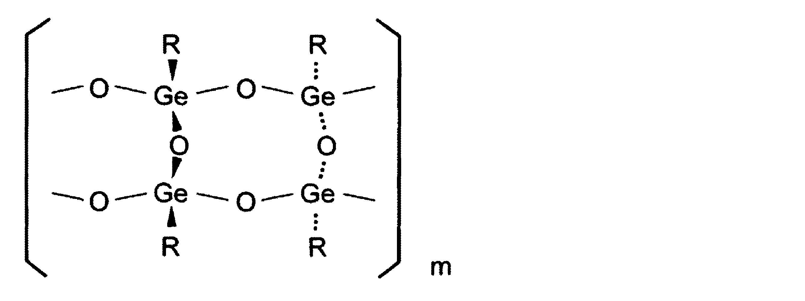
[式中、Rは-CH2CH2COOHを表わし、およびmは3-オキシゲルミルプロピオン酸プロピルエステル重合体の重量平均分子量から換算した重量平均重合度で、137±84(平均値±標準誤差3σ)の整数を表わす]を示し、その最小構成単位は(O1/2)3GeCH2CH2COOHであり、実験式はC6H10Ge2O7にて示される、8員環構造体であろうと推測されている。
3-オキシゲルミルプロピオン酸重合体については、特開2003-81843号公報等に記載されている方法により、製造することができる。また、当該特開2003-81843号公報に、3-オキシゲルミルプロピオン酸重合体は、下記表1および2の物性値を示すものとして記載されている。表1は光散乱法による分子量測定結果を示し、表2は粉末X線解析により求めた格子定数を示す。
3-オキシゲルミルプロピオン酸重合体は、特開昭54-115324号公報の図1~3から、次のような物性値を示す化合物として特定することができる。すなわち、大きい回折ピークが6.5°付近にあり、比較的大きい回折ピークが、11.6°、13.8°、18.4°、21.2°、および22.4°の各付近にあるという特徴的な粉末X線回折スペクトルを示す。赤外吸収(IR)スペクトルについて、大きい吸収バンドが800cm-1、900cm-1、および1700cm-1の各付近にあり、比較的大きい吸収バンドが560cm-1、705cm-1、760cm-1、780cm-1、1250cm-1、1350cm-1、および1400cm-1の各付近にある(但し、1400cm-1付近の吸収バンドはダブレットである)という特徴的な吸収を示す。DSCチャートについて、ピーク開始点が237℃付近にあり、ピーク頂点が256℃付近にあり、ピーク終了点が276℃付近にあり、および、熱量ΔHがほぼ59mcal/mgであるという特徴的なチャートを示す。

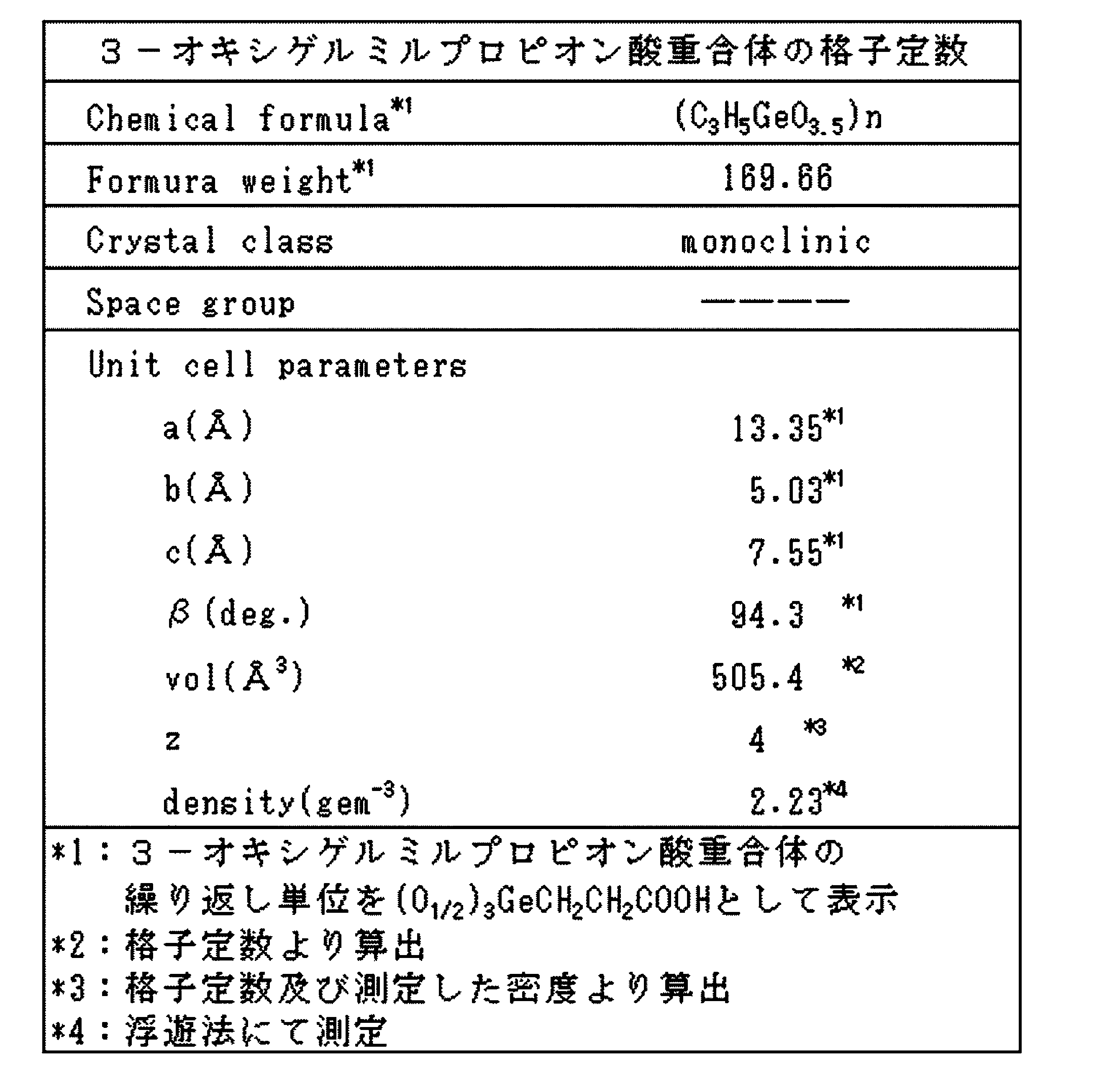
すなわち、3-オキシゲルミルプロピオン酸重合体の好ましい態様は、以下の一般式(II)
(C3H5GeO3.5)n (II)
(式中、nは548±337の重量平均重合度を表わす。)
で示され、および、重量平均分子量が9.29×10⁴±5.72×10⁴(平
均値±標準誤差)である、3-オキシゲルミルプロピオン酸重合体である。
(C3H5GeO3.5)n (II)
(式中、nは548±337の重量平均重合度を表わす。)
で示され、および、重量平均分子量が9.29×10⁴±5.72×10⁴(平
均値±標準誤差)である、3-オキシゲルミルプロピオン酸重合体である。
3-オキシゲルミルプロピオン酸重合体は、プロパゲルマニウム、ビス(2-カルボキシエチルゲルマニウム)三二酸化物、または2-カルボキシエチルゲルマン三二酸化物ということもある。
CCR2アンタゴニストを含む医薬組成物は、医薬として許容される賦形剤、担体および希釈剤などの製剤補助剤を適宜含んでいてもよい。医薬組成物は常法にしたがい、錠剤、カプセル剤、散剤、シロップ剤、顆粒剤、丸剤、懸濁剤、乳剤、液剤、粉体製剤、坐剤、点眼剤、点鼻剤、点耳剤、貼付剤、軟膏剤または注射剤などの剤形とすることができ、経口または非経口で投与することができる。好ましくは経口で投与する。投与方法、投与量および投与回数は、患者の年齢、体重および症状に応じて適宜選択することができる。例えば、成人に対しては、経口投与により、1~1000mg/day、好ましくは10~500mg/dayを、1回から数回に分割して投与すればよい。投与の開始時は、手術と組み合わせる場合は、その前、例えば手術前1日~数週間前時とすることができる。
CCR2アンタゴニストを含む医薬組成物の投与は、がんの処置のための他の療法、例えば手術、化学療法、および/または放射線療法と組み合わせて行うことができる。化学療法に関し、組み合わせて用いることのできる抗がん剤は、目的の効果が得られる限り特に限定されず、例えば、アルキル化薬、白金化合物、代謝拮抗薬、トポイソメラーゼ阻害薬、微小管阻害薬、抗生物質、ホルモン剤、抗体等のいずれであってもよい。より具体的な例として、ゲムシタビン、テガフール・ウラシル、およびフルオロウラシル等が挙げられる。
CCR2アンタゴニストを含む医薬組成物は、低FBXW7発現性のがん患者におけるがんの処置のためものとすることができる。特に、低FBXW7発現性のがん患者におけるがんの処置のために用いられることが好ましく、低FBXW7発現性のがん患者におけるトリプルネガティブ乳がんの処置のために用いられることがより好ましい。
[予後の予測、患者の選択]
本発明は、FBXW7遺伝子またはその発現産物の、がんの予後を予測するための、またはがんの早期発見のための、マーカーとしての使用を提供する。本発明は、FBXW7の発現レベルに関連した、またはがんニッチ形成抑制に関連した疾患または状態にある者、がん患者、またはがんを患う可能性のある者が、特定の薬物(例えば、上述の医薬組成物)での治療の恩恵に浴するかどうかを予測するために、および薬物治療を受ける/受けている対象者の薬物に対する応答または予後を予想するために、用いることができる。本発明者らの検討によると、もっとも悪性度の高いトリプルネガティブ乳がんを持つ患者のうち、Fbxw7が高い人の再発は1割にも満たないが、低い人は5割の確率で再発する。この結果は、Fbxw7量を測定することで、がん患者の予後が予測可能であることを示している。
本発明は、FBXW7遺伝子またはその発現産物の、がんの予後を予測するための、またはがんの早期発見のための、マーカーとしての使用を提供する。本発明は、FBXW7の発現レベルに関連した、またはがんニッチ形成抑制に関連した疾患または状態にある者、がん患者、またはがんを患う可能性のある者が、特定の薬物(例えば、上述の医薬組成物)での治療の恩恵に浴するかどうかを予測するために、および薬物治療を受ける/受けている対象者の薬物に対する応答または予後を予想するために、用いることができる。本発明者らの検討によると、もっとも悪性度の高いトリプルネガティブ乳がんを持つ患者のうち、Fbxw7が高い人の再発は1割にも満たないが、低い人は5割の確率で再発する。この結果は、Fbxw7量を測定することで、がん患者の予後が予測可能であることを示している。
そのため本発明を、FBXW7遺伝子またはその発現産物を検出する工程を含む、特定の薬物(例えば、上述の医薬組成物)による処置の対象となる患者の選択のために用いることができる。すなわち本発明に基づき、特定の薬物(例えば、上述の医薬組成物)治療において、患者を選択することができる。そのため、本発明は、特定の薬物の新規な適応症における使用を提供する。例えば、本発明は、上述の医薬組成物の、FBXW7が低発現レベルにあるがん患者におけるがんの処置のための使用を提案する。
本発明が適用可能ながんには、各種固形がんが含まれる。固形がんの例は、膵臓がん、膠芽腫、食道がん、胃がん、結腸がん、肺がん、腎がん、甲状腺がん、耳下腺がん、頭頚部がん、骨・軟部肉腫、尿管がん、膀胱がん、子宮がん、肝がん、乳がん、前立腺がん、卵巣がん、卵管がん等である。ここで列挙した疾患は、例示であり、これらの他、FBXW7の発現レベルに関連した、またはがんニッチ形成抑制に関連した各種がんに対して、本発明の効果が期待できる。本願の効果が特に期待できるがんの例は、乳がん、メラノーマ、肺がんであり、好ましくは乳がんである。
乳がんには、主にホルモン受容体陽性乳がん、HER2陽性乳がん、トリプルネガティブ乳がんの3つのタイプが含まれる。これらのうち、ホルモン受容体陽性乳がんにはホルモン療法が、HER2陽性乳がんにはハーセプチンがそれぞれ用いられ、著効を示しているが、トリプルネガティブ乳がんに対しては決定的な治療法が確立されていなかった。本発明者らは、血液細胞中のFbxw7の発現量を測定することで、トリプルネガティブ乳がんの中でもより悪性度の高い患者を選択できることを見出した。なお、このように選択された患者に対しては、本発明は、例えばCCR2アンタゴニストを含む医薬組成物の投与を提案する。
[コンパニオン診断薬]
本発明はまた、FBXW7の発現レベルに関連した疾患または状態にある者、がん患者、またはがんを患う可能性のある者を対象とする、コンパニオン診断方法および診断のための製品、ならびにコンパニオン診断の結果、対象者の個別化療法(オーダーメイド療法)に用いるための製品を提供する。診断ための製品は、例えば、プローブ、プローブおよびプライマーのセット、キット等である。個別化療法(オーダーメイド療法)に用いるための製品は、コンパニオン診断の結果、特定の薬物(例えば、上述の医薬組成物)が有効であると判断される場合はその薬物を含み、薬物が有効でないと判断される場合は、その薬物を含まない。製品は、複数の薬物の組み合わせでありうる。
本発明はまた、FBXW7の発現レベルに関連した疾患または状態にある者、がん患者、またはがんを患う可能性のある者を対象とする、コンパニオン診断方法および診断のための製品、ならびにコンパニオン診断の結果、対象者の個別化療法(オーダーメイド療法)に用いるための製品を提供する。診断ための製品は、例えば、プローブ、プローブおよびプライマーのセット、キット等である。個別化療法(オーダーメイド療法)に用いるための製品は、コンパニオン診断の結果、特定の薬物(例えば、上述の医薬組成物)が有効であると判断される場合はその薬物を含み、薬物が有効でないと判断される場合は、その薬物を含まない。製品は、複数の薬物の組み合わせでありうる。
1. 方法
1.1 ヒト臨床標本の分析。
本研究におけるすべての臨床結果は、後向き研究由来である。末梢血標本は、日本・福岡の国立九州がんセンターで2000年~2005年の間に手術を受けた、乳がんを患う406人の日本人女性から得られた。すべての患者は、臨床病理学的知見に基づいて、乳がんを有することが明らかに同定されており、そしていずれも、手術前に化学療法または放射線療法を受けていなかった。Fbxw7 mRNAの測定のため、静脈カテーテルを通じた末梢血の収集を、全身麻酔下にある患者から、手術直前に行った。皮膚細胞による混入を回避するため、末梢血の最初の1.0mlを廃棄した;次の1.0mlを、総RNAの抽出のため、4.0mlのIsogen-LS(ニッポンジーン)と混合した。
1.1 ヒト臨床標本の分析。
本研究におけるすべての臨床結果は、後向き研究由来である。末梢血標本は、日本・福岡の国立九州がんセンターで2000年~2005年の間に手術を受けた、乳がんを患う406人の日本人女性から得られた。すべての患者は、臨床病理学的知見に基づいて、乳がんを有することが明らかに同定されており、そしていずれも、手術前に化学療法または放射線療法を受けていなかった。Fbxw7 mRNAの測定のため、静脈カテーテルを通じた末梢血の収集を、全身麻酔下にある患者から、手術直前に行った。皮膚細胞による混入を回避するため、末梢血の最初の1.0mlを廃棄した;次の1.0mlを、総RNAの抽出のため、4.0mlのIsogen-LS(ニッポンジーン)と混合した。
1.2 マウス。
loxP導入Fbxw7アレルに関してホモ接合性であるマウス(Fbxw7F/Fマウス)(前掲非特許文献23)を、Mx1-Creトランスジェニックマウス(43)と交配し、そして生じた子孫において、体重グラムあたり20μgのポリイノシン酸:ポリシチジン酸[ポリ(I:C)](Calbiochem)で、1日ごとに全部で6回腹腔内注射することによって、loxP導入アレルの欠失を誘導した。loxP導入Fbxw7アレルのエクソン5の欠失を、先に記載されるように、ゲノムDNAのポリメラーゼ連鎖反応(PCR)分析(前掲非特許文献23)によって確認した。Fbxw7F/Fマウスをまた、RbpJκF/F(44)またはc-MycF/F(45)マウス、あるいはLck-Cre(46)、LysM-Cre(47)、CD19-Cre(48)、CAG-Cre-ERT2(49)、またはCAG-EGFP(50)トランスジェニックマウスとも交配した。
loxP導入Fbxw7アレルに関してホモ接合性であるマウス(Fbxw7F/Fマウス)(前掲非特許文献23)を、Mx1-Creトランスジェニックマウス(43)と交配し、そして生じた子孫において、体重グラムあたり20μgのポリイノシン酸:ポリシチジン酸[ポリ(I:C)](Calbiochem)で、1日ごとに全部で6回腹腔内注射することによって、loxP導入アレルの欠失を誘導した。loxP導入Fbxw7アレルのエクソン5の欠失を、先に記載されるように、ゲノムDNAのポリメラーゼ連鎖反応(PCR)分析(前掲非特許文献23)によって確認した。Fbxw7F/Fマウスをまた、RbpJκF/F(44)またはc-MycF/F(45)マウス、あるいはLck-Cre(46)、LysM-Cre(47)、CD19-Cre(48)、CAG-Cre-ERT2(49)、またはCAG-EGFP(50)トランスジェニックマウスとも交配した。
1.3 骨髄移植。
C57BL/6または他のレシピエントマウス(8週齢)に致死用量(11Gy)のγ線を照射し、そして尾静脈を通じて、8週齢のCAG-EGFP、CAG-EGFP/Fbxw7F/F、またはCAg-EGFP/Mx1-Cre/Fbxw7F/Fマウスから単離したBM細胞(100μlのリン酸緩衝生理食塩水中の2.0x106)を注入した。移植2ヶ月後、レシピエントに、上述のようにポリ(I:C)を注入して、loxP導入Fbxw7アレルを欠失させ、そして最後のポリ(I:C)注入の3日後、これらに、以下に記載するように、B16F10、LLC、またはE0771細胞を注入した。BM移植後の各月、ならびに肺切開時点で、フローサイトメトリーにより、レシピエントの末梢血細胞をキメラ現象に関して調べた。
C57BL/6または他のレシピエントマウス(8週齢)に致死用量(11Gy)のγ線を照射し、そして尾静脈を通じて、8週齢のCAG-EGFP、CAG-EGFP/Fbxw7F/F、またはCAg-EGFP/Mx1-Cre/Fbxw7F/Fマウスから単離したBM細胞(100μlのリン酸緩衝生理食塩水中の2.0x106)を注入した。移植2ヶ月後、レシピエントに、上述のようにポリ(I:C)を注入して、loxP導入Fbxw7アレルを欠失させ、そして最後のポリ(I:C)注入の3日後、これらに、以下に記載するように、B16F10、LLC、またはE0771細胞を注入した。BM移植後の各月、ならびに肺切開時点で、フローサイトメトリーにより、レシピエントの末梢血細胞をキメラ現象に関して調べた。
1.4 腫瘍転移アッセイ。
リン酸緩衝生理食塩水中のB16F10(2.0x10⁵)、B16F1(2.0x10⁵)、またはLLC(5.0x10⁵)細胞の懸濁物を、8~11週齢の宿主マウスの尾静脈内に注入した。2週間後、動物を屠殺し、そして肺を取り除き、そしてブアン溶液または4%パラホルムアルデヒド中で固定した。NIH Image Jソフトウェアを用いて、可視B16F10腫瘍コロニーでの肺占有を分析した。E0771細胞(5.0x10⁵)を乳房脂肪体内に皮下注入した。ノギスを用いて腫瘍体積(mm3)を測定し、そして(W2xL)/2として計算する、式中、Wは幅でありそしてLは長さである。tdTomatoの安定発現のため、E0771細胞を、Mycエピトープタグ化tdTomatoをコードするレンチウイルスに、2日間感染させた。プロパゲルマニウム(3-オキシゲミルプロピオン酸(oxygemylpropinic acid)ポリマー)は、三和化学研究所(日本・名古屋)によって寄贈され、そしてマウスには、がん細胞注射の1日前に始まり、0.005%プロパゲルマニウムを含まないか、またはこれを補充した、正常固形飼料を与えた。実験をランダム化した。研究者は、動物研究における実験中、盲検化された。
リン酸緩衝生理食塩水中のB16F10(2.0x10⁵)、B16F1(2.0x10⁵)、またはLLC(5.0x10⁵)細胞の懸濁物を、8~11週齢の宿主マウスの尾静脈内に注入した。2週間後、動物を屠殺し、そして肺を取り除き、そしてブアン溶液または4%パラホルムアルデヒド中で固定した。NIH Image Jソフトウェアを用いて、可視B16F10腫瘍コロニーでの肺占有を分析した。E0771細胞(5.0x10⁵)を乳房脂肪体内に皮下注入した。ノギスを用いて腫瘍体積(mm3)を測定し、そして(W2xL)/2として計算する、式中、Wは幅でありそしてLは長さである。tdTomatoの安定発現のため、E0771細胞を、Mycエピトープタグ化tdTomatoをコードするレンチウイルスに、2日間感染させた。プロパゲルマニウム(3-オキシゲミルプロピオン酸(oxygemylpropinic acid)ポリマー)は、三和化学研究所(日本・名古屋)によって寄贈され、そしてマウスには、がん細胞注射の1日前に始まり、0.005%プロパゲルマニウムを含まないか、またはこれを補充した、正常固形飼料を与えた。実験をランダム化した。研究者は、動物研究における実験中、盲検化された。
1.5 細胞培養。
B16F10(東北大学細胞資源センターによって提供)、LLC(東北大学細胞資源センターによって提供)、B16F1(九州大学、S. Okanoによって寄贈)、およびE0771細胞(CH3 BioSystemsより購入)を、10%ウシ胎児血清(Invitrogen)、1mMピルビン酸ナトリウム、ペニシリン(100U/ml)、ストレプトマイシン(100μg/ml)、2mM L-グルタミン、および非必須アミノ酸(10ml/l、Gibco)を補充したRPMI 1640培地(Sigma)中で維持した。8~10週齢のマウスの脛骨および大腿骨から収集したBMより、BMSCを単離し、そしてこれらを、10%ウシ胎児血清、10%ウマ血清、2mM L-グルタミン、ペニシリン(100U/ml)、およびストレプトマイシン(100μg/ml)を補充したRPMI 1640培地中で培養した。24時間後、非接着細胞を取り除き、そして3日ごとに培地を補充しながら、接着細胞を維持した。loxP導入Fbxw7アレルを欠失させるため、BMSCを10μMタモキシフェン(Sigma)で2日間処理した。これらをまた、DAPT(Calbiochem)でも2日間処理して、Notchシグナル伝達を阻害した。先に記載されるように、胎性期第13.5日に胚からMEFを調製し、そして維持し(51)、そしてloxP導入Fbxw7アレルを欠失させるため、これらを2μMタモキシフェン(Sigma)で2日間処理した。
B16F10(東北大学細胞資源センターによって提供)、LLC(東北大学細胞資源センターによって提供)、B16F1(九州大学、S. Okanoによって寄贈)、およびE0771細胞(CH3 BioSystemsより購入)を、10%ウシ胎児血清(Invitrogen)、1mMピルビン酸ナトリウム、ペニシリン(100U/ml)、ストレプトマイシン(100μg/ml)、2mM L-グルタミン、および非必須アミノ酸(10ml/l、Gibco)を補充したRPMI 1640培地(Sigma)中で維持した。8~10週齢のマウスの脛骨および大腿骨から収集したBMより、BMSCを単離し、そしてこれらを、10%ウシ胎児血清、10%ウマ血清、2mM L-グルタミン、ペニシリン(100U/ml)、およびストレプトマイシン(100μg/ml)を補充したRPMI 1640培地中で培養した。24時間後、非接着細胞を取り除き、そして3日ごとに培地を補充しながら、接着細胞を維持した。loxP導入Fbxw7アレルを欠失させるため、BMSCを10μMタモキシフェン(Sigma)で2日間処理した。これらをまた、DAPT(Calbiochem)でも2日間処理して、Notchシグナル伝達を阻害した。先に記載されるように、胎性期第13.5日に胚からMEFを調製し、そして維持し(51)、そしてloxP導入Fbxw7アレルを欠失させるため、これらを2μMタモキシフェン(Sigma)で2日間処理した。
1.6 フローサイトメトリー。
乳がん患者の末梢血細胞をソーティングするため、本発明者らは、再発乳がんおよび転移を伴う4人の患者からヘパリン処理末梢血6mlを得た。1800rpm、4℃で25分間のFicoll(GE Healthcare)密度遠心分離によって、血液から単核細胞を単離した。1xBD Pharm Lyse緩衝液(BD Biosciences)で赤血球を溶解した。次いで、細胞ソーターSH800(Sony Biotechnology)での分析のため、CD45に対する抗体(HI100、Sony Biotechnology)およびCD326に対する抗体(9C4、Sony Biotechnology)で、単離細胞を染色した。マウスBMおよび末梢血の分析のため、溶血緩衝液(0.14M NH4Clおよび0.01M Tris-HCl、pH7.5)で赤血球を溶解し、そしてF4/80に対する抗体(BM8、eBioscience)、CD115に対する抗体(AFS98、eBioscience)、Mac1に対する抗体(M1/70、eBioscience)、Ly6Gに対する抗体(1A8、BD Biosciences)、およびLy6Cに対する抗体(AL-21、BD Biosciences)で残りの細胞を染色した。染色した細胞を、FACSCaliburフローサイトメーター(Becton Dickinson)で分析した。
乳がん患者の末梢血細胞をソーティングするため、本発明者らは、再発乳がんおよび転移を伴う4人の患者からヘパリン処理末梢血6mlを得た。1800rpm、4℃で25分間のFicoll(GE Healthcare)密度遠心分離によって、血液から単核細胞を単離した。1xBD Pharm Lyse緩衝液(BD Biosciences)で赤血球を溶解した。次いで、細胞ソーターSH800(Sony Biotechnology)での分析のため、CD45に対する抗体(HI100、Sony Biotechnology)およびCD326に対する抗体(9C4、Sony Biotechnology)で、単離細胞を染色した。マウスBMおよび末梢血の分析のため、溶血緩衝液(0.14M NH4Clおよび0.01M Tris-HCl、pH7.5)で赤血球を溶解し、そしてF4/80に対する抗体(BM8、eBioscience)、CD115に対する抗体(AFS98、eBioscience)、Mac1に対する抗体(M1/70、eBioscience)、Ly6Gに対する抗体(1A8、BD Biosciences)、およびLy6Cに対する抗体(AL-21、BD Biosciences)で残りの細胞を染色した。染色した細胞を、FACSCaliburフローサイトメーター(Becton Dickinson)で分析した。
1.7 組織学的、免疫組織化学的、および免疫組織蛍光分析。
ヘマトキシリン-エオジン染色のため、先に記載されるように、組織をブアン溶液中で固定し、パラフィン中に包埋し、厚さ4μmの連続切片に切り、そして染色した(52)。免疫組織化学分析のため、九州大学別府病院(日本・別府)から得た乳房組織マイクロアレイスライドを、Fbxw7に対する抗体(3D1、Abnova)で、そしてEnvision免疫染色系(DAKO)を使用して染色した。切片をヘマトキシリンで対比染色した。乳がん領域および周囲の間質の免疫組織化学染色強度を、陰性(低)または陽性(高)としてスコア付けした。免疫組織蛍光分析のため、先に記載されるように、組織を0.1Mリン酸緩衝液中の4%パラホルムアルデヒドで固定し、30%スクロース中に一晩包埋し、クリオスタットを用いて15μmの厚さで切片作製し、そして染色した(52)。TCRβに対する抗体(H57-597)、B220に対する抗体(RA3-6B2)、Mac1に対する抗体(M1/70)、およびc-Kitに対する抗体(2B8)をeBioscienceから得て;Ly6Cに対するもの(AL-21)、Ly6Gに対するもの(1A8)、そしてVE-カドヘリンに対するもの(11D4.1)をBD Biosciencesから得て;F4/80に対するもの(A3-1)をSeretecから得て;そしてFSPに対するもの(D9F9D)をCell Signaling Technologyから得た。各々、1:2000の希釈のAlexa 633またはAlexa 405(Molecular Probes)で標識した二次抗体で、免疫複合体を検出した。切片をFluoromount(Diagnostic BioSystems)中にマウントし、そしてレーザー走査型共焦点顕微鏡(LSM700、Carl Zeiss)で調べた。
ヘマトキシリン-エオジン染色のため、先に記載されるように、組織をブアン溶液中で固定し、パラフィン中に包埋し、厚さ4μmの連続切片に切り、そして染色した(52)。免疫組織化学分析のため、九州大学別府病院(日本・別府)から得た乳房組織マイクロアレイスライドを、Fbxw7に対する抗体(3D1、Abnova)で、そしてEnvision免疫染色系(DAKO)を使用して染色した。切片をヘマトキシリンで対比染色した。乳がん領域および周囲の間質の免疫組織化学染色強度を、陰性(低)または陽性(高)としてスコア付けした。免疫組織蛍光分析のため、先に記載されるように、組織を0.1Mリン酸緩衝液中の4%パラホルムアルデヒドで固定し、30%スクロース中に一晩包埋し、クリオスタットを用いて15μmの厚さで切片作製し、そして染色した(52)。TCRβに対する抗体(H57-597)、B220に対する抗体(RA3-6B2)、Mac1に対する抗体(M1/70)、およびc-Kitに対する抗体(2B8)をeBioscienceから得て;Ly6Cに対するもの(AL-21)、Ly6Gに対するもの(1A8)、そしてVE-カドヘリンに対するもの(11D4.1)をBD Biosciencesから得て;F4/80に対するもの(A3-1)をSeretecから得て;そしてFSPに対するもの(D9F9D)をCell Signaling Technologyから得た。各々、1:2000の希釈のAlexa 633またはAlexa 405(Molecular Probes)で標識した二次抗体で、免疫複合体を検出した。切片をFluoromount(Diagnostic BioSystems)中にマウントし、そしてレーザー走査型共焦点顕微鏡(LSM700、Carl Zeiss)で調べた。
1.8 イムノブロット分析。
溶解緩衝液[50mM Tris-HCl(pH7.5)、150mM NaCl、0.5% Triton X-100、10mM NaF、10mM Na4P2O7、0.4mM Na3VO4、0.4mM EDTA、ロイペプチン(20μg/ml)、アプロチニン(10μg/ml)、1mMフェニルメチルスルホニルフロリド]を用いて、BMSCから総タンパク質抽出物を調製した。先に記載されるように、抽出物(タンパク質20μg)をイムノブロット分析に供した(53)。Notch3に対する抗体(M-20)およびKLF5に対する抗体(BTEB2、H-300)をSanta Cruz Biotechnologyから得て;切断Notch1に対するもの(D1E11)、Notch2に対するもの(D76A6)、c-Junに対するもの(60A8)、p100-p52に対するもの(カタログ番号4882)、およびmTORに対するもの(7C10)は、Cell Signaling Technologyからであり;c-Mycに対するものはAbcamからであり;そしてグリセルアルデヒド-3-リン酸デヒドロゲナーゼ(GAPDH、装填対照)に対するものはBD Biosciencesからであった。
溶解緩衝液[50mM Tris-HCl(pH7.5)、150mM NaCl、0.5% Triton X-100、10mM NaF、10mM Na4P2O7、0.4mM Na3VO4、0.4mM EDTA、ロイペプチン(20μg/ml)、アプロチニン(10μg/ml)、1mMフェニルメチルスルホニルフロリド]を用いて、BMSCから総タンパク質抽出物を調製した。先に記載されるように、抽出物(タンパク質20μg)をイムノブロット分析に供した(53)。Notch3に対する抗体(M-20)およびKLF5に対する抗体(BTEB2、H-300)をSanta Cruz Biotechnologyから得て;切断Notch1に対するもの(D1E11)、Notch2に対するもの(D76A6)、c-Junに対するもの(60A8)、p100-p52に対するもの(カタログ番号4882)、およびmTORに対するもの(7C10)は、Cell Signaling Technologyからであり;c-Mycに対するものはAbcamからであり;そしてグリセルアルデヒド-3-リン酸デヒドロゲナーゼ(GAPDH、装填対照)に対するものはBD Biosciencesからであった。
1.9 レトロウイルス発現系。
赤血球凝集素エピトープタグ化マウスNICD1、マウスc-Myc、マウスKLF5、またはFLAGエピトープタグ化Fbxw7α(またはそのΔF突然変異体)をコードする相補DNAを、pMX-puro(東京大学、T. Kitamuraにより寄贈)内にサブクローニングし、そして生じたベクターを用いて、Plat E細胞(54)をトランスフェクションし、そしてそれによって組換えレトロウイルスを生成した。BMSCを組換えレトロウイルスに感染させ、そしてピューロマイシン(10μg/ml)を含有する培地中での選択に供した。各組換えタンパク質を安定発現している細胞を、実験のためにプールした。
赤血球凝集素エピトープタグ化マウスNICD1、マウスc-Myc、マウスKLF5、またはFLAGエピトープタグ化Fbxw7α(またはそのΔF突然変異体)をコードする相補DNAを、pMX-puro(東京大学、T. Kitamuraにより寄贈)内にサブクローニングし、そして生じたベクターを用いて、Plat E細胞(54)をトランスフェクションし、そしてそれによって組換えレトロウイルスを生成した。BMSCを組換えレトロウイルスに感染させ、そしてピューロマイシン(10μg/ml)を含有する培地中での選択に供した。各組換えタンパク質を安定発現している細胞を、実験のためにプールした。
1.10 RNAi。
shRNAベクターの構築およびRNAiを、先に記載されるように行った(55)。マウスCCL2をターゲティングする配列は、5’-GGTATTCCCTTTCATGAATAC-3’(SEQ ID NO: 1)であった。EGFPのRNAiベクターを対照として用いた。
RTおよびリアルタイムPCR分析。マウス細胞からIsogen(ニッポンジーン)を用いて単離した総RNA(1μg)を、QuantiTect逆転写キット(Qiagen)を用いて逆転写(RT)に供し、そして生じたcDNAを、StepOnePlusリアルタイムPCR系(Applied Biosystems)中のSYBR緑色PCRマスターミックスおよび特異的プライマーを用いてリアルタイムPCR分析に供した。PCRプライマーの配列(それぞれ、センスおよびアンチセンス)は、CCL2に関しては、5’-CAGCAGCAGGTGTCCCAAAG-3’(SEQ ID NO: 2)および5’-TGTCTGGACCCATTCCTTCTTG-3’(SEQ ID NO: 3)、そしてRps18に関しては、5’-GAGGACCTGGAGAGGCTGAAG-3’(SEQ ID NO: 4)および5’-CTGCGGCCAGTGGTCTTG-3’(SEQ ID NO: 5)であった。CCL2 mRNAの量を、Rps18 mRNAの量によって標準化した。ヒト臨床標本に関しては、Isogen-LS(ニッポンジーン)を用いて細胞から単離した総RNA(2.7μg)を、モロニー白血病ウイルス逆転写酵素(BRL)を用いたRTに供し、そして生じたcDNAを、LightCycler系(Roche Applied Science)中のSYBR-緑色I色素および特異的プライマーを用いたリアルタイムPCRに供した。PCRプライマーの配列(それぞれ、センスおよびアンチセンス)は、Fbxw7に関しては5’-CCTCCAGGAATGGCTAAAAA-3’(SEQ ID NO: 6)および5’-AAGAGTTCATCTAAAGCAAGCAA-3’(SEQ ID NO: 7)、そしてGAPDHに関しては5’-AGCCACATCGCTCAGACAC-3’(SEQ ID NO: 8)および5’-GCCCAATACGACCAAATCC-3’(SEQ ID NO: 9)であった。Fbxw7 mRNAの量を、GAPDH mRNAの量によって標準化した。
shRNAベクターの構築およびRNAiを、先に記載されるように行った(55)。マウスCCL2をターゲティングする配列は、5’-GGTATTCCCTTTCATGAATAC-3’(SEQ ID NO: 1)であった。EGFPのRNAiベクターを対照として用いた。
RTおよびリアルタイムPCR分析。マウス細胞からIsogen(ニッポンジーン)を用いて単離した総RNA(1μg)を、QuantiTect逆転写キット(Qiagen)を用いて逆転写(RT)に供し、そして生じたcDNAを、StepOnePlusリアルタイムPCR系(Applied Biosystems)中のSYBR緑色PCRマスターミックスおよび特異的プライマーを用いてリアルタイムPCR分析に供した。PCRプライマーの配列(それぞれ、センスおよびアンチセンス)は、CCL2に関しては、5’-CAGCAGCAGGTGTCCCAAAG-3’(SEQ ID NO: 2)および5’-TGTCTGGACCCATTCCTTCTTG-3’(SEQ ID NO: 3)、そしてRps18に関しては、5’-GAGGACCTGGAGAGGCTGAAG-3’(SEQ ID NO: 4)および5’-CTGCGGCCAGTGGTCTTG-3’(SEQ ID NO: 5)であった。CCL2 mRNAの量を、Rps18 mRNAの量によって標準化した。ヒト臨床標本に関しては、Isogen-LS(ニッポンジーン)を用いて細胞から単離した総RNA(2.7μg)を、モロニー白血病ウイルス逆転写酵素(BRL)を用いたRTに供し、そして生じたcDNAを、LightCycler系(Roche Applied Science)中のSYBR-緑色I色素および特異的プライマーを用いたリアルタイムPCRに供した。PCRプライマーの配列(それぞれ、センスおよびアンチセンス)は、Fbxw7に関しては5’-CCTCCAGGAATGGCTAAAAA-3’(SEQ ID NO: 6)および5’-AAGAGTTCATCTAAAGCAAGCAA-3’(SEQ ID NO: 7)、そしてGAPDHに関しては5’-AGCCACATCGCTCAGACAC-3’(SEQ ID NO: 8)および5’-GCCCAATACGACCAAATCC-3’(SEQ ID NO: 9)であった。Fbxw7 mRNAの量を、GAPDH mRNAの量によって標準化した。
1.11 抗体アレイおよびELISA。
ケモカインおよびサイトカインの血清濃度を、プロテオーム・プロファイラー・キット(ARY006およびARY015、R&D Systems)を用いて分析した。マウスまたはヒト血清およびマウスBMSCまたはMEF培養上清におけるCCL2レベルもまた、ELISA(Ready-SET-Goキット、eBioscience)によって測定した。BMSCまたはMEFによるCCL2放出の測定のため、細胞(24ウェルプレート中、ウェルあたり1×10⁴)を48時間培養した。
ケモカインおよびサイトカインの血清濃度を、プロテオーム・プロファイラー・キット(ARY006およびARY015、R&D Systems)を用いて分析した。マウスまたはヒト血清およびマウスBMSCまたはMEF培養上清におけるCCL2レベルもまた、ELISA(Ready-SET-Goキット、eBioscience)によって測定した。BMSCまたはMEFによるCCL2放出の測定のため、細胞(24ウェルプレート中、ウェルあたり1×10⁴)を48時間培養した。
1.12 ルシフェラーゼ・レポーター・アッセイ。
マウスCCL2遺伝子のプロモーター領域およびその欠失突然変異体を、ホタル(firefly)・ルシフェラーゼをコードするpGL2-Basic(Promega)内にサブクローニングした。FuGENE HD試薬(Promega)を使用して、レニラ属(Renilla)ルシフェラーゼに関するプロモーター構築物(0.25μg)および内部対照ベクターpRL-TK(0.25μg、Promega)でトランスフェクションする24時間前に、BMSCを24ウェルプレート中、ウェルあたり2x10⁴の密度で植え付けた。トランスフェクションの48時間後、二重ルシフェラーゼレポーターアッセイ系(Promega)およびLumat LB9507ルミノメーター(EG&G Berthold)を用いて、ルシフェラーゼ活性を測定した。ホタル・ルシフェラーゼ活性を、レニラ属ルシフェラーゼのものによって標準化した。
マウスCCL2遺伝子のプロモーター領域およびその欠失突然変異体を、ホタル(firefly)・ルシフェラーゼをコードするpGL2-Basic(Promega)内にサブクローニングした。FuGENE HD試薬(Promega)を使用して、レニラ属(Renilla)ルシフェラーゼに関するプロモーター構築物(0.25μg)および内部対照ベクターpRL-TK(0.25μg、Promega)でトランスフェクションする24時間前に、BMSCを24ウェルプレート中、ウェルあたり2x10⁴の密度で植え付けた。トランスフェクションの48時間後、二重ルシフェラーゼレポーターアッセイ系(Promega)およびLumat LB9507ルミノメーター(EG&G Berthold)を用いて、ルシフェラーゼ活性を測定した。ホタル・ルシフェラーゼ活性を、レニラ属ルシフェラーゼのものによって標準化した。
1.13 ChIP。
BMSC(2x10⁷)を、RPMI1640培地中の0.5%ホルムアルデヒドで5分間固定した。最終濃度0.125Mまでグリシンを添加することによって、固定を終結させ、そして次いで、細胞を氷冷リン酸緩衝生理食塩水で洗浄し、5mM Hepes-NaOH(pH8.0)、200mM KCl、1mM CaCl2、1.5mM MgCl2、5%スクロースおよび0.5%ノニデットP-40を含有する2mlの溶解緩衝液で溶解して、そして次いで、小球菌ヌクレアーゼ(New England Biolabs)に曝露して、1~5のヌクレオソームを含む、クロマチン断片を得た。ヌクレオソームを、20μlのDynabeads-プロテインG(ベリタス、日本)にコンジュゲート化された、Notch1に対する抗体(C-20、Santa Cruz Biotechnology)2μgと、4℃で6時間インキュベーションした。免疫沈降物質を洗浄し、クロマチンを溶出させ、そして架橋を逆転させた。フェノール-クロロホルム抽出、その後、イソプロパノールを用いた沈殿によって、DNA断片を精製し、そして次いで、これらを、SYBRセレクトPCR系(Applied Biosystems)を用いたリアルタイムPCR分析のテンプレートとして用いた。PCRプライマーの配列(それぞれ、センスおよびアンチセンス)は、CCL2(遠位)に関しては5’-GCTCACATTCCAGCTAAATATCTCT-3’(SEQ ID NO: 10)および5’-GAGTTATTGTCTGTTTCCCTCTCA-3’(SEQ ID NO: 11)、そしてCCL2(近位)に関しては5’-TTACTGGGGGTCCTTTCCCA-3’(SEQ ID NO: 12)および5’-GGAGTGGCTCTGCTTTCACT-3’(SEQ ID NO: 13)であった。クロマチン濃縮の度合いを投入量によって標準化した。
BMSC(2x10⁷)を、RPMI1640培地中の0.5%ホルムアルデヒドで5分間固定した。最終濃度0.125Mまでグリシンを添加することによって、固定を終結させ、そして次いで、細胞を氷冷リン酸緩衝生理食塩水で洗浄し、5mM Hepes-NaOH(pH8.0)、200mM KCl、1mM CaCl2、1.5mM MgCl2、5%スクロースおよび0.5%ノニデットP-40を含有する2mlの溶解緩衝液で溶解して、そして次いで、小球菌ヌクレアーゼ(New England Biolabs)に曝露して、1~5のヌクレオソームを含む、クロマチン断片を得た。ヌクレオソームを、20μlのDynabeads-プロテインG(ベリタス、日本)にコンジュゲート化された、Notch1に対する抗体(C-20、Santa Cruz Biotechnology)2μgと、4℃で6時間インキュベーションした。免疫沈降物質を洗浄し、クロマチンを溶出させ、そして架橋を逆転させた。フェノール-クロロホルム抽出、その後、イソプロパノールを用いた沈殿によって、DNA断片を精製し、そして次いで、これらを、SYBRセレクトPCR系(Applied Biosystems)を用いたリアルタイムPCR分析のテンプレートとして用いた。PCRプライマーの配列(それぞれ、センスおよびアンチセンス)は、CCL2(遠位)に関しては5’-GCTCACATTCCAGCTAAATATCTCT-3’(SEQ ID NO: 10)および5’-GAGTTATTGTCTGTTTCCCTCTCA-3’(SEQ ID NO: 11)、そしてCCL2(近位)に関しては5’-TTACTGGGGGTCCTTTCCCA-3’(SEQ ID NO: 12)および5’-GGAGTGGCTCTGCTTTCACT-3’(SEQ ID NO: 13)であった。クロマチン濃縮の度合いを投入量によって標準化した。
1.14 統計分析。
示すように、定量的データを統計分析に供した。<0.05のP値は、統計的に有意であると見なされた。
示すように、定量的データを統計分析に供した。<0.05のP値は、統計的に有意であると見なされた。
1.15 研究認可。
すべての動物実験は、九州大学の施設動物飼育および使用委員会によって認可され、そして動物飼育は、施設のガイドラインにしたがった。すべての臨床試料は、国立病院機構九州がんセンターの倫理委員会(第2001-15号;2001年10月16日)、九州大学倫理委員会(第302号;2006年2月9日)によって分析に関して認可された。本研究で分析したがん患者すべてから、書面のインフォームドコンセントを得た。
すべての動物実験は、九州大学の施設動物飼育および使用委員会によって認可され、そして動物飼育は、施設のガイドラインにしたがった。すべての臨床試料は、国立病院機構九州がんセンターの倫理委員会(第2001-15号;2001年10月16日)、九州大学倫理委員会(第302号;2006年2月9日)によって分析に関して認可された。本研究で分析したがん患者すべてから、書面のインフォームドコンセントを得た。
2. 結果
2.1 マウスにおいて、骨髄(BM)におけるFbxw7の欠失は、がん転移を促進する。
Fbxw7の大部分の研究は、腫瘍細胞におけるその機能に重点を置いてきており(28~30)、腫瘍発展に関連して、宿主微小環境におけるこのタンパク質の役割に関してはほとんど知られていない。宿主微小環境におけるFbxw7の役割を調べるため、本発明者らは、BMにおいて選択的にloxP導入(floxed)(F)Fbxw7アレルを欠失させるため、ポリイノシン酸:ポリシチジン酸[ポリ(I:C)]を注入したMx1-Cre/Fbxw7F/Fマウス(以後、Mx1-Cre/Fbxw7Δ/Δマウスと称する)の尾静脈内に、B16F10黒色腫細胞をトランスファーした。黒色腫細胞の肺への転移頻度は、対照マウスに比較して、Mx1-Cre/Fbxw7Δ/Δマウスで顕著に増加しており(図1、AおよびB)、そしてこの転移増加には、Mx1-Cre/Fbxw7Δ/Δマウスの早期の死亡が伴った(図1C)。ルイス肺がん腫(LLC)細胞(図1、DおよびE、ならびに図9、A~C)または低転移性潜在的B16F1黒色腫細胞(図1、FおよびG、ならびに図9、D~F)をこれらのマウスの尾静脈内に注入した際にも、同様の結果が得られた。したがって、BMにおけるFbxw7のレベルは、マウスにおけるがん転移の重要な決定要因であることが見出された。
2.1 マウスにおいて、骨髄(BM)におけるFbxw7の欠失は、がん転移を促進する。
Fbxw7の大部分の研究は、腫瘍細胞におけるその機能に重点を置いてきており(28~30)、腫瘍発展に関連して、宿主微小環境におけるこのタンパク質の役割に関してはほとんど知られていない。宿主微小環境におけるFbxw7の役割を調べるため、本発明者らは、BMにおいて選択的にloxP導入(floxed)(F)Fbxw7アレルを欠失させるため、ポリイノシン酸:ポリシチジン酸[ポリ(I:C)]を注入したMx1-Cre/Fbxw7F/Fマウス(以後、Mx1-Cre/Fbxw7Δ/Δマウスと称する)の尾静脈内に、B16F10黒色腫細胞をトランスファーした。黒色腫細胞の肺への転移頻度は、対照マウスに比較して、Mx1-Cre/Fbxw7Δ/Δマウスで顕著に増加しており(図1、AおよびB)、そしてこの転移増加には、Mx1-Cre/Fbxw7Δ/Δマウスの早期の死亡が伴った(図1C)。ルイス肺がん腫(LLC)細胞(図1、DおよびE、ならびに図9、A~C)または低転移性潜在的B16F1黒色腫細胞(図1、FおよびG、ならびに図9、D~F)をこれらのマウスの尾静脈内に注入した際にも、同様の結果が得られた。したがって、BMにおけるFbxw7のレベルは、マウスにおけるがん転移の重要な決定要因であることが見出された。
BMにおける特異的なFbxw7の消失が、実際にMx1-Cre/Fbxw7Δ/Δマウスにおける転移増進の原因であるのかどうかを調べるため、本発明者らは、CAGプロモーターの調節下の増進緑色蛍光タンパク質(EGFP)の導入遺伝子もまた有するMx1-Cre/Fbxw7F/FまたはFbxw7F/F(対照)マウス由来のBM細胞を、放射線照射したC57BL/6マウスに移植した(図1H)。レシピエントマウスには、続いて、ポリ(I:C)を注射して、Fbxw7のloxP導入アレルを欠失させ、そしてポリ(I:C)注射3日後に、がん細胞(B16F10またはLLC)を、これらのマウスにトランスファーした。肺への転移は、対照細胞を移植されたものよりも、CAG-EGFP/Mx1-Cre/Fbxw7F/F BM細胞を移植されたマウスにおいて、より顕著であった(図1、IおよびJ、ならびに図9、GおよびH)。対照的に、CAG-EGFPマウス由来のBMの移植に供し、そしてポリ(I:C)を注射したMx1-Cre/Fbxw7F/Fマウスにおいては、転移のこうした増進はまったく見られなかった(図1、K~M)。これらの結果はしたがって、BMにおけるFbxw7の喪失が、実際に、Mx1-Cre/Fbxw7Δ/Δマウスで観察される転移の頻度増加の原因であることを確認した。
本発明者らはまた、E0771マウス乳がん細胞の同所性移植後の、対照およびMx1-Cre/Fbxw7Δ/Δマウスにおける転移性腫瘍増殖も調べた。原発性腫瘍の増殖は、第17日および第20日に、Fbxw7欠損マウスにおいて促進されたが、増殖促進の効果は、より後の時期には観察されなかった(図2、AおよびB)。肺への転移は、突然変異マウスにおいて、顕著に増進された(図2、CおよびD)。肺重量(図2D)に加え、肺における総腫瘍面積(図2E)、腫瘍小結節数(図2F)、および小結節あたりの平均面積(図2G)は、対照に関するよりも、Mx1-Cre/Fbxw7Δ/Δマウスに関してより高かった。本発明者らは、次に、このモデルにおいて、転移性腫瘍の進行を監視した。細胞移植の12日後、肺においていかなる腫瘍細胞も検出されなかったが、第16日、対照およびMx1-Cre/Fbxw7Δ/Δマウス両方において、E0771細胞の転移が見られた(図2、H~J)。腫瘍小結節数および小結節あたりの平均面積は、移植の16日後、2つの遺伝子型間で異ならなかったが、第20日には、対照におけるよりもMx1-Cre/Fbxw7Δ/Δマウスにおいて、有意により高かった。転移前(premetastatic)ニッチは、BMDCのクラスターによって形成されることが先に示された(27)が、本発明者らは、こうしたEGFP+クラスターが、EGFP標識Mx1-Cre/Fbxw7Δ/Δまたは対照BM細胞で再構成した野生型(WT)マウスの肺において、第0日(E0771細胞移植前)に、すでに存在していたことを見出した(図10A)。これらのクラスターの数は、E0771細胞移植後の時間によって実質的に変化はなく、そして2つの遺伝子型間で異ならなかった(図10B)。対照的に、肺における分散浸潤BMDCの数は、Mx1-Cre/Fbxw7Δ/Δ BM細胞で再構成したマウスにおいて特異的に、腫瘍細胞移植後に増加した(図10、AおよびC)。T細胞に関するT細胞受容体β(TCRβ)(図3A)、B細胞に関するB220(図3B)、G-MDSCに関するLy6G(図3C)、Mo-MDSCに関するLy6C(図3D)、単球-マクロファージに関するF4/80(図3E)、間質細胞に関する線維芽細胞特異的タンパク質(FSP)(図3F)、骨髄細胞に関するMac1(図11A)、HPCに関するc-Kit(図11B)、およびEPCに関するVE-カドヘリン(図11C)に対する抗体を用いた免疫蛍光分析によって、Ly6C+、F4/80+、およびMac1+細胞の数が腫瘍周囲BMDCの間で増加している一方、非周囲BMDCの間では、B220+細胞の数が中程度に増加しているのみであることが明らかになった。これらの結果は、Mo-MDSCまたはより分化したマクロファージの集積が、Mx1-Cre/Fbxw7Δ/Δマウスにおける転移の促進の原因となっている可能性があることを示唆した。
本発明者らはまた、腫瘍細胞移植の2日前から32日後までの多様な時点で、マウスの末梢血における細胞を特徴付けた。末梢血におけるMac1+Ly6G-Ly6C+ Mo-MDSC(図10D)およびMac1+F4/80+CD115+単球-マクロファージ(図10E)の頻度は、腫瘍細胞移植前には、対照マウスに比較して、Mx1-Cre/Fbxw7Δ/Δマウスで増加していたが、この時点では、Mac1+Ly6G+Ly6C+未成熟MDSCの頻度は、2つの遺伝子型間で異ならなかった(図10F)。しかし、末梢血におけるこれらの後者の細胞の頻度は、第16日~第24日の間で、対照マウスにおけるよりもMx1-Cre/Fbxw7Δ/Δマウスにおいて、より大きな度合いに一過性に増加した。対照的に、BMにおけるMac1+Ly6G-Ly6C+ Mo-MDSCの頻度は、第0日(図10G)または第20日(図10H)で、2つの遺伝子型間で異ならなかった。総合すると、これらの結果は、Mx1-Cre/Fbxw7Δ/Δマウスにおける肺への転移の初期には、BMDCの浸潤増加が、BMにおけるMo-MDSCおよびマクロファージの分化よりも、これらの細胞の遊走または補充の増加を示す可能性があることを示唆した。
2.2 骨髄においてFbxw7を欠くマウスでは、血清ケモカインレベルが増加する。
Fbxw7欠損によって誘導されるMo-MDSCおよびマクロファージの可動化増加の原因となる機構を調べるため、本発明者らは、E0771細胞移植前および移植後の多様なサイトカインの血清濃度を調べた。サイトカインアレイ分析によって、CCL2、CCL12(MCP-5としてもまた知られる)、およびケモカインC-X-Cモチーフリガンド13(CXCL13、Bリンパ球走化性因子またはBLCとしてもまた知られる)のレベルが、E0771細胞移植前および移植後の両方で、対照マウスに比較して、Mx1-Cre/Fbxw7Δ/Δマウスにおいて2倍以上増加することが明らかになった(図4Aおよび図12)。CCL2およびCCL12はどちらも、共通の受容体CCR2に結合することによって、単球-マクロファージの遊走を誘導する。CCL12は、主にマクロファージから分泌されるが、LysM-Cre/Fbxw7F/Fマウスのこれらの細胞においてFbxw7を消失させても転移頻度には影響がなく(図13、AおよびB)、CCL12が転移の促進に大きくは関与していないことが示唆された。本発明者らはしたがって、CCL2に集中し、そしてCCL2の血清濃度が、E0771細胞移植前および移植後の両方で、対照マウスにおけるよりMx1-Cre/Fbxw7Δ/Δマウスにおいて、より大きいことを見出した(図4B)。
Fbxw7欠損によって誘導されるMo-MDSCおよびマクロファージの可動化増加の原因となる機構を調べるため、本発明者らは、E0771細胞移植前および移植後の多様なサイトカインの血清濃度を調べた。サイトカインアレイ分析によって、CCL2、CCL12(MCP-5としてもまた知られる)、およびケモカインC-X-Cモチーフリガンド13(CXCL13、Bリンパ球走化性因子またはBLCとしてもまた知られる)のレベルが、E0771細胞移植前および移植後の両方で、対照マウスに比較して、Mx1-Cre/Fbxw7Δ/Δマウスにおいて2倍以上増加することが明らかになった(図4Aおよび図12)。CCL2およびCCL12はどちらも、共通の受容体CCR2に結合することによって、単球-マクロファージの遊走を誘導する。CCL12は、主にマクロファージから分泌されるが、LysM-Cre/Fbxw7F/Fマウスのこれらの細胞においてFbxw7を消失させても転移頻度には影響がなく(図13、AおよびB)、CCL12が転移の促進に大きくは関与していないことが示唆された。本発明者らはしたがって、CCL2に集中し、そしてCCL2の血清濃度が、E0771細胞移植前および移植後の両方で、対照マウスにおけるよりMx1-Cre/Fbxw7Δ/Δマウスにおいて、より大きいことを見出した(図4B)。
Fbxw7欠損マウスにおいて見られる転移増進が、CCL2-CCR2経路に依存するかどうかを調べるため、本発明者らは、Mx1-Cre/Fbxw7Δ/ΔマウスをCCR2アンタゴニスト(プロパゲルマニウム)で処置した。Mx1-Cre/Fbxw7Δ/ΔマウスにおけるB16F10またはE0771細胞転移の度合いは、CCR2アンタゴニストの投与によって、有意に減弱した(図4、C~H)。EGFP標識Mx1-Cre/Fbxw7Δ/Δ BM細胞で再構成し、そしてE0771細胞を注入したWTマウスの肺におけるEGFP+Ly6C+細胞の頻度もまた、プロパゲルマニウムでの処置によって有意に減少した(図4H)。予期せぬことに、肺における腫瘍小結節の数は、処置によって影響を受けず(図4F)、一方、各小結節のサイズは、処置マウスにおいて顕著に減少した(図4G)。総合すると、これらの結果は、Fbxw7欠損BMDCによるCCL2の産生増加が、肺にすでに転移していた腫瘍の増殖を促進することを示唆した。
2.3 CCL2遺伝子は、Fbxw7欠損BMSCにおいて活性化される。
本発明者らは次に、BMのどの細胞が、Fbxw7欠損マウスにおいて、転移の促進およびCCL2血清濃度の増加の原因となるかを調べた。これらの実験のため、本発明者らは、異なる系譜でFbxw7に欠損を持つマウスを利用した。Cre遺伝子が活性化されて、顆粒球-マクロファージ系譜においてのみ、loxP導入Fbxw7アレルが欠失している、LysM-Cre/Fbxw7-/Δマウスは、対照マウスに比較して、転移の頻度の増加を示さなかった(図13、AおよびB)。さらに、Lck-Cre/Fbxw7Δ/Δマウス(Fbxw7のT細胞特異的欠失)もまたCD19-Cre/Fbxw7Δ/Δマウス(Fbxw7のB細胞特異的欠失)もいずれも、Mx1-Cre/Fbxw7Δ/Δマウスで見られるものと類似の転移増進を示さなかった(図13、C~F)。これらの結果は、骨髄系譜(顆粒球およびマクロファージ)もまたリンパ系譜(T細胞およびB細胞)もいずれも、BMにおけるFbxw7の消失による転移の促進には関与しないことを示唆した。
本発明者らは次に、BMのどの細胞が、Fbxw7欠損マウスにおいて、転移の促進およびCCL2血清濃度の増加の原因となるかを調べた。これらの実験のため、本発明者らは、異なる系譜でFbxw7に欠損を持つマウスを利用した。Cre遺伝子が活性化されて、顆粒球-マクロファージ系譜においてのみ、loxP導入Fbxw7アレルが欠失している、LysM-Cre/Fbxw7-/Δマウスは、対照マウスに比較して、転移の頻度の増加を示さなかった(図13、AおよびB)。さらに、Lck-Cre/Fbxw7Δ/Δマウス(Fbxw7のT細胞特異的欠失)もまたCD19-Cre/Fbxw7Δ/Δマウス(Fbxw7のB細胞特異的欠失)もいずれも、Mx1-Cre/Fbxw7Δ/Δマウスで見られるものと類似の転移増進を示さなかった(図13、C~F)。これらの結果は、骨髄系譜(顆粒球およびマクロファージ)もまたリンパ系譜(T細胞およびB細胞)もいずれも、BMにおけるFbxw7の消失による転移の促進には関与しないことを示唆した。
CCL2の血清レベル(図14A)もまた末梢血におけるMac1+F4/80+単球-マクロファージの頻度(図14B)もいずれも、LysM-Cre/Fbxw7-/Δマウスおよび対照マウスの間で異ならず、単球-マクロファージがFbxw7喪失に反応して産生されるCCL2の主要供給源ではないことが示唆された。本発明者らは、FSP+ BMSCが、E0771細胞の同所性移植後、マウス肺において、転移性腫瘍細胞およびMo-MDSCと共存することを見出した(図5A)。以前、BMSCがCCL2を分泌し、そしてBMからの単球の遊出に寄与することが示されたことを考慮し(31、32)、本発明者らは、本発明者らのモデルにおいて、BMSCがCCL2産生の主要供給源であるという仮説を立てた。この仮説を試験するため、本発明者らは、CAG-Cre-ERT2/Fbxw7F/Fマウス(または対照Fbxw7F/Fマウス)のBMからBMSCを単離し、そしてこれらを10μMタモキシフェンで処理して、loxP導入Fbxw7アレルの欠失を誘導した(図5B)。CCL2 mRNAの存在量は、対照細胞と比較して、CAG-Cre-ERT2/Fbxw7Δ/Δ BMSCで増加していた(図5C)。CAG-Cre-ERT2/Fbxw7Δ/Δ BMSCから培地へ放出されるCCL2の量もまた、対照細胞から放出されるものよりも実質的に多かった(図5D)。さらに、CAG-Cre-ERT2/Fbxw7Δ/Δ BMSCにWT Fbxw7α cDNAを導入すると、CCL2 mRNAの存在量の顕著な減少が生じる一方、Fボックスドメインを欠くFbxw7αの突然変異型(ΔF)のcDNAを導入してもこうした効果はなく(図5E)、Fbxw7が、BMSCにおいてCCL2産生を負に制御することが示唆された。
BMSCによるCCL2産生増加が、Fbxw7欠損マウスにおけるがん転移の促進に関与するかどうかを調べるため、本発明者らは、小分子ヘアピンRNA(shRNA)仲介性RNA干渉(RNAi)によって、CAG-Cre-ERT2/Fbxw7Δ/Δ BMSCからCCL2を枯渇させ(図5F)、そして次いでこれらの細胞を、B16F10黒色腫細胞と一緒に、尾静脈を通じてレシピエントマウス内にトランスファーした。BMSCは、移植の4ヶ月後であっても、骨髄および肺などの多くの組織で検出されることが示された(33)。レシピエントマウスにおける肺転移の度合いは、対照BMSCの同時注入後に見られるものと比較して、黒色腫細胞とFbxw7Δ/Δ BMSCの同時注入によって増加した(図5、GおよびH)。しかし、Fbxw7欠失のこの効果は、BMSCにおけるCCL2の枯渇によって無効になり、Fbxw7欠損BMSCによるCCL2産生増加が、転移促進に寄与していることが示唆された。
2.4 Fbxw7欠損BMSCにおけるNotch集積は、CCL2産生増加によって転移を促進する。
イムノブロット分析によって、調べたFbxw7基質のうち、Notch1細胞内ドメイン(NICD1)、c-Myc、およびKLF5が、Fbxw7欠損BMSCにおいて、高レベルに集積することが明らかになった(図6A)。WT BMSCにおいて、NICD1を強制的に発現させた結果、CCL2 mRNA存在量およびCCL2遺伝子プロモーター活性の両方が顕著に増加し、一方、c-MycまたはKLF5のものはこうした効果を持たなかった(図6、BおよびC)。γ-セクレターゼ阻害剤N-[N-(3,5-ジフルオロフェンアセチル)-L-アラニル]-S-フェニルグリシンt-ブチルエステル(DAPT)に曝露することによって、Fbxw7欠損BMSCにおけるNotchシグナル伝達を阻害した結果、CCL2 mRNA量の濃度依存性減少が生じた(図6D)。マウスCCL2遺伝子のプロモーターは、Notch結合に関する4つのコンセンサス配列を含有し(34)(図6E)、そしてこのプロモーターのWTまたは突然変異型のルシフェラーゼレポーターアッセイによって、完全プロモーター活性のためには、最初の2つの上流要素が必要であることが示された(図6F)。この発見は、Fbxw7Δ/Δ BMSCにおいて、NICD1がCCL2遺伝子プロモーターの遠位領域と会合することを示すクロマチン免疫沈降(ChIP)分析の結果と一致した(図6G)。総合すると、これらの観察は、BMSCにおけるNotch-CCL2経路が、Fbxw7欠損マウスにおける転移促進に寄与することを示唆した。
イムノブロット分析によって、調べたFbxw7基質のうち、Notch1細胞内ドメイン(NICD1)、c-Myc、およびKLF5が、Fbxw7欠損BMSCにおいて、高レベルに集積することが明らかになった(図6A)。WT BMSCにおいて、NICD1を強制的に発現させた結果、CCL2 mRNA存在量およびCCL2遺伝子プロモーター活性の両方が顕著に増加し、一方、c-MycまたはKLF5のものはこうした効果を持たなかった(図6、BおよびC)。γ-セクレターゼ阻害剤N-[N-(3,5-ジフルオロフェンアセチル)-L-アラニル]-S-フェニルグリシンt-ブチルエステル(DAPT)に曝露することによって、Fbxw7欠損BMSCにおけるNotchシグナル伝達を阻害した結果、CCL2 mRNA量の濃度依存性減少が生じた(図6D)。マウスCCL2遺伝子のプロモーターは、Notch結合に関する4つのコンセンサス配列を含有し(34)(図6E)、そしてこのプロモーターのWTまたは突然変異型のルシフェラーゼレポーターアッセイによって、完全プロモーター活性のためには、最初の2つの上流要素が必要であることが示された(図6F)。この発見は、Fbxw7Δ/Δ BMSCにおいて、NICD1がCCL2遺伝子プロモーターの遠位領域と会合することを示すクロマチン免疫沈降(ChIP)分析の結果と一致した(図6G)。総合すると、これらの観察は、BMSCにおけるNotch-CCL2経路が、Fbxw7欠損マウスにおける転移促進に寄与することを示唆した。
本発明者らは、この知見をさらに、遺伝子分析によって評価した。Notch依存性トランス活性化に必須の補因子であるRBP-Jκのさらなる消失は、Mx1-Cre/Fbxw7Δ/Δマウスにおいて見られる転移増進の顕著な減弱を生じた(図6、HおよびI)。対照的に、Mx1-Cre/Fbxw7Δ/Δマウスにおけるc-Mycの不活性化は、こうした効果を持たなかった(図6、JおよびK)。RBP-Jκのさらなる消失はまた、Mx1-Cre/Fbxw7Δ/ΔマウスにおいてCCL2の血清濃度を対照レベルまで減少させたが、c-Mycの消失ではこうした現象は起こらなかった(図6L)。
2.5 宿主微小環境におけるFBXW7の低発現は、乳がん患者における劣った予後と関連する。
マウスにおける観察をヒトにも広げるため、本発明者らは、乳がん標本の末梢血におけるFbxw7 mRNAの存在量を測定し、そしてFbxw7 mRNAの存在量および予後の間の関連を調べた。末梢血においてFBXW7が低発現である個体の予後は、対応するFBXW7高群におけるものより、有意に劣っており(図7、AおよびB)、本発明者らのモデルと一致した。これらの2つの群間の予後の相違は、エストロゲン(ER)およびプロゲステロン(PR)受容体の両方ならびにHer2に関して三重陰性である腫瘍を持つ患者に(図7、C~E)、あるいは高い組織学的グレードまたは低いステージの腫瘍を持つ患者に(図15、A~G)、分析を限定した際に、さらにより顕著であった。CD45-EpCAM(CD326)+循環腫瘍細胞の頻度は、最大で、総血液単核細胞の0.003%であり(図15H)、末梢血において、こうした細胞のFbxw7 mRNAの総存在量への寄与は無視できる程度であることが示唆された。
マウスにおける観察をヒトにも広げるため、本発明者らは、乳がん標本の末梢血におけるFbxw7 mRNAの存在量を測定し、そしてFbxw7 mRNAの存在量および予後の間の関連を調べた。末梢血においてFBXW7が低発現である個体の予後は、対応するFBXW7高群におけるものより、有意に劣っており(図7、AおよびB)、本発明者らのモデルと一致した。これらの2つの群間の予後の相違は、エストロゲン(ER)およびプロゲステロン(PR)受容体の両方ならびにHer2に関して三重陰性である腫瘍を持つ患者に(図7、C~E)、あるいは高い組織学的グレードまたは低いステージの腫瘍を持つ患者に(図15、A~G)、分析を限定した際に、さらにより顕著であった。CD45-EpCAM(CD326)+循環腫瘍細胞の頻度は、最大で、総血液単核細胞の0.003%であり(図15H)、末梢血において、こうした細胞のFbxw7 mRNAの総存在量への寄与は無視できる程度であることが示唆された。
原発性腫瘍病変におけるFbxw7の免疫組織化学的分析(図7F)によって、末梢血におけるFbxw7 mRNAの存在量が、腫瘍細胞におけるFbxw7発現と有意には相関しない(図7G)が、周囲の間質細胞におけるものとは非常に相関する(図7H)ことが明らかになった。これらの結果によって、末梢血におけるFbxw7ある mRNAの存在量は、腫瘍細胞周囲の間質細胞におけるFbxw7発現のマーカーでことが示唆された。本発明者らはまた、末梢血におけるFbxw7 mRNAの存在量の間で、負の相関が明らかであることも見出した(図7I)。CCL2の高い血清レベルは、乳がん患者において、劣った予後と相関する(S.A.およびK.M.、未公表データ)。これらのデータは、Fbxw7消失が、転移性腫瘍増殖を促進するCCL2の産生増加を生じさせるという本発明者らのモデルを裏付ける(図8)。
3. 考察
Fbxw7遺伝子は、ヒトがんと関連する多くの対応する突然変異によって(前掲非特許文献22)、ならびに条件付きノックアウトマウスにおいて観察される腫瘍形成によって(前掲非特許文献23、24、26)立証されるように、強力な腫瘍サプレッサーである。Fbxw7の抗がん機能は、増殖促進性腫瘍性タンパク質、例えばc-Myc(前掲非特許文献9、10)、Notch(前掲非特許文献11、12、および35)、サイクリンE(前掲非特許文献14~16)、c-Jun(前掲非特許文献17、18)、KLF5(前掲非特許文献19、20)、およびmTOR(前掲非特許文献21)、ならびに抗アポトーシス分子Mcl-1(36、37)の両方の特異的ユビキチン化によって仲介されると考えられる。以前の研究の大部分は、腫瘍細胞自体におけるFbxw7の腫瘍抑制的な役割に重点を置いてきた。推定上の腫瘍サプレッサーC/EBPδは、腫瘍細胞において、Fbxw7発現を阻害し、そしてmTORのFbxw7依存性分解の減弱を通じて、乳房腫瘍転移を促進することが示された(38)。本発明者らはここで、腫瘍抑制におけるFbxw7機能の新規側面、すなわち、宿主微小環境においてがん転移を抑制するその役割を発見した。本発明者らのデータはまた、Fbxw7によるがん転移の抑制に関する機構的洞察も提供する。本発明者らは、Fbxw7の主要基質の1つであるNotchが、がん発展に関して最もよく特徴付けられているケモカインの1つであるCCL2の遺伝子の転写を活性化することを見出した。乳がん患者においては、末梢血におけるFBXW7の発現減少が、劣った予後と関連することを示す本発明者らのヒト臨床データは、マウスにおける本発明者らの実験結果と一致するようである。本発明者らは、したがって、宿主微小環境におけるFbxw7-Notch-CCL2軸ががん転移を制限することを提唱する。
Fbxw7遺伝子は、ヒトがんと関連する多くの対応する突然変異によって(前掲非特許文献22)、ならびに条件付きノックアウトマウスにおいて観察される腫瘍形成によって(前掲非特許文献23、24、26)立証されるように、強力な腫瘍サプレッサーである。Fbxw7の抗がん機能は、増殖促進性腫瘍性タンパク質、例えばc-Myc(前掲非特許文献9、10)、Notch(前掲非特許文献11、12、および35)、サイクリンE(前掲非特許文献14~16)、c-Jun(前掲非特許文献17、18)、KLF5(前掲非特許文献19、20)、およびmTOR(前掲非特許文献21)、ならびに抗アポトーシス分子Mcl-1(36、37)の両方の特異的ユビキチン化によって仲介されると考えられる。以前の研究の大部分は、腫瘍細胞自体におけるFbxw7の腫瘍抑制的な役割に重点を置いてきた。推定上の腫瘍サプレッサーC/EBPδは、腫瘍細胞において、Fbxw7発現を阻害し、そしてmTORのFbxw7依存性分解の減弱を通じて、乳房腫瘍転移を促進することが示された(38)。本発明者らはここで、腫瘍抑制におけるFbxw7機能の新規側面、すなわち、宿主微小環境においてがん転移を抑制するその役割を発見した。本発明者らのデータはまた、Fbxw7によるがん転移の抑制に関する機構的洞察も提供する。本発明者らは、Fbxw7の主要基質の1つであるNotchが、がん発展に関して最もよく特徴付けられているケモカインの1つであるCCL2の遺伝子の転写を活性化することを見出した。乳がん患者においては、末梢血におけるFBXW7の発現減少が、劣った予後と関連することを示す本発明者らのヒト臨床データは、マウスにおける本発明者らの実験結果と一致するようである。本発明者らは、したがって、宿主微小環境におけるFbxw7-Notch-CCL2軸ががん転移を制限することを提唱する。
CCL2は、腫瘍環境において、がん細胞および非がん細胞の両方から分泌されると考えられる。ヒトがん細胞をマウス内に移植する異種移植モデルにおいて、ヒトCCL2に特異的な抗体またはマウスCCL2に特異的な抗体の投与は、腫瘍増殖および転移を阻害し、腫瘍および宿主環境の両方から分泌されるCCL2が、腫瘍発展において重要な役割を果たすという知見を支持する(前掲非特許文献4、6)。がん細胞におけるFbxw7-Notch1-CCL2軸はまた、転移においても重要な役割を果たすようである。本発明者らはここで、末梢血におけるFBXW7発現レベルが、乳がん患者における予後と関連することを示した。これらの結果は、したがって、FBXW7発現における体質的な(constitutional)変動が、予後の重要な決定要因でありうることを示唆する。本発明者らは、FBXW7発現レベルが、潜在的に、がん患者一般の強力な予後マーカーであり、そしてCCL2-CCR2系のターゲティングが、がん転移防止への合理的なアプローチであることが立証されうることを提唱する。実際、本発明者らは、CCR2アンタゴニスト、プロパゲルマニウムが、マウスにおいて、がん転移に対して顕著な阻害効果を有することを見出した。プロパゲルマニウムは、現在、B型肝炎ウイルス感染を有する個体の治療に臨床的に投与されており、そしてその長期安全性はよく立証されてきている。本発明者らの結果は、この薬剤を、ヒトにおいてがん転移を阻害する能力に関して評価することが正当であることを示唆する。
本発明者らは、肺における腫瘍小結節数および小結節あたりの平均面積が、乳がん細胞の同所性移植に供した対照におけるよりも、Fbxw7欠損マウスにおいて、より大きいことを示した。しかし、予期せぬことに、腫瘍小結節数は、プロパゲルマニウムでの処置によっては影響を受けず、一方、各小結節のサイズおよび関連するMo-MDSCの頻度は、こうした処置によって顕著に減少した。総合すると、これらの結果は、肺におけるMo-MDSCのCCL2依存性浸潤が、CXCL13(骨髄細胞上に発現される誘発受容体1、TREM-1としてもまた知られる)、上皮増殖因子、CXCL2(マクロファージ炎症性タンパク質2-アルファまたはMIP-2αとしてもまた知られる)および血小板由来増殖因子-AAなどの他の因子によって制御されうる肺への腫瘍細胞の補充よりも、むしろ、確立された転移性腫瘍の増殖に影響を及ぼすことを示唆する。これらのタンパク質の血清レベルは、E0771細胞移植後、対照と比較して、Mx1-Cre/Fbxw7Δ/Δマウスにおいて、2倍以上増加するかまたは減少した。Fbxw7欠損マウスにおける腫瘍細胞生着の増加の原因となる分子機構を解明するには、さらなる研究が必要である。
本研究において、Mo-MDSCおよびマクロファージの転移性腫瘍病変内への浸潤は、Fbxw7欠損マウスにおいて、CCL2などのケモカイン産生増加によって促進された。本発明者らは、BMSCがCCL2産生の主要供給源である可能性もあることを見出したが、他のBMDCのありうる寄与は排除できない。ヒトにおいて、線維芽細胞、内皮細胞、および平滑筋細胞などの他の細胞タイプから産生されるCCL2もまた、がん転移を促進すると考えられる(39~42)。本発明者らは、CCL2発現の上流制御因子として、Fbxw7およびNotchを同定し、これによって、このケモカイン産生が制御される機構に関する重要な洞察、ならびにがん治療の新規戦略開発に関する基礎の両方が提供される。
4. 実施例の項で引用した文献等
27. Kaplan RN, et al. VEGFR1-positive haematopoietic bone marrow progenitors initiate the pre-metastatic niche. Nature. 2005;438(7069):820-7.
28. Nakayama KI, Nakayama K. Ubiquitin ligases: cell-cycle control and cancer. Nat Rev Cancer. 2006;6(5):369-81.
29. Welcker M, Clurman BE. FBW7 ubiquitin ligase: a tumour suppressor at the crossroads of cell division, growth and differentiation. Nat Rev Cancer. 2008;8(2):83-93.
30. Wang Z, et al. Tumor suppressor functions of FBW7 in cancer development and progression. FEBS Lett. 2012;586(10):1409-18.
31. Molloy AP, et al. Mesenchymal stem cell secretion of chemokines during differentiation into osteoblasts, and their potential role in mediating interactions with breast cancer cells. Int J Cancer. 2009;124(2):326-32.
32. Shi C, et al. Bone marrow mesenchymal stem and progenitor cells induce monocyte emigration in response to circulating toll-like receptor ligands. Immunity. 2011;34(4):590-601.
33. Allers C, et al. Dynamic of distribution of human bone marrow-derived mesenchymal stem cells after transplantation into adult unconditioned mice. Transplantation. 2004;78(4):503-8.
34. Ling PD, Rawlins DR, Hayward SD. The Epstein-Barr virus immortalizing protein EBNA-2 is targeted to DNA by a cellular enhancer-binding protein. Proc Natl Acad Sci U S A. 1993;90(20):9237-41.
35. Ross DA, Kadesch T. The notch intracellular domain can function as a coactivator for LEF-1. Mol Cell Biol. 2001;21(22):7537-44.
36. Inuzuka H, et al. SCFFBW7 regulates cellular apoptosis by targeting MCL1 for ubiquitylation and destruction. Nature. 2011;471(7336):104-9.
37. Wertz IE, et al. Sensitivity to antitubulin chemotherapeutics is regulated by MCL1 and FBW7. Nature. 2011;471(7336):110-4.
38. Balamurugan K, et al. The tumour suppressor C/EBP δ inhibits FBXW7 expr
ession and promotes mammary tumour metastasis. EMBO J. 2010;29(24):4106-17.
39. Cushing SD, et al. Minimally modified low density lipoprotein induces monocyte chemotactic protein 1 in human endothelial cells and smooth muscle cells. Proc Natl Acad Sci U S A. 1990;87(13):5134-8.
40. Standiford TJ, Kunkel SL, Phan SH, Rollins BJ, Strieter RM. Alveolar macrophage-derived cytokines induce monocyte chemoattractant protein-1 expression from human pulmonary type II-like epithelial cells. J Biol Chem. 1991;266(15):9912-8.
41. Lu Y, et al. PTHrP-induced MCP-1 production by human bone marrow endothelial cells and osteoblasts promotes osteoclast differentiation and prostate cancer cell proliferation and invasion in vitro. Int J Cancer. 2007;121(4):724-33.
42. Silzle T, et al. Tumor-associated fibroblasts recruit blood monocytes into tumor tissue. Eur J Immunol. 2003;33(5):1311-20.
43. Kuhn R, Schwenk F, Aguet M, Rajewsky K. Inducible gene targeting in mice. Science. 1995;269(5229):1427-9.
44. Tanigaki K, et al. Regulation of δ/δ T cell lineage commitment and peripheral T cell responses by Notch/RBP-J signaling. Immunity. 2004;20(5):611-22.
45. de Alboran IM, et al. Analysis of C-MYC function in normal cells via conditional gene-targeted mutation. Immunity. 2001;14(1):45-55.
46. Wolfer A, Wilson A, Nemir M, MacDonald HR, Radtke F. Inactivation of Notch1 impairs VDJδ rearrangement and allows pre-TCR-independent survival of early δ lineage thymocytes. Immunity. 2002;16(6):869-79.
47. Clausen BE, Burkhardt C, Reith W, Renkawitz R, Forster I. Conditional gene targeting in macrophages and granulocytes using LysMcre mice. Transgenic Res. 1999;8(4):265-77.
48. Rickert RC, Roes J, Rajewsky K. B lymphocyte-specific, Cre-mediated mutagenesis in mice. Nucleic Acids Res. 1997;25(6):1317-8.
49. Hayashi S, McMahon AP. Efficient recombination in diverse tissues by a tamoxifen-inducible form of Cre: a tool for temporally regulated gene activation/inactivation in the mouse. Dev Biol. 2002;244(2):305-18.
50. Okabe M, Ikawa M, Kominami K, Nakanishi T, Nishimune Y. 'Green mice' as a source of ubiquitous green cells. FEBS Lett. 1997;407(3):313-9.
51. Nakayama K, et al. Mice lacking p27Kip1 display increased body size, multiple organ hyperplasia, retinal dysplasia, and pituitary tumors. Cell. 1996;85(5):707-20.
52. Nakayama K, et al. Targeted disruption of Skp2 results in accumulation of cyclin E and p27Kip1, polyploidy and centrosome overduplication. EMBO J. 2000;19(9):2069-81.
53. Kamura T, et al. Degradation of p57Kip2 mediated by SCFSkp2-dependent ubiquitylation. Proc Natl Acad Sci U S A. 2003;100(18):10231-6.
54. Morita S, Kojima T, Kitamura T. Plat-E: an efficient and stable system for transient packaging of retroviruses. Gene Ther. 2000;7(12):1063-6.
55. Kamura T, et al. Cytoplasmic ubiquitin ligase KPC regulates proteolysis of p27Kip1 at G1 phase. Nat Cell Biol. 2004;6(12):1229-35.
27. Kaplan RN, et al. VEGFR1-positive haematopoietic bone marrow progenitors initiate the pre-metastatic niche. Nature. 2005;438(7069):820-7.
28. Nakayama KI, Nakayama K. Ubiquitin ligases: cell-cycle control and cancer. Nat Rev Cancer. 2006;6(5):369-81.
29. Welcker M, Clurman BE. FBW7 ubiquitin ligase: a tumour suppressor at the crossroads of cell division, growth and differentiation. Nat Rev Cancer. 2008;8(2):83-93.
30. Wang Z, et al. Tumor suppressor functions of FBW7 in cancer development and progression. FEBS Lett. 2012;586(10):1409-18.
31. Molloy AP, et al. Mesenchymal stem cell secretion of chemokines during differentiation into osteoblasts, and their potential role in mediating interactions with breast cancer cells. Int J Cancer. 2009;124(2):326-32.
32. Shi C, et al. Bone marrow mesenchymal stem and progenitor cells induce monocyte emigration in response to circulating toll-like receptor ligands. Immunity. 2011;34(4):590-601.
33. Allers C, et al. Dynamic of distribution of human bone marrow-derived mesenchymal stem cells after transplantation into adult unconditioned mice. Transplantation. 2004;78(4):503-8.
34. Ling PD, Rawlins DR, Hayward SD. The Epstein-Barr virus immortalizing protein EBNA-2 is targeted to DNA by a cellular enhancer-binding protein. Proc Natl Acad Sci U S A. 1993;90(20):9237-41.
35. Ross DA, Kadesch T. The notch intracellular domain can function as a coactivator for LEF-1. Mol Cell Biol. 2001;21(22):7537-44.
36. Inuzuka H, et al. SCFFBW7 regulates cellular apoptosis by targeting MCL1 for ubiquitylation and destruction. Nature. 2011;471(7336):104-9.
37. Wertz IE, et al. Sensitivity to antitubulin chemotherapeutics is regulated by MCL1 and FBW7. Nature. 2011;471(7336):110-4.
38. Balamurugan K, et al. The tumour suppressor C/EBP δ inhibits FBXW7 expr
ession and promotes mammary tumour metastasis. EMBO J. 2010;29(24):4106-17.
39. Cushing SD, et al. Minimally modified low density lipoprotein induces monocyte chemotactic protein 1 in human endothelial cells and smooth muscle cells. Proc Natl Acad Sci U S A. 1990;87(13):5134-8.
40. Standiford TJ, Kunkel SL, Phan SH, Rollins BJ, Strieter RM. Alveolar macrophage-derived cytokines induce monocyte chemoattractant protein-1 expression from human pulmonary type II-like epithelial cells. J Biol Chem. 1991;266(15):9912-8.
41. Lu Y, et al. PTHrP-induced MCP-1 production by human bone marrow endothelial cells and osteoblasts promotes osteoclast differentiation and prostate cancer cell proliferation and invasion in vitro. Int J Cancer. 2007;121(4):724-33.
42. Silzle T, et al. Tumor-associated fibroblasts recruit blood monocytes into tumor tissue. Eur J Immunol. 2003;33(5):1311-20.
43. Kuhn R, Schwenk F, Aguet M, Rajewsky K. Inducible gene targeting in mice. Science. 1995;269(5229):1427-9.
44. Tanigaki K, et al. Regulation of δ/δ T cell lineage commitment and peripheral T cell responses by Notch/RBP-J signaling. Immunity. 2004;20(5):611-22.
45. de Alboran IM, et al. Analysis of C-MYC function in normal cells via conditional gene-targeted mutation. Immunity. 2001;14(1):45-55.
46. Wolfer A, Wilson A, Nemir M, MacDonald HR, Radtke F. Inactivation of Notch1 impairs VDJδ rearrangement and allows pre-TCR-independent survival of early δ lineage thymocytes. Immunity. 2002;16(6):869-79.
47. Clausen BE, Burkhardt C, Reith W, Renkawitz R, Forster I. Conditional gene targeting in macrophages and granulocytes using LysMcre mice. Transgenic Res. 1999;8(4):265-77.
48. Rickert RC, Roes J, Rajewsky K. B lymphocyte-specific, Cre-mediated mutagenesis in mice. Nucleic Acids Res. 1997;25(6):1317-8.
49. Hayashi S, McMahon AP. Efficient recombination in diverse tissues by a tamoxifen-inducible form of Cre: a tool for temporally regulated gene activation/inactivation in the mouse. Dev Biol. 2002;244(2):305-18.
50. Okabe M, Ikawa M, Kominami K, Nakanishi T, Nishimune Y. 'Green mice' as a source of ubiquitous green cells. FEBS Lett. 1997;407(3):313-9.
51. Nakayama K, et al. Mice lacking p27Kip1 display increased body size, multiple organ hyperplasia, retinal dysplasia, and pituitary tumors. Cell. 1996;85(5):707-20.
52. Nakayama K, et al. Targeted disruption of Skp2 results in accumulation of cyclin E and p27Kip1, polyploidy and centrosome overduplication. EMBO J. 2000;19(9):2069-81.
53. Kamura T, et al. Degradation of p57Kip2 mediated by SCFSkp2-dependent ubiquitylation. Proc Natl Acad Sci U S A. 2003;100(18):10231-6.
54. Morita S, Kojima T, Kitamura T. Plat-E: an efficient and stable system for transient packaging of retroviruses. Gene Ther. 2000;7(12):1063-6.
55. Kamura T, et al. Cytoplasmic ubiquitin ligase KPC regulates proteolysis of p27Kip1 at G1 phase. Nat Cell Biol. 2004;6(12):1229-35.
SEQ ID NO.:1 Mouse CCL2 targeting sequence
SEQ ID NO.:2 PCR Primer for CCL2
SEQ ID NO.:3 PCR Primer for CCL2
SEQ ID NO.:4 PCR Primer for Rps18
SEQ ID NO.:5 PCR Primer for Rps18
SEQ ID NO.:6 PCR Primer for Fbxw7
SEQ ID NO.:7 PCR Primer for Fbxw7
SEQ ID NO.:8 PCR Primer for GAPDH
SEQ ID NO.:9 PCR Primer for GAPDH
SEQ ID NO.:10 PCR Primer for CCL2(distal)
SEQ ID NO.:11 PCR Primer for CCL2(distal)
SEQ ID NO.:12 PCR Primer for CCL2(proximal)
SEQ ID NO.:13 PCR Primer for CCL2(proximal)
SEQ ID NO.:2 PCR Primer for CCL2
SEQ ID NO.:3 PCR Primer for CCL2
SEQ ID NO.:4 PCR Primer for Rps18
SEQ ID NO.:5 PCR Primer for Rps18
SEQ ID NO.:6 PCR Primer for Fbxw7
SEQ ID NO.:7 PCR Primer for Fbxw7
SEQ ID NO.:8 PCR Primer for GAPDH
SEQ ID NO.:9 PCR Primer for GAPDH
SEQ ID NO.:10 PCR Primer for CCL2(distal)
SEQ ID NO.:11 PCR Primer for CCL2(distal)
SEQ ID NO.:12 PCR Primer for CCL2(proximal)
SEQ ID NO.:13 PCR Primer for CCL2(proximal)
Claims (16)
- 対象においてFBXW7の発現レベルを検出する工程を含む、がんの予後を予測するための、またはがんの早期発見のための方法。
- がん患者においてFBXW7の発現レベルを検出する工程を含む、該がん患者におけるがんの処置方法を選択するための方法。
- がんが、乳がんである、請求項1または2に記載の方法。
- 対象またはがん患者においてFBXW7の発現レベルを検出する工程が、対象またはがん患者の末梢血におけるFBXW7の発現レベルを検出することによって行われる、請求項1~3のいずれか1項に記載の方法。
- 該がん患者におけるがんの処置方法として、CCR2アンタゴニストを含む医薬組成物の投与を選択する、請求項2~4のいずれか1項に記載の方法。
- CCR2アンタゴニストが、以下の一般式(I)
[(O1/2)3Ge-A-COOH]n (I)
(式中、nは100~1000の整数を表し、およびAは低級アルキレン基を表す。)
で示される有機酸重合体である、請求項5に記載の方法。 - 一般式(I)で示される有機酸重合体が、プロパゲルマニウムである、請求項6に記載の方法。
- 低FBXW7発現性のがん患者におけるがんの処置のための、CCR2アンタゴニストを含む医薬組成物。
- がんが、乳がんである、請求項8に記載の医薬組成物。
- 乳がんが、トリプルネガティブ乳がんである、請求項8または9に記載の医薬組成物。
- CCR2アンタゴニストが、以下の一般式(I)
[(O1/2)3Ge-A-COOH]n (I)
(式中、nは100~1000の整数を表し、およびAは低級アルキレン基を表す。)
で示される有機酸重合体である、請求項10に記載の医薬組成物。 - 一般式(I)で示される有機酸重合体が、プロパゲルマニウムである、請求項11に記載の医薬組成物。
- FBXW7遺伝子またはその発現産物の、がんの予後を予測するための、またはがんの早期発見のための、マーカーとしての使用。
- がんが、乳がんである、請求項13に記載の使用。
- FBXW7遺伝子またはその発現産物を検出する工程を含む、CCR2アンタゴニストを含む医薬組成物による処置の対象となる患者の選択方法。
- CCR2アンタゴニストが、以下の一般式(I)
[(O1/2)3Ge-A-COOH]n (I)
(式中、nは100~1000の整数を表し、およびAは低級アルキレン基を表す。)
で示される有機酸重合体である、請求項15に記載の方法。
Applications Claiming Priority (2)
| Application Number | Priority Date | Filing Date | Title |
|---|---|---|---|
| JP2014-266001 | 2014-12-26 | ||
| JP2014266001 | 2014-12-26 |
Publications (1)
| Publication Number | Publication Date |
|---|---|
| WO2016104777A1 true WO2016104777A1 (ja) | 2016-06-30 |
Family
ID=56150789
Family Applications (1)
| Application Number | Title | Priority Date | Filing Date |
|---|---|---|---|
| PCT/JP2015/086383 Ceased WO2016104777A1 (ja) | 2014-12-26 | 2015-12-25 | がんの処置のための方法 |
Country Status (1)
| Country | Link |
|---|---|
| WO (1) | WO2016104777A1 (ja) |
Cited By (7)
| Publication number | Priority date | Publication date | Assignee | Title |
|---|---|---|---|---|
| WO2018174053A1 (ja) * | 2017-03-22 | 2018-09-27 | 学校法人兵庫医科大学 | 標準化学療法に不応又は不耐で治癒切除不能な進行又は再発癌患者の治療のための医薬 |
| WO2020213665A1 (ja) * | 2019-04-17 | 2020-10-22 | 国立大学法人広島大学 | Il-6阻害剤及びccr2阻害剤を組み合わせて投与することを特徴とする泌尿器がんの治療剤 |
| JP2022537532A (ja) * | 2019-06-19 | 2022-08-26 | インターナショナル・ビジネス・マシーンズ・コーポレーション | 低温デバイスの熱化のための極低温包装 |
| US11692037B2 (en) | 2017-10-20 | 2023-07-04 | Hyogo College Of Medicine | Anti-IL-6 receptor antibody-containing medicinal composition for preventing post-surgical adhesion |
| US11851486B2 (en) | 2017-05-02 | 2023-12-26 | National Center Of Neurology And Psychiatry | Method for predicting and evaluating therapeutic effect in diseases related to IL-6 and neutrophils |
| EP4319752A4 (en) * | 2021-04-07 | 2025-03-26 | Repare Therapeutics Inc. | Carboxamide-pyrolopyrazine and pyridine compounds as MYT1 inhibitors and use thereof in the treatment of cancer |
| US12600772B2 (en) | 2018-01-31 | 2026-04-14 | Motokazu Kato | Therapeutic agent for asthma containing IL-6 inhibitor |
Citations (5)
| Publication number | Priority date | Publication date | Assignee | Title |
|---|---|---|---|---|
| JPS5916825A (ja) * | 1983-01-31 | 1984-01-28 | Ryuichi Sato | 有機ゲルマニウム重合体を主剤とする癌用薬剤 |
| WO2010030865A2 (en) * | 2008-09-11 | 2010-03-18 | The Regents Of The University Of California | Loss of fbxw7 is a biomarker of sensitivity to treatment of tumors with inhibitors of mtor |
| CN103191443A (zh) * | 2012-10-18 | 2013-07-10 | 山东大学 | 抑癌基因fbxw7在制备预防或治疗乳腺肿瘤药物中的应用、表达载体和诊断药物 |
| JP2013538838A (ja) * | 2010-09-27 | 2013-10-17 | プロクシマゲン リミテッド | 7−ヒドロキシ−ピラゾロ[1,5−a]ピリミジン化合物およびccr2レセプターアンタゴニストとしてのその使用 |
| WO2015020040A1 (ja) * | 2013-08-06 | 2015-02-12 | 国立大学法人九州大学 | 有機酸重合体を有効成分とする癌細胞の生着予防又は生着抑制のための医薬 |
-
2015
- 2015-12-25 WO PCT/JP2015/086383 patent/WO2016104777A1/ja not_active Ceased
Patent Citations (5)
| Publication number | Priority date | Publication date | Assignee | Title |
|---|---|---|---|---|
| JPS5916825A (ja) * | 1983-01-31 | 1984-01-28 | Ryuichi Sato | 有機ゲルマニウム重合体を主剤とする癌用薬剤 |
| WO2010030865A2 (en) * | 2008-09-11 | 2010-03-18 | The Regents Of The University Of California | Loss of fbxw7 is a biomarker of sensitivity to treatment of tumors with inhibitors of mtor |
| JP2013538838A (ja) * | 2010-09-27 | 2013-10-17 | プロクシマゲン リミテッド | 7−ヒドロキシ−ピラゾロ[1,5−a]ピリミジン化合物およびccr2レセプターアンタゴニストとしてのその使用 |
| CN103191443A (zh) * | 2012-10-18 | 2013-07-10 | 山东大学 | 抑癌基因fbxw7在制备预防或治疗乳腺肿瘤药物中的应用、表达载体和诊断药物 |
| WO2015020040A1 (ja) * | 2013-08-06 | 2015-02-12 | 国立大学法人九州大学 | 有機酸重合体を有効成分とする癌細胞の生着予防又は生着抑制のための医薬 |
Non-Patent Citations (5)
| Title |
|---|
| IBUSUKI, MUTSUKO. ET AL.: "Reduced expression of ubiquitin ligase FBXW7 mRNA is associated with poor prognosis in breast cancer patients", CANCER SCI., vol. 102, no. 2, 2011, pages 439 - 445, ISSN: 1347-9032 * |
| KANAE YUMIMOTO ET AL.: "Gan Shui no Bisho Kankyo ni Okeru Fbxw7 no Hatsugenryo ga Gan Ten'ino o Kitei suru", ANNUAL MEETING OF THE MOLECULAR BIOLOGY SOCIETY OF JAPAN PROGRAM YOSHISHU, vol. 37, 7 November 2014 (2014-11-07) * |
| SAYURI AKIYOSHI ET AL.: "Nyugan Kanja no Masshoketsu ni Okeru Saibo Shuki Chosetsu Inshi Fbxw7 Hatsugen no Igi", JOURNAL OF JAPAN SURGICAL SOCIETY, vol. 110, no. issue (2, 2009, pages 333, ISSN: 0301-4894 * |
| TAKASHI WADA ET AL.: "MCP-1-CCR2 in renal fibrosis", INFLAMMATION AND REGENERATION, vol. 24, no. 5, 2004, pages 567 - 572, ISSN: 1346-8022 * |
| YUMIMOTO, KANAE. ET AL.: "F-box protein FBXW7 inhibits cancer metastasis in a non- cell autonomous manner", J. CLIN. INVEST., vol. 125, no. 2, February 2015 (2015-02-01), pages 621 - 635, XP055281240, ISSN: 0021-9738, DOI: doi:10.1172/JCI78782 * |
Cited By (18)
| Publication number | Priority date | Publication date | Assignee | Title |
|---|---|---|---|---|
| JPWO2018174053A1 (ja) * | 2017-03-22 | 2019-12-12 | 学校法人兵庫医科大学 | 標準化学療法に不応又は不耐で治癒切除不能な進行又は再発癌患者の治療のための医薬 |
| WO2018174053A1 (ja) * | 2017-03-22 | 2018-09-27 | 学校法人兵庫医科大学 | 標準化学療法に不応又は不耐で治癒切除不能な進行又は再発癌患者の治療のための医薬 |
| US12509511B2 (en) | 2017-05-02 | 2025-12-30 | National Center Of Neurology And Psychiatry | Method for predicting and evaluating therapeutic effect in diseases related to IL-6 and neutrophils |
| US11851486B2 (en) | 2017-05-02 | 2023-12-26 | National Center Of Neurology And Psychiatry | Method for predicting and evaluating therapeutic effect in diseases related to IL-6 and neutrophils |
| US11692037B2 (en) | 2017-10-20 | 2023-07-04 | Hyogo College Of Medicine | Anti-IL-6 receptor antibody-containing medicinal composition for preventing post-surgical adhesion |
| US12600772B2 (en) | 2018-01-31 | 2026-04-14 | Motokazu Kato | Therapeutic agent for asthma containing IL-6 inhibitor |
| CN114072173A (zh) * | 2019-04-17 | 2022-02-18 | 国立大学法人广岛大学 | 以联合施用il-6抑制剂和ccr2抑制剂为特征的泌尿系统癌症治疗剂 |
| US20220204608A1 (en) * | 2019-04-17 | 2022-06-30 | Hiroshima University | Therapeutic agent for urological cancer which is characterized by being administered with il-6 inhibitor and ccr2 inhibitor in combination |
| JP7501876B2 (ja) | 2019-04-17 | 2024-06-18 | 国立大学法人広島大学 | Il-6阻害剤及びccr2阻害剤を組み合わせて投与することを特徴とする泌尿器がんの治療剤 |
| IL287220B1 (en) * | 2019-04-17 | 2025-11-01 | Univ Hiroshima | Therapeutic agent for urological cancer which is characterized by being administered with il-6 inhibitor and ccr2 inhibitor in combination |
| JPWO2020213665A1 (ja) * | 2019-04-17 | 2020-10-22 | ||
| IL287220B2 (en) * | 2019-04-17 | 2026-03-01 | Univ Hiroshima | A therapeutic agent for urological cancer characterized by administering an IL-6 inhibitor in combination with a CCR2 inhibitor |
| US12583918B2 (en) | 2019-04-17 | 2026-03-24 | Tokyo Women's Medical University | Therapeutic agent for urological cancer which is characterized by being administered with IL-6 inhibitor and CCR2 inhibitor in combination |
| WO2020213665A1 (ja) * | 2019-04-17 | 2020-10-22 | 国立大学法人広島大学 | Il-6阻害剤及びccr2阻害剤を組み合わせて投与することを特徴とする泌尿器がんの治療剤 |
| JP2022537532A (ja) * | 2019-06-19 | 2022-08-26 | インターナショナル・ビジネス・マシーンズ・コーポレーション | 低温デバイスの熱化のための極低温包装 |
| JP7479404B2 (ja) | 2019-06-19 | 2024-05-08 | インターナショナル・ビジネス・マシーンズ・コーポレーション | 低温デバイスの熱化のための極低温包装 |
| US12219737B2 (en) | 2019-06-19 | 2025-02-04 | International Business Machines Corporation | Cryogenic packaging for thermalization of low temperature devices |
| EP4319752A4 (en) * | 2021-04-07 | 2025-03-26 | Repare Therapeutics Inc. | Carboxamide-pyrolopyrazine and pyridine compounds as MYT1 inhibitors and use thereof in the treatment of cancer |
Similar Documents
| Publication | Publication Date | Title |
|---|---|---|
| WO2016104777A1 (ja) | がんの処置のための方法 | |
| Poh et al. | Inhibition of hematopoietic cell kinase activity suppresses myeloid cell-mediated colon cancer progression | |
| Halvorsen et al. | Emerging roles of regulatory T cells in tumour progression and metastasis | |
| Natoli et al. | Plinabulin, a distinct microtubule-targeting chemotherapy, promotes M1-like macrophage polarization and anti-tumor immunity | |
| Nomura et al. | β2-Microglobulin promotes the growth of human renal cell carcinoma through the activation of the protein kinase A, cyclic AMP–responsive element-binding protein, and vascular endothelial growth factor Axis | |
| JP2018513123A (ja) | Rorガンマ阻害剤を用いてがんを治療するための方法 | |
| US9493841B2 (en) | Treatment of angiogenesis disorders | |
| CN111420060B (zh) | 一种抗肿瘤的联合用药组合物及其应用 | |
| Sun et al. | RSPO2 inhibits BMP signaling to promote self-renewal in acute myeloid leukemia | |
| JP6587142B2 (ja) | マイクロrnaからなるがん治療剤 | |
| Mitsuhashi et al. | Programmed death (PD)‐1/PD‐ligand 1 blockade mediates antiangiogenic effects by tumor‐derived CXCL10/11 as a potential predictive biomarker | |
| Sangaletti et al. | Oncogene-driven intrinsic inflammation induces leukocyte production of tumor necrosis factor that critically contributes to mammary carcinogenesis | |
| Liu et al. | POLR2A blocks osteoclastic bone resorption and protects against osteoporosis by interacting with CREB1 | |
| Manara et al. | Engagement of CD99 activates distinct programs in ewing sarcoma and macrophages | |
| Li et al. | Deletion of wild-type p53 facilitates bone metastatic function by blocking the AIP4 mediated ligand-induced degradation of CXCR4 | |
| Janeeh et al. | Interplay between tumor cells and immune cells of the colorectal cancer tumor microenvironment: Wnt/β-catenin pathway | |
| Wan et al. | PDCD10-deficiency promotes malignant behaviors and tumor growth via triggering EphB4 kinase activity in glioblastoma | |
| Khaleel et al. | CXCR4/CXCL12 blockade therapy; a new horizon in TNBC therapy | |
| CN110592222B (zh) | Triml1作为肝癌的分子标记物的应用 | |
| Wu et al. | PRIM1 enhances colorectal cancer liver metastasis via promoting neutrophil recruitment and formation of neutrophil extracellular trap | |
| KR20230058408A (ko) | 암 면역 미세 환경 조절 물질 및 그에 따른 예방·진단·치료적 이용 | |
| Gehre et al. | Clonogenicity-based radioresistance determines the expression of immune suppressive immune checkpoint molecules after hypofractionated irradiation of MDA-MB-231 triple-negative breast cancer cells | |
| Bruyère et al. | Considering temozolomide as a novel potential treatment for esophageal cancer | |
| Gama et al. | AKT inhibition interferes with the expression of immune checkpoint proteins and increases NK-induced killing of HL60-AML cells | |
| JP2017206447A (ja) | スキルス性胃癌の治療剤、及び胃癌の予後の予測方法 |
Legal Events
| Date | Code | Title | Description |
|---|---|---|---|
| 121 | Ep: the epo has been informed by wipo that ep was designated in this application |
Ref document number: 15873345 Country of ref document: EP Kind code of ref document: A1 |
|
| NENP | Non-entry into the national phase |
Ref country code: DE |
|
| 122 | Ep: pct application non-entry in european phase |
Ref document number: 15873345 Country of ref document: EP Kind code of ref document: A1 |
|
| NENP | Non-entry into the national phase |
Ref country code: JP |