WO2016093362A1 - 人工組織及びその製造方法 - Google Patents
人工組織及びその製造方法 Download PDFInfo
- Publication number
- WO2016093362A1 WO2016093362A1 PCT/JP2015/084866 JP2015084866W WO2016093362A1 WO 2016093362 A1 WO2016093362 A1 WO 2016093362A1 JP 2015084866 W JP2015084866 W JP 2015084866W WO 2016093362 A1 WO2016093362 A1 WO 2016093362A1
- Authority
- WO
- WIPO (PCT)
- Prior art keywords
- cells
- islet
- fibroblasts
- vascular endothelial
- tissue
- Prior art date
- Legal status (The legal status is an assumption and is not a legal conclusion. Google has not performed a legal analysis and makes no representation as to the accuracy of the status listed.)
- Ceased
Links
Images

Classifications
-
- A—HUMAN NECESSITIES
- A61—MEDICAL OR VETERINARY SCIENCE; HYGIENE
- A61L—METHODS OR APPARATUS FOR STERILISING MATERIALS OR OBJECTS IN GENERAL; DISINFECTION, STERILISATION OR DEODORISATION OF AIR; CHEMICAL ASPECTS OF BANDAGES, DRESSINGS, ABSORBENT PADS OR SURGICAL ARTICLES; MATERIALS FOR BANDAGES, DRESSINGS, ABSORBENT PADS OR SURGICAL ARTICLES
- A61L27/00—Materials for grafts or prostheses or for coating grafts or prostheses
- A61L27/36—Materials for grafts or prostheses or for coating grafts or prostheses containing ingredients of undetermined constitution or reaction products thereof, e.g. transplant tissue, natural bone, extracellular matrix
- A61L27/38—Materials for grafts or prostheses or for coating grafts or prostheses containing ingredients of undetermined constitution or reaction products thereof, e.g. transplant tissue, natural bone, extracellular matrix containing added animal cells
- A61L27/3804—Materials for grafts or prostheses or for coating grafts or prostheses containing ingredients of undetermined constitution or reaction products thereof, e.g. transplant tissue, natural bone, extracellular matrix containing added animal cells characterised by specific cells or progenitors thereof, e.g. fibroblasts, connective tissue cells, kidney cells
-
- A—HUMAN NECESSITIES
- A61—MEDICAL OR VETERINARY SCIENCE; HYGIENE
- A61K—PREPARATIONS FOR MEDICAL, DENTAL OR TOILETRY PURPOSES
- A61K35/00—Medicinal preparations containing materials or reaction products thereof with undetermined constitution
- A61K35/12—Materials from mammals; Compositions comprising non-specified tissues or cells; Compositions comprising non-embryonic stem cells; Genetically modified cells
- A61K35/37—Digestive system
- A61K35/39—Pancreas; Islets of Langerhans
-
- A—HUMAN NECESSITIES
- A61—MEDICAL OR VETERINARY SCIENCE; HYGIENE
- A61K—PREPARATIONS FOR MEDICAL, DENTAL OR TOILETRY PURPOSES
- A61K35/00—Medicinal preparations containing materials or reaction products thereof with undetermined constitution
- A61K35/12—Materials from mammals; Compositions comprising non-specified tissues or cells; Compositions comprising non-embryonic stem cells; Genetically modified cells
- A61K35/44—Vessels; Vascular smooth muscle cells; Endothelial cells; Endothelial progenitor cells
-
- A—HUMAN NECESSITIES
- A61—MEDICAL OR VETERINARY SCIENCE; HYGIENE
- A61L—METHODS OR APPARATUS FOR STERILISING MATERIALS OR OBJECTS IN GENERAL; DISINFECTION, STERILISATION OR DEODORISATION OF AIR; CHEMICAL ASPECTS OF BANDAGES, DRESSINGS, ABSORBENT PADS OR SURGICAL ARTICLES; MATERIALS FOR BANDAGES, DRESSINGS, ABSORBENT PADS OR SURGICAL ARTICLES
- A61L27/00—Materials for grafts or prostheses or for coating grafts or prostheses
- A61L27/36—Materials for grafts or prostheses or for coating grafts or prostheses containing ingredients of undetermined constitution or reaction products thereof, e.g. transplant tissue, natural bone, extracellular matrix
- A61L27/3604—Materials for grafts or prostheses or for coating grafts or prostheses containing ingredients of undetermined constitution or reaction products thereof, e.g. transplant tissue, natural bone, extracellular matrix characterised by the human or animal origin of the biological material, e.g. hair, fascia, fish scales, silk, shellac, pericardium, pleura, renal tissue, amniotic membrane, parenchymal tissue, fetal tissue, muscle tissue, fat tissue, enamel
- A61L27/3633—Extracellular matrix [ECM]
-
- A—HUMAN NECESSITIES
- A61—MEDICAL OR VETERINARY SCIENCE; HYGIENE
- A61L—METHODS OR APPARATUS FOR STERILISING MATERIALS OR OBJECTS IN GENERAL; DISINFECTION, STERILISATION OR DEODORISATION OF AIR; CHEMICAL ASPECTS OF BANDAGES, DRESSINGS, ABSORBENT PADS OR SURGICAL ARTICLES; MATERIALS FOR BANDAGES, DRESSINGS, ABSORBENT PADS OR SURGICAL ARTICLES
- A61L27/00—Materials for grafts or prostheses or for coating grafts or prostheses
- A61L27/36—Materials for grafts or prostheses or for coating grafts or prostheses containing ingredients of undetermined constitution or reaction products thereof, e.g. transplant tissue, natural bone, extracellular matrix
- A61L27/38—Materials for grafts or prostheses or for coating grafts or prostheses containing ingredients of undetermined constitution or reaction products thereof, e.g. transplant tissue, natural bone, extracellular matrix containing added animal cells
- A61L27/3804—Materials for grafts or prostheses or for coating grafts or prostheses containing ingredients of undetermined constitution or reaction products thereof, e.g. transplant tissue, natural bone, extracellular matrix containing added animal cells characterised by specific cells or progenitors thereof, e.g. fibroblasts, connective tissue cells, kidney cells
- A61L27/3834—Cells able to produce different cell types, e.g. hematopoietic stem cells, mesenchymal stem cells, marrow stromal cells, embryonic stem cells
-
- A—HUMAN NECESSITIES
- A61—MEDICAL OR VETERINARY SCIENCE; HYGIENE
- A61L—METHODS OR APPARATUS FOR STERILISING MATERIALS OR OBJECTS IN GENERAL; DISINFECTION, STERILISATION OR DEODORISATION OF AIR; CHEMICAL ASPECTS OF BANDAGES, DRESSINGS, ABSORBENT PADS OR SURGICAL ARTICLES; MATERIALS FOR BANDAGES, DRESSINGS, ABSORBENT PADS OR SURGICAL ARTICLES
- A61L27/00—Materials for grafts or prostheses or for coating grafts or prostheses
- A61L27/36—Materials for grafts or prostheses or for coating grafts or prostheses containing ingredients of undetermined constitution or reaction products thereof, e.g. transplant tissue, natural bone, extracellular matrix
- A61L27/38—Materials for grafts or prostheses or for coating grafts or prostheses containing ingredients of undetermined constitution or reaction products thereof, e.g. transplant tissue, natural bone, extracellular matrix containing added animal cells
- A61L27/3886—Materials for grafts or prostheses or for coating grafts or prostheses containing ingredients of undetermined constitution or reaction products thereof, e.g. transplant tissue, natural bone, extracellular matrix containing added animal cells comprising two or more cell types
-
- A—HUMAN NECESSITIES
- A61—MEDICAL OR VETERINARY SCIENCE; HYGIENE
- A61L—METHODS OR APPARATUS FOR STERILISING MATERIALS OR OBJECTS IN GENERAL; DISINFECTION, STERILISATION OR DEODORISATION OF AIR; CHEMICAL ASPECTS OF BANDAGES, DRESSINGS, ABSORBENT PADS OR SURGICAL ARTICLES; MATERIALS FOR BANDAGES, DRESSINGS, ABSORBENT PADS OR SURGICAL ARTICLES
- A61L27/00—Materials for grafts or prostheses or for coating grafts or prostheses
- A61L27/50—Materials characterised by their function or physical properties, e.g. injectable or lubricating compositions, shape-memory materials, surface modified materials
- A61L27/60—Materials for use in artificial skin
-
- A—HUMAN NECESSITIES
- A61—MEDICAL OR VETERINARY SCIENCE; HYGIENE
- A61P—SPECIFIC THERAPEUTIC ACTIVITY OF CHEMICAL COMPOUNDS OR MEDICINAL PREPARATIONS
- A61P3/00—Drugs for disorders of the metabolism
- A61P3/08—Drugs for disorders of the metabolism for glucose homeostasis
- A61P3/10—Drugs for disorders of the metabolism for glucose homeostasis for hyperglycaemia, e.g. antidiabetics
-
- A—HUMAN NECESSITIES
- A61—MEDICAL OR VETERINARY SCIENCE; HYGIENE
- A61P—SPECIFIC THERAPEUTIC ACTIVITY OF CHEMICAL COMPOUNDS OR MEDICINAL PREPARATIONS
- A61P5/00—Drugs for disorders of the endocrine system
- A61P5/48—Drugs for disorders of the endocrine system of the pancreatic hormones
-
- C—CHEMISTRY; METALLURGY
- C12—BIOCHEMISTRY; BEER; SPIRITS; WINE; VINEGAR; MICROBIOLOGY; ENZYMOLOGY; MUTATION OR GENETIC ENGINEERING
- C12N—MICROORGANISMS OR ENZYMES; COMPOSITIONS THEREOF; PROPAGATING, PRESERVING, OR MAINTAINING MICROORGANISMS; MUTATION OR GENETIC ENGINEERING; CULTURE MEDIA
- C12N5/00—Undifferentiated human, animal or plant cells, e.g. cell lines; Tissues; Cultivation or maintenance thereof; Culture media therefor
- C12N5/06—Animal cells or tissues; Human cells or tissues
- C12N5/0602—Vertebrate cells
- C12N5/069—Vascular Endothelial cells
-
- C—CHEMISTRY; METALLURGY
- C12—BIOCHEMISTRY; BEER; SPIRITS; WINE; VINEGAR; MICROBIOLOGY; ENZYMOLOGY; MUTATION OR GENETIC ENGINEERING
- C12N—MICROORGANISMS OR ENZYMES; COMPOSITIONS THEREOF; PROPAGATING, PRESERVING, OR MAINTAINING MICROORGANISMS; MUTATION OR GENETIC ENGINEERING; CULTURE MEDIA
- C12N5/00—Undifferentiated human, animal or plant cells, e.g. cell lines; Tissues; Cultivation or maintenance thereof; Culture media therefor
- C12N5/06—Animal cells or tissues; Human cells or tissues
- C12N5/0697—Artificial constructs associating cells of different lineages, e.g. tissue equivalents
-
- A—HUMAN NECESSITIES
- A61—MEDICAL OR VETERINARY SCIENCE; HYGIENE
- A61K—PREPARATIONS FOR MEDICAL, DENTAL OR TOILETRY PURPOSES
- A61K35/00—Medicinal preparations containing materials or reaction products thereof with undetermined constitution
- A61K35/12—Materials from mammals; Compositions comprising non-specified tissues or cells; Compositions comprising non-embryonic stem cells; Genetically modified cells
- A61K35/28—Bone marrow; Haematopoietic stem cells; Mesenchymal stem cells of any origin, e.g. adipose-derived stem cells
-
- A—HUMAN NECESSITIES
- A61—MEDICAL OR VETERINARY SCIENCE; HYGIENE
- A61K—PREPARATIONS FOR MEDICAL, DENTAL OR TOILETRY PURPOSES
- A61K35/00—Medicinal preparations containing materials or reaction products thereof with undetermined constitution
- A61K35/12—Materials from mammals; Compositions comprising non-specified tissues or cells; Compositions comprising non-embryonic stem cells; Genetically modified cells
- A61K35/36—Skin; Hair; Nails; Sebaceous glands; Cerumen; Epidermis; Epithelial cells; Keratinocytes; Langerhans cells; Ectodermal cells
-
- C—CHEMISTRY; METALLURGY
- C12—BIOCHEMISTRY; BEER; SPIRITS; WINE; VINEGAR; MICROBIOLOGY; ENZYMOLOGY; MUTATION OR GENETIC ENGINEERING
- C12N—MICROORGANISMS OR ENZYMES; COMPOSITIONS THEREOF; PROPAGATING, PRESERVING, OR MAINTAINING MICROORGANISMS; MUTATION OR GENETIC ENGINEERING; CULTURE MEDIA
- C12N2502/00—Coculture with; Conditioned medium produced by
- C12N2502/13—Coculture with; Conditioned medium produced by connective tissue cells; generic mesenchyme cells, e.g. so-called "embryonic fibroblasts"
- C12N2502/1323—Adult fibroblasts
-
- C—CHEMISTRY; METALLURGY
- C12—BIOCHEMISTRY; BEER; SPIRITS; WINE; VINEGAR; MICROBIOLOGY; ENZYMOLOGY; MUTATION OR GENETIC ENGINEERING
- C12N—MICROORGANISMS OR ENZYMES; COMPOSITIONS THEREOF; PROPAGATING, PRESERVING, OR MAINTAINING MICROORGANISMS; MUTATION OR GENETIC ENGINEERING; CULTURE MEDIA
- C12N2502/00—Coculture with; Conditioned medium produced by
- C12N2502/13—Coculture with; Conditioned medium produced by connective tissue cells; generic mesenchyme cells, e.g. so-called "embryonic fibroblasts"
- C12N2502/1352—Mesenchymal stem cells
- C12N2502/1358—Bone marrow mesenchymal stem cells (BM-MSC)
-
- C—CHEMISTRY; METALLURGY
- C12—BIOCHEMISTRY; BEER; SPIRITS; WINE; VINEGAR; MICROBIOLOGY; ENZYMOLOGY; MUTATION OR GENETIC ENGINEERING
- C12N—MICROORGANISMS OR ENZYMES; COMPOSITIONS THEREOF; PROPAGATING, PRESERVING, OR MAINTAINING MICROORGANISMS; MUTATION OR GENETIC ENGINEERING; CULTURE MEDIA
- C12N2502/00—Coculture with; Conditioned medium produced by
- C12N2502/22—Coculture with; Conditioned medium produced by pancreatic cells
-
- C—CHEMISTRY; METALLURGY
- C12—BIOCHEMISTRY; BEER; SPIRITS; WINE; VINEGAR; MICROBIOLOGY; ENZYMOLOGY; MUTATION OR GENETIC ENGINEERING
- C12N—MICROORGANISMS OR ENZYMES; COMPOSITIONS THEREOF; PROPAGATING, PRESERVING, OR MAINTAINING MICROORGANISMS; MUTATION OR GENETIC ENGINEERING; CULTURE MEDIA
- C12N2503/00—Use of cells in diagnostics
- C12N2503/04—Screening or testing on artificial tissues
-
- C—CHEMISTRY; METALLURGY
- C12—BIOCHEMISTRY; BEER; SPIRITS; WINE; VINEGAR; MICROBIOLOGY; ENZYMOLOGY; MUTATION OR GENETIC ENGINEERING
- C12N—MICROORGANISMS OR ENZYMES; COMPOSITIONS THEREOF; PROPAGATING, PRESERVING, OR MAINTAINING MICROORGANISMS; MUTATION OR GENETIC ENGINEERING; CULTURE MEDIA
- C12N2533/00—Supports or coatings for cell culture, characterised by material
- C12N2533/50—Proteins
- C12N2533/52—Fibronectin; Laminin
-
- C—CHEMISTRY; METALLURGY
- C12—BIOCHEMISTRY; BEER; SPIRITS; WINE; VINEGAR; MICROBIOLOGY; ENZYMOLOGY; MUTATION OR GENETIC ENGINEERING
- C12N—MICROORGANISMS OR ENZYMES; COMPOSITIONS THEREOF; PROPAGATING, PRESERVING, OR MAINTAINING MICROORGANISMS; MUTATION OR GENETIC ENGINEERING; CULTURE MEDIA
- C12N2533/00—Supports or coatings for cell culture, characterised by material
- C12N2533/90—Substrates of biological origin, e.g. extracellular matrix, decellularised tissue
-
- C—CHEMISTRY; METALLURGY
- C12—BIOCHEMISTRY; BEER; SPIRITS; WINE; VINEGAR; MICROBIOLOGY; ENZYMOLOGY; MUTATION OR GENETIC ENGINEERING
- C12N—MICROORGANISMS OR ENZYMES; COMPOSITIONS THEREOF; PROPAGATING, PRESERVING, OR MAINTAINING MICROORGANISMS; MUTATION OR GENETIC ENGINEERING; CULTURE MEDIA
- C12N5/00—Undifferentiated human, animal or plant cells, e.g. cell lines; Tissues; Cultivation or maintenance thereof; Culture media therefor
Definitions
- the present invention relates to an artificial tissue and a manufacturing method thereof.
- Pancreatic islet transplantation is recognized as an effective treatment method for severely diabetic patients seeking to correct blood sugar. Islet transplantation is performed by isolating and extracting the islets of Langerhans from the donor pancreas and transplanting it into the recipient. It has been pointed out that it will fall off without being able to wear it. An important factor that prevents islet survival is ischemia immediately after transplantation.
- Non-Patent Documents 1 to 3 disclose culturing pancreatic islets with angiogenic factors such as VEGF to achieve angiogenesis.
- Non-Patent Document 2 discloses a method of inducing angiogenic factors by drug administration to achieve angiogenesis of pancreatic islets.
- Non-Patent Document 3 discloses a method for angiogenesis by co-transplantation with cells that secrete angiogenic factors such as mesenchymal stem cells.
- the present invention provides an artificial tissue including vascularized islets.
- the present invention includes islet cells, fibroblasts and / or cells having differentiation ability, extracellular matrix and vascular endothelial cells, and the fibroblasts and / or cells having differentiation ability,
- the extracellular matrix and the vascular endothelial cell constitute a three-dimensional tissue body in which a vascular network structure is formed, and the three-dimensional tissue body relates to an artificial tissue including a pancreatic islet in which a plurality of the islet cells are assembled.
- the present invention forms a three-dimensional tissue body by culturing fibroblasts and / or cells having differentiation ability, vascular endothelial cells, and extracellular matrix, and Including placing and culturing pancreatic islet cells at the same time as or after the formation of the three-dimensional tissue, wherein the fibroblasts and / or cells having differentiation ability, the extracellular matrix and the vascular endothelial cells are vascular network structures
- the present invention relates to a method of manufacturing an artificial tissue including a pancreatic islet in which a plurality of pancreatic islet cells are assembled.
- an artificial tissue including vascularized pancreatic islets can be provided.
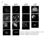
- FIG. 1 is a confocal laser scanning micrograph of the artificial tissues of Examples 1 to 3 and the islet culture of the comparative example.
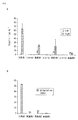
- FIG. 2 is a graph showing the results of measurement of insulin secretion in Examples 1 to 3, Controls 1 to 3 and Comparative Example, and FIG. 2A shows insulin secretion in low glucose medium (Low) and high glucose medium (High).
- FIG. 2B shows the ratio of insulin secretion in Examples 1 to 3 and Comparative Example (stimulation index).
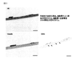
- FIG. 3 shows hematoxylin-eosin (HE) stained image (A), insulin immunostained image (B), and CD31 immunostained image (C) of a section of the artificial tissue used for transplantation into mice. The arrow in FIG.
- HE hematoxylin-eosin
- FIG. 4 is a phase contrast microscopic image (A) and an insulin and CD31 immunostained image (B) of the graft 14 days after transplantation.
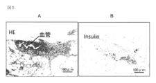
- FIG. 5 is a hematoxylin-eosin (HE) stained image (A) and an insulin immunostained image (B) of a section of the graft in the abdominal wall fat 14 days after transplantation.
- HE hematoxylin-eosin
- the present invention is vascularized from the viewpoint that ischemia immediately after transplantation, which is an important factor that prevents islet engraftment, is caused by the time required to form a vascular network between the transplanted islet and the transplant site. This is based on the knowledge that transplanting the islets can form a blood vessel network between the transplanted islets and the transplant site at an early stage, and the blood flow of the transplanted islets can be recovered early. Further, the present invention is based on the finding that vascularized islets can be obtained by culturing pancreatic islets with fibroblasts and / or cells having differentiation ability, vascular endothelial cells and extracellular matrix. .
- a blood vessel network between the transplant site and the transplanted islet is formed early, and blood flow between the transplant site and the transplanted islet can be generated early. It is expected that ischemia in the early stage of transplantation can be avoided and the survival of islets is improved. According to the present invention, in one or a plurality of embodiments, faster blood flow improvement can be expected than a conventional method of promoting angiogenesis after transplantation. According to the present invention, in one or a plurality of embodiments, improvement of transplantation efficiency and improvement of transplantation results can be expected finally.
- muscles and subcutaneouss that are more safe transplant sites but have poor blood flow and are difficult to engraft islets are effectively replaced with current intra-hepatic transplants. It can be used as a proper transplant site.
- it is possible to provide an artificial tissue that is excellent in insulin secretion ability due to sugar load.
- vascularized tissue refers to a tissue body including an islet and a vascular network structure.
- vascularized islet refers to an islet in which a vascular network structure is formed or an vascularized islet, and preferably an islet in which a vascular network structure is formed so as to be adjacent to the periphery of the islet. More preferably, it refers to a pancreatic islet capable of inflow of blood flow through a vascular network structure formed around the pancreatic islet.
- the “vascular network structure” refers to a hollow tubular structure formed by vascular endothelial cells, preferably a vascular network, more preferably a network network such as a capillary network. Refers to the structure.
- the artificial tissue in the present invention refers to what can be a functional or structural model of an islet or pancreas.
- the artificial tissue in the present invention may include a model capable of reproducing the function of the islet or a model for reproducing.
- the artificial tissue in the present invention can be used for islet transplantation in one or a plurality of embodiments.
- the cell may be a human-derived cell or a cell derived from an animal other than a human.
- the cells may be cells derived from mesenchymal stem cells, embryonic stem cells, induced pluripotent stem cells, and the like.
- the cell may be a cultured cell in one or more embodiments. Examples of the cultured cells include primary cultured cells, subcultured cells, and cell line cells in one or more embodiments.
- the present invention relates to an artificial tissue including pancreatic islet cells, fibroblasts and / or cells having differentiation ability, extracellular matrix and vascular endothelial cells (hereinafter referred to as “the artificial tissue of the present invention”).
- the artificial tissue of the present invention vascular endothelial cells form a vascular network structure.
- a plurality of islet cells, preferably 10 or more are gathered to form an islet.
- the artificial tissue of the present invention comprises a three-dimensional tissue body in which a fibroblast and / or a cell having differentiation ability, an extracellular matrix, and a vascular endothelial cell are formed with a vascular network structure.
- the three-dimensional tissue structure includes an islet in which a plurality of, preferably 10 or more, islet cells are assembled.
- the fibroblasts and / or cells having differentiation potential mean both fibroblasts, cells having differentiation ability, and both fibroblasts and cells having differentiation ability. There is a case.
- an islet is referred to as an aggregate of islet cells configured by aggregating a plurality of islet cells, preferably 10 or more.
- “islet cell” refers to a pancreatic endocrine cell capable of secreting insulin, glucagon, and the like in one or a plurality of embodiments, and preferably refers to a pancreatic endocrine cell capable of secreting insulin, glucagon, etc. .
- the islet is a collection of islet cells, and includes islet cell spheroids, preferably capable of secreting or inducing insulin, more preferably functioning closer to the islet in the living body.
- the islets include 1 or more, 10 or more, 20 or more, 30 or more, 40 or more, 50 or more, 60 or more, or 70 or more islet cells. Although an upper limit is not specifically limited, In one or some embodiment, 1000 or less may be sufficient.
- the islet is formed so as to be in contact with the vascular network structure, and the vascular network structure is preferably formed so as to surround the islet. Is formed.
- the artificial tissue of the present invention includes 1 or more, 5 or more, 10 or more, 15 or more, 20 or more, 25 or more, 30 or more, or 45 or more islets. . Although an upper limit is not specifically limited, In one or some embodiment, 1000 or less may be sufficient.
- the extracellular matrix may include a substance that can play the above-described role in in vitro cell culture, an artificially synthesized substance, or a part thereof.
- Specific examples of the extracellular matrix include fibronectin, gelatin, collagen, laminin, and polylysine in one or a plurality of embodiments.
- the extracellular matrix is not limited to these examples, and examples thereof include those disclosed in JP2007-228921A (Patent No. 4919464) and JP2012-115254A.
- the extracellular matrix is composed of a substance that does not show significant toxicity to the living body, such as fibronectin, gelatin, collagen, laminin, and polylysine. It is desirable.
- the extracellular matrix may be one type or two or more types.
- Examples of the combination of extracellular matrices include a combination of a first substance and a second substance that interacts with the first substance in one or a plurality of embodiments.
- the combination of the first substance and the second substance is a combination of a polymer having an RGD sequence and a polymer that interacts with a polymer having an RGD sequence, or having a positive charge.
- Examples include a combination of a polymer and a polymer having a negative charge.
- the combination of the first substance and the second substance include fibronectin and gelatin, fibronectin and ⁇ -polylysine, fibronectin and hyaluronic acid, fibronectin and dextran sulfate, fibronectin and heparin, and laminin in one or more embodiments. And gelatin.
- the artificial tissue of the present invention may include islet cells, fibroblasts and / or cells having differentiation ability and cells other than vascular endothelial cells.
- the cells other than the above include cells constituting organs other than pancreas such as hepatocytes and kidney cells in one or a plurality of embodiments.
- the present invention forms a three-dimensional tissue body by culturing fibroblasts and / or cells having differentiation ability, vascular endothelial cells, and extracellular matrix, and
- the present invention relates to a method for producing an artificial tissue having pancreatic islets (hereinafter, also referred to as “the production method of the present invention”), which comprises placing and culturing pancreatic islet cells simultaneously with or after the formation of a three-dimensional tissue body.
- the fibroblast and / or the cell having differentiation ability, the extracellular matrix and the vascular endothelial cell constitute a three-dimensional tissue in which a vascular network structure is formed, and the three-dimensional
- the tissue body can produce an artificial tissue including an islet constituted by aggregating 10 or more islet cells.
- an artificial tissue including an islet and a vascular network structure can be produced.
- a tissue in which a vascular network structure is formed around an islet preferably a tissue capable of inflow of blood flow to the islet cells through the vascular network structure is produced. be able to.
- the artificial tissue of the present invention can be easily produced.
- Examples of cells having differentiation ability include stem cells such as mesenchymal stem cells (MSC), embryonic stem cells (ES cells), and induced pluripotent stem cells (iPS cells) in one or a plurality of embodiments.
- the cell having differentiation ability may be, for example, a cell that can differentiate into a fibroblast, or a cell that can differentiate into another cell.
- the production method of the present invention includes culturing fibroblasts and / or cells having differentiation ability, vascular endothelial cells, and extracellular matrix to form a three-dimensional tissue body.
- formation of a three-dimensional tissue body can be performed by coating cells with a coating containing an extracellular matrix and culturing the coated cells in a three-dimensional arrangement (coated cells). Method).
- cells (one layer) and an extracellular matrix are alternately laminated and cultured (method by LBL). You may perform combining the method using a coated cell, and the method by LBL.
- the method using coated cells can be produced based on the examples and the methods disclosed in JP2012-115254A and the like.
- the method using LBL can be manufactured based on the method disclosed in Japanese Patent Application Laid-Open No. 2007-228921 (Patent No. 4919464).
- the production method of the present invention includes placing and culturing pancreatic islet cells simultaneously with or after the formation of the three-dimensional tissue.
- the islet cells may be arranged in combination with fibroblasts and / or cells having differentiation ability at the same time as the formation of the three-dimensional tissue. You may arrange
- the three-dimensional tissue is formed by culturing fibroblasts and / or cells having differentiation ability to form a cell layer, and then placing vascular endothelial cells on the formed cell layer. It can be performed by culturing to form a single vascular endothelial cell layer, then mixing fibroblasts and / or cells with differentiation ability and islet cells, placing them on the vascular endothelial cell layer, and culturing. .
- vascular endothelial cells are arranged on the cell layer and cultured to form one vascular endothelial cell layer. Then, fibroblasts and / or cells having differentiation ability are placed on the vascular endothelial cell layer and cultured to form a laminate having a vascular network structure, and then islet cells are placed on the laminate and cultured. This can be done.
- fibroblasts and / or cells having differentiation ability vascular endothelial cells and islet cells can be mixed and cultured.
- fibroblasts and / or cells having differentiation ability and vascular endothelial cells are mixed and cultured, and then islet cells are placed thereon and cultured.
- the production method of the present invention includes, as one or a plurality of embodiments, using islets in which a plurality of islet cells, preferably 10 or more, are gathered as islet cells.
- the islets include isolated islets and spheroids / clumps obtained by reconstructing islet cells.
- the production method of the present invention includes culturing fibroblasts and / or cells having differentiation ability, vascular endothelial cells, extracellular matrix, and islets.
- the number of islets to be arranged is 1 or more, 5 or more, 10 or more, 15 or more, 20 or more, 25 or more, 30 or more, or 45 or more. Although an upper limit is not specifically limited, In one or some embodiment, 1000 or less may be sufficient.
- fibroblasts hereinafter also referred to as “coated fibroblasts”
- vascular endothelial cells hereinafter also referred to as “coated vascular endothelial cells” covered with a coating containing an extracellular matrix are prepared.
- the coating containing the extracellular matrix can be formed by alternately laminating fibronectin and gelatin.
- the coated fibroblasts are placed on a substrate such as a cell culture insert and cultured.
- the arrangement of the coated fibroblasts is performed so that the coated fibroblasts are laminated in 4 layers or more, preferably 5 layers, 8 layers or 10 layers or more.
- the density of fibroblasts at the time of placement is 1 ⁇ 10 2 to 1 ⁇ 10 9 cells / cm 3 , 1 ⁇ 10 4 to 1 ⁇ 10 8 cells / cm 3, or 1 ⁇ 10 5. ⁇ 1 ⁇ 10 7 pieces / cm 3 .
- the culture is performed by adding a medium.
- the medium may include Eagle's MEM medium, Dulbecco's Modified Eagle medium (DMEM), Modified Eagle medium (MEM), Minimum Essential medium, RDMI, and GlutaMax medium.
- the medium is preferably a medium supplemented with serum.
- the culture temperature is 37 ° C. in one or more embodiments. In one or a plurality of embodiments, the culture time is 6 to 24 hours.
- the coated vascular endothelial cells are placed on the formed fibroblast laminate and cultured.
- the coated vascular endothelial cells are arranged so that one layer of vascular endothelial cells is formed.
- the density of vascular endothelial cells at the time of placement is 1 ⁇ 10 2 to 1 ⁇ 10 9 cells / cm 3 , 1 ⁇ 10 4 to 1 ⁇ 10 8 pieces / cm 3 or 1 ⁇ 10 5 to 1 ⁇ 10 7 pieces / cm 3 .
- the culture conditions are as described above.
- a mixture of coated fibroblasts and isolated islets is placed on vascular endothelial cells and cultured.
- the number of islets to be arranged, the density of the coated fibroblasts, and the culture conditions are as described above. Thereby, an artificial tissue in which a capillary-like structure is formed around the islet can be formed.
- coated fibroblasts and coated vascular endothelial cells are prepared. Coated fibroblasts and coated vascular endothelial cells are mixed and placed on a substrate such as a cell culture insert and cultured. The density and culture conditions of the coated fibroblasts and the coated vascular endothelial cells are the same as in the first embodiment. Thereby, the laminated body which has a vascular network structure is formed.
- the isolated islets are placed on the formed laminate and cultured.
- the number of islets to be arranged and the culture conditions are the same as in the first embodiment. Thereby, an artificial tissue in which a capillary-like structure is formed around the islet can be formed.
- a mesenchymal stem cell covered with a coating containing an extracellular matrix (hereinafter, “coated mesenchymal stem cell”) is prepared.
- the coated mesenchymal stem cells, the coated vascular endothelial cells and the isolated islets are mixed and placed on a substrate such as a cell culture insert and cultured.
- the density of the coated mesenchymal stem cells is similar to the density of the coated fiber subcells.
- the density of the coated vascular endothelial cells, the number of islets, and the culture conditions are the same as in the first embodiment. Thereby, an artificial tissue in which a capillary-like structure is formed around the islet can be formed.
- the present invention may relate to one or more of the following embodiments.
- the fibroblasts and / or cells having differentiation ability, the extracellular matrix and the vascular endothelial cells constitute a three-dimensional tissue in which a vascular network structure is formed,
- the three-dimensional tissue body is an artificial tissue including a pancreatic islet constituted by a collection of a plurality of the pancreatic islet cells.
- [5] Forming a three-dimensional tissue body by culturing fibroblasts and / or cells having differentiation ability, vascular endothelial cells, and an extracellular matrix, and simultaneously with the formation of the three-dimensional tissue body or Subsequently placing and culturing the islet cells, The fibroblasts and / or cells having differentiation ability, the extracellular matrix and the vascular endothelial cells constitute a three-dimensional tissue in which a vascular network structure is formed,
- the three-dimensional tissue body is a method for producing an artificial tissue including an islet constituted by a collection of a plurality of the islet cells.
- [6] The production method according to [5], wherein the fibroblast and / or the cell having differentiation ability is a cell whose surface is covered with a coating containing an extracellular matrix.
- From [5] to [5] including mixing and culturing the fibroblasts covered with a capsule and / or cells having differentiation ability, the vascular endothelial cells covered with a capsule, and the islet cells.
- [8] The production method according to any one of [8]. [10] culturing the fibroblasts covered with a coat and / or cells having differentiation ability and the vascular endothelial cells covered with a coat, and the islet cells on the cell layer formed by the culture. The production method according to any one of [5] to [8], comprising arranging and culturing.
- [11] Disposing and culturing the fibroblasts and / or cells having differentiation ability covered with a coating, and vascular endothelial cells covered with the coating on a cell layer formed by the culture Placing and culturing, and placing and culturing the pancreatic islet cells mixed with the fibroblasts and / or cells having differentiation potential covered with a capsule on the vascular endothelial cells, [5] The production method according to any one of [8]. [12] The production method according to any one of [5] to [11], wherein stem cells are used as the cells having differentiation ability. [13] The production method according to any one of [5] to [12], comprising producing the artificial tissue according to any one of [1] to [4].
- this solution is referred to as “FN Tris solution”.
- Gelatin Wang, 077-3155, 500 g
- this solution is referred to as “G Tris solution”.
- Tris-HCl 2-Amino-2-hydroxymethyl-1,3-propanol Hydrochloride (Tris-HCl) (012-17455, manufactured by Wako) was adjusted to 50 mM, adjusted to pH 7.4, and sterilized by autoclave.
- Vascular endothelial cell culture kit EGM-2 MV, manufactured by Lonza, CC-3202 ⁇ Cell> Human skin-derived fibroblasts (NHDF): Lonza, CC-2509 (newborn) Human umbilical cord-derived vascular endothelial cells (HUVEC): Lonza, CC-2517 Human bone marrow-derived mesenchymal stem cells (BM-MSC): manufactured by Promocell, C-12974 Mouse islet: Provided by Dr. Naoaki Sakata, Tohoku University
- FN-G thin film was prepared on NHDF by the following procedure. 1 ml of FN Tris solution was added to NHDF (1 ⁇ 10 7 cels), incubated for 1 minute, and centrifuged at 420 ⁇ g (2500 rpm) for 1 minute to remove the supernatant. 1 ml of Tris-HCl was added and washed for 1 minute, and centrifuged at 420 ⁇ g (2500 rpm) for 1 minute to remove the supernatant. Subsequently, the same operation was performed except that the G tris solution was used instead of the FN tris solution.
- a total of 9 steps ((FN / G) 4 FN) with incubation and washing as one step were performed to form a FN-G thin film on the NHDF surface to prepare a coated NHDF.
- a FN-G thin film was formed in the same manner except that HUVEC or BM-MSC was used instead of NHDF to prepare a coated HUVEC or a coated BM-MSC.
- a laminated tissue (4 layers, NHDF tissue) was obtained.
- Coated HUVEC suspended in DMEM (10% FBS) was seeded on NHDF tissue at 3 ⁇ 10 4 cells / well and cultured for 24 hours to obtain a laminated tissue (NHDF4 layer-HUVEC1 layer).
- 4 ⁇ 10 5 cells / well coated NHDF and 50 / well isolated islets were mixed, suspended in DMEM (10% FBS), seeded on the laminated tissue, and cultured for 24 hours. Thereby, an artificial tissue was obtained.
- Measurement of the amount of insulin secreted from the obtained artificial tissue and tissue observation of the artificial tissue were performed. Measurement of the amount of insulin secretion was performed by loading glucose solutions of different concentrations onto an artificial tissue and measuring the amount of insulin secreted into the solution by the ELISA method.
- pre-incubation with a low glucose medium 3.3 mM glucose (60 mg / dl) was performed by the following procedure. After 1 hour incubation with 10 mL of low glucose medium, the cells were washed 3 times with 5 mL of low glucose medium. After completion of the pre-incubation, the supernatant was collected after culturing in 10 mL of low glucose medium for 1 hour (sample L).
- sample H After collecting the supernatant, the medium was switched to 10 mL of high glucose medium (16.5 mM glucose (60 mg / dl), and cultured for 1 hour, and then the supernatant was collected (sample H). Each collected supernatant (sample L and sample H) was frozen at liquid nitrogen and stored at -80 ° C. After recovering the supernatant, the artificial tissue is washed with PBS, and fixed with 10% formalin for 3 hours for tissue section preparation (in the case of fluorescence observation, fixed with 4% PFA for 15 minutes) and then HE or immunostaining. Tissue observation was performed.
- the amount of insulin secretion in the collected supernatant was measured according to the attached document of the mouse insulin ELISA kit.
- the Eliser kit used was Leba's insulin-mouse (AKRIN-031) from Shibayagi. Specifically, a biotin-conjugated anti-insulin antibody was placed on an antibody solidified plate, and a sample was further placed and incubated at room temperature for 2 hours. After washing the wells, the peroxidase-avidin conjugate was allowed to react by incubation at room temperature for 30 minutes, washed and then reacted with the coloring solution for 30 minutes. After stopping the reaction, the plate was subjected to a plate reader (absorbance 450 nm, 620 nm).
- SI stimulation index
- the supernatant was collected for measuring the amount of insulin and the amount of insulin was measured in the same manner as above except that the islets were not seeded.
- a mixture of 8 ⁇ 10 5 cells / well of coated NHDF and 3 ⁇ 10 4 cells / well of coated HUVEC was seeded on an insert formed with an FN underlayer and cultured for 24 hours.
- isolated islets were seeded at 50 / well and cultured for 1 day to obtain an artificial tissue.
- the amount of insulin secretion and tissue observation were performed in the same manner as in Example 1.
- Example 2 8 ⁇ 10 5 cells / well coated BM-MSC, 3 ⁇ 10 4 cells / well coated HUVEC and 50 / well isolated islets were mixed and seeded on an insert with an FN underlayer formed, The artificial tissue was obtained by culturing for 1 day. Using the obtained artificial tissue, the amount of insulin secretion and tissue observation were performed in the same manner as in Example 1.
- FIG. 1 is a confocal laser scanning micrograph of the artificial tissues of Examples 1 to 3 and the islet culture of the comparative example.
- FIG. 2 is a graph showing the results of measurement of insulin secretion in Examples 1 to 3, Controls 1 to 3 and Comparative Example, and FIG. 2A shows insulin secretion in low glucose medium (Low) and high glucose medium (High).
- FIG. 2B shows a simulation index.
- the islets appear to be rice grains (FIGS. 1a, f and g).
- FIGS. 1a, f and g rice grains
- FIG. 1 in the artificial tissues of Examples 1 to 3, a vascular network structure is formed around the pancreatic islets, and a good shape is maintained for any pancreatic islets located in and on the tissue was observed. It was observed that the islets of the artificial tissues of Examples 1 and 2 were composed of a larger number compared to the artificial tissue of Example 3 (FIGS. 1b-c, gh and kl). .
- the artificial tissues of Examples 1 to 3 may cause insulin secretion in response to a change in glucose concentration, although the total amount of insulin secretion is lower than that of Comparative Example 1 (islet alone). It could be confirmed.
- FIG. 3A shows the result of hematoxylin-eosin (HE) staining of a section of the artificial tissue according to a conventional method. Furthermore, the results of immunostaining the artificial tissue for insulin and CD31 according to a conventional method are shown in FIGS. 3B and 3C, respectively. Pancreatic islets secreting insulin were observed on the cell layer of the obtained artificial tissue, and vascular structures were observed in the cell layer.
- HE hematoxylin-eosin
- the artificial tissue was transplanted into the abdominal wall fat of mice.
- the procedure was as follows. An artificial tissue was prepared on a 24-well transwell in the same manner as explained in Example 1. The obtained artificial tissue had a diameter of 6.5 mm.
- the artificial tissue contained 150 islets of BALB / c mice. According to the method described in Example 1, a glucose load was applied to the obtained artificial tissue, and after confirming that insulin secretion increased in response to the glucose load, the artificial tissue was transplanted into a mouse.
- SCID mouse immunodeficient mouse induced by diabetes with streptozotocin was laparotomized under inhalation anesthesia, and the adipose tissue adhering to the testis was spread, and an artificial tissue removed from the transwell with the membrane was placed on it. The artificial tissue was wrapped with an open membrane and returned to the abdominal cavity to close the abdomen, completing the transplantation.
- FIG. 4A shows a phase contrast microscopic image of the graft of the graft in the mouse abdominal wall fat 14 days after transplantation.
- FIG. 4B shows the result of immunostaining (CD31 and insulin) of the obtained artificial tissue according to a conventional method.
- a CD31 immunostaining positive part blood vessel, stained red
- insulin immunostaining positive part islet, stained green
- the graft in the mouse abdominal wall fat on the 14th day after transplantation was sectioned, and HE staining and immunostaining (in which DAB staining was used as the primary antibody) of anti-mouse insulin antibody for insulin were performed.
- the result of HE staining is shown in FIG. 5A.
- the result of insulin immunostaining is shown in FIG. 5B.
- the islets were engrafted in an integrated state with a cell layer composed of fibroblasts and vascular endothelial cells. A vascular network and internal blood flow (red blood cells) were confirmed in the cell layer. Insulin secretion from the islets was also confirmed.
- the purpose of this experiment was to investigate whether the artificial tissue of the present invention transplanted and engrafted in a living body actually lowers blood glucose level.
- 8 ⁇ 10 5 BALB / c mouse mesenchymal stem cells (MSC) and HUVEC 1 ⁇ 10 5 cells coated with fibronectin and gelatin were seeded in the transwell and cultured for 2 days.
- 150 BALB / c mouse islets just separated were seeded and cultured for 1 day to obtain an artificial tissue.
- Diabetically induced SCID mice were laparotomized under inhalation anesthesia, the fat tissue attached to the left testis was spread, the artificial tissue removed from the transwell was placed together with the membrane on the fat tissue, and this was wrapped in the remaining fat tissue. I put it back. The abdominal wall skin was closed and the operation was completed.
- 150 islets were wrapped in the fat tissue. Islet transplantation was performed on 4 animals and artificial tissue transplantation on 1 animal.
- the blood glucose level was normalized (200 mg / dL or less) on the 14th day after transplantation, and then decreased to about 130 mg / dL on the 21st day.
- the blood glucose level did not fall below about 300 mg / dL even on the 21st day of transplantation, and normalization was not achieved.
- the artificial tissue of the present invention can increase insulin secretion in response to glucose load, engraft in the living body, secrete sufficient insulin, and normalize blood glucose level. It was.
Landscapes
- Health & Medical Sciences (AREA)
- Life Sciences & Earth Sciences (AREA)
- Engineering & Computer Science (AREA)
- Biomedical Technology (AREA)
- Chemical & Material Sciences (AREA)
- Cell Biology (AREA)
- Zoology (AREA)
- General Health & Medical Sciences (AREA)
- Animal Behavior & Ethology (AREA)
- Medicinal Chemistry (AREA)
- Public Health (AREA)
- Veterinary Medicine (AREA)
- Epidemiology (AREA)
- Biotechnology (AREA)
- Chemical Kinetics & Catalysis (AREA)
- Bioinformatics & Cheminformatics (AREA)
- Organic Chemistry (AREA)
- Dermatology (AREA)
- Oral & Maxillofacial Surgery (AREA)
- Transplantation (AREA)
- Botany (AREA)
- Wood Science & Technology (AREA)
- Genetics & Genomics (AREA)
- Developmental Biology & Embryology (AREA)
- Urology & Nephrology (AREA)
- Pharmacology & Pharmacy (AREA)
- Immunology (AREA)
- Virology (AREA)
- Microbiology (AREA)
- General Engineering & Computer Science (AREA)
- Vascular Medicine (AREA)
- Biochemistry (AREA)
- Diabetes (AREA)
- Hematology (AREA)
- Biophysics (AREA)
- Physiology (AREA)
- Nutrition Science (AREA)
- Gastroenterology & Hepatology (AREA)
- Molecular Biology (AREA)
- General Chemical & Material Sciences (AREA)
Abstract
膵島細胞、線維芽細胞および/または分化能を有する細胞、細胞外マトリックス及び血管内皮細胞を含み、前記線維芽細胞および/または分化能を有する細胞、前記細胞外マトリックス及び前記血管内皮細胞は、血管網構造が形成された三次元組織体を構成し、前記三次元組織体は、前記膵島細胞が10個又はそれ以上集合した膵島を含む人工組織。
Description
本発明は、人工組織及びその製造方法に関する。
膵島移植は、血糖の是正を図る重症糖尿病患者に対する有効な治療方法として認められている。膵島移植は、ドナー膵臓の膵ランゲルハンス島を分離抽出し、レシピエントに移植することにより行われるが、慢性的なドナー不足が解消されていないことに加え、移植された膵島は多くの素因により生着できずに脱落するという問題が指摘されている。膵島の生着を妨げる重要な要因に移植直後の虚血が挙げられている。
膵島移植の成功率を向上させるための様々な研究がなされている。その一つの方法として、血管新生因子等により移植された膵島に血管化を図る方法がある(非特許文献1~3)。非特許文献1は、VEGFなどの血管新生因子で膵島を培養し、血管新生を図ることを開示する。非特許文献2は、薬剤投与により血管新生因子を誘導し、膵島の血管新生を図る方法を開示する。非特許文献3は、間葉系幹細胞など、血管新生因子を分泌する細胞との共移植により血管新生を図る方法を開示する。
Gorden DL,et al.Transplantation.1997 Feb 15;63(3):436−43
Samikannu B,et al.Plos One 2013 December,Volume 8,Issue 12,e82639
Sakata N,et al.Transplantation.2010 March 27;89(6):686−693
本発明は、血管化された膵島を含む人工組織を提供する。
本発明は、一又は複数の実施形態において、膵島細胞、線維芽細胞および/または分化能を有する細胞、細胞外マトリックス及び血管内皮細胞を含み、前記線維芽細胞および/または分化能を有する細胞、前記細胞外マトリックス及び前記血管内皮細胞は、血管網構造が形成された三次元組織体を構成し、前記三次元組織体は、前記膵島細胞が複数個集合した膵島を含む人工組織に関する。
本発明は、一又は複数の実施形態において、線維芽細胞および/または分化能を有する細胞と、血管内皮細胞と、細胞外マトリックスとを培養することにより三次元組織体を形成すること、及び前記三次元組織体の形成と同時又はその後に膵島細胞を配置して培養することを含み、前記線維芽細胞および/または分化能を有する細胞、前記細胞外マトリックス及び前記血管内皮細胞は、血管網構造が形成された三次元組織体を構成し、前記膵島細胞が複数個集合した膵島を含む人工組織を製造する方法に関する。
本発明によれば、血管化された膵島を含む人工組織を提供できる。
本発明は、膵島の生着を妨げる重要な要因である移植直後の虚血が、移植された膵島と移植部位との血管網の形成までに時間を要することにより生じるという観点から、血管化された膵島を移植することで移植された膵島と移植部位との血管網が早期に形成され、移植された膵島の血流が早期に回復されうる、との知見に基づく。また、本発明は、膵島を、線維芽細胞および/または分化能を有する細胞、血管内皮細胞及び細胞外マトリックスとともに培養することによって、血管化された膵島を得ることができる、との知見に基づく。
本発明によれば、一又は複数の実施形態において、移植部位と移植された膵島との血管網が早期に形成され、移植部位と移植された膵島との血流が早期に生じうることから、移植早期の虚血を回避でき、膵島の生着が向上することが期待できる。本発明によれば、一又は複数の実施形態において、従来の移植後に血管新生を促進する方法よりもより早い血流改善が期待できる。本発明によれば、一又は複数の実施形態において、最終的には移植効率の向上と移植成績の改善が期待できる。また、これらの結果、本発明によれば、一又は複数の実施形態において、さらに安全な移植部位でありながら血流に乏しく膵島が生着しにくい筋肉や皮下を現行の肝臓内移植に代わる有効な移植部位として使用することが可能になる。
本発明によれば、一又は複数の実施形態において、糖負荷によるインスリン分泌能に優れる人工組織を提供できる。
本発明によれば、一又は複数の実施形態において、糖負荷によるインスリン分泌能に優れる人工組織を提供できる。
本発明において「人工組織」とは、膵島と血管網構造とを含む組織体をいう。本発明おいて「血管化された膵島」とは、血管網構造が形成された膵島又は血管形成された膵島をいい、好ましくは膵島の周囲に隣接するように血管網構造が形成された膵島をいい、より好ましくは膵島の周囲に形成された血管網構造を通じて血流の流入が可能な膵島をいう。本発明において「血管網構造」とは、血管内皮細胞によって形成された中空の管状構造をいい、好ましくは脈管ネットワークをいい、より好ましくは毛細血管網のような網目状のネットワークに形成された構造をいう。
本発明における人工組織は、一又は複数の実施形態において、膵島又は膵臓の機能的又は構造的なモデルとなりうるものをいう。本発明における人工組織は、一又は複数の実施形態において、膵島の機能を再現可能なモデル又は再現するためのモデル等を含みうる。また、本発明における人工組織は、一又は複数の実施形態において、膵島移植に用いることができる。
本発明において、細胞は、一又は複数の実施形態において、ヒト由来の細胞であってもよいし、ヒト以外の動物由来の細胞であってもよい。細胞は、一又は複数の実施形態において、間葉系幹細胞、胚性幹細胞及び人工多能性幹細胞等の由来の細胞であってもよい。細胞は、一又は複数の実施形態において、培養細胞であってもよい。培養細胞としては、一又は複数の実施形態において、初代培養細胞、継代培養細胞、及び細胞株細胞等が挙げられる。
[人工組織]
本発明は、一又は複数の実施形態において、膵島細胞、線維芽細胞および/または分化能を有する細胞、細胞外マトリックス及び血管内皮細胞を含む人工組織(以下、「本発明の人工組織」)に関する。本発明の人工組織は、一又は複数の実施形態において、血管内皮細胞が、血管網構造を形成する。また、本発明の人工組織は、膵島細胞が複数個、好ましくは10個又はそれ以上集合して膵島を形成する。したがって、本発明の人工組織は、一又は複数の実施形態において、線維芽細胞および/または分化能を有する細胞、細胞外マトリックス及び血管内皮細胞が、血管網構造が形成された三次元組織体を構成し、三次元組織体は、前記膵島細胞が複数個、好ましくは10個又はそれ以上集合した膵島を含む。なお、線維芽細胞および/または分化能を有する細胞とは、線維芽細胞を意味する場合と、分化能を有する細胞を意味する場合と、線維芽細胞および分化能を有する細胞の両方を意味する場合がある。
本発明は、一又は複数の実施形態において、膵島細胞、線維芽細胞および/または分化能を有する細胞、細胞外マトリックス及び血管内皮細胞を含む人工組織(以下、「本発明の人工組織」)に関する。本発明の人工組織は、一又は複数の実施形態において、血管内皮細胞が、血管網構造を形成する。また、本発明の人工組織は、膵島細胞が複数個、好ましくは10個又はそれ以上集合して膵島を形成する。したがって、本発明の人工組織は、一又は複数の実施形態において、線維芽細胞および/または分化能を有する細胞、細胞外マトリックス及び血管内皮細胞が、血管網構造が形成された三次元組織体を構成し、三次元組織体は、前記膵島細胞が複数個、好ましくは10個又はそれ以上集合した膵島を含む。なお、線維芽細胞および/または分化能を有する細胞とは、線維芽細胞を意味する場合と、分化能を有する細胞を意味する場合と、線維芽細胞および分化能を有する細胞の両方を意味する場合がある。
膵島は、一又は複数の実施形態において、膵島細胞が複数個、好ましくは10個以上集合することにより構成される膵島細胞の集合体という。本発明において「膵島細胞」は、一又は複数の実施形態において、インスリンやグルカゴン等を分泌可能な膵内分泌細胞をいい、好ましくはインスリンやグルカゴン等を分泌して血糖調節可能な膵内分泌細胞をいう。膵島は、一又は複数の実施形態において、膵島細胞の集合体であって、膵島細胞スフェロイドが挙げられ、好ましくはインスリンの分泌又はその誘導が可能であり、より好ましくは生体における膵島により近い機能を有し、さらに好ましくは生体の膵島と同等の機能を有する。膵島は、一又は複数の実施形態において、膵島細胞を1個以上、10個以上、20個以上、30個以上、40個以上、50個以上、60個以上又は70個以上含む。上限は特に限定されないが、一又は複数の実施形態において、1000個以下であってもよい。
膵島は、血管網構造を通じて容易に酸素や栄養素等を流入させる点から、一又は複数の実施形態において、血管網構造と接するように形成されており、好ましくは膵島を取り囲むように血管網構造が形成されている。
本発明の人工組織は、一又は複数の実施形態において、膵島を1個以上、5個以上、10個以上、15個以上、20個以上、25個以上、30個以上、又は45個以上含む。上限は特に限定されないが、一又は複数の実施形態において、1000個以下であってもよい。
細胞外マトリックスは、一又は複数の実施形態において、in vitro細胞培養において上記の役割を果たしうる物質や、人工的に合成された物質やその一部を含んでもよい。細胞外マトリックスの具体例としては、一又は複数の実施形態において、フィブロネクチン、ゼラチン、コラーゲン、ラミニン及びポリリジン等が挙げられる。細胞外マトリックスは、これらの例に限定されるものではなく、特開2007−228921号公報(特許第4919464号)及び特開2012−115254号公報に開示のものが挙げられる。本発明の人工組織を生体に移植する場合には、細胞外マトリックスは、例えばフィブロネクチン、ゼラチン、コラーゲン、ラミニン及びポリリジン等の、生体に対して有意な毒性を示さない物質で構成されたものであることが望ましい。
細胞外マトリックスは、一種類であってもよいし、二種類以上であってもよい。細胞外マトリックスの組み合わせとしては、一又は複数の実施形態において、第1物質及び第1物質と相互作用する第2物質の組み合わせが挙げられる。第1物質と第2物質との組み合わせとしては、一又は複数の実施形態において、RGD配列を有する高分子及びRGD配列を有する高分子と相互作用する高分子の組み合わせ、又は、正の電荷を有する高分子及び負の電荷を有する高分子の組み合わせが挙げられる。第1物質と第2物質との組み合わせの具体例としては、一又は複数の実施形態において、フィブロネクチンとゼラチン、フィブロネクチンとε−ポリリジン、フィブロネクチンとヒアルロン酸、フィブロネクチンとデキストラン硫酸、フィブロネクチンとヘパリン、及びラミニンとゼラチン等が挙げられる。
本発明の人工組織は、一又は複数の実施形態において、膵島細胞、線維芽細胞および/または分化能を有する細胞及び血管内皮細胞以外の細胞を含んでいてもよい。上記以外の細胞としては、一又は複数の実施形態において、肝細胞、腎細胞等の膵臓以外の臓器を構成する細胞等が挙げられる。
[人工組織の製造方法]
本発明は、一又は複数の実施形態において、線維芽細胞および/または分化能を有する細胞と、血管内皮細胞と、細胞外マトリックスとを培養することにより三次元組織体を形成すること、及び 前記三次元組織体の形成と同時又はその後に膵島細胞を配置して培養することを含む、膵島を有する人工組織を製造する方法(以下、「本発明の製造方法」ともいう)に関する。本発明の製造方法によれば、線維芽細胞および/または分化能を有する細胞、前記細胞外マトリックス及び前記血管内皮細胞は、血管網構造が形成された三次元組織体を構成し、前記三次元組織体は、前記膵島細胞が10個又はそれ以上集合することにより構成された膵島を含む人工組織を製造することができる。
本発明は、一又は複数の実施形態において、線維芽細胞および/または分化能を有する細胞と、血管内皮細胞と、細胞外マトリックスとを培養することにより三次元組織体を形成すること、及び 前記三次元組織体の形成と同時又はその後に膵島細胞を配置して培養することを含む、膵島を有する人工組織を製造する方法(以下、「本発明の製造方法」ともいう)に関する。本発明の製造方法によれば、線維芽細胞および/または分化能を有する細胞、前記細胞外マトリックス及び前記血管内皮細胞は、血管網構造が形成された三次元組織体を構成し、前記三次元組織体は、前記膵島細胞が10個又はそれ以上集合することにより構成された膵島を含む人工組織を製造することができる。
本発明の製造方法によれば、一又は複数の実施形態において、膵島と血管網構造とを含む人工組織を製造することができる。本発明の製造方法によれば、一又は複数の実施形態において、膵島の周囲に血管網構造が形成された組織、好ましくは血管網構造を通じて膵島細胞に血流の流入が可能な組織を製造することができる。本発明の製造方法によれば、本発明の人工組織を容易に製造することができる。
分化能を有する細胞としては、一又は複数の実施形態において、間葉系幹細胞(MSC)、胚性幹細胞(ES細胞)及び人工多能性幹細胞(iPS細胞)などの幹細胞が例示される。分化能を有する細胞は、例えば線維芽細胞に分化しうる細胞であってもよく、他の細胞に分化しうる細胞であってもよい。
本発明の製造方法は、線維芽細胞および/または分化能を有する細胞、血管内皮細胞、及び細胞外マトリックスを培養して三次元組織体を形成することを含む。
三次元組織体の形成は、一又は複数の実施形態において、細胞を細胞外マトリックスを含む被膜で被覆し、被覆された細胞を三次元に配置して培養することにより行うことができる(被覆細胞を用いた方法)。また、そのほかの方法としては、細胞(1層)と細胞外マトリックスとを交互に積層して培養することが挙げられる(LBLによる方法)。被覆細胞を用いた方法とLBLによる方法とを組み合わせて行ってもよい。被覆細胞を用いた方法は、一又は複数の実施形態において、実施例、及び特開2012−115254号等に開示された方法等に基づき作製できる。LBLによる方法は、一又は複数の実施形態において、特開2007−228921号公報(特許第4919464号)に開示された方法等に基づき作製できる。
本発明の製造方法は、三次元組織体の形成と同時又はその後に膵島細胞を配置して培養することを含む。膵島細胞の配置は、一又は複数の実施形態において、三次元組織体の形成と同時に、線維芽細胞および/または分化能を有する細胞と混合して配置してもよいし、三次元組織体の形成後に三次元組織体上に配置してもよい。
三次元組織体の形成は、一又は複数の実施形態において、線維芽細胞および/または分化能を有する細胞を培養して細胞層を形成した後、形成した細胞層上に血管内皮細胞を配置し培養して1層の血管内皮細胞層を形成し、ついで線維芽細胞および/または分化能を有する細胞と膵島細胞とを混合して血管内皮細胞層上に配置し培養することにより行うことができる。その他の方法としては、線維芽細胞および/または分化能を有する細胞を培養して細胞層を形成した後、細胞層上に血管内皮細胞を配置し培養して1層の血管内皮細胞層を形成し、ついで血管内皮細胞層上に線維芽細胞および/または分化能を有する細胞を配置し培養して血管網構造が形成された積層体を形成した後、積層体上に膵島細胞を配置し培養することにより行うことができる。その他の方法としては、線維芽細胞および/または分化能を有する細胞と血管内皮細胞と膵島細胞を混合して培養することにより行うことができる。また、その他の方法としては、線維芽細胞および/または分化能を有する細胞と血管内皮細胞とを混合して培養した後、その上に膵島細胞を配置して培養することにより行うことができる。
本発明の製造方法は、一又は複数の実施形態として、膵島細胞として、膵島細胞が複数個、好ましくは10個又はそれ以上集合した膵島を使用することを含む。膵島は、一又は複数の実施形態において、単離した膵島、及び膵島細胞を再構築したスフェロイド/塊等が挙げられる。本発明の製造方法は、一又は複数の実施形態において、線維芽細胞および/または分化能を有する細胞、血管内皮細胞、細胞外マトリックス、及び膵島を培養することを含む。
配置する膵島の個数は、一又は複数の実施形態において、1個以上、5個以上、10個以上、15個以上、20個以上、25個以上、30個以上、又は45個以上である。上限は特に限定されないが、一又は複数の実施形態において、1000個以下であってもよい。
以下に、本発明を好適な実施形態を示しながら詳細に説明する。但し、本発明は、以下の実施形態に限定されるものではないことはいうまでもない。
(実施形態1)
まず、細胞外マトリックスを含む被膜で覆われた線維芽細胞(以下、「被覆線維芽細胞」ともいう)及び血管内皮細胞(以下、「被覆血管内皮細胞」ともいう)を準備する。細胞外マトリックスを含む被膜は、一又は複数の実施形態において、フィブロネクチン及びゼラチンを交互に積層することにより形成できる。
まず、細胞外マトリックスを含む被膜で覆われた線維芽細胞(以下、「被覆線維芽細胞」ともいう)及び血管内皮細胞(以下、「被覆血管内皮細胞」ともいう)を準備する。細胞外マトリックスを含む被膜は、一又は複数の実施形態において、フィブロネクチン及びゼラチンを交互に積層することにより形成できる。
被覆線維芽細胞をセルカルチャーインサート等の基材上に配置して培養する。被覆線維芽細胞の配置は、被覆線維芽細胞が4層以上、好ましくは5層、8層又は10層以上積層されるように行う。配置時の線維芽細胞の密度は、一又は複数の実施形態において、1×10²~1×10⁹個/cm3、1×104~1×108個/cm3又は1×105~1×107個/cm3である。
培養は、一又は複数の実施形態において、培地を添加して行う。培地は、一又は複数の実施形態において、Eagle’s MEM培地、Dulbecco’s Modified Eagle培地(DMEM)、Modified Eagle培地(MEM)、Minimum Essential培地、RDMI、及びGlutaMax培地等が挙げられる。培地は、一又は複数の実施形態において、血清を添加した培地が好ましい。培養温度は、一又は複数の実施形態において、37℃である。培養時間は、一又は複数の実施形態において、6~24時間である。
培養は、一又は複数の実施形態において、培地を添加して行う。培地は、一又は複数の実施形態において、Eagle’s MEM培地、Dulbecco’s Modified Eagle培地(DMEM)、Modified Eagle培地(MEM)、Minimum Essential培地、RDMI、及びGlutaMax培地等が挙げられる。培地は、一又は複数の実施形態において、血清を添加した培地が好ましい。培養温度は、一又は複数の実施形態において、37℃である。培養時間は、一又は複数の実施形態において、6~24時間である。
次に、形成された線維芽細胞の積層体の上に被覆血管内皮細胞を配置して培養する。被覆血管内皮細胞は、血管内皮細胞の層が1層形成されるように配置する。配置時の血管内皮細胞の密度は、一又は複数の実施形態において、1×10²~1×10⁹個/cm3、1
×104~1×10⁸個/cm3又は1×105~1×107個/cm3である。培養条件は上述のとおりである。
×104~1×10⁸個/cm3又は1×105~1×107個/cm3である。培養条件は上述のとおりである。
ついで、血管内皮細胞の上に被覆線維芽細胞と単離した膵島との混合物を配置して培養する。配置する膵島の個数、被覆線維芽細胞の密度及び培養条件は上記のとおりである。これにより、膵島の周囲に毛細血管様構造が形成された人工組織が形成できる。
(実施形態2)
まず、被覆線維芽細胞及び被覆血管内皮細胞を準備する。被覆線維芽細胞及び被覆血管内皮細胞を混合し、セルカルチャーインサート等の基材上に配置して培養する。被覆線維芽細胞及び被覆血管内皮細胞の密度及び培養条件は実施形態1と同様である。これにより、血管網構造を有する積層体が形成される。
まず、被覆線維芽細胞及び被覆血管内皮細胞を準備する。被覆線維芽細胞及び被覆血管内皮細胞を混合し、セルカルチャーインサート等の基材上に配置して培養する。被覆線維芽細胞及び被覆血管内皮細胞の密度及び培養条件は実施形態1と同様である。これにより、血管網構造を有する積層体が形成される。
ついで、形成された積層体上に、単離した膵島を配置して培養する。配置する膵島の個数及び培養条件は実施形態1と同様である。これにより、膵島の周囲に毛細血管様構造が形成された人工組織が形成できる。
(実施形態3)
まず、細胞外マトリックスを含む被膜で覆われた間葉系幹細胞(以下、「被覆間葉系幹細胞」)を準備する。
ついで、被覆間葉系幹細胞、被覆血管内皮細胞及び単離した膵島を混合し、セルカルチャーインサート等の基材上に配置して培養する。被覆間葉系幹細胞の密度は、被覆線維亜細胞の密度と同様である。被覆血管内皮細胞の密度、膵島の個数及び培養条件は実施形態1と同様である。これにより、膵島の周囲に毛細血管様構造が形成された人工組織が形成できる。
まず、細胞外マトリックスを含む被膜で覆われた間葉系幹細胞(以下、「被覆間葉系幹細胞」)を準備する。
ついで、被覆間葉系幹細胞、被覆血管内皮細胞及び単離した膵島を混合し、セルカルチャーインサート等の基材上に配置して培養する。被覆間葉系幹細胞の密度は、被覆線維亜細胞の密度と同様である。被覆血管内皮細胞の密度、膵島の個数及び培養条件は実施形態1と同様である。これにより、膵島の周囲に毛細血管様構造が形成された人工組織が形成できる。
本発明は、以下の一又は複数の実施形態に関しうる。
[1] 膵島細胞、線維芽細胞および/または分化能を有する細胞、細胞外マトリックス及び血管内皮細胞を含み、
前記線維芽細胞および/または分化能を有する細胞、前記細胞外マトリックス及び前記血管内皮細胞は、血管網構造が形成された三次元組織体を構成し、
前記三次元組織体は、前記膵島細胞が複数個集合することにより構成された膵島を含む、人工組織。
[2] 前記膵島は、10個又はそれ以上の膵島細胞が集合することにより構成された膵島である、[1]記載の人工組織。
[3] 前記膵島は、前記血管網構造と隣接する、[1]又は[2]に記載の人工組織。
[4] 前記膵島を5個又はそれ以上含む、[1]から[3]のいずれかに記載の人工組織。
[5] 線維芽細胞および/または分化能を有する細胞と、血管内皮細胞と、細胞外マトリックスとを培養することにより三次元組織体を形成すること、及び
前記三次元組織体の形成と同時又はその後に膵島細胞を配置して培養することを含み、
前記線維芽細胞および/または分化能を有する細胞、前記細胞外マトリックス及び前記血管内皮細胞は、血管網構造が形成された三次元組織体を構成し、
前記三次元組織体は、前記膵島細胞が複数個集合することにより構成された膵島を含む人工組織を製造する方法。
[6] 前記線維芽細胞および/または分化能を有する細胞は、表面が細胞外マトリックスを含む被膜で覆われた細胞である、[5]記載の製造方法。
[7] 前記血管内皮細胞は、表面が細胞外マトリックスを含む被膜で覆われた細胞である、[5]又は[6]に記載の製造方法。
[8] 前記膵島細胞として、10個又はそれ以上の膵島細胞が集合することにより構成された膵島を使用することを含む、[5]から[7]のいずれかに記載の製造方法。
[9] 被膜で覆われた前記線維芽細胞および/または分化能を有する細胞、被膜で覆われた前記血管内皮細胞、及び前記膵島細胞を混合して培養することを含む、[5]から[8]のいずれかに記載の製造方法。
[10] 被膜で覆われた前記線維芽細胞および/または分化能を有する細胞及び被膜で覆われた前記血管内皮細胞を培養すること、及び前記培養により形成された細胞層上に前記膵島細胞を配置して培養することを含む、[5]から[8]のいずれかに記載の製造方法。
[11] 被膜で覆われた前記線維芽細胞および/または分化能を有する細胞を配置して培養してすること、前記培養により形成された細胞層上に被膜で覆われた前記血管内皮細胞を配置して培養すること、及び前記血管内皮細胞の上に、被膜で覆われた前記線維芽細胞および/または分化能を有する細胞と前記膵島細胞を混合して配置し培養することを含む、[5]から[8]のいずれかに記載の製造方法。
[12] 分化能を有する細胞として幹細胞が用いられる、請求項[5]から[11]のいずれかに記載の製造方法。
[13] [1]から[4]のいずれかに記載の人工組織を製造することを含む、[5]から[12]のいずれかに記載の製造方法。
[1] 膵島細胞、線維芽細胞および/または分化能を有する細胞、細胞外マトリックス及び血管内皮細胞を含み、
前記線維芽細胞および/または分化能を有する細胞、前記細胞外マトリックス及び前記血管内皮細胞は、血管網構造が形成された三次元組織体を構成し、
前記三次元組織体は、前記膵島細胞が複数個集合することにより構成された膵島を含む、人工組織。
[2] 前記膵島は、10個又はそれ以上の膵島細胞が集合することにより構成された膵島である、[1]記載の人工組織。
[3] 前記膵島は、前記血管網構造と隣接する、[1]又は[2]に記載の人工組織。
[4] 前記膵島を5個又はそれ以上含む、[1]から[3]のいずれかに記載の人工組織。
[5] 線維芽細胞および/または分化能を有する細胞と、血管内皮細胞と、細胞外マトリックスとを培養することにより三次元組織体を形成すること、及び
前記三次元組織体の形成と同時又はその後に膵島細胞を配置して培養することを含み、
前記線維芽細胞および/または分化能を有する細胞、前記細胞外マトリックス及び前記血管内皮細胞は、血管網構造が形成された三次元組織体を構成し、
前記三次元組織体は、前記膵島細胞が複数個集合することにより構成された膵島を含む人工組織を製造する方法。
[6] 前記線維芽細胞および/または分化能を有する細胞は、表面が細胞外マトリックスを含む被膜で覆われた細胞である、[5]記載の製造方法。
[7] 前記血管内皮細胞は、表面が細胞外マトリックスを含む被膜で覆われた細胞である、[5]又は[6]に記載の製造方法。
[8] 前記膵島細胞として、10個又はそれ以上の膵島細胞が集合することにより構成された膵島を使用することを含む、[5]から[7]のいずれかに記載の製造方法。
[9] 被膜で覆われた前記線維芽細胞および/または分化能を有する細胞、被膜で覆われた前記血管内皮細胞、及び前記膵島細胞を混合して培養することを含む、[5]から[8]のいずれかに記載の製造方法。
[10] 被膜で覆われた前記線維芽細胞および/または分化能を有する細胞及び被膜で覆われた前記血管内皮細胞を培養すること、及び前記培養により形成された細胞層上に前記膵島細胞を配置して培養することを含む、[5]から[8]のいずれかに記載の製造方法。
[11] 被膜で覆われた前記線維芽細胞および/または分化能を有する細胞を配置して培養してすること、前記培養により形成された細胞層上に被膜で覆われた前記血管内皮細胞を配置して培養すること、及び前記血管内皮細胞の上に、被膜で覆われた前記線維芽細胞および/または分化能を有する細胞と前記膵島細胞を混合して配置し培養することを含む、[5]から[8]のいずれかに記載の製造方法。
[12] 分化能を有する細胞として幹細胞が用いられる、請求項[5]から[11]のいずれかに記載の製造方法。
[13] [1]から[4]のいずれかに記載の人工組織を製造することを含む、[5]から[12]のいずれかに記載の製造方法。
以下、実施例及び比較例を用いて本発明をさらに説明する。ただし、本発明は以下の実施例に限定して解釈されない。
実施例では以下の試薬、カルチャーインサート、培地及び細胞を使用した。
<ヒトフィブロネクチン/トリス溶液>
フィブロネクチン(BFN)(Sigma製(F−2006,5mg),Fibronectin from Human plasma)を50mM Tris−HCl(pH=7.4)に溶解し、0.2mg/mlのFN/トリス溶液を調製した(5mgに25mlを加える)。この溶液を更に50mM Tris−HCl(pH=7.4)で5倍希釈することで0.04mg/mLのFN/トリス溶液を調製した。以下、この溶液を「FNトリス溶液」という。
分子量=450kDa、水溶性、コラーゲン結合部位を有する。
<ゼラチン/トリス溶液>
ゼラチン(Wako製,077−3155,500g)を50mM Tris−HCl(pH=7.4)に溶解し、0.2mg/mlに調製した。この溶液を更に50mM Tris−HCl(pH=7.4)で5倍希釈することで0.04mg/mLのG/トリス溶液を調製した。以下、この溶液を「Gトリス溶液」という。
<Tris−HCl>
2−Amino−2−hydroxymethyl−1,3−propanediol Hydrochloride(Tris−HCl)(012−17455,Wako製)を50mMに調製後、pHを7.4に調整してオートクレーブにより滅菌した。
<カルチャーインサート>
Corning製、3470、24well、材質:PET、ポアサイズ:0.4μm、ポア密度:1.6×10⁶/cm2、底面積:0.33cm2(その他のポアサイズ有り)
<培地>
DMEM,high glucose:Wako製,043−30085(10%FBS及び1%抗生物質を添加したもの)
血管内皮細胞培養キット:EGM−2 MV,Lonza製,CC−3202
<細胞>
ヒト皮膚由来線維芽細胞(NHDF):Lonza製,CC−2509(新生児)
ヒト臍帯由来血管内皮細胞(HUVEC):Lonza製,CC−2517
ヒト骨髄由来間葉系幹細胞(BM−MSC):Promocell製,C−12974
マウス膵島:国立大学法人東北大学・坂田直昭博士より提供
<ヒトフィブロネクチン/トリス溶液>
フィブロネクチン(BFN)(Sigma製(F−2006,5mg),Fibronectin from Human plasma)を50mM Tris−HCl(pH=7.4)に溶解し、0.2mg/mlのFN/トリス溶液を調製した(5mgに25mlを加える)。この溶液を更に50mM Tris−HCl(pH=7.4)で5倍希釈することで0.04mg/mLのFN/トリス溶液を調製した。以下、この溶液を「FNトリス溶液」という。
分子量=450kDa、水溶性、コラーゲン結合部位を有する。
<ゼラチン/トリス溶液>
ゼラチン(Wako製,077−3155,500g)を50mM Tris−HCl(pH=7.4)に溶解し、0.2mg/mlに調製した。この溶液を更に50mM Tris−HCl(pH=7.4)で5倍希釈することで0.04mg/mLのG/トリス溶液を調製した。以下、この溶液を「Gトリス溶液」という。
<Tris−HCl>
2−Amino−2−hydroxymethyl−1,3−propanediol Hydrochloride(Tris−HCl)(012−17455,Wako製)を50mMに調製後、pHを7.4に調整してオートクレーブにより滅菌した。
<カルチャーインサート>
Corning製、3470、24well、材質:PET、ポアサイズ:0.4μm、ポア密度:1.6×10⁶/cm2、底面積:0.33cm2(その他のポアサイズ有り)
<培地>
DMEM,high glucose:Wako製,043−30085(10%FBS及び1%抗生物質を添加したもの)
血管内皮細胞培養キット:EGM−2 MV,Lonza製,CC−3202
<細胞>
ヒト皮膚由来線維芽細胞(NHDF):Lonza製,CC−2509(新生児)
ヒト臍帯由来血管内皮細胞(HUVEC):Lonza製,CC−2517
ヒト骨髄由来間葉系幹細胞(BM−MSC):Promocell製,C−12974
マウス膵島:国立大学法人東北大学・坂田直昭博士より提供
[被覆細胞の調製]
以下の手順でNHDFにFN−G薄膜を作製した。NHDF(1×10⁷cels)にFNトリス溶液を1ml加え、1分間インキュベートし、420×g(2500rpm)で1分間遠心分離を行い上澄みを除去した。Tris−HClを1ml加えて1分間洗浄し、420×g(2500rpm)で1分間遠心分離を行い上澄みを除去した。続いて、FNトリス溶液に替えてGトリス溶液を使用した以外は、同様の操作を行った。インキュベート及び洗浄を1ステップとして計9ステップ((FN/G)4FN)を行うことによりNHDF表面にFN−G薄膜を形成し、被覆NHDFを調製した。
NHDFに替えてHUVEC又はBM−MSCを用いた以外は、同様にしてFN−G薄膜の形成を行い、被覆HUVEC又は被覆BM−MSCを調製した。
以下の手順でNHDFにFN−G薄膜を作製した。NHDF(1×10⁷cels)にFNトリス溶液を1ml加え、1分間インキュベートし、420×g(2500rpm)で1分間遠心分離を行い上澄みを除去した。Tris−HClを1ml加えて1分間洗浄し、420×g(2500rpm)で1分間遠心分離を行い上澄みを除去した。続いて、FNトリス溶液に替えてGトリス溶液を使用した以外は、同様の操作を行った。インキュベート及び洗浄を1ステップとして計9ステップ((FN/G)4FN)を行うことによりNHDF表面にFN−G薄膜を形成し、被覆NHDFを調製した。
NHDFに替えてHUVEC又はBM−MSCを用いた以外は、同様にしてFN−G薄膜の形成を行い、被覆HUVEC又は被覆BM−MSCを調製した。
[人工組織の作製]
カルチャーインサート内に0.2mg/mL FN/トリス溶液を100μL添加して15分間以上インキュベートした後、Tris−HClで洗浄することによってインサート表面にFN下地膜を形成させた。
DMEM(10%FBS)に懸濁した被覆NHDFを4×10⁵cells/wellでFN下地膜を形成したインサートに播種し、培地を添加した(内側:0.3mL、外側:1mL)。1~2時間37℃でインキュベートした後、インサートの外側に培地をさらに1mL添加してインサートの内側と外側の培地をつなげた。24時間培養して積層組織(4層、NHDF組織)が得られた。
DMEM(10%FBS)に懸濁した被覆HUVECを3×10⁴cells/wellでNHDF組織上に播種し、24時間培養して積層組織(NHDF4層−HUVEC1層)が得られた。
4×10⁵cells/wellの被覆NHDFと、50個/wellの単離した膵島とを混合し、DMEM(10%FBS)に懸濁して積層組織上に播種し、24時間培養した。これにより、人工組織を得た。
カルチャーインサート内に0.2mg/mL FN/トリス溶液を100μL添加して15分間以上インキュベートした後、Tris−HClで洗浄することによってインサート表面にFN下地膜を形成させた。
DMEM(10%FBS)に懸濁した被覆NHDFを4×10⁵cells/wellでFN下地膜を形成したインサートに播種し、培地を添加した(内側:0.3mL、外側:1mL)。1~2時間37℃でインキュベートした後、インサートの外側に培地をさらに1mL添加してインサートの内側と外側の培地をつなげた。24時間培養して積層組織(4層、NHDF組織)が得られた。
DMEM(10%FBS)に懸濁した被覆HUVECを3×10⁴cells/wellでNHDF組織上に播種し、24時間培養して積層組織(NHDF4層−HUVEC1層)が得られた。
4×10⁵cells/wellの被覆NHDFと、50個/wellの単離した膵島とを混合し、DMEM(10%FBS)に懸濁して積層組織上に播種し、24時間培養した。これにより、人工組織を得た。
得られた人工組織から分泌されるインスリン量の測定及び人工組織の組織観察を行った。
インスリン分泌量の測定は、異なる濃度のグルコース溶液を人工組織に負荷し、溶液中に分泌されるインスリン量をELISA法で測定することにより行った。まず、以下の手順で低グルコース培地(3.3mM グルコース(60mg/dl)でのプレインキュベーションを行った。
10mLの低グルコース培地で1時間インキュベートした後、5mLの低グルコース培地で3回洗浄した。プレインキュベーション終了後、10mLの低グルコース培地で1時間培養した後、上清を回収した(サンプルL)。
上清を回収後、10mLの高グルコース培地(16.5mM グルコース(60mg/dl)に培地を切り替え、1時間培養した後、上清を回収した(サンプルH)。
回収した各上清(サンプルL及びサンプルH)は液体窒素で凍結後、−80℃で保存した。
上清を回収した後、人工組織をPBSで洗浄し、組織切片作製用に10%ホルマリンで3時間固定化(蛍光観察の場合、4%PFAで15分間固定)してHE及び免疫染色等で組織観察を行った。
インスリン分泌量の測定は、異なる濃度のグルコース溶液を人工組織に負荷し、溶液中に分泌されるインスリン量をELISA法で測定することにより行った。まず、以下の手順で低グルコース培地(3.3mM グルコース(60mg/dl)でのプレインキュベーションを行った。
10mLの低グルコース培地で1時間インキュベートした後、5mLの低グルコース培地で3回洗浄した。プレインキュベーション終了後、10mLの低グルコース培地で1時間培養した後、上清を回収した(サンプルL)。
上清を回収後、10mLの高グルコース培地(16.5mM グルコース(60mg/dl)に培地を切り替え、1時間培養した後、上清を回収した(サンプルH)。
回収した各上清(サンプルL及びサンプルH)は液体窒素で凍結後、−80℃で保存した。
上清を回収した後、人工組織をPBSで洗浄し、組織切片作製用に10%ホルマリンで3時間固定化(蛍光観察の場合、4%PFAで15分間固定)してHE及び免疫染色等で組織観察を行った。
回収した上清におけるインスリン分泌量は、マウス用インスリンエライザキットの添付書類に則り測定した。使用したエライザキットはシバヤギ社のレビス インスリン−マウス(AKRIN−031)である。具体的には、抗体固層化プレートにビオチン結合抗インスリン抗体を配置し、さらにサンプルを配置した上で2時間常温でインキュベートした。ウェルを洗浄後、ペルオキシダーゼ・アビジン結合物を30分常温のインキュベートで反応させ、洗浄後、30分間発色液に反応させ、反応停止後にプレートリーダー(吸光度450nm、620nm)にかけた。得られたデータを標準濃度のデータに基づいて各サンプルのインスリン分泌量を計測した。
また、得られたインスリン分泌量に基づき、下記式stimulation index(SI:刺激指数)を算出した。
SI=(高濃度グルコース負荷時に分泌されたインスリン量)/(低濃度グルコース負荷時に分泌されたインスリン量)
また、得られたインスリン分泌量に基づき、下記式stimulation index(SI:刺激指数)を算出した。
SI=(高濃度グルコース負荷時に分泌されたインスリン量)/(低濃度グルコース負荷時に分泌されたインスリン量)
コントロールとして、膵島を播種しない以外は上記と同様にして、インスリン量の測定のための上清の回収及びインスリン量の測定を行った。
8×10⁵cells/wellの被覆NHDFと3×104cells/wellの被覆HUVECとの混合物をFN下地膜を形成したインサートに播種し、24時間培養した。得られた積層組織上に、単離した膵島を50個/wellで播種し、1日培養して人工組織を得た。得られた人工組織を用いて、実施例1と同様にインスリン分泌量の測定及び組織観察を行った。
8×10⁵cells/wellの被覆BM−MSCと、3×104cells/wellの被覆HUVECと、50個/wellの単離した膵島を混合し、FN下地膜を形成したインサートに播種し、1日培養して人工組織を得た。得られた人工組織を用いて、実施例1と同様にインスリン分泌量の測定及び組織観察を行った。
(比較例)
未処理のインサートに対して膵島を播種した以外は、実施例1と同様にインスリン分泌量の測定及び組織観察を行った。
未処理のインサートに対して膵島を播種した以外は、実施例1と同様にインスリン分泌量の測定及び組織観察を行った。
(コントロール1~3)
膵島を使用しない以外は、実施例1~3と同様に人工組織を作製した。
膵島を使用しない以外は、実施例1~3と同様に人工組織を作製した。
以上の結果を図1及び図2に示す。図1は、実施例1~3の人工組織及び比較例の膵島培養物の共焦点レーザ顕微鏡写真である。図2は実施例1~3、コントロール1~3及び比較例におけるインスリン分泌量の測定結果を示すグラフであって、図2Aは低グルコース培地(Low)及び高グルコース培地(Hight)におけるインスリン分泌量を示し、図2Bはstimulation indexを示す。
図1の実施例1~3の画像において米粒形にみえるのが膵島である(図1a,f及びg)。図1に示すように、実施例1~3の人工組織は、膵島の周囲に血管網構造が形成され、かつ組織内部及び表面に位置するいずれの膵島についても良好な形態が維持されていることが観察された。実施例1及び2の人工組織の膵島は、実施例3の人工組織と比較してより多くの数により構成されていることが観察された(図1b−c、g−h及びk−l)。
図2A及びBに示すように、実施例1~3の人工組織は、比較例1(膵島単独)よりもインスリン総分泌量は低下するものの、グルコース濃度変化に応答したインスリン分泌が行われることが確認できた。
本実験は、本発明の人工組織を生体に移植して、実際に生着し、インスリンを分泌するかどうかを調べることを目的とした。
実施例1に記載した方法に従って人工組織を得た。常法に従って人工組織の切片をヘマトキシリン・エオシン(HE)染色した結果を図3Aに示す。さらに、常法に従って人工組織をインスリンおよびCD31に関して免疫染色した結果を、それぞれ図3Bおよび図3Cに示す。得られた人工組織の細胞層上にインスリンを分泌する膵島が認められ、細胞層に血管構造が認められた。
実施例1に記載した方法に従って人工組織を得た。常法に従って人工組織の切片をヘマトキシリン・エオシン(HE)染色した結果を図3Aに示す。さらに、常法に従って人工組織をインスリンおよびCD31に関して免疫染色した結果を、それぞれ図3Bおよび図3Cに示す。得られた人工組織の細胞層上にインスリンを分泌する膵島が認められ、細胞層に血管構造が認められた。
人工組織をマウスの腹壁脂肪内に移植した。手順は以下のとおりであった。
実施例1で説明得したのと同様にして24ウェルのトランスウェルの上に人工組織を作成した。得られた人工組織の直径は6.5mmであった。人工組織はBALB/cマウスの膵島150個を含んでいた。実施例1に記載した方法に従って、得られた人工組織にグルコース負荷をかけ、グルコース負荷に応答してインスリン分泌が増加することを確認した後、人工組織をマウスに移植した。
実施例1で説明得したのと同様にして24ウェルのトランスウェルの上に人工組織を作成した。得られた人工組織の直径は6.5mmであった。人工組織はBALB/cマウスの膵島150個を含んでいた。実施例1に記載した方法に従って、得られた人工組織にグルコース負荷をかけ、グルコース負荷に応答してインスリン分泌が増加することを確認した後、人工組織をマウスに移植した。
ストレプトゾトシンにて糖尿病に誘導した免疫不全マウス(SCIDマウス)を吸入麻酔下で開腹し、精巣に付着する脂肪組織を広げ、その上にトランスウェルより膜ごと外した人工組織を留置し、最後に余った膜で人工組織を包み腹腔内に戻して閉腹して移植を完了した。
移植後14日目のマウス腹壁脂肪内の移植片の移植片の位相差顕微鏡像を図4Aに示す。得られた人工組織を常法に従って免疫染色(CD31およびインスリン)した結果を図4Bに示す。図4Bにおいて、インスリン免疫染色陽性部分(膵島、緑色に染色)の周囲にCD31免疫染色陽性部分(血管、赤色に染色)が見られた(血管化膵島の形成)。
移植後14日目のマウス腹壁脂肪内の移植片を切片化し、HE染色、およびインスリンについては抗マウスインスリン抗体を一次抗体とする免疫染色(染色はDAB染色)を行った。HE染色の結果を図5Aに示す。インスリン免疫染色の結果を図5Bに示す。膵島は線維芽細胞および血管内皮細胞で構成された細胞層と一体化した状態で生着していた。細胞層内に血管網と内部の血流(赤血球)が確認された。また、膵島からのインスリン分泌も確認された。
本実験は、生体に移植されて生着した本発明の人工組織が、実際に血糖値を低下させるかどうかを調べることを目的とした。
トランスウェルにフィブロネクチンとゼラチンでコートしたBALB/cマウス間葉系幹細胞(MSC) 8×10⁵個とHUVEC 1×10⁵細胞を撒き、2日間培養した。その上に分離したばかりのBALB/cマウス膵島150個を撒き、1日培養して人工組織を得た。糖尿病を誘導したSCIDマウスを吸入麻酔下で開腹し、左精巣に付着する脂肪織を広げ、トランスウェルより外した人工組織をメンブレンごと脂肪織に置き、余った脂肪織でこれを包み、腹のなかに戻した。腹壁皮膚を閉創し手術を終了した。対照群は150個の膵島を上記脂肪織に包んだものであった。膵島移植を4匹に、人工組織移植を1匹に行った。
トランスウェルにフィブロネクチンとゼラチンでコートしたBALB/cマウス間葉系幹細胞(MSC) 8×10⁵個とHUVEC 1×10⁵細胞を撒き、2日間培養した。その上に分離したばかりのBALB/cマウス膵島150個を撒き、1日培養して人工組織を得た。糖尿病を誘導したSCIDマウスを吸入麻酔下で開腹し、左精巣に付着する脂肪織を広げ、トランスウェルより外した人工組織をメンブレンごと脂肪織に置き、余った脂肪織でこれを包み、腹のなかに戻した。腹壁皮膚を閉創し手術を終了した。対照群は150個の膵島を上記脂肪織に包んだものであった。膵島移植を4匹に、人工組織移植を1匹に行った。
人工組織移植群では、移植後14日目には血糖値が正常化(200mg/dL以下)し、その後21日目には約130mg/dLまで低下した。一方、膵島移植群では、移植21日目においても血糖値は約300mg/dLよりも低下せず、正常化には至らなかった。
以上の結果より、本発明の人工組織はグルコース負荷に応答してインスリン分泌を増加させることができ、生体内に生着して十分なインスリンを分泌し、血糖値を正常化させることが確認された。
Claims (13)
- 膵島細胞、線維芽細胞および/または分化能を有する細胞、細胞外マトリックス及び血管内皮細胞を含み、
前記線維芽細胞および/または分化能を有する細胞、前記細胞外マトリックス及び前記血管内皮細胞は、血管網構造が形成された三次元組織体を構成し、
前記三次元組織体は、前記膵島細胞が複数個集合することにより構成された膵島を含む、人工組織。 - 前記膵島は、10個又はそれ以上の膵島細胞が集合することにより構成された膵島である、請求項1記載の人工組織。
- 前記膵島は、前記血管網構造と隣接する、請求項1又は2に記載の人工組織。
- 前記膵島を5個又はそれ以上含む、請求項1から3のいずれかに記載の人工組織。
- 線維芽細胞および/または分化能を有する細胞と、血管内皮細胞と、細胞外マトリックスとを培養することにより三次元組織体を形成すること、及び
前記三次元組織体の形成と同時又はその後に膵島細胞を配置して培養することを含み、
前記線維芽細胞および/または分化能を有する細胞、前記細胞外マトリックス及び前記血管内皮細胞は、血管網構造が形成された三次元組織体を構成し、
前記三次元組織体は、前記膵島細胞が複数個集合することにより構成された膵島を含む人工組織を製造する方法。 - 前記線維芽細胞および/または分化能を有する細胞は、表面が細胞外マトリックスを含む被膜で覆われた細胞である、請求項5記載の製造方法。
- 前記血管内皮細胞は、表面が細胞外マトリックスを含む被膜で覆われた細胞である、請求項5又は6に記載の製造方法。
- 前記膵島細胞として、10個又はそれ以上の膵島細胞が集合することにより構成された膵島を使用することを含む、請求項5から7のいずれかに記載の製造方法。
- 被膜で覆われた前記線維芽細胞および/または分化能を有する細胞、被膜で覆われた前記血管内皮細胞、及び前記膵島細胞を混合して培養することを含む、請求項5から8のいずれかに記載の製造方法。
- 被膜で覆われた前記線維芽細胞および/または分化能を有する細胞及び被膜で覆われた前記血管内皮細胞を培養すること、及び前記培養により形成された細胞層上に前記膵島細胞を配置して培養することを含む、請求項5から8のいずれかに記載の製造方法。
- 被膜で覆われた前記線維芽細胞および/または分化能を有する細胞を配置して培養してすること、前記培養により形成された細胞層上に被膜で覆われた前記血管内皮細胞を配置して培養すること、及び前記血管内皮細胞の上に、被膜で覆われた前記線維芽細胞および/または分化能を有する細胞と前記膵島細胞を混合して配置し培養することを含む、請求項5から8のいずれかに記載の製造方法。
- 分化能を有する細胞として幹細胞が用いられる、請求項5から11のいずれかに記載の製造方法。
- 請求項1から4のいずれかに記載の人工組織を製造することを含む、請求項5から12のいずれかに記載の製造方法。
Priority Applications (2)
| Application Number | Priority Date | Filing Date | Title |
|---|---|---|---|
| JP2016563760A JP6482036B2 (ja) | 2014-12-08 | 2015-12-07 | 人工組織及びその製造方法 |
| US15/533,724 US10799615B2 (en) | 2014-12-08 | 2015-12-07 | Artificial tissue and method for producing same |
Applications Claiming Priority (2)
| Application Number | Priority Date | Filing Date | Title |
|---|---|---|---|
| JP2014248292 | 2014-12-08 | ||
| JP2014-248292 | 2014-12-08 |
Publications (1)
| Publication Number | Publication Date |
|---|---|
| WO2016093362A1 true WO2016093362A1 (ja) | 2016-06-16 |
Family
ID=56107539
Family Applications (1)
| Application Number | Title | Priority Date | Filing Date |
|---|---|---|---|
| PCT/JP2015/084866 Ceased WO2016093362A1 (ja) | 2014-12-08 | 2015-12-07 | 人工組織及びその製造方法 |
Country Status (3)
| Country | Link |
|---|---|
| US (1) | US10799615B2 (ja) |
| JP (1) | JP6482036B2 (ja) |
| WO (1) | WO2016093362A1 (ja) |
Cited By (2)
| Publication number | Priority date | Publication date | Assignee | Title |
|---|---|---|---|---|
| WO2018143286A1 (ja) * | 2017-01-31 | 2018-08-09 | 凸版印刷株式会社 | 三次元組織体及びその製造方法、並びに、三次元組織体の形成剤 |
| JP2019033733A (ja) * | 2017-08-21 | 2019-03-07 | 凸版印刷株式会社 | 外来の細胞が移植された非ヒト動物の作製方法 |
Families Citing this family (2)
| Publication number | Priority date | Publication date | Assignee | Title |
|---|---|---|---|---|
| CN113564099A (zh) * | 2021-07-14 | 2021-10-29 | 中国医学科学院生物医学工程研究所 | 一种人工胰腺及其构建方法 |
| CN116622618A (zh) * | 2023-05-26 | 2023-08-22 | 昆明理工大学 | 一种血管化胰岛类器官及制备方法和应用 |
Citations (4)
| Publication number | Priority date | Publication date | Assignee | Title |
|---|---|---|---|---|
| WO2012133629A1 (ja) * | 2011-03-29 | 2012-10-04 | 国立大学法人大阪大学 | 人工皮膚モデルの製造方法、及び人工皮膚モデル |
| WO2013047639A1 (ja) * | 2011-09-27 | 2013-04-04 | 公立大学法人横浜市立大学 | 組織及び臓器の作製方法 |
| JP2013521797A (ja) * | 2010-03-17 | 2013-06-13 | 国立大学法人京都大学 | 膵由来非内分泌上皮細胞と血管内皮細胞との組織複合体による膵島様組織の培養法 |
| WO2015012158A1 (ja) * | 2013-07-23 | 2015-01-29 | 公立大学法人横浜市立大学 | 生物学的組織に血管系を付与する方法 |
Family Cites Families (7)
| Publication number | Priority date | Publication date | Assignee | Title |
|---|---|---|---|---|
| US4678566A (en) * | 1984-07-20 | 1987-07-07 | Terumo Kabushiki Kaisha | Apparatus for separating proteins from blood plasma |
| US5266480A (en) * | 1986-04-18 | 1993-11-30 | Advanced Tissue Sciences, Inc. | Three-dimensional skin culture system |
| US5116753A (en) * | 1991-07-30 | 1992-05-26 | The Salk Institute For Biological Studies | Maintenance of pancreatic islets |
| US8663626B2 (en) * | 2007-03-12 | 2014-03-04 | Technion Research & Development Foundation Limited | Vascularized islets and methods of producing same |
| US20130084266A1 (en) * | 2009-10-29 | 2013-04-04 | The General Hospital Corporation | Methods for generating pancreatic tissue |
| JP5850419B2 (ja) | 2010-11-11 | 2016-02-03 | 国立大学法人大阪大学 | 細胞の三次元構造体、及び、これを製造する方法 |
| WO2012111000A1 (en) | 2011-02-14 | 2012-08-23 | Technion Research And Development Foundation Ltd | Tissue engineering construct comprising fibrin |
-
2015
- 2015-12-07 JP JP2016563760A patent/JP6482036B2/ja not_active Expired - Fee Related
- 2015-12-07 WO PCT/JP2015/084866 patent/WO2016093362A1/ja not_active Ceased
- 2015-12-07 US US15/533,724 patent/US10799615B2/en not_active Expired - Fee Related
Patent Citations (4)
| Publication number | Priority date | Publication date | Assignee | Title |
|---|---|---|---|---|
| JP2013521797A (ja) * | 2010-03-17 | 2013-06-13 | 国立大学法人京都大学 | 膵由来非内分泌上皮細胞と血管内皮細胞との組織複合体による膵島様組織の培養法 |
| WO2012133629A1 (ja) * | 2011-03-29 | 2012-10-04 | 国立大学法人大阪大学 | 人工皮膚モデルの製造方法、及び人工皮膚モデル |
| WO2013047639A1 (ja) * | 2011-09-27 | 2013-04-04 | 公立大学法人横浜市立大学 | 組織及び臓器の作製方法 |
| WO2015012158A1 (ja) * | 2013-07-23 | 2015-01-29 | 公立大学法人横浜市立大学 | 生物学的組織に血管系を付与する方法 |
Non-Patent Citations (5)
| Title |
|---|
| ASANO, Y. ET AL., MICROSCOPY, vol. 63, no. 3, 17 February 2014 (2014-02-17), pages 219 - 226, ISSN: 2050-5701 * |
| BARANSKI, J. D. ET AL.: "Geometric control of vascular networks to enhance engineered tissue integration and function", PNAS, vol. 110, no. 19, 7 May 2013 (2013-05-07), pages 7586 - 7591, XP055177651, ISSN: 1091-6490, DOI: doi:10.1073/pnas.1217796110 * |
| KAUFMAN-FRANCIS, K. ET AL.: "Engineered Vascular Beds Provide Key Signals to Pancreatic Hormone- Producing Cells", PLOS ONE, vol. 7, no. 7, pages e40741, XP055139013, ISSN: 1932-6203, DOI: doi:10.1371/journal.pone.0040741 * |
| NISHIGUCHI, A. ET AL.: "Effects of angiogenic factors and 3D-microenvironments on vascularization within sandwich cultures", BIOMATERIALS, vol. 35, 18 March 2014 (2014-03-18), pages 4739 - 4748, XP028832855, ISSN: 0142-9612, DOI: doi:10.1016/j.biomaterials.2014.01.079 * |
| SABRA, G. ET AL.: "A 3D Cell Culture System: Separation Distance Between INS-1 Cell and Endothelial Cell Monolayers Co-Cultured in Fibrin Influences INS-1 Cells Insulin Secretion", BIOTECHNOLOGY AND BIOENGINEERING, vol. 110, no. 2, 24 September 2012 (2012-09-24), pages 619 - 627, ISSN: 1097-0290 * |
Cited By (5)
| Publication number | Priority date | Publication date | Assignee | Title |
|---|---|---|---|---|
| WO2018143286A1 (ja) * | 2017-01-31 | 2018-08-09 | 凸版印刷株式会社 | 三次元組織体及びその製造方法、並びに、三次元組織体の形成剤 |
| US20190351098A1 (en) * | 2017-01-31 | 2019-11-21 | Toppan Printing Co., Ltd | Three-dimensional tissue body, method for producing same, and formation agent for three-dimensional tissue body |
| US11179495B2 (en) * | 2017-01-31 | 2021-11-23 | Toppan Printing Co., Ltd. | Three-dimensional tissue body, method for producing same, and formation agent for three-dimensional tissue body |
| US12616776B2 (en) | 2017-01-31 | 2026-05-05 | Toppan Printing Co., Ltd. | Three-dimensional tissue body, method for producing same, and formation agent for three-dimensional tissue body |
| JP2019033733A (ja) * | 2017-08-21 | 2019-03-07 | 凸版印刷株式会社 | 外来の細胞が移植された非ヒト動物の作製方法 |
Also Published As
| Publication number | Publication date |
|---|---|
| JP6482036B2 (ja) | 2019-03-13 |
| US20170333598A1 (en) | 2017-11-23 |
| US10799615B2 (en) | 2020-10-13 |
| JPWO2016093362A1 (ja) | 2017-09-28 |
Similar Documents
| Publication | Publication Date | Title |
|---|---|---|
| US20240336898A1 (en) | Formation of three-dimensional organ from pluripotent stem cells, method for generating cell condensate for self-organization | |
| Devalliere et al. | Improving functional re-endothelialization of acellular liver scaffold using REDV cell-binding domain | |
| Shimizu et al. | Bioengineering of a functional sheet of islet cells for the treatment of diabetes mellitus | |
| JP6489484B2 (ja) | 自己組織化用細胞集合体の作製方法 | |
| KR102184473B1 (ko) | 생물학적 조직에 혈관계를 부여하는 방법 | |
| US20190136169A1 (en) | Engineered Lumenized Vascular Networks and Support Matrix | |
| Wan et al. | Importance of the stem cell microenvironment for ophthalmological cell-based therapy | |
| JP2022535855A (ja) | 肝臓細胞を作製および使用する方法 | |
| Wang et al. | Reconstruction of renal glomerular tissue using collagen vitrigel scaffold | |
| JP6482036B2 (ja) | 人工組織及びその製造方法 | |
| WO2014148647A1 (ja) | 肝臓組織型スフェロイド | |
| WO2019189324A1 (ja) | 細胞塊融合法 | |
| Smandri et al. | ECM-derived biomaterials for regulating tissue multicellularity and maturation | |
| CN104940997A (zh) | 一种组织工程化人类心肌组织 | |
| CN116622618A (zh) | 一种血管化胰岛类器官及制备方法和应用 | |
| Saxena | Esophagus tissue engineering: designing and crafting the components for the “hybrid construct” approach | |
| TW201000110A (en) | Method of differentiating mammalian progenitor cells into insulin producing pancreatic islet cells | |
| Ahmadi et al. | Generation of glucose sensitive insulin‐secreting cells from human induced pluripotent stem cells on optimized polyethersulfone hybrid nanofibrous scaffold | |
| CN110894492A (zh) | 一种基于胰腺脱细胞支架的胰腺癌体外3d模型的构建方法 | |
| US20220249613A1 (en) | Cell culture for treating disease in the lower limbs | |
| WO2003042375A1 (en) | Stem cells originating in salivary duet epithelial cells and use thereof | |
| Karlsson et al. | Human dermal fibroblasts: a potential cell source for endothelialization of vascular grafts | |
| CN111849859B (zh) | 一种经基因编辑的功能性肝实质细胞的制备方法及其应用 | |
| Pla-Palacín et al. | Liver Tissue Engineering | |
| CN119162084A (zh) | 一种复合型口腔鳞癌类器官模型及其制备方法与应用 |
Legal Events
| Date | Code | Title | Description |
|---|---|---|---|
| 121 | Ep: the epo has been informed by wipo that ep was designated in this application |
Ref document number: 15867281 Country of ref document: EP Kind code of ref document: A1 |
|
| DPE2 | Request for preliminary examination filed before expiration of 19th month from priority date (pct application filed from 20040101) | ||
| ENP | Entry into the national phase |
Ref document number: 2016563760 Country of ref document: JP Kind code of ref document: A |
|
| NENP | Non-entry into the national phase |
Ref country code: DE |
|
| 122 | Ep: pct application non-entry in european phase |
Ref document number: 15867281 Country of ref document: EP Kind code of ref document: A1 |