WO2015174273A1 - Breast measurement method and measurement device - Google Patents
Breast measurement method and measurement device Download PDFInfo
- Publication number
- WO2015174273A1 WO2015174273A1 PCT/JP2015/062804 JP2015062804W WO2015174273A1 WO 2015174273 A1 WO2015174273 A1 WO 2015174273A1 JP 2015062804 W JP2015062804 W JP 2015062804W WO 2015174273 A1 WO2015174273 A1 WO 2015174273A1
- Authority
- WO
- WIPO (PCT)
- Prior art keywords
- measurement
- normal
- breast
- region
- correlation data
- Prior art date
Links
Images


Classifications
-
- A—HUMAN NECESSITIES
- A61—MEDICAL OR VETERINARY SCIENCE; HYGIENE
- A61B—DIAGNOSIS; SURGERY; IDENTIFICATION
- A61B5/00—Measuring for diagnostic purposes; Identification of persons
- A61B5/145—Measuring characteristics of blood in vivo, e.g. gas concentration, pH value; Measuring characteristics of body fluids or tissues, e.g. interstitial fluid, cerebral tissue
- A61B5/14546—Measuring characteristics of blood in vivo, e.g. gas concentration, pH value; Measuring characteristics of body fluids or tissues, e.g. interstitial fluid, cerebral tissue for measuring analytes not otherwise provided for, e.g. ions, cytochromes
-
- A—HUMAN NECESSITIES
- A61—MEDICAL OR VETERINARY SCIENCE; HYGIENE
- A61B—DIAGNOSIS; SURGERY; IDENTIFICATION
- A61B10/00—Other methods or instruments for diagnosis, e.g. instruments for taking a cell sample, for biopsy, for vaccination diagnosis; Sex determination; Ovulation-period determination; Throat striking implements
-
- A—HUMAN NECESSITIES
- A61—MEDICAL OR VETERINARY SCIENCE; HYGIENE
- A61B—DIAGNOSIS; SURGERY; IDENTIFICATION
- A61B5/00—Measuring for diagnostic purposes; Identification of persons
- A61B5/103—Detecting, measuring or recording devices for testing the shape, pattern, colour, size or movement of the body or parts thereof, for diagnostic purposes
- A61B5/107—Measuring physical dimensions, e.g. size of the entire body or parts thereof
- A61B5/1072—Measuring physical dimensions, e.g. size of the entire body or parts thereof measuring distances on the body, e.g. measuring length, height or thickness
-
- A—HUMAN NECESSITIES
- A61—MEDICAL OR VETERINARY SCIENCE; HYGIENE
- A61B—DIAGNOSIS; SURGERY; IDENTIFICATION
- A61B5/00—Measuring for diagnostic purposes; Identification of persons
- A61B5/145—Measuring characteristics of blood in vivo, e.g. gas concentration, pH value; Measuring characteristics of body fluids or tissues, e.g. interstitial fluid, cerebral tissue
- A61B5/1455—Measuring characteristics of blood in vivo, e.g. gas concentration, pH value; Measuring characteristics of body fluids or tissues, e.g. interstitial fluid, cerebral tissue using optical sensors, e.g. spectral photometrical oximeters
- A61B5/14551—Measuring characteristics of blood in vivo, e.g. gas concentration, pH value; Measuring characteristics of body fluids or tissues, e.g. interstitial fluid, cerebral tissue using optical sensors, e.g. spectral photometrical oximeters for measuring blood gases
-
- A—HUMAN NECESSITIES
- A61—MEDICAL OR VETERINARY SCIENCE; HYGIENE
- A61B—DIAGNOSIS; SURGERY; IDENTIFICATION
- A61B5/00—Measuring for diagnostic purposes; Identification of persons
- A61B5/43—Detecting, measuring or recording for evaluating the reproductive systems
- A61B5/4306—Detecting, measuring or recording for evaluating the reproductive systems for evaluating the female reproductive systems, e.g. gynaecological evaluations
- A61B5/4312—Breast evaluation or disorder diagnosis
-
- A—HUMAN NECESSITIES
- A61—MEDICAL OR VETERINARY SCIENCE; HYGIENE
- A61B—DIAGNOSIS; SURGERY; IDENTIFICATION
- A61B5/00—Measuring for diagnostic purposes; Identification of persons
- A61B5/72—Signal processing specially adapted for physiological signals or for diagnostic purposes
- A61B5/7271—Specific aspects of physiological measurement analysis
- A61B5/7275—Determining trends in physiological measurement data; Predicting development of a medical condition based on physiological measurements, e.g. determining a risk factor
-
- A—HUMAN NECESSITIES
- A61—MEDICAL OR VETERINARY SCIENCE; HYGIENE
- A61B—DIAGNOSIS; SURGERY; IDENTIFICATION
- A61B5/00—Measuring for diagnostic purposes; Identification of persons
- A61B5/72—Signal processing specially adapted for physiological signals or for diagnostic purposes
- A61B5/7271—Specific aspects of physiological measurement analysis
- A61B5/7278—Artificial waveform generation or derivation, e.g. synthesising signals from measured signals
-
- A—HUMAN NECESSITIES
- A61—MEDICAL OR VETERINARY SCIENCE; HYGIENE
- A61B—DIAGNOSIS; SURGERY; IDENTIFICATION
- A61B8/00—Diagnosis using ultrasonic, sonic or infrasonic waves
-
- A—HUMAN NECESSITIES
- A61—MEDICAL OR VETERINARY SCIENCE; HYGIENE
- A61B—DIAGNOSIS; SURGERY; IDENTIFICATION
- A61B8/00—Diagnosis using ultrasonic, sonic or infrasonic waves
- A61B8/08—Detecting organic movements or changes, e.g. tumours, cysts, swellings
- A61B8/0825—Detecting organic movements or changes, e.g. tumours, cysts, swellings for diagnosis of the breast, e.g. mammography
-
- A—HUMAN NECESSITIES
- A61—MEDICAL OR VETERINARY SCIENCE; HYGIENE
- A61B—DIAGNOSIS; SURGERY; IDENTIFICATION
- A61B8/00—Diagnosis using ultrasonic, sonic or infrasonic waves
- A61B8/08—Detecting organic movements or changes, e.g. tumours, cysts, swellings
- A61B8/0833—Detecting organic movements or changes, e.g. tumours, cysts, swellings involving detecting or locating foreign bodies or organic structures
- A61B8/085—Detecting organic movements or changes, e.g. tumours, cysts, swellings involving detecting or locating foreign bodies or organic structures for locating body or organic structures, e.g. tumours, calculi, blood vessels, nodules
-
- G—PHYSICS
- G16—INFORMATION AND COMMUNICATION TECHNOLOGY [ICT] SPECIALLY ADAPTED FOR SPECIFIC APPLICATION FIELDS
- G16H—HEALTHCARE INFORMATICS, i.e. INFORMATION AND COMMUNICATION TECHNOLOGY [ICT] SPECIALLY ADAPTED FOR THE HANDLING OR PROCESSING OF MEDICAL OR HEALTHCARE DATA
- G16H50/00—ICT specially adapted for medical diagnosis, medical simulation or medical data mining; ICT specially adapted for detecting, monitoring or modelling epidemics or pandemics
- G16H50/30—ICT specially adapted for medical diagnosis, medical simulation or medical data mining; ICT specially adapted for detecting, monitoring or modelling epidemics or pandemics for calculating health indices; for individual health risk assessment
-
- A—HUMAN NECESSITIES
- A61—MEDICAL OR VETERINARY SCIENCE; HYGIENE
- A61B—DIAGNOSIS; SURGERY; IDENTIFICATION
- A61B2562/00—Details of sensors; Constructional details of sensor housings or probes; Accessories for sensors
- A61B2562/04—Arrangements of multiple sensors of the same type
-
- A—HUMAN NECESSITIES
- A61—MEDICAL OR VETERINARY SCIENCE; HYGIENE
- A61B—DIAGNOSIS; SURGERY; IDENTIFICATION
- A61B5/00—Measuring for diagnostic purposes; Identification of persons
- A61B5/70—Means for positioning the patient in relation to the detecting, measuring or recording means
- A61B5/708—Breast positioning means
Definitions
- the present invention relates to a breast measurement method and a breast measurement apparatus for measuring the characteristics of a tumor existing inside a breast.
- optical measurement methods using light of a predetermined wavelength such as near infrared light have been used in non-invasive living body measurement such as breast measurement in breast cancer examination (for example, see Patent Documents 1 and 2).
- measurement light having a predetermined wavelength is incident on the measurement target region of the subject via the light incident portion.
- emitted outside through a light emission part is detected, and the internal information of a measurement object area
- the amount of hemoglobin (oxygenated hemoglobin amount HbO 2 , deoxygenated hemoglobin amount Hb, or total hemoglobin amount tHb) in the measurement target region can be acquired as internal information of the measurement target region.
- Non-Patent Document 1 describes that the amount of hemoglobin is acquired by near-infrared spectroscopic measurement, and the characteristics of the tumor are evaluated from the acquired amount of hemoglobin.
- JP 2001-264245 A Japanese Patent Laying-Open No. 2005-049238
- Breast measurement using the optical measurement method described above is an effective technique for measuring and evaluating the characteristics of a tumor mass in breast cancer examinations.
- the inventor of the present application has examined the breast measurement method and the measurement accuracy in detail, and as a result, the measurement light is transmitted by the layer of the light absorption site such as the muscle of the chest wall that exists in the back of the fat or mammary gland layer in the breast. It was found that the accuracy of the amount of hemoglobin obtained by the optical measurement method may be reduced due to the effect of light absorption.
- the characteristic of the tumor may not be correctly evaluated.
- the magnitude of the influence of the measurement light absorption by the light absorption portion varies depending on the size of the breast, the position of the measurement target region, the measurement angle, and the like, it is difficult to obtain the influence of the light absorption portion in a general manner.
- the present invention has been made to solve the above problems, and it is possible to improve the measurement accuracy of the characteristics of a tumor existing in the breast regardless of the influence of a light absorption site such as a chest wall muscle.
- An object of the present invention is to provide a breast measurement method and a breast measurement apparatus.
- a breast measurement method is a breast measurement method for measuring characteristics of a tumor existing inside a breast to be measured, and (1) a distance measurement target inside the breast.
- For the set light absorption site measure the distance from the skin to the light absorption site for the normal area of the breast where the tumor is not present or the target area of the breast where the tumor is present.
- Light absorption site A correlation data acquisition step for acquiring normal correlation data between the distance of the normal hemoglobin and the amount of normal hemoglobin, and (3) acquiring the distance to the light absorption site with a predetermined measurement method for the target region and the target with the optical measurement method Based on the target data acquisition step for acquiring the target hemoglobin amount in the region, (4) the normal correlation data, the normal hemoglobin amount obtained from the distance to the light absorption site in the target region, and the target hemoglobin amount in the target region And an evaluation value calculating step for calculating a characteristic evaluation value of a tumor existing in the target region.
- a breast measurement device is a breast measurement device that measures the characteristics of a tumor existing inside a breast to be measured, and (a) a light absorption part set as a distance measurement target inside the breast, Distance measurement that measures the distance from the skin to the light-absorbing site using a predetermined measurement method for the normal area of the breast where the tumor is not present or the measurement area that is the target area of the breast where the tumor is present.
- the distance measuring unit obtains the distance to the light absorption site, and the optical measuring unit is normal by the optical measuring unit.
- a correlation data storage unit for storing normal correlation data between the distance to the light absorption site and the normal hemoglobin amount, which is created by acquiring the normal hemoglobin amount, and (d) the target acquired by the normal correlation data and the distance measurement unit Evaluation to calculate the characteristic evaluation value of the tumor existing in the target region based on the normal hemoglobin amount obtained from the distance to the light absorption site in the region and the target hemoglobin amount in the target region acquired by the optical measurement unit And a value calculation unit.
- a part that affects the measurement result by absorbing light in the optical measurement is separated from the mass to be measured and evaluated. , And set as a light absorption part of a distance measurement target.
- the distance from the skin to the light absorption site is measured by a predetermined distance measurement method, and measurement light having a predetermined wavelength is measured. It is set as the structure which measures the amount of hemoglobin in a measurement area
- normal correlation data created by measurement results for a plurality of normal regions and indicating the correlation between the distance to the light absorption site and the amount of normal hemoglobin is prepared, and measurement is performed on a target region including a tumor. To obtain the distance to the light absorption site in the target region and the target hemoglobin amount. Then, referring to the measurement data for the plurality of normal regions and the target region, the normal correlation data and the estimated value of the normal hemoglobin amount obtained from the distance to the light absorption site in the target region, and the target region actually Based on the measured target hemoglobin amount, a characteristic evaluation value of a tumor in the target region is calculated.
- the configuration for evaluating the measurement result of the hemoglobin amount in the target region including the tumor with reference to the normal correlation data between the distance to the light absorption site in the normal region and the hemoglobin amount Considering the distance from the skin to the light absorption site and the influence of the light absorption site on the measurement result of the amount of hemoglobin, it is possible to suitably evaluate the characteristics of the tumor existing in the target region. This makes it possible to improve the measurement accuracy and evaluation accuracy of the characteristics of the tumor existing inside the breast, regardless of the influence of the light absorption site.
- the hemoglobin amount in the measurement region acquired by the optical measurement method is, for example, the total hemoglobin amount.
- the oxygenated hemoglobin amount or the deoxygenated hemoglobin amount may be acquired as the hemoglobin amount.
- it may be configured to acquire at least one of an oxygenated hemoglobin amount, a deoxygenated hemoglobin amount, or a total hemoglobin amount as the hemoglobin amount.
- a predetermined measurement method for measuring a distance from the skin to a light absorption site with respect to a measurement region that is a normal region or a target region of the breast, and a measurement region Prepared normal correlation data indicating the correlation between the distance to the light absorption site and the amount of normal hemoglobin, which was created from the measurement results for multiple normal regions, using an optical measurement method that measures the amount of hemoglobin at To obtain the distance to the light absorption site and the target hemoglobin amount in the target region, and obtain the normal correlation data and the light in the target region.
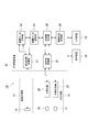
- FIG. 1 is a diagram schematically showing a configuration of an embodiment of a breast measuring apparatus.
- FIG. 2 is a diagram schematically showing ultrasonic measurement and optical measurement for a measurement region by the breast measurement apparatus shown in FIG.
- FIG. 3 is a block diagram showing an example of a specific configuration of the measurement processing apparatus in the breast measurement apparatus shown in FIG.
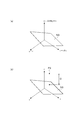
- FIG. 4 is a diagram showing (a) a normal region of a breast in which no tumor is present, and (b) a target region of the breast in which a tumor is present.
- FIGS. 5A and 5B are diagrams showing a first example of a method for evaluating the characteristics of a tumor based on the measurement result obtained by the breast measurement apparatus shown in FIG. FIG.
- FIG. 6 is a diagram illustrating a second embodiment of the method for evaluating the characteristics of a tumor based on the measurement result obtained by the breast measuring apparatus illustrated in FIG.
- FIGS. 7A and 7B are diagrams showing a third embodiment of the method for evaluating the characteristics of a tumor based on the measurement result obtained by the breast measuring apparatus shown in FIG.
- FIGS. 8A and 8B are diagrams showing additional evaluation parameters in the evaluation of the characteristics of the mass (b).
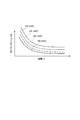
- FIG. 9 is a graph showing a plurality of normal correlation data prepared according to the menopause state of the subject.
- FIG. 10 is a graph showing a plurality of normal correlation data prepared according to the age of the subject.
- FIG. 9 is a graph showing a plurality of normal correlation data prepared according to the menopause state of the subject.
- FIG. 11 is a graph showing selection of normal correlation data based on a measurement result for a normal region set for a subject in the subject region.
- FIG. 12 is a graph showing the correction of normal correlation data based on the measurement result for the normal region set for the subject in the target region.
- FIG. 13 is a graph showing normal correlation data created from the measurement results for a plurality of normal regions set for the subject in the subject region.
- FIG. 14 is a diagram illustrating a modified example of the measurement probe.
- FIG. 1 is a block diagram schematically showing a configuration of an embodiment of a breast measuring apparatus according to the present invention.
- FIG. 2 is a diagram schematically showing ultrasonic measurement and optical measurement for a measurement region by the breast measurement apparatus shown in FIG.
- the breast measurement apparatus 1A according to the present embodiment is a measurement apparatus that measures the characteristics of a tumor existing inside a breast to be measured.
- a measurement region 50 is set as a measurement target at a predetermined position of the subject's breast.
- the measurement region 50 is a normal region of the breast where the tumor 55 does not exist inside, or a target region of the breast where the tumor 55 exists inside.
- the breast measurement region 50 is composed of skin 51, fat 52, mammary gland 53, and the like, and a mass 55 to be measured and evaluated usually exists at a position on the mammary gland 53. Further, at a position farther (deeper) than the fat 52, the mammary gland 53, and the like as seen from the skin 51, there is a site such as a muscle of the chest wall where light absorption is large. In the following, this part of the chest wall such as a muscle is referred to as a light absorption part 56. As will be described later, the light absorption portion 56 is set as a distance measurement target in the measurement performed by the breast measurement apparatus 1A.
- the measurement 1 and 2 includes a distance measurement unit 10, an optical measurement unit 15, a measurement probe 18, and a measurement processing device 30.
- the measurement probe 18 includes an ultrasonic measurement unit (ultrasonic probe) 11 included in the distance measurement unit 10, a light incident unit (light incident probe) 16 and a light output unit (light output probe) 17 included in the optical measurement unit 15. Are integrally held.
- the measurement of the measurement area 50 of the subject by the measurement apparatus 1A is performed in a state where the measurement probe 18 is applied to the measurement area 50 as shown in FIG.
- sequence and positional relationship of the ultrasonic measurement part 11, the light-injection part 16, and the light-projection part 17 in the measurement probe 18 are set appropriately according to the specific measurement content.
- the distance measurement unit 10 applies the above-described light absorption portion 56 set as a distance measurement target to a normal region that does not include the tumor 55 to be evaluated or a breast measurement region 50 that is a target region including the tumor 55.
- the distance measuring means measures the distance (depth) from the skin 51 to the light absorbing portion 56 by a predetermined measuring method (distance measuring method).
- a predetermined measuring method distance measuring method
- an ultrasonic measurement method is used as a method for measuring the distance to the light absorption portion 56.
- the distance measurement unit 10 by the ultrasonic measurement method includes an ultrasonic measurement unit 11.
- the ultrasonic measurement unit 11 is configured by, for example, an acoustic lens, a matching layer, a vibrator, and a damper in order from the tip portion side in contact with the measurement region 50.
- the ultrasonic measurement unit 11 transmits ultrasonic waves to the measurement region 50 using a transmitter, and receives the reflected ultrasonic waves using a receiver, whereby ultrasonic measurement data (for example, ultrasonic waves) in the measurement region 50 is received.
- Image data The measurement data acquired by the ultrasonic measurement unit 11 is sent to the measurement processing device 30 as an ultrasonic measurement signal.
- the optical measurement unit 15 uses measurement light having a predetermined wavelength (for example, near-infrared light) to cause measurement light to enter the measurement region 50 via the light incident unit 16 and to be emitted via the light emission unit 17.
- This is an optical measurement unit that measures the amount of hemoglobin in the measurement region 50 by an optical measurement method that detects incident light.
- time-resolved spectroscopy TRS method: Time Resolved Spectroscopy
- the hemoglobin amount in the measurement region 50 acquired by the optical measurement method will be mainly described as the total hemoglobin amount tHb.
- Patent Document 2 can be referred to for obtaining the hemoglobin amount by the optical measurement method.
- the optical measurement unit 15 based on the TRS method includes a light incident unit 16 that is used for incidence of light to the measurement region 50, a light emission unit 17 that is used for emission of light from the measurement region 50, a light source device 20, and light detection.
- the apparatus 25 is comprised.
- the light source device 20 supplies pulse measurement light having a predetermined wavelength and causes the pulse measurement light to enter the measurement region 50 via the light incident unit 16.
- the light detection device 25 detects the emitted light emitted from the measurement region 50 via the light emission unit 17 and sends the acquired detection data to the measurement processing device 30 as an optical measurement signal.
- the outgoing light detected by the light detection device 25 is incident from the light incident part 16 and is measured from the measurement area 50 as schematically shown by a range 58 in FIG. This is the measurement light that has propagated through 50 while being absorbed, scattered, etc., and has reached the light emitting portion 17. Further, the light source device 20 and the light incident portion 16 and the light detection device 25 and the light emitting portion 17 are optically connected via an optical system such as an optical fiber, respectively.
- the light source device 20 is a semiconductor picosecond pulse laser light source including, for example, a laser diode and a drive circuit.
- the laser diode is stably lit by a drive circuit, and supplies near infrared light in three wavelength ranges of wavelengths 760 nm, 800 nm, and 830 nm as measurement light.
- the light source device 20 is not limited to a laser diode, and for example, a solid laser light source, a light emitting diode (LED), or the like may be used, or a super luminescent diode (SLD), a lamp light source, or the like is selected.
- a configuration combined with a filter may be used.
- the light detection device 25 is constituted by, for example, a photomultiplier tube having high sensitivity characteristics with respect to light in the near-infrared wavelength region, and an amplifier.
- the light detection device 25 may be configured to reduce the influence of light other than near-infrared light supplied from the light source device 20 by providing a wavelength selection filter.
- the light detection device 25 is not limited to a photomultiplier tube, and for example, a semiconductor photodetector such as an avalanche photodiode may be used.
- the three light incident portions 16 and the light emitting portion 17 correspond to the configuration in which the near infrared light in the three wavelength ranges is used as the measurement light. Is shown.
- the ultrasonic measurement signal output from the ultrasonic measurement unit 11 of the distance measurement unit 10 and the optical measurement signal output from the light detection device 25 of the optical measurement unit 15 are respectively input to the measurement processing device 30.
- the measurement processing device 30 controls the measurement operation by the distance measurement unit 10 and the optical measurement unit 15, and performs measurement processing for performing necessary data processing, analysis, and the like for the measurement signal input from the distance measurement unit 10 and the optical measurement unit 15. Means.
- the measurement processing device 30 is configured by a computer, for example.
- a display condition 38 used to display information related to breast measurement such as measurement conditions in the breast measurement device 1A, information on measurement results, and information necessary for breast measurement
- An input device 39 used for inputting an instruction or the like by an operator is connected.
- FIG. 3 is a block diagram showing an example of a specific configuration of the measurement processing device 30 in the breast measurement device 1A shown in FIG.
- the measurement processing device 30 in this embodiment includes a distance measurement processing unit 31, an optical measurement processing unit 32, a correlation data storage unit 33, a correlation data creation unit 34, an evaluation value calculation unit 35, and a tumor characteristic evaluation unit 36. And is configured.
- the distance measurement processing unit 31 performs data processing on the ultrasonic measurement signal input from the distance measurement unit 10, and acquires the distance from the skin 51 to the light absorption site 56 in the measurement region 50.
- ultrasonic image data capable of observing the tissue state under the skin in the measurement region 50 of the subject is acquired as measurement data.
- the distance measurement processing unit 31 for example, an ultrasonic image acquired as a measurement result in the distance measurement unit 10 is displayed on the display device 38, and the operator inputs from the input device 39 as a result determined from the ultrasonic image.
- the distance to the light absorption part 56 is acquired by the information.
- the distance measurement processing unit 31 may perform image processing and analysis on the ultrasonic image with a predetermined algorithm, and automatically acquire the distance to the light absorption portion 56 as a result.
- the optical measurement processing unit 32 performs data processing on the optical measurement signal input from the optical measurement unit 15 and acquires the amount of hemoglobin in the measurement region 50.
- a time-resolved response waveform for the pulse measurement light is obtained by time-resolved measurement using the time-correlated single-photon counting method, and based on this time-resolved response waveform, total hemoglobin is obtained.
- the amount of hemoglobin such as the amount is calculated.
- the correlation data storage unit 33 has normal correlation data between the distance from the skin 51 to the light absorption site 56 and the normal hemoglobin amount, which is the hemoglobin amount in the normal region, for the normal region of the breast in which no tumor 55 exists. (Correlation data acquisition step).
- the normal correlation data stored in the correlation data storage unit 33 obtains the distance to the light absorption part 56 by the distance measurement unit 10 for each of the plurality of normal regions, and at the normal region by the light measurement unit 15. It is the correlation data created by acquiring the amount of normal hemoglobin.
- one normal region may be set for each of a plurality of subjects.
- a plurality of normal regions may be set for the subject, or a plurality of normal regions may be set for a plurality of subjects.
- the correlation data storage unit 33 is provided with a correlation data creation unit 34 for creating normal correlation data from the measurement results of the distance measurement unit 10 and the optical measurement unit 15. ing.
- the correlation data creation unit 34 is unnecessary.
- the target area of the breast in which the tumor 55 is present which is the target of measurement and evaluation by the breast measurement apparatus 1A
- measurement is performed in the same manner as the normal area described above.
- the distance from the skin 51 to the light absorption part 56 in the target region is acquired by the distance measurement unit 10 and the distance measurement processing unit 31.
- the target hemoglobin amount which is the amount of hemoglobin in the target region is acquired by the optical measurement unit 15 and the optical measurement processing unit 32 (target data acquisition step).
- the evaluation value calculation unit 35 is an evaluation value calculation unit that calculates a characteristic evaluation value for the tumor 55 present in the target region. Specifically, the evaluation value calculation unit 35 acquires normal correlation data from the correlation data storage unit 33 (correlation data acquisition step). Further, the distance to the light absorption part 56 measured in the target region is acquired from the distance measurement processing unit 31, and the target hemoglobin amount measured in the target region is acquired from the light measurement processing unit 32. Then, the evaluation value calculation unit 35 is based on the normal correlation data and the normal hemoglobin amount (estimated value of the normal hemoglobin amount) obtained from the distance to the light absorption site 56 in the target region and the target hemoglobin amount in the target region. Then, the characteristic evaluation value of the tumor 55 is calculated (evaluation value calculation step).
- a tumor characteristic evaluation unit 36 is provided for the evaluation value calculation unit 35.
- the tumor characteristic evaluation unit 36 refers to the characteristic evaluation value calculated by the evaluation value calculation unit 35 and performs necessary evaluation on the characteristic of the tumor 55 existing in the target region.
- the characteristics of the mass 55 to be evaluated include Ki-67, HER2, ER, PGR, nuclear grade, histological grade, lymph node metastasis, histological classification, and the like.
- the measurement processing device 30 stores measurement data such as a measurement result by the distance measurement unit 10 and the optical measurement unit 15, a characteristic evaluation value calculated by the evaluation value calculation unit 35, and the like.
- a storage unit may be provided.
- the distance measurement unit 10 calculates the distance from the skin 51 to the light absorption region 56 by a predetermined distance measurement method. While measuring, it is set as the structure which measures the amount of hemoglobin in a measurement area
- normal correlation data indicating the correlation between the distance to the light absorption site 56 and the amount of normal hemoglobin, prepared based on the measurement results for a plurality of normal regions, is prepared and stored in the correlation data storage unit 33.
- measurement is performed on the target region including the tumor 55, and the distance to the light absorption portion 56 in the target region and the target hemoglobin amount are acquired.
- the normal correlation data and the estimated value of the normal hemoglobin amount obtained from the distance to the light absorption part 56 in the target region, and the actual value in the target region The characteristic evaluation value of the tumor 55 existing in the target region is calculated based on the measured amount of target hemoglobin.
- the presence of the light absorption portion 56 such as the muscle of the chest wall in the back of the layer of fat 52 and mammary gland 53 in the breast. May be a problem. That is, in the optical measurement method, the mass characteristics such as the state of the tumor 55 are evaluated by measuring the amount of hemoglobin in the measurement region 50 including the tumor 55. However, in the measurement region 50, when the distance from the skin 51 to the light absorption part 56 is small, one of the measurement light propagating through the measurement region 50 is shown in FIG. The part reaches the light absorption part 56.
- the measurement light reaches the light absorption part 56 such as the chest wall muscle in this way, the absorption of the near infrared measurement light in the light absorption part 56 is large, so that the amount of hemoglobin obtained from the measurement result by the optical measurement method is It will be greatly estimated. Therefore, it is difficult to evaluate a tumor with the correct amount of hemoglobin in evaluating the tumor characteristics.
- the measurement result of the hemoglobin amount in the target region including the tumor 55 is evaluated with reference to the normal correlation data between the distance to the light absorption site 56 in the normal region and the hemoglobin amount.
- a tumor existing inside the target region in consideration of the distance from the skin 51 to the light absorption site 56 in the target region and the influence of the light absorption site 56 on the measurement result of the hemoglobin amount.
- 55 characteristic evaluation can be performed suitably. This makes it possible to improve the measurement accuracy and evaluation accuracy of the characteristics of the tumor 55 existing inside the breast, regardless of the influence of the light absorbing portion 56.
- the hemoglobin amount in the measurement region acquired by the optical measurement unit 15 by the optical measurement method using near infrared light or the like is, for example, the total hemoglobin amount.
- the oxygenated hemoglobin amount or the deoxygenated hemoglobin amount may be acquired as the hemoglobin amount. In general, it may be configured to acquire at least one of an oxygenated hemoglobin amount, a deoxygenated hemoglobin amount, or a total hemoglobin amount as the hemoglobin amount.
- the chest wall muscle described above can be mentioned.
- the chest wall muscles include the external intercostal muscles, the internal intercostal muscles, the inferior muscles, the long rib levator muscle, the short rib levator muscle, the transthoracic muscle, and the like, and include the ribs.
- the chest wall muscle is thin, it is necessary to consider the lung existing deeper than the chest wall muscle as the light absorbing portion 56 that absorbs light.
- the light absorption part 56 that affects the measurement result when the measurement light having a predetermined wavelength is absorbed to some extent in the optical measurement inside the breast is a muscle of the chest wall, a rib, a mammary gland, or a lung.
- the measurement accuracy of the characteristics of the tumor 55 existing inside the breast can be preferably improved.
- the light absorption part 56 is appropriately set in consideration of the light absorption characteristics in each part existing in the breast or in the vicinity thereof.
- the distance measurement unit 10 transmits the ultrasonic wave to the measurement region 50 and receives the reflected ultrasonic wave with respect to the measurement method used for measuring the distance to the light absorption portion 56.
- an ultrasonic measurement method for acquiring ultrasonic measurement data of the measurement region 50 is preferable.
- the ultrasonic measurement method as a measurement method for measuring the distance from the skin 51 to the light absorption site 56 in the measurement region 50, the distance measurement to the light absorption site 56 is suitably executed. be able to.
- the evaluation value calculation unit 35 calculates a difference value between the estimated value of the normal hemoglobin amount obtained for the target region and the target hemoglobin amount as the characteristic evaluation value.
- a configuration can be used.
- the evaluation value calculation unit 35 can use a configuration in which a corrected evaluation value obtained by correcting the target hemoglobin amount by the estimated value of the normal hemoglobin amount obtained for the target region is calculated as the characteristic evaluation value.
- the evaluation and determination of the characteristic of the tumor 55 based on the measurement result can be suitably executed.
- the correlation data storage unit 33 prepares hemoglobin amount correction data created based on the correlation between the distance to the light absorption site 56 and the normal hemoglobin amount as normal correlation data,
- the evaluation value calculation unit 35 may calculate a correction evaluation value obtained by correcting the target hemoglobin amount using the correction value obtained from the hemoglobin amount correction data and the distance to the light absorption site in the target region as the characteristic evaluation value.
- the evaluation and determination of the characteristics of the mass 55 based on the measurement result can be suitably executed. it can.
- the evaluation value calculation unit 35 acquires a single or a plurality of evaluation parameters related to the tumor 55 in addition to the above-described characteristic evaluation values, and the characteristic evaluation value and the single or plural evaluation parameters.
- a configuration in which the characteristics of the tumor 55 are evaluated based on the evaluation parameters is also possible. Even with such a configuration, it is possible to further improve the measurement accuracy of the characteristics of the tumor 55 existing inside the breast.
- FIG. 4 is a diagram showing (a) a normal region of the breast where the tumor 55 does not exist inside, and (b) a target region of the breast where the tumor 55 exists inside.
- the distance from the skin 51 to the light absorbing portion 56 in the measurement region 50 is x1
- the distance from the skin 51 to the surface of the tumor 55 is x2.
- n is an abbreviation for “normal” and indicates a normal region
- t is an abbreviation for “tumor” and includes a tumor
- C is an abbreviation for “chest wall” and indicates a light absorption site such as a muscle of the chest wall
- p is an abbreviation for “patient” and indicates a subject.
- the total hemoglobin amount tHb is mainly assumed as the hemoglobin amount in the measurement region 50.
- FIGS. 5A and 5B are diagrams showing a first embodiment of a method for evaluating the characteristics of the tumor 55 based on the measurement results obtained by the breast measurement apparatus 1A shown in FIGS.
- the distance x1_nc from the skin 51 to the light absorption part 56 is measured by the distance measuring unit 10 for a normal region where the tumor 55 does not exist.
- the optical measurement unit 15 measures a normal hemoglobin amount tHb_n that is the total hemoglobin amount in the normal region. Such measurement is performed on a plurality of subjects with respect to normal regions set at a plurality of positions.
- the distance x1_nc to the light absorption site 56 for the normal region and the normal hemoglobin amount tHb_n Normal correlation data is created.
- This normal correlation data is expressed by the following equation (1).
- tHb a1 ⁇ x1 3 + b1 ⁇ x1 2 + c1 ⁇ x1 + d1 (1)
- An approximate expression such as Alternatively, the normal correlation data may be prepared as a correlation table. Also, as will be described later, a plurality of normal correlation data may be prepared according to the attributes of the normal area.
- the distance measurement unit 10 measures the distance x1_tc from the position immediately above the tumor 55 to the light absorbing portion 56 with respect to the target region where the tumor 55 exists. Moreover, the target hemoglobin amount tHb_t, which is the total hemoglobin amount in the target region, is measured by the optical measurement unit 15 from the position immediately above the tumor 55.
- FIG. 5B shows a measurement data point P1 based on the measurement result in the target region, together with the normal correlation data graph N1.
- a difference value ⁇ tHb_t ⁇ tn between the estimated value tHb_tn of the normal hemoglobin amount and the measured target hemoglobin amount tHb_t is calculated as the characteristic evaluation value of the tumor 55 existing in the target region.
- the difference value ⁇ tHb_t ⁇ tn of the amount of hemoglobin obtained in this way indicates the total amount of hemoglobin in which the influence of the light absorption site 56 such as a chest wall muscle in the target region is reduced. Therefore, by evaluating the tumor characteristic using the difference value ⁇ tHb_t ⁇ tn as a characteristic evaluation value, it is possible to accurately evaluate the tumor 55 in the target region.
- the difference value of the hemoglobin amount is obtained as the characteristic evaluation value of the tumor.
- the present invention is not limited to such a configuration, and for example, the ratio value of the hemoglobin amount may be used as the characteristic evaluation value.
- FIG. 6 is a diagram showing a second embodiment of the method for evaluating the characteristics of the tumor 55 based on the measurement results obtained by the breast measurement apparatus 1A shown in FIGS.
- the distance measurement unit 10 measures the distance x1_nc from the skin 51 to the light absorption site 56 with respect to a normal region where the tumor 55 does not exist.
- the optical measurement unit 15 measures the normal hemoglobin amount tHb_n in the normal region. Such measurement is performed on a plurality of subjects with respect to normal regions set at a plurality of positions.
- hemoglobin amount correction data that is correlation data of the correction value is created.
- This correction data may be prepared as an approximate expression or may be prepared as a correction table.
- the value of the distance x1 is large, the influence of the light absorption portion 56 is sufficiently reduced, and the correction value at that time is set to 1, and the hemoglobin at each distance x1 is used as a reference.
- the corrected correlation data is obtained by the correction value for the quantity.
- the distance measurement unit 10 measures the distance x1_tc from the position immediately above the tumor 55 to the light absorbing portion 56 with respect to the target region where the tumor 55 exists. Further, the target hemoglobin amount tHb_t in the target region is measured by the optical measurement unit 15 from the position immediately above the tumor 55.
- the hemoglobin amount correction value corresponding to the estimated value of the normal hemoglobin amount for the target region is applied to the hemoglobin amount correction data by applying the distance x1_tc to the light absorption site 56 in the target region. Ask for. Then, a corrected evaluation value obtained by correcting the measured target hemoglobin amount tHb_t by the obtained correction value is calculated as a characteristic evaluation value of the tumor 55 existing in the target region. For example, when the correction data shown in FIG. 6 is used, the product of the target hemoglobin amount and the correction value (correction coefficient) is used as the correction evaluation value.
- the corrected evaluation value of the amount of hemoglobin obtained in this way indicates the total amount of hemoglobin in which the influence of the light absorption site 56 such as the muscle of the chest wall in the target region is reduced. Therefore, by evaluating the mass characteristic using the corrected evaluation value as the characteristic evaluation value, it is possible to accurately evaluate the mass 55 in the target region.
- correction data is prepared in advance as normal correlation data. However, normal normal correlation data is prepared and the amount of normal hemoglobin for the target region obtained from the normal correlation data. The target hemoglobin amount may be corrected based on the estimated value.
- FIG. 7 (a) and 7 (b) are diagrams showing a third embodiment of the method for evaluating the characteristics of the tumor 55 based on the measurement results obtained by the breast measurement apparatus 1A shown in FIGS. 1 to 3.
- one or more evaluation parameters regarding the tumor 55 are additionally acquired, and the evaluation of the tumor 55 is performed based on the characteristic evaluation value and the additional evaluation parameter. It is good also as a structure which performs.
- the process until the calculation of the difference value ⁇ tHb_t ⁇ tn is the same as that in the first embodiment.
- the distance x2_t (see FIG. 4) from the skin 51 to the tumor 55 and the tumor diameter r are acquired based on the ultrasonic measurement data by the distance measuring unit 10. Further, such measurement is performed on a plurality of subjects (patients) having a representative tumor 55, and measurement data of a difference value, a distance to the tumor, and a tumor diameter are acquired.
- the distance measurement unit 10 measures the distance x1_p to the light absorption part 56 with respect to the subject region of the subject where the tumor 55 exists.
- the distance measurement unit 10 measures the distance x2_p to the tumor and the tumor diameter r_p, and plots the measurement data point P3 as shown in FIG. 7B. Then, the distance D of the measurement data point P3 from the tumor characteristic determination surface N3 is calculated, and this is used as a characteristic evaluation value to evaluate the characteristic of the tumor 55 in the target region.
- the additional evaluation parameters related to the tumor 55 are not limited to the distance to the tumor 55 and the tumor diameter r, and various parameters can be used as the evaluation parameters.
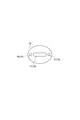
- the tumor diameter r as shown in FIGS. 8A and 8B, more specifically, the measurement surface defined by the light incident part 16 and the light emitting part 17 arranged on the measurement region 50
- the longitudinal diameter (thickness) of the mass 55 is a
- the lateral diameter in the direction parallel to the measurement surface is b
- the lateral diameter in the direction perpendicular to the measurement surface is c
- (1) thickness a, (2 ) Thickness a ⁇ lateral diameter b, (3) thickness a ⁇ lateral diameter c, (4) product of diameters in three directions a ⁇ b ⁇ c, etc. can be used as evaluation parameters.
- the distance to the tumor 55 for example, (1) the distance from the skin to the surface of the tumor, (2) the distance from the skin to the center of the tumor, (3) the distance from the skin to the rear edge of the tumor, etc. Can be used as When the mass 55 in the target region is small, for example, the thickness of the mammary gland 53, the distance from the skin 51 to the mammary gland 53, and the like may be used as the evaluation parameters.
- the hemoglobin amount acquired by the optical measurement unit 15 is not limited to the total hemoglobin amount tHb.
- the oxygenated hemoglobin amount HbO 2 and the deoxygenated hemoglobin amount Hb may be acquired.
- optical parameters such as an absorption coefficient, an equivalent scattering coefficient, and a refractive index may be acquired.
- the normal correlation data used for evaluating the characteristics of the tumor 55 in the breast measurement apparatus 1A shown in FIGS. 1 to 3 will be further described.
- a single correlation data is used.
- a plurality of normal correlation data may be prepared as normal correlation data in accordance with the attribute of the normal area.
- the selection of normal correlation data used in the evaluation value calculation is performed in the evaluation value calculation process in the evaluation value calculation unit 35 in the attribute of the target region. Based on this, it is possible to use a configuration for selecting normal correlation data used for deriving an estimated value of the amount of normal hemoglobin for the target region.
- the normal correlation used for deriving the estimated value of the normal hemoglobin amount for the target region based on the measurement result for the normal region set for the subject of the target region A configuration for selecting data can be used.
- a single normal correlation data is appropriately selected from a plurality of normal correlation data prepared according to the attribute of the normal region and stored in the correlation data storage unit 33 by a predetermined selection method, and the selected Measurement accuracy of the characteristics of the tumor 55 existing inside the breast by applying the normal correlation data to derive the estimated value of the normal hemoglobin amount for the target area and calculating the characteristic evaluation value of the tumor 55 in the target area Can be further improved.
- a plurality of normal correlation data is used as described above, specifically, in the correlation data storage unit 33, depending on the age of the subject, which is an attribute of the normal region, or the menopausal state of the subject A plurality of normal correlation data may be prepared.
- an attribute of a measurement region referred to in creation, selection, etc. of normal correlation data generally, an attribute of the measurement region itself, an attribute of a subject in the measurement region, or the like can be used.
- the correlation data storage unit 33 may prepare correlation data created from measurement results for a plurality of normal areas set for the subject in the target area as normal correlation data.
- the normal correlation data applied to the measurement result in the target region including the tumor 55 can be suitably set according to the subject having the target region.
- the evaluation value calculation unit 35 derives the estimated value of the normal hemoglobin amount for the target region based on the measurement results for the single or plural normal regions set for the subject in the target region.
- the normal correlation data used for the correction may be corrected. Even with such a configuration, the normal correlation data applied to the measurement result in the target region can be suitably set according to the subject having the target region.
- FIG. 9 shows a plurality of normal correlation data prepared in accordance with the menopause state of the subject in consideration of the menopause state of the subject as an attribute of the measurement region (normal region, target region) in the subject. It is a graph.
- a graph A1 shows normal correlation data for the normal region of the subject before menopause
- a graph A2 shows normal correlation data for the normal region of the subject after menopause.
- the correlation between the distance x1 to the light absorption site 56 and the hemoglobin amount tHb changes depending on whether the subject's menopause state is before menopause or after menopause. Therefore, a plurality of normal correlation data A1 and A2 are prepared in the correlation data storage unit 33, and the evaluation value calculation unit 35 of the subject in the target region including the tumor 55 input from the input device 39 by the operator is prepared.
- the measurement accuracy of the characteristics of the tumor 55 can be improved.
- the structure which prepares the normal correlation data before and after menopause was shown, for example, the structure etc. which prepare the normal correlation data after chemotherapy (chemo) and after chemotherapy are used. Also good.
- FIG. 10 is a graph showing a plurality of normal correlation data prepared according to the age of the subject in consideration of the age of the subject as the attribute of the measurement region (normal region, target region) in the subject. is there.
- graph A3 shows normal correlation data for subjects in their 20s
- graph A4 shows normal correlation data for subjects in their 30s
- graph A5 shows subjects in their 40s.
- the graph A6 shows normal correlation data for subjects in their 50s.
- the distance x1 to the light absorption site 56 and the hemoglobin amount tHb are similar to those in the menopause state. And the correlation changes. Accordingly, a plurality of normal correlation data A3 to A6 is prepared in the correlation data storage unit 33, and the evaluation value calculation unit 35 of the subject in the target region including the tumor 55 input from the input device 39 by the operator is prepared. By selecting normal correlation data based on age or the like, the measurement accuracy of the characteristics of the tumor 55 can be improved.
- the evaluation value calculation unit 35 when a plurality of normal correlation data is prepared according to the attributes of the normal region, as shown in the example about the menopause state and age of the subject, the evaluation value calculation unit 35 As described above, the normal correlation data can be selected based on a corresponding attribute (for example, menopausal state, age) of the target region. Alternatively, in addition to such a configuration, the attribute of the target area is not directly considered, as shown in FIG. 11, based on the measurement result for the normal area set for the subject of the target area, A configuration may be used in which normal correlation data is selected.
- the setting of the normal region in the subject of the subject region including the tumor 55 specifically, for example, when the tumor 55 exists in one breast of the subject, the other breast in which the tumor 55 does not exist A method of performing measurement by setting a normal region can be used. Alternatively, in one breast where the tumor 55 exists, a method of setting a region where the tumor 55 does not exist as a normal region may be used.
- FIG. 12 is a graph showing correction of normal correlation data based on the measurement result for the normal area set for the subject in the subject area.
- the normal region is set for the subject in the target region with respect to the normal correlation data B1 before correction prepared in the correlation data storage unit 33.
- the evaluation value calculation unit 35 corrects the correlation data based on the measurement data point B0 that is the measurement result for the normal region, and applies the corrected normal correlation data B2 in the calculation of the characteristic evaluation value.
- FIG. 12 shows a measurement data point B0 when three normal areas are set. Moreover, it is good also as a structure which correct
- FIG. 13 is a graph showing normal correlation data created from the measurement results for a plurality of normal regions set for the subject in the subject region.
- a plurality of normal regions are set for the subject in the subject region, and a plurality of measurement data points C0 that are the measurement results are acquired.
- normal correlation data C1 is created based on the correlations at the plurality of measurement data points C0 and stored in the correlation data storage unit 33.
- the characteristic evaluation of the tumor 55 by the breast measurement apparatus 1A of the above embodiment is performed when, for example, the subject having the tumor 55 in the breast is observed for a long period of time, or when anticancer drug treatment is performed.
- This is considered to be an effective method for the follow-up observation of the effects of.
- measurement is performed at a plurality of positions with respect to the normal region of the subject to be measured, and normal correlation data for the subject is prepared. Becomes important.
- the breast measurement method and the breast measurement apparatus according to the present invention are not limited to the above-described embodiments and configuration examples, and various modifications are possible.
- the ultrasonic measurement method is used as a predetermined distance measurement method for acquiring the distance to the light absorption site.
- the configuration is not limited to such a configuration.
- light that is a kind of optical measurement method is used. It is good also as a structure which measures the distance to a light absorption site
- time-resolved spectroscopy (TRS method: Time Resolved Spectroscopy) is used in the above-described embodiment.
- the hemoglobin amount may be measured by phase modulation spectroscopy using light (PMS method: Phase Modulation Spectroscopy) or a method using CW light as measurement light.
- PMS method Phase Modulation Spectroscopy
- CW light CW light
- FIG. 1 illustrates a multi-channel configuration in which a plurality of light incident portions 16 and a plurality of light emission portions 17 are combined.
- measurement probes having various configurations can be used.
- the measurement probe 18 of the modification shown in FIG. 14 uses a one-channel configuration in which a single light incident portion 16 and a single light emitting portion 17 are combined.
- measurement light of different wavelengths is incident on the measurement region 50 from the light incident portion 16 in time series.
- the arrangement of the light incident unit 16 and the light emitting unit 17 with respect to the ultrasonic measurement unit 11 is not limited to the configuration example illustrated in FIGS. 1 and 14 and may be variously changed.
- the breast measurement method is a breast measurement method for measuring the characteristics of a tumor existing inside the breast to be measured, and (1) a tumor for a light absorption site set as a distance measurement target inside the breast.
- the distance to the light absorption site is obtained by a predetermined measurement method, and the normal hemoglobin amount in the normal region is obtained by the optical measurement method, and the distance to the light absorption site and normal Hemoglobin
- the breast measurement apparatus is a breast measurement apparatus that measures the characteristics of a tumor existing inside a breast to be measured, and (a) a light absorption part set as a distance measurement target inside the breast.
- the distance to measure the distance from the skin to the light-absorbing site with the specified measurement method for the normal area of the breast where the tumor is not present or the measurement area which is the target area of the breast where the tumor is present A measurement unit; and (b) a light source device that causes measurement light to enter the measurement region via the light incidence unit, and a light detection device that detects the emitted light emitted from the measurement region via the light emission unit.
- the optical measurement unit that measures the amount of hemoglobin in the measurement region, and (c) for each of the plurality of normal regions, the distance measurement unit acquires the distance to the light absorption site, and the optical measurement unit normal Data storage unit that stores the normal correlation data between the distance to the light absorption site and the normal hemoglobin amount, which is created by acquiring the normal hemoglobin amount in the region, and (d) acquired by the normal correlation data and the distance measurement unit Based on the normal hemoglobin amount obtained from the distance to the light absorption site in the target region and the target hemoglobin amount obtained by the optical measurement unit, the characteristic evaluation value of the tumor existing in the target region is calculated. And an evaluation value calculation unit for calculating.
- the hemoglobin amount in the measurement region acquired by the optical measurement method is, for example, the total hemoglobin amount.
- the oxygenated hemoglobin amount or the deoxygenated hemoglobin amount may be acquired as the hemoglobin amount.
- it may be configured to acquire at least one of an oxygenated hemoglobin amount, a deoxygenated hemoglobin amount, or a total hemoglobin amount as the hemoglobin amount.
- the breast measurement method uses a plurality of normal correlations as normal correlation data according to the attribute of the normal region in the correlation data acquisition step. It is preferable to prepare data and select normal correlation data used for deriving the amount of normal hemoglobin based on the attribute of the target region in the evaluation value calculation step. Similarly, the breast measurement apparatus prepares a plurality of normal correlation data as normal correlation data in the correlation data storage unit according to the attribute of the normal region, and the evaluation value calculation unit determines that the normal value is normal based on the attribute of the target region. It is preferable to select normal correlation data used for deriving the amount of hemoglobin.
- the breast measurement method prepares a plurality of normal correlation data according to the attributes of the normal area as normal correlation data in the correlation data acquisition step, and in the evaluation value calculation step, The normal correlation data used for deriving the normal hemoglobin amount may be selected based on the measurement result for the normal region set for the examiner.
- the breast measurement apparatus prepares a plurality of normal correlation data according to the attributes of the normal region as normal correlation data in the correlation data storage unit, and is set for the subject in the target region in the evaluation value calculation unit.
- the normal correlation data used for deriving the normal hemoglobin amount may be selected based on the measurement result for the normal region.
- a single normal correlation data is appropriately selected from a plurality of normal correlation data prepared according to the attributes of the normal region by a predetermined selection method, and the selected normal correlation data is applied to the target.
- the breast measurement method depends on the age of the subject as an attribute of the normal region or the menopausal state in the correlation data acquisition step.
- a plurality of normal correlation data may be prepared.
- the breast measurement apparatus may have a configuration in which a plurality of normal correlation data is prepared in the correlation data storage unit according to the age of the subject who is the attribute of the normal region or the menopause state.
- the attribute of the measurement region referred to in the creation and selection of normal correlation data, generally, the attribute of the measurement region itself, the attribute of the subject in the measurement region, or the like can be used.
- the breast measurement method may be configured to prepare correlation data created from measurement results for a plurality of normal regions set for the subject in the target region as normal correlation data in the correlation data acquisition step.
- the breast measurement device may have a configuration in which correlation data created from measurement results for a plurality of normal regions set for a subject in the target region is prepared as normal correlation data in the correlation data storage unit. According to such a configuration, normal correlation data applied to the measurement result of the target region including the tumor can be suitably set according to the subject having the target region.
- the breast measurement method may be configured to correct the normal correlation data used for deriving the normal hemoglobin amount based on the measurement result for the normal region set for the subject in the evaluation region in the evaluation value calculation step.
- the breast measurement apparatus may be configured such that the evaluation value calculation unit corrects the normal correlation data used for deriving the normal hemoglobin amount based on the measurement result for the normal region set for the subject in the subject region.
- normal correlation data applied to the measurement result of the target region including the tumor can be suitably set according to the subject having the target region.
- the breast measurement method is a predetermined measurement method that measures the distance to the light absorption site, and transmits ultrasonic waves to the measurement area and receives reflected ultrasonic waves, thereby measuring the ultrasonic waves in the measurement area.
- An ultrasonic measurement method for acquiring data is preferable.
- the predetermined measurement method for measuring the distance to the light absorption site transmits an ultrasonic wave to the measurement region and receives the reflected ultrasonic wave. It is preferable that the ultrasonic measurement method acquires ultrasonic measurement data in the measurement region.
- the ultrasonic measurement method as a measurement method for measuring the distance from the skin to the light absorption site in the measurement region, the distance measurement to the light absorption site can be suitably performed.
- the breast measurement method calculates a difference value between the normal hemoglobin amount obtained for the target region and the target hemoglobin amount as the characteristic evaluation value. It is good also as a structure.
- the breast measurement apparatus may be configured such that the evaluation value calculation unit calculates a difference value between the normal hemoglobin amount obtained for the target region and the target hemoglobin amount as the characteristic evaluation value.
- the breast measurement method may be configured to calculate a corrected evaluation value obtained by correcting the target hemoglobin amount with the normal hemoglobin amount obtained for the target region as the characteristic evaluation value in the evaluation value calculating step.
- the breast measurement apparatus may be configured such that the evaluation value calculation unit calculates a corrected evaluation value obtained by correcting the target hemoglobin amount with the normal hemoglobin amount obtained for the target region as the characteristic evaluation value.
- the configuration using the difference value of the hemoglobin amount or the evaluation value obtained by correcting the hemoglobin amount as the characteristic evaluation value of the tumor in the target region it is preferable to evaluate and judge the characteristic of the tumor based on the measurement result. Can be executed.
- the breast measurement method prepares hemoglobin amount correction data created based on the correlation between the distance to the light absorption site and the normal hemoglobin amount as normal correlation data in the correlation data acquisition step, and in the evaluation value calculation step As the characteristic evaluation value, a corrected evaluation value obtained by correcting the target hemoglobin amount using a correction value obtained from the hemoglobin amount correction data and the distance to the light absorption site in the target region may be calculated.
- the breast measurement apparatus prepares, as the normal correlation data, hemoglobin amount correction data created based on the correlation between the distance to the light absorption site and the normal hemoglobin amount in the correlation data storage unit, and the evaluation value calculation unit
- the characteristic evaluation value may be a corrected evaluation value obtained by correcting the target hemoglobin amount using the correction value obtained from the hemoglobin amount correction data and the distance to the light absorption site in the target region.
- the hemoglobin amount correction data used for correcting the target hemoglobin amount is prepared as the normal correlation data, it is possible to preferably execute the evaluation and determination on the characteristics of the tumor based on the measurement result. .
- the measurement light of a predetermined wavelength is absorbed to a certain extent and affects the measurement result, and the light absorption part set as the distance measurement target is specifically
- the light absorption site set in is preferably a chest wall muscle, rib, mammary gland, or lung. By taking into consideration the influence of the light absorption at these parts on the measurement result, the measurement accuracy of the characteristics of the tumor existing inside the breast can be preferably improved.
- Such a light absorption site is preferably set appropriately in consideration of light absorption characteristics and the like in each site existing in or near the breast.
- the breast measurement method acquires a single or a plurality of evaluation parameters related to a tumor in addition to the characteristic evaluation value, and based on the characteristic evaluation value and the single or a plurality of evaluation parameters, It is good also as a structure which evaluates the characteristic.
- the breast measurement device acquires a single or a plurality of evaluation parameters related to the tumor in addition to the characteristic evaluation value, and based on the characteristic evaluation value and the single or a plurality of evaluation parameters, It is good also as a structure which evaluates the characteristic of a tumor. Even with such a configuration, it is possible to further improve the measurement accuracy of the characteristics of the tumor existing inside the breast.
- the present invention can be used as a breast measurement method and a breast measurement apparatus capable of improving the measurement accuracy of the characteristics of a tumor existing in the breast.
- DESCRIPTION OF SYMBOLS 1A ... Breast measuring device, 10 ... Distance measuring part, 11 ... Ultrasonic measuring part, 15 ... Optical measuring part, 16 ... Light incident part, 17 ... Light emitting part, 18 ... Measuring probe, 20 ... light source device, 25 ... light detection device, DESCRIPTION OF SYMBOLS 30 ... Measurement processing apparatus, 31 ... Distance measurement process part, 32 ... Optical measurement process part, 33 ... Correlation data storage part, 34 ... Correlation data creation part, 35 ... Evaluation value calculation part, 36 ... Mass characteristic evaluation part, 38 ... Display device, 39... Input device, DESCRIPTION OF SYMBOLS 50 ... Measurement area
Abstract
A breast measurement device (1A) is configured from a distance measurement unit (10) for measuring the distance from skin to a light absorption site in a measurement region of a breast which is a normal region not including a tumor or a subject region including a tumor, a light measurement unit (15) for measuring a hemoglobin amount in the measurement region by a light measurement method using a predetermined wavelength of measurement light, a correlation data storage unit (33) for storing data of a normal correlation between the distance to the light absorption site and a normal hemoglobin amount created from a plurality of measurement results from a normal region, and an evaluation value calculation unit (35) for calculating a tumor characteristic evaluation value on the basis of a subject hemoglobin amount for the subject region and the normal hemoglobin amount obtained from the normal correlation data and the distance to the light absorption site in the subject region. Through this configuration, a breast measurement method and measurement device are realized whereby a characteristic of a tumor within a breast can be measured with enhanced precision.
Description
本発明は、乳房内部に存在する腫瘤の特性を計測する乳房計測方法、及び乳房計測装置に関するものである。
The present invention relates to a breast measurement method and a breast measurement apparatus for measuring the characteristics of a tumor existing inside a breast.
近年、乳がん検査での乳房計測などの非侵襲の生体計測において、近赤外光などの所定波長の光を用いた光計測法が用いられている(例えば、特許文献1、2参照)。このような光計測法では、被検者の計測対象領域に対して、光入射部を介して所定波長の計測光を入射させる。そして、計測対象領域の内部を伝搬して光出射部を介して外部へと出射される出射光を検出し、その検出結果から計測対象領域の内部情報を取得する。
In recent years, optical measurement methods using light of a predetermined wavelength such as near infrared light have been used in non-invasive living body measurement such as breast measurement in breast cancer examination (for example, see Patent Documents 1 and 2). In such an optical measurement method, measurement light having a predetermined wavelength is incident on the measurement target region of the subject via the light incident portion. And the emitted light which propagates the inside of a measurement object area | region and is radiate | emitted outside through a light emission part is detected, and the internal information of a measurement object area | region is acquired from the detection result.
光計測法では、計測対象領域の内部情報として、例えば、計測対象領域内でのヘモグロビン量(酸素化ヘモグロビン量HbO2、脱酸素化ヘモグロビン量Hb、または総ヘモグロビン量tHb)を取得することができる。例えば、非特許文献1には、近赤外分光計測によって、ヘモグロビン量を取得するとともに、取得されたヘモグロビン量から腫瘤の特性を評価することが記載されている。
In the optical measurement method, for example, the amount of hemoglobin (oxygenated hemoglobin amount HbO 2 , deoxygenated hemoglobin amount Hb, or total hemoglobin amount tHb) in the measurement target region can be acquired as internal information of the measurement target region. . For example, Non-Patent Document 1 describes that the amount of hemoglobin is acquired by near-infrared spectroscopic measurement, and the characteristics of the tumor are evaluated from the acquired amount of hemoglobin.
上記した光計測法を用いた乳房計測は、乳がん検査における腫瘤の特性の計測、評価等において有効な手法である。本願発明者は、このような乳房計測方法、及びその計測精度について詳細に検討した結果、乳房における脂肪や乳腺の層の奥に存在する、胸壁の筋肉などの光吸収部位の層によって計測光が吸収され、この光吸収の影響により、光計測法で取得されるヘモグロビン量の精度が低下する場合があることを見出した。
Breast measurement using the optical measurement method described above is an effective technique for measuring and evaluating the characteristics of a tumor mass in breast cancer examinations. The inventor of the present application has examined the breast measurement method and the measurement accuracy in detail, and as a result, the measurement light is transmitted by the layer of the light absorption site such as the muscle of the chest wall that exists in the back of the fat or mammary gland layer in the breast. It was found that the accuracy of the amount of hemoglobin obtained by the optical measurement method may be reduced due to the effect of light absorption.
このように、胸壁の筋肉等の光吸収部位の影響でヘモグロビン量の計測精度が低下した場合、それによる腫瘤の特性の評価が正しく行われない可能性がある。また、光吸収部位による計測光の吸収の影響の大きさは、乳房の大きさ、計測対象領域の位置、計測角度等によって異なるため、光吸収部位の影響を一概に求めることは難しい。
As described above, when the measurement accuracy of the hemoglobin amount decreases due to the influence of the light absorption site such as the muscle of the chest wall, the characteristic of the tumor may not be correctly evaluated. In addition, since the magnitude of the influence of the measurement light absorption by the light absorption portion varies depending on the size of the breast, the position of the measurement target region, the measurement angle, and the like, it is difficult to obtain the influence of the light absorption portion in a general manner.
本発明は、以上の問題点を解決するためになされたものであり、胸壁の筋肉等の光吸収部位の影響にかかわらず、乳房内部に存在する腫瘤の特性の計測精度を向上することが可能な乳房計測方法、及び乳房計測装置を提供することを目的とする。
The present invention has been made to solve the above problems, and it is possible to improve the measurement accuracy of the characteristics of a tumor existing in the breast regardless of the influence of a light absorption site such as a chest wall muscle. An object of the present invention is to provide a breast measurement method and a breast measurement apparatus.
このような目的を達成するために、本発明による乳房計測方法は、計測対象の乳房内部に存在する腫瘤の特性を計測する乳房計測方法であって、(1)乳房内部で距離の計測対象として設定された光吸収部位について、腫瘤が内部に存在しない乳房の正常領域、または腫瘤が内部に存在する乳房の対象領域である計測領域に対して、皮膚からの光吸収部位までの距離を計測する所定の計測法と、光入射部を介して計測光を計測領域に入射させて光出射部を介して出射される出射光を検出して、計測領域でのヘモグロビン量を計測する光計測法とを用い、(2)複数の正常領域のそれぞれに対して、所定の計測法で光吸収部位までの距離を取得するとともに、光計測法で正常領域での正常ヘモグロビン量を取得して作成された、光吸収部位までの距離と正常ヘモグロビン量との正常相関データを取得する相関データ取得ステップと、(3)対象領域に対して、所定の計測法で光吸収部位までの距離を取得するとともに、光計測法で対象領域での対象ヘモグロビン量を取得する対象データ取得ステップと、(4)正常相関データ及び対象領域での光吸収部位までの距離から求められる正常ヘモグロビン量と、対象領域での対象ヘモグロビン量とに基づいて、対象領域に存在する腫瘤の特性評価値を算出する評価値算出ステップとを有することを特徴とする。
In order to achieve such an object, a breast measurement method according to the present invention is a breast measurement method for measuring characteristics of a tumor existing inside a breast to be measured, and (1) a distance measurement target inside the breast. For the set light absorption site, measure the distance from the skin to the light absorption site for the normal area of the breast where the tumor is not present or the target area of the breast where the tumor is present. A predetermined measurement method, and an optical measurement method for measuring the amount of hemoglobin in the measurement region by detecting measurement light incident on the measurement region via the light incident unit and detecting the emitted light emitted through the light emission unit; (2) For each of a plurality of normal regions, the distance to the light absorption site is obtained by a predetermined measurement method, and the normal hemoglobin amount in the normal region is obtained by an optical measurement method. , Light absorption site A correlation data acquisition step for acquiring normal correlation data between the distance of the normal hemoglobin and the amount of normal hemoglobin, and (3) acquiring the distance to the light absorption site with a predetermined measurement method for the target region and the target with the optical measurement method Based on the target data acquisition step for acquiring the target hemoglobin amount in the region, (4) the normal correlation data, the normal hemoglobin amount obtained from the distance to the light absorption site in the target region, and the target hemoglobin amount in the target region And an evaluation value calculating step for calculating a characteristic evaluation value of a tumor existing in the target region.
また、本発明による乳房計測装置は、計測対象の乳房内部に存在する腫瘤の特性を計測する乳房計測装置であって、(a)乳房内部で距離の計測対象として設定された光吸収部位について、腫瘤が内部に存在しない乳房の正常領域、または腫瘤が内部に存在する乳房の対象領域である計測領域に対して、所定の計測法で、皮膚からの光吸収部位までの距離を計測する距離計測部と、(b)光入射部を介して計測光を計測領域に入射させる光源装置、及び計測領域から光出射部を介して出射される出射光を検出する光検出装置を有し、光計測法で、計測領域でのヘモグロビン量を計測する光計測部と、(c)複数の正常領域のそれぞれに対して、距離計測部によって光吸収部位までの距離を取得するとともに、光計測部によって正常領域での正常ヘモグロビン量を取得して作成された、光吸収部位までの距離と正常ヘモグロビン量との正常相関データを記憶する相関データ記憶部と、(d)正常相関データ及び距離計測部によって取得された対象領域での光吸収部位までの距離から求められる正常ヘモグロビン量と、光計測部によって取得された対象領域での対象ヘモグロビン量とに基づいて、対象領域に存在する腫瘤の特性評価値を算出する評価値算出部とを備えることを特徴とする。
A breast measurement device according to the present invention is a breast measurement device that measures the characteristics of a tumor existing inside a breast to be measured, and (a) a light absorption part set as a distance measurement target inside the breast, Distance measurement that measures the distance from the skin to the light-absorbing site using a predetermined measurement method for the normal area of the breast where the tumor is not present or the measurement area that is the target area of the breast where the tumor is present. And (b) a light source device that causes measurement light to enter the measurement region via the light incident portion, and a light detection device that detects the emitted light emitted from the measurement region via the light emission portion, And (c) for each of a plurality of normal regions, the distance measuring unit obtains the distance to the light absorption site, and the optical measuring unit is normal by the optical measuring unit. In the area A correlation data storage unit for storing normal correlation data between the distance to the light absorption site and the normal hemoglobin amount, which is created by acquiring the normal hemoglobin amount, and (d) the target acquired by the normal correlation data and the distance measurement unit Evaluation to calculate the characteristic evaluation value of the tumor existing in the target region based on the normal hemoglobin amount obtained from the distance to the light absorption site in the region and the target hemoglobin amount in the target region acquired by the optical measurement unit And a value calculation unit.
上記した乳房計測方法、及び乳房計測装置では、乳房内部にある腫瘤の特性計測において、計測、評価の対象となる腫瘤とは別に、光計測において光が吸収されて計測結果に影響を与える部位を、距離の計測対象の光吸収部位として設定する。また、腫瘤を含まない正常領域、または腫瘤を含む対象領域である乳房の計測領域について、所定の距離計測法によって、皮膚からの光吸収部位までの距離を計測するとともに、所定波長の計測光を用いる光計測法によって、計測領域でのヘモグロビン量を計測する構成とする。
In the above-described breast measurement method and breast measurement apparatus, in the measurement of the characteristics of the tumor in the breast, a part that affects the measurement result by absorbing light in the optical measurement is separated from the mass to be measured and evaluated. , And set as a light absorption part of a distance measurement target. In addition, for a normal region that does not include a tumor or a measurement region of a breast that is a target region that includes a tumor, the distance from the skin to the light absorption site is measured by a predetermined distance measurement method, and measurement light having a predetermined wavelength is measured. It is set as the structure which measures the amount of hemoglobin in a measurement area | region with the optical measurement method to be used.
また、上記構成において、複数の正常領域に対する計測結果によって作成された、光吸収部位までの距離と正常ヘモグロビン量との相関を示す正常相関データを用意するとともに、腫瘤を含む対象領域に対して計測を行って、対象領域での光吸収部位までの距離と、対象ヘモグロビン量とを取得する。そして、これらの複数の正常領域、及び対象領域についての計測データを参照し、正常相関データ及び対象領域での光吸収部位までの距離から得られる正常ヘモグロビン量の推定値と、対象領域で実際に計測された対象ヘモグロビン量とに基づいて、対象領域内にある腫瘤の特性評価値を算出する。
Also, in the above configuration, normal correlation data created by measurement results for a plurality of normal regions and indicating the correlation between the distance to the light absorption site and the amount of normal hemoglobin is prepared, and measurement is performed on a target region including a tumor. To obtain the distance to the light absorption site in the target region and the target hemoglobin amount. Then, referring to the measurement data for the plurality of normal regions and the target region, the normal correlation data and the estimated value of the normal hemoglobin amount obtained from the distance to the light absorption site in the target region, and the target region actually Based on the measured target hemoglobin amount, a characteristic evaluation value of a tumor in the target region is calculated.
このように、正常領域での光吸収部位までの距離とヘモグロビン量との正常相関データを参照して、腫瘤を含む対象領域でのヘモグロビン量の計測結果について評価する構成によれば、対象領域における皮膚からの光吸収部位までの距離、及びその光吸収部位によるヘモグロビン量の計測結果への影響を考慮して、対象領域の内部に存在する腫瘤の特性評価を好適に行うことができる。これにより、光吸収部位の影響にかかわらず、乳房内部に存在する腫瘤の特性の計測精度、評価精度を向上することが可能となる。
In this way, according to the configuration for evaluating the measurement result of the hemoglobin amount in the target region including the tumor with reference to the normal correlation data between the distance to the light absorption site in the normal region and the hemoglobin amount, Considering the distance from the skin to the light absorption site and the influence of the light absorption site on the measurement result of the amount of hemoglobin, it is possible to suitably evaluate the characteristics of the tumor existing in the target region. This makes it possible to improve the measurement accuracy and evaluation accuracy of the characteristics of the tumor existing inside the breast, regardless of the influence of the light absorption site.
なお、上記構成の乳房計測方法、計測装置において、光計測法によって取得される計測領域でのヘモグロビン量は、例えば総ヘモグロビン量である。あるいは、ヘモグロビン量として、酸素化ヘモグロビン量、または脱酸素化ヘモグロビン量を取得する構成としても良い。一般には、ヘモグロビン量として、酸素化ヘモグロビン量、脱酸素化ヘモグロビン量、または総ヘモグロビン量の少なくとも1つを取得する構成とすれば良い。
In the breast measurement method and measurement apparatus having the above-described configuration, the hemoglobin amount in the measurement region acquired by the optical measurement method is, for example, the total hemoglobin amount. Alternatively, the oxygenated hemoglobin amount or the deoxygenated hemoglobin amount may be acquired as the hemoglobin amount. In general, it may be configured to acquire at least one of an oxygenated hemoglobin amount, a deoxygenated hemoglobin amount, or a total hemoglobin amount as the hemoglobin amount.
本発明の乳房計測方法、及び乳房計測装置によれば、乳房の正常領域または対象領域である計測領域に対して、皮膚からの光吸収部位までの距離を計測する所定の計測法と、計測領域でのヘモグロビン量を計測する光計測法とを用い、複数の正常領域に対する計測結果によって作成された、光吸収部位までの距離と正常ヘモグロビン量との相関を示す正常相関データを用意するとともに、腫瘤を含む対象領域に対して所定の計測法及び光計測法による計測を行って、対象領域での光吸収部位までの距離と、対象ヘモグロビン量とを取得し、正常相関データ及び対象領域での光吸収部位までの距離から求められる正常ヘモグロビン量と、対象領域での対象ヘモグロビン量とに基づいて、対象領域での腫瘤の特性評価値を算出することにより、光吸収部位の影響にかかわらず、乳房内部に存在する腫瘤の特性の計測精度を向上することが可能となる。
According to the breast measurement method and the breast measurement apparatus of the present invention, a predetermined measurement method for measuring a distance from the skin to a light absorption site with respect to a measurement region that is a normal region or a target region of the breast, and a measurement region Prepared normal correlation data indicating the correlation between the distance to the light absorption site and the amount of normal hemoglobin, which was created from the measurement results for multiple normal regions, using an optical measurement method that measures the amount of hemoglobin at To obtain the distance to the light absorption site and the target hemoglobin amount in the target region, and obtain the normal correlation data and the light in the target region. Based on the amount of normal hemoglobin determined from the distance to the absorption site and the amount of target hemoglobin in the target region, by calculating the characteristic evaluation value of the mass in the target region, Despite the effects of absorption site, it is possible to improve the measurement accuracy of the characteristics of the tumor that is present inside the breast.
以下、図面とともに本発明による乳房計測方法、及び乳房計測装置の実施形態について詳細に説明する。なお、図面の説明においては同一要素には同一符号を付し、重複する説明を省略する。また、図面の寸法比率は、説明のものと必ずしも一致していない。
Hereinafter, embodiments of a breast measurement method and a breast measurement apparatus according to the present invention will be described in detail with reference to the drawings. In the description of the drawings, the same elements are denoted by the same reference numerals, and redundant description is omitted. Further, the dimensional ratios in the drawings do not necessarily match those described.
図1は、本発明による乳房計測装置の一実施形態の構成を概略的に示すブロック図である。また、図2は、図1に示した乳房計測装置による計測領域に対する超音波計測、及び光計測について模式的に示す図である。本実施形態による乳房計測装置1Aは、計測対象の乳房内部に存在する腫瘤の特性を計測する計測装置である。乳房計測装置1Aを用いた計測では、図2に示すように、その計測対象として、被検者の乳房の所定位置に計測領域50が設定される。
FIG. 1 is a block diagram schematically showing a configuration of an embodiment of a breast measuring apparatus according to the present invention. FIG. 2 is a diagram schematically showing ultrasonic measurement and optical measurement for a measurement region by the breast measurement apparatus shown in FIG. The breast measurement apparatus 1A according to the present embodiment is a measurement apparatus that measures the characteristics of a tumor existing inside a breast to be measured. In the measurement using the breast measurement apparatus 1A, as shown in FIG. 2, a measurement region 50 is set as a measurement target at a predetermined position of the subject's breast.
計測領域50は、腫瘤55が内部に存在しない乳房の正常領域、または腫瘤55が内部に存在する乳房の対象領域である。乳房の計測領域50は、皮膚51、脂肪52、及び乳腺53等によって構成され、計測、評価の対象となる腫瘤55は、通常は乳腺53上の位置に存在する。また、皮膚51からみて脂肪52、乳腺53等よりも遠い(深い)位置には、光の吸収が大きい胸壁の筋肉等の部位が存在する。以下においては、この胸壁の筋肉等の部位を光吸収部位56とする。光吸収部位56は、後述するように、本乳房計測装置1Aで実行される計測において、距離の計測対象として設定される。
The measurement region 50 is a normal region of the breast where the tumor 55 does not exist inside, or a target region of the breast where the tumor 55 exists inside. The breast measurement region 50 is composed of skin 51, fat 52, mammary gland 53, and the like, and a mass 55 to be measured and evaluated usually exists at a position on the mammary gland 53. Further, at a position farther (deeper) than the fat 52, the mammary gland 53, and the like as seen from the skin 51, there is a site such as a muscle of the chest wall where light absorption is large. In the following, this part of the chest wall such as a muscle is referred to as a light absorption part 56. As will be described later, the light absorption portion 56 is set as a distance measurement target in the measurement performed by the breast measurement apparatus 1A.
図1、図2に示した乳房計測装置1Aは、距離計測部10と、光計測部15と、計測プローブ18と、計測処理装置30とを備えて構成されている。計測プローブ18は、距離計測部10に含まれる超音波計測部(超音波プローブ)11と、光計測部15に含まれる光入射部(光入射プローブ)16及び光出射部(光出射プローブ)17とを一体に保持して構成されている。本計測装置1Aによる被検者の計測領域50に対する計測は、図2に示すように、この計測プローブ18を計測領域50に当てた状態で行われる。なお、計測プローブ18における超音波計測部11、光入射部16、及び光出射部17の配列、位置関係は、具体的な計測内容等に応じて適切に設定される。
1 and 2 includes a distance measurement unit 10, an optical measurement unit 15, a measurement probe 18, and a measurement processing device 30. The breast measurement device 1A shown in FIG. The measurement probe 18 includes an ultrasonic measurement unit (ultrasonic probe) 11 included in the distance measurement unit 10, a light incident unit (light incident probe) 16 and a light output unit (light output probe) 17 included in the optical measurement unit 15. Are integrally held. The measurement of the measurement area 50 of the subject by the measurement apparatus 1A is performed in a state where the measurement probe 18 is applied to the measurement area 50 as shown in FIG. In addition, the arrangement | sequence and positional relationship of the ultrasonic measurement part 11, the light-injection part 16, and the light-projection part 17 in the measurement probe 18 are set appropriately according to the specific measurement content.
距離計測部10は、距離の計測対象として設定された上記の光吸収部位56について、評価すべき腫瘤55を含まない正常領域、または腫瘤55を含む対象領域である乳房の計測領域50に対して、所定の計測法(距離計測法)で、皮膚51からの光吸収部位56までの距離(深さ)を計測する距離計測手段である。本実施形態では、光吸収部位56までの距離の計測法としては、超音波計測法が用いられている。
The distance measurement unit 10 applies the above-described light absorption portion 56 set as a distance measurement target to a normal region that does not include the tumor 55 to be evaluated or a breast measurement region 50 that is a target region including the tumor 55. The distance measuring means measures the distance (depth) from the skin 51 to the light absorbing portion 56 by a predetermined measuring method (distance measuring method). In the present embodiment, an ultrasonic measurement method is used as a method for measuring the distance to the light absorption portion 56.
超音波計測法による距離計測部10は、超音波計測部11を有して構成されている。超音波計測部11は、具体的には例えば、計測領域50に接する先端部分側から順に、音響レンズ、マッチング層、振動子、ダンパーによって構成される。超音波計測部11は、送波器によって計測領域50に対して超音波を送信し、反射された超音波を受波器によって受信することで、計測領域50の超音波計測データ(例えば超音波画像データ)を取得する。超音波計測部11によって取得された計測データは、超音波計測信号として計測処理装置30へと送出される。
The distance measurement unit 10 by the ultrasonic measurement method includes an ultrasonic measurement unit 11. Specifically, the ultrasonic measurement unit 11 is configured by, for example, an acoustic lens, a matching layer, a vibrator, and a damper in order from the tip portion side in contact with the measurement region 50. The ultrasonic measurement unit 11 transmits ultrasonic waves to the measurement region 50 using a transmitter, and receives the reflected ultrasonic waves using a receiver, whereby ultrasonic measurement data (for example, ultrasonic waves) in the measurement region 50 is received. Image data). The measurement data acquired by the ultrasonic measurement unit 11 is sent to the measurement processing device 30 as an ultrasonic measurement signal.
光計測部15は、所定波長の計測光(例えば近赤外光)を用いて、光入射部16を介して計測光を計測領域50に入射させて光出射部17を介して出射される出射光を検出する光計測法で、計測領域50でのヘモグロビン量を計測する光計測手段である。本実施形態では、光計測法について、具体的に、パルス光に対する検出光の時間分解波形を利用する時間分解分光法(TRS法:Time Resolved Spectroscopy)が用いられている。なお、光計測法によって取得される計測領域50でのヘモグロビン量については、以下においては主に総ヘモグロビン量tHbとして説明する。また、光計測法によるヘモグロビン量の取得については、例えば特許文献2を参照することができる。
The optical measurement unit 15 uses measurement light having a predetermined wavelength (for example, near-infrared light) to cause measurement light to enter the measurement region 50 via the light incident unit 16 and to be emitted via the light emission unit 17. This is an optical measurement unit that measures the amount of hemoglobin in the measurement region 50 by an optical measurement method that detects incident light. In the present embodiment, specifically, time-resolved spectroscopy (TRS method: Time Resolved Spectroscopy) using a time-resolved waveform of detection light with respect to pulsed light is used as the optical measurement method. In the following, the hemoglobin amount in the measurement region 50 acquired by the optical measurement method will be mainly described as the total hemoglobin amount tHb. Moreover, for example, Patent Document 2 can be referred to for obtaining the hemoglobin amount by the optical measurement method.
TRS法による光計測部15は、計測領域50への光の入射に用いられる光入射部16と、計測領域50からの光の出射に用いられる光出射部17と、光源装置20と、光検出装置25とを有して構成されている。光源装置20は、所定波長のパルス計測光を供給して、光入射部16を介してパルス計測光を計測領域50に入射させる。また、光検出装置25は、計測領域50から光出射部17を介して出射される出射光を検出し、取得された検出データを、光計測信号として計測処理装置30へと送出する。
The optical measurement unit 15 based on the TRS method includes a light incident unit 16 that is used for incidence of light to the measurement region 50, a light emission unit 17 that is used for emission of light from the measurement region 50, a light source device 20, and light detection. The apparatus 25 is comprised. The light source device 20 supplies pulse measurement light having a predetermined wavelength and causes the pulse measurement light to enter the measurement region 50 via the light incident unit 16. In addition, the light detection device 25 detects the emitted light emitted from the measurement region 50 via the light emission unit 17 and sends the acquired detection data to the measurement processing device 30 as an optical measurement signal.
光検出装置25で検出される出射光は、例えば、図2において計測領域50の内部での計測光の伝搬経路を範囲58によって模式的に示すように、光入射部16から入射されて計測領域50の内部を吸収、散乱等されつつ伝搬し、光出射部17へと到達した計測光である。また、光源装置20と光入射部16との間、及び光検出装置25と光出射部17との間は、それぞれ、光ファイバなどの光学系を介して、光学的に接続されている。
The outgoing light detected by the light detection device 25 is incident from the light incident part 16 and is measured from the measurement area 50 as schematically shown by a range 58 in FIG. This is the measurement light that has propagated through 50 while being absorbed, scattered, etc., and has reached the light emitting portion 17. Further, the light source device 20 and the light incident portion 16 and the light detection device 25 and the light emitting portion 17 are optically connected via an optical system such as an optical fiber, respectively.
光計測部15の具体的な構成については、光源装置20は、例えば、レーザダイオードと、駆動回路とによって構成される半導体ピコ秒パルスレーザ光源である。レーザダイオードは、駆動回路によって安定点灯され、波長760nm、800nm、830nmの3個の波長域の近赤外光を、計測光として供給する。また、光源装置20としては、レーザダイオードに限らず、例えば、固体レーザ光源、発光ダイオード(LED)等を用いても良く、あるいは、スーパールミネッセントダイオード(SLD)、ランプ系光源等を波長選択フィルタと組み合わせた構成を用いても良い。
As for a specific configuration of the optical measurement unit 15, the light source device 20 is a semiconductor picosecond pulse laser light source including, for example, a laser diode and a drive circuit. The laser diode is stably lit by a drive circuit, and supplies near infrared light in three wavelength ranges of wavelengths 760 nm, 800 nm, and 830 nm as measurement light. Further, the light source device 20 is not limited to a laser diode, and for example, a solid laser light source, a light emitting diode (LED), or the like may be used, or a super luminescent diode (SLD), a lamp light source, or the like is selected. A configuration combined with a filter may be used.
また、光検出装置25は、例えば、近赤外の波長域の光に対して高い感度特性を有する光電子増倍管と、増幅器とによって構成される。また、光検出装置25は、波長選択フィルタを設けることにより、光源装置20から供給された近赤外光以外の光の影響を低減するように構成しても良い。また、光検出装置25としては、光電子増倍管に限らず、例えば、アバランシェフォトダイオードなどの半導体光検出器を用いても良い。なお、図1においては、上記したように3個の波長域の近赤外光を計測光とする構成に対応して、計測プローブ18において、3個の光入射部16、及び光出射部17を示している。
Further, the light detection device 25 is constituted by, for example, a photomultiplier tube having high sensitivity characteristics with respect to light in the near-infrared wavelength region, and an amplifier. The light detection device 25 may be configured to reduce the influence of light other than near-infrared light supplied from the light source device 20 by providing a wavelength selection filter. Further, the light detection device 25 is not limited to a photomultiplier tube, and for example, a semiconductor photodetector such as an avalanche photodiode may be used. In FIG. 1, as described above, in the measurement probe 18, the three light incident portions 16 and the light emitting portion 17 correspond to the configuration in which the near infrared light in the three wavelength ranges is used as the measurement light. Is shown.
距離計測部10の超音波計測部11から出力された超音波計測信号、及び光計測部15の光検出装置25から出力された光計測信号は、それぞれ計測処理装置30へと入力される。計測処理装置30は、距離計測部10、光計測部15による計測動作の制御、及び距離計測部10、光計測部15から入力された計測信号についての必要なデータ処理、解析等を行う計測処理手段である。
The ultrasonic measurement signal output from the ultrasonic measurement unit 11 of the distance measurement unit 10 and the optical measurement signal output from the light detection device 25 of the optical measurement unit 15 are respectively input to the measurement processing device 30. The measurement processing device 30 controls the measurement operation by the distance measurement unit 10 and the optical measurement unit 15, and performs measurement processing for performing necessary data processing, analysis, and the like for the measurement signal input from the distance measurement unit 10 and the optical measurement unit 15. Means.
計測処理装置30は、例えば、コンピュータによって構成される。また、この計測処理装置30に対し、本乳房計測装置1Aにおける計測条件、計測結果の情報等の乳房計測に関する情報の操作者への表示に用いられる表示装置38、及び乳房計測について必要な情報、指示等の操作者による入力に用いられる入力装置39が接続されている。
The measurement processing device 30 is configured by a computer, for example. In addition, for this measurement processing device 30, a display condition 38 used to display information related to breast measurement such as measurement conditions in the breast measurement device 1A, information on measurement results, and information necessary for breast measurement, An input device 39 used for inputting an instruction or the like by an operator is connected.
図3は、図1に示した乳房計測装置1Aにおける計測処理装置30の具体的な構成の一例を示すブロック図である。本実施形態における計測処理装置30は、距離計測処理部31と、光計測処理部32と、相関データ記憶部33と、相関データ作成部34と、評価値算出部35と、腫瘤特性評価部36とを有して構成されている。
FIG. 3 is a block diagram showing an example of a specific configuration of the measurement processing device 30 in the breast measurement device 1A shown in FIG. The measurement processing device 30 in this embodiment includes a distance measurement processing unit 31, an optical measurement processing unit 32, a correlation data storage unit 33, a correlation data creation unit 34, an evaluation value calculation unit 35, and a tumor characteristic evaluation unit 36. And is configured.
距離計測処理部31は、距離計測部10から入力された超音波計測信号についてデータ処理を行って、計測領域50における皮膚51からの光吸収部位56までの距離を取得する。距離計測部10において超音波計測法を用いる構成では、計測データとして、被検者の計測領域50での皮膚下の組織状態等を観察可能な超音波画像データが取得される。
The distance measurement processing unit 31 performs data processing on the ultrasonic measurement signal input from the distance measurement unit 10, and acquires the distance from the skin 51 to the light absorption site 56 in the measurement region 50. In the configuration using the ultrasonic measurement method in the distance measurement unit 10, ultrasonic image data capable of observing the tissue state under the skin in the measurement region 50 of the subject is acquired as measurement data.
距離計測処理部31では、例えば、距離計測部10での計測結果として取得された超音波画像を表示装置38に表示するとともに、操作者が超音波画像から判断した結果として入力装置39から入力した情報によって、光吸収部位56までの距離を取得する。あるいは、距離計測処理部31は、超音波画像に対して所定のアルゴリズムで画像処理、解析を行って、その結果として光吸収部位56までの距離を自動で取得しても良い。
In the distance measurement processing unit 31, for example, an ultrasonic image acquired as a measurement result in the distance measurement unit 10 is displayed on the display device 38, and the operator inputs from the input device 39 as a result determined from the ultrasonic image. The distance to the light absorption part 56 is acquired by the information. Alternatively, the distance measurement processing unit 31 may perform image processing and analysis on the ultrasonic image with a predetermined algorithm, and automatically acquire the distance to the light absorption portion 56 as a result.
光計測処理部32は、光計測部15から入力された光計測信号についてデータ処理を行って、計測領域50におけるヘモグロビン量を取得する。光計測部15においてTRS法を用いる構成では、時間相関単一光子計数法を用いた時間分解計測によって、パルス計測光に対する時間分解応答波形が取得され、この時間分解応答波形に基づいて、総ヘモグロビン量などのヘモグロビン量が算出される。
The optical measurement processing unit 32 performs data processing on the optical measurement signal input from the optical measurement unit 15 and acquires the amount of hemoglobin in the measurement region 50. In the configuration using the TRS method in the optical measurement unit 15, a time-resolved response waveform for the pulse measurement light is obtained by time-resolved measurement using the time-correlated single-photon counting method, and based on this time-resolved response waveform, total hemoglobin is obtained. The amount of hemoglobin such as the amount is calculated.
相関データ記憶部33は、腫瘤55が内部に存在しない乳房の正常領域についての、皮膚51からの光吸収部位56までの距離と、正常領域でのヘモグロビン量である正常ヘモグロビン量との正常相関データを記憶する相関データ記憶手段である(相関データ取得ステップ)。相関データ記憶部33に記憶される正常相関データは、複数の正常領域のそれぞれに対して、距離計測部10によって光吸収部位56までの距離を取得するとともに、光計測部15によって正常領域での正常ヘモグロビン量を取得することで作成された相関データである。
The correlation data storage unit 33 has normal correlation data between the distance from the skin 51 to the light absorption site 56 and the normal hemoglobin amount, which is the hemoglobin amount in the normal region, for the normal region of the breast in which no tumor 55 exists. (Correlation data acquisition step). The normal correlation data stored in the correlation data storage unit 33 obtains the distance to the light absorption part 56 by the distance measurement unit 10 for each of the plurality of normal regions, and at the normal region by the light measurement unit 15. It is the correlation data created by acquiring the amount of normal hemoglobin.
正常相関データを作成するための計測対象となる複数の正常領域としては、具体的には例えば、複数人の被検者に対して各1箇所の正常領域を設定しても良く、1人の被検者に対して複数箇所の正常領域を設定しても良く、あるいは、複数人の被検者に対して各複数箇所の正常領域を設定しても良い。また、図3に示す構成例では、相関データ記憶部33に対して、距離計測部10、及び光計測部15での計測結果から正常相関データを作成するための相関データ作成部34が設けられている。ただし、正常相関データがあらかじめ作成されて相関データ記憶部33に記憶されている等の場合には、相関データ作成部34は不要である。
As a plurality of normal regions to be measured for creating normal correlation data, specifically, for example, one normal region may be set for each of a plurality of subjects. A plurality of normal regions may be set for the subject, or a plurality of normal regions may be set for a plurality of subjects. In the configuration example shown in FIG. 3, the correlation data storage unit 33 is provided with a correlation data creation unit 34 for creating normal correlation data from the measurement results of the distance measurement unit 10 and the optical measurement unit 15. ing. However, when normal correlation data is created in advance and stored in the correlation data storage unit 33, the correlation data creation unit 34 is unnecessary.
また、本乳房計測装置1Aによる計測、評価の対象となる、腫瘤55が内部に存在する乳房の対象領域については、上記した正常領域と同様に計測が実行される。これにより、距離計測部10及び距離計測処理部31によって、対象領域での皮膚51からの光吸収部位56までの距離が取得される。また、光計測部15及び光計測処理部32によって、対象領域でのヘモグロビン量である対象ヘモグロビン量が取得される(対象データ取得ステップ)。
Also, for the target area of the breast in which the tumor 55 is present, which is the target of measurement and evaluation by the breast measurement apparatus 1A, measurement is performed in the same manner as the normal area described above. Thereby, the distance from the skin 51 to the light absorption part 56 in the target region is acquired by the distance measurement unit 10 and the distance measurement processing unit 31. Moreover, the target hemoglobin amount which is the amount of hemoglobin in the target region is acquired by the optical measurement unit 15 and the optical measurement processing unit 32 (target data acquisition step).
評価値算出部35は、対象領域に存在する腫瘤55についての特性評価値を算出する評価値算出手段である。具体的には、評価値算出部35は、相関データ記憶部33から、正常相関データを取得する(相関データ取得ステップ)。また、距離計測処理部31から、対象領域で計測された光吸収部位56までの距離を取得し、光計測処理部32から、対象領域で計測された対象ヘモグロビン量を取得する。そして、評価値算出部35は、正常相関データ及び対象領域での光吸収部位56までの距離から求められる正常ヘモグロビン量(正常ヘモグロビン量の推定値)と、対象領域での対象ヘモグロビン量とに基づいて、腫瘤55の特性評価値を算出する(評価値算出ステップ)。
The evaluation value calculation unit 35 is an evaluation value calculation unit that calculates a characteristic evaluation value for the tumor 55 present in the target region. Specifically, the evaluation value calculation unit 35 acquires normal correlation data from the correlation data storage unit 33 (correlation data acquisition step). Further, the distance to the light absorption part 56 measured in the target region is acquired from the distance measurement processing unit 31, and the target hemoglobin amount measured in the target region is acquired from the light measurement processing unit 32. Then, the evaluation value calculation unit 35 is based on the normal correlation data and the normal hemoglobin amount (estimated value of the normal hemoglobin amount) obtained from the distance to the light absorption site 56 in the target region and the target hemoglobin amount in the target region. Then, the characteristic evaluation value of the tumor 55 is calculated (evaluation value calculation step).
また、図3に示す構成例では、評価値算出部35に対して、腫瘤特性評価部36が設けられている。腫瘤特性評価部36は、評価値算出部35において算出された特性評価値を参照して、対象領域に存在する腫瘤55の特性について必要な評価を行う。評価が行われる腫瘤55の特性としては、例えばKi-67、HER2、ER、PGR、核グレード、組織学的グレード、リンパ節転移、組織学的分類などがあげられる。なお、このような腫瘤特性評価部36については、不要であれば設けない構成としても良い。また、計測処理装置30については、相関データ記憶部33に加えて、距離計測部10、光計測部15による計測結果、評価値算出部35で算出された特性評価値等の計測データを記憶する記憶部を設けても良い。
In the configuration example shown in FIG. 3, a tumor characteristic evaluation unit 36 is provided for the evaluation value calculation unit 35. The tumor characteristic evaluation unit 36 refers to the characteristic evaluation value calculated by the evaluation value calculation unit 35 and performs necessary evaluation on the characteristic of the tumor 55 existing in the target region. The characteristics of the mass 55 to be evaluated include Ki-67, HER2, ER, PGR, nuclear grade, histological grade, lymph node metastasis, histological classification, and the like. In addition, about such a tumor characteristic evaluation part 36, it is good also as a structure which is not provided if unnecessary. In addition to the correlation data storage unit 33, the measurement processing device 30 stores measurement data such as a measurement result by the distance measurement unit 10 and the optical measurement unit 15, a characteristic evaluation value calculated by the evaluation value calculation unit 35, and the like. A storage unit may be provided.
本実施形態による乳房計測装置1A、及びそれによる乳房計測方法の効果について説明する。
The effect of the breast measurement apparatus 1A according to the present embodiment and the breast measurement method using the same will be described.
図1~図3に示した乳房計測装置1A、及び乳房計測方法では、被検者の乳房内部にある腫瘤55の特性計測において、計測、評価の対象となる腫瘤55とは別に、光計測において光が吸収されて計測結果に影響を与える部位を、距離の計測対象の光吸収部位56として設定する。また、腫瘤55を含まない正常領域、または腫瘤55を含む対象領域である乳房の計測領域50について、所定の距離計測法で距離計測部10によって、皮膚51からの光吸収部位56までの距離を計測するとともに、所定波長の計測光を用いる光計測法で光計測部15によって、計測領域でのヘモグロビン量を計測する構成とする。
In the breast measuring apparatus 1A and the breast measuring method shown in FIGS. 1 to 3, in the characteristic measurement of the tumor 55 in the breast of the subject, in the optical measurement, separately from the tumor 55 to be measured and evaluated. A part that absorbs light and affects the measurement result is set as a light absorption part 56 that is a distance measurement target. Further, for a normal region that does not include the tumor 55 or a measurement region 50 of the breast that is the target region including the tumor 55, the distance measurement unit 10 calculates the distance from the skin 51 to the light absorption region 56 by a predetermined distance measurement method. While measuring, it is set as the structure which measures the amount of hemoglobin in a measurement area | region by the optical measurement part 15 with the optical measurement method using the measurement light of a predetermined wavelength.
また、上記構成において、複数の正常領域に対する計測結果によって作成された、光吸収部位56までの距離と正常ヘモグロビン量との相関を示す正常相関データを用意して、相関データ記憶部33に記憶するとともに、腫瘤55を含む対象領域に対して計測を行って、対象領域での光吸収部位56までの距離と、対象ヘモグロビン量とを取得する。そして、これらの複数の正常領域、及び対象領域についての計測データを参照し、正常相関データ及び対象領域での光吸収部位56までの距離から得られる正常ヘモグロビン量の推定値と、対象領域で実際に計測された対象ヘモグロビン量とに基づいて、対象領域に存在する腫瘤55の特性評価値を算出する。
In the above configuration, normal correlation data indicating the correlation between the distance to the light absorption site 56 and the amount of normal hemoglobin, prepared based on the measurement results for a plurality of normal regions, is prepared and stored in the correlation data storage unit 33. At the same time, measurement is performed on the target region including the tumor 55, and the distance to the light absorption portion 56 in the target region and the target hemoglobin amount are acquired. Then, referring to the measurement data for the plurality of normal regions and the target region, the normal correlation data and the estimated value of the normal hemoglobin amount obtained from the distance to the light absorption part 56 in the target region, and the actual value in the target region The characteristic evaluation value of the tumor 55 existing in the target region is calculated based on the measured amount of target hemoglobin.
ここで、光計測法を用いた乳房内部にある腫瘤55の特性計測では、上述したように、乳房における脂肪52や乳腺53の層の奥にある、胸壁の筋肉などの光吸収部位56の存在が問題となる場合がある。すなわち、光計測法では、腫瘤55を含む計測領域50でのヘモグロビン量を計測することで、腫瘤55の状態といった腫瘤特性を評価する。しかしながら、計測領域50において、皮膚51から光吸収部位56までの距離が小さい場合、図2に計測光の伝搬経路範囲58の例を示すように、計測領域50の内部を伝搬する計測光の一部が光吸収部位56に到達する。
Here, in the characteristic measurement of the mass 55 inside the breast using the optical measurement method, as described above, the presence of the light absorption portion 56 such as the muscle of the chest wall in the back of the layer of fat 52 and mammary gland 53 in the breast. May be a problem. That is, in the optical measurement method, the mass characteristics such as the state of the tumor 55 are evaluated by measuring the amount of hemoglobin in the measurement region 50 including the tumor 55. However, in the measurement region 50, when the distance from the skin 51 to the light absorption part 56 is small, one of the measurement light propagating through the measurement region 50 is shown in FIG. The part reaches the light absorption part 56.
このように計測光が胸壁の筋肉などの光吸収部位56に到達した場合、光吸収部位56における近赤外計測光の吸収が大きいために、光計測法での計測結果から求められるヘモグロビン量が大きく見積もられることとなる。したがって、腫瘤特性の評価において、正しいヘモグロビン量で腫瘤を評価することが難しくなる。
When the measurement light reaches the light absorption part 56 such as the chest wall muscle in this way, the absorption of the near infrared measurement light in the light absorption part 56 is large, so that the amount of hemoglobin obtained from the measurement result by the optical measurement method is It will be greatly estimated. Therefore, it is difficult to evaluate a tumor with the correct amount of hemoglobin in evaluating the tumor characteristics.
これに対して、上記したように、正常領域での光吸収部位56までの距離とヘモグロビン量との正常相関データを参照して、腫瘤55を含む対象領域でのヘモグロビン量の計測結果について評価する構成によれば、対象領域における皮膚51からの光吸収部位56までの距離、及びその光吸収部位56によるヘモグロビン量の計測結果への影響を適切に考慮して、対象領域の内部に存在する腫瘤55の特性評価を好適に行うことができる。これにより、光吸収部位56の影響にかかわらず、乳房内部に存在する腫瘤55の特性の計測精度、評価精度を向上することが可能となる。
On the other hand, as described above, the measurement result of the hemoglobin amount in the target region including the tumor 55 is evaluated with reference to the normal correlation data between the distance to the light absorption site 56 in the normal region and the hemoglobin amount. According to the configuration, a tumor existing inside the target region in consideration of the distance from the skin 51 to the light absorption site 56 in the target region and the influence of the light absorption site 56 on the measurement result of the hemoglobin amount. 55 characteristic evaluation can be performed suitably. This makes it possible to improve the measurement accuracy and evaluation accuracy of the characteristics of the tumor 55 existing inside the breast, regardless of the influence of the light absorbing portion 56.
なお、上記構成の乳房計測装置1A、及び乳房計測方法において、近赤外光等を用いた光計測法で光計測部15によって取得される計測領域でのヘモグロビン量は、例えば、総ヘモグロビン量である。あるいは、ヘモグロビン量として、酸素化ヘモグロビン量、または脱酸素化ヘモグロビン量を取得する構成としても良い。一般には、ヘモグロビン量として、酸素化ヘモグロビン量、脱酸素化ヘモグロビン量、または総ヘモグロビン量の少なくとも1つを取得する構成とすれば良い。
In the breast measurement apparatus 1A and the breast measurement method configured as described above, the hemoglobin amount in the measurement region acquired by the optical measurement unit 15 by the optical measurement method using near infrared light or the like is, for example, the total hemoglobin amount. is there. Alternatively, the oxygenated hemoglobin amount or the deoxygenated hemoglobin amount may be acquired as the hemoglobin amount. In general, it may be configured to acquire at least one of an oxygenated hemoglobin amount, a deoxygenated hemoglobin amount, or a total hemoglobin amount as the hemoglobin amount.
また、光計測において考慮すべき計測領域50での光吸収部位56としては、具体的には、上記した胸壁の筋肉があげられる。胸壁の筋肉としては、外肋間筋、内肋間筋、肋下筋、長肋骨挙筋、短肋骨挙筋、胸横筋などの胸壁筋や大胸筋などであり、肋骨も含む。また、胸壁の筋肉が薄い場合、胸壁の筋肉よりも深部に存在する肺を、光を吸収する光吸収部位56として考慮する必要がある。
Further, as the light absorption portion 56 in the measurement region 50 to be considered in the optical measurement, specifically, the chest wall muscle described above can be mentioned. The chest wall muscles include the external intercostal muscles, the internal intercostal muscles, the inferior muscles, the long rib levator muscle, the short rib levator muscle, the transthoracic muscle, and the like, and include the ribs. In addition, when the chest wall muscle is thin, it is necessary to consider the lung existing deeper than the chest wall muscle as the light absorbing portion 56 that absorbs light.
一般には、乳房内部で光計測において所定波長の計測光がある程度以上に吸収されて計測結果に影響を与える光吸収部位56については、胸壁の筋肉、肋骨、乳腺、または肺であることが好ましい。これらの部位での光吸収の計測結果への影響を考慮することによって、乳房内部に存在する腫瘤55の特性の計測精度を好適に向上することができる。このような光吸収部位56については、乳房内部、またはその近傍に存在する各部位における光吸収特性等を考慮して、適切に設定することが好ましい。
Generally, it is preferable that the light absorption part 56 that affects the measurement result when the measurement light having a predetermined wavelength is absorbed to some extent in the optical measurement inside the breast is a muscle of the chest wall, a rib, a mammary gland, or a lung. By taking into consideration the influence of the light absorption at these parts on the measurement results, the measurement accuracy of the characteristics of the tumor 55 existing inside the breast can be preferably improved. It is preferable that the light absorption part 56 is appropriately set in consideration of the light absorption characteristics in each part existing in the breast or in the vicinity thereof.
また、距離計測部10において、光吸収部位56までの距離の計測に用いられる計測法については、上記したように、計測領域50に対して超音波を送信し、反射された超音波を受信することで、計測領域50の超音波計測データを取得する超音波計測法であることが好ましい。このように、計測領域50での皮膚51からの光吸収部位56までの距離を計測する計測法として、超音波計測法を用いることにより、光吸収部位56までの距離の計測を好適に実行することができる。
In addition, as described above, the distance measurement unit 10 transmits the ultrasonic wave to the measurement region 50 and receives the reflected ultrasonic wave with respect to the measurement method used for measuring the distance to the light absorption portion 56. Thus, an ultrasonic measurement method for acquiring ultrasonic measurement data of the measurement region 50 is preferable. As described above, by using the ultrasonic measurement method as a measurement method for measuring the distance from the skin 51 to the light absorption site 56 in the measurement region 50, the distance measurement to the light absorption site 56 is suitably executed. be able to.
腫瘤55の特性評価値については、具体的には、評価値算出部35において、特性評価値として、対象領域について求められた正常ヘモグロビン量の推定値と、対象ヘモグロビン量との差分値を算出する構成を用いることができる。あるいは、評価値算出部35において、特性評価値として、対象領域について求められた正常ヘモグロビン量の推定値によって対象ヘモグロビン量を補正した補正評価値を算出する構成を用いることができる。
Regarding the characteristic evaluation value of the tumor 55, specifically, the evaluation value calculation unit 35 calculates a difference value between the estimated value of the normal hemoglobin amount obtained for the target region and the target hemoglobin amount as the characteristic evaluation value. A configuration can be used. Alternatively, the evaluation value calculation unit 35 can use a configuration in which a corrected evaluation value obtained by correcting the target hemoglobin amount by the estimated value of the normal hemoglobin amount obtained for the target region is calculated as the characteristic evaluation value.
このように、対象領域での腫瘤55の特性評価値として、ヘモグロビン量の差分値、またはヘモグロビン量を補正した評価値を用いる構成によれば、計測結果に基づく腫瘤55の特性についての評価、判断を好適に実行することができる。
As described above, according to the configuration using the difference value of the hemoglobin amount or the evaluation value obtained by correcting the hemoglobin amount as the characteristic evaluation value of the tumor 55 in the target region, the evaluation and determination of the characteristic of the tumor 55 based on the measurement result Can be suitably executed.
また、腫瘤55の特性評価値について、相関データ記憶部33において、正常相関データとして、光吸収部位56までの距離と正常ヘモグロビン量との相関に基づいて作成されたヘモグロビン量補正データを用意し、評価値算出部35において、特性評価値として、ヘモグロビン量補正データ及び対象領域での光吸収部位までの距離から求められる補正値によって対象ヘモグロビン量を補正した補正評価値を算出する構成としても良い。
For the characteristic evaluation value of the mass 55, the correlation data storage unit 33 prepares hemoglobin amount correction data created based on the correlation between the distance to the light absorption site 56 and the normal hemoglobin amount as normal correlation data, The evaluation value calculation unit 35 may calculate a correction evaluation value obtained by correcting the target hemoglobin amount using the correction value obtained from the hemoglobin amount correction data and the distance to the light absorption site in the target region as the characteristic evaluation value.
このように、正常相関データとして、対象ヘモグロビン量の補正に用いられるヘモグロビン量補正データを用意しておく構成によっても、計測結果に基づく腫瘤55の特性についての評価、判断を好適に実行することができる。
As described above, even when the hemoglobin amount correction data used for correcting the target hemoglobin amount is prepared as the normal correlation data, the evaluation and determination of the characteristics of the mass 55 based on the measurement result can be suitably executed. it can.
また、腫瘤55の特性の評価については、評価値算出部35において、上記の特性評価値に加えて、腫瘤55に関する単一または複数の評価パラメータを取得し、特性評価値、及び単一または複数の評価パラメータに基づいて、腫瘤55の特性を評価する構成としても良い。このような構成によっても、乳房内部に存在する腫瘤55の特性の計測精度をさらに向上することができる。
In addition, regarding the evaluation of the characteristics of the tumor 55, the evaluation value calculation unit 35 acquires a single or a plurality of evaluation parameters related to the tumor 55 in addition to the above-described characteristic evaluation values, and the characteristic evaluation value and the single or plural evaluation parameters. A configuration in which the characteristics of the tumor 55 are evaluated based on the evaluation parameters is also possible. Even with such a configuration, it is possible to further improve the measurement accuracy of the characteristics of the tumor 55 existing inside the breast.
図1~図3に示した乳房計測装置1Aによる乳房内部での腫瘤55の特性の計測、評価方法について、さらに具体的に説明する。ここで、図4は、(a)腫瘤55が内部に存在しない乳房の正常領域、及び(b)腫瘤55が内部に存在する乳房の対象領域について示す図である。以下の説明においては、図4(a)、(b)に示すように、計測領域50での皮膚51から光吸収部位56までの距離をx1、皮膚51から腫瘤55の表面までの距離をx2、腫瘤55の径をrとする。
A method for measuring and evaluating the characteristics of the tumor 55 inside the breast by the breast measuring apparatus 1A shown in FIGS. 1 to 3 will be described more specifically. Here, FIG. 4 is a diagram showing (a) a normal region of the breast where the tumor 55 does not exist inside, and (b) a target region of the breast where the tumor 55 exists inside. In the following description, as shown in FIGS. 4A and 4B, the distance from the skin 51 to the light absorbing portion 56 in the measurement region 50 is x1, and the distance from the skin 51 to the surface of the tumor 55 is x2. Let r be the diameter of the mass 55.
また、以下の説明において、各パラメータの表記に用いられる略語について、「n」は「normal」の略であって正常領域を示し、「t」は「tumor」の略であって腫瘤を含む対象領域を示し、「c」は「chest wall」の略であって胸壁の筋肉などの光吸収部位を示し、「p」は「patient」の略であって被検者を示している。また、以下に説明する実施例では、計測領域50でのヘモグロビン量として、主に総ヘモグロビン量tHbを想定している。
In addition, in the following description, for the abbreviations used for notation of each parameter, “n” is an abbreviation for “normal” and indicates a normal region, and “t” is an abbreviation for “tumor” and includes a tumor. “C” is an abbreviation for “chest wall” and indicates a light absorption site such as a muscle of the chest wall, and “p” is an abbreviation for “patient” and indicates a subject. In the embodiment described below, the total hemoglobin amount tHb is mainly assumed as the hemoglobin amount in the measurement region 50.
図5(a)、(b)は、図1~図3に示した乳房計測装置1Aでの計測結果による腫瘤55の特性の評価方法の第1実施例について示す図である。本実施例では、まず、腫瘤55が存在しない正常領域に対して、距離計測部10によって、皮膚51から光吸収部位56までの距離x1_ncを計測する。また、光計測部15によって、正常領域での総ヘモグロビン量である正常ヘモグロビン量tHb_nを計測する。このような計測を、複数の被検者に対し、複数の位置で設定された正常領域について行う。
FIGS. 5A and 5B are diagrams showing a first embodiment of a method for evaluating the characteristics of the tumor 55 based on the measurement results obtained by the breast measurement apparatus 1A shown in FIGS. In the present embodiment, first, the distance x1_nc from the skin 51 to the light absorption part 56 is measured by the distance measuring unit 10 for a normal region where the tumor 55 does not exist. Further, the optical measurement unit 15 measures a normal hemoglobin amount tHb_n that is the total hemoglobin amount in the normal region. Such measurement is performed on a plurality of subjects with respect to normal regions set at a plurality of positions.
次に、複数の正常領域に対して取得された計測データを参照し、図5(a)にグラフN1によって示すように、正常領域についての光吸収部位56までの距離x1_ncと、正常ヘモグロビン量tHb_nとの正常相関データを作成する。この正常相関データは、下記の式(1)
tHb=a1×x13+b1×x12+c1×x1+d1 …(1)
のような近似式として用意しても良い。あるいは、正常相関データは、相関テーブルとして用意しても良い。また、後述するように、正常領域の属性に応じて、複数の正常相関データを用意する構成としても良い。 Next, referring to the measurement data acquired for a plurality of normal regions, as shown by a graph N1 in FIG. 5A, the distance x1_nc to thelight absorption site 56 for the normal region and the normal hemoglobin amount tHb_n Normal correlation data is created. This normal correlation data is expressed by the following equation (1).
tHb = a1 × x1 3 + b1 × x1 2 + c1 × x1 + d1 (1)
An approximate expression such as Alternatively, the normal correlation data may be prepared as a correlation table. Also, as will be described later, a plurality of normal correlation data may be prepared according to the attributes of the normal area.
tHb=a1×x13+b1×x12+c1×x1+d1 …(1)
のような近似式として用意しても良い。あるいは、正常相関データは、相関テーブルとして用意しても良い。また、後述するように、正常領域の属性に応じて、複数の正常相関データを用意する構成としても良い。 Next, referring to the measurement data acquired for a plurality of normal regions, as shown by a graph N1 in FIG. 5A, the distance x1_nc to the
tHb = a1 × x1 3 + b1 × x1 2 + c1 × x1 + d1 (1)
An approximate expression such as Alternatively, the normal correlation data may be prepared as a correlation table. Also, as will be described later, a plurality of normal correlation data may be prepared according to the attributes of the normal area.
続いて、腫瘤55が存在する対象領域に対して、距離計測部10によって、腫瘤55の直上の位置から、皮膚51から光吸収部位56までの距離x1_tcを計測する。また、光計測部15によって、腫瘤55の直上の位置から、対象領域での総ヘモグロビン量である対象ヘモグロビン量tHb_tを計測する。図5(b)には、正常相関データのグラフN1と合わせて、対象領域での計測結果による計測データ点P1を示している。
Subsequently, the distance measurement unit 10 measures the distance x1_tc from the position immediately above the tumor 55 to the light absorbing portion 56 with respect to the target region where the tumor 55 exists. Moreover, the target hemoglobin amount tHb_t, which is the total hemoglobin amount in the target region, is measured by the optical measurement unit 15 from the position immediately above the tumor 55. FIG. 5B shows a measurement data point P1 based on the measurement result in the target region, together with the normal correlation data graph N1.
対象領域に対する計測が完了したら、正常相関データの式(1)に対して、対象領域での光吸収部位56までの距離x1_tcを代入して、下記の式(2)
tHb_tn=a1×(x1_tc)3
+b1×(x1_tc)2+c1×(x1_tc)+d1 …(2)
に示す対象領域についての正常ヘモグロビン量の推定値tHb_tnを求める。そして、下記の式(3)
ΔtHb_t-tn=tHb_t-tHb_tn
=tHb_t-{a1×(x1_tc)3
+b1×(x1_tc)2+c1×(x1_tc)+d1} …(3)
によって、対象領域に存在する腫瘤55の特性評価値として、正常ヘモグロビン量の推定値tHb_tnと、計測された対象ヘモグロビン量tHb_tとの差分値ΔtHb_t-tnを算出する。 When the measurement for the target region is completed, the distance x1_tc to thelight absorption site 56 in the target region is substituted into the normal correlation data formula (1), and the following formula (2)
tHb_tn = a1 × (x1_tc) 3
+ B1 × (x1_tc) 2 + c1 × (x1_tc) + d1 (2)
The estimated value tHb_tn of the normal hemoglobin amount for the target region shown in FIG. And the following formula (3)
ΔtHb_t-tn = tHb_t-tHb_tn
= THb_t- {a1 × (x1_tc) 3
+ B1 × (x1_tc) 2 + c1 × (x1_tc) + d1} (3)
Thus, a difference value ΔtHb_t−tn between the estimated value tHb_tn of the normal hemoglobin amount and the measured target hemoglobin amount tHb_t is calculated as the characteristic evaluation value of thetumor 55 existing in the target region.
tHb_tn=a1×(x1_tc)3
+b1×(x1_tc)2+c1×(x1_tc)+d1 …(2)
に示す対象領域についての正常ヘモグロビン量の推定値tHb_tnを求める。そして、下記の式(3)
ΔtHb_t-tn=tHb_t-tHb_tn
=tHb_t-{a1×(x1_tc)3
+b1×(x1_tc)2+c1×(x1_tc)+d1} …(3)
によって、対象領域に存在する腫瘤55の特性評価値として、正常ヘモグロビン量の推定値tHb_tnと、計測された対象ヘモグロビン量tHb_tとの差分値ΔtHb_t-tnを算出する。 When the measurement for the target region is completed, the distance x1_tc to the
tHb_tn = a1 × (x1_tc) 3
+ B1 × (x1_tc) 2 + c1 × (x1_tc) + d1 (2)
The estimated value tHb_tn of the normal hemoglobin amount for the target region shown in FIG. And the following formula (3)
ΔtHb_t-tn = tHb_t-tHb_tn
= THb_t- {a1 × (x1_tc) 3
+ B1 × (x1_tc) 2 + c1 × (x1_tc) + d1} (3)
Thus, a difference value ΔtHb_t−tn between the estimated value tHb_tn of the normal hemoglobin amount and the measured target hemoglobin amount tHb_t is calculated as the characteristic evaluation value of the
このようにして求められたヘモグロビン量の差分値ΔtHb_t-tnは、対象領域での胸壁の筋肉などの光吸収部位56の影響が低減された総ヘモグロビン量を示している。したがって、この差分値ΔtHb_t-tnを特性評価値として腫瘤特性を評価することで、対象領域にある腫瘤55について、的確な評価を行うことができる。なお、この実施例では、腫瘤の特性評価値としてヘモグロビン量の差分値を求めているが、このような構成に限らず、例えばヘモグロビン量の比の値を特性評価値としても良い。
The difference value ΔtHb_t−tn of the amount of hemoglobin obtained in this way indicates the total amount of hemoglobin in which the influence of the light absorption site 56 such as a chest wall muscle in the target region is reduced. Therefore, by evaluating the tumor characteristic using the difference value ΔtHb_t−tn as a characteristic evaluation value, it is possible to accurately evaluate the tumor 55 in the target region. In this embodiment, the difference value of the hemoglobin amount is obtained as the characteristic evaluation value of the tumor. However, the present invention is not limited to such a configuration, and for example, the ratio value of the hemoglobin amount may be used as the characteristic evaluation value.
図6は、図1~図3に示した乳房計測装置1Aでの計測結果による腫瘤55の特性の評価方法の第2実施例について示す図である。本実施例では、まず、第1実施例と同様に、腫瘤55が存在しない正常領域に対して、距離計測部10によって、皮膚51から光吸収部位56までの距離x1_ncを計測する。また、光計測部15によって、正常領域での正常ヘモグロビン量tHb_nを計測する。このような計測を、複数の被検者に対し、複数の位置で設定された正常領域について行う。
FIG. 6 is a diagram showing a second embodiment of the method for evaluating the characteristics of the tumor 55 based on the measurement results obtained by the breast measurement apparatus 1A shown in FIGS. In the present embodiment, first, as in the first embodiment, the distance measurement unit 10 measures the distance x1_nc from the skin 51 to the light absorption site 56 with respect to a normal region where the tumor 55 does not exist. Further, the optical measurement unit 15 measures the normal hemoglobin amount tHb_n in the normal region. Such measurement is performed on a plurality of subjects with respect to normal regions set at a plurality of positions.
次に、複数の正常領域に対して取得された計測データを参照し、図6にグラフN2によって示すように、正常相関データとして、正常領域についての光吸収部位56までの距離x1_ncと、正常ヘモグロビン量tHb_nとの相関に基づいて、補正値の相関データであるヘモグロビン量補正データを作成する。この補正データは、近似式として用意しても良く、あるいは、補正テーブルとして用意しても良い。なお、図6に示す例では、距離x1の値が大きい場合には光吸収部位56の影響が充分に小さくなるとして、そのときの補正値を1とし、それを基準として各距離x1でのヘモグロビン量に対する補正値による補正相関データを求めている。
Next, referring to measurement data acquired for a plurality of normal regions, as shown by a graph N2 in FIG. 6, as normal correlation data, the distance x1_nc to the light absorption site 56 for the normal region, and normal hemoglobin Based on the correlation with the amount tHb_n, hemoglobin amount correction data that is correlation data of the correction value is created. This correction data may be prepared as an approximate expression or may be prepared as a correction table. In the example shown in FIG. 6, when the value of the distance x1 is large, the influence of the light absorption portion 56 is sufficiently reduced, and the correction value at that time is set to 1, and the hemoglobin at each distance x1 is used as a reference. The corrected correlation data is obtained by the correction value for the quantity.
続いて、腫瘤55が存在する対象領域に対して、距離計測部10によって、腫瘤55の直上の位置から、皮膚51から光吸収部位56までの距離x1_tcを計測する。また、光計測部15によって、腫瘤55の直上の位置から、対象領域での対象ヘモグロビン量tHb_tを計測する。
Subsequently, the distance measurement unit 10 measures the distance x1_tc from the position immediately above the tumor 55 to the light absorbing portion 56 with respect to the target region where the tumor 55 exists. Further, the target hemoglobin amount tHb_t in the target region is measured by the optical measurement unit 15 from the position immediately above the tumor 55.
対象領域に対する計測が完了したら、ヘモグロビン量補正データに対して、対象領域での光吸収部位56までの距離x1_tcを適用して、対象領域についての正常ヘモグロビン量の推定値に対応するヘモグロビン量補正値を求める。そして、対象領域に存在する腫瘤55の特性評価値として、求められた補正値によって、計測された対象ヘモグロビン量tHb_tを補正した補正評価値を算出する。例えば、図6に示した補正データを用いる場合、対象ヘモグロビン量と補正値(補正係数)との積を補正評価値とする。
When the measurement for the target region is completed, the hemoglobin amount correction value corresponding to the estimated value of the normal hemoglobin amount for the target region is applied to the hemoglobin amount correction data by applying the distance x1_tc to the light absorption site 56 in the target region. Ask for. Then, a corrected evaluation value obtained by correcting the measured target hemoglobin amount tHb_t by the obtained correction value is calculated as a characteristic evaluation value of the tumor 55 existing in the target region. For example, when the correction data shown in FIG. 6 is used, the product of the target hemoglobin amount and the correction value (correction coefficient) is used as the correction evaluation value.
このようにして求められたヘモグロビン量の補正評価値は、対象領域での胸壁の筋肉などの光吸収部位56の影響が低減された総ヘモグロビン量を示している。したがって、この補正評価値を特性評価値として腫瘤特性を評価することで、対象領域にある腫瘤55について、的確な評価を行うことができる。なお、この実施例では、正常相関データとして補正データをあらかじめ用意しておく構成としているが、通常の正常相関データを用意しておき、その正常相関データから求められた対象領域についての正常ヘモグロビン量の推定値によって対象ヘモグロビン量を補正する構成としても良い。
The corrected evaluation value of the amount of hemoglobin obtained in this way indicates the total amount of hemoglobin in which the influence of the light absorption site 56 such as the muscle of the chest wall in the target region is reduced. Therefore, by evaluating the mass characteristic using the corrected evaluation value as the characteristic evaluation value, it is possible to accurately evaluate the mass 55 in the target region. In this embodiment, correction data is prepared in advance as normal correlation data. However, normal normal correlation data is prepared and the amount of normal hemoglobin for the target region obtained from the normal correlation data. The target hemoglobin amount may be corrected based on the estimated value.
図7(a)、(b)は、図1~図3に示した乳房計測装置1Aでの計測結果による腫瘤55の特性の評価方法の第3実施例について示す図である。上記した第1実施例では、正常領域、対象領域に対する皮膚51から光吸収部位56までの距離x1と、総ヘモグロビン量tHbとの計測結果を用い、腫瘤55の特性評価値として差分値ΔtHb_t-tnを求めている。これに対して、ヘモグロビン量の差分値に加えて、腫瘤55に関する単一または複数の評価パラメータを追加的に取得し、特性評価値、及び追加的な評価パラメータに基づいて、腫瘤55についての評価を行う構成としても良い。
7 (a) and 7 (b) are diagrams showing a third embodiment of the method for evaluating the characteristics of the tumor 55 based on the measurement results obtained by the breast measurement apparatus 1A shown in FIGS. 1 to 3. FIG. In the first embodiment described above, the difference value ΔtHb_t−tn is used as the characteristic evaluation value of the tumor 55 using the measurement result of the distance x1 from the skin 51 to the light absorption site 56 with respect to the normal region and the target region and the total hemoglobin amount tHb. Seeking. On the other hand, in addition to the difference value of the hemoglobin amount, one or more evaluation parameters regarding the tumor 55 are additionally acquired, and the evaluation of the tumor 55 is performed based on the characteristic evaluation value and the additional evaluation parameter. It is good also as a structure which performs.
本実施例では、差分値ΔtHb_t-tnの算出までは第1実施例と同様である。続いて、距離計測部10による超音波計測データに基づいて、皮膚51から腫瘤55までの距離x2_t(図4参照)と、腫瘤径rとを取得する。また、このような計測を代表的な腫瘤55を持つ複数の被検者(患者)に対して行って、それぞれ差分値、腫瘤までの距離、腫瘤径の計測データを取得する。
In the present embodiment, the process until the calculation of the difference value ΔtHb_t−tn is the same as that in the first embodiment. Subsequently, the distance x2_t (see FIG. 4) from the skin 51 to the tumor 55 and the tumor diameter r are acquired based on the ultrasonic measurement data by the distance measuring unit 10. Further, such measurement is performed on a plurality of subjects (patients) having a representative tumor 55, and measurement data of a difference value, a distance to the tumor, and a tumor diameter are acquired.
そして、図7(a)に示すように、例えば、腫瘤までの距離x2_tをx軸、腫瘤径rをy軸、差分値ΔtHb_t-tnをz軸とした座標軸を設定して、それらの計測値でグラフを描いて計測結果について解析を行い、腫瘤特性判定面N3を導出する。この腫瘤特性判定面N3は、例えば、下記の式(4)
z=a2×x+b2×y+d2 …(4)
によって表される近似式等として用意される。なお、この腫瘤特性判定面N3は、平面であっても、曲面であっても良い。 Then, as shown in FIG. 7A, for example, coordinate axes are set with the distance x2_t to the tumor being the x-axis, the tumor diameter r being the y-axis, and the difference value ΔtHb_t−tn being the z-axis. A graph is drawn to analyze the measurement result, and a tumor characteristic determination surface N3 is derived. The mass characteristic determination surface N3 is, for example, the following formula (4)
z = a2 * x + b2 * y + d2 (4)
Is prepared as an approximate expression represented by The mass characteristic determination surface N3 may be a flat surface or a curved surface.
続いて、腫瘤55が存在する被検者の対象領域に対して、距離計測部10によって、光吸収部位56までの距離x1_pを計測する。また、光計測部15によって、対象領域での対象ヘモグロビン量tHb_pを計測する。そして、対象領域についての正常ヘモグロビン量の推定値tHb_pnを求め、下記の式(5)
ΔtHb_p-pn=tHb_p-tHb_pn
=tHb_p-{a1×(x1_p)3
+b1×(x1_p)2+c1×(x1_p)+d1} …(5)
によって、差分値ΔtHb_p-pnを算出する。 Subsequently, thedistance measurement unit 10 measures the distance x1_p to the light absorption part 56 with respect to the subject region of the subject where the tumor 55 exists. In addition, the optical measurement unit 15 measures the target hemoglobin amount tHb_p in the target region. Then, an estimated value tHb_pn of the normal hemoglobin amount for the target region is obtained, and the following equation (5)
ΔtHb_p-pn = tHb_p-tHb_pn
= THb_p- {a1 × (x1_p) 3
+ B1 × (x1_p) 2 + c1 × (x1_p) + d1} (5)
To calculate the difference value ΔtHb_p-pn.
ΔtHb_p-pn=tHb_p-tHb_pn
=tHb_p-{a1×(x1_p)3
+b1×(x1_p)2+c1×(x1_p)+d1} …(5)
によって、差分値ΔtHb_p-pnを算出する。 Subsequently, the
ΔtHb_p-pn = tHb_p-tHb_pn
= THb_p- {a1 × (x1_p) 3
+ B1 × (x1_p) 2 + c1 × (x1_p) + d1} (5)
To calculate the difference value ΔtHb_p-pn.
さらに、距離計測部10によって、腫瘤までの距離x2_p、及び腫瘤径r_pを計測し、図7(b)に示すように、計測データ点P3をプロットする。そして、この計測データ点P3の腫瘤特性判定面N3からの距離Dを算出し、これを特性評価値として、対象領域にある腫瘤55の特性を評価する。
Further, the distance measurement unit 10 measures the distance x2_p to the tumor and the tumor diameter r_p, and plots the measurement data point P3 as shown in FIG. 7B. Then, the distance D of the measurement data point P3 from the tumor characteristic determination surface N3 is calculated, and this is used as a characteristic evaluation value to evaluate the characteristic of the tumor 55 in the target region.
なお、このような構成において、腫瘤55に関する追加的な評価パラメータとしては、上記した腫瘤55までの距離、及び腫瘤径rに限らず、様々なパラメータを評価パラメータとして用いることが可能である。例えば、腫瘤径rについては、図8(a)、(b)に示すように、より具体的に、計測領域50上に配置される光入射部16及び光出射部17によって規定される計測面に対して、腫瘤55の縦径(厚さ)をa、計測面に平行な方向の横径をb、計測面に直交する方向の横径をcとして、(1)厚さa、(2)厚さa×横径b、(3)厚さa×横径c、(4)3方向の径の積a×b×c、等を評価パラメータとして用いることができる。
In such a configuration, the additional evaluation parameters related to the tumor 55 are not limited to the distance to the tumor 55 and the tumor diameter r, and various parameters can be used as the evaluation parameters. For example, for the tumor diameter r, as shown in FIGS. 8A and 8B, more specifically, the measurement surface defined by the light incident part 16 and the light emitting part 17 arranged on the measurement region 50 On the other hand, assuming that the longitudinal diameter (thickness) of the mass 55 is a, the lateral diameter in the direction parallel to the measurement surface is b, and the lateral diameter in the direction perpendicular to the measurement surface is c, (1) thickness a, (2 ) Thickness a × lateral diameter b, (3) thickness a × lateral diameter c, (4) product of diameters in three directions a × b × c, etc. can be used as evaluation parameters.
また、腫瘤55までの距離については、例えば、(1)皮膚から腫瘤表面までの距離、(2)皮膚から腫瘤中心までの距離、(3)皮膚から腫瘤後縁までの距離、等を評価パラメータとして用いることができる。また、対象領域における腫瘤55が小さい場合には、例えば、乳腺53の厚さ、皮膚51から乳腺53までの距離、等を評価パラメータとして用いても良い。
As for the distance to the tumor 55, for example, (1) the distance from the skin to the surface of the tumor, (2) the distance from the skin to the center of the tumor, (3) the distance from the skin to the rear edge of the tumor, etc. Can be used as When the mass 55 in the target region is small, for example, the thickness of the mammary gland 53, the distance from the skin 51 to the mammary gland 53, and the like may be used as the evaluation parameters.
また、光計測部15によって取得されるヘモグロビン量については、総ヘモグロビン量tHbに限らず、例えば酸素化ヘモグロビン量HbO2、脱酸素化ヘモグロビン量Hbを取得する構成としても良い。あるいは、光計測部15によって、水、脂肪、コラーゲンなどの物質の特性を取得しても良い。また、吸収係数、等価散乱係数、屈折率などの光学パラメータそのものを取得しても良い。
Further, the hemoglobin amount acquired by the optical measurement unit 15 is not limited to the total hemoglobin amount tHb. For example, the oxygenated hemoglobin amount HbO 2 and the deoxygenated hemoglobin amount Hb may be acquired. Or you may acquire the characteristic of substances, such as water, fat, and collagen, by the optical measurement part 15. FIG. Further, optical parameters such as an absorption coefficient, an equivalent scattering coefficient, and a refractive index may be acquired.
図1~図3に示した乳房計測装置1Aにおいて腫瘤55の特性の評価に用いられる正常相関データについて、さらに説明する。腫瘤55を含まない乳房の正常領域に関し、複数の正常領域に対する光吸収部位56までの距離、及びヘモグロビン量の計測結果から作成される正常相関データについては、上記実施形態では単一の相関データを用意する構成としているが、相関データ記憶部33において、正常相関データとして、正常領域の属性に応じて複数の正常相関データを用意しておく構成としても良い。
The normal correlation data used for evaluating the characteristics of the tumor 55 in the breast measurement apparatus 1A shown in FIGS. 1 to 3 will be further described. Regarding the normal correlation data created from the measurement results of the distance to the light absorption site 56 and the amount of hemoglobin with respect to a plurality of normal areas with respect to the normal area of the breast not including the mass 55, in the above embodiment, a single correlation data is used. In the correlation data storage unit 33, a plurality of normal correlation data may be prepared as normal correlation data in accordance with the attribute of the normal area.
また、このように複数の正常相関データが用意されている場合、評価値の算出において用いられる正常相関データの選択については、評価値算出部35での評価値算出処理において、対象領域の属性に基づいて、対象領域についての正常ヘモグロビン量の推定値の導出に用いる正常相関データを選択する構成を用いることができる。あるいは、評価値算出部35での評価値算出処理において、対象領域の被検者について設定された正常領域に対する計測結果に基づいて、対象領域についての正常ヘモグロビン量の推定値の導出に用いる正常相関データを選択する構成を用いることができる。
In addition, when a plurality of normal correlation data is prepared in this way, the selection of normal correlation data used in the evaluation value calculation is performed in the evaluation value calculation process in the evaluation value calculation unit 35 in the attribute of the target region. Based on this, it is possible to use a configuration for selecting normal correlation data used for deriving an estimated value of the amount of normal hemoglobin for the target region. Alternatively, in the evaluation value calculation process in the evaluation value calculation unit 35, the normal correlation used for deriving the estimated value of the normal hemoglobin amount for the target region based on the measurement result for the normal region set for the subject of the target region A configuration for selecting data can be used.
このように、正常領域の属性に応じて用意され、相関データ記憶部33に記憶された複数の正常相関データから所定の選択方法によって単一の正常相関データを適切に選択し、その選択された正常相関データを適用して、対象領域についての正常ヘモグロビン量の推定値の導出、及び対象領域における腫瘤55の特性評価値の算出を行うことにより、乳房内部に存在する腫瘤55の特性の計測精度をさらに向上することができる。
In this way, a single normal correlation data is appropriately selected from a plurality of normal correlation data prepared according to the attribute of the normal region and stored in the correlation data storage unit 33 by a predetermined selection method, and the selected Measurement accuracy of the characteristics of the tumor 55 existing inside the breast by applying the normal correlation data to derive the estimated value of the normal hemoglobin amount for the target area and calculating the characteristic evaluation value of the tumor 55 in the target area Can be further improved.
また、上記のように複数の正常相関データを用いる場合には、具体的には、相関データ記憶部33において、正常領域の属性である被検者の年齢、または被検者の閉経状態に応じて複数の正常相関データを用意する構成としても良い。また、正常相関データの作成、選択等で参照される計測領域の属性としては、一般には、計測領域自体の属性、または計測領域の被検者の属性等を用いることができる。
In addition, when a plurality of normal correlation data is used as described above, specifically, in the correlation data storage unit 33, depending on the age of the subject, which is an attribute of the normal region, or the menopausal state of the subject A plurality of normal correlation data may be prepared. In addition, as an attribute of a measurement region referred to in creation, selection, etc. of normal correlation data, generally, an attribute of the measurement region itself, an attribute of a subject in the measurement region, or the like can be used.
また、正常相関データについては、相関データ記憶部33において、正常相関データとして、対象領域の被検者について設定された複数の正常領域に対する計測結果から作成された相関データを用意する構成としても良い。このような構成によれば、腫瘤55を含む対象領域での計測結果に対して適用される正常相関データを、その対象領域を有する被検者に応じて好適に設定することができる。
For normal correlation data, the correlation data storage unit 33 may prepare correlation data created from measurement results for a plurality of normal areas set for the subject in the target area as normal correlation data. . According to such a configuration, the normal correlation data applied to the measurement result in the target region including the tumor 55 can be suitably set according to the subject having the target region.
あるいは、正常相関データについて、評価値算出部35において、対象領域の被検者について設定された単一または複数の正常領域に対する計測結果に基づいて、対象領域についての正常ヘモグロビン量の推定値の導出に用いる正常相関データを補正する構成としても良い。このような構成によっても、対象領域での計測結果に対して適用される正常相関データを、その対象領域を有する被検者に応じて好適に設定することができる。
Alternatively, with respect to the normal correlation data, the evaluation value calculation unit 35 derives the estimated value of the normal hemoglobin amount for the target region based on the measurement results for the single or plural normal regions set for the subject in the target region. The normal correlation data used for the correction may be corrected. Even with such a configuration, the normal correlation data applied to the measurement result in the target region can be suitably set according to the subject having the target region.
図9は、被検者における計測領域(正常領域、対象領域)の属性として、被検者の閉経状態を考慮し、被検者の閉経状態に応じて用意された複数の正常相関データについて示すグラフである。図9において、グラフA1は、閉経前の被検者の正常領域についての正常相関データを示し、グラフA2は、閉経後の被検者の正常領域についての正常相関データを示している。
FIG. 9 shows a plurality of normal correlation data prepared in accordance with the menopause state of the subject in consideration of the menopause state of the subject as an attribute of the measurement region (normal region, target region) in the subject. It is a graph. In FIG. 9, a graph A1 shows normal correlation data for the normal region of the subject before menopause, and a graph A2 shows normal correlation data for the normal region of the subject after menopause.
図9に示すように、被検者の閉経状態が閉経前、閉経後のいずれであるかによって、光吸収部位56までの距離x1と、ヘモグロビン量tHbとの相関関係が変化する。したがって、相関データ記憶部33において複数の正常相関データA1、A2を用意しておき、評価値算出部35において、操作者によって入力装置39から入力された腫瘤55を含む対象領域の被検者の閉経状態等に基づいて正常相関データを選択することで、腫瘤55の特性の計測精度を向上することができる。なお、図9では、閉経前、閉経後の正常相関データを用意する構成を示したが、例えば、化学療法(ケモ)前、化学療法後の正常相関データを用意しておく構成等を用いても良い。
As shown in FIG. 9, the correlation between the distance x1 to the light absorption site 56 and the hemoglobin amount tHb changes depending on whether the subject's menopause state is before menopause or after menopause. Therefore, a plurality of normal correlation data A1 and A2 are prepared in the correlation data storage unit 33, and the evaluation value calculation unit 35 of the subject in the target region including the tumor 55 input from the input device 39 by the operator is prepared. By selecting normal correlation data based on the menopausal state or the like, the measurement accuracy of the characteristics of the tumor 55 can be improved. In addition, in FIG. 9, although the structure which prepares the normal correlation data before and after menopause was shown, for example, the structure etc. which prepare the normal correlation data after chemotherapy (chemo) and after chemotherapy are used. Also good.
図10は、被検者における計測領域(正常領域、対象領域)の属性として、被検者の年齢を考慮し、被検者の年齢に応じて用意された複数の正常相関データについて示すグラフである。図10において、グラフA3は、20代の被検者についての正常相関データを示し、グラフA4は、30代の被検者についての正常相関データを示し、グラフA5は、40代の被検者についての正常相関データを示し、グラフA6は、50代の被検者についての正常相関データを示している。
FIG. 10 is a graph showing a plurality of normal correlation data prepared according to the age of the subject in consideration of the age of the subject as the attribute of the measurement region (normal region, target region) in the subject. is there. In FIG. 10, graph A3 shows normal correlation data for subjects in their 20s, graph A4 shows normal correlation data for subjects in their 30s, and graph A5 shows subjects in their 40s. The graph A6 shows normal correlation data for subjects in their 50s.
図10に示すように、被検者の年齢が20代、30代、40代、50代のいずれであるかによって、閉経状態と同様に、光吸収部位56までの距離x1と、ヘモグロビン量tHbとの相関関係が変化する。したがって、相関データ記憶部33において複数の正常相関データA3~A6を用意しておき、評価値算出部35において、操作者によって入力装置39から入力された腫瘤55を含む対象領域の被検者の年齢等に基づいて正常相関データを選択することで、腫瘤55の特性の計測精度を向上することができる。
As shown in FIG. 10, depending on whether the subject's age is in their 20s, 30s, 40s, or 50s, the distance x1 to the light absorption site 56 and the hemoglobin amount tHb are similar to those in the menopause state. And the correlation changes. Accordingly, a plurality of normal correlation data A3 to A6 is prepared in the correlation data storage unit 33, and the evaluation value calculation unit 35 of the subject in the target region including the tumor 55 input from the input device 39 by the operator is prepared. By selecting normal correlation data based on age or the like, the measurement accuracy of the characteristics of the tumor 55 can be improved.
また、図9、10において被検者の閉経状態、年齢についての例を示したように、正常領域の属性に応じて複数の正常相関データが用意されている場合、評価値算出部35での正常相関データの選択については、上述したように、対象領域の対応する属性(例えば閉経状態、年齢等)に基づいて選択する構成を用いることができる。あるいは、そのような構成以外にも、対象領域の属性については直接的には考慮せず、図11に示すように、対象領域の被検者について設定された正常領域に対する計測結果に基づいて、正常相関データを選択する構成としても良い。
9 and 10, when a plurality of normal correlation data is prepared according to the attributes of the normal region, as shown in the example about the menopause state and age of the subject, the evaluation value calculation unit 35 As described above, the normal correlation data can be selected based on a corresponding attribute (for example, menopausal state, age) of the target region. Alternatively, in addition to such a configuration, the attribute of the target area is not directly considered, as shown in FIG. 11, based on the measurement result for the normal area set for the subject of the target area, A configuration may be used in which normal correlation data is selected.
図11に示す例では、図10と同様に用意された20代~50代の正常相関データA3~A6に対し、腫瘤55を含む対象領域の被検者の実年齢が30代であった場合に、30代に対応する正常相関データA4をそのまま選択せず、対象領域の被検者について対象領域以外の位置に正常領域を設定し、その正常領域に対する計測結果である図11に示す計測データ点A0を参照して、計測データ点A0から最も近い50代の正常相関データA6を、被検者に適用する正常相関データとして選択する。このような方法によっても、腫瘤55の特性の計測精度を向上することができる。
In the example shown in FIG. 11, when the actual age of the subject in the target region including the tumor 55 is 30 in comparison with the normal correlation data A3 to A6 in the 20s to 50s prepared as in FIG. In addition, the normal correlation data A4 corresponding to the thirties is not selected as it is, the normal region is set at a position other than the target region for the subject in the target region, and the measurement data shown in FIG. Referring to point A0, normal correlation data A6 in the 50s closest to measurement data point A0 is selected as normal correlation data to be applied to the subject. Also by such a method, the measurement accuracy of the characteristic of the tumor 55 can be improved.
なお、腫瘤55を含む対象領域の被検者における正常領域の設定については、具体的には例えば、被検者の一方の乳房に腫瘤55が存在する場合に、腫瘤55が存在しない他方の乳房において正常領域を設定して計測を行う方法を用いることができる。あるいは、腫瘤55が存在する一方の乳房において、腫瘤55が存在しない領域を正常領域に設定する方法を用いても良い。
In addition, regarding the setting of the normal region in the subject of the subject region including the tumor 55, specifically, for example, when the tumor 55 exists in one breast of the subject, the other breast in which the tumor 55 does not exist A method of performing measurement by setting a normal region can be used. Alternatively, in one breast where the tumor 55 exists, a method of setting a region where the tumor 55 does not exist as a normal region may be used.
図12は、対象領域の被検者について設定された正常領域に対する計測結果に基づく正常相関データの補正について示すグラフである。図12に示す例では、相関データ記憶部33に用意されている補正前の正常相関データB1に対し、対象領域の被検者について正常領域を設定する。そして、評価値算出部35において、正常領域に対する計測結果である計測データ点B0に基づいて相関データの補正を行って、補正後の正常相関データB2を特性評価値の算出において適用する。
FIG. 12 is a graph showing correction of normal correlation data based on the measurement result for the normal area set for the subject in the subject area. In the example shown in FIG. 12, the normal region is set for the subject in the target region with respect to the normal correlation data B1 before correction prepared in the correlation data storage unit 33. Then, the evaluation value calculation unit 35 corrects the correlation data based on the measurement data point B0 that is the measurement result for the normal region, and applies the corrected normal correlation data B2 in the calculation of the characteristic evaluation value.
なお、この場合に対象領域の被検者について設定される正常領域については、単一の正常領域を設定しても良く、あるいは、複数の正常領域を設定しても良い。図12においては、3個の正常領域を設定した場合の計測データ点B0を示している。また、正常領域の計測結果については、その被検者の正常領域について以前に取得された計測結果を用いて補正を行う構成としても良い。
In this case, for the normal area set for the subject in the subject area, a single normal area may be set, or a plurality of normal areas may be set. FIG. 12 shows a measurement data point B0 when three normal areas are set. Moreover, it is good also as a structure which correct | amends about the measurement result of a normal area | region using the measurement result acquired previously about the normal area | region of the subject.
図13は、対象領域の被検者について設定された複数の正常領域に対する計測結果から作成された正常相関データについて示すグラフである。図13に示す例では、対象領域の被検者について複数の正常領域を設定し、その計測結果である複数の計測データ点C0を取得する。そして、この複数の計測データ点C0での相関に基づいて正常相関データC1を作成して、相関データ記憶部33に記憶しておく。
FIG. 13 is a graph showing normal correlation data created from the measurement results for a plurality of normal regions set for the subject in the subject region. In the example shown in FIG. 13, a plurality of normal regions are set for the subject in the subject region, and a plurality of measurement data points C0 that are the measurement results are acquired. Then, normal correlation data C1 is created based on the correlations at the plurality of measurement data points C0 and stored in the correlation data storage unit 33.
ここで、上記実施形態の乳房計測装置1Aによる腫瘤55の特性評価は、例えば、乳房内部に腫瘤55を有する被検者について、化学療法の効果を長期間観察する場合、あるいは抗がん剤治療による効果について経過観察する場合等に、有効な手法になると考えられる。このような場合、図13に示したように、計測対象の被検者の正常部位に対して複数の位置で計測を行って、その被検者のための正常相関データを用意しておくことが重要になる。
Here, the characteristic evaluation of the tumor 55 by the breast measurement apparatus 1A of the above embodiment is performed when, for example, the subject having the tumor 55 in the breast is observed for a long period of time, or when anticancer drug treatment is performed. This is considered to be an effective method for the follow-up observation of the effects of. In such a case, as shown in FIG. 13, measurement is performed at a plurality of positions with respect to the normal region of the subject to be measured, and normal correlation data for the subject is prepared. Becomes important.
本発明による乳房計測方法、及び乳房計測装置は、上記した実施形態及び構成例に限られるものではなく、様々な変形が可能である。例えば、上記実施形態では、光吸収部位までの距離を取得する所定の距離計測法として超音波計測法を用いているが、このような構成に限らず、例えば、光計測法の一種である光CT計測法等によって光吸収部位までの距離を計測する構成としても良い。
The breast measurement method and the breast measurement apparatus according to the present invention are not limited to the above-described embodiments and configuration examples, and various modifications are possible. For example, in the above-described embodiment, the ultrasonic measurement method is used as a predetermined distance measurement method for acquiring the distance to the light absorption site. However, the configuration is not limited to such a configuration. For example, light that is a kind of optical measurement method is used. It is good also as a structure which measures the distance to a light absorption site | part by CT measurement method etc.
また、計測領域でのヘモグロビン量を取得する光計測法については、上記実施形態では時間分解分光法(TRS法:Time Resolved Spectroscopy)を用いているが、このような構成に限らず、例えば、変調光を利用する位相変調分光法(PMS法:Phase Modulation Spectroscopy)、あるいは計測光としてCW光を用いる方法等によってヘモグロビン量を計測する構成としても良い。また、計測領域50に乳輪(ニップル)が存在する場合には、その厚さを考慮しても良い。
In addition, as for the optical measurement method for obtaining the hemoglobin amount in the measurement region, time-resolved spectroscopy (TRS method: Time Resolved Spectroscopy) is used in the above-described embodiment. The hemoglobin amount may be measured by phase modulation spectroscopy using light (PMS method: Phase Modulation Spectroscopy) or a method using CW light as measurement light. Moreover, when the areola (nipple) exists in the measurement area | region 50, you may consider the thickness.
また、乳房計測装置1Aにおける計測プローブ18については、図1においては、複数の光入射部16及び複数の光出射部17を組み合わせた多チャンネルの構成を例示しているが、このような構成に限らず、具体的には様々な構成の計測プローブを用いることが可能である。例えば、図14に示す変形例の計測プローブ18では、単一の光入射部16、及び単一の光出射部17を組み合わせた1チャンネルの構成を用いている。このような1チャンネルの計測プローブでは、光入射部16から異なる波長の計測光が時系列的に計測領域50に入射される。また、超音波計測部11に対する光入射部16及び光出射部17の配置についても、図1、図14に示した構成例に限らず、様々に変更して良い。
As for the measurement probe 18 in the breast measurement apparatus 1A, FIG. 1 illustrates a multi-channel configuration in which a plurality of light incident portions 16 and a plurality of light emission portions 17 are combined. Specifically, measurement probes having various configurations can be used. For example, the measurement probe 18 of the modification shown in FIG. 14 uses a one-channel configuration in which a single light incident portion 16 and a single light emitting portion 17 are combined. In such a one-channel measurement probe, measurement light of different wavelengths is incident on the measurement region 50 from the light incident portion 16 in time series. Further, the arrangement of the light incident unit 16 and the light emitting unit 17 with respect to the ultrasonic measurement unit 11 is not limited to the configuration example illustrated in FIGS. 1 and 14 and may be variously changed.
上記実施形態による乳房計測方法では、計測対象の乳房内部に存在する腫瘤の特性を計測する乳房計測方法であって、(1)乳房内部で距離の計測対象として設定された光吸収部位について、腫瘤が内部に存在しない乳房の正常領域、または腫瘤が内部に存在する乳房の対象領域である計測領域に対して、皮膚からの光吸収部位までの距離を計測する所定の計測法と、光入射部を介して計測光を計測領域に入射させて光出射部を介して出射される出射光を検出して、計測領域でのヘモグロビン量を計測する光計測法とを用い、(2)複数の正常領域のそれぞれに対して、所定の計測法で光吸収部位までの距離を取得するとともに、光計測法で正常領域での正常ヘモグロビン量を取得して作成された、光吸収部位までの距離と正常ヘモグロビン量との正常相関データを取得する相関データ取得ステップと、(3)対象領域に対して、所定の計測法で光吸収部位までの距離を取得するとともに、光計測法で対象領域での対象ヘモグロビン量を取得する対象データ取得ステップと、(4)正常相関データ及び対象領域での光吸収部位までの距離から求められる正常ヘモグロビン量と、対象領域での対象ヘモグロビン量とに基づいて、対象領域に存在する腫瘤の特性評価値を算出する評価値算出ステップとを有する構成としている。
The breast measurement method according to the above embodiment is a breast measurement method for measuring the characteristics of a tumor existing inside the breast to be measured, and (1) a tumor for a light absorption site set as a distance measurement target inside the breast. A predetermined measurement method for measuring the distance from the skin to the light-absorbing site with respect to the normal region of the breast in which the breast does not exist or the measurement region that is the target region of the breast in which the tumor is present, and the light incident part And (2) a plurality of normal using an optical measurement method in which measurement light is incident on the measurement region and the emitted light emitted through the light emitting unit is detected and the amount of hemoglobin in the measurement region is measured. For each of the areas, the distance to the light absorption site is obtained by a predetermined measurement method, and the normal hemoglobin amount in the normal region is obtained by the optical measurement method, and the distance to the light absorption site and normal Hemoglobin A correlation data acquisition step for acquiring normal correlation data with a quantity; and (3) acquiring a distance to a light absorption site with respect to the target region by a predetermined measurement method, and target hemoglobin in the target region by the optical measurement method The target data acquisition step for acquiring the amount, and (4) the normal correlation data and the normal hemoglobin amount obtained from the distance to the light absorption site in the target region, and the target hemoglobin amount in the target region, And an evaluation value calculating step for calculating a characteristic evaluation value of the existing tumor.
また、上記実施形態による乳房計測装置では、計測対象の乳房内部に存在する腫瘤の特性を計測する乳房計測装置であって、(a)乳房内部で距離の計測対象として設定された光吸収部位について、腫瘤が内部に存在しない乳房の正常領域、または腫瘤が内部に存在する乳房の対象領域である計測領域に対して、所定の計測法で、皮膚からの光吸収部位までの距離を計測する距離計測部と、(b)光入射部を介して計測光を計測領域に入射させる光源装置、及び計測領域から光出射部を介して出射される出射光を検出する光検出装置を有し、光計測法で、計測領域でのヘモグロビン量を計測する光計測部と、(c)複数の正常領域のそれぞれに対して、距離計測部によって光吸収部位までの距離を取得するとともに、光計測部によって正常領域での正常ヘモグロビン量を取得して作成された、光吸収部位までの距離と正常ヘモグロビン量との正常相関データを記憶する相関データ記憶部と、(d)正常相関データ及び距離計測部によって取得された対象領域での光吸収部位までの距離から求められる正常ヘモグロビン量と、光計測部によって取得された対象領域での対象ヘモグロビン量とに基づいて、対象領域に存在する腫瘤の特性評価値を算出する評価値算出部とを備える構成としている。
The breast measurement apparatus according to the above embodiment is a breast measurement apparatus that measures the characteristics of a tumor existing inside a breast to be measured, and (a) a light absorption part set as a distance measurement target inside the breast. The distance to measure the distance from the skin to the light-absorbing site with the specified measurement method for the normal area of the breast where the tumor is not present or the measurement area which is the target area of the breast where the tumor is present A measurement unit; and (b) a light source device that causes measurement light to enter the measurement region via the light incidence unit, and a light detection device that detects the emitted light emitted from the measurement region via the light emission unit. In the measurement method, the optical measurement unit that measures the amount of hemoglobin in the measurement region, and (c) for each of the plurality of normal regions, the distance measurement unit acquires the distance to the light absorption site, and the optical measurement unit normal Data storage unit that stores the normal correlation data between the distance to the light absorption site and the normal hemoglobin amount, which is created by acquiring the normal hemoglobin amount in the region, and (d) acquired by the normal correlation data and the distance measurement unit Based on the normal hemoglobin amount obtained from the distance to the light absorption site in the target region and the target hemoglobin amount obtained by the optical measurement unit, the characteristic evaluation value of the tumor existing in the target region is calculated. And an evaluation value calculation unit for calculating.
なお、上記構成の乳房計測方法、計測装置において、光計測法によって取得される計測領域でのヘモグロビン量は、例えば総ヘモグロビン量である。あるいは、ヘモグロビン量として、酸素化ヘモグロビン量、または脱酸素化ヘモグロビン量を取得する構成としても良い。一般には、ヘモグロビン量として、酸素化ヘモグロビン量、脱酸素化ヘモグロビン量、または総ヘモグロビン量の少なくとも1つを取得する構成とすれば良い。
In the breast measurement method and measurement apparatus having the above-described configuration, the hemoglobin amount in the measurement region acquired by the optical measurement method is, for example, the total hemoglobin amount. Alternatively, the oxygenated hemoglobin amount or the deoxygenated hemoglobin amount may be acquired as the hemoglobin amount. In general, it may be configured to acquire at least one of an oxygenated hemoglobin amount, a deoxygenated hemoglobin amount, or a total hemoglobin amount as the hemoglobin amount.
ここで、腫瘤が内部に存在しない乳房の正常領域に関して取得される正常相関データについては、乳房計測方法は、相関データ取得ステップにおいて、正常相関データとして、正常領域の属性に応じて複数の正常相関データを用意し、評価値算出ステップにおいて、対象領域の属性に基づいて、正常ヘモグロビン量の導出に用いる正常相関データを選択することが好ましい。同様に、乳房計測装置は、相関データ記憶部において、正常相関データとして、正常領域の属性に応じて複数の正常相関データを用意し、評価値算出部において、対象領域の属性に基づいて、正常ヘモグロビン量の導出に用いる正常相関データを選択することが好ましい。
Here, for normal correlation data acquired for a normal region of a breast in which no tumor exists, the breast measurement method uses a plurality of normal correlations as normal correlation data according to the attribute of the normal region in the correlation data acquisition step. It is preferable to prepare data and select normal correlation data used for deriving the amount of normal hemoglobin based on the attribute of the target region in the evaluation value calculation step. Similarly, the breast measurement apparatus prepares a plurality of normal correlation data as normal correlation data in the correlation data storage unit according to the attribute of the normal region, and the evaluation value calculation unit determines that the normal value is normal based on the attribute of the target region. It is preferable to select normal correlation data used for deriving the amount of hemoglobin.
あるいは、正常相関データについては、乳房計測方法は、相関データ取得ステップにおいて、正常相関データとして、正常領域の属性に応じて複数の正常相関データを用意し、評価値算出ステップにおいて、対象領域の被検者について設定された正常領域に対する計測結果に基づいて、正常ヘモグロビン量の導出に用いる正常相関データを選択することとしても良い。同様に、乳房計測装置は、相関データ記憶部において、正常相関データとして、正常領域の属性に応じて複数の正常相関データを用意し、評価値算出部において、対象領域の被検者について設定された正常領域に対する計測結果に基づいて、正常ヘモグロビン量の導出に用いる正常相関データを選択することとしても良い。
Alternatively, for normal correlation data, the breast measurement method prepares a plurality of normal correlation data according to the attributes of the normal area as normal correlation data in the correlation data acquisition step, and in the evaluation value calculation step, The normal correlation data used for deriving the normal hemoglobin amount may be selected based on the measurement result for the normal region set for the examiner. Similarly, the breast measurement apparatus prepares a plurality of normal correlation data according to the attributes of the normal region as normal correlation data in the correlation data storage unit, and is set for the subject in the target region in the evaluation value calculation unit. The normal correlation data used for deriving the normal hemoglobin amount may be selected based on the measurement result for the normal region.
このように、正常領域の属性に応じて用意された複数の正常相関データから所定の選択方法によって単一の正常相関データを適切に選択し、その選択された正常相関データを適用して、対象領域についての正常ヘモグロビン量の推定値の導出、及び対象領域における腫瘤の特性評価値の算出を行うことにより、乳房内部に存在する腫瘤の特性の計測精度をさらに向上することができる。
As described above, a single normal correlation data is appropriately selected from a plurality of normal correlation data prepared according to the attributes of the normal region by a predetermined selection method, and the selected normal correlation data is applied to the target. By deriving the estimated value of the normal hemoglobin amount for the region and calculating the characteristic evaluation value of the tumor in the target region, it is possible to further improve the measurement accuracy of the characteristic of the tumor existing inside the breast.
また、上記のように複数の正常相関データを用いる場合には、具体的には、乳房計測方法は、相関データ取得ステップにおいて、正常領域の属性である被検者の年齢、または閉経状態に応じて複数の正常相関データを用意する構成としても良い。同様に、乳房計測装置は、相関データ記憶部において、正常領域の属性である被検者の年齢、または閉経状態に応じて複数の正常相関データを用意する構成としても良い。なお、正常相関データの作成、選択等で参照される計測領域の属性としては、一般には、計測領域自体の属性、または計測領域の被検者の属性等を用いることができる。
Further, when a plurality of normal correlation data is used as described above, specifically, the breast measurement method depends on the age of the subject as an attribute of the normal region or the menopausal state in the correlation data acquisition step. A plurality of normal correlation data may be prepared. Similarly, the breast measurement apparatus may have a configuration in which a plurality of normal correlation data is prepared in the correlation data storage unit according to the age of the subject who is the attribute of the normal region or the menopause state. Note that, as the attribute of the measurement region referred to in the creation and selection of normal correlation data, generally, the attribute of the measurement region itself, the attribute of the subject in the measurement region, or the like can be used.
また、乳房計測方法は、相関データ取得ステップにおいて、正常相関データとして、対象領域の被検者について設定された複数の正常領域に対する計測結果から作成された相関データを用意する構成としても良い。同様に、乳房計測装置は、相関データ記憶部において、正常相関データとして、対象領域の被検者について設定された複数の正常領域に対する計測結果から作成された相関データを用意する構成としても良い。このような構成によれば、腫瘤を含む対象領域の計測結果に対して適用される正常相関データを、その対象領域を有する被検者に応じて好適に設定することができる。
Also, the breast measurement method may be configured to prepare correlation data created from measurement results for a plurality of normal regions set for the subject in the target region as normal correlation data in the correlation data acquisition step. Similarly, the breast measurement device may have a configuration in which correlation data created from measurement results for a plurality of normal regions set for a subject in the target region is prepared as normal correlation data in the correlation data storage unit. According to such a configuration, normal correlation data applied to the measurement result of the target region including the tumor can be suitably set according to the subject having the target region.
あるいは、乳房計測方法は、評価値算出ステップにおいて、対象領域の被検者について設定された正常領域に対する計測結果に基づいて、正常ヘモグロビン量の導出に用いる正常相関データを補正する構成としても良い。同様に、乳房計測装置は、評価値算出部において、対象領域の被検者について設定された正常領域に対する計測結果に基づいて、正常ヘモグロビン量の導出に用いる正常相関データを補正する構成としても良い。このような構成によっても、腫瘤を含む対象領域の計測結果に対して適用される正常相関データを、その対象領域を有する被検者に応じて好適に設定することができる。
Alternatively, the breast measurement method may be configured to correct the normal correlation data used for deriving the normal hemoglobin amount based on the measurement result for the normal region set for the subject in the evaluation region in the evaluation value calculation step. Similarly, the breast measurement apparatus may be configured such that the evaluation value calculation unit corrects the normal correlation data used for deriving the normal hemoglobin amount based on the measurement result for the normal region set for the subject in the subject region. . Also with such a configuration, normal correlation data applied to the measurement result of the target region including the tumor can be suitably set according to the subject having the target region.
また、乳房計測方法は、光吸収部位までの距離を計測する所定の計測法が、計測領域に対して超音波を送信し、反射された超音波を受信することで、計測領域の超音波計測データを取得する超音波計測法であることが好ましい。同様に、乳房計測装置は、距離計測部において、光吸収部位までの距離を計測する所定の計測法が、計測領域に対して超音波を送信し、反射された超音波を受信することで、計測領域の超音波計測データを取得する超音波計測法であることが好ましい。このように、計測領域での皮膚からの光吸収部位までの距離を計測する計測法として、超音波計測法を用いることにより、光吸収部位までの距離の計測を好適に実行することができる。
In addition, the breast measurement method is a predetermined measurement method that measures the distance to the light absorption site, and transmits ultrasonic waves to the measurement area and receives reflected ultrasonic waves, thereby measuring the ultrasonic waves in the measurement area. An ultrasonic measurement method for acquiring data is preferable. Similarly, in the breast measurement device, in the distance measurement unit, the predetermined measurement method for measuring the distance to the light absorption site transmits an ultrasonic wave to the measurement region and receives the reflected ultrasonic wave. It is preferable that the ultrasonic measurement method acquires ultrasonic measurement data in the measurement region. As described above, by using the ultrasonic measurement method as a measurement method for measuring the distance from the skin to the light absorption site in the measurement region, the distance measurement to the light absorption site can be suitably performed.
腫瘤の特性評価値については、具体的には、乳房計測方法は、評価値算出ステップにおいて、特性評価値として、対象領域について求められた正常ヘモグロビン量と、対象ヘモグロビン量との差分値を算出する構成としても良い。同様に、乳房計測装置は、評価値算出部において、特性評価値として、対象領域について求められた正常ヘモグロビン量と、対象ヘモグロビン量との差分値を算出する構成としても良い。
For the characteristic evaluation value of the tumor, specifically, in the evaluation value calculation step, the breast measurement method calculates a difference value between the normal hemoglobin amount obtained for the target region and the target hemoglobin amount as the characteristic evaluation value. It is good also as a structure. Similarly, the breast measurement apparatus may be configured such that the evaluation value calculation unit calculates a difference value between the normal hemoglobin amount obtained for the target region and the target hemoglobin amount as the characteristic evaluation value.
あるいは、乳房計測方法は、評価値算出ステップにおいて、特性評価値として、対象領域について求められた正常ヘモグロビン量によって対象ヘモグロビン量を補正した補正評価値を算出する構成としても良い。同様に、乳房計測装置は、評価値算出部において、特性評価値として、対象領域について求められた正常ヘモグロビン量によって対象ヘモグロビン量を補正した補正評価値を算出する構成としても良い。
Alternatively, the breast measurement method may be configured to calculate a corrected evaluation value obtained by correcting the target hemoglobin amount with the normal hemoglobin amount obtained for the target region as the characteristic evaluation value in the evaluation value calculating step. Similarly, the breast measurement apparatus may be configured such that the evaluation value calculation unit calculates a corrected evaluation value obtained by correcting the target hemoglobin amount with the normal hemoglobin amount obtained for the target region as the characteristic evaluation value.
このように、対象領域での腫瘤の特性評価値として、ヘモグロビン量の差分値、またはヘモグロビン量を補正した評価値を用いる構成によれば、計測結果に基づく腫瘤の特性についての評価、判断を好適に実行することができる。
As described above, according to the configuration using the difference value of the hemoglobin amount or the evaluation value obtained by correcting the hemoglobin amount as the characteristic evaluation value of the tumor in the target region, it is preferable to evaluate and judge the characteristic of the tumor based on the measurement result. Can be executed.
また、乳房計測方法は、相関データ取得ステップにおいて、正常相関データとして、光吸収部位までの距離と正常ヘモグロビン量との相関に基づいて作成されたヘモグロビン量補正データを用意し、評価値算出ステップにおいて、特性評価値として、ヘモグロビン量補正データ及び対象領域での光吸収部位までの距離から求められる補正値によって対象ヘモグロビン量を補正した補正評価値を算出する構成としても良い。同様に、乳房計測装置は、相関データ記憶部において、正常相関データとして、光吸収部位までの距離と正常ヘモグロビン量との相関に基づいて作成されたヘモグロビン量補正データを用意し、評価値算出部において、特性評価値として、ヘモグロビン量補正データ及び対象領域での光吸収部位までの距離から求められる補正値によって対象ヘモグロビン量を補正した補正評価値を算出する構成としても良い。
The breast measurement method prepares hemoglobin amount correction data created based on the correlation between the distance to the light absorption site and the normal hemoglobin amount as normal correlation data in the correlation data acquisition step, and in the evaluation value calculation step As the characteristic evaluation value, a corrected evaluation value obtained by correcting the target hemoglobin amount using a correction value obtained from the hemoglobin amount correction data and the distance to the light absorption site in the target region may be calculated. Similarly, the breast measurement apparatus prepares, as the normal correlation data, hemoglobin amount correction data created based on the correlation between the distance to the light absorption site and the normal hemoglobin amount in the correlation data storage unit, and the evaluation value calculation unit The characteristic evaluation value may be a corrected evaluation value obtained by correcting the target hemoglobin amount using the correction value obtained from the hemoglobin amount correction data and the distance to the light absorption site in the target region.
このように、正常相関データとして、対象ヘモグロビン量の補正に用いられるヘモグロビン量補正データを用意しておく構成によっても、計測結果に基づく腫瘤の特性についての評価、判断を好適に実行することができる。
As described above, even with the configuration in which the hemoglobin amount correction data used for correcting the target hemoglobin amount is prepared as the normal correlation data, it is possible to preferably execute the evaluation and determination on the characteristics of the tumor based on the measurement result. .
乳房内部で光計測において所定波長の計測光がある程度以上に吸収されて計測結果に影響を与える部位であって、距離の計測対象として設定される光吸収部位については、具体的には、乳房内部で設定された光吸収部位は、胸壁の筋肉、肋骨、乳腺、または肺であることが好ましい。これらの部位での光吸収の計測結果への影響を考慮することによって、乳房内部に存在する腫瘤の特性の計測精度を好適に向上することができる。このような光吸収部位については、乳房内部、またはその近傍に存在する各部位における光吸収特性等を考慮して、適切に設定することが好ましい。
In the optical measurement inside the breast, the measurement light of a predetermined wavelength is absorbed to a certain extent and affects the measurement result, and the light absorption part set as the distance measurement target is specifically The light absorption site set in is preferably a chest wall muscle, rib, mammary gland, or lung. By taking into consideration the influence of the light absorption at these parts on the measurement result, the measurement accuracy of the characteristics of the tumor existing inside the breast can be preferably improved. Such a light absorption site is preferably set appropriately in consideration of light absorption characteristics and the like in each site existing in or near the breast.
また、乳房計測方法は、評価値算出ステップにおいて、特性評価値に加えて、腫瘤に関する単一または複数の評価パラメータを取得し、特性評価値、及び単一または複数の評価パラメータに基づいて、腫瘤の特性を評価する構成としても良い。同様に、乳房計測装置は、評価値算出部において、特性評価値に加えて、腫瘤に関する単一または複数の評価パラメータを取得し、特性評価値、及び単一または複数の評価パラメータに基づいて、腫瘤の特性を評価する構成としても良い。このような構成によっても、乳房内部に存在する腫瘤の特性の計測精度をさらに向上することができる。
In the evaluation value calculation step, the breast measurement method acquires a single or a plurality of evaluation parameters related to a tumor in addition to the characteristic evaluation value, and based on the characteristic evaluation value and the single or a plurality of evaluation parameters, It is good also as a structure which evaluates the characteristic. Similarly, in the evaluation value calculation unit, the breast measurement device acquires a single or a plurality of evaluation parameters related to the tumor in addition to the characteristic evaluation value, and based on the characteristic evaluation value and the single or a plurality of evaluation parameters, It is good also as a structure which evaluates the characteristic of a tumor. Even with such a configuration, it is possible to further improve the measurement accuracy of the characteristics of the tumor existing inside the breast.
本発明は、乳房内部に存在する腫瘤の特性の計測精度を向上することが可能な乳房計測方法、及び乳房計測装置として利用可能である。
The present invention can be used as a breast measurement method and a breast measurement apparatus capable of improving the measurement accuracy of the characteristics of a tumor existing in the breast.
1A…乳房計測装置、10…距離計測部、11…超音波計測部、15…光計測部、16…光入射部、17…光出射部、18…計測プローブ、
20…光源装置、25…光検出装置、
30…計測処理装置、31…距離計測処理部、32…光計測処理部、33…相関データ記憶部、34…相関データ作成部、35…評価値算出部、36…腫瘤特性評価部、38…表示装置、39…入力装置、
50…計測領域(正常領域、対象領域)、51…皮膚、52…脂肪、53…乳腺、55…腫瘤、56…光吸収部位、58…伝搬経路範囲。 DESCRIPTION OFSYMBOLS 1A ... Breast measuring device, 10 ... Distance measuring part, 11 ... Ultrasonic measuring part, 15 ... Optical measuring part, 16 ... Light incident part, 17 ... Light emitting part, 18 ... Measuring probe,
20 ... light source device, 25 ... light detection device,
DESCRIPTION OFSYMBOLS 30 ... Measurement processing apparatus, 31 ... Distance measurement process part, 32 ... Optical measurement process part, 33 ... Correlation data storage part, 34 ... Correlation data creation part, 35 ... Evaluation value calculation part, 36 ... Mass characteristic evaluation part, 38 ... Display device, 39... Input device,
DESCRIPTION OFSYMBOLS 50 ... Measurement area | region (normal area | region, object area | region), 51 ... Skin, 52 ... Fat, 53 ... Mammary gland, 55 ... Mass, 56 ... Light absorption part, 58 ... Propagation path range.
20…光源装置、25…光検出装置、
30…計測処理装置、31…距離計測処理部、32…光計測処理部、33…相関データ記憶部、34…相関データ作成部、35…評価値算出部、36…腫瘤特性評価部、38…表示装置、39…入力装置、
50…計測領域(正常領域、対象領域)、51…皮膚、52…脂肪、53…乳腺、55…腫瘤、56…光吸収部位、58…伝搬経路範囲。 DESCRIPTION OF
20 ... light source device, 25 ... light detection device,
DESCRIPTION OF
DESCRIPTION OF
Claims (14)
- 計測対象の乳房内部に存在する腫瘤の特性を計測する乳房計測方法であって、
乳房内部で距離の計測対象として設定された光吸収部位について、腫瘤が内部に存在しない乳房の正常領域、または腫瘤が内部に存在する乳房の対象領域である計測領域に対して、皮膚からの前記光吸収部位までの距離を計測する所定の計測法と、光入射部を介して計測光を前記計測領域に入射させて光出射部を介して出射される出射光を検出して、前記計測領域でのヘモグロビン量を計測する光計測法とを用い、
複数の前記正常領域のそれぞれに対して、前記所定の計測法で前記光吸収部位までの距離を取得するとともに、前記光計測法で前記正常領域での正常ヘモグロビン量を取得して作成された、前記光吸収部位までの距離と前記正常ヘモグロビン量との正常相関データを取得する相関データ取得ステップと、
前記対象領域に対して、前記所定の計測法で前記光吸収部位までの距離を取得するとともに、前記光計測法で前記対象領域での対象ヘモグロビン量を取得する対象データ取得ステップと、
前記正常相関データ及び前記対象領域での前記光吸収部位までの距離から求められる前記正常ヘモグロビン量と、前記対象領域での前記対象ヘモグロビン量とに基づいて、前記対象領域に存在する前記腫瘤の特性評価値を算出する評価値算出ステップと
を有することを特徴とする乳房計測方法。 A breast measurement method for measuring characteristics of a tumor existing in a breast to be measured,
For the light absorption part set as the distance measurement target inside the breast, the normal area of the breast where the tumor is not present inside, or the measurement area which is the target area of the breast where the tumor is present inside the breast A predetermined measurement method for measuring the distance to the light absorption site, and the measurement light is detected by the measurement light incident on the measurement area via the light incident part and detected by the light emission part. Using optical measurement method to measure hemoglobin amount at
For each of the plurality of normal regions, the distance to the light absorption site by the predetermined measurement method, and the amount of normal hemoglobin in the normal region was acquired by the optical measurement method, Correlation data acquisition step of acquiring normal correlation data between the distance to the light absorption site and the normal hemoglobin amount;
A target data acquisition step for acquiring the distance to the light absorption site by the predetermined measurement method for the target region, and acquiring the target hemoglobin amount in the target region by the optical measurement method,
Based on the normal correlation data and the normal hemoglobin amount obtained from the distance to the light absorption site in the target region, and the target hemoglobin amount in the target region, characteristics of the tumor existing in the target region A breast measurement method comprising: an evaluation value calculation step for calculating an evaluation value. - 前記相関データ取得ステップにおいて、前記正常相関データとして、前記正常領域の属性に応じて複数の正常相関データを用意し、
前記評価値算出ステップにおいて、前記対象領域の前記属性に基づいて、前記正常ヘモグロビン量の導出に用いる前記正常相関データを選択する
ことを特徴とする請求項1記載の乳房計測方法。 In the correlation data acquisition step, as the normal correlation data, a plurality of normal correlation data is prepared according to the attribute of the normal region,
The breast measurement method according to claim 1, wherein, in the evaluation value calculation step, the normal correlation data used for derivation of the normal hemoglobin amount is selected based on the attribute of the target region. - 前記相関データ取得ステップにおいて、前記正常相関データとして、前記正常領域の属性に応じて複数の正常相関データを用意し、
前記評価値算出ステップにおいて、前記対象領域の被検者について設定された前記正常領域に対する計測結果に基づいて、前記正常ヘモグロビン量の導出に用いる前記正常相関データを選択する
ことを特徴とする請求項1記載の乳房計測方法。 In the correlation data acquisition step, as the normal correlation data, a plurality of normal correlation data is prepared according to the attribute of the normal region,
The normal value data used for deriving the normal hemoglobin amount is selected based on a measurement result for the normal region set for the subject in the target region in the evaluation value calculating step. The breast measurement method according to 1. - 前記光吸収部位までの距離を計測する前記所定の計測法は、前記計測領域に対して超音波を送信し、反射された超音波を受信することで、前記計測領域の超音波計測データを取得する超音波計測法であることを特徴とする請求項1~3のいずれか一項記載の乳房計測方法。 The predetermined measurement method for measuring the distance to the light absorbing portion transmits ultrasonic waves to the measurement region and receives reflected ultrasonic waves to acquire ultrasonic measurement data of the measurement region. The breast measurement method according to any one of claims 1 to 3, wherein the method is an ultrasonic measurement method.
- 前記評価値算出ステップにおいて、前記特性評価値として、前記対象領域について求められた前記正常ヘモグロビン量と、前記対象ヘモグロビン量との差分値を算出することを特徴とする請求項1~4のいずれか一項記載の乳房計測方法。 5. The evaluation value calculating step, wherein the difference value between the normal hemoglobin amount obtained for the target region and the target hemoglobin amount is calculated as the characteristic evaluation value. The breast measurement method according to one item.
- 乳房内部で設定された前記光吸収部位は、胸壁の筋肉、肋骨、乳腺、または肺であることを特徴とする請求項1~5のいずれか一項記載の乳房計測方法。 The breast measurement method according to any one of claims 1 to 5, wherein the light absorption site set in the breast is a muscle of the chest wall, a rib, a mammary gland, or a lung.
- 前記評価値算出ステップにおいて、前記特性評価値に加えて、前記腫瘤に関する単一または複数の評価パラメータを取得し、前記特性評価値、及び前記単一または複数の評価パラメータに基づいて、前記腫瘤の特性を評価することを特徴とする請求項1~6のいずれか一項記載の乳房計測方法。 In the evaluation value calculating step, in addition to the characteristic evaluation value, a single or a plurality of evaluation parameters related to the mass are obtained, and based on the characteristic evaluation value and the single or a plurality of evaluation parameters, the mass of the mass is acquired. The breast measurement method according to any one of claims 1 to 6, wherein the characteristic is evaluated.
- 計測対象の乳房内部に存在する腫瘤の特性を計測する乳房計測装置であって、
乳房内部で距離の計測対象として設定された光吸収部位について、腫瘤が内部に存在しない乳房の正常領域、または腫瘤が内部に存在する乳房の対象領域である計測領域に対して、所定の計測法で、皮膚からの前記光吸収部位までの距離を計測する距離計測部と、
光入射部を介して計測光を前記計測領域に入射させる光源装置、及び前記計測領域から光出射部を介して出射される出射光を検出する光検出装置を有し、光計測法で、前記計測領域でのヘモグロビン量を計測する光計測部と、
複数の前記正常領域のそれぞれに対して、前記距離計測部によって前記光吸収部位までの距離を取得するとともに、前記光計測部によって前記正常領域での正常ヘモグロビン量を取得して作成された、前記光吸収部位までの距離と前記正常ヘモグロビン量との正常相関データを記憶する相関データ記憶部と、
前記正常相関データ及び前記距離計測部によって取得された前記対象領域での前記光吸収部位までの距離から求められる前記正常ヘモグロビン量と、前記光計測部によって取得された前記対象領域での対象ヘモグロビン量とに基づいて、前記対象領域に存在する前記腫瘤の特性評価値を算出する評価値算出部と
を備えることを特徴とする乳房計測装置。 A breast measurement device that measures the characteristics of a tumor existing inside a breast to be measured,
For light absorption sites set as distance measurement targets inside the breast, a predetermined measurement method is used for a normal region of the breast where no tumor exists inside or a measurement region which is a target region of the breast where a tumor exists inside Then, a distance measuring unit that measures the distance from the skin to the light absorption site,
A light source device that causes measurement light to be incident on the measurement region via a light incident portion, and a light detection device that detects outgoing light emitted from the measurement region via a light emission portion. An optical measurement unit that measures the amount of hemoglobin in the measurement region;
For each of the plurality of normal regions, the distance measurement unit obtains a distance to the light absorption site, and the light measurement unit is created by obtaining a normal hemoglobin amount in the normal region, A correlation data storage unit for storing normal correlation data between the distance to the light absorption site and the normal hemoglobin amount;
The normal hemoglobin amount obtained from the normal correlation data and the distance to the light absorption site in the target region acquired by the distance measurement unit, and the target hemoglobin amount in the target region acquired by the light measurement unit And an evaluation value calculation unit that calculates a characteristic evaluation value of the tumor existing in the target region. - 前記相関データ記憶部において、前記正常相関データとして、前記正常領域の属性に応じて複数の正常相関データを用意し、
前記評価値算出部において、前記対象領域の前記属性に基づいて、前記正常ヘモグロビン量の導出に用いる前記正常相関データを選択する
ことを特徴とする請求項8記載の乳房計測装置。 In the correlation data storage unit, as the normal correlation data, prepare a plurality of normal correlation data according to the attribute of the normal region,
The breast measurement apparatus according to claim 8, wherein the evaluation value calculation unit selects the normal correlation data used for deriving the normal hemoglobin amount based on the attribute of the target region. - 前記相関データ記憶部において、前記正常相関データとして、前記正常領域の属性に応じて複数の正常相関データを用意し、
前記評価値算出部において、前記対象領域の被検者について設定された前記正常領域に対する計測結果に基づいて、前記正常ヘモグロビン量の導出に用いる前記正常相関データを選択する
ことを特徴とする請求項8記載の乳房計測装置。 In the correlation data storage unit, as the normal correlation data, prepare a plurality of normal correlation data according to the attribute of the normal region,
The said evaluation value calculation part selects the said normal correlation data used for derivation | leading-out of the said amount of normal hemoglobin based on the measurement result with respect to the said normal area | region set about the subject of the said object area | region. 9. The breast measurement apparatus according to 8. - 前記距離計測部において、前記光吸収部位までの距離を計測する前記所定の計測法は、前記計測領域に対して超音波を送信し、反射された超音波を受信することで、前記計測領域の超音波計測データを取得する超音波計測法であることを特徴とする請求項8~10のいずれか一項記載の乳房計測装置。 In the distance measurement unit, the predetermined measurement method for measuring the distance to the light absorption part transmits an ultrasonic wave to the measurement region and receives a reflected ultrasonic wave. The breast measurement apparatus according to any one of claims 8 to 10, which is an ultrasonic measurement method for acquiring ultrasonic measurement data.
- 前記評価値算出部において、前記特性評価値として、前記対象領域について求められた前記正常ヘモグロビン量と、前記対象ヘモグロビン量との差分値を算出することを特徴とする請求項8~11のいずれか一項記載の乳房計測装置。 12. The evaluation value calculation unit calculates a difference value between the normal hemoglobin amount obtained for the target region and the target hemoglobin amount as the characteristic evaluation value. The breast measurement apparatus according to one item.
- 乳房内部で設定された前記光吸収部位は、胸壁の筋肉、肋骨、乳腺、または肺であることを特徴とする請求項8~12のいずれか一項記載の乳房計測装置。 The breast measurement apparatus according to any one of claims 8 to 12, wherein the light absorption site set inside the breast is a muscle of the chest wall, a rib, a mammary gland, or a lung.
- 前記評価値算出部において、前記特性評価値に加えて、前記腫瘤に関する単一または複数の評価パラメータを取得し、前記特性評価値、及び前記単一または複数の評価パラメータに基づいて、前記腫瘤の特性を評価することを特徴とする請求項8~13のいずれか一項記載の乳房計測装置。 In the evaluation value calculation unit, in addition to the characteristic evaluation value, a single or a plurality of evaluation parameters related to the mass are obtained, and based on the characteristic evaluation value and the single or a plurality of evaluation parameters, the mass of the mass is acquired. The breast measurement apparatus according to any one of claims 8 to 13, wherein the characteristic is evaluated.
Priority Applications (3)
| Application Number | Priority Date | Filing Date | Title |
|---|---|---|---|
| US15/310,508 US10631764B2 (en) | 2014-05-15 | 2015-04-28 | Breast measurement method and measurement device |
| CN201580024924.8A CN106456138B (en) | 2014-05-15 | 2015-04-28 | Breast measurement method and measuring device |
| EP15792123.0A EP3143940B1 (en) | 2014-05-15 | 2015-04-28 | Breast measurement device |
Applications Claiming Priority (2)
| Application Number | Priority Date | Filing Date | Title |
|---|---|---|---|
| JP2014101302A JP6358573B2 (en) | 2014-05-15 | 2014-05-15 | Operation method of breast measurement apparatus and breast measurement apparatus |
| JP2014-101302 | 2014-05-15 |
Publications (1)
| Publication Number | Publication Date |
|---|---|
| WO2015174273A1 true WO2015174273A1 (en) | 2015-11-19 |
Family
ID=54479815
Family Applications (1)
| Application Number | Title | Priority Date | Filing Date |
|---|---|---|---|
| PCT/JP2015/062804 WO2015174273A1 (en) | 2014-05-15 | 2015-04-28 | Breast measurement method and measurement device |
Country Status (5)
| Country | Link |
|---|---|
| US (1) | US10631764B2 (en) |
| EP (1) | EP3143940B1 (en) |
| JP (1) | JP6358573B2 (en) |
| CN (1) | CN106456138B (en) |
| WO (1) | WO2015174273A1 (en) |
Families Citing this family (4)
| Publication number | Priority date | Publication date | Assignee | Title |
|---|---|---|---|---|
| WO2020178522A2 (en) * | 2019-03-05 | 2020-09-10 | Micro Photon Devices Srl | Bimodal ultrasonic probe comprising an optical device for diagnosis |
| CN111671395B (en) * | 2020-05-28 | 2021-04-23 | 重庆大学 | Device for auxiliary diagnosis of breast cancer |
| CN112545492B (en) * | 2020-11-27 | 2022-08-16 | 江西省农业科学院农业经济与信息研究所 | Bovine rumen filling degree measuring device and bovine rumen filling degree measuring system |
| EP4295778A1 (en) * | 2021-02-22 | 2023-12-27 | FUJIFILM Corporation | Medical image processing device, medical image processing method, and program |
Citations (3)
| Publication number | Priority date | Publication date | Assignee | Title |
|---|---|---|---|---|
| JPH09500304A (en) * | 1993-07-16 | 1997-01-14 | ノン インヴェイシヴ テクノロジイ,インク. | Breast tissue examination using time-resolved spectroscopy |
| JP2009077931A (en) * | 2007-09-26 | 2009-04-16 | Toshiba Corp | Biological testing device and biological testing method |
| WO2013163385A1 (en) * | 2012-04-27 | 2013-10-31 | The Research Foundation Of State University Of New York | Self-referencing optical measurement for breast cancer detection |
Family Cites Families (9)
| Publication number | Priority date | Publication date | Assignee | Title |
|---|---|---|---|---|
| JP4364995B2 (en) | 2000-03-21 | 2009-11-18 | 浜松ホトニクス株式会社 | Calculation method of optical path distribution inside scattering medium |
| CA2481518A1 (en) * | 2002-04-06 | 2003-10-23 | Randall L. Barbour | A system and method for quantifying the dynamic response of a target system |
| JP4167144B2 (en) | 2003-07-29 | 2008-10-15 | 浜松ホトニクス株式会社 | Scatter absorber measurement system |
| US20080009748A1 (en) * | 2006-05-16 | 2008-01-10 | The Regents Of The University Of California | method and apparatus for the determination of intrinsic spectroscopic tumor markers by broadband-frequency domain technology |
| EP2020910B1 (en) * | 2006-05-19 | 2019-07-10 | Northshore University Health System | Apparatus for recognizing abnormal tissue using the detection of early increase in microvascular blood content |
| JP5219440B2 (en) * | 2007-09-12 | 2013-06-26 | キヤノン株式会社 | measuring device |
| JPWO2010143421A1 (en) * | 2009-06-10 | 2012-11-22 | パナソニック株式会社 | Optical fusion imaging method, optical fusion imaging apparatus, program, and integrated circuit |
| US8475377B2 (en) | 2009-09-28 | 2013-07-02 | First Sense Medical, Llc | Multi-modality breast cancer test system |
| US10517565B2 (en) * | 2012-07-25 | 2019-12-31 | The University Of Connecticut | Dual-probe imaging system and process of using same |
-
2014
- 2014-05-15 JP JP2014101302A patent/JP6358573B2/en active Active
-
2015
- 2015-04-28 WO PCT/JP2015/062804 patent/WO2015174273A1/en active Application Filing
- 2015-04-28 CN CN201580024924.8A patent/CN106456138B/en active Active
- 2015-04-28 US US15/310,508 patent/US10631764B2/en active Active
- 2015-04-28 EP EP15792123.0A patent/EP3143940B1/en active Active
Patent Citations (3)
| Publication number | Priority date | Publication date | Assignee | Title |
|---|---|---|---|---|
| JPH09500304A (en) * | 1993-07-16 | 1997-01-14 | ノン インヴェイシヴ テクノロジイ,インク. | Breast tissue examination using time-resolved spectroscopy |
| JP2009077931A (en) * | 2007-09-26 | 2009-04-16 | Toshiba Corp | Biological testing device and biological testing method |
| WO2013163385A1 (en) * | 2012-04-27 | 2013-10-31 | The Research Foundation Of State University Of New York | Self-referencing optical measurement for breast cancer detection |
Non-Patent Citations (2)
| Title |
|---|
| QUING ZHU ET AL.: "Noninvasive monitoring of breast cancer during neoadjuvant chemotherapy using optical tomography with ultrasound localization", NEOPLASIA, vol. 10, no. 10, October 2008 (2008-10-01), pages 1028 - 1040, XP009110288, ISSN: 1522-8002 * |
| See also references of EP3143940A4 * |
Also Published As
| Publication number | Publication date |
|---|---|
| CN106456138B (en) | 2019-05-10 |
| CN106456138A (en) | 2017-02-22 |
| EP3143940A4 (en) | 2018-01-03 |
| US10631764B2 (en) | 2020-04-28 |
| JP2015217012A (en) | 2015-12-07 |
| EP3143940A1 (en) | 2017-03-22 |
| JP6358573B2 (en) | 2018-07-18 |
| US20170119292A1 (en) | 2017-05-04 |
| EP3143940B1 (en) | 2021-12-29 |
Similar Documents
| Publication | Publication Date | Title |
|---|---|---|
| JP5340648B2 (en) | Subject information calculation apparatus and subject information calculation method | |
| US9635349B2 (en) | Second generation hand held optical imager | |
| JP5541662B2 (en) | Subject information acquisition apparatus and control method thereof | |
| JP5183381B2 (en) | Measuring apparatus and measuring method | |
| US9360551B2 (en) | Object information acquiring apparatus and control method thereof | |
| JP6659766B2 (en) | Optical measuring device and ultrasonic diagnostic device | |
| JP2012237595A (en) | Optical tomography device | |
| WO2015174273A1 (en) | Breast measurement method and measurement device | |
| JP2012187358A (en) | Examination device | |
| JP2016064113A (en) | Ultrasonic diagnostic apparatus and biological light measurement device | |
| US20050277817A1 (en) | Noninvasive measurement system for monitoring activity condition of living body | |
| JP2015223210A (en) | Subject information acquisition device | |
| WO2019225612A1 (en) | Blood vessel detection device and method therefor | |
| JP2017029610A (en) | Photoacoustic apparatus, reliability acquisition method, and program | |
| CN103784165A (en) | Ultrasonic diagnosis device | |
| US20150165234A1 (en) | Real-time tumor perfusion imaging during radiation therapy delivery | |
| JP6049780B2 (en) | Photoacoustic device | |
| CN106455994B (en) | Photo-acoustic device | |
| JP6486733B2 (en) | Ultrasonic diagnostic apparatus and biopsy apparatus | |
| JP2017164222A (en) | Processing device and processing method | |
| US10849537B2 (en) | Processing apparatus and processing method | |
| JP2017006541A (en) | Subject information acquisition apparatus and subject information acquisition method | |
| JP6704760B2 (en) | Ultrasonic diagnostic device and biopsy device | |
| JP2017086173A (en) | Subject information acquisition device and control method thereof |
Legal Events
| Date | Code | Title | Description |
|---|---|---|---|
| 121 | Ep: the epo has been informed by wipo that ep was designated in this application |
Ref document number: 15792123 Country of ref document: EP Kind code of ref document: A1 |
|
| WWE | Wipo information: entry into national phase |
Ref document number: 15310508 Country of ref document: US |
|
| NENP | Non-entry into the national phase |
Ref country code: DE |
|
| REEP | Request for entry into the european phase |
Ref document number: 2015792123 Country of ref document: EP |
|
| WWE | Wipo information: entry into national phase |
Ref document number: 2015792123 Country of ref document: EP |