WO2015153102A1 - Methods and compositions for immunomodulation - Google Patents
Methods and compositions for immunomodulation Download PDFInfo
- Publication number
- WO2015153102A1 WO2015153102A1 PCT/US2015/020614 US2015020614W WO2015153102A1 WO 2015153102 A1 WO2015153102 A1 WO 2015153102A1 US 2015020614 W US2015020614 W US 2015020614W WO 2015153102 A1 WO2015153102 A1 WO 2015153102A1
- Authority
- WO
- WIPO (PCT)
- Prior art keywords
- antigen
- cell
- pharmaceutical composition
- cells
- exogenous antigen
- Prior art date
Links
- 238000000034 method Methods 0.000 title claims description 149
- 239000000203 mixture Substances 0.000 title claims description 19
- 230000002519 immonomodulatory effect Effects 0.000 title description 2
- 102000036639 antigens Human genes 0.000 claims abstract description 547
- 108091007433 antigens Proteins 0.000 claims abstract description 547
- 239000000427 antigen Substances 0.000 claims abstract description 545
- 108090000765 processed proteins & peptides Proteins 0.000 claims description 211
- 102000004196 processed proteins & peptides Human genes 0.000 claims description 206
- 229920001184 polypeptide Polymers 0.000 claims description 201
- 210000004027 cell Anatomy 0.000 claims description 199
- 210000003958 hematopoietic stem cell Anatomy 0.000 claims description 153
- 108090000623 proteins and genes Proteins 0.000 claims description 103
- 239000008194 pharmaceutical composition Substances 0.000 claims description 93
- 210000003743 erythrocyte Anatomy 0.000 claims description 88
- 102000004169 proteins and genes Human genes 0.000 claims description 80
- 241000282414 Homo sapiens Species 0.000 claims description 61
- 208000037265 diseases, disorders, signs and symptoms Diseases 0.000 claims description 61
- 238000011282 treatment Methods 0.000 claims description 57
- 150000007523 nucleic acids Chemical class 0.000 claims description 52
- 208000023275 Autoimmune disease Diseases 0.000 claims description 48
- 210000000267 erythroid cell Anatomy 0.000 claims description 48
- 150000001413 amino acids Chemical class 0.000 claims description 46
- 230000004927 fusion Effects 0.000 claims description 44
- 239000002243 precursor Substances 0.000 claims description 44
- 201000010099 disease Diseases 0.000 claims description 41
- 102000039446 nucleic acids Human genes 0.000 claims description 40
- 108020004707 nucleic acids Proteins 0.000 claims description 40
- NOESYZHRGYRDHS-UHFFFAOYSA-N insulin Chemical compound N1C(=O)C(NC(=O)C(CCC(N)=O)NC(=O)C(CCC(O)=O)NC(=O)C(C(C)C)NC(=O)C(NC(=O)CN)C(C)CC)CSSCC(C(NC(CO)C(=O)NC(CC(C)C)C(=O)NC(CC=2C=CC(O)=CC=2)C(=O)NC(CCC(N)=O)C(=O)NC(CC(C)C)C(=O)NC(CCC(O)=O)C(=O)NC(CC(N)=O)C(=O)NC(CC=2C=CC(O)=CC=2)C(=O)NC(CSSCC(NC(=O)C(C(C)C)NC(=O)C(CC(C)C)NC(=O)C(CC=2C=CC(O)=CC=2)NC(=O)C(CC(C)C)NC(=O)C(C)NC(=O)C(CCC(O)=O)NC(=O)C(C(C)C)NC(=O)C(CC(C)C)NC(=O)C(CC=2NC=NC=2)NC(=O)C(CO)NC(=O)CNC2=O)C(=O)NCC(=O)NC(CCC(O)=O)C(=O)NC(CCCNC(N)=N)C(=O)NCC(=O)NC(CC=3C=CC=CC=3)C(=O)NC(CC=3C=CC=CC=3)C(=O)NC(CC=3C=CC(O)=CC=3)C(=O)NC(C(C)O)C(=O)N3C(CCC3)C(=O)NC(CCCCN)C(=O)NC(C)C(O)=O)C(=O)NC(CC(N)=O)C(O)=O)=O)NC(=O)C(C(C)CC)NC(=O)C(CO)NC(=O)C(C(C)O)NC(=O)C1CSSCC2NC(=O)C(CC(C)C)NC(=O)C(NC(=O)C(CCC(N)=O)NC(=O)C(CC(N)=O)NC(=O)C(NC(=O)C(N)CC=1C=CC=CC=1)C(C)C)CC1=CN=CN1 NOESYZHRGYRDHS-UHFFFAOYSA-N 0.000 claims description 30
- 210000002865 immune cell Anatomy 0.000 claims description 29
- 230000006058 immune tolerance Effects 0.000 claims description 29
- 230000003834 intracellular effect Effects 0.000 claims description 29
- 238000000684 flow cytometry Methods 0.000 claims description 27
- 230000003247 decreasing effect Effects 0.000 claims description 25
- 210000001995 reticulocyte Anatomy 0.000 claims description 22
- 239000000126 substance Substances 0.000 claims description 21
- 208000035475 disorder Diseases 0.000 claims description 20
- 230000005934 immune activation Effects 0.000 claims description 20
- 230000001404 mediated effect Effects 0.000 claims description 20
- 210000003289 regulatory T cell Anatomy 0.000 claims description 19
- 230000001225 therapeutic effect Effects 0.000 claims description 19
- 230000006378 damage Effects 0.000 claims description 17
- 230000001965 increasing effect Effects 0.000 claims description 17
- 108090001061 Insulin Proteins 0.000 claims description 15
- 102000004877 Insulin Human genes 0.000 claims description 15
- 210000001744 T-lymphocyte Anatomy 0.000 claims description 15
- 230000001939 inductive effect Effects 0.000 claims description 15
- 229940125396 insulin Drugs 0.000 claims description 15
- 150000003384 small molecules Chemical class 0.000 claims description 15
- 230000008569 process Effects 0.000 claims description 14
- 230000004087 circulation Effects 0.000 claims description 13
- 102000035160 transmembrane proteins Human genes 0.000 claims description 13
- 108091005703 transmembrane proteins Proteins 0.000 claims description 13
- 239000013543 active substance Substances 0.000 claims description 12
- 210000000170 cell membrane Anatomy 0.000 claims description 11
- 230000002163 immunogen Effects 0.000 claims description 11
- 102000003886 Glycoproteins Human genes 0.000 claims description 10
- 108090000288 Glycoproteins Proteins 0.000 claims description 10
- 239000013566 allergen Substances 0.000 claims description 10
- 210000003719 b-lymphocyte Anatomy 0.000 claims description 10
- 208000024891 symptom Diseases 0.000 claims description 10
- 230000007423 decrease Effects 0.000 claims description 9
- 210000000952 spleen Anatomy 0.000 claims description 9
- 206010016946 Food allergy Diseases 0.000 claims description 8
- 108091028043 Nucleic acid sequence Proteins 0.000 claims description 8
- 239000012472 biological sample Substances 0.000 claims description 8
- 239000003795 chemical substances by application Substances 0.000 claims description 8
- 208000027866 inflammatory disease Diseases 0.000 claims description 8
- 239000013604 expression vector Substances 0.000 claims description 7
- 201000006417 multiple sclerosis Diseases 0.000 claims description 7
- 210000005259 peripheral blood Anatomy 0.000 claims description 7
- 230000002829 reductive effect Effects 0.000 claims description 7
- 206010039073 rheumatoid arthritis Diseases 0.000 claims description 7
- 210000001519 tissue Anatomy 0.000 claims description 7
- 206010067584 Type 1 diabetes mellitus Diseases 0.000 claims description 6
- 238000010253 intravenous injection Methods 0.000 claims description 6
- 239000011886 peripheral blood Substances 0.000 claims description 6
- 102100022641 Coagulation factor IX Human genes 0.000 claims description 5
- 238000002965 ELISA Methods 0.000 claims description 5
- 208000026935 allergic disease Diseases 0.000 claims description 5
- 229960004222 factor ix Drugs 0.000 claims description 5
- 210000000265 leukocyte Anatomy 0.000 claims description 5
- 230000004044 response Effects 0.000 claims description 5
- KISWVXRQTGLFGD-UHFFFAOYSA-N 2-[[2-[[6-amino-2-[[2-[[2-[[5-amino-2-[[2-[[1-[2-[[6-amino-2-[(2,5-diamino-5-oxopentanoyl)amino]hexanoyl]amino]-5-(diaminomethylideneamino)pentanoyl]pyrrolidine-2-carbonyl]amino]-3-hydroxypropanoyl]amino]-5-oxopentanoyl]amino]-5-(diaminomethylideneamino)p Chemical compound C1CCN(C(=O)C(CCCN=C(N)N)NC(=O)C(CCCCN)NC(=O)C(N)CCC(N)=O)C1C(=O)NC(CO)C(=O)NC(CCC(N)=O)C(=O)NC(CCCN=C(N)N)C(=O)NC(CO)C(=O)NC(CCCCN)C(=O)NC(C(=O)NC(CC(C)C)C(O)=O)CC1=CC=C(O)C=C1 KISWVXRQTGLFGD-UHFFFAOYSA-N 0.000 claims description 4
- 210000002237 B-cell of pancreatic islet Anatomy 0.000 claims description 4
- 208000011231 Crohn disease Diseases 0.000 claims description 4
- 102000047918 Myelin Basic Human genes 0.000 claims description 4
- 108700021862 Myelin Proteolipid Proteins 0.000 claims description 4
- 102000055324 Myelin Proteolipid Human genes 0.000 claims description 4
- 101710107068 Myelin basic protein Proteins 0.000 claims description 4
- 208000008267 Peanut Hypersensitivity Diseases 0.000 claims description 4
- GBOGMAARMMDZGR-UHFFFAOYSA-N UNPD149280 Natural products N1C(=O)C23OC(=O)C=CC(O)CCCC(C)CC=CC3C(O)C(=C)C(C)C2C1CC1=CC=CC=C1 GBOGMAARMMDZGR-UHFFFAOYSA-N 0.000 claims description 4
- 239000003124 biologic agent Substances 0.000 claims description 4
- 238000001574 biopsy Methods 0.000 claims description 4
- GBOGMAARMMDZGR-JREHFAHYSA-N cytochalasin B Natural products C[C@H]1CCC[C@@H](O)C=CC(=O)O[C@@]23[C@H](C=CC1)[C@H](O)C(=C)[C@@H](C)[C@@H]2[C@H](Cc4ccccc4)NC3=O GBOGMAARMMDZGR-JREHFAHYSA-N 0.000 claims description 4
- GBOGMAARMMDZGR-TYHYBEHESA-N cytochalasin B Chemical compound C([C@H]1[C@@H]2[C@@H](C([C@@H](O)[C@@H]3/C=C/C[C@H](C)CCC[C@@H](O)/C=C/C(=O)O[C@@]23C(=O)N1)=C)C)C1=CC=CC=C1 GBOGMAARMMDZGR-TYHYBEHESA-N 0.000 claims description 4
- 239000003937 drug carrier Substances 0.000 claims description 4
- 229960000301 factor viii Drugs 0.000 claims description 4
- 230000007774 longterm Effects 0.000 claims description 4
- 210000001165 lymph node Anatomy 0.000 claims description 4
- 201000008383 nephritis Diseases 0.000 claims description 4
- 210000004248 oligodendroglia Anatomy 0.000 claims description 4
- 201000010853 peanut allergy Diseases 0.000 claims description 4
- 239000000523 sample Substances 0.000 claims description 4
- 108010076282 Factor IX Proteins 0.000 claims description 3
- 108700012920 TNF Proteins 0.000 claims description 3
- 101710168820 2S seed storage albumin protein Proteins 0.000 claims description 2
- 208000031212 Autoimmune polyendocrinopathy Diseases 0.000 claims description 2
- 208000029713 Catastrophic antiphospholipid syndrome Diseases 0.000 claims description 2
- 108010054218 Factor VIII Proteins 0.000 claims description 2
- 102000001690 Factor VIII Human genes 0.000 claims description 2
- 208000024869 Goodpasture syndrome Diseases 0.000 claims description 2
- 241000282553 Macaca Species 0.000 claims description 2
- 208000002366 Nut Hypersensitivity Diseases 0.000 claims description 2
- 241000282577 Pan troglodytes Species 0.000 claims description 2
- 241000288906 Primates Species 0.000 claims description 2
- 241000282898 Sus scrofa Species 0.000 claims description 2
- 201000007023 Thrombotic Thrombocytopenic Purpura Diseases 0.000 claims description 2
- 208000037908 antibody-mediated disorder Diseases 0.000 claims description 2
- 229940031422 benefix Drugs 0.000 claims description 2
- 208000022993 cryopyrin-associated periodic syndrome Diseases 0.000 claims description 2
- 230000003111 delayed effect Effects 0.000 claims description 2
- 239000002552 dosage form Substances 0.000 claims description 2
- 229940048921 humira Drugs 0.000 claims description 2
- 239000002919 insect venom Substances 0.000 claims description 2
- 239000007788 liquid Substances 0.000 claims description 2
- 239000006194 liquid suspension Substances 0.000 claims description 2
- 206010028417 myasthenia gravis Diseases 0.000 claims description 2
- 238000003860 storage Methods 0.000 claims description 2
- 238000011269 treatment regimen Methods 0.000 claims description 2
- 231100000611 venom Toxicity 0.000 claims description 2
- 208000034280 venom allergy Diseases 0.000 claims description 2
- 230000014509 gene expression Effects 0.000 description 78
- 235000018102 proteins Nutrition 0.000 description 70
- -1 e.g. Proteins 0.000 description 52
- 210000001772 blood platelet Anatomy 0.000 description 51
- HVLSXIKZNLPZJJ-TXZCQADKSA-N HA peptide Chemical compound C([C@@H](C(=O)N[C@@H](CC(O)=O)C(=O)N[C@@H](C(C)C)C(=O)N1[C@@H](CCC1)C(=O)N[C@@H](CC(O)=O)C(=O)N[C@@H](CC=1C=CC(O)=CC=1)C(=O)N[C@@H](C)C(O)=O)NC(=O)[C@H]1N(CCC1)C(=O)[C@@H](N)CC=1C=CC(O)=CC=1)C1=CC=C(O)C=C1 HVLSXIKZNLPZJJ-TXZCQADKSA-N 0.000 description 50
- 229940024606 amino acid Drugs 0.000 description 48
- 235000001014 amino acid Nutrition 0.000 description 43
- 102000028180 Glycophorins Human genes 0.000 description 40
- 108091005250 Glycophorins Proteins 0.000 description 40
- 210000004369 blood Anatomy 0.000 description 39
- 239000008280 blood Substances 0.000 description 39
- 230000001086 cytosolic effect Effects 0.000 description 27
- 238000010186 staining Methods 0.000 description 27
- 239000012634 fragment Substances 0.000 description 25
- 239000012528 membrane Substances 0.000 description 22
- 210000004379 membrane Anatomy 0.000 description 22
- 230000027455 binding Effects 0.000 description 21
- 210000000130 stem cell Anatomy 0.000 description 20
- 238000001262 western blot Methods 0.000 description 20
- 230000004069 differentiation Effects 0.000 description 19
- 102000003951 Erythropoietin Human genes 0.000 description 18
- 108090000394 Erythropoietin Proteins 0.000 description 18
- 102000036693 Thrombopoietin Human genes 0.000 description 18
- 108010041111 Thrombopoietin Proteins 0.000 description 18
- 229940105423 erythropoietin Drugs 0.000 description 18
- OXCMYAYHXIHQOA-UHFFFAOYSA-N potassium;[2-butyl-5-chloro-3-[[4-[2-(1,2,4-triaza-3-azanidacyclopenta-1,4-dien-5-yl)phenyl]phenyl]methyl]imidazol-4-yl]methanol Chemical compound [K+].CCCCC1=NC(Cl)=C(CO)N1CC1=CC=C(C=2C(=CC=CC=2)C2=N[N-]N=N2)C=C1 OXCMYAYHXIHQOA-UHFFFAOYSA-N 0.000 description 18
- 102100031573 Hematopoietic progenitor cell antigen CD34 Human genes 0.000 description 17
- 101000777663 Homo sapiens Hematopoietic progenitor cell antigen CD34 Proteins 0.000 description 17
- 108020004999 messenger RNA Proteins 0.000 description 17
- 230000002093 peripheral effect Effects 0.000 description 16
- 102100022002 CD59 glycoprotein Human genes 0.000 description 15
- 102000053602 DNA Human genes 0.000 description 15
- 108020004414 DNA Proteins 0.000 description 15
- 101000897400 Homo sapiens CD59 glycoprotein Proteins 0.000 description 15
- 108010052285 Membrane Proteins Proteins 0.000 description 15
- 102000006834 complement receptors Human genes 0.000 description 15
- 108010047295 complement receptors Proteins 0.000 description 15
- 102000004127 Cytokines Human genes 0.000 description 14
- 108090000695 Cytokines Proteins 0.000 description 14
- 206010028980 Neoplasm Diseases 0.000 description 14
- 102000035195 Peptidases Human genes 0.000 description 14
- 108091005804 Peptidases Proteins 0.000 description 14
- 239000004365 Protease Substances 0.000 description 14
- 230000000295 complement effect Effects 0.000 description 14
- 101710169336 5'-deoxyadenosine deaminase Proteins 0.000 description 13
- 102000055025 Adenosine deaminases Human genes 0.000 description 13
- 102000004157 Hydrolases Human genes 0.000 description 13
- 108090000604 Hydrolases Proteins 0.000 description 13
- 102000004317 Lyases Human genes 0.000 description 13
- 108090000856 Lyases Proteins 0.000 description 13
- 102000018697 Membrane Proteins Human genes 0.000 description 13
- 101710163270 Nuclease Proteins 0.000 description 13
- 108010069013 Phenylalanine Hydroxylase Proteins 0.000 description 13
- 102100038223 Phenylalanine-4-hydroxylase Human genes 0.000 description 13
- 125000003275 alpha amino acid group Chemical group 0.000 description 13
- 210000003013 erythroid precursor cell Anatomy 0.000 description 13
- 108091003079 Bovine Serum Albumin Proteins 0.000 description 12
- 201000011510 cancer Diseases 0.000 description 12
- 238000004519 manufacturing process Methods 0.000 description 12
- 239000003053 toxin Substances 0.000 description 12
- 231100000765 toxin Toxicity 0.000 description 12
- 108700012359 toxins Proteins 0.000 description 12
- 238000001890 transfection Methods 0.000 description 12
- 108700028369 Alleles Proteins 0.000 description 11
- 102100039619 Granulocyte colony-stimulating factor Human genes 0.000 description 11
- 102000001554 Hemoglobins Human genes 0.000 description 11
- 108010054147 Hemoglobins Proteins 0.000 description 11
- 108700019146 Transgenes Proteins 0.000 description 11
- 241000700605 Viruses Species 0.000 description 11
- 239000000463 material Substances 0.000 description 11
- 239000004055 small Interfering RNA Substances 0.000 description 11
- 108010065511 Amylases Proteins 0.000 description 10
- 102000013142 Amylases Human genes 0.000 description 10
- 101000599951 Homo sapiens Insulin-like growth factor I Proteins 0.000 description 10
- 102100037852 Insulin-like growth factor I Human genes 0.000 description 10
- 102100039064 Interleukin-3 Human genes 0.000 description 10
- 108010002386 Interleukin-3 Proteins 0.000 description 10
- 102000015696 Interleukins Human genes 0.000 description 10
- 108010063738 Interleukins Proteins 0.000 description 10
- 241000699670 Mus sp. Species 0.000 description 10
- 235000019418 amylase Nutrition 0.000 description 10
- 238000004520 electroporation Methods 0.000 description 10
- 239000003102 growth factor Substances 0.000 description 10
- 238000010361 transduction Methods 0.000 description 10
- 230000026683 transduction Effects 0.000 description 10
- 108091032973 (ribonucleotides)n+m Proteins 0.000 description 9
- VOXZDWNPVJITMN-ZBRFXRBCSA-N 17β-estradiol Chemical compound OC1=CC=C2[C@H]3CC[C@](C)([C@H](CC4)O)[C@@H]4[C@@H]3CCC2=C1 VOXZDWNPVJITMN-ZBRFXRBCSA-N 0.000 description 9
- 108010017080 Granulocyte Colony-Stimulating Factor Proteins 0.000 description 9
- 210000001185 bone marrow Anatomy 0.000 description 9
- 230000005779 cell damage Effects 0.000 description 9
- 208000037887 cell injury Diseases 0.000 description 9
- 230000002255 enzymatic effect Effects 0.000 description 9
- 102000037865 fusion proteins Human genes 0.000 description 9
- 108020001507 fusion proteins Proteins 0.000 description 9
- 229940076264 interleukin-3 Drugs 0.000 description 9
- 125000005647 linker group Chemical group 0.000 description 9
- 239000002502 liposome Substances 0.000 description 9
- 210000002540 macrophage Anatomy 0.000 description 9
- 239000000047 product Substances 0.000 description 9
- 102000005962 receptors Human genes 0.000 description 9
- 108020003175 receptors Proteins 0.000 description 9
- YBJHBAHKTGYVGT-ZKWXMUAHSA-N (+)-Biotin Chemical compound N1C(=O)N[C@@H]2[C@H](CCCCC(=O)O)SC[C@@H]21 YBJHBAHKTGYVGT-ZKWXMUAHSA-N 0.000 description 8
- 241000699666 Mus <mouse, genus> Species 0.000 description 8
- 108010003723 Single-Domain Antibodies Proteins 0.000 description 8
- 108090000190 Thrombin Proteins 0.000 description 8
- 101000926072 Varicella-zoster virus (strain Dumas) Envelope glycoprotein C Proteins 0.000 description 8
- 230000000735 allogeneic effect Effects 0.000 description 8
- 210000000601 blood cell Anatomy 0.000 description 8
- 238000012217 deletion Methods 0.000 description 8
- 230000037430 deletion Effects 0.000 description 8
- 230000000925 erythroid effect Effects 0.000 description 8
- 239000003446 ligand Substances 0.000 description 8
- 150000002632 lipids Chemical class 0.000 description 8
- 238000011068 loading method Methods 0.000 description 8
- 210000003593 megakaryocyte Anatomy 0.000 description 8
- 210000004940 nucleus Anatomy 0.000 description 8
- 235000019419 proteases Nutrition 0.000 description 8
- 239000000758 substrate Substances 0.000 description 8
- 229960004072 thrombin Drugs 0.000 description 8
- 230000003612 virological effect Effects 0.000 description 8
- 241000193738 Bacillus anthracis Species 0.000 description 7
- 102000009410 Chemokine receptor Human genes 0.000 description 7
- 108050000299 Chemokine receptor Proteins 0.000 description 7
- 102000019034 Chemokines Human genes 0.000 description 7
- 108010012236 Chemokines Proteins 0.000 description 7
- 102100039620 Granulocyte-macrophage colony-stimulating factor Human genes 0.000 description 7
- 101001076292 Homo sapiens Insulin-like growth factor II Proteins 0.000 description 7
- 102100025947 Insulin-like growth factor II Human genes 0.000 description 7
- ROHFNLRQFUQHCH-YFKPBYRVSA-N L-leucine Chemical compound CC(C)C[C@H](N)C(O)=O ROHFNLRQFUQHCH-YFKPBYRVSA-N 0.000 description 7
- 102220478492 Tropomyosin alpha-1 chain_G3S_mutation Human genes 0.000 description 7
- 230000001464 adherent effect Effects 0.000 description 7
- 210000003040 circulating cell Anatomy 0.000 description 7
- 230000021615 conjugation Effects 0.000 description 7
- 230000003394 haemopoietic effect Effects 0.000 description 7
- 229940068935 insulin-like growth factor 2 Drugs 0.000 description 7
- 210000003093 intracellular space Anatomy 0.000 description 7
- 210000004185 liver Anatomy 0.000 description 7
- 239000002609 medium Substances 0.000 description 7
- 230000003204 osmotic effect Effects 0.000 description 7
- 210000002381 plasma Anatomy 0.000 description 7
- 230000001105 regulatory effect Effects 0.000 description 7
- 238000006467 substitution reaction Methods 0.000 description 7
- 239000004382 Amylase Substances 0.000 description 6
- DHMQDGOQFOQNFH-UHFFFAOYSA-N Glycine Chemical compound NCC(O)=O DHMQDGOQFOQNFH-UHFFFAOYSA-N 0.000 description 6
- 108010017213 Granulocyte-Macrophage Colony-Stimulating Factor Proteins 0.000 description 6
- 108010043121 Green Fluorescent Proteins Proteins 0.000 description 6
- 102000004144 Green Fluorescent Proteins Human genes 0.000 description 6
- 101000799466 Homo sapiens Thrombopoietin receptor Proteins 0.000 description 6
- ROHFNLRQFUQHCH-UHFFFAOYSA-N Leucine Natural products CC(C)CC(N)C(O)=O ROHFNLRQFUQHCH-UHFFFAOYSA-N 0.000 description 6
- 108020004459 Small interfering RNA Proteins 0.000 description 6
- 102100034196 Thrombopoietin receptor Human genes 0.000 description 6
- 208000027418 Wounds and injury Diseases 0.000 description 6
- 230000001745 anti-biotin effect Effects 0.000 description 6
- 230000008901 benefit Effects 0.000 description 6
- 150000001720 carbohydrates Chemical class 0.000 description 6
- 230000003197 catalytic effect Effects 0.000 description 6
- 238000006243 chemical reaction Methods 0.000 description 6
- 230000000694 effects Effects 0.000 description 6
- 239000012091 fetal bovine serum Substances 0.000 description 6
- 239000005090 green fluorescent protein Substances 0.000 description 6
- JYGXADMDTFJGBT-VWUMJDOOSA-N hydrocortisone Chemical compound O=C1CC[C@]2(C)[C@H]3[C@@H](O)C[C@](C)([C@@](CC4)(O)C(=O)CO)[C@@H]4[C@@H]3CCC2=C1 JYGXADMDTFJGBT-VWUMJDOOSA-N 0.000 description 6
- 210000000987 immune system Anatomy 0.000 description 6
- 238000000338 in vitro Methods 0.000 description 6
- 210000004263 induced pluripotent stem cell Anatomy 0.000 description 6
- 230000002757 inflammatory effect Effects 0.000 description 6
- 208000014674 injury Diseases 0.000 description 6
- 235000005772 leucine Nutrition 0.000 description 6
- 210000004962 mammalian cell Anatomy 0.000 description 6
- 210000004898 n-terminal fragment Anatomy 0.000 description 6
- 244000052769 pathogen Species 0.000 description 6
- 230000000717 retained effect Effects 0.000 description 6
- 230000024033 toxin binding Effects 0.000 description 6
- 238000012546 transfer Methods 0.000 description 6
- 208000024893 Acute lymphoblastic leukemia Diseases 0.000 description 5
- 108010008532 Deoxyribonuclease I Proteins 0.000 description 5
- 102100030012 Deoxyribonuclease-1 Human genes 0.000 description 5
- 102000004889 Interleukin-6 Human genes 0.000 description 5
- 108090001005 Interleukin-6 Proteins 0.000 description 5
- ZDXPYRJPNDTMRX-VKHMYHEASA-N L-glutamine Chemical compound OC(=O)[C@@H](N)CCC(N)=O ZDXPYRJPNDTMRX-VKHMYHEASA-N 0.000 description 5
- KZSNJWFQEVHDMF-BYPYZUCNSA-N L-valine Chemical compound CC(C)[C@H](N)C(O)=O KZSNJWFQEVHDMF-BYPYZUCNSA-N 0.000 description 5
- 102100039373 Membrane cofactor protein Human genes 0.000 description 5
- 108091027967 Small hairpin RNA Proteins 0.000 description 5
- 102000004338 Transferrin Human genes 0.000 description 5
- 108090000901 Transferrin Proteins 0.000 description 5
- 102000004887 Transforming Growth Factor beta Human genes 0.000 description 5
- 108090001012 Transforming Growth Factor beta Proteins 0.000 description 5
- KZSNJWFQEVHDMF-UHFFFAOYSA-N Valine Natural products CC(C)C(N)C(O)=O KZSNJWFQEVHDMF-UHFFFAOYSA-N 0.000 description 5
- 230000004913 activation Effects 0.000 description 5
- 238000004458 analytical method Methods 0.000 description 5
- 229940098773 bovine serum albumin Drugs 0.000 description 5
- 235000014633 carbohydrates Nutrition 0.000 description 5
- 239000000306 component Substances 0.000 description 5
- 230000000139 costimulatory effect Effects 0.000 description 5
- 238000012258 culturing Methods 0.000 description 5
- 102000003675 cytokine receptors Human genes 0.000 description 5
- 108010057085 cytokine receptors Proteins 0.000 description 5
- 210000004443 dendritic cell Anatomy 0.000 description 5
- 239000003814 drug Substances 0.000 description 5
- 229960005309 estradiol Drugs 0.000 description 5
- 210000004700 fetal blood Anatomy 0.000 description 5
- ZDXPYRJPNDTMRX-UHFFFAOYSA-N glutamine Natural products OC(=O)C(N)CCC(N)=O ZDXPYRJPNDTMRX-UHFFFAOYSA-N 0.000 description 5
- 235000004554 glutamine Nutrition 0.000 description 5
- 230000006698 induction Effects 0.000 description 5
- 238000004949 mass spectrometry Methods 0.000 description 5
- 230000002503 metabolic effect Effects 0.000 description 5
- ZRKFYGHZFMAOKI-QMGMOQQFSA-N tgfbeta Chemical compound C([C@H](NC(=O)[C@H](C(C)C)NC(=O)CNC(=O)[C@H](CCC(O)=O)NC(=O)[C@H](CCCNC(N)=N)NC(=O)[C@H](CC(N)=O)NC(=O)[C@H](CC(C)C)NC(=O)[C@H]([C@@H](C)O)NC(=O)[C@H](CCC(O)=O)NC(=O)[C@H]([C@@H](C)O)NC(=O)[C@H](CC(C)C)NC(=O)CNC(=O)[C@H](C)NC(=O)[C@H](CO)NC(=O)[C@H](CCC(N)=O)NC(=O)[C@@H](NC(=O)[C@H](C)NC(=O)[C@H](C)NC(=O)[C@@H](NC(=O)[C@H](CC(C)C)NC(=O)[C@@H](N)CCSC)C(C)C)[C@@H](C)CC)C(=O)N[C@@H]([C@@H](C)O)C(=O)N[C@@H](C(C)C)C(=O)N[C@@H](CC=1C=CC=CC=1)C(=O)N[C@@H](C)C(=O)N1[C@@H](CCC1)C(=O)N[C@@H]([C@@H](C)O)C(=O)N[C@@H](CC(N)=O)C(=O)N[C@@H](CCC(O)=O)C(=O)N[C@@H](C)C(=O)N[C@@H](CC=1C=CC=CC=1)C(=O)N[C@@H](CCCNC(N)=N)C(=O)N[C@@H](C)C(=O)N[C@@H](CC(C)C)C(=O)N1[C@@H](CCC1)C(=O)N1[C@@H](CCC1)C(=O)N[C@@H](CCCNC(N)=N)C(=O)N[C@@H](CCC(O)=O)C(=O)N[C@@H](CCCNC(N)=N)C(=O)N[C@@H](CO)C(=O)N[C@@H](CCCNC(N)=N)C(=O)N[C@@H](CC(C)C)C(=O)N[C@@H](CC(C)C)C(O)=O)C1=CC=C(O)C=C1 ZRKFYGHZFMAOKI-QMGMOQQFSA-N 0.000 description 5
- 239000012581 transferrin Substances 0.000 description 5
- 239000004474 valine Substances 0.000 description 5
- 235000014393 valine Nutrition 0.000 description 5
- 102000010637 Aquaporins Human genes 0.000 description 4
- 108010063290 Aquaporins Proteins 0.000 description 4
- 102000015081 Blood Coagulation Factors Human genes 0.000 description 4
- 108010039209 Blood Coagulation Factors Proteins 0.000 description 4
- 102100032912 CD44 antigen Human genes 0.000 description 4
- 108010009575 CD55 Antigens Proteins 0.000 description 4
- 102000000844 Cell Surface Receptors Human genes 0.000 description 4
- 108010001857 Cell Surface Receptors Proteins 0.000 description 4
- 101710184994 Complement control protein Proteins 0.000 description 4
- 206010011968 Decreased immune responsiveness Diseases 0.000 description 4
- 102000004190 Enzymes Human genes 0.000 description 4
- 108090000790 Enzymes Proteins 0.000 description 4
- 102100035716 Glycophorin-A Human genes 0.000 description 4
- 108091005904 Hemoglobin subunit beta Proteins 0.000 description 4
- 101000868273 Homo sapiens CD44 antigen Proteins 0.000 description 4
- 102000000589 Interleukin-1 Human genes 0.000 description 4
- 108010002352 Interleukin-1 Proteins 0.000 description 4
- 102000003814 Interleukin-10 Human genes 0.000 description 4
- 108090000174 Interleukin-10 Proteins 0.000 description 4
- 102100030694 Interleukin-11 Human genes 0.000 description 4
- 102000004388 Interleukin-4 Human genes 0.000 description 4
- 108090000978 Interleukin-4 Proteins 0.000 description 4
- DCXYFEDJOCDNAF-REOHCLBHSA-N L-asparagine Chemical compound OC(=O)[C@@H](N)CC(N)=O DCXYFEDJOCDNAF-REOHCLBHSA-N 0.000 description 4
- WHUUTDBJXJRKMK-VKHMYHEASA-N L-glutamic acid Chemical compound OC(=O)[C@@H](N)CCC(O)=O WHUUTDBJXJRKMK-VKHMYHEASA-N 0.000 description 4
- OUYCCCASQSFEME-QMMMGPOBSA-N L-tyrosine Chemical compound OC(=O)[C@@H](N)CC1=CC=C(O)C=C1 OUYCCCASQSFEME-QMMMGPOBSA-N 0.000 description 4
- 102000007651 Macrophage Colony-Stimulating Factor Human genes 0.000 description 4
- 108010046938 Macrophage Colony-Stimulating Factor Proteins 0.000 description 4
- 108090000279 Peptidyltransferases Proteins 0.000 description 4
- 108010033576 Transferrin Receptors Proteins 0.000 description 4
- 102100026144 Transferrin receptor protein 1 Human genes 0.000 description 4
- 108060008682 Tumor Necrosis Factor Proteins 0.000 description 4
- 108030003572 Valine decarboxylases Proteins 0.000 description 4
- 238000002835 absorbance Methods 0.000 description 4
- 102000019997 adhesion receptor Human genes 0.000 description 4
- 108010013985 adhesion receptor Proteins 0.000 description 4
- 229940025131 amylases Drugs 0.000 description 4
- 230000000890 antigenic effect Effects 0.000 description 4
- 210000003651 basophil Anatomy 0.000 description 4
- 229960002685 biotin Drugs 0.000 description 4
- 239000011616 biotin Substances 0.000 description 4
- 235000020958 biotin Nutrition 0.000 description 4
- 239000003114 blood coagulation factor Substances 0.000 description 4
- 210000000988 bone and bone Anatomy 0.000 description 4
- 239000002738 chelating agent Substances 0.000 description 4
- HVYWMOMLDIMFJA-DPAQBDIFSA-N cholesterol Chemical compound C1C=C2C[C@@H](O)CC[C@]2(C)[C@@H]2[C@@H]1[C@@H]1CC[C@H]([C@H](C)CCCC(C)C)[C@@]1(C)CC2 HVYWMOMLDIMFJA-DPAQBDIFSA-N 0.000 description 4
- 238000003776 cleavage reaction Methods 0.000 description 4
- 230000024203 complement activation Effects 0.000 description 4
- 210000004748 cultured cell Anatomy 0.000 description 4
- 210000000805 cytoplasm Anatomy 0.000 description 4
- 210000004292 cytoskeleton Anatomy 0.000 description 4
- UREBDLICKHMUKA-CXSFZGCWSA-N dexamethasone Chemical compound C1CC2=CC(=O)C=C[C@]2(C)[C@]2(F)[C@@H]1[C@@H]1C[C@@H](C)[C@@](C(=O)CO)(O)[C@@]1(C)C[C@@H]2O UREBDLICKHMUKA-CXSFZGCWSA-N 0.000 description 4
- 229960003957 dexamethasone Drugs 0.000 description 4
- 239000012636 effector Substances 0.000 description 4
- 229940088598 enzyme Drugs 0.000 description 4
- 238000001506 fluorescence spectroscopy Methods 0.000 description 4
- 230000006870 function Effects 0.000 description 4
- 239000008187 granular material Substances 0.000 description 4
- 238000002347 injection Methods 0.000 description 4
- 239000007924 injection Substances 0.000 description 4
- 210000004698 lymphocyte Anatomy 0.000 description 4
- 230000007246 mechanism Effects 0.000 description 4
- 210000003924 normoblast Anatomy 0.000 description 4
- 239000011824 nuclear material Substances 0.000 description 4
- 230000001717 pathogenic effect Effects 0.000 description 4
- 230000007017 scission Effects 0.000 description 4
- QZAYGJVTTNCVMB-UHFFFAOYSA-N serotonin Chemical compound C1=C(O)C=C2C(CCN)=CNC2=C1 QZAYGJVTTNCVMB-UHFFFAOYSA-N 0.000 description 4
- 210000002966 serum Anatomy 0.000 description 4
- 238000004611 spectroscopical analysis Methods 0.000 description 4
- 210000002536 stromal cell Anatomy 0.000 description 4
- DSNBHJFQCNUKMA-SCKDECHMSA-N thromboxane A2 Chemical compound OC(=O)CCC\C=C/C[C@@H]1[C@@H](/C=C/[C@@H](O)CCCCC)O[C@@H]2O[C@H]1C2 DSNBHJFQCNUKMA-SCKDECHMSA-N 0.000 description 4
- 235000002374 tyrosine Nutrition 0.000 description 4
- OUYCCCASQSFEME-UHFFFAOYSA-N tyrosine Natural products OC(=O)C(N)CC1=CC=C(O)C=C1 OUYCCCASQSFEME-UHFFFAOYSA-N 0.000 description 4
- 108010047303 von Willebrand Factor Proteins 0.000 description 4
- 102100036537 von Willebrand factor Human genes 0.000 description 4
- 229960001134 von willebrand factor Drugs 0.000 description 4
- MTCFGRXMJLQNBG-REOHCLBHSA-N (2S)-2-Amino-3-hydroxypropansäure Chemical compound OC[C@H](N)C(O)=O MTCFGRXMJLQNBG-REOHCLBHSA-N 0.000 description 3
- 102100025573 1-alkyl-2-acetylglycerophosphocholine esterase Human genes 0.000 description 3
- 102100033639 Acetylcholinesterase Human genes 0.000 description 3
- 208000014697 Acute lymphocytic leukaemia Diseases 0.000 description 3
- 208000031261 Acute myeloid leukaemia Diseases 0.000 description 3
- 102000009027 Albumins Human genes 0.000 description 3
- 108010088751 Albumins Proteins 0.000 description 3
- 108010024976 Asparaginase Proteins 0.000 description 3
- DCXYFEDJOCDNAF-UHFFFAOYSA-N Asparagine Natural products OC(=O)C(N)CC(N)=O DCXYFEDJOCDNAF-UHFFFAOYSA-N 0.000 description 3
- 241000894006 Bacteria Species 0.000 description 3
- 231100000699 Bacterial toxin Toxicity 0.000 description 3
- 102000004506 Blood Proteins Human genes 0.000 description 3
- 108010017384 Blood Proteins Proteins 0.000 description 3
- 108050006947 CXC Chemokine Proteins 0.000 description 3
- 102000019388 CXC chemokine Human genes 0.000 description 3
- 102000020313 Cell-Penetrating Peptides Human genes 0.000 description 3
- 108010051109 Cell-Penetrating Peptides Proteins 0.000 description 3
- 108010035532 Collagen Proteins 0.000 description 3
- 102000008186 Collagen Human genes 0.000 description 3
- 241000711573 Coronaviridae Species 0.000 description 3
- 102100021238 Dynamin-2 Human genes 0.000 description 3
- LFQSCWFLJHTTHZ-UHFFFAOYSA-N Ethanol Chemical compound CCO LFQSCWFLJHTTHZ-UHFFFAOYSA-N 0.000 description 3
- 102000018233 Fibroblast Growth Factor Human genes 0.000 description 3
- 108050007372 Fibroblast Growth Factor Proteins 0.000 description 3
- 108010019236 Fucosyltransferases Proteins 0.000 description 3
- 102000006471 Fucosyltransferases Human genes 0.000 description 3
- 102000003688 G-Protein-Coupled Receptors Human genes 0.000 description 3
- 108090000045 G-Protein-Coupled Receptors Proteins 0.000 description 3
- 239000004471 Glycine Substances 0.000 description 3
- 102000009465 Growth Factor Receptors Human genes 0.000 description 3
- 108010009202 Growth Factor Receptors Proteins 0.000 description 3
- 102100021519 Hemoglobin subunit beta Human genes 0.000 description 3
- HTTJABKRGRZYRN-UHFFFAOYSA-N Heparin Chemical compound OC1C(NC(=O)C)C(O)OC(COS(O)(=O)=O)C1OC1C(OS(O)(=O)=O)C(O)C(OC2C(C(OS(O)(=O)=O)C(OC3C(C(O)C(O)C(O3)C(O)=O)OS(O)(=O)=O)C(CO)O2)NS(O)(=O)=O)C(C(O)=O)O1 HTTJABKRGRZYRN-UHFFFAOYSA-N 0.000 description 3
- 241000282412 Homo Species 0.000 description 3
- 101000817607 Homo sapiens Dynamin-2 Proteins 0.000 description 3
- 101001074244 Homo sapiens Glycophorin-A Proteins 0.000 description 3
- 101000868279 Homo sapiens Leukocyte surface antigen CD47 Proteins 0.000 description 3
- 101001063392 Homo sapiens Lymphocyte function-associated antigen 3 Proteins 0.000 description 3
- 101000961414 Homo sapiens Membrane cofactor protein Proteins 0.000 description 3
- 101000799461 Homo sapiens Thrombopoietin Proteins 0.000 description 3
- 101000694103 Homo sapiens Thyroid peroxidase Proteins 0.000 description 3
- 206010020751 Hypersensitivity Diseases 0.000 description 3
- 108090000177 Interleukin-11 Proteins 0.000 description 3
- 108010065805 Interleukin-12 Proteins 0.000 description 3
- 102000013462 Interleukin-12 Human genes 0.000 description 3
- 108010002350 Interleukin-2 Proteins 0.000 description 3
- 102100020873 Interleukin-2 Human genes 0.000 description 3
- 102100021592 Interleukin-7 Human genes 0.000 description 3
- 108010002586 Interleukin-7 Proteins 0.000 description 3
- 102000004890 Interleukin-8 Human genes 0.000 description 3
- 108090001007 Interleukin-8 Proteins 0.000 description 3
- 239000007760 Iscove's Modified Dulbecco's Medium Substances 0.000 description 3
- CKLJMWTZIZZHCS-REOHCLBHSA-N L-aspartic acid Chemical compound OC(=O)[C@@H](N)CC(O)=O CKLJMWTZIZZHCS-REOHCLBHSA-N 0.000 description 3
- KDXKERNSBIXSRK-YFKPBYRVSA-N L-lysine Chemical compound NCCCC[C@H](N)C(O)=O KDXKERNSBIXSRK-YFKPBYRVSA-N 0.000 description 3
- COLNVLDHVKWLRT-QMMMGPOBSA-N L-phenylalanine Chemical compound OC(=O)[C@@H](N)CC1=CC=CC=C1 COLNVLDHVKWLRT-QMMMGPOBSA-N 0.000 description 3
- AYFVYJQAPQTCCC-GBXIJSLDSA-N L-threonine Chemical compound C[C@@H](O)[C@H](N)C(O)=O AYFVYJQAPQTCCC-GBXIJSLDSA-N 0.000 description 3
- 102000004058 Leukemia inhibitory factor Human genes 0.000 description 3
- 108090000581 Leukemia inhibitory factor Proteins 0.000 description 3
- 102100032913 Leukocyte surface antigen CD47 Human genes 0.000 description 3
- 102100030984 Lymphocyte function-associated antigen 3 Human genes 0.000 description 3
- KDXKERNSBIXSRK-UHFFFAOYSA-N Lysine Natural products NCCCCC(N)C(O)=O KDXKERNSBIXSRK-UHFFFAOYSA-N 0.000 description 3
- OKKJLVBELUTLKV-UHFFFAOYSA-N Methanol Chemical compound OC OKKJLVBELUTLKV-UHFFFAOYSA-N 0.000 description 3
- 108091034117 Oligonucleotide Proteins 0.000 description 3
- 102100036851 Platelet glycoprotein IX Human genes 0.000 description 3
- 101710191888 Platelet glycoprotein IX Proteins 0.000 description 3
- 102000010780 Platelet-Derived Growth Factor Human genes 0.000 description 3
- 108010038512 Platelet-Derived Growth Factor Proteins 0.000 description 3
- RJKFOVLPORLFTN-LEKSSAKUSA-N Progesterone Chemical compound C1CC2=CC(=O)CC[C@]2(C)[C@@H]2[C@@H]1[C@@H]1CC[C@H](C(=O)C)[C@@]1(C)CC2 RJKFOVLPORLFTN-LEKSSAKUSA-N 0.000 description 3
- 241000700584 Simplexvirus Species 0.000 description 3
- FAPWRFPIFSIZLT-UHFFFAOYSA-M Sodium chloride Chemical compound [Na+].[Cl-] FAPWRFPIFSIZLT-UHFFFAOYSA-M 0.000 description 3
- 102000000852 Tumor Necrosis Factor-alpha Human genes 0.000 description 3
- 230000001594 aberrant effect Effects 0.000 description 3
- 230000007815 allergy Effects 0.000 description 3
- 230000030741 antigen processing and presentation Effects 0.000 description 3
- 229960003272 asparaginase Drugs 0.000 description 3
- DCXYFEDJOCDNAF-UHFFFAOYSA-M asparaginate Chemical compound [O-]C(=O)C(N)CC(N)=O DCXYFEDJOCDNAF-UHFFFAOYSA-M 0.000 description 3
- 235000009582 asparagine Nutrition 0.000 description 3
- 229960001230 asparagine Drugs 0.000 description 3
- 230000001580 bacterial effect Effects 0.000 description 3
- 239000000688 bacterial toxin Substances 0.000 description 3
- 125000002091 cationic group Chemical group 0.000 description 3
- 229920001436 collagen Polymers 0.000 description 3
- 230000002950 deficient Effects 0.000 description 3
- 230000000593 degrading effect Effects 0.000 description 3
- 239000002158 endotoxin Substances 0.000 description 3
- 229940126864 fibroblast growth factor Drugs 0.000 description 3
- 229960002897 heparin Drugs 0.000 description 3
- 229920000669 heparin Polymers 0.000 description 3
- 108010045676 holotransferrin Proteins 0.000 description 3
- 102000053400 human TPO Human genes 0.000 description 3
- 229960000890 hydrocortisone Drugs 0.000 description 3
- 230000001900 immune effect Effects 0.000 description 3
- 238000000099 in vitro assay Methods 0.000 description 3
- 238000001727 in vivo Methods 0.000 description 3
- 208000015181 infectious disease Diseases 0.000 description 3
- 238000003780 insertion Methods 0.000 description 3
- 230000037431 insertion Effects 0.000 description 3
- 230000002608 insulinlike Effects 0.000 description 3
- 230000003993 interaction Effects 0.000 description 3
- 208000032839 leukemia Diseases 0.000 description 3
- 238000000386 microscopy Methods 0.000 description 3
- 230000035772 mutation Effects 0.000 description 3
- 210000003643 myeloid progenitor cell Anatomy 0.000 description 3
- 210000000440 neutrophil Anatomy 0.000 description 3
- 230000003287 optical effect Effects 0.000 description 3
- 244000045947 parasite Species 0.000 description 3
- 230000035699 permeability Effects 0.000 description 3
- 230000002085 persistent effect Effects 0.000 description 3
- COLNVLDHVKWLRT-UHFFFAOYSA-N phenylalanine Natural products OC(=O)C(N)CC1=CC=CC=C1 COLNVLDHVKWLRT-UHFFFAOYSA-N 0.000 description 3
- 235000008729 phenylalanine Nutrition 0.000 description 3
- 108010058237 plasma protein fraction Proteins 0.000 description 3
- 229940002993 plasmanate Drugs 0.000 description 3
- 230000010118 platelet activation Effects 0.000 description 3
- XNSAINXGIQZQOO-SRVKXCTJSA-N protirelin Chemical compound NC(=O)[C@@H]1CCCN1C(=O)[C@@H](NC(=O)[C@H]1NC(=O)CC1)CC1=CN=CN1 XNSAINXGIQZQOO-SRVKXCTJSA-N 0.000 description 3
- 230000028327 secretion Effects 0.000 description 3
- 238000010561 standard procedure Methods 0.000 description 3
- 230000009885 systemic effect Effects 0.000 description 3
- 230000003614 tolerogenic effect Effects 0.000 description 3
- 230000009466 transformation Effects 0.000 description 3
- 230000014616 translation Effects 0.000 description 3
- 238000013519 translation Methods 0.000 description 3
- 241000701161 unidentified adenovirus Species 0.000 description 3
- SFLSHLFXELFNJZ-QMMMGPOBSA-N (-)-norepinephrine Chemical compound NC[C@H](O)C1=CC=C(O)C(O)=C1 SFLSHLFXELFNJZ-QMMMGPOBSA-N 0.000 description 2
- UCTWMZQNUQWSLP-VIFPVBQESA-N (R)-adrenaline Chemical compound CNC[C@H](O)C1=CC=C(O)C(O)=C1 UCTWMZQNUQWSLP-VIFPVBQESA-N 0.000 description 2
- 229930182837 (R)-adrenaline Natural products 0.000 description 2
- ALYNCZNDIQEVRV-UHFFFAOYSA-N 4-aminobenzoic acid Chemical compound NC1=CC=C(C(O)=O)C=C1 ALYNCZNDIQEVRV-UHFFFAOYSA-N 0.000 description 2
- PLIKAWJENQZMHA-UHFFFAOYSA-N 4-aminophenol Chemical compound NC1=CC=C(O)C=C1 PLIKAWJENQZMHA-UHFFFAOYSA-N 0.000 description 2
- 101710199554 A protein Proteins 0.000 description 2
- 108010022752 Acetylcholinesterase Proteins 0.000 description 2
- 101710159080 Aconitate hydratase A Proteins 0.000 description 2
- 101710159078 Aconitate hydratase B Proteins 0.000 description 2
- 102100033647 Activity-regulated cytoskeleton-associated protein Human genes 0.000 description 2
- 208000035285 Allergic Seasonal Rhinitis Diseases 0.000 description 2
- 206010058284 Allergy to arthropod sting Diseases 0.000 description 2
- 102100020895 Ammonium transporter Rh type A Human genes 0.000 description 2
- 206010002198 Anaphylactic reaction Diseases 0.000 description 2
- 108010083359 Antigen Receptors Proteins 0.000 description 2
- 102000006306 Antigen Receptors Human genes 0.000 description 2
- 239000004475 Arginine Substances 0.000 description 2
- 102000016904 Armadillo Domain Proteins Human genes 0.000 description 2
- 108010014223 Armadillo Domain Proteins Proteins 0.000 description 2
- 102100033735 Bactericidal permeability-increasing protein Human genes 0.000 description 2
- 102100032412 Basigin Human genes 0.000 description 2
- 108030001720 Bontoxilysin Proteins 0.000 description 2
- 102000053028 CD36 Antigens Human genes 0.000 description 2
- 108010045374 CD36 Antigens Proteins 0.000 description 2
- 108091033409 CRISPR Proteins 0.000 description 2
- CURLTUGMZLYLDI-UHFFFAOYSA-N Carbon dioxide Chemical compound O=C=O CURLTUGMZLYLDI-UHFFFAOYSA-N 0.000 description 2
- 102000014914 Carrier Proteins Human genes 0.000 description 2
- 108010078791 Carrier Proteins Proteins 0.000 description 2
- 102100025064 Cellular tumor antigen p53 Human genes 0.000 description 2
- 208000010833 Chronic myeloid leukaemia Diseases 0.000 description 2
- 241000193403 Clostridium Species 0.000 description 2
- 102100026735 Coagulation factor VIII Human genes 0.000 description 2
- 206010053567 Coagulopathies Diseases 0.000 description 2
- 208000015943 Coeliac disease Diseases 0.000 description 2
- 206010009900 Colitis ulcerative Diseases 0.000 description 2
- 108010048623 Collagen Receptors Proteins 0.000 description 2
- 206010009944 Colon cancer Diseases 0.000 description 2
- 102100032768 Complement receptor type 2 Human genes 0.000 description 2
- 108010069514 Cyclic Peptides Proteins 0.000 description 2
- 102000001189 Cyclic Peptides Human genes 0.000 description 2
- 108020003215 DNA Probes Proteins 0.000 description 2
- 108091008102 DNA aptamers Proteins 0.000 description 2
- 239000003298 DNA probe Substances 0.000 description 2
- 102000052510 DNA-Binding Proteins Human genes 0.000 description 2
- 101710096438 DNA-binding protein Proteins 0.000 description 2
- 101100239628 Danio rerio myca gene Proteins 0.000 description 2
- 241000289632 Dasypodidae Species 0.000 description 2
- 102100036337 Dematin Human genes 0.000 description 2
- 101710088199 Dematin Proteins 0.000 description 2
- 102000016911 Deoxyribonucleases Human genes 0.000 description 2
- 108010053770 Deoxyribonucleases Proteins 0.000 description 2
- 241000702421 Dependoparvovirus Species 0.000 description 2
- 229920002307 Dextran Polymers 0.000 description 2
- 102100037241 Endoglin Human genes 0.000 description 2
- 101710146739 Enterotoxin Proteins 0.000 description 2
- 102100031690 Erythroid transcription factor Human genes 0.000 description 2
- 101710100588 Erythroid transcription factor Proteins 0.000 description 2
- 201000003542 Factor VIII deficiency Diseases 0.000 description 2
- 108010049003 Fibrinogen Proteins 0.000 description 2
- 102000008946 Fibrinogen Human genes 0.000 description 2
- 108010012088 Fibrinogen Receptors Proteins 0.000 description 2
- 102000003972 Fibroblast growth factor 7 Human genes 0.000 description 2
- 108090000385 Fibroblast growth factor 7 Proteins 0.000 description 2
- 238000012413 Fluorescence activated cell sorting analysis Methods 0.000 description 2
- 102100020715 Fms-related tyrosine kinase 3 ligand protein Human genes 0.000 description 2
- 101710162577 Fms-related tyrosine kinase 3 ligand protein Proteins 0.000 description 2
- 108091006027 G proteins Proteins 0.000 description 2
- 102000030782 GTP binding Human genes 0.000 description 2
- 108091000058 GTP-Binding Proteins 0.000 description 2
- 208000015872 Gaucher disease Diseases 0.000 description 2
- 206010061988 Gestational trophoblastic tumour Diseases 0.000 description 2
- 108010017544 Glucosylceramidase Proteins 0.000 description 2
- 102000004547 Glucosylceramidase Human genes 0.000 description 2
- WHUUTDBJXJRKMK-UHFFFAOYSA-N Glutamic acid Natural products OC(=O)C(N)CCC(O)=O WHUUTDBJXJRKMK-UHFFFAOYSA-N 0.000 description 2
- 206010072579 Granulomatosis with polyangiitis Diseases 0.000 description 2
- 208000009292 Hemophilia A Diseases 0.000 description 2
- 102000003745 Hepatocyte Growth Factor Human genes 0.000 description 2
- 108090000100 Hepatocyte Growth Factor Proteins 0.000 description 2
- 102100035833 Histo-blood group ABO system transferase Human genes 0.000 description 2
- 101001075525 Homo sapiens Ammonium transporter Rh type A Proteins 0.000 description 2
- 101000871785 Homo sapiens Bactericidal permeability-increasing protein Proteins 0.000 description 2
- 101000911390 Homo sapiens Coagulation factor VIII Proteins 0.000 description 2
- 101001071776 Homo sapiens Glycophorin-B Proteins 0.000 description 2
- 101000802660 Homo sapiens Histo-blood group ABO system transferase Proteins 0.000 description 2
- 101000935043 Homo sapiens Integrin beta-1 Proteins 0.000 description 2
- 101001055956 Homo sapiens Mannan-binding lectin serine protease 1 Proteins 0.000 description 2
- 101000738771 Homo sapiens Receptor-type tyrosine-protein phosphatase C Proteins 0.000 description 2
- 101000914484 Homo sapiens T-lymphocyte activation antigen CD80 Proteins 0.000 description 2
- 108060003951 Immunoglobulin Proteins 0.000 description 2
- 102100025306 Integrin alpha-IIb Human genes 0.000 description 2
- 102000000507 Integrin alpha2 Human genes 0.000 description 2
- 102000003996 Interferon-beta Human genes 0.000 description 2
- 108090000467 Interferon-beta Proteins 0.000 description 2
- 108010074328 Interferon-gamma Proteins 0.000 description 2
- 102000008070 Interferon-gamma Human genes 0.000 description 2
- 108010002616 Interleukin-5 Proteins 0.000 description 2
- 102000000743 Interleukin-5 Human genes 0.000 description 2
- 108010002335 Interleukin-9 Proteins 0.000 description 2
- 102000000585 Interleukin-9 Human genes 0.000 description 2
- XEEYBQQBJWHFJM-UHFFFAOYSA-N Iron Chemical compound [Fe] XEEYBQQBJWHFJM-UHFFFAOYSA-N 0.000 description 2
- 108010041872 Islet Amyloid Polypeptide Proteins 0.000 description 2
- 102000036770 Islet Amyloid Polypeptide Human genes 0.000 description 2
- XUJNEKJLAYXESH-REOHCLBHSA-N L-Cysteine Chemical compound SC[C@H](N)C(O)=O XUJNEKJLAYXESH-REOHCLBHSA-N 0.000 description 2
- ONIBWKKTOPOVIA-BYPYZUCNSA-N L-Proline Chemical compound OC(=O)[C@@H]1CCCN1 ONIBWKKTOPOVIA-BYPYZUCNSA-N 0.000 description 2
- QNAYBMKLOCPYGJ-REOHCLBHSA-N L-alanine Chemical compound C[C@H](N)C(O)=O QNAYBMKLOCPYGJ-REOHCLBHSA-N 0.000 description 2
- ODKSFYDXXFIFQN-BYPYZUCNSA-P L-argininium(2+) Chemical compound NC(=[NH2+])NCCC[C@H]([NH3+])C(O)=O ODKSFYDXXFIFQN-BYPYZUCNSA-P 0.000 description 2
- HNDVDQJCIGZPNO-YFKPBYRVSA-N L-histidine Chemical compound OC(=O)[C@@H](N)CC1=CN=CN1 HNDVDQJCIGZPNO-YFKPBYRVSA-N 0.000 description 2
- AGPKZVBTJJNPAG-WHFBIAKZSA-N L-isoleucine Chemical compound CC[C@H](C)[C@H](N)C(O)=O AGPKZVBTJJNPAG-WHFBIAKZSA-N 0.000 description 2
- FFEARJCKVFRZRR-BYPYZUCNSA-N L-methionine Chemical compound CSCC[C@H](N)C(O)=O FFEARJCKVFRZRR-BYPYZUCNSA-N 0.000 description 2
- QIVBCDIJIAJPQS-VIFPVBQESA-N L-tryptophane Chemical compound C1=CC=C2C(C[C@H](N)C(O)=O)=CNC2=C1 QIVBCDIJIAJPQS-VIFPVBQESA-N 0.000 description 2
- 208000007811 Latex Hypersensitivity Diseases 0.000 description 2
- 108010014603 Leukocidins Proteins 0.000 description 2
- 102000004882 Lipase Human genes 0.000 description 2
- 108090001060 Lipase Proteins 0.000 description 2
- 239000004367 Lipase Substances 0.000 description 2
- 102000052508 Lipopolysaccharide-binding protein Human genes 0.000 description 2
- 108010053632 Lipopolysaccharide-binding protein Proteins 0.000 description 2
- 102000004083 Lymphotoxin-alpha Human genes 0.000 description 2
- 108090000542 Lymphotoxin-alpha Proteins 0.000 description 2
- 239000004472 Lysine Substances 0.000 description 2
- 102100026061 Mannan-binding lectin serine protease 1 Human genes 0.000 description 2
- 102100026046 Mannan-binding lectin serine protease 2 Human genes 0.000 description 2
- 101710117460 Mannan-binding lectin serine protease 2 Proteins 0.000 description 2
- 108010087870 Mannose-Binding Lectin Proteins 0.000 description 2
- 102100026553 Mannose-binding protein C Human genes 0.000 description 2
- 101710146216 Membrane cofactor protein Proteins 0.000 description 2
- 102000014171 Milk Proteins Human genes 0.000 description 2
- 108010011756 Milk Proteins Proteins 0.000 description 2
- 101100390675 Mus musculus Fgf15 gene Proteins 0.000 description 2
- 241000204031 Mycoplasma Species 0.000 description 2
- QIAFMBKCNZACKA-UHFFFAOYSA-N N-benzoylglycine Chemical compound OC(=O)CNC(=O)C1=CC=CC=C1 QIAFMBKCNZACKA-UHFFFAOYSA-N 0.000 description 2
- 241000588653 Neisseria Species 0.000 description 2
- 241000244206 Nematoda Species 0.000 description 2
- 208000015914 Non-Hodgkin lymphomas Diseases 0.000 description 2
- 102100038494 Nuclear receptor subfamily 1 group I member 2 Human genes 0.000 description 2
- JGSARLDLIJGVTE-MBNYWOFBSA-N Penicillin G Chemical compound N([C@H]1[C@H]2SC([C@@H](N2C1=O)C(O)=O)(C)C)C(=O)CC1=CC=CC=C1 JGSARLDLIJGVTE-MBNYWOFBSA-N 0.000 description 2
- 206010057249 Phagocytosis Diseases 0.000 description 2
- 101710124951 Phospholipase C Proteins 0.000 description 2
- 102000023159 Platelet Glycoprotein GPIb-IX Complex Human genes 0.000 description 2
- 108010045766 Platelet Glycoprotein GPIb-IX Complex Proteins 0.000 description 2
- 102000010995 Pleckstrin homology domains Human genes 0.000 description 2
- 108050001185 Pleckstrin homology domains Proteins 0.000 description 2
- 208000020584 Polyploidy Diseases 0.000 description 2
- 208000006664 Precursor Cell Lymphoblastic Leukemia-Lymphoma Diseases 0.000 description 2
- 108010001511 Pregnane X Receptor Proteins 0.000 description 2
- 101710184309 Probable sucrose-6-phosphate hydrolase Proteins 0.000 description 2
- ONIBWKKTOPOVIA-UHFFFAOYSA-N Proline Natural products OC(=O)C1CCCN1 ONIBWKKTOPOVIA-UHFFFAOYSA-N 0.000 description 2
- 108010014608 Proto-Oncogene Proteins c-kit Proteins 0.000 description 2
- 102000016971 Proto-Oncogene Proteins c-kit Human genes 0.000 description 2
- 108091008103 RNA aptamers Proteins 0.000 description 2
- 239000003391 RNA probe Substances 0.000 description 2
- 102000044126 RNA-Binding Proteins Human genes 0.000 description 2
- 101710105008 RNA-binding protein Proteins 0.000 description 2
- 102100037422 Receptor-type tyrosine-protein phosphatase C Human genes 0.000 description 2
- 108010008281 Recombinant Fusion Proteins Proteins 0.000 description 2
- 102000007056 Recombinant Fusion Proteins Human genes 0.000 description 2
- 241000606701 Rickettsia Species 0.000 description 2
- 206010039251 Rubber sensitivity Diseases 0.000 description 2
- 206010048908 Seasonal allergy Diseases 0.000 description 2
- MTCFGRXMJLQNBG-UHFFFAOYSA-N Serine Natural products OCC(N)C(O)=O MTCFGRXMJLQNBG-UHFFFAOYSA-N 0.000 description 2
- 208000008555 Shellfish Hypersensitivity Diseases 0.000 description 2
- 208000005718 Stomach Neoplasms Diseases 0.000 description 2
- 102400000472 Sucrase Human genes 0.000 description 2
- 101710112652 Sucrose-6-phosphate hydrolase Proteins 0.000 description 2
- 102100027222 T-lymphocyte activation antigen CD80 Human genes 0.000 description 2
- MUMGGOZAMZWBJJ-DYKIIFRCSA-N Testostosterone Chemical compound O=C1CC[C@]2(C)[C@H]3CC[C@](C)([C@H](CC4)O)[C@@H]4[C@@H]3CCC2=C1 MUMGGOZAMZWBJJ-DYKIIFRCSA-N 0.000 description 2
- AYFVYJQAPQTCCC-UHFFFAOYSA-N Threonine Natural products CC(O)C(N)C(O)=O AYFVYJQAPQTCCC-UHFFFAOYSA-N 0.000 description 2
- 239000004473 Threonine Substances 0.000 description 2
- QIVBCDIJIAJPQS-UHFFFAOYSA-N Tryptophan Natural products C1=CC=C2C(CC(N)C(O)=O)=CNC2=C1 QIVBCDIJIAJPQS-UHFFFAOYSA-N 0.000 description 2
- 206010054094 Tumour necrosis Diseases 0.000 description 2
- 108010079194 Type C Phospholipases Proteins 0.000 description 2
- 102000014384 Type C Phospholipases Human genes 0.000 description 2
- 201000006704 Ulcerative Colitis Diseases 0.000 description 2
- 108010027007 Uromodulin Proteins 0.000 description 2
- 102000018614 Uromodulin Human genes 0.000 description 2
- 108010062497 VLDL Lipoproteins Proteins 0.000 description 2
- 208000008766 Valinemia Diseases 0.000 description 2
- 241000607626 Vibrio cholerae Species 0.000 description 2
- HCHKCACWOHOZIP-UHFFFAOYSA-N Zinc Chemical compound [Zn] HCHKCACWOHOZIP-UHFFFAOYSA-N 0.000 description 2
- 241000222126 [Candida] glabrata Species 0.000 description 2
- LCQWKKZWHQFOAH-IOSLPCCCSA-N [[(2r,3s,4r,5r)-3,4-dihydroxy-5-[6-(methylamino)purin-9-yl]oxolan-2-yl]methoxy-hydroxyphosphoryl] phosphono hydrogen phosphate Chemical compound C1=NC=2C(NC)=NC=NC=2N1[C@@H]1O[C@H](COP(O)(=O)OP(O)(=O)OP(O)(O)=O)[C@@H](O)[C@H]1O LCQWKKZWHQFOAH-IOSLPCCCSA-N 0.000 description 2
- 229940022698 acetylcholinesterase Drugs 0.000 description 2
- OIRDTQYFTABQOQ-KQYNXXCUSA-N adenosine Chemical compound C1=NC=2C(N)=NC=NC=2N1[C@@H]1O[C@H](CO)[C@@H](O)[C@H]1O OIRDTQYFTABQOQ-KQYNXXCUSA-N 0.000 description 2
- 230000002776 aggregation Effects 0.000 description 2
- 238000004220 aggregation Methods 0.000 description 2
- 235000004279 alanine Nutrition 0.000 description 2
- 230000036783 anaphylactic response Effects 0.000 description 2
- 208000003455 anaphylaxis Diseases 0.000 description 2
- 230000000259 anti-tumor effect Effects 0.000 description 2
- ODKSFYDXXFIFQN-UHFFFAOYSA-N arginine Natural products OC(=O)C(N)CCCNC(N)=N ODKSFYDXXFIFQN-UHFFFAOYSA-N 0.000 description 2
- 238000003556 assay Methods 0.000 description 2
- 208000006673 asthma Diseases 0.000 description 2
- 238000004630 atomic force microscopy Methods 0.000 description 2
- 230000001363 autoimmune Effects 0.000 description 2
- 229940065181 bacillus anthracis Drugs 0.000 description 2
- 210000002960 bfu-e Anatomy 0.000 description 2
- 108091008324 binding proteins Proteins 0.000 description 2
- 230000033228 biological regulation Effects 0.000 description 2
- 229940053031 botulinum toxin Drugs 0.000 description 2
- 239000001506 calcium phosphate Substances 0.000 description 2
- 229910000389 calcium phosphate Inorganic materials 0.000 description 2
- 235000011010 calcium phosphates Nutrition 0.000 description 2
- 238000004113 cell culture Methods 0.000 description 2
- 230000022131 cell cycle Effects 0.000 description 2
- 230000011712 cell development Effects 0.000 description 2
- 230000024245 cell differentiation Effects 0.000 description 2
- 230000001413 cellular effect Effects 0.000 description 2
- 238000005119 centrifugation Methods 0.000 description 2
- 235000012000 cholesterol Nutrition 0.000 description 2
- 208000032852 chronic lymphocytic leukemia Diseases 0.000 description 2
- 230000035602 clotting Effects 0.000 description 2
- 230000015271 coagulation Effects 0.000 description 2
- 238000005345 coagulation Methods 0.000 description 2
- 230000001332 colony forming effect Effects 0.000 description 2
- 230000004154 complement system Effects 0.000 description 2
- 239000002299 complementary DNA Substances 0.000 description 2
- 238000007906 compression Methods 0.000 description 2
- 230000006835 compression Effects 0.000 description 2
- 239000003246 corticosteroid Substances 0.000 description 2
- DDRJAANPRJIHGJ-UHFFFAOYSA-N creatinine Chemical compound CN1CC(=O)NC1=N DDRJAANPRJIHGJ-UHFFFAOYSA-N 0.000 description 2
- XUJNEKJLAYXESH-UHFFFAOYSA-N cysteine Natural products SCC(N)C(O)=O XUJNEKJLAYXESH-UHFFFAOYSA-N 0.000 description 2
- 235000018417 cysteine Nutrition 0.000 description 2
- 230000007812 deficiency Effects 0.000 description 2
- 238000011161 development Methods 0.000 description 2
- 230000018109 developmental process Effects 0.000 description 2
- 239000010432 diamond Substances 0.000 description 2
- AAXYAFFKOSNMEB-MHARETSRSA-N diguanosine triphosphate Chemical compound C1=NC(C(N=C(N)N2)=O)=C2N1[C@@H]([C@H](O)[C@@H]1O)O[C@@H]1COP(O)(=O)OP(O)(=O)OP(O)(=O)OC[C@H]([C@@H](O)[C@H]1O)O[C@H]1N1C=NC2=C1NC(N)=NC2=O AAXYAFFKOSNMEB-MHARETSRSA-N 0.000 description 2
- 238000009826 distribution Methods 0.000 description 2
- VYFYYTLLBUKUHU-UHFFFAOYSA-N dopamine Chemical compound NCCC1=CC=C(O)C(O)=C1 VYFYYTLLBUKUHU-UHFFFAOYSA-N 0.000 description 2
- 229940079593 drug Drugs 0.000 description 2
- 210000001671 embryonic stem cell Anatomy 0.000 description 2
- 210000002889 endothelial cell Anatomy 0.000 description 2
- 238000005516 engineering process Methods 0.000 description 2
- 239000003623 enhancer Substances 0.000 description 2
- 239000000147 enterotoxin Substances 0.000 description 2
- 231100000655 enterotoxin Toxicity 0.000 description 2
- 230000007159 enucleation Effects 0.000 description 2
- 210000003979 eosinophil Anatomy 0.000 description 2
- 229960005139 epinephrine Drugs 0.000 description 2
- 229940011871 estrogen Drugs 0.000 description 2
- 239000000262 estrogen Substances 0.000 description 2
- 229940012952 fibrinogen Drugs 0.000 description 2
- 102000010660 flotillin Human genes 0.000 description 2
- 108060000864 flotillin Proteins 0.000 description 2
- 230000005714 functional activity Effects 0.000 description 2
- BTCSSZJGUNDROE-UHFFFAOYSA-N gamma-aminobutyric acid Chemical compound NCCCC(O)=O BTCSSZJGUNDROE-UHFFFAOYSA-N 0.000 description 2
- 239000007789 gas Substances 0.000 description 2
- 206010017758 gastric cancer Diseases 0.000 description 2
- 230000012010 growth Effects 0.000 description 2
- LNEPOXFFQSENCJ-UHFFFAOYSA-N haloperidol Chemical compound C1CC(O)(C=2C=CC(Cl)=CC=2)CCN1CCCC(=O)C1=CC=C(F)C=C1 LNEPOXFFQSENCJ-UHFFFAOYSA-N 0.000 description 2
- 239000003228 hemolysin Substances 0.000 description 2
- 208000009429 hemophilia B Diseases 0.000 description 2
- 208000002672 hepatitis B Diseases 0.000 description 2
- 239000000833 heterodimer Substances 0.000 description 2
- 210000001624 hip Anatomy 0.000 description 2
- 210000003630 histaminocyte Anatomy 0.000 description 2
- HNDVDQJCIGZPNO-UHFFFAOYSA-N histidine Natural products OC(=O)C(N)CC1=CN=CN1 HNDVDQJCIGZPNO-UHFFFAOYSA-N 0.000 description 2
- 210000004408 hybridoma Anatomy 0.000 description 2
- 239000012216 imaging agent Substances 0.000 description 2
- 230000036039 immunity Effects 0.000 description 2
- 102000018358 immunoglobulin Human genes 0.000 description 2
- 238000001114 immunoprecipitation Methods 0.000 description 2
- 230000002779 inactivation Effects 0.000 description 2
- 239000003112 inhibitor Substances 0.000 description 2
- 230000002401 inhibitory effect Effects 0.000 description 2
- 230000017730 intein-mediated protein splicing Effects 0.000 description 2
- 229940079322 interferon Drugs 0.000 description 2
- 229960001388 interferon-beta Drugs 0.000 description 2
- 235000011073 invertase Nutrition 0.000 description 2
- VCJMYUPGQJHHFU-UHFFFAOYSA-N iron(3+);trinitrate Chemical compound [Fe+3].[O-][N+]([O-])=O.[O-][N+]([O-])=O.[O-][N+]([O-])=O VCJMYUPGQJHHFU-UHFFFAOYSA-N 0.000 description 2
- AGPKZVBTJJNPAG-UHFFFAOYSA-N isoleucine Natural products CCC(C)C(N)C(O)=O AGPKZVBTJJNPAG-UHFFFAOYSA-N 0.000 description 2
- 229960000310 isoleucine Drugs 0.000 description 2
- 235000014705 isoleucine Nutrition 0.000 description 2
- 210000003734 kidney Anatomy 0.000 description 2
- 201000005391 latex allergy Diseases 0.000 description 2
- 235000019421 lipase Nutrition 0.000 description 2
- 229920006008 lipopolysaccharide Polymers 0.000 description 2
- 239000003550 marker Substances 0.000 description 2
- 230000035800 maturation Effects 0.000 description 2
- 210000000135 megakaryocyte-erythroid progenitor cell Anatomy 0.000 description 2
- 201000001441 melanoma Diseases 0.000 description 2
- GLVAUDGFNGKCSF-UHFFFAOYSA-N mercaptopurine Chemical compound S=C1NC=NC2=C1NC=N2 GLVAUDGFNGKCSF-UHFFFAOYSA-N 0.000 description 2
- 210000002901 mesenchymal stem cell Anatomy 0.000 description 2
- 229930182817 methionine Natural products 0.000 description 2
- 235000006109 methionine Nutrition 0.000 description 2
- 239000011325 microbead Substances 0.000 description 2
- 235000021239 milk protein Nutrition 0.000 description 2
- 230000004048 modification Effects 0.000 description 2
- 238000012986 modification Methods 0.000 description 2
- 210000001616 monocyte Anatomy 0.000 description 2
- 210000000865 mononuclear phagocyte system Anatomy 0.000 description 2
- 150000002772 monosaccharides Chemical group 0.000 description 2
- 230000000877 morphologic effect Effects 0.000 description 2
- 239000002105 nanoparticle Substances 0.000 description 2
- 210000000822 natural killer cell Anatomy 0.000 description 2
- 229960002748 norepinephrine Drugs 0.000 description 2
- SFLSHLFXELFNJZ-UHFFFAOYSA-N norepinephrine Natural products NCC(O)C1=CC=C(O)C(O)=C1 SFLSHLFXELFNJZ-UHFFFAOYSA-N 0.000 description 2
- 235000015097 nutrients Nutrition 0.000 description 2
- 230000002018 overexpression Effects 0.000 description 2
- XQYZDYMELSJDRZ-UHFFFAOYSA-N papaverine Chemical compound C1=C(OC)C(OC)=CC=C1CC1=NC=CC2=CC(OC)=C(OC)C=C12 XQYZDYMELSJDRZ-UHFFFAOYSA-N 0.000 description 2
- 239000002245 particle Substances 0.000 description 2
- 230000008506 pathogenesis Effects 0.000 description 2
- 230000037361 pathway Effects 0.000 description 2
- 230000008782 phagocytosis Effects 0.000 description 2
- DCWXELXMIBXGTH-UHFFFAOYSA-N phosphotyrosine Chemical compound OC(=O)C(N)CC1=CC=C(OP(O)(O)=O)C=C1 DCWXELXMIBXGTH-UHFFFAOYSA-N 0.000 description 2
- 239000013612 plasmid Substances 0.000 description 2
- 201000004338 pollen allergy Diseases 0.000 description 2
- 238000003752 polymerase chain reaction Methods 0.000 description 2
- 239000011148 porous material Substances 0.000 description 2
- 230000001323 posttranslational effect Effects 0.000 description 2
- 210000005211 primary lymphoid organ Anatomy 0.000 description 2
- 239000000583 progesterone congener Substances 0.000 description 2
- 235000013930 proline Nutrition 0.000 description 2
- 235000021251 pulses Nutrition 0.000 description 2
- 239000000018 receptor agonist Substances 0.000 description 2
- 229940044601 receptor agonist Drugs 0.000 description 2
- 230000001177 retroviral effect Effects 0.000 description 2
- 230000003248 secreting effect Effects 0.000 description 2
- 238000000926 separation method Methods 0.000 description 2
- 230000009919 sequestration Effects 0.000 description 2
- 235000004400 serine Nutrition 0.000 description 2
- 229940076279 serotonin Drugs 0.000 description 2
- 201000004336 shellfish allergy Diseases 0.000 description 2
- 230000007727 signaling mechanism Effects 0.000 description 2
- 108091069025 single-strand RNA Proteins 0.000 description 2
- 239000011780 sodium chloride Substances 0.000 description 2
- 239000007787 solid Substances 0.000 description 2
- 238000000638 solvent extraction Methods 0.000 description 2
- 239000007858 starting material Substances 0.000 description 2
- 201000011549 stomach cancer Diseases 0.000 description 2
- 201000000596 systemic lupus erythematosus Diseases 0.000 description 2
- 229940124597 therapeutic agent Drugs 0.000 description 2
- 238000002560 therapeutic procedure Methods 0.000 description 2
- 235000008521 threonine Nutrition 0.000 description 2
- SLGRAIAQIAUZAQ-UHFFFAOYSA-N toxoflavin Chemical compound CN1N=CN=C2C1=NC(=O)N(C)C2=O SLGRAIAQIAUZAQ-UHFFFAOYSA-N 0.000 description 2
- 108091006106 transcriptional activators Proteins 0.000 description 2
- 108091006107 transcriptional repressors Proteins 0.000 description 2
- XETCRXVKJHBPMK-MJSODCSWSA-N trehalose 6,6'-dimycolate Chemical compound C([C@@H]1[C@H]([C@H](O)[C@@H](O)[C@@H](O[C@@H]2[C@@H]([C@@H](O)[C@H](O)[C@@H](COC(=O)C(CCCCCCCCCCC3C(C3)CCCCCCCCCCCCCCCCCC)C(O)CCCCCCCCCCCCCCCCCCCCCCCCC)O2)O)O1)O)OC(=O)C(C(O)CCCCCCCCCCCCCCCCCCCCCCCCC)CCCCCCCCCCC1CC1CCCCCCCCCCCCCCCCCC XETCRXVKJHBPMK-MJSODCSWSA-N 0.000 description 2
- QORWJWZARLRLPR-UHFFFAOYSA-H tricalcium bis(phosphate) Chemical compound [Ca+2].[Ca+2].[Ca+2].[O-]P([O-])([O-])=O.[O-]P([O-])([O-])=O QORWJWZARLRLPR-UHFFFAOYSA-H 0.000 description 2
- 241000712461 unidentified influenza virus Species 0.000 description 2
- 108020003234 urea transporter Proteins 0.000 description 2
- 102000006030 urea transporter Human genes 0.000 description 2
- 210000005166 vasculature Anatomy 0.000 description 2
- 239000013598 vector Substances 0.000 description 2
- 229940118696 vibrio cholerae Drugs 0.000 description 2
- 239000011701 zinc Substances 0.000 description 2
- 229910052725 zinc Inorganic materials 0.000 description 2
- DNXHEGUUPJUMQT-UHFFFAOYSA-N (+)-estrone Natural products OC1=CC=C2C3CCC(C)(C(CC4)=O)C4C3CCC2=C1 DNXHEGUUPJUMQT-UHFFFAOYSA-N 0.000 description 1
- JWZZKOKVBUJMES-UHFFFAOYSA-N (+-)-Isoprenaline Chemical compound CC(C)NCC(O)C1=CC=C(O)C(O)=C1 JWZZKOKVBUJMES-UHFFFAOYSA-N 0.000 description 1
- FDKWRPBBCBCIGA-REOHCLBHSA-N (2r)-2-azaniumyl-3-$l^{1}-selanylpropanoate Chemical compound [Se]C[C@H](N)C(O)=O FDKWRPBBCBCIGA-REOHCLBHSA-N 0.000 description 1
- JWICNZAGYSIBAR-LEEGLKINSA-N (4s)-4-[[2-[[(2s)-2-[[(2s)-2-[[(2s)-2-aminopropanoyl]amino]-3-carboxypropanoyl]amino]-3-hydroxypropanoyl]amino]acetyl]amino]-5-[[2-[[(2s)-3-carboxy-1-[[(2s)-1-[[1-[[(2s)-1-[[(2s)-4-carboxy-1-[[2-[[2-[[2-[[(2s)-1-[[(1s)-1-carboxy-4-(diaminomethylideneamino Chemical compound NC(N)=NCCC[C@@H](C(O)=O)NC(=O)[C@H](C(C)C)NC(=O)CNC(=O)CNC(=O)CNC(=O)[C@H](CCC(O)=O)NC(=O)[C@H](C)NC(=O)C(CC(C)C)NC(=O)[C@@H](NC(=O)[C@H](CC(O)=O)NC(=O)CNC(=O)[C@H](CCC(O)=O)NC(=O)CNC(=O)[C@H](CO)NC(=O)[C@H](CC(O)=O)NC(=O)[C@H](C)N)CC1=CC=CC=C1 JWICNZAGYSIBAR-LEEGLKINSA-N 0.000 description 1
- 102000040650 (ribonucleotides)n+m Human genes 0.000 description 1
- TZCPCKNHXULUIY-RGULYWFUSA-N 1,2-distearoyl-sn-glycero-3-phosphoserine Chemical compound CCCCCCCCCCCCCCCCCC(=O)OC[C@H](COP(O)(=O)OC[C@H](N)C(O)=O)OC(=O)CCCCCCCCCCCCCCCCC TZCPCKNHXULUIY-RGULYWFUSA-N 0.000 description 1
- 108010020567 12E7 Antigen Proteins 0.000 description 1
- 102000008482 12E7 Antigen Human genes 0.000 description 1
- 108091071338 17 family Proteins 0.000 description 1
- NVKAWKQGWWIWPM-ABEVXSGRSA-N 17-β-hydroxy-5-α-Androstan-3-one Chemical compound C1C(=O)CC[C@]2(C)[C@H]3CC[C@](C)([C@H](CC4)O)[C@@H]4[C@@H]3CC[C@H]21 NVKAWKQGWWIWPM-ABEVXSGRSA-N 0.000 description 1
- DBPWSSGDRRHUNT-CEGNMAFCSA-N 17α-hydroxyprogesterone Chemical compound C1CC2=CC(=O)CC[C@]2(C)[C@@H]2[C@@H]1[C@@H]1CC[C@@](C(=O)C)(O)[C@@]1(C)CC2 DBPWSSGDRRHUNT-CEGNMAFCSA-N 0.000 description 1
- XOHUEYCVLUUEJJ-UHFFFAOYSA-N 2,3-Bisphosphoglyceric acid Chemical compound OP(=O)(O)OC(C(=O)O)COP(O)(O)=O XOHUEYCVLUUEJJ-UHFFFAOYSA-N 0.000 description 1
- XOHUEYCVLUUEJJ-UHFFFAOYSA-I 2,3-Diphosphoglycerate Chemical compound [O-]P(=O)([O-])OC(C(=O)[O-])COP([O-])([O-])=O XOHUEYCVLUUEJJ-UHFFFAOYSA-I 0.000 description 1
- LDGWQMRUWMSZIU-LQDDAWAPSA-M 2,3-bis[(z)-octadec-9-enoxy]propyl-trimethylazanium;chloride Chemical compound [Cl-].CCCCCCCC\C=C/CCCCCCCCOCC(C[N+](C)(C)C)OCCCCCCCC\C=C/CCCCCCCC LDGWQMRUWMSZIU-LQDDAWAPSA-M 0.000 description 1
- KSXTUUUQYQYKCR-LQDDAWAPSA-M 2,3-bis[[(z)-octadec-9-enoyl]oxy]propyl-trimethylazanium;chloride Chemical compound [Cl-].CCCCCCCC\C=C/CCCCCCCC(=O)OCC(C[N+](C)(C)C)OC(=O)CCCCCCC\C=C/CCCCCCCC KSXTUUUQYQYKCR-LQDDAWAPSA-M 0.000 description 1
- WXHLLJAMBQLULT-UHFFFAOYSA-N 2-[[6-[4-(2-hydroxyethyl)piperazin-1-yl]-2-methylpyrimidin-4-yl]amino]-n-(2-methyl-6-sulfanylphenyl)-1,3-thiazole-5-carboxamide;hydrate Chemical compound O.C=1C(N2CCN(CCO)CC2)=NC(C)=NC=1NC(S1)=NC=C1C(=O)NC1=C(C)C=CC=C1S WXHLLJAMBQLULT-UHFFFAOYSA-N 0.000 description 1
- BFSVOASYOCHEOV-UHFFFAOYSA-N 2-diethylaminoethanol Chemical compound CCN(CC)CCO BFSVOASYOCHEOV-UHFFFAOYSA-N 0.000 description 1
- ZWUCUYUBBSFKJG-UHFFFAOYSA-N 3-amino-3-(4-hydroxy-3,5-diiodophenyl)propanoic acid Chemical compound OC(=O)CC(N)C1=CC(I)=C(O)C(I)=C1 ZWUCUYUBBSFKJG-UHFFFAOYSA-N 0.000 description 1
- 102100040842 3-galactosyl-N-acetylglucosaminide 4-alpha-L-fucosyltransferase FUT3 Human genes 0.000 description 1
- XEDONBRPTABQFB-UHFFFAOYSA-N 4-[(2-formyl-3-hydroxyphenoxy)methyl]benzoic acid Chemical compound C1=CC(C(=O)O)=CC=C1COC1=CC=CC(O)=C1C=O XEDONBRPTABQFB-UHFFFAOYSA-N 0.000 description 1
- 102100022464 5'-nucleotidase Human genes 0.000 description 1
- CALMOSBYAPVNLT-UHFFFAOYSA-M 5-methyl-1-(1-methylpiperidin-1-ium-1-yl)-4-phenylhexan-3-ol;bromide Chemical compound [Br-].C=1C=CC=CC=1C(C(C)C)C(O)CC[N+]1(C)CCCCC1 CALMOSBYAPVNLT-UHFFFAOYSA-M 0.000 description 1
- 102100027123 55 kDa erythrocyte membrane protein Human genes 0.000 description 1
- 101710122622 55 kDa erythrocyte membrane protein Proteins 0.000 description 1
- BSYNRYMUTXBXSQ-FOQJRBATSA-N 59096-14-9 Chemical compound CC(=O)OC1=CC=CC=C1[14C](O)=O BSYNRYMUTXBXSQ-FOQJRBATSA-N 0.000 description 1
- XMRPGKVKISIQBV-BJMCWZGWSA-N 5alpha-pregnane-3,20-dione Chemical compound C([C@@H]1CC2)C(=O)CC[C@]1(C)[C@@H]1[C@@H]2[C@@H]2CC[C@H](C(=O)C)[C@@]2(C)CC1 XMRPGKVKISIQBV-BJMCWZGWSA-N 0.000 description 1
- PQQJLQOVPLELNH-UHFFFAOYSA-N 6-[6-[6-[6-[[12,14-dihydroxy-10,13-dimethyl-17-(5-oxo-2h-furan-3-yl)-1,2,3,4,5,6,7,8,9,11,12,15,16,17-tetradecahydrocyclopenta[a]phenanthren-3-yl]oxy]-4-hydroxy-2-methyloxan-3-yl]oxy-4-hydroxy-2-methyloxan-3-yl]oxy-4-hydroxy-2-methyloxan-3-yl]oxy-3,4,5-tr Chemical compound CC1OC(OC2CC3C(C4C(C5(CCC(C5(C)C(O)C4)C=4COC(=O)C=4)O)CC3)(C)CC2)CC(O)C1OC(OC1C)CC(O)C1OC(OC1C)CC(O)C1OC1OC(C(O)=O)C(O)C(O)C1O PQQJLQOVPLELNH-UHFFFAOYSA-N 0.000 description 1
- STQGQHZAVUOBTE-UHFFFAOYSA-N 7-Cyan-hept-2t-en-4,6-diinsaeure Natural products C1=2C(O)=C3C(=O)C=4C(OC)=CC=CC=4C(=O)C3=C(O)C=2CC(O)(C(C)=O)CC1OC1CC(N)C(O)C(C)O1 STQGQHZAVUOBTE-UHFFFAOYSA-N 0.000 description 1
- XXSCONYSQQLHTH-UHFFFAOYSA-N 9h-fluoren-9-ylmethanol Chemical compound C1=CC=C2C(CO)C3=CC=CC=C3C2=C1 XXSCONYSQQLHTH-UHFFFAOYSA-N 0.000 description 1
- 229930008281 A03AD01 - Papaverine Natural products 0.000 description 1
- 108010049290 ADP Ribose Transferases Proteins 0.000 description 1
- 102000009062 ADP Ribose Transferases Human genes 0.000 description 1
- 102100031585 ADP-ribosyl cyclase/cyclic ADP-ribose hydrolase 1 Human genes 0.000 description 1
- RZVAJINKPMORJF-UHFFFAOYSA-N Acetaminophen Chemical compound CC(=O)NC1=CC=C(O)C=C1 RZVAJINKPMORJF-UHFFFAOYSA-N 0.000 description 1
- 102400000630 Acylation stimulating protein Human genes 0.000 description 1
- 101800000415 Acylation stimulating protein Proteins 0.000 description 1
- 241000701242 Adenoviridae Species 0.000 description 1
- 108010078606 Adipokines Proteins 0.000 description 1
- 102000014777 Adipokines Human genes 0.000 description 1
- 102100035248 Alpha-(1,3)-fucosyltransferase 4 Human genes 0.000 description 1
- 101710092462 Alpha-hemolysin Proteins 0.000 description 1
- 102100026882 Alpha-synuclein Human genes 0.000 description 1
- 101710197219 Alpha-toxin Proteins 0.000 description 1
- 241000710929 Alphavirus Species 0.000 description 1
- 102100034452 Alternative prion protein Human genes 0.000 description 1
- 102100022749 Aminopeptidase N Human genes 0.000 description 1
- QGZKDVFQNNGYKY-UHFFFAOYSA-O Ammonium Chemical compound [NH4+] QGZKDVFQNNGYKY-UHFFFAOYSA-O 0.000 description 1
- 102000013455 Amyloid beta-Peptides Human genes 0.000 description 1
- 108010090849 Amyloid beta-Peptides Proteins 0.000 description 1
- 206010061424 Anal cancer Diseases 0.000 description 1
- 241001465677 Ancylostomatoidea Species 0.000 description 1
- 102100032187 Androgen receptor Human genes 0.000 description 1
- 206010002388 Angina unstable Diseases 0.000 description 1
- 102000009840 Angiopoietins Human genes 0.000 description 1
- 108010009906 Angiopoietins Proteins 0.000 description 1
- 102000037829 Anion channels Human genes 0.000 description 1
- 108091006515 Anion channels Proteins 0.000 description 1
- 102000008102 Ankyrins Human genes 0.000 description 1
- 108010049777 Ankyrins Proteins 0.000 description 1
- 108090000672 Annexin A5 Proteins 0.000 description 1
- 102000004121 Annexin A5 Human genes 0.000 description 1
- 108010039940 Annexin A7 Proteins 0.000 description 1
- 102100034273 Annexin A7 Human genes 0.000 description 1
- 108020005544 Antisense RNA Proteins 0.000 description 1
- 208000007860 Anus Neoplasms Diseases 0.000 description 1
- 102000005666 Apolipoprotein A-I Human genes 0.000 description 1
- 108010059886 Apolipoprotein A-I Proteins 0.000 description 1
- 102000004888 Aquaporin 1 Human genes 0.000 description 1
- 108090001004 Aquaporin 1 Proteins 0.000 description 1
- 108090000991 Aquaporin 3 Proteins 0.000 description 1
- 102100037332 Aquaporin-3 Human genes 0.000 description 1
- 235000017060 Arachis glabrata Nutrition 0.000 description 1
- 244000105624 Arachis hypogaea Species 0.000 description 1
- 235000010777 Arachis hypogaea Nutrition 0.000 description 1
- 235000018262 Arachis monticola Nutrition 0.000 description 1
- 241000180579 Arca Species 0.000 description 1
- 241000712892 Arenaviridae Species 0.000 description 1
- 241000712891 Arenavirus Species 0.000 description 1
- 102000004452 Arginase Human genes 0.000 description 1
- 108700024123 Arginases Proteins 0.000 description 1
- 241001292006 Arteriviridae Species 0.000 description 1
- 241000228212 Aspergillus Species 0.000 description 1
- 101800001288 Atrial natriuretic factor Proteins 0.000 description 1
- 102400001282 Atrial natriuretic peptide Human genes 0.000 description 1
- 101800001890 Atrial natriuretic peptide Proteins 0.000 description 1
- 241000700663 Avipoxvirus Species 0.000 description 1
- 102100025218 B-cell differentiation antigen CD72 Human genes 0.000 description 1
- 102100038080 B-cell receptor CD22 Human genes 0.000 description 1
- 102100024222 B-lymphocyte antigen CD19 Human genes 0.000 description 1
- 102100022005 B-lymphocyte antigen CD20 Human genes 0.000 description 1
- 208000031729 Bacteremia Diseases 0.000 description 1
- 108010062877 Bacteriocins Proteins 0.000 description 1
- 241001518086 Bartonella henselae Species 0.000 description 1
- 241000606108 Bartonella quintana Species 0.000 description 1
- 102100028239 Basal cell adhesion molecule Human genes 0.000 description 1
- 101710172654 Basal cell adhesion molecule Proteins 0.000 description 1
- 108010064528 Basigin Proteins 0.000 description 1
- 206010004272 Benign hydatidiform mole Diseases 0.000 description 1
- 208000001593 Bernard-Soulier syndrome Diseases 0.000 description 1
- 102100030802 Beta-2-glycoprotein 1 Human genes 0.000 description 1
- 101710180007 Beta-2-glycoprotein 1 Proteins 0.000 description 1
- 102400001242 Betacellulin Human genes 0.000 description 1
- 101800001382 Betacellulin Proteins 0.000 description 1
- 206010004593 Bile duct cancer Diseases 0.000 description 1
- 206010005003 Bladder cancer Diseases 0.000 description 1
- 108010007726 Bone Morphogenetic Proteins Proteins 0.000 description 1
- 102000007350 Bone Morphogenetic Proteins Human genes 0.000 description 1
- 206010005949 Bone cancer Diseases 0.000 description 1
- 208000018084 Bone neoplasm Diseases 0.000 description 1
- 241000589968 Borrelia Species 0.000 description 1
- 208000003174 Brain Neoplasms Diseases 0.000 description 1
- 108090000715 Brain-derived neurotrophic factor Proteins 0.000 description 1
- 102000004219 Brain-derived neurotrophic factor Human genes 0.000 description 1
- 206010006187 Breast cancer Diseases 0.000 description 1
- 208000026310 Breast neoplasm Diseases 0.000 description 1
- 241001148106 Brucella melitensis Species 0.000 description 1
- 102100035875 C-C chemokine receptor type 5 Human genes 0.000 description 1
- 102100023702 C-C motif chemokine 13 Human genes 0.000 description 1
- 102100023705 C-C motif chemokine 14 Human genes 0.000 description 1
- 102100023703 C-C motif chemokine 15 Human genes 0.000 description 1
- 102100023700 C-C motif chemokine 16 Human genes 0.000 description 1
- 102100023701 C-C motif chemokine 18 Human genes 0.000 description 1
- 102100036842 C-C motif chemokine 19 Human genes 0.000 description 1
- 102100021943 C-C motif chemokine 2 Human genes 0.000 description 1
- 102100036848 C-C motif chemokine 20 Human genes 0.000 description 1
- 102100036846 C-C motif chemokine 21 Human genes 0.000 description 1
- 102100036850 C-C motif chemokine 23 Human genes 0.000 description 1
- 102100036849 C-C motif chemokine 24 Human genes 0.000 description 1
- 102100021933 C-C motif chemokine 25 Human genes 0.000 description 1
- 102100021935 C-C motif chemokine 26 Human genes 0.000 description 1
- 102100021936 C-C motif chemokine 27 Human genes 0.000 description 1
- 102100021942 C-C motif chemokine 28 Human genes 0.000 description 1
- 102100032367 C-C motif chemokine 5 Human genes 0.000 description 1
- 102100032366 C-C motif chemokine 7 Human genes 0.000 description 1
- 102100034871 C-C motif chemokine 8 Human genes 0.000 description 1
- 102100031650 C-X-C chemokine receptor type 4 Human genes 0.000 description 1
- 102100025248 C-X-C motif chemokine 10 Human genes 0.000 description 1
- 102100025279 C-X-C motif chemokine 11 Human genes 0.000 description 1
- 102100025277 C-X-C motif chemokine 13 Human genes 0.000 description 1
- 102100025250 C-X-C motif chemokine 14 Human genes 0.000 description 1
- 102100039396 C-X-C motif chemokine 16 Human genes 0.000 description 1
- 102100039435 C-X-C motif chemokine 17 Human genes 0.000 description 1
- 102100039398 C-X-C motif chemokine 2 Human genes 0.000 description 1
- 102100036189 C-X-C motif chemokine 3 Human genes 0.000 description 1
- 102100036150 C-X-C motif chemokine 5 Human genes 0.000 description 1
- 102100036153 C-X-C motif chemokine 6 Human genes 0.000 description 1
- 102100036170 C-X-C motif chemokine 9 Human genes 0.000 description 1
- 102100028668 C-type lectin domain family 4 member C Human genes 0.000 description 1
- 239000002126 C01EB10 - Adenosine Substances 0.000 description 1
- 102100037084 C4b-binding protein alpha chain Human genes 0.000 description 1
- 101710159767 C4b-binding protein alpha chain Proteins 0.000 description 1
- 102100032957 C5a anaphylatoxin chemotactic receptor 1 Human genes 0.000 description 1
- 108010040471 CC Chemokines Proteins 0.000 description 1
- 102000001902 CC Chemokines Human genes 0.000 description 1
- 101150049756 CCL6 gene Proteins 0.000 description 1
- 101150011672 CCL9 gene Proteins 0.000 description 1
- 108010072756 CCN Intercellular Signaling Proteins Proteins 0.000 description 1
- 102000006998 CCN Intercellular Signaling Proteins Human genes 0.000 description 1
- 102100031168 CCN family member 2 Human genes 0.000 description 1
- 102100037917 CD109 antigen Human genes 0.000 description 1
- 102100035893 CD151 antigen Human genes 0.000 description 1
- 102100024210 CD166 antigen Human genes 0.000 description 1
- 102100021992 CD209 antigen Human genes 0.000 description 1
- 102100027207 CD27 antigen Human genes 0.000 description 1
- 101150013553 CD40 gene Proteins 0.000 description 1
- 102100032937 CD40 ligand Human genes 0.000 description 1
- 102100036008 CD48 antigen Human genes 0.000 description 1
- 108010084313 CD58 Antigens Proteins 0.000 description 1
- 102100025222 CD63 antigen Human genes 0.000 description 1
- 102100027221 CD81 antigen Human genes 0.000 description 1
- 102100027217 CD82 antigen Human genes 0.000 description 1
- 102100035793 CD83 antigen Human genes 0.000 description 1
- 238000010354 CRISPR gene editing Methods 0.000 description 1
- 108090000835 CX3C Chemokine Receptor 1 Proteins 0.000 description 1
- 102100039196 CX3C chemokine receptor 1 Human genes 0.000 description 1
- 108060001064 Calcitonin Proteins 0.000 description 1
- 102400000113 Calcitonin Human genes 0.000 description 1
- OYPRJOBELJOOCE-UHFFFAOYSA-N Calcium Chemical compound [Ca] OYPRJOBELJOOCE-UHFFFAOYSA-N 0.000 description 1
- 101710103933 Calcium and integrin-binding protein 1 Proteins 0.000 description 1
- 102100032216 Calcium and integrin-binding protein 1 Human genes 0.000 description 1
- 101710205660 Calcium-transporting ATPase Proteins 0.000 description 1
- 241000714198 Caliciviridae Species 0.000 description 1
- 241000589875 Campylobacter jejuni Species 0.000 description 1
- 241000222122 Candida albicans Species 0.000 description 1
- 241000222173 Candida parapsilosis Species 0.000 description 1
- 241000222178 Candida tropicalis Species 0.000 description 1
- 241000700198 Cavia Species 0.000 description 1
- 101150075117 Ccl12 gene Proteins 0.000 description 1
- 102100025051 Cell division control protein 42 homolog Human genes 0.000 description 1
- 206010008342 Cervix carcinoma Diseases 0.000 description 1
- LZZYPRNAOMGNLH-UHFFFAOYSA-M Cetrimonium bromide Chemical compound [Br-].CCCCCCCCCCCCCCCC[N+](C)(C)C LZZYPRNAOMGNLH-UHFFFAOYSA-M 0.000 description 1
- 108091006146 Channels Proteins 0.000 description 1
- 108010008951 Chemokine CXCL12 Proteins 0.000 description 1
- 241000606161 Chlamydia Species 0.000 description 1
- QMBJSIBWORFWQT-DFXBJWIESA-N Chlormadinone acetate Chemical compound C1=C(Cl)C2=CC(=O)CC[C@]2(C)[C@@H]2[C@@H]1[C@@H]1CC[C@@](C(C)=O)(OC(=O)C)[C@@]1(C)CC2 QMBJSIBWORFWQT-DFXBJWIESA-N 0.000 description 1
- 208000006332 Choriocarcinoma Diseases 0.000 description 1
- 208000005443 Circulating Neoplastic Cells Diseases 0.000 description 1
- 241000193163 Clostridioides difficile Species 0.000 description 1
- 241000193155 Clostridium botulinum Species 0.000 description 1
- 108700022831 Clostridium difficile toxB Proteins 0.000 description 1
- 241000193468 Clostridium perfringens Species 0.000 description 1
- 108010081427 Clostridium perfringens alpha toxin Proteins 0.000 description 1
- 241000224483 Coccidia Species 0.000 description 1
- ACTIUHUUMQJHFO-UHFFFAOYSA-N Coenzym Q10 Natural products COC1=C(OC)C(=O)C(CC=C(C)CCC=C(C)CCC=C(C)CCC=C(C)CCC=C(C)CCC=C(C)CCC=C(C)CCC=C(C)CCC=C(C)CCC=C(C)C)=C(C)C1=O ACTIUHUUMQJHFO-UHFFFAOYSA-N 0.000 description 1
- 108010071942 Colony-Stimulating Factors Proteins 0.000 description 1
- 208000001333 Colorectal Neoplasms Diseases 0.000 description 1
- 102100030135 Complement C1q tumor necrosis factor-related protein 5 Human genes 0.000 description 1
- 108010078015 Complement C3b Proteins 0.000 description 1
- 108010028778 Complement C4 Proteins 0.000 description 1
- 108010053085 Complement Factor H Proteins 0.000 description 1
- 102000016550 Complement Factor H Human genes 0.000 description 1
- 108010034753 Complement Membrane Attack Complex Proteins 0.000 description 1
- 108090000056 Complement factor B Proteins 0.000 description 1
- 102000003712 Complement factor B Human genes 0.000 description 1
- 108090000059 Complement factor D Proteins 0.000 description 1
- 102000003706 Complement factor D Human genes 0.000 description 1
- 102100030886 Complement receptor type 1 Human genes 0.000 description 1
- 206010010356 Congenital anomaly Diseases 0.000 description 1
- 208000028702 Congenital thrombocyte disease Diseases 0.000 description 1
- RYGMFSIKBFXOCR-UHFFFAOYSA-N Copper Chemical compound [Cu] RYGMFSIKBFXOCR-UHFFFAOYSA-N 0.000 description 1
- 241000186227 Corynebacterium diphtheriae Species 0.000 description 1
- 241000606678 Coxiella burnetii Species 0.000 description 1
- 102000004420 Creatine Kinase Human genes 0.000 description 1
- 108010042126 Creatine kinase Proteins 0.000 description 1
- 239000004971 Cross linker Substances 0.000 description 1
- 229930188224 Cryptophycin Natural products 0.000 description 1
- 229920000858 Cyclodextrin Polymers 0.000 description 1
- 102000015833 Cystatin Human genes 0.000 description 1
- 102100033149 Cytochrome b5 reductase 4 Human genes 0.000 description 1
- 108030005700 Cytochrome-b5 reductases Proteins 0.000 description 1
- 102100035298 Cytokine SCM-1 beta Human genes 0.000 description 1
- 241000701022 Cytomegalovirus Species 0.000 description 1
- 102000010831 Cytoskeletal Proteins Human genes 0.000 description 1
- 108010037414 Cytoskeletal Proteins Proteins 0.000 description 1
- 101710112752 Cytotoxin Proteins 0.000 description 1
- FDKWRPBBCBCIGA-UWTATZPHSA-N D-Selenocysteine Natural products [Se]C[C@@H](N)C(O)=O FDKWRPBBCBCIGA-UWTATZPHSA-N 0.000 description 1
- 150000008574 D-amino acids Chemical class 0.000 description 1
- 230000004544 DNA amplification Effects 0.000 description 1
- 241000450599 DNA viruses Species 0.000 description 1
- MQJKPEGWNLWLTK-UHFFFAOYSA-N Dapsone Chemical compound C1=CC(N)=CC=C1S(=O)(=O)C1=CC=C(N)C=C1 MQJKPEGWNLWLTK-UHFFFAOYSA-N 0.000 description 1
- 108010019673 Darbepoetin alfa Proteins 0.000 description 1
- FMGSKLZLMKYGDP-UHFFFAOYSA-N Dehydroepiandrosterone Natural products C1C(O)CCC2(C)C3CCC(C)(C(CC4)=O)C4C3CC=C21 FMGSKLZLMKYGDP-UHFFFAOYSA-N 0.000 description 1
- VPGRYOFKCNULNK-ACXQXYJUSA-N Deoxycorticosterone acetate Chemical compound C1CC2=CC(=O)CC[C@]2(C)[C@@H]2[C@@H]1[C@@H]1CC[C@H](C(=O)COC(=O)C)[C@@]1(C)CC2 VPGRYOFKCNULNK-ACXQXYJUSA-N 0.000 description 1
- 102100022874 Dexamethasone-induced Ras-related protein 1 Human genes 0.000 description 1
- SHIBSTMRCDJXLN-UHFFFAOYSA-N Digoxigenin Natural products C1CC(C2C(C3(C)CCC(O)CC3CC2)CC2O)(O)C2(C)C1C1=CC(=O)OC1 SHIBSTMRCDJXLN-UHFFFAOYSA-N 0.000 description 1
- BWGNESOTFCXPMA-UHFFFAOYSA-N Dihydrogen disulfide Chemical compound SS BWGNESOTFCXPMA-UHFFFAOYSA-N 0.000 description 1
- 102100025012 Dipeptidyl peptidase 4 Human genes 0.000 description 1
- 108010053187 Diphtheria Toxin Proteins 0.000 description 1
- 102000016607 Diphtheria Toxin Human genes 0.000 description 1
- 102100023471 E-selectin Human genes 0.000 description 1
- 102100025137 Early activation antigen CD69 Human genes 0.000 description 1
- 241001115402 Ebolavirus Species 0.000 description 1
- 102100036993 Ecto-ADP-ribosyltransferase 4 Human genes 0.000 description 1
- 101710150833 Ecto-ADP-ribosyltransferase 4 Proteins 0.000 description 1
- 102100029722 Ectonucleoside triphosphate diphosphohydrolase 1 Human genes 0.000 description 1
- 108010036395 Endoglin Proteins 0.000 description 1
- 206010014733 Endometrial cancer Diseases 0.000 description 1
- 206010014759 Endometrial neoplasm Diseases 0.000 description 1
- 102100031785 Endothelial transcription factor GATA-2 Human genes 0.000 description 1
- 241000588914 Enterobacter Species 0.000 description 1
- 241000194033 Enterococcus Species 0.000 description 1
- 101710204425 Enterotoxin type B Proteins 0.000 description 1
- 241000709661 Enterovirus Species 0.000 description 1
- 101710091045 Envelope protein Proteins 0.000 description 1
- 102100023688 Eotaxin Human genes 0.000 description 1
- 101800003838 Epidermal growth factor Proteins 0.000 description 1
- 102000018651 Epithelial Cell Adhesion Molecule Human genes 0.000 description 1
- 108010074604 Epoetin Alfa Proteins 0.000 description 1
- 241000710803 Equine arteritis virus Species 0.000 description 1
- 208000031637 Erythroblastic Acute Leukemia Diseases 0.000 description 1
- 101710111526 Erythroferrone Proteins 0.000 description 1
- 101710115036 Erythroid membrane-associated protein Proteins 0.000 description 1
- 102100036825 Erythroid membrane-associated protein Human genes 0.000 description 1
- 208000036566 Erythroleukaemia Diseases 0.000 description 1
- 102100036509 Erythropoietin receptor Human genes 0.000 description 1
- 101710102442 Erythropoietin receptor Proteins 0.000 description 1
- 241000588724 Escherichia coli Species 0.000 description 1
- DNXHEGUUPJUMQT-CBZIJGRNSA-N Estrone Chemical compound OC1=CC=C2[C@H]3CC[C@](C)(C(CC4)=O)[C@@H]4[C@@H]3CCC2=C1 DNXHEGUUPJUMQT-CBZIJGRNSA-N 0.000 description 1
- 108010087819 Fc receptors Proteins 0.000 description 1
- 102000009109 Fc receptors Human genes 0.000 description 1
- 108010044495 Fetal Hemoglobin Proteins 0.000 description 1
- 101800000974 Fibrinopeptide A Proteins 0.000 description 1
- 102400000525 Fibrinopeptide A Human genes 0.000 description 1
- 101800003778 Fibrinopeptide B Proteins 0.000 description 1
- 102400001063 Fibrinopeptide B Human genes 0.000 description 1
- 108090000569 Fibroblast Growth Factor-23 Proteins 0.000 description 1
- 102000004042 Fibroblast Growth Factor-23 Human genes 0.000 description 1
- 108090000379 Fibroblast growth factor 2 Proteins 0.000 description 1
- 102000003974 Fibroblast growth factor 2 Human genes 0.000 description 1
- 108010029961 Filgrastim Proteins 0.000 description 1
- 241000711950 Filoviridae Species 0.000 description 1
- 241000710781 Flaviviridae Species 0.000 description 1
- 241000710831 Flavivirus Species 0.000 description 1
- 102100035067 Folylpolyglutamate synthase, mitochondrial Human genes 0.000 description 1
- 208000004262 Food Hypersensitivity Diseases 0.000 description 1
- 102100020997 Fractalkine Human genes 0.000 description 1
- 241000589601 Francisella Species 0.000 description 1
- 102000001390 Fructose-Bisphosphate Aldolase Human genes 0.000 description 1
- 108010068561 Fructose-Bisphosphate Aldolase Proteins 0.000 description 1
- 241000233866 Fungi Species 0.000 description 1
- 230000010337 G2 phase Effects 0.000 description 1
- 102100039835 Galactoside alpha-(1,2)-fucosyltransferase 1 Human genes 0.000 description 1
- 102100040837 Galactoside alpha-(1,2)-fucosyltransferase 2 Human genes 0.000 description 1
- 102100021260 Galactosylgalactosylxylosylprotein 3-beta-glucuronosyltransferase 1 Human genes 0.000 description 1
- 102000000802 Galectin 3 Human genes 0.000 description 1
- 108010001517 Galectin 3 Proteins 0.000 description 1
- 208000022072 Gallbladder Neoplasms Diseases 0.000 description 1
- 101000798427 Gallus gallus Basigin Proteins 0.000 description 1
- 201000003741 Gastrointestinal carcinoma Diseases 0.000 description 1
- 102000004878 Gelsolin Human genes 0.000 description 1
- 108090001064 Gelsolin Proteins 0.000 description 1
- CEAZRRDELHUEMR-URQXQFDESA-N Gentamicin Chemical compound O1[C@H](C(C)NC)CC[C@@H](N)[C@H]1O[C@H]1[C@H](O)[C@@H](O[C@@H]2[C@@H]([C@@H](NC)[C@@](C)(O)CO2)O)[C@H](N)C[C@@H]1N CEAZRRDELHUEMR-URQXQFDESA-N 0.000 description 1
- 229930182566 Gentamicin Natural products 0.000 description 1
- 102000004038 Glia Maturation Factor Human genes 0.000 description 1
- 108090000495 Glia Maturation Factor Proteins 0.000 description 1
- 108090000079 Glucocorticoid Receptors Proteins 0.000 description 1
- 102100033417 Glucocorticoid receptor Human genes 0.000 description 1
- 108091052347 Glucose transporter family Proteins 0.000 description 1
- 102000042092 Glucose transporter family Human genes 0.000 description 1
- 102000005720 Glutathione transferase Human genes 0.000 description 1
- 108010070675 Glutathione transferase Proteins 0.000 description 1
- ZWZWYGMENQVNFU-UHFFFAOYSA-N Glycerophosphorylserin Natural products OC(=O)C(N)COP(O)(=O)OCC(O)CO ZWZWYGMENQVNFU-UHFFFAOYSA-N 0.000 description 1
- 229930186217 Glycolipid Natural products 0.000 description 1
- 102100036430 Glycophorin-B Human genes 0.000 description 1
- 102100023849 Glycophorin-C Human genes 0.000 description 1
- 102100038923 Glycoprotein Xg Human genes 0.000 description 1
- 102000051366 Glycosyltransferases Human genes 0.000 description 1
- 108700023372 Glycosyltransferases Proteins 0.000 description 1
- 208000009329 Graft vs Host Disease Diseases 0.000 description 1
- 102000004858 Growth differentiation factor-9 Human genes 0.000 description 1
- 108090001086 Growth differentiation factor-9 Proteins 0.000 description 1
- 102100034221 Growth-regulated alpha protein Human genes 0.000 description 1
- 102100039939 Growth/differentiation factor 8 Human genes 0.000 description 1
- 108010010234 HDL Lipoproteins Proteins 0.000 description 1
- 102000015779 HDL Lipoproteins Human genes 0.000 description 1
- 102100030595 HLA class II histocompatibility antigen gamma chain Human genes 0.000 description 1
- 206010067122 Haemolytic transfusion reaction Diseases 0.000 description 1
- 241000606768 Haemophilus influenzae Species 0.000 description 1
- 101000766090 Haloarcula argentinensis Cruxrhodopsin-1 Proteins 0.000 description 1
- 101710113864 Heat shock protein 90 Proteins 0.000 description 1
- 102100034051 Heat shock protein HSP 90-alpha Human genes 0.000 description 1
- 101710121697 Heat-stable enterotoxin Proteins 0.000 description 1
- 108010006464 Hemolysin Proteins Proteins 0.000 description 1
- 241000711557 Hepacivirus Species 0.000 description 1
- 101800001649 Heparin-binding EGF-like growth factor Proteins 0.000 description 1
- 241000709715 Hepatovirus Species 0.000 description 1
- GVGLGOZIDCSQPN-PVHGPHFFSA-N Heroin Chemical compound O([C@H]1[C@H](C=C[C@H]23)OC(C)=O)C4=C5[C@@]12CCN(C)[C@@H]3CC5=CC=C4OC(C)=O GVGLGOZIDCSQPN-PVHGPHFFSA-N 0.000 description 1
- 241000700586 Herpesviridae Species 0.000 description 1
- 229920000209 Hexadimethrine bromide Polymers 0.000 description 1
- 241000238631 Hexapoda Species 0.000 description 1
- 208000017604 Hodgkin disease Diseases 0.000 description 1
- 208000021519 Hodgkin lymphoma Diseases 0.000 description 1
- 208000010747 Hodgkins lymphoma Diseases 0.000 description 1
- 101000893701 Homo sapiens 3-galactosyl-N-acetylglucosaminide 4-alpha-L-fucosyltransferase FUT3 Proteins 0.000 description 1
- 101000678236 Homo sapiens 5'-nucleotidase Proteins 0.000 description 1
- 101000777636 Homo sapiens ADP-ribosyl cyclase/cyclic ADP-ribose hydrolase 1 Proteins 0.000 description 1
- 101001022185 Homo sapiens Alpha-(1,3)-fucosyltransferase 4 Proteins 0.000 description 1
- 101000757160 Homo sapiens Aminopeptidase N Proteins 0.000 description 1
- 101000934359 Homo sapiens B-cell differentiation antigen CD72 Proteins 0.000 description 1
- 101000884305 Homo sapiens B-cell receptor CD22 Proteins 0.000 description 1
- 101000980825 Homo sapiens B-lymphocyte antigen CD19 Proteins 0.000 description 1
- 101000897405 Homo sapiens B-lymphocyte antigen CD20 Proteins 0.000 description 1
- 101000978379 Homo sapiens C-C motif chemokine 13 Proteins 0.000 description 1
- 101000978381 Homo sapiens C-C motif chemokine 14 Proteins 0.000 description 1
- 101000978376 Homo sapiens C-C motif chemokine 15 Proteins 0.000 description 1
- 101000978375 Homo sapiens C-C motif chemokine 16 Proteins 0.000 description 1
- 101000978371 Homo sapiens C-C motif chemokine 18 Proteins 0.000 description 1
- 101000713106 Homo sapiens C-C motif chemokine 19 Proteins 0.000 description 1
- 101000897480 Homo sapiens C-C motif chemokine 2 Proteins 0.000 description 1
- 101000713099 Homo sapiens C-C motif chemokine 20 Proteins 0.000 description 1
- 101000713085 Homo sapiens C-C motif chemokine 21 Proteins 0.000 description 1
- 101000713081 Homo sapiens C-C motif chemokine 23 Proteins 0.000 description 1
- 101000713078 Homo sapiens C-C motif chemokine 24 Proteins 0.000 description 1
- 101000897486 Homo sapiens C-C motif chemokine 25 Proteins 0.000 description 1
- 101000897493 Homo sapiens C-C motif chemokine 26 Proteins 0.000 description 1
- 101000897494 Homo sapiens C-C motif chemokine 27 Proteins 0.000 description 1
- 101000897477 Homo sapiens C-C motif chemokine 28 Proteins 0.000 description 1
- 101000797762 Homo sapiens C-C motif chemokine 5 Proteins 0.000 description 1
- 101000797758 Homo sapiens C-C motif chemokine 7 Proteins 0.000 description 1
- 101000946794 Homo sapiens C-C motif chemokine 8 Proteins 0.000 description 1
- 101000922348 Homo sapiens C-X-C chemokine receptor type 4 Proteins 0.000 description 1
- 101000858088 Homo sapiens C-X-C motif chemokine 10 Proteins 0.000 description 1
- 101000858060 Homo sapiens C-X-C motif chemokine 11 Proteins 0.000 description 1
- 101000858064 Homo sapiens C-X-C motif chemokine 13 Proteins 0.000 description 1
- 101000858068 Homo sapiens C-X-C motif chemokine 14 Proteins 0.000 description 1
- 101000889133 Homo sapiens C-X-C motif chemokine 16 Proteins 0.000 description 1
- 101000889048 Homo sapiens C-X-C motif chemokine 17 Proteins 0.000 description 1
- 101000889128 Homo sapiens C-X-C motif chemokine 2 Proteins 0.000 description 1
- 101000947193 Homo sapiens C-X-C motif chemokine 3 Proteins 0.000 description 1
- 101000947186 Homo sapiens C-X-C motif chemokine 5 Proteins 0.000 description 1
- 101000947177 Homo sapiens C-X-C motif chemokine 6 Proteins 0.000 description 1
- 101000947172 Homo sapiens C-X-C motif chemokine 9 Proteins 0.000 description 1
- 101000766907 Homo sapiens C-type lectin domain family 4 member C Proteins 0.000 description 1
- 101000867983 Homo sapiens C5a anaphylatoxin chemotactic receptor 1 Proteins 0.000 description 1
- 101000777550 Homo sapiens CCN family member 2 Proteins 0.000 description 1
- 101000738399 Homo sapiens CD109 antigen Proteins 0.000 description 1
- 101000914511 Homo sapiens CD27 antigen Proteins 0.000 description 1
- 101000716130 Homo sapiens CD48 antigen Proteins 0.000 description 1
- 101000934368 Homo sapiens CD63 antigen Proteins 0.000 description 1
- 101000914479 Homo sapiens CD81 antigen Proteins 0.000 description 1
- 101000914469 Homo sapiens CD82 antigen Proteins 0.000 description 1
- 101000946856 Homo sapiens CD83 antigen Proteins 0.000 description 1
- 101000727061 Homo sapiens Complement receptor type 1 Proteins 0.000 description 1
- 101000941929 Homo sapiens Complement receptor type 2 Proteins 0.000 description 1
- 101000804771 Homo sapiens Cytokine SCM-1 beta Proteins 0.000 description 1
- 101000620808 Homo sapiens Dexamethasone-induced Ras-related protein 1 Proteins 0.000 description 1
- 101000908391 Homo sapiens Dipeptidyl peptidase 4 Proteins 0.000 description 1
- 101000622123 Homo sapiens E-selectin Proteins 0.000 description 1
- 101000934374 Homo sapiens Early activation antigen CD69 Proteins 0.000 description 1
- 101001012447 Homo sapiens Ectonucleoside triphosphate diphosphohydrolase 1 Proteins 0.000 description 1
- 101000881679 Homo sapiens Endoglin Proteins 0.000 description 1
- 101001066265 Homo sapiens Endothelial transcription factor GATA-2 Proteins 0.000 description 1
- 101000978392 Homo sapiens Eotaxin Proteins 0.000 description 1
- 101000854520 Homo sapiens Fractalkine Proteins 0.000 description 1
- 101000885616 Homo sapiens Galactoside alpha-(1,2)-fucosyltransferase 1 Proteins 0.000 description 1
- 101000893710 Homo sapiens Galactoside alpha-(1,2)-fucosyltransferase 2 Proteins 0.000 description 1
- 101000894906 Homo sapiens Galactosylgalactosylxylosylprotein 3-beta-glucuronosyltransferase 1 Proteins 0.000 description 1
- 101000905336 Homo sapiens Glycophorin-C Proteins 0.000 description 1
- 101000833785 Homo sapiens Glycophorin-E Proteins 0.000 description 1
- 101000955552 Homo sapiens Glycoprotein Xg Proteins 0.000 description 1
- 101000746367 Homo sapiens Granulocyte colony-stimulating factor Proteins 0.000 description 1
- 101001069921 Homo sapiens Growth-regulated alpha protein Proteins 0.000 description 1
- 101001082627 Homo sapiens HLA class II histocompatibility antigen gamma chain Proteins 0.000 description 1
- 101000878602 Homo sapiens Immunoglobulin alpha Fc receptor Proteins 0.000 description 1
- 101001078158 Homo sapiens Integrin alpha-1 Proteins 0.000 description 1
- 101001078133 Homo sapiens Integrin alpha-2 Proteins 0.000 description 1
- 101000994378 Homo sapiens Integrin alpha-3 Proteins 0.000 description 1
- 101000994375 Homo sapiens Integrin alpha-4 Proteins 0.000 description 1
- 101000994369 Homo sapiens Integrin alpha-5 Proteins 0.000 description 1
- 101000994365 Homo sapiens Integrin alpha-6 Proteins 0.000 description 1
- 101001046687 Homo sapiens Integrin alpha-E Proteins 0.000 description 1
- 101001078143 Homo sapiens Integrin alpha-IIb Proteins 0.000 description 1
- 101000935040 Homo sapiens Integrin beta-2 Proteins 0.000 description 1
- 101001015004 Homo sapiens Integrin beta-3 Proteins 0.000 description 1
- 101000599852 Homo sapiens Intercellular adhesion molecule 1 Proteins 0.000 description 1
- 101001057504 Homo sapiens Interferon-stimulated gene 20 kDa protein Proteins 0.000 description 1
- 101001076407 Homo sapiens Interleukin-1 receptor antagonist protein Proteins 0.000 description 1
- 101001055144 Homo sapiens Interleukin-2 receptor subunit alpha Proteins 0.000 description 1
- 101001055222 Homo sapiens Interleukin-8 Proteins 0.000 description 1
- 101001018097 Homo sapiens L-selectin Proteins 0.000 description 1
- 101000777628 Homo sapiens Leukocyte antigen CD37 Proteins 0.000 description 1
- 101000980823 Homo sapiens Leukocyte surface antigen CD53 Proteins 0.000 description 1
- 101000608935 Homo sapiens Leukosialin Proteins 0.000 description 1
- 101000878605 Homo sapiens Low affinity immunoglobulin epsilon Fc receptor Proteins 0.000 description 1
- 101000917826 Homo sapiens Low affinity immunoglobulin gamma Fc region receptor II-a Proteins 0.000 description 1
- 101000917824 Homo sapiens Low affinity immunoglobulin gamma Fc region receptor II-b Proteins 0.000 description 1
- 101000917858 Homo sapiens Low affinity immunoglobulin gamma Fc region receptor III-A Proteins 0.000 description 1
- 101000917839 Homo sapiens Low affinity immunoglobulin gamma Fc region receptor III-B Proteins 0.000 description 1
- 101001023379 Homo sapiens Lysosome-associated membrane glycoprotein 1 Proteins 0.000 description 1
- 101000604993 Homo sapiens Lysosome-associated membrane glycoprotein 2 Proteins 0.000 description 1
- 101100340530 Homo sapiens MTIF3 gene Proteins 0.000 description 1
- 101000934372 Homo sapiens Macrosialin Proteins 0.000 description 1
- 101001008874 Homo sapiens Mast/stem cell growth factor receptor Kit Proteins 0.000 description 1
- 101000946889 Homo sapiens Monocyte differentiation antigen CD14 Proteins 0.000 description 1
- 101000934338 Homo sapiens Myeloid cell surface antigen CD33 Proteins 0.000 description 1
- 101000581981 Homo sapiens Neural cell adhesion molecule 1 Proteins 0.000 description 1
- 101001128911 Homo sapiens Neutral cholesterol ester hydrolase 1 Proteins 0.000 description 1
- 101000622137 Homo sapiens P-selectin Proteins 0.000 description 1
- 101000595923 Homo sapiens Placenta growth factor Proteins 0.000 description 1
- 101000947178 Homo sapiens Platelet basic protein Proteins 0.000 description 1
- 101001043564 Homo sapiens Prolow-density lipoprotein receptor-related protein 1 Proteins 0.000 description 1
- 101001104083 Homo sapiens Rabphilin-3A Proteins 0.000 description 1
- 101000650817 Homo sapiens Semaphorin-4D Proteins 0.000 description 1
- 101000739767 Homo sapiens Semaphorin-7A Proteins 0.000 description 1
- 101000884271 Homo sapiens Signal transducer CD24 Proteins 0.000 description 1
- 101000596234 Homo sapiens T-cell surface protein tactile Proteins 0.000 description 1
- 101000914514 Homo sapiens T-cell-specific surface glycoprotein CD28 Proteins 0.000 description 1
- 101000835745 Homo sapiens Teratocarcinoma-derived growth factor 1 Proteins 0.000 description 1
- 101000800116 Homo sapiens Thy-1 membrane glycoprotein Proteins 0.000 description 1
- 101000611023 Homo sapiens Tumor necrosis factor receptor superfamily member 6 Proteins 0.000 description 1
- 101000851376 Homo sapiens Tumor necrosis factor receptor superfamily member 8 Proteins 0.000 description 1
- 101000760337 Homo sapiens Urokinase plasminogen activator surface receptor Proteins 0.000 description 1
- 101000749634 Homo sapiens Uromodulin Proteins 0.000 description 1
- 101000622304 Homo sapiens Vascular cell adhesion protein 1 Proteins 0.000 description 1
- 101000956004 Homo sapiens Vitamin D-binding protein Proteins 0.000 description 1
- 101000964694 Homo sapiens Zona pellucida-binding protein 1 Proteins 0.000 description 1
- 102100027735 Hyaluronan mediated motility receptor Human genes 0.000 description 1
- 208000006937 Hydatidiform mole Diseases 0.000 description 1
- 229920001612 Hydroxyethyl starch Polymers 0.000 description 1
- 244000141009 Hypericum perforatum Species 0.000 description 1
- 235000017309 Hypericum perforatum Nutrition 0.000 description 1
- 102000013463 Immunoglobulin Light Chains Human genes 0.000 description 1
- 108010065825 Immunoglobulin Light Chains Proteins 0.000 description 1
- 102100038005 Immunoglobulin alpha Fc receptor Human genes 0.000 description 1
- 206010062016 Immunosuppression Diseases 0.000 description 1
- 229930010555 Inosine Natural products 0.000 description 1
- UGQMRVRMYYASKQ-KQYNXXCUSA-N Inosine Chemical compound O[C@@H]1[C@H](O)[C@@H](CO)O[C@H]1N1C2=NC=NC(O)=C2N=C1 UGQMRVRMYYASKQ-KQYNXXCUSA-N 0.000 description 1
- 102000003746 Insulin Receptor Human genes 0.000 description 1
- 108010001127 Insulin Receptor Proteins 0.000 description 1
- 102100036721 Insulin receptor Human genes 0.000 description 1
- 108090000723 Insulin-Like Growth Factor I Proteins 0.000 description 1
- 102100039688 Insulin-like growth factor 1 receptor Human genes 0.000 description 1
- 102100034349 Integrase Human genes 0.000 description 1
- 102100025323 Integrin alpha-1 Human genes 0.000 description 1
- 102100025305 Integrin alpha-2 Human genes 0.000 description 1
- 102100032819 Integrin alpha-3 Human genes 0.000 description 1
- 102100032818 Integrin alpha-4 Human genes 0.000 description 1
- 102100032817 Integrin alpha-5 Human genes 0.000 description 1
- 102100032816 Integrin alpha-6 Human genes 0.000 description 1
- 102100022341 Integrin alpha-E Human genes 0.000 description 1
- 101710149643 Integrin alpha-IIb Proteins 0.000 description 1
- 102100025304 Integrin beta-1 Human genes 0.000 description 1
- 102100025390 Integrin beta-2 Human genes 0.000 description 1
- 102100032999 Integrin beta-3 Human genes 0.000 description 1
- 201000006347 Intellectual Disability Diseases 0.000 description 1
- 102100037877 Intercellular adhesion molecule 1 Human genes 0.000 description 1
- 108010014726 Interferon Type I Proteins 0.000 description 1
- 102000002227 Interferon Type I Human genes 0.000 description 1
- 102100027268 Interferon-stimulated gene 20 kDa protein Human genes 0.000 description 1
- 102000014150 Interferons Human genes 0.000 description 1
- 108010050904 Interferons Proteins 0.000 description 1
- 229940119178 Interleukin 1 receptor antagonist Drugs 0.000 description 1
- 102000051628 Interleukin-1 receptor antagonist Human genes 0.000 description 1
- 101710187487 Interleukin-12 subunit beta Proteins 0.000 description 1
- 102100036701 Interleukin-12 subunit beta Human genes 0.000 description 1
- 108090000176 Interleukin-13 Proteins 0.000 description 1
- 102000003816 Interleukin-13 Human genes 0.000 description 1
- 101800003050 Interleukin-16 Proteins 0.000 description 1
- 102400000531 Interleukin-16 Human genes 0.000 description 1
- 108010065637 Interleukin-23 Proteins 0.000 description 1
- 102000013264 Interleukin-23 Human genes 0.000 description 1
- 102100036705 Interleukin-23 subunit alpha Human genes 0.000 description 1
- 101710184597 Interleukin-23 subunit alpha Proteins 0.000 description 1
- 102100033499 Interleukin-34 Human genes 0.000 description 1
- 101710181549 Interleukin-34 Proteins 0.000 description 1
- 108091007973 Interleukin-36 Proteins 0.000 description 1
- 102100026236 Interleukin-8 Human genes 0.000 description 1
- 208000008839 Kidney Neoplasms Diseases 0.000 description 1
- 102100020880 Kit ligand Human genes 0.000 description 1
- 241000588747 Klebsiella pneumoniae Species 0.000 description 1
- 108700021430 Kruppel-Like Factor 4 Proteins 0.000 description 1
- PWKSKIMOESPYIA-BYPYZUCNSA-N L-N-acetyl-Cysteine Chemical compound CC(=O)N[C@@H](CS)C(O)=O PWKSKIMOESPYIA-BYPYZUCNSA-N 0.000 description 1
- 150000008575 L-amino acids Chemical class 0.000 description 1
- 102100033467 L-selectin Human genes 0.000 description 1
- 108010007622 LDL Lipoproteins Proteins 0.000 description 1
- 102000007330 LDL Lipoproteins Human genes 0.000 description 1
- 206010023825 Laryngeal cancer Diseases 0.000 description 1
- 241000589248 Legionella Species 0.000 description 1
- 241000589242 Legionella pneumophila Species 0.000 description 1
- 208000007764 Legionnaires' Disease Diseases 0.000 description 1
- 241000713666 Lentivirus Species 0.000 description 1
- 241000700563 Leporipoxvirus Species 0.000 description 1
- 241000589902 Leptospira Species 0.000 description 1
- 102100031586 Leukocyte antigen CD37 Human genes 0.000 description 1
- 102100024221 Leukocyte surface antigen CD53 Human genes 0.000 description 1
- 102100039564 Leukosialin Human genes 0.000 description 1
- 206010061523 Lip and/or oral cavity cancer Diseases 0.000 description 1
- 102000001845 Lipid-Linked Proteins Human genes 0.000 description 1
- 102000004895 Lipoproteins Human genes 0.000 description 1
- 108090001030 Lipoproteins Proteins 0.000 description 1
- 241000186779 Listeria monocytogenes Species 0.000 description 1
- 101710164436 Listeriolysin O Proteins 0.000 description 1
- 102100038007 Low affinity immunoglobulin epsilon Fc receptor Human genes 0.000 description 1
- 102100029204 Low affinity immunoglobulin gamma Fc region receptor II-a Human genes 0.000 description 1
- 102100029185 Low affinity immunoglobulin gamma Fc region receptor III-B Human genes 0.000 description 1
- 206010058467 Lung neoplasm malignant Diseases 0.000 description 1
- 208000016604 Lyme disease Diseases 0.000 description 1
- 241000701043 Lymphocryptovirus Species 0.000 description 1
- 241000712899 Lymphocytic choriomeningitis mammarenavirus Species 0.000 description 1
- 206010025323 Lymphomas Diseases 0.000 description 1
- 102100026894 Lymphotoxin-beta Human genes 0.000 description 1
- 108090000362 Lymphotoxin-beta Proteins 0.000 description 1
- 102100035133 Lysosome-associated membrane glycoprotein 1 Human genes 0.000 description 1
- 102100038225 Lysosome-associated membrane glycoprotein 2 Human genes 0.000 description 1
- 241000711828 Lyssavirus Species 0.000 description 1
- 108010009474 Macrophage Inflammatory Proteins Proteins 0.000 description 1
- 102000009571 Macrophage Inflammatory Proteins Human genes 0.000 description 1
- 102100025136 Macrosialin Human genes 0.000 description 1
- 206010073059 Malignant neoplasm of unknown primary site Diseases 0.000 description 1
- 208000032271 Malignant tumor of penis Diseases 0.000 description 1
- 108010031099 Mannose Receptor Proteins 0.000 description 1
- 102100027754 Mast/stem cell growth factor receptor Kit Human genes 0.000 description 1
- 102400001156 Medin Human genes 0.000 description 1
- 101800003015 Medin Proteins 0.000 description 1
- 108700031312 Membrane Cofactor Proteins 0.000 description 1
- 102000050019 Membrane Cofactor Human genes 0.000 description 1
- 102000012750 Membrane Glycoproteins Human genes 0.000 description 1
- 108010090054 Membrane Glycoproteins Proteins 0.000 description 1
- 201000009906 Meningitis Diseases 0.000 description 1
- 206010027406 Mesothelioma Diseases 0.000 description 1
- 102000005741 Metalloproteases Human genes 0.000 description 1
- 108010006035 Metalloproteases Proteins 0.000 description 1
- 241000351643 Metapneumovirus Species 0.000 description 1
- 108700011259 MicroRNAs Proteins 0.000 description 1
- 102100035877 Monocyte differentiation antigen CD14 Human genes 0.000 description 1
- 241000712045 Morbillivirus Species 0.000 description 1
- 208000003445 Mouth Neoplasms Diseases 0.000 description 1
- 108010014251 Muramidase Proteins 0.000 description 1
- 102000016943 Muramidase Human genes 0.000 description 1
- 241001529936 Murinae Species 0.000 description 1
- 101100222387 Mus musculus Cxcl15 gene Proteins 0.000 description 1
- 101001026869 Mus musculus F-box/LRR-repeat protein 3 Proteins 0.000 description 1
- 101100335081 Mus musculus Flt3 gene Proteins 0.000 description 1
- 101001024425 Mus musculus Ig gamma-2A chain C region secreted form Proteins 0.000 description 1
- 241000186362 Mycobacterium leprae Species 0.000 description 1
- 241000187479 Mycobacterium tuberculosis Species 0.000 description 1
- 208000033776 Myeloid Acute Leukemia Diseases 0.000 description 1
- 102100025243 Myeloid cell surface antigen CD33 Human genes 0.000 description 1
- 108010056852 Myostatin Proteins 0.000 description 1
- HSMNQINEKMPTIC-UHFFFAOYSA-N N-(4-aminobenzoyl)glycine Chemical compound NC1=CC=C(C(=O)NCC(O)=O)C=C1 HSMNQINEKMPTIC-UHFFFAOYSA-N 0.000 description 1
- 108010062010 N-Acetylmuramoyl-L-alanine Amidase Proteins 0.000 description 1
- 230000004988 N-glycosylation Effects 0.000 description 1
- 108091061960 Naked DNA Proteins 0.000 description 1
- 208000001894 Nasopharyngeal Neoplasms Diseases 0.000 description 1
- 206010061306 Nasopharyngeal cancer Diseases 0.000 description 1
- 108090000028 Neprilysin Proteins 0.000 description 1
- 102000003729 Neprilysin Human genes 0.000 description 1
- 108010025020 Nerve Growth Factor Proteins 0.000 description 1
- 102000015336 Nerve Growth Factor Human genes 0.000 description 1
- 102100027347 Neural cell adhesion molecule 1 Human genes 0.000 description 1
- 102100028762 Neuropilin-1 Human genes 0.000 description 1
- 108010084810 Neurotransmitter Transport Proteins Proteins 0.000 description 1
- 102000005665 Neurotransmitter Transport Proteins Human genes 0.000 description 1
- 108090000742 Neurotrophin 3 Proteins 0.000 description 1
- 102100029268 Neurotrophin-3 Human genes 0.000 description 1
- 108090000099 Neurotrophin-4 Proteins 0.000 description 1
- 102000003683 Neurotrophin-4 Human genes 0.000 description 1
- 102100032087 Neutral cholesterol ester hydrolase 1 Human genes 0.000 description 1
- 108010064862 Nicotinamide phosphoribosyltransferase Proteins 0.000 description 1
- 102000015532 Nicotinamide phosphoribosyltransferase Human genes 0.000 description 1
- SNIOPGDIGTZGOP-UHFFFAOYSA-N Nitroglycerin Chemical compound [O-][N+](=O)OCC(O[N+]([O-])=O)CO[N+]([O-])=O SNIOPGDIGTZGOP-UHFFFAOYSA-N 0.000 description 1
- 239000000006 Nitroglycerin Substances 0.000 description 1
- 238000000636 Northern blotting Methods 0.000 description 1
- 241000714209 Norwalk virus Species 0.000 description 1
- 208000010505 Nose Neoplasms Diseases 0.000 description 1
- 241001112535 Novirhabdovirus Species 0.000 description 1
- 102000007399 Nuclear hormone receptor Human genes 0.000 description 1
- 108020005497 Nuclear hormone receptor Proteins 0.000 description 1
- 108020004711 Nucleic Acid Probes Proteins 0.000 description 1
- 206010030155 Oesophageal carcinoma Diseases 0.000 description 1
- 229940123680 Oncomodulin Drugs 0.000 description 1
- 102100031945 Oncomodulin-1 Human genes 0.000 description 1
- 102000004140 Oncostatin M Human genes 0.000 description 1
- 108090000630 Oncostatin M Proteins 0.000 description 1
- 206010031096 Oropharyngeal cancer Diseases 0.000 description 1
- 206010057444 Oropharyngeal neoplasm Diseases 0.000 description 1
- 241000150452 Orthohantavirus Species 0.000 description 1
- 241000712464 Orthomyxoviridae Species 0.000 description 1
- 241000700629 Orthopoxvirus Species 0.000 description 1
- 108010058846 Ovalbumin Proteins 0.000 description 1
- 206010033128 Ovarian cancer Diseases 0.000 description 1
- 206010061535 Ovarian neoplasm Diseases 0.000 description 1
- 102100023472 P-selectin Human genes 0.000 description 1
- 108091008606 PDGF receptors Proteins 0.000 description 1
- 101710126211 POU domain, class 5, transcription factor 1 Proteins 0.000 description 1
- 206010061902 Pancreatic neoplasm Diseases 0.000 description 1
- 241000711504 Paramyxoviridae Species 0.000 description 1
- 208000000733 Paroxysmal Hemoglobinuria Diseases 0.000 description 1
- 241000701945 Parvoviridae Species 0.000 description 1
- 208000002471 Penile Neoplasms Diseases 0.000 description 1
- 206010034299 Penile cancer Diseases 0.000 description 1
- 108010056995 Perforin Proteins 0.000 description 1
- 102000004503 Perforin Human genes 0.000 description 1
- 241000150350 Peribunyaviridae Species 0.000 description 1
- 102100022239 Peroxiredoxin-6 Human genes 0.000 description 1
- BELBBZDIHDAJOR-UHFFFAOYSA-N Phenolsulfonephthalein Chemical compound C1=CC(O)=CC=C1C1(C=2C=CC(O)=CC=2)C2=CC=CC=C2S(=O)(=O)O1 BELBBZDIHDAJOR-UHFFFAOYSA-N 0.000 description 1
- 241000713137 Phlebovirus Species 0.000 description 1
- 102100036050 Phosphatidylinositol N-acetylglucosaminyltransferase subunit A Human genes 0.000 description 1
- 108010043045 Phospholipase A2 Receptors Proteins 0.000 description 1
- 102000004050 Phospholipase A2 Receptors Human genes 0.000 description 1
- 108091000080 Phosphotransferase Proteins 0.000 description 1
- 241000235645 Pichia kudriavzevii Species 0.000 description 1
- 101800001016 Picornain 3C-like protease Proteins 0.000 description 1
- 102100035194 Placenta growth factor Human genes 0.000 description 1
- 206010035226 Plasma cell myeloma Diseases 0.000 description 1
- 102100039902 Plasma membrane ascorbate-dependent reductase CYBRD1 Human genes 0.000 description 1
- 108700039194 Plasma membrane ascorbate-dependent reductase CYBRD1 Proteins 0.000 description 1
- 102000015795 Platelet Membrane Glycoproteins Human genes 0.000 description 1
- 108010010336 Platelet Membrane Glycoproteins Proteins 0.000 description 1
- 102100036154 Platelet basic protein Human genes 0.000 description 1
- 102100024616 Platelet endothelial cell adhesion molecule Human genes 0.000 description 1
- 102000004211 Platelet factor 4 Human genes 0.000 description 1
- 108090000778 Platelet factor 4 Proteins 0.000 description 1
- 102100038394 Platelet glycoprotein VI Human genes 0.000 description 1
- 101710194982 Platelet glycoprotein VI Proteins 0.000 description 1
- 102000011653 Platelet-Derived Growth Factor Receptors Human genes 0.000 description 1
- 102100040990 Platelet-derived growth factor subunit B Human genes 0.000 description 1
- 101710183389 Pneumolysin Proteins 0.000 description 1
- 241000711902 Pneumovirus Species 0.000 description 1
- 208000005374 Poisoning Diseases 0.000 description 1
- 102100029740 Poliovirus receptor Human genes 0.000 description 1
- 229920002873 Polyethylenimine Polymers 0.000 description 1
- 101000606032 Pomacea maculata Perivitellin-2 31 kDa subunit Proteins 0.000 description 1
- 101000606027 Pomacea maculata Perivitellin-2 67 kDa subunit Proteins 0.000 description 1
- 241001135989 Porcine reproductive and respiratory syndrome virus Species 0.000 description 1
- ZLMJMSJWJFRBEC-UHFFFAOYSA-N Potassium Chemical compound [K] ZLMJMSJWJFRBEC-UHFFFAOYSA-N 0.000 description 1
- 241000700625 Poxviridae Species 0.000 description 1
- 108010071690 Prealbumin Proteins 0.000 description 1
- 108091000054 Prion Proteins 0.000 description 1
- 102100033237 Pro-epidermal growth factor Human genes 0.000 description 1
- 102100025803 Progesterone receptor Human genes 0.000 description 1
- 102100040678 Programmed cell death protein 1 Human genes 0.000 description 1
- 102100033762 Proheparin-binding EGF-like growth factor Human genes 0.000 description 1
- 108010057464 Prolactin Proteins 0.000 description 1
- 102000003946 Prolactin Human genes 0.000 description 1
- 102100021923 Prolow-density lipoprotein receptor-related protein 1 Human genes 0.000 description 1
- 206010060862 Prostate cancer Diseases 0.000 description 1
- 208000000236 Prostatic Neoplasms Diseases 0.000 description 1
- 108010029485 Protein Isoforms Proteins 0.000 description 1
- 102000001708 Protein Isoforms Human genes 0.000 description 1
- 108010076504 Protein Sorting Signals Proteins 0.000 description 1
- 101710188315 Protein X Proteins 0.000 description 1
- 101710099377 Protein argonaute 2 Proteins 0.000 description 1
- 102100034207 Protein argonaute-2 Human genes 0.000 description 1
- 102000004022 Protein-Tyrosine Kinases Human genes 0.000 description 1
- 108090000412 Protein-Tyrosine Kinases Proteins 0.000 description 1
- 102000016202 Proteolipids Human genes 0.000 description 1
- 108010010974 Proteolipids Proteins 0.000 description 1
- 241000588769 Proteus <enterobacteria> Species 0.000 description 1
- 241000588770 Proteus mirabilis Species 0.000 description 1
- 108010019674 Proto-Oncogene Proteins c-sis Proteins 0.000 description 1
- 241000125945 Protoparvovirus Species 0.000 description 1
- 241000589517 Pseudomonas aeruginosa Species 0.000 description 1
- 101000762949 Pseudomonas aeruginosa (strain ATCC 15692 / DSM 22644 / CIP 104116 / JCM 14847 / LMG 12228 / 1C / PRS 101 / PAO1) Exotoxin A Proteins 0.000 description 1
- 241000589540 Pseudomonas fluorescens Species 0.000 description 1
- 229930185560 Pseudouridine Natural products 0.000 description 1
- PTJWIQPHWPFNBW-UHFFFAOYSA-N Pseudouridine C Natural products OC1C(O)C(CO)OC1C1=CNC(=O)NC1=O PTJWIQPHWPFNBW-UHFFFAOYSA-N 0.000 description 1
- 108010025832 RANK Ligand Proteins 0.000 description 1
- 102000014128 RANK Ligand Human genes 0.000 description 1
- 102100040040 Rabphilin-3A Human genes 0.000 description 1
- 241000700159 Rattus Species 0.000 description 1
- 101100247004 Rattus norvegicus Qsox1 gene Proteins 0.000 description 1
- 208000015634 Rectal Neoplasms Diseases 0.000 description 1
- 206010038389 Renal cancer Diseases 0.000 description 1
- 241000702247 Reoviridae Species 0.000 description 1
- 241001113283 Respirovirus Species 0.000 description 1
- 241000712907 Retroviridae Species 0.000 description 1
- 241000711931 Rhabdoviridae Species 0.000 description 1
- 102000006382 Ribonucleases Human genes 0.000 description 1
- 108010083644 Ribonucleases Proteins 0.000 description 1
- 241000122129 Roseolovirus Species 0.000 description 1
- 239000006146 Roswell Park Memorial Institute medium Substances 0.000 description 1
- 241000702670 Rotavirus Species 0.000 description 1
- 241000710801 Rubivirus Species 0.000 description 1
- 241001533467 Rubulavirus Species 0.000 description 1
- 230000018199 S phase Effects 0.000 description 1
- 101100033336 Saccharomyces cerevisiae (strain ATCC 204508 / S288c) REC107 gene Proteins 0.000 description 1
- 208000004337 Salivary Gland Neoplasms Diseases 0.000 description 1
- 206010061934 Salivary gland cancer Diseases 0.000 description 1
- 241000607142 Salmonella Species 0.000 description 1
- 206010039491 Sarcoma Diseases 0.000 description 1
- 102100027744 Semaphorin-4D Human genes 0.000 description 1
- 102100037545 Semaphorin-7A Human genes 0.000 description 1
- 206010040070 Septic Shock Diseases 0.000 description 1
- 102000054727 Serum Amyloid A Human genes 0.000 description 1
- 108700028909 Serum Amyloid A Proteins 0.000 description 1
- 108010045517 Serum Amyloid P-Component Proteins 0.000 description 1
- 241000533293 Sesbania emerus Species 0.000 description 1
- 108010017898 Shiga Toxins Proteins 0.000 description 1
- 241000607768 Shigella Species 0.000 description 1
- 101710084578 Short neurotoxin 1 Proteins 0.000 description 1
- 102100038081 Signal transducer CD24 Human genes 0.000 description 1
- 241000710960 Sindbis virus Species 0.000 description 1
- 108020004682 Single-Stranded DNA Proteins 0.000 description 1
- 208000000453 Skin Neoplasms Diseases 0.000 description 1
- 208000021712 Soft tissue sarcoma Diseases 0.000 description 1
- 102000013275 Somatomedins Human genes 0.000 description 1
- 102100021941 Sorcin Human genes 0.000 description 1
- 101710089292 Sorcin Proteins 0.000 description 1
- 238000002105 Southern blotting Methods 0.000 description 1
- 108010019965 Spectrin Proteins 0.000 description 1
- 102000005890 Spectrin Human genes 0.000 description 1
- 241000713675 Spumavirus Species 0.000 description 1
- 241000191967 Staphylococcus aureus Species 0.000 description 1
- 101001047889 Staphylococcus aureus Delta-hemolysin Proteins 0.000 description 1
- 101000730486 Staphylococcus aureus Phospholipase C Proteins 0.000 description 1
- 241000191963 Staphylococcus epidermidis Species 0.000 description 1
- 108010039445 Stem Cell Factor Proteins 0.000 description 1
- 102100021685 Stomatin Human genes 0.000 description 1
- 108700037714 Stomatin Proteins 0.000 description 1
- 241000194017 Streptococcus Species 0.000 description 1
- 241000193996 Streptococcus pyogenes Species 0.000 description 1
- 108010011834 Streptolysins Proteins 0.000 description 1
- 208000006011 Stroke Diseases 0.000 description 1
- 102100021669 Stromal cell-derived factor 1 Human genes 0.000 description 1
- 230000005867 T cell response Effects 0.000 description 1
- 102100035268 T-cell surface protein tactile Human genes 0.000 description 1
- 102100027213 T-cell-specific surface glycoprotein CD28 Human genes 0.000 description 1
- BFSBNVPBVGFFCF-UHFFFAOYSA-N Tabtoxin Natural products CC(O)C(C(O)=O)NC(=O)C(N)CCC1(O)CNC1=O BFSBNVPBVGFFCF-UHFFFAOYSA-N 0.000 description 1
- QJJXYPPXXYFBGM-LFZNUXCKSA-N Tacrolimus Chemical compound C1C[C@@H](O)[C@H](OC)C[C@@H]1\C=C(/C)[C@@H]1[C@H](C)[C@@H](O)CC(=O)[C@H](CC=C)/C=C(C)/C[C@H](C)C[C@H](OC)[C@H]([C@H](C[C@H]2C)OC)O[C@@]2(O)C(=O)C(=O)N2CCCC[C@H]2C(=O)O1 QJJXYPPXXYFBGM-LFZNUXCKSA-N 0.000 description 1
- 108030001722 Tentoxilysin Proteins 0.000 description 1
- 102100026404 Teratocarcinoma-derived growth factor 1 Human genes 0.000 description 1
- 208000024313 Testicular Neoplasms Diseases 0.000 description 1
- 206010057644 Testis cancer Diseases 0.000 description 1
- 239000004098 Tetracycline Substances 0.000 description 1
- 108030007214 Tetrahydrofolate synthases Proteins 0.000 description 1
- 240000001068 Thogoto virus Species 0.000 description 1
- 108090000166 Thrombin receptors Proteins 0.000 description 1
- 102000003790 Thrombin receptors Human genes 0.000 description 1
- 101710148535 Thrombopoietin receptor Proteins 0.000 description 1
- 102100033523 Thy-1 membrane glycoprotein Human genes 0.000 description 1
- 208000024770 Thyroid neoplasm Diseases 0.000 description 1
- 102100030859 Tissue factor Human genes 0.000 description 1
- 241000710924 Togaviridae Species 0.000 description 1
- 229930190196 Tolaasin Natural products 0.000 description 1
- 102000002689 Toll-like receptor Human genes 0.000 description 1
- 108020000411 Toll-like receptor Proteins 0.000 description 1
- 241000711517 Torovirus Species 0.000 description 1
- 206010044248 Toxic shock syndrome Diseases 0.000 description 1
- 231100000650 Toxic shock syndrome Toxicity 0.000 description 1
- 101710182532 Toxin a Proteins 0.000 description 1
- 102000003929 Transaminases Human genes 0.000 description 1
- 108090000340 Transaminases Proteins 0.000 description 1
- 102000040945 Transcription factor Human genes 0.000 description 1
- 108091023040 Transcription factor Proteins 0.000 description 1
- 108010009583 Transforming Growth Factors Proteins 0.000 description 1
- 102000009618 Transforming Growth Factors Human genes 0.000 description 1
- 102100021398 Transforming growth factor-beta-induced protein ig-h3 Human genes 0.000 description 1
- 208000003441 Transfusion reaction Diseases 0.000 description 1
- 102100039649 Translation initiation factor IF-3, mitochondrial Human genes 0.000 description 1
- 206010052779 Transplant rejections Diseases 0.000 description 1
- 102100029290 Transthyretin Human genes 0.000 description 1
- 241000589884 Treponema pallidum Species 0.000 description 1
- 102000003672 Tropomodulin Human genes 0.000 description 1
- 108090000089 Tropomodulin Proteins 0.000 description 1
- 102000005937 Tropomyosin Human genes 0.000 description 1
- 108010030743 Tropomyosin Proteins 0.000 description 1
- 108010078814 Tumor Suppressor Protein p53 Proteins 0.000 description 1
- 102100033725 Tumor necrosis factor receptor superfamily member 16 Human genes 0.000 description 1
- 102100022153 Tumor necrosis factor receptor superfamily member 4 Human genes 0.000 description 1
- 102100040245 Tumor necrosis factor receptor superfamily member 5 Human genes 0.000 description 1
- 102100040403 Tumor necrosis factor receptor superfamily member 6 Human genes 0.000 description 1
- 102100036857 Tumor necrosis factor receptor superfamily member 8 Human genes 0.000 description 1
- 208000007814 Unstable Angina Diseases 0.000 description 1
- 208000007097 Urinary Bladder Neoplasms Diseases 0.000 description 1
- 102100024689 Urokinase plasminogen activator surface receptor Human genes 0.000 description 1
- 102100040613 Uromodulin Human genes 0.000 description 1
- 208000006105 Uterine Cervical Neoplasms Diseases 0.000 description 1
- 208000002495 Uterine Neoplasms Diseases 0.000 description 1
- 241000700618 Vaccinia virus Species 0.000 description 1
- 241000701067 Varicellovirus Species 0.000 description 1
- 108010073929 Vascular Endothelial Growth Factor A Proteins 0.000 description 1
- 108010019530 Vascular Endothelial Growth Factors Proteins 0.000 description 1
- 102100023543 Vascular cell adhesion protein 1 Human genes 0.000 description 1
- 102100039037 Vascular endothelial growth factor A Human genes 0.000 description 1
- 102100033177 Vascular endothelial growth factor receptor 2 Human genes 0.000 description 1
- 108090000169 Vesicle-associated membrane protein 2 Proteins 0.000 description 1
- 102000003786 Vesicle-associated membrane protein 2 Human genes 0.000 description 1
- 241000711970 Vesiculovirus Species 0.000 description 1
- 101100344454 Vibrio cholerae serotype O1 (strain ATCC 39315 / El Tor Inaba N16961) rtxA gene Proteins 0.000 description 1
- 102100038611 Vitamin D-binding protein Human genes 0.000 description 1
- 206010047700 Vomiting Diseases 0.000 description 1
- 206010047741 Vulval cancer Diseases 0.000 description 1
- 241000607447 Yersinia enterocolitica Species 0.000 description 1
- 241000607477 Yersinia pseudotuberculosis Species 0.000 description 1
- 108010017070 Zinc Finger Nucleases Proteins 0.000 description 1
- 102100040791 Zona pellucida-binding protein 1 Human genes 0.000 description 1
- 229920000392 Zymosan Polymers 0.000 description 1
- MMWCIQZXVOZEGG-HOZKJCLWSA-N [(1S,2R,3S,4S,5R,6S)-2,3,5-trihydroxy-4,6-diphosphonooxycyclohexyl] dihydrogen phosphate Chemical compound O[C@H]1[C@@H](O)[C@H](OP(O)(O)=O)[C@@H](OP(O)(O)=O)[C@H](O)[C@H]1OP(O)(O)=O MMWCIQZXVOZEGG-HOZKJCLWSA-N 0.000 description 1
- AITFWTOEOZOQSI-KCRXGDJASA-N [1-[(2s,3s,4r,5r)-5-(4-amino-2-oxopyrimidin-1-yl)-3,4-dihydroxyoxolan-2-yl]ethoxy-hydroxyphosphoryl] phosphono hydrogen phosphate Chemical compound O[C@@H]1[C@H](O)[C@@H](C(OP(O)(=O)OP(O)(=O)OP(O)(O)=O)C)O[C@H]1N1C(=O)N=C(N)C=C1 AITFWTOEOZOQSI-KCRXGDJASA-N 0.000 description 1
- HMNZFMSWFCAGGW-XPWSMXQVSA-N [3-[hydroxy(2-hydroxyethoxy)phosphoryl]oxy-2-[(e)-octadec-9-enoyl]oxypropyl] (e)-octadec-9-enoate Chemical compound CCCCCCCC\C=C\CCCCCCCC(=O)OCC(COP(O)(=O)OCCO)OC(=O)CCCCCCC\C=C\CCCCCCCC HMNZFMSWFCAGGW-XPWSMXQVSA-N 0.000 description 1
- KHYOUGAATNYCAZ-XVFCMESISA-N [[(2r,3s,4r,5r)-3,4-dihydroxy-5-(4-oxo-2-sulfanylidenepyrimidin-1-yl)oxolan-2-yl]methoxy-hydroxyphosphoryl] phosphono hydrogen phosphate Chemical compound O1[C@H](COP(O)(=O)OP(O)(=O)OP(O)(O)=O)[C@@H](O)[C@@H](O)[C@@H]1N1C(=S)NC(=O)C=C1 KHYOUGAATNYCAZ-XVFCMESISA-N 0.000 description 1
- BZKPWHYZMXOIDC-UHFFFAOYSA-N acetazolamide Chemical compound CC(=O)NC1=NN=C(S(N)(=O)=O)S1 BZKPWHYZMXOIDC-UHFFFAOYSA-N 0.000 description 1
- 229960000571 acetazolamide Drugs 0.000 description 1
- 230000002378 acidificating effect Effects 0.000 description 1
- 230000001154 acute effect Effects 0.000 description 1
- 208000021841 acute erythroid leukemia Diseases 0.000 description 1
- 102000011759 adducin Human genes 0.000 description 1
- 108010076723 adducin Proteins 0.000 description 1
- 229960005305 adenosine Drugs 0.000 description 1
- 108700010877 adenoviridae proteins Proteins 0.000 description 1
- 239000000478 adipokine Substances 0.000 description 1
- 230000002411 adverse Effects 0.000 description 1
- 238000013019 agitation Methods 0.000 description 1
- 239000000556 agonist Substances 0.000 description 1
- 150000001345 alkine derivatives Chemical class 0.000 description 1
- 239000002776 alpha toxin Substances 0.000 description 1
- 108010030291 alpha-Galactosidase Proteins 0.000 description 1
- 102000005840 alpha-Galactosidase Human genes 0.000 description 1
- 108090000185 alpha-Synuclein Proteins 0.000 description 1
- 230000004075 alteration Effects 0.000 description 1
- 229960004567 aminohippuric acid Drugs 0.000 description 1
- YUCHAYRHHXJNQK-UHFFFAOYSA-N amitivir Chemical compound N#CNC1=NN=CS1 YUCHAYRHHXJNQK-UHFFFAOYSA-N 0.000 description 1
- PLOPBXQQPZYQFA-AXPWDRQUSA-N amlintide Chemical compound C([C@@H](C(=O)NCC(=O)N[C@@H](C)C(=O)N[C@@H]([C@@H](C)CC)C(=O)N[C@@H](CC(C)C)C(=O)N[C@@H](CO)C(=O)N[C@@H](CO)C(=O)N[C@@H]([C@@H](C)O)C(=O)N[C@@H](CC(N)=O)C(=O)N[C@@H](C(C)C)C(=O)NCC(=O)N[C@@H](CO)C(=O)N[C@@H](CC(N)=O)C(=O)N[C@@H]([C@@H](C)O)C(=O)N[C@@H](CC=1C=CC(O)=CC=1)C(N)=O)NC(=O)[C@H](CC(N)=O)NC(=O)[C@H](CC(N)=O)NC(=O)[C@H](CO)NC(=O)[C@H](CO)NC(=O)[C@H](CC=1NC=NC=1)NC(=O)[C@@H](NC(=O)[C@H](CC(C)C)NC(=O)[C@H](CC=1C=CC=CC=1)NC(=O)[C@H](CC(N)=O)NC(=O)[C@H](C)NC(=O)[C@H](CC(C)C)NC(=O)[C@H](CCCNC(N)=N)NC(=O)[C@H](CCC(N)=O)NC(=O)[C@@H](NC(=O)[C@H](C)NC(=O)[C@H]1NC(=O)[C@H]([C@@H](C)O)NC(=O)[C@H](C)NC(=O)[C@H]([C@@H](C)O)NC(=O)[C@H](CC(N)=O)NC(=O)[C@@H](NC(=O)[C@@H](N)CCCCN)CSSC1)[C@@H](C)O)C(C)C)C1=CC=CC=C1 PLOPBXQQPZYQFA-AXPWDRQUSA-N 0.000 description 1
- 239000003098 androgen Substances 0.000 description 1
- 108010080146 androgen receptors Proteins 0.000 description 1
- AEMFNILZOJDQLW-QAGGRKNESA-N androst-4-ene-3,17-dione Chemical compound O=C1CC[C@]2(C)[C@H]3CC[C@](C)(C(CC4)=O)[C@@H]4[C@@H]3CCC2=C1 AEMFNILZOJDQLW-QAGGRKNESA-N 0.000 description 1
- 229960003473 androstanolone Drugs 0.000 description 1
- 229960005471 androstenedione Drugs 0.000 description 1
- AEMFNILZOJDQLW-UHFFFAOYSA-N androstenedione Natural products O=C1CCC2(C)C3CCC(C)(C(CC4)=O)C4C3CCC2=C1 AEMFNILZOJDQLW-UHFFFAOYSA-N 0.000 description 1
- 230000001387 anti-histamine Effects 0.000 description 1
- 230000003110 anti-inflammatory effect Effects 0.000 description 1
- 210000000612 antigen-presenting cell Anatomy 0.000 description 1
- 239000000739 antihistaminic agent Substances 0.000 description 1
- 229940125715 antihistaminic agent Drugs 0.000 description 1
- 201000011165 anus cancer Diseases 0.000 description 1
- 230000001640 apoptogenic effect Effects 0.000 description 1
- 238000003782 apoptosis assay Methods 0.000 description 1
- 238000013459 approach Methods 0.000 description 1
- 229960000271 arbutin Drugs 0.000 description 1
- 229940009098 aspartate Drugs 0.000 description 1
- 235000003704 aspartic acid Nutrition 0.000 description 1
- 125000004429 atom Chemical group 0.000 description 1
- QVGXLLKOCUKJST-UHFFFAOYSA-N atomic oxygen Chemical compound [O] QVGXLLKOCUKJST-UHFFFAOYSA-N 0.000 description 1
- 201000003639 autosomal recessive cerebellar ataxia Diseases 0.000 description 1
- 229960002170 azathioprine Drugs 0.000 description 1
- LMEKQMALGUDUQG-UHFFFAOYSA-N azathioprine Chemical compound CN1C=NC([N+]([O-])=O)=C1SC1=NC=NC2=C1NC=N2 LMEKQMALGUDUQG-UHFFFAOYSA-N 0.000 description 1
- 150000001540 azides Chemical class 0.000 description 1
- 201000005008 bacterial sepsis Diseases 0.000 description 1
- 229940092524 bartonella henselae Drugs 0.000 description 1
- 229940092523 bartonella quintana Drugs 0.000 description 1
- 230000009286 beneficial effect Effects 0.000 description 1
- 108010081355 beta 2-Microglobulin Proteins 0.000 description 1
- 102000015736 beta 2-Microglobulin Human genes 0.000 description 1
- WGDUUQDYDIIBKT-UHFFFAOYSA-N beta-Pseudouridine Natural products OC1OC(CN2C=CC(=O)NC2=O)C(O)C1O WGDUUQDYDIIBKT-UHFFFAOYSA-N 0.000 description 1
- OQFSQFPPLPISGP-UHFFFAOYSA-N beta-carboxyaspartic acid Natural products OC(=O)C(N)C(C(O)=O)C(O)=O OQFSQFPPLPISGP-UHFFFAOYSA-N 0.000 description 1
- 108700006666 betaIG-H3 Proteins 0.000 description 1
- 230000001588 bifunctional effect Effects 0.000 description 1
- 208000026900 bile duct neoplasm Diseases 0.000 description 1
- 230000015572 biosynthetic process Effects 0.000 description 1
- 210000003969 blast cell Anatomy 0.000 description 1
- 210000001109 blastomere Anatomy 0.000 description 1
- 238000006664 bond formation reaction Methods 0.000 description 1
- 210000002798 bone marrow cell Anatomy 0.000 description 1
- 229940112869 bone morphogenetic protein Drugs 0.000 description 1
- 210000004556 brain Anatomy 0.000 description 1
- 229940077737 brain-derived neurotrophic factor Drugs 0.000 description 1
- 229940038698 brucella melitensis Drugs 0.000 description 1
- 229950004443 bunolol Drugs 0.000 description 1
- 229960004015 calcitonin Drugs 0.000 description 1
- BBBFJLBPOGFECG-VJVYQDLKSA-N calcitonin Chemical compound N([C@H](C(=O)N[C@@H](CC(C)C)C(=O)NCC(=O)N[C@@H](CCCCN)C(=O)N[C@@H](CC(C)C)C(=O)N[C@@H](CO)C(=O)N[C@@H](CCC(N)=O)C(=O)N[C@@H](CCC(O)=O)C(=O)N[C@@H](CC(C)C)C(=O)N[C@@H](CC=1NC=NC=1)C(=O)N[C@@H](CCCCN)C(=O)N[C@@H](CC(C)C)C(=O)N[C@@H](CCC(N)=O)C(=O)N[C@@H]([C@@H](C)O)C(=O)N[C@@H](CC=1C=CC(O)=CC=1)C(=O)N1[C@@H](CCC1)C(=O)N[C@@H](CCCNC(N)=N)C(=O)N[C@@H]([C@@H](C)O)C(=O)N[C@@H](CC(N)=O)C(=O)N[C@@H]([C@@H](C)O)C(=O)NCC(=O)N[C@@H](CO)C(=O)NCC(=O)N[C@@H]([C@@H](C)O)C(=O)N1[C@@H](CCC1)C(N)=O)C(C)C)C(=O)[C@@H]1CSSC[C@H](N)C(=O)N[C@@H](CO)C(=O)N[C@@H](CC(N)=O)C(=O)N[C@@H](CC(C)C)C(=O)N[C@@H](CO)C(=O)N[C@@H]([C@@H](C)O)C(=O)N1 BBBFJLBPOGFECG-VJVYQDLKSA-N 0.000 description 1
- 229910052791 calcium Inorganic materials 0.000 description 1
- 239000011575 calcium Substances 0.000 description 1
- 229960005069 calcium Drugs 0.000 description 1
- 235000001465 calcium Nutrition 0.000 description 1
- 229940095731 candida albicans Drugs 0.000 description 1
- 208000032343 candida glabrata infection Diseases 0.000 description 1
- 210000000234 capsid Anatomy 0.000 description 1
- FAKRSMQSSFJEIM-RQJHMYQMSA-N captopril Chemical compound SC[C@@H](C)C(=O)N1CCC[C@H]1C(O)=O FAKRSMQSSFJEIM-RQJHMYQMSA-N 0.000 description 1
- 229960000830 captopril Drugs 0.000 description 1
- 239000001569 carbon dioxide Substances 0.000 description 1
- 229910002092 carbon dioxide Inorganic materials 0.000 description 1
- 208000002458 carcinoid tumor Diseases 0.000 description 1
- NSQLIUXCMFBZME-MPVJKSABSA-N carperitide Chemical compound C([C@H]1C(=O)NCC(=O)NCC(=O)N[C@@H](CCCNC(N)=N)C(=O)N[C@@H](CCSC)C(=O)N[C@@H](CC(O)=O)C(=O)N[C@@H](CCCNC(N)=N)C(=O)N[C@H](C(NCC(=O)N[C@@H](C)C(=O)N[C@@H](CCC(N)=O)C(=O)N[C@@H](CO)C(=O)NCC(=O)N[C@@H](CC(C)C)C(=O)NCC(=O)N[C@@H](CSSC[C@@H](C(=O)N1)NC(=O)[C@H](CO)NC(=O)[C@H](CO)NC(=O)[C@H](CCCNC(N)=N)NC(=O)[C@H](CCCNC(N)=N)NC(=O)[C@H](CC(C)C)NC(=O)[C@@H](N)CO)C(=O)N[C@@H](CC(N)=O)C(=O)N[C@@H](CO)C(=O)N[C@@H](CC=1C=CC=CC=1)C(=O)N[C@@H](CCCNC(N)=N)C(=O)N[C@@H](CC=1C=CC(O)=CC=1)C(O)=O)=O)[C@@H](C)CC)C1=CC=CC=C1 NSQLIUXCMFBZME-MPVJKSABSA-N 0.000 description 1
- 229920006317 cationic polymer Polymers 0.000 description 1
- 108010051348 cdc42 GTP-Binding Protein Proteins 0.000 description 1
- 238000012832 cell culture technique Methods 0.000 description 1
- 230000032823 cell division Effects 0.000 description 1
- 230000010261 cell growth Effects 0.000 description 1
- 230000006037 cell lysis Effects 0.000 description 1
- 230000003833 cell viability Effects 0.000 description 1
- 230000030570 cellular localization Effects 0.000 description 1
- 230000004700 cellular uptake Effects 0.000 description 1
- 210000001175 cerebrospinal fluid Anatomy 0.000 description 1
- 201000010881 cervical cancer Diseases 0.000 description 1
- JQXXHWHPUNPDRT-YOPQJBRCSA-N chembl1332716 Chemical compound O([C@](C1=O)(C)O\C=C/[C@@H]([C@H]([C@@H](OC(C)=O)[C@H](C)[C@H](O)[C@H](C)[C@@H](O)[C@@H](C)/C=C\C=C(C)/C(=O)NC=2C(O)=C3C(O)=C4C)C)OC)C4=C1C3=C(O)C=2\C=N\N1CCN(C)CC1 JQXXHWHPUNPDRT-YOPQJBRCSA-N 0.000 description 1
- MYPYJXKWCTUITO-KIIOPKALSA-N chembl3301825 Chemical compound O([C@@H]1[C@@H](O)[C@H](O)[C@@H](CO)O[C@H]1OC1=C2C=C3C=C1OC1=CC=C(C=C1Cl)[C@@H](O)[C@H](C(N[C@@H](CC(N)=O)C(=O)N[C@H]3C(=O)N[C@H]1C(=O)N[C@H](C(N[C@H](C3=CC(O)=CC(O)=C3C=3C(O)=CC=C1C=3)C(O)=O)=O)[C@H](O)C1=CC=C(C(=C1)Cl)O2)=O)NC(=O)[C@@H](CC(C)C)NC)[C@H]1C[C@](C)(N)C(O)[C@H](C)O1 MYPYJXKWCTUITO-KIIOPKALSA-N 0.000 description 1
- 239000013043 chemical agent Substances 0.000 description 1
- 238000012412 chemical coupling Methods 0.000 description 1
- 239000002975 chemoattractant Substances 0.000 description 1
- 229960001616 chlormadinone acetate Drugs 0.000 description 1
- ZPEIMTDSQAKGNT-UHFFFAOYSA-N chlorpromazine Chemical compound C1=C(Cl)C=C2N(CCCN(C)C)C3=CC=CC=C3SC2=C1 ZPEIMTDSQAKGNT-UHFFFAOYSA-N 0.000 description 1
- 229960001076 chlorpromazine Drugs 0.000 description 1
- 208000006990 cholangiocarcinoma Diseases 0.000 description 1
- 230000002759 chromosomal effect Effects 0.000 description 1
- 229940110767 coenzyme Q10 Drugs 0.000 description 1
- 235000017471 coenzyme Q10 Nutrition 0.000 description 1
- ACTIUHUUMQJHFO-UPTCCGCDSA-N coenzyme Q10 Chemical compound COC1=C(OC)C(=O)C(C\C=C(/C)CC\C=C(/C)CC\C=C(/C)CC\C=C(/C)CC\C=C(/C)CC\C=C(/C)CC\C=C(/C)CC\C=C(/C)CC\C=C(/C)CCC=C(C)C)=C(C)C1=O ACTIUHUUMQJHFO-UPTCCGCDSA-N 0.000 description 1
- 208000029742 colonic neoplasm Diseases 0.000 description 1
- 238000011284 combination treatment Methods 0.000 description 1
- 108010052926 complement C3d,g Proteins 0.000 description 1
- 239000003184 complementary RNA Substances 0.000 description 1
- 238000010276 construction Methods 0.000 description 1
- 239000010949 copper Substances 0.000 description 1
- 229910052802 copper Inorganic materials 0.000 description 1
- 229960001334 corticosteroids Drugs 0.000 description 1
- 230000008878 coupling Effects 0.000 description 1
- 238000010168 coupling process Methods 0.000 description 1
- 238000005859 coupling reaction Methods 0.000 description 1
- CVSVTCORWBXHQV-UHFFFAOYSA-N creatine Chemical compound NC(=[NH2+])N(C)CC([O-])=O CVSVTCORWBXHQV-UHFFFAOYSA-N 0.000 description 1
- 229940109239 creatinine Drugs 0.000 description 1
- 125000000853 cresyl group Chemical group C1(=CC=C(C=C1)C)* 0.000 description 1
- 239000003431 cross linking reagent Substances 0.000 description 1
- 108010006226 cryptophycin Proteins 0.000 description 1
- PSNOPSMXOBPNNV-VVCTWANISA-N cryptophycin 1 Chemical compound C1=C(Cl)C(OC)=CC=C1C[C@@H]1C(=O)NC[C@@H](C)C(=O)O[C@@H](CC(C)C)C(=O)O[C@H]([C@H](C)[C@@H]2[C@H](O2)C=2C=CC=CC=2)C/C=C/C(=O)N1 PSNOPSMXOBPNNV-VVCTWANISA-N 0.000 description 1
- PSNOPSMXOBPNNV-UHFFFAOYSA-N cryptophycin-327 Natural products C1=C(Cl)C(OC)=CC=C1CC1C(=O)NCC(C)C(=O)OC(CC(C)C)C(=O)OC(C(C)C2C(O2)C=2C=CC=CC=2)CC=CC(=O)N1 PSNOPSMXOBPNNV-UHFFFAOYSA-N 0.000 description 1
- 239000012531 culture fluid Substances 0.000 description 1
- 208000030381 cutaneous melanoma Diseases 0.000 description 1
- 230000001351 cycling effect Effects 0.000 description 1
- 238000006352 cycloaddition reaction Methods 0.000 description 1
- 108050004038 cystatin Proteins 0.000 description 1
- 230000009089 cytolysis Effects 0.000 description 1
- 231100000599 cytotoxic agent Toxicity 0.000 description 1
- 239000002619 cytotoxin Substances 0.000 description 1
- GVJHHUAWPYXKBD-UHFFFAOYSA-N d-alpha-tocopherol Natural products OC1=C(C)C(C)=C2OC(CCCC(C)CCCC(C)CCCC(C)C)(C)CCC2=C1C GVJHHUAWPYXKBD-UHFFFAOYSA-N 0.000 description 1
- 229960000860 dapsone Drugs 0.000 description 1
- 229960005029 darbepoetin alfa Drugs 0.000 description 1
- STQGQHZAVUOBTE-VGBVRHCVSA-N daunorubicin Chemical compound O([C@H]1C[C@@](O)(CC=2C(O)=C3C(=O)C=4C=CC=C(C=4C(=O)C3=C(O)C=21)OC)C(C)=O)[C@H]1C[C@H](N)[C@H](O)[C@H](C)O1 STQGQHZAVUOBTE-VGBVRHCVSA-N 0.000 description 1
- 229960000975 daunorubicin Drugs 0.000 description 1
- ZESRJSPZRDMNHY-UHFFFAOYSA-N de-oxy corticosterone Natural products O=C1CCC2(C)C3CCC(C)(C(CC4)C(=O)CO)C4C3CCC2=C1 ZESRJSPZRDMNHY-UHFFFAOYSA-N 0.000 description 1
- 230000009849 deactivation Effects 0.000 description 1
- FMGSKLZLMKYGDP-USOAJAOKSA-N dehydroepiandrosterone Chemical compound C1[C@@H](O)CC[C@]2(C)[C@H]3CC[C@](C)(C(CC4)=O)[C@@H]4[C@@H]3CC=C21 FMGSKLZLMKYGDP-USOAJAOKSA-N 0.000 description 1
- 239000000412 dendrimer Substances 0.000 description 1
- 229920000736 dendritic polymer Polymers 0.000 description 1
- 230000001419 dependent effect Effects 0.000 description 1
- 238000009795 derivation Methods 0.000 description 1
- 238000001514 detection method Methods 0.000 description 1
- 150000001982 diacylglycerols Chemical class 0.000 description 1
- 229960002069 diamorphine Drugs 0.000 description 1
- 238000009792 diffusion process Methods 0.000 description 1
- QONQRTHLHBTMGP-UHFFFAOYSA-N digitoxigenin Natural products CC12CCC(C3(CCC(O)CC3CC3)C)C3C11OC1CC2C1=CC(=O)OC1 QONQRTHLHBTMGP-UHFFFAOYSA-N 0.000 description 1
- WDJUZGPOPHTGOT-XUDUSOBPSA-N digitoxin Chemical compound C1[C@H](O)[C@H](O)[C@@H](C)O[C@H]1O[C@@H]1[C@@H](C)O[C@@H](O[C@@H]2[C@H](O[C@@H](O[C@@H]3C[C@@H]4[C@]([C@@H]5[C@H]([C@]6(CC[C@@H]([C@@]6(C)CC5)C=5COC(=O)C=5)O)CC4)(C)CC3)C[C@@H]2O)C)C[C@@H]1O WDJUZGPOPHTGOT-XUDUSOBPSA-N 0.000 description 1
- 229960000648 digitoxin Drugs 0.000 description 1
- SHIBSTMRCDJXLN-KCZCNTNESA-N digoxigenin Chemical compound C1([C@@H]2[C@@]3([C@@](CC2)(O)[C@H]2[C@@H]([C@@]4(C)CC[C@H](O)C[C@H]4CC2)C[C@H]3O)C)=CC(=O)OC1 SHIBSTMRCDJXLN-KCZCNTNESA-N 0.000 description 1
- 239000000539 dimer Substances 0.000 description 1
- 208000037765 diseases and disorders Diseases 0.000 description 1
- 238000010494 dissociation reaction Methods 0.000 description 1
- 230000005593 dissociations Effects 0.000 description 1
- 229960003638 dopamine Drugs 0.000 description 1
- 238000012377 drug delivery Methods 0.000 description 1
- 239000003596 drug target Substances 0.000 description 1
- 210000003162 effector t lymphocyte Anatomy 0.000 description 1
- 235000013601 eggs Nutrition 0.000 description 1
- 230000012202 endocytosis Effects 0.000 description 1
- 210000001163 endosome Anatomy 0.000 description 1
- 230000002708 enhancing effect Effects 0.000 description 1
- 230000000688 enterotoxigenic effect Effects 0.000 description 1
- 230000007613 environmental effect Effects 0.000 description 1
- 229940116977 epidermal growth factor Drugs 0.000 description 1
- 229960003388 epoetin alfa Drugs 0.000 description 1
- 108010002601 epoetin beta Proteins 0.000 description 1
- 229960004579 epoetin beta Drugs 0.000 description 1
- 230000034841 erythrocyte clearance Effects 0.000 description 1
- 108010022946 erythrogenic toxin Proteins 0.000 description 1
- 230000010437 erythropoiesis Effects 0.000 description 1
- AQNDDEOPVVGCPG-UHFFFAOYSA-N esmolol Chemical compound COC(=O)CCC1=CC=C(OCC(O)CNC(C)C)C=C1 AQNDDEOPVVGCPG-UHFFFAOYSA-N 0.000 description 1
- 229960003745 esmolol Drugs 0.000 description 1
- 229930182833 estradiol Natural products 0.000 description 1
- 102000015694 estrogen receptors Human genes 0.000 description 1
- 108010038795 estrogen receptors Proteins 0.000 description 1
- 229960003399 estrone Drugs 0.000 description 1
- SQHOAFZGYFNDQX-UHFFFAOYSA-N ethyl-[7-(ethylamino)-2,8-dimethylphenothiazin-3-ylidene]azanium;chloride Chemical compound [Cl-].S1C2=CC(=[NH+]CC)C(C)=CC2=NC2=C1C=C(NCC)C(C)=C2 SQHOAFZGYFNDQX-UHFFFAOYSA-N 0.000 description 1
- VJJPUSNTGOMMGY-MRVIYFEKSA-N etoposide Chemical compound COC1=C(O)C(OC)=CC([C@@H]2C3=CC=4OCOC=4C=C3[C@@H](O[C@H]3[C@@H]([C@@H](O)[C@@H]4O[C@H](C)OC[C@H]4O3)O)[C@@H]3[C@@H]2C(OC3)=O)=C1 VJJPUSNTGOMMGY-MRVIYFEKSA-N 0.000 description 1
- 229960005420 etoposide Drugs 0.000 description 1
- 238000000605 extraction Methods 0.000 description 1
- 208000024519 eye neoplasm Diseases 0.000 description 1
- 206010016165 failure to thrive Diseases 0.000 description 1
- 206010016256 fatigue Diseases 0.000 description 1
- 239000011790 ferrous sulphate Substances 0.000 description 1
- 235000003891 ferrous sulphate Nutrition 0.000 description 1
- 239000012894 fetal calf serum Substances 0.000 description 1
- MYRIFIVQGRMHRF-OECXYHNASA-N fibrinopeptide b Chemical compound N([C@@H](C(C)C)C(=O)N[C@@H](CC(N)=O)C(=O)N[C@@H](CC(O)=O)C(=O)N[C@@H](CC(N)=O)C(=O)N[C@@H](CCC(O)=O)C(=O)N[C@@H](CCC(O)=O)C(=O)NCC(=O)N[C@@H](CC=1C=CC=CC=1)C(=O)N[C@@H](CC=1C=CC=CC=1)C(=O)N[C@@H](CO)C(=O)N[C@@H](C)C(=O)N[C@@H](CCCNC(N)=N)C(O)=O)C(=O)CNC(=O)[C@@H]1CCC(=O)N1 MYRIFIVQGRMHRF-OECXYHNASA-N 0.000 description 1
- 229960004177 filgrastim Drugs 0.000 description 1
- 102000034287 fluorescent proteins Human genes 0.000 description 1
- 108091006047 fluorescent proteins Proteins 0.000 description 1
- 235000013305 food Nutrition 0.000 description 1
- 239000013568 food allergen Substances 0.000 description 1
- 235000020932 food allergy Nutrition 0.000 description 1
- 238000013467 fragmentation Methods 0.000 description 1
- 238000006062 fragmentation reaction Methods 0.000 description 1
- 239000012737 fresh medium Substances 0.000 description 1
- 230000002538 fungal effect Effects 0.000 description 1
- 201000010175 gallbladder cancer Diseases 0.000 description 1
- 210000004475 gamma-delta t lymphocyte Anatomy 0.000 description 1
- 230000002496 gastric effect Effects 0.000 description 1
- 229930195712 glutamate Natural products 0.000 description 1
- 235000013922 glutamic acid Nutrition 0.000 description 1
- 239000004220 glutamic acid Substances 0.000 description 1
- 229960003711 glyceryl trinitrate Drugs 0.000 description 1
- 230000013595 glycosylation Effects 0.000 description 1
- 238000006206 glycosylation reaction Methods 0.000 description 1
- 229930004094 glycosylphosphatidylinositol Natural products 0.000 description 1
- PCHJSUWPFVWCPO-UHFFFAOYSA-N gold Chemical compound [Au] PCHJSUWPFVWCPO-UHFFFAOYSA-N 0.000 description 1
- 229910052737 gold Inorganic materials 0.000 description 1
- 239000010931 gold Substances 0.000 description 1
- 208000024908 graft versus host disease Diseases 0.000 description 1
- 239000001963 growth medium Substances 0.000 description 1
- 229940047650 haemophilus influenzae Drugs 0.000 description 1
- 201000009277 hairy cell leukemia Diseases 0.000 description 1
- 229960003878 haloperidol Drugs 0.000 description 1
- 201000010536 head and neck cancer Diseases 0.000 description 1
- 208000014829 head and neck neoplasm Diseases 0.000 description 1
- 230000002607 hemopoietic effect Effects 0.000 description 1
- 208000031169 hemorrhagic disease Diseases 0.000 description 1
- 230000023597 hemostasis Effects 0.000 description 1
- 208000006454 hepatitis Diseases 0.000 description 1
- 231100000283 hepatitis Toxicity 0.000 description 1
- 229940027278 hetastarch Drugs 0.000 description 1
- 239000000710 homodimer Substances 0.000 description 1
- 229940088597 hormone Drugs 0.000 description 1
- 239000005556 hormone Substances 0.000 description 1
- 210000005260 human cell Anatomy 0.000 description 1
- 230000002209 hydrophobic effect Effects 0.000 description 1
- BJRNKVDFDLYUGJ-RMPHRYRLSA-N hydroquinone O-beta-D-glucopyranoside Chemical compound O[C@@H]1[C@@H](O)[C@H](O)[C@@H](CO)O[C@H]1OC1=CC=C(O)C=C1 BJRNKVDFDLYUGJ-RMPHRYRLSA-N 0.000 description 1
- SUSRLLXAXAIZPH-OBPIAQAESA-N hydroquinone beta-D-glucopyranoside Natural products OC[C@H]1O[C@@H](Cc2ccc(O)cc2)[C@H](O)[C@@H](O)[C@@H]1O SUSRLLXAXAIZPH-OBPIAQAESA-N 0.000 description 1
- 125000002887 hydroxy group Chemical group [H]O* 0.000 description 1
- 229960002899 hydroxyprogesterone Drugs 0.000 description 1
- IWBJJCOKGLUQIZ-HQKKAZOISA-N hyperforin Chemical compound OC1=C(CC=C(C)C)C(=O)[C@@]2(CC=C(C)C)C[C@H](CC=C(C)C)[C@](CCC=C(C)C)(C)[C@]1(C(=O)C(C)C)C2=O IWBJJCOKGLUQIZ-HQKKAZOISA-N 0.000 description 1
- QOVWXXKVLJOKNW-UHFFFAOYSA-N hyperforin Natural products CC(C)C(=O)C12CC(CC=C(C)C)(CC(CC=C(C)C)C1CCC=C(C)C)C(=C(CC=C(C)C)C2=O)O QOVWXXKVLJOKNW-UHFFFAOYSA-N 0.000 description 1
- MPGWGYQTRSNGDD-UHFFFAOYSA-N hypericin Chemical compound OC1=CC(O)=C(C2=O)C3=C1C1C(O)=CC(=O)C(C4=O)=C1C1=C3C3=C2C(O)=CC(C)=C3C2=C1C4=C(O)C=C2C MPGWGYQTRSNGDD-UHFFFAOYSA-N 0.000 description 1
- 229940005608 hypericin Drugs 0.000 description 1
- PHOKTTKFQUYZPI-UHFFFAOYSA-N hypericin Natural products Cc1cc(O)c2c3C(=O)C(=Cc4c(O)c5c(O)cc(O)c6c7C(=O)C(=Cc8c(C)c1c2c(c78)c(c34)c56)O)O PHOKTTKFQUYZPI-UHFFFAOYSA-N 0.000 description 1
- 230000032226 immune complex clearance Effects 0.000 description 1
- 230000037189 immune system physiology Effects 0.000 description 1
- 230000005847 immunogenicity Effects 0.000 description 1
- 230000001506 immunosuppresive effect Effects 0.000 description 1
- 230000001771 impaired effect Effects 0.000 description 1
- 238000000530 impalefection Methods 0.000 description 1
- 210000004969 inflammatory cell Anatomy 0.000 description 1
- 230000004054 inflammatory process Effects 0.000 description 1
- 230000004941 influx Effects 0.000 description 1
- 230000005764 inhibitory process Effects 0.000 description 1
- 229960003786 inosine Drugs 0.000 description 1
- 229960003130 interferon gamma Drugs 0.000 description 1
- 108010018844 interferon type III Proteins 0.000 description 1
- 229940028894 interferon type ii Drugs 0.000 description 1
- 239000003407 interleukin 1 receptor blocking agent Substances 0.000 description 1
- 229940076144 interleukin-10 Drugs 0.000 description 1
- 229940117681 interleukin-12 Drugs 0.000 description 1
- 229940124829 interleukin-23 Drugs 0.000 description 1
- 229940100601 interleukin-6 Drugs 0.000 description 1
- 229940100994 interleukin-7 Drugs 0.000 description 1
- 229940096397 interleukin-8 Drugs 0.000 description 1
- XKTZWUACRZHVAN-VADRZIEHSA-N interleukin-8 Chemical compound C([C@H](NC(=O)[C@H](CC(O)=O)NC(=O)[C@H](CC=1C2=CC=CC=C2NC=1)NC(=O)[C@@H](NC(C)=O)CCSC)C(=O)N[C@@H](CC(O)=O)C(=O)N[C@@H](CC(O)=O)C(=O)N[C@@H](CC(C)C)C(=O)N[C@@H](CC(N)=O)C(=O)N[C@@H](CC=1C=CC=CC=1)C(=O)N[C@@H]([C@@H](C)O)C(=O)NCC(=O)N[C@@H](CCSC)C(=O)N1[C@H](CCC1)C(=O)N1[C@H](CCC1)C(=O)N[C@@H](C)C(=O)N[C@H](CC(O)=O)C(=O)N[C@H](CCC(O)=O)C(=O)N[C@H](CC(O)=O)C(=O)N[C@H](CC=1C=CC(O)=CC=1)C(=O)N[C@H](CO)C(=O)N1[C@H](CCC1)C(N)=O)C1=CC=CC=C1 XKTZWUACRZHVAN-VADRZIEHSA-N 0.000 description 1
- 229940047122 interleukins Drugs 0.000 description 1
- 201000004332 intermediate coronary syndrome Diseases 0.000 description 1
- 201000002313 intestinal cancer Diseases 0.000 description 1
- 230000000968 intestinal effect Effects 0.000 description 1
- 238000011835 investigation Methods 0.000 description 1
- 229910052742 iron Inorganic materials 0.000 description 1
- BAUYGSIQEAFULO-UHFFFAOYSA-L iron(2+) sulfate (anhydrous) Chemical compound [Fe+2].[O-]S([O-])(=O)=O BAUYGSIQEAFULO-UHFFFAOYSA-L 0.000 description 1
- 229910000359 iron(II) sulfate Inorganic materials 0.000 description 1
- 238000002955 isolation Methods 0.000 description 1
- 108010076401 isopeptidase Proteins 0.000 description 1
- 229940039009 isoproterenol Drugs 0.000 description 1
- MOYKHGMNXAOIAT-JGWLITMVSA-N isosorbide dinitrate Chemical compound [O-][N+](=O)O[C@H]1CO[C@@H]2[C@H](O[N+](=O)[O-])CO[C@@H]21 MOYKHGMNXAOIAT-JGWLITMVSA-N 0.000 description 1
- 229960000201 isosorbide dinitrate Drugs 0.000 description 1
- 201000010982 kidney cancer Diseases 0.000 description 1
- 210000001865 kupffer cell Anatomy 0.000 description 1
- 206010023841 laryngeal neoplasm Diseases 0.000 description 1
- 229940115932 legionella pneumophila Drugs 0.000 description 1
- IXHBTMCLRNMKHZ-LBPRGKRZSA-N levobunolol Chemical compound O=C1CCCC2=C1C=CC=C2OC[C@@H](O)CNC(C)(C)C IXHBTMCLRNMKHZ-LBPRGKRZSA-N 0.000 description 1
- 108091005630 lipid-anchored proteins Proteins 0.000 description 1
- 238000001638 lipofection Methods 0.000 description 1
- 238000004811 liquid chromatography Methods 0.000 description 1
- 201000007270 liver cancer Diseases 0.000 description 1
- 208000014018 liver neoplasm Diseases 0.000 description 1
- 230000004807 localization Effects 0.000 description 1
- 210000004072 lung Anatomy 0.000 description 1
- 201000005202 lung cancer Diseases 0.000 description 1
- 208000020816 lung neoplasm Diseases 0.000 description 1
- 210000002751 lymph Anatomy 0.000 description 1
- 230000033334 lymphocyte anergy Effects 0.000 description 1
- 239000006166 lysate Substances 0.000 description 1
- 229960000274 lysozyme Drugs 0.000 description 1
- 239000004325 lysozyme Substances 0.000 description 1
- 235000010335 lysozyme Nutrition 0.000 description 1
- 230000017156 mRNA modification Effects 0.000 description 1
- 229920002521 macromolecule Polymers 0.000 description 1
- 239000002122 magnetic nanoparticle Substances 0.000 description 1
- 238000002826 magnetic-activated cell sorting Methods 0.000 description 1
- 230000014759 maintenance of location Effects 0.000 description 1
- 201000004792 malaria Diseases 0.000 description 1
- 208000015486 malignant pancreatic neoplasm Diseases 0.000 description 1
- 125000000311 mannosyl group Chemical group C1([C@@H](O)[C@@H](O)[C@H](O)[C@H](O1)CO)* 0.000 description 1
- 238000005259 measurement Methods 0.000 description 1
- 229960001428 mercaptopurine Drugs 0.000 description 1
- 230000004060 metabolic process Effects 0.000 description 1
- 239000002207 metabolite Substances 0.000 description 1
- XZWYZXLIPXDOLR-UHFFFAOYSA-N metformin Chemical compound CN(C)C(=N)NC(N)=N XZWYZXLIPXDOLR-UHFFFAOYSA-N 0.000 description 1
- 229960003105 metformin Drugs 0.000 description 1
- 239000002679 microRNA Substances 0.000 description 1
- 230000000813 microbial effect Effects 0.000 description 1
- 244000000010 microbial pathogen Species 0.000 description 1
- 108010079904 microcin Proteins 0.000 description 1
- 238000001000 micrograph Methods 0.000 description 1
- 238000000520 microinjection Methods 0.000 description 1
- 235000013336 milk Nutrition 0.000 description 1
- 239000008267 milk Substances 0.000 description 1
- 210000004080 milk Anatomy 0.000 description 1
- OBBCSXFCDPPXOL-UHFFFAOYSA-N misonidazole Chemical compound COCC(O)CN1C=CN=C1[N+]([O-])=O OBBCSXFCDPPXOL-UHFFFAOYSA-N 0.000 description 1
- 229950010514 misonidazole Drugs 0.000 description 1
- 238000002156 mixing Methods 0.000 description 1
- 230000009456 molecular mechanism Effects 0.000 description 1
- 210000000214 mouth Anatomy 0.000 description 1
- 108091005494 multipass transmembrane proteins Proteins 0.000 description 1
- 102000035085 multipass transmembrane proteins Human genes 0.000 description 1
- 201000000050 myeloid neoplasm Diseases 0.000 description 1
- 230000002071 myeloproliferative effect Effects 0.000 description 1
- 208000010125 myocardial infarction Diseases 0.000 description 1
- 208000037830 nasal cancer Diseases 0.000 description 1
- 210000000581 natural killer T-cell Anatomy 0.000 description 1
- 229940053128 nerve growth factor Drugs 0.000 description 1
- 229940032018 neurotrophin 3 Drugs 0.000 description 1
- 229940097998 neurotrophin 4 Drugs 0.000 description 1
- 230000007935 neutral effect Effects 0.000 description 1
- 239000002853 nucleic acid probe Substances 0.000 description 1
- 239000002773 nucleotide Substances 0.000 description 1
- 201000008106 ocular cancer Diseases 0.000 description 1
- 150000002482 oligosaccharides Polymers 0.000 description 1
- 108010079918 oncomodulin Proteins 0.000 description 1
- 108010046821 oprelvekin Proteins 0.000 description 1
- 229960001840 oprelvekin Drugs 0.000 description 1
- 210000000056 organ Anatomy 0.000 description 1
- 150000007524 organic acids Chemical class 0.000 description 1
- 201000006958 oropharynx cancer Diseases 0.000 description 1
- 229940092253 ovalbumin Drugs 0.000 description 1
- 229910052760 oxygen Inorganic materials 0.000 description 1
- 239000001301 oxygen Substances 0.000 description 1
- 238000006213 oxygenation reaction Methods 0.000 description 1
- 229960002404 palifermin Drugs 0.000 description 1
- 201000002528 pancreatic cancer Diseases 0.000 description 1
- 208000008443 pancreatic carcinoma Diseases 0.000 description 1
- 229960001789 papaverine Drugs 0.000 description 1
- 201000003045 paroxysmal nocturnal hemoglobinuria Diseases 0.000 description 1
- 238000005192 partition Methods 0.000 description 1
- 230000007918 pathogenicity Effects 0.000 description 1
- 235000020232 peanut Nutrition 0.000 description 1
- 229940056360 penicillin g Drugs 0.000 description 1
- 210000003819 peripheral blood mononuclear cell Anatomy 0.000 description 1
- 230000003836 peripheral circulation Effects 0.000 description 1
- 108091005706 peripheral membrane proteins Proteins 0.000 description 1
- 210000001539 phagocyte Anatomy 0.000 description 1
- 229960003531 phenolsulfonphthalein Drugs 0.000 description 1
- 150000003905 phosphatidylinositols Chemical class 0.000 description 1
- 150000004713 phosphodiesters Chemical class 0.000 description 1
- 102000020233 phosphotransferase Human genes 0.000 description 1
- 210000002826 placenta Anatomy 0.000 description 1
- 210000005134 plasmacytoid dendritic cell Anatomy 0.000 description 1
- 210000004623 platelet-rich plasma Anatomy 0.000 description 1
- 231100000572 poisoning Toxicity 0.000 description 1
- 230000000607 poisoning effect Effects 0.000 description 1
- 229920000729 poly(L-lysine) polymer Polymers 0.000 description 1
- 238000002264 polyacrylamide gel electrophoresis Methods 0.000 description 1
- 229920000642 polymer Polymers 0.000 description 1
- 102000054765 polymorphisms of proteins Human genes 0.000 description 1
- 102000040430 polynucleotide Human genes 0.000 description 1
- 108091033319 polynucleotide Proteins 0.000 description 1
- 239000002157 polynucleotide Substances 0.000 description 1
- 239000011591 potassium Substances 0.000 description 1
- 229910052700 potassium Inorganic materials 0.000 description 1
- 230000003389 potentiating effect Effects 0.000 description 1
- 229960002847 prasterone Drugs 0.000 description 1
- 238000001556 precipitation Methods 0.000 description 1
- 125000001844 prenyl group Chemical group [H]C([*])([H])C([H])=C(C([H])([H])[H])C([H])([H])[H] 0.000 description 1
- 238000002203 pretreatment Methods 0.000 description 1
- 230000002265 prevention Effects 0.000 description 1
- 150000003141 primary amines Chemical group 0.000 description 1
- 125000002924 primary amino group Chemical group [H]N([H])* 0.000 description 1
- 230000001023 pro-angiogenic effect Effects 0.000 description 1
- 238000012545 processing Methods 0.000 description 1
- 239000000186 progesterone Substances 0.000 description 1
- 229960003387 progesterone Drugs 0.000 description 1
- 108090000468 progesterone receptors Proteins 0.000 description 1
- 230000005522 programmed cell death Effects 0.000 description 1
- 230000000770 proinflammatory effect Effects 0.000 description 1
- 229940097325 prolactin Drugs 0.000 description 1
- 230000002062 proliferating effect Effects 0.000 description 1
- 230000035755 proliferation Effects 0.000 description 1
- 230000002035 prolonged effect Effects 0.000 description 1
- 238000000159 protein binding assay Methods 0.000 description 1
- 108020001580 protein domains Proteins 0.000 description 1
- 230000002797 proteolythic effect Effects 0.000 description 1
- 230000006337 proteolytic cleavage Effects 0.000 description 1
- 210000001938 protoplast Anatomy 0.000 description 1
- SSKVDVBQSWQEGJ-UHFFFAOYSA-N pseudohypericin Natural products C12=C(O)C=C(O)C(C(C=3C(O)=CC(O)=C4C=33)=O)=C2C3=C2C3=C4C(C)=CC(O)=C3C(=O)C3=C(O)C=C(O)C1=C32 SSKVDVBQSWQEGJ-UHFFFAOYSA-N 0.000 description 1
- 210000001243 pseudopodia Anatomy 0.000 description 1
- PTJWIQPHWPFNBW-GBNDHIKLSA-N pseudouridine Chemical compound O[C@@H]1[C@H](O)[C@@H](CO)O[C@H]1C1=CNC(=O)NC1=O PTJWIQPHWPFNBW-GBNDHIKLSA-N 0.000 description 1
- 238000000746 purification Methods 0.000 description 1
- 210000001568 pyrenocyte Anatomy 0.000 description 1
- 239000002719 pyrimidine nucleotide Substances 0.000 description 1
- 150000003230 pyrimidines Chemical class 0.000 description 1
- 230000006798 recombination Effects 0.000 description 1
- 238000005215 recombination Methods 0.000 description 1
- 206010038038 rectal cancer Diseases 0.000 description 1
- 201000001275 rectum cancer Diseases 0.000 description 1
- 230000010076 replication Effects 0.000 description 1
- 230000000241 respiratory effect Effects 0.000 description 1
- 230000004043 responsiveness Effects 0.000 description 1
- 238000010839 reverse transcription Methods 0.000 description 1
- 210000000614 rib Anatomy 0.000 description 1
- 108020004418 ribosomal RNA Proteins 0.000 description 1
- 229960001225 rifampicin Drugs 0.000 description 1
- 102000014452 scavenger receptors Human genes 0.000 description 1
- 108010078070 scavenger receptors Proteins 0.000 description 1
- HFHDHCJBZVLPGP-UHFFFAOYSA-N schardinger α-dextrin Chemical compound O1C(C(C2O)O)C(CO)OC2OC(C(C2O)O)C(CO)OC2OC(C(C2O)O)C(CO)OC2OC(C(O)C2O)C(CO)OC2OC(C(C2O)O)C(CO)OC2OC2C(O)C(O)C1OC2CO HFHDHCJBZVLPGP-UHFFFAOYSA-N 0.000 description 1
- 208000011571 secondary malignant neoplasm Diseases 0.000 description 1
- 229940055619 selenocysteine Drugs 0.000 description 1
- 235000016491 selenocysteine Nutrition 0.000 description 1
- ZKZBPNGNEQAJSX-UHFFFAOYSA-N selenocysteine Natural products [SeH]CC(N)C(O)=O ZKZBPNGNEQAJSX-UHFFFAOYSA-N 0.000 description 1
- 239000012679 serum free medium Substances 0.000 description 1
- 230000019491 signal transduction Effects 0.000 description 1
- 230000011664 signaling Effects 0.000 description 1
- 208000037968 sinus cancer Diseases 0.000 description 1
- 201000000849 skin cancer Diseases 0.000 description 1
- 238000001179 sorption measurement Methods 0.000 description 1
- 238000012358 sourcing Methods 0.000 description 1
- 230000009870 specific binding Effects 0.000 description 1
- 210000004989 spleen cell Anatomy 0.000 description 1
- 210000004988 splenocyte Anatomy 0.000 description 1
- 208000010110 spontaneous platelet aggregation Diseases 0.000 description 1
- 108010092231 staphylococcal alpha-toxin Proteins 0.000 description 1
- 210000001562 sternum Anatomy 0.000 description 1
- 230000000638 stimulation Effects 0.000 description 1
- 108010075210 streptolysin O Proteins 0.000 description 1
- 230000001629 suppression Effects 0.000 description 1
- 239000004094 surface-active agent Substances 0.000 description 1
- 108010051816 symplocamide A Proteins 0.000 description 1
- XFQCUAOQRAZAJB-UHFFFAOYSA-N symplocamide A Natural products CN1C(=O)C(C(C)CC)N(C2=O)C(O)CCC2NC(=O)C(CCCNC(N)=O)NC(=O)C(NC(=O)C(CCC(N)=O)NC(=O)CCC)C(C)OC(=O)C(C(C)C)NC(=O)C1CC1=CC=C(OC)C(Br)=C1 XFQCUAOQRAZAJB-UHFFFAOYSA-N 0.000 description 1
- XFQCUAOQRAZAJB-APLIVAFESA-N symplocamide a Chemical compound C([C@H]1C(=O)N[C@H](C(=O)O[C@H](C)[C@@H](C(N[C@@H](CCCNC(N)=O)C(=O)N[C@H]2CC[C@@H](O)N(C2=O)[C@@H]([C@@H](C)CC)C(=O)N1C)=O)NC(=O)[C@H](CCC(N)=O)NC(=O)CCC)C(C)C)C1=CC=C(OC)C(Br)=C1 XFQCUAOQRAZAJB-APLIVAFESA-N 0.000 description 1
- 238000003786 synthesis reaction Methods 0.000 description 1
- 108010055631 tabtoxin Proteins 0.000 description 1
- BFSBNVPBVGFFCF-WDOVLDDZSA-N tabtoxin Chemical compound C[C@@H](O)[C@@H](C(O)=O)NC(=O)[C@@H](N)CC[C@]1(O)CNC1=O BFSBNVPBVGFFCF-WDOVLDDZSA-N 0.000 description 1
- 229960001967 tacrolimus Drugs 0.000 description 1
- QJJXYPPXXYFBGM-SHYZHZOCSA-N tacrolimus Natural products CO[C@H]1C[C@H](CC[C@@H]1O)C=C(C)[C@H]2OC(=O)[C@H]3CCCCN3C(=O)C(=O)[C@@]4(O)O[C@@H]([C@H](C[C@H]4C)OC)[C@@H](C[C@H](C)CC(=C[C@@H](CC=C)C(=O)C[C@H](O)[C@H]2C)C)OC QJJXYPPXXYFBGM-SHYZHZOCSA-N 0.000 description 1
- 229950008461 talimogene laherparepvec Drugs 0.000 description 1
- 230000008685 targeting Effects 0.000 description 1
- 201000003120 testicular cancer Diseases 0.000 description 1
- 238000012360 testing method Methods 0.000 description 1
- 229960003604 testosterone Drugs 0.000 description 1
- 108010052441 tetanolysin Proteins 0.000 description 1
- 229930101283 tetracycline Natural products 0.000 description 1
- 229960002180 tetracycline Drugs 0.000 description 1
- 235000019364 tetracycline Nutrition 0.000 description 1
- 150000003522 tetracyclines Chemical class 0.000 description 1
- 125000003396 thiol group Chemical group [H]S* 0.000 description 1
- 206010043554 thrombocytopenia Diseases 0.000 description 1
- 230000003582 thrombocytopenic effect Effects 0.000 description 1
- 230000003022 thrombocytopoietic effect Effects 0.000 description 1
- 210000001541 thymus gland Anatomy 0.000 description 1
- 201000002510 thyroid cancer Diseases 0.000 description 1
- 230000000451 tissue damage Effects 0.000 description 1
- 231100000827 tissue damage Toxicity 0.000 description 1
- 238000004448 titration Methods 0.000 description 1
- 235000010384 tocopherol Nutrition 0.000 description 1
- 229960001295 tocopherol Drugs 0.000 description 1
- 229930003799 tocopherol Natural products 0.000 description 1
- 239000011732 tocopherol Substances 0.000 description 1
- 230000002103 transcriptional effect Effects 0.000 description 1
- 230000002463 transducing effect Effects 0.000 description 1
- 230000009261 transgenic effect Effects 0.000 description 1
- 230000032258 transport Effects 0.000 description 1
- 150000003626 triacylglycerols Chemical class 0.000 description 1
- 230000001960 triggered effect Effects 0.000 description 1
- 235000011178 triphosphate Nutrition 0.000 description 1
- 239000001226 triphosphate Substances 0.000 description 1
- UNXRWKVEANCORM-UHFFFAOYSA-N triphosphoric acid Chemical compound OP(O)(=O)OP(O)(=O)OP(O)(O)=O UNXRWKVEANCORM-UHFFFAOYSA-N 0.000 description 1
- 229950009795 tucaresol Drugs 0.000 description 1
- 102000003390 tumor necrosis factor Human genes 0.000 description 1
- 241001529453 unidentified herpesvirus Species 0.000 description 1
- 210000000689 upper leg Anatomy 0.000 description 1
- 201000005112 urinary bladder cancer Diseases 0.000 description 1
- VBEQCZHXXJYVRD-GACYYNSASA-N uroanthelone Chemical compound C([C@@H](C(=O)N[C@H](C(=O)N[C@@H](CS)C(=O)N[C@@H](CC(N)=O)C(=O)N[C@@H](CS)C(=O)N[C@H](C(=O)N[C@@H]([C@@H](C)CC)C(=O)NCC(=O)N[C@@H](CC=1C=CC(O)=CC=1)C(=O)N[C@@H](CO)C(=O)NCC(=O)N[C@@H](CC(O)=O)C(=O)N[C@@H](CCCNC(N)=N)C(=O)N[C@@H](CS)C(=O)N[C@@H](CCC(N)=O)C(=O)N[C@@H]([C@@H](C)O)C(=O)N[C@@H](CCCNC(N)=N)C(=O)N[C@@H](CC(O)=O)C(=O)N[C@@H](CC(C)C)C(=O)N[C@@H](CCCNC(N)=N)C(=O)N[C@@H](CC=1C2=CC=CC=C2NC=1)C(=O)N[C@@H](CC=1C2=CC=CC=C2NC=1)C(=O)N[C@@H](CCC(O)=O)C(=O)N[C@@H](CC(C)C)C(=O)N[C@@H](CCCNC(N)=N)C(O)=O)C(C)C)[C@@H](C)O)NC(=O)[C@H](CO)NC(=O)[C@H](CC(O)=O)NC(=O)[C@H](CC(C)C)NC(=O)[C@H](CO)NC(=O)[C@H](CCC(O)=O)NC(=O)[C@@H](NC(=O)[C@H](CC=1NC=NC=1)NC(=O)[C@H](CCSC)NC(=O)[C@H](CS)NC(=O)[C@@H](NC(=O)CNC(=O)CNC(=O)[C@H](CC(N)=O)NC(=O)[C@H](CC(C)C)NC(=O)[C@H](CS)NC(=O)[C@H](CC=1C=CC(O)=CC=1)NC(=O)CNC(=O)[C@H](CC(O)=O)NC(=O)[C@H](CC=1C=CC(O)=CC=1)NC(=O)[C@H](CO)NC(=O)[C@H](CO)NC(=O)[C@H]1N(CCC1)C(=O)[C@H](CS)NC(=O)CNC(=O)[C@H]1N(CCC1)C(=O)[C@H](CC=1C=CC(O)=CC=1)NC(=O)[C@H](CO)NC(=O)[C@@H](N)CC(N)=O)C(C)C)[C@@H](C)CC)C1=CC=C(O)C=C1 VBEQCZHXXJYVRD-GACYYNSASA-N 0.000 description 1
- 206010046766 uterine cancer Diseases 0.000 description 1
- 206010046885 vaginal cancer Diseases 0.000 description 1
- 208000013139 vaginal neoplasm Diseases 0.000 description 1
- 230000003966 vascular damage Effects 0.000 description 1
- 230000035899 viability Effects 0.000 description 1
- 108010054967 vibriocin Proteins 0.000 description 1
- 244000052613 viral pathogen Species 0.000 description 1
- 229940088594 vitamin Drugs 0.000 description 1
- 229930003231 vitamin Natural products 0.000 description 1
- 235000013343 vitamin Nutrition 0.000 description 1
- 239000011782 vitamin Substances 0.000 description 1
- 150000003722 vitamin derivatives Chemical class 0.000 description 1
- 230000008673 vomiting Effects 0.000 description 1
- 201000005102 vulva cancer Diseases 0.000 description 1
- 239000002699 waste material Substances 0.000 description 1
- 229940098232 yersinia enterocolitica Drugs 0.000 description 1
- GVJHHUAWPYXKBD-IEOSBIPESA-N α-tocopherol Chemical compound OC1=C(C)C(C)=C2O[C@@](CCC[C@H](C)CCC[C@H](C)CCCC(C)C)(C)CCC2=C1C GVJHHUAWPYXKBD-IEOSBIPESA-N 0.000 description 1
Classifications
-
- A—HUMAN NECESSITIES
- A61—MEDICAL OR VETERINARY SCIENCE; HYGIENE
- A61K—PREPARATIONS FOR MEDICAL, DENTAL OR TOILETRY PURPOSES
- A61K35/00—Medicinal preparations containing materials or reaction products thereof with undetermined constitution
- A61K35/12—Materials from mammals; Compositions comprising non-specified tissues or cells; Compositions comprising non-embryonic stem cells; Genetically modified cells
- A61K35/14—Blood; Artificial blood
- A61K35/18—Erythrocytes
-
- A—HUMAN NECESSITIES
- A61—MEDICAL OR VETERINARY SCIENCE; HYGIENE
- A61K—PREPARATIONS FOR MEDICAL, DENTAL OR TOILETRY PURPOSES
- A61K35/00—Medicinal preparations containing materials or reaction products thereof with undetermined constitution
- A61K35/12—Materials from mammals; Compositions comprising non-specified tissues or cells; Compositions comprising non-embryonic stem cells; Genetically modified cells
-
- C—CHEMISTRY; METALLURGY
- C07—ORGANIC CHEMISTRY
- C07K—PEPTIDES
- C07K14/00—Peptides having more than 20 amino acids; Gastrins; Somatostatins; Melanotropins; Derivatives thereof
- C07K14/435—Peptides having more than 20 amino acids; Gastrins; Somatostatins; Melanotropins; Derivatives thereof from animals; from humans
- C07K14/575—Hormones
- C07K14/62—Insulins
-
- A—HUMAN NECESSITIES
- A61—MEDICAL OR VETERINARY SCIENCE; HYGIENE
- A61K—PREPARATIONS FOR MEDICAL, DENTAL OR TOILETRY PURPOSES
- A61K35/00—Medicinal preparations containing materials or reaction products thereof with undetermined constitution
- A61K35/12—Materials from mammals; Compositions comprising non-specified tissues or cells; Compositions comprising non-embryonic stem cells; Genetically modified cells
- A61K35/28—Bone marrow; Haematopoietic stem cells; Mesenchymal stem cells of any origin, e.g. adipose-derived stem cells
-
- A—HUMAN NECESSITIES
- A61—MEDICAL OR VETERINARY SCIENCE; HYGIENE
- A61K—PREPARATIONS FOR MEDICAL, DENTAL OR TOILETRY PURPOSES
- A61K39/00—Medicinal preparations containing antigens or antibodies
-
- A—HUMAN NECESSITIES
- A61—MEDICAL OR VETERINARY SCIENCE; HYGIENE
- A61K—PREPARATIONS FOR MEDICAL, DENTAL OR TOILETRY PURPOSES
- A61K48/00—Medicinal preparations containing genetic material which is inserted into cells of the living body to treat genetic diseases; Gene therapy
-
- A—HUMAN NECESSITIES
- A61—MEDICAL OR VETERINARY SCIENCE; HYGIENE
- A61P—SPECIFIC THERAPEUTIC ACTIVITY OF CHEMICAL COMPOUNDS OR MEDICINAL PREPARATIONS
- A61P1/00—Drugs for disorders of the alimentary tract or the digestive system
- A61P1/04—Drugs for disorders of the alimentary tract or the digestive system for ulcers, gastritis or reflux esophagitis, e.g. antacids, inhibitors of acid secretion, mucosal protectants
-
- A—HUMAN NECESSITIES
- A61—MEDICAL OR VETERINARY SCIENCE; HYGIENE
- A61P—SPECIFIC THERAPEUTIC ACTIVITY OF CHEMICAL COMPOUNDS OR MEDICINAL PREPARATIONS
- A61P13/00—Drugs for disorders of the urinary system
- A61P13/12—Drugs for disorders of the urinary system of the kidneys
-
- A—HUMAN NECESSITIES
- A61—MEDICAL OR VETERINARY SCIENCE; HYGIENE
- A61P—SPECIFIC THERAPEUTIC ACTIVITY OF CHEMICAL COMPOUNDS OR MEDICINAL PREPARATIONS
- A61P19/00—Drugs for skeletal disorders
- A61P19/02—Drugs for skeletal disorders for joint disorders, e.g. arthritis, arthrosis
-
- A—HUMAN NECESSITIES
- A61—MEDICAL OR VETERINARY SCIENCE; HYGIENE
- A61P—SPECIFIC THERAPEUTIC ACTIVITY OF CHEMICAL COMPOUNDS OR MEDICINAL PREPARATIONS
- A61P21/00—Drugs for disorders of the muscular or neuromuscular system
- A61P21/02—Muscle relaxants, e.g. for tetanus or cramps
-
- A—HUMAN NECESSITIES
- A61—MEDICAL OR VETERINARY SCIENCE; HYGIENE
- A61P—SPECIFIC THERAPEUTIC ACTIVITY OF CHEMICAL COMPOUNDS OR MEDICINAL PREPARATIONS
- A61P21/00—Drugs for disorders of the muscular or neuromuscular system
- A61P21/04—Drugs for disorders of the muscular or neuromuscular system for myasthenia gravis
-
- A—HUMAN NECESSITIES
- A61—MEDICAL OR VETERINARY SCIENCE; HYGIENE
- A61P—SPECIFIC THERAPEUTIC ACTIVITY OF CHEMICAL COMPOUNDS OR MEDICINAL PREPARATIONS
- A61P25/00—Drugs for disorders of the nervous system
-
- A—HUMAN NECESSITIES
- A61—MEDICAL OR VETERINARY SCIENCE; HYGIENE
- A61P—SPECIFIC THERAPEUTIC ACTIVITY OF CHEMICAL COMPOUNDS OR MEDICINAL PREPARATIONS
- A61P29/00—Non-central analgesic, antipyretic or antiinflammatory agents, e.g. antirheumatic agents; Non-steroidal antiinflammatory drugs [NSAID]
-
- A—HUMAN NECESSITIES
- A61—MEDICAL OR VETERINARY SCIENCE; HYGIENE
- A61P—SPECIFIC THERAPEUTIC ACTIVITY OF CHEMICAL COMPOUNDS OR MEDICINAL PREPARATIONS
- A61P3/00—Drugs for disorders of the metabolism
- A61P3/08—Drugs for disorders of the metabolism for glucose homeostasis
- A61P3/10—Drugs for disorders of the metabolism for glucose homeostasis for hyperglycaemia, e.g. antidiabetics
-
- A—HUMAN NECESSITIES
- A61—MEDICAL OR VETERINARY SCIENCE; HYGIENE
- A61P—SPECIFIC THERAPEUTIC ACTIVITY OF CHEMICAL COMPOUNDS OR MEDICINAL PREPARATIONS
- A61P37/00—Drugs for immunological or allergic disorders
-
- A—HUMAN NECESSITIES
- A61—MEDICAL OR VETERINARY SCIENCE; HYGIENE
- A61P—SPECIFIC THERAPEUTIC ACTIVITY OF CHEMICAL COMPOUNDS OR MEDICINAL PREPARATIONS
- A61P37/00—Drugs for immunological or allergic disorders
- A61P37/02—Immunomodulators
-
- A—HUMAN NECESSITIES
- A61—MEDICAL OR VETERINARY SCIENCE; HYGIENE
- A61P—SPECIFIC THERAPEUTIC ACTIVITY OF CHEMICAL COMPOUNDS OR MEDICINAL PREPARATIONS
- A61P37/00—Drugs for immunological or allergic disorders
- A61P37/02—Immunomodulators
- A61P37/06—Immunosuppressants, e.g. drugs for graft rejection
-
- A—HUMAN NECESSITIES
- A61—MEDICAL OR VETERINARY SCIENCE; HYGIENE
- A61P—SPECIFIC THERAPEUTIC ACTIVITY OF CHEMICAL COMPOUNDS OR MEDICINAL PREPARATIONS
- A61P37/00—Drugs for immunological or allergic disorders
- A61P37/08—Antiallergic agents
-
- A—HUMAN NECESSITIES
- A61—MEDICAL OR VETERINARY SCIENCE; HYGIENE
- A61P—SPECIFIC THERAPEUTIC ACTIVITY OF CHEMICAL COMPOUNDS OR MEDICINAL PREPARATIONS
- A61P43/00—Drugs for specific purposes, not provided for in groups A61P1/00-A61P41/00
-
- A—HUMAN NECESSITIES
- A61—MEDICAL OR VETERINARY SCIENCE; HYGIENE
- A61P—SPECIFIC THERAPEUTIC ACTIVITY OF CHEMICAL COMPOUNDS OR MEDICINAL PREPARATIONS
- A61P7/00—Drugs for disorders of the blood or the extracellular fluid
- A61P7/02—Antithrombotic agents; Anticoagulants; Platelet aggregation inhibitors
-
- A—HUMAN NECESSITIES
- A61—MEDICAL OR VETERINARY SCIENCE; HYGIENE
- A61P—SPECIFIC THERAPEUTIC ACTIVITY OF CHEMICAL COMPOUNDS OR MEDICINAL PREPARATIONS
- A61P7/00—Drugs for disorders of the blood or the extracellular fluid
- A61P7/04—Antihaemorrhagics; Procoagulants; Haemostatic agents; Antifibrinolytic agents
-
- C—CHEMISTRY; METALLURGY
- C07—ORGANIC CHEMISTRY
- C07K—PEPTIDES
- C07K14/00—Peptides having more than 20 amino acids; Gastrins; Somatostatins; Melanotropins; Derivatives thereof
- C07K14/435—Peptides having more than 20 amino acids; Gastrins; Somatostatins; Melanotropins; Derivatives thereof from animals; from humans
- C07K14/46—Peptides having more than 20 amino acids; Gastrins; Somatostatins; Melanotropins; Derivatives thereof from animals; from humans from vertebrates
- C07K14/47—Peptides having more than 20 amino acids; Gastrins; Somatostatins; Melanotropins; Derivatives thereof from animals; from humans from vertebrates from mammals
-
- C—CHEMISTRY; METALLURGY
- C12—BIOCHEMISTRY; BEER; SPIRITS; WINE; VINEGAR; MICROBIOLOGY; ENZYMOLOGY; MUTATION OR GENETIC ENGINEERING
- C12N—MICROORGANISMS OR ENZYMES; COMPOSITIONS THEREOF; PROPAGATING, PRESERVING, OR MAINTAINING MICROORGANISMS; MUTATION OR GENETIC ENGINEERING; CULTURE MEDIA
- C12N5/00—Undifferentiated human, animal or plant cells, e.g. cell lines; Tissues; Cultivation or maintenance thereof; Culture media therefor
- C12N5/06—Animal cells or tissues; Human cells or tissues
- C12N5/0602—Vertebrate cells
- C12N5/0634—Cells from the blood or the immune system
- C12N5/0641—Erythrocytes
-
- C—CHEMISTRY; METALLURGY
- C12—BIOCHEMISTRY; BEER; SPIRITS; WINE; VINEGAR; MICROBIOLOGY; ENZYMOLOGY; MUTATION OR GENETIC ENGINEERING
- C12N—MICROORGANISMS OR ENZYMES; COMPOSITIONS THEREOF; PROPAGATING, PRESERVING, OR MAINTAINING MICROORGANISMS; MUTATION OR GENETIC ENGINEERING; CULTURE MEDIA
- C12N5/00—Undifferentiated human, animal or plant cells, e.g. cell lines; Tissues; Cultivation or maintenance thereof; Culture media therefor
- C12N5/06—Animal cells or tissues; Human cells or tissues
- C12N5/0602—Vertebrate cells
- C12N5/0634—Cells from the blood or the immune system
- C12N5/0644—Platelets; Megakaryocytes
-
- A—HUMAN NECESSITIES
- A61—MEDICAL OR VETERINARY SCIENCE; HYGIENE
- A61K—PREPARATIONS FOR MEDICAL, DENTAL OR TOILETRY PURPOSES
- A61K35/00—Medicinal preparations containing materials or reaction products thereof with undetermined constitution
- A61K35/12—Materials from mammals; Compositions comprising non-specified tissues or cells; Compositions comprising non-embryonic stem cells; Genetically modified cells
- A61K2035/122—Materials from mammals; Compositions comprising non-specified tissues or cells; Compositions comprising non-embryonic stem cells; Genetically modified cells for inducing tolerance or supression of immune responses
-
- A—HUMAN NECESSITIES
- A61—MEDICAL OR VETERINARY SCIENCE; HYGIENE
- A61K—PREPARATIONS FOR MEDICAL, DENTAL OR TOILETRY PURPOSES
- A61K35/00—Medicinal preparations containing materials or reaction products thereof with undetermined constitution
- A61K35/12—Materials from mammals; Compositions comprising non-specified tissues or cells; Compositions comprising non-embryonic stem cells; Genetically modified cells
- A61K2035/124—Materials from mammals; Compositions comprising non-specified tissues or cells; Compositions comprising non-embryonic stem cells; Genetically modified cells the cells being hematopoietic, bone marrow derived or blood cells
-
- A—HUMAN NECESSITIES
- A61—MEDICAL OR VETERINARY SCIENCE; HYGIENE
- A61K—PREPARATIONS FOR MEDICAL, DENTAL OR TOILETRY PURPOSES
- A61K39/00—Medicinal preparations containing antigens or antibodies
- A61K2039/51—Medicinal preparations containing antigens or antibodies comprising whole cells, viruses or DNA/RNA
- A61K2039/515—Animal cells
- A61K2039/5156—Animal cells expressing foreign proteins
-
- A—HUMAN NECESSITIES
- A61—MEDICAL OR VETERINARY SCIENCE; HYGIENE
- A61K—PREPARATIONS FOR MEDICAL, DENTAL OR TOILETRY PURPOSES
- A61K39/00—Medicinal preparations containing antigens or antibodies
- A61K39/35—Allergens
Definitions
- the field of the invention is pharmaceutical compositions for the treatment of diseases and disorders.
- the invention in certain aspects, relates to isolated enucleated hematopoietic cells expressing an antigen.
- Enucleated hematopoietic cells will be referred to herein as "EHCs" (or in its singular form: “EHC”).
- EHCs Enucleated hematopoietic cells
- the enucleated hematopoietic cells or EHCs lack nuclear material.
- the EHCs can be are erythroid cells or thromboid cells.
- EHCs lacking nuclear material are red blood cells, erythrocytes, reticulocytes, or platelets.
- the enucleated hematopoietic cells or EHCs are nucleated precursor erythroid cells or precursor thromboid cells that are, e.g., induced to lose their nuclear material or are rendered functionally enucleated and incapable of replication.
- the exogenous antigen-expressing EHC is a circulating cell, such as a red blood cell.
- the exogenous antigen-expressing EHC is cultured from a hematopoietic precursor using defined factors.
- the exogenous antigen-expressing EHC is a thromboid cell, such as a platelet.
- the thromboid cell is cultured from a hematopoietic precursor using defined factors.
- the exogenous antigen-expressing EHC is a primary cell isolated from a patient, for either autologous or allogeneic use, that is contacted with an antigen.
- exogenous antigen-expressing EHCs that are capable of inducing immune tolerance when administered to a subject, e.g. in form of a pharmaceutical composition comprising the exogenous antigen-expressing EHCs.
- the exogenous antigen expressed by the EHCs can be tailored to a specific disease, disorder or condition.
- the exogenous antigen-expressing EHCs can comprise antigen in multiple ways, such as e.g. surface display, intracellular expression, intracellular loading, or surface conjugation, of the antigen of interest.
- the exogenous antigen-expressing EHCs may manage diseases of aberrant immune activation more effectively and/or with fewer side effects than existing treatments.
- exogenous antigen-expressing EHCs may selectively modulate the immune system while leaving the broader immune system physiology substantially unperturbed.
- exogenous antigen-expressing EHCs may induce the destruction, deactivation, and/or anergy of antigen-specific T and B lymphocytes.
- exogenous antigen-expressing EHCs may induce the
- exogenous antigen-expressing EHCs that comprise exogenous antigen that is recognized by immune cells in autoimmune diseases, such as, e.g. multiple sclerosis, type 1 diabetes, rheumatoid arthritis, and membranous nephritis.
- exogenous antigen-expressing EHCs that comprise exogenous antigen that is recognized by immune cells in inflammatory diseases, such as, e.g. Crohn's disease, ulcerative colitis, celiac disease, or other idiopathic
- exogenous antigen-expressing EHCs that comprise exogenous antigen that is recognized by immune cells in human leukocyte antigen (HLA) mismatch-mediated diseases, such as, e.g. graft-versus-host disease or organ transplant rejection.
- HLA human leukocyte antigen
- exogenous antigen-expressing EHCs that comprise exogenous antigen that is recognized by immune cells in allergic diseases, such as, e.g. asthma, peanut allergy, shellfish allergy, pollen allergy, milk protein allergy, insect sting allergy, and latex allergy.
- allergic diseases such as, e.g. asthma, peanut allergy, shellfish allergy, pollen allergy, milk protein allergy, insect sting allergy, and latex allergy.
- exogenous antigen-expressing EHCs that comprise exogenous antigen that is a therapeutic protein whose efficacy or potency is reduced or impaired by immune cells, such as, e.g., clotting factor VIII in hemophilia A, clotting factor IX in hemophilia B, anti-tumor necrosis factor alpha (TNFa) antibodies in rheumatoid arthritis and other inflammatory diseases, glucocerebrosidase in Gaucher' s disease, or asparaginase in acute lymphoblastic leukemia (ALL).
- clotting factor VIII in hemophilia A
- clotting factor IX in hemophilia B
- TNFa anti-tumor necrosis factor alpha
- ALL acute lymphoblastic leukemia
- exogenous antigen-expressing EHCs that comprise exogenous antigen that comprises full-length, truncations, and chimeric fusions of polypeptides that a) mediate complement regulation, b) that mediate binding and
- the exogenous antigen comprises full-length, truncations, and chimeric fusions of polypeptides that are enzymatically active in the conversion of one small molecule substrate into another small molecule product or of one polypetide substrate into a second polypeptide product, including, e.g., cleavage of the polypeptide substrate.
- the invention in certain aspects, also provides methods of treatment of disease using the exogenous antigen-expressing EHCs and pharmaceutical compositions thereof provided herein.
- a method of inducing immune tolerance comprises administering to a human subject suffering from or at risk of developing an autoimmune disease, disorder or condition, a pharmaceutical composition comprising an enucleated hematopoietic cell expressing an exogenous antigen, wherein the pharmaceutical composition is administered in an amount effective to induce immune tolerance in the subject to the antigen mediating the autoimmune disease, disorder or condition.
- the autoimmune disease is selected from the group consisting of multiple sclerosis, type 1 diabetes, and those listed in Table F.
- the method further comprises administering the pharmaceutical composition at least twice over a treatment period such that the autoimmune disease, disorder or condition is treated, or a symptom thereof is decreased.
- the method further comprises administering the pharmaceutical composition at least twice over a treatment period such that the autoimmune disease, disorder or condition is prevented.
- the method further comprises administering the pharmaceutical composition a sufficient number of times over a treatment period such that the percentage of antigen-specific immune cells is substantially decreased during the treatment period.
- the immune cell is a T cell. In some embodiments, the immune cell is a B cell.
- the decrease in concentration of antigen-specific immune cells is measured by flow-cytometry from a biological sample taken from the subject.
- the biological sample is a lymph node biopsy, a spleen sample, or peripheral blood.
- the concentration of antigen-specific immune cells is decreased by at least about 1%, 5%, 10%, 15%, 20%, 25%, 30%, 35%, 40%, 45%, 50%, 55%, 60%, 65%, 70%, 75%, 80%, 85%, 90%, 95%, 96%, 97%, 98%, 99%, 99.5%, 99.9%, 99.99%, or greater than 99.99% during part or the entirety of the treatment period.
- the concentration of antigen-specific immune cells is decreased by at least about 1%, 5%, 10%, 15%, 20%, 25%, 30%, 35%, 40%, 45%, 50%, 55%, 60%, 65%, 70%, 75%, 80%, 85%, 90%, 95%, 96%, 97%, 98%, 99%, 99.5%, 99.9%, 99.99%, or greater than 99.99% within about 1, 5, 10, 15, 20, 30, 40, or 50 minutes, or about 1, 2, 3, 4, 5, 6, 7, 8, 9, 10, 11, 12, 13, 14, 15, 16, 17, 18, 19, 20, 21, 22, or 23 hours, or 1, 2, 3, 4, 5, or 6 days or about 1, 2, 3, 4, 5, or 6 weeks of the administration.
- the pharmaceutical composition is administered a sufficient number of times over a treatment period such that the concentration of antigen-specific immune cells is substantially decreased for at least about one week, two weeks, three weeks, four weeks, one month, two months, three months, four months, five months, six months, or greater than six months.
- the pharmaceutical composition is administered a sufficient number of times over a treatment period such that the concentration of antigen-specific immune cells is substantially decreased for a period of time at least as long as the treatment period.
- the pharmaceutical composition is administered a sufficient number of times over a treatment period such that the concentration of antigen-specific antibodies in circulation is substantially decreased during the treatment period.
- the concentration of antigen-specific antibodies in circulation is measured by ELISA.
- the concentration of antigen-specific circulating antibody is decreased by at least about 1%, 5%, 10%, 15%, 20%, 25%, 30%, 35%, 40%, 45%, 50%, 55%, 60%, 65%, 70%, 75%, 80%, 85%, 90%, 95%, 96%, 97%, 98%, 99%, 99.5%, 99.9%, 99.99%, or greater than 99.99% during part or the entirety of the treatment period.
- the concentration of antigen-specific antibody is decreased by at least about 1%, 5%, 10%, 15%, 20%, 25%, 30%, 35%, 40%, 45%, 50%, 55%, 60%, 65%, 70%, 75%, 80%, 85%, 90%, 95%, 96%, 97%, 98%, 99%, 99.5%, 99.9%, 99.99%, or greater than 99.99%) within about 1, 5, 10, 15, 20, 30, 40, or 50 minutes, or about 1, 2, 3, 4, 5, 6, 7, 8, 9, 10, 11, 12, 13, 14, 15, 16, 17, 18, 19, 20, 21, 22, or 23 hours, or 1, 2, 3, 4, 5, or 6 days or about 1, 2, 3, 4, 5, or 6 weeks of the administration.
- the pharmaceutical composition is administered a sufficient number of times over a treatment period such that the concentration of antigen-specific circulating antibody is substantially decreased for at least about one week, two weeks, three weeks, four weeks, one month, two months, three months, four months, five months, six months, or greater than six months.
- the pharmaceutical composition is administered a sufficient number of times over a treatment period such that the concentration of antigen-specific circulating antibody is substantially decreased for a period of time at least as long as the treatment period.
- the pharmaceutical composition is administered a sufficient number of times over a treatment period such that the percentage of antigen- specific regulatory T cells is substantially increased during the treatment period.
- the decrease in concentration of antigen-specific immune cells is measured by flow-cytometry from a biological sample taken from the subject.
- the biological sample is a lymph node biopsy, a spleen sample, or peripheral blood.
- the concentration of antigen-specific regulatory T cells is increased by at least about 1%, 5%, 10%, 15%, 20%, 25%, 30%, 35%, 40%, 45%, 50%, 55%, 60%, 65%, 70%, 75%, 80%, 85%, 90%, 95%, 96%, 97%, 98%, 99%, 99.5%, 99.9%, 99.99%, or greater than 99.99% during part or the entirety of the treatment period.
- the concentration of antigen-specific regulatory T cells is increased by at least about 1%, 5%, 10%, 15%, 20%, 25%, 30%, 35%, 40%, 45%, 50%, 55%, 60%, 65%, 70%, 75%, 80%, 85%, 90%, 95%, 96%, 97%, 98%, 99%, 99.5%, 99.9%, 99.99%, or greater than 99.99%) within about 1, 5, 10, 15, 20, 30, 40, or 50 minutes, or about 1, 2, 3, 4, 5, 6, 7, 8, 9, 10, 11, 12, 13, 14, 15, 16, 17, 18, 19, 20, 21, 22, or 23 hours, or 1, 2, 3, 4, 5, or 6 days or about 1, 2, 3, 4, 5, or 6 weeks of the administration.
- the pharmaceutical composition is administered a sufficient number of times over a treatment period such that the concentration of antigen-specific regulatory T cells is substantially increased for at least about one week, two weeks, three weeks, four weeks, one month, two months, three months, four months, five months, six months, or greater than six months.
- the pharmaceutical composition is administered a sufficient number of times over a treatment period such that the concentration of antigen- specific regulatory T cells is substantially increased for a period of time at least as long as the treatment period.
- the pharmaceutical composition is administered a sufficient number of times over a treatment period such that one or more symptoms of the autoimmune disease, disorder or condition is prevented, decreased or delayed.
- the treatment period is not longer than a year, six months, three months, two months, one month, two weeks, one week, three days, two days, one day.
- the time interval between administrations within the treatment period is no longer than the period in which the number of enucleated
- hematopoietic cells expressing an exogenous antigen is reduced to less than about 5%, 10%, 15%, 20%, 25%, 30%, 35%, 40%, 45%, 50%, 55%, 60%, 65%, 70%, 75%, 80%, 85%, 90%, or 95% of the number of enucleated hematopoietic cells expressing an exogenous antigen present in the administered pharmaceutical composition.
- the frequency of administration is sufficient to effectively reduce the concentration of antigen-specific immune cells below a level that is associated with a symptom of autoimmune disease, disorder or condition.
- the frequency of administration is sufficient to effectively reduce the concentration of antigen-specific circulating antibody below a level that is associated with a symptom of autoimmune disease, disorder or condition.
- the frequency of administration is sufficient to effectively increase the concentration of antigen-specific, regulatory T cells above a threshold level that is associated with a symptom of autoimmune disease, disorder or condition.
- the enucleated hematopoietic cell is an erythroid cell, a thromboid cell, or a precursor thereof.
- the erythroid cell is an erythrocyte or a reticulocyte.
- the thromboid cell is a platelet.
- the enucleated hematopoietic cell is isolated from a donor.
- the enucleated hematopoietic cell is autologously derived from the subject.
- the enucleated hematopoietic cell is allogeneically derived.
- the enucleated hematopoietic cell is xenogeneically derived.
- the nucleated hematopoietic cell is derived from a nucleated precursor cell by a culture-based process that induces the expulsion of its nucleus.
- the enucleated hematopoietic cell is generated from a nucleated precursor cell that is chemically or physical manipulated to remove its nucleus.
- the enucleated hematopoietic cell is generated by irradiation or chemical destruction of the nucleus of a nucleated precursor cell.
- the chemical destruction is carried out with Cytochalasin B.
- the irradiation is carried out with at least 5 Gy, 7 Gy, 10 Gy, 15 Gy, 25 Gy, 30 Gy, 40 Gy or at least 50 Gy.
- the exogenous antigen is a polypeptide encoded by an exogenous nucleic acid.
- the exogenous antigen is associated with the cell membrane of the enucleated hematopoietic cell.
- the exogenous antigen is a fusion or a chimera
- the fusion or chimera comprises at least one of an S domain, an A domain or a U domain, wherein the S domain is a surface domain on the enucleated hematopoietic cell, wherein the A domain is an anchor in or on the cell membrane, wherein the U domain faces the intracellular, unexposed side of the enucleated hematopoietic cell, and wherein the S domain, the A domain, and/or the U domain are of different polypeptide origin.
- the S domain and/or the A domain comprises at least 5, 6, 7, 8, 9, 10, 15, 20, 25, 30, 40, 50, 60, 70, 80, 90, 100, 150, 200, 250, or at least 500 amino acids. In some embodiments, the S domain and/or A domain comprises at least 500, 750, or at least 1,000 amino acids.
- the exogenous antigen is selected from the group consisting of myelin basic protein, proteolipid protein, myelin oligodendrocyte glycoprotein, pancreatic beta cell antigen, insulin, and those listed in Table F, Table 6, and Table 8.
- the enucleated hematopoietic cell comprises at least 10 copies, 100 copies, 1,000 copies, 10,000 copies, 25,000 copies, 50,000 copies, 100,000 copies, 500,000 copies, 1,000,000 copies, or 2,000,000 copies of the exogenous antigen.
- the pharmaceutical composition further comprises a pharmaceutically active agent.
- the method further comprises the step of administering a pharmaceutically active agent, wherein the pharmaceutically active agent is administered prior to, after, or concurrent with the pharmaceutical composition.
- the pharmaceutical composition is administered intravenously.
- the pharmaceutically active agent is selected from a biological agent, a small molecule agent, or a nucleic acid agent.
- the pharmaceutical composition further comprises a pharmaceutically acceptable carrier.
- the method further comprises the step of selecting for treatment a subject suffering from or at risk of an autoimmune disease, disorder or condition selected from the group consisting of: thrombotic thrombocytopenic purpura, CAPS, APS, myasthenia gravis, Goodpasture's syndrome, membraneous nephritis, type 1 diabetes, rheumatoid arthritis, multiple sclerosis, Crohn's disease, or those listed in Table F and Table G.
- an autoimmune disease, disorder or condition selected from the group consisting of: thrombotic thrombocytopenic purpura, CAPS, APS, myasthenia gravis, Goodpasture's syndrome, membraneous nephritis, type 1 diabetes, rheumatoid arthritis, multiple sclerosis, Crohn's disease, or those listed in Table F and Table G.
- a pharmaceutical composition comprising an enucleated hematopoietic cell expressing an exogenous antigen, wherein administration of an effective amount of the pharmaceutical composition is capable of inducing immune tolerance in a human subject suffering from or at risk of developing an autoimmune disease, disorder or condition administered by the method of any one of the preceding claims.
- the pharmaceutical composition further comprises a pharmaceutically acceptable carrier.
- the pharmaceutical composition comprises a population of hematopoietic cells expressing an exogenous antigen. In some embodiments, the
- composition comprises at least 1x10³ hematopoietic cells expressing an exogenous antigen.
- the hematopoietic cells expressing an exogenous antigen are provided in a volume of about 10 nl, 100 nl, 1 ⁇ , 10 ⁇ , 100 ⁇ , 1 ml, 10 ml, 20 ml, or 50 ml. In other embodiments of the pharmaceutical composition, the hematopoietic cells expressing an exogenous antigen are provided in a volume of about 1 ml, 10 ml, 20 ml, 50 ml, 100 ml, 250 ml, or 500 ml.
- the composition is formulated for long-term storage. In some embodiments of the pharmaceutical composition, the composition is frozen. In some embodiments, the pharmaceutical composition comprises a pharmaceutically active agent.
- the pharmaceutically active agent is selected from a biological agent, a small molecule agent, or a nucleic acid agent.
- a dosage form comprising the compositions described herein formulated as a liquid suspension for intravenous injection.
- a medical device comprising a container holding the pharmaceutical compositions described herein and an applicator for intravenous injection of the pharmaceutical composition to the subject.
- a medical kit comprising the pharmaceutical compositions described herein and a medical device for intravenous injection of the pharmaceutical composition to the subject.
- hematopoietic cells expressing an exogenous antigen of the pharmaceutical composition administered by any of the methods described herein.
- hematopoietic cells expressing an exogenous antigen as disclosed herein.
- the population of hematopoietic cells expressing an exogenous antigen is formulated as a liquid.
- the population of hematopoietic cells expressing an exogenous antigen is frozen.
- exogenous nucleic acid encoding the exogenous antigen described herein.
- an enucleated hematopoietic cell comprising an exogenous antigen that comprises at least one of an S domain, an A domain or a U domain, wherein the S domain is an extracellular surface domain, the A domain is an anchor, and the U domain is intracellularly localized, and wherein the enucleated hematopoietic cell is capable of inducing immune tolerance when administered to a subject.
- the exogenous antigen is a fusion or a chimeric polypeptide.
- the S domain, the A domain, and/or the U domain are of different polypeptide origin.
- the S domain and/or the A domain comprises at least 5, 6, 7, 8, 9, 10, 15, 20, 25, 30, 40, 50, 60, 70, 80, 90, 100, 150, 200, 250, or at least 500 amino acids.
- the S domain and/or A domain comprises at least 500, 750, or at least 1,000 amino acids.
- the exogenous antigen is selected from the group consisting of myelin basic protein, proteolipid protein, myelin oligodendrocyte glycoprotein, pancreatic beta cell antigen, insulin, and those listed in Table F, Table 6, and Table 8.
- the enucleated hematopoietic cell comprises at least 10 copies, 100 copies, 1,000 copies, 10,000 copies, 25,000 copies, 50,000 copies, 100,000 copies, 500,000 copies, 1,000,000 copies, or 2,000,000 copies of the exogenous antigen.
- the enucleated hematopoietic cell is an erythroid cell, a thromboid cell, or a precursor thereof.
- the erythroid cell is an erythrocyte or a reticulocyte.
- the thromboid cell is a platelet.
- the enucleated hematopoietic cell is isolated from a donor. In some embodiments, the enucleated hematopoietic cell is autologously derived from the subject. In some embodiments, the enucleated hematopoietic cell is allogeneically derived. In some embodiments, the enucleated hematopoietic cell is xenogeneically derived.
- the enucleated hematopoietic cell is derived from a nucleated precursor cell by a culture-based process that induces the expulsion of its nucleus.
- the enucleated hematopoietic cell is generated from a nucleated precursor cell that is chemically or physical manipulated to remove its nucleus.
- the enucleated hematopoietic cell is generated by irradiation or chemical destruction of the nucleus of a nucleated precursor cell.
- the chemical destruction is carried out with Cytochalasin B.
- the irradiation is carried out with at least 5 Gy, 7 Gy, 10 Gy, 15 Gy, 25 Gy, 30 Gy, 40 Gy or at least 50 Gy.
- the exogenous antigen is a polypeptide encoded by an exogenous nucleic acid.
- the cell is derived from a human source.
- the cell is derived from a non-human source, selected from the group consisting of pig, chimpanzee, macaque, a non-human primate, and a non-primate mammal.
- the polypeptide antigen is localized intracellularly. In some embodiments of the enucleated hematopoietic cell disclosed herein, the polypeptide antigen is localized extracellularly on the surface of the cell. In some embodiments of the enucleated hematopoietic cell disclosed herein, the polypeptide antigen is fused to an endogenous cell protein. In some embodiments of the enucleated hematopoietic cell disclosed herein, the polypeptide antigen is fused to an intracellular region of an endogenous transmembrane protein.
- the polypeptide antigen is fused to an extracellular region of an endogenous transmembrane protein. In some embodiments of the enucleated hematopoietic cell disclosed herein, the polypeptide antigen is fused to a glycosylphosphatidylinisotol (GPI) anchored protein.
- GPI glycosylphosphatidylinisotol
- tissue culture batch comprising the enucleated hematopoietic cells described herein.
- hematopoietic cells described herein.
- composition comprising the cell populations described herein.
- a method of inducing immune tolerance comprising administering to a subject in need thereof the pharmaceutical compositions described herein in an amount and/or a frequency sufficient to induce immune tolerance in the subject.
- a method of treating an immune activation disease comprising administering to a subject in need thereof the pharmaceutical
- compositions described herein in an amount and/or frequency sufficient to treat the immune activation disease.
- the disease is selected from the group consisting of a self- antibody mediated disease, an autoimmune disease, an inflammatory disease, an allergic disease, an HLA-mismatch mediated disease, and a disease treatable by an immunogenic therapeutic protein.
- a method of reducing or alleviating an immune activation in response to a therapeutic protein treatment regimen comprising administering to a subject in need thereof the pharmaceutical compositions described herein in an amount and/or frequency sufficient to substantially reduce or alleviate the immune activation.
- the therapeutic protein is selected from the group consisting of those listed in Table I, Table J, and Table 7.
- an expression vector comprising a nucleic acid sequence encoding an endogenous erythroid cell protein fused with one or more exogenous polypeptide antigens selected from the group consisting of those listed in Table F, Table G, Table H, Table I, Table J, Table 6, Table 7, and Table 8.
- a messenger R A comprising a nucleic acid sequence encoding an endogenous erythroid cell protein fused with one or more exogenous polypeptide antigens selected from the group consisting of those listed in Table F, Table G, Table H, Table I, Table J, Table 6, Table 7, and Table 8.
- a method of inducing immune tolerance comprising administering to a human subject suffering from or at risk of developing an allergen-mediated disease, disorder or condition, a pharmaceutical composition comprising an enucleated hematopoietic cell expressing an exogenous antigen, wherein the
- composition is administered in an amount effective to induce immune tolerance in the subject to the allergen mediating the disease, disorder or condition.
- the exogenous antigen is selected from the group consisting of Ara hi, 2S albumin, hyalauronidase, and those listed in Table H.
- the allergen-mediated disease, disorder or condition is selected from the group consisting of peanut allergy, tree nut allergy, insect venom allergy, and those listed in Table H.
- a method of inducing immune tolerance comprising administering to a human subject suffering from or at risk of developing a human leukocyte antigen (HLA) mismatch-mediated disease, disorder or condition, a pharmaceutical composition comprising an enucleated hematopoietic cell expressing an exogenous antigen, wherein the pharmaceutical composition is administered in an amount effective to induce immune tolerance in the subject to the HLA mediating the disease, disorder or condition.
- HLA human leukocyte antigen
- a method of inducing immune tolerance comprising: administering to a human subject suffering from or at risk of developing a disease, disorder or condition that can be treated by an immunogenic therapeutic molecule, a pharmaceutical composition comprising an enucleated hematopoietic cell expressing an exogenous antigen, wherein the pharmaceutical composition is administered in an amount effective to induce immune tolerance in the subject to the immunogenic therapeutic molecule used to treat the disease, disorder or condition.
- the therapeutic molecule is selected from the group consisting of Recombinant (factor VIII), Benefix (factor IX), Humira (anti-TNFa), and those listed in Table I, Table J, and Table 7.
- Fig.l is a collection of flow cytometry plots of red blood cells contacted with fluorescently labeled IgG encapsulated within liposomes. Cells are shown that are incubated with no liposomes (left), a low dose of liposomes (center), and a high dose of liposomes (left). On the bottom histograms, the percentage of cells that are fluorescent is shown.
- Fig. 2 is a plot of cell surface expression levels assessed by quantitative flow cytometry. The plot shows of various cell surface receptors - glycophorin A (solid triangles), cKIT (dashed squares), transferrin receptor (dotted diamonds) - and an exogenous surface transgene (open circles) during the course of erythroid cell differentiation.
- Fig. 3 A-C, F, I-M, O-Z, and AA-AU is a collection of flow cytometry plots and Western blots that demonstrate the expression of a vast array of exemplary antigens on the surface, in the cytoplasm, as fusions, and as intact proteins, in three cell types, enucleated erythroid cells, nucleated erythroid precursor cells, and erythroleukemic cells.
- FIG. 3 A-C, F, I-M, and O-S shows the exogenous expression of surface and cytoplasmic proteins on enucleated cultured erythroid cells.
- Fig. 3A Expression of glycophorin A with an HA epitope tag at the cytoplasmic C terminus assessed by expression of co-translated GFP.
- Fig. 3B Expression of glycophorin A with an HA epitope tag at the N terminus between the leader sequence and the body of the gene assessed by anti-HA staining.
- Fig. 3C Expression of complement receptor 1 -derived fragment of ⁇ 70 kDa with an HA epitope tag at the N terminus assessed by anti-HA staining.
- Fig. 3 J- Expression of antibody scFv fused to C terminus of Kell-derived fragment of 79 amino acids assessed by anti-HA staining.
- Fig. 3 T-AO shows the exogenous expression of surface and cytoplasmic proteins on nucleated cultured erythroid precursor cells.
- Fig. 3X- Expression of complement receptor 1-derived fragment of -210 kDa with an HA epitope tag at the N terminus assessed by anti-HA staining [00132] Fig. 3Y- Expression of complement receptor 1 -derived fragment of -230 kDa fused to the N terminus of glycophorin A with an HA epitope tag at the N terminus assessed by anti-HA staining.
- Fig. 3AB Expression of HA tag fused to the extracellular C terminus of Kell, assessed by anti-HA staining.
- Fig. 3AC Expression of Kell-derived fragment of 71 amino acids with HA tag at the C (extracellular) terminus assessed by anti-HA staining.
- Fig. 3AE Expression of antibody scFv fused to C terminus of Kell-derived fragment of 71 amino acids assessed by anti-HA staining.
- Fig. 3 AP-AU shows the exogenous expression of surface and cytoplasmic proteins on K562 erythroleukemia cells.
- Fig. 3 AT- Cytoplasmic expression of adenosine deaminase fused to HA tag assessed by anti-HA Western blot. Expected size approximately 40 kDa.
- Fig. 4 is a collection of flow cytometry histograms that measure fluorescence in primary platelets that have been transfected with mRNA encoding a fluorescent protein
- GFP GFP
- A Untransfected platelets.
- B Platelets transfected with 3 ug GFP mRNA.
- Fig. 5 shows protein expression and enzymatic activity of transgenic erythroid cells in culture.
- A is a Western blot of exogenously expressed adenosine deaminase detected with an anti-HA antibody over the course of differentiation, from nucleated precursor cells ("Diff I D5") through to enucleated erythroid cells ("Diff III D8").
- B is a bar chart of inosine produced from adenosine by intact adenosine deaminase-expressing 293T cells.
- (C) is a Western blot of the exogenously expressed phenylalanine hydroxylase detected with an anti-HA antibody at various time points over the course of differentiation, from nucleated precursor cells ("Diff I D5") through to enucleated erythroid cells ("Diff III D8").
- D is a bar chart of tyrosine produced from phenylalanine by lysates of cultured
- Fig. 6 shows immune complex capture and transfer to macrophages by cultured erythroid cells that overexpress complement receptor 1 (CR1).
- A is a flow cytometry plot that shows the capture of fluorescent immune complexes (white histogram) and complement- deficient immune complexes (shaded histogram) by cultured erythroid cells that overexpress CR1.
- (B) is a bar chart of flow cytometry data assessing the uptake of fluorescent immune complexes (hashed bars), complement deficient immune complexes (gray bars), or no immune complexes (black bars) by macrophages (left set) or macrophages incubated with cultured erythroid cells that overexpress CR1 (right set).
- Fig. 7 shows the activity of an antibody scFv that binds hepatitis B surface antigen (scFv) on the surface of a cultured erythroid cell.
- A is a flow cytometry histogram showing binding of 450 nM antigen (white histogram) or no antigen (gray histogram).
- B is a titration of binding signal assessed by flow cytometry for a range of antigen concentrations.
- C-D are flow cytometry plots of blood cells from mice that had been injected with fluorescent antigen and cultured erythroid cells that (C) do not or (D) do express scFv.
- the y- axis measures antigen fluorescence.
- the x-axis measures fluorescence of the cultured cells.
- Fig. 8 shows the specific clearance of circulating antibodies mediated by exogenous antigen-expressing EHCs in vivo.
- A is a set of flow cytometry plots that show no binding (top) and binding (bottom) of circulating Dylight650-labeled mouse anti-HA antibody to CFSE-labeled cultured human erythroid cells isolated from a recipient mouse that either do not (top) or do (bottom) express HA epitope tag on their surface.
- the x-axis measures CFSE fluorescence.
- the y-axis measures anti-HA antibody Dylight650
- (B) is data from an HA epitope tag substrate ELISA comparing anti-HA antibody levels over time in plasma collected from mice injected with anti-HA antibody (open circles, solid line), anti-HA antibody followed by cultured human erythroid cells that do not express HA epitope tag (dashed line), or anti-HA antibody followed by cultured human erythroid cells that do express HA epitope tag (dotted line).
- (C) is a set of flow cytometry plots that show no binding (top) and binding (bottom) of Dylight650-labeled mouse anti-biotin antibody to CFSE-labeled primary human erythrocytes that either are not (top) or are (bottom) conjugated with biotin on their surface.
- the x-axis measures CFSE fluorescence.
- the y-axis measures anti-biotin antibody Dylight650 fluorescence.
- D is data from a biotin substrate ELISA comparing anti-biotin antibody levels over time in plasma collected from mice injected with anti-biotin antibody (open circles, solid line), anti-biotin antibody followed by cultured human erythroid cells that are not conjugated to biotin (dashed line), or anti-biotin antibody followed by cultured human erythroid cells that are conjugated to biotin (dotted line).
- FIG. 9 shows the clearance rate of cultured human eyrthroid cells in a mouse.
- A is a representative flow cytometry dot-plot of drawn blood, stained for human glycophorin A (y-axis) and CFSE (x-axis), in which human cultured cells are double-positive.
- (B) is a plot of the clearance rate over time as a percentage of double-positive cells remaining after NSG mice were injected with with human red blood cells (solid circles), cultured enucleated erythroid cells (dashed diamonds), cultured enucleated erythroid cells that express an intracellular exogenous protein (dotted squares) and cultured enucleated erythroid cells that express a surface exogenous protein (open triangles).
- Fig. 10 is an assessment of adverse events following injection of cultured human erythroid cells into a mouse.
- A-B show levels of (A) fibrinopeptide A and (B)
- fibrinopeptide B assessed by ELISA in plasma collected from mice 20 minutes (black), 6 hours (gray), and 48 hours (white) after injection with (1) human red blood cells, (2) cultured human erythroid cells, (3) cultured human erythroid cells expressing an exogenous cytoplasmic protein, (4) cultured human erythroid cells expressing an exogenous surface transgene, or (5) recombinant protein.
- C-D show microscope images of histologically stained sections of spleen for mice injected with (C) cultured human erythroid cells and (D) recombinant protein.
- Fig. 11 tracks the expression of exogenous protein on cultured erythroid cells in circulation.
- A is flow cytometry data of blood drawn from a mouse that was injected with cultured human erythroid cells expressing an exogenous surface protein, showing the the percent of cultured human erythroid cells that are HA-positive over time.
- B is a Western blot of blood drawn from two mice, wherein one mouse was injected with cultured human erythroid cells expressing an exogenous cytoplasmic protein, and wherein the other mouse was injected with the purified recombinantly-produced exogenous protein in the absence of any cells, showing the level of HA-containing protein in the blood over time.
- Fig. 12 is an assessment of expansion and differentiation of cultured human erythroid cells.
- A is a plot of expansion rates for distinct cultures of in vitro differentiated erythroid cells that contain transgenes (dashed line and dotted line) and cells that do not contain a transgene (solid line).
- B is a flow cytometry plot of cell surface markers GPA and CKIT for distinct cultures of cultured human erythroid cells that do not (left) or do (right) contain a transgene.
- (C) is a flow cytometry plot of cultured human erythroid cells that do not (left) or do (right) contain a transgene, wherein the cells are stained with DNA stain DRAQ5 (y-axis) and anti-glycophorin A (x-axis), which identifies distinct populations of (1) enucleated cells, (2) nucleated cells, and (3) nuclei.
- Fig. 13A is a schematic of three ways in which an antigen may be localized in an exogenous antigen-expressing EHC.
- B is a schematic of three ways in which an antigen localized in or on an exogenousan exogenous antigen-expressing EHC may act on a target in circulation.
- C is a schematic of an auto-catalytic fusion of an endogenous polypeptide anchor to an antigen utilizing a SpyTag-SpyCatcher mechanism.
- isolated EHCs of the invention comprise one or more antigens that comprise or consist of polypeptides.
- the antigen is a full-length protein.
- the antigen is comprised of one or more polypeptides contained within the full-length protein, of any length greater than approximately 7 amino acids.
- the polypeptides comprising the antigen may comprise one or more immunological epitopes which may be conformational epitopes or may be linear epitopes.
- the antigen may be comprised of one or more polypeptides from one or more different proteins.
- EHCs of the invention comprise one or more antigens that comprise or consist of carbohydrates. In certain aspects, EHCs of the invention comprise one or more antigens that comprise or consist of lipids. In certain aspects, EHCs of the invention comprise one or more antigens that comprise or consist of one or more polypeptides, lipids, and/or carbohydrates, and any combination thereof.
- the cells can be circulating cells, such as EHCs.
- the EHCs can be cultured from hematopoietic precursors using defined factors such as e.g. stem cell factor, cytokines such as IL-3 and IL-6, insulin, transferrin, erythropoietin, hydrocortisone, and estrogens.
- aspects of the invention relate to methods of culturing EHCs to comprise exogenous antigens of interest.
- the exogenous antigens of interest can be introduced by a number of methods, such as, e.g. intracellular expression, cell-surface expression, fusion to an endogenous protein, conjugation by chemical or enzymatic means to a cell surface protein, or physical loading into the intracellular space.
- the antigen-comprising cells of the invention may be used as therapeutic agents.
- aspects of the invention relate to the use of these antigen-comprising cells in the treatment of diseases of immune activation by the induction of peripheral tolerance.
- induction of peripheral tolerance means the deletion or inactivation of antigen-specific immune cells, such as, e.g. CD8 T lymphocytes (CD8 T cells), CD4 T lymphocytes (CD4 T cells), CD4 T regulatory lymphocytes (Treg), or B lymphocytes (B cells).
- Diseases of immune activation include autoimmune diseases, such as, e.g. multiple sclerosis, type 1 diabetes, rheumatoid arthritis, and membranous nephritis.
- Diseases of immune activation also include inflammatory diseases, such as, e.g.
- Diseases of immune activation also include allergic diseases, such as, e.g. asthma, peanut allergy, shellfish allergy, pollen allergy, milk protein allergy, insect sting allergy, and latex allergy.
- Diseases of immune activation also include immune activation in response to a therapeutic protein, administered to treat a primary condition, that lessens the efficacy of the therapeutic protein, such as, e.g., clotting factor VIII in hemophilia A, clotting factor IX in hemophilia B, anti-tumor necrosis factor alpha (TNFa) antibodies in rheumatoid arthritis and other inflammatory diseases,
- glucocerebrosidase in Gaucher' s disease or asparaginase in acute lymphoblastic leukemia (ALL).
- ALL acute lymphoblastic leukemia
- the body has evolved sophisticated mechanisms for the prevention of aberrant immune activation and autoimmune disease, collectively termed immune tolerance.
- Central tolerance refers to the antigen-specific deletion of autoreactive T cells and B cells during development in the primary lymphoid organs, e.g. thymus and bone marrow.
- Peripheral tolerance refers to the deletion or inactivation of mature T and B lymphocytes outside of the primary lymphoid organs.
- Peripheral tolerance includes the suppression of autoreactive lymphocytes by regulatory T cells (Tregs) or the induction of anergy or non-responsiveness in antigen-specific effector lymphocytes by exposure to continuous low doses of antigen in the absence of costimulatory "danger" signals.
- Treg activation and lymphocyte anergy can be induced by the secretion of inhibitory factors such as, for example, TGF-beta, IL-10, and IL-4.
- Immune activation in response to antigen often requires a secondary "danger" signal, such as a toll-like receptor ligand, often derived from microbial or viral pathogens (Matzinger, Annu Rev Immuno 1994).
- danger signals include double-stranded RNA, single-stranded DNA, lipopolysaccharide, bacterial lipoproteins, fiagellin, zymosan, and others.
- Antigen presenting cells that receive both antigen and danger signal display costimulatory molecules on their surface, like CD80 and CD86, in addition to the antigenic peptides. T cells that recognize both the antigenic peptide and the costimulatory molecules become activated. Those that receive just the antigenic peptide signal become anergic.
- peripheral immunologial tolerance can be induced by self antigens from apoptosing cells (Griffith and Ferguson, Immunity 2011; Green et al., Nat Rev Immunol 2009). Though the exact mechanisms are not fully understood from a molecular perspective, self proteins such as HSP90 and other damage-associated molecular patterns facilitate uptake by dendritic cells. Dendritic cell receptors like CD205 recognize these signals, cross-present antigen, and induce tolerogenic cytokines and suppress costimulatory protein expression (Bonifaz, J Exp Med 2002).
- EHCs are a prominent source of dying cells.
- a large number of erythrocytes are cleared after apoptosis-like programmed cell death, called eryptosis, each day (more than 1% per day in humans, approximately 1 x 10 11 cells).
- eryptotic erythrocytes are characterized by phosphatidylserine asymmetry, membrane heterogeneity, and annexin-V binding, analogous to apoptotic nucleated cells.
- EHCs are also persistent in the body. Erythrocytes circulate for up to 120 days in the adult human. As such, EHCs that comprise an antigen of interest may enable the persistent exposure of the antigen to the host. As described above, though the exact molecular mechanisms are not fully understood, it is thought that persistent exposure to antigen can induce peripheral tolerance through antigen presentation in the absence of costimulatory signals, leading to the expansion of regulatory T cells, the deletion and anergy of effector T and B cells, and the secretion of anti-inflammatory and pro-tolerogenic cytokines.
- Cultured EHCs of the invention comprising an exogenous antigen of interest may have distinct advantages over antigen that is non-covalently attached to erythrocytes, e.g. via a polypeptide binding domain.
- One advantage may be that the bio-distribution of an EHC comprising an exogenous antigen of interest is more defined than that of a polypeptide composition of antigen with targeting domain.
- the EHC comprising an exogenous antigen of interest will be confined to the vasculature and to places that erythrocytes typically reside, e.g. spleen.
- EHCs comprising an exogenous antigen of interest will not be filtered out of the kidney or exit into peripheral tissue, problems that may arise when polypeptide antigen compositions are administered.
- the dose of exogenous antigen per EHC may be significantly higher if the cells comprising an exogenous antigen of interest are cultured than if a polypeptide antigen is directly injected into the bloodstream and distributed across approximately 10 trillion erythrocytes in the bloodstream.
- intracellular localization may be advantageous because it may mask the immunogenic antigen from the immune system and thus prevent or reduce immune activation. This configuration is not possible with a polypeptide antigen composition.
- Cultured EHCs that express an exogenous antigen of interest may have distinct advantages over antigen that is osmotically loaded into EHCs.
- the cultured EHCs comprising an exogenous antigen of interest will have cell membranes and cytoskeleton that are substantially unaltered, in contrast to the product of an osmotic loading procedure, in which large pores breach the integrity of the cell membrane and cytoskeleton.
- the morphology and biophysical characteristics of EHCs are crucial determinants of the cell's bio-distribution, circulation, and interaction with the vasculature and immune cells (e.g. Pries et al.,
- Exogenous antigen that is physically attached to a cultured EHC for example by direct fusion to an endogenous cytoplasmic protein or fusion to an endogenous transmembrane protein, will not leak out of the cell and be exposed to the immune system until the EHC is consumed. The problem of leakage may arise if the cell is contacted with the antigen using an osmotic loading procedure in which the cell membrane can be damaged.
- Cultured EHCs that express an antigen of interest can be administered directly to a subject in need of the antigen. A separation and purification of the antigen during manufacture of the product is not required. This is in contrast to an osmotic loading product, in which the antigen must be synthesized and purified separately and then combined with the cell, and may provide a significant cost and time advantage in the manufacture of the product.
- the cultured EHCs that express an antigen of interest can be scaled up by propagation in culture. Large, industrial-size batches of cells may be produced to generate a substantially uniform pharmaceutical composition of EHCs for a given antigen that can be used to universally treat many subjects. In contrast, osmotic loading is generally limited to a one- donor-to-one-subject scale.
- Exogenous antigen-expressing enucleated hematopoietic cells of the invention can be generated by any method described herein.
- the steps comprise contacting isolated optionally cultured cells derived from hematopoietic stem cells with an antigen.
- Hematopoietic stem cells give rise to all of the blood cell types found in mammalian blood including myeloid (monocytes and macrophages, neutorphils, basophils, eosinophils, erythrocytes, megakaryocytes/platelets, dendritic cells) and lymphoid lineages (T-cells, B- cells, NK-cells).
- Hematopoietic stem cells may be isolated from the bone marrow of adult bones including, for example, femur, hip, rib, or sternum bones. Cells may be obtained directly from the hip, for example, by removal of cells from the bone marrow using aspiration with a needle and syringe. Alternatively, hematopoietic stem cells may be isolated from normal peripheral blood following pre-treatment with cytokines such as, for example, granulocyte colony stimulating factor (G-CSF). G-CSF mobilizes the release of cells from the bone marrow compartment into the peripheral circulation. Other sources of hematopoietic stem cells include umbilical cord blood and placenta.
- cytokines such as, for example, granulocyte colony stimulating factor (G-CSF).
- G-CSF granulocyte colony stimulating factor
- Other sources of hematopoietic stem cells include umbilical cord blood and placenta.
- Isolated hematopoietic stem cells may be cultured, expanded and differentiated ex vivo to provide a variety of source material to generate exogenous antigen-expressing EHCs.
- hematopoietic stem cells isolated from bone marrow, cytokine-stimulated peripheral blood or umbilical cord blood may be expanded and differentiated ex vivo into mature erythrocytes (Giarratana et al., Nature Biotech. 23:69-74 (2005); U.S. Patent
- CD34+ cells are isolated from bone marrow or peripheral or cord blood using, for example, magnetic microbead selection and Mini-MACS columns (Miltenyi Biotech).
- the cells are subsequently cultured in modified serum-free medium supplemented with 1% bovine serum albumin (BSA), 120 ⁇ g/ml iron- saturated human transferrin, 900 ng/ml ferrous sulfate, 90 ng/ml ferric nitrate and 10 ⁇ g/ml insulin and maintained at 37° C. in 5% carbon dioxide in air. Expansion and differentiation of the cell culture may occur in multiple steps.
- BSA bovine serum albumin
- the cells in the initial growth step following isolation, may be expanded in the medium described herein in the presence of multiple growth factors including, for example, hydrocortisone, stem cell factor, IL-3, and erythropoietin.
- the cells may optionally be co-cultured, for example, on an adherent stromal layer in the presence of erythropoietin.
- the cells may be cultured on an adherent stromal layer in culture medium in the absence of exogenous factors.
- the adherent stromal layer may be murine MS-5 stromal cells, for example.
- the adherent stromal layer may be mesenchymal stromal cells derived from adult bone marrow.
- the adherent stromal cells may be maintained in RPMI supplemented with 10% fetal calf serum, for example.
- the erythroid precursor cells and cell populations derived therefrom are not co-cultured with non-EHCs, e.g., with an adherent stromal layer, i.e. they are cultured in the absence of non-EHCs.
- EHCs comprising an antigen are cultured in the absence of non-EHCs and are differentiated so that greater than 10%, 15%, 20%, 25%, 30%, 35%, 40%, 45%, 50%, 55%, 60%, 65%, 70%, 75%, 80%, 85%, 90%, 95% or greater than 98% of EHCs are enucleated and the population of enucleated cells is obtained without an enrichment step, such as gravitational separation, magnetic or fluorescent sorting, irradiation, poisoning of nucleated cells, and the like to select for enucleated cells.
- an enrichment step such as gravitational separation, magnetic or fluorescent sorting, irradiation, poisoning of nucleated cells, and the like to select for enucleated cells.
- CD34+ hematopoietic stem cells may be expanded in vitro in the absence of the adherent stromal cell layer in medium containing various factors including, for example, Flt3 ligand, stem cell factor, thrombopoietin, erythropoietin, and insulin growth factor.
- the resulting erythroid precursor cells may be characterized by the surface expression of CD36 and GPA, and may be transfused into a subject where terminal differentiation to mature erythrocytes is allowed to occur.
- the EHC population comprises a plurality of enucleated EHCs that comprise an antigen polypeptide that is retained during enucleation.
- the resulting isolated enucleated EHC comprising an antigen polypeptide exhibits substantially the same osmotic membrane fragility as a corresponding isolated, unmodified, uncultured EHC.
- the EHC population comprises a plurality of erythrocyte precursor cells in substantially the same stage of differentiation and/or cell cycle stage, wherein the precursor cells comprise a recombinant nucleic acid encoding an antigen.
- the majority of erythrocyte precursor cells that comprise a recombinant nucleic acid encoding an antigen are capable of differentiating into mature erythrocytes that retain the antigen without retaining the recombinant nucleic acid.
- the primary cells may be collected through venipuncture, capillary puncture, or arterial puncture. From the collected whole blood erythrocytes, platelets or other cells may then be isolated using one, or a combination of techniques including plasma depletion, density gradient, Hetastarch, PrepaCyte-CB, and centrifugation. Allogeneic/ and autologous sourcing
- generating an exogenousan exogenous antigen-expressing EHC comprises contacting isolated optionally cultured cells that are autologous and/or allogeneic to the subject with an antigen.
- erythrocytes allogeneic to the subject include one or more of blood type specific erythrocytes or one or more universal donor erythrocytes.
- exogenous antigen-expressing EHCs may be generated through fusion of erythrocytes, e.g., between erythrocytes autologous to the subject and one or more allogeneic erythrocytes, liposomes, and/or artificial vesicles.
- autologous transfusion of exogenous antigen-expressing EHCs includes isolating erythrocytes, reticulocytes or hematopoietic stem cells from a subject, generating a suitable exogenous antigen-expressing EHC by contacting the cell with an antigen by methods described herein and administering (e.g., by transfusion) the exogenous antigen-expressing EHC into the same subject.
- allogeneic transfusion of exogenous antigen-expressing EHCs includes isolating erythrocytes, reticulocytes or hematopoietic stem cells from a donor, generating a suitable exogenous antigen-expressing EHC by contacting the cell with an antigen by methods described herein and administering (e.g., by transfusion) the exogenous antigen-expressing EHC into a subject that is different from the donor.
- allogeneic cells are used for transfusion, care needs to be taken to use a compatible ABO blood group to prevent an acute intravascular hemolytic transfusion reaction which is characterized by complement activation and lysis of incompatible erythrocytes.
- the ABO blood types are defined based on the presence or absence of the blood type antigens A and B,
- Group O erythrocytes lack either of these antigenic monosaccharide structures.
- Subjects with group A erythrocytes have naturally occurring antibodies to group B erythrocytes whereas subjects with group B erythrocytes have antibodies to group A erythrocytes.
- Blood group AB subjects have neither antibody and blood group O individuals have both. Subjects with either anti-A and/or anti-B antibodies cannot receive a transfusion of blood containing the corresponding antigen.
- group O erythrocytes contain neither A nor B antigens, they can be safely transfused into recipients of any ABO blood group, e.g., group A, B, AB, or O recipients.
- Group O erythrocytes are considered universal and may be used in all blood transfusions.
- group A erythrocytes may be given to group A and AB recipients
- group B erythrocytes may be given to group B and AB recipients
- group AB erythrocytes may only be given to AB recipients.
- exogenous antigen-expressing EHCs are generated by contecting erythrocytes or their precursors with an antigen the sourced erythrocytes or their precursors are matched for compatibility with the recipient.
- an exogenous antigen- expressing EHC comprising a non-group O erythrocyte to a universal blood type. Enzymatic removal of the immunodominant monosaccharides on the surface of group A and group B erythrocytes may be used to generate a population of group O-like exogenous antigen- expressing EHCs (See, e.g., Liu et al, Nat. Biotech. 25:454-464 (2007)). Group B exogenous antigen-expressing EHCs may be converted using an a-galactosidase derived from green coffee beans.
- a-N-acetylgalactosaminidase and a-galactosidase enzymatic activities derived from E. meningosepticum bacteria may be used to respectively remove the immunodominant A and B antigens (Liu et al., Nat. Biotech. 25:454-464 (2007)), if present on the exogenous antigen-expressing EHCs.
- packed red blood cells isolated as described herein are incubated in 200 mM glycine (pH 6.8) and 3 mM NaCl in the presence of either a-N-acetylgalactosaminidase and ⁇ -galactosidase (about 300 ⁇ g/ml packed red blood cells) for 60 min at 26° C. After treatment, the red blood cells are washed by 3-4 rinses in saline with centrifugation and ABO-typed according to standard blood banking techniques.
- the exogenous antigen-expressing EHCs described herein may be generated in the following way.
- erythroid precursor cells are isolated. These cells may alternatively be autologous to the patient or from substantially universal donor blood.
- the cells may be ABO type O, rhesus factor Rh r/r, Duffy -/-, and large Kell antigen Kl negative.
- a recombinant nucleic acid encoding the antigen is introduced.
- the recombinant nucleic acid encoding the antigen can be under the control of an erythroid-specific promoter, such as a GATA-1 promoter (see e.g., Repik et al., Clin Exp Immunol 2005, 140:230).
- the recombinant nucleic acid encoding the antigen can be introduced in any way known in the art, for example, as plasmid DNA, virus, or mRNA. Nucleic acid introduction can be achieved by a variety of standard methods, e.g., transfection, transduction, or electroporation. Platelet Derivation and Maturation
- the exogenous antigen-expressing EHCs described herein may be generated by contacting platelets with an antigen.
- an antigen For each day an adult human produces 2x lO u red blood cells, and about one-half as many white cells and platelets. In humans, nearly all blood cell production occurs in the red bone marrowthat represents a hierarchical developmental system composed of hematopoietic stem cells, intermediate level progenitors and maturing cells committed to each lineage.
- megakaryocytes cells committed to platelet production
- blast cell level of differentiation growing to a size 10 times the diameter of most other bone marrow and blood cells, and containing up to 128 times the normal chromosomal complement
- these cells give rise to blood platelets.
- the developing megakaryocyte precursor enters a unique cell cycle characterized by a brief (about 1 h) Gl phase, a typical (7 h) S phase, a very brief ( ⁇ 45 min) G2 phase, followed by the endomitotic phase (an aborted M phase).
- the cell develops a highly polyploid nucleus, it also develops demarcation membranes necessary for cytoplasmic fragmentation. This event is accompanied by expression of glycoprotein GPIIbllla (platelet fibrinogen receptor; Papayannopoulou et al., Exp. Hematol., 24: 660-9, 1996) and GPIb (von Willibrand factor receptor; Kaushansky et al, Nature, 369: 568-571, 1994), the granules that contain ADP, serotonin, -thromboglobulin, and other substances critical for mature platelet function.
- GPIIbllla platelet fibrinogen receptor
- GPIb von Willibrand factor receptor
- megakaryocytes are derived from pluripotent marrow stem cellsthat retain the capacity to extensively self-renew, or to differentiate into all of the elements of the blood. Platelet production is in part regulated by signaling mechanisms induced by interaction between thrombopoietin (TPO) and its cellular receptor TPOR/MPUc- MPL.
- TPO thrombopoietin
- Thrombopoietin is a hematopoietic growth factor involved in stimulation of megakaryocytopoiesis and platelet production.
- TPO is expressed in liver and kidney, and, in response to platelet demand, its expression may be also upregulated in the bone marrow microenvironment (Kato et al., Stem Cells, 16: 322-328, 1998; McCarty et al., Blood, 86:3668-3675, 1995).
- TPO expression is mostly constitutive, the TPO levels are believed to be regulated by sequestering by platelets (Fielder et al, Blood 87: 2154, 1996).
- Human TPO (hTPO) cDNA encodes a 353 amino acid-long polypeptide.
- the full-length hTPO secreted from mammalian cells after cleavage of the signal peptide consists of 332 amino acids.
- the predicted molecular mass of this protein is 38 kDa, the molecular masses reported from measurements of material in serum or in culture fluid from recombinant cells vary from 18 to 85 kD (glycosylation, and post-translational proteolytic processing).
- the cell surface receptor for TPO (TPOR/MPL/c-MPL) is a product of the protooncogene c-mpl, a homologue of v-mpl, an envelope protein of the myeloproliferative leukaemia virus (MPLV) shown to induce a pan-myeloid disorder (Wendling, Virol, 149:242-246, 1986).
- the human c-mpl gene codes for a protein of 635 aa having a predicted molecular weight of 71 kD (Vigon et al, Proc. Natl. Acad. Sci. USA, 89:5640-44, 1992; Mignotte et al, Genomics, 20: 5-12, 1994).
- TPOR/MPL/c-MPL manifest a severe thrombocytopenic phenotype (Gurney et al., Science, 265: 1445, 1994; Kaushansky et al, J. Clin. Invest., 96: 1683, 1995; de Sauvage et al, J. Exp. Med., 183: 651, 1996).
- cytokines e.g., stem cell factor [SCF], IL-1, IL-3, IL-6, IL-11, leukaemia inhibiting factor [LIF], G-CSF, GM-CSF, M-CSF, erythropoietin (EPO), kit ligand, and -interferon
- SCF stem cell factor
- IL-1 IL-1
- IL-3 IL-3
- IL-6 IL-11
- LIF leukaemia inhibiting factor
- G-CSF GM-CSF
- M-CSF erythropoietin
- EPO erythropoietin
- the resulting platelets are small disc-shaped cell fragments which undergo a rapid transformation when they encounter sites of vascular damage. They become more spherical and extrude pseudopodia, their fibrinogen receptors are activated leading to aggregation, and they release their granule contents and eventually they form a plug which is responsible for primary hemostasis (Siess, W., Physiol. Rev. 69: 58-178, 1989). Activation of platelets is also implicated in the pathogenesis of unstable angina, myocardial infarction and stroke (Packham, M. A., Can J. Physiol Pharmacol. 72: 278-284).
- thrombin In contrast, thrombin, TXA 2 , and ADP, activate G- protein coupled receptors directly and induce platelet aggregation and granule release (Hourani, S. M, and Cusack, N. J., Pharmacol. Rev. 43: 243-298, 1991).
- the major events involved in platelet activation are believed to be the result of the activation of ⁇ -isoforms of phospho lipase C (PLC) leading to the generation of inositol 1,4,5 triphosphate and diacylglycerol.
- PLC phospho lipase C
- Platelets mainly contain two isoforms, PLC-P2 and PLC-P3.
- Platelet receptors which mediate platelet adhesion and aggregation are located on the two major platelet surface glycoprotein complexes. These complexes are the glycoprotein Ib-IX complex which facilitates platelet adhesion by binding von Willebrand factor (vWF), and the glycoprotein Ilb-IIIa complex which links platelets into aggregates by binding to fibrinogen.
- vWF von Willebrand factor
- vWF von Willebrand factor
- IIb-IIIa complex which links platelets into aggregates by binding to fibrinogen.
- Patients with the Bernard-Soulier syndrome, a congenital bleeding disorder show deficient platelet adhesion due to a deficiency in the glycoprotein Ib-IX complex which binds vWF, mild thrombocytopenia, and large lymphocoid platelets.
- Glycoprotein V is a major (-12,000 molecules/platelet), heavily glycosylated platelet membrane protein (Mr 82,000). Exposure of platelets to thrombin liberates a 69 kDa soluble fragment termed GPVfl. GPV can interact non-covalently with the GPIb-IX complex a complex formed by the non-covalent association of GPIb (consisting of GPIba, a 145 kDa protein, disulfide linked to GPIbp, a 24 kDa protein) with GPIX (a 22 kDa protein).
- GPIBa, ⁇ , and GPIX contain one or more homologous 24 amino acid leucine-rich domains. These domains are also found in a large family of leucine-rich glycoproteins (LRG).
- GPV is a marker for the megakaryocyte cell lineage.
- a monoclonal antibody specific for GPV does not bind to red cells, leukocytese endothelial cells, or cell lines such as HEL or MEG-01 which are known to express platelet megakaryocyte markers.
- Mature GPV is composed of 543 amino acids which contain a single
- transmembrane domain a short cytoplasmic domain (16 residues) and a large extracellular domain with 8 potential N-glycosylation sites.
- Analysis of the extracellular domain revealed the presence of 15 tandem Leu-rich repeats of 24 amino acids with homology to GPIba, and identified a cleavage site for thrombin near the C-terminus with homology to the Aa chain of fibrinogen.
- Sources for generating exogenous antigen-expressing EHCs described herein include circulating cells such as EHCs.
- a suitable cell source may be isolated from a subject as described herein from patient-derived hematopoietic or erythroid progenitor cells, derived from immortalized EHC lines, or derived from induced pluripotent stem cells, optionally cultured and differentiated.
- Methods for generating erythrocytes using cell culture techniques are well known in the art, e.g., Giarratana et al., Blood 2011, 118:5071, Huang et al., Mol Ther 2013, epub ahead of print September 3, or Kurita et al, PLOS One 2013, 8:e59890. Protocols vary according to growth factors, starting cell lines, culture period, and
- EHCs can be cultured from hematopoietic progenitor cells, including, for example, CD34+ hematopoietic progenitor cells (Giarratana et al., Blood 2011, 118:5071), induced pluripotent stem cells (Kurita et al, PLOS One 2013, 8:e59890), and embryonic stem cells (Hirose et al. 2013 Stem Cell Reports 1 :499). Cocktails of growth and differentiation factors that are suitable to expand and differentiate progenitor cells are known in the art.
- suitable expansion and differentiation factors include, but are not limited to, stem cell factor (SCF), an interleukin (IL) such as IL-1, IL-2, IL-3, IL-4, IL-5, IL-6, IL-7, IL-8, IL-9, IL-11, IL-12, CSF, G-CSF, thrombopoietin (TPO), GM-CSF, erythropoietin (EPO), Flt3, Flt2, PIXY 321, and leukemia inhibitory factor (LIF).
- SCF stem cell factor
- IL interleukin
- EHCs can be cultured from hematopoietic progenitors, such as CD34+ cells, by contacting the progenitor cells with defined factors in a multi-step culture process.
- EHCs can be cultured from hematopoietic progenitors in a three-step process.
- the first step may comprise contacting the cells in culture with stem cell factor (SCF) at 1-1000 ng/mL, erythropoietin (EPO) at 1-100 U/mL, and interleukin-3 (IL-3) at 0.1- 100 ng/mL.
- SCF stem cell factor
- EPO erythropoietin
- IL-3 interleukin-3
- the first step optionally comprises contacting the cells in culture with a ligand that binds and activates a nuclear hormone receptor, such as e.g., the glucocorticoid receptor, the estrogen receptor, the progesterone receptor, the androgen receptor, or the pregnane x receptor.
- a nuclear hormone receptor such as e.g., the glucocorticoid receptor, the estrogen receptor, the progesterone receptor, the androgen receptor, or the pregnane x receptor.
- the ligands for these receptors include, for example, a corticosteroid, such as, e.g., dexamethasone at 10 nM-100 ⁇ or hydrocortisone at 10 nM-100 ⁇ ; an estrogen, such as, e.g., beta-estradiol at 10 nM-100 ⁇ ; a progestogen, such as, e.g., progesterone at 10 nM- 100 ⁇ , hydroxyprogesterone at 10 nM-100 ⁇ , 5a-dihydroprogesterone at 10 nM-100 ⁇ , 11 -deoxycorticosterone at 10 nM-100 ⁇ , or a synthetic progestin, such as, e.g.,
- an androgen such as, e.g., testosterone at 10 nM- 100 ⁇ , dihydrotestosterone at 10 nM-100 ⁇ or androstenedione at 10 nM-100 ⁇
- a pregnane x receptor ligand such as,
- the first step may also optionally comprise contacting the cells in culture with an insulin-like molecule, such as, e.g., insulin at 1-50 ⁇ g/mL, insulin-like growth factor 1 (IGF-1) at 1-50 ⁇ g/mL, insulin-like growth factor 2 (IGF-2) at 1-50 ⁇ g/mL, or mechano-growth factor at 1-50 ⁇ g/mL.
- insulin-like molecule such as, e.g., insulin at 1-50 ⁇ g/mL, insulin-like growth factor 1 (IGF-1) at 1-50 ⁇ g/mL, insulin-like growth factor 2 (IGF-2) at 1-50 ⁇ g/mL, or mechano-growth factor at 1-50 ⁇ g/mL.
- the first step further may optionally comprise contacting the cells in culture with transferrin at 0.1-5 mg/rnL.
- the first step may optionally comprise contacting the cells in culture with one or more interleukins (IL) or growth factors such as, e.g., IL-1, IL-2, IL-4, IL-5, IL-6, IL-7, IL-8, IL-9, IL-11, IL-12, granulocyte colony-stimulating factor (G-CSF), macrophage colony- stimulating factor (M-CSF), granulocyte-macrophage colony-stimulating factor (GM-CSF), thrombopoietin, fibroblast growth factor (FGF), platelet-derived growth factor (PDGF), transforming growth factor beta (TGF-B), tumor necrosis factor alpha (TNF-A),
- IL-1, IL-2, IL-4, IL-5, IL-6, IL-7, IL-8, IL-9, IL-11, IL-12 granulocyte colony-stimulating factor (G-CSF), macrophage colony- stimulating factor (M-CSF
- MGDF megakaryocyte growth and development factor
- LIF leukemia inhibitory factor
- Flt3 ligand Flt3 ligand
- the first step may also optionally comprise contacting the cells in culture with serum proteins or non-protein molecules such as, e.g., fetal bovine serum (1-20%), human plasma (1-20%), plasmanate (1-20%), human serum (1-20%), albumin (0.1- 100 mg/mL), or heparin (0.1-10 U/mL).
- serum proteins or non-protein molecules such as, e.g., fetal bovine serum (1-20%), human plasma (1-20%), plasmanate (1-20%), human serum (1-20%), albumin (0.1- 100 mg/mL), or heparin (0.1-10 U/mL).
- the second step may comprise contacting the cells in culture with stem cell factor (SCF) at 1-1000 ng/mL and erythropoietin (EPO) at 1-100 U/mL.
- SCF stem cell factor
- EPO erythropoietin
- the second step may also optionally comprise contacting the cells in culture with an insulin-like molecule, such as e.g., insulin at 1-50 ⁇ g/mL, insulin-like growth factor 1 (IGF-1) at 1-50 ⁇ g/mL, insulin-like growth factor 2 (IGF-2) at 1-50 ⁇ g/mL, or mechano-growth factor at 1-50 ⁇ g/mL.
- IGF-1 insulin-like growth factor 1
- IGF-2 insulin-like growth factor 2
- mechano-growth factor at 1-50 ⁇ g/mL.
- the second step may further optionally comprise contacting the cells in culture with transferrin at 0.1-5 mg/mL.
- the second may also optionally comprise contacting the cells in culture with serum proteins or non-protein molecules such as, e.g., fetal bovine serum (1-20%), human plasma (1-20%), plasmanate (1-20%), human serum (1-20%), albumin (0.1-100 mg/mL), or heparin (0.1-10 U/mL).
- serum proteins or non-protein molecules such as, e.g., fetal bovine serum (1-20%), human plasma (1-20%), plasmanate (1-20%), human serum (1-20%), albumin (0.1-100 mg/mL), or heparin (0.1-10 U/mL).
- the third step may comprise contacting the cells in culture with erythropoietin (EPO) at 1-100 U/mL.
- the third step may optionally comprise contacting the cells in culture with stem cell factor (SCF) at 1-1000 ng/mL.
- SCF stem cell factor
- the third step may further optionally comprise contacting the cells in culture with an insulin-like molecule, such as e.g., insulin at 1-50 ⁇ g/mL, insulin-like growth factor 1 (IGF-1) at 1-50 ⁇ g/mL, insulin-like growth factor 2 (IGF-2) at 1-50 ⁇ g/mL, or mechano-growth factor at 1-50 ⁇ g/mL.
- IGF-1 insulin-like growth factor 1
- IGF-2 insulin-like growth factor 2
- the third step may also optionally comprise contacting the cells in culture with transferrin at 0.1-5 mg/mL.
- the third step may also optionally comprise contacting the cells in culture with serum proteins or nonprotein molecules such as, e.g., fetal bovine serum (1-20%), human plasma (1-20%), plasmanate (1-20%), human serum (1-20%), albumin (0.1-100 mg/rnL), or heparin (0.1-10 U/mL).
- serum proteins or nonprotein molecules such as, e.g., fetal bovine serum (1-20%), human plasma (1-20%), plasmanate (1-20%), human serum (1-20%), albumin (0.1-100 mg/rnL), or heparin (0.1-10 U/mL).
- the culture process may optionally comprise contacting cells by a method known in the art with a molecule, e.g., a DNA molecule, an RNA molecule, a mRNA, an siRNA, a microRNA, a IncRNA, a shRNA, a hormone, or a small molecule, that activates or knocks down one or more genes.
- Target genes can include, for example, genes that encode a transcription factor, a growth factor, or a growth factor receptor, including but not limited to, e.g., GATAl, GATA2, CMyc, hTERT, p53, EPO, SCF, insulin, EPO-R, SCF-R, transferrin- R, insulin-R.
- CD34+ cells are placed in a culture containing varying amounts of IMDM, FBS, glutamine, BSA, holotransferrin, insulin, dexamethasone, ⁇ - estradiol, IL-3, SCF, and erythropoietin, in three separate differentiation stages for a total of 22 days.
- CD34+ cells are placed in a culture containing varying amounts of IMDM, FBS, glutamine, BSA, holotransferrin, insulin, dexamethasone, ⁇ - estradiol, IL-3, SCF, and thrombopoietin, in three separate differentiation stages for a total of 14 days.
- CD34+ cells are placed in a culture containing varying amounts of IMDM, FBS, glutamine, BSA, holotransferrin, insulin, dexamethasone, ⁇ - estradiol, IL-3, SCF, and GCSF, in three separate differentiation stages for a total of 15 days.
- cells that comprise an exogenous antigen of interest may be comprised of or derived from a plurality of circulating cells including, but not limited to, those listed in Table A.
- the circulating cells of the invention are EHCs, such as, e.g. nucleated red blood cells, red blood cell precursors or enucleated red blood cells.
- the EHCs are a cord blood stem cell, a CD34+ cell, a hematopoietic stem cell (HSC), a spleen colony forming (CFU-S) cell, a common myeloid progenitor (CMP) cell, a blastocyte colony-forming cell, a burst forming unit-erythroid (BFU-E), a megakaryocyte-erythroid progenitor (MEP) cell, an erythroid colony-forming unit (CFU-E), a reticulocyte, an erythrocyte, an induced pluripotent stem cell (iPSC), a mesenchymal stem cell (MSC), a polychtomratic normoblast, an orthochtomratic normoblast, or those listed in Table Al, or a combination thereof.
- HSC hematopoietic stem cell
- CFU-S spleen colony forming
- CMP common myeloid progenitor
- BFU-E
- the EHCs are immortal or immortalized cells, for example immortalized erythroblast cells generated by retroviral transduction of CD34+ hematopoietic progenitor cells to express Oct4, Sox2, Klf4, cMyc, and suppress TP53 (e.g. Huang et al, Mol Ther 2013, epub ahead of print September 3).
- Erythrocyte compositions are herein provided, wherein a plurality of erythrocytes express an exogenous antigen of interest or a fragment thereof.
- the cells may be cultured from patient-derived hematopoietic or erythroid progenitor cells, derived from immortalized EHC lines, or derived from induced pluripotent stem cells.
- Exogenous antigens can be introduced by transfection of single or multiple copies of genes, transduction with a virus, or electroporation in the presence of DNA or RNA.
- Methods for expression of exogenous proteins in mammalian cells are well known in the art. For example, expression of exogenous factor IX in hematopoietic cells is induced by viral transduction of CD34+ progenitor cells, see Chang et al, Nat Biotechnol 2006, 24: 1017.
- the erythrocyte compositions described herein may be generated in the following way.
- erythroid precursor cells are isolated. These cells may alternatively be autologous to the patient or from substantially universal donor blood.
- the cells may be ABO type O, rhesus factor Rh r/r, Duffy -/-, and large Kell antigen Kl negative.
- the nucleic acids encoding the exogenous antigen are introduced.
- the nucleic acid encoding the exogenous antigen can be under the control of an erythroid-specific promoter, such as a GATA-1 promoter (see e.g.
- the nucleic acid encoding the exogenous antigen can be introduced in any way known in the art, for example, as plasmid DNA, virus, or mRNA. Nucleic acid introduction can be achieved by a variety of standard methods, e.g. transfection, transduction, or electroporation.
- Nucleic acids such as DNA expression vectors or mRNA for producing the antigen of interest may be introduced into progenitor cells, which can be isolated from an original source or obtained from expanded above via routine recombinant technology as provided herein.
- the expression vectors can be designed such that they can incorporate into the genome of cells by homologous or nonhomologous recombination by methods known in the art.
- a nucleic acid encoding a polypeptide that can selectively target and cut the genome for example a CRISPR/Cas9, transcriptional activator-like effector nuclease (TALEN), or zinc finger nuclease
- TALEN transcriptional activator-like effector nuclease
- a particular genomic location for example the CR1 locus (lq32.2), the hemoglobin locus (1 lpl5.4), or another erythroid-associated protein including, but not limited to, those listed in Table C.
- the nucleic acid is an RNA molecule, or a DNA molecule that encodes for an RNA molecule, that silences or represses the expression of a target gene.
- the molecule can be a small interfering RNA (siRNA), an antisense RNA molecule, or a short hairpin RNA (shRNA) molecule.
- Methods for transferring expression vectors into progenitor cells include, but are not limited to, viral mediated gene transfer, liposome mediated transfer, transformation, gene guns, transfection and transduction, e.g., viral mediated gene transfer such as the use of vectors based on DNA viruses such as adenovirus, adenoassociated virus and herpes virus, as well as retroviral based vectors.
- viral mediated gene transfer such as the use of vectors based on DNA viruses such as adenovirus, adenoassociated virus and herpes virus, as well as retroviral based vectors.
- modes of gene transfer include e.g., naked DNA, CaP04 precipitation, DEAE dextran, electroporation, protoplast fusion, lipofection, and cell microinjection.
- any of the genetically modified progenitor cells described herein can be cultured under suitable conditions allowing for differentiation into mature enucleated red blood cells, e.g., the in vitro culturing process described herein.
- the resulting enucleated red blood cells display and express proteins associated with mature erythrocytes, e.g. hemoglobin, glycophorin A, which can be validated and quantified by standard methods (e.g., Western blotting or FACS analysis).
- an antigen is capable of interacting with a target, e.g., to associate with or bind to a target.
- An antigen can comprise or may consist essentially of a polypeptide.
- the antigen comprises a polypeptide, a carbohydrate, a nucleic acid, a lipid, a small molecule, or a combination thereof.
- antigens do not interact with a target but act as payloads to be delivered by the exogenous antigen-expressing EHC to a cell, tissue or other site in the body of a subject.
- antigens comprise polypeptides.
- Reciver polypeptides may range in size from 6 amino acids to 3000 amino acids and may exceed 6, 10, 15, 20, 25, 30, 35, 40, 50, 60, 70, 80, 90, 100, 150, 200, 300, 400 or may exceed 500 amino acids.
- Reciver polypeptides may range in size from about 20 amino acids to about 500 amino acids, from about 30 amino acids to about 500 amino acids or from about 40 amino acids to about 500 amino acids.
- the antigen polypeptide comprises a chimeric or fusion protein which may comprise two or more distinct protein domains.
- These chimeric antigens are heterologous or exogenous in the sense that the various domains are derived from different sources, and as such, are not found together in nature and can be encoded e.g., by recombinant nucleic acids.
- Antigen polypeptides can be produced by a number of methods, many of which are well known in the art and also described herein. For example, antigen polypeptides can be obtained by extraction (e.g., from isolated cells), by expression of a recombinant nucleic acid encoding the antigen polypeptide, or by chemical synthesis.
- Antigen polypeptides can be produced by, for example, recombinant technology, and expression vectors encoding the polypeptide introduced into host cells (e.g., by
- conservative changes that can generally be made to an amino acid sequence without altering activity. These changes are termed conservative substitutions or mutations; that is, an amino acid belonging to a grouping of amino acids having a particular size, charge or other characteristic can be substituted for another amino acid. Substitutions for an amino acid sequence may be selected from other members of the class to which the amino acid belongs.
- the nonpolar (hydrophobic) amino acids include alanine, leucine, isoleucine, valine, proline, phenylalanine, tryptophan, methionine, and tyrosine.
- the polar neutral amino acids include glycine, serine, threonine, cysteine, tyrosine, asparagine and glutamine.
- the positively charged (basic) amino acids include arginine, lysine and histidine.
- the negatively charged (acidic) amino acids include aspartic acid and glutamic acid. Such alterations are not expected to substantially affect apparent molecular weight as determined by polyacrylamide gel electrophoresis or isoelectric point.
- Conservative substitutions also include substituting optical isomers of the sequences for other optical isomers, specifically D amino acids for L amino acids for one or more residues of a sequence. Moreover, all of the amino acids in a sequence may undergo a D to L isomer substitution.
- Exemplary conservative substitutions include, but are not limited to, Lys for Arg and vice versa to maintain a positive charge; Glu for Asp and vice versa to maintain a negative charge; Ser for Thr so that a free ⁇ OH is maintained; and Gin for Asn to maintain a free NH2.
- point mutations, deletions, and insertions of the polypeptide sequences or corresponding nucleic acid sequences may in some cases be made without a loss of function of the polypeptide or nucleic acid fragment. Substitutions may include, e.g., 1, 2, 3, or more residues.
- Any teaching of a specific amino acid sequence or a recombinant nucleic acid encoding the polypeptide or teaching of the name of the name thereof includes any conservative substitution point mutations, deletions, and insertions of those polypeptide sequences or corresponding nucleic acid sequences and any sequence depositied for the protein or gene in a database that can be made without a loss of function of the polypeptide or nucleic acid fragment.
- the antigen polypeptide is associated with the membrane of the exogenous antigen-expressing EHC. In other embodiments, the antigen polypeptide is not associated with the membrane of the exogenous antigen-expressing EHC.
- the mass ratio of lipid to antigen in the exogenous antigen- expressing EHC is less than 1:1000, approximately 1:1000, approximately 1:500,
- the mass ratio of non-exogenous antigen polypeptide to antigen in the exogenous antigen-expressing EHC is less than 1:1000, approximately 1:1000, approximately 1:500, approximately 1:250, approximately 1:100, approximately 1:50, approximately 1:25, approximately 1:10, approximately 1:9, approximately 1:8,
- the polypeptide antigen is located on the surface and is exposed to the environment around the exogenous antigen-expressing EHC. In some embodiments, the polypeptide antigen is located inside and faces the unexposed side of the exogenous antigen-expressing EHC. [00235] In certain embodiments, the polypeptide antigen comprises at least one of the following domains, an S domain (surface), an A domain (anchor), and/or a U domain (unexposed), wherein the S domain is a surface domain exposed to the environment around the exogenous antigen-expressing EHC, wherein the A domain is an anchor, and wherein the U domain is located within and/or faces the unexposed side of the exogenous antigen- expressing EHC.
- the antigen polypeptide comprises i) one or more additional S domains, termed S' domains, or ii) one or more additional U domains, termed U' domains.
- the S domain and the A domain form part of the same polypeptide chain.
- the A domain and the U domain form part of the same polypeptide chain.
- any one or more of the S, A, U domain is added to the exogenous antigen-expressing EHC externally.
- any one or more of the S, A, U domain is produced within the exogenous antigen-expressing EHC.
- any one or more of the S, A, U domain is a polypeptide.
- any one or more of the S, A, U domain is not a polypeptide.
- the A domain is a membrane polypeptide.
- the A domain can be, e.g., an integral membrane polypeptide or a membrane associated polypeptide.
- the A domain may be selected from one of the following classes, including but not limited to, for example, alpha-helical bitopic, alpha-helical polytopic, beta-barrel transmembrane, all alpha monotopic/peripheral, all beta monotopic/peripheral, alpha/beta monotopic/peripheral, alpha + beta monotopic/peripheral, alpha helical peptides, beta-hairpin peptides, beta-helical peptides, type 1 transmembrane protein (N-terminus extracellular), type 2 transmembrane protein (N-terminus intracellular), type 3 transmembrane protein, type 4A transmembrane protein, type 4B transmembrane protein, lipid-anchored protein,
- the A domain is endogenous, e.g., endogenous to an EHC, a platelet, or a hematopoietic cell. In some embodiments, the A domain is endogenous to a mammalian cell.
- the A domain is exogenous, e.g., exogenous to an EHC, a platelet, or a hematopoietic cell. In some embodiments, the A domain is exogenous to a mammalian cell.
- the A domain may be selected from the following molecules or fragments thereof, including but not limited to, CD1, CD2, CD3, CD4, CD5, CD6, CD7, CD8, CD9, CD10, CDl la, CDl lb, CDl lc, CD12w, CD13, CD14, CD15, CD16, CDwl7, CD18, CD19, CD20, CD21, CD22, CD23, CD24, CD25, CD26, CD27, CD28, CD29, CD30, CD31, CD32, CD33, CD34, CD35, CD36, CD37, CD38, CD39, CD40, CD41, CD42, CD43, CD44, CD45, CD46, CD47, CD48, CD49a, CD49b, CD49c, CD49d, CD49e, CD49f, CD53, CD54, CD55, CD56, CD57, CD58, CD59, CD61, CD62E, CD62L, CD62P, CD63, CD68, CD69, CD71, CD72,
- the S domain is a protein or a polypeptide. In other embodiments, the S domain is a nucleic acid. In some embodiments, the S domain is a chemical. In certain embodiment the S domain is a small molecule.
- the S domain is a polypeptide selected from or derived from one or more of the following classes, including but not limited to, a flexible linker, an epitope tag, an enzyme, a protease, a nuclease, an antigen, an antibody-like molecule, a ligand of an antibody, a growth factor, a cytokine, a chemokine, a growth factor receptor, a cytokine receptor, a chemokine receptor, an enzymatic recognition sequence, a transpeptidase recognition sequence, a protease recognition sequence, a cleavable domain, an intein, a DNA binding protein, and RNA binding protein, a complement regulatory molecule, a complement cascade molecule, a clotting cascade molecule, a chelator, a complement regulatory domain, an SCR domain, a CCP domain, an immunoglobulin or immunogloblulin-like domain, an armadillo repeat, a
- the S domain is a non-polypeptide molecule, for example a nucleic acid, a carbohydrate, or a small molecule.
- the S domain is a nucleic acid selected from one or more of the following classes, including but not limited to, a DNA aptamer, an RNA aptamer, an siRNA, a shRNA, a single-strand RNA probe, a single strand DNA probe, an mRNA, a chemically modified oligonucleotide.
- the S domain is a small molecule selected from one or more of the following classes, including but not limited to, a chelator, DOT A, a radionuclide, an isotope, an imaging agent, a fluorescent molecule, a chemiluminescent molecule, a gas.
- the U domain is a protein or a polypeptide. In other embodiments, the U domain is a nucleic acid. In some embodiments, the U domain is a chemical. In certain embodiment the U domain is a small molecule.
- the U domain is a polypeptide selected from or derived from one or more of the following classes, including but not limited to, a flexible linker, an epitope tag, an enzyme, a protease, a nuclease, an antigen, an antibody-like molecule, a ligand of an antibody, a growth factor, a cytokine, a chemokine, a growth factor receptor, a cytokine receptor, a chemokine receptor, an enzymatic recognition sequence, a transpeptidase recognition sequence, a protease recognition sequence, a cleavable domain, an intein, a DNA binding protein, and RNA binding protein, a complement regulatory molecule, a complement cascade molecule, a clotting cascade molecule, a chelator, a complement regulatory domain, an SCR domain, a CCP domain, an immunoglobulin or immunogloblulin-like domain, an armadillo repeat,
- the U domain is a non-polypeptide molecule, for example a nucleic acid, a carbohydrate, or a small molecule.
- the U domain is a nucleic acid selected from one or more of the following classes, including but not limited to, a DNA aptamer, an RNA aptamer, an siRNA, a shRNA, a single-strand RNA probe, a single strand DNA probe, an mRNA, a chemically modified oligonucleotide.
- the U domain is a small molecule selected from one or more of the following classes, including but not limited to, a chelator, DOT A, a radionuclide, an isotope, an imaging agent, a fluorescent molecule, a chemiluminescent molecule, a gas.
- a chelator such as DOT A
- DOT A such as DOT A
- radionuclide such as DOT A
- isotope an imaging agent
- a fluorescent molecule such as a fluorescent protein
- chemiluminescent molecule such as chemiluminescent molecule
- gas a gas.
- antigen polypeptides include: the polypeptide antigen comprises glycophorin A with HA epitope tag at the N terminus; the polypeptide antigen comprises the leader sequence of glycophorin A, HA epitope tag, and the body sequence of glycophorin A; the polypeptide antigen comprises complement receptor 1 (CRl); the polypeptide antigen comprises the leader sequence of CRl, HA epitope tag, the body sequence of CRl; the polypeptide antigen comprises the leader sequence of CRl, HA epitope tag, six SCR domains of LHR-A and LHR-B of CRl, the membrane proximal two SCR domains of CRl, the transmembrane region of CRl, and the intracellular region of CRl; the polypeptide antigen comprises the leader sequence of CRl, HA epitope tag, nine SCR domains of LHR-A and LHR-B and LHR-C of CRl, the membrane proximal two SCR domains of
- the polypeptide antigen comprises the leader sequence of CD59, scFv, an HA epitope tag, and the body of CD59; the polypeptide antigen comprises the leader sequence of CD59, and HA epitope tag, and the body of CD59; the polypeptide antigen comprises adenosine deaminase and an HA epitope tag; the polypeptide antigen comprises
- the polypeptide antigen comprises adenosine deaminase, a (Gly3Ser)2 flexible linker, phenylalanine hydroxylase, and an HA epitope tag;
- the polypeptide antigen comprises glycophorin A, adenosine deaminase at the cytoplasmic C terminus, and an HA epitope tag;
- the polypeptide antigen comprises glycophorin A, phenylalanine hydroxylase at the cytoplasmic C terminus, and an HA epitope tag.
- the antigen is capable or interacting with a macrophage.
- the antigen polypeptide may comprise one or more of: the complement receptor (Rieu et al., J. Cell Biol. 127:2081-2091 (1994)), the scavenger receptor (Brasseur et al, Photochem. Photobiol. 69:345-352 (1999)), the transferrin receptor (Dreier et al., Bioconjug. Chem. 9:482-489 (1998); Hamblin et al, J. Photochem. Photobiol. 26:4556 (1994)); the Fc receptor (Rojanasakul et al., Pharm. Res.
- antigens capable or interacting with a macrophages include: low density lipoproteins (Mankertz et al., Biochem. Biophys. Res. Commun. 240: 112-115 (1997); von Baeyer et al., Int. J. Clin. Pharmacol. Ther. Toxicol. 31 :382-386 (1993)), very low density lipoproteins (Tabas et al, J. Cell Biol. 115:1547-1560 (1991)), mannose residues and other carbohydrate moieties (Pittet et al, Nucl. Med. Biol.
- poly-cationic molecules such as poly-L-lysine (Hamblin et al, J. Photochem. Photobiol. 26:45-56 (1994)), liposomes (Bakker-Woudenberg et al, J. Drug Target. 2:363-371 (1994); Betageri et al, J. Pharm. Pharmacol. 45:48-53 (1993)) and 2-macroglobulin (Chu et al., J. Immunol. 152: 1538- 1545 (1994)).
- poly-cationic molecules such as poly-L-lysine (Hamblin et al, J. Photochem. Photobiol. 26:45-56 (1994)), liposomes (Bakker-Woudenberg et al, J. Drug Target. 2:363-371 (1994); Betageri et al, J. Pharm. Pharmacol. 45:48-53 (1993)) and 2-macroglobulin (Chu et al., J. Immuno
- compositions containing EHCs comprising an antigen having functional activities that are either i) not present in native EHCs of the same lineage, or ii) present in native EHCs of the same lineage in reduced levels or reduced activity levels as compared to the EHCs comprising the antigen.
- functional activities include
- EHCs have higher levels of a complement receptor polypeptide, such as CR1, than native EHCs of the same lineage by virtue of comprising a CR-1 antigen.
- the EHCs comprising an antigen have higher levels of a complement receptor agonist polypeptide or complement associated polypeptide than native EHCs of the same lineage, including but not limited to, the polypeptides listed in table 6 and table 8.
- the complement receptor antigen polypeptide comprises a human Complement Receptor-1 (CRl) polypeptide, variant, or functional fragment thereof.
- the CRl antigen polypeptide may be derived from one or more than one of the native alleles of CRl, e.g., the A allele (also termed the F allele or CR1 * 1 allele), the B allele (also termed the S allele or CR1 *2 allele), the C allele (also termed the F' allele or CR1 *3 allele), or the D allele (also termed the CR1 *4 allele).
- the sequences and database accession numbers for these native forms are provided in table 3.
- the CRl antigen polypeptide contains a domain of a CRl polypeptide.
- the CRl polypeptide may comprise one or more short consensus repeat (SCR) domains, also termed complement control protein (CCP) modules or Sushi domains, e.g., Genbank accession number AAV65577.1.
- the CRl antigen polypeptide comprises one or more Short Consensus Repeats (SCRs), e.g., 1, 2, 3, 4, 5, 6, 7, 8, 9, 10, 11, 12, 13, 14, 15, 16, 17, 18, 19, 20, 21, 22, 23, 24, 25, 26, 27, 28, 29, 30, 31, 32, 33, 34, 35, 36, 37, 38, 39, 40, 41, 42, 43, 44 or greater than 44 SCRs.
- SCRs Short Consensus Repeats
- the CRl antigen polypeptide comprises one or more long homologous repeat (LHR) units of CRl, e.g., LHR-A, LHR-B, LHR-C, or LHR-D, e.g., 1, 2, 3, 4, 5, 6 or greater than 6 LHR domains.
- LHR long homologous repeat
- polypeptide may comprise one or more than one extracellular domains of CRl fused to another cell membrane protein, e.g., glycophorin A, glycophorin B, glycophorin C, glycophorin D, kell, band 3, aquaporin 1, glut 1, kidd antigen protein, rhesus antigen, including, but not limited to the cell surface moieties listed in table 1 and table 6.
- an EHC contains a recombinant nucleic acid encoding a complement receptor antigen polypeptide, or alternatively or in combination, a complement receptor agonist antigen polypeptide or complement associated antigen polypeptide including but not limited to, the polypeptides, and agonists to the polypeptides, listed in table 8.
- the EHCs further contain an exogenous decay-accelerating factor (CD59, GenBank: CAG46523.1) polypeptide, or an exogenous membrane cofactor (CD46, GenBank: BAA 12224.1) polypeptide, or a variant or functional fragment thereof, or a combination thereof.
- CRl activities include binding to C3b-containing immune complexes and shuttling of these immune complexes from circulation to liver and spleen macrophages of the reticuloendothelial system.
- the immune complex Upon encounter with cells of the reticuloendothelial system, the immune complex is endocytosed by the phyagocytic cell but the red blood cell is spared to continue its circulation. The removal of the immune complex sometimes results in proteolytic cleavage of CRl from the surface of the red blood cell.
- To measure binding activity one can perform an in vitro binding assay between EHCs and immune complexes.
- compositions containing EHCs containing an antigen comprising a native polypeptide at a level greater than that of a hematopoietic cell of the same lineage not comprising the antigen polypeptide are provided.
- populations of EHCs contain antigens, such as complement receptor 1 levels at least about 1.1, e.g., 1.2, 1.3, 1.4, 1.5, 1.6, 1.7, 1.8, 1.9, 2, 3, 4, 5, 6, 7, 8, 9, 10, 15, 20, 25, 30, 35, 40, 45, 50, 60, 70, 80, 90, 100, 150, 200, 250, 300, 350, 400, 450, 500, 600, 700, 800, 900, 1000, 2000, 3000, 4000, 5000, 6000, 7000, 8000, 9000, 10000, or more than 10000 times greater than corresponding hematopoietic cells of the same lineage that lack the CRl antigen polypeptide.
- compositions that contain populations of EHCs with CRl levels of at least about 2500, 5000, 6000, 7000, 8000, 9000, 10000, 15000, 20000, 25000, 30000, 40000, 50000, 100000, 200000, 300000, 400000, 500000, 600000, 700000, 800000, 900000, 1000000, or more than 1000000 molecules per cell.
- CRl levels in wild-type and exogenous antigen-expressing EHCs can be measured and quantified by, for example, flow cytometry with antibodies specific for CRl .
- EHCs comprising an antigen
- populations of EHCs comprising an antigen and compositions of EHCs comprising an antigen.
- the antigen interacts with a circulating pathogen, such as a virus or a bacterium.
- the EHC expresses a recombinant gene encoding an antibody, scFv, or nanobody specific for the circulating pathogen.
- the antibody, scFv, or nanobody may be expressed as a fusion protein.
- the antibody, scFv, or nanobody antigen or another antigen with affinity to circulating pathogens is loaded into or onto the EHC.
- the antibody, scFv, or nanobody antigen or the other antigen with affinity to circulating pathogens may be localized intracellularly or extracellularly.
- the antigen is specific for a viral or bacterial antigen, such as a surface, envelope or capsid antigen.
- a viral or bacterial antigen such as a surface, envelope or capsid antigen.
- EHCs comprising an antigen
- populations of EHCs comprising an antigen and compositions of EHCs comprising an antigen.
- the antigen interacts with a toxin, preferably a foreign toxin, such as derived from a pathogen or otherwise from the environment.
- the EHC expresses a recombinant gene encoding an antigen comprising an amino acid sequence derived from lipopolysaccharide-binding protein (LBP), bactericidal/permeability- increasing protein (BPI), amyloid P component, or a cationic protein.
- Toxin-binding antigens may be expressed as a fusion protein.
- toxin-binding antigens may be loaded into or onto the EHC.
- Toxin-binding antigens may be localized intracellularly or extracellularly.
- the toxin binding antigen is specific for a bacterial toxin such as botulinum or anthrax.
- exogenous antigen-expressing EHCs may express an antigen capable of enhancing its ability to sequester a target.
- Potential sequestration enhancement antigens include the polypeptide transporters including, but not limited to, those in table 1.
- the antigen comprises a polypeptide that comprises an amino acid sequence derived from Duffy Antigen Receptor for Chemokines (DARC).
- the EHC expresses a recombinant gene encoding an amino acid sequence derived from Duffy Antigen Receptor for Chemokines (DARC).
- the DARC antigen may be expressed as a full-length protein or a fragment thereof. DARC may be expressed as a fusion protein.
- DARC protein is loaded into or onto the EHC. In some embodiments, the loaded DARC is additionally functionalized or otherwise modified.
- the DARC antigen molecule may be localized intracellularly or extracellularly.
- DARC was identified as a potent multi-ligand chemokine receptor.
- DARC belongs to the family of rhodopsin-like seven-helix transmembrane proteins. Besides erythrocytes DARC is expressed in post capillary venular endothelial cells, which are the primary site of leukocyte transmigration in most tissues. DARC provides a highly specific binding site for both CC and CXC chemokines. DARC is thought to possess a higher affinity for ELR motif CXC chemokines. CXC chemokines are neutrophil chemoattractants and may potentially be pro-angiogenic.
- DARC dissociation constant
- DARC is thought to be a) multi-specific; b) unable to initiate intracellular signals, and c) chemokines bound to erythrocyte surface are believed to be inaccessible to their normal target inflammatory cells (Neote, J Biol Chem, 1993). Erythrocytes may play a role in the regulation of inflammatory processes through the presence of DARC
- Inflammatory signaling molecules such as cytokines
- cytokines can trigger local and systemic tissue damage when present in high concentrations. Bursts of cytokines are implicated in the pathogenesis of bacterial sepsis, rheumatoid arthritis, and several other inflammatory diseases.
- Modified EHCs that exogenously express natural cytokine receptors or synthetic antibody-like receptor mimics can sequester the inflammatory cytokines.
- An exemplary chemokine receptor is DARC.
- EHCs comprising an antigen that is a cytokine receptor or chemokine receptor, including, but not limited to DARC.
- EHCs expressing DARC antigen may be used to modulate chemokine levels in circulation and/or within the body's peripheral tissues.
- the EHCs comprising a DARC antigen can either be marked for destruction or can slowly release the inflammatory mediators back into circulation, but at a low and diffuse concentration.
- the EHC comprising an antigen that comprises a chemokine or cytokine receptor may act as a reservoir for signal transduction peptides.
- the antigen comprises a polypeptide that comprises an amino acid sequence derived from an antibody.
- the EHC expresses a recombinant gene encoding an amino acid sequence derived from an antibody.
- the antibody antigen may be expressed as a full-length protein or a fragment thereof.
- the antibody may be expressed as a fusion protein.
- the antibody protein is loaded into or onto the EHC.
- the loaded antibody is additionally functionalized or otherwise modified.
- the antibody antigen may be localized intracellularly or extracellularly.
- the antigen comprises an antibody amino acid sequence that is specific for a desired target.
- the antibody is a scFv.
- the antibody is a nanobody.
- the EHCs comprise an antigen that comprises an antibody or fragment thereof that is specific for a target and is located on the cell surface.
- a variable fragment (Fv) of an antibody specific for botulinum toxin binding is expressed on the surface of the EHC.
- Botulinum toxin binding antibodies are known in the art (Amersdorfer, Inf and Immunity, 1997), as is the expression of the Fv portion of an antibody (Hoedemaeker, Journ of Bio Chemistry, 1997).
- the toxin is retained by the EHC through the Fv region, sequestered and shuttled via the circulatory system to the liver for clearance from the body.
- the antigen comprises a polypeptide that comprises an amino acid sequence derived from a scFv antibody.
- the EHC expresses a recombinant gene encoding an amino acid sequence derived from a scFv antibody.
- the scFv antibody antigen may be expressed as a full-length protein or a fragment thereof.
- the scFv antibody may be expressed as a fusion protein.
- the scFv protein is loaded into or onto the EHC. Suitable scFv antigen polypeptides that may be expressed by EHCs include, but are not limited to, those listed in table 6.
- scFv antibodies have been constructed mainly from hybridoma, spleen cells from immunized mice, and B lymphocytes from human.
- the variable region of an antibody is formed by the noncovalent heterodimer of the variable domains of the
- V(H) and V(L) domains which can then be used in the construction of a recombinant scFv antibody.
- scFvs The production of scFvs is known in the art and require m NA to first be isolated from hybridoma (or also from the spleen, lymph cells, and bone morrow) followed by reverse transcription into cDNA to serve as a template for antibody gene amplification (PCR).
- PCR antibody gene amplification
- the scFv antigen may be made specific to any target molecule including, but not limited to, those in table 4.
- a scFv antigen specific for anthrax toxin may be expressed on a EHC.
- an effective dose of a population of EHC comprising an antigen molecule specific for anthrax toxin can be used to capture and sequester the anthrax toxin.
- the EHC migrates to the liver where clearance occurs.
- erythrocytes comprise an antigen comprising a camelid- derived nanobody expressed on the surface of the cell.
- Nanobodies are usually 12-15 kDa. They are considerably smaller than antibodies and scFv. Nanobodies may thus be easier to transfect, and the nanobody antigen will be more easily expressed, translated and or transported to the cell surface in an EHC.
- nanobody antigens are employed to minimize immunogenic effects caused by a specific antigen. Nanobodies because of their small size will offer reduced immunogenic potential.
- antigen nanobodies are employed because they limit changes in the mechanical and morphological behavior of the plasma membrane of the EHC. This may allow the EHC to exhibit normal circulatory red blood cell behavior.
- antigen nanobodies are employed because they have an increased ability to recognize hidden or uncommon epitopes compared to standard antibodies. For example, they can bind to small enzymatic cavities of a target and modulate the molecular behavior of the target.
- EHCs comprise antigen nanobodies with specificity to target epitopes of molecules in the human complement system. Such EHCs may be administered to a subject in need thereof to selectively deplete one or more over-active factors of the complement system.
- C5 may be targeted by EHCs comprising antigen nanobodies with specificity to target epitopes of C5 and cleared from the system by the EHCs upon administration of the cells into a subject. This approach is suitable to provide a therapeutic effect, e.g., for a complement disorder, such as paroxysmal nocturnal hemoglobinuria.
- EHCs comprise antigen nanobodies with specificity to target epitopes of molecules including, but not limted to, those listed in table 4.
- the antigen comprises a polypeptide that comprises an amino acid sequence derived from one of proteases, nucleases, amylase, lyase (sucrase) or hydrolase (DNase, lipase).
- the EHC expresses a recombinant gene encoding an amino acid sequence derived from one of proteases, nucleases, amylase, lyase (sucrase) or hydrolase (DNase, lipase).
- Antigen proteases, nucleases, amylases, lyases and hydrolases may be expressed as a full-length protein or a fragment thereof.
- Antigen proteases, nucleases, amylases, lyases and hydrolases may be expressed as a fusion protein.
- antigen proteases, nucleases, amylases, lyases or hydrolases are loaded into or onto the EHC.
- the loaded antigen proteases, nucleases, amylases, lyases or hydrolases are additionally functionalized or otherwise modified.
- the antigen protease, nuclease, amylase, lyase or hydrolase antigen molecule may be localized intracellularly or extracellularly.
- EHCs comprise an antigen comprising a protease, a nuclease, an amylase, a lyase or a hydrolase.
- the EHC comprising a protease, a nuclease, an amylase, a lyase or a hydrolase antigen is capable of degrading a target on the EHC independent of circulatory clearance, e.g., by macrophages in the liver.
- EHCs comprising an antigen comprising a protease, a nuclease, an amylase, a lyase or a hydrolase may be administered to a subject in need thereof to treat a cancer by selectively degrading metabolites that are essential for cancer cell growth.
- asparaginase is used to decrease local asparagine levels to treat acute lymphoblastic leukemia and acute myeloid leukemia.
- Suitable antigens may, e.g., comprise one or both of the two major classes of enzymes capable of degrading target molecules, lyases and hydrolases.
- EHCs are provided comprising an antigen comprising a molecule including but not limited to those listed in table 6.
- erythrocytes comprise an antigen comprising a lyase.
- the lyase is valine decarboxylase.
- Valine decarboxylase antigen may be expressed within the intracellular space of the EHC.
- EHCs comprising a valine decarboxylase antigen may be administered to a subject in need thereof to modulate valine levels within the blood.
- EHCs comprising a valine decarboxylase antigen are suitable to treat valinemia, an inherited disorder that increases levels of the amino acid valine in the blood. Affected individuals typically develop vomiting, failure to thrive, intellectual disability, and fatigue. Valinemia is caused by a deficiency of the valine transaminase enzyme and has an autosomal recessive pattern of inheritance.
- erythrocytes comprise an antigen comprising a hydrolase.
- the hydrolase is deoxyribonuclease I (DNase I).
- DNase I antigen may be expressed on the surface of the EHC.
- EHCs comprising a DNase I antigen may be administered to a subject in need thereof to preferentially cleave circulating DNA at phosphodiester linkages adjacent to a pyrimidine nucleotide, yielding 5 '-phosphate- terminated polynucleotides with a free hydroxyl group on position 3'. On average tetra- nucleotides are produced.
- EHCs comprising a DNase I antigen are suitable to treat conditions exacerbated by high levels of immunogenic DNA in circulation, such as systemic lupus erythematosus (SLE).
- SLE systemic lupus erythematosus
- the antigen is capable of responding to an external stimulus, e.g., upon binding to a ligand or contacting the stimulus, wherein responding entails, for example, moving, re-folding, changing conformation, forming a dimer, forming a homodimer, forming a heterodimer, forming a multimer, transducing a signal, emitting energy in a detectable form (e.g., fluorescence), functionally interacting with another antigen, or functionally interacting with a non-exogenous antigen polypeptide.
- a detectable form e.g., fluorescence
- exogenous antigen-expressing EHCs comprising an exogenous antigen polypeptide capable of interacting with a target. Further provided herein are exogenous antigen-expressing EHCs comprising a non-polypeptide exogenous antigen capable of interacting with a target.
- the exogenous antigen-expressing EHCs may be administered to a subject in need thereof to modulate the amount or concentration of a target residing in the circulatory system of the subject.
- a suitable exogenous antigen may be chosen to interact with a specific target.
- Suitable targets include entities that are associated with a specific disease, disorder, or condition. However, targets may also be chosen independent of a specific disease, disorder, or condition.
- the target is an antibody or antibody-like molecule, for example an autoimmune or a self-antibody, or a foreign antibody, or a therapeutic antibody, including but not limited to, e.g., an antibody against beta-2 glycoprotein 1, an antibody against I/i antigen, an antibody against the NCI domain of collagen a3(IV), an antibody against platelet glycoprotein, an antibody against phospholipase A2 receptor, an antibody against erythrocyte glycophorin A, B, or C, or an antibody against erythrocyte Rh antigen.
- an antibody against beta-2 glycoprotein 1 an antibody against I/i antigen
- an antibody against platelet glycoprotein an antibody against phospholipase A2 receptor
- an antibody against erythrocyte glycophorin A, B, or C or an antibody against erythrocyte Rh antigen.
- the target is a molecule of the complement cascade, for example CI, Clr, Cls, Clq, C2, C2a, C2b, C3, C3a, C3b, C4, C4b, C4a, C3bBb, C3bBb3b, C4b2b, C4b2b3b, C5, C5a, C5b, C6, C7, C8, C9, poly-C9, membrane attack complex.
- Factor B Factor D, Properdin, C3, C3a, C3b, iC3b, C3c, C3dg, C3dk, C3e, Bb, Factor I, Clq, Clr, Cls, C4, C4a, C4b, C2, C4bp, Mannose-Binding Lectin (MBL), MBL-Associated Serine Protease 1 (MASP1), MBL-Associated Serine Protease 2 (MASP2), C5, C5a,
- the target is an immune complex, for example an IgG immune complex, an IgA immune complex, an IgM immune complex.
- the target is an amyloid placque, for example a placque comprised of beta amyloid, IAPP (Amylin), alpha-synuclein, PrPSc, huntingtin, calcitonin, atrial natriuretic factor, apolipoprotein AI, serum amyloid A, medin, prolactin, transthyretin, lysozyme, beta 2 microglobulin, gelsolin, keratoepithelin, cystatin, immunoglobulin light chain AL, S-IBM.
- beta amyloid for example a placque comprised of beta amyloid, IAPP (Amylin), alpha-synuclein, PrPSc, huntingtin, calcitonin, atrial natriuretic factor, apolipoprotein AI, serum amyloid A, medin, prolactin, transthyretin, lysozyme, beta 2 microglobulin, gelsolin, keratoepithelin
- the target is a bacterium, for example Enterococcus, Streptococcus, or Mycobacteria, Rickettsia, Mycoplasma, Neisseria meningitides, Neisseria gonorrheoeae, Legionella, Vibrio cholerae, Streptococci, Staphylococcus aureus,
- bacterium for example Enterococcus, Streptococcus, or Mycobacteria, Rickettsia, Mycoplasma, Neisseria meningitides, Neisseria gonorrheoeae, Legionella, Vibrio cholerae, Streptococci, Staphylococcus aureus,
- Clostridium spp. Clostridium spp., enterotoxigenic Eschericia coli, and Bacillus anthracis.
- Other pathogens for which bacteremia has been reported at some level include the following: Rickettsia,
- Bartonella henselae Bartonella quintana, Coxiella burnetii, chlamydia, Mycobacterium leprae, Salmonella; shigella; Yersinia enterocolitica; Yersinia pseudotuberculosis; Legionella pneumophila; Mycobacterium tuberculosis; Listeria monocytogenes; Mycoplasma spp.; Pseudomonas fluorescens; Vibrio cholerae; Haemophilus influenzae; Bacillus anthracis; Treponema pallidum; Leptospira; Borrelia; Corynebacterium diphtheriae; Francisella;
- the target is a virus, including but limited to, those whose infection involves injection of genetic materials into host cells upon binding to cell surface receptors, viruses whose infection is mediated by cell surface receptors.
- viruses can be selected from Paramyxoviridae (e.g., pneumovirus, morbillivirus, metapneumovirus, respirovirus or rubulavirus), Adenoviridae (e.g.,
- Arenaviridae e.g., arenavirus such as lymphocytic choriomeningitis virus
- Arteriviridae e.g., porcine respiratory and reproductive syndrome virus or equine arteritis virus
- Bunyaviridae e.g., phlebovirus or hantavirus
- Caliciviridae e.g., Norwalk virus
- Coronaviridae e.g., coronavirus or torovirus
- Filoviridae e.g., Ebola-like viruses
- Flaviviridae e.g., hepacivirus or flavivirus
- Herpesviridae e.g., simplexvirus, varicellovirus, cytomegalovirus, roseolovirus, or lymphocryptovirus
- Orthomyxoviridae e.g., influenza virus or thogotovirus
- Parvoviridae e.g., parvovirus
- Picomaviridae e.g., enterovirus or hepatovirus
- Poxviridae e.g., orthopoxvirus, avipoxvirus, or leporipoxvirus
- Retroviridae e.g., lentivirus or spumavirus
- Reoviridae e.g., rotavirus
- Rhabdoviridae e.g., lyssavirus, novirhabdovirus, or vesiculovirus
- Togaviridae e.g., alphavirus
- the target is a parasite, including but not limited to, for example, intestinal or blood-borne parasites, protozoa, trypanosomes; haemoprotozoa and parasites capable of causing malaria; enteric and systemic cestodes including taeniid cestodes; enteric coccidians; enteric flagellate protozoa; filarial nematodes; gastrointestinal and systemic nematodes and hookworms.
- a parasite including but not limited to, for example, intestinal or blood-borne parasites, protozoa, trypanosomes; haemoprotozoa and parasites capable of causing malaria; enteric and systemic cestodes including taeniid cestodes; enteric coccidians; enteric flagellate protozoa; filarial nematodes; gastrointestinal and systemic nematodes and hookworms.
- the target is a fungus, including but not limited to, for example, Candida albicans, Candida glabrata, Aspergillus, T. glabrata, Candida tropicalis, C. krusei, and C. parapsilosis.
- the target is a bacterial toxin, including but not limited to, for example, AB toxin, alpha toxin, anthrax toxin, bacteriocin, botunlinum toxin, cholesterol- dependent cytolysin, Clostridium botulinum C3 toxin, Clostridium difficile toxin A, Clostridium difficile toxin B, Clostridium enterotoxin, Clostridium perfringens alpha toxin, Clostridium perfringens beta toxin, Cord factor, Cryl Ac, Cryptophycin, Delta endotoxin, Diphtheria toxin, Enterotoxin type B, erythrogenic toxin, exfoliatin, haemolysin E, heat-labile enterotoxin, heat-stable enterotoxin, hemolysin, leukocidin, lipopolysaccharide, Listeriolysin O, microcin, Panton
- Staphylococcus aureus alpha toxin Staphylococcus aureus beta toxin, Staphylococcus aureus delta toxin, Streptolysin, Symplocamide A, tabtoxin, tetanolysin, tetanospasmin, thiol- activated cytolysin, tolaasin, toxic shock syndrome toxin, toxoflavin, trehalose dimycolate, verocytotoxin, and vibriocin.
- the target is a prion protein, including but not limited to, for example, PRP, PRPc, PRPsc, PRPres.
- the target is a cytokine or a chemokine or a growth factor, including but not limited to, for example, acylation stimulating protein, adipokine, albinterferon, CCL1, CCL11, CCL12, CCL13, CCL14, CCL15, CCL16, CCL17, CCL18, CCL19, CCL2, CCL20, CCL21, CCL22, CCL23, CCL24, CCL25, CCL26, CCL27, CCL28, CCL3, CCL5, CCL6, CCL7, CCL8, CCL9, colony-stimulating factor, CX3CL1, CX3CR1, CXCL1, CXCL10, CXCL11, CXCL13, CXCL14, CXCL15, CXCL16, CXCL17, CXCL2, CXCL3, CXCL5, CXCL6, CXCL7, CXCL9, erythropoietin, Gc-MAF,
- the target is a small molecule, for example a chemical, an amino acid, an atom, an element, an organic acid, ⁇ 2000 Da, ⁇ 1000 Da, ⁇ 500 Da, including but not limited to, for example, iron, copper, calcium, potassium, ethanol, methanol, glycine, alanine, valine, leucine, isoleucine, serine, cysteine, selenocysteine, threonine, methionine, proline, phenylalanine, tyrosine, tryptophan, histidine, lysine, arginine, aspartate, glutamate, asparagine, glutamine.
- a small molecule for example a chemical, an amino acid, an atom, an element, an organic acid, ⁇ 2000 Da, ⁇ 1000 Da, ⁇ 500 Da, including but not limited to, for example, iron, copper, calcium, potassium, ethanol, methanol, glycine, alanine, valine, leucine, is
- the target is a lipid, lipid complex, proteolipid complex, or cholesterol, including but not limited to for example, LDL, VLDL, HDL, HDL2B, triglycerides, LP(a), cholesterol.
- the target is a mammalian cell, including but not limited to, for example, a human cell, a circulating cell, an immune cell, a neutrophil, an eosinophil, a basophil, a lymphocyte, a monocyte, a B cell, a T cell, a CD4+ T cell, a CD8+ T cell, a gamma-delta T cell, a regulatory T cell, a natural killer cell, a natural killer T cell, a macrophage, a Kupffer cell, a dendritic cell, a cancer cell, a cancer stem cell, a circulating tumor cell, a cancer cell from one of the following cancers including, but not limited to,
- ACUTE lymphoblastic leukaemia ALL
- ACUTE myeloid leukaemia AML
- anal cancer bile duct cancer, bladder cancer, bone cancer, bowel cancer, brain tumours, breast cancer, cancer of unknown primary, cancer spread to bone, cancer spread to brain, cancer spread to liver, cancer spread to lung, carcinoid, cervical cancer, choriocarcinoma, chronic lymphocytic leukaemia (CLL), chronic myeloid leukaemia (CML), colon cancer, colorectal cancer, endometrial cancer, eye cancer, gallbladder cancer, gastric cancer, gestational trophoblastic tumours (GTT), hairy cell leukaemia, head and neck cancer, hodgkin lymphoma, kidney cancer, laryngeal cancer, leukaemia, liver cancer, lung cancer, lymphoma, melanoma skin cancer, mesothelioma, men's cancer, molar pregnancy, mouth and oropharyngeal cancer, my
- the polypeptide antigen is expressed within the exogenous antigen-expressing EHC.
- the polypeptide antigen may be exhibited on the surface of the exogenous antigen-expressing EHC or may reside within the exogenous antigen- expressing EHC.
- the polypeptide antigen is conjugated to the exogenous antigen-expressing EHC.
- the polypeptide antigen usually is conjugated to the surface of the exogenous antigen-expressing EHC. Conjugation may be achieved chemically or
- Non-polypeptide antigens may also be conjugated to an exogenous antigen-expressing EHC. In some embodiments, the antigen is not conjugated to the exogenous antigen-expressing EHC.
- the polypeptide antigen is loaded into the exogenous antigen-expressing EHC.
- Non-polypeptide antigens may also be loaded within an exogenous antigen-expressing EHC.
- the antigen is not loaded into or onto the exogenous antigen-expressing EHC.
- the exogenous antigen-expressing EHC comprises an antigen that is optionally expressed from a recombinant nucleic acid, conjugated to the EHC, loaded into or onto the EHC, and any combination thereof.
- the exogenous antigen-expressing EHC comprises a therapeutic agent or other payload.
- the exogenous antigen-expressing EHC is generated by contacting a suitable isolated cell, e.g., an EHC, a reticulaocyte, an EHC precursor, a platelet, or a platelet precursor, with a recombinant nucleic acid encoding an antigen polypeptide.
- a suitable isolated cell e.g., an EHC, a reticulaocyte, an EHC precursor, a platelet, or a platelet precursor
- the antigen polypeptide is encoded by a DNA, which is contacted with a nucleated erythroid precursor cell or a nucleated platelet precursor cell.
- the antigen polypeptide is encoded by an RNA, which is contacted with a platelet, a nucleate EHC, a nucleated platelet precursor cell, or a reticulocyte.
- the antigen is a polypeptide, which is contacted with a primary platelet, a nucleated EHC, a nucleated platelet precursor cell, a reticulocyte, or an erythrocyte.
- a antigen polypeptide may be expressed from a transgene introduced into an EHC by electroporation, chemical or polymeric transfection, viral transduction, mechanical membrane disruption, or other method; an antigen polypeptide that is expressed from mR A that is introduced into a cell by electroporation, chemical or polymeric transfection, viral transduction, mechanical membrane disruption, or other method; an antigen polypeptide that is over-expressed from the native locus by the introduction of an external factor, e.g., a transcriptional activator, transcriptional repressor, or secretory pathway enhancer; and/or an antigen polypeptide that is synthesized, extracted, or produced from a production cell or other external system and incorporated into the EHC.
- an external factor e.g., a transcriptional activator, transcriptional repressor, or secretory pathway enhancer
- the antigen is a full-length protein.
- the antigen is comprised of one or more polypeptides contained within the full-length protein, of any length greater than approximately 7 amino acids.
- the polypeptides can be 7, 8, 9, 10, 11, 12, 13, 14, 15, 16, 17, 18, 19, 20, or more than 20 amino acids, e.g 30, 40, 50, 60, 70, 80, 90, 100, or more than 100 amino acids.
- the polypeptides comprising the antigen may comprise one or more immunological epitopes which may be conformational epitopes or may be linear epitopes.
- the antigen may be comprised of one or more polypeptides from one or more different proteins.
- the antigen of interest can be expressed in the circulating cell by fusion to an endogenous cell protein, including but not limited to those listed in Table B and Table C. Fusion to an endogenous protein may be necessary because it is thought that during the natural process of differentiation and enucleation, the EHC sheds and discards many of the endogenous proteins required for erythropoiesis but not for mature erythrocyte function such as, e.g. c-Kit (SCF receptor) and transferrin. See e.g. Keerthivasan et al, Stem Cells
- Proteins that are retained include certain membrane proteins such as, e.g. glycophorin A, band 3, and aquaporin; certain cytoplasmic proteins such as, e.g. hemoglobin alpha, hemoglobin beta, and adenosine deaminase; and cytoskeletal proteins.
- the antigen of interest can be expressed in the intracellular space of the EHC by a number of methods, including direct expression of the transgene, fusion to an endogenous intracellular protein such as, e.g., hemoglobin, fusion to the intracellular domain of endogenous cell surface proteins such as, e.g. Band 3, glycophorin A, Kell, or fusion to a structural component of the erythroid cytoskeleton.
- an endogenous intracellular protein such as, e.g., hemoglobin
- endogenous cell surface proteins such as, e.g. Band 3, glycophorin A, Kell
- fusion to a structural component of the erythroid cytoskeleton e.g., erythroid cytoskeleton.
- the antigen of interest can be expressed on the extracellular surface of the EHC by a number of methods, including direct expression of the transgene if it contains a transmembrane domain or other membrane attachment domain, fusion to an endogenous erythroid membrane protein or to the transmembrane domain of said protein such as, e.g. Band 3, glycophorin A, or Kell; or fusion to the GPI-linker acceptor peptide of an endogenous erythroid GPI-linked cell surface protein such as, e.g. acetylcholinesterase, CD55, CD58 or CD59 (see, e.g. Kooyman et al, Science 1995).
- direct expression of the transgene if it contains a transmembrane domain or other membrane attachment domain, fusion to an endogenous erythroid membrane protein or to the transmembrane domain of said protein such as, e.g. Band 3, glycophorin A, or Kell; or fusion to the GPI-linker acceptor
- the antigen of interest can be conjugated to the surface of a cultured EHC by various chemical and enzymatic means, including but not limited to those listed in Table D, Table Dl, and Table E.
- These methods include chemical conjugation with bifunctional cross- linking agents such as, e.g. an NHS ester-maleimide heterobifunctional crosslinker to connect a primary amine group with a reduced thiol group.
- bifunctional cross- linking agents such as, e.g. an NHS ester-maleimide heterobifunctional crosslinker to connect a primary amine group with a reduced thiol group.
- These methods also include enzymatic strategies such as, e.g. transpeptidase reaction mediated by a sortase enzyme to connect one polypeptide containing the acceptor sequence LPXTG or LPXTA with a polypeptide containing the N-terminal donor sequence GGG, see e.g.
- the methods also include combination methods, such as e.g. sortase-mediated conjugation of Click Chemistry handles (an azide and an alkyne) on the antigen and the cell, respectively, followed by a cycloaddition reaction to chemically bond the antigen to the cell, see e.g.
- a catalytic bond- forming polypeptide domain can be expressed on or in an EHC, either intracellularly or extracellularly.
- SpyTag and SpyCatcher are termed SpyTag and SpyCatcher.
- SpyTag and SpyCatcher undergo isopeptide bond formation between Aspl 17 on SpyTag and Lys31 on SpyCatcher (Zakeri and Howarth, JACS 2010, 132:4526).
- the reaction is compatible with the cellular environment and highly specific for protein/peptide conjugation (Zakeri, B.; Fierer, J. O.; Celik, E.; Chittock, E.
- SpyTag and SpyCatcher has been shown to direct post-translational topological modification in elastin-like protein. For example, placement of SpyTag at the N-terminus and SpyCatcher at the C-terminus directs formation of circular elastin-like proteins (Zhang et al, Journal of the American Chemical Society, 2013).
- the components SpyTag and SpyCatcher can be interchanged such that a system in which molecule A is fused to SpyTag and molecule B is fused to SpyCatcher is functionally equivalent to a system in which molecule A is fused to SpyCatcher and molecule B is fused to SpyTag.
- a system in which molecule A is fused to SpyTag and molecule B is fused to SpyCatcher is functionally equivalent to a system in which molecule A is fused to SpyCatcher and molecule B is fused to SpyTag.
- SpyTag and SpyCatcher when used, it is to be understood that the complementary molecule could be substituted in its place.
- a catalytic bond- forming polypeptide such as a SpyTag/SpyCatcher system, can be used to attach an exogenous antigen of interest to the surface of an EHC.
- the SpyTag polypeptide sequence can be expressed on the extracellular surface of the EHC.
- the SpyTag polypeptide can be, for example, fused to the N terminus of a type-1 or type-3
- transmembrane protein e.g. glycophorin A
- glycophorin A fused to the C terminus of a type-2
- transmembrane protein e.g. Kell
- a multi-pass transmembrane protein e.g. Band 3
- a GPI- acceptor polypeptide e.g. CD55 or CD59
- a lipid-chain-anchored polypeptide fused to a peripheral membrane protein.
- the nucleic acid sequence encoding the SpyTag fusion can be expressed within an EHC.
- An exogenous antigen of interest can be fused to SpyCatcher.
- the nucleic acid sequence encoding the SpyCatcher fusion can be expressed and secreted from the same EHC that expresses the SpyTag fusion.
- the nucleic acid sequence encoding the SpyCatcher fusion can be produced exogenously, for example in a bacterial, fungal, insect, mammalian, or cell-free production system. Upon reaction of the SpyTag and SpyCatcher polypeptides, a covalent bond will be formed that attaches the exogenous antigen of interest to the surface of the EHC.
- the SpyTag polypeptide may be expressed as a fusion to the N terminus of glycophorin A under the control of the Gatal promoter in an EHC.
- An exogenous antigen of interest for example the exogenous antigens listed in Table F, Table G, Table H, Table I and Table J, fused to the SpyCatcher polypeptide sequence can be expressed under the control of the Gatal promoter in the same EHC.
- an isopeptide bond will be formed between the SpyTag and SpyCatcher polypeptides, forming a covalent bond between the EHC surface and the exogenous antigen of interest.
- the SpyTag polypeptide may be expressed as a fusion to the N terminus of glycophorin A under the control of the Gatal promoter in an EHC.
- An exogenous antigen of interest for example the exogenous antigens listed in Table F, Table G, Table H, Table I and Table J, fused to the SpyCatcher polypeptide sequence can be expressed in a suitable mammalian cell expression system, for example HEK293 cells.
- the SpyCatcher fusion polypeptide can be brought in contact with the cell. Under suitable reaction conditions, an isopeptide bond will be formed between the SpyTag and SpyCatcher polypeptides, forming a covalent bond between the EHC surface and the exogenous antigen of interest.
- a catalytic bond-forming polypeptide such as a SpyTag/SpyCatcher system, can be used to anchor the exogenous antigen of interest to the intracellular space of an EHC.
- the SpyTag polypeptide sequence can be expressed in the intracellular space of the EHC by a number of methods, including direct expression of the transgene, fusion to an endogenous intracellular protein such as, e.g., hemoglobin, fusion to the intracellular domain of endogenous cell surface proteins such as, e.g. Band 3, glycophorin A, Kell, or fusion to a structural component of the erythroid cytoskeleton.
- the SpyTag sequence is not limited to a polypeptide terminus and may be integrated within the interior sequence of an endogenous polypeptide such that polypeptide translation and localization is not perturbed.
- An exogenous antigen of interest can be fused to SpyCatcher.
- the nucleic acid sequence encoding the SpyCatcher fusion can be expressed within the same EHC that expresses the SpyTag fusion.
- a covalent bond will be formed that acts to anchor the antigen of interest in the intracellular space of the EHC.
- an EHC may express SpyTag fused to hemoglobin beta intracellularly.
- the EHC may be genetically modified with a gene sequence that includes a hemoglobin promoter, beta globin gene and a SpyTag sequence such that upon translation, intracellular beta globin is fused to SpyTag at is C terminus.
- the EHC expresses a Gatal promoter-led gene that codes for SpyCatcher driving phenylalanine hydroxylase (PAH) expression such that upon translation, intracellular PAH is fused to SpyCatcher at its N terminus.
- PHA phenylalanine hydroxylase
- the SpyTag polypeptide can be expressed as a fusion to the exogenous antigen of interest within an EHC.
- the SpyCatcher polypeptide can be expressed as a fusion to the C terminus (intracellular) of glycophorin A within the same EHC.
- an isopeptide bond will be formed between the SpyTag and SpyCatcher polypeptides, forming a covalent bond between the membrane- anchored endogenous erythroid polypeptide and the exogenous antigen of interest.
- the exogenous antigen of interest may be physically loaded into a cultured EHC (as opposed to expressed) by a number of methods, including osmotic loading or hypotonic-hypertonic cycling in which exogenous antigen diffuses through pores introduced into the EHC membrane (see e.g. Cremel and Godfrin, Int J Pharm 2013) and fusion to a cell penetrating peptide, such as one derived from a bacterial toxin, see e.g. Kwon et al, J Contr Rel 2009.
- the exogenous antigen of interest may be expressed from a transgene introduced into an EHC by electroporation, chemical or polymeric transfection, viral transduction, mechanical membrane disruption, or other method; an exogenous antigen that is expressed from mRNA that is introduced into a cell by electroporation, chemical or polymeric transfection, viral transduction, mechanical membrane disruption, or other method; an exogenous antigen polypeptide that is over-expressed from the native locus by the
- an external factor e.g. a transcriptional activator, transcriptional repressor, or secretory pathway enhancer
- an exogenous antigen that is synthesized, extracted, or produced from a production cell or other external system and incorporated into the EHC.
- EHCs of the invention may optionally be loaded with materials (payload) such as peptides, proteins, DNA, RNA, siRNA, and other macromolecules by applying controlled injury to the cell for a predetermined amount of time in order to cause perturbations in the cell membrane such that the materials can be delivered to the inside of the cell (e.g.
- the EHC is a reticulocyte.
- reticulocytes may be loaded with an mRNA encoding an exogenous antigen by controlled cell injury.
- the mRNA may be naked or modified, as desired. mRNA modification that improve mRNA stability and/or decrease immunogenicity include, e.g.
- ARCA anti-reverse cap analog (m 2 7 ⁇ °GP 3 G), GP 3 G (Unmethylated Cap Analog), m 7 GP 3 G (Monomethylated Cap Analog), m 3 2 2 7 GP 3 G (Trimethylated Cap Analog), m5CTP (5'-methyl-cytidine triphosphate), m6ATP (N6-methyl-adenosine-5'-triphosphate), s2UTP (2-thio-uridine triphosphate), and ⁇
- the EHC is an erythrocyte.
- erythrocytes may be loaded with an exogenous antigen by controlled cell injury.
- the cell injury can be caused by, for example, pressure induced by mechanical strain or shear forces, subjecting the cell to deformation, constriction, rapid stretching, rapid compression, or pulse of high shear rate.
- the controlled cell injury leads to uptake of material (payload) into the cytoplasm of the cell from the surrounding cell medium.
- controlled cell injury based on controlled cell deformation (e.g. mechanical deformation of the cell as it passes through the constriction) leads to uptake of material (payload) by diffusion rather than endocytosis.
- the material (payload) is present in the cytoplasm rather than in endosomes following cellular uptake upon the controlled injury thereby making the material readily available to the cell.
- Controlled cell injury e.g. by controlled deformation, preserves cell viability (e.g. greater than 50%, 70%, or greater than 90%).
- controlled cell injury e.g. by controlled deformation, preserves the state of cellular differentiation and activity.
- a combination treatment is used, e.g., controlled injury by deformation followed by or preceded by, e.g.,
- surfactants may be used.
- Mechanical deformation methods are particularly suitable for cells that do not tolerate other membrane permeability increasing methods well, e.g. show decreased viability or a different state of differentiation after performing such methods. Mechanical deformation methods are also suitable for material (payload) that does not tolerate other membrane permeability increasing methods well. Alternatively or in addition, the payload may not be sufficiently introduced into the cell using alternative methods, e.g. because of e.g. charge, hydrophobicity, or size of the payload.
- a population of reticulocytes is provided that has been subjected to controlled cell injury by controlled deformation.
- the cells can, e.g., be compressed and deformed by passage through a micro-channel having a diameter less than that of an individual reticulocyte, thereby causing perturbations in the cell membrane such that the membrane becomes porous.
- Cells are moved, e.g., pushed, through the channels or conduits by application of pressure.
- the compression and deformation occurs in a delivery medium comprising a payload, e.g. an exogenous polypeptide or oligonucleotide (e.g. DNA, RNA, such as mRNA).
- a payload e.g. an exogenous polypeptide or oligonucleotide (e.g. DNA, RNA, such as mRNA).
- the delivery medium may comprise an exogenous antigen listed in Table F, Table G, Table H, Table I and Table J or coding mRNA thereof.
- the reticulocyte takes up and retains the exogenous material.
- the cells are optionally incubated in a delivery medium that contains the material (payload).
- the cells may be maintained in the delivery medium for a few minutes to recover, e.g. to close the injury caused by passing through the constriction. This may occur at room temperature.
- Controlled cell injury as used herein includes: i) virus-mediated transfection (e.g. Herpes simplex virus, Adeno virus, Adeno-associated virus, Vaccinia virus, or Sindbis virus), ii) chemically-mediated transfection, e.g.
- virus-mediated transfection e.g. Herpes simplex virus, Adeno virus, Adeno-associated virus, Vaccinia virus, or Sindbis virus
- chemically-mediated transfection e.g.
- cationic polymer calcium phosphate, cationic lipid, polymers, and nanoparticles, such as cyclodextrin, liposomes, cationic liposomes, DEAE- dextran, polyethyleneimine, dendrimer, polybrene, calcium phosphate, lipofectin, DOTAP, lipofectamine, CTAB/DOPE, DOTMA; and iii) physically-mediated transfection, including direct injection, biolistic particle delivery, electroporation, laser-irradiation, sonoporation, magnetic nanoparticles, and controlled deformation (e.g.
- micro-needle as exemplified by micro-needle, nano-needle, femtosyringe, atomic-force microscopy (AFM) tip, gene gun (e.g. gold nanoparticles), Amaxa Nucleofector, phototransfection (multi-photon laser),
- AFM atomic-force microscopy
- EHCs described herein comprising one or more desired DNA, RNA (e.g. mRNA), or polypeptides comprising antigen.
- Exogenous antigen of interest can be detected on the EHC of the invention.
- the presence of the exogenous antigen can be validated and quantified using standard molecular biology methods, e.g. Western blotting or FACS analysis.
- Exogenous antigens present in the intracellular environment may be quantified upon cell lysis or using fluorescent detection.
- the EHC is generated using a precursor hematopoietic cell, e.g., a CD34+ cell, an erythrocyte, a platelet, a megakaryocyte, or a neutrophil as a source.
- the precursor hematopoietic cell is isolated from a human donor by a GMP-compliant process.
- the starting cells are sourced from an autologous donor.
- the starting cells are sourced from an allogeneic donor.
- the donor may be typed for blood cell antigen polymorphisms and/or the donor is genotyped for blood cell antigens.
- the donor can be a universal blood donor.
- the donor has the Bombay phenotype, .ie.does not express the H antigen.
- the donor has ABO blood type O and is Rh-negative.
- the EHC is generated using CD34+ hematopoietic progenitor cells, mobilized peripheral CD34+ cells, or bone marrow-derived CD34+ cells as a source for the starting material.
- the starting cells are derived from umbilical cord blood, are induced pluripotent stem cells or are embryonic stem cells.
- the exogenous antigen-expressing EHC may be cultured. Cultured EHCs can be scaled up from bench-top scale to bioreactor scale. For example, the EHCs are cultured until they reach saturation density, e.g., lxlO 5 , lxlO 6 , lxlO 7 , or greater than lxlO 7 EHCs per ml. Optionally, upon reaching saturation density, the EHCs can be transferred to a larger volume of fresh medium.
- the exogenous antigen-expressing EHCs may be cultured in a bioreactor, such as, e.g., a Wave-type bioreactor, a stirred-tank bioreactor.
- a bioreactor such as, e.g., a Wave-type bioreactor, a stirred-tank bioreactor.
- the bioreactor can be oxygenated.
- the bioreactor may optionally contain one or more impellers, a recycle stream, a media inlet stream, and control components to regulate the influx of media and nutrients or to regulate the outflux of media, nutrients, and waste products.
- the bioreactor may contain a population of human EHCs comprising an exogenous antigen that shed their intracellular DNA over the course of the culture process.
- the bioreactor may contain a population of human EHCs, enucleated EHCs, and pyrenocytes after culture.
- the human EHCs and enucleated EHCs comprise an exogenous antigen and the exogenous antigen is retained by the enucleated EHC, whereas the recombinant nucleic acid encoding the exogenous antigen is not retained by the enucleated cell.
- the enucleated EHC comprising the exogenous antigen exhibits substantially the same osmotic membrane fragility as a corresponding isolated unmodified, uncultured EHC.
- the population of exogenous antigen-expressing EHCs generated from EHCs or EHC precursors in the bioreactor undergo a total expansion of greater than 20,000-fold in 14 days or greater.
- the exogenous antigen is introduced into a cultured or freshly isolated EHC precursor and after introduction of a recombinant nucleic acid encoding the exogenous antigen the population of exogenous antigen-expressing EHCs generated from the EHC precursors in the bioreactor expands in the bioreactor from the precursor cells by more than 20,000-fold.
- the bioreactor is a Wave bioreactor or a impeller-driven agitator.
- the bioreactor may be aerated by means of a sparger.
- the bioreactor is disposable.
- the bioreactor is CIP (cleaned in place).
- the final number of exogenous antigen-expressing EHCs that may be obtained in a bioreactor setting as described herein can be greater than 10 9 , 10 10 , 10 11 , 10 12 , 10 13 or greater than 10 13 EHCs.
- the density of exogenous antigen-expressing EHCs may be monitored during culture by measuring cell density by hemacytometer counting or by optical density reading at 600 nm.
- the culture process is monitored for pH levels, oxygenation, agitation rate, and/or recycle rate. Processes and Properties
- the identity of the exogenous antigen-expressing EHCs can be assessed by in vitro assays. For example, the identity of the exogenous antigen-expressing EHCs is assessed by counting the number of EHCs in a population, e.g., by microscopy, by flow cytometry, or by hemacytometry. Alternatively or in addition, the identity of the exogenous antigen- expressing EHCs is assessed by analysis of protein content of the EHCs, e.g., by flow cytometry, Western blot, immunoprecipitation, fluorescence spectroscopy,
- the protein content assayed is a non-surface protein, e.g., an integral membrane protein, hemoglobin, adult hemoglobin, fetal hemoglobin, embryonic hemoglobin, a cytoskeletal protein.
- the protein content assayed is a surface protein, e.g., a differentiation marker, a receptor, a co-receptor, a transporter, a glycoprotein.
- the surface protein is selected from the list including, but not limited to, glycophorin A, CKIT, transferrin receptor, Band3, Kell, CD45, CD46, CD47, CD55, CD59, CR1.
- the identity of the exogenous antigen-expressing EHCs is assessed by analysis of the exogenous antigen content of the EHCs, e.g., by flow cytometry, Western blot, immunoprecipitation, fluorescence spectroscopy, chemiluminescence, mass spectrometry, or absorbance spectroscopy.
- the identity of the exogenous antigen-expressing EHCs can be assessed by the mR A content of the EHCs, e.g., by RT- PCR, flow cytometry, or northern blot.
- the identity of the exogenous antigen-expressing EHCs can be assessed by nuclear material content, e.g., by flow cytometry, microscopy, or southern blot, using, e.g., a nuclear stain or a nucleic acid probe.
- the identity of the exogenous antigen-expressing EHCs is assessed by lipid content of the EHCs, e.g by flow cytometry, liquid chromatography, or by mass spectrometry.
- the identity of the exogenous antigen-expressing EHCs is assessed by metabolic activity of the EHCs, e.g by mass spectrometry, chemiluminescence, fluorescence spectroscopy, absorbance spectroscopy.
- Metabolic acitivity can be assessed by ATP consumption rate and/or the metabolic activity is assessed measuring 2,3- diphosphoglycerate (2,3-DPG) level in the exogenous antigen-expressing EHC.
- the metabolic activity can be assessed as the rate of metabolism of one of the following, including but not limited to, Acetylsalicylic acid, N-Acetylcystein, 4-Aminophenol,
- Azathioprine Bunolol, Captopril, Chlorpromazine, Dapsone, Daunorubicin,
- the identity of the exogenous antigen-expressing EHCs is assessed by partitioning of a substrate by the EHCs, e.g by mass spectrometry, chemiluminescence, fluorescence spectroscopy, or absorbance spectroscopy.
- the substrate can be one of the following, including but not limited to, Acetazolamide, Arbutine, Bumetamide, Creatinine, Darstine, Desethyldorzolamide, Digoxigenin digitoxoside, Digoxin-16'-glucuronide,
- the population of exogenous antigen-expressing EHCs is differentiated from a precursor cell.
- the differentiation state of the population of exogenous antigen-expressing EHCs is assessed by an in vitro assay.
- the in vitro assays include those described herein for assessing the identity of the EHCs, including but not limited to expansion rate, number, protein content or expression level, mRNA content or expression level, lipid content, partition of a substrate, catalytic activity, or metabolic activity.
- the exogenous antigen-expressing EHCs are cultured and the differentiation state of the EHCs is assessed at multiple time points over the course of the culture process.
- Exogenous antigen-expressing EHCs may be generated using reticulocytes as a source for starting material.
- the purity of isolated reticulocytes may be assessed using microscopy in that reticulocytes are characterized by a reticular (mesh-like) network of ribosomal RNA that becomes visible under a microscope with certain stains such as new methylene blue or brilliant cresyl blue.
- Surface expression of transferrin receptor (CD71) is also higher on reticulocytes and decreases and they mature to erythrocytes, allowing for enrichment and analysis of reticulocyte populations using anti-CD71 antibodies (See, e.g., Miltenyi CD71 microbeads product insert No. 130-046-201).
- analysis of creatine and hemoglobin AIC content and pyruvate kinase, aspartate aminotransferase, and porphobilinogen deaminase enzyme activity may be used to assess properties of the isolated reticulocytes relative to mature erythrocytes (See, e.g., Brun et al, Blood 76:2397-2403 (1990)).
- the activity of porphobilinogen deaminase is nearly 9 fold higher whereas the hemoglobin AIC content is nearly 10 fold less in reticulocytes relative to mature erythrocytes.
- cells suitable for generating exogenous antigen-expressing EHCs are differentiated ex vivo and/or in vivo from one or more stem cells.
- the one or more stem cells are one or more hematopoietic stem cells.
- Various assays may be performed to confirm the ex vivo differentiation of cultured hematopoietic stem cells into reticulocytes and erythrocytes, including, for example, microscopy, hematology, flow cytometry, deformability measurements, enzyme activities, and
- the phenotype of cultured hematopoietic stem cells may be assessed using microscopy of cells stained, for example, with Cresyl Brilliant blue. Reticulocytes, for example, exhibit a reticular network of ribosomal RNA under these staining conditions whereas erythrocytes are devoid of staining.
- Enucleated cells may also be monitored for standard hematological variables including mean corpuscular volume (MCV; femtoliters (fL)), mean corpuscular hemoglobin concentration (MCHC; %) and mean corpuscular hemoglobin (MCH; pg/cell) using, for example, an XE2100 automat (Sysmex, Roche Diagnostics).
- MCV mean corpuscular volume
- fL femtoliters
- MCHC mean corpuscular hemoglobin concentration
- MH mean corpuscular hemoglobin
- the exogenous antigen-expressing EHCs are assessed for their basic physical properties, e.g., size, mass, volume, diameter, buoyancy, density, and membrane properties, e.g., viscosity, deformability fluctuation, and fluidity.
- the diameter of the exogenous antigen-expressing EHCs is measured by microscopy or by automated instrumentation, e.g., a hematological analysis instrument. In one embodiment the diameter of the exogenous antigen-expressing EHCs is between about 1-20 microns. In one embodiment, the diameter of the exogenous antigen-expressing EHCs is at least in one dimension between about 1-20 microns. In one embodiment, the diameter of the exogenous antigen-expressing EHCs is less than about 1 micron. In one embodiment, the diameter of the EHCs in one dimension is larger than about 20 microns.
- the diameter of the exogenous antigen-expressing EHCs is between about 1 micron and about 20 microns, between about 2 microns and about 20 microns between about 3 microns and about 20 microns between about 4 microns and about 20 microns between about 5 microns and about 20 microns between about 6 microns and about 20 microns, between about 5 microns and about 15 microns or between about 10 microns and about 30 microns.
- the mean corpuscular volume of the exogenous antigen- expressing EHCs is measured using a hematological analysis instrument.
- the volume of the mean corpuscular volume of the EHCs is greater than 10 fL, 20 fL, 30 fL, 40 fL, 50 fL, 60 fL, 70 fL, 80 fL, 90 fL, 100 fL, 110 fL, 120 fL, 130 fL, 140 fL, 150 fL, or greater than 150 fL.
- the mean corpuscular volume of the EHCs is less than 30 fL, 40 fL, 50 fL, 60 fL, 70 fL, 80 fL, 90 fL, 100 fL, 110 fL, 120 fL, 130 fL, 140 fL, 150 fL, 160 fL, 170 fL, 180 fL, 190 fL, 200 fL, or less than 200 fL. In one embodiment the mean corpuscular volume of the EHCs is between 80 - 100 femtoliters (fL).
- the average buoyant mass of the exogenous antigen- expressing EHCs is measured using a suspended microchannel resonatory or a double suspended microchannel resonatory (see e.g., Byun et al PNAS 2013 110(19):7580 and Bryan et al. Lab Chip 2014 14(3):569).
- the dry density of the exogenous antigen-expressing EHCs is measured by buoyant mass in an H20-D20 exchange assay (see e.g., Feijo Delgado et al, PLOS One 2013 8(7):e67590).
- the exogenous antigen-expressing EHCs have an average membrane deformability fluctuation of standard deviation greater than 10, 20, 30, 40, 50, 60, 70, 80, 90, 100 or greater than 100 mrad as measured by spatial light interference microscopy (SLIM) (see e.g., Bhaduri et al, Sci Reports 2014, 4:6211).
- SLIM spatial light interference microscopy
- the average membrane viscosity of a population of exogenous antigen-expressing EHCs is measured by detecting the average fluorescence upon incubation with viscosity-dependent quantum yield fluorophores (see e.g., Haidekker et al. Chem & Biol 2001 8(2): 123).
- the membrane fluidity of the exogenous antigen-expressing EHCs is measured by fluorescence polarization, e.g., with BMG Labtech POLARstar Omega microplate reader.
- reticulocytes may be separated from nucleated cells on day 15 of culture, for example, by passage through a deleukocyting filter (e.g., Leucolab LCG2, Macopharma) and subsequently assayed using ektacytometry.
- the enucleated cells are suspended in 4% polyvinylpyrrolidone solution and then exposed to an increasing osmotic gradient from 60 to 450 mosM. Changes in the laser diffraction pattern (deformability index) of the cells are recorded as a function of osmolarity, to assess the dynamic deformability of the cell membrane.
- the maximum deformability index achieved at a physiologically relevant osmolarity is related to the mean surface area of erythrocytes.
- the exogenous antigen-expressing EHCs are analyzed for hemoglobin contents.
- Assays of hemoglobin may be used to assess the phenotype of differentiated cells (Giarratana et al., Nature Biotech. 23:69-74 (2005)).
- HPLC high performance liquid chromatography
- Bio-Rad Variant II Hb analyzer Bio- Rad Laboratories
- Oxygen equilibrium may be measured using a continuous method with a double-wavelength spectrophotometer (e.g., Hemox analyzer, TCS).
- the binding properties of hemoglobin may be assessed using flash photolysis. In this method, the rebinding of CO to intracellular hemoglobin tetramers are analyzed at 436 nm after photolysis with a 10 nanosecond pulse at 532 nm.
- exogenous antigen-expressing EHCs described herein can be purified following manufacture if desired. Many suitable methods of purification are known in the art.
- the exogenous antigen-expressing EHCs are purified by centrifugation, magnetophoresis, irradiation, acoustophoresis, and chemical or physical enucleation.
- exogenous antigen-expressing EHCs are purified by ex vivo maturation with, e.g., a stromal cell co-culture.
- exogenous antigen-expressing EHCs are purified by chemical or enzymatic treatment of EHCs, e.g by treatment with a
- the exogenous antigen-expressing EHCs are purified by disabling any residual replicative potential of the exogenous antigen-expressing EHCs.
- the exogenous antigen-expressing EHCs are subjected to radiation, e.g., X rays, gamma rays, beta particles, alpha particles, neutrons, protons, elemental nuclei, UV rays in order to damage residual replication-competent nucleic acids.
- Ionizing radiation is energy transmitted via X rays, gamma rays, beta particles (high-speed electrons), alpha particles (the nucleus of the helium atom), neutrons, protons, and other heavy ions such as the nuclei of argon, nitrogen, carbon, and other elements.
- X rays and gamma rays are electromagnetic waves like light, but their energy is much higher than that of light (their wavelengths are much shorter).
- Ultraviolet (UV) light is a radiation of intermediate energy that can damage cells but UV light differs from the forms of
- electromagnetic radiation mentioned above in that it does not cause ionization (loss of an electron) in atoms or molecules, but rather excitation (change in energy level of an electron).
- Radio-induced ionizations may act directly on the cellular component molecules or indirectly on water molecules, causing water-derived radicals. Radicals react with nearby molecules in a very short time, resulting in breakage of chemical bonds or oxidation (addition of oxygen atoms) of the affected molecules.
- the major effect in cells is DNA breaks. Since DNA consists of a pair of complementary double strands, breaks of either a single strand or both strands can occur. However, the latter is believed to be much more important biologically. Most single-strand breaks can be repaired normally thanks to the double-stranded nature of the DNA molecule (the two strands complement each other, so that an intact strand can serve as a template for repair of its damaged, opposite strand). In the case of double-strand breaks, however, repair is more difficult and erroneous rejoining of broken ends may occur. These so-called misrepairs result in induction of mutations, chromosome aberrations, or cell death.
- Deletion of DNA segments is the predominant form of radiation damage in cells that survive irradiation. It may be caused by (1) misrepair of two separate double-strand breaks in a DNA molecule with joining of the two outer ends and loss of the fragment between the breaks or (2) the process of cleaning (enzyme digestion of nucleotides ⁇ the component molecules of DNA) of the broken ends before rejoining to repair one double- strand break.
- Radiations differ not only by their constituents (electrons, protons, neutrons, etc.) but also by their energy. Radiations that cause dense ionization along their track (such as neutrons) are called high-linear-energy-transfer (high-LET) radiation, a physical parameter to describe average energy released per unit length of the track. (See the accompanying figure.) Low-LET radiations produce ionizations only sparsely along their track and, hence, almost homogeneously within a cell. Radiation dose is the amount of energy per unit of biological material (e.g., number of ionizations per cell).
- high-LET radiations are more destructive to biological material than low-LET radiations—such as X and gamma rays— because at the same dose, the low-LET radiations induce the same number of radicals more sparsely within a cell, whereas the high-LET radiations— such as neutrons and alpha particles- -transfer most of their energy to a small region of the cell.
- the localized DNA damage caused by dense ionizations from high-LET radiations is more difficult to repair than the diffuse DNA damage caused by the sparse ionizations from low-LET radiations.
- a population of exogenous antigen-expressing EHCs are subjected to gamma irradiation using an irradiation dose of more than 1, 5, 10, 15, 20, 25, 30, 35, 40, 50, 60, 70, 80, 90, 100, or more than 100 kGy.
- a population of exogenous antigen-expressing EHCs are subjected to X-ray irradiation using an irradiation dose of more than 0.1, 0.5, 1, 5, 10, 15, 20, 25, 30, 35, 40, 50, 60, 70, 80, 90, 100, 200, 300, 400, 500, 600, 700, 800, 900, 1000, 2000, 3000, 4000, 5000, 6000, 7000, 8000, 9000, 10000, or greater than 10000 mSv.
- the purity of a population of exogenous antigen-expressing EHCs can be assessed by measuring the homogeneity of the population.
- the average distribution width of the exogenous antigen-expressing EHCs is measured by a hematological analysis instrument.
- the population of exogenous antigen-expressing EHCs has a reticulocyte to non-reticulocyte ratio greater than 10, 100, 1000, 10 4 , 10 5 , 10 6 , or greater than 10 6 .
- the homogeneity of the population of exogenous antigen-expressing EHCs may be assessed by measuring the stromal cell content of the population. In one embodiment, the population of exogenous antigen-expressing EHCs has less than 1 ppb of stromal cells. Alternatively or in addition, the homogeneity of the population of exogenous antigen- expressing EHCs is assessed by measuring the viral titer and/or a bacterial colony forming potential of the population.
- the homogeneity of a population of exogenous antigen- expressing EHCs is assessed by an in vitro assay.
- the in vitro assays include those described herein for assessing the identity of the EHCs, including but not limited to expansion rate, number, protein content or expression level, mRNA content or expression level, lipid content, partition of a substrate, catalytic activity, or metabolic activity.
- Mature erythrocytes for use in generating the exogenous antigen-expressing EHCs may be isolated using various methods such as, for example, a cell washer, a continuous flow cell separator, density gradient separation, fluorescence-activated cell sorting (FACS), Miltenyi immunomagnetic depletion (MACS), or a combination of these methods (See, e.g., van der Berg et al, Clin. Chem. 33: 1081-1082 (1987); Bar-Zvi et al, J. Biol.
- Erythrocytes may be isolated from whole blood by simple centrifugation (See, e.g., van der Berg et al, Clin. Chem. 33:1081-1082 (1987)).
- EDTA- anticoagulated whole blood may be centrifuged at 800 x g for 10 min at 4° C.
- the platelet-rich plasma and buffy coat are removed and the red blood cells are washed three times with isotonic saline solution (NaCl, 9 g/L).
- erythrocytes may be isolated using density gradient centrifugation with various separation mediums such as, for example, Ficoll, Hypaque, Histopaque, Percoll, Sigmacell, or combinations thereof.
- various separation mediums such as, for example, Ficoll, Hypaque, Histopaque, Percoll, Sigmacell, or combinations thereof.
- a volume of Histopaque- 1077 is layered on top of an equal volume of Histopaque- 1119.
- EDTA-anticoagulated whole blood diluted 1 : 1 in an equal volume of isotonic saline solution (NaCl, 9 g/L) is layered on top of the Histopaque and the sample is centrifuged at 700 x g for 30 min at room temperature.
- granulocytes migrate to the 1077/1119 interface, lymphocytes, other mononuclear cells and platelets remain at the plasma/ 1077 interface, and the red blood cells are pelleted.
- the red blood cells are washed twice with isotonic saline solution.
- erythrocytes may be isolated by centrifugation using a Percoll step gradient (See, e.g., Bar-Zvi et al, J. Biol. Chem. 262: 17719-17723 (1987)).
- a Percoll step gradient See, e.g., Bar-Zvi et al, J. Biol. Chem. 262: 17719-17723 (1987)).
- fresh blood is mixed with an anticoagulant solution containing 75 mM sodium citrate and 38 mM citric acid and the cells washed briefly in Hepes-buffered saline.
- Leukocytes and platelets are removed by adsorption with a mixture of a-cellulose and Sigmacell (1 : 1).
- the erythrocytes are further isolated from reticulocytes and residual white blood cells by centrifugation through a 45/75% Percoll step gradient for 10 min at 2500 rpm in a Sorvall SS34 rotor.
- the erythrocytes are recovered in the pellet while reticulocytes band at the 45/75% interface and the remaining white blood cells band at the 0/45% interface.
- the Percoll is removed from the erythrocytes by several washes in Hepes-buffered saline.
- Other materials that may be used to generate density gradients for isolation of erythrocytes include OptiPrepTM, a 60%> solution of iodixanol in water (from Axis-Shield, Dundee, Scotland).
- Erythrocytes may be separated from reticulocytes, for example, using flow cytometry (See, e.g., Goodman el al, Exp. Biol. Med. 232: 1470-1476 (2007)).
- whole blood is centrifuged (550xg, 20 min, 25° C.) to separate cells from plasma.
- the cell pellet is resuspended in phosphate buffered saline solution and further fractionated on Ficoll-Paque (1.077 density), for example, by centrifugation (400xg, 30 min, 25° C.) to separate the erythrocytes from the white blood cells.
- the resulting cell pellet is resuspended in RPMI supplemented with 10% fetal bovine serum and sorted on a FACS instrument such as, for example, a Becton Dickinson FACSCalibur (BD Biosciences, Franklin Lakes, N.J., USA) based on size and granularity.
- a FACS instrument such as, for example, a Becton Dickinson FACSCalibur (BD Biosciences, Franklin Lakes, N.J., USA) based on size and granularity.
- Erythrocytes may be isolated by immunomagnetic depletion (See, e.g., Goodman, el al., (2007) Exp. Biol. Med. 232: 1470-1476).
- magnetic beads with cell-type specific antibodies are used to eliminate non- erythrocytes.
- erythrocytes are isolated from the majority of other blood components using a density gradient as described herein followed by immunomagnetic depletion of any residual reticulocytes.
- the cells are pre-treated with human antibody serum for 20 min at 25° C and then treated with antibodies against reticulocyte specific antigens such as, for example, CD71 and CD36.
- the antibodies may be directly attached to magnetic beads or conjugated to PE, for example, to which magnetic beads with anti-PE antibody will react.
- the antibody-magnetic bead complex is able to selectively extract residual reticulocytes, for example, from the erythrocyte population.
- Erythrocytes may also be isolated using apheresis.
- the process of apheresis involves removal of whole blood from a patient or donor, separation of blood components using centrifugation or cell sorting, withdrawal of one or more of the separated portions, and transfusion of remaining components back into the patient or donor.
- a number of instruments are currently in use for this purpose such as for example the Amicus and Alyx instruments from Baxter (Deerfield, III, USA), the Trima Accel instrument from Gambro BCT
- the exogenous antigen-expressing EHCs are differentiated ex vivo and/or in vivo from one or more reticulocytes.
- Reticulocytes may be used to generate exogenous antigen-expressing EHCs.
- Reticulocytes are immature red blood cells and compose approximately 1% of the red blood cells in the human body. Reticulocytes develop and mature in the bone marrow. Once released into circulation, reticulocytes rapidly undergo terminal differentiation to mature erythrocytes. Like mature erythrocytes, reticulocytes do not have a cell nucleus. Unlike mature erythrocytes, reticulocytes maintain the ability to perform protein synthesis.
- recombinant nucleic acid such as mRNA
- encoding an exogenous antigen is introduced into reticulocytes to generate exogenous antigen- expressing EHCs.
- Reticulocytes of varying age may be isolated from peripheral blood based on the differences in cell density as the reticulocytes mature. Reticulocytes may be isolated from peripheral blood using differential centrifugation through various density gradients. For example, Percoll gradients may be used to isolate reticulocytes (See, e.g., Noble el al, Blood 74:475-481 (1989)).
- Sterile isotonic Percoll solutions of density 1.096 and 1.058 g/ml are made by diluting Percoll (Sigma-Aldrich, Saint Louis, Mo., USA) to a final concentration of 10 mM triethanolamine, 117 mM NaCl, 5 mM glucose, and 1.5 mg/ml bovine serum albumin (BSA). These solutions have an osmolality between 295 and 310 mOsm. Five milliliters, for example, of the first Percoll solution (density 1.096) is added to a sterile 15 ml conical centrifuge tube.
- Two milliliters, for example, of the second Percoll solution (density 1.058) is layered over the higher density first Percoll solution.
- Two to four milliliters of whole blood are layered on top of the tube.
- the tube is centrifuged at 250 x g for 30 min in a refrigerated centrifuge with swing-out tube holders. Reticulocytes and some white cells migrate to the interface between the two Percoll layers.
- the cells at the interface are transferred to a new tube and washed twice with phosphate buffered saline (PBS) with 5 mM glucose, 0.03 mM sodium azide and 1 mg/ml BSA. Residual white blood cells are removed by chromatography in PBS over a size exclusion column.
- PBS phosphate buffered saline
- reticulocytes may be isolated by positive selection using an immunomagnetic separation approach (See, e.g., Brun et al, Blood 76:2397-2403 (1990)).
- This approach takes advantage of the large number of transferrin receptors that are expressed on the surface of reticulocytes relative to erythrocytes prior to maturation.
- Magnetic beads coated with an antibody to the transferrin receptor may be used to selectively isolate reticulocytes from a mixed blood cell population.
- Antibodies to the transferrin receptor of a variety of mammalian species, including human, are available from commercial sources (e.g., Affinity BioReagents, Golden, Colo., USA; Sigma-Aldrich, Saint Louis, Mo., USA).
- the transferrin antibody may be directly linked to the magnetic beads.
- the transferrin antibody may be indirectly linked to the magnetic beads via a secondary antibody.
- mouse monoclonal antibody 10D2 Affinity BioReagents, Golden, Colo., USA
- immunomagnetic beads coated with a sheep anti-mouse immunoglobulin G Dynal/Invitrogen, Carlsbad, Calif, USA.
- immunomagnetic beads are then incubated with a leukocyte-depleted red blood cell fraction.
- the beads and red blood cells are incubated at 22° C with gentle mixing for 60-90 min followed by isolation of the beads with attached reticulocytes using a magnetic field.
- the isolated reticulocytes may be removed from the magnetic beads using, for example,
- reticulocytes may be isolated from in vitro growth and maturation of CD34+ hematopoietic stem cells using the methods described herein.
- Terminally-differentiated, enucleated erythrocytes can be separated from other cells based on their DNA content.
- cells are first labeled with a vital DNA dye, such as Hoechst 33342 (Invitrogen Corp.).
- Hoechst 33342 is a cell-permeant nuclear counterstain that emits blue fluorescence when bound to double-stranded DNA.
- Undifferentiated precursor cells, macrophages or other nucleated cells in the culture are stained by Hoechst 33342, while enucleated erythrocytes are Hoechst-negative.
- the Hoechst- positive cells can be separated from enucleated erythrocytes by using fluorescence activated cell sorters or other cell sorting techniques.
- the Hoechst dye can be removed from the isolated erythrocytes by dialysis or other suitable methods.
- a population of exogenous antigen-expressing EHCs can be purified by reducing the nuclear material content of the population of EHCs. For example, the enucleation rate of the population of EHCs is increased, and/or the number of enucleated exogenous antigen- expressing EHCs is increased or enriched.
- populations of exogenous antigen-expressing EHCs can be incubated with a small molecule, e.g., an actin inhibitor, e.g., cytochalasin A, B, C, D, E, F, H, J, and then centrifuged to remove nuclear material.
- a population of exogenous antigen-expressing EHCs can be mechanically manipulated by passing through progressively smaller size-restrictive filters to remove nuclear material.
- the population of exogenous antigen-expressing EHCs may also be incubated on a fibronectin-coated plastic surface to increase the removal of nuclear material.
- the population of exogenous antigen-expressing EHCs is incubated in co-culture with stromal cells, e.g., macrophages, to increase the removal of nuclear material.
- the population of exogenous antigen-expressing EHCs is greater than 5%, 10%, 15%, 20%, 25%, 30%, 35%, 40%, 45%, 50%, 55%, 60%, 65%, 70%, 75%, 80%, 85%, 90%, 95%, 99%, 99.5%, 99.9%, or greater than 99.9% enucleated.
- the exogenous antigen-expressing EHCs are not co- cultured with support cells, e.g., with an adherent stromal layer.
- the population of exogenous antigen-expressing EHCs is generated by contacting EHCs with an exogenous antigen and differentiating the EHCs to obtain a population of enucleated cells comprising the exogenous antigen.
- the population of exogenous antigen-expressing EHCs is obtained without an enrichment step, such as gravitational separation, magnetic or fluorescent sorting, irradiation, poisoning of nucleated cells, and the like to select for enucleated cells.
- the population of exogenous antigen-expressing EHCs is comprised of greater than 5%, 10%, 15%, 20%, 25%, 30%, 35%, 40%, 45%, 50%, 55%, 60%, 65%, 70%, 75%, 80%, 85%, 90%, 95%, 99%, 99.5%, 99.9%, or greater than 99.9% of exogenous antigen-expressing EHCs that lack nuclear material as assessed by an assay to detect nuclear material such as those described herein.
- exogenous antigen such as an exogenous antigen polypeptide exhibited by exogenous antigen-expressing EHCs is assessed.
- exogenous antigen polypeptide such as an exogenous antigen polypeptide exhibited by exogenous antigen-expressing EHCs is assessed.
- suitable assays are available and known in the art.
- the exogenous antigen is a polypeptide on the surface of the exogenous antigen-expressing EHC.
- the presence of the exogenous antigen can be assessed by assays including but not limited to flow cytometry, western blotting, RT-PCR, Northern blotting, Coombs rosetting, mass spectrometry.
- the exogenous antigen is a polypeptide in the interior of the exogenous antigen-expressing EHC.
- the presence of the exogenous antigen can be assessed by assays including but not limited to Western blotting, RT-PCR, Norther blotting, PCR, Southern blotting, mass spectrometry.
- the exogenous antigen is a nucleic acid on the surface of the exogenous antigen-expressing EHC.
- the presence of the exogenous antigen can be assessed by assays including but not limited to flow cytometry, flow cytometry with a homologous fluorescent probe, southern blotting, northern blotting, PCR.
- the exogenous antigen is a nucleic acid in the interior of the exogenous antigen-expressing EHC. The presence of the exogenous antigen can be assessed by assays including but not limited to southern blotting, northern blotting, PCR.
- the exogenous antigen is a small molecule on the surface of the exogenous antigen-expressing EHC.
- the presence of the exogenous antigen can be assessed by assays including but not limited to flow cytometry, mass spectrometry.
- the exogenous antigen is a small molecule in the interior of the exogenous antigen-expressing EHC.
- the presence of the exogenous antigen can be assessed by assays including but not limited to nass spectrometry, fluorescence spectroscopy.
- the exogenous antigen is a lipid in the membrane of the exogenous antigen-expressing EHC.
- the presence of the exogenous antigen can be assessed by assays including but not limited to mass spectrometry, flow cytometry, membrane solubility, fluorescence polarization, spatial light interferences microscopy.
- the exogenous antigen is fluorescent or is fused to a fluoresecent molecule or is co-expressed from a recombinant nucleic acid (e.g., in a vector) with a fluorescent reporter protein like GFP.
- a fluorescent reporter protein like GFP.
- the presence of the exogenous antigen in or on the exogenous antigen-expressing EHC can be assessed by assays including but not limited to flow cytometry, fluorescence spectroscopy, absorbance spectroscopy.
- the exogenous antigen is a gaseous molecule.
- the presence of the exogenous antigen in or on the exogenous antigen-expressing EHC can be assessed by assays including but not limited to chemiluminescence assays, mass spectroscopy.
- the presence of the exogenous antigen in or on the exogenous antigen-expressing EHC can be assessed by flow cytometry in a quantitative fashion using calibration beads such as commercially available cytometry calibration beads to quantify the number of exogenous antigens on an individual EHC.
- the presence of the exogenous antigen in or on the exogenous antigen-expressing EHC can be assessed by Western blot in a quantitative fashion using a standard of known concentration that is detectable using the same detection reagents as the exogenous antigen, and in this way the number of exogenous antigens on an individual EHC can be quantified.
- the presence of two or more different exogenous antigens can be assessed by the same or different methods, either simultaneously, in sequential fashion, or in parallel.
- an exogenous antigen on the surface can be assessed by flow cytometry using an antibody specific to the exogenous antigen and a different exogenous antigen not on the surface that is fluoresecent can be assessed by fluorescent signal using a different channel in flow cytometry.
- an exogenous antigen on the surface can be assessed by flow cytometry and a different exogenous antigen not on the surface can be assessed by Western blot.
- the exogenous antigen is retained on the exogenous antigen-expressing EHC following terminal differentiation of the cell source.
- the exogenous antigen-expressing EHC is generated from a cultured EHC and the expression or presence of the exogenous antigen is assessed following terminal differentiation of the cell by a suitable method, e.g., by flow cytometry, Western blot, immunoprecipitation, fluorescence spectroscopy, chemiluminescence, Southern blot, Northern blot, or absorbance spectroscopy.
- the exogenous antigen is retained on the exogenous antigen-expressing EHC following circulation in vivo after administration of the exogenous antigen-expressing EHC to a subject.
- the exogenous antigen-expressing EHC can be injected into a laboratory animal or animal model, such as a mouse intravenously, e.g., via the tail vein, or is injected into a human intravenously.
- exogenous antigen on the exogenous antigen-expressing EHC is assessed by suitable assay, e.g., by flow cytometry, Western blot, immunoprecipitation, fluorescence spectroscopy, chemiluminescence, Southern blot, Northern blot, or absorbance spectroscopy.
- suitable assay e.g., by flow cytometry, Western blot, immunoprecipitation, fluorescence spectroscopy, chemiluminescence, Southern blot, Northern blot, or absorbance spectroscopy.
- the biological activity of the exogenous antigen in or on the exogenous antigen-expressing EHC, the overall biological activity of the EHC, and the overall activity of a population of EHCs can be assessed by in vitro assays.
- the activity of the exogenous antigen-expressing EHC is rapidly iterated using a model cell line.
- a library of suitable exogenous antigens is expressed in a model cell line, e.g., HEK293T or K562, and the activity is assessed via a suitable assay; then the best exogenous antigen candidate, e.g., the one that is expressed at the highest level or one that demonstrates the highest activity in the suitable assay, is expressed, e.g., in cultured EHCs to generate exogenous antigen-expressing EHCs.
- the activity of the exogenous antigen-expressing EHC is rapidly iterated using a cultured mouse model.
- a library of suitable exogenous antigens is expressed in cultured mouse EHCs; activity is assessed in a suitable mouse model of disease or a suitable mouse model system for assessing activity; the best exogenous antigen candidate, e.g., the one that is expressed at the highest level or the one that demonstrates the highest activity in the suitable assay, is then expressed, e.g., in cultured EHCs to generate exogenous antigen-expressing EHC.
- the exogenous antigen is an enzyme and the activity of the exogenous antigen can be assessed by an enzymatic assay in which the disappearance of a specific substrate molecule is detected or the appearance of a specific product molecule is detected.
- assays include but are not limited to, colorimetric assays, mass spectrometry, HPLC, fluorescent assays.
- the exogenous antigen is adenosine deaminase (ADA) and the enzymatic assay detects the conversion of adenosine to inosine;
- the exogenous antigen is phenylalanine hydroxylase (PAH) and the assay detects the conversion of phenylalanine to tyrosine;
- the exogenous antigen is phenylalanine ammonia lyase (PAL) and the assay detects the conversion of phenylalanine to trans-cinnamic acid;
- the exogenous antigen is thymidine phosphorylase (TP) and the assay detects the conversion of thymidine to thymine and 2-deoxy-ribose;
- the exogenous antigen is Purine nucleoside phosphorylase (PNP) and the assay detects the conversion of inosine to hypoxanthine, adenos
- activity of the exogenous antigen-expressing EHC is assessed in an animal model, for example a mouse model, and immunodeficient mouse, or an NSG mouse, of a disease, for example a metabolic disease or an enzyme deficiency, or that can demonstrate the effect of the exogenous antigen-expressing EHC, for example a mouse into which a substrate is injected and the product of the exogenous antigen-mediated conversion measured.
- an animal model for example a mouse model, and immunodeficient mouse, or an NSG mouse
- a disease for example a metabolic disease or an enzyme deficiency
- the exogenous antigen is complement receptor 1 (CR1) polypeptide, a derivative or functional fragment thereof.
- CR1 exogenous antigen can be assessed in several ways including, for example, the specific capture of immune complexes by the CRl exogenous antigen, the efficient transfer of the immune complexes to macrophages, or the in vivo clearance of immune complexes from a mouse.
- Functionality of EHCs overexpressing CRl exogenous antigen may be assessed by one or more processes: capture of immune complexes on the EHC surface comprising CRl exogenous antigen, release of the immune complexes to macrophages while sparing the EHC comprising CRl exogenous antigen, and proper circulation of the EHCs comprising CRl exogenous antigen. These three parameters can be assayed in vitro. Immune complex capture assays are described in the art, e.g., Oudin et al, J Immunol 2000 and Schifferli et al, J Immunol 1991.
- labeled immune complexes are incubated with EHCs expressing native CRl or CRl exogenous antigen polypeptide or a fragment thereof and the number of immune complexes captured by the EHCs is assayed by flow cytometry.
- Macrophage transfer assays are described in the art, e.g., Kuhn et al., J Immunol 1998.
- labeled immune complexes loaded onto erythrocytes expressing native CRl or CRl exogenous antigen polypeptide or a fragment thereof are incubated with macrophages.
- the transfer of immune complex from erythrocyte surface to macrophage, and the consumption or sparing of erythrocytes by macrophages, can be measured by flow cytometry.
- Proper circulation can be predicted by analyzing EHC morphology and deformability. Morphology of EHCs expressing native CRl or CRl exogenous antigen polypeptide or a fragment thereof can be assessed by eye using standard microscopy techniques, as described e.g., by
- an exogenous CRl antigen-expressing EHC (the EHC comprises a CRl polypeptide exogenous antigen) is incubated with immune complexes, such as in vivtro generated immune complexes or patient-derived immune complexes.
- immune complexes such as in vivtro generated immune complexes or patient-derived immune complexes.
- the capture of the immune complexes by the CRl exogenous antigen is assessed by, for example, flow cytometry using a fluorescent marker in the immune complex or by flow cytometry using a secondary detection agent against an element of the immune complex.
- the exogenous CRl antigen-expressing EHC is first incubated wih immune complexes and then incubated with macrophages, such as primary macrophages, primary monocytes, cultured macrophages, cultured monocytpes, U937 cells, PMA-activated U937 cells, AA9 cells, RAW 264.7 cells, J774 Cells, THP1 cells, KG-1 cells, NR8383 cells, MV-4-11 cells, 3D4/31 cells, MD cells, Fcwf-4 cells, DH82 cells.
- macrophages such as primary macrophages, primary monocytes, cultured macrophages, cultured monocytpes, U937 cells, PMA-activated U937 cells, AA9 cells, RAW 264.7 cells, J774 Cells, THP1 cells, KG-1 cells, NR8383 cells, MV-4-11 cells, 3D4/31 cells, MD cells, Fcwf-4 cells, DH82 cells.
- the macrophages are assayed by, for example, flow cytometry or radiography, for the presence of immune complexes transferred by the exogenous CR1 antigen-expressing EHC.
- the transfer of captured immune complexes from cultured EHCs to macrophages is a standard assay in the art, see for example: Repik et al. 2005 Clin Exp Immunol. 140:230; Li et al. 2010 Infection Immunity 78(7):3129.
- activity of the exogenous CR1 antigen-expressing EHC is assessed in an animal model.
- a suitable mouse model may be used, such as an immunodeficient mouse, or an NSG mouse.
- the mouse disease model can be for example an immune complex disease, such as lupus.
- Mouse models include NZBWF1/J, MRL/MpJ, MRL/MpJ-Fas(lpr), Smn.C3-Fasl/J, NZM2410/Aeg, 129S4-Cd48, Cg-Slel, NZM-Slel Sle2 Sle3/LmoJ, and BXSB.129P2.
- a disease phenotype may be introduced into a mouse, e.g., by injection of immune complexes.
- the exogenous CR1 antigen-expressing EHCs may be injected into any suitable mouse (or other animal model) to test one or more biological effects of the EHC, e.g., the clearance of the injected immune complexes by the exogenous CR1 antigen-expressing EHC.
- the exogenous antigen is a complement regulatory molecule or has complement regulatory activity.
- This activity of the exogenous antigen can be assessed by both in vitro and in vivo assays.
- the activity of the exogenous antigen can be assessed by measuring the reduction in an in vitro complement activation assay, e.g., CH50 assay that measures complement-mediated lysis of sensitized sheep erythroctyes, or AH50 assay that measured alternate pathway complement-mediated lysis of non-sensitized rabbit erythrocytes.
- the activity of the exogenous antigen can be assessed by detecting the cleavage or absence of cleavage, which may or may not expose a neoepitope, of a recombinant complement component that has been incubated with the exogenous antigen, including but not limited to e.g., the cleavage of recombinant C2 into C2a and C2b, the cleavage of factor B into factor Ba and factor Bb, the cleavage of factor C3b into iC3bH and iC3bL, the cleavage of C3bBb into C3b and Bb, the cleavage of C4bBb into C4b and Bb, or the cleavage of factor C4b into iC4bH and iC4bL.
- the cleavage of recombinant C2 into C2a and C2b including but not limited to e.g., the cleavage of recombinant C2
- the cleavage or absence of cleavage of a suitable recombinant complement component can be assessed by protein analysis methods known in the art including, but not limited to, e.g., chromatography, gel electrophoresis, ELISA, and western blotting.
- Suitable in vivo assays for exogenous antigen activity include injection of the exogenous antigen-expressing EHC into animal, for example a mouse, and examining the deposition of complement factors, for example membrane attack complex, by histological staining.
- the exogenous antigen is capable of binding or capturing a target and the activity of the exogenous antigen can be assessed by detecting the captured target on the exogenous antigen in vitro or in vivo.
- the exogenous antigen-expressing EHC is incubated with the target in vitro, and the capture of the target by the exogenous antigen is detectected using an in vitro assay including but not limited to, for example, flow cytometry,
- the exogenous antigen-expressing EHC is incubated with the target in vitro, and the capture of the target by the exogenous antigen is detected using an in vitro co-culture assay including but not limited to for example a macrophage consumption assay of opsonized exogenous antigen-expressing EHC, a T cell activation assay, a B cell stimulation assay, a mast cell degranulation assay, an infectious potential assay.
- an in vitro co-culture assay including but not limited to for example a macrophage consumption assay of opsonized exogenous antigen-expressing EHC, a T cell activation assay, a B cell stimulation assay, a mast cell degranulation assay, an infectious potential assay.
- the exogenous antigen-expressing EHC is incubated with the target in vitro, and the release or off-rate of the captured target is measured using an in vitro assay including but not limited to, for example, flow cytometry, immunohistochemistry, magnetic separation, radiography, colony-forming assays, microscopy.
- the capture of the target by the exogenous antigen-expressing EHC can be assayed in an in vivo assay, for example in an animal, including a mouse model of diseases in which the target is naturally present in the mouse. Suitable diseases include bacterial infections, viral infections, fungal infections, immune complex diseases, self-antibody diseases, hyperlipidemia, hyperglycemia. In other mouse models, the target is administered to the mouse externally, e.g., by injection or by feeding.
- the capture of the target by the exogenous antigen-expressing EHC is assayed either by examining the animal, e.g the plasma, the tissue, for reduction or retention of the target, or by isolating or collecting the exogenous antigen-expressing EHC from the animal, e.g., from the blood, from the plasma, from a tissue, and assaying the presence of the target on the exogenous antigen using an in vitro assay including, but not limited to, for example, flow cytometry,
- the exogenous antigen is capable of binding or capturing a target and substantially increasing the clearance of the target in vivo, or substantially reducing the concentration of the target in circulation.
- the activity of the exogenous antigen on the exogenous antigen-expressing EHC can be assessed by detecting the enhanced clearance of the target in vitro or in vivo.
- the exogenous antigen-expressing EHC is incubated with the target in vitro, and the capture of the target by the exogenous antigen is detectected using an in vitro assay including but not limited to, for example, flow cytometry,
- the exogenous antigen-expressing EHC is incubated in a co- culture assay with a cell known to promote clearance, for example a macrophage or a monocyte, and the clearance of the target and exogenous antigen-expressing EHC is assessed by, for example, flow cytometry, immunohistochemistry, magnetic separation, radiography, colony-forming assays, microscopy.
- the exogenous antigen-expressing EHC is incubated with the target in vitro, and the capture of the target by the exogenous antigen is detectected using an in vitro assay including but not limited to, for example, flow cytometry,
- the exogenous antigen-expressing EHC is incubated in a physical system that mimics the clearance mechanism of the EHC in vivo, for example an artificial spleen, a microchannel, a packed column, a resin, a tissue explant, a centrifuge, and the clearance of the target and exogenous antigen-expressing EHC is assessed by, for example, flow cytometry, immunohistochemistry, magnetic separation, radiography, colony-forming assays, microscopy.
- the clearance of the target by the exogenous antigen- expressing EHC is assayed in an in vivo assay, for example in an animal, including, for example, a mouse model of diseases in which the target is naturally present in the mouse, for example bacterial infection, viral infection, fungal infection, immune complex disease, self- antibody disease, hyperlipidemia, hyperglycemia, or for example, a mouse model in which the target is administered to the mouse externally, e.g., by injection or by feeding.
- the clearance of the target by the exogenous antigen-expressing EHC is assayed either by examining the animal, e.g the plasma, the tissue, for reduction of the target, or by isolating or collecting the exogenous antigen-expressing EHC from the animal, e.g., from the blood, from the plasma, from a tissue, and assaying the presence of the target on the exogenous antigen using an in vitro assay including, but not limited to, for example, flow cytometry, immunohistochemistry, magnetic separation, radiography, colony-forming assays, microscopy.
- the exogenous antigen-expressing EHC is capable of delivering a suitable exogenous antigen to a specific subcellular compartment, for example a lysosome.
- an exogenous antigen may be delivered to the lysosomal compartment of a target cell, e.g., a macrophage.
- the successful delivery of the exogenous antigen to the lysosomal compartment of a target cell is assessed by microscopy and the detection of punctate spots corresponding to a fluorescent exogenous antigen or fluorescent exogenous antigen detection agent.
- the successful delivery of the exogenous antigen to the lysosomal compartment of a target cell is assessed by microscopy and the colocalization of a fluorescent exogenous antigen detection agent with a fluorescent detection agent for a known lysosomal marker, e.g., lysotracker, LAMP-1.
- the exogenous antigen is an enzyme that can degrade toxic components that have built up in the lysosome of a cell exhibiting the genotype or phenotype of, or derived from a patient with, a lysosomal storage disease.
- the exogenous antigen is capable of degrading the toxic material built up in the cell and rescue the cell phenotype, e.g., preventing cell death.
- the population of exogenous antigen-expressing EHCs can be assessed for the inability of the EHCs to replicate, the ability of the EHCs to circulate safely through the vasculature, and the lack of immunogenicity of the EHCs.
- the safety of the population of exogenous antigen- expressing EHCs is assessed by measuring the replication potential of the population of EHCs using a suitable in vitro or in vivo assay.
- tests for a substantial inability of the exogenous antigen-expressing EHCs to self-replicate include: a) a susbstanital inability to form a tumor when injected into an immunocompromised mouse; b) a substantial inability to form a colony when cultured in soft agar; c) a substantial inability to incorporate thymidine in a thymidine incorporation assay; d) a substantial lack of positive signal upon transfection with DNA encoding a fluorescent reporter, e.g., less than 10%, 1%, 0.1%, 0.01%, 0.001%, 1 ppm, 100 ppb, 10 ppb, 1 ppb, 100 ppt, 10 ppt, 1 ppt, or less than 1 ppt positive signal
- the safety of the population of exogenous antigen- expressing EHCs is assessed by measuring the ability of an administered EHCto circulate in vivo (in the circulatory system of a subject) without causing substantial vascular occlusion or induction of the clotting cascade.
- the circulation pharmacokinetics of the exogenous antigen-expressing EHCs may be assessed.
- the circulation pharmacokinetics of the exogenous antigen- expressing EHCs is assessed by injecting the EHC into an animal intravenously, such as a mouse.
- the mouse can be an NSG (nod-SCID-gamma) immunodeficient mouse.
- the mouse is depleted of macrophages prior to injection with the EHC, e.g., by intraperitoneal injection of human red blood cells, or by intravenous injection with clodronate liposomes.
- the exogenous antigen-expressing EHCs can be labeled with a fluoresecent dye, e.g., CFSE.
- EHCs After injection of the EHCs, blood is drawn and the number of exogenous antigen-expressing EHCs remaining is assessed by, e.g., flow cytometry, western blot, and the clearance rate of the exogenous antigen-expressing EHCs is deduced from these data.
- Human red blood cells can be injected into the same animal model as the exogenous antigen-expressing EHCs and the clearance rates of the EHCs and human red blood cells are compared.
- the risk of activation of the clotting cascade by the exogenous antigen-expressing EHC is assessed with an in vitro assay.
- the exogenous antigen-expressing EHC is incubated with human blood and clotting cascade activation is assessed by measuring the time required for coagulation in the presence of kaolin, negatively-charged phospholipids, and calcium (activated partial thromboplastn time (aPTT) test), see e.g., Exner and Rickard, Biomedicine 1977 27(2):62, or by measuring the time required for coagulation in the presence of thromboplastin and calcium (prothrombin time (PT) test), see e.g., Jaques and Dunlop 1945, Am J Physiol 145:67.
- the normal range for the aPTT test is approximately 25-38 seconds.
- the normal range for the PT test is
- any adverse events induced by the exogenous antigen- expressing EHCs are assessed by injecting the EHCs into an animal intravenously and assessing the activation of the clotting cascade.
- the level of clotting cascade induction is assessed by drawing blood and assessing the levels of clotting cascade components in the plasma by, e.g., Western Blot or ELISA.
- the clotting cascade components are typically fibrinogen breakdown products, e.g., fibrmopeptide A and fibrmopeptide B.
- the level of clotting cascade induction is assessed by drawing blood and assessing the levels of clotting activity in the plasma by platelet activation assay, e.g., incubating the plasma with platelets and assessing the activation of the platelets by flow cytometry, e.g., by staining for markers of activation, e.g., by staining for PAC-1, CD62p, or CD40L.
- platelet activation assay e.g., incubating the plasma with platelets and assessing the activation of the platelets by flow cytometry, e.g., by staining for markers of activation, e.g., by staining for PAC-1, CD62p, or CD40L.
- any adverse events induced by the exogenous antigen- expressing EHCs are assessed by injecting the EHCs into an animal intravenously and assessing the activation of inflammatory pathways.
- the level of inflammation can be assessed by drawing blood and assessing the levels of inflammatory cytokines in the plasma by, e.g., Western Blot or ELISA.
- the inflammatory citokines are interferon gamma, tumor necrosis factor alpha, or IL-12 fragment p70.
- any adverse events induced by the exogenous antigen- expressing EHCs are assessed by injecting the EHCs into an animal intravenously and assessing the status of tissues, e.g., liver, spleen, heart, lungs, brain, skin, kidneys.
- the status of tissue can be assessed by gross necropsy, dissection of the tissue, histological staining, and imaging by microscopy.
- the ability of the exogenous antigen-expressing EHC to circulate in vivo without causing substantial vascular occlusion or activation of the clotting cascade is assessed by measuring the deformability of the EHCs.
- the deformability of the exogenous antigen-expressing EHC is assessed using an in vitro assay.
- the assay is an osmotic fragility assay.
- Mechanical fragility of the exogenous antigen-expressing EHC can be assessed by measuring the structural integrity in response to shear stress in a Couett-type shearing system.
- the deformability of the exogenous antigen-expressing EHC is assessed using an Ektacytometer.
- the deformability of the exogenous antigen-expressing EHC is assessed by measuring the elongation index at a defined pressure by laser diffraction using a laser-assisted optical rotational cell analyzer (LORCA) instrument. In one embodiment, the deformability of the exogenous antigen-expressing EHC is assessed by measuring the transit time through a series of micron-scale constrictions of defined dimensions at a fixed pressure in a microfluidic device. In one embodiment, the deformability of the exogenous antigen-expressing EHC is assessed by measuring the survival rate through a series of micron-scale constrictions of defined dimensions at a fixed pressure in a microfluidic device.
- LORCA laser-assisted optical rotational cell analyzer
- the microfluidic device can be selected from one of the following, including but not limited to, a poly dimethyl siloxane (PDMS) chip with micron-scale constrictions (e.g., Hoelzle et al. J Vis Exp 2014 91 :e51474); a chip with funnel-shaped constrictions (e.g., Guo et al. Lab Chip 2012 12: 1143); a PDMS chip with pillars (e.g., Zhang et al. PNAS 2012 109(46):18707); or an in vitro artificial spleen microbead packed column (Guillaume DePlaine et al, Blood 2011, 117(8)).
- PDMS poly dimethyl siloxane
- the ability of the exogenous antigen-expressing EHC to circulate in vivo without causing substantial vascular occlusion or activation of the clotting cascade is assessed by measuring the vascular occlusion of the EHCs.
- Vascular occlusion of the exogenous antigen-expressing EHC can be assessed using an in vitro assay.
- the vascular occlusion of the exogenous antigen-expressing EHC is assessed using an ex vivo assay.
- the exogenous antigen-expressing EHC is incubated at a 1 : 1 ratio with reference human red blood cells and induction of multi-cell rosettes are assessed by light microscopy in comparison to a reference assay with Rh-mismatched blood.
- vascular occlusion of the exogenous antigen-expressing EHC is assessed by measuring the adhesion of the EHCs to human vascular endothelial cells under flow conditions, see e.g., Kaul DK, Finnegan E, and Barabino GA (2009) Microcirculation 16(1):97-111.
- vascular occlusion is assessed by measuring the peripheral resistance unit (PRU) increase in an ex vivo perfusion assay of rat vascular endothelium, see e.g., Kaul, Fabry and Nagel, PNAS 1989, 86:3356.
- PRU peripheral resistance unit
- vascular occlusion is assessed by intravital microscopy, see e.g., Zennadi et al.
- Vascular occlusion may also be assessed by measuring flow rates and adhesion of the EHCs in vitro graduated height flow chambers, see e.g., Zennadi et al 2004, Blood 104(12):3774.
- the safety of the population of exogenous antigen- expressing EHCs is assessed by measuring the immunogenicity of the population of EHCs using a suitable in vitro or in vivo assay.
- the population of exogenous antigen-expressing EHCs a) does not induce agglutination in a Coombs test using serum from the intended recipient subject or b) does not induce agglutination in a Coombs test using pooled human serum.
- the population of exogenous antigen-expressing EHCs is derived from a progenitor cell that has been genotyped for the predominant blood group antigens and matched to the blood group antigen genotype of the recipient.
- the population of exogenous antigen-expressing EHCs comprises an exogenous antigen or other exogenous protein that has less than 10%, 1%,
- the population of exogenous antigen-expressing EHCs comprises an exogenous antigen or other exogenous protein that has less than 10%>, 1%>, 0.1%, 0.01%, 0.001%, or less than 0.001% reactivity in an in vitro T cell activation assay, e.g., Antitope Inc. EpiScreen assay.
- exogenous antigen-expressing EHCs derived from erythrocytes can be centrifuged and resuspended in appropriate solution (e.g., standard AS-3 solution) for infusion into subjects in need thereof.
- appropriate solution e.g., standard AS-3 solution
- the exogenous antigen-expressing EHCs to be infused have the same ABO type as that of the recipient to minimize the risk of infusion-associated immune reactions.
- the exogenous antigen-expressing EHCs can also be pretreated to remove blood type-specific antigens or otherwise reduce
- Methods suitable for this purpose include, but are not limited to, those described in U.S. Patent Application Publication Nos. 20010006772 and 20030207247.
- compositions containing EHCs having effective levels of exogenous antigen of interest contain a plurality of EHCs, e.g., lxl 0 3 cells, or lxl 0 4 , lxlO 5 , lxlO 6 , lxlO 7 , lxlO 8 , lxlO 9 , lxlO 10 , lxlO 11 , lxlO 12 , or greater than lxl 0 12 cells.
- EHCs of the invention can, for example, be administered as packed red blood cells in a saline solution at a concentration of 10%, 20%, 30%, 40%, 50%, 60%, 70%, 80%, 90%, or greater than 90% mass to volume ratio (%m/v).
- the time of administration to a patient may range from 10 minutes to four hours, or more.
- the cultured EHCs of the invention can be stored in an appropriate buffer, e.g. an FDA-approved anticoagulant preservative solution such as anticoagulant citrate-dextrose A (ACD-A), citrate -phosphate dextrose (CPD), Citratephosphate-dextrose-dextrose (CP2D), or citrate-phosphate-dextrose-adenine (CPDA-1).
- ACD-A anticoagulant citrate-dextrose A
- CPD citrate -phosphate dextrose
- CP2D Citratephosphate-dextrose-dextrose
- CPDA-1 citrate-phosphate-dextrose-adenine
- the cultured EHCs of the invention can be stored in an approved additive solution, e.g. AS-1 (Adsol), AS-3 (Nutricel), AS-5 (Optisol), or AS-7 (SOLX).
- AS-1 Adsol
- AS-3 Nutricel
- AS-5 Optisol
- SOLX SOLX
- the cultured EHCs of the invention can stored in a glycerol cryoprotective solution.
- the compositions may be frozen and stored for up to 10 years.
- Frozen cells may be thawed and deglycerolized by successive washing steps, for example with 0.9%) sodium chloride before use.
- the cultured EHCs of the invention may be treated with a phagocytosis-inducing agent such as, e.g. calcium ionophore or BS3.
- the plurality of cultured EHCs of the invention may comprise EHCs that have been treated with a phagocytosis-inducing agent, such as, e.g. calcium ionophore or BS3, for differing lengths of time, such that upon administration to a subject, the plurality of cultured EHCs of the invention are phagocytosed at different rates, e.g. continuously over the course of one day or several days rather than as a bolus.
- compositions and pharmaceutical compositions comprising a plurality of cultured EHCs that comprise an exogenous antigen of interest.
- the compositions and pharmaceutical compositions may comprise a solution of appropriate storage buffer such as, e.g. anticoagulant citrate-dextrose A.
- the compositions and pharmaceutical compositions comprising the plurality of cultured EHCs that comprise an exogenous antigen of interest may additionally comprise an approved additive such as, e.g. Adsol.
- the compositions and pharmaceutical compositions comprising the plurality of cultured EHCs that comprise an exogenous antigen of interest may additionally comprise a glycerol cryoprotective solution for frozen storage.
- EHCs comprising an exogenous antigen of interest selected from one or more of the antigens of Table F, Table G, Table H, Table I and Table J, such as e.g. myelin basic protein, proteolipid protein, myelin oligodendrocyte glycoprotein, pancreatic beta cell antigen, insulin, flagellin, gluten, 2S albumin, hyalauronidase, factor VIII, factor IX, and anti-TNFa, adenosine deaminase, L- asparaginase, rasburicase, antithymocyte globulin, L-arginase, L-methionase, preproinsulin, proinsulin, insulin, GAD65, GAD67, IA-2, ⁇ -2 ⁇ , thyroglobulin, thyroid peroxidase, thyrotropin receptor, myelin oligodendrocyte glycoprotein, proteolipid protein, collagen II, collagen
- expression vectors encoding one or more antigens of Table F, Table G, Table H, Table I and Table J, such as e.g. myelin basic protein, proteolipid protein, myelin oligodendrocyte glycoprotein, pancreatic beta cell antigen, insulin, flagellin, gluten, Ara hi, IS albumin, hyalauronidase, factor VIII, factor IX, and anti-TNFa, optionally fused to one of the endogenous erythroid proteins of Table C.
- antigens of Table F, Table G, Table H, Table I and Table J such as e.g. myelin basic protein, proteolipid protein, myelin oligodendrocyte glycoprotein, pancreatic beta cell antigen, insulin, flagellin, gluten, Ara hi, IS albumin, hyalauronidase, factor VIII, factor IX, and anti-TNFa, optionally fused to one of the endogenous erythroid proteins of Table C.
- compositions comprising exogenous antigen- expressing EHCs that are suitable for administration to a subject.
- the pharmaceutical compositions generally comprise a population of exogenous antigen-expressing EHCs and a pharmaceutically-acceptable carrier in a form suitable for administration to a subject.
- Pharmaceutically-acceptable carriers are determined in part by the particular composition being administered, as well as by the particular method used to administer the composition. Accordingly, there is a wide variety of suitable formulations of pharmaceutical compositions comprising a population of exogenous antigen-expressing EHCs. (See, e.g., Remington's Pharmaceutical Sciences, Mack Publishing Co., Easton, Pa. 18th ed. (1990)).
- the formulations of pharmaceutical compositions comprising a population of exogenous antigen-expressing EHCs. (See, e.g., Remington's Pharmaceutical Sciences, Mack Publishing Co., Easton, Pa. 18th ed. (1990)).
- compositions are generally formulated as sterile, substantially isotonic and in full compliance with all Good Manufacturing Practice (GMP) regulations of the U.S. Food and Drug Administration.
- GMP Good Manufacturing Practice
- compositions include excipients that are generally safe, non-toxic, and desirable, including excipients that are acceptable for veterinary use as well as for human pharmaceutical use.
- excipients can be solid, liquid, semisolid, or, in the case of an aerosol composition, gaseous.
- Examples of carriers or diluents include, but are not limited to, water, saline, Ringer's solutions, dextrose solution, and 5% human serum albumin.
- the use of such media and compounds for pharmaceutically active substances is well known in the art. Except insofar as any conventional media or compound is incompatible with the exogenous antigen- expressing EHCs described herein, use thereof in the compositions is contemplated.
- Supplementary therapeutic agents may also be incorporated into the compositions.
- a pharmaceutical composition is formulated to be compatible with its intended route of administration.
- the exogenous antigen-expressing EHCs can be administered by parenteral, topical, intravenous, oral, subcutaneous, intraarterial, intradermal, transdermal, rectal, intracranial, intraperitoneal, intranasal; intramuscular route or as inhalants.
- the exogenous antigen-expressing EHCs can optionally be administered in combination with other therapeutic agents that are at least partly effective in treating the disease, disorder or condition for which the exogenous antigen-expressing EHCs are intended.
- Solutions or suspensions used for parenteral, intradermal, or subcutaneous application can include the following components: a sterile diluent such as water for injection, saline solution, fixed oils, polyethylene glycols, glycerine, propylene glycol or other synthetic solvents; antibacterial compounds such as benzyl alcohol or methyl parabens; antioxidants such as ascorbic acid or sodium bisulfite; chelating compounds such as ethylenediaminetetraacetic acid (EDTA); buffers such as acetates, citrates or phosphates, and compounds for the adjustment of tonicity such as sodium chloride or dextrose.
- the pH can be adjusted with acids or bases, such as hydrochloric acid or sodium hydroxide.
- the parenteral preparation can be enclosed in ampoules, disposable syringes or multiple dose vials made of glass or plastic.
- compositions suitable for injectable use include sterile aqueous solutions (where water soluble) or dispersions and sterile powders for the extemporaneous preparation of sterile injectable solutions or dispersion.
- suitable carriers include physiological saline, bacteriostatic water, Cremophor ELTM (BASF, Parsippany, N.J.) or phosphate buffered saline (PBS).
- the composition must be sterile and should be fluid to the extent that easy syringeability exists. It must be stable under the conditions of manufacture and storage and must be preserved against the contaminating action of microorganisms such as bacteria and fungi.
- the carrier can be a solvent or dispersion medium containing, e.g., water, ethanol, polyol (e.g., glycerol, propylene glycol, and liquid polyethylene glycol, and the like), and suitable mixtures thereof.
- the proper fluidity can be maintained, e.g., by the use of a coating such as lecithin, by the maintenance of the required particle size in the case of dispersion and by the use of surfactants.
- Prevention of the action of microorganisms can be achieved by various antibacterial and antifungal compounds, e.g., parabens, chlorobutanol, phenol, ascorbic acid, thimerosal, and the like.
- isotonic compounds e.g., sugars, polyalcohols such as manitol, sorbitol, sodium chloride in the composition.
- Prolonged absorption of the injectable compositions can be brought about by including in the composition a compound which delays absorption, e.g., aluminum monostearate and gelatin.
- Sterile injectable solutions can be prepared by incorporating the exogenous antigen-expressing EHCs in an effective amount and in an appropriate solvent with one or a combination of ingredients enumerated herein, as desired.
- dispersions are prepared by incorporating the exogenous antigen-expressing EHCs into a sterile vehicle that contains a basic dispersion medium and any desired other ingredients.
- methods of preparation are vacuum drying and freeze-drying that yields a powder of the active ingredient plus any additional desired ingredient from a previously sterile-filtered solution thereof.
- exogenous antigen- expressing EHCs can be administered in the form of a depot injection or implant preparation which can be formulated in such a manner to permit a sustained or pulsatile release of the exogenous antigen-expressing EHCs, their exogenous antigen(s) and/or their oprional payload(s).
- Oral compositions generally include an inert diluent or an edible carrier. They can be enclosed in gelatin capsules or compressed into tablets. For the purpose of oral therapeutic administration, the exogenous antigen-expressing EHCs can be incorporated with excipients and used in the form of tablets, troches, or capsules. Oral compositions can also be prepared using a fluid carrier for use as a mouthwash, wherein the compound in the fluid carrier is applied orally and swished and expectorated or swallowed. Pharmaceutically compatible binding compounds, and/or adjuvant materials can be included as part of the composition.
- the tablets, pills, capsules, troches and the like can contain any of the following ingredients, or compounds of a similar nature: a binder such as microcrystalline cellulose, gum tragacanth or gelatin; an excipient such as starch or lactose, a disintegrating compound such as alginic acid, Primogel, or corn starch; a lubricant such as magnesium stearate or Sterotes; a glidant such as colloidal silicon dioxide; a sweetening compound such as sucrose or saccharin; or a flavoring compound such as peppermint, methyl salicylate, or orange flavoring.
- a binder such as microcrystalline cellulose, gum tragacanth or gelatin
- an excipient such as starch or lactose, a disintegrating compound such as alginic acid, Primogel, or corn starch
- a lubricant such as magnesium stearate or Sterotes
- a glidant such as colloidal silicon dioxide
- the exogenous antigen-expressing EHCs are delivered in the form of an aerosol spray from pressured container or dispenser which contains a suitable propellant, e.g., a gas such as carbon dioxide, or a nebulizer.
- a suitable propellant e.g., a gas such as carbon dioxide, or a nebulizer.
- compositions comprising exogenous antigen- expressing EHCs can also be by transmucosal or transdermal means.
- penetrants appropriate to the barrier to be permeated are used in the formulation.
- penetrants are generally known in the art, and include, e.g., for transmucosal administration, detergents, bile salts, and fusidic acid derivatives.
- Transmucosal administration can be accomplished through the use of nasal sprays or suppositories.
- the modified red blood cells are formulated into ointments, salves, gels, or creams as generally known in the art.
- exogenous antigen-expressing EHCs can also be prepared as pharmaceutical compositions in the form of suppositories (e.g., with conventional suppository bases such as cocoa butter and other glycerides) or retention enemas for rectal delivery.
- suppositories e.g., with conventional suppository bases such as cocoa butter and other glycerides
- retention enemas for rectal delivery.
- the exogenous antigen-expressing EHCs are prepared with carriers that will decrease the rate with which exogenous antigen-expressing EHCs are eliminated from the body of a subject.
- controlled release formulation are suitable, including implants and microencapsulated delivery systems.
- Biodegradable, biocompatible polymers can be used, such as ethylene vinyl acetate, polyanhydrides, polyglycolic acid, collagen, polyorthoesters, and polylactic acid. Methods for preparation of such formulations will be apparent to those skilled in the art. The materials can also be obtained commercially from Alza Corporation and Nova Pharmaceuticals, Inc.
- the pharmaceutical composition comprising exogenous antigen-expressing EHCs is administered intravenously into a subject that would benefit from the pharmaceutical composition.
- the composition is administered to the lymphatic system, e.g., by intralymphatic injection or by intranodal injection (see e.g., Senti et al, 2008 PNAS 105(46): 17908), or by intramuscular injection, by subcutaneous administration, by direct injection into the thymus, or into the liver.
- the pharmaceutical composition comprising exogenous antigen-expressing EHCs is administered as a liquid suspension.
- the pharmaceutical composition is administered as a coagulated formulation that is capable of forming a depot following administration, and in a preferred embodiment slowly release exogenous antigen-expressing EHCs into circulation, or in a preferred embodiment remain in depot form.
- the pharmaceutical composition comprising exogenous antigen-expressing EHCs is stored using methods and buffer compositions that are capable of maintaining viability of the exogenous antigen-expressing EHCs.
- methods and buffer compositions that are capable of maintaining viability of the exogenous antigen-expressing EHCs.
- Pharmaceutically acceptable carriers or excipients may be used to deliver the exogenous antigen-expressing EHCs described herein.
- Excipient refers to an inert substance used as a diluent or vehicle.
- Pharmaceutically acceptable carriers are used, in general, with a compound so as to make the compound useful for a therapy or as a product.
- a pharmaceutically acceptable carrier is a material that is combined with the substance for delivery to a subject.
- Conventional pharmaceutical carriers, aqueous, powder or oily bases, thickeners and the like may be necessary or desirable.
- the carrier is essential for delivery, e.g., to solubilize an insoluble compound for liquid delivery; a buffer for control of the pH of the substance to preserve its activity; or a diluent to prevent loss of the substance in the storage vessel.
- the carrier is for convenience, e.g., a liquid for more convenient administration.
- Pharmaceutically acceptable salts of the compounds described herein may be synthesized according to methods known to those skilled in the arts.
- compositions are highly purified to be free of contaminants, are biocompatible and not toxic, and are suited to administration to a subject. If water is a constituent of the carrier, the water is highly purified and processed to be free of contaminants, e.g., endotoxins.
- the pharmaceutically acceptable carrier may be lactose, dextrose, sucrose, sorbitol, mannitol, starch, gum acacia, calcium phosphate, alginates, gelatin, calcium silicate, micro-crystalline cellulose, polyvinylpyrrolidone, cellulose, water, syrup, methyl cellulose, methylhydroxy benzoate, propylhydroxy benzoate, talc, magnesium stearate, and/or mineral oil, but is not limited thereto.
- the pharmaceutical composition may further include a lubricant, a wetting agent, a sweetener, a flavor enhancer, an emulsifying agent, a suspension agent, and/or a preservative.
- compositions containing exogenous antigen- expressing EHCs having effective levels of exogenous antigens contain a plurality of exogenous antigen-expressing EHCs, e.g., lxlO 3 EHCs, or lxlO 4 , lxlO 5 , lxlO 6 , lxlO 7 , lxlO 8 , lxlO 9 , lxlO 10 , lxlO 11 , lxlO 12 , or greater than lxlO 12 EHCs.
- exogenous antigen-expressing EHCs e.g., lxlO 3 EHCs, or lxlO 4 , lxlO 5 , lxlO 6 , lxlO 7 , lxlO 8 , lxlO 9 , lxlO 10 , lxlO 11 , lxlO 12 , or greater than lxlO 12
- exogenous antigen-expressing EHCs generated from EHCs may be administered as packed red blood cells in a saline solution at a concentration of 10%, 20%>, 30%>, 40%>, 50%, 60%, 70%, 80%, 90%, or greater than 90% mass to volume ratio (%m/v).
- the time of administration to a patient may range from 10 minutes to four hours, or more.
- exogenous antigen-expressing EHCs generated from EHCs can be stored in an appropriate buffer, e.g., an FDA-approved anticoagulant preservative solution such as anticoagulant citrate-dextrose A (ACD-A), citrate -phosphate dextrose (CPD), Citratephosphate-dextrose-dextrose (CP2D), or citrate -phosphate-dextrose-adenine (CPDA-1).
- ACD-A anticoagulant citrate-dextrose A
- CPD citrate -phosphate dextrose
- CP2D Citratephosphate-dextrose-dextrose
- CPDA-1 citrate -phosphate-dextrose-adenine
- exogenous antigen-expressing EHCs generated from EHCs can be stored in an approved additive solution, e.g., AS-1 (Adsol), AS-3 (Nutricel), AS-5 (Optisol), or AS-7 (SOLX).
- exogenous antigen-expressing EHCs generated from EHCs can stored in a glycerol cryoprotective solution. The compositions may be frozen and stored for up to 10 years. Frozen cells may be thawed and deglycerolized by successive washing steps, for example with 0.9% sodium chloride before use.
- compositions and pharmaceutical compositions comprising a plurality of cultured EHCs that comprise an exogenous antigen.
- the compositions and pharmaceutical compositions may comprise a solution of appropriate storage buffer such as, e.g., anticoagulant citrate-dextrose A.
- the compositions and pharmaceutical compositions comprising the plurality of cultured EHCs that comprise an exogenous antigen may additionally comprise an approved additive such as, e.g., Adsol.
- the compositions and pharmaceutical compositions comprising the plurality of cultured EHCs that comprise exogenous antigen may additionally comprise a glycerol cryoprotective solution for frozen storage.
- the exogenous antigen-expressing EHC is able to form a multi-complex aggregate, e.g., a dimer, a trimer, a multimer, with another exogenous antigen- expressing EHC.
- the exogenous antigen-expressing EHC is able to form a multi-complex aggregate, e.g., a dimer, a trimer, a multimer, with component of the circulatory system, e.g an erythrocyte, a reticulocyte, a platelet, a macrophage, a lymphocyte, a T cell, a B cell, a mast cell.
- component of the circulatory system e.g an erythrocyte, a reticulocyte, a platelet, a macrophage, a lymphocyte, a T cell, a B cell, a mast cell.
- the dosage and frequency of the administration of the exogenous antigen- expressing EHCs and pharmaceutical compositions thereof can be determined by the attending physician based on various factors such as the severity of disease, the patient's age, sex and diet, the severity of any inflammation, time of administration, and other clinical factors.
- an intravenous administration is initiated at a dose which is minimally effective, and the dose is increased over a pre-selected time course until a positive effect is observed. Subsequently, incremental increases in dosage are made limiting to levels that produce a corresponding increase in effect while taking into account any adverse affects that may appear.
- Non- limited examples of suitable dosages can range, for example, from lxl 0 3 to lxlO 14 , from l x lO 3 to l x lO 7 , from l lO 5 to l x lO 6 , from l x lO 7 to l x lO 11 , from l x lO 8 to l x lO 9 , from l x lO 10 to l x lO 14 , from l x lO 11 to l x lO 13 , or from 5x lO u to 5x l0 12 exogenous antigen-expressing EHCs.
- Each dose of exogenous antigen-expressing EHCs can be administered at intervals such as once daily, once weekly, twice weekly, once monthly, or twice monthly.
- Each EHC may express a range of antigen molecules, for example, from about 100 to 10 A 7, or from about 10 A 3 to 10 A 6.
- Specific examples include about 1000, 3000, 5000, lxlO , 3 ⁇ 10 ⁇ 4, 5 ⁇ 10 ⁇ 4, 1 ⁇ 10 ⁇ 5, 3 ⁇ 10 ⁇ 5, 5 ⁇ 10 ⁇ 5, 1 ⁇ 10 ⁇ 6, 3 ⁇ 10 ⁇ 6, 5 ⁇ 10 ⁇ 6, 1 ⁇ 10 ⁇ 7, or more exogenous antigen molecules per EHC.
- EHC-based proportional dosage is the number of exogenous antigen- expressing EHCs administered as a dose relative to a naturally occurring quantity of circulating entities.
- the circulating entities may be cells, e.g., erythrocytes, reticulocytes, or lymphocytes, or targets, e.g., antigens, antibodies, viruses, toxins, cytokines, etc.
- the units are defined as exogenous antigen-expressing EHC per circulating entity, ie SCMRC/CE.
- This dosage unit may include 10 "7 , 10 “6 , 10 “5 , 10 “4 , 10 “3 , 10 ⁇ 2 , 10 "1 , 1, 10, 10 2 , 10 3 , 10 4 , 10 5 , 10 6 , 10 7 , 10 8 , 10 9 .
- compositions described herein comprise an exogenous antigen-expressing EHC and optionally a pharmaceutically active or therapeutic agent.
- the therapeutic agent can be a biological agent, a small molecule agent, or a nucleic acid agent.
- Dosage forms are provided that comprise a pharmaceutical composition comprising an exogenous antigen-expressing EHC described herein.
- the dosage form is formulated as a liquid suspension for intravenous injection.
- Medical devices comprise a container holding a pharmaceutical composition comprising an exogenous antigen-expressing EHC described herein and an applicator for intravenous injection of the pharmaceutical composition to a subject.
- Medical kits are provided that comprise a pharmaceutical composition
- a pharmaceutically acceptable suspension of exogenous antigen-expressing EHCs is preferably packaged in a volume of approximately 10 to approximately 250 ml.
- the packaging can be a syringe or an IV bag suitable for transfusions.
- Administration of the suspension is carried out, e.g., by intravenous or intra-arterial injection, optionally using a drip from an IV bag or the like.
- the administration is typically carried out intravenously in the arm or via a central catheter. For administrations exceeding 50 ml use of a drip is preferred.
- the methods include administering to a subject in need of induction of immune tolerance a pharmaceutical composition of the erythrocyte cells that comprise an exogenous antigen of interest provided herein in an amount and/or a dosing frequency sufficient to induce immune tolerance in the subject.
- compositions of the invention provide erythrocyte cells that comprise an exogenous antigen of interest that are useful to promote or enhance immune tolerance.
- Immune tolerance may be used to treat, prevent, or reduce the severity of a disease, disorder, or condition associated with immune activation.
- Diseases of immune activation include autoimmune diseases, such as, e.g.
- Diseases of immune activation also include inflammatory diseases, such as, e.g. Crohn's disease, ulcerative colitis, celiac disease, or other idiopathic inflammatory bowl diseases, and those listed in Table G.
- Diseases of immune activation also include allergic diseases, such as, e.g. asthma, peanut allergy, shellfish allergy, pollen allergy, milk protein allergy, insect sting allergy, and latex allergy, and those listed in Table H.
- Diseases of immune activation also include immune activation in response to a therapeutic protein, administered to treat a primary condition, that lessens the efficacy of the therapeutic protein, such as, e.g., clotting factor VIII in hemophilia A, clotting factor IX in hemophilia B, antitumor necrosis factor alpha (TNFa) antibodies in rheumatoid arthritis and other inflammatory diseases, glucocerebrosidase in Gaucher's disease, or asparaginase in acute lymphoblastic leukemia (ALL), and those listed in Table I, Table J, Table 5, and Table 7.
- clotting factor VIII in hemophilia A clotting factor IX in hemophilia B
- TNFa antitumor necrosis factor alpha
- ALL acute lymphoblastic leukemia
- the methods include administering to a subject in need of induction of treatment a pharmaceutical composition of the erythrocyte cells that comprise an exogenous antigen of interest provided herein in an amount sufficient to treat the immune activation disease.
- a subject that has or is suspected of having an immune activation disease such as autoimmune disease, inflammatory disease or allergic disease would benefit from the treatment methods provided.
- a patient is suffering from an autoimmune disease or condition or a self-antibody mediated disease or condition, in which the patient's immune system is active against an endogenous molecule, for example a protein antigen, such that the immune system attacks the endogenous molecule, induces inflammation, damages tissue, and otherwise causes the symptoms of the autoimmune or self-antibody disease or conditions.
- the immune response might be driven by antibodies that bind to the endogenous molecule, or it may be driven by overactive T cells that attack cells expressing the endogenous molecule, or it may be driven by other immune cells such as regulatory T cells, NK cells, NKT cells, or B cells.
- the antigenic protein or a fragment thereof may be expressed on an enucleated hematopoietic cell of the invention.
- a population of these cells when administered once or more to the patient suffering from the disease or condition, would be sufficient to induce tolerance to the antigenic protein such that it no longer induced activation of the immune system, and thus would treat or ameliorate the symptoms of the underlying disease or condition.
- a patient suffering from acquired thrombotic thrombocytopenic purpura has an aberrant self-antibody mediated disease in which antibodies are generated against endogenous ADAMTS13 protein rendering it ineffective at performing its von Willebrand Factor-cleaving activities, which results in microthrombi forming throughout the vasculature and consequent thrombocytopenia.
- the ADAMTS13 antigen is expressed on an eucleated hematopoietic cell and administered to a patient suffering from TTP in an amount effective to induce tolerance to ADAMTS13, thus reducing the quantity of inhibitory anti-ADAMTS13 self-antibodies in circulation and restoring the ability of the body to cleave von Willebrand Factor thus reducing the symptoms of the disease.
- only the antigenic fragment of ADAMTS13 is expressed on the enucleated hematopoietic cell.
- the full-length ADAMTS13 is expressed on the enucleated hematopoietic cell.
- the full-length ADAMTS13 is expressed on the enucleated hematopoietic cell and is enzymatically active, such that the administered cell product is able to both induce tolerance and also
- a patient suffering from atypical hemolytic anemic syndrome has an aberrant self-antibody response to the endogenous protein Complement Factor H (CFH), preventing CFH from performing it's complement regulatory function.
- Chematopoietic cell the endogenous protein Complement Factor H
- the CFH antigen is expressed on an enucleated hematopoietic cell and administered to a patient suffering from aHUS in an amount effective to induce tolerance to CFH, thus reducing the quantity of inhibitory anti-CFH self-antibodies in circulation and restoring the ability of the body to inhibit complement thus reducing the symptoms of the disease.
- only the antigenic fragment of CFH is expressed on the enucleated hematopoietic cell.
- the full-length CFH is expressed on the enucleated hematopoietic cell.
- the full-length CFH is expressed on the enucleated hematopoietic cell and is therapeutically active, such that the administered cell product is able to both induce tolerance and also therapeutically promote complement regulation.
- a patient suffering from multiple sclerosis has an autoimmune response to the polypeptide myelin that sheathes neurons.
- T cells attack the myelin and the resultant inflammation causes demyelination of the nerve fibers and impairs the ability for electrical signals to be sent along the nerves leading to the symptoms of multiple sclerosis.
- the myelin antigen is expressed on an enucleated hematopoietic cell and administered to a patient suffering from MS in an amount effective to induce tolerance to myelin antigen, thus reducing the anti-myelin immune response and restoring the ability of the body to send electrical impulses down myelinated nerve fibers, thus reducing the symptoms of the disease.
- only one or more antigenic fragments of myelin are expressed on the enucleated hematopoietic cell.
- the full-length myelin protein is expressed on the enucleated hematopoietic cell.
- a patient suffering from type 1 diabetes has an autoimmune response to the beta islet cells of the pancreas.
- T cells kill the beta islet cells reducing or eliminating the pancreas' ability to produce and secrete insulin, which leads to the symptoms and pathology of T ID.
- the beta cell antigen is expressed on an enucleated hematopoietic cell and administered to a patient suffering from T ID in an amount effective to induce tolerance to beta cell antigen, thus reducing the anti- beta cell immune response and restoring the ability of the pancreas to produce and secrete insulin, thus reducing the symptoms of the disease.
- beta cell antigen only one or more antigenic fragments of beta cell antigen are expressed on the enucleated hematopoietic cell.
- the full-length beta cell protein is expressed on the enucleated hematopoietic cell.
- the methods include administering to a subject in need of reducing or alleviating an immune activation in response to a therapeutic protein treatment regimen a pharmaceutical composition of the erythrocyte cells that comprise an exogenous antigen of interest provided herein in an amount sufficient to reduce or alleviate the immune activation in response to a therapeutic protein treatment regimen.
- a patient is suffering from a disease or condition for which a therapeutic protein can be administered to treat or ameliorate the symptoms of the disease or condition, but the therapeutic protein is immunogenic such that the patient elicits an immune response against the therapeutic protein such that it is no longer effective at treating or ameliorating the original disease.
- the immunogenic therapeutic protein might be derived from a non-human source, e.g. bovine, porcine, or non-human primate, or from a non-mammalian source, e.g. bacterial, fungal, or plat-derived, or the immunogenic therapeutic protein may be derived from a human source but the repetitive exposure and dosing might be sufficient to induce immunogenicity.
- the immune response might be driven by antibodies that bind to the immunogenic therapeutic protein and inhibit its function (neutralizing antibodies) or that bind to the immunogenic therapeutic protein and trigger its clearance by other immune cells (opsonizing antibodies).
- the immunogenic therapeutic protein or an antigenic fragment thereof may be expressed on an enucleated hematopoietic cell of the invention. A population of these cells, when
- the immunogenic therapeutic protein expressed on the surface of the enucleated hematopoietic cell of the invention is therapeutically active on the cell in circulation, such that the composition of cell and protein is able to both induce tolerance and treat or ameliorate the symptoms of the underlying disease or condition when administered to the patient.
- the antigenic fragment of the immunogenic therapeutic protein expressed on the surface of the enucleated hematopoietic cell of the invention is not therapeutically active on the cell in circulation, such that the composition of cell and protein is able to induce tolerance when administered to the patient but a separate formulation of immunogenic therapeutic protein is administered to treat or ameliorate the symptoms of the underlying disease or condition.
- a patient suffering from hemophilia A requires infusions of clotting factor VIII (FVIII) to restore proper coagulation.
- FVIII clotting factor VIII
- an exogenous FVIII expressing enucleated hematopoietic cell is administered to the patient suffering from hemophilia A such that (1) the levels of circulating active FVIII are restored to a level necessary to ameliorate the symptoms and prevent severe uncontrolled bleeding, and such that (2) tolerance is induced to FVIII.
- a patient suffering from rheumatoid arthritis requires injections of anti-TNFa antibody to reduce the inflammation associated with that disease.
- patients develop neutralizing antibodies against the anti-TNFa antibody that render the therapeutic antibody ineffective.
- the patient typically suffers a worsening of symptoms and either has to increase the dose of the anti-TNFa antibody or switch to a different anti-TNFa antibody with a different coding sequence of amino acids.
- an enucleated hematopoietic cell expressing an antigenic fragment of anti- TNFa is administered to a patient with rheumatoid arthritis who has developed neutralizing antibodies against the anti-TNFa antibody.
- the composition is administered at a dose sufficient to induce tolerance to the anti-TNFa antibody, allowing effective administration of anti-TNFa to reduce the circulating TNFa levels and thus reduce the symptoms of rheumatoid arthritis in the patient.
- a patient suffering from phenylketonuria is treated with pegylated phenylalanine ammonia lyase (PAL), a non-human enzyme.
- PAL pegylated phenylalanine ammonia lyase
- the patient develops opsonizing and neutralizing antibodies against PAL that also elicit an allergic reaction upon administration of the therapeutic protein. This immune response not only renders the PAL ineffective, it also threatens the health of the patient.
- enucleated hematopoietic cells expressing exogenous PAL are administered to a patient suffering from PKU in an amount sufficient to induce tolerance to PAL.
- the cell-expressed PAL is active on the cell, and the composition is able to reduce the circulating levels of phenylalanine and treat or ameliorate the symptoms of phenylketonuria in addition to preventing a dangerous immune reaction against PAL.
- an antigenic fragment of PAL is expressed on the enucleated hematopoietic cell, and this cell-expressed fragment is not therapeutically active, so a separate formulation of PAL is administered to the patient to treat or ameliorate the symptoms of phenylketonuria.
- the tolerance-inducing cell composition can be administered prior to administering the therapeutic formulation of PAL, or the tolerance-inducing cell composition can be administered concurrent to the administration of the therapeutic formulation of PAL.
- a patient is suffering from an allergic disease, for example an allergy to animal dander, black walnut, brazil nut, cashew nut, chestnut, dust mites, egg, english walnut, fish , hazelnut, insect venom, latex, milk, mold, peanuts, pollen, grass, shellfish, soy, tree nuts, or wheat.
- a patient suffering from an allergy may mount an immune response upon contact with the antigenic fragment of the allergen, for example through diet, skin contact, injection, or environmental exposure.
- the immune response may involve IgE antibody, sensitized mast cells, degranulation, histamine release, and anaphylaxis, as well as canonical immune cells like T cells, B cells, dendritic cells, T regulatory cells, NK cells, neutrophils, and NKT cells.
- the allergic reaction may cause discomfort or it may be severe enough to cause death, and thus requires constant vigilance on the part of the sufferer as well as his or her family and caretakers.
- the antigenic protein or a fragment thereof may be expressed on an enucleated hematopoietic cell of the invention.
- a population of these cells when administered once or more to the patient suffering from the allergic disease or condition, would be sufficient to induce tolerance to the antigenic protein such that it no longer induced activation of the immune system upon exposure, and thus would treat or ameliorate the symptoms of the underlying allergic disease or condition.
- a patient suffering from peanut allergy has an immune response following exposure to peanut antigen AraHl .
- AraHl is expressed on an enucleated hematopoietic cell and administered to a patient suffering from peanut allergy in an amount effective to induce tolerance to AraHl antigen, thus reducing the allergic immune response and restoring the ability of the individual to safely consume peanuts, thus reducing the symptoms of the disease.
- only one or more antigenic fragments of AraHl are expressed on the enucleated hematopoietic cell.
- the full-length AraHl protein is expressed on the enucleated hematopoietic cell.
- Certain aspects of the invention relate to EHCs that comprise antigen that is recognized by immune cells in human leukocyte antigen (HLA) mismatch-mediated diseases, such as, e.g. graft-versus-host disease or organ transplant rejection.
- HLA human leukocyte antigen
- a patient is suffering from a disease or condition of human leukocyte antigen (HLA)-mismatch in which immune cells are activated against HLA antigens on a tissue and attack the tissue.
- HLA human leukocyte antigen
- HLA-mismatch condition is Graft- versus-Host Disease (GVHD) in which a patient has received an allogeneic bone marrow transplantation from a donor who is not a perfect match and in which the transplanted immune cells (graft) become activated and attack the recipients organs (host), which are recognized as foreign, causing the damage of host tissues and organs and leading to severe consequences including death.
- HLA-mismatch immune activation is typically mediated by T cells, but can also involve T regulatory cells, NK cells, NKT cells, B cells, antibodies, dendritic cells, monocytes, macrophages, and neutrophils.
- the antigenic HLA molecule or a fragment thereof may be expressed on an enucleated hematopoietic cell of the invention. A population of these cells, when
- the HLA molecule expressed on the surface of the cell also contains a peptide loaded into the HLA molecule.
- the erythrocyte cells that comprise an exogenous antigen that are used for the methods described herein can be derived autologously, i.e. from the same subject, or may be allogeneically derived, i.e. from a different cell donor.
- compositions may be administered to the subject for example by intravenous transfusion or intramuscular injection.
- the exogenous antigen-expressing EHC comprising a CR1 exogenous antigen is not generated in a mouse and/or is not generated from mouse erythroid cells. In some embodiments, the exogenous antigen-expressing EHC comprising a CR1 exogenous antigen is not generated in a laboratory animal and/or is not generated from an erythroid cells derived from a laboratory animal. In some embodiments, the exogenous antigen-expressing EHC is generated from megakaryocytes or platelets. In some
- the exogenous antigen-expressing EHC is generated from an erythroid cell, such as, e.g. an erythrocyte or a reticulocyte. In some embodiments, the exogenous antigen- expressing EHC is not generated from a neutrophil, an eosinophil, or a basophil. In some embodiments, the exogenous antigen-expressing EHC is not generated from a monocyte or a macrophage. In some embodiments, the exogenous antigen-expressing EHC is not generated from a CD34 Thy-l + hematopoietic stem cell or cell populations enriched in CD34 Lin or CD34 hy-l + LuT cells.
- the exogenous antigen-expressing EHC does not comprise an exogenous antigen comprising an extracellular domain of an HIV coreceptor. In some embodiments, the exogenous antigen-expressing EHC does not comprise an exogenous antigen capable of binding to a virus. In some embodiments, the exogenous antigen-expressing EHC does not comprise an exogenous antigen comprising CD4. In some embodiments, the exogenous antigen-expressing EHC does not comprise an exogenous antigen comprising an HIV coreceptor.
- the exogenous antigen- expressing EHC does not comprise an exogenous antigen comprising CXCR4, CCR5, CCRl, CCR2, CCR3, CCR4, CCR8, CXCR1, CXCR2, CXCR3, CXCR6, GPR15, APJ, CMKLR1, or CX3CR1 or any combination thereof.
- the exogenous antigen- expressing EHC does not contain an exogenous nucleic acid encoding an adenosine deaminase antigen.
- the exogenous antigen-expressing EHC does not comprise an exogenous antigen comprising adenosine deaminase (ADA).
- ADA adenosine deaminase
- the exogenous antigen-expressing EHC does not comprise an exogenous nucleic acid encoding an oncogene. In some embodiments, the exogenous antigen-expressing EHC does not comprise an exogenous antigen comprising oncogene. In some embodiments, the exogenous antigen-expressing EHC does not contain an exogenous nucleic acid encoding cdxl, cdx2, or cdx4. In some embodiments, the exogenous antigen-expressing EHC does not comprise an exogenous antigen comprising cdxl, cdx2, or cdx4, or any combination thereof.
- the exogenous antigen-expressing EHC does not comprise an exogenous antigen comprising a chimeric polypeptide comprising a ligand binding domain. In some embodiments, the exogenous antigen-expressing EHC does not comprise an exogenous antigen comprising an S domain that is capable of binding a ligand.
- the exogenous antigen-expressing EHC does not comprise an exogenous antigen comprising CD3 ⁇ , CD3r
- the exogenous antigen- expressing EHC does not comprise an exogenous antigen comprising E6 or E7 genes of human papillomavirus. In some embodiments, the exogenous antigen-expressing EHC does not comprise an exogenous antigen comprising a tumor antigen.
- the exogenous antigen-expressing EHC does not comprise an exogenous antigen comprising glucocerebrosidase. In some embodiments, the exogenous antigen-expressing EHC does not comprise an exogenous antigen comprising asparaginase. In some embodiments, the exogenous antigen-expressing EHC does not comprise an exogenous antigen comprising arginine deiminase.
- the exogenous antigen-expressing EHC does not comprise a fusion molecule capable of promoting fusion of the exogenous antigen-expressing EHC to a target cell that is i) different from and/or ii) acts independent of the exogenous antigen, wherein the exogenous antigen is capable of interacting with a target.
- the exogenous antigen-expressing EHC does not comprise an exogenous antigen comprising Syncytin-1.
- the exogenous antigen-expressing EHC does not comprise a photosensitive synthetic compound, such as, e.g. a compound that can be activated by photons or quenchable compounds.
- the exogenous antigen-expressing EHC does not comprise an activatable molecular detection agent capable of producing a detectable response. In some embodiments, the exogenous antigen-expressing EHC does not comprise a diagnostic compound. In some embodiments, the exogenous antigen-expressing EHC does not comprise a virus or bacterium. In some embodiments, the exogenous antigen-expressing EHC is not generated from or does not comprise an autologous CD34+ cell. In some embodiments, methods of treatment and prevention using exogenous antigen-expressing EHCs generated from erythroid cells described herein do not comprise the step of detecting the erythroid cell in vivo, e.g., through a detection agent that is associated with the erythroid cell.
- the exogenous antigen-expressing EHC is not generated from a human donor pluripotent hematopoietic stem cell. In some embodiments, a population of exogenous antigen-expressing EHCs is not expanded in a bioreactor. In some embodiments, the population of exogenous antigen-expressing EHCs after expansion and/or differentiation does not comprise a single species of differentiated human blood cells. In some embodiments, the exogenous antigen-expressing EHC is not a differentiated, mature human blood cell. In some embodiments, the exogenous antigen-expressing EHC is not generated from a blood cell derived from a universal donor, e.g. blood type O, Rh factor negative.
- an exogenous ADA polypeptide antigen-expressing EHC is not used to treat severe combined immune deficiency (ADA-SCID).
- methods of expansion and differentiation of the exogenous antigen-expressing EHCs do not include culturing the exogenous antigen-expressing EHCs in a medium comprising a myeloproliferative receptor (mpl) ligand.
- the exogenous antigen- expressing EHC does not comprise a payload comprising a synthetic triphosphorylated nucleoside analog.
- the exogenous antigen-expressing EHC does not comprise a payload comprising 2',3'-dideoxycytidine-5'-triphosphate (ddCTP) and/or 3'- azido-3'-deoxythymidine-5 '-triphosphate (AZT-TP).
- the exogenous antigen-expressing EHC does not comprise a payload comprising a bisphosphonate.
- the exogenous antigen-expressing EHC is generated by contacting an erythroid cell with an exogenous antigen and optionally a payload without lysing and resealing the cells to incorporate the exogenous antigen and/or payload.
- the exogenous antigen-expressing EHC is generated by contacting an erythroid cell with an exogenous antigen and optionally a payload, wherein contacting does not comprise hypotonic dialysis. In some embodiments, the exogenous antigen-expressing EHC is generated by contacting an erythroid cell with an exogenous antigen and optionally a payload, wherein contacting does not include loading the exogenous antigen and/or payload into or onto the erythroid cell. In some embodiments, the exogenous antigen is generated in an entity that is not the erythroid cell to be contacted and/or the exogenous antigen is isolated from a sample that does not comprise the erythroid cell to be contacted. For example, for a polypeptide exogenous antigen suitable entities include a cell line, an in vitro expression system, a bacterial expression system, etc.
- the exogenous antigen polypeptide expressed by the EHC is present on the surface of the EHC but is not non-covalently bound to the surface of the EHC.
- the non-covalent attachment of the antigen to the surface of the EHC is not mediated by an antibody, an antibody-fragment, an antibody-like polypeptide, or a non-antibody polypeptide binding scaffold.
- the non-covalent attachment of the exogenous antigen to the surface of the EHC is not directed against an erythroid cell antigen such as Band 3 (CD233), aquaporin -1, Glut-1, Kidd antigen, RhAg / Rh50 (CD241), Rh (CD240), Rh30 CE (CD240CE), Rh30D (CD240D), Kx, glycophorin B (CD235b), glycophorin C (CD235c), glycophorin D (CD235d), Kell (CD238), Duffy / DARCi (CD234), CR1 (CD35 ), DAF (CD55), globoside, CD44, ICAM-4 (CD242), Lu / B- CAM (CD239), XG1 / XG2 (CD99), EMMPRIN / neur.
- an erythroid cell antigen such as Band 3 (CD233), aquaporin -1, Glut-1, Kidd antigen, RhAg / Rh50 (CD241), Rh (
- CD147 thelin
- JMH glycosyltransferase
- Cartwright Dombrock
- C4A / CAB Scianna
- MER2 s tomatin
- BA-1 CD24
- GPIV CD36
- CD 108 CD 139
- H antigen CD 173
- the exogenous antigen polypeptide is not generated apart from the EHC and then conjugated to the EHC. In some embodiments, the exogenous antigen polypeptide is not enzymatically conjugated, e.g. through an autocatalytic isopeptide bond- forming reaction such as carried out, e.g. by a transpeptidase, a sortase, and/or isopeptidase. In one embodiment, the exogenous antigen polypeptide is not enzymatically conjugated using a sortase.
- the exogenous antigen polypeptide is not chemically conjugated, e.g. through a cross-linking agent such as a carbodiimide (including sortase 1- Ethyl-3-(3-dimethylaminopropyl)carbodiimide (EDC)).
- a cross-linking agent such as a carbodiimide (including sortase 1- Ethyl-3-(3-dimethylaminopropyl)carbodiimide (EDC)).
- the exogenous antigen is not generated apart from the EHC and then encapsulated by the EHC. In one embodiment, the encapsulation of the exogenous antigen is not mediated by hypotonic dialysis of the EHC in the presence of exogenous antigen.
- any concentration range, percentage range, ratio range, or integer range is to be understood to include the value of any integer within the recited range and, when appropriate, fractions thereof (such as one tenth and one hundredth of an integer), unless otherwise indicated.
- administering means introducing a composition, such as an exogenous antigen-expressing EHC, or agent into a subject and includes concurrent and sequential introduction of a composition or agent.
- the introduction of a composition or agent into a subject is by any suitable route, including orally,
- Administration includes self- administration and the administration by another.
- a suitable route of administration allows the composition or the agent to perform its intended function. For example, if a suitable route is intravenous, the composition is administered by introducing the composition or agent into a vein of the subject. Administration can be carried out by any suitable route, [00508] "Anchor” or “anchor domain” or “A domain” is used to refer to the portion of an exogenous antigen polypeptide, including a fusion or chimeric exogenous antigen
- the exogenous antigen polypeptide may interact with the lipid cell membrane layer via a phospholipid tail insertion, covalent binding to a lipid layer constituent, an ionic bond, hydrogen bond, or via a single or multi-pass transmembrane polypeptide domain that cross one or more of the lipid cell membrane layers.
- antibody encompasses an immunoglobulin whether natural or partly or wholly synthetically produced, and fragments thereof.
- the term also covers any protein having a binding domain which is homologous to an immunoglobulin binding domain. These proteins can be derived from natural sources, or partly or wholly synthetically produced.
- Antibody further includes a polypeptide comprising a framework region from an immunoglobulin gene or fragments thereof that specifically binds and recognizes an antigen.
- antibody is meant to include whole antibodies, polyclonal, monoclonal and recombinant antibodies, fragments thereof, and further includes single-chain antibodies, humanized antibodies; murine antibodies; chimeric, mouse-human, mouse-primate, primate-human monoclonal antibodies, anti-idiotype antibodies, antibody fragments, such as, e.g., scFv, (scFv)2, Fab, Fab', and F(ab')2, F(abl)2, Fv, dAb, and Fd fragments, diabodies, and antibody-related polypeptides.
- Antibody includes bispecific antibodies and multispecific antibodies so long as they exhibit the desired biological activity or function.
- antigen binding fragment refers to fragments of an intact immunoglobulin, and any part of a polypeptide including antigen binding regions having the ability to specifically bind to the antigen.
- the antigen binding fragment may be a F(ab')2 fragment, a Fab' fragment, a Fab fragment, a Fv fragment, or a scFv fragment, but is not limited thereto.
- a Fab fragment has one antigen binding site and contains the variable regions of a light chain and a heavy chain, the constant region of the light chain, and the first constant region CHI of the heavy chain.
- a Fab' fragment differs from a Fab fragment in that the Fab' fragment additionally includes the hinge region of the heavy chain, including at least one cysteine residue at the C-terminal of the heavy chain CHI region.
- the F(ab')2 fragment is produced whereby cysteine residues of the Fab' fragment are joined by a disulfide bond at the hinge region.
- a Fv fragment is the minimal antibody fragment having only heavy chain variable regions and light chain variable regions, and a recombinant technique for producing the Fv fragment is well known in the art.
- Two-chain Fv fragments may have a structure in which heavy chain variable regions are linked to light chain variable regions by a non- covalent bond.
- Single-chain Fv (scFv) fragments generally may have a dimer structure as in the two-chain Fv fragments in which heavy chain variable regions are covalently bound to light chain variable regions via a peptide linker or heavy and light chain variable regions are directly linked to each other at the C-terminal thereof.
- the antigen binding fragment may be obtained using a protease (for example, a whole antibody is digested with papain to obtain Fab fragments, and is digested with pepsin to obtain F(ab')2 fragments),and may be prepared by a genetic recombinant technique.
- a dAb fragment consists of a VH domain.
- Single-chain antibody molecules may comprise a polymer with a number of individual molecules, for example, dimmer, trimer or other polymers.
- Applicator refers to any device used to connect to a subject. This includes, e.g., needles, cannulae, catheters, and tubing.
- Associated with when used to describe the relationships among multiple compounds or molecules encompasses such as, e.g., any interaction between an exogenous antigen and a target or between an exogenous antigen-expressing EHC and a target. This includes enzymatic interaction, ionic binding, covalent binding, non-covalent binding, hydrogen bonding, London forces, van der Waals forces, hydrophobic interaction, lipophilic interactions, magnetic interactions, electrostatic interactions, and the like.
- Associated with when used to describe the relationships among a target, entity, compound, agent, or molecule and a disease, disorder, condition, symptom or phenotype is any link that may reasonably be made between them, including a causal link, or a statistical significant link, an empirically established link, a suggested link, whether or not causative of the disease, disorder, condition, symptom or phenotype.
- Autoimmune disorders generally are conditions in which a subject's immune system attacks the body's own cells, causing tissue destruction. Autoimmune disorders may be diagnosed using blood tests, cerebrospinal fluid analysis, electromyogram (measures muscle function), and magnetic resonance imaging of the brain, but antibody testing in the blood, for self-antibodies (or auto-antibodies) is particularly useful. Usually, IgG class antibodies are associated with autoimmune diseases.
- Binding describes an interaction among compounds or molecules, e.g., between an exogenous antigen and a target or between an exogenous antigen-expressing EHC and a target, that comes about by covalent binding or non-covalent binding, including ionic binding, electrostatic interactions, hydrogen bonding, London forces, van der Waals forces, hydrophobic interaction, lipophilic interactions, and similar.
- the "biological activity of a polypeptide” refers to any molecular activity or phenotype (such as, e.g., binding, signal transduction, catalytic, etc.) that is caused by the polypeptide, such as an exogenous antigen polypeptide.
- biological sample refers to any type of material of biological origin isolated from a subject, including, for example, DNA, R A, lipids, carbohydrates, and protein.
- biological sample includes tissues, cells and biological fluids isolated from a subject.
- Biological samples include, e.g., but are not limited to, whole blood, plasma, serum, semen, saliva, tears, urine, fecal material, sweat, buccal, skin, cerebrospinal fluid, bone marrow, bile, hair, muscle biopsy, organ tissue or other material of biological origin known by those of ordinary skill in the art.
- Biological samples can be obtained from, e.g., biopsies of internal organs or from cancers.
- Biological samples can be obtained from subjects for diagnosis or research or can be obtained from healthy subjects, as controls or for basic research.
- the "clearance rate" as used herein is calculated by measuring the amount or concentration of, e.g., exogenous antigen, target-exogenous antigen, or exogenous antigen- expressing EHCs remaining in the circulatory system of a subject over time.
- 1%, 2%, 3%, 4%, 5%, 10%, 15%, 20%, 30%, 40%, 50%, 60%, 70%, 80%, 90%, 95%, or 99% of target detected in a first sample may still be detected in a second sample that is taken 1 hour, 5 hours, 10 hours, 24 hours, 2 days, 3 days, 4 days, 5 days, 6 days, 7 days, 2 weeks, 3 weeks, 4 weeks, 2 months, 3 months, 4 months, 5 months, 6 months, 7 months, 8 months, 9 months, 10 months, 11 months, 12 months, 2 years, 3 years, 4 years, or 5 years later.
- the clearance rate may alternatively be expressed as: number of entities (e.g., target/exogenous antigen) per unit of time (e.g., per day).
- An increase in clearance rate is a rate greater than that exhibited in an untreated or healthy suitable control subject.
- a decrease in clearance rate is a rate less than that exhibited in an untreated or healthy suitable control subject.
- the increase or decrease may be 1%, 2%, 3%, 4%, 5%, 10%, 15%, 20%, 30%, 40%, 50%, 60%, 70%, 80%, 90%, 100%, 150%, 200%, 500%, 1000% or may be 1.1-fold, 1.2-fold, 1.3 fold, 1.4-fold, 1.5-fold, 2-fold, 3-fold, 4-fold, 5-fold, 10-fold, 20-fold, 50-fold, 100-fold, 500-fold, or 1000-fold.
- “Cleaving” as used herein is a process that disrupts a bonding interaction present in a target, such as a polypeptide or nucleic e.g., to produce two or more entities that after cleaving can be separated from one another.
- the separation can involve, e.g., disrupt an ionic bond, a covalent bond, a polar covalent bond, a non-polar covalent bond, or a metallic bond.
- cleaving applies to polypeptide targets, cleavage can involve breaking one or more peptide bonds.
- cleaving applies to small molecule targets, cleavage can involve breaking one or more carbon or sulfide bonds.
- cleavage can involve breaking one or more phosphodiester bonds.
- cleaving applies to microbes such as bacteria, fungi, or viruses
- cleavage can involve lysis of a membrane or capsid structure.
- Cleaving can be carried out by an enzyme, e.g., a catalytically active exogenous antigen polypeptide.
- Exogenous antigens can comprise, e.g., exonuclease, endonuclease, or protease activity.
- the "circulatory concentration” is the concentration of a target, e.g., a cell, polypeptide (such as an antibody, pathogenic antigen, etc.), therapeutic agent, small molecule, metabolite or other entity, an exogenous antigen or an exogenous antigen-expressing EHC in the space defined as the circulatory system.
- the concentration may be defined as the number of free (unbound) entities in a given volume. In other embodiments, the concentration may be defined as the total number of entities in a given volume.
- CDR complementarity determining region
- a "complex" as used herein comprises an association of two or more entities.
- a complex may comprise one or more polypeptides, nucleic acid, lipids, carbohydrates, inorganic compounds, organic compounds, and the like.
- a complex can be functional (multiunit polypeptides) or non-functional (e.g., aggregates or precipitates) and may have beneficial or detrimental properties (e.g., immune complexes).
- Complexes may be naturally occurring or may be man-made or synthetic. Synthetic complexes include higher order entities, e.g., subcellular structures and cells if they comprise a synthetic compound or molecule.
- amino acid sequences one of skill will recognize that individual substitutions, deletions or additions to a nucleic acid, peptide, polypeptide, or protein sequence which alters, adds or deletes a single amino acid or a small percentage of amino acids in the encoded sequence is a "conservatively modified variant" where the alteration results in the substitution of an amino acid with a chemically similar amino acid.
- conservative amino acid substitution is illustrated by a substitution among amino acids within each of the following groups: (1) glycine, alanine, valine, leucine, and isoleucine, (2) phenylalanine, tyrosine, and tryptophan, (3) serine and threonine, (4) aspartate and glutamate, (5) glutamine and asparagine, and (6) lysine, arginine and histidine.
- Decrease in the context of a symptom of a treated disease, disorder or condition, refers to a reduction in measurable or conveyable parameters associated with the disease or condition that manifest as symptoms.
- measurable parameters are a reduction in the subject's body temperature, a reduction in the concentration of targets in a sample taken from the subject, reduction in the intensity of inflammation or size of an inflamed area, reduction in the number of infiltrating cells, reduction in the number of episodes associated with the disease, disorder or condition, increase/decrease in organ size, weight gain/loss, etc.
- conveyable parameters are, e.g., the subject's own assessment of well being and quality of life.
- the decrease may be quantified as one, or a combination of, the following parameters: reduced inflammation, reduced flare-ups, reduced fatigue, reduced blood clotting, reduced swelling, increased energy, or increased hair growth, etc.
- the parameters that may be quantified are those appropriate for assessing the specific disease, disorder or condition that is being treated. Delay, in the context of symptoms of a treated disease, disorder or condition, refers to the significant extension of a manageable health condition that would otherwise become exacerbated, using a treatment.
- Degrading is defined as the process in which a target is either directly, or indirectly, reduced, inactivated, decomposed, deconstructed, lysed, dissolved, broken, lessened, impaired, weakened, deteriorated, diminished, or partitioned.
- a fusion comprising polypeptides of different polypeptide origin refers to the organism or species from which a genetic sequence encoding the polypeptide, the polypeptide, or portion thereof, is sourced.
- a fusion comprising polypeptides of different polypeptide origin may include an exogenous antigen polypeptide that is encoded by the genetic sequence for human adenosine deaminase and the genetic sequence for phenylalanine hydroxylase from chromobacterium violaceum.
- a "domain” is a part of a polypeptide, such as an exogenous antigen polypeptide that is generally having a 3 -dimensional structure and may exhibit a distinct activity, function, such as, e.g., a catalytic, an enzymatic, a structural role, or a binding function.
- an enriched population of cells it is meant a population of cells that is substantially comprised of a particular cell of interest.
- 50% or more of the cells in the population are the cells of interest, e.g., 50%, 60%>, 70%>, usually 80%, 85%, 90%, more usually 92%, 95%, 96%, 97%, 98%, or 99%, sometimes as much as 100% of the cells in the population.
- the separation of cells of interest from a complex mixture or heterogeneous culture of cells may be performed by any convenient means known in the art, for example, by affinity separation techniques such as magnetic separation using magnetic beads coated with an affinity reagent, affinity chromatography, or "panning" with an affinity reagent attached to a solid matrix, e.g., plate, or other convenient technique.
- affinity separation techniques such as magnetic separation using magnetic beads coated with an affinity reagent, affinity chromatography, or "panning" with an affinity reagent attached to a solid matrix, e.g., plate, or other convenient technique.
- Other techniques providing accurate separation include fluorescence activated cell sorters, which can have varying degrees of sophistication, such as multiple color channels, low angle and obtuse light scattering detecting channels, impedance channels, etc.
- the cells may be selected against dead cells by employing dyes associated with dead cells. Any technique may be employed which is not unduly detrimental to the viability of the desired cells.
- Enucleation is the rendering of a cell to a non-replicative state, either through inactivation or removal of the nucleus.
- An "epitope” includes any segment on an antigen to which an antibody or other ligand or binding molecule binds.
- An epitope may consist of chemically active surface groupings of molecules such as amino acids or sugar side chains and usually have specific three dimensional structural characteristics, as well as specific charge characteristics.
- exogenous antigens comprise specific epitopes.
- targets comprise specific epitopes.
- Erythroid cells include nucleated red blood cells, red blood cell precursors, and enucleated red blood cells and those listed in Table Al .
- the erythroid cells are a cord blood stem cell, a CD34+ cell, a hematopoietic stem cell (HSC), a spleen colony forming (CFU-S) cell, a common myeloid progenitor (CMP) cell, a blastocyte colony-forming cell, a burst forming unit-erythroid (BFU-E), a megakaryocyte-erythroid progenitor (MEP) cell, an erythroid colony-forming unit (CFU-E), a reticulocyte, an erythrocyte, an induced pluripotent stem cell (iPSC), a mesenchymal stem cell (MSC), a polychromatic normoblast, an orthochromatic normoblast, or a combination thereof.
- HSC hematopoietic stem cell
- the erythroid cells are immortal or immortalized cells.
- immortalized erythroblast cells can be generated by retroviral transduction of CD34+ hematopoietic progenitor cells to express Oct4, Sox2, Klf4, cMyc, and suppress TP53 (e.g., as described in Huang et al, Mol Ther 2013, epub ahead of print September 3).
- the cells may be intended for autologous use or provide a source for allogeneic transfusion.
- Erythroid cells can be contacted with an exogenous antigen to generate an exogenous antigen-expressing EHC.
- Erythroid cells comprising an exogenous antigen are one example of an exogenous antigen-expressing EHC.
- erythroid cells are cultured.
- erythroid progenitor cells are contacted with an exogenous antigen to generate an exogenous antigen-expressing EHC.
- thromboid cell refers to a cell of the stem cell- megakaryocyte-platelet lineage, including for example megakayrocytes and platelets, or cells that are induced to differentiate by thrombopoietin, or cells that express surface markers associated with this lineage, for example CD41 (GP Ilb/IIIa), CD42a (GPIX), CD42b (GPIb), and CD61 (avb3, vitronectin receptor), PAC-1 (activated Ilb/IIIa), CD62P (P-selectin), CD31 (PECAM) and CD63.
- CD41 GP Ilb/IIIa
- CD42a GPIX
- CD42b GPIb
- CD61 avb3, vitronectin receptor
- PAC-1 activated Ilb/IIIa
- CD62P P-selectin
- CD31 PECAM
- CD63 CD63
- EHC enucleated hematopoietic cell
- excipient refers to an inert substance added to a pharmaceutical composition to further facilitate administration of a compound.
- excipients include, but are not limited to, calcium carbonate, calcium phosphate, various sugars and types of starch, cellulose derivatives, gelatin, vegetable oils, anti-coagulants, and polyethylene glycols.
- exogenous means a cellular component or function that is generated by a carbohydrate, polysaccharide, lipid, oligonucleotide or polypeptide not found naturally within a cell or the enhancement or manipulation of a cellular component or function that is endogenous to a cell, including, e.g., a fusion protein comprising an exogenous polypeptide antigen and an endogenous protein or a functional fragment thereof.
- exogenous antigen including an exogenous antigen polypeptide is "exogenous” or “heterologous”, thus it may either not naturally exist, such as a fusion or chimera comprising domains of different polypeptide or species origin, it may not naturally occur in a naturally occurring cell, such as an unmodified erythrocyte or platelet, it may not function in the same way as a naturally occurring polypeptide would, or it may not naturally occur in the quantity that the exogenous antigen polypeptide occurs, e.g., in embodiments in which the exogenous antigen is overexpressed as compared to the expression of a naturally occurring polypeptide in an unmodified cell.
- the polypeptide exogenous antigen is expressed from an exogenous nucleic acid.
- the exogenous antigen is isolated from a source and loaded into or conjugated to an exogenous antigen-expressing EHC.
- exogenous when used in the context of nucleic acid includes a transgene and recombinant nucleic acids.
- expression refers to the process to produce a polypeptide, such as an exogenous antigen polypeptide including transcription and translation. Expression may be, e.g., increased by a number of approaches, including:
- EHCs that comprise an exogenous polypeptide that was at one time actively expressed by the EHC but active expression (defined as transcription and translation) since has ceased.
- the exogenous antigen polypeptide was actively expressed (i.e. transcribed and translated) by an EHC prior to the enucleation event and the antigen polypeptide is retained by the EHC after enucleation but no longer actively expressed, e.g.
- the EHC may comprise an exogenous antigen polypeptide encoded by an exogenous nucleic acid.
- the exogenous antigen polypeptide is retained by the EHC whereas the exogenous nucleic acid is not retained, such EHC is said to be "antigen- expressing” or “expressing antigen” even in the event that the active expression (transcription and translation) of the antigen polypeptide is effectively terminated and/or the EHC does not contain a substantial amount of a replicating nucleic acid.
- a "functional" exogenous antigen or exogenous antigen-expressing EHC exhibits a desired or specified activity or characteristic, including enzymatic, catalytic or metabolic activity, structural integrity, immunogenic complementarity, target binding, and correct localization or is capable of promoting a desired or specified effect or phenotype.
- Fusion or chimera is a polypeptide sequence, or corresponding encoding nucleotide sequence, that is derived from the combination of two or more sequences that are not found together in nature. This may be a combination of separate sequences derived from separate genes within the same genome, or from heterologous genes derived from distinctly different species' genomes.
- Genetic material refers to nucleic acid molecules having nucleotide sequences of adenosine, thymine, uracil, cytosine, and guanine capable of encoding a gene.
- variable chain used herein is understood to include a full-length heavy chain including a variable region (VH) having amino acid sequences that determine specificity for antigens and a constant region having three constant domains (CHI, CH2, and CH3), and fragments thereof.
- light chain used herein is understood to include a full-length light chain including a variable region (VL) having amino acid sequences that determine specificity for antigens and a constant region (CL), and fragments thereof.
- homolog indicates polypeptides, including exogenous antigen polypeptide that have the same or conserved residues at a corresponding position in their primary, secondary or tertiary structure.
- Functional homologs include exogenous antigens and other polypeptides that exhibit similar function and/or specificity (e.g., for a particular target).
- a naturally occurring intact antibody, or immunoglobulin includes four polypeptides: two full-length light chains and two full-length heavy chains, in which each light chain is linked to a heavy chain by disulfide bonds.
- Each heavy chain has a constant region and a variable region.
- each light chain has a constant region and a variable region.
- the light chain constant region can be either kappa ( ⁇ ) or lambda ( ⁇ ) type.
- the variable regions differ in sequence among antibodies and are used in the binding and specificity of a given antibody to its particular antigen.
- the term “increase,” “enhance,” “stimulate,” and/or “induce” generally refers to the act of improving or increasing, either directly or indirectly, a concentration, level, function, activity, or behavior relative to the natural, expected, or average, or relative to a control condition.
- the term “inhibit,” “suppress,” “decrease,” “interfere,” and/or “reduce” generally refers to the act of reducing, either directly or indirectly, a concentration, level, function, activity, or behavior relative to the natural, expected, or average, or relative to a control condition.
- a "library” as used herein includes a collection of nucleic acid molecules (e.g., DNA, RNA) having diverse nucleic acid sequences, a genetically diverse collection of clones, a collection of diverse polypeptides, a diverse collection of cells, such as EHCs, etc.
- a mammalian subject includes all mammals, including without limitation, humans, domestic animals (e.g., dogs, cats and the like), farm animals (e.g., cows, sheep, pigs, horses and the like) and laboratory animals (e.g., monkey, rats, mice, rabbits, guinea pigs and the like).
- the terms "individual,” “subject,” “host,” and “patient,” are used interchangeably herein and refer to any mammalian subject for whom diagnosis, treatment, or therapy is desired, particularly humans.
- the methods described herein are applicable to both human therapy and veterinary applications.
- the subject is a mammal, and in other embodiments the subject is a human.
- Medical device refers to any device, apparatus or machine used to deliver a dose of an exogenous antigen-expressing EHC and/or a therapeutic agent. This includes containers, bottles, vials, syringes, bags, cartridges, cassettes, magazines, cylinders, or canisters.
- Medical kit refers to a packaged unit that includes a medical device or applicator, an appropriate dosage of exogenous antigen-expressing EHC, optionally including a therapeutic agent, and relevant labeling and instructions.
- modulator generally refers to the ability to alter, by increase or decrease, e.g., directly or indirectly promoting/stimulating/upregulating or interfering with/inhibiting/downregulating a specific concentration, level, expression, function or behavior, such as, e.g., to act as an antagonist or agonist.
- a modulator may increase and/or decrease a certain concentration, level, activity or function relative to a control, or relative to the average level of activity that would generally be expected or relative to a control level of activity.
- Membrane as used herein is a boundary layer that separates an interior space from an exterior space comprising one or more biological compounds, typically lipids, and optionally polypeptides. Membranes can be lipid bilayers. In certain embodiments, membranes comprise one or more of phosphatidylcholine, sphingomyelin,
- membranes comprise one or more polypeptides such as ankyrin and coenzyme Q10. Included in the definition of membrane are cell membranes comprising, e.g., a phospholipid bilayer and cell membrane associated polypeptides.
- nucleic acid molecule refers to a single or double-stranded polymer of deoxyribonucleotide or ribonucleotide bases. It includes chromosomal DNA and self- replicating plasmids, vectors, m NA, tR A, siR A, etc. which may be recombinant and from which exogenous polypeptides may be expressed when the nucleic acid is introduced into a cell.
- Orthologs are defined as genes in different species that evolved from a common ancestral gene by speciation.
- compositions, carriers, diluents and reagents capable of administration to or upon a subject without the production of undesirable physiological effects to a degree that would prohibit administration of the composition.
- pharmaceutically-acceptable excipient includes an excipient that is useful in preparing a pharmaceutical composition that is generally safe, non-toxic, and desirable, and includes excipients that are acceptable for veterinary use as well as for human pharmaceutical use. Such excipients can be solid, liquid, semisolid, or, in the case of an aerosol composition, gaseous.
- the term "pharmaceutically acceptable carrier” includes any of the standard pharmaceutical carriers, such as a phosphate buffered saline solution, water, emulsions such as an oil/water or water/oil, and various types of wetting agents.
- the term also encompasses any of the agents approved by a regulatory agency of the US Federal government or listed in the US Pharmacopeia for use in animals, including humans, as well as any carrier or diluent that does not cause significant irritation to a subject and does not abrogate the biological activity and properties of the administered compound.
- Some agents may be administered as "pharmaceutically acceptable salt", e.g., prepared from inorganic and organic acids. Salts derived from inorganic acids include hydrochloric acid, hydrobromic acid, sulfuric acid, nitric acid, phosphoric acid, and the like.
- Salts derived from organic acids include acetic acid, propionic acid, glycolic acid, pyruvic acid, oxalic acid, malic acid, malonic acid, succinic acid, maleic acid, fumaric acid, tartaric acid, citric acid, benzoic acid, cinnamic acid, mandelic acid, methanesulfonic acid, ethanesulfonic acid, p-toluene-sulfonic acid, salicylic acid, and the like.
- Salts can also be prepared from inorganic and organic bases. Salts derived from inorganic bases, include by way of example only, sodium, potassium, lithium, ammonium, calcium and magnesium salts.
- Salts derived from organic bases include, but are not limited to, salts of primary, secondary and tertiary amines. Any ordinary skilled person in the art will know how to select a proper pharmaceutically acceptable carrier, a pharmaceutically acceptable salt thereof for implementing this invention without undue experimentation.
- the term "pharmaceutical composition” refers to one or more of the compounds described herein, such as, e.g., an exogenous antigen-expressing EHC mixed or intermingled with, or suspended in one or more other chemical components, such as physiologically acceptable carriers and excipients.
- One purpose of a pharmaceutical composition is to facilitate administration of a compound to a subject.
- polypeptide molecules having sequences associated with a desired function or activity, such as exogenous antigen polypeptides.
- a polypeptide is a term that refers to a chain of amino acid residues, regardless of post- translational modification (e.g., phosphorylation or glycosylation) and/or complexation with additional polypeptides, synthesis into multisubunit complexes, with nucleic acids and/or carbohydrates, or other molecules. Proteoglycans therefore also are referred to herein as polypeptides.
- the exogenous antigen-expressing EHC comprises a polypeptide exogenous antigen.
- the exogenous antigen-expressing EHC comprises one or more non-exogenous antigen polypeptides that are optionally membrane-associated.
- pharmaceutically active agent or “pharmaceutical agent” is defined as any compound, e.g., a small molecule drug, or a biologic (e.g., a polypeptide drug or a nucleic acid drug) that when administered to a subject has a measurable or conveyable effect on the subject, e.g., it alleviates or decreases a symptom of a disease, disorder or condition.
- the pharmaceutical agent may be administered prior to, in combination with, or following the delivery of an exogenous antigen-expressing EHC.
- the pharmaceutically active agent exerts a synergistic treatment effect with the exogenous antigen-expressing EHC. In some embodiments, the pharmaceutically active agent exerts an additive treatment effect with the exogenous antigen-expressing EHC.
- a “promoter” is defined as an array of nucleic acid control sequences that direct transcription of an operably linked nucleic acid. Promoters include necessary nucleic acid sequences near the start site of transcription. A promoter also optionally includes distal enhancer or repressor elements. A “constitutive” promoter is a promoter that is active under most environmental and developmental conditions. An “inducible” promoter is a promoter that is active under environmental or developmental regulation.
- the term “operably linked” refers to a functional linkage between a nucleic acid expression control sequence (such as a promoter, or array of transcription factor binding sites) and a second nucleic acid sequence, wherein the expression control sequence directs transcription of the nucleic acid
- exogenous antigen is an entity capable of interacting with a target, e.g., to associate with or bind to a target.
- An exogenous antigen can comprise or can consist essentially of a polypeptide.
- the exogenous antigen comprises a polypeptide, a carbohydrate, a nucleic acid, a lipid, a small molecule, or a combination thereof.
- the antigen is "exogenous" in the sense that it is an exogenous or heterologous compound or molecule with regard to its presence in the EHC.
- the antigen is "exogenous" in the sense that it is a man-made compound or molecule, such as a fusion or chimera, a non-naturally occurring polypeptide, carbohydrate, nucleic acid, lipid, or combination thereof, or a man-made small molecule or other therapeutic agent.
- the exogenous antigen may comprise a fusion or chimera comprising one or more of an S domain, an A domain and a U domain.
- the S domain is a surface domain exposed to the environment around the EHC, such as the circulatory system of a subject.
- the A domain is an anchor domain that attaches the S domain to the cell membrane of the EHC.
- the U domain faces the unexposed side of or is located within (i.e.
- an exogenous antigen may be located on the surface of the exogenous antigen-expressing EHC or may be located within the EHC.
- the exogenous antigen may be associated with the membrane of the exogenous antigen-expressing EHC, e.g., the exogenous antigen is anchored in, conjugated to or otherwise bound to the membrane.
- the exogenous antigen may be conjugated to the membrane of the exogenous antigen-expressing EHC by chemical or enzymatic conjugation. In other embodiments, the exogenous antigen is not conjugated to the membrane.
- the exogenous antigen is not associated with the membrane of the exogenous antigen-expressing EHC and is located within the membrane-encapsulated intracellular space of the EHC. In some embodiments, an exogenous antigen located within the intracellular space of the EHC does not substantially diffuse out of the EHC and/or may not permeate the membrane. In other embodiments, the exogenous antigen may substantially diffuse out of the EHC and/or may permeate the membrane. In some embodiments, the exogenous antigen is loaded, e.g., introduced into or put onto the EHC. An exogenous antigen that is loaded is not biologically synthesized by the exogenous antigen-expressing EHC.
- exogenous antigen suitable for loading may be e.g., produced in a cell-based expression system, isolated from a biological sample, chemically or enzymatically synthesized, and then loaded into or onto the EHC.
- the exogenous antigen may be further modified by the exogenous antigen-expressing EHC after loading.
- the exogenous antigen is not modified after loading.
- the exogenous antigen polypeptide is not loaded onto or into the EHC.
- the exogenous antigen is made, e.g., biologically synthesized by the exogenous antigen-expressing EHC.
- an exogenous antigen polypeptide is expressed by the exogenous antigen-expressing EHC from an exogenous nucleic acid molecule (e.g., a DNA or mRNA) that was introduced into the EHC.
- the exogenous antigen may have a biological function that is retained when the antigen is expressed on the EHC.
- the exogenous antigen may bind to and/or sequester a target.
- the exogenous antigen may exhibit a catalytic activity toward the target, e.g., the exogenous antigen may convert or modify the target, or may degrade the target.
- a product may then optionally be released from the exogenous antigen.
- Residency of an exogenous antigen-expressing EHC refers to the period of time it spends in a physiological location.
- the specific location of the exogenous antigen- expressing EHC may change during its lifetime and "residency” applies to the period of time spent in various environments, including vascular circulation, peripheral tissues, capillaries, digestive system, pulmonary system, nasal tissues, epidermal surface, and interstitial tissue.
- the exogenous antigen-expressing EHC resides in the circulatory system of a subject.
- Replicating nucleic acid refers to deoxyribonucleic acid (DNA) that is capable of being copied by enzymes dedicated to the increasing the number of copies of the DNA.
- DNA replication leads to the production of two identical replicas from one original DNA molecule.
- DNA replication comprises the incorporation of nucleotides into a growing DNA strand by DNA polymerase matched to the template strand one at a time via the creation of phosphodiester bonds.
- Sequestering is defined as cloistering, occluding, separating, segregating, hiding, insulating, or isolating of a target and preventing it from freely interacting with its environment.
- Specifically binding or “specifically interacting”, as used herein, describes any interaction between two entities (e.g., a target with an exogenous antigen, such as an antibody with an antigen, a receptor with a ligand, an enzyme with a substrate, biotin with avidin, etc.) that is saturable, often reversible and so competitive, as these terms are understood by those of ordinary skill in the chemical and biochemical arts, e.g., Specific binding involving biological molecules such as, e.g., proteins, peptides and nucleic acid occurs when one member of the binding pair has a site with a shape and distribution of charged, polar, or hydrophobic moieties such that the interaction of the cognate ligand with that site is characterized by favorable energetics (i.e., a negative free energy of binding).
- the specificity of the interaction may be measured or expressed as a binding constant (Kd).
- Kd may range from a mM range to a fM range, including pM ranges, ⁇ ranges and nM ranges. Typical Kd values are below about 10 ⁇ 6 M, below about 10 ⁇ 7 M, below about 10 "8 M, and in some embodiments below about 10 ⁇ 9 M.
- the term “substantially” or “substantial” refers, e.g., to the presence, level, or concentration of an entity in a particular space, the effect of one entity on another entity, or the effect of a treatment.
- an activity, level or concentration of an entity is substantially increased if the increase is 2-fold, 3-fold, 4-fold, 5-fold, 10-fold, 50- fold, 100-fold, or 1000-fold relative to a baseline.
- An activity, level or concentration of an entity is also substantially increased if the increase is 5%, 10%, 20%, 30%, 40%, 50%, 60%, 70%, 80%, 90%, 100%, 200%, or 500% relative to a baseline.
- An entity may be substantially present in a particular space if it can be detected by methods known in the art. An entity may not be substantially present in a particular space if it is present at levels below the limit of detection for assays and methods known in the art. In some embodiments, an entity may not be substantially present in a particular space if it is barely detectable but only in nonfunctional quantities or minute quantities that do not cause or change a phenotype.
- an entity may not be substantially present in a particular population if it is present and can be detected only in a small number of constituents making up the population, e.g., less than 10%, 9%, 8%, 7%, 6%, 5%, 4%, 3% 2% or less than 1%, 0.5%, 0.1% of constituents of the population.
- an exogenous nucleic acid may not be retained upon enucleation, the cell is rendered non-replicative, and the enucleated cell is incapable of continued expression of the exogenous antigen polypeptide encoded by the exogenous nucleic acid. The loss of the ability of the cell to continue to significantly translate the exogenous polypeptide "effectively terminates" protein expression.
- the exogenous antigen-expressing EHC is substantially incapable of self-replication, e.g., the replication of nucleic acids.
- the exogenous antigen-expressing EHC does not substantially incorporate a nucleoside if contacted with labeled nucleoside, such as thymidine, in an incorporation assay.
- the exogenous antigen- expressing EHC does not contain a substantial amount of self-replicating nucleic acids.
- substantially identity of polynucleotide or nucleic acid sequences means that a polynucleotide comprises a sequence that has at least 25% sequence identity. Alternatively, percent identity can be any integer from 25% to 100%. More preferred embodiments include at least: 25%, 30%, 35%, 40%, 45%, 50%, 55%, 60%, 65%, 70%, 75%, 80%, 85%, 90%, 95%), or 99% compared to a reference sequence using the programs described herein;
- Synthetic refers to a compound or molecule that is either man-made and non- naturally occurring, or if it is naturally occurring is placed in a context or location that it would not naturally exist, or if it naturally exists in the context or location is in a state of purity, or is present in an amount, concentration or number that it would not naturally be present in the context or location.
- Synthetic entities can be isolated or purified compounds that are optionally chemically or enzymatically modified from their natural state, exogenous nucleic acids, exogenous (heterologous) exogenous antigens, and the like.
- the presence of a synthetic compound or molecule, as defined herein, in any entity renders the entire entity "synthetic".
- a cell comprising an exogenous antigen is a synthetic cell.
- a "target,” as used herein, is an entity capable of interacting with an exogenous antigen, e.g., to associate with or bind to an exogenous antigen.
- a “target” includes, but is not limited to a polypeptide (e.g., an antibody or antibody-related polypeptide, a complement constituent, an amyloid protein, a pathogen, a toxin, a prion), a molecule (e.g., a metabolite, a steroid, a hormone, a carbohydrate; an oligosaccharide; a chemical; a polysaccharide, a DNA; an R A; a lipid, an amino acid, an element, a toxin or pathogen), a complex (e.g., an immune complex), or a cell (e.g., a cancer cell, a macrophage, a bacterium, a fungus, a virus, or a parasite).
- a target is intended to be detected
- a "target self-antibody,” as used herein, is a self-antibody associated with an autoimmune disease. Such self-antibodies may be detected and analyzed using antibody binding tests involving contacting the subject's antibodies to samples of the subject's own tissue, usually thyroid, stomach, liver, and kidney tissue. Antibodies binding to the "self tissue (comprising self-antigens) indicate an autoimmune disorder.
- Transgene or “exogenous nucleic acid” refers to a foreign or native nucleotide sequence that is introduced into an EHC. Transgene and exogenous nucleic acid are used interchangeably herein and encompass recombinant nucleic acids.
- treat As used herein, “treat,” “treating,” and/or “treatment” are an approach for obtaining beneficial or desired clinical results, pharmacologic and/or physiologic effect, e.g., alleviation of the symptoms, preventing or eliminating said symptoms, and refer to both therapeutic treatment and prophylactic or preventative treatment of the specific disease, disorder or condition.
- pharmacologic and/or physiologic effect include, but are not limited to, preventing the disease, disorder or condition from occurring in a subject that may be predisposed to the disease, disorder or condition but does not yet experience or exhibit symptoms of the disease (prophylactic treatment), alleviation of symptoms of the disease, disorder or condition, diminishment of extent of the disease, disorder or condition, stabilization (i.e., not worsening) of the disease, disorder or condition, preventing spread of the disease, disorder or condition, delaying or slowing of the disease, disorder or condition progression, amelioration or palliation of the disease, disorder or condition, and combinations thereof, as well as prolonging survival as compared to expected survival if not receiving treatment.
- a “therapeutic agent” or “therapeutic molecule” includes a compound or molecule that, when present in an effective amount, produces a desired therapeutic effect, pharmacologic and/or physiologic effect on a subject in need thereof.
- the term "therapeutically effective amount” or "effective amount” is an amount of an agent being administered to a subject sufficient to effect beneficial or desired clinical results, pharmacologic and/or physiologic effects.
- An effective amount can be administered in one or more administrations.
- An effective amount is typically sufficient to palliate, ameliorate, stabilize, reverse, slow or delay the progression of the disease state.
- the effective amount thus refers to a quantity of an agent or frequency of administration of a specific quantity of an agent sufficient to reasonably achieve a desired therapeutic and/or prophylactic effect.
- it may include an amount that results in the prevention of, treatment of, or a decrease in, the symptoms associated with a disease or condition that is being treated, e.g., the disease or medical conditions associated with autoimmune response, overactive immune activation, or inhibitory antibody generation for which immune tolerance is desired, or the diseases or medical conditions associated with a target polypeptide.
- the amount of a therapeutic composition administered to the subject will depend on the type and severity of the disease and on the characteristics of the individual, such as general health, pathologic conditions, diets, age, sex, body weight and tolerance to drugs. It will also depend on the degree, severity and type of disease.
- the effective amount will depend on the methods of formulation and administration used, e.g., administration time, administration route, excretion speed, and reaction sensitivity. The skilled artisan will be able to determine appropriate dosages depending on these and other factors.
- the compositions can also be administered in combination with one or more additional therapeutic compounds.
- a desirable dosage of the pharmaceutical composition may be in the range of about 0.001 to 100 mg/kg for an adult.
- an intravenous administration is initiated at a dose which is minimally effective, and the dose is increased over a pre-selected time course until a positive effect is observed. Subsequently, incremental increases in dosage are made limiting to levels that produce a corresponding increase in effect while taking into account any adverse affects that may appear.
- Non-limited examples of suitable dosages can range, for example, from 1 10 10 to 1 10 14 , from 1 x 10 11 to 1 10 13 , or from 5 x 10 11 to 5 x 10 12 exogenous antigen- expressing EHCs of the present invention.
- exogenous antigen-expressing EHCs include about 5x l0 10 , 6x l0 10 , 7x l0 10 , 8x l0 10 , 9x l0 10 , l x lO 11 , 2x lO n , 3x l0 n , 4x lO n , 5x lO n , 6x lO n , 7x lO n , 8x l0 n , 9X 10 11 , 1 x 10 12 , or more exogenous antigen-expressing EHCs of the present invention.
- Each dose of exogenous antigen-expressing EHCs can be administered at intervals such as once daily, once weekly, twice weekly, once monthly, or twice monthly.
- Unbound refers to the state of a target with which the exogenous antigen is capable of interacting.
- An unbound target is not associated with another entity or an exogenous antigen.
- An unbound exogenous antigen is not associated with another entity or a target.
- a target is considered “bound” once it is associated with the exogenous antigen or another entity.
- Unbound targets include soluble forms of the target in circulation. Bound targets include targets that are embedded, associated with, linked to, or otherwise interacting with entities in circulation or peripheral tissue. Entities with which a target may interact include circulating cells, peripheral endothelial tissue, immune complexes, glycolipids, microbes, immunoglobulins, serum albumin, clotting factors, lipoproteins, and electrolytes.
- a “variant” is a polypeptide which differs from the original protein by one or more amino acid substitutions, deletions, insertions, or other modifications. These modifications do not significantly change the biological activity of the original protein. In many cases, a variant retains at least 10%, 20%, 30%, 40%, 50%, 60%, 70%, 80%, 90%, 95%, or 100% of the biological activity of original protein. The biological activity of a variant can also be higher than that of the original protein.
- a variant can be naturally- occurring, such as by allelic variation or polymorphism, or be deliberately engineered.
- the amino acid sequence of a variant is substantially identical to that of the original protein.
- a variant shares at least 50%, 60%, 70%, 80%, 85%, 90%), 95%), 99%), or more global sequence identity or similarity with the original protein.
- Sequence identity or similarity can be determined using various methods known in the art, such as Basic Local Alignment Tool (BLAST), dot matrix analysis, or the dynamic programming method.
- BLAST Basic Local Alignment Tool
- GCG Genetics Computer Group
- GAP Needleman-Wunsch algorithm
- the term "vector” is a nucleic acid molecule, preferably self- replicating, which transfers and/or replicates an inserted nucleic acid molecule, such as a transgene or exogenous nucleic acid into and/or between host cells. It includes a plasmid or viral chromosome into whose genome a fragment of recombinant DNA is inserted and used to introduce recombinant DNA, or a transgene, into an EHC.
- Example 1 Culture of erythroid cells with heterologous gene expression
- CD34 cells are isolated from peripheral blood by supermagnetic microbead selection by the use of Mini-MACS columns (Miltenyi Biotec; 94% +/- 3% purity).
- the cells are cultured in erythroid differentiation medium (EDM) on the basis of IMDM supplemented with stabilized glutamine, 330 ug/mL holo-human transferrin, 10 ug/mL recombinant human insulin, 2 IU/mL heparin, and 5% solvent/detergent virus-inactivated plasma.
- EDM erythroid differentiation medium
- the expansion procedure comprises 3 steps.
- the first step 10 A 4/mL CD34 cells are cultured in EDM in the presence of 1 uM hydrocortisone, 100 ng/mL SCF, 5 ng/mL IL-3, and 3 IU/mL Epo.
- 1 volume of cell culture is diluted in 4 volumes of fresh medium containing SCF, IL-3, Epo, and hydrocortisone.
- the second step day 7 to day 11
- the cells are resuspended at 10 A 5/mL in EDM supplemented with SCF and Epo.
- the third step day 11 to day 18
- the cells are cultured in EDM supplemented with Epo alone.
- Cell counts are adjusted to 7.5xlO A 5 to 1 ⁇ 10 ⁇ 6 and 5-10 x 10 A 6 cells/mL on days 11 and 15, respectively. Beyond day 18, the culture medium containing Epo is renewed twice a week. The cultures are maintained at 37°C in 5% C02 in air.
- the coding sequence of the antigen of interest is placed under the control of an erythroid-specific promoter, e.g. GATA-1, and terminated with a poly-A tail, see e.g. Repik et al, Clin Exp Immunol 2005, 140:230.
- This sequence is encoded in a lentiviral vector (e.g. EFl, System Biosciences, Inc.).
- the vector is produced by standard methods from 293T cells.
- the lentiviral vector is transduced into human hematopoietic progenitor cell, e.g.
- CD34+ cell or immortalized erythroblast or iPS cell for example as described by Chang et al., Nat Biotechnol 2006, 24: 1017, during days 1-4 of culturing. Subsequent expansion and differentiation stages are performed as described above.
- Example 2 Loading of protein into erythroid cell by hypotonic/hypertonic cycling
- Antigen in this case OVA, is added to a RBC suspension at a final concentration of 0.5 or 5 mg/ml with a hematocrit (Hct) of 70%.
- Hct hematocrit
- RBC hematocrit
- Hct hematocrit
- OVA-loaded RBC are washed four times with a 0.9% NaCl + 0.2%> glucose solution, centrifuged at 1000 x g for 10 min at 4C and adjusted to a Hct of 50% with plasma or buffer.
- Antigen with exposed, reduced thiol group (antigen-SH) is prepared by incubation with 5 mM TCEP. The reducing agent is removed by size exclusion
- Cells expressing surface proteinst that contain the Sortase A acceptor sequence, LPXTG are incubated with 100 uM sortase A in a reaction buffer consisting of 50 mM Tris- Cl, pH 7.5, 150 mM NaCl, 1 mM CaCl 2 , for 4 h at 37 °C. lOx molar excess of nucleophile, GGG-antigen, is introduced and the reaction is allowed to proceed at room temperature overnight. Following the reaction, excess sortase and unreacted nucleophile is removed by pelleting and washing the cells.
- Example 5 Cell surface labeling with click chemistry
- Cells are reacted with Alkyne-NHS Ester to conjugate the alkyne group to exposed primary amines on the cell surface, per manufacturer's instructions (e.g. Glen Research). Excess alkyne-NHS ester is removed by pelleting and washing the cells. Antigen is reacted with Azide-NHS ester to conjugate the azide group to exposed primary amines on the antigen, per manufacturer's instructions (e.g. Thermo Scientific). Excess azide-NHS ester is removed by size exclusion chromatography. Alkyne-cells and azide-antigen are reacted by copper-catalyzed cycloaddition at room temperature.
- Alkyne-NHS Ester to conjugate the alkyne group to exposed primary amines on the cell surface, per manufacturer's instructions (e.g. Glen Research). Excess alkyne-NHS ester is removed by pelleting and washing the cells. Antigen is reacted with Azide-NHS ester
- Example 6 Measurement of T cell proliferation in a mouse
- CD8 + T cells from OTI (CD45.2 + ) mouse spleens are isolated using a CD8 magnetic bead negative selection kit (Miltenyi Biotec) as per the manufacturer's instructions. Freshly isolated CD8 OTI cells are resuspended in PBS and labeled with 1 ⁇ CFSE (Invitrogen) for 6 min at room temperature, and the reaction is quenched for 1 min with an equal volume of Iscove's modified Dulbecco's medium (IMDM) with 10% (vol/vol) FBS (Gibco). Cells are washed, counted, and resuspended in pure IMDM before injection.
- IMDM Iscove's modified Dulbecco's medium
- a total of 3 10 6 CFSE-labeled CD8 + OTI cells are injected i.v. into the tail vein of recipient CD45.1 + mice.
- OVA-comprising erythroid cells are injected 24 h following adoptive transfer.
- Splenocytes are harvested 5 d following antigen administration and stained for analysis by flow cytometry.
- Example 7 OVA antigen challenge model in a mouse
- mice are i.v. administered 100 of OVA-comprising erythroid cells into the tail vein.
- mice are challenged with 5 ⁇ g of OVA and 25 ng of ultrapure E. coli LPS (InvivoGen) in 25 ⁇ ⁇ of saline injected intradermally into each rear leg pad (Hock method: total dose of 10 ⁇ g of OVA and 50 ng of LPS). Quantification of antigen specific B and T cells and serum antibody titer is described below.
- Example 8 Quantification of antigen specific B and T cells in a mouse
- mice are killed 4 d following OVA challenge, described above, and spleen and draining lymph node cells are isolated for restimulation.
- flow cytometry analysis of intracellular cytokines cells are restimulated in the presence of 1 mg/mL OVA or 1 ⁇ g/mL SIINFEKL peptide (Genscript) for 3 h.
- Brefeldin-A (5 ⁇ g/mL; Sigma) is added, and restimulation is resumed for an additional 3 h before staining and flow cytometry analysis.
- ELISA measurements of secreted factors cells are restimulated in the presence of 100 ⁇ g/mL OVA or 1 ⁇ g/mL SIINFEKL peptide for 4 d. Cells are spun, and the media are collected for ELISA analysis using IFN- ⁇ and IL-10 Ready-Set-Go kits (eBioscience) as per the manufacturer's instructions.
- OVA-specific serum IgG is detected by incubating mouse serum at varying dilutions on OVA-coated plates, followed by a final incubation with goat anti-mouse IgG- HRP (Southern Biotech).
- An expression cassette containing the coding sequence of Kell and SpyTag is synthesized and inserted in the lentiviral vector EF1.
- a population of CD34+ cells is transformed with the vector.
- the expression of Kell- SpyTag fusion protein is quantified by FACS.
- Cells that express Kell-SpyTag extracellularly are then placed in a solution of Ara h(l-6) peptides fused to the SpyCatcher sequence and a cMyc tag. Following incubation, the cells are sorted using FACs to quantify covalent isopeptide conjugation of Kell-SpyTag to SpyCatcher- ArahX-cMyc.
- the cells are also hypotonically lysed and the presence of Kell- SpyTag-SpyCatcher-ArahX-cMyc is quantified by Western blot.
- ADAMTS13 (Uniprot ID Q76LX8) - were purchased as cDNA from Dharmacon (GE Life Sciences) or synthesized de novo by DNA2.0 and Genscript.
- CR1 complement receptor 1
- DNA2.0 DNA2.0
- pj series a standard cloning vector
- PCR polymerase chain reaction
- the upstream oligo consisted of 25 nt homologous to the upstream pM insertion site and 25 nt homologous to the start of CR1
- the downstream oligo consisted of 25 nt homologous to the downstream pM insertion site and 25 nt homologous to the end of CR1.
- the amplified product was purified by gel electrophoresis (Qiagen).
- the pM vector was linearized by PCR with tail-to-tail oligos homologous to the upstream and downstream insertion sites and purified by PCR purification (Qiagen).
- the CR1 amplicon was ligated into the linearized pM vector by Gibson assembly, described in detail in Gibson 2011, Methods Enzymology Vol 498, p. 394. Sequences were confirmed by Sanger sequencing.
- the gene for Kell was purchased as cDNA and supplied in a standard cloning vector (pj series).
- the gene for an antibody scFv specific to hepatitis B surface antigen (scFv, described in Bose 2003, Molecular Immunology 40:617) was synthesized by a commercial vendor (DNA2.0) and supplied in a standard cloning vector (pj series).
- the genes was amplified out of the pJ vectors by polymerase chain reaction (PCR) using oligos with nonhomologous terminal sequences to prepare for insertion into the mammalian expression vector (System Biosciences, pM series).
- Kell was amplified with an upstream oligo consisting of 25 nt homologous to the upstream pM insertion site and 25 nt homologous to the 5' terminus of Kell, and a downstream oligo consisting of 25 nt homologous to the 5' terminus of scFv and 25 nt homologous to the 3' terminus of Kell.
- scFv was amplified with an upstream oligo consisting of 25 nt homologous to the 3' terminus of Kell insertion site and 25 nt homologous to the 5 ' terminus of scFv, and a downstream oligo consisting of 25 nt homologous to the downstream pM insertion site and 25 nt homologous to the 3 ' terminus of scFv.
- the amplified products were purified by gel electrophoresis (Qiagen).
- the pM vector was linearized by PCR with tail-to-tail oligos homologous to the upstream and downstream insertion sites and purified by PCR purification (Qiagen).
- the Kell and scFv amplicons were ligated into the linearized pM vector by one -pot Gibson assembly, described in detail in Gibson 2011, Methods Enzymology Vol 498, p. 394. Sequences were confirmed by Sanger sequencing.
- the gene for Kell was purchased as cDNA and supplied in a standard cloning vector (pj series).
- the gene for an antibody scFv specific to hepatitis B surface antigen (scFv, described in Bose 2003, Molecular Immunology 40:617) was synthesized by a commercial vendor (DNA2.0) and supplied in a standard cloning vector (pj series).
- the genes was amplified out of the pJ vectors by polymerase chain reaction (PCR) using oligos with nonhomologous terminal sequences to prepare for insertion into the mammalian expression vector (System Biosciences, pM series).
- Kell was amplified with an upstream oligo consisting of 25 nt homologous to the upstream pM insertion site and 25 nt homologous to the 5' terminus of Kell; and a downstream oligo consisting of 25 nt homologous to the 5' terminus of scFv, 24 nt encoding a (GlyGlyGlySer)x2 spacer, and 25 nt homologous to the 3' terminus of Kell.
- an upstream oligo consisting of 25 nt homologous to the upstream pM insertion site and 25 nt homologous to the 5' terminus of Kell
- a downstream oligo consisting of 25 nt homologous to the 5' terminus of scFv, 24 nt encoding a (GlyGlyGlySer)x2 spacer, and 25 nt homologous to the 3' terminus of Kell.
- scFv was amplified with an upstream oligo consisting of 25 nt homologous to the 3' terminus of Kell insertion site, 24 nt encoding a (GlyGlyGlySer)x2 spacer, and 25 nt homologous to the 5 ' terminus of scFv; and a downstream oligo consisting of 25 nt homologous to the downstream pM insertion site and 25 nt homologous to the 3 ' terminus of scFv.
- the amplified products were purified by gel electrophoresis (Qiagen).
- the pM vector was linearized by PCR with tail-to-tail oligos homologous to the upstream and downstream insertion sites and purified by PCR purification (Qiagen).
- the Kell and scFv amplicons were ligated into the linearized pM vector by one -pot Gibson assembly, described in detail in Gibson 2011, Methods Enzymology Vol 498, p. 394. Sequences were confirmed by Sanger sequencing.
- the gene for Kell was purchased as cDNA and supplied in a standard cloning vector (pj series).
- the gene for an antibody scFv specific to hepatitis B surface antigen (scFv, described in Bose 2003, Molecular Immunology 40:617) was synthesized by a commercial vendor (DNA2.0) and supplied in a standard cloning vector (pj series).
- the genes was amplified out of the pJ vectors by polymerase chain reaction (PCR) using oligos with nonhomologous terminal sequences to prepare for insertion into the mammalian expression vector (System Biosciences, pM series).
- Kell was amplified with an upstream oligo consisting of 25 nt homologous to the upstream pM insertion site and 25 nt homologous to the 5' terminus of Kell; and a downstream oligo consisting of 25 nt homologous to the 5' terminus of scFv, 24 nt encoding a (GlyGlyGlySer)x2 spacer, and 25 nt homologous to the 3' terminus of Kell.
- an upstream oligo consisting of 25 nt homologous to the upstream pM insertion site and 25 nt homologous to the 5' terminus of Kell
- a downstream oligo consisting of 25 nt homologous to the 5' terminus of scFv, 24 nt encoding a (GlyGlyGlySer)x2 spacer, and 25 nt homologous to the 3' terminus of Kell.
- scFv was amplified with an upstream oligo consisting of 25 nt homologous to the 3' terminus of Kell insertion site, 24 nt encoding a (GlyGlyGlySer)x2 spacer, and 25 nt homologous to the 5 ' terminus of scFv; and a downstream oligo consisting of 25 nt homologous to the downstream pM insertion site, the 27 nt sequence
- tacccctatgacgtgcccgactatgcc (Seq. ID No. 8) encoding an HA epitope tag, and 25 nt homologous to the 3 ' terminus of scFv.
- the amplified products were purified by gel electrophoresis (Qiagen).
- the pM vector was linearized by PCR with tail-to-tail oligos homologous to the upstream and downstream insertion sites.
- the downstream primer additionally contained the 27 nt sequence tacccctatgacgtgcccgactatgcc (Seq. ID No. 8) encoding an HA epitope tag.
- the linearized vector was purified by PCR purification
- CR1 complement receptor 1
- GFP green fluorescent protein
- PCR polymerase chain reaction
- the upstream oligo consisted of 25 nt homologous to the upstream pM insertion site and 25 nt homologous to the start of CRl
- the downstream oligo consisted of 54 nt homologous to the viral-derived T2A sequence gagggcagaggaagtcttctaacatgcggtgacgtggaggsgsstcccggcct (Seq.
- the GFP gene was amplified out of the pJ vector by polymerase chain reaction (PCR) using oligos with non-homologous terminal sequences to prepare for insertion into the mammalian expression vector (System Biosciences, pM series): the upstream oligo consisted of 54 nt homologous to the viral-derived T2A sequence gagggcagaggaagtcttctaacatgcggtgacgtggaggsgsstcccggcct (Seq. ID No.
- the downstream oligo consisted of 25 nt homologous to the downstream pM insertion site and 25 nt homologous to the end of GFP.
- the amplified products were purified by gel electrophoresis (Qiagen).
- the pM vector was linearized by PCR with tail-to-tail oligos homologous to the upstream and downstream insertion sites and purified by PCR purification (Qiagen).
- the CRl and GFP amplicons were ligated together and into the linearized pM vector by Gibson assembly, described in detail in Gibson 2011, Methods Enzymology Vol 498, p. 394. Sequences were confirmed by Sanger sequencing.
- a gene of interest is cloned into the multiple cloning site of the pSP64 vector (Promega) using standard molecular biology methods.
- the vector is digested with EcoRI (NEB) to generate a linearized dsDNA vector containing the SP6 promoter, gene of interest, and 30 nucleotide long poly-A tail.
- mRNA is synthesized by reaction with SP6 RNA polymerase (Promega) according to manufacturer's instructions, including recommended concentrations of 5 ' cap analog (ARC A) to synthesize capped mRNA transcript.
- the reaction mixture is then treated with DNAse to digest the template vector (Riboprobe from Promega) and the mRNA is purified using the EZNA MicroElute RNA Clean-Up kit (Omega).
- CD34 cells are isolated from peripheral blood by supermagnetic microbead selection by the use of Mini-MACS columns (Miltenyi Biotec; 94% +/- 3% purity).
- the cells are cultured in erythroid differentiation medium (EDM) on the basis of IMDM supplemented with stabilized glutamine, 330 ⁇ g/mL holo-human transferrin, 10 ⁇ g/mL recombinant human insulin, 2 IU/mL heparin, and 5% solvent/detergent virus-inactivated plasma.
- EDM erythroid differentiation medium
- the expansion procedure comprises 3 steps.
- the first step 10 A 4/mL CD34+ cells are cultured in EDM in the presence of 1 ⁇ hydrocortisone, 100 ng/mL SCF, 5 ng/mL IL-3, and 3 IU/mL EPO.
- 1 volume of cell culture is diluted in 4 volumes of fresh medium containing SCF, IL-3, EPO, and hydrocortisone.
- the cells are resuspended at 10 A 5/mL in EDM supplemented with SCF and EPO.
- the third step day 11 to day 18
- the cells are cultured in EDM supplemented with EPO alone.
- Cell counts are adjusted to 7.5xlO A 5 to 1 ⁇ 10 ⁇ 6 and 5-10 x 10 A 6 cells/mL on days 11 and 15, respectively. Beyond day 18, the culture medium containing EPO is renewed twice a week. The cultures are maintained at 37°C in 5% C02 in air.
- Mouse erythroid progenitors are isolated from fetal livers. Fetal livers are purchased from Charles River Labs. Livers are put in 1ml PBS on ice. Pipette up and down to get a single-cell suspension solution and pass by a 70um strainer (BD Falcon 35-2235). Rinse the mesh with 1 ml PBS. Combine the flow through (1ml per embryo). Pellet the cells at 1.5k RPM for 5 min, re-suspend with red cell lysis buffer (Ammonium Chloride Solution from Stemcell), and incubate on ice for 10 mins.
- red cell lysis buffer Ammonium Chloride Solution from Stemcell
- Biotinylated anti-mouse TER119 (BD Pharmingen, #553672) at (at 1 ul/l * 10 A 6 cells) and incubate at 4C for 15 min. Add Ms Lineage Panel (Fisher Scientific (Thermo Fisher).
- Purified erythroid progenitors are cultured in differentiation medium comprising (for 40 mL): IMDM : 29ml, FBS (Stem Cell): 6ml (Final 15%), 10% BSA in IMDM (Stem Cell): 4ml (Final 1%), 10 mg/ml Holo-transferrin: 2000ul (Final: 500ug/ml), 100*L- Glutamine: 400ul, 100* penicillin streptomycin: 400ul, lOU/ul Epo: 2ul (Final : 0.5U/ml), lOmg/ml Insulin: 40ul (Final: lOug/ml).
- Donated CD34+ cells are acquired from the Fred Hutchinson Cancer Research Center.
- the CD34+ enriched cells are plated in a serum- free medium at 2-4 x 10 A 4 cells/mL and medium refreshment is done on day 4 by adding an equal volume of media.
- cells are counted and analyzed: 1.5 x 10 A 5 cells are washed and placed in 1 mL of the same medium supplemented with a cytokine cocktail consisting of TPO 30 ng/mL, SCF 1 ng/mL, interleukin (IL)-6 7.5 ng/mL and IL-9 13.5 ng/mL] to induce megakaryocyte differentiation.
- 1/2-1/4 of the suspension culture is replaced with fresh medium.
- cytokines are purchased from Peprotech. The cultures are incubated in a humidified atmosphere (10% C02) at 39°C for the first 6 days of culture and 37°C for the last 8 days. Viable nucleated cells are counted with a hemocytometer (0.4% trypan blue; Invitrogen, Burlington, ON, Canada).
- Clonogenic progenitor cells are assayed using MethoCult H4436 for myeloid CPC, and MegaCult-C for colony-forming unit-megakaryocyte (CFU-Mk), according to manufacturer's instructions (StemCell Technologies, Vancouver, BC, Canada).
- CFU-Mk colony-forming unit-megakaryocyte
- Irradiation of a population of exogenous antigen-expressing EHCs can be performed to ensure that they are incapable of replication.
- Such protocols are similar to those known in the art for irradiating cells, e.g., primary red blood cells. Briefly, one unit (350 ml) of whole blood is taken and divided into two aliquots of 175 ml each, 10 such units are thus divided into 20 aliquots.
- One aliquot (175 ml) from each unit of blood is subjected to gamma irradiation of 25 Gy, and not exceeding 50 Gy, by a self-contained gamma cell irradiator (GammaCell 1000, Theratronics).
- the blood is then stored at 4C under conventional blood banking conditions. Sampling is done from these 10 irradiated and 10 non-irradiated blood bags on days 0, 7, 14, and 21 with the help of sampling site coupler (Fenwal, USA). Tests for cell proliferation are conducted, including a thymidine incorporation assay to quantify any mitotic potential. Supernatant is assayed for free hemoglobin by absorbance spectroscopy, and for free lactate dehydrogenase by colorimetric assay (Pierce) to evaluate levels of cell lysis.
- Erythroid cells are grown to semiconfluence (1 to 4 X 10 A 4 cells per cm2) on 12- mm diameter coverslips coated with collagen in IMDM medium supplemented with 100 units/mi of penicillin and 100 units/ml of streptomycin. The collagen is necessary to prevent all the cells from falling off the coverslip during centrifugation.
- Cells are grown to monolayers (5 X 10⁴ cells per cm2) on coverslips either in the same medium or in Dulbecco's modified Eagle's medium with 10% calf serum. It is not necessary to coat the cell coverslips with collagen.
- the coverslips are inverted (cell side down) and placed into the bottom of 15 -ml Corex centrifuge tubes containing 2-5 ml of medium with lOg of cytochalasin B per ml.
- the centrifuge tubes with the coverslips are placed immediately into a Sorvall RC-2 centrifuge that has been warmed to 37C by spinning the (SS 34) rotor with the head in place for about 1 hr at 10,000 rpm (with the temperature regulator set at 37-39°).
- the length of time and speed of centrifugation are crucial factors for successful enucleation.
- Cells are spun at 9000 rpm for 1 hr at 37 ⁇ 20 and cells are spun at 6500 rpm for 50 min at 37 ⁇ - 20. After centrifugation, the coverslips are removed from the centrifuge and placed cell side up into 35 -mm (Falcon) tissue culture dishes (Biolquest) containing 3 ml of medium without cytochalasin B. Within 30-60 min at 370, the cells are morphologically normal and 90-99% lacked nuclei. Enucleated cells are removed from the coverslips by treatment with trypsin-EDTA (Grand Island Biological Co.) and the cells are suspended in normal medium.
- trypsin-EDTA Gibnd Island Biological Co.
- the enucleated cells are then replated in small drops on 22- mm2 coverslips kept in 35 -mm tissue culture dishes and placed in an incubator. At time intervals after replating, the coverslips are mounted on slides (12) and observations on the enucleates are made with Zeiss phase contrast, polarized light, and Nomarski optics.
- the nucleic acid of interest is scaled up to provide approximately 5 ug nucleic acid per 10 A 5 EHCs to be loaded, e.g., a cell, such as an erythroid cell, a platelet, or a hematopoietic precursor cell.
- the nucleic acid is diluted in Opti-MEM Medium (Life Technologies).
- the diluted nucleic is then combined with a transfection reagent (Trans-IT for DNA, Trans-IT mRNA for mRNA, Trans-IT siRNA for siRNA, Minis Bio) at a 1 : 1 volume ratio and allowed to form complexes for 5 minutes at room temperature.
- the nucleic acid complex is added to cells for 12-24 hours.
- the media can be exchanged with fresh media such that the transfection reagents are no longer present.
- the gene of interest is cloned into the multiple cloning site of lentivirus vector pCDH with the MSCV promoter sequence from System Biosciences.
- Lentivirus is produced in 293T cells by transfecting the cells with lipofectamine.
- 5x10 ⁇ 6 293T cells (Lenti-X 293T Cell Line, Clontech catalog #632180) are plated in a P10 petri dish the day before transfection. Cell confluency should be around 70%.
- One plate is transfected per construct. 20 ⁇ (10 ⁇ g) pPACKHl (System Biosciences) plasmid mix + 2 ⁇ g lenti construct + 20 ⁇ Plus reagent (LifeTechnologies, Catalog # 11514-015) are combined in 400 ⁇ Optimem and incubated 15 min at RT.
- LF2000 LifeTechnologies, Catalog # 11668-019
- 30 ⁇ of LF2000 is diluted into 400 ⁇ Optimem, added dropwise to DNA mix, and incubated for 15 min RT.
- DNA mix is added to cells (cells are in 9 ml of Optimem). Cells are incubated for 6 hours and then the medium is changed to DMEM/10% FBS.
- the virus supernatant is collected 48 hours post-transfection by centrifugation at 1,500 rpm for 5 minutes. The supernatant is collected and frozen in 1 ml aliquots at -80°C.
- Target cells are transduced at day 3-7 of the culture process described herein.
- 5xlO A 5 cultured cells are plated in 500 ⁇ ⁇ of medium containing 20 ⁇ g/mL polybrene in a 24-well plate. For each virus, cells are transduced in triplicate wells. Virus supernatant is added in another 500 ⁇ ⁇ of medium and the sample is mixed by pipetting. Infection is achieved by spinoculation, spinning the plate at 2000 rpm for 90 minutes at room
- the cells are incubated at 37C overnight, and the next day 1 mL of fresh IMDM medium with appropriate cytokines is added.
- RNAIMax RNAIMax
- RNA and reagent are first diluted in Opti-MEM basal media (Invitrogen, Carlsbad, Calif). 100 ng/uL RNA is diluted 5 x and 5 ⁇ ,, of RNAIMax per ⁇ g of RNA is diluted 10 ⁇ . The diluted components are pooled and incubated 15 minutes at room temperature before they are dispensed to culture media. For TRANSIT-mRNA transfections, 100 ng/uL RNA is diluted 10* in Opti-MEM and BOOST reagent is added (at a
- RNA transfections are performed in Nutristem xenofree hES media (STEMGENT®, Cambridge, Mass.) or Opti-MEM plus 2% FBS.
- Successful introduction of the mRNA transcript into host cells can be monitored using various known methods, such as a fluorescent label or reporter protein, such as Green Fluorescent Protein (GFP).
- GFP Green Fluorescent Protein
- Successful transfection of a modified mRNA can also be determined by measuring the protein expression level of the target polypeptide by e.g., Western Blotting or immunocytochemistry. Similar methods may be followed for large volume scale-up to multi-liter (5-10,000 L) culture format following similar RNA-lipid complex ratios.
- mRNA ecoding the transgene of interest can be purchased from multiple commercial vendors (e.g., IDT-DNA, Coralville IA). Electroporation parameters are optimized by transfecting erythroid lineage cells with mRNA transcripts and measuring transfection efficiency by quantitative RT-PCR with primers designed to specifically detect the exogenous transcripts.
- Opti-MEM Invitrogen, Carlsbad, Calif.
- Cell density may vary from 1 x 10 ⁇ 6 cell/50 ⁇ to a density of 2.5 x 10 ⁇ 6 cells/500 and require from 110V to 145 V to trans feet cells with similar efficiencies measured in transcript copies per cell.
- Large multi-liter (5-10,000 L) electroporation may be performed similar to large volume flow electroporation strategies similar to methods described with the above described constraints (Li et al, 2002; Geng et al, 2010).
- Cells including primary terminally-differentiated cells e.g., erythrocytes, can be loaded with exogenous protein on their surface and in their cytoplasm.
- the loading of proteins can be performed using liposomes.
- Lipids (Pro-Ject reagent, Pierce) in organic solvent were dried under nitrogen into a thin film in glass scintillation vial. Approximately 2 uL lipids were used per 10 A 5 cells. Polyclonal mouse IgG (Abeam) was labeled with Dylight-650 (Pierce) per manufacturer's instructions. Protein solution at 0.1 mg/mL in PBS was added to the dried lipid mixture. The solution was pipetted several times, incubated for 5 minutes at room temperature, then vortexed vigorously to generate encapsulating liposomes. Serum-free medium was added to bring the total volume to 500 uL per 10 A 5 cells. The liposomal mixture was then incubated with the cells for 3-4 hours at 37C.
- Figure 1 shows the loading of an exogenous protein, in this case fluorescently- labeled IgG, into primary erythrocytes with liposomes.
- the loading is measured by flow cytometry.
- the loading is dose-dependent, as 0.06% of cells are fluorescent without liposomes, -60% of cells are fluorescent at a low liposome dose, and ⁇ 85%> of cells are fluorescent at a high liposome dose.
- the data in figure 1 is strong proof that exogenous proteins can be loaded into erythroid cells with liposomes.
- Cells may be loaded using a microfluidic device containing 1 ⁇ , 2 ⁇ , 3 ⁇ , 4 ⁇ , 5 ⁇ , 10 ⁇ wide channels that transiently porate the cells, allowing a payload to enter when the cells are pressured through the system.
- the silicon-based devices are fabricated at the Massachusetts Institute of Technology microfabrication facility using photolithography and deep reactive ion etching techniques.
- the process is repeated on the opposite side of the wafer (i.e., the one not containing the etched channels) using a different mask, which contains the access hole patterns, and using a thicker photoresist AZ9260 (AZ Electronic Materials).
- Wet oxidation is then used to grow 100- 200 nm of silicon oxide before the wafer is anodically bonded to a Pyrex wafer and diced into individual devices.
- devices are visually inspected and mounted onto a holder with inlet and outlet reservoirs (all designed in-house and produced by Firstcut). These reservoirs interface with the device using Buna-N O-rings (McMaster-Carr) to provide proper sealing.
- the inlet reservoir is connected to a home-made pressure regulator system using Teflon tubing to provide the necessary driving force to push material through the device.
- a population of erythroid cells is first suspended in the desired delivery buffer [growth medium, PBS, or PBS supplemented with 3% FBS and 1% F-68 Pluronics (Sigma)], mixed with the desired delivery material, and placed in the device's inlet reservoir.
- This reservoir is connected to a compressed air line controlled by a regulator, and the selected pressure (0-70 psi) is used to drive the fluid through the device.
- Treated cells are then collected from the outlet reservoir. Cells are incubated at room temperature in the delivery solution for 5-20 min after treatment to ensure hole closure before being subjected to any further treatment. To deliver fluorescently labeled
- phenylalanine ammonia hydroxylase PAH
- the experiments are conducted as described above such that the delivery buffer contained 0.1-0.3 mg/mL PAH.
- GFP knockdown is measured as the percentage reduction in a cell population's average fluorescence intensity relative to untreated controls.
- Traut's reagent (2-iminothiolane HC1, Pierce) to thiolate primary amines.
- Traut's reagent is dissolved in Tris buffer pH 8 with EDTA to prevent oxidation of sulfhydryls. Approximately 1 pmol Traut's reagent is used to treat 10 A 6 cells. Incubate Traut's reagent with cells for 1 hour at room temperature. Remove excess or unreacted reagent by centrifugation and washing the cells. The number of available sulfhydryl groups can be measured using Ellman's Reagent.
- SMCC amine-to-sulfhydryl crosslinker
- HWMVLPWLPGTLDGGSGCRG in a mammalian expression vector (Genlantis).
- the full fusion protein is produced by transient transfection of HEK-293T cells using standard methods and purified on a Ni-NTA affinity resin (Pierce) according to manufacturer's instructions.
- the purified fusion protein is incubated with mouse erythrocytes at > 100 nM concentration to allow for rapid equilibration and binding of the peptide to glycophorin A.
- Traut's reagent (Thermo Fisher) is used to generate sulfhydryl groups on an amine-containing suitable exogenous antigen polypeptide molecule following manufacturer's protocol.
- the reaction mixture is incubated for 1 h at room temperature (RT) on a shaker and washed through a spin desalting column (Zeba, MWCO 7K, Thermo Scientific) following the manufacturer's instructions to remove the unreacted Traut's reagent.
- the generation of sulfhydryl groups on the modified polypeptide is quantified using Ellman's Reagent (Pierce) based on the manufacturer's protocol.
- a centrifugal filter device Microcon, Millipore Co.
- a suitable exogenous antigen polypeptide in this instance mouse IgG, was purchased from Abeam and was added at 0.25 mg/mL to a RBC suspension in isotonic solution at a hematocrit (Hct) of 70%.
- the suspension was dialyzed in 250 mL of a hypotonic solution containg 10 mM sodium phosphate pH 7.4, 10 mM sodium bicarbonate, and 20 mM glucose, stirred at 15 rpm for 1 hour at 4C.
- the cells were then isotonically resealed by adding 1/10 volume of resealing solution comprising 5 mM adenine, 100 mM inosine, 100 mM sodium pyruvate, 100 mM sodium phosphate, 100 mM glucose, 12% (w/v) NaCl at pH 7.4. Cells were then incubated at 37C for 30 minutes.
- the LMWP -polypeptide conjugates are isolated by ion-exchange chromatography using a heparin affinity column followed by five rounds of centrifugal filtration (molecular weight cut-off: 5,000 Da). Pooled LMWP -polypeptide conjugates are concentrated, and the degree of conjugation determined by MALDI-TOF mass spectroscopy.
- HBSS Hank's balanced salt solution
- Erythroid cells were cultured as described herein.
- a transgene encoding glycophorin A with an HA tag on the C-terminus fused to GFP with an intervening viral T2A peptide was constructed by Gibson assembly as described herein.
- the transgene was introduced into the erythroid cells by lentiviral transduction as described herein. Two days after transduction, cells were collected, washed in PBS buffer, and analyzed on a flow cytometer (Attune, Life Technologies). Transduction efficiency was assessed as the percentage of GFP-positive cells in the population.
- the level of protein expression can be detected as early as 2 days after transfection by flow cytometry with antibodies specific for the protein or for a co-expressed epitope tag.
- Erythroid cells were cultured as described herein.
- a transgene encoding glycophorin A with an HA tag at the N-terminus was constructed by Gibson assembly as described herein.
- the transgene was introduced into the erythroid cells by lentiviral transduction as described herein. Two days after transduction, cells were collected, washed in PBS buffer, and stained with 1 :50 dilution of mouse anti-HA antibody (Abeam) for 1 hr.
- Abeam mouse anti-HA antibody
- the level of protein expression can be detected as early as 8-12 hours after transfection by Western Blot.
- Erythroid cells were cultured as described herein.
- a transgene encoding adenosine deaminase with an HA tag at the C-terminus was constructed by Gibson assembly as described herein.
- the transgene was introduced into the erythroid cells by lentiviral transduction as described herein. Two days after transduction, cells were collected, washed in PBS buffer, and lysed in RIPA cell lysis buffer (Pierce). Cell lysate was denatured by boiling in 100 mM DTT, then loaded onto a NuPage SDS-PAGE pre-cast gel.
- the cell compositions described herein are treated for 30 min with 0.15 microM of calcium ionophore A23187 (Sigma Aldrich, Saint Quentin Fallavier, France) at 37C or with 5 mM of BS3 (Fischer Bioblock Scientific, Illkirch, France) at room temperature (RT). After processing, the final products are stored at 2-8C in suitable buffer.
- exogenous proteins in and on cultured cells can be assessed quantitatively by flow cytometry (if the protein is expressed on the surface) or by Western blot (for proteins expressed in the cytoplasm).
- Anti-mouse Fc-binding quantitative flow cytometry beads (Simply Cellular Calibration) were purchased from Bangs Labs. Fluorescently labeled mouse antibodies against relevant cell surface receptors - glycophorin A, Ckit, and transferrin receptor - were purchased from BioLegend. Fluorescently labeled mouse antibody against the HA epitope tag was purchased from Life Technologies. Erythroid cells were cultured as described herein. A transgene encoding glycophorin A with an HA tag at the N-terminus was constructed by Gibson assembly as described herein. The transgene was introduced into the erythroid cells by lentiviral transduction as described herein.
- 2x10 A 5 cells were collected, washed in PBS buffer, and stained with 1 : 100 dilution of one of the above-listed antibodies for 1 hr. Cells were washed and analyzed on a flow cytometer (Attune, Life Technologies). The protocol was repeated for each of the four antibodies listed above. Quantification was performed according to manufacturer's instructions. Briefly, one drop of each of the five bead samples was incubated with 1 :100 dilution of an above-listed antibody. The beads were incubated for 1 hr, washed in PBS, and analyzed on a flow cytometer (Attune, Life Technologies). The protocol was repeated for each of the four antibodies listed above. Calibration curves were fit using the manufacturer's provided excel spreadsheets, from which quantification of fluorescence intensity for the cell-based signals was derived.
- Erythroid cells were cultured as described herein.
- a transgene encoding adenosine deaminase with an HA tag at the C-terminus was constructed by Gibson assembly as described herein.
- the transgene was introduced into the erythroid cells by lentiviral transduction as described herein. Two days after transduction, cells were collected, washed in PBS buffer, and lysed in RIP A cell lysis buffer (Pierce).
- the transgene was introduced into HEK293T cells by transient transfection using lipofectamine 2000 (Life technologies). Cells were cultured for one week and the supernatant was harvested. Recombinant protein was purified on an HA affinity column (Pierce) according to manufacturer's instructions. Protein concentration was assessed by absorbance at 280 nm.
- Erythroid cells in culture were collected at seven time points during a four-stage in vitro differentiation process.
- the cells are nucleated hematopoietic precursors.
- the cells are predominantly enucleated erythroid cells.
- GPA solid triangles
- CKIT dashed squares
- a receptor for stem cell factor starts high then decreases to ⁇ 1 ⁇ 10 ⁇ 4 copies per cell as differentiation ensues.
- TR (dotted diamonds), necessary for the transport of iron into erythroid cells, increases initially then gradually declines to ⁇ 1 ⁇ 10 ⁇ 5 copies per cell.
- the transgene (open circles) is introduced at the end of the second differentiation stage ("Diff 1") and is steadily expressed at approximately lxl 0 A 5 copies per cell throughout differentiation. The above data demonstrate that transgenes are robustly expressed in cultured cells.
- exogenous proteins in and on cultured cells can be assessed by flow cytometry (if the protein is expressed on the surface) as described herein, or by Western blot (for proteins expressed in the cytoplasm) as described herein.
- flow cytometry if the protein is expressed on the surface
- Western blot for proteins expressed in the cytoplasm
- the fluorescence of the reporter protein can be used as a proxy for expression of the upstream gene, and assessed by flow cytometry as described herein.
- Figure 3 A-S shows the exogenous expression of surface and cytoplasmic proteins on enucleated cultured erythroid cells.
- the above data conclusively demonstrate that multiple protein classes - including cytoplasmic, surface, intact, fusions to type I membrane proteins, fusions to type II membrane proteins, fusions to GPI-linked membrane proteins, intracellular fusions, overexpressed, and de novo expressed - can be expressed on multiple cell types including cultured enucleated erythroid cells, cultured nucleated erthyroid precursor cells, and K562 erythroleukemia cells.
- Figure 3 B and 3 F demonstrate the simultaneous expression of two exogenous proteins in an enucleated cultured cell.
- FIG. 3 B Erythroid cells were cultured as described herein.
- a transgene construct encoding glycophorin A signal sequence, an HA epitope tag, glycophorin A coding sequence, viral T2A cleavable sequence and GFP was assembled by Gibson assembly as described herein.
- the transgene was introduced into the erythroid cells by lentiviral transduction as described herein.
- the cells were cultured to terminal differentiation as described herein. Cells were analyzed by flow cytometry as described herein, using a fluorescent anti-HA antibody and GFP fluorescence to detect expression of both transgenes.
- FIG. 3 F Erythroid cells were cultured as described herein.
- a transgene construct encoding glycophorin A signal sequence, antibody scFv specific to hepatitis B surface antigen, HA epitope tag, glycophorin A coding sequence, viral T2A cleavable sequence and GFP was assembled by Gibson assembly as described herein.
- the transgene was introduced into the erythroid cells by lentiviral transduction as described herein.
- the cells were cultured to terminal differentiation as described herein. Cells were analyzed by flow cytometry as described herein, using a fluorescent anti-HA antibody and GFP fluorescence to detect expression of both transgenes.
- the mixture was incubated for 5 minutes, then added to the washed platelet population.
- the combination was incubated for 24 hours at room temperature with slow rocking.
- Platelet expression of the transgene was assayed by flow cytometry measuring GFP fluorescence.
- Surface proteins can also be assayed by flow cytometry.
- Cytoplasmic or other intracellularly-expressed proteins can also be assayed by Western blot.
- Figure 4 is a collection of flow cytometry plots that demonstrate the translation of exogenous mRNA encoding a transgene, in this case GFP, by platelets.
- the GFP is detected by fluorescence in the FL1 channel after excitation with a 488 nm laser.
- Figure 5 demonstrates the activity for enzymes contained on erythroid cells. Biochemical activity of cytoplasmic enzymes was assessed by Western blot for retention of a protein over the course of differentiation. Biological activity of cytoplasmic enzymes was assessed by in vitro enzymatic activity assay.
- Figure 5 shows the activity of two different intracellular enzymes expressed in cultured erythroid cells.
- a transgene encoding adenosine deaminase with an HA tag at the C-terminus was constructed by Gibson assembly as described herein.
- the transgene was introduced into HEK-293T cells by lipofectamine transfection (Life Technologies) as described herein.
- Enzymatic activity is assayed using a protocol derived from Helenius 2012, Biochim Biophys Acta 1823(10): 1967, in which a specific mixture of enzymes convert purines into uric acid and H202 followed by fluorometric detection of the generated H202.
- KRPG Krebs Ringer phosphate glucose
- H202-detecting mixture containing HRP final concentration 1 U/ml, Sigma
- Amplex Red reagent 60 ⁇ , Invitrogen, Molecular Probes
- Erythroid cells were cultured as described herein.
- a transgene encoding phenylalanine hydroxylase with an HA tag at the C-terminus was constructed by Gibson assembly as described herein.
- the transgene was introduced into the erythroid cells by lentiviral transduction as described herein.
- Two days after transduction cells were collected, washed in PBS buffer, and lysed in RIPA cell lysis buffer (Pierce). Cell lysates (64 ug total protein) were added to 1 mL reaction buffer containing 100 mM Tris-HCl, pH 7.5, 4 mM DTT, 4 mM Phenylalanine, 33 ⁇ g catalase, and 0.4 mM DMPH4 (all from Sigma).
- the exogenously expressed microbial protein phenylalanine hydroxylase is detected by anti-HA Western blot at various time points over the course of differentiation, from nucleated precursor cells ("Diff I D5") through to enucleated erythroid cells ("Diff III D8").
- FIG. 5B shows the enzymatic conversion of adenosine to inosine by intact adenosine deaminase-expressing 293T cells.
- Figure 5D shows the enzymatic conversion of phenylalanine to tyrosine by lysates of cultured phenylalanine hydroxylase- expressing enucleated erythroid cells.
- Figure 6 shows both biochemical and biological activity for complement receptor 1 (CRl) over-expressed on the surface of cultured erythroid cells.
- Biochemical activity of CRl was assessed by flow cytometry for binding to an immune complex.
- Biological activity of CRl was assessed by transfer of immune complexes to macrophages in a co-culture assay.
- Erythroid cells were cultured as described herein.
- a transgene construct encoding complement receptor 1 (CRl) was constructed by Gibson assembly as described herein.
- the transgene was introduced into the erythroid cells by lentiviral transduction as described herein.
- Transgene expression levels were assessed by flow cytometry as described herein using an anti-CRl antibody (Abeam).
- the cells were cultured to terminal differentiation as described herein.
- Dylight 650-labeled bovine serum albumin (BSA-650) was incubated with polyclonal rabbit anti-BSA (Abeam) in an excess of antibody for 30 minutes at room temp.
- the complexes were then mixed with human serum at a 1 : 1 volume ratio for 30 minutes at 37 C. Control complexes were either not mixed with human serum or mixed with heat- inactivated human serum.
- Figure 6 shows the biochemical and biological activity of complement receptor 1 (CR1) exogenously over-expressed on cultured erythroid cells.
- CR1 complement receptor 1
- Figure 6A shows the biochemical activity of CR1, defined as the capture of immune complexes in vitro.
- the black histogram shows the capture of BSA-based immune complexes by CR1 over-expressed on cultured erythroid cells.
- the shaded histogram shows the minimal background binding to complexes of BSA and IgG that lack human complement, demonstrating that the binding event is CR1 -mediated.
- Figure 6B shows the biological activity of CR1, defined as the transfer of captured immune complexes from cultured erythroid cells to macrophages. This is a standard assay in the field, see: Repik et al. 2005 Clin Exp Immunol. 140:230; Li et al. 2010 Infection Immunity 78(7):3129. Transfer is assessed by flow cytometry and measured as the intensity of labeled immune complex-derived fluorescence on macrophages. In this assay,
Landscapes
- Health & Medical Sciences (AREA)
- Life Sciences & Earth Sciences (AREA)
- Engineering & Computer Science (AREA)
- Chemical & Material Sciences (AREA)
- Organic Chemistry (AREA)
- Bioinformatics & Cheminformatics (AREA)
- General Health & Medical Sciences (AREA)
- Biomedical Technology (AREA)
- Medicinal Chemistry (AREA)
- Veterinary Medicine (AREA)
- Pharmacology & Pharmacy (AREA)
- Public Health (AREA)
- Animal Behavior & Ethology (AREA)
- Immunology (AREA)
- Biotechnology (AREA)
- Zoology (AREA)
- Genetics & Genomics (AREA)
- Chemical Kinetics & Catalysis (AREA)
- General Chemical & Material Sciences (AREA)
- Nuclear Medicine, Radiotherapy & Molecular Imaging (AREA)
- Wood Science & Technology (AREA)
- Hematology (AREA)
- Cell Biology (AREA)
- Biochemistry (AREA)
- Microbiology (AREA)
- General Engineering & Computer Science (AREA)
- Diabetes (AREA)
- Epidemiology (AREA)
- Developmental Biology & Embryology (AREA)
- Neurology (AREA)
- Orthopedic Medicine & Surgery (AREA)
- Physical Education & Sports Medicine (AREA)
- Virology (AREA)
- Molecular Biology (AREA)
- Pain & Pain Management (AREA)
- Rheumatology (AREA)
- Endocrinology (AREA)
- Obesity (AREA)
- Proteomics, Peptides & Aminoacids (AREA)
- Neurosurgery (AREA)
Abstract
Description


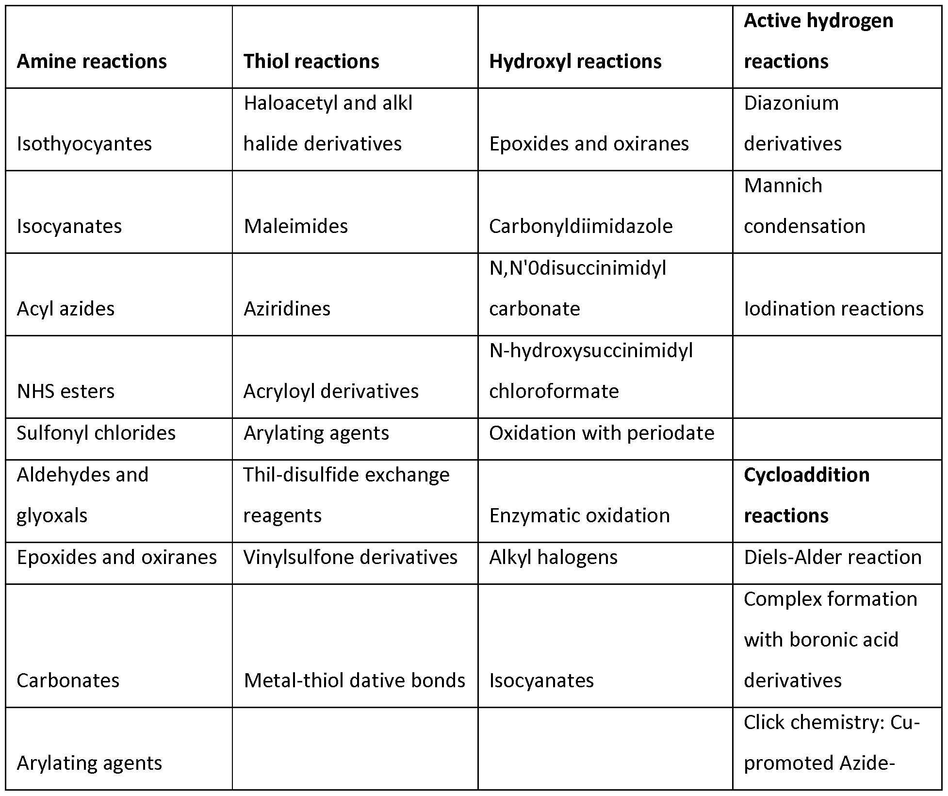


Claims
Priority Applications (24)
| Application Number | Priority Date | Filing Date | Title |
|---|---|---|---|
| RU2016142671A RU2736495C2 (en) | 2014-04-01 | 2015-03-13 | Method and compositions for immunomodulation |
| DK15712504.8T DK3125927T3 (en) | 2014-04-01 | 2015-03-13 | METHODS AND COMPOSITIONS FOR IMMUNU MODULATION |
| EP19188451.9A EP3583946A1 (en) | 2014-04-01 | 2015-03-13 | Enucleated hematopoietic cells comprising an exogenous antigen |
| KR1020227036647A KR20220150986A (en) | 2014-04-01 | 2015-03-13 | Methods and compositions for immunomodulation |
| BR112016022814A BR112016022814A8 (en) | 2014-04-01 | 2015-03-13 | pharmaceutical composition and its method of preparation, nucleic acid, and medical device or medical kit |
| EP15712504.8A EP3125927B1 (en) | 2014-04-01 | 2015-03-13 | Methods and compositions for immunomodulation |
| PL15712504T PL3125927T3 (en) | 2014-04-01 | 2015-03-13 | Methods and compositions for immunomodulation |
| CN201580024460.0A CN106456744A (en) | 2014-04-01 | 2015-03-13 | Methods and compositions for immunomodulation |
| CA2944492A CA2944492A1 (en) | 2014-04-01 | 2015-03-13 | Methods and compositions for immunomodulation |
| MX2020000621A MX2020000621A (en) | 2014-04-01 | 2015-03-13 | Methods and compositions for immunomodulation. |
| MX2016012928A MX371121B (en) | 2014-04-01 | 2015-03-13 | Methods and compositions for immunomodulation. |
| ES15712504T ES2865825T3 (en) | 2014-04-01 | 2015-03-13 | Methods and compositions for immunomodulation |
| AU2015241422A AU2015241422B2 (en) | 2014-04-01 | 2015-03-13 | Methods and compositions for immunomodulation |
| KR1020167030287A KR20170005801A (en) | 2014-04-01 | 2015-03-13 | Methods and compositions for immunomodulation |
| US15/301,046 US10869898B2 (en) | 2014-04-01 | 2015-03-13 | Methods and compositions for immunomodulation |
| JP2016560527A JP6735233B2 (en) | 2014-04-01 | 2015-03-13 | Immunoregulatory methods and compositions |
| IL248143A IL248143B (en) | 2014-04-01 | 2016-09-29 | Methods and compositions for immunomodulation |
| US15/911,924 US11554141B2 (en) | 2014-04-01 | 2018-03-05 | Methods and compositions for immunomodulation |
| US16/383,236 US11576934B2 (en) | 2014-04-01 | 2019-04-12 | Methods and compositions for immunomodulation |
| AU2019208203A AU2019208203B2 (en) | 2014-04-01 | 2019-07-24 | Methods and compositions for immunomodulation |
| US16/557,789 US20190388473A1 (en) | 2014-04-01 | 2019-08-30 | Methods and compositions for immunomodulation |
| IL278643A IL278643B (en) | 2014-04-01 | 2020-11-11 | Methods and compositions for immunomodulation |
| IL285565A IL285565B (en) | 2014-04-01 | 2021-08-12 | Methods and compositions for immunomodulation |
| AU2021232711A AU2021232711A1 (en) | 2014-04-01 | 2021-09-15 | Methods and compositions for immunomodulation |
Applications Claiming Priority (20)
| Application Number | Priority Date | Filing Date | Title |
|---|---|---|---|
| US201461973763P | 2014-04-01 | 2014-04-01 | |
| US201461973764P | 2014-04-01 | 2014-04-01 | |
| US61/973,763 | 2014-04-01 | ||
| US61/973,764 | 2014-04-01 | ||
| US201461991319P | 2014-05-09 | 2014-05-09 | |
| US61/991,319 | 2014-05-09 | ||
| US201462006828P | 2014-06-02 | 2014-06-02 | |
| US201462006825P | 2014-06-02 | 2014-06-02 | |
| US201462006829P | 2014-06-02 | 2014-06-02 | |
| US201462006832P | 2014-06-02 | 2014-06-02 | |
| US62/006,829 | 2014-06-02 | ||
| US62/006,825 | 2014-06-02 | ||
| US62/006,832 | 2014-06-02 | ||
| US62/006,828 | 2014-06-02 | ||
| US201462025367P | 2014-07-16 | 2014-07-16 | |
| US62/025,367 | 2014-07-16 | ||
| US201462059100P | 2014-10-02 | 2014-10-02 | |
| US62/059,100 | 2014-10-02 | ||
| USPCT/US2014/065304 | 2014-11-12 | ||
| PCT/US2014/065304 WO2015073587A2 (en) | 2013-11-18 | 2014-11-12 | Synthetic membrane-receiver complexes |
Related Child Applications (4)
| Application Number | Title | Priority Date | Filing Date |
|---|---|---|---|
| US15/301,046 A-371-Of-International US10869898B2 (en) | 2014-04-01 | 2015-03-13 | Methods and compositions for immunomodulation |
| US15/911,924 Continuation US11554141B2 (en) | 2014-04-01 | 2018-03-05 | Methods and compositions for immunomodulation |
| US16/383,236 Continuation US11576934B2 (en) | 2014-04-01 | 2019-04-12 | Methods and compositions for immunomodulation |
| US16/557,789 Continuation US20190388473A1 (en) | 2014-04-01 | 2019-08-30 | Methods and compositions for immunomodulation |
Publications (1)
| Publication Number | Publication Date |
|---|---|
| WO2015153102A1 true WO2015153102A1 (en) | 2015-10-08 |
Family
ID=54241093
Family Applications (1)
| Application Number | Title | Priority Date | Filing Date |
|---|---|---|---|
| PCT/US2015/020614 WO2015153102A1 (en) | 2014-04-01 | 2015-03-13 | Methods and compositions for immunomodulation |
Country Status (18)
| Country | Link |
|---|---|
| US (4) | US10869898B2 (en) |
| EP (2) | EP3125927B1 (en) |
| JP (4) | JP6735233B2 (en) |
| KR (2) | KR20170005801A (en) |
| CN (1) | CN106456744A (en) |
| AU (3) | AU2015241422B2 (en) |
| BR (1) | BR112016022814A8 (en) |
| CA (1) | CA2944492A1 (en) |
| DK (1) | DK3125927T3 (en) |
| ES (1) | ES2865825T3 (en) |
| HU (1) | HUE054471T2 (en) |
| IL (3) | IL248143B (en) |
| MA (1) | MA39819A (en) |
| MX (1) | MX2020000621A (en) |
| PL (1) | PL3125927T3 (en) |
| PT (1) | PT3125927T (en) |
| RU (2) | RU2020136119A (en) |
| WO (1) | WO2015153102A1 (en) |
Cited By (92)
| Publication number | Priority date | Publication date | Assignee | Title |
|---|---|---|---|---|
| WO2017065280A1 (en) * | 2015-10-14 | 2017-04-20 | 株式会社メガカリオン | Method for producing purified platelets |
| WO2017123644A1 (en) | 2016-01-11 | 2017-07-20 | Rubius Therapeutics, Inc. | Compositions and methods related to multimodal therapeutic cell systems for immune indications |
| WO2018009838A1 (en) | 2016-07-07 | 2018-01-11 | Rubius Therapeutics, Inc. | Compositions and methods related to therapeutic cell systems expressing exogenous rna |
| WO2018102740A1 (en) | 2016-12-02 | 2018-06-07 | Rubius Therapeutics, Inc. | Compositions and methods related to cell systems for penetrating solid tumors |
| WO2018151829A1 (en) | 2017-02-17 | 2018-08-23 | Rubius Therapeutics, Inc. | Functionalized erythroid cells |
| US10124336B2 (en) | 2013-08-16 | 2018-11-13 | Massachusetts Institute Of Technology | Selective delivery of material to cells |
| WO2019017937A1 (en) | 2017-07-19 | 2019-01-24 | Rubius Therapeutics, Inc. | Compositions and methods related to multimodal therapeutic cell systems for cardiometabolic disease |
| WO2019017940A1 (en) | 2017-07-19 | 2019-01-24 | Rubius Therapeutics, Inc. | Compositions and methods related to multimodal therapeutic cell systems for infectious disease |
| WO2019040516A1 (en) | 2017-08-22 | 2019-02-28 | Rubius Therapeutics, Inc. | Lipid nanoparticle methods and compositions for producing engineered erythroid cells |
| US10253296B2 (en) | 2013-11-18 | 2019-04-09 | Rubius Therapeutics, Inc. | Synthetic membrane-receiver complexes |
| WO2019090148A2 (en) | 2017-11-03 | 2019-05-09 | Rubius Therapeutics, Inc. | Compositions and methods related to therapeutic cell systems for tumor growth inhibition |
| WO2019126818A1 (en) | 2017-12-23 | 2019-06-27 | Rubius Therapeutics, Inc. | Artificial antigen presenting cells and methods of use |
| WO2019133881A1 (en) | 2017-12-29 | 2019-07-04 | Rubius Therapeutics, Inc. | Gene editing and targeted transcriptional modulation for enginerering erythroid cells |
| WO2019140116A2 (en) | 2018-01-10 | 2019-07-18 | Rubius Therapeutics, Inc. | Amplifiable rnas for therapeutic cell systems |
| WO2019173798A1 (en) | 2018-03-08 | 2019-09-12 | Rubius Therapeutics, Inc. | Therapeutic cell systems and methods for treating cancer and infectious diseases |
| WO2019183292A1 (en) | 2018-03-20 | 2019-09-26 | Rubius Therapeutics, Inc. | Therapeutic cell systems and methods for treating hyperuricemia and gout |
| WO2019183294A1 (en) | 2018-03-20 | 2019-09-26 | Rubius Therapeutics, Inc. | Therapeutic cell systems and methods for treating homocystinuria |
| US10526573B2 (en) | 2014-11-14 | 2020-01-07 | Massachusetts Institute Of Technology | Disruption and field enabled delivery of compounds and compositions into cells |
| WO2020117852A1 (en) | 2018-12-03 | 2020-06-11 | Rubius Therapeutics, Inc. | Artificial antigen presenting cells including hla-e and hla-g molecules and methods of use |
| US10696944B2 (en) | 2011-10-17 | 2020-06-30 | Massachusetts Institute Of Technology | Intracellular delivery |
| WO2020150608A1 (en) | 2019-01-18 | 2020-07-23 | Flagship Pioneering, Inc. | Trem compositions and uses thereof |
| WO2020172472A1 (en) | 2019-02-20 | 2020-08-27 | Rubius Therapeutics, Inc. | Engineered erythroid cells including loadable antigen-presenting polypeptides and methods of use |
| WO2020181013A1 (en) | 2019-03-04 | 2020-09-10 | Flagship Pioneering Innovations Vi, Llc | Circular polyribonucleotides and pharmaceutical compositions thereof |
| WO2020198403A2 (en) | 2019-03-25 | 2020-10-01 | Flagship Pioneering Innovations Vi, Llc | Compositions comprising modified circular polyribonucleotides and uses thereof |
| WO2020219909A1 (en) * | 2019-04-26 | 2020-10-29 | Rubius Therapeutics, Inc. | Buffered compositions including enucleated erythroid cells |
| WO2020243560A1 (en) | 2019-05-31 | 2020-12-03 | Flagship Pioneering, Inc. | Uses of trem compositions to modulate trna pools |
| WO2020252436A1 (en) | 2019-06-14 | 2020-12-17 | Flagship Pioneering Innovations Vi, Llc | Circular rnas for cellular therapy |
| US10869898B2 (en) | 2014-04-01 | 2020-12-22 | Rubius Therapeutics, Inc. | Methods and compositions for immunomodulation |
| WO2020257730A1 (en) | 2019-06-19 | 2020-12-24 | Flagship Pioneering Innovations Vi, Llc | Compositions comprising circular polyribonucleotides for protein modulation and uses thereof |
| WO2020264333A1 (en) | 2019-06-26 | 2020-12-30 | Orphan Technologies, Ltd. | Pegylated cystathionine beta synthase for enzyme therapy for treatment of homocystinuria |
| CN112285361A (en) * | 2020-09-27 | 2021-01-29 | 中国人民解放军空军军医大学 | Reagents to eliminate the interference of anti-CD38 monoclonal antibody drugs in the detection of anti-human globulin |
| RU2743363C1 (en) * | 2020-06-03 | 2021-02-17 | федеральное государственное бюджетное образовательное учреждение высшего образования "Ставропольский государственный аграрный университет" | Method for testing immunological tolerance in animals |
| WO2021092064A1 (en) | 2019-11-04 | 2021-05-14 | Flagship Pioneering, Inc. | Trem compositions for con-rare codons and related uses |
| WO2021092073A1 (en) | 2019-11-04 | 2021-05-14 | Flagship Pioneering, Inc. | Methods of modifying a nucleic acid sequence |
| WO2021155177A1 (en) | 2020-01-29 | 2021-08-05 | Flagship Pioneering Innovations Vi, Llc | Compositions comprising linear polyribonucleotides for protein modulation and uses thereof |
| WO2021155175A1 (en) | 2020-01-29 | 2021-08-05 | Flagship Pioneering Innovations Vi, Llc | Compositions for translation and methods of use thereof |
| WO2021155171A1 (en) | 2020-01-29 | 2021-08-05 | Flagship Pioneering Innovations Vi, Llc | Delivery of compositions comprising circular polyribonucleotides |
| WO2021162731A1 (en) | 2020-02-10 | 2021-08-19 | Rubius Therapeutics, Inc. | Engineered erythroid cells including hla-g polypeptides and methods of use thereof |
| US11111472B2 (en) | 2014-10-31 | 2021-09-07 | Massachusetts Institute Of Technology | Delivery of biomolecules to immune cells |
| US11125739B2 (en) | 2015-01-12 | 2021-09-21 | Massachusetts Institute Of Technology | Gene editing through microfluidic delivery |
| US11166996B2 (en) | 2018-12-12 | 2021-11-09 | Flagship Pioneering Innovations V, Inc. | Anellovirus compositions and methods of use |
| WO2021236952A1 (en) | 2020-05-20 | 2021-11-25 | Flagship Pioneering, Inc. | Compositions and methods for producing human polyclonal antibodies |
| WO2021236930A1 (en) | 2020-05-20 | 2021-11-25 | Flagship Pioneering Innovations Vi, Llc | Immunogenic compositions and uses thereof |
| WO2021236980A1 (en) | 2020-05-20 | 2021-11-25 | Flagship Pioneering Innovations Vi, Llc | Coronavirus antigen compositions and their uses |
| WO2021243290A1 (en) | 2020-05-29 | 2021-12-02 | Flagship Pioneering Innovations Vi, Llc | Trem compositions and methods relating thereto |
| WO2021243301A2 (en) | 2020-05-29 | 2021-12-02 | Flagship Pioneering Innovations Vi, Llc. | Trem compositions and methods relating thereto |
| WO2021262799A1 (en) | 2020-06-23 | 2021-12-30 | Flagship Pioneering, Inc. | Anti-viral compounds and methods of using same |
| GB2597623A (en) * | 2017-08-07 | 2022-02-02 | Univ California | Platform for generating safe cell therapeutics |
| WO2022051629A1 (en) | 2020-09-03 | 2022-03-10 | Flagship Pioneering Innovations Vi, Llc | Immunogenic compositions and uses thereof |
| RU2766780C1 (en) * | 2020-12-30 | 2022-03-15 | федеральное государственное бюджетное образовательное учреждение высшего образования "Ставропольский государственный аграрный университет" | Method for determining the antigenic load in animals |
| US11299698B2 (en) | 2015-07-09 | 2022-04-12 | Massachusetts Institute Of Technology | Delivery of materials to anucleate cells |
| WO2022140702A1 (en) | 2020-12-23 | 2022-06-30 | Flagship Pioneering, Inc. | Compositions of modified trems and uses thereof |
| WO2022150569A1 (en) | 2021-01-08 | 2022-07-14 | Rubius Therapeutics, Inc. | METHODS OF INCREASING NKp30-POSITIVE LYMPHOCYTES IN A SUBJECT AND USES THEREOF |
| WO2022150578A1 (en) | 2021-01-08 | 2022-07-14 | Rubius Therapeutics, Inc. | Methods of treating a tumor in a human subject |
| WO2022197548A1 (en) | 2021-03-14 | 2022-09-22 | Rubius Therapeutics, Inc. | Methods of increasing nkg2d-positive lymphocytes in a subject and uses thereof |
| WO2022212784A1 (en) | 2021-03-31 | 2022-10-06 | Flagship Pioneering Innovations V, Inc. | Thanotransmission polypeptides and their use in treating cancer |
| WO2022256646A1 (en) | 2021-06-03 | 2022-12-08 | Rubius Therapeutics, Inc. | Methods of treating hpv16-positive or hpv16-associated cancer in a subject |
| WO2022263824A1 (en) | 2021-06-16 | 2022-12-22 | Xap Therapeutics Limited | Methods and compositions |
| WO2023009547A1 (en) | 2021-07-26 | 2023-02-02 | Flagship Pioneering Innovations Vi, Llc | Trem compositions and uses thereof |
| WO2023044006A1 (en) | 2021-09-17 | 2023-03-23 | Flagship Pioneering Innovations Vi, Llc | Compositions and methods for producing circular polyribonucleotides |
| US11613759B2 (en) | 2015-09-04 | 2023-03-28 | Sqz Biotechnologies Company | Intracellular delivery of biomolecules to cells comprising a cell wall |
| US20230110360A1 (en) * | 2020-04-16 | 2023-04-13 | Ix Layer Inc. | Systems and methods for access management and clustering of genomic, phenotype, and diagnostic data |
| WO2023069397A1 (en) | 2021-10-18 | 2023-04-27 | Flagship Pioneering Innovations Vi, Llc | Compositions and methods for purifying polyribonucleotides |
| WO2023096990A1 (en) | 2021-11-24 | 2023-06-01 | Flagship Pioneering Innovation Vi, Llc | Coronavirus immunogen compositions and their uses |
| WO2023096963A1 (en) | 2021-11-24 | 2023-06-01 | Flagship Pioneering Innovations Vi, Llc | Varicella-zoster virus immunogen compositions and their uses |
| WO2023097003A2 (en) | 2021-11-24 | 2023-06-01 | Flagship Pioneering Innovations Vi, Llc | Immunogenic compositions and their uses |
| WO2023122789A1 (en) | 2021-12-23 | 2023-06-29 | Flagship Pioneering Innovations Vi, Llc | Circular polyribonucleotides encoding antifusogenic polypeptides |
| WO2023122745A1 (en) | 2021-12-22 | 2023-06-29 | Flagship Pioneering Innovations Vi, Llc | Compositions and methods for purifying polyribonucleotides |
| WO2023220729A2 (en) | 2022-05-13 | 2023-11-16 | Flagship Pioneering Innovations Vii, Llc | Double stranded dna compositions and related methods |
| WO2023220083A1 (en) | 2022-05-09 | 2023-11-16 | Flagship Pioneering Innovations Vi, Llc | Trem compositions and methods of use for treating proliferative disorders |
| WO2023230566A2 (en) | 2022-05-25 | 2023-11-30 | Flagship Pioneering Innovations Vii, Llc | Compositions and methods for modulating cytokines |
| WO2023230570A2 (en) | 2022-05-25 | 2023-11-30 | Flagship Pioneering Innovations Vii, Llc | Compositions and methods for modulating genetic drivers |
| WO2023230573A2 (en) | 2022-05-25 | 2023-11-30 | Flagship Pioneering Innovations Vii, Llc | Compositions and methods for modulation of immune responses |
| WO2023230549A2 (en) | 2022-05-25 | 2023-11-30 | Flagship Pioneering Innovations Vii, Llc | Compositions and methods for modulation of tumor suppressors and oncogenes |
| WO2023230578A2 (en) | 2022-05-25 | 2023-11-30 | Flagship Pioneering Innovations Vii, Llc | Compositions and methods for modulating circulating factors |
| WO2023240274A1 (en) * | 2022-06-10 | 2023-12-14 | The Medical College Of Wisconsin, Inc. | Immune tolerance induction for auto-immune diseases through platelet targeted gene therapy involving myelin oligodendrocyte glycoprotein (mog) polypeptide. |
| WO2023250112A1 (en) | 2022-06-22 | 2023-12-28 | Flagship Pioneering Innovations Vi, Llc | Compositions of modified trems and uses thereof |
| WO2024077191A1 (en) | 2022-10-05 | 2024-04-11 | Flagship Pioneering Innovations V, Inc. | Nucleic acid molecules encoding trif and additionalpolypeptides and their use in treating cancer |
| WO2024097664A1 (en) | 2022-10-31 | 2024-05-10 | Flagship Pioneering Innovations Vi, Llc | Compositions and methods for purifying polyribonucleotides |
| WO2024102799A1 (en) | 2022-11-08 | 2024-05-16 | Flagship Pioneering Innovations Vi, Llc | Compositions and methods for producing circular polyribonucleotides |
| WO2024129988A1 (en) | 2022-12-14 | 2024-06-20 | Flagship Pioneering Innovations Vii, Llc | Compositions and methods for delivery of therapeutic agents to bone |
| WO2024151685A1 (en) | 2023-01-09 | 2024-07-18 | Beth Israel Deaconess Medical Center, Inc. | Recombinant nucleic acid molecules and their use in wound healing |
| WO2024151687A1 (en) | 2023-01-09 | 2024-07-18 | Flagship Pioneering Innovations V, Inc. | Genetic switches and their use in treating cancer |
| WO2024151673A2 (en) | 2023-01-09 | 2024-07-18 | President And Fellows Of Harvard College | Recombinant nucleic acid molecules and their use in wound healing |
| WO2024173828A1 (en) | 2023-02-17 | 2024-08-22 | Flagship Pioneering Innovations Vii, Llc | Dna compositions comprising modified uracil |
| WO2024173836A2 (en) | 2023-02-17 | 2024-08-22 | Flagship Pioneering Innovations Vii, Llc | Dna compositions comprising modified cytosine |
| WO2024192422A1 (en) | 2023-03-15 | 2024-09-19 | Flagship Pioneering Innovations Vi, Llc | Immunogenic compositions and uses thereof |
| WO2024192420A1 (en) | 2023-03-15 | 2024-09-19 | Flagship Pioneering Innovations Vi, Llc | Compositions comprising polyribonucleotides and uses thereof |
| WO2024216128A1 (en) | 2023-04-12 | 2024-10-17 | Flagship Pioneering Innovations Vi, Llc | Trems for use in correction of missense mutations |
| WO2025006684A1 (en) | 2023-06-28 | 2025-01-02 | Flagship Pioneering Innovations Vi, Llc | Circular polyribonucleotides encoding antifusogenic polypeptides |
| WO2025042786A1 (en) | 2023-08-18 | 2025-02-27 | Flagship Pioneering Innovations Vi, Llc | Compositions comprising circular polyribonucleotides and uses thereof |
| WO2025096807A2 (en) | 2023-10-31 | 2025-05-08 | Flagship Pioneering Innovations Vii, Llc | Novel therapeutic dna forms |
Families Citing this family (61)
| Publication number | Priority date | Publication date | Assignee | Title |
|---|---|---|---|---|
| CN108129554A (en) | 2010-08-10 | 2018-06-08 | 洛桑聚合联合学院 | Erythrocyte binding therapeutic agent |
| US9517257B2 (en) | 2010-08-10 | 2016-12-13 | Ecole Polytechnique Federale De Lausanne (Epfl) | Erythrocyte-binding therapeutics |
| US9850296B2 (en) | 2010-08-10 | 2017-12-26 | Ecole Polytechnique Federale De Lausanne (Epfl) | Erythrocyte-binding therapeutics |
| US9745548B2 (en) | 2012-03-15 | 2017-08-29 | Flodesign Sonics, Inc. | Acoustic perfusion devices |
| US9950282B2 (en) | 2012-03-15 | 2018-04-24 | Flodesign Sonics, Inc. | Electronic configuration and control for acoustic standing wave generation |
| US10689609B2 (en) | 2012-03-15 | 2020-06-23 | Flodesign Sonics, Inc. | Acoustic bioreactor processes |
| US10322949B2 (en) | 2012-03-15 | 2019-06-18 | Flodesign Sonics, Inc. | Transducer and reflector configurations for an acoustophoretic device |
| US9752113B2 (en) | 2012-03-15 | 2017-09-05 | Flodesign Sonics, Inc. | Acoustic perfusion devices |
| US9458450B2 (en) | 2012-03-15 | 2016-10-04 | Flodesign Sonics, Inc. | Acoustophoretic separation technology using multi-dimensional standing waves |
| US10704021B2 (en) | 2012-03-15 | 2020-07-07 | Flodesign Sonics, Inc. | Acoustic perfusion devices |
| US10967298B2 (en) | 2012-03-15 | 2021-04-06 | Flodesign Sonics, Inc. | Driver and control for variable impedence load |
| US10737953B2 (en) | 2012-04-20 | 2020-08-11 | Flodesign Sonics, Inc. | Acoustophoretic method for use in bioreactors |
| US9745569B2 (en) | 2013-09-13 | 2017-08-29 | Flodesign Sonics, Inc. | System for generating high concentration factors for low cell density suspensions |
| CN105939767B (en) | 2014-01-08 | 2018-04-06 | 弗洛设计声能学公司 | Sound electrophoretic apparatus with alliteration electrophoresis chamber |
| ES2874884T3 (en) | 2014-02-21 | 2021-11-05 | Ecole Polytechnique Fed De Lausanne Epfl Epfl Tto | Glucose Targeting Therapeutic Compounds |
| US10046056B2 (en) | 2014-02-21 | 2018-08-14 | École Polytechnique Fédérale De Lausanne (Epfl) | Glycotargeting therapeutics |
| US10953101B2 (en) | 2014-02-21 | 2021-03-23 | École Polytechnique Fédérale De Lausanne (Epfl) | Glycotargeting therapeutics |
| US10946079B2 (en) | 2014-02-21 | 2021-03-16 | Ecole Polytechnique Federale De Lausanne | Glycotargeting therapeutics |
| US9744483B2 (en) | 2014-07-02 | 2017-08-29 | Flodesign Sonics, Inc. | Large scale acoustic separation device |
| US11021699B2 (en) | 2015-04-29 | 2021-06-01 | FioDesign Sonics, Inc. | Separation using angled acoustic waves |
| US11708572B2 (en) | 2015-04-29 | 2023-07-25 | Flodesign Sonics, Inc. | Acoustic cell separation techniques and processes |
| US11377651B2 (en) | 2016-10-19 | 2022-07-05 | Flodesign Sonics, Inc. | Cell therapy processes utilizing acoustophoresis |
| US11459540B2 (en) | 2015-07-28 | 2022-10-04 | Flodesign Sonics, Inc. | Expanded bed affinity selection |
| US11474085B2 (en) | 2015-07-28 | 2022-10-18 | Flodesign Sonics, Inc. | Expanded bed affinity selection |
| US11085035B2 (en) | 2016-05-03 | 2021-08-10 | Flodesign Sonics, Inc. | Therapeutic cell washing, concentration, and separation utilizing acoustophoresis |
| US11214789B2 (en) | 2016-05-03 | 2022-01-04 | Flodesign Sonics, Inc. | Concentration and washing of particles with acoustics |
| EP3506919A4 (en) * | 2016-09-02 | 2020-04-15 | The Board of Trustees of the University of Illinois | PEPTIDE OBTAINED FROM KIF13B AND METHOD FOR INHIBITING ANGIOGENESIS |
| JP2020513248A (en) | 2016-10-19 | 2020-05-14 | フロデザイン ソニックス, インク.Flodesign Sonics, Inc. | Affinity cell extraction by sound |
| US20200038484A1 (en) * | 2017-02-06 | 2020-02-06 | The Board Of Trustees Of The Leland Stanford Junior University | Bioconjugation Methods for Targeted in Situ Therapeutic Delivery |
| WO2018170150A2 (en) * | 2017-03-16 | 2018-09-20 | Seattle Children's Hospital (dba Seattle Children's Research Institute) | Engraftable cell-based immunotherapy for long-term delivery of therapeutic proteins |
| US11253579B2 (en) | 2017-06-16 | 2022-02-22 | The University Of Chicago | Compositions and methods for inducing immune tolerance |
| US20190062448A1 (en) * | 2017-07-31 | 2019-02-28 | Tizona Therapeutics | Anti-cd39 antibodies, compositions comprising anti-cd39 antibodies and methods of using anti-cd39 antibodies |
| US10960071B2 (en) | 2017-08-07 | 2021-03-30 | The Regents Of The University Of California | Platform for generating safe cell therapeutics |
| CN107510839A (en) * | 2017-08-31 | 2017-12-26 | 广东颜值科技有限公司 | A kind of immunomodulator and its preparation method and application |
| CN114900773A (en) | 2017-12-14 | 2022-08-12 | 弗洛设计声能学公司 | Acoustophoretic system, method for operating acoustophoretic system, and method for controlling acoustic transducer and acoustic system |
| JP2021509812A (en) | 2018-01-05 | 2021-04-08 | プレートレット バイオジェネシス, インコーポレイテッド | Compositions and Methods for Producing Megakaryocytes |
| CA3092062A1 (en) | 2018-02-22 | 2019-08-29 | Entrada Therapeutics, Inc. | Compositions and methods for treating mitochondrial neurogastrointestinal encephalopathy |
| JP2021524846A (en) * | 2018-05-16 | 2021-09-16 | シーエスエル、リミテッド | Soluble complement receptor type 1 mutants and their use |
| AU2019291955A1 (en) * | 2018-06-29 | 2021-02-18 | Platelet Biogenesis, Inc. | Compositions for drug delivery and methods of use thereof |
| US12331320B2 (en) | 2018-10-10 | 2025-06-17 | The Research Foundation For The State University Of New York | Genome edited cancer cell vaccines |
| KR102762828B1 (en) * | 2018-12-11 | 2025-02-05 | 큐32 바이오 인크. | Fusion protein constructs for complement-related diseases |
| GB2583560A (en) | 2018-12-11 | 2020-11-04 | Admirx Inc | Fusion protein constructs for complement associated disease |
| CN109810992A (en) * | 2019-02-01 | 2019-05-28 | 天津师范大学 | A full-length cDNA sequence encoding the extracellular ATP hydrolase CD39 of flounder and its application |
| CN110244053B (en) * | 2019-05-09 | 2022-03-11 | 北京大学第三医院(北京大学第三临床医学院) | Molecular marker for diagnosing lupus nephritis and pulmonary hypertension disease and application thereof |
| CN110215737B (en) * | 2019-06-13 | 2021-07-02 | 厦门华厦学院 | Affinity monolithic column based on graphene-nanogold composite interface ultrahigh-load aptamer and preparation method thereof |
| EP4025048A2 (en) | 2019-09-05 | 2022-07-13 | Hemanext Inc. | Methods for the preservation of reagent red blood cells using carbon monoxide |
| US20220395563A1 (en) * | 2019-11-11 | 2022-12-15 | The Regents Of The University Of California | Polymeric nanoparticles that target liver sinusoidal endothelial cells to induce antigen-specific immune tolerance |
| US20230040275A1 (en) * | 2019-11-19 | 2023-02-09 | Spark Therapeutics, Inc. | Secretable protein induced immune tolerization and treatment of autoimmune, allergic and other diseases and disorders |
| IL293628A (en) * | 2019-12-12 | 2022-08-01 | Iltoo Pharma | Interleukin 2 chimeric structures |
| WO2021203042A1 (en) * | 2020-04-02 | 2021-10-07 | Lineage Cell Therapeutics, Inc. | A universal adoptive immunotherapy approach to treat covid-19 and future emerging infectious diseases |
| WO2021257480A2 (en) | 2020-06-14 | 2021-12-23 | Catalyst Biosciences, Inc. | Complement factor i-related compositions and methods |
| WO2022005991A1 (en) * | 2020-06-29 | 2022-01-06 | Memorial Sloan Kettering Cancer Center | Platform for assessing in vivo and in vitro hapten-capture in a cell bound system |
| US20230346988A1 (en) * | 2020-06-29 | 2023-11-02 | Memorial Sloan Kettering Cancer Center | Systems for in vivo monitoring of immune cells in patients undergoing cellular immunotherapy |
| US20230349902A1 (en) * | 2020-07-27 | 2023-11-02 | Commissariat A L'energie Atomique Et Aux Energies Alternatives | Novel t cells and the uses thereof |
| CN112156111B (en) * | 2020-08-31 | 2022-10-18 | 中南大学湘雅三医院 | Application of cord blood platelet mitochondria in preparation of medicine for treating autoimmune disease |
| CA3199737A1 (en) * | 2020-11-30 | 2022-06-02 | Tobin Dickerson | Microneedle devices and methods, and skin condition assays |
| CN112544989A (en) * | 2020-12-18 | 2021-03-26 | 完美(广东)日用品有限公司 | A composition with muscle protecting effect |
| CA3203709A1 (en) * | 2020-12-29 | 2022-07-07 | Sqz Biotechnologies Company | Formulations of activating antigen carriers |
| WO2023164466A1 (en) * | 2022-02-23 | 2023-08-31 | Sqz Biotechnologies Company | Methods of producing glial cells and uses thereof |
| CN115381950B (en) * | 2022-09-06 | 2023-08-18 | 天津医科大学总医院 | Application of Rab35 or downstream regulatory molecules thereof in treating chronic pain after fracture |
| WO2024148139A2 (en) * | 2023-01-05 | 2024-07-11 | Arizona Board Of Regents On Behalf Of Arizona State University | Metabolite-based particles and compositions thereof for preventing and treating neurological diseases and disorders |
Citations (3)
| Publication number | Priority date | Publication date | Assignee | Title |
|---|---|---|---|---|
| US20040133922A1 (en) * | 2002-11-27 | 2004-07-08 | Hiroo Okamoto | Receiving apparatus, printer and printing control method for digital image information |
| WO2005118780A1 (en) * | 2004-06-04 | 2005-12-15 | Universite Pierre Et Marie Curie - Paris Vi | A method for producing red blood cells |
| WO2007039150A2 (en) * | 2005-09-23 | 2007-04-12 | Cellerix, S.L. | Cell populations having immunoregulatory activity, method for isolation and uses |
Family Cites Families (86)
| Publication number | Priority date | Publication date | Assignee | Title |
|---|---|---|---|---|
| US4327710A (en) | 1980-06-18 | 1982-05-04 | The United States Of America As Represented By The Secretary Of Agriculture | Process for encapsulating additives in resealed erythrocytes for disseminating chemicals via the circulatory system |
| IL86650A0 (en) | 1987-06-30 | 1988-11-30 | Biophor Corp | Animal derived cells and liposomes,having an antigenic protein incorporated into their membrane |
| US5258499A (en) | 1988-05-16 | 1993-11-02 | Vestar, Inc. | Liposome targeting using receptor specific ligands |
| US5676954A (en) | 1989-11-03 | 1997-10-14 | Vanderbilt University | Method of in vivo delivery of functioning foreign genes |
| IT1245867B (en) | 1991-06-03 | 1994-10-25 | Consiglio Nazionale Ricerche | VACCINES USING ERYTHROCYTES AS ANTIGEN VEHICLES |
| ES2109260T3 (en) | 1991-06-14 | 1998-01-16 | Europ Communities | TRANSFORMED Erythrocytes, PROCEDURE TO PREPARE THEM AND THEIR USE IN PHARMACEUTICAL COMPOSITIONS. |
| US5830462A (en) | 1993-02-12 | 1998-11-03 | President & Fellows Of Harvard College | Regulated transcription of targeted genes and other biological events |
| US5599705A (en) | 1993-11-16 | 1997-02-04 | Cameron; Robert B. | In vitro method for producing differentiated universally compatible mature human blood cells |
| US6984379B1 (en) | 1994-04-08 | 2006-01-10 | Children's Hospital of LosAngeles | Gene therapy by administration of genetically engineered CD34+ cells obtained from cord blood |
| US6132764A (en) | 1994-08-05 | 2000-10-17 | Targesome, Inc. | Targeted polymerized liposome diagnostic and treatment agents |
| US5648248A (en) | 1994-12-30 | 1997-07-15 | Boehringer Ingelheim International Gmbh | Methods for producing differentiated cells from immature hematopoietic cells |
| US6060052A (en) | 1995-10-30 | 2000-05-09 | Systemix, Inc. | Methods for use of Mpl ligands with primitive human hematopoietic stem cells |
| US6225290B1 (en) | 1996-09-19 | 2001-05-01 | The Regents Of The University Of California | Systemic gene therapy by intestinal cell transformation |
| TW520297B (en) | 1996-10-11 | 2003-02-11 | Sequus Pharm Inc | Fusogenic liposome composition and method |
| JP2001503396A (en) | 1996-10-11 | 2001-03-13 | アルザ コーポレイション | Therapeutic liposome compositions and methods |
| JP2001505563A (en) | 1996-11-21 | 2001-04-24 | ニューヨーク・ブラッド・センター・インコーポレイテッド | How to convert blood type |
| DE69732225T2 (en) | 1997-05-05 | 2005-06-23 | Dideco S.R.L., Mirandola | Process for the encapsulation of biologically active substances in erythrocytes and apparatus therefor |
| EP1104455A2 (en) | 1998-06-25 | 2001-06-06 | Hemosol Inc. | Media and methods for expansion of erythroid stem cells for the production of hemoglobin |
| WO2007143451A2 (en) | 2006-05-30 | 2007-12-13 | Terman David S | Targeted delivery of oncolytic viruses, anti-tumor proteins, plasmids, toxins, hemolysins & chemotherapy |
| AU2002306766A1 (en) | 2001-03-16 | 2002-10-03 | Johns Hopkins University School Of Medicine | Immune modulation by transduced hematopoietic stem cells expressing antigens and antigen-presenting cell regulatory molecules |
| US20030207247A1 (en) | 2001-12-05 | 2003-11-06 | Cerus Corporation | Preparation of red blood cells having reduced immunogenicity |
| US20030133922A1 (en) * | 2002-01-15 | 2003-07-17 | Kasha John R. | Oral tolerance using allogeneic platelets in ITP |
| WO2004029200A2 (en) | 2002-09-26 | 2004-04-08 | Children's Medical Center Corporation | Method of enhancing proliferation and/or hematopoietic differentiation of stem cells |
| AU2003298016A1 (en) | 2002-11-27 | 2004-06-23 | Regents Of The University Of Minnesota | Homologous recombination in multipotent adult progenitor cells |
| DE60333487D1 (en) | 2002-12-13 | 2010-09-02 | Genetix Pharmaceuticals Inc | THERAPEUTIC RETROVIRUS VECTORS FOR GENE THERAPY |
| DE10335833A1 (en) | 2003-08-05 | 2005-03-03 | Curevac Gmbh | Transfection of blood cells with mRNA for immune stimulation and gene therapy |
| CA2574802A1 (en) * | 2004-07-20 | 2006-02-02 | Isogenis, Inc. | Specific inhibition of autoimmunity and diseases associated with autoantigens |
| FR2873925B1 (en) | 2004-08-05 | 2006-10-13 | Erytech Pharma Soc Par Actions | METHOD AND DEVICE FOR LYSE-RESCALING FOR THE INCORPORATION OF ACTIVE PRINCIPLE, IN PARTICULAR ASPARAGINASE OR INOSITOL HEXAPHOSPHATE, IN ERYTHROCYTES |
| DE102004054536A1 (en) | 2004-11-06 | 2006-05-11 | Capsulution Nanoscience Ag | Multimodal modified cells as cellular dosage forms for active substances and as diagnostic cell particles |
| FR2884717B1 (en) | 2005-04-25 | 2009-07-03 | Erytech Pharma Soc Par Actions | ERYTHROCYTES CONTAINING ARGININE DEIMINASE |
| FR2891843A1 (en) | 2005-10-06 | 2007-04-13 | Erytech Pharma Soc Par Actions | ERYTHROCYTES CONTAINING 5-FLUOROURACILE |
| US7462485B2 (en) | 2005-10-07 | 2008-12-09 | Glaser Lawrence F | Modified erythrocytes and uses thereof |
| US20070243137A1 (en) | 2006-04-18 | 2007-10-18 | Nanoprobes, Inc. | Cell and sub-cell methods for imaging and therapy |
| US7534595B2 (en) | 2006-06-12 | 2009-05-19 | Biomarin Pharmaceutical Inc. | Compositions of prokaryotic phenylalanine ammonia-lyase and methods of using compositions thereof |
| US7531341B1 (en) | 2006-06-12 | 2009-05-12 | Biomarin Pharmaceutical Inc. | Compositions of prokaryotic phenylalanine ammonia-lyase and methods of using compositions thereof |
| EP2012122A1 (en) * | 2007-07-06 | 2009-01-07 | Medigene AG | Mutated parvovirus structural proteins as vaccines |
| FR2919804B1 (en) | 2007-08-08 | 2010-08-27 | Erytech Pharma | COMPOSITION AND ANTI-TUMOR THERAPEUTIC VACCINE |
| US7537923B2 (en) | 2007-08-17 | 2009-05-26 | Biomarin Pharmaceutical Inc. | Compositions of prokaryotic phenylalanine ammonia-lyase and methods of treating cancer using compositions thereof |
| US9260692B2 (en) | 2007-10-31 | 2016-02-16 | Universitat Zurich | Use of modified cells for the treatment of multiple sclerosis |
| EP2057998A1 (en) | 2007-10-31 | 2009-05-13 | Universitätsklinikum Hamburg-Eppendorf | Use of modified cells for the treatment of multiple sclerosis |
| FR2925339B1 (en) | 2007-12-24 | 2010-03-05 | Erytech Pharma | DRUG FOR THE TREATMENT OF PANCREATIC CANCER |
| WO2009101467A1 (en) | 2008-02-13 | 2009-08-20 | Erytech Pharma | Formulation and method for the prevention and treatment of skeletal manifestation of gaucher's disease |
| FR2928270B1 (en) | 2008-03-10 | 2011-01-21 | Erytech Pharma | FORMULATION METHOD FOR THE PREVENTION OR TREATMENT OF BONE METASTASES AND OTHER BONE DISEASES |
| US8231895B2 (en) | 2008-05-22 | 2012-07-31 | Universidade De Coimbra | Targeted delivery to human diseases and disorders |
| US20120034156A1 (en) * | 2010-08-03 | 2012-02-09 | Searete Llc, A Limited Liability Corporation Of The State Of Delaware | Artificial cells |
| US8211656B2 (en) | 2008-08-13 | 2012-07-03 | The Invention Science Fund I, Llc | Biological targeting compositions and methods of using the same |
| US20100040546A1 (en) | 2008-08-13 | 2010-02-18 | Searete Llc, A Limited Liability Corporation Of The State Of Delaware | Biological targeting compositions and methods of using the same |
| FR2938332B1 (en) | 2008-11-07 | 2011-11-25 | Erytech Pharma | PREDICTIVE TEST FOR NEUTRALIZATION OF ASPARAGINASE ACTIVITY |
| EA201100765A1 (en) | 2008-12-15 | 2012-04-30 | Бинд Биосаиэнсис | Long-term circulation nanoparticles |
| FR2944106B1 (en) | 2009-04-03 | 2012-09-28 | Erytech Pharma | METHOD OF DETERMINING INOSITOL HEXAPHOSPHATE (IHP). |
| JP6215533B2 (en) | 2009-04-09 | 2017-10-18 | サンガモ セラピューティクス, インコーポレイテッド | Targeted integration into stem cells |
| EP2432496B1 (en) | 2009-05-19 | 2018-03-28 | ETH Zurich | Control of uric acid homeostasis |
| PT2493487T (en) | 2009-10-27 | 2016-11-08 | Erytech Pharma | COMPOSITION TO INDUCE SPECIFIC IMMUNITY TOLERANCE |
| KR20130048193A (en) | 2010-03-02 | 2013-05-09 | 에프. 호프만-라 로슈 아게 | Expression vector |
| AU2011224257A1 (en) | 2010-03-12 | 2012-10-11 | The Regents Of The University Of California | Triggered cargo release from nanoparticle stabilized liposomes |
| US9517257B2 (en) | 2010-08-10 | 2016-12-13 | Ecole Polytechnique Federale De Lausanne (Epfl) | Erythrocyte-binding therapeutics |
| CN101985634B (en) * | 2010-11-04 | 2012-06-13 | 中国人民解放军第四军医大学 | GFP membrane type expression vector and method for sorting vector-transfected positive cells |
| CN102199199A (en) * | 2011-04-06 | 2011-09-28 | 中国人民解放军第四军医大学 | Membrane localization staphylococcal enterotoxin A and gene sequence thereof |
| CN113337402B (en) | 2011-10-17 | 2024-11-29 | 麻省理工学院 | Intracellular delivery |
| CN110075284A (en) | 2012-02-15 | 2019-08-02 | 洛桑聚合联合学院 | Erythrocyte binding therapeutic agent |
| EP4052717A1 (en) | 2012-03-13 | 2022-09-07 | Celularity Inc. | Modified erythrocyte precursor cells and uses thereof |
| RU2667639C2 (en) | 2012-03-21 | 2018-09-21 | Эритек Фарма | Medicament for treatment of acute myeloid leukemia (aml) |
| EP2669381A1 (en) | 2012-05-30 | 2013-12-04 | AmVac AG | Method for expression of heterologous proteins using a recombinant negative-strand RNA virus vector comprising a mutated P protein |
| KR20140011912A (en) | 2012-07-20 | 2014-01-29 | 도꾸리쯔교세이호징 리가가쿠 겐큐소 | Human erythroid progenitor cell line and method for producing human enucleated red blood cells |
| WO2014066945A1 (en) | 2012-11-01 | 2014-05-08 | The Sydney Children's Hospital Network (Randwick & Westmead) | Genetically-modified probiotic for treatment of phenylketonuria |
| US9675678B2 (en) | 2013-01-29 | 2017-06-13 | The Regents Of The University Of Colorado, A Body Corporate | Compositions and methods for treatment of homocystinuria |
| CN103224957B (en) * | 2013-04-16 | 2017-08-04 | 福建三一造血技术有限公司 | The method of external evoked generation ASPase containing L II red blood cell medicine |
| FR3005420B1 (en) | 2013-05-07 | 2015-09-18 | Erytech Pharma | METHOD OF STABILIZING SUSPENSIONS OF ERYTHROCYTES ENCAPSULATING AN ACTIVE INGREDIENT, SUSPENSIONS OBTAINED |
| JP6367314B2 (en) | 2013-05-10 | 2018-08-01 | エリデル ソチエタ ペル アチオニ | Method for preparing red blood cells loaded with one or more pharmaceutically important substances and red blood cells thus obtained |
| ES2901383T3 (en) | 2013-05-10 | 2022-03-22 | Whitehead Inst Biomedical Res | In vitro production of red blood cells with sortase-markable proteins |
| AU2014262469B2 (en) | 2013-05-10 | 2019-11-14 | Whitehead Institute For Biomedical Research | Protein modification of living cells using sortase |
| EP2813234A1 (en) | 2013-06-11 | 2014-12-17 | Erytech Pharma | Composition of erythrocytes encapsulating phenylalanine hydroxylase and therapeutic use thereof |
| WO2015073587A2 (en) | 2013-11-18 | 2015-05-21 | Rubius Therapeutics, Inc. | Synthetic membrane-receiver complexes |
| FR3017299B1 (en) | 2014-02-12 | 2018-05-18 | Erytech Pharma | PHARMACEUTICAL COMPOSITION COMPRISING ERYTHROCYTES ENCAPSULATING A PLP ENZYME AND ITS COFACTOR |
| KR20170005801A (en) | 2014-04-01 | 2017-01-16 | 루비우스 테라퓨틱스, 아이엔씨. | Methods and compositions for immunomodulation |
| US20180135012A1 (en) | 2015-05-13 | 2018-05-17 | Rubius Therapeutics, Inc. | Membrane-receiver complex therapeutics |
| EP3294760B1 (en) | 2015-05-13 | 2021-03-24 | Synlogic Operating Company, Inc. | Bacteria engineered to reduce hyperphenylalaninemia |
| EP3187190A1 (en) | 2015-12-31 | 2017-07-05 | Erytech Pharma | Method of treating a mammal, including human, against cancer using methionine and asparagine depletion |
| SG10201913837TA (en) | 2016-01-11 | 2020-03-30 | Rubius Therapeutics Inc | Compositions and methods related to multimodal therapeutic cell systems for cancer indications |
| US20190161730A1 (en) | 2016-07-07 | 2019-05-30 | Rubius Therapeutics, Inc. | Compositions and methods related to therapeutic cell systems expressing exogenous rna |
| SG10202105661TA (en) | 2016-12-02 | 2021-07-29 | Rubius Therapeutics Inc | Compositions and methods related to cell systems for penetrating solid tumors |
| CA3052142A1 (en) | 2017-02-17 | 2018-08-23 | Rubius Therapeutics, Inc. | Functionalized erythroid cells |
| US20190062788A1 (en) | 2017-08-22 | 2019-02-28 | Rubius Therapeutics, Inc. | Lipid nanoparticle methods and compositions for producing engineered erythroid cells |
| EP3703751A2 (en) | 2017-11-03 | 2020-09-09 | Rubius Therapeutics, Inc. | Compositions and methods related to therapeutic cell systems for tumor growth inhibition |
| WO2019133881A1 (en) | 2017-12-29 | 2019-07-04 | Rubius Therapeutics, Inc. | Gene editing and targeted transcriptional modulation for enginerering erythroid cells |
| US20190330591A1 (en) | 2018-01-10 | 2019-10-31 | Rubius Therapeutics, Inc. | Amplifiable rnas for therapeutic cell systems |
-
2015
- 2015-03-13 KR KR1020167030287A patent/KR20170005801A/en not_active Ceased
- 2015-03-13 CN CN201580024460.0A patent/CN106456744A/en active Pending
- 2015-03-13 PL PL15712504T patent/PL3125927T3/en unknown
- 2015-03-13 JP JP2016560527A patent/JP6735233B2/en not_active Expired - Fee Related
- 2015-03-13 WO PCT/US2015/020614 patent/WO2015153102A1/en active Application Filing
- 2015-03-13 CA CA2944492A patent/CA2944492A1/en not_active Abandoned
- 2015-03-13 RU RU2020136119A patent/RU2020136119A/en unknown
- 2015-03-13 BR BR112016022814A patent/BR112016022814A8/en not_active IP Right Cessation
- 2015-03-13 ES ES15712504T patent/ES2865825T3/en active Active
- 2015-03-13 HU HUE15712504A patent/HUE054471T2/en unknown
- 2015-03-13 EP EP15712504.8A patent/EP3125927B1/en active Active
- 2015-03-13 DK DK15712504.8T patent/DK3125927T3/en active
- 2015-03-13 KR KR1020227036647A patent/KR20220150986A/en active Pending
- 2015-03-13 MX MX2020000621A patent/MX2020000621A/en unknown
- 2015-03-13 MA MA039819A patent/MA39819A/en unknown
- 2015-03-13 EP EP19188451.9A patent/EP3583946A1/en not_active Withdrawn
- 2015-03-13 PT PT157125048T patent/PT3125927T/en unknown
- 2015-03-13 RU RU2016142671A patent/RU2736495C2/en active
- 2015-03-13 AU AU2015241422A patent/AU2015241422B2/en not_active Ceased
- 2015-03-13 US US15/301,046 patent/US10869898B2/en not_active Expired - Fee Related
-
2016
- 2016-09-29 IL IL248143A patent/IL248143B/en unknown
-
2018
- 2018-03-05 US US15/911,924 patent/US11554141B2/en active Active
-
2019
- 2019-04-12 US US16/383,236 patent/US11576934B2/en active Active
- 2019-07-24 AU AU2019208203A patent/AU2019208203B2/en not_active Ceased
- 2019-08-05 JP JP2019143575A patent/JP6865253B2/en not_active Expired - Fee Related
- 2019-08-30 US US16/557,789 patent/US20190388473A1/en not_active Abandoned
-
2020
- 2020-11-11 IL IL278643A patent/IL278643B/en unknown
-
2021
- 2021-04-05 JP JP2021063956A patent/JP2021102649A/en active Pending
- 2021-08-12 IL IL285565A patent/IL285565B/en unknown
- 2021-09-15 AU AU2021232711A patent/AU2021232711A1/en not_active Withdrawn
-
2023
- 2023-01-10 JP JP2023001668A patent/JP2023052281A/en not_active Withdrawn
Patent Citations (3)
| Publication number | Priority date | Publication date | Assignee | Title |
|---|---|---|---|---|
| US20040133922A1 (en) * | 2002-11-27 | 2004-07-08 | Hiroo Okamoto | Receiving apparatus, printer and printing control method for digital image information |
| WO2005118780A1 (en) * | 2004-06-04 | 2005-12-15 | Universite Pierre Et Marie Curie - Paris Vi | A method for producing red blood cells |
| WO2007039150A2 (en) * | 2005-09-23 | 2007-04-12 | Cellerix, S.L. | Cell populations having immunoregulatory activity, method for isolation and uses |
Non-Patent Citations (2)
| Title |
|---|
| MILLAN C G ET AL: "Drug, enzyme and peptide delivery using erythrocytes as carriers", JOURNAL OF CONTROLLED RELEASE, ELSEVIER, AMSTERDAM, NL, vol. 95, no. 1, 20 February 2004 (2004-02-20), pages 27 - 49, XP004487812, ISSN: 0168-3659, DOI: 10.1016/J.JCONREL.2003.11.018 * |
| RONCAROLO MARIA-GRAZIA ET AL: "Regulatory T-cell immunotherapy for tolerance to self antigens and alloantigens in humans", THE JOURNAL OF IMMUNOLOGY, NATURE PUB. GROUP, GB, vol. 7, no. 8, 1 August 2007 (2007-08-01), pages 585 - 598, XP002469607, ISSN: 1474-1733, DOI: 10.1038/NRI2138 * |
Cited By (128)
| Publication number | Priority date | Publication date | Assignee | Title |
|---|---|---|---|---|
| US10696944B2 (en) | 2011-10-17 | 2020-06-30 | Massachusetts Institute Of Technology | Intracellular delivery |
| US10870112B2 (en) | 2013-08-16 | 2020-12-22 | Massachusetts Institute Of Technology | Selective delivery of material to cells |
| US11806714B2 (en) | 2013-08-16 | 2023-11-07 | Massachusetts Institute Of Technology | Selective delivery of material to cells |
| US10124336B2 (en) | 2013-08-16 | 2018-11-13 | Massachusetts Institute Of Technology | Selective delivery of material to cells |
| US10329531B2 (en) | 2013-11-18 | 2019-06-25 | Rubius Therapeutics, Inc. | Synthetic membrane-receiver complexes |
| US10301594B1 (en) | 2013-11-18 | 2019-05-28 | Rubius Therapeutics, Inc | Synthetic membrane-receiver complexes |
| US10344263B2 (en) | 2013-11-18 | 2019-07-09 | Rubius Therapeutics, Inc. | Synthetic membrane-receiver complexes |
| US10253296B2 (en) | 2013-11-18 | 2019-04-09 | Rubius Therapeutics, Inc. | Synthetic membrane-receiver complexes |
| US10557119B2 (en) | 2013-11-18 | 2020-02-11 | Rubius Therapeutics, Inc. | Erythroid cells comprising phenylalanine ammonia lyase |
| US10301593B2 (en) | 2013-11-18 | 2019-05-28 | Rubius Therapeutics, Inc. | Synthetic membrane-receiver complexes |
| US11576934B2 (en) | 2014-04-01 | 2023-02-14 | Rubius Therapeutics, Inc. | Methods and compositions for immunomodulation |
| US10869898B2 (en) | 2014-04-01 | 2020-12-22 | Rubius Therapeutics, Inc. | Methods and compositions for immunomodulation |
| US11554141B2 (en) | 2014-04-01 | 2023-01-17 | Rubius Therapeutics, Inc. | Methods and compositions for immunomodulation |
| US11111472B2 (en) | 2014-10-31 | 2021-09-07 | Massachusetts Institute Of Technology | Delivery of biomolecules to immune cells |
| US10526573B2 (en) | 2014-11-14 | 2020-01-07 | Massachusetts Institute Of Technology | Disruption and field enabled delivery of compounds and compositions into cells |
| US12130281B2 (en) | 2015-01-12 | 2024-10-29 | Massachusetts Institute Of Technology | Gene editing through microfluidic delivery |
| US11125739B2 (en) | 2015-01-12 | 2021-09-21 | Massachusetts Institute Of Technology | Gene editing through microfluidic delivery |
| US11299698B2 (en) | 2015-07-09 | 2022-04-12 | Massachusetts Institute Of Technology | Delivery of materials to anucleate cells |
| US11613759B2 (en) | 2015-09-04 | 2023-03-28 | Sqz Biotechnologies Company | Intracellular delivery of biomolecules to cells comprising a cell wall |
| JP7287634B2 (en) | 2015-10-14 | 2023-06-06 | 株式会社メガカリオン | Method for producing purified platelets |
| US10851343B2 (en) | 2015-10-14 | 2020-12-01 | Megakaryon Corporation | Method for producing purified platelets |
| JPWO2017065280A1 (en) * | 2015-10-14 | 2018-09-13 | 株式会社メガカリオン | Method for producing purified platelets |
| WO2017065280A1 (en) * | 2015-10-14 | 2017-04-20 | 株式会社メガカリオン | Method for producing purified platelets |
| US10456421B2 (en) | 2016-01-11 | 2019-10-29 | Rubius Therapeutics, Inc. | Compositions and methods related to engineered erythoid cells comprising 4-1BBL |
| WO2017123644A1 (en) | 2016-01-11 | 2017-07-20 | Rubius Therapeutics, Inc. | Compositions and methods related to multimodal therapeutic cell systems for immune indications |
| WO2017123646A1 (en) | 2016-01-11 | 2017-07-20 | Rubius Therapeutics, Inc. | Compositions and methods related to multimodal therapeutic cell systems for cancer indications |
| US10517897B1 (en) | 2016-01-11 | 2019-12-31 | Rubius Therapeutics, Inc. | Methods related to engineered erythoid cells comprising 4-1BBL |
| US10568910B2 (en) | 2016-01-11 | 2020-02-25 | Rubius Therapeutics, Inc. | Compositions and methods related to engineered erythroid cells comprising IL-15 |
| CN108697733A (en) * | 2016-01-11 | 2018-10-23 | 鲁比厄斯治疗法股份有限公司 | Composition related with the multi-mode therapeutic cells system of cancer indication and method |
| EP4019026A1 (en) | 2016-01-11 | 2022-06-29 | Rubius Therapeutics, Inc. | Compositions and methods related to multimodal therapeutic cell systems for cancer indications |
| EP3481943A1 (en) * | 2016-07-07 | 2019-05-15 | Rubius Therapeutics, Inc. | Compositions and methods related to therapeutic cell systems expressing exogenous rna |
| WO2018009838A1 (en) | 2016-07-07 | 2018-01-11 | Rubius Therapeutics, Inc. | Compositions and methods related to therapeutic cell systems expressing exogenous rna |
| JP2019520829A (en) * | 2016-07-07 | 2019-07-25 | ルビウス セラピューティクス, インコーポレイテッド | Compositions and methods related to therapeutic cell lines expressing foreign RNA |
| CN109526226A (en) * | 2016-07-07 | 2019-03-26 | 鲁比厄斯治疗法股份有限公司 | Composition related with the expression therapeutic cells system of exogenous RNA and method |
| WO2018102740A1 (en) | 2016-12-02 | 2018-06-07 | Rubius Therapeutics, Inc. | Compositions and methods related to cell systems for penetrating solid tumors |
| JP2020507329A (en) * | 2017-02-17 | 2020-03-12 | ルビウス セラピューティクス, インコーポレイテッド | Functional erythroid cells |
| US11020435B2 (en) | 2017-02-17 | 2021-06-01 | Rubius Therapeutics, Inc. | Functionalized erythroid cells |
| WO2018151829A1 (en) | 2017-02-17 | 2018-08-23 | Rubius Therapeutics, Inc. | Functionalized erythroid cells |
| WO2019017940A1 (en) | 2017-07-19 | 2019-01-24 | Rubius Therapeutics, Inc. | Compositions and methods related to multimodal therapeutic cell systems for infectious disease |
| WO2019017937A1 (en) | 2017-07-19 | 2019-01-24 | Rubius Therapeutics, Inc. | Compositions and methods related to multimodal therapeutic cell systems for cardiometabolic disease |
| US11248213B2 (en) | 2017-08-07 | 2022-02-15 | The Regents Of The University Of California | Platform for generating safe cell therapeutics |
| US12049644B2 (en) | 2017-08-07 | 2024-07-30 | The Regents Of The University Of California | Platform for generating safe cell therapeutics |
| US11674121B2 (en) | 2017-08-07 | 2023-06-13 | The Regents Of The University Of California | Platform for generating safe cell therapeutics |
| GB2597623A (en) * | 2017-08-07 | 2022-02-02 | Univ California | Platform for generating safe cell therapeutics |
| US12264336B2 (en) | 2017-08-07 | 2025-04-01 | The Regents Of The University Of California | Platform for generating safe cell therapeutics |
| WO2019040516A1 (en) | 2017-08-22 | 2019-02-28 | Rubius Therapeutics, Inc. | Lipid nanoparticle methods and compositions for producing engineered erythroid cells |
| EP4328314A2 (en) | 2017-08-22 | 2024-02-28 | Tessera Therapeutics, Inc. | Lipid nanoparticle methods and compositions for producing engineered erythroid cells |
| WO2019090148A2 (en) | 2017-11-03 | 2019-05-09 | Rubius Therapeutics, Inc. | Compositions and methods related to therapeutic cell systems for tumor growth inhibition |
| WO2019126818A1 (en) | 2017-12-23 | 2019-06-27 | Rubius Therapeutics, Inc. | Artificial antigen presenting cells and methods of use |
| RU2763798C1 (en) * | 2017-12-23 | 2022-01-11 | Рубиус Терапьютикс, Инк. | Artificial antigen-presenting cells and methods for their application |
| AU2018389346B2 (en) * | 2017-12-23 | 2022-08-25 | Rubius Therapeutics, Inc. | Artificial antigen presenting cells and methods of use |
| WO2019133881A1 (en) | 2017-12-29 | 2019-07-04 | Rubius Therapeutics, Inc. | Gene editing and targeted transcriptional modulation for enginerering erythroid cells |
| WO2019140116A2 (en) | 2018-01-10 | 2019-07-18 | Rubius Therapeutics, Inc. | Amplifiable rnas for therapeutic cell systems |
| WO2019173798A1 (en) | 2018-03-08 | 2019-09-12 | Rubius Therapeutics, Inc. | Therapeutic cell systems and methods for treating cancer and infectious diseases |
| US11141433B2 (en) | 2018-03-08 | 2021-10-12 | Rubius Therapeutics, Inc. | Therapeutic cell systems and methods for treating cancer and infectious diseases |
| WO2019183294A1 (en) | 2018-03-20 | 2019-09-26 | Rubius Therapeutics, Inc. | Therapeutic cell systems and methods for treating homocystinuria |
| WO2019183292A1 (en) | 2018-03-20 | 2019-09-26 | Rubius Therapeutics, Inc. | Therapeutic cell systems and methods for treating hyperuricemia and gout |
| WO2020117852A1 (en) | 2018-12-03 | 2020-06-11 | Rubius Therapeutics, Inc. | Artificial antigen presenting cells including hla-e and hla-g molecules and methods of use |
| CN113366102A (en) * | 2018-12-03 | 2021-09-07 | 鲁比厄斯治疗法股份有限公司 | Artificial antigen presenting cells comprising HLA-E and HLA-G molecules and methods of use |
| US11166996B2 (en) | 2018-12-12 | 2021-11-09 | Flagship Pioneering Innovations V, Inc. | Anellovirus compositions and methods of use |
| US11446344B1 (en) | 2018-12-12 | 2022-09-20 | Flagship Pioneering Innovations V, Inc. | Anellovirus compositions and methods of use |
| WO2020150608A1 (en) | 2019-01-18 | 2020-07-23 | Flagship Pioneering, Inc. | Trem compositions and uses thereof |
| WO2020172472A1 (en) | 2019-02-20 | 2020-08-27 | Rubius Therapeutics, Inc. | Engineered erythroid cells including loadable antigen-presenting polypeptides and methods of use |
| WO2020181013A1 (en) | 2019-03-04 | 2020-09-10 | Flagship Pioneering Innovations Vi, Llc | Circular polyribonucleotides and pharmaceutical compositions thereof |
| WO2020198403A2 (en) | 2019-03-25 | 2020-10-01 | Flagship Pioneering Innovations Vi, Llc | Compositions comprising modified circular polyribonucleotides and uses thereof |
| WO2020219909A1 (en) * | 2019-04-26 | 2020-10-29 | Rubius Therapeutics, Inc. | Buffered compositions including enucleated erythroid cells |
| WO2020243560A1 (en) | 2019-05-31 | 2020-12-03 | Flagship Pioneering, Inc. | Uses of trem compositions to modulate trna pools |
| WO2020252436A1 (en) | 2019-06-14 | 2020-12-17 | Flagship Pioneering Innovations Vi, Llc | Circular rnas for cellular therapy |
| WO2020257730A1 (en) | 2019-06-19 | 2020-12-24 | Flagship Pioneering Innovations Vi, Llc | Compositions comprising circular polyribonucleotides for protein modulation and uses thereof |
| WO2020264333A1 (en) | 2019-06-26 | 2020-12-30 | Orphan Technologies, Ltd. | Pegylated cystathionine beta synthase for enzyme therapy for treatment of homocystinuria |
| WO2021092073A1 (en) | 2019-11-04 | 2021-05-14 | Flagship Pioneering, Inc. | Methods of modifying a nucleic acid sequence |
| WO2021092064A1 (en) | 2019-11-04 | 2021-05-14 | Flagship Pioneering, Inc. | Trem compositions for con-rare codons and related uses |
| WO2021155175A1 (en) | 2020-01-29 | 2021-08-05 | Flagship Pioneering Innovations Vi, Llc | Compositions for translation and methods of use thereof |
| WO2021155177A1 (en) | 2020-01-29 | 2021-08-05 | Flagship Pioneering Innovations Vi, Llc | Compositions comprising linear polyribonucleotides for protein modulation and uses thereof |
| WO2021155171A1 (en) | 2020-01-29 | 2021-08-05 | Flagship Pioneering Innovations Vi, Llc | Delivery of compositions comprising circular polyribonucleotides |
| WO2021162731A1 (en) | 2020-02-10 | 2021-08-19 | Rubius Therapeutics, Inc. | Engineered erythroid cells including hla-g polypeptides and methods of use thereof |
| US20230110360A1 (en) * | 2020-04-16 | 2023-04-13 | Ix Layer Inc. | Systems and methods for access management and clustering of genomic, phenotype, and diagnostic data |
| WO2021236980A1 (en) | 2020-05-20 | 2021-11-25 | Flagship Pioneering Innovations Vi, Llc | Coronavirus antigen compositions and their uses |
| WO2021236930A1 (en) | 2020-05-20 | 2021-11-25 | Flagship Pioneering Innovations Vi, Llc | Immunogenic compositions and uses thereof |
| WO2021236952A1 (en) | 2020-05-20 | 2021-11-25 | Flagship Pioneering, Inc. | Compositions and methods for producing human polyclonal antibodies |
| WO2021243290A1 (en) | 2020-05-29 | 2021-12-02 | Flagship Pioneering Innovations Vi, Llc | Trem compositions and methods relating thereto |
| WO2021243301A2 (en) | 2020-05-29 | 2021-12-02 | Flagship Pioneering Innovations Vi, Llc. | Trem compositions and methods relating thereto |
| RU2743363C1 (en) * | 2020-06-03 | 2021-02-17 | федеральное государственное бюджетное образовательное учреждение высшего образования "Ставропольский государственный аграрный университет" | Method for testing immunological tolerance in animals |
| WO2021262799A1 (en) | 2020-06-23 | 2021-12-30 | Flagship Pioneering, Inc. | Anti-viral compounds and methods of using same |
| WO2022051629A1 (en) | 2020-09-03 | 2022-03-10 | Flagship Pioneering Innovations Vi, Llc | Immunogenic compositions and uses thereof |
| CN112285361B (en) * | 2020-09-27 | 2023-12-05 | 中国人民解放军空军军医大学 | Reagents to exclude interference from anti-CD38 monoclonal antibody drugs in anti-globulin detection |
| CN112285361A (en) * | 2020-09-27 | 2021-01-29 | 中国人民解放军空军军医大学 | Reagents to eliminate the interference of anti-CD38 monoclonal antibody drugs in the detection of anti-human globulin |
| WO2022140702A1 (en) | 2020-12-23 | 2022-06-30 | Flagship Pioneering, Inc. | Compositions of modified trems and uses thereof |
| RU2766780C1 (en) * | 2020-12-30 | 2022-03-15 | федеральное государственное бюджетное образовательное учреждение высшего образования "Ставропольский государственный аграрный университет" | Method for determining the antigenic load in animals |
| WO2022150578A1 (en) | 2021-01-08 | 2022-07-14 | Rubius Therapeutics, Inc. | Methods of treating a tumor in a human subject |
| WO2022150569A1 (en) | 2021-01-08 | 2022-07-14 | Rubius Therapeutics, Inc. | METHODS OF INCREASING NKp30-POSITIVE LYMPHOCYTES IN A SUBJECT AND USES THEREOF |
| WO2022197548A1 (en) | 2021-03-14 | 2022-09-22 | Rubius Therapeutics, Inc. | Methods of increasing nkg2d-positive lymphocytes in a subject and uses thereof |
| WO2022212784A1 (en) | 2021-03-31 | 2022-10-06 | Flagship Pioneering Innovations V, Inc. | Thanotransmission polypeptides and their use in treating cancer |
| WO2022256646A1 (en) | 2021-06-03 | 2022-12-08 | Rubius Therapeutics, Inc. | Methods of treating hpv16-positive or hpv16-associated cancer in a subject |
| WO2022263824A1 (en) | 2021-06-16 | 2022-12-22 | Xap Therapeutics Limited | Methods and compositions |
| WO2023009547A1 (en) | 2021-07-26 | 2023-02-02 | Flagship Pioneering Innovations Vi, Llc | Trem compositions and uses thereof |
| WO2023044006A1 (en) | 2021-09-17 | 2023-03-23 | Flagship Pioneering Innovations Vi, Llc | Compositions and methods for producing circular polyribonucleotides |
| EP4464783A2 (en) | 2021-09-17 | 2024-11-20 | Flagship Pioneering Innovations VI, LLC | Compositions and methods for producing circular polyribonucleotides |
| WO2023069397A1 (en) | 2021-10-18 | 2023-04-27 | Flagship Pioneering Innovations Vi, Llc | Compositions and methods for purifying polyribonucleotides |
| WO2023096990A1 (en) | 2021-11-24 | 2023-06-01 | Flagship Pioneering Innovation Vi, Llc | Coronavirus immunogen compositions and their uses |
| WO2023097003A2 (en) | 2021-11-24 | 2023-06-01 | Flagship Pioneering Innovations Vi, Llc | Immunogenic compositions and their uses |
| WO2023096963A1 (en) | 2021-11-24 | 2023-06-01 | Flagship Pioneering Innovations Vi, Llc | Varicella-zoster virus immunogen compositions and their uses |
| WO2023122745A1 (en) | 2021-12-22 | 2023-06-29 | Flagship Pioneering Innovations Vi, Llc | Compositions and methods for purifying polyribonucleotides |
| WO2023122789A1 (en) | 2021-12-23 | 2023-06-29 | Flagship Pioneering Innovations Vi, Llc | Circular polyribonucleotides encoding antifusogenic polypeptides |
| WO2023220083A1 (en) | 2022-05-09 | 2023-11-16 | Flagship Pioneering Innovations Vi, Llc | Trem compositions and methods of use for treating proliferative disorders |
| WO2023220729A2 (en) | 2022-05-13 | 2023-11-16 | Flagship Pioneering Innovations Vii, Llc | Double stranded dna compositions and related methods |
| WO2023230549A2 (en) | 2022-05-25 | 2023-11-30 | Flagship Pioneering Innovations Vii, Llc | Compositions and methods for modulation of tumor suppressors and oncogenes |
| WO2023230578A2 (en) | 2022-05-25 | 2023-11-30 | Flagship Pioneering Innovations Vii, Llc | Compositions and methods for modulating circulating factors |
| WO2023230566A2 (en) | 2022-05-25 | 2023-11-30 | Flagship Pioneering Innovations Vii, Llc | Compositions and methods for modulating cytokines |
| WO2023230570A2 (en) | 2022-05-25 | 2023-11-30 | Flagship Pioneering Innovations Vii, Llc | Compositions and methods for modulating genetic drivers |
| WO2023230573A2 (en) | 2022-05-25 | 2023-11-30 | Flagship Pioneering Innovations Vii, Llc | Compositions and methods for modulation of immune responses |
| WO2023240274A1 (en) * | 2022-06-10 | 2023-12-14 | The Medical College Of Wisconsin, Inc. | Immune tolerance induction for auto-immune diseases through platelet targeted gene therapy involving myelin oligodendrocyte glycoprotein (mog) polypeptide. |
| WO2023250112A1 (en) | 2022-06-22 | 2023-12-28 | Flagship Pioneering Innovations Vi, Llc | Compositions of modified trems and uses thereof |
| WO2024077191A1 (en) | 2022-10-05 | 2024-04-11 | Flagship Pioneering Innovations V, Inc. | Nucleic acid molecules encoding trif and additionalpolypeptides and their use in treating cancer |
| WO2024097664A1 (en) | 2022-10-31 | 2024-05-10 | Flagship Pioneering Innovations Vi, Llc | Compositions and methods for purifying polyribonucleotides |
| WO2024102799A1 (en) | 2022-11-08 | 2024-05-16 | Flagship Pioneering Innovations Vi, Llc | Compositions and methods for producing circular polyribonucleotides |
| WO2024129988A1 (en) | 2022-12-14 | 2024-06-20 | Flagship Pioneering Innovations Vii, Llc | Compositions and methods for delivery of therapeutic agents to bone |
| WO2024151673A2 (en) | 2023-01-09 | 2024-07-18 | President And Fellows Of Harvard College | Recombinant nucleic acid molecules and their use in wound healing |
| WO2024151687A1 (en) | 2023-01-09 | 2024-07-18 | Flagship Pioneering Innovations V, Inc. | Genetic switches and their use in treating cancer |
| WO2024151685A1 (en) | 2023-01-09 | 2024-07-18 | Beth Israel Deaconess Medical Center, Inc. | Recombinant nucleic acid molecules and their use in wound healing |
| WO2024173828A1 (en) | 2023-02-17 | 2024-08-22 | Flagship Pioneering Innovations Vii, Llc | Dna compositions comprising modified uracil |
| WO2024173836A2 (en) | 2023-02-17 | 2024-08-22 | Flagship Pioneering Innovations Vii, Llc | Dna compositions comprising modified cytosine |
| WO2024192422A1 (en) | 2023-03-15 | 2024-09-19 | Flagship Pioneering Innovations Vi, Llc | Immunogenic compositions and uses thereof |
| WO2024192420A1 (en) | 2023-03-15 | 2024-09-19 | Flagship Pioneering Innovations Vi, Llc | Compositions comprising polyribonucleotides and uses thereof |
| WO2024216128A1 (en) | 2023-04-12 | 2024-10-17 | Flagship Pioneering Innovations Vi, Llc | Trems for use in correction of missense mutations |
| WO2025006684A1 (en) | 2023-06-28 | 2025-01-02 | Flagship Pioneering Innovations Vi, Llc | Circular polyribonucleotides encoding antifusogenic polypeptides |
| WO2025042786A1 (en) | 2023-08-18 | 2025-02-27 | Flagship Pioneering Innovations Vi, Llc | Compositions comprising circular polyribonucleotides and uses thereof |
| WO2025096807A2 (en) | 2023-10-31 | 2025-05-08 | Flagship Pioneering Innovations Vii, Llc | Novel therapeutic dna forms |
Also Published As
Similar Documents
| Publication | Publication Date | Title |
|---|---|---|
| US11576934B2 (en) | Methods and compositions for immunomodulation | |
| US10557119B2 (en) | Erythroid cells comprising phenylalanine ammonia lyase | |
| US20210180015A1 (en) | Membrane-Receiver Complex Therapeutics | |
| HK40020173A (en) | Enucleated hematopoietic cells comprising an exogenous antigen |
Legal Events
| Date | Code | Title | Description |
|---|---|---|---|
| 121 | Ep: the epo has been informed by wipo that ep was designated in this application |
Ref document number: 15712504 Country of ref document: EP Kind code of ref document: A1 |
|
| ENP | Entry into the national phase |
Ref document number: 2944492 Country of ref document: CA |
|
| WWE | Wipo information: entry into national phase |
Ref document number: 248143 Country of ref document: IL |
|
| ENP | Entry into the national phase |
Ref document number: 2016560527 Country of ref document: JP Kind code of ref document: A |
|
| WWE | Wipo information: entry into national phase |
Ref document number: 15301046 Country of ref document: US Ref document number: MX/A/2016/012928 Country of ref document: MX |
|
| NENP | Non-entry into the national phase |
Ref country code: DE |
|
| ENP | Entry into the national phase |
Ref document number: 2015241422 Country of ref document: AU Date of ref document: 20150313 Kind code of ref document: A |
|
| ENP | Entry into the national phase |
Ref document number: 20167030287 Country of ref document: KR Kind code of ref document: A |
|
| REEP | Request for entry into the european phase |
Ref document number: 2015712504 Country of ref document: EP |
|
| WWE | Wipo information: entry into national phase |
Ref document number: 2015712504 Country of ref document: EP |
|
| ENP | Entry into the national phase |
Ref document number: 2016142671 Country of ref document: RU Kind code of ref document: A |
|
| REG | Reference to national code |
Ref country code: BR Ref legal event code: B01A Ref document number: 112016022814 Country of ref document: BR |
|
| ENP | Entry into the national phase |
Ref document number: 112016022814 Country of ref document: BR Kind code of ref document: A2 Effective date: 20160930 |