WO2015137406A1 - 肺扁平上皮癌と肺腺癌の鑑別評価方法 - Google Patents
肺扁平上皮癌と肺腺癌の鑑別評価方法 Download PDFInfo
- Publication number
- WO2015137406A1 WO2015137406A1 PCT/JP2015/057176 JP2015057176W WO2015137406A1 WO 2015137406 A1 WO2015137406 A1 WO 2015137406A1 JP 2015057176 W JP2015057176 W JP 2015057176W WO 2015137406 A1 WO2015137406 A1 WO 2015137406A1
- Authority
- WO
- WIPO (PCT)
- Prior art keywords
- st6galnac1
- squamous cell
- cell carcinoma
- dna
- adenocarcinoma
- Prior art date
- Legal status (The legal status is an assumption and is not a legal conclusion. Google has not performed a legal analysis and makes no representation as to the accuracy of the status listed.)
- Ceased
Links
Classifications
-
- C—CHEMISTRY; METALLURGY
- C12—BIOCHEMISTRY; BEER; SPIRITS; WINE; VINEGAR; MICROBIOLOGY; ENZYMOLOGY; MUTATION OR GENETIC ENGINEERING
- C12Q—MEASURING OR TESTING PROCESSES INVOLVING ENZYMES, NUCLEIC ACIDS OR MICROORGANISMS; COMPOSITIONS OR TEST PAPERS THEREFOR; PROCESSES OF PREPARING SUCH COMPOSITIONS; CONDITION-RESPONSIVE CONTROL IN MICROBIOLOGICAL OR ENZYMOLOGICAL PROCESSES
- C12Q1/00—Measuring or testing processes involving enzymes, nucleic acids or microorganisms; Compositions therefor; Processes of preparing such compositions
- C12Q1/68—Measuring or testing processes involving enzymes, nucleic acids or microorganisms; Compositions therefor; Processes of preparing such compositions involving nucleic acids
- C12Q1/6876—Nucleic acid products used in the analysis of nucleic acids, e.g. primers or probes
- C12Q1/6883—Nucleic acid products used in the analysis of nucleic acids, e.g. primers or probes for diseases caused by alterations of genetic material
- C12Q1/6886—Nucleic acid products used in the analysis of nucleic acids, e.g. primers or probes for diseases caused by alterations of genetic material for cancer
-
- G—PHYSICS
- G01—MEASURING; TESTING
- G01N—INVESTIGATING OR ANALYSING MATERIALS BY DETERMINING THEIR CHEMICAL OR PHYSICAL PROPERTIES
- G01N33/00—Investigating or analysing materials by specific methods not covered by groups G01N1/00 - G01N31/00
- G01N33/48—Biological material, e.g. blood, urine; Haemocytometers
- G01N33/50—Chemical analysis of biological material, e.g. blood, urine; Testing involving biospecific ligand binding methods; Immunological testing
- G01N33/53—Immunoassay; Biospecific binding assay; Materials therefor
- G01N33/575—Immunoassay; Biospecific binding assay; Materials therefor for cancer
- G01N33/5752—Immunoassay; Biospecific binding assay; Materials therefor for cancer of the lungs
-
- C—CHEMISTRY; METALLURGY
- C12—BIOCHEMISTRY; BEER; SPIRITS; WINE; VINEGAR; MICROBIOLOGY; ENZYMOLOGY; MUTATION OR GENETIC ENGINEERING
- C12Q—MEASURING OR TESTING PROCESSES INVOLVING ENZYMES, NUCLEIC ACIDS OR MICROORGANISMS; COMPOSITIONS OR TEST PAPERS THEREFOR; PROCESSES OF PREPARING SUCH COMPOSITIONS; CONDITION-RESPONSIVE CONTROL IN MICROBIOLOGICAL OR ENZYMOLOGICAL PROCESSES
- C12Q2600/00—Oligonucleotides characterized by their use
- C12Q2600/112—Disease subtyping, staging or classification
-
- C—CHEMISTRY; METALLURGY
- C12—BIOCHEMISTRY; BEER; SPIRITS; WINE; VINEGAR; MICROBIOLOGY; ENZYMOLOGY; MUTATION OR GENETIC ENGINEERING
- C12Q—MEASURING OR TESTING PROCESSES INVOLVING ENZYMES, NUCLEIC ACIDS OR MICROORGANISMS; COMPOSITIONS OR TEST PAPERS THEREFOR; PROCESSES OF PREPARING SUCH COMPOSITIONS; CONDITION-RESPONSIVE CONTROL IN MICROBIOLOGICAL OR ENZYMOLOGICAL PROCESSES
- C12Q2600/00—Oligonucleotides characterized by their use
- C12Q2600/158—Expression markers
Definitions
- the present invention relates to a novel marker that makes it possible to easily distinguish lung cancer tissue types even in micro-tissue specimens such as biopsy specimens, and more specifically, a technique for differentially evaluating squamous cell carcinoma and adenocarcinoma at the molecular level About.
- Lung cancer which causes 70,000 deaths annually in Japan, is broadly divided into small cell cancer and non-small cell lung cancer.
- Non-small cell lung cancer is further divided into adenocarcinoma, squamous cell carcinoma, large cell cancer and other cancers. It is classified as a rare tissue type.
- anticancer agents pemetrexed
- molecular targeted therapeutic agents bevacizumab
- squamous cell carcinoma and other non-small cell lung cancer non-squamous cell carcinoma
- the histological basis of lung squamous cell carcinoma is the presence of intercellular bridges or keratinization in the cancer tissue.
- the degree of differentiation of lung squamous cell carcinoma is determined by the degree of intercellular bridge or keratinization.
- Squamous cell carcinoma with low differentiation (poorly differentiated squamous cell carcinoma) has a degree of intercellular bridge and keratinization in a small area of the entire cancer tissue.
- lung adenocarcinoma is roughly classified into those containing a component of Bronchioloalveolar type (BAC) and those not containing.
- BAC Bronchioloalveolar type
- Adenocarcinoma containing the BAC component is morphologically easy to diagnose, but if not included, it may be difficult to differentiate from poorly differentiated squamous cell carcinoma. So far, P40, CK5, CK6, DSG3, TTF-1, and Napsin A have been used as markers for immunohistochemical staining for differentiation between squamous cell carcinoma and adenocarcinoma, but the accuracy is not always sufficient and the accuracy is higher. Currently, there is a need for markers. In addition, differential diagnosis may greatly depend on the pathologist's subjectivity, and an objective and universal determination method is required.
- RNA-seq Non-Patent Document 1
- CAGE Cap Analysis Gene Expression: Non-Patent Document 2
- the CAGE method has a feature that the activity of the transcription start site can be comprehensively quantified by selecting a long capped RNA such as mRNA and sequencing the 5 'end randomly and in large quantities.
- the present invention relates to providing a method for objectively and rapidly distinguishing and evaluating lung squamous cell carcinoma and lung adenocarcinoma with high accuracy.
- the present inventors extracted RNA from lesions of patients with lung squamous cell carcinoma and lung adenocarcinoma and covered the expression state in the vicinity of the transcription start region (Transcript Start Site; TSS) as sequence information using the CAGE analysis method. As a result, it was found that the expression level of DNA containing a specific transcription initiation region is significantly different between the two, and that this can be used as an index to distinguish between the squamous cell carcinoma and adenocarcinoma.
- TSS Transcript Start Site
- the present invention relates to the following 1 to 4).
- 1) Whether the lesion is squamous cell carcinoma, comprising a step of measuring the expression level of one or more expression products of DNA including a transcription initiation region for a biological sample collected from a lesion of a lung cancer patient A method for differentially evaluating whether or not it is an adenocarcinoma, wherein the DNA comprises a base at an arbitrary position in the transcription initiation region in the base sequence represented by SEQ ID NOs: 1 to 213 and one or more bases continuous downstream thereof And The evaluation method, wherein the transcription start region is a region whose both ends are defined by the first base of the base sequence represented by SEQ ID NOs: 1 to 213 and the 101st base from the 3 ′ end.
- the DNA comprises a base at an arbitrary position in the transcription initiation region and one or more bases continuous downstream thereof, Use of the expression product, wherein the transcription initiation region is a region defined at both ends by the first base of the base sequence represented by SEQ ID NOs: 1 to 213 and the 101st base from the 3 ′ end.
- the present invention it is discriminated whether the cancer lesion of a lung cancer patient is squamous cell carcinoma or adenocarcinoma, furthermore, whether it is poorly differentiated squamous cell carcinoma or adenocarcinoma, and further, poorly differentiated squamous It is possible to distinguish between epithelial cancer and adenocarcinoma that does not contain a BAC component, thereby enabling rapid diagnosis.
- squamous cell carcinoma pulmonary squamous cell carcinoma
- squamous cell carcinoma means a cancer that develops from the bronchial squamous epithelium (squamous cell metaplasia).
- Adenocarcinoma pulmonary adenocarcinoma refers to cancer that originates in lung glandular cells (bronchial columnar epithelium, alveolar epithelium, bronchial exocrine gland, etc.), and bronchioloalveolar type: BAC) is broadly divided into those that contain and those that do not.
- evaluation means differential evaluation or measurement of whether a cancer lesion derived from a lung cancer patient is squamous cell carcinoma or adenocarcinoma.
- Examples of biological samples used in the present invention include biopsy specimens and excised specimens collected from lesions of lung cancer patients to be evaluated.
- an RNA extract is prepared, and when it is subjected to measurement at the protein level, a protein extract is prepared.
- Any known method can be used as a method for extracting RNA from a biological sample. Specific examples include Ambion RiboPure kit manufactured by Life Technologies, miRNeasy manufactured by Qiagen, and RNeasy manufactured by Qiagen. Among these, the miRNeasy kit manufactured by Qiagen is preferably used.
- nucleic acid or “polynucleotide” means DNA or RNA.
- DNA includes not only double-stranded DNA but also single-stranded DNAs comprising a sense strand and an antisense strand constituting the DNA. Accordingly, the DNA includes double-stranded genomic DNA, single-stranded cDNA, single-stranded DNA having a sequence complementary to the DNA, and the like.
- RNA includes any of total RNA, mRNA, rRNA, and synthetic RNA.
- a transcription product of DNA consisting of the nucleotide sequence shown in SEQ ID NOs: 1 to 213 is a squamous cell carcinoma (low For specimens that are differentiated lung squamous cell carcinoma) and specimens that are adenocarcinomas (lung adenocarcinomas that do not contain a BAC component), using a CAGE (Cap Analysis Gene Expression) analysis method, downstream including the transcription start region on the genome 100
- CAGE Cap Analysis Gene Expression
- RNA RNA-derived profile group obtained from “squamous cell carcinoma” and a clinical specimen-derived profile group obtained from “adenocarcinoma” is performed in the R / Bioconductor edgeR package. (Bioinformatics. 2010 Jan 1; 26 (1): 139-40), and FDR (false discovery rate) 1% is set as a threshold value and extracted.
- a DNA consisting of a base at an arbitrary position (transcription start point) in the transcription start region and one or more bases continuous downstream thereof (hereinafter referred to as “sequence numbers 1 to 213
- An expression product (referred to as “the expression product of the present invention”) (referred to as “the expression product of the present invention”) (referred to as “DNA containing a transcription start point”) is a biomarker for differential evaluation of lung squamous cell carcinoma and lung adenocarcinoma Specifically, for lung cancer, whether it is squamous cell carcinoma or adenocarcinoma, further poorly differentiated squamous cell carcinoma or adenocarcinoma, further poorly differentiated squamous cell carcinoma or BAC component It can be a biomarker for differential evaluation of adenocarcinoma that does not contain.
- the DNA expression product containing the transcription start point in SEQ ID NOs: 1 to 5 is a marker whose expression level increases in lung adenocarcinoma
- the DNA expression product containing the transcription start point in SEQ ID NOs: 6 to 213 is lung adenocarcinoma It is a marker that decreases the expression level.
- the “transfer start region” refers to a region including a transfer start point.
- the transcription start point from a specific promoter is not limited to a single base, and may be a base present at a plurality of positions downstream of the promoter on the genome.
- a region including a plurality of these transfer start points is referred to as a transfer start region in this specification. More specifically, the transfer start area is an area between the transfer start point located on the most 5 ′ side and the transfer start point located on the most 3 ′ side among the plurality of transfer start points.
- each of the base sequences represented by SEQ ID NOs: 1 to 213 has a 5 ′ end corresponding to a region defined at both ends by the base at position 1 (5 ′ end) and the 101st base from the 3 ′ end. This is a base region to be formed.
- each of the base sequences represented by SEQ ID NOs: 1 to 213 shows a transcription start region and 100 bases following the transcription start point located on the most 3 ′ side in the transcription start region.
- such a transcription start region is also referred to as “a transcription start region shown in SEQ ID NOs: 1 to 213”.
- the position of the transcription start region shown in SEQ ID NOs: 1 to 213 on the genome and the gene information related thereto are as shown in Tables 1-1 to 1-9 described later.
- the DNA whose expression level of the expression product is measured is a base sequence at any position (transcription start point) in the transcription start region in the base sequence represented by SEQ ID NOs: 1 to 213 and 1 downstream thereof. It is DNA consisting of a base sequence of bases or more.
- the number of bases in the downstream base sequence may be any number that can identify the expression product. Examples of the number of bases include 1 base or more, 5 bases or more, 10 bases or more, 15 bases or more, 20 bases or more, 25 bases or more, 30 bases or more, 40 bases or more, 50 bases or more.
- the said base number 10 bases or less, 15 bases or less, 20 bases or less, 25 bases or less, 30 bases or less, 40 bases or less, 50 bases or less, 100 bases or less are mentioned, for example.
- the number of bases on the downstream side is not particularly necessary in the case of measurement by the CAGE method, but in the case of measurement by hybridization or PCR, any part up to about 100 bases downstream is targeted in order to ensure the accuracy.
- the DNA consisting of the transcription initiation region and 100 bases downstream thereof if it has a length of at least 20 bases, there is a high probability that it can be identified even in an experimental system targeting the entire genome.
- the DNA also includes DNA having a base sequence substantially identical to the base sequence of the DNA as long as the expression product can be a biomarker for discriminating lung squamous cell carcinoma and lung adenocarcinoma.
- Such an expression product of the present invention can be distinguished from lung squamous cell carcinoma and lung adenocarcinoma by grasping the expression level by combining one or two or more of them.
- SEQ ID NO: 2 The DNA expression products containing the transcription start sites in SEQ ID NO: 3, SEQ ID NO: 5 and SEQ ID NO: 7 can be classified with a specificity of 100% and a sensitivity of 100% when the threshold values shown in Table 2 are set. That is, these can be surely discriminated only by one expression level.
- the number and content of the combination can be selected as appropriate.
- expression products of arbitrary plural DNAs may be combined, and further within the range that can contribute to the evaluation of the present invention, transcription initiation in SEQ ID NOs: 1 to 213
- the expression product of the present invention includes a transcription product and a translation product expressed from the DNA.
- the transcription product include RNA generated by transcription from the DNA, preferably mRNA.
- Specific examples of the translation product include a protein encoded by the RNA.
- ST6GALNAC1 alpha-N-acetyl-neuraminyl-2,3-beta-galactosyl-1,3) -N-acetylgalactosaminide alpha-2,6- UniProtKB / Swiss-Prot: SIA7A_HUMAN, Q9NSC7
- SPATS2 spermatogenesis associated, serine-rich 2; UniProtKB / Swiss-Prot: SPAS2_HUMAN , Q86XZ4
- the transcription product of the DNA consisting of the base sequence shown in SEQ ID NO: 3 is specifically expressed in adenocarcinoma
- the transcription of the DNA consisting of the base sequence shown in SEQ ID NO: 7 Since the product is specifically expressed in squamous cell carcinoma, ST6GALNAC1 is an adenocarcinoma marker and SPATS2 is a squamous cell carcinoma marker. When both are combined, the differentiation between adenocarcinoma and squamous cell carcinoma is extremely high. Useful.
- protein markers such as P40, CK5, CK6, DSG3 (Desmoglein-3), TTF-1 (Thyroid transcription factor-1), and Napsin A, which have been conventionally used for differentiation from squamous cell carcinoma and adenocarcinoma.
- P40, CK5, CK6, DSG3 (Desmoglein-3), TTF-1 (Thyroid transcription factor-1), and Napsin A which have been conventionally used for differentiation from squamous cell carcinoma and adenocarcinoma.
- suitable combinations include TTF-1 / ST6GALNAC1, CK5 / ST6GALNAC1, DSG3 / ST6GALNAC1, CK5 / SPATS2, DSG3 / SPATS2, p40 / ST6GALNAC1, ST6GALNAC1 / SPATS2, Napsin A / ST6GALNAS1p2 Combination, more preferably a combination of two markers TTF-1 / ST6GALNAC1, more preferably ST6GALNAC1 / TTF-1 / CK5, ST6GALNAC1 / TTF-1 / DSG3, ST6GALNAC1 / TTF-1 / p40, ST6GALNAC1 / SPATS2 / DSG3, ST6GALNAC1 / SPATS2 / CK5, and ST6GALNAC1 / SPA
- the target of the measurement or detection of the expression product is cDNA artificially synthesized from the RNA, DNA encoding the RNA, protein encoded from the RNA, molecule interacting with the protein, and interaction with the RNA. Also included are molecules that act or molecules that interact with the DNA.
- molecules that interact with RNA, DNA or protein DNA, RNA, protein, polysaccharide, oligosaccharide, monosaccharide, lipid, fatty acid, and their phosphates, alkylated products, sugar adducts, and the like, and Any of the above-mentioned complexes can be mentioned.
- the expression level comprehensively means the expression level and activity of the expression product.
- RNA, cDNA or DNA is targeted as a method for measuring the expression level, nucleic acid amplification represented by PCR method, real-time RT-PCR method, SmartAmp method, LAMP method, etc., using DNA hybridizing therewith as a primer Methods, hybridization methods using nucleic acids that hybridize to these as probes (DNA chips, DNA microarrays, dot blot hybridizations, slot blot hybridizations, northern blot hybridizations, etc.), methods for determining base sequences, or combinations thereof You can choose from different methods.
- the probe or primer used for the measurement that is, the primer for specifically recognizing and amplifying the expression product (transcription product) of the present invention or the nucleic acid derived therefrom, or the RNA or the nucleic acid derived therefrom is specific.
- “specifically recognize” means that, for example, in the Northern blot method, substantially only the expression product (transcription product) of the present invention or a nucleic acid derived therefrom can be detected. This means that the detected product or product can be determined to be the transcript or the nucleic acid derived therefrom so that only the nucleic acid is produced.
- an oligonucleotide containing a certain number of nucleotides complementary to DNA consisting of the base sequence represented by SEQ ID NOs: 1 to 213 or its complementary strand can be used.
- the “complementary strand” refers to the other strand with respect to one strand of double-stranded DNA comprising A: T (U in the case of RNA) and G: C base pairs.
- “complementary” is not limited to the case where the certain number of consecutive nucleotide regions are completely complementary sequences, preferably 80% or more, more preferably 90% or more, and still more preferably 95% or more. What is necessary is just to have the identity on arrangement
- the identity of the base sequence can be determined by the algorithm such as BLAST.
- BLAST a nucleotide sequence
- it only needs to be capable of specific annealing and chain extension, and is usually 10 bases or more, preferably 15 bases or more, more preferably 20 bases or more, and for example 100 bases or less.
- Those having a chain length of preferably 50 bases or less, more preferably 35 bases or less are mentioned.
- the DNA sequence when used as a probe, it only needs to be capable of specific hybridization, and has at least a part or all of the DNA sequence (or its complementary strand) consisting of the base sequence represented by SEQ ID NOs: 1 to 213, for example, A chain length of 10 bases or more, preferably 15 bases or more and, for example, 100 bases or less, preferably 50 bases or less, more preferably 25 bases or less is used.
- the “oligonucleotide” may be DNA or RNA, and may be synthesized or natural.
- the probe used for hybridization is usually labeled.
- the probe DNA is first labeled with a radioisotope, a fluorescent substance, etc., and then the obtained labeled DNA is transferred to a nylon membrane or the like according to a conventional method. Hybridize with RNA. Thereafter, a signal derived from the label can be detected and measured using the formed duplex of the labeled DNA and RNA.
- cDNA is prepared from RNA derived from a biological sample according to a conventional method so that the target expression product of the present invention (in this case, transcription product) can be amplified.
- a pair of prepared primers (a normal strand that binds to the cDNA ( ⁇ strand) and a reverse strand that binds to the + strand) are hybridized therewith.
- PCR is performed according to a conventional method, and the obtained amplified double-stranded DNA is detected.
- detection of the amplified double-stranded DNA use a method of detecting the labeled double-stranded DNA produced by performing the above PCR using a primer previously labeled with RI, a fluorescent substance, etc. Can do.
- nucleic acid derived from the expression product of the present invention (in this case, a transcription product) is immobilized on a support.
- a nucleic acid derived from the expression product of the present invention (in this case, a transcription product) is immobilized on a support.
- labeled cDNA or cRNA prepared from mRNA is bound on the microarray, and the label on the microarray is detected, whereby the mRNA expression level can be measured.
- the nucleic acid immobilized on the array may be any nucleic acid that hybridizes specifically (ie, substantially only to the target nucleic acid) under stringent conditions.
- the expression product of the present invention transcription
- the product may be a nucleic acid having the entire sequence or a partial sequence.
- the “partial sequence” includes a nucleic acid consisting of at least 15 to 25 bases.
- stringent conditions can usually include washing conditions of about “1 ⁇ SSC, 0.1% SDS, 37 ° C.”, and more stringent hybridization conditions include “0.5 ⁇ SSC, 0.1%.
- a more stringent hybridization condition a condition of “0.1 ⁇ SSC, 0.1% SDS, 65 ° C.” can be mentioned.
- Hybridization conditions are described in J. Sambrook et al., Molecular Cloning: A Laboratory Manual, Thrd Edition, Cold Spring Harbor Laboratory Press (2001) and the like.
- Examples of the method for determining the base sequence include the CAGE method, the TSS-seq method, the RNA-seq method, the DGE method, and the SAGE method, and the CAGE method is preferable.
- the expression level is measured using the CAGE method, it can be carried out in accordance with the method described in Examples below.
- a protein translation product encoded by DNA containing the transcription start site in SEQ ID NOs: 1 to 213, a molecule that interacts with the protein, a molecule that interacts with RNA, or a molecule that interacts with DNA Protein chip analysis
- immunoassay eg, immunohistochemical analysis (immunohistochemical staining), ELISA, etc.
- 1-hybrid method PNAS 100, 12271-12276 (2003)
- 2-hybrid method Biol Reprod. 58, 302-311 (1998)
- an antibody against the expression product of the present invention in this case, a translation product
- a biological sample an antibody against the expression product of the present invention
- the polypeptide in the sample bound to the antibody is detected, and the level is detected.
- This is done by measuring.
- an antibody that binds to a primary antibody labeled with a radioisotope, a fluorescent substance, an enzyme, or the like as a secondary antibody is used. Labeling is performed, and signals derived from these labeling substances are measured with a radiation measuring instrument, a fluorescence detector or the like.
- the antibody against the translation product may be a polyclonal antibody or a monoclonal antibody. These antibodies can be produced according to a known method. Specifically, the polyclonal antibody is used to immunize non-human animals such as rabbits by using a protein expressed and purified in E. coli or the like according to a conventional method, or by synthesizing a partial polypeptide of the protein according to a conventional method, It can be obtained from the serum of the immunized animal according to a conventional method.
- a monoclonal antibody is obtained by immunizing a non-human animal such as a mouse with a protein expressed and purified in Escherichia coli according to a conventional method or a partial polypeptide of the protein, and fusing the obtained spleen cells with myeloma cells. It can be obtained from the prepared hybridoma cells.
- a biological sample isolated from a patient is fixed in formalin by a conventional method, embedded in paraffin, sliced into tissue pieces, and pasted on a slide glass. It is preferably used as a section sample.
- an enzyme-labeled antibody such as alkaline phosphatase or peroxidase can be used, but it is preferable to perform highly sensitive detection using the ABC method of Vector or the EnVision detection system of DAKO.
- the expression level of the expression product of the present invention in a biological sample collected from a cancer lesion of a lung cancer patient is measured, and based on the expression level, the lesion is a squamous cell carcinoma or an adenocarcinoma. It is identified whether there is.
- the expression level of the detected expression product of the present invention is evaluated by comparing it with a control level.
- the “control level” means, for example, the expression level of the expression product in a diseased tissue isolated from an adenocarcinoma patient or a normal tissue isolated from a lung cancer patient, or the expression in a group of healthy people who do not develop lung cancer. The expression level of the product.
- the expression level of the expression product in the lesion of the target patient is close to the expression level in a diseased tissue, normal tissue or tissue derived from a healthy person isolated from an adenocarcinoma patient, or belongs to the range of the expression level, or If the expression level is significantly higher (or lower) than the expression level, it can be evaluated that the lung cancer lesion of the patient is unlikely to be squamous cell carcinoma.
- the evaluation of the lesion of lung cancer in the present invention can also be performed by increasing / decreasing the expression level of the expression product of the present invention.
- a control level a standard value (threshold level) is set based on the expression level of the expression product derived from a normal tissue, a diseased tissue isolated from an adenocarcinoma patient, or a healthy human tissue, for example.
- the expression level of the expression product in the biological sample is compared with a standard value (for example, a range of ⁇ 2SD is set as an allowable range). For example, when the expression level of the expression product in a patient-derived biological sample is higher or lower than a threshold level, it can be evaluated that the patient's lesion is unlikely to be squamous cell carcinoma.
- the tissue type of lung cancer can be easily evaluated even with a micro sample such as a biopsy sample.
- a molecular-targeted therapeutic agent that has been shown to have an added therapeutic effect when administered with a low-toxic anticancer drug (pemetrexed) or in combination with an anticancer drug Administration of (bevacizumab) or the like can be performed as a first line treatment.
- pemetrexed low-toxic anticancer drug
- bevacizumab anticancer drug Administration of
- the test kit for evaluating a lesion of the lung cancer of the present invention contains a test reagent for measuring the expression level of the expression product of the present invention in a biological sample separated from a patient.
- a test reagent for nucleic acid amplification or hybridization containing an oligonucleotide that specifically binds (hybridizes) to the expression product (transcription product) or the like of the present invention, or the expression product (translation product) of the present invention.
- a reagent for immunological measurement including an antibody that recognizes Oligonucleotides, antibodies and the like included in the kit can be obtained by known methods as described above.
- the test kit can contain a labeling reagent, a buffer solution, a chromogenic substrate, a secondary antibody, a blocking agent, instruments and controls necessary for the test, and the like.
- Example 1 Extraction and verification of transcription initiation region enabling differentiation between adenocarcinoma and squamous cell carcinoma (1) Obtaining a specimen sample A specimen (sample) is obtained from a lung cancer lesion by surgical resection or needle biopsy. did. The samples used were 15 specimens (including 3 specimens of adenocarcinoma (adenocarcinoma not containing BAC component) and 12 specimens of poorly differentiated squamous cell carcinoma) as samples for extracting the transcription start region, and 20 specimens (of which: Adenocarcinoma (adenocarcinoma not containing BAC component) is 10 specimens, and poorly differentiated squamous cell carcinoma is 10 specimens).
- tissue pieces were appropriately frozen and stored at -80 ° C.
- the preserved tissue piece is placed in a 2 mL microtube so that the tissue piece is 50 mg or less, QIAzol manufactured by Qiagen is added, and one zirconia bead is sealed and crushed by osmosis treatment using TissueLyser manufactured by Qiagen. did.
- RNA was prepared for RNA according to the attached protocol using miRNeasy mini kit manufactured by Qiagen.
- the prepared RNA was measured for ultraviolet absorption (230, 260, 280 nm) with a spectrophotometer to calculate 260/230, 260/280 ratios, and the quality of the RNA was tested.
- electrophoresis was performed using BioAnalyzer RNA nano chip manufactured by Agilent, and the RIN value indicating the degree of RNA degradation was calculated to test the degree of RNA degradation.
- CAGE library preparation 5 ⁇ g of purified RNA was prepared, and unamplified untagged CAGE method (“Cell Engineering, separate volume, Advanced Method for Next-Generation Sequencer Purpose”, Juno Kanno, Satoshi Suzuki, Gakken Medical Shujunsha, 2012 (Published on September 19), CAGE library was prepared by Chapter 3 3, “Exhaustive promoter analysis (non-amplified CAGE method using Illumina sequencer)”).
- CAGE library was prepared by Chapter 3 3, “Exhaustive promoter analysis (non-amplified CAGE method using Illumina sequencer)”.
- the purified RNA was subjected to reverse transcription reaction and purified, and then aldehyde was formed by participation of a ribose diol with sodium periodate, and biotin hydrazide was added to add biotin to the aldehyde group.
- RNA / cDNA double strand biotinylated with avidin magnetic beads was bound to the bead surface, and the cDNA was released and recovered by RNase H digestion and heat treatment.
- sequencing was performed using HiSeq 2500 manufactured by Illumina.
- the standard conditions of AMPure XP (manufactured by Beckman Coulter) used for purification, buffer replacement, etc. in this step are conditions for recovering nucleic acids having a length of 100 bases or more in the case of double strands.
- the CAGE library produced by this process using this is composed of double-stranded DNA having a chain length of 100 bases or more.
- Example 2 Differentiation of Adenocarcinoma and Squamous Cell Carcinoma Using Protein Expression as an Index (1) Specimen Forty-five surgical specimens were used for lung adenocarcinoma, including bronchioloalveolar carcinoma (BAC)> There were 7 cases, 22 cases of lung adenocarcinoma ⁇ Adenocarcinoma with BAC> with alveolar epithelial replacement component, and 16 cases of lung adenocarcinoma ⁇ Adenocarcinoma without BAC> without alveolar epithelial replacement component.
- BAC bronchioloalveolar carcinoma
- Antibody 1 Anti-TTF-1 antibody (DAKO) 2) Anti-Napsin A antibody (Leica Biosystem, “NCL-L-Napsin A”) 3) Anti-p40 antibody (Millipore Co., “PC373”) 4) Anti-CK5 antibody (Leica Biosystem, “NCL-CK5”) 5) Anti-CK6 antibody (Gene Tex, “GTX73556”) 6) Anti-Desmoglein 3 antibody (BIO CARE Medical, “ACR419A, C”) 7) Anti-ST6GALNAC1 antibody (SIGMA Life science, “HPA014975” 8) Anti-SPAT2 antibody (SIGMA Life science, “HPA038643”
- ST6GALNAC1 is high in both sensitivity and specificity, and that the sensitivity of Napsin A and TTF-1 is low, but it may be positive in the specimen that becomes ST6GALNAC1 ( ⁇ ).
- CK5 / DSG3 / p40 has high sensitivity and specificity, but the behavior is similar, and therefore a tendency to become negative in some squamous cell carcinomas was recognized.
- the sensitivity of SPATS2 was not so high, it was found that even squamous cell carcinoma in which CK5 / DSG3 / p40 is negative may be positive.
- CK6 may also be positive in squamous cell carcinoma in which CK5 / DSG3 / p40 is negative, but tends to be positive in adenocarcinoma and tends to have low specificity. From these results, it was suggested that more accurate discrimination could be performed by using a plurality of markers in combination with complementary information rather than a single marker.
- TTF-1 and p40 are a combination of markers often used in pathological diagnosis.
- the combination of TTF-1 and p40 is the 13th position.
- the combination using either ST6GALNAC1 or SPATS2 occupies the 1st to 9th positions, and it is shown that these two proteins are essential for high-precision discrimination that cannot be realized by the combination of conventional markers. It was.
- TTF-1 and ST6GALNAC1 were able to correctly distinguish all 45 cases of adenocarcinoma and 28 of 29 cases of squamous cell carcinoma.
- ST6GALNAC1 / TTF-1 / CK5 2) ST6GALNAC1 / TTF-1 / DSG3 3) ST6GALNAC1 / TTF-1 / p40 4) ST6GALNAC1 / SPATS2 / DSG3 5) ST6GALNAC1 / SPATS2 / CK5 6) ST6GALNAC1 / SPATS2 / p40 This result suggests that ST6GALNAC1 is useful for complete discrimination.
- the pathohistological type of lung cancer of a patient is distinguished from an adenocarcinomas that are difficult to distinguish pathologically and histologically, particularly squamous cell carcinoma or adenocarcinoma, and low Differentiating squamous cell carcinoma or adenocarcinoma, or poorly differentiated squamous cell carcinoma or adenocarcinoma that does not contain BAC components, such as a trained pathologist or clinical laboratory technician Even if it is not based on the subjectivity of an expert, it can be performed objectively and quickly. In other words, it can be suitably used for tests (POCT: Point of ⁇ Care Testing) performed by a medical worker from the patient's sample collection to analysis.
- POCT Point of ⁇ Care Testing
Landscapes
- Health & Medical Sciences (AREA)
- Life Sciences & Earth Sciences (AREA)
- Chemical & Material Sciences (AREA)
- Immunology (AREA)
- Engineering & Computer Science (AREA)
- Organic Chemistry (AREA)
- Proteomics, Peptides & Aminoacids (AREA)
- Analytical Chemistry (AREA)
- Pathology (AREA)
- Molecular Biology (AREA)
- Genetics & Genomics (AREA)
- Wood Science & Technology (AREA)
- Zoology (AREA)
- Biochemistry (AREA)
- Biotechnology (AREA)
- Physics & Mathematics (AREA)
- Microbiology (AREA)
- General Health & Medical Sciences (AREA)
- Hematology (AREA)
- Urology & Nephrology (AREA)
- Biomedical Technology (AREA)
- Oncology (AREA)
- Biophysics (AREA)
- Hospice & Palliative Care (AREA)
- Bioinformatics & Cheminformatics (AREA)
- General Engineering & Computer Science (AREA)
- Food Science & Technology (AREA)
- General Physics & Mathematics (AREA)
- Cell Biology (AREA)
- Medicinal Chemistry (AREA)
- Measuring Or Testing Involving Enzymes Or Micro-Organisms (AREA)
- Investigating Or Analysing Biological Materials (AREA)
Abstract
Description
本出願は、2014年3月12日に出願した日本国特願2014-049186及び2014年9月9日に出願した日本国特願2014-183418の優先権を主張するものであり、その全内容は本明細書において参照として援用される。
また、本明細書に引用される全ての文献は、あらゆる目的から全体として参照により援用される。いずれの文献の引用も、それが本発明に関する先行技術であることを認めるものと解釈されてはならない。
近年、扁平上皮癌とそれ以外の非小細胞肺癌(非扁平上皮癌)で治療効果や副作用が大きく異なる抗癌剤(ペメトレキセド)や分子標的治療薬(ベバシズマブ)が登場し、治療方針決定にこれらを正確に鑑別することが必須になっている。ところが両者の鑑別は、生検検体などの微小検体では、病理組織学的に鑑別困難な場合がある。現在、病理組織診断の際には、細胞や組織形態のみではなく、扁平上皮癌や腺癌に特異的なマーカーを用いた免疫組織染色を用いて総合的に診断を行っているが、それでも微小検体では鑑別が難しい症例は多数存在し、特に分化度の低い癌の場合には鑑別が難しい。
また、鑑別診断は病理医の主観に大きく依存することもあり、客観的かつ普遍的な判定方法が必要とされている。
1)肺癌患者の病変部から採取された生体試料について、転写開始領域を含むDNAの1種又は2種以上の発現産物の発現レベルを測定する工程を含む、当該病変が扁平上皮癌であるか腺癌であるかを鑑別評価する方法であって、該DNAが配列番号1~213で示される塩基配列における、転写開始領域の任意の位置の塩基とその下流に連続する1塩基以上からなるDNAであり、
該転写開始領域が、配列番号1~213で示される塩基配列の1番目の塩基と3’末端から101番目の塩基によって両端が規定される領域である、評価方法。
2)前記DNAの転写産物と特異的にハイブリダイズするオリゴヌクレオチド、又は前記DNAの翻訳産物を認識する抗体を含有する1)の方法に用いる、扁平上皮癌であるか腺癌であるかを鑑別評価するための検査用キット。
3)転写開始領域を含むDNAの1種又は2種以上の発現産物の、肺癌患者の病変が扁平上皮癌であるか腺癌であるかを鑑別評価するためのマーカーとしての使用であって、該DNAが配列番号1~213で示される塩基配列における、転写開始領域の任意の位置の塩基とその下流に連続する1塩基以上からなるDNAであり、
該転写開始領域が、配列番号1~213で示される塩基配列の1番目の塩基と3’末端から101番目の塩基によって両端が規定される領域である、前記発現産物の使用。
4)肺癌患者の病変部から採取された生体試料について、ST6GALNAC1及び/又はSPATS2のタンパク質の発現レベルを測定する工程を含む、当該病変が扁平上皮癌であるか腺癌であるかを鑑別評価する方法。
また、腺癌(肺腺癌)とは、肺の腺細胞(気管支の線毛円柱上皮、肺胞上皮、気管支の外分泌腺など)から発生する癌を意味し、肺胞上皮置換型(Bronchioloalveolar type:BAC)の成分を含むものと含まないものに大別される。
本発明において、評価とは、肺癌患者由来の癌病変が扁平上皮癌であるか腺癌であるかを鑑別評価又は測定することを意味する。
生体試料からRNAを抽出する方法は、公知の任意の方法を用いることができる。具体的には、ライフテクノロジーズ社製Ambion RiboPureキット、キアゲン社製miRNeasy、同社製RNeasyが例示できるが、これらのうちキアゲン社製miRNeasyキットが好適に用いられる。
したがって、配列番号1~213で示される塩基配列における、転写開始領域の任意の位置(転写開始点)の塩基とその下流に連続する1塩基以上からなるDNA(以下、「配列番号1~213における転写開始点を含むDNA」と称する)の(又はそれによってコードされる)発現産物(「本発明の発現産物」と云う)は、肺扁平上皮癌と肺腺癌を鑑別評価するためのバイオマーカー、具体的には、肺癌についてそれが扁平上皮癌であるか腺癌であるか、更には低分化扁平上皮癌であるか腺癌であるか、更には低分化扁平上皮癌であるかBAC成分を含まない腺癌であるかを鑑別評価するためのバイオマーカーとなり得る。尚、配列番号1~5における転写開始点を含むDNAの発現産物は、肺腺癌で発現レベルが上がるマーカーであり、配列番号6~213における転写開始点を含むDNAの発現産物は肺腺癌で発現レベルが下がるマーカーである。
配列番号1~213において示される転写開始領域のゲノム上の位置、及びそれに関連する遺伝子情報等は後記表1-1~表1-9に示すとおりである。
ここで、下流に続く塩基配列の塩基数は、発現産物を特定できる数であればよい。当該塩基数としては、例えば1塩基以上、5塩基以上、10塩基以上、15塩基以上、20塩基以上、25塩基以上、30塩基以上、40塩基以上、50塩基以上が挙げられる。また、当該塩基数としては、例えば10塩基以下、15塩基以下、20塩基以下、25塩基以下、30塩基以下、40塩基以下、50塩基以下、100塩基以下が挙げられる。
下流側の塩基数としては、CAGE法による測定の場合には特に必要ないが、ハイブリダイゼーションやPCRによる測定の際にはその精度を担保するために下流100塩基程度までの何れかの部分を対象とすることができ、転写開始領域とその下流100塩基からなるDNAのうち、少なくとも20塩基以上の長さのものであればゲノム全体を対象にした実験系であっても特定できる確率が高い。
また、発現レベルとは、当該発現産物の発現量や活性を包括的に意味する。
具体的には、配列番号1~213で示される塩基配列からなるDNA又はその相補鎖に相補的な一定数のヌクレオチドを含むオリゴヌクレオチドを利用することができる。ここで「相補鎖」とは、A:T(RNAの場合はU)、G:Cの塩基対からなる2本鎖DNAの一方の鎖に対する他方の鎖を指す。また、「相補的」とは、当該一定数の連続したヌクレオチド領域で完全に相補配列である場合に限られず、好ましくは80%以上、より好ましくは90%以上、さらに好ましくは95%以上の塩基配列上の同一性を有すればよい。塩基配列の同一性は、前記BLAST等のアルゴリズムにより決定することができる。
斯かるオリゴヌクレオチドは、プライマーとして用いる場合には、特異的なアニーリング及び鎖伸長ができればよく、通常、例えば10塩基以上、好ましくは15塩基以上、より好ましくは20塩基以上、かつ例えば100塩基以下、好ましくは50塩基以下、より好ましくは35塩基以下の鎖長を有するものが挙げられる。また、プローブとして用いる場合には、特異的なハイブリダイゼーションができればよく、配列番号1~213で示される塩基配列からなるDNA(又はその相補鎖)の少なくとも一部若しくは全部の配列を有し、例えば10塩基以上、好ましくは15塩基以上、かつ例えば100塩基以下、好ましくは50塩基以下、より好ましくは25塩基以下の鎖長のものが用いられる。
なお、ここで、「オリゴヌクレオチド」は、DNAあるいはRNAであることができ、合成されたものでも天然のものでもよい。また、ハイブリダイゼーションに用いるプローブは、通常標識したものが用いられる。
前記アレイに固定化される核酸としては、ストリンジェントな条件下に特異的(すなわち、実質的に目的の核酸のみに)にハイブリダイズする核酸であればよく、例えば、本発明の発現産物(転写産物)の全配列を有する核酸であってもよく、部分配列からなる核酸であってもよい。ここで、「部分配列」とは、少なくとも15~25塩基からなる核酸が挙げられる。
ここでストリンジェントな条件は、通常「1×SSC、0.1%SDS、37℃」程度の洗浄条件を挙げることができ、より厳しいハイブリダイズ条件としては「0.5×SSC、0.1%SDS、42℃」程度、さらに厳しいハイブリダイズ条件としては「0.1×SSC、0.1%SDS、65℃」程度の条件を挙げることができる。ハイブリダイズ条件は、J. Sambrook et al., Molecular Cloning: A Laboratory Manual, Thrd Edition, Cold Spring Harbor Laboratory Press (2001)等に記載されている。
CAGE法を用いて、発現レベルを測定する場合、後記実施例に記載した方法に準じて実施することができる。
例えば、測定対象としてタンパク質が用いられる場合は、本発明の発現産物(この場合、翻訳産物)に対する抗体を生体試料と接触させ、当該抗体に結合した試料中のポリペプチドを検出し、そのレベルを測定することによって実施される。例えば、ウェスタンブロット法によれば、一次抗体として上記の抗体を用いた後、二次抗体として放射性同位元素、蛍光物質又は酵素等で標識した一次抗体に結合する抗体を用いて、その一次抗体を標識し、これら標識物質由来のシグナルを放射線測定器、蛍光検出器等で測定することが行われる。
尚、上記翻訳産物に対する抗体は、ポリクローナル抗体であっても、モノクローナル抗体であってもよい。これらの抗体は、公知の方法に従って製造することができる。具体的には、ポリクローナル抗体は、常法に従って大腸菌等で発現し精製したタンパク質を用いて、あるいは常法に従って当該タンパク質の部分ポリペプチドを合成して、家兎等の非ヒト動物に免疫し、該免疫動物の血清から常法に従って得ることが可能である。
一方、モノクローナル抗体は、常法に従って大腸菌等で発現し精製したタンパク質又は該タンパク質の部分ポリペプチドをマウス等の非ヒト動物に免疫し、得られた脾臓細胞と骨髄腫細胞とを細胞融合させて調製したハイブリドーマ細胞から得ることができる。
また、免疫組織化学分析法を行う場合には、患者から分離した生体試料を常法によりホルマリン固定をした後、パラフィンに包埋して組織片に薄切し、スライドガラスに貼り付けたものを切片試料として使用するのが好ましい。二次抗体としては、アルカリホスファターゼやペルオキシダーゼ等の酵素標識抗体を用いることができるが、Vector社のABC法やDAKO社のEnVision検出システム等を用いて高感度な検出を行うのが好ましい。
ここで、「対照レベル」とは、例えば、腺癌患者から分離された病変組織若しくは肺癌患者から分離された正常組織における当該発現産物の発現レベル、又は肺癌を発症していない健常人群における当該発現産物の発現レベルが挙げられる。
例えば、対象患者の病変部の当該発現産物の発現レベルが、腺癌患者から分離された病変組織、正常組織或いは健常人由来の組織における発現レベルに近い、当該発現レベルの範囲内に属する、或いは当該発現レベルより有意に高い(又は低い)場合には、当該患者の肺癌の病変は扁平上皮癌である可能性は低いと評価できる。
また、当該検査用キットには、上記抗体や核酸の他、標識試薬、緩衝液、発色基質、二次抗体、ブロッキング剤や、試験に必要な器具やコントロール等を含むことができる。
(1)検体試料の入手
検体(サンプル)は、肺癌病変部より、外科的切除や針生検法などによって入手した。使用したサンプルは、転写開始領域抽出用サンプルとして15検体(うち、腺癌(BAC成分を含まない腺癌)が3検体、低分化扁平上皮癌が12検体)、検証用サンプルとして20検体(うち、腺癌(BAC成分を含まない腺癌)が10検体、低分化扁平上皮癌が10検体)である。
摘出された組織片は、適宜冷凍処理されて-80℃で保存した。保存組織片は、2mLマイクロチューブに組織片を50mg以下になるように入れてキアゲン社製QIAzolを添加して、ジルコニアビーズを1個入れて密閉し、キアゲン社製TissueLyserを用いて浸透処理により破砕した。
破砕・抽出処理を行った試料は、キアゲン社製miRNeasy mini kitにより、添付されたプロトコルに従ってRNA調製を行った。調製後のRNAは、分光高度計による紫外吸収(230、260、280nm)を測定して、260/230、260/280比を算出し、そのRNAの質を検定した。また、アジレント社製BioAnalyzer RNA nano chipにより電気泳動を行い、RNA分解度を示すRIN値を算出して、RNAの分解度合いを検定した。
精製RNAを5μg用意し、非増幅非タグ化CAGE法(「細胞工学別冊 次世代シークエンサー目的別アドバンストメソッド」、菅野純夫、鈴木穣監修、学研メディカル秀潤社、2012年09月19日発行)内、第3章3、“網羅的プロモーター解析(イルミナシーケンサーを用いた非増幅CAGE法)”参照)により、CAGEライブラリーを調製した。具体的には、精製RNAを逆転写反応に供して精製後、過ヨウ素酸ナトリウムによりリボースのジオールを参加してアルデヒド化し、ビオチンヒドラジドを添加してアルデヒド基にビオチンを付加した。RNaseIにより一本鎖RNA部分を消化・精製後、アビジン磁気ビーズによりビオチン化されたRNA/cDNA二本鎖のみをビーズ表面に結合させ、RNaseH消化及び熱処理によりcDNAを遊離させて回収した。回収したcDNAの両端にシーケンスに必要なアダプターを連結させた後、イルミナ社製HiSeq2500によりシーケンスを行った。なお、本工程において精製・緩衝液置換等に用いるAMPure XP(ベックマン・コールター社製)の標準的な条件では、二本鎖の場合で100塩基以上の長さの核酸が回収される条件であり、これを採用した本工程により生産されるCAGEライブラリーは100塩基以上の鎖長をもつ二本鎖DNAからなる。
i)リファレンス転写開始領域の準備
ヒトの初代培養細胞や細胞株、さらに組織等を含め合計約1000ものヒトサンプルについて転写開始点の活性がゲノムワイドに測定されたプロファイルするプロジェクトである「FANTOM5」(論文投稿中)において同定された転写開始領域のうち、ヒトリファレンスゲノムhg19上に定義された約18万の転写開始領域をリファレンス転写開始領域とした。
シーケンシングにより得られたリードとヒトのリファレンスゲノム(hg19)のアラインメントをbwa(Bioinformatics. 2009 Jul 15;25(14):1754-60)を用いて行った。マッピングクオリティが20以上、かつアラインメントの開始位置が、リファレンス転写開始領域内に位置するようなアラインメントだけを選択し、各転写開始領域のリード数を数え上げた。各ライブラリーの総リード数と、RLE(Genome Biol. 2010;11(10):R106)法により推定されたライブラリサイズを用いて、カウントを100万あたりのリード数(counts per million)に変換する。
(A)活性の異なる転写開始領域の抽出
上記で定量された、転写開始領域抽出用各サンプルでの転写活性について、「腺癌(BAC成分を含まない腺癌)」から得られた臨床検体由来プロファイル群、「低分化扁平上皮癌」から得られた臨床検体由来プロファイル群の間における差分解析をR/Bioconductor edgeRパッケージ(Bioinformatics. 2010 Jan 1;26(1):139-40)を用いて行った。すなわち、二群間で発現量の平均が異なるかどうか(発現量の平均が等しいことを帰無仮説とし、この帰無仮説が真であることを仮定した場合、測定結果が偶然に起きる確率を計算する)を統計的に検定するものである。閾値としてFDR(false discovery rate)1%を設定したところ、これよりも小さな転写開始領域を含むDNAを213個同定した(表1-1~表1-9)。この基準は、該当する閾値により抽出される候補のうち99%は有意に発現差があると統計的に推定されたものであり、通常広く使われるP値(発現差が無いことを仮定した場合に偶然起きる確率)を5%とする場合よりも厳しい基準である。


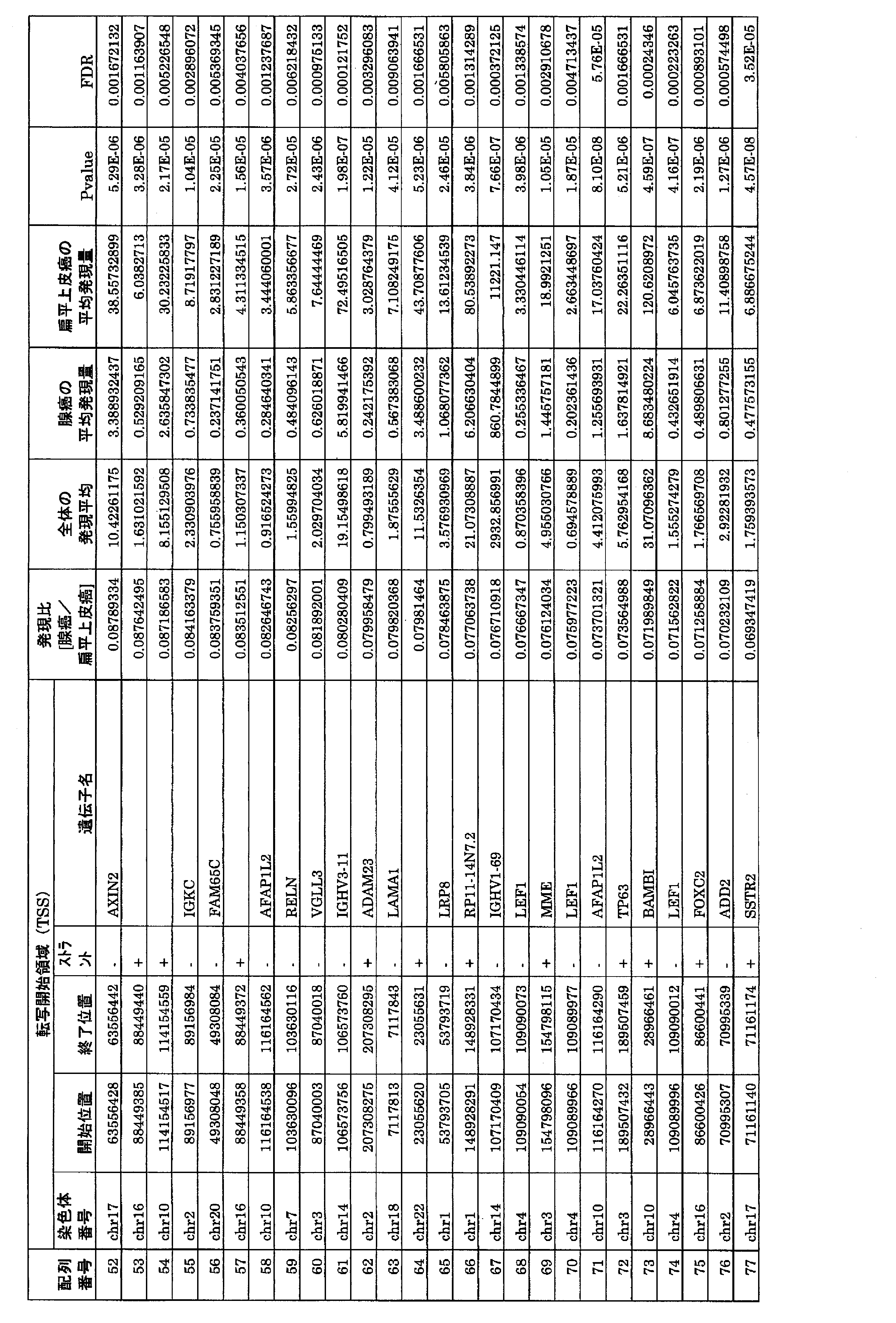

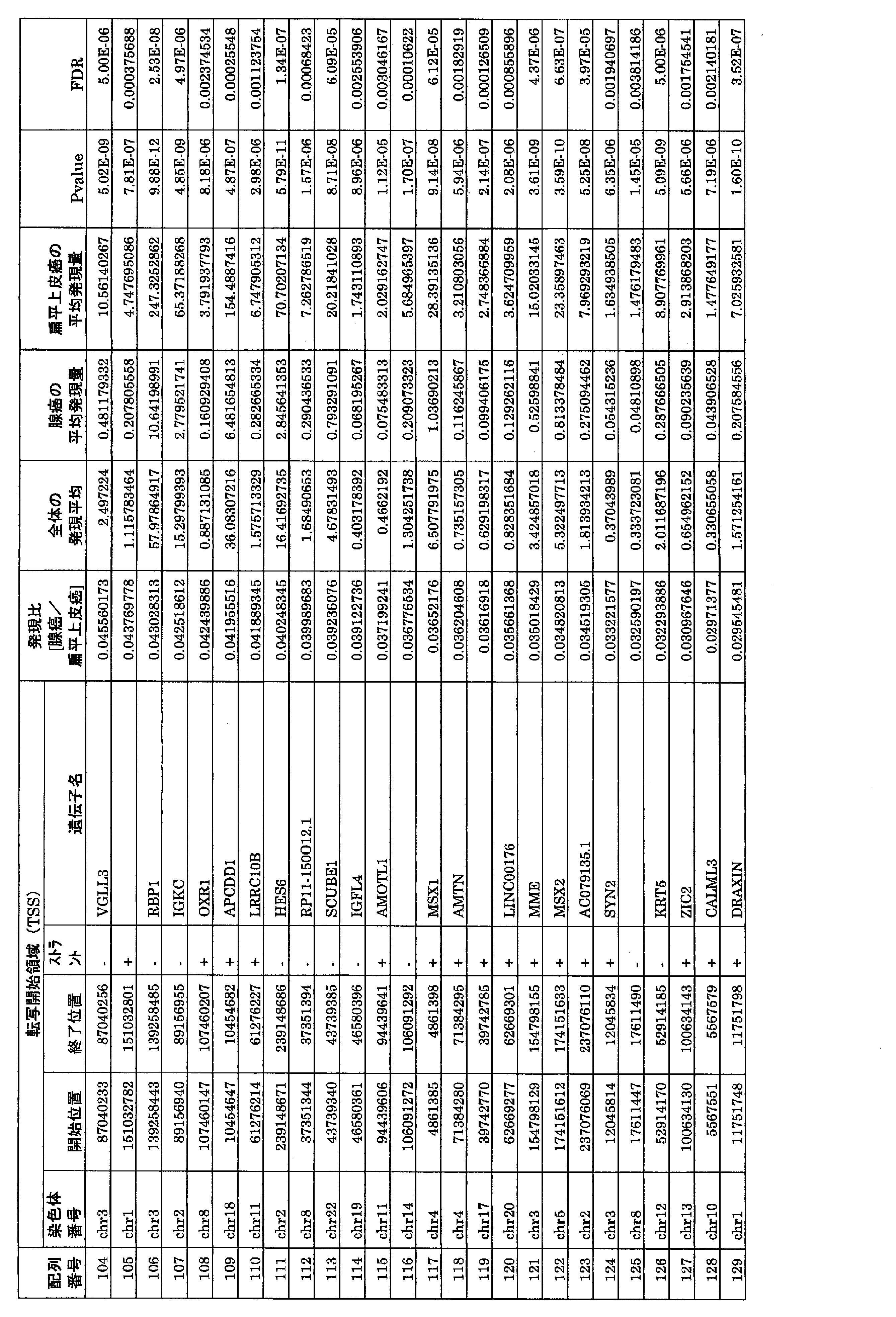
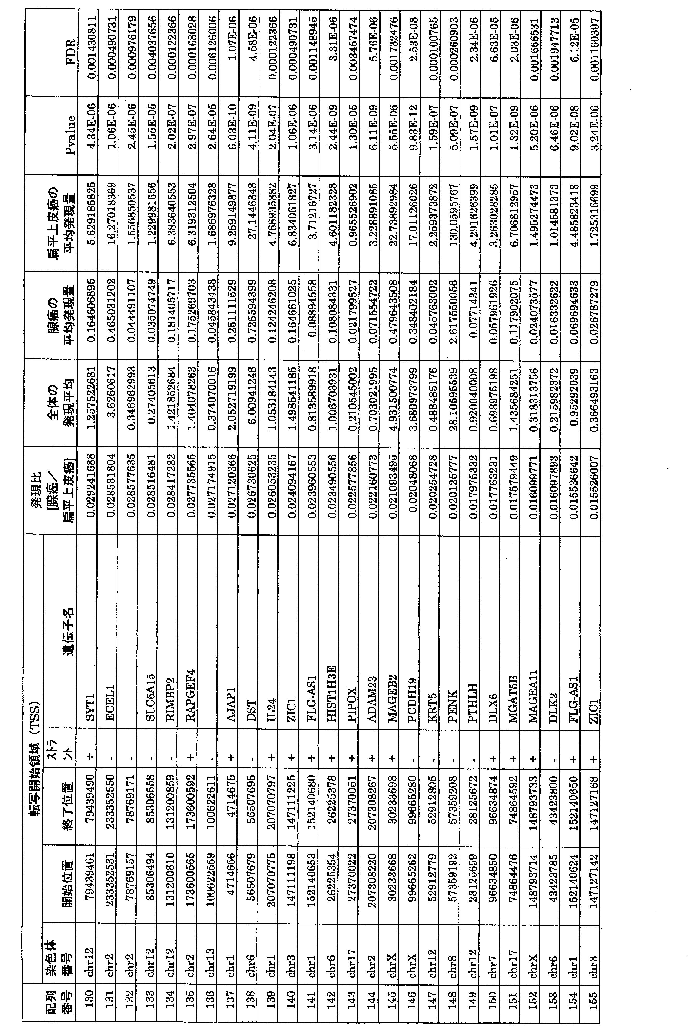
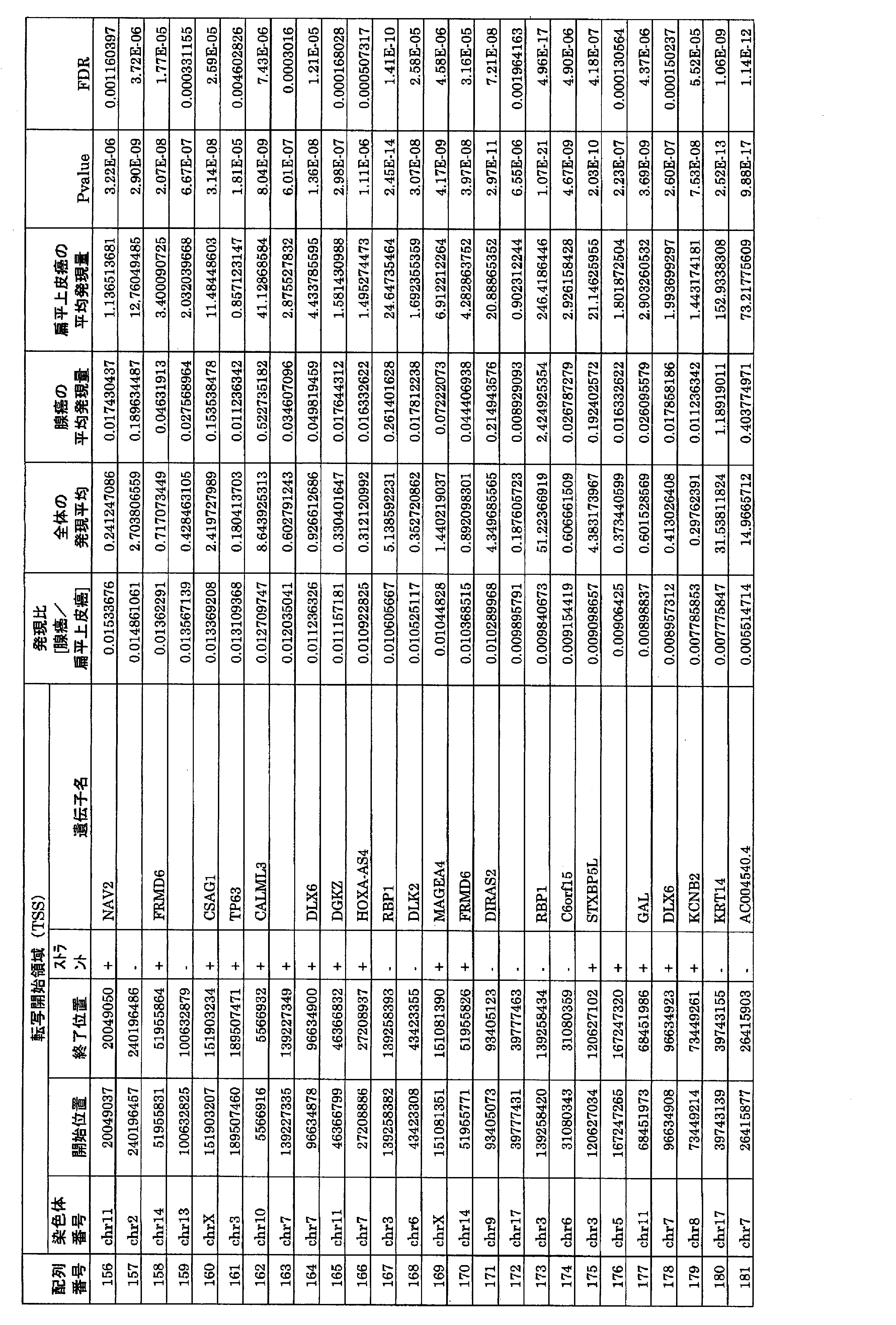


上記(A)で同定された転写開始領域のうち、一つの発現レベルのみを用いて腺癌(BAC成分を含まない腺癌)か扁平上皮癌(低分化肺扁平上皮癌)かを分類できるかどうかを考える。それぞれについて、何らかの閾値を設定することで、転写開始領域抽出用サンプル、検証用サンプル共に特異度100%・感度100%で分類できることを確認した。表2に、その閾値の例を示す(ある群における最大値の方が、その他の群における最小値よりも小さい場合、これらの平均を表2に示している)。

(1)検体
肺腺癌には手術検体45例を用い、その内訳は細気管支肺胞上皮癌<bronchioloalveolar carcinoma (BAC)>が7例、肺胞上皮置換性増殖成分を伴う肺腺癌<Adenocarcinoma with BAC>が22例、肺胞上皮置換性増殖成分を伴わない肺腺癌<Adenocarcinoma without BAC>が16例であった。一方、肺扁平上皮癌として手術検体29例を用い、その内訳は高分化及び中分化扁平上皮癌<well and moderately differentiated squamous cell carcinoma (SCC)>が18例、低分化扁平上皮癌<poorly differentiated SCC>が11例であった。
肺腺癌および肺扁平上皮癌の合計79例を対象に、腺癌マーカーとして、ST6GALNAC1、Napsin及びTTF-1、扁平上皮癌マーカーとしてCK5、CK6、Desmoglein 3(DSG3)、p40、及びSPATS2の抗体を用いて免疫染色により各タンパクの発現を評価した。
i)抗体
1)抗TTF-1抗体(DAKO社)
2)抗Napsin A抗体(Leica Biosystem社、「NCL-L-Napsin A」)
3)抗p40抗体(Millipore Co.社、「PC373」)
4)抗CK5抗体(Leica Biosystem社、「NCL-CK5」)
5)抗CK6抗体(Gene Tex社、「GTX73556」)
6)抗Desmoglein 3抗体(BIO CARE Medical社、「ACR419A, C」)
7)抗ST6GALNAC1抗体(SIGMA Life science社、「HPA014975」
8)抗SPATS2抗体(SIGMA Life science社、「HPA038643」
患者から分離した生体試料を常法によりホルマリン固定をした後、パラフィンに包埋して組織片に薄切し、スライドガラスに貼り付けたものを切片試料とした。次いで切片試料を、以下に示す条件で熱処理して抗原を賦活化した。次いで、各マーカータンパク質に対する抗体(一次抗体)を以下の条件で添加して反応させ、緩衝液にて十分な洗浄後、二次抗体としてEnvisionを用いて以下の条件で反応させ、緩衝液にて十分な洗浄後DABを用いて発色させた。その標本を光学顕微鏡下で陽性・陰性を観察した。

癌細胞の核もしくは細胞質に中等度以上の染色強度で染色性を認めた場合を陽性とした。各症例の代表切片において、陽性を示す癌細胞が認められない場合をScore 0、50%未満の癌細胞に陽性像を認める場合をScore 1、50%以上の癌細胞に陽性像を認める場合をScore 2とし、Score 0、1を陰性、Score 2を陽性と判定した。これらの評価は2人の病理医により行った。
(a)各マーカーの腺癌および扁平上皮癌のマーカーとしての有用性を評価した。すなわち、腺癌マーカーに対する感度・特異度を腺癌の鑑別に対して求め、同様に扁平上皮癌マーカーに対する感度・特異度を扁平上皮癌の鑑別能力に対して評価した。またp値はFisherの正確検定により算出した。

上記の腺癌3マーカー、扁平上皮癌5マーカーから任意の2つを組み合わせた24通りに対して、腺癌と扁平上皮癌の鑑別能力を検討した。その結果を表5に示す。

上記の腺癌3マーカー、扁平上皮癌5マーカーから任意の3つを組み合わせた場合には、全56通りが存在する。その中でも、以下の6通りについては腺癌と扁平上皮癌を完全に鑑別可能であった。
1)ST6GALNAC1/TTF-1/CK5
2)ST6GALNAC1/TTF-1/DSG3
3)ST6GALNAC1/TTF-1/p40
4)ST6GALNAC1/SPATS2/DSG3
5)ST6GALNAC1/SPATS2/CK5
6)ST6GALNAC1/SPATS2/p40
この結果より、完全な鑑別にはST6GALNAC1が有用であることが示唆される。
Claims (14)
- 肺癌患者の病変部から採取された生体試料について、転写開始領域を含むDNAの1種又は2種以上の発現産物の発現レベルを測定する工程を含む、当該病変が扁平上皮癌であるか腺癌であるかを鑑別評価する方法であって、該DNAが配列番号1~213で示される塩基配列における、転写開始領域の任意の位置の塩基とその下流に連続する少なくとも1塩基以上からなるDNAであり、該転写開始領域が、配列番号1~213で示される塩基配列の1番目の塩基と3’末端から101番目の塩基によって両端が規定される領域である、評価方法。
- 扁平上皮癌が低分化扁平上皮癌である、請求項1記載の方法。
- 腺癌がBAC成分を含まない腺癌である、請求項1又は2記載の方法。
- 配列番号1~213で示される塩基配列が、配列番号2、配列番号3、配列番号5及び配列番号7で示される塩基配列である、請求項1~3のいずれか1記載の方法。
- さらに、前記DNAの発現産物の発現レベルを対照レベルと比較する工程を含む、請求項1~4のいずれか1項記載の方法。
- さらに、前記DNAの発現産物の発現レベルを閾値レベルと比較する工程を含む、請求項1~4のいずれか1項記載の方法。
- 発現産物の発現レベルの測定が、転写産物の量又は翻訳産物の量を測定することによって行われる、請求項1~6のいずれか1項記載の方法。
- 前記DNAの転写産物と特異的にハイブリダイズするオリゴヌクレオチド、又は前記DNAの翻訳産物を認識する抗体を含有する請求項1~7のいずれか1項記載の方法に用いる扁平上皮癌であるか腺癌であるかを鑑別評価するための検査用キット。
- 転写開始領域を含むDNAの1種又は2種以上の発現産物の、肺癌患者の病変が扁平上皮癌であるか腺癌であるかを鑑別評価するためのマーカーとしての使用であって、該DNAが配列番号1~213で示される塩基配列における、転写開始領域の任意の位置の塩基とその下流に連続する1塩基以上からなるDNAであり、
該転写開始領域が、配列番号1~213で示される塩基配列の1番目の塩基と3’末端から101番目の塩基によって両端が規定される領域である、前記発現産物の使用。 - 肺癌患者の病変部から採取された生体試料について、ST6GALNAC1及び/又はSPATS2のタンパク質の発現レベルを測定する工程を含む、当該病変が扁平上皮癌であるか腺癌であるかを鑑別評価する方法。
- 更にP40、CK5、CK6、DSG3、TTF-1及びNapsin Aから選ばれる1種以上のタンパク質の発現レベルを測定する工程を含む、請求項10記載の方法。
- 下記1)~9)で示される2種のタンパク質の発現レベルを測定する工程を含む、請求項11記載の方法。
1)TTF-1/ST6GALNAC1
2)CK5/ST6GALNAC1
3)DSG3/ST6GALNAC1
4)CK5/SPATS2
5)DSG3/SPATS2
6)p40/ST6GALNAC1
7)ST6GALNAC1/SPATS2
8)Napsin A/ST6GALNAC1
9)p40/SPATS2 - 下記1)~6)で示される3種のタンパク質の発現レベルを測定する工程を含む、請求項11記載の方法。
1)ST6GALNAC1/TTF-1/CK5
2)ST6GALNAC1/TTF-1/DSG3
3)ST6GALNAC1/TTF-1/p40
4)ST6GALNAC1/SPATS2/DSG3
5)ST6GALNAC1/SPATS2/CK5
6)ST6GALNAC1/SPATS2/p40 - 免疫組織化学分析法によりタンパク質の発現レベルを測定する、請求項10~13のいずれか1項記載の方法。
Priority Applications (2)
| Application Number | Priority Date | Filing Date | Title |
|---|---|---|---|
| US15/125,366 US10787711B2 (en) | 2014-03-12 | 2015-03-11 | Method for differentiating between lung squamous cell carcinoma and lung adenocarcinoma |
| JP2016507797A JP6949315B2 (ja) | 2014-03-12 | 2015-03-11 | 肺扁平上皮癌と肺腺癌の鑑別評価方法 |
Applications Claiming Priority (4)
| Application Number | Priority Date | Filing Date | Title |
|---|---|---|---|
| JP2014-049186 | 2014-03-12 | ||
| JP2014049186 | 2014-03-12 | ||
| JP2014-183418 | 2014-09-09 | ||
| JP2014183418 | 2014-09-09 |
Publications (1)
| Publication Number | Publication Date |
|---|---|
| WO2015137406A1 true WO2015137406A1 (ja) | 2015-09-17 |
Family
ID=54071847
Family Applications (1)
| Application Number | Title | Priority Date | Filing Date |
|---|---|---|---|
| PCT/JP2015/057176 Ceased WO2015137406A1 (ja) | 2014-03-12 | 2015-03-11 | 肺扁平上皮癌と肺腺癌の鑑別評価方法 |
Country Status (3)
| Country | Link |
|---|---|
| US (1) | US10787711B2 (ja) |
| JP (1) | JP6949315B2 (ja) |
| WO (1) | WO2015137406A1 (ja) |
Cited By (1)
| Publication number | Priority date | Publication date | Assignee | Title |
|---|---|---|---|---|
| CN113186287A (zh) * | 2021-05-10 | 2021-07-30 | 深圳康华君泰生物科技有限公司 | 用于非小细胞肺癌分型的生物标志物及其应用 |
Families Citing this family (2)
| Publication number | Priority date | Publication date | Assignee | Title |
|---|---|---|---|---|
| CN113122627B (zh) * | 2019-12-30 | 2023-08-11 | 广州医科大学附属第三医院(广州重症孕产妇救治中心、广州柔济医院) | Igfl4基因作为标志物在卵巢癌中诊断及治疗的应用 |
| EP4150117A4 (en) * | 2020-05-12 | 2024-05-29 | The Board of Trustees of the Leland Stanford Junior University | System and method for gene expression and tissue of origin inference from cell-free dna |
Family Cites Families (1)
| Publication number | Priority date | Publication date | Assignee | Title |
|---|---|---|---|---|
| CA2568413A1 (en) * | 2004-05-28 | 2005-12-15 | Dana-Farber Cancer Institute, Inc. | Compositions, kits, and methods for identification, assessment, prevention, and therapy of cancer |
-
2015
- 2015-03-11 US US15/125,366 patent/US10787711B2/en not_active Expired - Fee Related
- 2015-03-11 WO PCT/JP2015/057176 patent/WO2015137406A1/ja not_active Ceased
- 2015-03-11 JP JP2016507797A patent/JP6949315B2/ja not_active Expired - Fee Related
Non-Patent Citations (7)
| Title |
|---|
| BROWN, A.F. ET AL.: "Tissue-preserving antibody cocktails to differentiate primary squamous cell carcinoma, adenocarcinoma, and small cell carcinoma of lung", ARCHIVES OF PATHOLOGY & LABORATORY MEDICINE, vol. 137, no. 9, pages 1274 - 1281, XP055224484 * |
| KACZKOWSKI, B. ET AL.: "Pan Cancer Biomarkers and Disruption of Gene Regulatory Networks in Cancer Inferred from CAGE Data of FANTOM5 Project", 36TH ANNUAL MEETING OF THE MOLECULAR BIOLOGY SOCIETY OF JAPAN PROGRAM YOSHISHU, 20 November 2013 (2013-11-20) * |
| KAZUYA TAKAMOCHI ET AL.: "CAGE-ho ni yoru Hai Genpatsu Henpei Johigan to Sengan no Shinki Kanbetsu Marker no Dotei", JAPANESE JOURNAL OF LUNG CANCER, vol. 54, no. 5, 5 October 2014 (2014-10-05), pages 388 * |
| LOPEZ-FERRER, A. ET AL.: "Differences in the O- Glycosylation Patterns Between Lung Squamous Cell Carcinoma and Adenocarcinoma", AMERICAN JOURNAL OF CLINICAL PATHOLOGY, vol. 118, no. 5, November 2002 (2002-11-01), pages 749 - 755, XP055224481 * |
| SAYUMI SHIRAKAWA ET AL.: "Sialyl Tn Antigen ( STn ) Gosei Koso ST6GalNAc I to STn Antigen no Hatsugen Sokan ni Tsuite", DAI 29 KAI THE JAPANESE SOCIETY OF CARBOHYDRATE RESEARCH NENKAI YOSHISHU, 10 August 2009 (2009-08-10), pages 142 * |
| TAKAMOCHI, K. ET AL.: "NOVEL BIOMARKERS TO DISCRIMINATE BETWEEN PRIMARY LUNG SQUAMOUS CELL CARCINOMA AND ADENOCARCINOMA IDENTIFIED BY THE CAP ANALYSIS OF GENE EXPRESSION (CAGE) METHOD", ANNALS OF ONCOLOGY, vol. 25, no. Supplement 4, 28 September 2014 (2014-09-28), pages iv406 - iv408, XP055224486 * |
| TIAN, S. ET AL.: "Hierarchical-TGDR, Combining biological hierarchy with a regularization method for multi-class classification of lung cancer samples via high-throughput gene - expression data", SYSTEMS BIOMEDICINE, vol. 1, no. 4, 20 September 2013 (2013-09-20), pages 278 - 287 * |
Cited By (2)
| Publication number | Priority date | Publication date | Assignee | Title |
|---|---|---|---|---|
| CN113186287A (zh) * | 2021-05-10 | 2021-07-30 | 深圳康华君泰生物科技有限公司 | 用于非小细胞肺癌分型的生物标志物及其应用 |
| CN113186287B (zh) * | 2021-05-10 | 2023-03-24 | 深圳康华君泰生物科技有限公司 | 用于非小细胞肺癌分型的生物标志物及其应用 |
Also Published As
| Publication number | Publication date |
|---|---|
| JP6949315B2 (ja) | 2021-10-13 |
| US10787711B2 (en) | 2020-09-29 |
| JPWO2015137406A1 (ja) | 2017-04-06 |
| US20170073766A1 (en) | 2017-03-16 |
Similar Documents
| Publication | Publication Date | Title |
|---|---|---|
| EP2971177B1 (en) | Compositions and methods for detecting and determining a prognosis for prostate cancer | |
| US20220093251A1 (en) | Novel biomarkers and diagnostic profiles for prostate cancer | |
| CN112626207B (zh) | 一种用于区分非侵袭性和侵袭性无功能垂体腺瘤的基因组合 | |
| EP3077531A1 (en) | Novel rna-biomarkers for diagnosis of prostate cancer | |
| CN110229899B (zh) | 用于结直肠癌早期诊断或预后预测的血浆标记物组合 | |
| JP6949315B2 (ja) | 肺扁平上皮癌と肺腺癌の鑑別評価方法 | |
| KR102329555B1 (ko) | 비용종을 갖는 비부비동염의 진단을 위한 정보 제공 방법 | |
| JP6711968B2 (ja) | 子宮体がんのリンパ節転移能の評価方法 | |
| US11519042B2 (en) | Method for the diagnosis or prognosis, in vitro, of lung cancer | |
| US11473147B2 (en) | Method for the diagnosis or prognosis, in vitro, of testicular cancer | |
| KR102259695B1 (ko) | 대장암에 대한 항암제 감수성 예측을 위한 신규 바이오마커 | |
| KR102384992B1 (ko) | 대장암 환자의 연령 특이적 바이오마커 및 이의 용도 | |
| WO2015105191A1 (ja) | リンパ節腫脹病変の評価方法 | |
| WO2015115544A1 (ja) | 大腸がんの転移又は再発リスクの評価方法 | |
| JP5897823B2 (ja) | 膀胱ガン診断用組成物及び方法 | |
| JP6820014B2 (ja) | 子宮体がんのリンパ節転移能の評価方法 | |
| US11459605B2 (en) | Method for the diagnosis or prognosis, in vitro, of prostate cancer | |
| EP4310501A1 (en) | Companion marker for assessment method of lymph node metastasis ability of endometrial cancer | |
| WO2007088971A1 (ja) | がん予後判定に利用できる遺伝子群 | |
| WO2015115545A1 (ja) | 乳がんの転移又は再発リスクの評価方法 | |
| KR20250175547A (ko) | FAT1(FAT Atypical Cadherin 1) 또는 ITGB8(Integrin beta 8)을 포함하는 혐색소 신세포암 진단용 바이오마커 조성물 | |
| JP5170734B2 (ja) | がんの検査に使用する遺伝子のスクリーニング方法と遺伝子セット | |
| CN103571936A (zh) | 微核酸-196a分子及-196b分子侦测口腔癌的方法及其引物、探针与试剂盒 | |
| BG67180B1 (bg) | Метод и кит за откриване на онкофузионен протеин |
Legal Events
| Date | Code | Title | Description |
|---|---|---|---|
| 121 | Ep: the epo has been informed by wipo that ep was designated in this application |
Ref document number: 15761426 Country of ref document: EP Kind code of ref document: A1 |
|
| ENP | Entry into the national phase |
Ref document number: 2016507797 Country of ref document: JP Kind code of ref document: A |
|
| NENP | Non-entry into the national phase |
Ref country code: DE |
|
| WWE | Wipo information: entry into national phase |
Ref document number: 15125366 Country of ref document: US |
|
| 122 | Ep: pct application non-entry in european phase |
Ref document number: 15761426 Country of ref document: EP Kind code of ref document: A1 |