WO2015133186A1 - 細胞撮像制御装置および方法並びにプログラム - Google Patents
細胞撮像制御装置および方法並びにプログラム Download PDFInfo
- Publication number
- WO2015133186A1 WO2015133186A1 PCT/JP2015/051072 JP2015051072W WO2015133186A1 WO 2015133186 A1 WO2015133186 A1 WO 2015133186A1 JP 2015051072 W JP2015051072 W JP 2015051072W WO 2015133186 A1 WO2015133186 A1 WO 2015133186A1
- Authority
- WO
- WIPO (PCT)
- Prior art keywords
- cell
- focus
- information
- image
- colony
- Prior art date
- Legal status (The legal status is an assumption and is not a legal conclusion. Google has not performed a legal analysis and makes no representation as to the accuracy of the status listed.)
- Ceased
Links
Images




Classifications
-
- C—CHEMISTRY; METALLURGY
- C12—BIOCHEMISTRY; BEER; SPIRITS; WINE; VINEGAR; MICROBIOLOGY; ENZYMOLOGY; MUTATION OR GENETIC ENGINEERING
- C12M—APPARATUS FOR ENZYMOLOGY OR MICROBIOLOGY; APPARATUS FOR CULTURING MICROORGANISMS FOR PRODUCING BIOMASS, FOR GROWING CELLS OR FOR OBTAINING FERMENTATION OR METABOLIC PRODUCTS, i.e. BIOREACTORS OR FERMENTERS
- C12M41/00—Means for regulation, monitoring, measurement or control, e.g. flow regulation
- C12M41/30—Means for regulation, monitoring, measurement or control, e.g. flow regulation of concentration
- C12M41/36—Means for regulation, monitoring, measurement or control, e.g. flow regulation of concentration of biomass, e.g. colony counters or by turbidity measurements
-
- G—PHYSICS
- G02—OPTICS
- G02B—OPTICAL ELEMENTS, SYSTEMS OR APPARATUS
- G02B21/00—Microscopes
- G02B21/06—Means for illuminating specimens
- G02B21/08—Condensers
- G02B21/14—Condensers affording illumination for phase-contrast observation
-
- G—PHYSICS
- G02—OPTICS
- G02B—OPTICAL ELEMENTS, SYSTEMS OR APPARATUS
- G02B21/00—Microscopes
- G02B21/24—Base structure
- G02B21/241—Devices for focusing
- G02B21/244—Devices for focusing using image analysis techniques
-
- G—PHYSICS
- G02—OPTICS
- G02B—OPTICAL ELEMENTS, SYSTEMS OR APPARATUS
- G02B21/00—Microscopes
- G02B21/36—Microscopes arranged for photographic purposes or projection purposes or digital imaging or video purposes including associated control and data processing arrangements
- G02B21/365—Control or image processing arrangements for digital or video microscopes
Definitions
- the present invention relates to a cell imaging control apparatus, method, and program for performing focus control of an imaging apparatus that images cells.
- pluripotent stem cells such as ES cells, iPS cells, and STAP cells, differentiation-induced cells, etc. are imaged over time, and the quality of the cell culture state is judged by capturing changes over time of the images.
- a way to do it has been proposed.
- a microscope is used as an apparatus for imaging a cell.
- autofocus control is performed, whereby an image of a cell with an optimal focus position is obtained. Is imaged.
- Patent Document 1 discloses that a focus position is obtained by performing autofocus control for each cell, and the focus position and information on a container in which the cell is cultured are obtained. It has been proposed to store them in correspondence.
- Patent Document 2 acquires a focus position by performing autofocus control based on a bright-field image, and adds an offset according to the type of target molecule to the acquired focus position, thereby performing fluorescence observation.
- a method for acquiring the focus position has been proposed.
- Patent Documents 3 to 5 propose that the focus is offset according to the cell type, the shape of the well bottom, and the thickness of the medium.
- Patent Document 6 proposes performing autofocus control based on illumination unevenness information
- Patent Document 7 acquires the position of a fertilized egg as a subject and fertilizes based on the position. It has been proposed to perform autofocus control by limiting the focus search range when imaging an egg at a high magnification.
- the morphology of the stem cell colony changes depending on the culture period. Specifically, as the culture of stem cell colonies proceeds, undifferentiated cells are in a dense state in the central part of the stem cell colonies, and the state is changed from undifferentiated cells to differentiated cells in the peripheral part. Since the nucleus / cytoplasm ratio is different between undifferentiated cells and differentiated cells, the height of the cell nuclei from the cell placement surface is different between the central portion and the peripheral portion of the stem cell colony.
- Patent Documents 1 to 7 there is no proposal for a method for solving the above-described problems.
- focus parameters are only determined based on predetermined conditions and information at the initial time, the cell morphology changes with the above-described cell growth (elapsed time). There is a problem that the accuracy of focus is lowered.
- the present invention provides a cell imaging control apparatus, method, and program that can perform focus control more efficiently when imaging cultured cells and can improve focus accuracy.
- the purpose is to do.
- the cell imaging control device of the present invention includes an information acquisition unit that acquires at least one of information related to the maturity of a cell to be cultured and observation position information in a cell colony, and a cell acquired by the information acquisition unit
- a focus parameter determination unit that determines a focus parameter based on at least one of information related to maturity level and observation position information, and focus that performs focus control in an imaging device that captures an image of a cell based on the focus parameter
- a control unit includes an information acquisition unit that acquires at least one of information related to the maturity of a cell to be cultured and observation position information in a cell colony, and a cell acquired by the information acquisition unit
- a focus parameter determination unit that determines a focus parameter based on at least one of information related to maturity level and observation position information, and focus that performs focus control in an imaging device that captures an image of a cell based on the focus parameter
- a control unit that controls focus control in an imaging device that captures an image of a cell based on the focus parameter
- the cell imaging control device of the present invention may include an image acquisition unit that acquires a cell image, and the information acquisition unit may acquire information related to maturity by analyzing the cell image.
- an image acquisition unit that acquires a cell image and a colony region specifying unit that specifies a cell colony region in the cell image are provided, and the information acquisition unit is a colony region specified by the colony region specifying unit.
- the observation position information may be acquired by accepting designation of the position within.
- an image acquisition unit that acquires a cell image and a colony region specifying unit that specifies a cell colony region in the cell image are provided, and the information acquisition unit is a colony region specified by the colony region specifying unit.
- the observation position information may be acquired based on the image information within.
- the cell culture period can be acquired as information related to maturity.
- At least one of a focus search initial position, a focus search range, a focus search width, a focus search order, and the number of focus operations can be determined as a focus parameter.
- the focus parameter determination unit may have a function representing a relationship between the observation position information and the focus search initial position, and may determine the focus search initial position as a focus parameter based on the function and the observation position information. .
- the above function may be a function in which the initial position of the focus search changes along the Gaussian distribution shape as the observation position information moves from the central part to the peripheral part of the cell colony.
- the information acquisition unit may acquire both information related to maturity and observation position information, and the focus parameter determination unit determines a focus search initial position as a focus parameter, and the observation position information is In the case of the peripheral portion in the cell colony, the focus search initial position closer to the cell installation surface side may be determined as the maturity degree advances.
- the information acquisition unit may acquire both information related to maturity and observation position information, and the focus parameter determination unit acquires a focus search initial position as a focus parameter, and the observation position information is When the cell is in the middle of the colony, the focus search initial position is determined at a position farther from the cell placement surface when the maturity is in the middle stage than in the early and late stages. May be.
- the cell imaging control method of the present invention obtains at least one of information related to the maturity of a cell to be cultured and observation position information in the colony of the cell, and obtains information related to the maturity of the cell and observation position information.
- a focus parameter is determined based on at least one, and focus control is performed in an imaging device that captures an image of a cell based on the determined focus parameter.
- the cell imaging control program of the present invention is acquired by an information acquisition unit, an information acquisition unit that acquires at least one of information related to the maturity of a cell to be cultured and observation position information in a cell colony.
- a focus parameter determination unit that determines a focus parameter based on at least one of information related to cell maturity and observation position information, and a cell image based on the focus parameter determined by the focus parameter determination unit It is made to function as a focus control part which performs focus control in the imaging device which images.
- the cell imaging control apparatus, method, and program of the present invention at least one of information related to the maturity level of a cell to be cultured and observation position information in a cell colony is acquired and related to the maturity level of the cell.
- the focus parameter is determined based on at least one of the information and the observation position information, and the focus control is performed in the imaging device that captures the cell image based on the determined focus parameter. If the focus search initial position is determined according to the degree of observation and the observation position in the cell colony, the focus position can be searched more efficiently, and the amount of cell image data captured at each focus position is also reduced. can do. In addition, the focus accuracy can be improved.
- the block diagram which shows schematic structure of the stem cell culture observation system using one Embodiment of the cell imaging control apparatus of this invention.
- Diagram showing schematic configuration of phase contrast microscope The figure which shows an example of the form of the stem cell colony in the initial stage of culture, the middle stage of culture, and the latter stage Illustration for explaining the nucleus / cytoplasm ratio of undifferentiated cells and differentiated cells.
- the figure which shows an example of the table which matched the kind of cell, culture conditions, culture period, observation position, and the focus search initial position of autofocus Diagram for explaining the distance function representing the relationship between observation position information in cell colonies and focus parameters
- the flowchart for demonstrating the effect
- FIG. 1 is a block diagram showing a schematic configuration of a stem cell culture observation system.
- the stem cell culture observation system of the present embodiment includes a cell culture device 1, a cell imaging control device 2, a phase contrast microscope 3, a display 4, and an input device 5, as shown in FIG.
- the cell culture device 1 is a device for culturing cells.
- Examples of cells to be cultured include stem cells such as iPS cells, ES cells, and STAP cells, cells such as nerves, skin, and liver that are induced to differentiate from stem cells, and cancer cells.
- stem cells such as iPS cells, ES cells, and STAP cells
- cells such as nerves, skin, and liver that are induced to differentiate from stem cells, and cancer cells.
- a plurality of culture containers in which stem cells to be cultured are seeded in a medium are accommodated.
- the cell culture device 1 includes a stage 10, a transport unit 11, and a control unit 12.
- Stage 10 is where a culture vessel to be imaged by phase contrast microscope 3 is installed.
- the culture container include a plate obtained by planarly culturing stem cells and a flask obtained by suspension culture.
- the stage 10 is configured to be movable in the X direction and the Y direction orthogonal to each other within the installation surface of the culture vessel by a driving mechanism (not shown). Note that the direction orthogonal to the X direction and the Y direction is the Z direction, and the focus position is changed in the Z direction to perform focus control. The focus control will be described later in detail.
- the transport unit 11 selects a culture container to be photographed from among a plurality of culture containers accommodated at a predetermined position in the cell culture apparatus 1, and transports the selected culture container to the stage 10.
- the control unit 12 controls the entire cell culture device 1 and controls environmental conditions such as temperature, humidity, and CO 2 concentration in the cell culture device 1 in addition to the operations of the stage 10 and the transport unit 11 described above.
- the temperature, the configuration for adjusting the humidity and CO 2 concentration can be a known configuration.
- the phase-contrast microscope 3 corresponds to an imaging device and captures a phase image of cells in a culture vessel installed on the stage 10.
- the phase-contrast microscope 3 of the present embodiment includes an autofocus function that automatically changes the focus position, and performs focus control based on a focus control signal output from a focus control unit 25 described later.
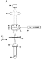
- FIG. 2 is a diagram showing a schematic configuration of the phase-contrast microscope 3.
- the phase-contrast microscope 3 has a white light source 31 that emits white light and a ring-shaped slit, and the white light emitted from the white light source 31 is incident to emit ring-shaped illumination light.
- an objective lens that irradiates the cells in the culture vessel 15 installed on the stage 10 with the ring-shaped illumination light emitted from the slit plate 32. 33.
- a phase difference lens 34, an imaging lens 37, and an image sensor 38 are provided on the opposite side of the stage 10 from the white light source 31.
- the phase difference lens 34 includes an objective lens 35 and a phase plate 36.
- the phase plate 36 is configured by forming a phase ring on a transparent plate that is transparent to the wavelength of the ring-shaped illumination light.
- the size of the slit of the slit plate 32 described above is in a conjugate relationship with this phase ring.
- the phase ring is a phase film that shifts the phase of incident light by a quarter wavelength, and attenuates the incident light. And a neutral density filter formed in a ring shape.
- the direct light incident on the phase difference lens 34 is collected by the objective lens 35 and passes through the phase ring, so that the phase is shifted by 1 ⁇ 4 wavelength and the brightness is weakened.
- most of the diffracted light diffracted by the cells in the culture vessel 15 passes through the transparent plate of the phase plate, and its phase and brightness do not change.
- the phase difference lens 34 is moved in the direction of arrow A shown in FIG. 2 by a driving mechanism (not shown). As the phase difference lens 34 moves in this way, the focus position is changed and focus control is performed.
- the drive mechanism moves the phase difference lens 34 based on the focus control signal output from the focus control unit 25 of the cell imaging control device 2.
- phase contrast microscope 3 of the present embodiment is configured such that a plurality of phase difference lenses 34 having different magnifications can be exchanged.
- the phase difference lens 34 may be replaced automatically according to an instruction input from the user, or may be replaced manually by the user.
- low-magnification imaging for macro observation and high-magnification imaging for detailed observation are performed.
- a phase difference lens 34 of 1 to 4 times is used.
- high-magnification imaging a 10 to 20 times phase difference lens 34 is used.
- low magnification imaging and high magnification imaging only need to have relatively different magnifications, and are not limited to these magnifications.
- the imaging lens 37 receives direct light and diffracted light that have passed through the phase difference lens 34 and forms an image of these lights on the image sensor 38.
- the imaging element 38 captures a phase image of the cell by photoelectrically converting the image formed by the imaging lens 37.
- a charge-coupled device (CCD) image sensor, a complementary metal-oxide semiconductor (CMOS) image sensor, or the like is used as the image sensor 38.
- the phase contrast microscope 3 is used as the imaging device.
- a microscope having another autofocus function may be used, for example, a differential interference microscope may be used. Good.
- the cell imaging control device 2 includes a low-magnification image acquisition unit 20, a colony region specifying unit 21, an information acquisition unit 22, a focus parameter determination unit 23, and a control unit 24.
- the control unit 24 includes a focus control unit 25 and a display control unit 26.
- the cell imaging control device 2 is a computer in which an embodiment of the cell imaging control program of the present invention is installed in a computer.
- the cell imaging control device 2 includes a central processing unit, a semiconductor memory, a hard disk, and the like, and an embodiment of a cell imaging control program is installed on the hard disk. Then, this program is executed by the control unit 24 having the central processing unit, whereby a low-magnification image acquiring unit 20, a colony region specifying unit 21, an information acquiring unit 22, a focus parameter determining unit 23, as shown in FIG.
- the focus control unit 25 and the display control unit 26 operate.
- the low-magnification image acquisition unit 20 acquires the cell image captured by the phase-contrast microscope 3 by performing the above-described low-magnification imaging.
- the low-magnification cell image acquired by the low-magnification image acquisition unit 20 may be one image obtained by imaging one cell colony, or a plurality of image groups obtained by dividing one cell colony into a plurality of rectangular divided regions. Good. A plurality of cell colonies may be included in one image.
- the low-magnification image acquisition unit 20 stores identification information for identifying cell colonies and low-magnification cell images in association with each other. For example, when one cell colony is captured as a single low-magnification cell image, the identification information of the cell colony and the low-magnification cell image are stored in a one-to-one correspondence. When one cell colony is imaged as a low-magnification cell image of a plurality of divided regions, the identification information of the cell colony and the low-magnification cell image groups of the plurality of divided regions are stored in association with each other. When a plurality of cell colonies are captured as a single low-magnification cell image, the identification information of each cell colony and one low-magnification cell image are stored in association with each other.
- the identification information of the cell colony and the low-magnification cell image can be read and displayed immediately.
- the colony region specifying unit 21 specifies the position of the cell colony region in the low-magnification cell image acquired by the low-magnification image acquisition unit 20.
- the cell colony region is automatically extracted by template matching or the like to specify the position of the cell colony region. That's fine.
- the display control unit 26 causes the display 4 to display a low-magnification cell image, the user designates a cell colony region in the low-magnification cell image using the input device 5, and the designated coordinates.
- the colony region specifying unit 21 may acquire position information such as
- the information acquisition unit 22 acquires cell type information, culture condition information, information related to cell maturity, and observation position information in a cell colony.
- the information acquired by the information acquisition unit 22 is used when the focus parameter determination unit 23 determines a focus parameter used for autofocus control.
- cell type information examples include iPS cells, ES cells, cardiomyocytes after differentiation induction, and liver cells. Since the size (the height in the Z direction) differs depending on the cell type, the upper limit and the lower limit of the autofocus focus search range can be set by acquiring the cell type information.
- the cell type information is not limited to the cells listed above, but may include other type information.
- the maturity level of the cell is divided into three stages of the initial stage of culture, the middle stage of culture, and the late stage of culture, and the information acquisition unit 22 acquires information related to the maturity level of the cell, Get the stage to which the maturity of the cell belongs.
- the cell maturity is divided into the early stage, middle stage and late stage of culture.
- the morphology of the cell colony differs depending on each stage, and the focus search initial position of autofocus is set according to the form. This is because it is desirable.
- the height h of the stem cell nucleus at the center of the colony region is the same.
- FIG. 4 shows a plan view (upper view) and an elevation view (lower view) of undifferentiated cells and differentiated cells.
- Differentiated cells have a lower nucleus / cytoplasm ratio than undifferentiated cells and thus have a lower nucleus height. Therefore, as shown in FIG. 3, the height h center of the stem cell nucleus in the center of the colony region is higher by ⁇ h than the height h edge of the stem cell nucleus in the peripheral portion.
- the height h center of the stem cell nucleus in the central part of the colony region is the same as the height h edge of the stem cell nucleus in the peripheral part, but the nucleus of the intermediate stem cell between the central part and the peripheral part is the same.
- the height h middle is increased by ⁇ h.
- the focus search initial position of autofocus is set in accordance with this form change.
- the information related to the maturity level of the cell acquired by the information acquisition unit 22 may be any information as long as it is information indicating the stage of the maturity level of the cell.
- the culture period measured by a timer or the like is related to the maturity level. It can be acquired as information.
- the image information of the cell colony region in the low magnification cell image is analyzed, and the size of the cell colony, the number of cells in the stem cell colony, or the number of cells per unit area smaller than the stem cell colony is calculated. You may measure and acquire such information as information relevant to maturity. For example, it is determined that the maturity degree is advanced as the number of measured cells is larger.
- the image information of the cell colony region may be acquired based on the position information of the cell colony specified by the colony region specifying unit 21.
- the area, perimeter, maximum diameter, etc. of the cell colony can be acquired.
- individual cells or nuclei and nucleoli in the cells may be detected by pattern matching, and the number of detected individual cells may be counted. .
- the brightness of the image of the cell colony region, and the texture such as uniformity and roughness may be acquired as information related to maturity.
- the degree of maturity advances and the density of the stem cells increases, and the stem cells are further stacked to gradually increase the luminance of the image. Therefore, it can be said that the higher the brightness is, the more mature the progress is.
- the feature quantity of the shape of the stem cell colony may be acquired.
- the shape of the stem cell colony gradually approaches a circular shape, and then the differentiation of the peripheral portion proceeds and the complexity of the edge increases. Therefore, the feature amount of the change in the shape of the stem cell colony can be acquired as the feature amount related to the maturity.
- the feature quantity of the thickness of the stem cell colony may be acquired. As the maturity of stem cells progresses, the stem cell colonies gradually become thicker. Therefore, such a feature quantity of the thickness of the stem cell colony can be acquired as a feature quantity related to maturity.
- the thickness of the stem cell colony may be measured by a separately provided measuring device.
- the information related to the maturity described above may be set and input by the user using the input device 5, and not only the culture period and the size of the cell colony described above, but also the cell passage number
- the user may input the information related to the degree.
- the cell maturity is divided into three stages, but is not limited to three stages, and may be divided into two stages or four or more stages.
- various intervals can be set for the intervals of each stage according to the culture conditions and the like.
- the culture conditions acquired by the information acquisition unit 22 include the type of scaffold and medium, and whether or not different types of heterogeneous cells (feeder cells) are used. Even in the same culture period, the stage of maturity varies depending on these culture conditions.
- the focus search initial position of autofocus is set in consideration of the culture conditions.
- the culture conditions are not limited to the above-mentioned conditions, but may be any conditions as long as they affect the cell growth rate.
- culture conditions such as temperature, humidity or CO 2 concentration are included. May be.
- the culture condition information is set and input by the user using the input device 5, for example, but the temperature and humidity as described above may be the conditions measured with a thermometer or hygrometer.
- the observation position information in the cell colony acquired by the information acquisition unit 22 is the position in the XY direction in the cell colony region imaged by the low magnification imaging, and the position to be subjected to the high magnification imaging. It is the coordinate information shown.
- observation position information in the cell colony region is acquired, and an autofocus focus search initial position is set based on the observation position information.
- the observation position information in the cell colony region may be set and input by the user using the input device 5, for example.
- the present invention is not limited to this, and a table in which cell maturity levels and observation position information are associated with each other may be stored, and observation position information may be determined according to cell maturity levels. For example, when the stage of cell maturity is the initial stage of culture and the middle stage of culture, the central position of the cell colony is used as observation position information. It is only necessary to observe the undifferentiated cells as the center by using the intermediate part of the information as observation position information.
- the central position of the cell colony is observed as the observation position information, and the undifferentiated cells are mainly observed, and the middle stage and late stage of culture
- the peripheral part of the cell colony may be used as observation position information to observe the differentiated cell as a center.
- the observation position information may be acquired by analyzing the image information of the cell colony region in the low magnification cell image. Specifically, the number of cells per unit area in the cell colony region is measured as the density, and a position where the density is equal to or higher than a preset threshold or a position below the threshold is acquired as observation position information. Good.
- the position where the density is equal to or higher than a preset threshold is used as observation position information, and when the cell maturity is in the later stage of culture, the density is May be set as observation position information at a position less than a preset threshold value. That is, the determination method for setting the observation position information may be changed depending on the stage of cell maturity.
- the focus parameter determination unit 23 is based on the cell type information acquired by the information acquisition unit 22, information on the culture conditions, information related to the maturity of the cells, and observation position information in the cell colonies. Thus, a focus parameter used for autofocus control of the phase-contrast microscope 3 is determined.
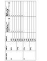
- FIG. 5 is a diagram showing an example of a table in which cell types, culture conditions, culture periods, observation positions, and autofocus focus search initial positions are associated with each other.
- the focus parameter determination unit 23 determines the focus search initial position by referring to the table shown in FIG. 5 based on the information acquired by the information acquisition unit 22.
- the relative relationship of the offset amount (distance) from the bottom surface of the culture vessel 15 is shown as the focus search initial position, but actually the absolute value of the offset amount is acquired, The offset amount is determined as the focus search initial position.
- the ES cell offset amount is actually set but not shown.
- the culture condition information is condition A
- the observation position information is the center
- the culture period is in the initial stage of culture and the culture If the focus search initial position offset is relatively small (closer to the cell installation surface side) in the later stage, and the focus search initial position offset is in the middle culture stage
- the amount may be relatively large (away from the cell installation surface).
- the offset amount is changed according to the culture period. Instead, a relatively small offset amount may be determined.
- the focus search initial position corresponding to the observation position information in the cell colony region is determined as a discrete value based on a relative positional relationship from a reference position such as the center position in the cell colony region, for example. Alternatively, it may be determined as a continuous value based on the relative positional relationship.
- the distance from the reference position to the observation position is set as a variable, and a distance function that outputs a focus search initial position corresponding to the variable is set in advance, and the focus is set based on the observation position and the distance function.
- the initial search position may be determined.
- the center (reference position) of the cell colony region is defined as the average ⁇ of the normal distribution, and the radius of the cell colony region It is possible to use a function of a normal distribution as in the following equation where is a normal distribution variance ⁇ 2 .
- the horizontal axis in FIG. 6 is the position in the cell colony region, and the vertical axis is the focus search initial position. Further, a range from zero on the vertical axis to the value of the graph can be used as the focus search range.
- the cell colony region may have a shape other than a circle, and there are various ways of concentrating cells. Therefore, a distance function other than the normal distribution may be used in accordance with the method. Alternatively, a discretely varying distance function such as a stepped pyramid shape may be used.
- the distance function as described above may be set for each combination of cell type, culture condition, and culture period information.
- the distance function may be set such that the offset amount in the periphery of the cell colony region decreases as the culture period becomes longer (the focus search initial position is closer to the cell installation surface side).
- a distance function for each culture period is set using a distance function that associates the distance from the reference position to the observation position and the offset amount, and a curve that associates the culture period and the cell size.
- the distance function for each culture period (concentration) may be set using a curve in which the density of cells and the height of cell nuclei are associated with each other.
- a hole may be formed at the center of the cell colony region in the later stage of culture. Therefore, in this case, a normal distribution distance function may be set such that the middle portion between the central portion and the peripheral portion of the cell colony region is the peak position. In addition, since the hole spreads concentrically as the culture period becomes longer, the peak position of the normal distribution may be moved toward the periphery according to the culture period.
- the absolute position and the focus search initial position may be associated with each other.
- the absolute position is a relative position from the origin position when the entire culture region (for example, the entire well plate) in the culture vessel to be observed is viewed.
- the thickness information for each position in the XY plane of the bottom of the culture vessel 15 and the thickness information for each position in the XY plane of the scaffold are acquired, and the focus search initial position is also taken into consideration. It may be determined.
- the focus parameter determination unit 23 also determines the focus search range, the focus search width, the focus search order, the number of focus operations, and the like.
- the focus search range is a focus position change range in the autofocus control, and has a lower limit value and an upper limit value.
- the focus search range for example, as described above, a table in which the cell type and the focus search range are associated with each other is set in advance, and the table is based on the cell type information acquired by the information acquisition unit 22.
- the focus search range may be determined by referring to it. Also, a table in which the observation position information in the cell colony region and the focus search range are associated with each other is set in advance, or the distance function as described above is used. Then, the focus search range corresponding to the observation position information may be determined.
- a table in which the culture period is associated with the focus search range may be set in advance.
- the focus search range may be expanded as the culture period becomes longer.
- the focus search range may be determined for each combination of cell type, observation position information, and culture period.
- the focus search width is a change width when the focus position is changed once in autofocus control. As culture progresses and cell colonies expand, it is considered that the density of cells varies depending on the location. Therefore, it is conceivable that the probability that the cell nucleus is out of the focus search range increases with the focus search width set in accordance with the initial stage of the culture where the variation in cell density is small.
- a table in which the culture period is associated with the focus search width may be set in advance, and the focus search width may be increased as the culture period becomes longer.
- a table in which the observation position information in the cell colony region is associated with the focus search width may be set in advance, and the focus search width in the peripheral portion may be wider than the central portion of the cell colony region.
- the focus search width may be determined for each combination of the observation position information in the cell colony and the culture period.
- the auto focus control when the auto focus control is performed by sequentially changing the focus position from the high position to the low position with reference to the bottom surface of the culture vessel 15, the auto focus is performed by sequentially changing the focus position from the low position to the high position.
- the control is performed and a case where the auto focus control is performed by alternately repeating the change of the focus position in the direction approaching the bottom surface of the culture vessel 15 and the change of the focus position in the direction away from the culture container 15.
- a table in which the culture period and the focus search order are associated with each other is set in advance, and in the initial culture period or the intermediate culture period, as described above, from a low position to a high position, or from a high position to a low position.
- the focus position may be searched, and in the latter stage of culture, the focus position may be searched by alternately repeating the search for the focus position from the high position and the search for the focus position from the low position.
- the focus search order may be determined for each combination of observation position information and culture period in a cell colony.
- the number of focus operations is the upper limit of the number of focus position changes in autofocus control. It can be said that strict focus accuracy is necessary in order to accurately recognize and evaluate the morphology of cell nuclei and tissues (for example, nucleolus) corresponding to the cell nuclei in the early to mid-culture. On the other hand, as the culture progresses, cell nuclei and tissues corresponding thereto tend to be difficult to visually recognize due to cell differentiation and lamination, and in this case, it is considered that strict focus accuracy is not necessary.
- a table in which the culture period is associated with the number of focus operations may be set in advance, and the number of focus operations may be reduced as the culture progresses.
- a table in which the observation position information in the cell colony region is associated with the number of focus operations is set in advance, and by increasing the number of focus operations toward the center rather than the periphery of the cell colony region, The focus accuracy may be increased.
- you want to focus on the periphery of the cell colony area increase the number of focusing operations in the periphery rather than the center of the cell colony area to increase the focus accuracy of the periphery. Also good.
- the number of focus operations may be determined for each combination of the observation position information in the cell colony and the culture period.
- the information acquisition unit 22 may acquire information on the amount of the medium, and the focus search range may be determined in consideration of this information.
- the focus position is set on the surface of the medium on which the suspended matter is present, and an appropriate cell image may not be captured. Therefore, information on the surface position of the medium may be acquired by acquiring information on the amount of the medium, and the surface position may be excluded from the autofocus search range.
- the information on the amount of medium may be set and input by the user using the input device 5.
- the focus control unit 25 outputs a focus control signal to the phase-contrast microscope 3 based on the focus parameter determined by the focus parameter determination unit 23, thereby causing the phase-contrast lens 30 in the phase-contrast microscope 3 to move in the Z direction.
- Autofocus control is performed by moving it.
- a known method can be used. For example, the contrast of a cell image captured at each focus position is calculated, and the focus position at which the contrast is maximized is set as the final focus position. You just have to do it.
- the focus control unit 25 controls the control unit 12 of the cell culture device 1 based on the position of the cell colony region specified by the colony region specifying unit 21 and the observation position information acquired by the information acquisition unit 22. A signal is output, and the movement of the stage 10 in the XY direction is controlled so that the observation position in the cell colony region is imaged at high magnification.
- high-magnification imaging is performed in a state where the position of the culture vessel 15 in the XY direction and the focus position of the phase-contrast microscope 3 are adjusted by the focus control unit 25, and a high-magnification cell image is captured.
- the display control unit 26 displays a low magnification cell image and a high magnification cell image on the display 4.
- the input device 5 includes a mouse, a keyboard, etc., and accepts setting input by the user. Specifically, the input device 5 accepts setting input of cell type information, culture conditions, information related to cell maturity, and observation position information in the cell colony region as described above.
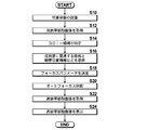
- the culture to be photographed is selected from the plurality of accommodated culture containers by the transport unit 11, and the selected culture container 15 is placed on the stage 10 (S10).
- the magnification of the phase difference lens 34 of the phase-contrast microscope 3 is set to a low magnification, a low-magnification cell image is captured, and the captured low-magnification captured image is acquired by the low-magnification image acquisition unit 20 (S12). .
- the low magnification cell image acquired by the low magnification image acquisition unit 20 is output to the display control unit 26 and displayed on the display 4 by the display control unit 26.
- the low-magnification cell image acquired by the low-magnification image acquisition unit 20 is output to the colony region specifying unit 21, and the colony region specifying unit 21 is based on the input low-magnification cell image.
- the position of the colony region in the inside is specified (S14).
- the information acquisition unit 22 acquires cell type information, culture condition information, information related to cell maturity, and observation position information in a cell colony by setting input by the user (S16). .
- the focus parameter determination unit 23 determines a focus parameter used for autofocus control in the phase-contrast microscope 3 based on the input information, and outputs the focus parameter to the focus control unit 25 (S18).
- the focus control unit 25 Based on the position of the cell colony region specified by the colony region specifying unit 21 and the observation position information acquired by the information acquisition unit 22, the focus control unit 25 is configured to capture the observation position at a high magnification. Is moved in the XY direction. Next, the focus control unit 25 performs autofocus control of the phase-contrast microscope 3 based on the input focus parameter (S20). At this time, the phase difference lens 34 in the phase contrast microscope 3 is replaced with a phase difference lens 34 for high magnification imaging.
- the phase-contrast microscope 3 captures a high-magnification cell image for observation in a state where the autofocus is finished and the optimum focus position is set.
- the high-magnification cell image captured by the phase-contrast microscope 3 is acquired by the display control unit 26 of the cell imaging control device 2 (S22), and the display control unit 26 displays the input high-magnification cell image for observation on the display 4. It is displayed (S24).
- the culture period of the cells to be cultured and the observation position information in the cell colony are acquired, the focus parameter is determined based on the acquired culture period and the observation position information, Based on the determined focus parameter, focus control is performed in an imaging device that captures an image of a cell, so that the focus position can be searched more efficiently, and cell image data captured at each focus position The amount can also be reduced.
- the imaging target is not limited to a stem cell colony, and other types of cell colonies may be used.
- blood vessel position information in cardiomyocyte colonies or skin cell colonies may be acquired as observation position information, and a focus search initial position corresponding to the observation position information may be determined.
- the focus search initial position may be changed according to the culture period. For example, the focus search initial position is set to a deeper position (a position closer to the cell installation surface) as the culture period progresses. Also good.
- both information related to cell maturity such as information on a cell culture period, and observation position information in a cell colony are acquired, and based on these information
- the focus search initial position may be determined according to the length of the culture period.
- the observation position information in the cell colony may be acquired, and the focus search initial position may be determined such that the offset amount is smaller in the peripheral part than in the central part in the cell colony.
- the focus parameter determination unit 23 determines the focus search initial position, the focus search range, and the like as the focus parameters, and searches the focus position based on these to perform autofocus control.
- the present invention is not limited to this.
- the focus parameter determination unit 23 determines the focus position as the focus parameter, and the focus control unit 25 controls the phase-contrast microscope 3 so that the focus position is obtained. Also good.
Landscapes
- Chemical & Material Sciences (AREA)
- Physics & Mathematics (AREA)
- Engineering & Computer Science (AREA)
- Analytical Chemistry (AREA)
- General Physics & Mathematics (AREA)
- Optics & Photonics (AREA)
- Life Sciences & Earth Sciences (AREA)
- Zoology (AREA)
- Bioinformatics & Cheminformatics (AREA)
- Multimedia (AREA)
- Computer Vision & Pattern Recognition (AREA)
- Organic Chemistry (AREA)
- Health & Medical Sciences (AREA)
- Wood Science & Technology (AREA)
- Sustainable Development (AREA)
- Biochemistry (AREA)
- General Engineering & Computer Science (AREA)
- General Health & Medical Sciences (AREA)
- Genetics & Genomics (AREA)
- Microbiology (AREA)
- Biotechnology (AREA)
- Biomedical Technology (AREA)
- Automatic Focus Adjustment (AREA)
- Microscoopes, Condenser (AREA)
- Apparatus Associated With Microorganisms And Enzymes (AREA)
- Investigating Or Analysing Materials By Optical Means (AREA)
Abstract
培養される細胞を撮像する際におけるフォーカス制御をより効率的に行うことができ、フォーカス精度の向上を図ることができる細胞撮像制御装置および方法並びにプログラムを提供する。培養される細胞の成熟度に関連する情報および細胞のコロニー内における観察位置情報の少なくとも1つを取得する情報取得部22と、細胞の成熟度に関連する情報および観察位置情報の少なくとも1つに基づいて、フォーカスパラメータを決定するフォーカスパラメータ決定部23と、フォーカスパラメータに基づいて、細胞の画像を撮像する撮像装置におけるフォーカス制御を行うフォーカス制御部25とを備える。
Description
本発明は、細胞を撮像する撮像装置のフォーカス制御を行う細胞撮像制御装置および方法並びにプログラムに関するものである。
従来、ES細胞、iPS細胞、STAP細胞などの多能性幹細胞や、分化誘導された細胞などを経時的に撮像し、その画像の経時的な変化を捉えることで細胞の培養状態の良否判定を行う方法が提案されている。
そして、上述したように細胞を撮像する装置として顕微鏡が用いられるが、顕微鏡で細胞の画像を撮像する際には、いわゆるオートフォーカス制御が行われ、これによりフォーカス位置が最適な状態で細胞の画像が撮像される。
このようなオートフォーカス制御によって細胞を撮像する装置として、特許文献1には、細胞毎にオートフォーカス制御を行うことによってフォーカス位置を取得し、このフォーカス位置と細胞が培養される容器の情報とを対応づけて記憶しておくことが提案されている。
また、特許文献2には、明視野画像に基づいてオートフォーカス制御を行うことによってフォーカス位置を取得し、この取得したフォーカス位置に標的分子の種類に応じたオフセットを付加することによって蛍光観察時のフォーカス位置を取得する方法が提案されている。
また、特許文献3~特許文献5には、細胞の種類、ウェル底部の形状、培地の厚みに応じてフォーカスをオフセットさせることが提案されている。
また、特許文献6には、照明のムラ情報に基づいてオートフォーカス制御を行うことが提案されており、特許文献7には、被写体である受精卵の位置を取得し、その位置に基づいて受精卵を高倍率で撮像する際に、フォーカス探索範囲を限定してオートフォーカス制御を行うことが提案されている。
ここで、たとえば上述したような幹細胞を培養する際、幹細胞コロニーの形態は培養期間に応じて変化する。具体的には、幹細胞コロニーの培養が進むと、幹細胞コロニーの中心部では未分化細胞が密集した状態となり、周辺部では未分化細胞から分化細胞に変化した状態となる。未分化細胞と分化細胞とでは核/細胞質の比が異なるので、幹細胞コロニーの中心部と周辺部とで細胞核の細胞設置面からの高さが異なる状態となる。
このような状態において、幹細胞コロニー内の全ての細胞核が探索されるようにオートフォーカス制御を行ったのでは、その探索範囲が広くなるため、最適なフォーカス位置を見つけ出すまでの計測時間が長くなり、また、その際に取得されるフォーカス位置毎の細胞画像のデータ量も多くなってしまう。
特許文献1~特許文献7においては、上述したような問題を解決する方法については何も提案されていない。また、従来は、初期時刻での所定の条件や情報などに基づいてフォーカスパラメータを決定するのみであるので、上述したような細胞の成長(時間経過)によって細胞の形態が変化することで、オートフォーカスの精度が低下する問題がある。
本発明は、上記の問題に鑑み、培養される細胞を撮像する際におけるフォーカス制御をより効率的に行うことができ、フォーカス精度の向上を図ることができる細胞撮像制御装置および方法並びにプログラムを提供することを目的とする。
本発明の細胞撮像制御装置は、培養される細胞の成熟度に関連する情報および細胞のコロニー内における観察位置情報の少なくとも1つを取得する情報取得部と、情報取得部によって取得された、細胞の成熟度に関連する情報および観察位置情報の少なくとも1つに基づいて、フォーカスパラメータを決定するフォーカスパラメータ決定部と、フォーカスパラメータに基づいて、細胞の画像を撮像する撮像装置におけるフォーカス制御を行うフォーカス制御部とを備えたことを特徴とする。
また、本発明の細胞撮像制御装置においては、細胞の画像を取得する画像取得部を備え、情報取得部は、細胞の画像を解析することによって成熟度に関連する情報を取得してもよい。
また、細胞の画像を取得する画像取得部と、細胞の画像内における細胞のコロニーの領域を特定するコロニー領域特定部とを設け、情報取得部は、コロニー領域特定部によって特定されたコロニーの領域内の位置の指定を受け付けることにより観察位置情報を取得してもよい。
また、細胞の画像を取得する画像取得部と、細胞の画像内における細胞のコロニーの領域を特定するコロニー領域特定部とを設け、情報取得部は、コロニー領域特定部によって特定されたコロニーの領域内の画像情報に基づいて、観察位置情報を取得してもよい。
また、成熟度に関連する情報として、細胞の培養期間を取得することができる。
また、フォーカスパラメータとして、フォーカス探索初期位置、フォーカス探索範囲、フォーカス探索幅、フォーカス探索順およびフォーカス動作回数のうちの少なくとも1つを決定することができる。
また、フォーカスパラメータ決定部は、観察位置情報とフォーカス探索初期位置との関係を表す関数を有し、その関数と観察位置情報とに基づいて、フォーカス探索初期位置をフォーカスパラメータとして決定してもよい。
また、上記関数は、観察位置情報が細胞のコロニーの中央部から周辺部に向かうにつれてフォーカス探索初期位置がガウス分布形状に沿って変化する関数としてもよい。
また、情報取得部は、成熟度に関連する情報と観察位置情報との両方を取得してもよいし、フォーカスパラメータ決定部は、フォーカスパラメータとしてフォーカス探索初期位置を決定し、かつ観察位置情報が細胞のコロニー内の周辺部である場合、成熟度が進行しているほど細胞の設置面側に近いフォーカス探索初期位置を決定してもよい。
また、情報取得部は、成熟度に関連する情報と観察位置情報との両方を取得してもよいし、フォーカスパラメータ決定部は、フォーカスパラメータとしてフォーカス探索初期位置を取得し、かつ観察位置情報が細胞のコロニー内の中央部である場合、成熟度が初期段階および後期段階である場合よりも成熟度が中期段階である場合の方が細胞の設置面から遠い位置にフォーカス探索初期位置を決定してもよい。
本発明の細胞撮像制御方法は、培養される細胞の成熟度に関連する情報および細胞のコロニー内における観察位置情報の少なくとも1つを取得し、細胞の成熟度に関連する情報および観察位置情報の少なくとも1つに基づいて、フォーカスパラメータを決定し、その決定したフォーカスパラメータに基づいて、細胞の画像を撮像する撮像装置におけるフォーカス制御を行うことを特徴とする。
本発明の細胞撮像制御プログラムは、コンピュータを、培養される細胞の成熟度に関連する情報および細胞のコロニー内における観察位置情報の少なくとも1つを取得する情報取得部と、情報取得部によって取得された、細胞の成熟度に関連する情報および観察位置情報の少なくとも1つに基づいて、フォーカスパラメータを決定するフォーカスパラメータ決定部と、フォーカスパラメータ決定部によって決定されたフォーカスパラメータに基づいて、細胞の画像を撮像する撮像装置におけるフォーカス制御を行うフォーカス制御部として機能させることを特徴とする。
本発明の細胞撮像制御装置および方法並びにプログラムによれば、培養される細胞の成熟度に関連する情報および細胞のコロニー内における観察位置情報の少なくとも1つを取得し、細胞の成熟度に関連する情報および観察位置情報の少なくとも1つに基づいてフォーカスパラメータを決定し、その決定したフォーカスパラメータに基づいて、細胞の画像を撮像する撮像装置におけるフォーカス制御を行うようにしたので、たとえば、細胞の成熟度や細胞コロニー内の観察位置に応じてフォーカス探索初期位置を決定するようにすれば、より効率よくフォーカス位置の探索を行うことができ、フォーカス位置毎に撮像される細胞画像のデータ量も削減することができる。また、フォーカス精度の向上を図ることができる。
以下、本発明の細胞撮像制御装置および方法並びにプログラムの一実施形態を用いた細胞培養観察システムについて、図面を参照しながら詳細に説明する。まずは、その全体構成について説明する。図1は、幹細胞培養観察システムの概略構成を示すブロック図である。
本実施形態の幹細胞培養観察システムは、図1に示すように、細胞培養装置1、細胞撮像制御装置2と、位相差顕微鏡3と、ディスプレイ4と、入力装置5とを備えている。
細胞培養装置1は、細胞の培養を行うための装置である。培養対象の細胞としては、たとえばiPS細胞、ES細胞、STAP細胞といった幹細胞や、幹細胞から分化誘導された神経や皮膚や肝臓などの細胞や、がん細胞などがある。細胞培養装置1内には、培養対象の幹細胞を培地に播種した培養容器が複数収容されている。そして、細胞培養装置1は、ステージ10と搬送部11と制御部12とを備えている。
ステージ10は、位相差顕微鏡3による撮影対象の培養容器が設置されるものである。培養容器としては、幹細胞を平面培養したプレートや浮遊培養したフラスコなどがある。ステージ10は、図示省略した駆動機構によって、培養容器の設置面内において、直交するX方向およびY方向に移動可能に構成される。なお、このX方向およびY方向に直交する方向がZ方向であり、このZ方向についてフォーカス位置が変更されてフォーカス制御が行われる。フォーカス制御については、後で詳述する。
搬送部11は、細胞培養装置1内の所定位置に収容されている複数の培養容器の中から撮影対象の培養容器を選択し、その選択した培養容器をステージ10まで搬送する。
制御部12は、細胞培養装置1全体を制御し、上述したステージ10や搬送部11の動作以外に、細胞培養装置1内の温度、湿度およびCO2濃度などの環境条件を制御する。なお、温度、湿度およびCO2濃度を調整するための構成については、公知な構成を用いることができる。
位相差顕微鏡3は、撮像装置に相当し、ステージ10上に設置された培養容器内の細胞の位相画像を撮像する。本実施形態の位相差顕微鏡3は、フォーカス位置を自動的に変更するオートフォーカス機能を備え、後述するフォーカス制御部25から出力されたフォーカス制御信号に基づいてフォーカス制御を行う。
図2は、位相差顕微鏡3の概略構成を示す図である。位相差顕微鏡3は、図2に示すように、白色光を出射する白色光源31と、リング形状のスリットを有し、白色光源31から出射された白色光が入射されてリング状照明光を出射するスリット板32と、スリット板32から射出されたリング状照明光が入射され、その入射されたリング状照明光をステージ10上に設置された培養容器15内の細胞に対して照射する対物レンズ33とを備えている。
そして、ステージ10に対して白色光源31とは反対側に、位相差レンズ34と、結像レンズ37と、撮像素子38とが設けられている。
位相差レンズ34は、対物レンズ35と、位相板36とを備える。位相板36は、リング状照明光の波長に対して透明な透明板に対して位相リングを形成して構成される。なお、上述したスリット板32のスリットの大きさは、この位相リングと共役な関係にある
位相リングは、入射された光の位相を1/4波長ずらす位相膜と、入射された光を減光する減光フィルタとがリング状に形成されて構成される。位相差レンズ34に入射された直接光は対物レンズ35によって集光され、位相リングを通過することによって位相が1/4波長ずれるとともに、その明るさが弱められる。一方、培養容器15内の細胞によって回折された回折光は大部分が位相板の透明板を通過し、その位相および明るさは変化しない。
位相リングは、入射された光の位相を1/4波長ずらす位相膜と、入射された光を減光する減光フィルタとがリング状に形成されて構成される。位相差レンズ34に入射された直接光は対物レンズ35によって集光され、位相リングを通過することによって位相が1/4波長ずれるとともに、その明るさが弱められる。一方、培養容器15内の細胞によって回折された回折光は大部分が位相板の透明板を通過し、その位相および明るさは変化しない。
位相差レンズ34は、図示省略した駆動機構によって図2に示す矢印A方向に移動する。このように位相差レンズ34が移動することによってフォーカス位置が変更されてフォーカス制御が行われる。駆動機構は、細胞撮像制御装置2のフォーカス制御部25から出力されたフォーカス制御信号に基づいて位相差レンズ34を移動させる。
また、本実施形態の位相差顕微鏡3は、倍率の異なる複数の位相差レンズ34を交換可能に構成されている。位相差レンズ34の交換については、ユーザからの指示入力に応じて自動的に行う構成としてもよいし、ユーザが手動で交換するようにしてもよい。
本実施形態においては、マクロ観察のための低倍率撮像と詳細観察のための高倍率撮像とが行われるが、低倍率撮像の際には1倍~4倍の位相差レンズ34が用いられ、高倍率撮像の際には10倍~20倍の位相差レンズ34が用いられる。ただし、低倍率撮像と高倍率撮像とは相対的に倍率が異なっていればよく、これらの倍率に限定されない。
結像レンズ37は、位相差レンズ34を通過した直接光および回折光が入射され、これらの光を撮像素子38に結像する。撮像素子38は、結像レンズ37によって結像された像を光電変換することによって細胞の位相画像を撮像する。撮像素子38としては、CCD(charge-coupled device)イメージセンサやCMOS(Complementary Metal-Oxide Semiconductor)イメージセンサなどが用いられる。
なお、本実施形態においては、撮像装置として位相差顕微鏡3を用いるようにしたが、その他のオートフォーカス機能を備えた顕微鏡を用いるようにしてもよく、たとえば微分干渉顕微鏡などを用いるようにしてもよい。
図1に戻り、細胞撮像制御装置2は、低倍率画像取得部20と、コロニー領域特定部21と、情報取得部22と、フォーカスパラメータ決定部23と、制御部24とを備えている。制御部24は、フォーカス制御部25と表示制御部26とを備えている。
細胞撮像制御装置2は、コンピュータに対して本発明の細胞撮像制御プログラムの一実施形態がインストールされたものである。細胞撮像制御装置2は、中央処理装置、半導体メモリおよびハードディスクなどを備えており、ハードディスクに細胞撮像制御プログラムの一実施形態がインストールされている。そして、このプログラムが中央処理装置を有する制御部24によって実行されることによって、図1に示すような低倍率画像取得部20、コロニー領域特定部21、情報取得部22、フォーカスパラメータ決定部23、フォーカス制御部25および表示制御部26が動作する。
低倍率画像取得部20は、位相差顕微鏡3が、上述した低倍率撮像を行って撮像した細胞画像を取得する。低倍率画像取得部20によって取得される低倍率細胞画像は、1つの細胞コロニーを撮像した1枚の画像でもよいし、1つの細胞コロニーを矩形の複数の分割領域で分割した複数の画像群でもよい。また、1枚の画像内に複数の細胞コロニーが含まれていてもよい。
低倍率画像取得部20は、細胞コロニーを識別するための識別情報と低倍率細胞画像とを対応づけて記憶する。たとえば、1つの細胞コロニーを1枚の低倍率細胞画像として撮像した場合には、その細胞コロニーの識別情報と低倍率細胞画像とが1対1で対応づけられて記憶される。また、1つの細胞コロニーを複数の分割領域の低倍率細胞画像として撮像した場合には、その細胞コロニーの識別情報と複数の分割領域の低倍率細胞画像群とが対応づけられて記憶される。また、複数の細胞コロニーを1枚の低倍率細胞画像として撮像した場合には、その各細胞コロニーの識別情報と1枚の低倍率細胞画像とが対応づけられて記憶される。
このように細胞コロニーの識別情報と低倍率細胞画像とを対応づけて記憶して管理することによって、たとえばユーザが入力装置5から細胞コロニーの識別情報を入力した際、その識別情報に対応づけられた低倍率細胞画像を即座に読み出して表示等することができる。
コロニー領域特定部21は、低倍率画像取得部20によって取得された低倍率細胞画像内における細胞コロニー領域の位置を特定する。細胞コロニー領域を特定する方法としては、たとえば低倍率細胞画像を2値化画像に変換した後、テンプレートマッチングなどによって細胞コロニー領域を自動的に抽出し、細胞コロニー領域の位置を特定するようにすればよい。また、細胞コロニー領域の自動抽出については、上述した方法に限らず、その他の公知な方法を用いるようにしてもよい。
また、自動抽出に限らず、表示制御部26によってディスプレイ4に低倍率細胞画像を表示させ、ユーザが入力装置5を用いて低倍率細胞画像内における細胞コロニー領域を指定し、その指定された座標などの位置情報をコロニー領域特定部21が取得するようにしてもよい。
情報取得部22は、細胞の種類情報と、培養条件の情報と、細胞の成熟度に関連する情報と、細胞コロニー内の観察位置情報とを取得する。情報取得部22によって取得された上記の情報は、フォーカスパラメータ決定部23においてオートフォーカス制御に用いられるフォーカスパラメータを決定する際に用いられる。
細胞の種類情報としては、たとえばiPS細胞、ES細胞、分化誘導後の心筋細胞や肝臓細胞などがある。細胞の種類によって大きさ(Z方向の高さ)が異なっているため、細胞の種類情報を取得することによってオートフォーカスのフォーカス探索範囲の上限や下限を設定することができる。なお、細胞の種類情報としては、上記で列挙した細胞に限らず、その他の種類情報を含めるようにしてもよい。
また、本実施形態においては、細胞の成熟度を培養初期、培養中期および培養後期の3段階に区分し、情報取得部22によって細胞の成熟度に関連する情報を取得することによって、撮像対象の細胞の成熟度が属する段階を取得する。
細胞の成熟度を培養初期、培養中期および培養後期と区分するのは、これらのそれぞれの段階によって細胞コロニーの形態が異なっており、その形態に応じてオートフォーカスのフォーカス探索初期位置を設定することが望ましいからである。
具体的には、たとえば幹細胞コロニーの場合、培養初期は、図3に示すようにコロニー領域内に未分化細胞が均一に分布しているため、コロニー領域の中心部の幹細胞の核の高さhcenterと周辺部の幹細胞の核の高さhedgeは同じである。
そして、培養中期になると、図3に示すようにコロニー領域の中心部は未分化細胞が密集して分布し、周辺部は分化が始まって分化細胞が分布することになる。図4は、未分化細胞と分化細胞の平面図(上段の図)と立面図(下段の図)とを示す。分化細胞は未分化細胞よりも核/細胞質の比が低下するので核の高さが低くなる。したがって、図3に示すように、コロニー領域の中心部の幹細胞の核の高さhcenterは、周辺部の幹細胞の核の高さhedgeよりもΔhだけ高くなる。
さらに、培養後期になると、図3に示すようにコロニー領域の中心部において分化が起こり易くなり、いわゆるホールと呼ばれる現象が起こることがある。したがって、コロニー領域の中心部の幹細胞の核の高さhcenterと周辺部の幹細胞の核の高さhedgeとは同じになるが、中心部と周辺部との間の中間部の幹細胞の核の高さhmiddleはΔhだけ高くなる。
上述したように、培養初期と培養中期と培養後期とで細胞コロニー領域の形態が変化するので、本実施形態においては、この形態変化に応じてオートフォーカスのフォーカス探索初期位置を設定する。
情報取得部22によって取得される細胞の成熟度に関連する情報は、細胞の成熟度の段階を示す情報であれば如何なる情報でもよく、たとえばタイマなどによって計測された培養期間を成熟度に関連する情報として取得することができる。また、培養期間に限らず、たとえば低倍率細胞画像内の細胞コロニー領域の画像情報を解析し、細胞コロニーの大きさや、幹細胞コロニー内の細胞数または幹細胞コロニーよりも小さい単位面積当たりの細胞数を計測し、これらの情報を成熟度に関連する情報として取得するようにしてもよい。たとえば、計測した細胞数が多いほど成熟度が進んでいると判断する。細胞コロニー領域の画像情報については、コロニー領域特定部21によって特定された細胞コロニーの位置情報に基づいて取得するようにすればよい。
細胞コロニーの大きさとしては、細胞コロニーの面積、周囲長、最大径などを取得することができる。また、細胞コロニー内の細胞数の計測については、たとえば個々の細胞または細胞内の核や核小体をパターンマッチングなどによって検出し、その検出した個々の細胞の数をカウントするようにすればよい。
また、たとえば細胞コロニー領域の画像の輝度や、均一性や粗さなどのテクスチャを成熟度に関連する情報として取得するようにしてもよい。たとえば撮像対象の細胞が幹細胞である場合には、その成熟度が進行すると幹細胞の密集度が高くなり、さらに幹細胞が積層されて、画像の輝度が次第に高くなる。したがって、輝度が高いほど成熟度が進行しているといえる。
また、成熟度が進行して上述したように幹細胞が増殖して積層された状態となった場合、画像の均一性が高くなり、また凹凸の少ない滑らかな画像となる。したがって、画像の均一性が高いほど、または画像が滑らかであるほど成熟度が進行しているといえる。画像の均一性や滑らかさの特徴量の取得方法については、既に公知な手法を用いることができる。
また、成熟度に関連する情報として、幹細胞コロニーの形状の特徴量を取得するようにしてもよい。幹細胞の成熟度が進行すると幹細胞コロニーの形状は次第に円形に近づいた後、周辺部分の分化が進行してエッジの複雑度が大きくなる。したがって、このような幹細胞コロニーの形状の変化の特徴量を成熟度に関連する特徴量として取得することができる。
また、成熟度に関連する情報として、幹細胞コロニーの厚さの特徴量を取得するようにしてもよい。幹細胞の成熟度が進行すると幹細胞コロニーは次第に厚くなっていく。したがって、このような幹細胞コロニーの厚さの特徴量を成熟度に関連する特徴量として取得することができる。幹細胞コロニーの厚さについては、別途設けられた計測装置によって計測するようにすればよい。
また、上述した成熟度に関連する情報は、ユーザが入力装置5を用いて設定入力するようにしてもよく、上述した培養期間や細胞コロニーの大きさなどだけでなく、細胞の継代数を成熟度に関連する情報としてユーザが入力するようにしてもよい。
なお、本実施形態においては、細胞の成熟度を3段階に区分するようにしたが、3段階に限らず、2段階や4段階以上に区分してもよい。また、各段階の間隔についても、培養条件などに応じて種々の間隔を設定することができる。
また、情報取得部22によって取得される培養条件としては、足場や培地の種類や、培養対象の細胞とは異なる種類の異種細胞(フィーダー細胞)を使用しているか否かの条件などがある。同じ培養期間であっても、これらの培養条件によって成熟度の段階が異なることになるので、本実施形態では、培養条件も考慮してオートフォーカスのフォーカス探索初期位置を設定する。
また、培養条件は、上記に挙げた条件に限らず、細胞の成長速度に影響する条件であれば如何なる条件でもよく、たとえば温度、湿度またはCO2濃度などの培養の環境条件なども含めるようにしてもよい。なお、培養条件の情報は、たとえばユーザによって入力装置5を用いて設定入力されるが、上述したような温度や湿度などは温度計や湿度計で計測した条件を用いるようにしてもよい。
また、情報取得部22によって取得される細胞コロニー内の観察位置情報は、低倍率撮像によって撮像された細胞コロニー領域内におけるX-Y方向の位置であって、高倍率撮像の対象となる位置を示す座標情報である。
上述したように、たとえば図3に示す培養中期および培養後期の細胞コロニー領域においては、細胞コロニー領域内のX-Y方向の位置によって核の高さが異なる。したがって、本実施形態においては、細胞コロニー領域内の観察位置情報を取得し、その観察位置情報によってオートフォーカスのフォーカス探索初期位置を設定する。細胞コロニー領域内の観察位置情報は、たとえばユーザが入力装置5を用いて設定入力するようにすればよい。
また、これに限らず、細胞の成熟度の段階と観察位置情報とを対応づけたテーブルを記憶しておき、細胞の成熟度の段階に応じて観察位置情報を決定するようにしてもよい。たとえば、細胞の成熟度の段階が培養初期および培養中期である場合には、細胞コロニーの中央位置を観察位置情報とし、培養後期である場合には、細胞コロニーの中心部と周辺部との間の中間部を観察位置情報とすることによって未分化細胞を中心として観察するようにすればよい。または、これに限らず、細胞の成熟度の段階が培養初期である場合には、細胞コロニーの中央位置を観察位置情報として未分化細胞を中心に観察するようにし、培養中期および培養後期である場合には、細胞コロニーの周辺部を観察位置情報とすることによって分化細胞を中心に観察するようにしてもよい。
または、低倍率細胞画像内の細胞コロニー領域の画像情報を解析することによって観察位置情報を取得するようにしてもよい。具体的には、細胞コロニー領域内の単位面積当たりの細胞数を密集度として計測し、密集度が予め設定された閾値以上の位置または閾値未満の位置を観察位置情報として取得するようにしてもよい。または、細胞の成熟度が培養初期および培養中期である場合には、密集度が予め設定された閾値以上の位置を観察位置情報とし、細胞の成熟度が培養後期である場合には、密集度が予め設定された閾値未満の位置を観察位置情報とするようにしてもよい。すなわち、細胞の成熟度の段階によって、観察位置情報を設定するための判定方法を変更するようにしてもよい。
次に、フォーカスパラメータ決定部23は、情報取得部22によって取得された細胞の種類情報と、培養条件の情報と、細胞の成熟度に関連する情報と、細胞コロニー内の観察位置情報とに基づいて、位相差顕微鏡3のオートフォーカス制御に用いられるフォーカスパラメータを決定する。
図5は、細胞の種類、培養条件、培養期間および観察位置と、オートフォーカスのフォーカス探索初期位置とを対応づけたテーブルの一例を示す図である。
フォーカスパラメータ決定部23には、たとえば図5に示すようなテーブルが予め設定されている。フォーカスパラメータ決定部23は、情報取得部22によって取得された情報に基づいて、図5に示すテーブルを参照することによってフォーカス探索初期位置を決定する。なお、図5に示すテーブルにおいては、フォーカス探索初期位置として培養容器15の底面からのオフセット量(距離)の相対的な関係を示しているが、実際にはオフセット量の絶対値が取得され、そのオフセット量がフォーカス探索初期位置として決定される。また、図5に示すテーブルにおいてES細胞のオフセット量については実際には設定されているが図示省略している。
具体的には、たとえば細胞の種類情報がiPS細胞であり、培養条件の情報が条件Aであり、観察位置情報が中心部である場合には、培養期間が培養初期の段階である場合と培養後期の段階である場合に、フォーカス探索初期位置のオフセット量を相対的に小さくし(細胞の設置面側に近くする)、培養期間が培養中期の段階である場合に、フォーカス探索初期位置のオフセット量を相対的に大きくする(細胞の設置面から遠くする)ようにすればよい。
また、同様に、細胞の種類情報がiPS細胞であり、培養条件の情報が条件Aであっても、観察位置情報が周辺部である場合には、培養期間に応じてオフセット量を変更することなく、相対的に小さいオフセット量を決定するようにすればよい。
このように図5に示すテーブルを参照してオフセット量を取得することによって、細胞コロニーの形態に応じた適切なフォーカス探索初期位置を決定することができる。
また、細胞コロニー領域内の観察位置情報に応じたフォーカス探索初期位置については、たとえば、細胞コロニー領域内の中心位置などの基準位置からの相対的な位置関係に基づいて離散的な値で決定するようにしてもよいし、相対的な位置関係に基づいて連続的な値で決定するようにしてもよい。
具体的には、たとえば基準位置から観察位置までの距離を変数とし、その変数に応じたフォーカス探索初期位置を出力する距離関数を予め設定しておき、観察位置とその距離関数とに基づいてフォーカス探索初期位置を決定するようにしてもよい。
距離関数としては、たとえば細胞コロニー領域が円形であると予測される場合には、図6に示すように、細胞コロニー領域の中心(基準位置)を正規分布の平均μとし、細胞コロニー領域の半径を正規分布の分散σ2とした下式のような正規分布の関数を用いることができる。図6の横軸が細胞コロニー領域内の位置であり、縦軸がフォーカス探索初期位置である。また、縦軸のゼロからグラフの値までの範囲を、フォーカス探索範囲として用いることができる。

そして、上述したような距離関数を、細胞の種類、培養条件および培養期間の情報の組み合わせ毎に設定するようにすればよい。具体的には、たとえば培養期間が長くなるほど細胞コロニー領域の周辺部のオフセット量が小さくなる(フォーカス探索初期位置が細胞の設置面側に近くなる)距離関数を設定するようにすればよい。この際、たとえば基準位置から観察位置までの距離とオフセット量を対応づけた距離関数と、培養期間と細胞サイズとを対応づけた曲線とを用いて培養期間毎の距離関数を設定するようにしてもよいし、細胞の密集度と細胞核の高さとを対応づけた曲線を用いて培養期間(密集度)毎の距離関数を設定するようにしてもよい。
また、図3で示したように培養後期には、細胞コロニー領域の中心部にホールが形成される場合がある。したがって、この場合、細胞コロニー領域の中心部と周辺部との間の中間部がピーク位置となるような正規分布の距離関数を設定するようにしてもよい。また、ホールは培養期間が長くなるにつれて同心円状に広がっていくので、培養期間に応じて正規分布のピーク位置を周辺部に向かって移動させるようにしてもよい。
また、上述したように基準位置から相対的な位置関係に基づいてフォーカス探索初期位置を決定するのではなく、絶対的な位置とフォーカス探索初期位置とを対応づけておいてもよい。なお、ここでいう絶対的な位置とは、観察対象となる培養容器内の培養領域全体(たとえばウェルプレート全体)を見たときの原点位置からの相対位置のことである。
また、さらに培養容器15の底部のX-Y面内の位置毎の厚さや、足場のX-Y面内の位置毎の厚さ情報などを取得し、これらも考慮してフォーカス探索初期位置を決定するようにしてもよい。
また、上記説明では、オートフォーカスにおけるフォーカス探索初期位置を決定する方法について説明したが、フォーカスパラメータ決定部23は、フォーカス探索範囲、フォーカス探索幅、フォーカス探索順、フォーカス動作回数なども決定する。
フォーカス探索範囲は、オートフォーカス制御におけるフォーカス位置の変更範囲であり、下限値と上限値とを有する。フォーカス探索範囲については、たとえば、上述したように細胞の種類とフォーカス探索範囲とを対応づけたテーブルを予め設定しておき、情報取得部22によって取得された細胞の種類情報に基づいて上記テーブルを参照することによってフォーカス探索範囲を決定するようにすればよい
また、細胞コロニー領域内の観察位置情報とフォーカス探索範囲とを対応づけたテーブルを予め設定したり、上述したような距離関数を用いたりして、観察位置情報に応じたフォーカス探索範囲を決定するようにしてもよい。
また、細胞コロニー領域内の観察位置情報とフォーカス探索範囲とを対応づけたテーブルを予め設定したり、上述したような距離関数を用いたりして、観察位置情報に応じたフォーカス探索範囲を決定するようにしてもよい。
また、培養期間とフォーカス探索範囲とを対応づけたテーブルを予め設定するようにしてもよい。この場合、たとえば培養期間が長くなるほどフォーカス探索範囲を広げるようにすればよい。
また、細胞の種類、観察位置情報および培養期間の組み合わせ毎にフォーカス探索範囲を決定するようにしてもよい。
フォーカス探索幅とは、オートフォーカス制御における1回のフォーカス位置の変更の際の変更幅のことである。培養が進み細胞コロニーが拡大してくると、場所に応じて細胞の密集度にばらつきが生じることが考えられる。したがって、細胞の密集度のばらつきが小さい培養初期に合わせて設定したフォーカス探索幅では、細胞核がフォーカス探索範囲を外れる確率が上がることが考えられる。
したがって、たとえば培養期間とフォーカス探索幅とを対応づけたテーブルを予め設定しておき、培養期間が長くなるほどフォーカス探索幅を広くするようにしてもよい。
また、細胞コロニー領域の周辺ほど個々の細胞が周りの細胞から受ける外力が小さくなって動きやすくなり、よりばらつきが大きくなる。したがって、細胞コロニー領域内の観察位置情報とフォーカス探索幅とを対応づけたテーブルを予め設定しておき、細胞コロニー領域の中心部よりも周辺部のフォーカス探索幅を広くするようにしてもよい。
また、細胞コロニー内の観察位置情報および培養期間の組み合わせ毎にフォーカス探索幅を決定するようにしてもよい。
フォーカス探索順としては、培養容器15の底面を基準として高い位置から低い位置にフォーカス位置を順次変更してオートフォーカス制御を行う場合と、低い位置から高い位置にフォーカス位置を順次変更してオートフォーカス制御を行う場合と、培養容器15の底面に近づいていく方向へのフォーカス位置の変更と離れていく方向へのフォーカス位置の変更とを交互に繰り返してオートフォーカス制御を行う場合とがある。
分化細胞や、コロニー周辺の細胞は培養容器15の底面に這うように分布していることが多いため、培養容器15の底面側(低い位置)からフォーカス位置を探索する方が効率的である。一方、未分化細胞や細胞コロニー中心付近の細胞は立っていることが多いので、培養容器15の底面から離れた位置からフォーカス位置を探索する方が効率的である。
したがって、細胞コロニー領域内の観察位置情報とフォーカス探索順とを対応づけたテーブルを予め設定しておき、細胞コロニー領域の中心部については低い位置から高い位置に向かってフォーカス位置を探索し、細胞コロニーの周辺部については高い位置から低い位置に向かってフォーカス位置を探索するようにしてもよい。
また、培養が進むと、上述したようにフォーカス位置のばらつきが大きくなることから、どちらか一方からフォーカス位置を探索するよりも、高い位置と低い位置とからで挟んで探索した方が細胞コロニー全体での探索回数が減らせてトータルで効率的となる場合があると考えられる。
したがって、たとえば培養期間とフォーカス探索順とを対応づけたテーブルを予め設定しておき、培養初期や培養中期は、上述したように低い位置から高い位置に向かって、または高い位置から低い位置に向かってフォーカス位置を探索し、培養後期は、高い位置からのフォーカス位置の探索と低い位置からのフォーカス位置の探索とを交互に繰り返して挟んでフォーカス位置を探索するようにしてもよい。
また、細胞コロニー内の観察位置情報および培養期間の組み合わせ毎にフォーカス探索順を決定するようにしてもよい。
また、フォーカス動作回数とは、オートフォーカス制御におけるフォーカス位置の変更回数の上限である。培養初期~培養中期において、細胞核やそれに準ずる組織(たとえば核小体)の形態を厳密に認識・評価するためには、厳密なフォーカス精度が必要であるといえる。一方、培養が進んでくると細胞の分化や積層により細胞核やそれに準ずる組織が視認しづらくなってくる傾向にあり、その場合は厳密なフォーカス精度が必要でなくなると考えられる。
したがって、たとえば培養期間とフォーカス動作回数とを対応づけたテーブルを予め設定しておき、培養が進むほどフォーカス動作回数を少なくするようにしてもよい。また、逆に、培養が進むにつれて発現する性状(核内のクロマチン凝集)を評価したい場合もあり、このような場合には、培養が進むほどフォーカス動作回数を増やしてフォーカス精度を上げるようにしてもよい。
また、細胞コロニー領域内の観察位置情報とフォーカス動作回数とを対応づけたテーブルを予め設定しておき、細胞コロニー領域の周辺部よりも中央部ほどフォーカス動作回数を多くすることによって、中央部のフォーカス精度を上げるようにしてもよい。また、逆に細胞コロニー領域の周辺部を重点的に観察したい場合には、細胞コロニー領域の中央部よりも周辺部ほどフォーカス動作回数を多くすることによって、周辺部のフォーカス精度を上げるようにしてもよい。
また、細胞コロニー内の観察位置情報および培養期間の組み合わせ毎にフォーカス動作回数を決定するようにしてもよい。
また、情報取得部22によって培地の量の情報を取得し、この情報も考慮してフォーカス探索範囲を決定するようにしてもよい。細胞を培養する際、ゴミや死んだ細胞などの浮遊物が培地の表面に存在する場合がある。このような場合、画像のコントラストに基づいてオートフォーカス制御を行うと、浮遊物が存在する培地表面にフォーカス位置が設定され、適切な細胞画像を撮像することができない場合がある。そこで、培地の量の情報を取得することによって培地の表面位置の情報を取得し、その表面位置をオートフォーカス探索範囲から外すようにすればよい。なお、培地の量の情報は、ユーザが入力装置5を用いて設定入力するようにすればよい。
次に、フォーカス制御部25は、フォーカスパラメータ決定部23において決定されたフォーカスパラメータに基づいてフォーカス制御信号を位相差顕微鏡3に出力し、これにより位相差顕微鏡3における位相差レンズ30をZ方向に移動させることによってオートフォーカス制御を行う。なお、オートフォーカス制御については、既に公知な方法を用いることができ、たとえばフォーカス位置毎に撮像された細胞画像のコントラストを算出し、そのコントラストが最大となるフォーカス位置を最終的なフォーカス位置として設定するようにすればよい。
また、フォーカス制御部25は、コロニー領域特定部21によって特定された細胞コロニー領域の位置と、情報取得部22によって取得された観察位置情報とに基づいて、細胞培養装置1の制御部12に制御信号を出力し、細胞コロニー領域内の上記観察位置が高倍率撮像されるようにステージ10のX-Y方向の移動を制御する。
そして、フォーカス制御部25によって培養容器15のX-Y方向の位置と位相差顕微鏡3のフォーカス位置とが調整された状態で高倍率撮像が行われ、高倍率細胞画像が撮像される。
表示制御部26は、低倍率細胞画像や高倍率細胞画像をディスプレイ4に表示させる。
入力装置5は、マウスやキーボードなどを備え、ユーザによる設定入力を受け付ける。具体的には、入力装置5は、上述したような細胞の種類情報、培養条件、細胞の成熟度に関連する情報、細胞コロニー領域内の観察位置情報の設定入力を受け付ける。
次に、上述した細胞培養観察システムの作用について、図6に示すフローチャートを参照しながら説明する。
まず、細胞培養装置1において、搬送部11によって、収容されている複数の培養容器の中から撮影対象の培養が選択され、その選択された培養容器15がステージ10に設置される(S10)。
そして、位相差顕微鏡3の位相差レンズ34の倍率が低倍率に設定されて低倍率細胞画像が撮像され、その撮像された低倍率撮像画像が低倍率画像取得部20によって取得される(S12)。低倍率画像取得部20によって取得された低倍率細胞画像は表示制御部26に出力され、表示制御部26によってディスプレイ4に表示される。
また、低倍率画像取得部20によって取得された低倍率細胞画像は、コロニー領域特定部21に出力され、コロニー領域特定部21は、入力された低倍率細胞画像に基づいて、その低倍率細胞画像内におけるコロニー領域の位置を特定する(S14)。
一方、ユーザによる設定入力などによって情報取得部22は、細胞の種類情報と、培養条件の情報と、細胞の成熟度に関連する情報と、細胞コロニー内の観察位置情報とを取得する(S16)。
そして、情報取得部22によって取得された上記情報は、フォーカスパラメータ決定部23に出力される。フォーカスパラメータ決定部23は、入力された上記情報に基づいて、位相差顕微鏡3におけるオートフォーカス制御に用いられるフォーカスパラメータを決定し、フォーカス制御部25に出力する(S18)。
フォーカス制御部25は、コロニー領域特定部21によって特定された細胞コロニー領域の位置と情報取得部22によって取得された観察位置情報とに基づいて、上記観察位置が高倍率撮像されるようにステージ10をX-Y方向に移動させる。次いで、フォーカス制御部25は、入力されたフォーカスパラメータに基づいて、位相差顕微鏡3のオートフォーカス制御を行う(S20)。なお、このとき位相差顕微鏡3における位相差レンズ34は高倍率撮像の位相差レンズ34に交換されている。
そして、位相差顕微鏡3は、オートフォーカスが終了し、最適なフォーカス位置に設定された状態で観察用の高倍率細胞画像を撮像する。位相差顕微鏡3によって撮像された高倍率細胞画像は細胞撮像制御装置2の表示制御部26によって取得され(S22)、表示制御部26は、入力された観察用の高倍率細胞画像をディスプレイ4に表示させる(S24)。
上記実施形態の細胞培養観察システムによれば、培養される細胞の培養期間および細胞コロニー内における観察位置情報を取得し、その取得した培養期間および観察位置情報に基づいてフォーカスパラメータを決定し、その決定したフォーカスパラメータに基づいて、細胞の画像を撮像する撮像装置におけるフォーカス制御を行うようにしたので、より効率よくフォーカス位置の探索を行うことができ、フォーカス位置毎に撮像される細胞画像のデータ量も削減することができる。
なお、上記実施形態では、幹細胞コロニーを撮像する際におけるフォーカスパラメータの決定方法について説明したが、撮像する対象は幹細胞コロニーに限定されず、その他の種類の細胞コロニーでもよい。たとえば心筋細胞コロニーや皮膚細胞コロニーにおける血管の位置情報を観察位置情報として取得し、その観察位置情報に対応するフォーカス探索初期位置を決定するようにしてもよい。また、この場合も培養期間に応じてフォーカス探索初期位置を変更するようにしてもよく、たとえば培養期間が進むほどフォーカス探索初期位置を深い位置(細胞の設置面に近い位置)にするようにしてもよい。
また、上記実施形態の細胞培養観察システムにおいては、細胞の培養期間の情報等の細胞の成熟度に関連する情報と、細胞コロニー内の観察位置情報との両方を取得し、これらの情報に基づいてフォーカスパラメータを決定するようにしたが、これらの情報のいずれか一方のみを取得し、その情報に基づいてフォーカスパラメータを決定するようにしてもよい。たとえば細胞の培養期間の情報のみを取得し、その培養期間の長さに応じてフォーカス探索初期位置を決定するようにしてもよい。また、細胞コロニー内の観察位置情報のみを取得し、細胞コロニー内の中央部よりも周辺部の方がオフセット量が小さくなるようなフォーカス探索初期位置を決定するようにしてもよい。
また、上記実施形態の細胞培養観察システムにおいては、フォーカスパラメータ決定部23によってフォーカスパラメータとしてフォーカス探索初期位置やフォーカス探索範囲などを決定し、これらに基づいてフォーカス位置を探索してオートフォーカス制御を行うようにしたが、これに限らず、たとえば、フォーカスパラメータ決定部23においてフォーカスパラメータとしてフォーカス位置を決定し、フォーカス制御部25が、そのフォーカス位置となるように位相差顕微鏡3を制御するようにしてもよい。
1 細胞培養装置
2 細胞撮像制御装置
3 位相差顕微鏡
4 ディスプレイ
5 入力装置
10 ステージ
11 搬送部
12 制御部
15 培養容器
20 低倍率画像取得部
21 コロニー領域特定部
22 情報取得部
23 フォーカスパラメータ決定部
24 制御部
25 フォーカス制御部
26 表示制御部
30 位相差レンズ
31 白色光源
32 スリット板
33 対物レンズ
34 位相差レンズ
35 対物レンズ
36 位相板
37 結像レンズ
38 撮像素子
2 細胞撮像制御装置
3 位相差顕微鏡
4 ディスプレイ
5 入力装置
10 ステージ
11 搬送部
12 制御部
15 培養容器
20 低倍率画像取得部
21 コロニー領域特定部
22 情報取得部
23 フォーカスパラメータ決定部
24 制御部
25 フォーカス制御部
26 表示制御部
30 位相差レンズ
31 白色光源
32 スリット板
33 対物レンズ
34 位相差レンズ
35 対物レンズ
36 位相板
37 結像レンズ
38 撮像素子
Claims (12)
- 培養される細胞の成熟度に関連する情報および前記細胞のコロニー内における観察位置情報の少なくとも1つを取得する情報取得部と、
前記情報取得部によって取得された、前記細胞の成熟度に関連する情報および前記観察位置情報の少なくとも1つに基づいて、フォーカスパラメータを決定するフォーカスパラメータ決定部と、
前記フォーカスパラメータに基づいて、前記細胞の画像を撮像する撮像装置におけるフォーカス制御を行うフォーカス制御部とを備えたことを特徴とする細胞撮像制御装置。 - 前記細胞の画像を取得する画像取得部を備え、
前記情報取得部が、前記細胞の画像を解析することによって前記成熟度に関連する情報を取得する請求項1記載の細胞撮像制御装置。 - 前記細胞の画像を取得する画像取得部と、
前記細胞の画像内における前記細胞のコロニーの領域を特定するコロニー領域特定部とを備え、
前記情報取得部が、前記コロニー領域特定部によって特定されたコロニーの領域内の位置の指定を受け付けることにより前記観察位置情報を取得する請求項1記載の細胞撮像制御装置。 - 前記細胞の画像を取得する画像取得部と、
前記細胞の画像内における前記細胞のコロニーの領域を特定するコロニー領域特定部とを備え、
前記情報取得部が、前記コロニー領域特定部によって特定されたコロニーの領域内の画像情報に基づいて、前記観察位置情報を取得する請求項1記載の細胞撮像制御装置。 - 前記成熟度に関連する情報が、前記細胞の培養期間である請求項1から4いずれか1項記載の細胞撮像制御装置。
- 前記フォーカスパラメータが、フォーカス探索初期位置、フォーカス探索範囲、フォーカス探索幅、フォーカス探索順およびフォーカス動作回数のうちの少なくとも1つを含む請求項1から5いずれか1項記載の細胞撮像制御装置。
- 前記フォーカスパラメータ決定部が、前記観察位置情報とフォーカス探索初期位置との関係を表す関数を有し、
該関数と前記観察位置情報とに基づいて、前記フォーカス探索初期位置を前記フォーカスパラメータとして決定する請求項1から6いずれか1項記載の細胞撮像制御装置。 - 前記関数が、前記観察位置情報が前記細胞のコロニーの中央部から周辺部に向かうにつれて前記フォーカス探索初期位置がガウス分布形状に沿って変化する関数である請求項7記載の細胞撮像制御装置。
- 前記情報取得部が、前記成熟度に関連する情報と前記観察位置情報との両方を取得し、
前記フォーカスパラメータ決定部が、前記フォーカスパラメータとしてフォーカス探索初期位置を決定し、かつ前記観察位置情報が前記細胞のコロニー内の周辺部である場合、前記成熟度が進行しているほど前記細胞の設置面側に近い前記フォーカス探索初期位置を決定する請求項1から8いずれか1項記載の細胞撮像制御装置。 - 前記情報取得部が、前記成熟度に関連する情報と前記観察位置情報との両方を取得し、
前記フォーカスパラメータ決定部が、前記フォーカスパラメータとしてフォーカス探索初期位置を取得し、かつ前記観察位置情報が前記細胞のコロニー内の中央部である場合、前記成熟度が初期段階および後期段階である場合よりも前記成熟度が中期段階である場合の方が前記細胞の設置面から遠い前記フォーカス探索初期位置を決定する請求項1から9いずれか1項記載の細胞撮像制御装置。 - 培養される細胞の成熟度に関連する情報および前記細胞のコロニー内における観察位置情報の少なくとも1つを取得し、
前記細胞の成熟度に関連する情報および前記観察位置情報の少なくとも1つに基づいて、フォーカスパラメータを決定し、
該決定したフォーカスパラメータに基づいて、前記細胞の画像を撮像する撮像装置におけるフォーカス制御を行うことを特徴とする細胞撮像制御方法。 - コンピュータを、培養される細胞の成熟度に関連する情報および前記細胞のコロニー内における観察位置情報の少なくとも1つを取得する情報取得部と、
前記情報取得部によって取得された、前記細胞の成熟度に関連する情報および前記観察位置情報の少なくとも1つに基づいて、フォーカスパラメータを決定するフォーカスパラメータ決定部と、
該フォーカスパラメータ決定部によって決定されたフォーカスパラメータに基づいて、前記細胞の画像を撮像する撮像装置におけるフォーカス制御を行うフォーカス制御部として機能させることを特徴とする細胞撮像制御プログラム。
Priority Applications (1)
| Application Number | Priority Date | Filing Date | Title |
|---|---|---|---|
| US15/251,193 US10538729B2 (en) | 2014-03-04 | 2016-08-30 | Cell imaging control device, method, and program |
Applications Claiming Priority (2)
| Application Number | Priority Date | Filing Date | Title |
|---|---|---|---|
| JP2014-041687 | 2014-03-04 | ||
| JP2014041687A JP6173950B2 (ja) | 2014-03-04 | 2014-03-04 | 細胞撮像制御装置および方法並びにプログラム |
Related Child Applications (1)
| Application Number | Title | Priority Date | Filing Date |
|---|---|---|---|
| US15/251,193 Continuation US10538729B2 (en) | 2014-03-04 | 2016-08-30 | Cell imaging control device, method, and program |
Publications (1)
| Publication Number | Publication Date |
|---|---|
| WO2015133186A1 true WO2015133186A1 (ja) | 2015-09-11 |
Family
ID=54054998
Family Applications (1)
| Application Number | Title | Priority Date | Filing Date |
|---|---|---|---|
| PCT/JP2015/051072 Ceased WO2015133186A1 (ja) | 2014-03-04 | 2015-01-16 | 細胞撮像制御装置および方法並びにプログラム |
Country Status (3)
| Country | Link |
|---|---|
| US (1) | US10538729B2 (ja) |
| JP (1) | JP6173950B2 (ja) |
| WO (1) | WO2015133186A1 (ja) |
Families Citing this family (8)
| Publication number | Priority date | Publication date | Assignee | Title |
|---|---|---|---|---|
| JP6595156B2 (ja) * | 2014-03-04 | 2019-10-23 | 富士フイルム株式会社 | 細胞画像取得装置および方法並びにプログラム |
| JP6522535B2 (ja) | 2016-02-29 | 2019-05-29 | 富士フイルム株式会社 | 細胞観察装置および方法 |
| JP6992748B2 (ja) * | 2016-06-15 | 2022-02-03 | ソニーグループ株式会社 | 情報処理装置、観察システム、観察方法及びプログラム |
| US20190331905A1 (en) * | 2016-07-01 | 2019-10-31 | Sony Corporation | Image acquisition method, image acquisition apparatus, program, and culture vessel |
| JP6580012B2 (ja) * | 2016-09-28 | 2019-09-25 | 富士フイルム株式会社 | 撮影画像評価装置および方法並びにプログラム |
| JP6785984B2 (ja) * | 2017-09-29 | 2020-11-18 | 富士フイルム株式会社 | 観察装置、観察方法および観察プログラム |
| JP2019070751A (ja) * | 2017-10-10 | 2019-05-09 | オリンパス株式会社 | 観察装置および焦点調節方法 |
| JPWO2023053540A1 (ja) * | 2021-09-30 | 2023-04-06 |
Citations (3)
| Publication number | Priority date | Publication date | Assignee | Title |
|---|---|---|---|---|
| WO2009125547A1 (ja) * | 2008-04-09 | 2009-10-15 | 株式会社ニコン | 培養装置の制御装置及び制御プログラム |
| JP2011008245A (ja) * | 2009-05-29 | 2011-01-13 | Olympus Corp | 生体観察装置 |
| JP2011022322A (ja) * | 2009-07-15 | 2011-02-03 | Sanyo Electric Co Ltd | 培養物観察システム |
Family Cites Families (6)
| Publication number | Priority date | Publication date | Assignee | Title |
|---|---|---|---|---|
| JP3736278B2 (ja) | 2000-04-12 | 2006-01-18 | 松下電器産業株式会社 | 生化学物質の観察方法 |
| JP4782391B2 (ja) | 2004-06-16 | 2011-09-28 | オリンパス株式会社 | 顕微鏡システム |
| JP4974060B2 (ja) | 2008-02-19 | 2012-07-11 | 横河電機株式会社 | 創薬スクリーニング方法 |
| EP2447752A4 (en) | 2009-05-08 | 2018-07-04 | Nikon Corporation | Focus control device, and incubation and observation device |
| WO2012117647A1 (ja) * | 2011-02-28 | 2012-09-07 | 三洋電機株式会社 | 観察プログラムおよび観察装置 |
| JP5826561B2 (ja) | 2011-08-24 | 2015-12-02 | オリンパス株式会社 | 顕微鏡システム、標本画像生成方法及びプログラム |
-
2014
- 2014-03-04 JP JP2014041687A patent/JP6173950B2/ja not_active Expired - Fee Related
-
2015
- 2015-01-16 WO PCT/JP2015/051072 patent/WO2015133186A1/ja not_active Ceased
-
2016
- 2016-08-30 US US15/251,193 patent/US10538729B2/en not_active Expired - Fee Related
Patent Citations (3)
| Publication number | Priority date | Publication date | Assignee | Title |
|---|---|---|---|---|
| WO2009125547A1 (ja) * | 2008-04-09 | 2009-10-15 | 株式会社ニコン | 培養装置の制御装置及び制御プログラム |
| JP2011008245A (ja) * | 2009-05-29 | 2011-01-13 | Olympus Corp | 生体観察装置 |
| JP2011022322A (ja) * | 2009-07-15 | 2011-02-03 | Sanyo Electric Co Ltd | 培養物観察システム |
Also Published As
| Publication number | Publication date |
|---|---|
| JP2015166829A (ja) | 2015-09-24 |
| US10538729B2 (en) | 2020-01-21 |
| JP6173950B2 (ja) | 2017-08-02 |
| US20160369223A1 (en) | 2016-12-22 |
Similar Documents
| Publication | Publication Date | Title |
|---|---|---|
| JP6595156B2 (ja) | 細胞画像取得装置および方法並びにプログラム | |
| JP6173950B2 (ja) | 細胞撮像制御装置および方法並びにプログラム | |
| JP6461128B2 (ja) | 細胞評価装置および方法並びにプログラム | |
| JP6143365B2 (ja) | 細胞画像評価装置および方法並びにプログラム | |
| JP6301199B2 (ja) | 細胞評価装置および方法並びにプログラム | |
| US8064661B2 (en) | Cell culture device, image processing device and cell detecting system | |
| US10330907B2 (en) | Cell imaging control device, method, and program | |
| JP6071007B2 (ja) | 観察画像撮影評価装置および方法並びにプログラム | |
| JP6066492B2 (ja) | 細胞画像評価装置および方法並びにプログラム | |
| JP6130801B2 (ja) | 細胞領域表示制御装置および方法並びにプログラム | |
| US20190195777A1 (en) | Captured image evaluation apparatus, captured image evaluation method, and captured image evaluation program | |
| JP2018157830A (ja) | 細胞画像取得装置および方法並びにプログラム | |
| JP2023117361A (ja) | 細胞画像解析方法およびプログラム | |
| KR20180104041A (ko) | 세포 관찰 장치 및 방법 | |
| CN103460110B (zh) | 显微镜系统、服务器和程序 | |
| CN103460110A (zh) | 显微镜系统、服务器和程序 |
Legal Events
| Date | Code | Title | Description |
|---|---|---|---|
| 121 | Ep: the epo has been informed by wipo that ep was designated in this application |
Ref document number: 15758639 Country of ref document: EP Kind code of ref document: A1 |
|
| NENP | Non-entry into the national phase |
Ref country code: DE |
|
| 122 | Ep: pct application non-entry in european phase |
Ref document number: 15758639 Country of ref document: EP Kind code of ref document: A1 |