WO2014141968A1 - 内視鏡システム - Google Patents
内視鏡システム Download PDFInfo
- Publication number
- WO2014141968A1 WO2014141968A1 PCT/JP2014/055638 JP2014055638W WO2014141968A1 WO 2014141968 A1 WO2014141968 A1 WO 2014141968A1 JP 2014055638 W JP2014055638 W JP 2014055638W WO 2014141968 A1 WO2014141968 A1 WO 2014141968A1
- Authority
- WO
- WIPO (PCT)
- Prior art keywords
- image
- unit
- information
- candidate
- endoscope
- Prior art date
Links
Images


Classifications
-
- A—HUMAN NECESSITIES
- A61—MEDICAL OR VETERINARY SCIENCE; HYGIENE
- A61B—DIAGNOSIS; SURGERY; IDENTIFICATION
- A61B1/00—Instruments for performing medical examinations of the interior of cavities or tubes of the body by visual or photographical inspection, e.g. endoscopes; Illuminating arrangements therefor
- A61B1/00002—Operational features of endoscopes
- A61B1/00004—Operational features of endoscopes characterised by electronic signal processing
- A61B1/00009—Operational features of endoscopes characterised by electronic signal processing of image signals during a use of endoscope
-
- A—HUMAN NECESSITIES
- A61—MEDICAL OR VETERINARY SCIENCE; HYGIENE
- A61B—DIAGNOSIS; SURGERY; IDENTIFICATION
- A61B1/00—Instruments for performing medical examinations of the interior of cavities or tubes of the body by visual or photographical inspection, e.g. endoscopes; Illuminating arrangements therefor
- A61B1/00002—Operational features of endoscopes
- A61B1/0002—Operational features of endoscopes provided with data storages
-
- A—HUMAN NECESSITIES
- A61—MEDICAL OR VETERINARY SCIENCE; HYGIENE
- A61B—DIAGNOSIS; SURGERY; IDENTIFICATION
- A61B1/00—Instruments for performing medical examinations of the interior of cavities or tubes of the body by visual or photographical inspection, e.g. endoscopes; Illuminating arrangements therefor
- A61B1/00002—Operational features of endoscopes
- A61B1/00043—Operational features of endoscopes provided with output arrangements
- A61B1/00045—Display arrangement
- A61B1/0005—Display arrangement combining images e.g. side-by-side, superimposed or tiled
-
- A—HUMAN NECESSITIES
- A61—MEDICAL OR VETERINARY SCIENCE; HYGIENE
- A61B—DIAGNOSIS; SURGERY; IDENTIFICATION
- A61B1/00—Instruments for performing medical examinations of the interior of cavities or tubes of the body by visual or photographical inspection, e.g. endoscopes; Illuminating arrangements therefor
- A61B1/00147—Holding or positioning arrangements
-
- A—HUMAN NECESSITIES
- A61—MEDICAL OR VETERINARY SCIENCE; HYGIENE
- A61B—DIAGNOSIS; SURGERY; IDENTIFICATION
- A61B1/00—Instruments for performing medical examinations of the interior of cavities or tubes of the body by visual or photographical inspection, e.g. endoscopes; Illuminating arrangements therefor
- A61B1/267—Instruments for performing medical examinations of the interior of cavities or tubes of the body by visual or photographical inspection, e.g. endoscopes; Illuminating arrangements therefor for the respiratory tract, e.g. laryngoscopes, bronchoscopes
- A61B1/2676—Bronchoscopes
-
- G—PHYSICS
- G06—COMPUTING; CALCULATING OR COUNTING
- G06T—IMAGE DATA PROCESSING OR GENERATION, IN GENERAL
- G06T19/00—Manipulating 3D models or images for computer graphics
- G06T19/003—Navigation within 3D models or images
-
- A—HUMAN NECESSITIES
- A61—MEDICAL OR VETERINARY SCIENCE; HYGIENE
- A61B—DIAGNOSIS; SURGERY; IDENTIFICATION
- A61B1/00—Instruments for performing medical examinations of the interior of cavities or tubes of the body by visual or photographical inspection, e.g. endoscopes; Illuminating arrangements therefor
- A61B1/04—Instruments for performing medical examinations of the interior of cavities or tubes of the body by visual or photographical inspection, e.g. endoscopes; Illuminating arrangements therefor combined with photographic or television appliances
- A61B1/05—Instruments for performing medical examinations of the interior of cavities or tubes of the body by visual or photographical inspection, e.g. endoscopes; Illuminating arrangements therefor combined with photographic or television appliances characterised by the image sensor, e.g. camera, being in the distal end portion
-
- A—HUMAN NECESSITIES
- A61—MEDICAL OR VETERINARY SCIENCE; HYGIENE
- A61B—DIAGNOSIS; SURGERY; IDENTIFICATION
- A61B34/00—Computer-aided surgery; Manipulators or robots specially adapted for use in surgery
- A61B34/20—Surgical navigation systems; Devices for tracking or guiding surgical instruments, e.g. for frameless stereotaxis
- A61B2034/2046—Tracking techniques
- A61B2034/2065—Tracking using image or pattern recognition
-
- A—HUMAN NECESSITIES
- A61—MEDICAL OR VETERINARY SCIENCE; HYGIENE
- A61B—DIAGNOSIS; SURGERY; IDENTIFICATION
- A61B5/00—Measuring for diagnostic purposes; Identification of persons
- A61B5/06—Devices, other than using radiation, for detecting or locating foreign bodies ; determining position of probes within or on the body of the patient
- A61B5/065—Determining position of the probe employing exclusively positioning means located on or in the probe, e.g. using position sensors arranged on the probe
-
- A—HUMAN NECESSITIES
- A61—MEDICAL OR VETERINARY SCIENCE; HYGIENE
- A61B—DIAGNOSIS; SURGERY; IDENTIFICATION
- A61B6/00—Apparatus for radiation diagnosis, e.g. combined with radiation therapy equipment
- A61B6/02—Devices for diagnosis sequentially in different planes; Stereoscopic radiation diagnosis
- A61B6/03—Computerised tomographs
- A61B6/032—Transmission computed tomography [CT]
-
- A—HUMAN NECESSITIES
- A61—MEDICAL OR VETERINARY SCIENCE; HYGIENE
- A61B—DIAGNOSIS; SURGERY; IDENTIFICATION
- A61B6/00—Apparatus for radiation diagnosis, e.g. combined with radiation therapy equipment
- A61B6/52—Devices using data or image processing specially adapted for radiation diagnosis
- A61B6/5211—Devices using data or image processing specially adapted for radiation diagnosis involving processing of medical diagnostic data
- A61B6/5223—Devices using data or image processing specially adapted for radiation diagnosis involving processing of medical diagnostic data generating planar views from image data, e.g. extracting a coronal view from a 3D image
-
- G—PHYSICS
- G06—COMPUTING; CALCULATING OR COUNTING
- G06T—IMAGE DATA PROCESSING OR GENERATION, IN GENERAL
- G06T2210/00—Indexing scheme for image generation or computer graphics
- G06T2210/41—Medical
Definitions
- the present invention relates to an endoscope system for imaging an inside of a subject with an imaging means.
- endoscopes having an insertion portion that can be inserted into a body cavity or the like have been widely used in the medical field and the like.
- it is inserted into a luminal organ that branches in a complicated manner like the bronchus in the body cavity, and the target site (affected tissue) on the terminal side of the luminal organ is examined, or a biopsy or treatment using a treatment instrument is performed.
- it may be difficult to introduce the distal end of the insertion portion up to the vicinity of the target site only with the endoscopic image obtained upon insertion.
- an endoscope image as an observation image obtained by an endoscope and image data of a three-dimensional region of the bronchus are generated.
- an image synthesizing unit that superimposes and displays a virtual endoscopic image, a virtual endoscopic schematic diagram image, and the like on the endoscopic image;
- An image correction unit for matching the directions is disclosed.
- the content is generated based on the endoscopic image acquired by the imaging unit of the endoscope and the three-dimensional image data of the bronchus. It has been difficult to estimate the position of the distal end of the endoscope insertion portion by comparing the image with the virtual endoscopic image. More specifically, the three-dimensional position of the distal end of the endoscope insertion unit corresponds to the CT coordinate system or the CT coordinate system after being aligned with the CT coordinate system used when generating the virtual endoscopic image. It is managed by the attached coordinate system.
- the three-dimensional position of the tip of the endoscope insertion portion is changed from the three-dimensional position by the CT coordinate system due to disturbance or the like. It often happens that they are displaced. In such a case, it is necessary to set the distal end of the endoscope insertion portion to a known reference position such as a carina in the bronchus and perform realignment (realignment).
- candidate position information as candidate information of a position acquired until reaching and a virtual endoscopic image corresponding to the candidate position information are presented as candidate information on a three-dimensional or two-dimensional tomographic image of the bronchus. It is.
- the above conventional example does not disclose the contents of such presentation.
- the present invention has been made in view of the above points, and candidate information suitable for re-alignment in a case where a shift between both positions in which an endoscopic image and a virtual endoscopic image are generated has occurred.
- An object of the present invention is to provide an endoscope system capable of presenting.
- An endoscope system includes an image storage unit that stores three-dimensional image information of a subject acquired in advance, and the three-dimensional image information is drawn endoscopically from a predetermined viewpoint position.
- a virtual endoscopic image generation unit that generates a virtual endoscopic image, an imaging unit that is provided in the endoscope and images the inside of the predetermined lumen organ, and a predetermined lumen organ of the subject
- a position detection unit for detecting the position of the distal end of the endoscope insertion unit, and a two-dimensional tomographic image obtained by cutting out the three-dimensional image information in a predetermined direction based on position information of the distal end of the endoscope insertion unit by the position detection unit
- candidate position information that is a candidate for performing image comparison between the virtual endoscopic image and an endoscopic image generated by imaging of the imaging unit, and the virtual endoscope corresponding to the candidate position information
- Candidate information presentation that presents mirror image as candidate information And, equipped with a.
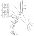
- FIG. 1 is a diagram showing an overall configuration of an endoscope system according to a first embodiment of the present invention.
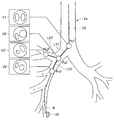
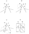
- FIG. 2A is a diagram showing a bronchial shape image.
- FIG. 2B is a diagram showing a distance range of a constant value from a spar in a bronchial shape image.
- FIG. 2C is a diagram showing a constant distance range from a branch point in a bronchial shape image.
- FIG. 2D is a diagram showing a presentation example of candidate information.
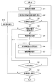
- FIG. 3 is a flowchart showing a representative example of processing contents of the first embodiment.
- FIG. 4 is a diagram showing a route that is a distance from a branch point along the core line.
- FIG. 4 is a diagram showing a route that is a distance from a branch point along the core line.
- FIG. 5A is a diagram illustrating an overall configuration of an endoscope system according to a modification of the first embodiment.
- FIG. 5B is an explanatory diagram of registration as a process of associating two coordinate systems.
- FIG. 6 is an explanatory diagram of the operation in the second embodiment of the present invention.
- FIG. 7 is an explanatory diagram of the operation in the third embodiment of the present invention.
- FIG. 8 is an explanatory diagram of an operation in a modification of the third embodiment.
- FIG. 9 is an explanatory diagram of an operation for storing candidate information from changes in an endoscopic image.
- FIG. 10A is a diagram showing a presentation example of candidate information different from FIG. 2D.
- FIG. 10B is a diagram showing a presentation example of candidate information different from FIG. 10A.
- FIG. 10C is a diagram showing a presentation example of candidate information different from FIG. 10A.
- FIG. 10D is a diagram showing a presentation example of candidate information different from FIG. 10A.
- FIG. 10E is a diagram showing a presentation example of candidate information different from FIG. 10A.
- FIG. 10F is a diagram showing a presentation example of candidate information different from FIG. 10A.
- FIG. 10G is a diagram showing a presentation example of candidate information different from FIG. 10A.
- FIG. 11 is a diagram showing an example of displaying an estimated position on a tomographic image including a bronchus.
- FIG. 12 is a diagram showing an example in which each position and the corresponding VBS in FIG. 11 are associated with character information.
- FIG. 12 is a diagram showing an example in which each position and the corresponding VBS in FIG. 11 are associated with character information.
- FIG. 13 is a view showing a display example corresponding to the modification of FIG.
- FIG. 14 is a diagram showing a display example in which the moving direction is further displayed in FIG.
- FIG. 15 is a diagram showing a display example that further displays the visual field range in FIG.
- FIG. 16 is a diagram showing a display example in which the visual field range is also displayed three-dimensionally on one tomographic image.
- FIG. 17 is a diagram illustrating an example of displaying a stored position in an area.
- FIG. 18 is a diagram illustrating an example in which a bronchial tree and a tomographic image are superimposed and displayed.
- FIG. 19 is a diagram showing an example of display using two tomographic images.
- FIG. 20 is a diagram illustrating an example of display on a cut surface including a bronchial tree at a stored position.
- FIG. 21 is an explanatory diagram of the setting method of FIG.
- the endoscope system 1 As shown in FIG. 1, the endoscope system 1 according to the first embodiment of the present invention is inserted into a bronchus 2 (FIG. 2A) as a predetermined luminal organ in a patient as a subject to be examined.
- An endoscope apparatus 4 including the endoscope 3 and an insertion support apparatus 5 that is used together with the endoscope apparatus 4 and supports the insertion of the endoscope 3 are mainly configured.
- the endoscope apparatus 4 performs signal processing on the endoscope 3, a light source device 6 that supplies illumination light to the endoscope 3, and an image sensor 7 that constitutes an imaging unit mounted on the endoscope 3. It has a camera control unit (abbreviated as CCU) 8 as a signal processing device, and a monitor 9 for displaying an endoscopic image generated by the CCU 8.
- CCU camera control unit
- the endoscope 3 includes an elongated insertion portion (or endoscope insertion portion) 11 having flexibility, and an operation portion 12 provided at the rear end of the insertion portion 11.
- the part 13 is provided with an illumination window and an observation window.
- a light guide 14 that transmits illumination light is inserted into the insertion unit 11 and the operation unit 12, and an incident end of the light guide 14 is connected to the light source device 6 and is generated by a light source lamp or LED (not shown) in the light source device 6. The illuminated light is incident on the incident end.
- the illumination light transmitted by the light guide 14 is emitted forward from an emission end (tip surface) attached to the illumination window.
- an objective lens 15 that forms an objective optical system that forms an image of a subject is attached to the observation window, and an imaging element 7 such as a CCD is disposed at the imaging position, and the objective lens 15 and the imaging element 7
- An imaging device 16 is formed as an imaging means (or imaging unit) that images the bronchus 2 as a predetermined luminal organ into which the insertion unit 11 is inserted.
- the image sensor 7 is connected to the CCU 8 through a signal line inserted through the insertion unit 11 and the operation unit 12.
- the CCU 8 generates an image signal of a captured image corresponding to an optical image formed on the imaging surface of the image sensor 7 by an image signal generation circuit (not shown) therein, and outputs the image signal to the monitor 9.
- the monitor 9 displays an image (moving image) of the image signal as an endoscopic image (also referred to as a captured image).
- the insertion portion 11 of the endoscope 3 is provided with a bendable bending portion 19 at the rear end of the distal end portion 13, and the operator performs an operation of rotating a bending operation knob 20 provided on the operation portion 12.
- the bending portion 19 can be bent in any direction, up and down, left and right.
- the insertion support device 5 uses, as a DVD, a Blu-ray disc, a flash, and CT data as three-dimensional image information of a patient generated by known CT (Computed Tomography) for a patient to be examined by the endoscope 3. It has CT data acquisition part 21 taken in via portable storage media, such as memory, and CT image data storage part 22 which memorizes CT data taken in by this CT data acquisition part 21.
- CT data acquisition part 21 taken in via portable storage media, such as memory
- CT image data storage part 22 which memorizes CT data taken in by this CT data acquisition part 21.
- the CT image data storage unit 22 may store CT data (as 3D image information of a patient as a subject) generated by CT via a communication line, the Internet, or the like.
- the CT image data storage unit 22 can be configured by a hard disk device, a flash memory, a DVD, or the like.
- the CT image data storage unit 22 constituting the image storage means includes a CT image data obtained by separating image data from the CT data, and a first coordinate system corresponding to the CT image data obtained by separating position information from the CT data. It has the matching image information storage part 22a memorize
- the insertion support device 5 includes a lumen organ extraction circuit serving as a lumen organ extraction unit that extracts three-dimensional image data of the bronchus 2 as a predetermined lumen organ from the CT image data in the CT image data storage unit 22.
- a bronchus extraction unit 23 including an arithmetic processing unit (abbreviated as CPU) is provided.
- the bronchus extraction unit 23 obtains 3D shape information (shape data) representing the hollow shape of the bronchus 2 and 3D shape from the extracted 3D data (more specifically, 3D volume data) of the bronchi 2. Image information (image data) is generated. That is, the bronchi extraction unit 23 includes a bronchial shape image generation unit 23a as a bronchial shape image generation unit that generates a bronchial shape image as a hollow three-dimensional bronchial shape image from the extracted three-dimensional data of the bronchi 2. .
- the bronchi extracting unit 23 extracts the three-dimensional position data in the first coordinate system (or CT coordinate system) corresponding to the three-dimensional data in association with the three-dimensional data.
- the bronchus extraction unit 23 includes an association information storage unit 23b including a memory that stores association information in which 3D shape data of the bronchi 2 (that is, bronchial shape data) and 3D position data are associated with each other.
- the insertion support device 5 is a virtual internal as a virtual endoscopic image corresponding to an endoscopic image generated by imaging with the imaging device 16 provided at the distal end portion 13 of the insertion portion 11 in the endoscope 3.
- a VBS image generation unit 24 is provided as virtual endoscopic image generation means for generating an endoscopic image (referred to as a VBS image).
- the VBS image generation unit 24 receives imaging system characteristic information (the focal length of the objective lens 15, the number of pixels of the image sensor 7, the pixel size, etc.) related to the imaging device 16 of the endoscope 3 from, for example, the input device 31. Is done.
- the VBS image generation unit 24 includes information on the position of the imaging device 16 of the endoscope 3 that is actually inserted into the bronchus 2 (also referred to as the position of the tip of the insertion unit 11), and the subject in the bronchus 2 by the imaging device 16 Generation for generating a VBS image for virtually drawing an endoscopic image obtained by endoscopically imaging the inside of the bronchus 2 with the position as a viewpoint position based on the characteristic information for imaging the image and the bronchial shape data A circuit, a CPU, and the like are included.
- the insertion support apparatus 5 also includes an image processing unit 25 configured by a CPU, an image processing circuit, and the like that perform image matching between the endoscopic image input from the CCU 8 and the VBS image of the VBS image generation unit 24.
- an image processing unit 25 configured by a CPU, an image processing circuit, and the like that perform image matching between the endoscopic image input from the CCU 8 and the VBS image of the VBS image generation unit 24.
- a storage unit 27 including a memory or the like.
- the insertion support device 5 includes an MPR image generation unit 28 that generates a CT tomographic image (referred to as an MPR image) as a multi-section reconstructed image based on the CT image data stored in the CT image data storage unit 22, and an MPR image A pointing device such as a mouse that generates a path setting screen as an insertion path setting screen having an MPR image generated by the generating unit 28 and sets a path for insertion into the target site in the bronchus 2 of the endoscope 3 And a route setting unit 29.
- MPR image generation unit 28 that generates a CT tomographic image (referred to as an MPR image) as a multi-section reconstructed image based on the CT image data stored in the CT image data storage unit 22, and an MPR image A pointing device such as a mouse that generates a path setting screen as an insertion path setting screen having an MPR image generated by the generating unit 28 and sets a path for insertion into the target site in the bronchus 2 of the endoscope 3
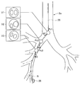
- the path setting unit 29 inserts (in the insertion unit 11) in the bronchus 2 from the CT image data and the bronchial shape image 2a. It has a function of a route data generation unit 29a such as a route data generation circuit that generates route data from a start position to a target position near the target region 36.
- a route data generation unit 29a such as a route data generation circuit that generates route data from a start position to a target position near the target region 36.
- a path to a target position near the target portion 36 is indicated by R.
- the endoscope system 1 further includes an input device 31 including a keyboard and a pointing device for inputting setting information to the route setting unit 29. Further, the surgeon can input parameters and data when performing image processing from the input device 31 to the image processing unit 25, and can select and instruct the control operation to the control unit 26. .
- the route setting unit 29 sends information on the set route to the VBS image generation unit 24, the MPR image generation unit 28, and the control unit 26.
- the VBS image generation unit 24 and the MPR image generation unit 28 generate a VBS image and an MPR image along the route, respectively, and the control unit 26 controls the operation of each unit along the route.
- the image processing unit 25 receives an endoscopic image generated by the CCU 8 and a VBS image generated by the VBS image generation unit 24.
- the bronchial shape image generated by the bronchial shape image generating unit 23 a is also input to the image processing unit 25.
- the alignment processing unit 25a by the image processing unit 25 is installed.
- the three-dimensional position of the tip of the insertion portion 11 is estimated (or calculated) by image matching at.
- a three-dimensional position (known position) that can be specified from the bronchial shape image 2a by the CT coordinate system, such as the entrance of the bronchus 2 or the carina K (see FIG. 2A), or its neighboring position is set as a moving image matching start position.
- the VBS image generation unit generates a VBS image based on the position information.
- the operator inserts the distal end of the insertion portion 11 so that the endoscopic image looks the same as the VBS image.
- the alignment processing unit 25a of the image processing unit 25 matches the endoscope image and the VBS image under the set conditions (within an error that can ensure a predetermined accuracy). Start image matching.
- the alignment processing unit 25a has a function of a position estimation unit 25b that detects or calculates the position of the distal end of the insertion unit 11 by estimation.
- a position estimation unit 42b that provides a position sensor 41 for position detection at the distal end portion 13 and detects (estimates) the three-dimensional position of the distal end of the insertion portion 11 using the position sensor 41 as in a modification shown in FIG. May be configured.
- the distal end of the insertion portion 11 is used in the same meaning as the distal end of the endoscope 3.
- the image processing unit 25 generates an image to be displayed on the monitor 32 under the control of the display control unit 26 a that controls display in the control unit 26. Under the control of the display control unit 26 a, the image processing unit 25 normally outputs the image signal (video signal) of the bronchial shape image 2 a generated by the bronchial shape image generation unit 23 a to the monitor 32.
- the bronchial shape image 2a is displayed as a two-dimensional tomographic image cut out in a cross section along the direction passing through the center of the (main) lumen, for example.
- the display is not limited to a two-dimensional tomographic image, and a three-dimensional image may be displayed.
- a three-dimensional image for example, it may be displayed in a projection view by a parallel projection method or in a perspective view so that the inside of the lumen can be seen.
- a core line 35 passing through the center of the lumen of the bronchus 2 is also displayed on the bronchial shape image 2 a displayed on the monitor 32.
- the core wire 35 is generated by, for example, the bronchial shape image generation unit 23a, but the image processing unit 25 may generate the core wire 35.
- the core wire 35 is displayed. Therefore, the insertion operation of the insertion portion 11 can be easily performed by referring to the display. . Further, by performing an operation of inserting along the core wire 35, the position of the distal end of the insertion portion 11 by image matching can be estimated in a short time.
- the image processing unit 25 uses the matching (image matching) of both the endoscopic image and the VBS image under the CT coordinate system in the operation of inserting into the deep side (terminal side) of the bronchus 2. A process for estimating the moved position or moving distance of the distal end of the insertion portion 11 is performed. That is, when both images match at a certain position, the imaging device 16 moves in accordance with an operation of moving the distal end of the insertion portion 11 along the core line 35 (for insertion). Changes.
- the alignment processing unit 25a uses the VBS image (output from the VBS image generation unit 24) when it moves on the route along the core line 35 to best match the endoscopic image. Is selected by image processing, and the three-dimensional position corresponding to the selected VBS image is calculated (estimated) as the position of the tip of the insertion unit 11. Therefore, the moved movement distance can also be calculated (estimated) from the difference between the two positions. Note that the distal end of the insertion unit 11 may be moved at a position deviated from the core wire 35. Therefore, the VBS image generation unit 24 generates a VBS image at a position eccentric from the core wire 35 by an appropriate distance. The processed VBS image may be output to the alignment processing unit 25a.
- the control unit 26 corrects the route data generated by the route data generation unit 29a (before insertion of the insertion unit 11 of the endoscope 3) by the position of the distal end of the insertion unit 11 calculated by the alignment processing unit 25a. You may make it do.
- control unit 26 has a function of a condition determination unit 26c configured by a comparison circuit or the like that determines whether or not the distal end of the insertion unit 11 estimated by the image processing unit 25 satisfies a predetermined condition.
- a condition determination unit 26c configured by a comparison circuit or the like that determines whether or not the distal end of the insertion unit 11 estimated by the image processing unit 25 satisfies a predetermined condition.
- the position of the tip of the corresponding insertion unit 11 is stored in the storage unit 27.
- the VBS image corresponding to the position of the distal end is stored in the storage unit 27 as candidate information to be presented (for presentation) when presentation is necessary.
- the storage unit 27 is a candidate for the case where the position of the distal end of the insertion unit 11 estimated (calculated) when the determination result is obtained is presented later.
- the candidate information storage unit 27a functions as a storage unit that stores the position information and stores the candidate position information and a VBS image corresponding to the candidate position information as candidate information for presentation.
- the candidate information for presentation includes candidate position information (candidate position information) and a VBS image corresponding to the candidate position information, but corresponds to the candidate position information (in other words, at the candidate position).
- the generated VBS image may also be a candidate VBS image.
- the image processing unit 25 is controlled by the candidate information presentation control unit 26b of the display control unit 26a, and the candidate information generation unit 25c generates candidate information to be displayed on the monitor 32 as candidate information. It has a function.
- the image processing unit 25 temporarily stores an endoscopic image and a VBS image when performing image matching between an endoscopic image and a VBS image, or an image memory 25d used as a work area for image processing. Have.
- An image memory 25d may be provided outside the image processing unit 25.
- the condition determination unit 26c functions as a predetermined region to determine whether or not the distal end of the insertion unit 11 is located in a feature region as a characteristic region related to bifurcation in the bronchi 2 as a predetermined condition. Have Then, as will be described later, in the case where the feature region determination unit 26d determines that the tip of the insertion unit 11 is located (or exists) in the feature region, candidate information is stored in the candidate information storage unit 27a.
- a constant value dth is set for the distance between two positions (or points). And between the position of the front-end
- the setting of the feature region and the value of the constant value dth can be designated by the surgeon from the input device 31, for example.
- candidate information before the current position of the distal end of the insertion unit 11 is stored in the storage unit 27.
- the position estimation accuracy (using image matching) of the tip of the insertion unit 11 is reduced by the movement operation of the insertion unit 11, and re-alignment (that is, re-alignment) is performed.
- the candidate information for the realignment can be presented.
- the operator may input an instruction to perform realignment to the image processing unit 25 or the control unit 26 from a keyboard, a mouse, or the like constituting the input device 31.
- the display control unit 26a of the control unit 26 displays candidate information near the tip position of the current insertion unit 11 as a candidate information storage unit 27a of the storage unit 27. From which the candidate information is presented on the monitor 32 via the image processing unit 25.
- the display control unit 26a that performs control for presenting candidate information on the monitor 32 has a function of a candidate information presentation control unit 26b that performs control of candidate information presentation. Note that the display control unit 26a may read out candidate information (for presentation) from the candidate information storage unit 27a and perform control to display the candidate information on the monitor 32 without passing through the image processing unit 25.
- the candidate information When the candidate information is presented on the monitor 32, for example, information on candidate positions when performing image comparison on the bronchial shape image 2a shown in FIG. 2D as a two-dimensional tomographic image and VBS corresponding to the information on the candidate positions. Display an image.
- the alignment processing unit 25a of the image processing unit 25 estimates (calculates) the distal end of the insertion unit 11 using image matching when the distal end of the insertion unit 11 is moved. There may be an image matching error that makes it impossible to perform image matching within accuracy. In this case, the alignment processing unit 25a of the image processing unit 25 generates an image matching error signal and displays on the monitor 32 that the image matching error has occurred. The alignment processing unit 25 a sends an image matching error signal to the control unit 26, and the candidate information presentation control unit 26 b of the control unit 26 performs control for presenting candidate information on the monitor 32. For this reason, the monitor 32 forms candidate information presenting means for presenting candidate information to the surgeon.
- the candidate information presenting means includes a candidate information storage unit 27a for storing candidate information (for presentation), an image processing unit 25 for outputting an image signal of candidate information, and a candidate information presentation control unit 26b together with the monitor 32. It may be defined as The surgeon performs the alignment process again using the candidate information. By performing the alignment process again, the surgeon can continue the insertion operation of the insertion portion 11 from the vicinity of the position where the alignment process is performed again. If the surgeon determines that the accuracy of the image matching has deteriorated, the above processing may be performed by instructing realignment.
- the alignment processing unit 25a, candidate information generation unit 25c, bronchus extraction unit 23, VBS image generation unit 24, and control unit 26 of the image processing unit 25 in FIG. 1 may be configured using a CPU or FPGA ( A hardware device such as a field programmable gate array) or a dedicated electronic circuit element may be used.
- the endoscope system 1 having such a configuration includes a CT image data storage unit 22 as image storage means for storing 3D image information of a subject acquired in advance, and a predetermined viewpoint position with respect to the 3D image information.
- a VBS image generating unit 24 as a virtual endoscopic image generating means for generating a virtual endoscopic image drawn endoscopically from, and the endoscope 3 is provided to image the inside of the predetermined lumen organ
- An imaging device 16 serving as an imaging unit
- a position estimation unit 25b serving as a position detection unit that detects the position of the distal end of the endoscope insertion unit 11 within a predetermined lumen organ of the subject, and the position detection unit
- a monitor 32 as candidate information presenting means for presenting the virtual endoscopic image
- the alignment processing unit 25 a of the image processing unit 25 outputs the VBS image (image signal thereof) of the VBS image generation unit 24 at the reference position to the monitor 32.
- the surgeon designates one of the reference positions from the input device 31, inserts the tip of the insertion unit 11 into the designated reference position, and performs alignment processing on the alignment processing unit 25a of the image processing unit 25. Instruct to do.
- the alignment processing unit 25a estimates (calculates) the position of the tip of the insertion unit 11 by image matching based on the result of the alignment process. Specifically, using the alignment information as an initial value of image matching, a VBS image that best matches the endoscopic image is calculated by image processing.
- step S3 the alignment processing unit 25a determines whether or not the tip of the insertion unit 11 can be estimated within a predetermined condition by image matching (for example, whether the estimated position is inside the bronchus) (that is, whether the position is successfully estimated). Is determined. If the determination in step S3 is successful, the position of the distal end of the insertion unit 11 estimated by the position estimation unit 25b of the image processing unit 25 is displayed on the monitor 32 at the estimated position on the bronchial shape image 2a. While being displayed superimposed, the process of step S4 is performed. On the other hand, if the determination is unsuccessful, the process of step S6 is executed.
- step S4 The position information of the tip of the insertion unit 11 estimated by the position estimation unit 25b in step S4 is input to the feature region determination unit 26d of the control unit 26. Then, the feature region determination unit 26d is located in the feature region in which the estimated position of the distal end of the insertion unit 11 is within a certain value dth (such as dth1, dth2) from the spur Spi and the branch point Bi in the bronchus 2. It is determined whether or not.
- step S4 in the case of a determination result that is within a certain value dth, candidate information is stored in the storage unit 27 as shown in step S5. For example, as shown in FIG.
- step S5 when the distal end of the insertion portion 11 is inserted deeper than the carina K and the distal end P2 of the insertion portion 11 falls within a predetermined value dth1 from the next spur Sp2, the estimated distal end of the insertion portion 11 is inserted.
- the position P2 becomes the candidate position Pc2, and the corresponding VBS image I2 is stored in the storage unit 27 as candidate information together with the candidate position Pc2.
- step S8 After the process of step S5, it is reflected in the process of step S8.
- the process of step S8 is executed without performing the process of recording candidate information in step S5.
- FIG. 2B shows an example in which the candidate position Pci is stored when it is determined that it is within the predetermined value dth1 from the spur Spi.
- the estimated distal end of the insertion portion 11 is branched.
- FIG. 2C shows a case where the distal ends P1 ′ and P2 ′ of the insertion portion 11 are within a certain value dth2 from the branch points B1 and B2.
- the constant value dth1 and the constant value dth2 may be set to the same value or different values.
- step S8 it is detected whether the surgeon has generated an instruction signal (trigger) for presenting candidate information from the input device 31. If it is not possible to detect that the image matching estimation has failed, for example, if the endoscope moves suddenly, the estimated position by image matching satisfies the predetermined condition (whether it is inside the bronchus). In the case of another position, an instruction signal is generated when the surgeon determines that the alignment is performed again.
- an instruction signal is generated when the surgeon determines that the alignment is performed again.
- the position estimation unit 25b determines (estimates) whether or not the distal end of the insertion unit 11 has been inserted to the target position.
- the surgeon refers to the display displayed on the monitor 32 and moves the distal end of the insertion portion 11 to the deep side of the bronchus 2 as shown in step S10. Insert into.
- the process returns to step S2 to estimate the tip position of the insertion unit 11, but the initial value of image matching uses the estimated position of the previous image matching instead of the alignment information.
- the target position is inserted in step S19, the insertion operation in FIG. 3 is terminated.
- step S6 the display control unit 26a automatically generates an instruction signal for presenting candidate information.
- candidate information is read from candidate information storage part 27a of storage part 27. And candidate information is displayed on the monitor 32 as shown to step S7.
- candidate position Pc2 stored in the information storage unit 27a
- VBS image I2 corresponding to the candidate position Pc2
- a VBS image corresponding to the candidate position Pc2 is displayed as candidate information together with the candidate position Pc2.
- an endoscopic image corresponding to the candidate position Pc2 may also be displayed as indicated by a dotted line.
- VBS image or the endoscopic image may be displayed movably, and the user may be able to superimpose and display (composite display) one image on the other image with a mouse or the like. This makes it easier for the user to check the degree of image matching.
- 2D shows an example of presentation of one candidate information, a plurality of candidate information may be presented as shown in FIGS. 10A to 10F described later. When the candidate information is displayed, the process returns to step S1, and the surgeon performs the alignment process again with reference to the candidate information displayed on the monitor 32.
- candidate information for presentation is stored in the storage unit 27 while the operator performs an operation of inserting the insertion unit 11, and the tip of the insertion unit 11 is stored.
- the position estimation is unsuccessful, the candidate information for presentation suitable for alignment can be presented as candidate information, so that the insertion operation can be performed smoothly.
- the position of the tip of the insertion portion 11 is estimated using image processing for image matching, so that the error gradually increases from the initial alignment state by the image processing algorithm. It becomes easy. Even in such a case, it is possible to perform the operation of insertion from the vicinity of the re-aligned position to the deeper side by reducing the error sufficiently by re-aligning by re-image matching.
- the conditions in the above description may be changed as follows.
- Candidate information (for presentation) is stored in the storage unit 27 when the estimated position of the tip of the insertion unit 11 satisfies one condition within a predetermined value dth1 or dth2 from the spur Spi or the branch point Bi, respectively.
- (B) Candidate information may be stored when the estimated position of the tip of the insertion portion 11 satisfies two conditions that are within the constant values dth1 and dth2 from the spur Spi and the branch point Bi, respectively.
- candidate information may be stored when the estimated distance between the distal end of the insertion portion 11 and the core wire 35 is within a predetermined value dth3.
- candidate information may be stored when a branch image is seen in the endoscopic image.
- the constant values dth1 to dth3 may be set to the same value, or the bronchi at the spur Spi or the branch point Bi. You may set according to the distance to the value according to a diameter, the branch site
- F Further, in the examples of FIGS. 2B and 2C, the case where the condition for storing is determined depending on whether the linear distance between the two points is within a certain value or not is shown. However, as shown in FIG.
- the candidate information is stored depending on whether or not the point P on the core line 35 closest to the position and the path (distance measured along the core line 35) dw on the core line 35 to the branch point Bi is within a predetermined value dwth. It is good as a condition.
- FIG. 5A shows a configuration of an endoscope system 1B according to a modification example of the first embodiment.
- An endoscope system 1B shown in FIG. 5A is similar to the endoscope system 1 of FIG.
- a position sensor 41 for detecting the position is provided.
- measurement processing that performs processing for measuring (detecting) a three-dimensional position (also simply referred to as a position) of the position sensor 41 at a predetermined position in the insertion support apparatus 5 outside the endoscope 3 and the subject.
- An apparatus or measurement processing unit 42 is provided.
- a detection signal from the position sensor 41 is input to the measurement processing unit 42.
- This measurement processing unit 42 uses the position sensor 41 to provide a three-dimensional position of the imaging device 16 that constitutes an imaging means inserted into the bronchus 2 as a predetermined luminal organ, or a three-dimensional position of the distal end of the insertion unit 11. It has the function of the position estimation part 42b as a position detection means to detect or estimate. Note that FIG. 5A of this modification does not include the position estimation unit 25b in the image processing unit 25 in FIG. As a means or method for position detection (position estimation), for example, one using magnetism can be used.
- a position sensor 41 composed of a coil detects an alternating magnetic field generated from a plurality of antennas 42a connected to the measurement processing unit 42, and the amplitude and phase of the signal in the signal detected by the position sensor 41 (amplitude detection)
- the distance from the antenna 42a to the position sensor 41 is measured by the measurement processing unit 42 (provided with a circuit and a phase detection circuit).
- the measurement processing unit 42 can specify the three-dimensional position of the position sensor 41 by providing three or more antennas 42a at different known positions.
- an AC signal may be applied to a coil constituting the position sensor 41 to generate an AC magnetic field around the coil, and the position of the position sensor 41 may be detected by detecting the AC magnetic field on the antenna 42a side.
- a magnetic position detection device using a coil has been described.
- the configurations of the position sensor 41 and the measurement processing unit 42 are not limited to those described above.
- a plurality of coils for position detection are arranged at predetermined intervals along the longitudinal direction of the insertion portion 11, the shape of the insertion portion 11 is estimated from the positions of the plurality of coils, and the position of the distal end portion 13 or the like is detected. You may be able to do that.
- the position information of the tip of the insertion unit 11 detected (estimated) by the measurement processing unit 42 is output to the control unit 26 and the image processing unit 25.
- Position alignment with the position (position information) in the second coordinate system as the coordinate system is performed.
- the control unit 26 has functions of registration (registration) of both coordinate systems and an alignment unit (and alignment control unit) 26e that controls the coordinate system.
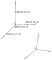
- FIG. 5B shows an explanatory diagram of the registration operation.
- the operator sequentially sets the distal end portion 13 (or the position sensor 41) of the endoscope 3 at, for example, four points Q0 to Q3 in the vicinity of the entrance of the bronchus 2, and the first coordinate system O-XYZ and the second coordinate system are set.
- An instruction or instruction input for associating positions in the coordinate system o-xyz is performed from the input device 31.
- the input device 31 forms an instruction input unit or an instruction input unit that gives an instruction for position association.
- the tip 13 (or the position sensor 41) is moved to a position Q0 (0, 0, 0) of the origin O in the first coordinate system O-XYZ, a position Q1 (1, 0, 0) on the X coordinate, Y
- the operator sequentially sets the position Q2 (0, 1, 0) on the coordinates and the position Q3 (0, 0, 1) on the Z coordinates, and the surgeon instructs the position association.
- the positions sequentially measured by the measurement processing unit 42 at each position are (x0, y0, z0), (x1, y1, z1), (x2, y2, z2), and (x3, y3, z3).
- the control unit 27 performs position association and controls to store the position association information in the storage unit 27.
- Position association information in this case (specifically, Q0 (0, 0, 0), Q1 (1, 0, 0), Q2 (0, 1, 0), Q3 in the first coordinate system O-XYZ) (0, 0, 1) is (x0, y0, z0), (x1, y1, z1), (x2, y2, z2), (x3, y3, z3) in the second coordinate system o-xyz.
- the alignment unit 26e determines conversion information that associates arbitrary positions in both coordinate systems using the position association information stored in the storage unit 27.
- the alignment unit 26e stores this conversion information in the storage unit 27.
- coordinate positions Q0 (0, 0, 0), Q1 (1, 0, 0), Q2 (0, 1, 0), Q3 (0, 0, 1) and corresponding coordinate positions ( x0, y0, z0), (x1, y1, z1), (x2, y2, z2), (x3, y3, z3) are simplified to Q0 ⁇ (x0, y0, z0), Q1 ⁇ (x1, y1, z1), Q2 ⁇ (x2, y2, z2), and Q3 ⁇ (x3, y3, z3).
- position association may be performed (determined) using three points from which one of the points is omitted.
- the surgeon sequentially brings the distal end portion 13 of the endoscope 3 into contact with the position specified in the first coordinate system.
- a VBS image is used as a method of expressing the position specified in the first coordinate system. That is, the surgeon operates the endoscope so that the VBS image and the endoscope image look the same.
- the surgeon inserts the insertion portion 11 of the endoscope 2 into the bronchus 2 and starts performing endoscopy.
- the position of the CT coordinate system (first coordinate system) corresponding to the position of the distal end of the insertion unit 11 estimated by the position estimation unit 42b deviates from the condition of being inside the lumen of the bronchus 2.
- the position estimation unit 42b deviates from the condition of being inside the lumen of the bronchus 2.
- the configuration of the second modification example of the first embodiment is the same as that of the first modification example.
- the alignment processing unit 25a of the image processing unit 25 uses a position sensor by image matching. Processing for improving the accuracy of alignment using 41 is performed.
- the first embodiment it is necessary to perform an operation so that the distal end portion of the endoscope 3 comes into contact with a designated position, but this operation is difficult and is a factor that deteriorates the accuracy of the estimated position. Therefore, by estimating the position of the distal end portion 13 of the endoscope 3 in the CT coordinate system by image matching and changing the designated position based on the position, the distal end portion 13 of the endoscope 3 is surely moved. Touch the specified position. Thereby, the error of the position shift can be adjusted to a predetermined value or less.
- the alignment processing unit 25a of the image processing unit 25 has a function of a second alignment unit that performs alignment by image matching, and as described above, the measurement processing unit 42
- the position estimation unit 42b detects (or estimates) the position of the distal end of the insertion unit 11.
- the alignment processing unit 25a has a function of an alignment monitoring processing unit that monitors the alignment state of both coordinate systems by image matching. For example, when the distal end of the insertion unit 11 is inserted into the bronchus 2, the position of the endoscope image and the position estimation unit 42b in the second coordinate system changes as the distal end of the insertion unit 11 moves.
- the VBS image generated by the position information of the CT coordinate system (first coordinate system) and input to the image processing unit 25 also changes corresponding to the change of the position in the second coordinate system.
- the alignment processing unit 25a of the image processing unit 25 monitors both images, and when the shift amount of both images shifts to a preset value or more, the registration fails (or the alignment is necessary). Is determined.
- the candidate information for presentation is stored in the storage unit 27 while the operator performs an operation of inserting the insertion unit 11, and the distal end of the insertion unit 11 is stored.
- the candidate information for presentation suitable for alignment can be presented in the case where the estimation of the position fails, so that the insertion operation can be performed smoothly.
- the position of the distal end of the insertion portion 11 is estimated using the position sensor 41 after the initial alignment, and the distance from the aligned position increases. The error tends to increase from the initial alignment state.
- the position sensor 41 is used, and the position is re-adjusted using image matching so that the error is sufficiently small, and the operation for insertion from the vicinity of the re-aligned position to the deeper side is performed. It becomes possible to perform smoothly.
- the candidate information for presentation is stored in the candidate information storage unit 27a of the storage unit 27, but after the candidate information is stored, You may make it provide the memory
- the candidate information prohibition may be canceled when moving beyond the limit distance, and a candidate information storage restriction unit that stores the next candidate information when the predetermined condition described above is satisfied may be provided.
- a candidate information storage restriction unit that stores the next candidate information when the predetermined condition described above is satisfied may be provided.
- it may be a position that first reaches the feature area, a position that lastly falls within the feature area, a position that is closest to the branch point Bi, or a position where the previous branch can be easily identified.
- the operator may input an instruction to the image processing unit 25 or the control unit 26 from a keyboard, a mouse, or the like constituting the input device 31.
- FIG. 1 and FIG. 5A a configuration in which the storage restriction unit 26f (of candidate information) is provided in the control unit 26 is indicated by a dotted line.
- the limit distance for limiting the storage may be set based on information about the bifurcation point Bi of the bronchus 2 and the spur Spi. Specifically, the limit distance may be set based on, for example, the distance between adjacent branch points Bi and Bi + 1 that exist along the bronchial core 35. As a specific example, for example, 1/2 of the distance between adjacent branch points may be set as the limit distance.
- the candidate selection storage unit 27a further stores the presentation selection information or the presentation restriction information so as to include the presentation restriction information storage unit 27b that stores the presentation selection information or the presentation restriction information. May be.
- the position Pi ′ when the position Pi ′ that first satisfies the condition (condition existing in the feature region) that is within the predetermined value dth2 from the branch point Bi is set as the predetermined condition, the position Pi ′ is stored as candidate information as candidate information.
- (Bi, 1) is also added and stored as the presentation restriction information.
- (Bi, 1) means (branch point Bi, the number of the first position that satisfies the condition).
- the distal end of the insertion portion 11 when the distal end of the insertion portion 11 is moved to the deep side, when the next position Pi + 1 ′ is set at an appropriate time interval, and when this position Pi + 1 ′ is stored as candidate information as candidate information, for example, ( Bi, 2) is additionally stored.
- the surgeon has an effect of facilitating the alignment work.
- the three-dimensional image data of the bronchus 2 as a predetermined luminal organ may not be extracted from the CT image data but directly generated by volume rendering from the CT image data. .
- the bronchi extraction unit 23 in FIG. 1 or FIG. 5A is unnecessary, and the VBS image generation unit 24 generates the two types of images.
- the present embodiment has the same configuration as the endoscope system 1 in FIG. 1 or the endoscope system 1B in FIG. 5A. Although this embodiment is close to the content of (b) described above, candidate information is stored when the distance between two points satisfies the first threshold and the second threshold. In the present embodiment, for example, after the estimated position of the distal end of the insertion portion 11 falls within the first threshold value dt2 from the branch point Bi at Pi, the process further proceeds to Pi + 1 to reach the second threshold value dt1 from the branch point Bi.
- FIG. 6 is an explanatory diagram of an operation of storing candidate information during an insertion operation according to the second embodiment.
- the estimated distance Pi between the distal end of the insertion portion 11 and the position of the next branch point Bi is A first determination as to whether or not the value is within a certain value dth2 is performed by the feature region determination unit 26d of the control unit 26.
- the feature region determination unit 26d performs further estimation after the distance between the position Pi of the distal end of the insertion unit 11 estimated by the first determination and the position of the branch point Bi is within the first threshold dt2.
- a second determination is made as to whether or not the distance between the position Pi of the tip of the section 11 and the position of the branch point Bi is equal to or greater than a second threshold value dt1.
- dt1 a second threshold value
- the distal end of the insertion portion 11 reaches the vicinity of the branch point Bi as shown in FIG. It is possible to store candidate information in the state that has been set.
- the candidate information for presentation is stored in the storage unit 27 while the operator performs an operation of inserting the insertion unit 11 as in the first embodiment, and the insertion unit 11 Since the candidate information for presentation can be presented in the case where the estimation of the position of the tip of the head fails, the insertion operation can be performed smoothly. Furthermore, according to the present embodiment, it is possible to accurately detect the state in which the distal end of the insertion portion 11 has reached a position near the branch point Bi. As a specific example of the present embodiment, the case where two threshold values are set for the distance between the position of the distal end of the insertion portion 11 and the branch point Bi has been described. However, the distance between the position of the distal end of the insertion portion 11 and the spar Spi. Alternatively, the distance between the position on the core wire 35 closest to the distal end of the insertion portion 11 and the branch point Bi may be used.
- the endoscope system of the present embodiment has the same configuration as the endoscope system 1 in FIG. 1 or the endoscope system 1B in FIG. 5A.
- the distal end of the insertion section 11 passes near a certain branch point and moves to the next bronchial branch (twig) branched from the vicinity of the branch point, the distal end of the insertion section 11 acquired before that The candidate information is stored at the position.
- FIG. 7 is a diagram for explaining the operation in this embodiment.
- the distal end of the insertion portion 11 is further moved to reach (or moved to) the position Pk + 1 in one branched bronchial branch (twig).
- the candidate information at the position Pk is stored.
- the position of the tip of the insertion portion 11 is indicated by a small circle.
- the case where the position Pk + 1 in one bronchial branch (twig) is reached (or moved) means that the core wire 35 closest to the position of the distal end of the insertion portion 11 is that of a twig as shown in FIG. Applicable.
- a circle area having a radius dt is set as a distance centered on the bifurcation point Bi, and the feature area determination unit 26d determines that the estimated tip of the insertion unit 11 is After determining the first state existing in a position within the circle region, the second state moved outside the circle region may be determined.
- FIG. 7 a circle area having a radius dt is set as a distance centered on the bifurcation point Bi
- the radius dt is set to a distance between the position of the branch point Bi and the spur Spi or a value slightly larger than this distance.
- FIG. 7 is a diagram for explaining the operation in this modification. As shown in FIG. 7, for example, a boundary Bt (indicated by a dotted line in FIG. 7) where the distance on the core line 35 from the branch point Bi is dt may be set. In this way, when the boundary Bt is set, substantially the same effect is obtained. Further, as a modification of the present embodiment, when both the distances from the two branch points through which the distal end of the insertion portion 11 has sequentially passed are increased, they are acquired in a state where no alignment error occurs immediately before the increase. You may make it memorize
- FIG. 8 is a diagram for explaining the operation in this modification. As shown in FIG.
- the estimated position Pk of the insertion portion 11 passes through the branch point Bi-1 and the branch point Bi next to the branch point Bi-1, and is inserted into the deeper (end) side of the bronchus.
- the feature region determination unit 26d monitors the following conditions.
- the feature region determination unit 26d satisfies this condition when both the first distance d1 between the branch point Bi-1 and the position Pk and the second distance d2 between the branch point Bi and the position Pk are increased.
- a VBS image corresponding to the position acquired at a position (for example, Pk-1) in a state where no alignment error has occurred immediately before the condition is stored as candidate information.
- This modification has substantially the same effect as the second embodiment.
- candidate information may be stored when the distance dg between the centroids of the twigs (bronch 2 is branched) that is a feature amount of the branch in the endoscopic image changes. good.
- the upper and lower endoscopic images Ik and Ik + 1 are images obtained when, for example, the distal end of the insertion unit 11 is acquired at the positions Pk and Pk + 1, and the image processing unit 25 performs binarization processing or the like.
- the extracted dark part is calculated as twig (parts) images Ia and Ib.
- the image processing unit 25 calculates the centroids Ga and Gb of the images Ia and Ib of the respective twigs, and estimates the distance dg between the centroids.
- the image processing unit 25 stores candidate information at the position Pk, for example. To do. Even if it does in this way, it has an effect substantially the same as 3rd Embodiment.
- the outer circles of the two twig images Ia and Ib in FIG. 9 indicate the lumen inner wall of the bronchus 2.
- the candidate information is stored when the representative radius or the ratio of the representative radii when the twig is regarded as an ellipse is changed. Also good.
- the candidate information may be stored when the sum of the pixels of the images Ia and Ib changes.
- FIG. 10A shows candidate positions stored as candidate information when the predetermined condition is satisfied in the above-described embodiment as Pc1, Pc2, and Pc3, and corresponding VBS images as V1, V2, and V3.
- FIG. 10A shows candidate positions stored as candidate information when the predetermined condition is satisfied in the above-described embodiment as Pc1, Pc2, and Pc3, and corresponding VBS images as V1, V2, and V3.
- the endoscopic image corresponding to the candidate position Pci may also be displayed at a position where it can be easily compared with the VBS image Vi.
- the target region 36 is superimposed and displayed on the bronchial shape image 2a image
- the path R for inserting the insertion portion 11 of the endoscope 3 is superimposed and displayed on the bronchial shape image 2a image.
- candidate information By presenting candidate information in this manner, the user can easily perform re-alignment in a short time and can be smoothly inserted into the target portion 36. Even when it is determined that the distal end of the insertion portion 11 of the endoscope 3 has moved away from the path R (as path data) by a predetermined distance (such as a lumen diameter), for example, candidate information as shown in FIG. 10A May be presented. Further, as shown in FIG. 10B, the stored candidate positions Pc1, Pc2, Pc3 ′, and Pc3 may be displayed so as to be connected by lines L12, L23 ′, and L23 along the movement. In the case of FIG.
- candidate information is stored in the order of candidate positions Pc1, Pc2, and Pc3 ′, then returns from the candidate position Pc3 ′ to the candidate position Pc2 again, and the candidate on the twig side is different from the twig on the candidate position Pc3 ′ side.
- An example in the case of moving to the position Pc3 is shown. Since it has moved as described above, the lines L12, L23 ′ and L23 are displayed (presented). A VBS image corresponding to the candidate position Pc3 ′ is indicated by V3 ′. Others are the same as the content demonstrated in FIG. 10A, and have the same effect.
- FIG. 10C shows the position of the distal end of the insertion portion 11 when a predetermined time elapses from the time when the distal end of the insertion portion 11 of the endoscope 3 stores candidate information at the candidate position Pci in the case of FIG. 10A, for example. Is obtained, and the movement direction Mi from the candidate position Pci is calculated and stored as candidate information or as supplementary information of the candidate information. Then, as shown in FIG. 10C, when the candidate information is presented, the movement direction Mi is also displayed (presented) when the candidate information described in FIG. 10A is presented. Others are the same as the contents described in FIG. 10A, and have the same effects as those in FIG. 10A, and can be more easily aligned by referring to the movement direction Mi. FIG.
- 10D also stores the line-of-sight direction Ci (based on the objective lens 15) of the imaging device 16 when the candidate information is further stored at the position Pci of the distal end of the insertion portion 11 of the endoscope 3 in the case of FIG. 10A, for example. Then, as shown in FIG. 10D, the line-of-sight direction Ci is also displayed (presented) when the candidate information described with reference to FIG. 10A is presented. Others are the same as the content demonstrated in FIG. 10A. It becomes easy to perform alignment by presenting the line-of-sight direction Ci.
- FIG. 10D shows a case where the line-of-sight direction Ci is displayed two-dimensionally.
- the line-of-sight direction Ci ′ is displayed using, for example, a quadrangular pyramid, and the line-of-sight direction Ci ′ is displayed (presented) so that the user can easily recognize it as a three-dimensional direction. You may do it.
- the user can more easily recognize the line-of-sight direction Ci ′.
- FIG. 10F shows the case of storing candidate information by storing information on the inner diameter Di calculated from the three-dimensional shape of the bronchus 2 as information on the lumen diameter of the bronchi 2 when candidate information is further stored in the case of FIG. 10A, for example. Shows an example in which information on the stored inner diameter Di is also presented.
- the user can more easily grasp the position of the distal end of the insertion portion 11.
- the other functions and effects are the same as those of FIG. 10A.
- character information (specifically, A, B, C, and D) may be attached to four positions, and the corresponding VBS image may be displayed in association with the same character information.
- FIG. 10G character information (specifically, A, B, C, and D) may be attached to four positions, and the corresponding VBS image may be displayed in association with the same character information.
- candidate information is stored in the order of candidate positions Pc1, Pc2, and Pc3 ′, and then returns from the candidate position Pc3 ′ to the candidate position Pc2, and is different from the twigs on the candidate position Pc3 ′ side.
- An example in the case of moving to the candidate position Pc3 on the twig side is shown.
- the VBS image may be arranged below the bronchial shape image 2a.
- the user can confirm the position in time series, and the position of the distal end of the insertion portion 11 can be more easily grasped.
- the order and time of storing candidate information may be added to the VBS image and displayed. Further, the assignment of the character information to the position may be reversed in time series with Pc3, Pc3 ′, Pc2, and Pc1, or may be arranged in the order closer to the position where the position estimation was last successful.
- FIG. 11 shows an example of display on the monitor 32 applicable to the first to third embodiments described above.
- the upper left of FIG. 11 shows a CT tomographic image of a transverse section including the bronchi of the patient
- the upper right of FIG. 11 shows a CT tomographic image of a vertical section perpendicular to the front of the patient
- the lower left of FIG. 11 is parallel to the front of the patient.
- FIG. 11 displays the estimated position (point) of the distal end of the insertion unit 11 (or the imaging device 16 provided at the distal end) on a tomographic image, for example, in the same color circle or other shape.
- the four positions are displayed in a color (for example, blue) different from the display color of the tomographic image.
- the position and the VBS image corresponding to the position may be displayed in association with each other as shown in FIGS. 10A to 10F.
- character information (specifically, A, B, C, D) may be attached to four positions, and the corresponding VBS image may be displayed in association with the same character information.
- FIG. 11 it is an example showing a state in which a position is superimposed on a tomographic image, and a display associated with the position and character information of the corresponding VBS image is omitted.
- the estimated position is shown in the same color and shape in the case where it is on the cut surface and in other cases, but as shown in FIG. Different shapes (for example, the former is a circle and the latter is a square) may be displayed depending on whether they are not on the surface, or the color may be changed. Moreover, you may make it display only when it exists on a cut surface.
- information on the moving direction may be displayed as indicated by an arrow in FIG.
- candidate information is stored at a position (at the tip of the insertion portion 11) when a predetermined condition such as within a coincidence value from a branch point or the like is satisfied, information on a position immediately after that is also stored.
- the field of view range of the imaging device 16 or the field of view of the VBS image at the candidate position may be displayed as shown in FIG.
- the visual field range is also projected onto the cut surface.
- FIG. 17 shows a presentation example in this case.
- a hatched area is an area that has already been stored (registered) as a candidate area corresponding to the candidate position in the storage unit 27, and a cross-hatched area represents an unregistered area.
- the area for display (presentation) may be within a range satisfying a condition that is within a certain range from the position where the user has instructed re-alignment, a twig in the bronchus 2, You may display simultaneously on the extracted part.
- the tomographic image and the bronchial tree are displayed in combination so that the user can easily understand which part of the lung field the tomographic image (MPR image) in FIGS. May be.
- the positional relationship between the tomographic image and the bronchial tree may be set so that the stored point is on the tomographic image, or may be moved by an offset from that point in an arbitrary direction.
- 18 shows an example showing one tomographic image, but it may be combined with two or more tomographic images as shown in FIG. The above-described MPR images of FIGS.
- 11 to 19 include a body axis section (axial section, axial section) horizontal to the body, a vertical sagittal section (sagittal section, sagittal section), and a transverse coronal section.
- a body axis section axial section, axial section
- sagittal section sagittal section
- sagittal section sagittal section
- transverse coronal section a transverse coronal section.
- One of three cross sections is used.
- a cutting plane including the bronchial branch at the point stored (registered) as shown in FIG. 20 may be set so that the branch is easily recognized.
- the cut surface shown in FIG. 20 is set by the following setting method.
- a As shown in FIG. 21A, one bronchial branch T1 where the position (point) Q1 stored (registered) as a candidate position in the storage unit 27 exists is obtained.
- the cross product is obtained from the direction vectors of the two bronchial branches T1 and T2 (for example, a vector connecting the start point and the end point in the bronchial branch, a vector connecting the point on the core 35 between the start point and the bronchial branch).
- d a vector connecting the start point and the end point in the bronchial branch, a vector connecting the point on the core 35 between the start point and the bronchial branch.
- a plane including the branch point B and having the direction vector obtained in c as a normal line is obtained.
- a tomographic image is cut out in a region R1 corresponding to the length and direction vector of the core line 35 of the bronchial branch T1 on the plane obtained in d and where the registered point Q1 exists.
- FIG. 21C shows a region R2 cut out by the same setting method as that shown in FIG. 21B in the bronchial branch T3 where the terminal point Q2 in FIG. 21A exists.
- FIG. 21D shows the cut out regions R1 and R2 are bonded together to generate a combined tomographic image.
- a cutting plane showing the bronchial branch including the stored (registered) position (point) shown in FIG. 20 is set.
- the candidate position can be easily grasped from the relationship with the bronchial branch. For this reason, it has the effect that it becomes easy to perform the process of alignment more smoothly.
- an embodiment configured by partially combining the embodiments including the above-described modifications also belongs to the present invention.
Priority Applications (4)
| Application Number | Priority Date | Filing Date | Title |
|---|---|---|---|
| EP14765447.9A EP2904958A4 (en) | 2013-03-12 | 2014-03-05 | ENDOSCOPE SYSTEM |
| CN201480003007.7A CN104780826B (zh) | 2013-03-12 | 2014-03-05 | 内窥镜系统 |
| JP2014544287A JP5718537B2 (ja) | 2013-03-12 | 2014-03-05 | 内視鏡システム |
| US14/487,611 US9326660B2 (en) | 2013-03-12 | 2014-09-16 | Endoscope system with insertion support apparatus |
Applications Claiming Priority (2)
| Application Number | Priority Date | Filing Date | Title |
|---|---|---|---|
| JP2013049290 | 2013-03-12 | ||
| JP2013-049290 | 2013-03-12 |
Related Child Applications (1)
| Application Number | Title | Priority Date | Filing Date |
|---|---|---|---|
| US14/487,611 Continuation US9326660B2 (en) | 2013-03-12 | 2014-09-16 | Endoscope system with insertion support apparatus |
Publications (1)
| Publication Number | Publication Date |
|---|---|
| WO2014141968A1 true WO2014141968A1 (ja) | 2014-09-18 |
Family
ID=51536634
Family Applications (1)
| Application Number | Title | Priority Date | Filing Date |
|---|---|---|---|
| PCT/JP2014/055638 WO2014141968A1 (ja) | 2013-03-12 | 2014-03-05 | 内視鏡システム |
Country Status (5)
| Country | Link |
|---|---|
| US (1) | US9326660B2 (zh) |
| EP (1) | EP2904958A4 (zh) |
| JP (1) | JP5718537B2 (zh) |
| CN (1) | CN104780826B (zh) |
| WO (1) | WO2014141968A1 (zh) |
Cited By (14)
| Publication number | Priority date | Publication date | Assignee | Title |
|---|---|---|---|---|
| JP2017047009A (ja) * | 2015-09-03 | 2017-03-09 | 東芝メディカルシステムズ株式会社 | 医用画像表示装置 |
| JP2017055954A (ja) * | 2015-09-16 | 2017-03-23 | 富士フイルム株式会社 | 内視鏡位置特定装置、方法およびプログラム |
| JPWO2016042811A1 (ja) * | 2014-09-19 | 2017-04-27 | オリンパス株式会社 | 医療用観察システム |
| CN106725853A (zh) * | 2014-11-07 | 2017-05-31 | 常州朗合医疗器械有限公司 | 导航路径规划装置 |
| JP2018121686A (ja) * | 2017-01-30 | 2018-08-09 | セイコーエプソン株式会社 | 内視鏡操作支援システム |
| WO2019027032A1 (ja) * | 2017-08-04 | 2019-02-07 | Hoya株式会社 | 内視鏡形状表示装置、内視鏡システム |
| JP2019511285A (ja) * | 2016-03-10 | 2019-04-25 | ボディ・ビジョン・メディカル・リミテッドBody Vision Medical Ltd. | 多視点ポーズ推定を使用するための方法およびシステム |
| US10733747B2 (en) | 2017-06-30 | 2020-08-04 | Fujifilm Corporation | Image registration apparatus, image registration method, and image registration program |
| CN111937377A (zh) * | 2018-04-09 | 2020-11-13 | 奥林巴斯株式会社 | 内窥镜业务辅助系统和内窥镜业务辅助方法 |
| US20200367733A1 (en) | 2018-02-21 | 2020-11-26 | Olympus Corporation | Medical system and medical system operating method |
| CN113100943A (zh) * | 2020-12-31 | 2021-07-13 | 杭州堃博生物科技有限公司 | 生理通道内的导航处理方法、装置、系统、设备与介质 |
| JP2021519142A (ja) * | 2018-03-28 | 2021-08-10 | オーリス ヘルス インコーポレイテッド | 位置センサの位置合わせのためのシステム及び方法 |
| US11819284B2 (en) | 2016-06-30 | 2023-11-21 | Intuitive Surgical Operations, Inc. | Graphical user interface for displaying guidance information during an image-guided procedure |
| US11937880B2 (en) | 2017-04-18 | 2024-03-26 | Intuitive Surgical Operations, Inc. | Graphical user interface for monitoring an image-guided procedure |
Families Citing this family (37)
| Publication number | Priority date | Publication date | Assignee | Title |
|---|---|---|---|---|
| US8672837B2 (en) | 2010-06-24 | 2014-03-18 | Hansen Medical, Inc. | Methods and devices for controlling a shapeable medical device |
| US9057600B2 (en) | 2013-03-13 | 2015-06-16 | Hansen Medical, Inc. | Reducing incremental measurement sensor error |
| US9629595B2 (en) | 2013-03-15 | 2017-04-25 | Hansen Medical, Inc. | Systems and methods for localizing, tracking and/or controlling medical instruments |
| US9014851B2 (en) | 2013-03-15 | 2015-04-21 | Hansen Medical, Inc. | Systems and methods for tracking robotically controlled medical instruments |
| US9271663B2 (en) | 2013-03-15 | 2016-03-01 | Hansen Medical, Inc. | Flexible instrument localization from both remote and elongation sensors |
| US11020016B2 (en) | 2013-05-30 | 2021-06-01 | Auris Health, Inc. | System and method for displaying anatomy and devices on a movable display |
| JP5945643B2 (ja) * | 2014-06-10 | 2016-07-05 | オリンパス株式会社 | 内視鏡システム、内視鏡システムの作動方法 |
| JP6356623B2 (ja) * | 2015-03-18 | 2018-07-11 | 富士フイルム株式会社 | 画像処理装置、方法、及びプログラム |
| KR102186739B1 (ko) * | 2015-06-26 | 2020-12-04 | 인더스마트 주식회사 | 저속 장거리 인터페이스용 안드로이드 시스템을 위한 다중 영상 장치 및 이미지 처리 방법 |
| AU2016323982A1 (en) | 2015-09-18 | 2018-04-12 | Auris Health, Inc. | Navigation of tubular networks |
| US10143526B2 (en) | 2015-11-30 | 2018-12-04 | Auris Health, Inc. | Robot-assisted driving systems and methods |
| US10244926B2 (en) | 2016-12-28 | 2019-04-02 | Auris Health, Inc. | Detecting endolumenal buckling of flexible instruments |
| JP2018139846A (ja) * | 2017-02-28 | 2018-09-13 | 富士フイルム株式会社 | 内視鏡システム及びその作動方法 |
| US10475235B2 (en) | 2017-03-29 | 2019-11-12 | Fujifilm Corporation | Three-dimensional image processing apparatus, three-dimensional image processing method, and three-dimensional image processing program |
| US11490782B2 (en) | 2017-03-31 | 2022-11-08 | Auris Health, Inc. | Robotic systems for navigation of luminal networks that compensate for physiological noise |
| US10022192B1 (en) | 2017-06-23 | 2018-07-17 | Auris Health, Inc. | Automatically-initialized robotic systems for navigation of luminal networks |
| US10555778B2 (en) | 2017-10-13 | 2020-02-11 | Auris Health, Inc. | Image-based branch detection and mapping for navigation |
| US11058493B2 (en) | 2017-10-13 | 2021-07-13 | Auris Health, Inc. | Robotic system configured for navigation path tracing |
| CN110869173B (zh) | 2017-12-14 | 2023-11-17 | 奥瑞斯健康公司 | 用于估计器械定位的系统与方法 |
| JP7059377B2 (ja) | 2017-12-18 | 2022-04-25 | オーリス ヘルス インコーポレイテッド | 管腔ネットワーク内の器具の追跡およびナビゲーションの方法およびシステム |
| JP7225259B2 (ja) | 2018-03-28 | 2023-02-20 | オーリス ヘルス インコーポレイテッド | 器具の推定位置を示すためのシステム及び方法 |
| WO2019231895A1 (en) | 2018-05-30 | 2019-12-05 | Auris Health, Inc. | Systems and methods for location sensor-based branch prediction |
| US10898275B2 (en) | 2018-05-31 | 2021-01-26 | Auris Health, Inc. | Image-based airway analysis and mapping |
| EP3801280A4 (en) | 2018-05-31 | 2022-03-09 | Auris Health, Inc. | ROBOTIC SYSTEMS AND LUMINAL NETWORK NAVIGATION METHODS THAT DETECT PHYSIOLOGICAL NOISE |
| EP3801189A4 (en) | 2018-05-31 | 2022-02-23 | Auris Health, Inc. | PATH-BASED NAVIGATION OF TUBULAR NETWORKS |
| JP7023195B2 (ja) | 2018-07-13 | 2022-02-21 | 富士フイルム株式会社 | 検査支援装置、方法およびプログラム |
| JP7023196B2 (ja) | 2018-07-13 | 2022-02-21 | 富士フイルム株式会社 | 検査支援装置、方法およびプログラム |
| JP7071240B2 (ja) | 2018-07-26 | 2022-05-18 | 富士フイルム株式会社 | 検査支援装置、方法およびプログラム |
| JP7040630B2 (ja) * | 2018-09-20 | 2022-03-23 | 日本電気株式会社 | 位置推定装置、位置推定方法、及びプログラム |
| WO2020086912A1 (en) * | 2018-10-26 | 2020-04-30 | Intuitive Surgical Operations, Inc. | Mixed reality systems and methods for indicating an extent of a field of view of an imaging device |
| CN114340540B (zh) | 2019-08-30 | 2023-07-04 | 奥瑞斯健康公司 | 器械图像可靠性系统和方法 |
| EP4021331A4 (en) | 2019-08-30 | 2023-08-30 | Auris Health, Inc. | SYSTEMS AND METHODS FOR WEIGHT-BASED REGISTRATION OF POSITION SENSORS |
| WO2021137108A1 (en) | 2019-12-31 | 2021-07-08 | Auris Health, Inc. | Alignment interfaces for percutaneous access |
| EP4084720A4 (en) | 2019-12-31 | 2024-01-17 | Auris Health Inc | ALIGNMENT TECHNIQUES FOR PERCUTANE ACCESS |
| EP4084721A4 (en) | 2019-12-31 | 2024-01-03 | Auris Health Inc | IDENTIFICATION OF AN ANATOMIC FEATURE AND AIMING |
| CN113143168A (zh) * | 2020-01-07 | 2021-07-23 | 日本电气株式会社 | 医疗辅助操作方法、装置、设备和计算机存储介质 |
| DE102020128199A1 (de) * | 2020-10-27 | 2022-04-28 | Carl Zeiss Meditec Ag | Individualisierung von generischen Referenzmodellen für Operationen basierend auf intraoperativen Zustandsdaten |
Citations (10)
| Publication number | Priority date | Publication date | Assignee | Title |
|---|---|---|---|---|
| JP2002200030A (ja) | 2000-12-27 | 2002-07-16 | Olympus Optical Co Ltd | 内視鏡位置検出装置 |
| JP2003265408A (ja) * | 2002-03-19 | 2003-09-24 | Mitsubishi Electric Corp | 内視鏡誘導装置および方法 |
| JP2004089483A (ja) | 2002-08-30 | 2004-03-25 | Olympus Corp | 内視鏡装置 |
| JP2009056238A (ja) | 2007-09-03 | 2009-03-19 | Olympus Medical Systems Corp | 内視鏡装置 |
| JP2009279251A (ja) * | 2008-05-23 | 2009-12-03 | Olympus Medical Systems Corp | 医療機器 |
| JP2011000173A (ja) * | 2009-06-16 | 2011-01-06 | Toshiba Corp | 内視鏡検査支援システム |
| WO2011094518A2 (en) * | 2010-01-28 | 2011-08-04 | The Penn State Research Foundation | Image-based global registration system and method applicable to bronchoscopy guidance |
| WO2011102012A1 (ja) * | 2010-02-22 | 2011-08-25 | オリンパスメディカルシステムズ株式会社 | 医療機器 |
| WO2012101888A1 (ja) * | 2011-01-24 | 2012-08-02 | オリンパスメディカルシステムズ株式会社 | 医療機器 |
| JP2013049290A (ja) | 2010-03-11 | 2013-03-14 | Autoliv Development Ab | シートベルト装着検知装置 |
Family Cites Families (7)
| Publication number | Priority date | Publication date | Assignee | Title |
|---|---|---|---|---|
| JP4899068B2 (ja) * | 2006-05-02 | 2012-03-21 | 国立大学法人名古屋大学 | 医療画像観察支援装置 |
| JP5372407B2 (ja) * | 2008-05-23 | 2013-12-18 | オリンパスメディカルシステムズ株式会社 | 医療機器 |
| JP5372406B2 (ja) * | 2008-05-23 | 2013-12-18 | オリンパスメディカルシステムズ株式会社 | 医療機器 |
| RU2011120186A (ru) * | 2008-10-20 | 2012-11-27 | Конинклейке Филипс Электроникс, Н.В. | Способ и система локализации на основе изображений |
| EP2433262B1 (en) * | 2009-05-18 | 2016-07-27 | Koninklijke Philips N.V. | Marker-free tracking registration and calibration for em-tracked endoscopic system |
| US20120130171A1 (en) * | 2010-11-18 | 2012-05-24 | C2Cure Inc. | Endoscope guidance based on image matching |
| US8900131B2 (en) * | 2011-05-13 | 2014-12-02 | Intuitive Surgical Operations, Inc. | Medical system providing dynamic registration of a model of an anatomical structure for image-guided surgery |
-
2014
- 2014-03-05 JP JP2014544287A patent/JP5718537B2/ja active Active
- 2014-03-05 EP EP14765447.9A patent/EP2904958A4/en not_active Withdrawn
- 2014-03-05 CN CN201480003007.7A patent/CN104780826B/zh active Active
- 2014-03-05 WO PCT/JP2014/055638 patent/WO2014141968A1/ja active Application Filing
- 2014-09-16 US US14/487,611 patent/US9326660B2/en active Active
Patent Citations (10)
| Publication number | Priority date | Publication date | Assignee | Title |
|---|---|---|---|---|
| JP2002200030A (ja) | 2000-12-27 | 2002-07-16 | Olympus Optical Co Ltd | 内視鏡位置検出装置 |
| JP2003265408A (ja) * | 2002-03-19 | 2003-09-24 | Mitsubishi Electric Corp | 内視鏡誘導装置および方法 |
| JP2004089483A (ja) | 2002-08-30 | 2004-03-25 | Olympus Corp | 内視鏡装置 |
| JP2009056238A (ja) | 2007-09-03 | 2009-03-19 | Olympus Medical Systems Corp | 内視鏡装置 |
| JP2009279251A (ja) * | 2008-05-23 | 2009-12-03 | Olympus Medical Systems Corp | 医療機器 |
| JP2011000173A (ja) * | 2009-06-16 | 2011-01-06 | Toshiba Corp | 内視鏡検査支援システム |
| WO2011094518A2 (en) * | 2010-01-28 | 2011-08-04 | The Penn State Research Foundation | Image-based global registration system and method applicable to bronchoscopy guidance |
| WO2011102012A1 (ja) * | 2010-02-22 | 2011-08-25 | オリンパスメディカルシステムズ株式会社 | 医療機器 |
| JP2013049290A (ja) | 2010-03-11 | 2013-03-14 | Autoliv Development Ab | シートベルト装着検知装置 |
| WO2012101888A1 (ja) * | 2011-01-24 | 2012-08-02 | オリンパスメディカルシステムズ株式会社 | 医療機器 |
Non-Patent Citations (1)
| Title |
|---|
| See also references of EP2904958A4 |
Cited By (19)
| Publication number | Priority date | Publication date | Assignee | Title |
|---|---|---|---|---|
| JPWO2016042811A1 (ja) * | 2014-09-19 | 2017-04-27 | オリンパス株式会社 | 医療用観察システム |
| CN106725853A (zh) * | 2014-11-07 | 2017-05-31 | 常州朗合医疗器械有限公司 | 导航路径规划装置 |
| JP2017047009A (ja) * | 2015-09-03 | 2017-03-09 | 東芝メディカルシステムズ株式会社 | 医用画像表示装置 |
| JP2017055954A (ja) * | 2015-09-16 | 2017-03-23 | 富士フイルム株式会社 | 内視鏡位置特定装置、方法およびプログラム |
| JP2019511285A (ja) * | 2016-03-10 | 2019-04-25 | ボディ・ビジョン・メディカル・リミテッドBody Vision Medical Ltd. | 多視点ポーズ推定を使用するための方法およびシステム |
| US11819284B2 (en) | 2016-06-30 | 2023-11-21 | Intuitive Surgical Operations, Inc. | Graphical user interface for displaying guidance information during an image-guided procedure |
| US10842349B2 (en) | 2017-01-30 | 2020-11-24 | Seiko Epson Corporation | Endoscope operation support system |
| JP2018121686A (ja) * | 2017-01-30 | 2018-08-09 | セイコーエプソン株式会社 | 内視鏡操作支援システム |
| US11937880B2 (en) | 2017-04-18 | 2024-03-26 | Intuitive Surgical Operations, Inc. | Graphical user interface for monitoring an image-guided procedure |
| US10733747B2 (en) | 2017-06-30 | 2020-08-04 | Fujifilm Corporation | Image registration apparatus, image registration method, and image registration program |
| WO2019027032A1 (ja) * | 2017-08-04 | 2019-02-07 | Hoya株式会社 | 内視鏡形状表示装置、内視鏡システム |
| US11800966B2 (en) | 2018-02-21 | 2023-10-31 | Olympus Corporation | Medical system and medical system operating method |
| US20200367733A1 (en) | 2018-02-21 | 2020-11-26 | Olympus Corporation | Medical system and medical system operating method |
| JP2021519142A (ja) * | 2018-03-28 | 2021-08-10 | オーリス ヘルス インコーポレイテッド | 位置センサの位置合わせのためのシステム及び方法 |
| JP7214747B2 (ja) | 2018-03-28 | 2023-01-30 | オーリス ヘルス インコーポレイテッド | 位置センサの位置合わせのためのシステム及び方法 |
| CN111937377B (zh) * | 2018-04-09 | 2022-06-21 | 奥林巴斯株式会社 | 内窥镜业务辅助系统、内窥镜业务辅助方法和记录介质 |
| US11910993B2 (en) | 2018-04-09 | 2024-02-27 | Olympus Corporation | Endoscopic task supporting system and endoscopic task supporting method for extracting endoscopic images from a plurality of endoscopic images based on an amount of manipulation of a tip of an endoscope |
| CN111937377A (zh) * | 2018-04-09 | 2020-11-13 | 奥林巴斯株式会社 | 内窥镜业务辅助系统和内窥镜业务辅助方法 |
| CN113100943A (zh) * | 2020-12-31 | 2021-07-13 | 杭州堃博生物科技有限公司 | 生理通道内的导航处理方法、装置、系统、设备与介质 |
Also Published As
| Publication number | Publication date |
|---|---|
| US20150057498A1 (en) | 2015-02-26 |
| CN104780826A (zh) | 2015-07-15 |
| JP5718537B2 (ja) | 2015-05-13 |
| EP2904958A1 (en) | 2015-08-12 |
| US9326660B2 (en) | 2016-05-03 |
| CN104780826B (zh) | 2016-12-28 |
| EP2904958A4 (en) | 2016-08-24 |
| JPWO2014141968A1 (ja) | 2017-02-16 |
Similar Documents
| Publication | Publication Date | Title |
|---|---|---|
| JP5718537B2 (ja) | 内視鏡システム | |
| JP5160699B2 (ja) | 医療機器 | |
| JP5687811B1 (ja) | 内視鏡システム | |
| JP5715311B2 (ja) | 内視鏡システム | |
| JP5715312B2 (ja) | 内視鏡システム | |
| US20130250081A1 (en) | System and method for determining camera angles by using virtual planes derived from actual images | |
| CN108135453B (zh) | 内窥镜系统和图像处理方法 | |
| JPWO2014156378A1 (ja) | 内視鏡システム | |
| US20170039707A1 (en) | Image processing apparatus | |
| JP2017225700A (ja) | 観察支援装置及び内視鏡システム | |
| JP4728456B1 (ja) | 医療機器 | |
| JP5613353B2 (ja) | 医療装置 | |
| US20230372024A1 (en) | Synthetic position in space of an endoluminal instrument | |
| US20210052146A1 (en) | Systems and methods for selectively varying resolutions |
Legal Events
| Date | Code | Title | Description |
|---|---|---|---|
| ENP | Entry into the national phase |
Ref document number: 2014544287 Country of ref document: JP Kind code of ref document: A |
|
| 121 | Ep: the epo has been informed by wipo that ep was designated in this application |
Ref document number: 14765447 Country of ref document: EP Kind code of ref document: A1 |
|
| WWE | Wipo information: entry into national phase |
Ref document number: 2014765447 Country of ref document: EP |
|
| NENP | Non-entry into the national phase |
Ref country code: DE |