WO2014136256A1 - X線撮影装置 - Google Patents
X線撮影装置 Download PDFInfo
- Publication number
- WO2014136256A1 WO2014136256A1 PCT/JP2013/056457 JP2013056457W WO2014136256A1 WO 2014136256 A1 WO2014136256 A1 WO 2014136256A1 JP 2013056457 W JP2013056457 W JP 2013056457W WO 2014136256 A1 WO2014136256 A1 WO 2014136256A1
- Authority
- WO
- WIPO (PCT)
- Prior art keywords
- imaging
- ray
- ray tube
- subject
- ray imaging
- Prior art date
- Legal status (The legal status is an assumption and is not a legal conclusion. Google has not performed a legal analysis and makes no representation as to the accuracy of the status listed.)
- Ceased
Links
Images


Classifications
-
- A—HUMAN NECESSITIES
- A61—MEDICAL OR VETERINARY SCIENCE; HYGIENE
- A61B—DIAGNOSIS; SURGERY; IDENTIFICATION
- A61B6/00—Apparatus or devices for radiation diagnosis; Apparatus or devices for radiation diagnosis combined with radiation therapy equipment
- A61B6/44—Constructional features of apparatus for radiation diagnosis
- A61B6/4429—Constructional features of apparatus for radiation diagnosis related to the mounting of source units and detector units
- A61B6/4458—Constructional features of apparatus for radiation diagnosis related to the mounting of source units and detector units the source unit or the detector unit being attached to robotic arms
-
- A—HUMAN NECESSITIES
- A61—MEDICAL OR VETERINARY SCIENCE; HYGIENE
- A61B—DIAGNOSIS; SURGERY; IDENTIFICATION
- A61B6/00—Apparatus or devices for radiation diagnosis; Apparatus or devices for radiation diagnosis combined with radiation therapy equipment
- A61B6/10—Safety means specially adapted therefor
- A61B6/102—Protection against mechanical damage, e.g. anti-collision devices
-
- A—HUMAN NECESSITIES
- A61—MEDICAL OR VETERINARY SCIENCE; HYGIENE
- A61B—DIAGNOSIS; SURGERY; IDENTIFICATION
- A61B6/00—Apparatus or devices for radiation diagnosis; Apparatus or devices for radiation diagnosis combined with radiation therapy equipment
- A61B6/40—Arrangements for generating radiation specially adapted for radiation diagnosis
-
- A—HUMAN NECESSITIES
- A61—MEDICAL OR VETERINARY SCIENCE; HYGIENE
- A61B—DIAGNOSIS; SURGERY; IDENTIFICATION
- A61B6/00—Apparatus or devices for radiation diagnosis; Apparatus or devices for radiation diagnosis combined with radiation therapy equipment
- A61B6/44—Constructional features of apparatus for radiation diagnosis
- A61B6/4429—Constructional features of apparatus for radiation diagnosis related to the mounting of source units and detector units
- A61B6/4464—Constructional features of apparatus for radiation diagnosis related to the mounting of source units and detector units the source unit or the detector unit being mounted to ceiling
-
- A—HUMAN NECESSITIES
- A61—MEDICAL OR VETERINARY SCIENCE; HYGIENE
- A61B—DIAGNOSIS; SURGERY; IDENTIFICATION
- A61B6/00—Apparatus or devices for radiation diagnosis; Apparatus or devices for radiation diagnosis combined with radiation therapy equipment
- A61B6/46—Arrangements for interfacing with the operator or the patient
- A61B6/467—Arrangements for interfacing with the operator or the patient characterised by special input means
Definitions
- the present invention relates to an X-ray imaging apparatus including an imaging table for imaging a subject in a standing position and an imaging stand for imaging a subject in a standing position.
- the imaging table is used to perform X-ray imaging in a state where a subject in a supine position is placed on a top board, and is also referred to as a supine imaging stand.
- the imaging stand performs X-ray imaging of a subject standing on the front of the screen, and is also called a standing position imaging table.
- Such an X-ray imaging apparatus includes an imaging table, an imaging stand, and an X-ray tube commonly used for these imaging table and imaging stand.
- the X-ray tube is supported by the X-ray tube support portion so as to be movable up and down, horizontally moved, and pivotable, and is located at a position opposite to a preliminarily selected one of the imaging table and the imaging stand. It is configured to move to.
- the supine position photographing for photographing the subject in the lying position and the standing position photographing for photographing the subject in the standing position Can be used properly.
- These standing-up and standing-up methods are based on the patient's imaging data acquired in advance using the Radiology Information System (RIS), which is a system for sharing information in hospitals.
- RIS Radiology Information System
- the subject's imaging data includes patient information such as the subject's name, sex, and age, information on the imaging region for the subject, tube voltage, and tube current. Information on the shooting conditions such as.
- the X-ray tube is used to prevent the subject's head from colliding with the X-ray tube or the like.
- Conventionally, such an operation for raising the X-ray tube has been performed every time the operator finishes imaging.
- a certain amount of force is required to raise the X-ray tube, and when the X-ray tube is raised, the imaging table is disposed at the operator's feet, which makes it difficult to operate. There is.
- Patent Document 1 discloses an X-ray imaging apparatus in which an X-ray tube is retracted by an operator operating a switch. Further, in Patent Document 2, when the height position of the imaging table becomes less than a predetermined value, or when the descent time or effective distance of the imaging table becomes more than a certain value, the X-ray tube is moved upward. An X-ray imaging apparatus is disclosed.
- Patent Document 1 or Patent Document 2 it is not necessary for the operator to raise the X-ray tube manually, so that the burden on the operator can be reduced.
- the operator since the operator needs to perform an operation such as moving the X-ray tube every time the lying position imaging for the subject is completed, there is no change in the need to perform complicated work, It becomes impossible to promptly guide the subject to the photographing stand or to leave the subject quickly from the photographing room.
- the present invention has been made to solve the above-described problems, and an object of the present invention is to provide an X-ray imaging apparatus capable of performing X-ray imaging work efficiently by automatically raising the X-ray tube.
- an X-ray tube that irradiates an X-ray toward a subject, an X-ray tube support portion that supports the X-ray tube so as to be movable up and down, and a subject in a prone state.
- an X-ray imaging apparatus including an imaging table for imaging and an imaging stand for imaging a subject in a standing position, a storage unit that stores imaging data indicating information related to X-ray imaging of the subject. Then, based on the imaging data stored in the storage unit, the X-ray imaging mode after the X-ray imaging is performed on the imaging table is determined, and after the X-ray imaging is performed on the imaging table, the X-ray imaging is performed on the imaging stand. Or when the X-ray imaging is completed after the X-ray imaging is performed on the imaging table, the X-ray tube is raised by the X-ray tube support unit after the X-ray imaging of the imaging table is completed. And an ascending control unit.
- the control unit finishes X-ray exposure from the X-ray tube for performing X-ray imaging on the imaging table.
- the X-ray tube is raised by the X-ray tube support portion.
- the control unit finishes the exposure of X-rays from the X-ray tube for performing X-ray imaging on the imaging table.
- the X-ray tube is raised by the X-ray tube support portion.
- the present invention further includes a rising prohibiting means for prohibiting the upward movement of the X-ray tube by the X-ray tube support portion.
- the invention according to claim 5 is the invention according to any one of claims 1 to 3, wherein the storage unit stores imaging data acquired from a radiology information system.
- the X-ray tube can be automatically raised when the subject gets off the imaging table, there is no need for the operator to perform complicated operations. Further, since the X-ray tube rises without waiting for the operator's operation, the X-ray tube is efficiently guided by promptly guiding the subject to the imaging stand or by quickly leaving the subject from the imaging room. It becomes possible to perform a line photographing work.
- the fifth aspect of the present invention it is possible to easily acquire imaging data by storing imaging data acquired from the radiology information system.
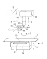
- 1 is a schematic diagram of an X-ray imaging apparatus according to the present invention.
- 1 is a perspective view of an X-ray tube support unit 4 and an imaging table 3 in an X-ray imaging apparatus according to the present invention.
- It is a block diagram which shows the main control systems of the X-ray imaging apparatus which concerns on this invention.
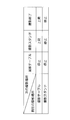
- It is a table
- FIG. 1 is a schematic diagram of an X-ray imaging apparatus according to the present invention.
- FIG. 2 is a perspective view of the imaging table 3 and the X-ray tube support 4 in the X-ray imaging apparatus according to the present invention.
- FIG. 3 is a block diagram showing a main control system of the X-ray imaging apparatus according to the present invention.
- the X-ray imaging apparatus performs imaging on the subject M and the console unit 1 and the high-voltage control device 2 installed in the operation room 101 for the operator to perform X-ray imaging operations.
- the imaging table 3, the imaging stand 5, and the X-ray tube support part 4 installed in the imaging room 100 are provided.
- the imaging room 100 and the operation room 101 are blocked by a partition wall 102.
- the console unit 1 installed in the operation room 101 has an operation panel 11 having a display unit composed of a touch panel type liquid crystal display. As shown in FIG. 3, the console unit 1 is related to the ascending control unit 3 for automatically raising the X-ray tube 42 supported by the X-ray tube support unit 4 and the X-ray imaging of the subject M. And a storage unit 14 for storing photographing data indicating information.
- the console unit 1 is connected to a radiology information system (RIS) 103 that is a system for managing examination reservations, examination states, medicines, etc., and examination statistics in the radiology department.
- the storage unit 14 stores imaging data acquired from the radiology information system 103.
- RIS radiology information system
- the imaging data indicating the information related to the X-ray imaging of the subject M stored in the storage unit 14 includes information on the technique of which one of the imaging table 3 and the imaging stand 5 is used, It includes patient information such as the name, sex, and age of the subject M, information on the imaging region for the subject M, or information on imaging conditions such as tube voltage and tube current.
- the high voltage control device 2 is disposed in the partition wall 102 in the operation chamber 101. As shown in FIG. 3, the high voltage control device 2 includes an operation panel 21 composed of a touch panel type liquid crystal display and a hand switch 23 for starting X-ray irradiation from the X-ray tube 42. Prepare. The high voltage control device 2 is for setting tube voltage and tube current of the X-ray tube 42 or X-ray irradiation conditions such as X-ray irradiation time. The high voltage control device 2 is controlled by the control unit 29 shown in FIG.
- the imaging table 3 is for imaging the subject M in a prone position, and houses a top plate 31 on which the subject M is placed and a flat panel detector 32 as an X-ray detector therein. And the elevating unit 34 for elevating and lowering the top plate 31 and the bucky unit 33.
- the bucky part 33 is horizontally movable in the G direction shown in FIG.
- the bucky portion 33 can be moved up and down in the F direction shown in FIG.
- the movement of the bucky unit 33 and the top plate 31 and other imaging operations in the imaging table 3 are controlled by the control unit 39.
- the photographing stand 5 is for photographing the subject M in a standing position, a screen 54 for raising the subject M on the front surface thereof, and a flat panel detector as an X-ray detector inside thereof. And a bucky part 53 in which 52 is accommodated.
- the bucky portion 53 can be moved up and down in FIG. The movement of the bucky unit 53 in the photographing stand 5 and other photographing operations are controlled by the control unit 59.
- the X-ray tube support unit 4 includes a support unit 61 that can move in the A direction and the B direction perpendicular to the ceiling of the imaging room 100, a support unit 62 that extends downward from the support unit 61, and the support unit 62.
- the moving unit 63 that moves up and down in the C direction and pivots in the D direction, and is pivotally supported by the lower end of the moving unit 63, and the operation unit 41, the X-ray tube 42, and the collimator mechanism 43 are integrated into the E direction.
- a support shaft 64 to be rotated.
- the X-ray tube 42 can move in the A, B, D, and E directions, and can move up and down in the C direction.
- the movement and lifting operation of the X-ray tube 42 in the X-ray tube support unit 4 is controlled by the control unit 49.
- the operation unit 41 is provided with an operation panel 44 composed of a touch panel type liquid crystal display.
- the subject M When X-ray imaging is performed by this X-ray imaging apparatus, the subject M is laid on the top plate 31 of the imaging table 3 at the time of lying-up imaging, and the screen on the imaging stand 5 is used at the time of standing imaging. The subject M is erected on the front surface of 54. Then, after moving the X-ray tube 42 and the like to a position where the subject M is sandwiched between the flat panel detector 32 or the flat panel detector 52, X-ray imaging is performed by irradiating X-rays.
- the X-ray imaging mode after performing X-ray imaging with the imaging table 3 is determined based on the imaging data stored in the storage unit 14, and the X-ray imaging is performed with the imaging table 3.
- the X-ray tube support unit is controlled by the ascending control unit 13. 4 adopts a configuration in which the X-ray tube 42 and the like are automatically raised.
- the prohibition switch 12 is displayed on the operation panel 11 of the console unit 1 and the prohibition switch 45 is displayed on the operation panel 44 of the X-ray tube support unit 4.
- the automatic elevation of the X-ray tube 42 and the like is prohibited.
- These rise prohibition switches 12 and 45 together with the rise control unit 13 function as a rise prohibition unit for prohibiting the lift operation of the X-ray tube 42.
- the storage unit 14 in the console unit 1 acquires and stores imaging data indicating information related to the X-ray imaging of the subject M from the radiology information system 103.
- the photographing data includes information on the technique that indicates which order of the photographing table 3 or the photographing stand 5 is used.
- the storage unit 14 stores X-ray imaging order and X-ray tube up / down information indicating when the X-ray tube 42 and the like should be raised in the form of a table, for example. .
- FIG. 4 is a table showing the lift information stored in the storage unit 14.
- the lift information shown in FIG. 4 is based on the X-ray tube 42 or the like when the X-ray imaging is performed at the next location where the X-ray imaging is performed when the X-ray imaging is performed at the imaging table 3.
- the ascending operation is stopped, and the X-ray tube 42 and the like are raised if the next place where X-ray imaging is to be performed is the imaging stand 5.
- the X-ray tube 42 or the like if the next place to perform X-ray imaging is the imaging table 3 when the X-ray imaging is currently performed at the imaging stand 5.
- the ascending operation is stopped and the ascending operation of the X-ray tube 42 or the like is stopped even if the next place where the X-ray imaging is performed is the imaging stand 5, and the X-ray imaging is not performed next. Even in this case, the ascending operation of the X-ray tube 42 or the like is stopped.
- the ascending control unit 13 in the console unit 1 determines whether or not the current X-ray imaging is performed on the imaging table 3 every time X-ray exposure from the X-ray tube 42 is completed during X-ray imaging. . In addition, every time the X-ray imaging is completed, the ascending control unit 13 in the console unit 1 has the ascending prohibition switch 12 on the operation panel 11 of the console unit 1 or the ascending prohibition switch 45 on the operation panel 44 of the X-ray tube support unit 4. Check if it is pressed.
- the ascent control unit 13 does not press the elevation prohibiting switches 12 and 45 and the next X-ray imaging is performed on the imaging stand 5.
- the ascending control unit 13 does not press the elevation prohibiting switches 12 and 45 and when all the X-ray imaging is completed,
- the X-ray tube 42 and the like are raised by the X-ray tube support portion 4.
- the X-ray tube 42 and the like can be automatically raised when the subject M gets off the top plate 31 of the imaging table 3. This eliminates the need for the operator to perform complicated operations. Further, when the X-ray exposure from the X-ray tube 42 is completed, the X-ray tube 42 and the like rise without waiting for the operator's operation, so that the subject M is promptly guided to the imaging stand 5. Alternatively, by quickly leaving the subject M from the imaging room 100, the X-ray imaging operation can be performed efficiently.
Landscapes
- Health & Medical Sciences (AREA)
- Engineering & Computer Science (AREA)
- Life Sciences & Earth Sciences (AREA)
- Medical Informatics (AREA)
- Optics & Photonics (AREA)
- Biomedical Technology (AREA)
- Biophysics (AREA)
- High Energy & Nuclear Physics (AREA)
- Robotics (AREA)
- Nuclear Medicine, Radiotherapy & Molecular Imaging (AREA)
- Automation & Control Theory (AREA)
- Pathology (AREA)
- Radiology & Medical Imaging (AREA)
- Physics & Mathematics (AREA)
- Heart & Thoracic Surgery (AREA)
- Molecular Biology (AREA)
- Surgery (AREA)
- Animal Behavior & Ethology (AREA)
- General Health & Medical Sciences (AREA)
- Public Health (AREA)
- Veterinary Medicine (AREA)
- Apparatus For Radiation Diagnosis (AREA)
Abstract
記憶部14に記憶された撮影データに基づいて撮影テーブル3でX線撮影行った後のX線撮影の形態を判定し、撮影テーブル3でX線撮影行った後に撮影スタンド5でX線撮影が行われる場合、あるいは、撮影テーブル3でX線撮影行った後にX線撮影を終了する場合には、上昇制御部13の制御によって、X線管支持部4によりX線管42等を自動的に上昇させる。ただし、コンソール部1の操作パネル11における上昇禁止スイッチ12またはX線管支持部4の操作パネル44における上昇禁止スイッチ45が押圧された場合には、X線管42の自動的な上昇を禁止する。
Description
この発明は、臥位状態の被検者を撮影するための撮影テーブルと、立位状態の被検者を撮影するための撮影スタンドとを備えたX線撮影装置に関する。
撮影テーブルは、天板上に臥位状態の被検者を載置した状態でX線撮影を行うものであり、臥位撮影台とも呼称される。また、撮影スタンドは、衝立の前面に起立した被検者のX線撮影を行うものであり、立位撮影台とも呼称される。このようなX線撮影装置は、撮影テーブルと、撮影スタンドと、これらの撮影テーブルおよび撮影スタンドに共通して使用されるX線管とを備えている。そして、X線管は、X線管支持部により、昇降、水平移動および回動可能に支持されており、撮影テーブルと撮影スタンドのうち、予め選択された術式に適合したものと対向する位置に移動する構成となっている。
臥位状態の被検者を撮影するための臥位撮影と、立位状態の被検者を撮影するための立位撮影とは、被検者における撮影部位や被検者の状態等に応じて使い分けられる。これらの立位撮影および臥位撮影の術式は、予め、病院内の情報共有のためのシステムである放射線科情報システム(RIS:Radiology Information System)を利用して取得した被検者の撮影データに基づいて選択される。ここで、被検者の撮影データは、上述した術式の情報以外に、被検者の氏名、性別および年齢等の患者情報と、被検者に対する撮影部位の情報と、管電圧および管電流等の撮影条件の情報を含んでいる。
ところで、撮影テーブルにおいて被検者の撮影を実行した後に、被検者を撮影テーブルにおける天板から降ろすときには、被検者の頭部とX線管等との衝突を防止するため、X線管を予め所定の高さまで上昇させておく必要がある。このようなX線管を上昇させるための操作は、従来、オペレータが撮影が終了するたびに行っていた。しかしながら、X線管を上昇させるのにはある程度の力が必要であり、また、X線管を上昇させる場合に、オペレータの足元には撮影テーブルが配置されていることから操作が行いにくいという問題がある。
これに対して、特許文献1には、オペレータがスイッチを操作することにより、X線管を退避移動させるようにしたX線撮影装置が開示されている。また、特許文献2には、撮影台の高さ位置が所定以下となった場合や、撮影台の降下時間または効果距離が一定以上となった場合には、X線管を上方に移動させるようにしたX線撮影装置が開示されている。
特許文献1または特許文献2に記載された構成を採用した場合においては、オペレータがX線管を手動で上昇させる必要がないことから、オペレータの負担を軽減させることは可能となる。しかしながら、オペレータは被検者に対する臥位撮影が終了するたびに、X線管の移動等の操作を実行する必要があることから、煩雑な作業を行う必要があることに変わりはなく、また、被検者を速やかに撮影スタンドに案内したり、被検者を撮影室から速やかに退室させることが不可能となる。
この発明は上記課題を解決するためになされたものであり、X線管を自動的に上昇させて効率的にX線撮影作業を実行することが可能なX線撮影装置を提供することを目的とする。
請求項1に記載の発明は、被検者に向けてX線を照射するX線管と、前記X線管を昇降可能に支持するX線管支持部と、臥位状態の被検者を撮影するための撮影テーブルと、立位状態の被検者を撮影するための撮影スタンドとを備えたX線撮影装置において、被検者のX線撮影に関する情報を示す撮影データを記憶する記憶部と、前記記憶部に記憶された撮影データに基づいて、撮影テーブルでX線撮影行った後のX線撮影の形態を判定し、撮影テーブルでX線撮影行った後に前記撮影スタンドでX線撮影が行われる場合、あるいは、撮影テーブルでX線撮影行った後にX線撮影を終了する場合に、前記撮影テーブルのX線撮影が終了した後に、前記X線管支持部により前記X線管を上昇させる上昇制御部と、を備えたことを特徴とする。
請求項2に記載の発明は、請求項1に記載の発明において、前記制御部は、前記撮影テーブルでX線撮影を行うための前記X線管からのX線の曝射が終了したときに、次のX線撮影が前記撮影スタンドで行われる場合には、前記X線管支持部により前記X線管を上昇させる。
請求項3に記載の発明は、請求項1に記載の発明において、前記制御部は、前記撮影テーブルでX線撮影を行うための前記X線管からのX線の曝射が終了したときに、全てのX線撮影が終了した場合には、前記X線管支持部により前記X線管を上昇させる。
請求項4に記載の発明は、請求項1から請求項3のいずれかに記載の発明において、前記X線管支持部による前記X線管の上昇動作を禁止するための上昇禁止手段を備える。
請求項5に記載の発明は、請求項1から請求項3のいずれかに記載の発明において、前記記憶部は、放射線科情報システムから取得した撮影データを記憶する。
請求項1に記載の発明によれば、被検者が撮影テーブルから降りるときに、X線管を自動的に上昇させることができることから、オペレータが煩雑な操作を行う必要がなくなる。また、オペレータの操作を待つことなくX線管が上昇することから、被検者を速やかに撮影スタンドに案内し、あるいは、被検者を撮影室から速やかに退室させることにより、効率的にX線撮影作業を実行することが可能となる。
請求項2および請求項3に記載の発明によれば、X線の曝射終了後に速やかにX線管を上昇させることが可能となる。
請求項4に記載の発明によれば、被検者の状況等を考慮し、必要に応じてX線管が自動的に上昇することを禁止することが可能となる。
請求項5に記載の発明によれば、放射線科情報システムから取得した撮影データを記憶することにより、撮影データを容易に取得することが可能となる。
以下、この発明の実施の形態を図面に基づいて説明する。図1は、この発明に係るX線撮影装置の概要図である。また、図2は、この発明に係るX線撮影装置における撮影テーブル3およびX線管支持部4の斜視図である。さらに、図3は、この発明に係るX線撮影装置の主要な制御系を示すブロック図である。
この発明に係るX線撮影装置は、操作者がX線撮影操作を実行するための操作室101に設置されたコンソール部1および高電圧制御装置2と、被検者Mに対して撮影を行うための撮影室100に設置された撮影テーブル3、撮影スタンド5およびX線管支持部4とを備える。撮影室100と操作室101とは、隔壁102により遮断されている。
操作室101に設置されたコンソール部1は、タッチパネル式の液晶表示器から構成される表示部を備えた操作パネル11を有する。図3に示すように、このコンソール部1は、X線管支持部4に支持されたX線管42を自動的の上昇させるための上昇制御部3と、被検者MのX線撮影に関する情報を示す撮影データを記憶する記憶部14とを備える。このコンソール部1は、放射線科部門の検査予約や検査状態、薬品等の管理、検査統計を管理するシステムである放射線科情報システム(RIS:Radiology Information System)103と接続されている。記憶部14は、放射線科情報システム103から取得した撮影データを記憶する。
なお、記憶部14に記憶される被検者MのX線撮影に関する情報を示す撮影データは、撮影テーブル3または撮影スタンド5のいずれをどのような順序で使用するかという術式の情報や、被検者Mの氏名、性別および年齢等の患者情報、被検者Mに対する撮影部位の情報、あるいは、管電圧および管電流等の撮影条件の情報を含んでいる。
高電圧制御装置2は、操作室101内において隔壁102に配設される。この高電圧制御装置2は、図3に示すように、タッチパネル式の液晶表示器から構成される操作パネル21と、X線管42からのX線の照射を開始するためのハンドスイッチ23とを備える。この高電圧制御装置2は、X線管42の管電圧や管電流、あるいは、X線照射時間等のX線の照射条件を設定するためのものである。この高電圧制御装置2は、図3に示す制御部29により制御される。
撮影テーブル3は、臥位状態の被検者Mを撮影するためのものであり、被検者Mを載置する天板31と、その内部にX線検出器としてのフラットパネルディテクタ32を収納したブッキー部33と、天板31およびブッキー部33を昇降させるための昇降部34とを備える。ブッキー部33は、図2に示すG方向に水平移動可能となっている。また、ブッキー部33は、天板31とともに、図2に示すF方向に昇降可能となっている。この撮影テーブル3におけるブッキー部33および天板31の移動やその他の撮影動作は、制御部39により制御される。
撮影スタンド5は、立位状態の被検者Mを撮影するためのものであり、その前面に被検者Mを起立させるための衝立54と、その内部にX線検出器としてのフラットパネルディテクタ52を収納したブッキー部53とを備える。ブッキー部53は、図1における上下方向に昇降可能となっている。この撮影スタンド5におけるブッキー部53の移動やその他の撮影動作は、制御部59により制御される。
X線管支持部4は、撮影室100の天井に対して互いに直交するA方向およびB方向に移動可能な支持部61と、この支持部61から下方に延びる支持部62と、この支持部62に対してC方向に昇降するともに、D方向に回動する移動部63と、この移動部63の下端部に軸支され、操作部41、X線管42およびコリメータ機構43を一体としてE方向に回動させる支持軸64とを備える。このため、X線管42は、A、B、D、E方向に移動可能であるとともに、C方向に昇降可能となっている。このX線管支持部4におけるX線管42の移動および昇降動作は、制御部49により制御される。なお、操作部41には、タッチパネル式の液晶表示器から構成される操作パネル44が付設されている。
このX線撮影装置により、X線撮影を行うときには、臥位撮影時には、撮影テーブル3の天板31上に被検者Mを横たわらせ、また、立位撮影時には、撮影スタンド5における衝立54の前面に被検者Mを起立させる。そして、フラットパネルディテクタ32またはフラットパネルディテクタ52との間に被検者Mを挟んだ対向位置にX線管42等を移動させた後に、X線を照射することによりX線撮影を実行する。
このようなX線撮影装置においてX線撮影を実行する場合に、撮影テーブル3において被検者Mの撮影を実行した後に、被検者Mを撮影テーブル3における天板31から降ろすときには、被検者Mの頭部とX線管42やコリメータ43等との衝突を防止するため、X線管42をコリメータ43等とともに、予め、所定の高さまで上昇させておく必要がある。
このため、このX線撮影装置においては、記憶部14に記憶された撮影データに基づいて撮影テーブル3でX線撮影行った後のX線撮影の形態を判定し、撮影テーブル3でX線撮影行った後に撮影スタンド5でX線撮影が行われる場合、あるいは、撮影テーブル3でX線撮影行った後にX線撮影を終了する場合には、上昇制御部13の制御によって、X線管支持部4によりX線管42等を自動的に上昇させる構成を採用している。
このとき、例えば、被検者Mに対して点滴がなされている場合等において、X線管42等が自動的に上昇した場合に支障が生ずる場合がある。このため、このX線撮影装置においては、コンソール部1の操作パネル11に対して上昇禁止スイッチ12を表示するとともに、X線管支持部4の操作パネル44に対して上昇禁止スイッチ45を表示させ、これらの上昇禁止スイッチ12、45が押圧された場合には、X線管42等の自動的な上昇を禁止している。これらの上昇禁止スイッチ12、45は、上昇制御部13とともに、X線管42の上昇動作を禁止するための上昇禁止手段として機能する。
この点について、より詳細に説明する。このX線撮影装置においては、最初に、コンソール部1における記憶部14が、放射線科情報システム103から、被検者MのX線撮影に関する情報を示す撮影データを取得して記憶する。この撮影データは、上述したように、撮影テーブル3または撮影スタンド5のいずれをどのような順序で使用するかという術式の情報を含んでいる。また、記憶部14には、X線撮影の順序と、どのような場合にX線管42等を上昇させるべきであるかというX線管昇降情報が、例えば、テーブルの形式で記憶されている。
図4は、記憶部14に記憶された昇降情報を示す表である。
図4に示す昇降情報は、現在X線撮影を実行している場所が撮影テーブル3であった場合において、次にX線撮影を実行する場所が撮影テーブル3であればX線管42等の上昇動作は停止し、次にX線撮影を実行する場所が撮影スタンド5であればX線管42等を上昇させ、次にX線撮影が行われない撮影終了の場合にはX線管42等を上昇させること、および、現在X線撮影を実行している場所が撮影スタンド5であった場合において、次にX線撮影を実行する場所が撮影テーブル3であればX線管42等の上昇動作は停止し、次にX線撮影を実行する場所が撮影スタンド5であってもX線管42等の上昇動作は停止し、次にX線撮影が行われない撮影終了の場合であってもX線管42等の上昇動作は停止することを示している。
コンソール部1における上昇制御部13は、X線撮影時にX線管42からのX線の曝射が終了するたびに、現在のX線撮影が撮影テーブル3で行われているか否かを判断する。また、コンソール部1における上昇制御部13は、X線撮影が終了するたびに、コンソール部1の操作パネル11における上昇禁止スイッチ12またはX線管支持部4の操作パネル44における上昇禁止スイッチ45が押圧されているか確認する。
そして、上昇制御部13は、撮影テーブル3においてX線撮影が行われていた場合において、上昇禁止スイッチ12、45が押圧されておらず、かつ、次のX線撮影が撮影スタンド5で行われる場合には、X線管42からのX線の曝射が終了したときに、X線管支持部4によりX線管42等を上昇させる。また、上昇制御部13は、撮影テーブル3においてX線撮影が行われていた場合において、上昇禁止スイッチ12、45が押圧されておらず、かつ、全てのX線撮影が終了した場合には、X線管42からのX線の曝射が終了したときに、X線管支持部4によりX線管42等を上昇させる。
このような構成を採用することにより、被検者Mが撮影テーブル3における天板31から降りるときに、X線管42等を自動的に上昇させることができる。このため、オペレータが煩雑な操作を行う必要がなくなる。また、X線管42からのX線の曝射が終了した時点で、オペレータの操作を待つことなくX線管42等が上昇することから、被検者Mを速やかに撮影スタンド5に案内し、あるいは、被検者Mを撮影室100から速やかに退室させることにより、効率的にX線撮影作業を実行することが可能となる。
1 コンソール部
2 高電圧制御装置
3 撮影テーブル
4 X線管支持部
5 撮影スタンド
11 操作パネル
12 上昇禁止スイッチ
13 上昇制御部
14 記憶部
21 操作パネル
23 ハンドスイッチ
29 制御部
31 天板
32 フラットパネルディテクタ
33 ブッキー部
34 昇降部
39 制御部
41 操作部
42 X線管
43 コリメータ
44 操作パネル
45 上昇禁止スイッチ
49 制御部
52 フラットパネルディテクタ
53 ブッキー部
54 衝立
100 撮影室
101 操作室
103 放射線科情報システム
M 被検者
2 高電圧制御装置
3 撮影テーブル
4 X線管支持部
5 撮影スタンド
11 操作パネル
12 上昇禁止スイッチ
13 上昇制御部
14 記憶部
21 操作パネル
23 ハンドスイッチ
29 制御部
31 天板
32 フラットパネルディテクタ
33 ブッキー部
34 昇降部
39 制御部
41 操作部
42 X線管
43 コリメータ
44 操作パネル
45 上昇禁止スイッチ
49 制御部
52 フラットパネルディテクタ
53 ブッキー部
54 衝立
100 撮影室
101 操作室
103 放射線科情報システム
M 被検者
Claims (5)
- 被検者に向けてX線を照射するX線管と、前記X線管を昇降可能に支持するX線管支持部と、臥位状態の被検者を撮影するための撮影テーブルと、立位状態の被検者を撮影するための撮影スタンドとを備えたX線撮影装置において、
被検者のX線撮影に関する情報を示す撮影データを記憶する記憶部と、
前記記憶部に記憶された撮影データに基づいて、撮影テーブルでX線撮影行った後のX線撮影の形態を判定し、撮影テーブルでX線撮影行った後に前記撮影スタンドでX線撮影が行われる場合、あるいは、撮影テーブルでX線撮影行った後にX線撮影を終了する場合に、前記撮影テーブルのX線撮影が終了した後に、前記X線管支持部により前記X線管を上昇させる上昇制御部と、
を備えたことを特徴とするX線撮影装置。 - 請求項1に記載のX線撮影装置において、
前記制御部は、前記撮影テーブルでX線撮影を行うための前記X線管からのX線の曝射が終了したときに、次のX線撮影が前記撮影スタンドで行われる場合には、前記X線管支持部により前記X線管を上昇させるX線撮影装置。 - 請求項1に記載のX線撮影装置において、
前記制御部は、前記撮影テーブルでX線撮影を行うための前記X線管からのX線の曝射が終了したときに、全てのX線撮影が終了した場合には、前記X線管支持部により前記X線管を上昇させるX線撮影装置。 - 請求項1から請求項3のいずれかに記載のX線撮影装置において、
前記X線管支持部による前記X線管の上昇動作を禁止するための上昇禁止手段を備えるX線撮影装置。 - 請求項1から請求項3のいずれかに記載のX線撮影装置において、
前記記憶部は、放射線科情報システムから取得した撮影データを記憶するX線撮影装置。
Priority Applications (1)
| Application Number | Priority Date | Filing Date | Title |
|---|---|---|---|
| PCT/JP2013/056457 WO2014136256A1 (ja) | 2013-03-08 | 2013-03-08 | X線撮影装置 |
Applications Claiming Priority (1)
| Application Number | Priority Date | Filing Date | Title |
|---|---|---|---|
| PCT/JP2013/056457 WO2014136256A1 (ja) | 2013-03-08 | 2013-03-08 | X線撮影装置 |
Publications (1)
| Publication Number | Publication Date |
|---|---|
| WO2014136256A1 true WO2014136256A1 (ja) | 2014-09-12 |
Family
ID=51490813
Family Applications (1)
| Application Number | Title | Priority Date | Filing Date |
|---|---|---|---|
| PCT/JP2013/056457 Ceased WO2014136256A1 (ja) | 2013-03-08 | 2013-03-08 | X線撮影装置 |
Country Status (1)
| Country | Link |
|---|---|
| WO (1) | WO2014136256A1 (ja) |
Citations (2)
| Publication number | Priority date | Publication date | Assignee | Title |
|---|---|---|---|---|
| JP2011030687A (ja) * | 2009-07-31 | 2011-02-17 | Fujifilm Corp | 放射線撮影システム、及び放射線撮影機器の制御装置 |
| JP2011104029A (ja) * | 2009-11-16 | 2011-06-02 | Shimadzu Corp | X線撮影装置 |
-
2013
- 2013-03-08 WO PCT/JP2013/056457 patent/WO2014136256A1/ja not_active Ceased
Patent Citations (2)
| Publication number | Priority date | Publication date | Assignee | Title |
|---|---|---|---|---|
| JP2011030687A (ja) * | 2009-07-31 | 2011-02-17 | Fujifilm Corp | 放射線撮影システム、及び放射線撮影機器の制御装置 |
| JP2011104029A (ja) * | 2009-11-16 | 2011-06-02 | Shimadzu Corp | X線撮影装置 |
Similar Documents
| Publication | Publication Date | Title |
|---|---|---|
| JP6056974B2 (ja) | X線撮影装置 | |
| RU2562013C1 (ru) | Универсальный рентгеновский комплекс | |
| US20100054423A1 (en) | X-ray diagnostic imaging apparatus and x-ray apparatus | |
| JP2019213583A (ja) | 近接操作式x線透視撮影装置 | |
| US9084582B2 (en) | Radiation imaging apparatus and method of controlling radiation imaging apparatus | |
| US20080240362A1 (en) | X-ray radiography apparatus and x-ray generator moving device | |
| JP5404508B2 (ja) | X線診断装置 | |
| JPWO2012070143A1 (ja) | X線検査装置 | |
| CN108366766B (zh) | 放射线摄影装置 | |
| JP6342151B2 (ja) | 移動型x線診断装置 | |
| JP6895727B2 (ja) | 寝台装置及びx線コンピュータ断層撮影装置 | |
| WO2014136256A1 (ja) | X線撮影装置 | |
| JP2017164426A (ja) | 放射線撮影装置 | |
| JP2019181188A (ja) | X線画像診断装置 | |
| JP6750746B2 (ja) | 放射線撮影装置 | |
| JP2010264054A (ja) | X線画像撮影装置 | |
| JP5999496B2 (ja) | 放射線撮影装置 | |
| CN108937977A (zh) | X射线透视装置 | |
| JP2009153579A (ja) | X線ct装置および医用撮像システム | |
| JP6385079B2 (ja) | 医用システムおよびコンピュータプログラム | |
| JP2015150054A (ja) | X線透視撮影台 | |
| WO2014041725A1 (ja) | 放射線撮影装置 | |
| JP3137467U (ja) | X線透視撮影装置 | |
| JP6460241B2 (ja) | X線撮影装置 | |
| CN112533538B (zh) | X射线摄影装置 |
Legal Events
| Date | Code | Title | Description |
|---|---|---|---|
| 121 | Ep: the epo has been informed by wipo that ep was designated in this application |
Ref document number: 13876894 Country of ref document: EP Kind code of ref document: A1 |
|
| NENP | Non-entry into the national phase |
Ref country code: DE |
|
| 122 | Ep: pct application non-entry in european phase |
Ref document number: 13876894 Country of ref document: EP Kind code of ref document: A1 |
|
| NENP | Non-entry into the national phase |
Ref country code: JP |