WO2014097762A1 - 高病原性トリインフルエンザに対する抗体 - Google Patents
高病原性トリインフルエンザに対する抗体 Download PDFInfo
- Publication number
- WO2014097762A1 WO2014097762A1 PCT/JP2013/080060 JP2013080060W WO2014097762A1 WO 2014097762 A1 WO2014097762 A1 WO 2014097762A1 JP 2013080060 W JP2013080060 W JP 2013080060W WO 2014097762 A1 WO2014097762 A1 WO 2014097762A1
- Authority
- WO
- WIPO (PCT)
- Prior art keywords
- antibody
- virus
- influenza
- clade
- strain
- Prior art date
- Legal status (The legal status is an assumption and is not a legal conclusion. Google has not performed a legal analysis and makes no representation as to the accuracy of the status listed.)
- Ceased
Links
Images



Classifications
-
- G—PHYSICS
- G01—MEASURING; TESTING
- G01N—INVESTIGATING OR ANALYSING MATERIALS BY DETERMINING THEIR CHEMICAL OR PHYSICAL PROPERTIES
- G01N33/00—Investigating or analysing materials by specific methods not covered by groups G01N1/00 - G01N31/00
- G01N33/48—Biological material, e.g. blood, urine; Haemocytometers
- G01N33/50—Chemical analysis of biological material, e.g. blood, urine; Testing involving biospecific ligand binding methods; Immunological testing
- G01N33/53—Immunoassay; Biospecific binding assay; Materials therefor
- G01N33/543—Immunoassay; Biospecific binding assay; Materials therefor with an insoluble carrier for immobilising immunochemicals
- G01N33/54366—Apparatus specially adapted for solid-phase testing
- G01N33/54373—Apparatus specially adapted for solid-phase testing involving physiochemical end-point determination, e.g. wave-guides, FETS, gratings
- G01N33/5438—Electrodes
-
- C—CHEMISTRY; METALLURGY
- C07—ORGANIC CHEMISTRY
- C07K—PEPTIDES
- C07K16/00—Immunoglobulins [IG], e.g. monoclonal or polyclonal antibodies
- C07K16/08—Immunoglobulins [IG], e.g. monoclonal or polyclonal antibodies against material from viruses
- C07K16/10—RNA viruses
- C07K16/108—Orthomyxoviridae (F), e.g. influenza virus
-
- C—CHEMISTRY; METALLURGY
- C07—ORGANIC CHEMISTRY
- C07K—PEPTIDES
- C07K2317/00—Immunoglobulins specific features
- C07K2317/70—Immunoglobulins specific features characterized by effect upon binding to a cell or to an antigen
- C07K2317/76—Antagonist effect on antigen, e.g. neutralization or inhibition of binding
Definitions
- the present invention relates to a specific and anti-affinity antibody against highly pathogenic avian influenza, in particular a monoclonal antibody having a dissociation constant of at least 1 ⁇ 10 ⁇ 10 M.
- H5N1 highly pathogenic influenza A
- H5N1 highly pathogenic influenza A
- Patent Document 1 7,879,326 (Patent Document 1)) produced by cell fusion by immunization with H5N1 antigen, and other monoclonal antibodies against highly pathogenic avian influenza (No. 4,625,485). Patent (Patent) Document 2)) there is.
- Non-patent Document 1 11 types of antibodies from Simons et al.
- the antibody of Foun et al. is an antibody using a peptide fragment of H5N1 as an immunogen, and the target epitope in this antibody is limited. The activity of the antibody is shown as a result of measurement by ELISA.
- An object of the present invention is to provide a specific and high affinity monoclonal antibody against highly pathogenic influenza.
- the present inventor prepared an antigen using a recombinant vaccinia virus and immunized the GANP (registered trademark) transgenic mouse to obtain a highly pathogenic avian bird.
- the inventors have found that monoclonal antibodies against influenza virus can be produced and have completed the present invention.
- the present invention is as follows. (1) An antibody against the HA protein of influenza virus having a dissociation constant of at least 1 ⁇ 10 ⁇ 10 M. (2) The antibody according to (1), wherein the dissociation constant is 1 ⁇ 10 ⁇ 10 M to 1 ⁇ 10 ⁇ 13 M. (3) The antibody according to (1), wherein the influenza virus is an influenza H5 subtype virus. (4) The antibody according to (3), wherein the influenza H5 subtype virus is influenza H5N1 virus.
- Influenza H5N1 virus is the Indogan Qinghai Lake strain (clade 2.2), Indonesia strain (clade 2.1), Anhui strain (clade 2.3), Vietnam strain (clade 1), Mongolian strain (A / whooper) (4) which is at least one selected from the group consisting of swan / Mongolia / 3/2005: clade 2.2) and Hokkaido strain (A / whooper swan / Hokaido / 1/2008: clade 2.3.2).
- the antibody according to (6) which is produced by a hybridoma having a deposit number of NITE BP-01216 or NITE BP-01217, or a reception number of NITE ABP-01737 or NITE ABP-01738.
- the antibody according to (1) wherein the antibody is a chimeric antibody, a humanized antibody or a humanized antibody.
- (11) A hybridoma that produces the monoclonal antibody according to (6).
- a hybridoma whose accession number is NITE BP-01216 or NITE BP-01217, or whose reception number is NITE ABP-01737 or NITE ABP-01738.
- the method for producing the antibody according to (1) comprising the following steps: (A) immunizing a non-human mammal with an influenza virus HA protein expressed using a recombinant vaccinia virus, and (b) collecting the antibody from the non-human mammal. .
- the method for producing a monoclonal antibody according to (6) or (7) comprising the following steps: (A) immunizing a non-human mammal with an influenza virus HA protein expressed using a recombinant vaccinia virus; (B) collecting antibody-producing cells from the immunized non-human mammal; (C) The method comprising: fusing the antibody-producing cells obtained in step (b) and myeloma cells; and (d) collecting the antibody from the fused cells obtained in step (c). (15) The method according to (13) or (14), wherein the non-human mammal is a GANP (registered trademark) transgenic non-human mammal.
- GANP registered trademark
- influenza virus characterized by detecting the influenza virus by contacting the antibody according to any one of (1) to (9) or the fragment according to (10) with a biological sample. Detection method.
- the influenza H5 subtype virus is influenza H5N1 virus.
- Influenza H5N1 virus has been identified as Indogan Qinghai Lake strain (clade 2.2), Indonesian strain (clade 2.1), Anhui strain (clade 2.3), Vietnam strain (clade 1), Mongolian strain (A / whooper) (18) which is at least one selected from the group consisting of swan / Mongolia / 3/2005: clade 2.2) and Hokkaido strain (A / whooper swan / Hokaido / 1/2008: clade 2.3.2). The method described. (20) A reagent for detecting influenza virus comprising the antibody according to any one of (1) to (9) or the fragment according to (10). (21) The reagent according to (20), wherein the influenza virus is an influenza H5 subtype virus.
- influenza H5 subtype virus is influenza H5N1 virus.
- Influenza H5N1 virus can be found in Indogan Qinghai Lake strain (clade 2.2), Indonesia strain (clade 2.1), Anhui strain (clade 2.3), Vietnam strain (clade 1), Mongolia strain (A / whooper) (22) which is at least one selected from the group consisting of swan / Mongolia / 3/2005: clade 2.2) and Hokkaido strain (A / whooper swan / Hokaido / 1/2008: clade 2.3.2). The reagent described.
- a pharmaceutical composition for anti-influenza virus comprising the antibody according to any one of (1) to (9) or the fragment according to (10).
- the pharmaceutical composition according to (24), wherein the influenza virus is an influenza H5 subtype virus.
- the pharmaceutical composition according to (25), wherein the influenza H5 subtype virus is influenza H5N1 virus.
- Influenza H5N1 virus has been identified as Indogan Qinghai Lake strain (clade 2.2), Indonesian strain (clade 2.1), Anhui strain (clade 2.3), Vietnamese strain (clade 1), Mongolian strain (A / whooper) (26) which is at least one selected from the group consisting of swan / Mongolia / 3/2005: clade 2.2) and Hokkaido strain (A / whooper swan / Hokaido / 1/2008: clade 2.3.2).
- the pharmaceutical composition as described.
- the present invention provides an antibody against a highly pathogenic avian influenza virus.
- the antibody of the present invention showed neutralizing activity against highly pathogenic avian influenza virus. Therefore, the antibody of the present invention is extremely useful as a reagent for detecting highly pathogenic avian influenza viruses or as a pharmaceutical composition for protecting against highly pathogenic avian influenza viruses.
- the present invention is a high affinity antibody against highly pathogenic avian influenza having a dissociation constant (KD) of at least 1 ⁇ 10 ⁇ 10 M, and a wide range of clade species, such as Indogan Qinghai Lake strain, Indonesian strain, Anhui strain , which can be detected or protected with a single antibody against Vietnamese, Mongolian and Hokkaido strains.
- Highly pathogenic avian influenza mediates chickens and is a highly lethal influenza that infects humans and has a different epidemic from seasonal influenza.
- infection is being controlled by international containment of highly pathogenic avian influenza and limited vaccine administration.
- the key to this strategy is to quickly discover and diagnose highly infected flu patients and to take effective treatments and take measures to prevent the spread of infection.
- hemagglutinin (HA) protein expressed using vaccinia virus is used as an antigen, and a highly pathogenic avian influenza is produced by using a high affinity monoclonal antibody production technique using GANP (registered trademark) transgenic mice.
- GANP registered trademark
- Viruses specifically and a wide range of clade species namely, Indangan Qinghai Lake strain (A / Bar-Headed goose / Qinghai / 1A / 05: clade 2.2), Indonesia strain (A / Indonesia / 05/2005: clade 2) .1), Anhui strain (A / Anhui / 1194/2005: clade 2.3), Vietnam strain (A / Vietnam / 1194/2004: clade 1), Mongolian strain (A / whooper swan / Mongolia / 3 / 005: clade 2.2), Hokkaido strain (A / whooper swan / Hokkaido / 1/2008: clade 2.3.2) are prepared with high affinity antibodies capable of detecting all with a single type of antibody. Succeeded.
- the antibody of the present invention is a molecule that binds to the HA protein via the heavy and light chain variable regions VH and VL.
- the antibodies of the present invention represent either polyclonal or monoclonal immunoglobulin molecules such as IgG, IgA, IgM, IgE, IgD, or subclasses such as IgG1, IgG2, IgG3 or IgG4.
- the antibody of the present invention also includes functional fragments or subsequences of immunoglobulin molecules such as Fab, Fab ′, F (ab ′) 2 , Fv, Fd, scFv and sdFv.
- the antigen used in the present invention is the HA protein of influenza virus.
- This protein can be expressed using vaccinia virus.
- vaccinia virus ⁇ Production of recombinant vaccinia virus having HA protein gene of influenza virus>
- the HA protein gene is registered with a predetermined accession number (Accession No.) in the GenBank database.
- accession number accession number (Accession No.) in the GenBank database.
- H5 subtypes for example, genes encoding HA proteins derived from influenza viruses belonging to each clade of H5N1, are registered in the GenBank accession number as follows.
- Clade 1 _A / Vietnam / 1109 / 2004_EF541040
- Clade 2.1 _A / Indonesia / 5 / 2005_EF541394
- Clade 2.2 _A / bar-headed goose / Qinghai / 1A / 2005_DQ659327
- Clade 2.3 _A / Anhui / 1 / 2005_DQ37171928
- a gene (cDNA) encoding the HA protein contained in the recombinant vaccinia virus of the present invention in addition to the above genes, a part thereof or a mutant sequence thereof can be used.
- DNA lacking part of the DNA encoding the HA protein of clade 1 DNA lacking part of the DNA encoding the HA protein of clade 2.1, and HA protein of clade 2.2
- a DNA in which a part of the DNA is deleted or a DNA in which a part of the DNA encoding the HA protein of clade 2.3 is deleted can be used.
- These HA protein genes have already been cloned and inserted into plasmids.
- HA protein gene in addition to the above HA protein gene (in the present specification, the full-length base sequence or the base sequence of the protein coding region.
- Genes can also be used. For example, it hybridizes under stringent conditions with a sequence consisting of a base sequence complementary to the base sequence shown in EF541040 and is complementary to the base sequence shown in EF541394, which encodes the HA protein of clade 1.
- Examples of stringent conditions include 0.1 ⁇ SSC to 10 ⁇ SSC, 0.1% to 1.0% SDS, and 20 ° C. to 80 ° C., and more specifically, 37 ° C. to 56 ° C.
- An example is a condition in which after pre-hybridization at 30 ° C. for 30 minutes or more, washing in 0.1 ⁇ SSC, 0.1% SDS for 10 to 20 minutes at room temperature is performed 1 to 3 times.
- DNA encoding the HA protein of Clade 2.1 the base sequence shown in DQ659327 and 50% or more, 60% or more, 70% or more, 80% or more, 90% or more, 95% or more, 98% or more, or DNA having a homology of 99% or more and encoding the HA protein of clade 2.2, 50% or more, 60% or more, 70% with the base sequence shown in DQ371728 Moreover, 80%, 90%, 95%, have a 98% or 99% or more homology, and it is possible using DNA encoding the
- the mutant gene is prepared, for example, using a site-directed displacement induction method described in “Molecular cloning: A Laboratory Manual (4th Edition)” or a mutation-introducing kit using a site-specific mutagenesis method. can do.
- mutagenesis kits include QuickChange TM Site-Directed Mutagenesis Kit (manufactured by Stratagene), GeneTaylor TM Site-Directed Mutagenesis System (manufactured by Invitrogen), TaKaRaectite-Site-Mit-Site-Mit-Site-Mit-Site-Mist
- Preferred examples include Mutan-Super Express Km and the like (manufactured by Takara Bio Inc.).
- the HA protein used as an antigen can be obtained by an ordinary genetic engineering technique based on the above gene information.
- the DNA encoding the HA protein is incorporated into a plasmid for homologous recombination and then introduced into an animal cell, followed by infecting the animal cell with the parent strain, vaccinia virus. After infection, the recombinant vaccinia virus may be cultured to express the HA protein.
- the HA protein can be expressed in large quantities in the form of complete sugar modification from the early stage to the late stage of vaccinia virus infection.
- a homologous recombination plasmid can include a promoter, a gene to be expressed, and the like.
- animal cells include rabbit kidney cells, monkey kidney cells, and human fibroblasts.
- the expressed HA protein can be purified by a known technique using a His tag or the like. The purification method is not limited, and affinity chromatography, HPLC purification, and the like are employed. Confirmation that the purified HA protein is the target can be performed, for example, by Western blotting, SDS-polyacrylamide gel electrophoresis, or the like.
- the majority of the HA protein expressed by the recombinant vaccinia virus forms a trimer similar to the HA protein on influenza virus particles and is used as an antigen.
- the protein produced as described above is immunized by administration to a non-human mammal by itself or together with a carrier and a diluent, and the antibody titer is measured.
- the type of non-human mammal to be immunized is not particularly limited, and examples include mice, rats, guinea pigs, rabbits, dogs, goats, etc., with mice being preferred.
- a transgenic non-human mammal called “GANP (registered trademark)” having a high-specificity antibody-producing ability (WO 2004/040971) as an animal to be immunized. Can also be used.
- GANP registered trademark
- transgenic non-human mammal is a non-human mammal into which a gene encoding a germinal center-associated nuclear protein has been introduced, and the animal is immunized with a predetermined antigen.
- it is an animal that can produce a high affinity or high specificity antibody against the antigen (WO 00/50611, Sakaguchi N. et al., J Immunol. 2005 Apr 15; 174 (8 ): 4485-94.).
- a GANP transgenic non-human mammal for example, a mouse
- the total dose of the antigen per animal is 10 to 2000 ⁇ g.
- an adjuvant and an antigen solution are generally mixed.
- the adjuvant include Freund's complete adjuvant (FCA), Freund's incomplete adjuvant (FIA), and aluminum hydroxide adjuvant.
- FCA Freund's complete adjuvant
- FIA Freund's incomplete adjuvant
- Aluminum hydroxide adjuvant examples include aluminum hydroxide adjuvant.
- Immunization is performed mainly by injecting intravenously, subcutaneously, intraperitoneally, intramuscularly, subcutaneously into the footpad, or the like.
- the immunization interval is not particularly limited, and immunization is performed 1 to 10 times, preferably 2 to 5 times at intervals of several days to several weeks, preferably 2 to 4 weeks.
- the antibody titer can be examined using blood collected from the immunized animal.
- the obtained serum is serially diluted, and the antibody titer is measured by enzyme immunoassay (ELISA (enzyme-linked immunosorbent assay) or EIA (enzyme immunoassay)), radioimmunoassay (RIA (radioimmunoassay)) and the like.
- ELISA enzyme immunoassay
- EIA enzyme immunoassay
- RIA radioimmunoassay
- an animal having a high antibody titer against the HA protein is subjected to final immunization.
- the immunity of the antigen and the measurement of the antibody titer are not limited to the above measurement methods.
- immunocompetent cells such as spleen cells
- regional lymph nodes are removed several days after the final immunization day, preferably 2 to 5 days later.
- the interval between blood collections is 1 to 4 weeks, preferably 1 to 2 weeks after immunization.
- polyclonal antibody when obtaining a polyclonal antibody, blood is collected on the day when the desired antibody titer is shown to obtain antiserum.
- antibody purification is required, purify by selecting a known method such as ammonium sulfate salting-out method, ion exchange chromatography, gel filtration chromatography, affinity chromatography, or a combination thereof. Can do. Thereafter, the reactivity of the polyclonal antibody in the antiserum is measured by an ELISA method or the like.
- the HANP transgenic non-human mammal is immunized with the HA protein, antibody-producing cells (for example, B cells) are collected from the immunized animal, and this antibody production is performed. Cells and myeloma cells are fused to produce a hybridoma (fusion cell line). And the target monoclonal antibody can be obtained by extract
- antibody-producing cells for example, B cells
- Antibody-producing cells are prepared from spleen cells or lymph node cells or spleen or lymph nodes of animals such as immunized non-human mammals. Even when a GANP transgenic non-human mammal is used, antibody-producing cells can be collected from spleen cells, lymph node cells, spleen, lymph nodes, or the like in the same manner as described above. Examples of lymph nodes include inguinal lymph nodes and mediastinal lymph nodes. Although it is not necessary to separate the antibody-producing cells from the collected cell population, it is desirable to separate only antibody-producing cells from the cell population. It is preferable to remove tissue debris and red blood cells as much as possible.
- myeloma cells to be fused with antibody-producing cells generally available cell lines of animals such as mice can be used.
- the cell line to be used is preferably one that has the property that it cannot survive in a HAT selection medium (medium containing hypoxanthine, aminopterin, and thymidine) and can survive only in a state fused with antibody-producing cells.
- HAT selection medium medium containing hypoxanthine, aminopterin, and thymidine
- myeloma cells include P3X63-Ag. 8).
- U1 (P3U1), P3 / NS I / 1-Ag4-1 (NS1), etc. are mentioned.
- Cell fusion is a commercially available medium such as DMEM or RPMI 1640 medium that does not contain fetal calf serum (FCS) and the like, and 1 ⁇ 10 6 to 1 ⁇ 10 7 cells / mL of spleen cells and / or lymph node cells.
- 1 ⁇ 10 5 to 1 ⁇ 10 6 cells / mL of myeloma cells are mixed (the ratio of spleen cells and / or lymph node cells to myeloma cells is preferably 5: 1), and in the presence of a cell fusion promoter.
- the cell fusion promoter polyethylene glycol having an average molecular weight of 200 to 20,000 daltons can be used.
- cells can be fused using Sendai virus.
- a person skilled in the art can fuse the antibody-producing cells and myeloma cells using a known cell fusion method. After the fusion, the cells are diluted with a HAT medium prepared with RPMI 1640 medium containing 10 to 20% FCS, for example, and then 0.5 to 3 ⁇ 10 5 cells are seeded in each well of a 96-well culture plate, and a CO 2 incubator. Incubate at
- a hybridoma producing the target antibody is selected from the cells after cell fusion treatment. Ten to 14 days after cell fusion, the cells selected in the HAT medium as described above form colonies. The culture supernatant of each well of the colony positive 96-well culture plate is collected and the antibody titer against HA is confirmed. As a confirmation method, an enzyme immunoassay (ELISA), a radioimmunoassay (RIA) or the like is performed. After confirming antibody production positive wells against HA, the cells are transferred to a 24-well or 12-well culture plate.
- ELISA enzyme immunoassay
- RIA radioimmunoassay
- the medium is preferably replaced with an HT medium (a medium containing hypoxanthine and thymidine) excluding aminopterin.
- HT medium is used as a recovery medium for hybridomas that continue to supply purine and pyrimidine precursors to the salvage pathway during the effect of residual aminopterin in the cells.
- the antibody titer in the culture supernatant is confirmed again. Since the hybridoma is unstable because it is a fused cell, and it is highly likely that the antibody producing ability is lost immediately, it is preferable to confirm the antibody titer for the second time.
- Cloning is performed to make the cells in the finally selected well into a single cell.
- the cell suspension is appropriately diluted with RPMI 1640 medium containing 10 to 20% FCS, and then seeded with 0.3 to 2 cells in each well of a 96-well culture plate.
- the number of cells in each well of the 96-well culture plate is preferably seeded so that there is one cell in each well so that there is a high probability of one cell in each well.
- the culture supernatant of the colony-positive well is collected 7 to 10 days later. At this time, it is preferable to confirm that it is a single colony after 3 to 5 days.
- the collected culture supernatant is checked for antibody titer.
- clones having high specificity for HA and low cross-reactivity for SARS S protein are selected.
- the number of cells in the selected well is increased to some extent to establish a hybridoma strain. Cloning may be performed several times as necessary.
- HA-specific monoclonal antibody is purified and collected from the established hybridoma strain by the following method. That is, a method of preparing an antibody from a culture supernatant cultured in a medium with reduced serum concentration, a method of preparing an antibody from a culture supernatant cultured in a commercially available serum-free medium, or injecting a hybridoma into the abdominal cavity of an animal. There is a method of collecting ascites and preparing antibodies from the ascites. The culture supernatant is collected from cells prepared at 0.1 to 4 ⁇ 10 5 cells / mL and cultured for 1 to 2 weeks.
- hybridomas are administered into the abdominal cavity of a myeloma cell-derived mammal and the same type of animal, and the hybridomas are proliferated in large quantities. Ascites fluid is collected after 1 to 2 weeks.
- Examples of the culture method include a method using a culture flask, a method using a spinner flask, a method using a shaker flask, and a method using a bioreactor.
- Antibody purification methods include HA affinity column purification method, ammonium sulfate salting-out fraction purification method by gel filtration chromatography, ion exchange chromatography purification method, etc., selecting these known methods as appropriate, Or it can refine
- the cell lines (hybridomas) producing the monoclonal antibodies of the present invention are referred to as “3E10 (rVV cl2.2)” and “7G10 (rVV cl2.2)”, both of which are independent administrative corporation products. Deposited internationally based on the Budapest Treaty at the Japan Institute for Evaluation Technology Patent Microorganisms Deposit Center (2-5-8 KITESA Kamashita, Kisarazu City, Chiba Prefecture 292-0818) based on the Budapest Treaty (original deposit date: February 2012) 1 day).
- accession numbers are NITE BP-01216 for “3E10 (rVV cl2.2)” and NITE BP-01217 for “7G10 (rVV cl2.2)”.
- the cell lines (hybridomas) that produce the monoclonal antibodies of the present invention are also referred to as “8C1 (rVV cl2.2)” and “14A7 (rVV cl2.2)”, both of which are listed in the Budapest Microorganism Depositary Center. (The original deposit date: October 18, 2013).
- the receipt number is NITE ABP-01737 for “8C1 (rVV cl2.2)” and NITE ABP-01738 for “14A7 (rVV cl2.2)”.
- the antibody of the present invention specifically binds to HA and exhibits high affinity. “Specific” means that it does not cross-react with subtypes other than the HA protein subtype used for immunization, and reacts only with the subtype protein used for immunization (binding / neutralization). For example, when H5N1 HA protein is used as an antigen as a subtype, the antibody of the present invention only reacts (binding / neutralizes) with H5 subtype protein or H5 subtype virus, and other H1N1, H1N2, etc. This means that there is no cross-reactivity with the subtype of.
- binding affinity can be determined by association constant (KA) and dissociation constant (KD).
- KD affinity equilibrium constant
- Its binding affinity can be detected as follows.
- the binding affinity has a dissociation constant (KD) of at least 1 ⁇ 10 ⁇ 10 M.
- KD dissociation constant
- the dissociation constant is 2 to 5 times, 5 to 10 times, 10 to 100 times, 100 to 1000 times.
- it has an affinity 1000 to 10,000 times higher.
- the dissociation constant for the binding affinity of the HA protein is, 1 ⁇ 10 -10 M, 5 ⁇ 10 -11 M, 1 ⁇ 10 -11 M, 5 ⁇ 10 - 12 M, 1 ⁇ 10 ⁇ 12 M, 5 ⁇ 10 ⁇ 13 M, 1 ⁇ 10 ⁇ 13 M, 5 ⁇ 10 ⁇ 14 M, 1 ⁇ 10 ⁇ 14 M, 5 ⁇ 10 ⁇ 15 M, or 1 ⁇ 10 ⁇ 15 M, and more preferably 1 ⁇ 10 ⁇ 10 M to 1 ⁇ 10 ⁇ 13 M.
- the value may be lower than these KDs and may have high affinity.
- the dissociation constant (KD) of the antibody to be measured for affinity is within about 1 to 100 times the KD of the antibody of the present invention, the antibody is substantially the same as the antibody of the present invention. Is included in the present invention.
- the association constant (KA) and dissociation constant (KD) can be measured using surface plasmon resonance (SPR), and it is possible to employ a known device and method for detecting the binding rate in real time and further monitoring it.
- SPR surface plasmon resonance
- Biacore registered trademark
- T200 GE Healthcare
- ProteON XPR36 Bio-Rad
- the KD value of a typical antibody as defined herein is sufficient to saturate half (50%) of the binding site of the HA antigen of the immobilized purified HA protein (A / Anhui / 1/2005). Defined as the concentration of antibody required.
- the antibody of the present invention can specifically bind to multiple types of virus strains. Examples of the virus strains to be bound include A / whooper swan / Monglia / 03/2005, A / whooper swan / Hokaido / 1/2008, A / duck / Hokaido / Vac-3 / 2007.
- the antibody fragment of the present invention is an antibody against HA and has the above KD value.
- the antibody fragment means a partial region of the antibody of the present invention.
- Fab, Fab ′, F (ab ′) 2 Fv, diabodies (dibodies), dsFv (disulphide-stabilized Fv), scFv (single chain Fv) and the like.
- the antibody fragment can be obtained by cleaving the antibody of the present invention with various proteolytic enzymes according to the purpose.
- Fab can be obtained by treating antibody molecules with papain, and F (ab ′) 2 can be obtained by treating antibody molecules with pepsin.
- Fab ′ can be obtained by cleaving the disulfide bond in the hinge region of F (ab ′) 2 .
- scFv cDNA encoding the H chain V region and L chain V region of the antibody is obtained, and DNA encoding scFv is constructed. By inserting this DNA into an expression vector and introducing the expression vector into a host organism for expression, scFv can be produced.
- diabody cDNA encoding the H chain V region and L chain V region of the antibody is obtained, and a DNA encoding scFv is constructed so that the length of the amino acid sequence of the peptide linker is 8 residues or less.
- a diabody can be produced by inserting this DNA into an expression vector and introducing the expression vector into a host organism for expression.
- dsFv cDNA encoding the H chain V region and L chain V region of the antibody is obtained, and a DNA encoding dsFv is constructed. By inserting this DNA into an expression vector and introducing the expression vector into a host organism for expression, dsFv can be produced.
- One preferred embodiment of the antibody of the present invention is a recombinant antibody.
- the recombinant antibody include, but are not limited to, chimeric antibodies, humanized antibodies, and humanized antibodies.
- human antibodies or consensus residues thereof, or amino acid residues present in CDR regions are known (Kabat, Sequences of Proteins of Immunological Interest, 4th Ed., US Department of Health and Human. Services.Public Health Service (1987)).
- human antibodies include antibodies in which one or more amino acid residues are replaced with amino acids present in any of the other human antibodies.
- CDR grafting refers to transplanting a complementarity determining region (CDR) from a variable region of a mouse antibody into a human variable region, wherein the framework region (FR) is derived from a human and the CDR is derived from a mouse. This is a method for producing a configured variable region. These humanized reshaped human variable regions are then linked to human constant regions. Methods for producing such humanized antibodies are well known in the art (see, for example, Nature, 321, 522-525 (1986); J. Mol. Biol., 196, 901-917 (1987)). .
- Humanized antibodies (fully human antibodies) generally have the same structure as that of a human antibody in the hypervariable region, which is the antigen-binding site of the V region, the rest of the V region, and the constant region. It is. However, the hypervariable site may be derived from another animal. Techniques for producing humanized antibodies are also known, and methods for producing gene sequences common to humans by genetic engineering techniques have been established.
- the humanized antibody can be obtained by, for example, a method using a human antibody-producing mouse having a human chromosome fragment containing H and L chain genes of human antibody (Tomizuka, K. et al., Nat. Genet., (1977) 16). 133-143; Kuroiwa, Y.
- the hybridoma of the present invention for example, a hybridoma whose accession number is NITE BP-01216 or NITE BP-01217, or whose reception number is NITE ABP-01737 or NITE ABP-01738), or extracted from the hybridoma Chimeric antibodies, humanized antibodies, and humanized antibodies can be prepared using DNA or RNA as a raw material according to the well-known methods described above.
- the anti-HA antibody of the present invention includes, for example, an antibody that binds to a site (for example, an epitope) to which the antibody of the present invention binds (recognizes), preferably the accession number is NITE BP- Examples include an antibody that binds to a site (eg, an epitope) to which a monoclonal antibody produced by a hybridoma of 01216 or NITE BP-01217 or a receipt number of NITE ABP-01737 or NITE ABP-01738 binds.
- the site to which the anti-HA antibody of the present invention binds is not limited as long as it is at least a part of the antigen HA.
- the present invention provides a method for detecting influenza virus, which comprises detecting an influenza virus by bringing the antibody or fragment thereof of the present invention into contact with a biological sample.
- a biological sample to be examined is collected from a subject such as an influenza patient, a patient suspected of influenza, or a health checkup examinee, and an HA measurement sample is prepared.
- the influenza virus to be detected is an influenza A virus or subtype, and examples include Indogan Qinghai Lake strain, Indonesia strain, Anhui strain, and Vietnam strain.
- Subtypes include, for example, H1N1, H1N2, H2N1, H2N2, H2N3, H3N2, H3N8, H4N6, H4N8, H5N1, H5N2, H5N3, H5N5, H5N8, H6N1, H6N2, H7N1, H7N9, H7N1, H7N9 H10N3, H11N1, H11N2, H11N9, H12N5, H13N6, H13N8, H15N8 or H16N3, and one or more of these are selected.
- an influenza H5 subtype influenza virus for example, H5N1 is preferable.
- the biological sample examples include blood, sputum, saliva, pharyngeal swab, nasal discharge, nasal discharge, and nasal aspirate.
- the antibody is contacted with a biological sample.
- Contact means that the antibody of the present invention or a fragment thereof (also referred to as an antibody or the like) and a biological sample to be tested are placed in the same environment so that they can be bound or reacted. For example, adding a biological sample to a container (well) containing an antibody or the like, adding an antibody or the like to a container (well) containing a biological sample, or culturing the antibody or the like and the biological sample in the same container It means to do.
- the measurement of HA can be performed by ELISA or EIA that is generally performed, and is not particularly limited.
- the antibody of the present invention that specifically binds to this amino acid sequence detects H5N1 influenza virus. Is particularly preferred. In the case of detection by ELISA, when the signal intensity is stronger than that of the negative control, it can be determined that influenza virus is contained in the test sample (ie, positive).
- kits and Reagents Containing Antibodies of the Present Invention antibodies against HA can be used as reagents or kits for detecting influenza viruses.
- the antibody of the present invention for example, a monoclonal antibody
- the monoclonal antibody can be combined with another solvent or solute to form a composition.
- distilled water, pH buffer reagent, salt, protein, surfactant and the like can be combined.
- Monoclonal antibodies can also be enzyme-labeled.
- labeling enzyme in addition to HRP (horseradish peroxidase), alkaline phosphatase, malate dehydrase, ⁇ -glucosidase, ⁇ -galactosidase, colloidal gold and the like can be used.
- the present invention when used in a kit, in addition to the antibody of the present invention, the above solvent, solute, enzyme labeling reagent, antigen-immobilized microplate, antibody dilution solution, OPD (orthophenylenediamine) tablet, substrate solution, A reaction stop solution, a concentrated washing solution, instructions for use, and the like can be included.
- a reaction medium such as a buffer solution that gives optimum conditions for the reaction, a buffer solution that is useful for stabilizing the reaction product, and a stabilizer for the reaction material may be included in the kit of the present invention.
- a PCR method can also be employed to detect HA mRNA or DNA.
- Primers used for PCR can also be included in the reagents or kits of the present invention.
- the region on which the primer is designed is not particularly limited as long as the HA protein can be detected.
- the length of the primer is 18 to 24 bases, preferably 20 to 22 bases. The following can be illustrated as a base sequence of a primer.
- the pharmaceutical composition of the present invention contains the antibody of the present invention as an active ingredient, is used for anti-influenza virus, and is effective for the prevention or treatment of influenza.
- influenza virus that is the target of prevention or treatment of influenza is an influenza A virus or subtype, and examples include Indogan Qinghai Lake strain, Indonesia strain, Anhui strain, and Vietnam strain.
- Subtypes are, for example, H1N1, H1N2, H2N1, H2N2, H2N3, H3N2, H3N8, H4N6, H4N8, H5N1, H5N2, H5N3, H5N5, H5N8, H6N1, H6N2, H7N1, H7N1, H7N1, H7N2, H7N1, H7N2, H7N , H11N1, H11N2, H11N9, H12N5, H13N6, H13N8, H15N8 or H16N3 are selected from one or more.
- H5 subtype influenza virus for example, H5N1 is preferable.
- the antibody of the present invention can prevent a subject from being infected with one or more influenza viruses (isolate or subtype) or reduce the sensitivity of the subject to the infection. Also, one or more symptoms or complications associated with influenza infection, such as fatigue, chills, fever, body pain, headache, cough, sore throat, nasal congestion, sinus closure, pneumonia, bronchitis, ear infection, ear Reduce or eliminate pain, etc., or prevent criticism of these symptoms. Furthermore, it is possible to shorten the period of suffering from these symptoms.
- the antibody of the present invention can be administered alone or together with a pharmaceutically acceptable carrier or diluent as a pharmaceutical composition, and the administration can be performed once or divided into several times.
- pharmaceutically acceptable carrier refers to excipients, diluents, extenders, disintegrants, stabilizers, preservatives, buffers, emulsifiers, fragrances, colorants, sweeteners, thickeners. , Flavoring agents, solubilizers or other additives.
- a composition can be prepared. These pharmaceutical compositions can be administered orally or parenterally.
- various excipients such as microcrystalline cellulose, sodium citrate, calcium carbonate, dipotassium phosphate and glycine can be used together with a disintegrant, a binder and the like.
- the disintegrant include starch, alginic acid, and certain types of silicate double salts.
- the binder include polyvinylpyrrolidone, sucrose, gelatin, and gum arabic.
- lubricants such as magnesium stearate, sodium lauryl sulfate, and talc are very effective for tablet formation.
- an emulsifier and a suspending agent may be used in combination, if necessary, and used with water, ethanol, propylene glycol, glycerin, etc., and diluents that combine them.
- Other forms for parenteral administration include injections that contain one or more active substances and are prescribed by conventional methods.
- injections for example, by dissolving or suspending in an pharmaceutically acceptable carrier such as physiological saline or commercially available distilled water for injection to an antibody concentration of 1 ⁇ g / ml to 100 mg / ml.
- an pharmaceutically acceptable carrier such as physiological saline or commercially available distilled water
- the injection thus produced is a ratio of 1 ⁇ g / kg to 100 mg / kg, preferably a ratio of 100 ⁇ g / kg to 15 mg / kg, in a single administration to a subject (eg, human influenza patient). And can be administered once to several times per day.
- the dosage is not limited to this range, and may vary depending on the weight and symptoms of the patient and individual administration routes.
- the dose may vary depending on the sensitivity of the patient to be treated, the way the drug is prescribed, the period of administration, and the interval between doses. In some cases, a dose lower than the lower limit of the above range may be appropriate. .
- injections can be prepared as non-aqueous diluents (for example, propylene glycol, polyethylene glycol, vegetable oils such as olive oil, alcohols such as ethanol), suspensions, or emulsions.
- non-aqueous diluents for example, propylene glycol, polyethylene glycol, vegetable oils such as olive oil, alcohols such as ethanol
- suspensions or emulsions.
- sterilization of the injection can be performed by filtration sterilization through a bacteria-retaining filter, blending of a bactericide, or irradiation.
- the injection can be produced as a form prepared at the time of use. That is, it can be used as a sterile solid or powder composition by lyophilization, etc., and dissolved in sterile water for injection or other solvent before use.
- the present invention will be described more specifically with reference to examples. However, the present invention is not limited to these examples.
- H5N1 antigen was prepared by recombinant vaccinia virus (rVV).
- rVV recombinant vaccinia virus
- a recombinant plasmid in which the H5N1 influenza HA gene was introduced into the homologous recombination vector was introduced into rabbit kidney cells RK13 and infected with the parent strain vaccinia virus to obtain a recombinant vaccinia virus (rVV) expressing HA.
- purification of His-Tag protein by a conventional method using TARON column chromatography was performed.
- the HA protein gene of H5N1 HPAIV has already been cloned and inserted into a plasmid.
- the HA protein gene of H5N1 HPAIV HA gene (GenBank accession numbers: EF541040, EF541394, DQ65937, DQ371937) inserted into the plasmid was used as a PCR template. That is, H5N1 HPAIV HA protein gene region was prepared by performing PCR using each gene-specific primer using cDNA of H5N1 HPAIV HA protein gene as a template. The resulting fragment was cloned into the plasmid pBMSF7C (Arch. Virol. Vol. 138, p.
- the expression promoter contained in the recombinant vaccinia virus of the present invention is inserted into the hemagglutinin (HA) gene region of vaccinia virus, and includes a poxvirus A type inclusion body (ATI) promoter and a plurality of repetitive vaccinia viruses.
- HA hemagglutinin
- ATI poxvirus A type inclusion body
- a recombinant vaccinia virus was produced by introducing the plasmid vector into a host vaccinia virus.
- the plasmid vector was introduced into the host by introducing plasmid vectors pBMSF7C-mCl1, pBMSF7C-mCl2.1, pBMSF7C-mCl2.2, and pBMSF7C-mCl2.3 into animal cells infected with the attenuated vaccinia virus LC16m8 strain, respectively.
- Recombinant vaccinia viruses (RVV-Flu HA (H5-mCl1), RVV-Flu HA (H5-mCl2), which cause homologous recombination in the hemagglutinin (HA) gene region of vaccinia virus and express the HA protein gene of H5N1 HPAIV, respectively.
- RVV-Flu HA H5-mCl2.2
- RVV-Flu HA H5-mCl2.3
- the HA protein was expressed by infecting the produced recombinant vaccinia virus into RK-13 cells, which are a rabbit kidney cell line. Subsequently, purification of HA protein to which His-Tag was added by a conventional method using TARON column chromatography was performed.
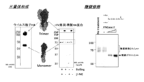
- HA protein The purification results of HA protein (Western blotting and CBB staining) are shown in FIG.
- the obtained purified HA protein was designated as “H5mcl2.2 antigen”.
- FIG. 2 the left panel is a diagram showing the HA protein constituting the influenza virus particle, the middle panel is a diagram showing the purified HA protein expressed by rVV, and the right panel is the diagram showing the purified HA protein. It is a figure which shows having received sugar chain modification.
- the purified protein yield was 0.5-2 mg.
- Mouse thymocytes of 2 ⁇ 10 5 cells / well were used as feeder cells, and the feeder cells and recombinant IL-6 (0.25 unit / ml) were added to the wells and cultured.
- HAT hypoxanthine, aminopterin, thymidine
- Cloning was performed according to a conventional method, and cell culture was performed in the presence of recombinant IL-6 0.25 unit / ml in 96 wells in the presence of 4 ⁇ 10 5 mouse thymocytes feeder cells. A subclone having the same activity as that of the original cell was established and stored in the proliferating clone, and used for the subsequent analysis. The above operation was performed using RPMI-1640, 10% -heat-immobilized fetal bovine serum, and HT (hypoxanthine, thymidine) medium. The obtained clones are shown in Table 1.
- clones indicated as “3E10”, “7G10”, “8C1”, and “14A7” represent deposit clones having the following deposit numbers or receipt numbers, respectively.
- Table 6 shows the evaluation results of the crossability using the candidate immunochromatography kit.
- Labeling method 0.3 mg of antibody was mixed with 1.3 ml of 0.4% fluorescent bead suspension and allowed to react at room temperature for 1 hour. After discarding the supernatant by centrifugation, 1% BSA solution was added and blocked for 1 hour. After discarding the supernatant by centrifugation, it was washed with 0.1% Trion X-100 solution at pH 7.7, suspended in 1% BSA solution at pH 7.7, and incubated at 37 ° C. for 48 hours. The fluorescent bead labeled antibody thus produced was used in place of the colloidal gold labeled antibody.
- influenza virus H5N1 (1 ⁇ 10 3 PFU / ml) virus solution diluted with MEM medium containing 1% BSA and 0.1 ml of hybridoma culture supernatant diluted 100 times, 1000 times, and 10000 times Were mixed and neutralized at 37 ° C. for 1 hour.
- 0.1 ml of a mixture of influenza virus solution and hybridoma culture supernatant was added to each well and spread throughout.
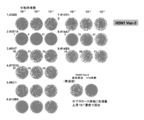
- FIG. 3 shows a photograph of the plate after culture. Compared to the negative control (no addition), 3E10, 10B9, 14A7 and 3B9 showed a clear difference in plaque size and number. The amount of virus used was confirmed by performing back titration (no addition), but it was a predetermined amount of virus.
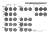
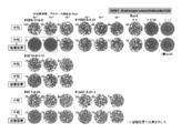
- FIG. 4 The results are shown in FIG. In FIG. 4, the numbers in the upper left of each plate indicate the number of plaques when the hybridoma culture supernatant (No. 1 to 4) is diluted 10 ⁇ 2 , 10 ⁇ 3 , and 10 ⁇ 4 .
- the control group was Back titration (Back), and the virus solution used for neutralization value measurement was added in stock solution (1 time) and diluted 10 times.
- Relenza (GlaxoSmithKline) was added at concentrations of 1 ⁇ M, 0.1 ⁇ M, and 0.01 ⁇ M, and it was confirmed that the appearance of plaques was possible. As shown in FIG.
- 4 animals / group were necropsied 3 days after the infection, and 4 animals / group were necropsied at the end of the experiment.
- Non-infected (diluted solution) inoculated group (4 mice per group) was used for monitoring body weight change.
- the antibody amount was 15 mg / kg / mouse, and were administered to Day 0 (immediately before virus infection) and Day 2 from the orbital vein nest. The method is shown in FIG.
- FIG. 6 Panel B shows the change in body weight after challenge infection with influenza virus.
- ⁇ 14A7-3-21-1 clone
- ⁇ 14A7-3-21-1 clone
- ⁇ 14A7-3-21-1 clone
- ⁇ 14A7-3-21-1 clone
- ⁇ Vehicle
- ⁇ no decrease in body weight
- the infected mouse group administered with 14A7-3-21-1 clone ( ⁇ ) had a survival rate of 25%, and the infected mouse group administered with Vehicle ( ⁇ ) had a survival rate of 50%.
- the infected mouse group ( ⁇ ) and the non-infected mouse group ( ⁇ ) to which the 3B9-11-6-9 clone was administered no decrease in the survival rate was observed. From the above results, the 3B9-11-6-9 clone having a wide binding ability to H5N1 highly pathogenic avian influenza virus showed a remarkable therapeutic effect even against highly pathogenic avian influenza virus infection.
- H3N1 A / whooper swan / Hokaido in 3B9-11-6-9 clone / 1/08 strain showed a therapeutic effect on highly pathogenic avian influenza virus.
- the antibody was administered to examine the therapeutic effect on mice (in vivo test).
- FIG. 7 is a diagram showing the results of studying the therapeutic effect of anti-HA monoclonal antibody administration (BALB / crice (Female, 9-week-old), H5N1 HPAIV 300 PFU, 15 mg / kg 3B9 administration group).
- FIG. 7 panel A shows the change in body weight after challenge infection with influenza virus. In the infected mouse group ( ⁇ ) in which the 3B9-11-6-9 clone was administered to Day 8 and the non-administered mouse group ( ⁇ ), significant weight loss was observed after 6 days of infection.
- FIG. 7 panel B shows the change in survival rate after challenge infection. On the 14th day of infection, an infected mouse group ( ⁇ ) administered to the infected mouse group Day0 ( ⁇ ), Day4 ((), Day6 ( ⁇ ), and Day8 administered with the 3B9-11-6-9 clone is shown.
- the survival rates were Day 4 (() 75%, Day 6 (() 50%, and Day 8 ( ⁇ ) 0%.
- the infected mouse group ( ⁇ ) and the non-infected mouse group ( ⁇ ) in which the 3B9-11-6-9 clone was administered to Day 0 ( ⁇ ) no decrease in the survival rate was observed. From the above results, the 3B9-11-6-9 clone having a wide binding ability to H5N1 highly pathogenic avian influenza virus shows a remarkable therapeutic effect even after administration after infection with highly pathogenic avian influenza virus. It was.
- the antibody of the present invention makes it possible to produce an ultrasensitive diagnostic kit. Further, since it is effective not only for detection of viruses of various clades but also for inhibition of virus growth, it is extremely useful for medical and clinical examinations.
- hybridomas “3E10 (rVV cl2.2)” and “7G10 (rVV cl2.2)” are both registered by the National Institute of Technology and Evaluation for Microorganisms (Kazusa Kamashizu, Kisarazu City, Chiba Prefecture 292-0818). -5-8 Deposited internationally at NITE Biotechnology Headquarters (Patent Microorganism Deposit Center) based on the Budapest Treaty (original deposit date: February 1, 2012). The accession number is NITE BP-01216 for “3E10 (rVV cl2.2)” and NITE BP-01217 for “7G10 (rVV cl2.2)”.
- Hybridomas “8C1 (rVV cl2.2)” and “14A7 (rVV cl2.2)” were both internationally deposited at the above-mentioned patent microorganism deposit center based on the Budapest Treaty (original deposit date: October 18, 2013) .
- the receipt number is NITE ABP-01737 for “8C1 (rVV cl2.2)” and NITE ABP-01738 for “14A7 (rVV cl2.2)”.
- SEQ ID NO: 1 synthetic DNA
- SEQ ID NO: 2 Synthetic DNA
Landscapes
- Health & Medical Sciences (AREA)
- Life Sciences & Earth Sciences (AREA)
- Immunology (AREA)
- Chemical & Material Sciences (AREA)
- Molecular Biology (AREA)
- Engineering & Computer Science (AREA)
- Biomedical Technology (AREA)
- Hematology (AREA)
- Urology & Nephrology (AREA)
- Medicinal Chemistry (AREA)
- General Health & Medical Sciences (AREA)
- Virology (AREA)
- Organic Chemistry (AREA)
- Biochemistry (AREA)
- Food Science & Technology (AREA)
- Pathology (AREA)
- Cell Biology (AREA)
- Biophysics (AREA)
- Microbiology (AREA)
- Genetics & Genomics (AREA)
- Biotechnology (AREA)
- Analytical Chemistry (AREA)
- Proteomics, Peptides & Aminoacids (AREA)
- General Physics & Mathematics (AREA)
- Physics & Mathematics (AREA)
- Preparation Of Compounds By Using Micro-Organisms (AREA)
- Peptides Or Proteins (AREA)
- Medicines Containing Antibodies Or Antigens For Use As Internal Diagnostic Agents (AREA)
- Micro-Organisms Or Cultivation Processes Thereof (AREA)
Abstract
本発明は、少なくとも1×10⁻¹⁰Mの解離定数を有する、インフルエンザウイルスのHAタンパク質に対する抗体、該抗体の製造方法、抗体と生体試料とを反応させて、インフルエンザウイルスを検出する方法、前記抗体を含むインフルエンザウイルス用医薬組成物に関する。
Description
本発明は、高病原性トリインフルエンザに対する特異的かつ抗親和性の抗体、特に、少なくとも1×10⁻¹⁰Mの解離定数を有するモノクローナル抗体に関する。
一般に、ヒトは他の動物が感染したインフルエンザに感染することはなく、季節性と呼ばれるA香港型(H3N1)、Aソ連型(H1N1)、B型インフルエンザなどに感染する。しかし、近年、鳥感染型由来の高病原性のA型インフルエンザ(H5N1)がヒトに感染し、重篤な肺炎を発症することが問題となっている。ヒトではこれまでに感染歴がないことから、ひとたび高病原性のインフルエンザ感染がおこると、急速に感染が拡大する。従って、高病原性インフルエンザ(H5N1)の流行をいち早く診断し、防疫、予防、ワクチン投与を行うため、治療薬剤などの開発を急ぐことが重要である。また、近年、様々な地域で高病原性インフルエンザウイルス単離が成功し、様々なウイルス株(クレード)の抗原が明らかになっている。高病原性インフルエンザの流行においても季節性と同じように、パンデミックな流行がおこれば多様な変異株が出現する可能性が高いものと想定されている。そのため、むやみに抗インフルエンザ薬を投与することは極力さけなければならない。従って、高病原性インフルエンザ感染を迅速に、そして特異的に検出することのできる簡易検査試薬を作製しピンポイントの治療を集中的に行うことが喫緊の課題である。
これまで、H5N1インフルエンザを検出するために特異的モノクローナル抗体が作製され、数多くの試みが行われている。例えば、H5N1インフルエンザ感染経過後の末梢B細胞を不死化して作製したモノクローナル抗体(Simmons CP et al.,PLoS Med 4:e178,2007(非特許文献1))、ファージディスプレイ法によるライブラリー技術により作製された抗体(Kashyap AK et al.,PNAS 105:5986,2008(非特許文献2)、Lim AP et al.,Virol.J.2008,5:130(非特許文献3))、マウスまたはラットに対しH5N1抗原による免疫を行って細胞融合により作製された抗体(米国特許7,879,326号(特許文献1))などが挙げられ、その他にも、高病原性トリインフルエンザに対するモノクローナル抗体(第4625485号特許(特許文献2))がある。
しかしながら、上記抗体は、広汎なウイルス株への反応性と検出感度について課題が残されている。例えば、Simmonsらの抗体(非特許文献1)は、BF−1からBF−19まで11種類が報告されているが、報告ではウイルス中和活性は述べられているものの、迅速診断検出に必要な高感度を得る為に重要な抗体の親和性はKD=9.25x10⁻⁷Mから2.07x10⁻⁷Mの低レベルである。またFoungらの報告(特許文献1)の抗体は、H5N1のペプチド断片を免疫原として用いた抗体であり、この抗体において対象となるエピトープは限定されている。抗体の活性はELISAで測定した結果として示されているが、6G6抗体に関しては1024倍希釈でOD=0.604程度であり、7F6に至ってはOD=0.139であり、実際のウイルス粒子を対象とした測定系で親和性が高いとは言い難い。
このような状況から、H5N1インフルエンザウイルスの各クレード(亜型)に対して同等かつ強力に認識して迅速に診断するために、より強力に抗原と結合することの出来る高親和性のモノクローナル抗体が求められる。
このような状況から、H5N1インフルエンザウイルスの各クレード(亜型)に対して同等かつ強力に認識して迅速に診断するために、より強力に抗原と結合することの出来る高親和性のモノクローナル抗体が求められる。
Simmons CP et al.,PLoS Med 4:e178,2007
Kashyap AK et al.,PNAS 105:5986,2008
Lim AP et al.,Virol.J.2008,5:130
本発明は、高病原性インフルエンザに対する特異的かつ高親和性のモノクローナル抗体を提供することを目的とする。
本発明者は、上記課題を解決するため鋭意研究を行った結果、組換えワクシニアウイルスを用いて抗原を調製し、これをGANP(登録商標)トランスジェニックマウスに免疫することにより、高病原性トリインフルエンザウイルスに対するモノクローナル抗体を製造し得ることを見出し、本発明を完成するに至った。
すなわち、本発明は以下の通りである。
(1)少なくとも1×10⁻¹⁰Mの解離定数を有する、インフルエンザウイルスのHAタンパク質に対する抗体。
(2)解離定数が1×10⁻¹⁰M~1×10⁻¹³Mである、(1)に記載の抗体。
(3)インフルエンザウイルスがインフルエンザH5亜型ウイルスである(1)に記載の抗体。
(4)インフルエンザH5亜型ウイルスがインフルエンザH5N1ウイルスである(3)に記載の抗体。
(5)インフルエンザH5N1ウイルスが、インドガン青海湖株(clade 2.2)、インドネシア株(clade 2.1)、安徽株(clade 2.3)、ベトナム株(clade 1)、モンゴル株(A/whooper swan/Mongolia/3/2005:clade 2.2)及び北海道株(A/whooper swan/Hokkaido/1/2008:clade 2.3.2)からなる群から選ばれる少なくとも1種である(4)に記載の抗体。
(6)抗体がモノクローナル抗体である(1)に記載の抗体。
(7)受託番号がNITE BP−01216若しくはNITE BP−01217、又は受領番号がNITE ABP−01737若しくはNITE ABP−01738であるハイブリドーマにより産生される、(6)に記載の抗体。
(8)抗体が、キメラ抗体、ヒト型化抗体又はヒト化抗体である、(1)に記載の抗体。
(9)(1)に記載の抗体が結合する部位に結合する抗体。
(10)(1)~(9)のいずれか1項に記載の抗体の断片。
(11)(6)に記載のモノクローナル抗体を産生するハイブリドーマ。
(12)受託番号がNITE BP−01216若しくはNITE BP−01217、又は受領番号がNITE ABP−01737若しくはNITE ABP−01738であるハイブリドーマ。
(13)(1)に記載の抗体の製造方法であって、以下の工程:
(a)組換えワクシニアウイルスを用いて発現させたインフルエンザウイルスのHAタンパク質を用いて非ヒト哺乳動物を免疫する工程、及び
(b)当該非ヒト哺乳動物から抗体を採取する工程
を含む、前記方法。
(14)(6)又は(7)に記載のモノクローナル抗体の製造方法であって、以下の工程:
(a)組換えワクシニアウイルスを用いて発現させたインフルエンザウイルスのHAタンパク質を用いて非ヒト哺乳動物を免疫する工程、
(b)前記免疫された非ヒト哺乳動物から抗体産生細胞を採取する工程、
(c)工程(b)で得られた抗体産生細胞とミエローマ細胞とを融合させる工程、及び
(d)工程(c)で得られた融合細胞から抗体を採取する工程
を含む、前記方法。
(15)非ヒト哺乳動物が、GANP(登録商標)トランスジェニック非ヒト哺乳動物である(13)又は(14)に記載の方法。
(16)(1)~(9)のいずれか1項に記載の抗体又は(10)に記載の断片と生体試料とを接触させて、インフルエンザウイルスを検出することを特徴とする、インフルエンザウイルスの検出方法。
(17)インフルエンザウイルスがインフルエンザH5亜型ウイルスである(16)に記載の方法。
(18)インフルエンザH5亜型ウイルスがインフルエンザH5N1ウイルスである(17)に記載の方法。
(19)インフルエンザH5N1ウイルスが、インドガン青海湖株(clade 2.2)、インドネシア株(clade 2.1)、安徽株(clade 2.3)、ベトナム株(clade 1)、モンゴル株(A/whooper swan/Mongolia/3/2005:clade 2.2)及び北海道株(A/whooper swan/Hokkaido/1/2008:clade 2.3.2)からなる群から選ばれる少なくとも1種である(18)に記載の方法。
(20)(1)~(9)のいずれか1項に記載の抗体又は(10)に記載の断片を含む、インフルエンザウイルス検出用試薬。
(21)インフルエンザウイルスがインフルエンザH5亜型ウイルスである(20)に記載の試薬。
(22)インフルエンザH5亜型ウイルスがインフルエンザH5N1ウイルスである(21)に記載の試薬。
(23)インフルエンザH5N1ウイルスが、インドガン青海湖株(clade 2.2)、インドネシア株(clade 2.1)、安徽株(clade 2.3)、ベトナム株(clade 1)、モンゴル株(A/whooper swan/Mongolia/3/2005:clade 2.2)及び北海道株(A/whooper swan/Hokkaido/1/2008:clade 2.3.2)からなる群から選ばれる少なくとも1種である(22)に記載の試薬。
(24)(1)~(9)のいずれか1項に記載の抗体又は(10)に記載の断片を含む、抗インフルエンザウイルス用医薬組成物。
(25)インフルエンザウイルスがインフルエンザH5亜型ウイルスである(24)に記載の医薬組成物。
(26)インフルエンザH5亜型ウイルスがインフルエンザH5N1ウイルスである(25)に記載の医薬組成物。
(27)インフルエンザH5N1ウイルスが、インドガン青海湖株(clade 2.2)、インドネシア株(clade 2.1)、安徽株(clade 2.3)、ベトナム株(clade 1)、モンゴル株(A/whooper swan/Mongolia/3/2005:clade 2.2)及び北海道株(A/whooper swan/Hokkaido/1/2008:clade 2.3.2)からなる群から選ばれる少なくとも1種である(26)に記載の医薬組成物。
(1)少なくとも1×10⁻¹⁰Mの解離定数を有する、インフルエンザウイルスのHAタンパク質に対する抗体。
(2)解離定数が1×10⁻¹⁰M~1×10⁻¹³Mである、(1)に記載の抗体。
(3)インフルエンザウイルスがインフルエンザH5亜型ウイルスである(1)に記載の抗体。
(4)インフルエンザH5亜型ウイルスがインフルエンザH5N1ウイルスである(3)に記載の抗体。
(5)インフルエンザH5N1ウイルスが、インドガン青海湖株(clade 2.2)、インドネシア株(clade 2.1)、安徽株(clade 2.3)、ベトナム株(clade 1)、モンゴル株(A/whooper swan/Mongolia/3/2005:clade 2.2)及び北海道株(A/whooper swan/Hokkaido/1/2008:clade 2.3.2)からなる群から選ばれる少なくとも1種である(4)に記載の抗体。
(6)抗体がモノクローナル抗体である(1)に記載の抗体。
(7)受託番号がNITE BP−01216若しくはNITE BP−01217、又は受領番号がNITE ABP−01737若しくはNITE ABP−01738であるハイブリドーマにより産生される、(6)に記載の抗体。
(8)抗体が、キメラ抗体、ヒト型化抗体又はヒト化抗体である、(1)に記載の抗体。
(9)(1)に記載の抗体が結合する部位に結合する抗体。
(10)(1)~(9)のいずれか1項に記載の抗体の断片。
(11)(6)に記載のモノクローナル抗体を産生するハイブリドーマ。
(12)受託番号がNITE BP−01216若しくはNITE BP−01217、又は受領番号がNITE ABP−01737若しくはNITE ABP−01738であるハイブリドーマ。
(13)(1)に記載の抗体の製造方法であって、以下の工程:
(a)組換えワクシニアウイルスを用いて発現させたインフルエンザウイルスのHAタンパク質を用いて非ヒト哺乳動物を免疫する工程、及び
(b)当該非ヒト哺乳動物から抗体を採取する工程
を含む、前記方法。
(14)(6)又は(7)に記載のモノクローナル抗体の製造方法であって、以下の工程:
(a)組換えワクシニアウイルスを用いて発現させたインフルエンザウイルスのHAタンパク質を用いて非ヒト哺乳動物を免疫する工程、
(b)前記免疫された非ヒト哺乳動物から抗体産生細胞を採取する工程、
(c)工程(b)で得られた抗体産生細胞とミエローマ細胞とを融合させる工程、及び
(d)工程(c)で得られた融合細胞から抗体を採取する工程
を含む、前記方法。
(15)非ヒト哺乳動物が、GANP(登録商標)トランスジェニック非ヒト哺乳動物である(13)又は(14)に記載の方法。
(16)(1)~(9)のいずれか1項に記載の抗体又は(10)に記載の断片と生体試料とを接触させて、インフルエンザウイルスを検出することを特徴とする、インフルエンザウイルスの検出方法。
(17)インフルエンザウイルスがインフルエンザH5亜型ウイルスである(16)に記載の方法。
(18)インフルエンザH5亜型ウイルスがインフルエンザH5N1ウイルスである(17)に記載の方法。
(19)インフルエンザH5N1ウイルスが、インドガン青海湖株(clade 2.2)、インドネシア株(clade 2.1)、安徽株(clade 2.3)、ベトナム株(clade 1)、モンゴル株(A/whooper swan/Mongolia/3/2005:clade 2.2)及び北海道株(A/whooper swan/Hokkaido/1/2008:clade 2.3.2)からなる群から選ばれる少なくとも1種である(18)に記載の方法。
(20)(1)~(9)のいずれか1項に記載の抗体又は(10)に記載の断片を含む、インフルエンザウイルス検出用試薬。
(21)インフルエンザウイルスがインフルエンザH5亜型ウイルスである(20)に記載の試薬。
(22)インフルエンザH5亜型ウイルスがインフルエンザH5N1ウイルスである(21)に記載の試薬。
(23)インフルエンザH5N1ウイルスが、インドガン青海湖株(clade 2.2)、インドネシア株(clade 2.1)、安徽株(clade 2.3)、ベトナム株(clade 1)、モンゴル株(A/whooper swan/Mongolia/3/2005:clade 2.2)及び北海道株(A/whooper swan/Hokkaido/1/2008:clade 2.3.2)からなる群から選ばれる少なくとも1種である(22)に記載の試薬。
(24)(1)~(9)のいずれか1項に記載の抗体又は(10)に記載の断片を含む、抗インフルエンザウイルス用医薬組成物。
(25)インフルエンザウイルスがインフルエンザH5亜型ウイルスである(24)に記載の医薬組成物。
(26)インフルエンザH5亜型ウイルスがインフルエンザH5N1ウイルスである(25)に記載の医薬組成物。
(27)インフルエンザH5N1ウイルスが、インドガン青海湖株(clade 2.2)、インドネシア株(clade 2.1)、安徽株(clade 2.3)、ベトナム株(clade 1)、モンゴル株(A/whooper swan/Mongolia/3/2005:clade 2.2)及び北海道株(A/whooper swan/Hokkaido/1/2008:clade 2.3.2)からなる群から選ばれる少なくとも1種である(26)に記載の医薬組成物。
本発明により、高病原性トリインフルエンザウイルスに対する抗体が提供される。本発明の抗体は、高病原性トリインフルエンザウイルスに対し中和活性を示した。従って、本発明の抗体は、高病原性トリインフルエンザウイルスの検出用試薬として、あるいは高病原性トリインフルエンザウイルス感染の防御用医薬組成物として極めて有用である。
以下、本発明を詳細に説明する。本明細書は、本願優先権主張の基礎となる特願2012−278228号明細書(出願日:2012年12月20日)の内容を包含する。
本発明の範囲はこれらの説明に限定されることはなく、以下の例示以外についても、本発明の趣旨を損なわない範囲で適宜変更し実施し得る。
本明細書において引用した全ての刊行物、例えば、先行技術文献、公開公報、特許公報およびその他の特許文献は、その全体が本明細書において参考として組み込まれる。
本発明の範囲はこれらの説明に限定されることはなく、以下の例示以外についても、本発明の趣旨を損なわない範囲で適宜変更し実施し得る。
本明細書において引用した全ての刊行物、例えば、先行技術文献、公開公報、特許公報およびその他の特許文献は、その全体が本明細書において参考として組み込まれる。
1.概要
本発明は、解離定数(KD)が少なくとも1×10⁻¹⁰Mを有する、高病原性トリインフルエンザに対する高親和性抗体であり、広範なクレード種、例えばインドガン青海湖株、インドネシア株、安徽株、ベトナム株、モンゴル株及び北海道株に対して単一抗体で検出し、あるいは防御することができるものである。
高病原性トリインフルエンザは鶏等を媒介し、ヒトに感染する高度に致死的なインフルエンザとして季節性インフルエンザとは異なる流行をする。現在、国際的な高病原性トリインフルエンザの封じ込めと限定的なワクチン投与によって感染の抑制を行っているところである。この方策の鍵は、いち早く、強毒性インフルエンザの感染者を発見及び診断し、有効な治療と感染拡大防御への対策を図ることである。従って、できるだけ速やかに簡便かつ広い範囲の高病原性トリインフルエンザの全てを高感度で的確に診断することができ、普及可能な検査試薬が求められている。
本発明においては、ワクシニアウイルスを用いて発現させたヘマグルチニン(HA)タンパク質を抗原として使用し、GANP(登録商標)トランスジェニックマウスによる高親和性モノクローナル抗体作製技術を用いることにより、高病原性トリインフルエンザウイルスを特異的に、しかも広範なクレード種、すなわちインドガン青海湖株(A/Bar−Headed goose/Qinghai/1A/05:clade 2.2)、インドネシア株(A/Indonesia/05/2005:clade 2.1)、安徽株(A/Anhui/1194/2005:clade 2.3)、ベトナム株(A/Vietnam/1194/2004:clade 1)、モンゴル株(A/whooper swan/Mongolia/3/2005:clade 2.2)、北海道株(A/whooper swan/Hokkaido/1/2008:clade 2.3.2)を、単一種類の抗体で全て検出することができる高親和性抗体を作製することに成功した。
本発明は、解離定数(KD)が少なくとも1×10⁻¹⁰Mを有する、高病原性トリインフルエンザに対する高親和性抗体であり、広範なクレード種、例えばインドガン青海湖株、インドネシア株、安徽株、ベトナム株、モンゴル株及び北海道株に対して単一抗体で検出し、あるいは防御することができるものである。
高病原性トリインフルエンザは鶏等を媒介し、ヒトに感染する高度に致死的なインフルエンザとして季節性インフルエンザとは異なる流行をする。現在、国際的な高病原性トリインフルエンザの封じ込めと限定的なワクチン投与によって感染の抑制を行っているところである。この方策の鍵は、いち早く、強毒性インフルエンザの感染者を発見及び診断し、有効な治療と感染拡大防御への対策を図ることである。従って、できるだけ速やかに簡便かつ広い範囲の高病原性トリインフルエンザの全てを高感度で的確に診断することができ、普及可能な検査試薬が求められている。
本発明においては、ワクシニアウイルスを用いて発現させたヘマグルチニン(HA)タンパク質を抗原として使用し、GANP(登録商標)トランスジェニックマウスによる高親和性モノクローナル抗体作製技術を用いることにより、高病原性トリインフルエンザウイルスを特異的に、しかも広範なクレード種、すなわちインドガン青海湖株(A/Bar−Headed goose/Qinghai/1A/05:clade 2.2)、インドネシア株(A/Indonesia/05/2005:clade 2.1)、安徽株(A/Anhui/1194/2005:clade 2.3)、ベトナム株(A/Vietnam/1194/2004:clade 1)、モンゴル株(A/whooper swan/Mongolia/3/2005:clade 2.2)、北海道株(A/whooper swan/Hokkaido/1/2008:clade 2.3.2)を、単一種類の抗体で全て検出することができる高親和性抗体を作製することに成功した。
2.本発明の抗体
本発明の抗体は、重鎖及び軽鎖の可変領域VH及びVLを介してHAタンパク質に結合する分子である。本発明の抗体は、IgG、IgA、IgM、IgE、IgD等のポリクローナル又はモノクローナル免疫グロブリン分子のいずれか、あるいは、IgG1、IgG2、IgG3又はIgG4等のサブクラスを表す。また、本発明の抗体には、例えば、Fab、Fab’、F(ab′)2、Fv、Fd、scFv及びsdFv等の免疫グロブリン分子の機能性断片又はサブ配列も含まれる。
以下、本発明の抗体の製造方法について説明する。
本発明の抗体は、重鎖及び軽鎖の可変領域VH及びVLを介してHAタンパク質に結合する分子である。本発明の抗体は、IgG、IgA、IgM、IgE、IgD等のポリクローナル又はモノクローナル免疫グロブリン分子のいずれか、あるいは、IgG1、IgG2、IgG3又はIgG4等のサブクラスを表す。また、本発明の抗体には、例えば、Fab、Fab’、F(ab′)2、Fv、Fd、scFv及びsdFv等の免疫グロブリン分子の機能性断片又はサブ配列も含まれる。
以下、本発明の抗体の製造方法について説明する。
2−1.抗原の調製
本発明において使用される抗原は、インフルエンザウイルスのHAタンパク質である。このタンパク質は、ワクシニアウイルスを用いて発現させることができる。
<インフルエンザウイルスのHAタンパク質遺伝子を有する組換えワクシニアウイルスの作製>
HAタンパク質遺伝子は、GenBankデータベースにおいて、所定のアクセッション番号(Accession No.)により登録されている。
例えば、H5の亜型、例えばH5N1の各クレードに属するインフルエンザウイルス由来のHAタンパク質をコードする遺伝子は、GenBankアクセッション番号に以下の通り登録されている。
クレード1:_A/Vietnam/1109/2004_EF541402
クレード2.1:_A/Indonesia/5/2005_EF541394
クレード2.2:_A/bar−headed goose/Qinghai/1A/2005_DQ659327
クレード2.3:_A/Anhui/1/2005_DQ371928
本発明において使用される抗原は、インフルエンザウイルスのHAタンパク質である。このタンパク質は、ワクシニアウイルスを用いて発現させることができる。
<インフルエンザウイルスのHAタンパク質遺伝子を有する組換えワクシニアウイルスの作製>
HAタンパク質遺伝子は、GenBankデータベースにおいて、所定のアクセッション番号(Accession No.)により登録されている。
例えば、H5の亜型、例えばH5N1の各クレードに属するインフルエンザウイルス由来のHAタンパク質をコードする遺伝子は、GenBankアクセッション番号に以下の通り登録されている。
クレード1:_A/Vietnam/1109/2004_EF541402
クレード2.1:_A/Indonesia/5/2005_EF541394
クレード2.2:_A/bar−headed goose/Qinghai/1A/2005_DQ659327
クレード2.3:_A/Anhui/1/2005_DQ371928
本発明の組換えワクシニアウイルスに含まれるHAタンパク質をコードする遺伝子(cDNA)としては、上記各遺伝子のほか、その一部やその変異配列も用いることができる。例えば、クレード1のHAタンパク質をコードするDNAの一部を欠損させたDNA、クレード2.1のHAタンパク質をコードするDNAの一部を欠損させたDNA、クレード2.2のHAタンパク質をコードするDNAの一部を欠損させたDNA、クレード2.3のHAタンパク質をコードするDNAの一部を欠損させたDNAを用いることができる。
これらHAタンパク質遺伝子は、すでにクローニングされ、プラスミドに挿入されている。
これらHAタンパク質遺伝子は、すでにクローニングされ、プラスミドに挿入されている。
また、本発明においては、上記HAタンパク質遺伝子(本明細書において、全長塩基配列、又はタンパク質のコード領域の塩基配列を指す。以下、他のHAタンパク質遺伝子についても同様。)のほか、その変異型遺伝子も使用することができる。例えば、EF541402に示される塩基配列に対し相補的な塩基配列からなる配列とストリンジェントな条件下でハイブリダイズし、かつ、クレード1のHAタンパク質をコードするDNA、EF541394に示される塩基配列に対し相補的な塩基配列からなる配列とストリンジェントな条件下でハイブリダイズし、かつ、クレード2.1のHAタンパク質をコードするDNA、DQ659327に示される塩基配列に対し相補的な塩基配列からなる配列とストリンジェントな条件下でハイブリダイズし、かつ、クレード2.2のHAタンパク質をコードするDNA、DQ371928に示される塩基配列に対し相補的な塩基配列からなる配列とストリンジェントな条件下でハイブリダイズし、かつ、クレード2.3のHAタンパク質をコードするDNAなどを用いることができる。
ストリンジェントな条件としては、たとえば、0.1×SSC~10×SSC、0.1%~1.0%SDS及び20℃~80℃の条件が挙げられ、より詳細には、37℃~56℃で30分以上プレハイブリダイゼーションを行った後、0.1×SSC、0.1%SDS中、室温で10~20分の洗浄を1~3回行う条件が挙げられる。ハイブリダイゼーション法の詳細な手順については、「Molecular Cloning:A Laboratory Manual(4th Edition)」(Cold Spring Harbor Laboratory Press(2012))等を参照することができる。
また、EF541402に示す塩基配列と50%以上、60%以上、70%以上、80%以上、90%以上、95%以上、98%以上又は99%以上の相同性を有し、かつ、クレード1のHAタンパク質をコードするDNA、EF541394に示す塩基配列と50%以上、60%以上、70%以上、80%以上、90%以上、95%以上、98%以上又は99%以上の相同性を有し、かつ、クレード2.1のHAタンパク質をコードするDNA、DQ659327に示す塩基配列と50%以上、60%以上、70%以上、80%以上、90%以上、95%以上、98%以上又は99%以上の相同性を有し、かつ、クレード2.2のHAタンパク質をコードするDNA、DQ371928に示す塩基配列と50%以上、60%以上、70%以上、80%以上、90%以上、95%以上、98%以上又は99%以上の相同性を有し、かつ、クレード2.3のHAタンパク質をコードするDNAを用いることができる。
変異型遺伝子は、例えば、上記「Molecular cloning:A Laboratory Manual(4th Edition)」等に記載の部位特異的変位誘発法、あるいは部位特異的突然変異誘発法を利用した変異導入用キットを用いて調製することができる。このような変異導入用キットとしては、例えば、QuickChangeTMSite−Directed Mutagenesis Kit(ストラタジーン社製)、GeneTailorTMSite−Directed Mutagenesis System(インビトロジェン社製)、TaKaRa Site−Directed Mutagenesis System(Mutan−K、Mutan−Super Express Km等:タカラバイオ社製)等が好ましく挙げられる。
従って、抗原として使用するHAタンパク質は、上記遺伝子情報を基に通常の遺伝子工学的手法により得ることができる。
例えば、本発明においては、上記HAタンパク質をコードするDNAを、相同組換え用プラスミドに組み込んだ後に動物細胞に導入し、続いて親株であるワクシニアウイルスを当該動物細胞に感染させる。感染後、組換えワクシニアウイルスを培養してHAタンパク質を発現させればよい。HAタンパク質は、ワクシニアウイルス感染前期から後期まで完全な糖修飾を受けた形で大量に発現することができる。
例えば、本発明においては、上記HAタンパク質をコードするDNAを、相同組換え用プラスミドに組み込んだ後に動物細胞に導入し、続いて親株であるワクシニアウイルスを当該動物細胞に感染させる。感染後、組換えワクシニアウイルスを培養してHAタンパク質を発現させればよい。HAタンパク質は、ワクシニアウイルス感染前期から後期まで完全な糖修飾を受けた形で大量に発現することができる。
相同組換えプラスミドには、プロモーター、発現対象遺伝子などを含めることができる。また、動物細胞としては、ウサギ腎細胞、サル腎細胞、ヒト繊維芽細胞などが挙げられる。発現させたHAタンパク質は、Hisタグなどを利用した公知手法により精製することができる。精製手法は限定されるものではなく、アフィニティクロマトグラフィー、HPLC精製などが採用される。精製されたHAタンパク質が目的のものであることの確認は、例えばウエスタンブロッティング、SDS−ポリアクリルアミドゲル電気泳動等により行なうことができる。
組換えワクシニアウイルスにより発現されたHAタンパク質の大部分はインフルエンザウイルス粒子上のHAタンパク質と同様に三量体を形成するので、これを抗原として用いる。
組換えワクシニアウイルスにより発現されたHAタンパク質の大部分はインフルエンザウイルス粒子上のHAタンパク質と同様に三量体を形成するので、これを抗原として用いる。
2−2.免疫
前記のようにして作製したタンパク質をそれ自体で、あるいは担体、希釈剤と共に非ヒト哺乳動物に投与することにより免疫し、抗体価を測定する。
免疫する非ヒト哺乳動物の種類は特に限定されるものではなく、例えばマウス、ラット、モルモット、ウサギ、イヌ、ヤギ等などが挙げられ、マウスが好ましい。
本発明の好ましい態様において、本発明の抗体を作製するために、免疫する動物として、高特異性抗体産生能を有する「GANP(登録商標)」と呼ばれるトランスジェニック非ヒト哺乳動物(WO2004/040971)を用いることもできる。
前記のようにして作製したタンパク質をそれ自体で、あるいは担体、希釈剤と共に非ヒト哺乳動物に投与することにより免疫し、抗体価を測定する。
免疫する非ヒト哺乳動物の種類は特に限定されるものではなく、例えばマウス、ラット、モルモット、ウサギ、イヌ、ヤギ等などが挙げられ、マウスが好ましい。
本発明の好ましい態様において、本発明の抗体を作製するために、免疫する動物として、高特異性抗体産生能を有する「GANP(登録商標)」と呼ばれるトランスジェニック非ヒト哺乳動物(WO2004/040971)を用いることもできる。
GANP(登録商標)トランスジェニック非ヒト哺乳動物とは、胚中心結合核タンパク質(Germinal center−associated nuclear protein)をコードする遺伝子が導入された非ヒト哺乳動物であり、当該動物を所定の抗原で免疫することにより、当該抗原に対して高親和性あるいは高特異性抗体を産生することができる動物である(WO00/50611号公報、Sakaguchi N.et al.,J Immunol.2005 Apr 15;174(8):4485−94.)。
例えばGANPトランスジェニック非ヒト哺乳動物(例えばマウス)は、上記公報又はSakaguchiらの文献に記載の方法により作製することが可能である。
例えばGANPトランスジェニック非ヒト哺乳動物(例えばマウス)は、上記公報又はSakaguchiらの文献に記載の方法により作製することが可能である。
抗原の動物1匹あたりの投与量は、全体で10~2000μgである。抗原を免疫する際は、アジュバントと抗原溶液を混ぜることが一般的であり、アジュバントの種類としては、フロイント完全アジュバント(FCA)、フロイント不完全アジュバント(FIA)、水酸化アルミニウムアジュバント等が挙げられる。免疫は、主として静脈内、皮下、腹腔内、筋中、足蹠皮下等に注入することにより行う。また、免疫の間隔は特に限定されず、数日から数週間間隔、好ましくは2~4週間間隔で、1~10回、好ましくは2~5回免疫を行う。
GANPトランスジェニック非ヒト哺乳動物に免疫する場合は、吸光度レベルが低下するまで放置する必要がなく、一般的なモノクローナル抗体作製法の免疫間隔に従って、最終免疫まで行えばよい。
GANPトランスジェニック非ヒト哺乳動物に免疫する場合は、吸光度レベルが低下するまで放置する必要がなく、一般的なモノクローナル抗体作製法の免疫間隔に従って、最終免疫まで行えばよい。
抗体価は、免疫した動物から採取した血液を用いて調べることができる。得られた血清を段階希釈し、酵素免疫測定法(ELISA(enzyme−linked immunosorbentassay)又はEIA(enzyme immunoassay))、放射性免疫測定法(RIA(radioimmunoassay))等で抗体価を測定する。
測定の結果、HAタンパク質に対する抗体価の高い動物に最終免疫を施す。但し、抗原の免疫と抗体価の測定に関しては上記測定法に限定するものではない。
その後、最終免疫日から数日後に、好ましくは2~5日後に免疫担当細胞(脾臓細胞等)又は所属リンパ節を摘出する。採血の間隔は、免疫して1~4週間後、好ましくは1~2週間後に行う。
測定の結果、HAタンパク質に対する抗体価の高い動物に最終免疫を施す。但し、抗原の免疫と抗体価の測定に関しては上記測定法に限定するものではない。
その後、最終免疫日から数日後に、好ましくは2~5日後に免疫担当細胞(脾臓細胞等)又は所属リンパ節を摘出する。採血の間隔は、免疫して1~4週間後、好ましくは1~2週間後に行う。
2−3.ポリクローナル抗体の製造
本発明において、ポリクローナル抗体を取得する場合は、上記所望の抗体価を示した日に採血して、抗血清を得る。抗体の精製が必要とされる場合は、硫安塩析法、イオン交換クロマトグラフィー、ゲル濾過クロマトグラフィー、アフィニティークロマトグラフィー等の公知の方法を適宜選択して、又はこれらを組み合わせることにより、精製することができる。その後は、抗血清中のポリクローナル抗体の反応性をELISA法などで測定する。
本発明において、ポリクローナル抗体を取得する場合は、上記所望の抗体価を示した日に採血して、抗血清を得る。抗体の精製が必要とされる場合は、硫安塩析法、イオン交換クロマトグラフィー、ゲル濾過クロマトグラフィー、アフィニティークロマトグラフィー等の公知の方法を適宜選択して、又はこれらを組み合わせることにより、精製することができる。その後は、抗血清中のポリクローナル抗体の反応性をELISA法などで測定する。
2−4.モノクローナル抗体の製造
本発明のモノクローナル抗体を得る方法としては、まず、前記HAタンパク質をGANPトランスジェニック非ヒト哺乳動物に免疫し、免疫動物から抗体産生細胞(例えばB細胞)を採取し、この抗体産生細胞とミエローマ細胞とを融合させ、ハイブリドーマ(融合細胞株)を作製する。そして、このハイブリドーマから産生される抗体を採取することにより、目的のモノクローナル抗体を得ることができる。
本発明のモノクローナル抗体を得る方法としては、まず、前記HAタンパク質をGANPトランスジェニック非ヒト哺乳動物に免疫し、免疫動物から抗体産生細胞(例えばB細胞)を採取し、この抗体産生細胞とミエローマ細胞とを融合させ、ハイブリドーマ(融合細胞株)を作製する。そして、このハイブリドーマから産生される抗体を採取することにより、目的のモノクローナル抗体を得ることができる。
(1)抗体産生細胞の調製
抗体産生細胞は、免疫した非ヒト哺乳動物等の動物の脾臓細胞若しくはリンパ節細胞又は脾臓若しくはリンパ節等から調製する。GANPトランスジェニック非ヒト哺乳動物を用いた場合でも、上記と同様に脾臓細胞若しくはリンパ節細胞又は脾臓若しくはリンパ節等から抗体産生細胞を採取することができる。
リンパ節は、例えば鼠径部リンパ節、縦隔リンパ節などが挙げられる。採取した細胞集団から特に抗体産生細胞の分離操作を行わなくてもよいが、細胞集団の中から抗体産生細胞のみを分離することが望ましい。必要に応じて、組織の残骸や赤血球をできる限り除去しておくことが好ましい。
抗体産生細胞は、免疫した非ヒト哺乳動物等の動物の脾臓細胞若しくはリンパ節細胞又は脾臓若しくはリンパ節等から調製する。GANPトランスジェニック非ヒト哺乳動物を用いた場合でも、上記と同様に脾臓細胞若しくはリンパ節細胞又は脾臓若しくはリンパ節等から抗体産生細胞を採取することができる。
リンパ節は、例えば鼠径部リンパ節、縦隔リンパ節などが挙げられる。採取した細胞集団から特に抗体産生細胞の分離操作を行わなくてもよいが、細胞集団の中から抗体産生細胞のみを分離することが望ましい。必要に応じて、組織の残骸や赤血球をできる限り除去しておくことが好ましい。
(2)細胞融合
抗体産生細胞と融合させるミエローマ細胞として、マウスなどの動物の一般に入手可能な細胞株を使用することができる。使用する細胞株としては、HAT選択培地(ヒポキサンチン、アミノプテリン、チミジンを含む培地)で生存できず、抗体産生細胞と融合した状態でのみ生存できる性質を有するものが好ましい。ミエローマ細胞としては、例えばP3X63−Ag.8.U1(P3U1)やP3/NS I/1−Ag4−1(NS1)等が挙げられる。
細胞融合は、ウシ胎児血清(FCS)等を含まないDMEMやRPMI1640培地などの一般に市販されている培地で、1×10⁶~1×10⁷個/mLの脾臓細胞及び/又はリンパ節細胞と1×105~1×106個/mLのミエローマ細胞とを混合し(脾臓細胞及び/又はリンパ節細胞とミエローマ細胞との細胞比は5:1が好ましい)、細胞融合促進剤存在下で融合を行う。細胞融合促進剤として、平均分子量200~20000ダルトンのポリエチレングリコールを使用することができる。
抗体産生細胞と融合させるミエローマ細胞として、マウスなどの動物の一般に入手可能な細胞株を使用することができる。使用する細胞株としては、HAT選択培地(ヒポキサンチン、アミノプテリン、チミジンを含む培地)で生存できず、抗体産生細胞と融合した状態でのみ生存できる性質を有するものが好ましい。ミエローマ細胞としては、例えばP3X63−Ag.8.U1(P3U1)やP3/NS I/1−Ag4−1(NS1)等が挙げられる。
細胞融合は、ウシ胎児血清(FCS)等を含まないDMEMやRPMI1640培地などの一般に市販されている培地で、1×10⁶~1×10⁷個/mLの脾臓細胞及び/又はリンパ節細胞と1×105~1×106個/mLのミエローマ細胞とを混合し(脾臓細胞及び/又はリンパ節細胞とミエローマ細胞との細胞比は5:1が好ましい)、細胞融合促進剤存在下で融合を行う。細胞融合促進剤として、平均分子量200~20000ダルトンのポリエチレングリコールを使用することができる。
また、電気刺激(例えばエレクトロポレーション)を利用した市販の細胞融合装置を用いて、融合させることもできる。さらに、センダイウイルスを用いて細胞を融合させることもできる。当業者であれば、公知の細胞融合方法を用いて、上記の抗体産生細胞とミエローマ細胞を融合させることができる。
融合後、例えば10~20%FCS含有RPMI1640培地などで作製したHAT培地で細胞を希釈後、96穴培養プレートの各ウェルに0.5~3×10⁵個ずつ細胞を播き込み、CO2インキュベーターで培養する。
融合後、例えば10~20%FCS含有RPMI1640培地などで作製したHAT培地で細胞を希釈後、96穴培養プレートの各ウェルに0.5~3×10⁵個ずつ細胞を播き込み、CO2インキュベーターで培養する。
(3)ハイブリドーマの樹立
次に、細胞融合処理後の細胞から目的とする抗体を産生するハイブリドーマを選別する。細胞融合から10~14日後に、前記したようにHAT培地で選択された細胞がコロニーを形成する。そのコロニー陽性96穴培養プレートの各ウェルの培養上清を採取して、HAに対する抗体価を確認する。確認方法としては、酵素免疫測定法(ELISA)や放射性免疫測定法(RIA)等で行う。HAに対する抗体産生陽性ウェルを確認できた後は、24穴や12穴培養プレートに細胞を移す。
次に、細胞融合処理後の細胞から目的とする抗体を産生するハイブリドーマを選別する。細胞融合から10~14日後に、前記したようにHAT培地で選択された細胞がコロニーを形成する。そのコロニー陽性96穴培養プレートの各ウェルの培養上清を採取して、HAに対する抗体価を確認する。確認方法としては、酵素免疫測定法(ELISA)や放射性免疫測定法(RIA)等で行う。HAに対する抗体産生陽性ウェルを確認できた後は、24穴や12穴培養プレートに細胞を移す。
ここで、培地はアミノプテリンを除いたHT培地(ヒポキサンチン、チミジン含有培地)に置き換えることが好ましい。HT培地は、細胞内に残留したアミノプテリンの影響がある間、サルベージ経路にプリンおよびピリミジンの前駆体を供給し続けるハイブリドーマの回復用培地として用いる。HT培地中でしばらく培養した後、再度培養上清中の抗体価を確認する。ハイブリドーマは融合細胞であるために不安定であり、すぐに抗体産生能が消失する可能性が高いため、2度目の抗体価の確認を行っておくことが好ましい。
本発明においてはSARS(Severe Acute Respiratory Syndrome)スパイク(S)蛋白とは交差反応せず、HAに対して高い特異性を有するハイブリドーマを取得することが必要であるため、培養上清レベルで他のSARS S蛋白との交差反応性をELISAやRIA等で確認することが好ましい。
本発明においてはSARS(Severe Acute Respiratory Syndrome)スパイク(S)蛋白とは交差反応せず、HAに対して高い特異性を有するハイブリドーマを取得することが必要であるため、培養上清レベルで他のSARS S蛋白との交差反応性をELISAやRIA等で確認することが好ましい。
最終的に選択されたウェル内の細胞について、単一の細胞にするためにクローニングを行う。クローニングは、例えば細胞懸濁液を10~20%のFCS含有RPMI1640培地などで適当に希釈後、96穴培養プレートの各ウェルに0.3~2個入るように細胞を播き込む。96穴培養プレートの各ウェルに入れる細胞の数は、1つのウェルに入る細胞が1個である確率が高くなるようにするため、好ましくは各ウェルに1個入るように細胞を播く。細胞播き込み後、7~10日後にコロニー陽性ウェルの培養上清を回収する。このとき、3~5日後にシングルコロニーであることを確認することが好ましい。回収した培養上清は、抗体価を確認する。ここでもHAに対して高い特異性を有し、かつSARS S蛋白に対する交差反応性が低いクローンを選択する。さらに選択されたウェルの細胞をある程度増やしてハイブリドーマ株を樹立する。クローニングは必要に応じて数回行っても良い。
(4)モノクローナル抗体の調製
樹立したハイブリドーマ株から以下の方法でHA特異的なモノクローナル抗体を精製及び採取する。すなわち、血清の濃度を抑えた培地で培養した培養上清から抗体を調製する方法、市販の無血清培地で培養した培養上清から抗体を調製する方法、動物の腹腔内にハイブリドーマを注入して、腹水を採取し、その腹水から抗体を調製する方法等がある。培養上清は、細胞を0.1~4×10⁵個/mLで調製し、1~2週間培養したものから採取する。腹水の場合は、ミエローマ細胞由来の哺乳動物と同種系動物の腹腔内にハイブリドーマを0.1~1×107個投与し、ハイブリドーマを大量に増殖させる。そして、1~2週間後に腹水を採取する。
樹立したハイブリドーマ株から以下の方法でHA特異的なモノクローナル抗体を精製及び採取する。すなわち、血清の濃度を抑えた培地で培養した培養上清から抗体を調製する方法、市販の無血清培地で培養した培養上清から抗体を調製する方法、動物の腹腔内にハイブリドーマを注入して、腹水を採取し、その腹水から抗体を調製する方法等がある。培養上清は、細胞を0.1~4×10⁵個/mLで調製し、1~2週間培養したものから採取する。腹水の場合は、ミエローマ細胞由来の哺乳動物と同種系動物の腹腔内にハイブリドーマを0.1~1×107個投与し、ハイブリドーマを大量に増殖させる。そして、1~2週間後に腹水を採取する。
培養方法としては、培養フラスコを用いる方法、スピナーフラスコを用いる方法、シェーカーフラスコを用いる方法、バイオリアクターを使用する方法等がある。
抗体の精製方法は、HAアフィニティカラムで精製する方法、硫安塩析分画からゲルろ過クロマトグラフィーで精製する方法、イオン交換クロマトグラフィーで精製する方法等があり、これら公知の方法を適宜選択し、又はこれらを組み合わせることにより精製することができる。
抗体の精製方法は、HAアフィニティカラムで精製する方法、硫安塩析分画からゲルろ過クロマトグラフィーで精製する方法、イオン交換クロマトグラフィーで精製する方法等があり、これら公知の方法を適宜選択し、又はこれらを組み合わせることにより精製することができる。
(5)微生物の寄託
本発明のモノクローナル抗体を産生する細胞株(ハイブリドーマ)は、「3E10(rVV cl2.2)」及び「7G10(rVV cl2.2)」と称し、いずれも、独立行政法人製品評価技術基盤機構特許微生物寄託センター(〒292−0818 千葉県木更津市かずさ鎌足2−5−8NITEバイオテクノロジー本部 特許微生物寄託センター)にブダペスト条約に基づき国際寄託した(原寄託日:2012年2月1日)。その受託番号は、「3E10(rVV cl2.2)」についてはNITE BP−01216であり、「7G10(rVV cl2.2)」についてはNITE BP−01217である。
また、本発明のモノクローナル抗体を産生する細胞株(ハイブリドーマ)は、「8C1(rVV cl2.2)」及び「14A7(rVV cl2.2)」と称し、いずれも、上記特許微生物寄託センターにブダペスト条約に基づき国際寄託した(原寄託日:2013年10月18日)。その受領番号は、「8C1(rVV cl2.2)」についてはNITE ABP−01737であり、「14A7(rVV cl2.2)」についてはNITE ABP−01738である。
本発明のモノクローナル抗体を産生する細胞株(ハイブリドーマ)は、「3E10(rVV cl2.2)」及び「7G10(rVV cl2.2)」と称し、いずれも、独立行政法人製品評価技術基盤機構特許微生物寄託センター(〒292−0818 千葉県木更津市かずさ鎌足2−5−8NITEバイオテクノロジー本部 特許微生物寄託センター)にブダペスト条約に基づき国際寄託した(原寄託日:2012年2月1日)。その受託番号は、「3E10(rVV cl2.2)」についてはNITE BP−01216であり、「7G10(rVV cl2.2)」についてはNITE BP−01217である。
また、本発明のモノクローナル抗体を産生する細胞株(ハイブリドーマ)は、「8C1(rVV cl2.2)」及び「14A7(rVV cl2.2)」と称し、いずれも、上記特許微生物寄託センターにブダペスト条約に基づき国際寄託した(原寄託日:2013年10月18日)。その受領番号は、「8C1(rVV cl2.2)」についてはNITE ABP−01737であり、「14A7(rVV cl2.2)」についてはNITE ABP−01738である。
2−5.抗体の性質
本発明の抗体は、HAに対して特異的に結合し、高い親和性を示すものである。
「特異的」とは、免疫に使用したHAタンパク質のサブタイプ以外のサブタイプとは交差反応せず、免疫に使用したサブタイプタンパク質とのみ反応(結合・中和)することを意味する。例えば、サブタイプとしてH5N1のHAタンパク質を抗原として用いた場合は、本発明の抗体はH5亜型のタンパク質又はH5亜型のウイルスとのみ反応(結合・中和)し、H1N1、H1N2などの他のサブタイプとの交差反応性がないことをいう。さらに、本発明の抗体は、同一のサブタイプ(例えばH5亜型)に属する限り、異なるクレード(例えばクレード1、2.1,、2.2、2.3)に対しても特異的に結合することができる。
結合親和性は、結合定数(KA)及び解離定数(KD)により決定することができる。親和性平衡定数(K)はKA/KDの比で表される。その結合親和性は、以下のようにして検出することができる。
本発明の抗体は、HAに対して特異的に結合し、高い親和性を示すものである。
「特異的」とは、免疫に使用したHAタンパク質のサブタイプ以外のサブタイプとは交差反応せず、免疫に使用したサブタイプタンパク質とのみ反応(結合・中和)することを意味する。例えば、サブタイプとしてH5N1のHAタンパク質を抗原として用いた場合は、本発明の抗体はH5亜型のタンパク質又はH5亜型のウイルスとのみ反応(結合・中和)し、H1N1、H1N2などの他のサブタイプとの交差反応性がないことをいう。さらに、本発明の抗体は、同一のサブタイプ(例えばH5亜型)に属する限り、異なるクレード(例えばクレード1、2.1,、2.2、2.3)に対しても特異的に結合することができる。
結合親和性は、結合定数(KA)及び解離定数(KD)により決定することができる。親和性平衡定数(K)はKA/KDの比で表される。その結合親和性は、以下のようにして検出することができる。
結合親和性は、少なくとも1×10⁻¹⁰Mの解離定数(KD)を有しており、この解離定数に対し、例えば2~5倍、5~10倍、10~100倍、100~1000倍又は1000~10,000倍高い親和性を有する。具体的には、本発明の抗体は、HAタンパク質の結合親和性に関する解離定数(KD)が、1×10⁻¹⁰M、5×10⁻¹¹M、1×10⁻¹¹M、5×10⁻¹²M、1×10⁻¹²M、5×10⁻¹³M、1×10⁻¹³M、5×10⁻¹⁴M、1×10⁻¹⁴M、5×10⁻¹⁵M、あるいは1×10⁻¹⁵Mであり、1×10⁻¹⁰M~1×10⁻¹³Mであることがより好ましい。あるいはこれらのKDよりも低い値であり高親和性であってもよい。
ここで、親和性の測定対象となる抗体の解離定数(KD)が、本発明の抗体のKDの約1~100倍以内であるときは、当該抗体は、本発明の抗体と実質的に同一であるとして本発明に含まれる。
結合定数(KA)及び解離定数(KD)は、表面プラスモン共鳴(SPR)を用いて測定することができ、結合率をリアルタイムで検出し、さらにモニタリングする公知の機器及び方法を採用することができる(例えばBiacore(登録商標)T200(GE Healthcare社)、ProteON XPR36(Bio−Rad社)など)。本明細書で規定される典型的な抗体のKD値は、固相化された精製HAタンパク質(A/Anhui/1/2005)のHA抗原の結合部位の半分(50%)を飽和させるのに必要な抗体の濃度として定義される。
また、本発明の抗体は、複数種類のウイルス株に対して特異的に結合することができる。結合するウイルス株としては、A/whooper swan/Monglia/03/2005,A/whooper swan/Hokkaido/1/2008,A/duck/Hokkaido/Vac−3/2007が挙げられる。
結合定数(KA)及び解離定数(KD)は、表面プラスモン共鳴(SPR)を用いて測定することができ、結合率をリアルタイムで検出し、さらにモニタリングする公知の機器及び方法を採用することができる(例えばBiacore(登録商標)T200(GE Healthcare社)、ProteON XPR36(Bio−Rad社)など)。本明細書で規定される典型的な抗体のKD値は、固相化された精製HAタンパク質(A/Anhui/1/2005)のHA抗原の結合部位の半分(50%)を飽和させるのに必要な抗体の濃度として定義される。
また、本発明の抗体は、複数種類のウイルス株に対して特異的に結合することができる。結合するウイルス株としては、A/whooper swan/Monglia/03/2005,A/whooper swan/Hokkaido/1/2008,A/duck/Hokkaido/Vac−3/2007が挙げられる。
2−6.抗体断片及び遺伝子組換え抗体
本発明においては、上記抗体の断片も提供する。ここで、本発明の抗体断片は、HAに対する抗体であって、上記KD値を有するものである。
抗体の断片としては、本発明の抗体の一部分の領域を意味し、例えば、Fab、Fab′、F(ab′)2、Fv、diabody(dibodies)、dsFv(disulfide−stabilized Fv)、scFv(single chain Fv)などが挙げられる。上記抗体断片は、本発明の抗体を目的に応じて各種タンパク質分解酵素で切断することにより得ることができる。
例えば、Fabは、抗体分子をパパインで処理することにより、F(ab′)2は、抗体分子をペプシンで処理することによりそれぞれ得ることができる。また、Fab′は、上記F(ab′)2のヒンジ領域のジスルフィド結合を切断することで得ることができる。
本発明においては、上記抗体の断片も提供する。ここで、本発明の抗体断片は、HAに対する抗体であって、上記KD値を有するものである。
抗体の断片としては、本発明の抗体の一部分の領域を意味し、例えば、Fab、Fab′、F(ab′)2、Fv、diabody(dibodies)、dsFv(disulfide−stabilized Fv)、scFv(single chain Fv)などが挙げられる。上記抗体断片は、本発明の抗体を目的に応じて各種タンパク質分解酵素で切断することにより得ることができる。
例えば、Fabは、抗体分子をパパインで処理することにより、F(ab′)2は、抗体分子をペプシンで処理することによりそれぞれ得ることができる。また、Fab′は、上記F(ab′)2のヒンジ領域のジスルフィド結合を切断することで得ることができる。
scFvの場合は、抗体のH鎖V領域及びL鎖V領域をコードするcDNAを取得し、scFvをコードするDNAを構築する。このDNAを発現ベクターに挿入し、当該発現ベクターを宿主生物に導入して発現させることにより、scFvを製造することができる。
diabodyの場合は、抗体のH鎖V領域及びL鎖V領域をコードするcDNAを取得し、ペプチドリンカーのアミノ酸配列の長さが8残基以下となるようにscFvをコードするDNAを構築する。このDNAを発現ベクターに挿入し、当該発現ベクターを宿主生物に導入して発現させることにより、diabodyを製造することができる。
dsFvの場合は、抗体のH鎖V領域及びL鎖V領域をコードするcDNAを取得し、dsFvをコードするDNAを構築する。このDNAを発現ベクターに挿入し、当該発現ベクターを宿主生物に導入して発現させることにより、dsFvを製造することができる。
diabodyの場合は、抗体のH鎖V領域及びL鎖V領域をコードするcDNAを取得し、ペプチドリンカーのアミノ酸配列の長さが8残基以下となるようにscFvをコードするDNAを構築する。このDNAを発現ベクターに挿入し、当該発現ベクターを宿主生物に導入して発現させることにより、diabodyを製造することができる。
dsFvの場合は、抗体のH鎖V領域及びL鎖V領域をコードするcDNAを取得し、dsFvをコードするDNAを構築する。このDNAを発現ベクターに挿入し、当該発現ベクターを宿主生物に導入して発現させることにより、dsFvを製造することができる。
本発明の抗体の好ましい態様の一つとして、遺伝子組換え抗体が挙げられる。遺伝子組換え抗体としては、限定はされないが、例えば、キメラ抗体、ヒト型化抗体及びヒト化抗体等が挙げられる。
当技術分野では、ヒト抗体若しくはそのコンセンサス残基、又はCDR領域に存在するアミノ酸残基が知られている(Kabat,Sequences of Proteins of Immunological Interest,4th Ed.,U.S.Department of Health and Human Services.Public Health Service(1987))。したがって、ヒト抗体には、1以上のアミノ酸残基が他のヒト抗体のいずれかに存在するアミノ酸と置換された抗体が含まれる。
当技術分野では、ヒト抗体若しくはそのコンセンサス残基、又はCDR領域に存在するアミノ酸残基が知られている(Kabat,Sequences of Proteins of Immunological Interest,4th Ed.,U.S.Department of Health and Human Services.Public Health Service(1987))。したがって、ヒト抗体には、1以上のアミノ酸残基が他のヒト抗体のいずれかに存在するアミノ酸と置換された抗体が含まれる。
ヒト型化抗体を作製する場合は、いわゆるCDRグラフティング(CDR移植)と呼ばれる手法を採用することができる。CDRグラフティングとは、マウス抗体の可変領域から相補性決定領域(CDR)をヒト可変領域に移植して、フレームワーク領域(FR)はヒト由来のものでCDRはマウス由来のものからなる、再構成した可変領域を作製する方法である。次に、これらのヒト型化された再構成ヒト可変領域をヒト定常領域に連結する。このようなヒト型化抗体の作製法は、当分野において周知である(例えばNature,321,522−525(1986);J.Mol.Biol.,196,901−917(1987)等を参照)。
ヒト化抗体(完全ヒト抗体)は、一般にV領域の抗原結合部位である超過変領域(Hyper Variable region)、V領域のその他の部分及び定常領域の構造が、ヒトの抗体と同じ構造を有するものである。但し、超可変部位は他の動物由来であってもよい。ヒト化抗体を作製する技術も公知であり、ヒトに共通の遺伝子配列については遺伝子工学的手法によって作製する方法が確立されている。ヒト化抗体は、例えば、ヒト抗体のH鎖及びL鎖の遺伝子を含むヒト染色体断片を有するヒト抗体産生マウスを用いた方法(Tomizuka,K.et al.,Nat.Genet.,(1977)16,133−143;Kuroiwa,Y.et al.,Nucleic Acids Res.,(1998)26,3447−3448;Yoshida,H.et al.,Animal Cell Technology:Basic and Applied Aspects,(1999)10,69−73(Kitagawa,Y.,Matuda,T.and Iijima,S.eds.),Kluwer Academic Publishers;Tomizuka,K.et al.,Proc.Natl.Acad.Sci.USA,(2000)97,722−727等を参照)、あるいは、ヒト抗体ライブラリーより選別したファージディスプレイ由来のヒト抗体を取得する方法(Wormstone,I.M.et al.,Investigative Ophthalmology & Visual Science.,(2002)43(7),2301−8;Carmen,S.et al.,Briefings in Functional Genomics and Proteomics,(2002)1(2),189−203;Siriwardena,D.et al.,Opthalmology,(2002)109(3),427−431等を参照)により取得することができる。
また、本発明においては、本発明のハイブリドーマ(例えば受託番号がNITE BP−01216若しくはNITE BP−01217、又は受領番号がNITE ABP−01737若しくはNITE ABP−01738であるハイブリドーマ)、又は当該ハイブリドーマから抽出したDNA若しくはRNAなどを原料として、上述した周知の方法に準じてキメラ抗体、ヒト型化抗体、ヒト化抗体を作製することができる。
2−7.本発明の抗体が結合する部位
さらに、本発明の抗HA抗体としては、例えば、本発明の抗体が結合(認識)する部位(例えばエピトープ)に結合する抗体、好ましくは、受託番号がNITE BP−01216若しくはNITE BP−01217、又は受領番号がNITE ABP−01737若しくはNITE ABP−01738のハイブリドーマにより産生されるモノクローナル抗体が結合する部位(例えばエピトープ)に結合する抗体が挙げられる。
本発明の抗HA抗体の結合する部位は、抗原であるHAの少なくとも一部であればよく限定はされない。
さらに、本発明の抗HA抗体としては、例えば、本発明の抗体が結合(認識)する部位(例えばエピトープ)に結合する抗体、好ましくは、受託番号がNITE BP−01216若しくはNITE BP−01217、又は受領番号がNITE ABP−01737若しくはNITE ABP−01738のハイブリドーマにより産生されるモノクローナル抗体が結合する部位(例えばエピトープ)に結合する抗体が挙げられる。
本発明の抗HA抗体の結合する部位は、抗原であるHAの少なくとも一部であればよく限定はされない。
3.検出方法
HAは、インフルエンザの臨床マーカーとして利用することができるため、本発明の抗体を生体試料と接触させ、生体試料中のHAを測定することにより、その測定結果を指標としてインフルエンザを検出又は診断することができる。
従って、本発明は、本発明の抗体又はその断片と生体試料とを接触させて、インフルエンザウイルスを検出することを特徴とする、インフルエンザウイルスの検出方法を提供する。
まず、インフルエンザ患者、インフルエンザが疑われる患者、あるいは健康診断受診者等の被験者から被検対象である生体試料を採取し、HA測定試料を調製する。検出の対象となるインフルエンザウイルスは、インフルエンザA型ウイルス又はサブタイプであり、インドガン青海湖株、インドネシア株、安徽株、ベトナム株などが挙げられる。サブタイプとしては、例えば、H1N1、H1N2、H2N1、H2N2、H2N3、H3N2、H3N8、H4N6、H4N8、H5N1、H5N2、H5N3、H5N5、H5N8、H6N1、H6N2、H7N1、H7N2、H7N3、H7N7、H8N4、H9N2、H10N3、H11N1、H11N2、H11N9、H12N5、H13N6、H13N8、H15N8又はH16N3が挙げられ、これらの中から1種又は2種以上が選択される。特にインフルエンザH5亜型インフルエンザウイルス(例えばH5N1)であることが好ましい。
HAは、インフルエンザの臨床マーカーとして利用することができるため、本発明の抗体を生体試料と接触させ、生体試料中のHAを測定することにより、その測定結果を指標としてインフルエンザを検出又は診断することができる。
従って、本発明は、本発明の抗体又はその断片と生体試料とを接触させて、インフルエンザウイルスを検出することを特徴とする、インフルエンザウイルスの検出方法を提供する。
まず、インフルエンザ患者、インフルエンザが疑われる患者、あるいは健康診断受診者等の被験者から被検対象である生体試料を採取し、HA測定試料を調製する。検出の対象となるインフルエンザウイルスは、インフルエンザA型ウイルス又はサブタイプであり、インドガン青海湖株、インドネシア株、安徽株、ベトナム株などが挙げられる。サブタイプとしては、例えば、H1N1、H1N2、H2N1、H2N2、H2N3、H3N2、H3N8、H4N6、H4N8、H5N1、H5N2、H5N3、H5N5、H5N8、H6N1、H6N2、H7N1、H7N2、H7N3、H7N7、H8N4、H9N2、H10N3、H11N1、H11N2、H11N9、H12N5、H13N6、H13N8、H15N8又はH16N3が挙げられ、これらの中から1種又は2種以上が選択される。特にインフルエンザH5亜型インフルエンザウイルス(例えばH5N1)であることが好ましい。
生体試料としては、血液、痰、唾液、咽頭ぬぐい液、鼻汁鼻かみ液、鼻腔吸引液等が挙げられる。
本発明の検出方法においては、上記抗体と生体試料とを接触させる。「接触」とは、本発明の抗体又はその断片(抗体等ともいう)と被検対象である生体試料とが結合ないし反応し得る状態となるように両者を同一環境下に置くことを意味し、例えば、抗体等が含まれる容器(ウェル)に生体試料を添加すること、生体試料が含まれる容器(ウェル)に抗体等を添加すること、あるいは抗体等と生体試料とを同一容器中で培養することなどを意味する。HAの測定は、一般に行われるELISAやEIA等によって行うことができ、特に限定されるものではない。
本発明の検出方法においては、上記抗体と生体試料とを接触させる。「接触」とは、本発明の抗体又はその断片(抗体等ともいう)と被検対象である生体試料とが結合ないし反応し得る状態となるように両者を同一環境下に置くことを意味し、例えば、抗体等が含まれる容器(ウェル)に生体試料を添加すること、生体試料が含まれる容器(ウェル)に抗体等を添加すること、あるいは抗体等と生体試料とを同一容器中で培養することなどを意味する。HAの測定は、一般に行われるELISAやEIA等によって行うことができ、特に限定されるものではない。
特に、インフルエンザ患者由来のHAには、上記アクセッション番号に示すHAタンパク質のアミノ酸配列が含まれているため、このアミノ酸配列に特異的に結合する本発明の抗体は、H5N1インフルエンザウイルスを検出するのに特に好ましい。ELISAによる検出の場合、陰性対照と比較してシグナル強度が強いときは、被検試料中にインフルエンザウイルスが含まれる(すなわち陽性である)と判断することができる。
4.本発明の抗体を含むキット、試薬
本発明においては、HAに対する抗体をインフルエンザウイルスの検出用試薬又はキットとして使用することができる。
本発明の抗体(例えばモノクローナル抗体)をインフルエンザの検出又は診断薬として用いる場合には、このモノクローナル抗体を他の溶媒や溶質と組み合わせて組成物とすることができる。例えば、蒸留水、pH緩衝試薬、塩、タンパク質、界面活性剤などを組み合わせることができる。また、モノクローナル抗体を酵素標識することもできる。標識酵素として、HRP(セイヨウワサビペルオキシダーゼ)の他に、アルカリホスファターゼ、リンゴ酸脱水酵素、α−グルコシダーゼ、α−ガラクトシダーゼ、金コロイドなどを用いることができる。
本発明においては、HAに対する抗体をインフルエンザウイルスの検出用試薬又はキットとして使用することができる。
本発明の抗体(例えばモノクローナル抗体)をインフルエンザの検出又は診断薬として用いる場合には、このモノクローナル抗体を他の溶媒や溶質と組み合わせて組成物とすることができる。例えば、蒸留水、pH緩衝試薬、塩、タンパク質、界面活性剤などを組み合わせることができる。また、モノクローナル抗体を酵素標識することもできる。標識酵素として、HRP(セイヨウワサビペルオキシダーゼ)の他に、アルカリホスファターゼ、リンゴ酸脱水酵素、α−グルコシダーゼ、α−ガラクトシダーゼ、金コロイドなどを用いることができる。
本発明をキットに用いる場合には、本発明の抗体の他に、上記の溶媒、溶質、酵素標識試薬、抗原固相化マイクロプレート、抗体希釈溶液、OPD(オルトフェニレンジアミン)錠、基質液、反応停止液、濃縮洗浄液、使用説明書などを含めることができる。また、反応の至適条件を与える緩衝液、反応生成物質の安定化に有用な緩衝液、反応物質の安定化剤などの反応媒体も本発明のキットに含まれ得る。
本発明においては、HAのmRNA又はDNAを検出するため、PCR法を採用することもできる。
PCRに使用するプライマーも、本発明の試薬又はキットに含めることができる。プライマーの設計の基礎とする領域はHAタンパク質を検出し得る限り、特に限定されるものではない。
プライマーの長さは、18~24塩基、好ましくは20~22塩基である。プライマーの塩基配列としては以下のものを例示することができる。

本発明においては、HAのmRNA又はDNAを検出するため、PCR法を採用することもできる。
PCRに使用するプライマーも、本発明の試薬又はキットに含めることができる。プライマーの設計の基礎とする領域はHAタンパク質を検出し得る限り、特に限定されるものではない。
プライマーの長さは、18~24塩基、好ましくは20~22塩基である。プライマーの塩基配列としては以下のものを例示することができる。
5.医薬組成物
本発明の医薬組成物は、本発明の抗体を有効成分として含有するものであり、抗インフルエンザウイルス用として使用され、インフルエンザの予防又は治療に有効である。
本発明においてインフルエンザの予防又は治療の対象となるインフルエンザウイルスはインフルエンザA型ウイルス又はサブタイプであり、インドガン青海湖株、インドネシア株、安徽株、ベトナム株などが挙げられる。サブタイプは、例えばH1N1、H1N2、H2N1、H2N2、H2N3、H3N2、H3N8、H4N6、H4N8、H5N1、H5N2、H5N3、H5N5、H5N8、H6N1、H6N2、H7N1、H7N2、H7N3、H7N7、H8N4、H9N2、H10N3、H11N1、H11N2、H11N9、H12N5、H13N6、H13N8、H15N8又はH16N3から1種又は2種以上が選択される。中でもH5亜型インフルエンザウイルス(例えばH5N1)であることが好ましい。
本発明の医薬組成物は、本発明の抗体を有効成分として含有するものであり、抗インフルエンザウイルス用として使用され、インフルエンザの予防又は治療に有効である。
本発明においてインフルエンザの予防又は治療の対象となるインフルエンザウイルスはインフルエンザA型ウイルス又はサブタイプであり、インドガン青海湖株、インドネシア株、安徽株、ベトナム株などが挙げられる。サブタイプは、例えばH1N1、H1N2、H2N1、H2N2、H2N3、H3N2、H3N8、H4N6、H4N8、H5N1、H5N2、H5N3、H5N5、H5N8、H6N1、H6N2、H7N1、H7N2、H7N3、H7N7、H8N4、H9N2、H10N3、H11N1、H11N2、H11N9、H12N5、H13N6、H13N8、H15N8又はH16N3から1種又は2種以上が選択される。中でもH5亜型インフルエンザウイルス(例えばH5N1)であることが好ましい。
本発明の抗体は、1以上のインフルエンザウイルス(分離株又はサブタイプ)に被験体が感染するのを予防し、又は当該感染に対する被験体の感受性を減少させることができる。また、インフルエンザ感染に付随する1以上の症状又は合併症、例えば、倦怠感、悪寒、発熱、体痛、頭痛、咳、咽頭痛、鼻閉、副鼻腔閉、肺炎、気管支炎、耳感染、耳痛等を軽減若しくは解消し、又はこれらの症状の憎悪を防止する。さらに、これらの症状に罹患している期間を短くすることも可能である。
本発明の抗体は、単独で、あるいは薬学的に許容される担体または希釈剤等と共に医薬組成物として投与することができ、またその投与は1回または数回に分けて行うことができる。
ここで「薬学的に許容され得る担体」とは、賦形剤、希釈剤、増量剤、崩壊剤、安定剤、保存剤、緩衝剤、乳化剤、芳香剤、着色剤、甘味剤、粘稠剤、矯味剤、溶解補助剤あるいはその他の添加剤等が挙げられる。そのような担体の一つ以上を用いることにより、錠剤、丸剤、散剤、顆粒剤、注射剤、液剤、カプセル剤、トローチ剤、エリキシル剤、懸濁剤、乳剤あるいはシロップ剤等の形態の医薬組成物を調製することができる。これらの医薬組成物は、経口又は非経口的に投与することができる。
経口投与の場合、微晶質セルロース、クエン酸ナトリウム、炭酸カルシウム、リン酸ジカリウム、グリシン等の種々の賦形剤を、崩壊剤、結合剤等とともに使用することができる。崩壊剤としては、澱粉、アルギン酸、ある種のケイ酸複塩などが挙げられ、結合剤としては、例えばポリビニルピロリドン、蔗糖、ゼラチン、アラビアゴムなどが挙げられる。また、ステアリン酸マグネシウム、ラウリル硫酸ナトリウム、タルク等の滑沢剤は錠剤形成に非常に有効である。経口投与用として水性懸濁液又はエリキシルにする場合は、必要により乳化剤、懸濁化剤を併用し、水、エタノール、プロピレングリコール、グリセリン等、およびそれらを組み合わせた希釈剤と共に使用することができる。
非経口投与のためのその他の形態としては、一つまたはそれ以上の活性物質を含み、常法により処方される注射剤などが含まれる。
非経口投与のためのその他の形態としては、一つまたはそれ以上の活性物質を含み、常法により処方される注射剤などが含まれる。
注射剤の場合には、例えば生理食塩水あるいは市販の注射用蒸留水等の薬学的に許容される担体中に1μg/ml~100mg/mlの抗体濃度となるように溶解または懸濁することにより製造することができる。このようにして製造された注射剤は、被検者(例えばヒトインフルエンザ患者)に対し、1回の投与において1μg/kg~100mg/kgの割合で、好ましくは100μg/kg~15mg/kgの割合で、1日あたり1回~数回投与することができる。
但し、投与量はこの範囲に限定されるものではなく、患者の体重および症状や個々の投与経路によって変動し得る。治療する患者の薬物に対する感受性の差異、薬剤の処方の仕方、投与期間および投与間隔によっても投与量に変動が生じてくるので、場合によっては前記範囲の下限より低い投与量が適当なこともある。
但し、投与量はこの範囲に限定されるものではなく、患者の体重および症状や個々の投与経路によって変動し得る。治療する患者の薬物に対する感受性の差異、薬剤の処方の仕方、投与期間および投与間隔によっても投与量に変動が生じてくるので、場合によっては前記範囲の下限より低い投与量が適当なこともある。
投与の形態としては、静脈内注射、皮下注射、皮内注射などが挙げられるが、好ましくは静脈内注射である。また、注射剤は、場合により、非水性の希釈剤(例えばプロピレングリコール、ポリエチレングリコール、オリーブ油等の植物油、エタノール等のアルコール類など)、懸濁剤あるいは乳濁剤として調製することもできる。そのような注射剤の無菌化は、バクテリア保留フィルターを通す濾過滅菌、殺菌剤の配合または照射により行うことができる。注射剤は、用時調製の形態として製造することができる。即ち、凍結乾燥法などによって無菌の固体又は粉末組成物とし、使用前に無菌の注射用蒸留水または他の溶媒に溶解して使用することができる。
以下、実施例により本発明をさらに具体的に説明する。但し、本発明はこれら実施例に限定されるものではない。
以下、実施例により本発明をさらに具体的に説明する。但し、本発明はこれら実施例に限定されるものではない。
HAタンパク質に対する高親和性抗体の作製
(1)抗原の作製
リコンビナントワクシニアウイルス(rVV)によるヒスチジン−タグ(His−Tag)H5N1抗原の大量精製を行った。相同組み換えベクターにH5N1インフルエンザHA遺伝子を導入したリコンビナントプラスミドをウサギ腎細胞RK13に遺伝子導入し、親株ワクシニアウイルスを感染させ、HAを発現する組換えワクシニアウイルス(rVV)を得た。哺乳類細胞に感染させた後、TARONカラムクロマトグラフィーを用いた常法によるHis−Tagタンパク質精製をおこなった。
(1)抗原の作製
リコンビナントワクシニアウイルス(rVV)によるヒスチジン−タグ(His−Tag)H5N1抗原の大量精製を行った。相同組み換えベクターにH5N1インフルエンザHA遺伝子を導入したリコンビナントプラスミドをウサギ腎細胞RK13に遺伝子導入し、親株ワクシニアウイルスを感染させ、HAを発現する組換えワクシニアウイルス(rVV)を得た。哺乳類細胞に感染させた後、TARONカラムクロマトグラフィーを用いた常法によるHis−Tagタンパク質精製をおこなった。
<実験手法>
H5N1 HPAIVのHAタンパク質遺伝子は、すでにクローニングされ、プラスミドに挿入されている。プラスミドに挿入されているH5N1 HPAIVのHAタンパク質遺伝子(GenBankアクセッション番号:EF541402、EF541394、DQ65937、DQ371928)のHAタンパク質遺伝子を、PCRの鋳型に用いた。すなわち、H5N1 HPAIVのHAタンパク質遺伝子のcDNAを鋳型とし、各遺伝子特異的プライマーを用いてPCRを行うことにより、H5N1 HPAIVのHAタンパク質遺伝子領域を調製した。
得られた断片をプラスミドpBMSF7C(Arch.Virol.vol.138,p.315−330,1994;特開平6−237773号公報参照)にクローニングした。
また、本発明の組換えワクシニアウイルスに含まれる発現プロモーターは、ワクシニアウイルスのヘマグルチニン(HA)遺伝子領域内に挿入されるものであり、ポックスウイルスA型封入体(ATI)プロモーター及び複数反復するワクシニアウイルス7.5kDaタンパク質(p7.5)前期発現プロモーターにより構成されるハイブリットプロモーターを使用した。
H5N1 HPAIVのHAタンパク質遺伝子は、すでにクローニングされ、プラスミドに挿入されている。プラスミドに挿入されているH5N1 HPAIVのHAタンパク質遺伝子(GenBankアクセッション番号:EF541402、EF541394、DQ65937、DQ371928)のHAタンパク質遺伝子を、PCRの鋳型に用いた。すなわち、H5N1 HPAIVのHAタンパク質遺伝子のcDNAを鋳型とし、各遺伝子特異的プライマーを用いてPCRを行うことにより、H5N1 HPAIVのHAタンパク質遺伝子領域を調製した。
得られた断片をプラスミドpBMSF7C(Arch.Virol.vol.138,p.315−330,1994;特開平6−237773号公報参照)にクローニングした。
また、本発明の組換えワクシニアウイルスに含まれる発現プロモーターは、ワクシニアウイルスのヘマグルチニン(HA)遺伝子領域内に挿入されるものであり、ポックスウイルスA型封入体(ATI)プロモーター及び複数反復するワクシニアウイルス7.5kDaタンパク質(p7.5)前期発現プロモーターにより構成されるハイブリットプロモーターを使用した。
上記プラスミドベクターを、宿主であるワクシニアウイルスに導入することで、組換えワクシニアウイルスを作製した。プラスミドベクターの宿主への導入は、弱毒ワクシニアウイルスLC16m8株を感染した動物細胞中にプラスミドベクターpBMSF7C−mCl1、pBMSF7C−mCl2.1、pBMSF7C−mCl2.2、pBMSF7C−mCl2.3をそれぞれ導入することにより、ワクシニアウイルスのヘマグルチニン(HA)遺伝子領域において相同組換えを引き起こし、H5N1 HPAIVのHAタンパク質遺伝子をそれぞれ発現する組換えワクシニアウイルス(RVV−Flu HA(H5−mCl1)、RVV−Flu HA(H5−mCl2.1)、RVV−Flu HA(H5−mCl2.2)、RVV−Flu HA(H5−mCl2.3)を作製した。
作製した組換えワクシニアウイルスをウサギ腎細胞株であるRK−13細胞に感染させることでHA蛋白質を発現させた。続いて、TARONカラムクロマトグラフィーを用いた常法によるHis−Tagを付加したHAタンパク質精製を行なった。
作製した組換えワクシニアウイルスをウサギ腎細胞株であるRK−13細胞に感染させることでHA蛋白質を発現させた。続いて、TARONカラムクロマトグラフィーを用いた常法によるHis−Tagを付加したHAタンパク質精製を行なった。
<精製結果>
HAタンパク質の精製結果(ウエスタンブロッティング及びCBB染色)を図1に示す。
また、得られた精製HAタンパク質を「H5mcl2.2抗原」とした。
ウエスタンブロッティング及びネイティブゲル電気泳動解析を行った結果、このタンパク質は、ウイルス粒子ヘマグルチン(HA)と同様に3量体を形成し糖鎖修飾を受けている抗原であることが確認された(図2)。図2において、左側のパネルはインフルエンザウイルス粒子を構成しているHA蛋白質を示す図であり、中央のパネルはrVVにより発現させた精製HAタンパク質を示す図であり、右側のパネルは精製HAタンパク質が糖鎖修飾を受けていることを示す図である。なお、精製蛋白収量は0.5−2mgであった。
HAタンパク質の精製結果(ウエスタンブロッティング及びCBB染色)を図1に示す。
また、得られた精製HAタンパク質を「H5mcl2.2抗原」とした。
ウエスタンブロッティング及びネイティブゲル電気泳動解析を行った結果、このタンパク質は、ウイルス粒子ヘマグルチン(HA)と同様に3量体を形成し糖鎖修飾を受けている抗原であることが確認された(図2)。図2において、左側のパネルはインフルエンザウイルス粒子を構成しているHA蛋白質を示す図であり、中央のパネルはrVVにより発現させた精製HAタンパク質を示す図であり、右側のパネルは精製HAタンパク質が糖鎖修飾を受けていることを示す図である。なお、精製蛋白収量は0.5−2mgであった。
(2)抗原の免疫
上記の通り得られた精製HAタンパク質(H5mcl2.2抗原)をBalb/c背景のGANP(登録商標)−トランスジェニックマウス(Tgマウス)に皮下免疫した。免疫方法は完全フロイントアジュバントに混和し、4週間後に不完全フロイントアジュバントに混和して追加免疫を行った。更に3回ブーストしたのち、最終免疫3日後に脾臓を摘出し、リンパ細胞を骨髄腫P3U1細胞とポリエチレングリコールを用いて融合した。融合後、骨髄腫1x105細胞/ウェル相当で96穴プレートに播種した。2x105細胞/ウェルのマウス胸腺細胞をフィーダー細胞とし、このフィーダー細胞とリコンビナントIL−6(0.25unit/ml)をウェルに加え培養した。培養時にはHAT(ヒポキサンチン、アミノプテリン、チミジン)選別を行った。
上記の通り得られた精製HAタンパク質(H5mcl2.2抗原)をBalb/c背景のGANP(登録商標)−トランスジェニックマウス(Tgマウス)に皮下免疫した。免疫方法は完全フロイントアジュバントに混和し、4週間後に不完全フロイントアジュバントに混和して追加免疫を行った。更に3回ブーストしたのち、最終免疫3日後に脾臓を摘出し、リンパ細胞を骨髄腫P3U1細胞とポリエチレングリコールを用いて融合した。融合後、骨髄腫1x105細胞/ウェル相当で96穴プレートに播種した。2x105細胞/ウェルのマウス胸腺細胞をフィーダー細胞とし、このフィーダー細胞とリコンビナントIL−6(0.25unit/ml)をウェルに加え培養した。培養時にはHAT(ヒポキサンチン、アミノプテリン、チミジン)選別を行った。
(3)抗原特異的高親和性モノクローナル抗体のスクリーニング
ハイブリドーマ上清中の特異的抗体活性を、リコンビナント抗原によってELISA法を用い測定した。
通常の陰性コントロールおよび上清不添加の際のペルオキシダーゼ反応(基質はOPD)のOD485測定値を基準として0.1以下を陰性とし、1.0以上を陽性クローンとして測定した。陽性クローンを再検査し、そのクローンを拡大培養し48穴プレートで3日間培養し、その上清を採取し、再度活性がOD485>0.5であることを確認した後、限界希釈法にて5個/ウェル、2個/ウェルで96穴プレートを用いてクローニングした。
クローニングは常法に従い、96穴にマウス胸腺細胞4x10⁵個のフィーダー細胞存在下にリコンビナントIL−6 0.25unit/ml存在下で細胞培養を行った。増殖クローンにオリジナル細胞と同等の活性を有するサブクローンを樹立、保存し、この後の解析に用いた。上記操作はRPMI−1640、10%−加熱非動化牛胎児血清、HT(ヒポキサンチン、チミジン)培地を用いて行った。
得られたクローンを表1に示す。
ハイブリドーマ上清中の特異的抗体活性を、リコンビナント抗原によってELISA法を用い測定した。
通常の陰性コントロールおよび上清不添加の際のペルオキシダーゼ反応(基質はOPD)のOD485測定値を基準として0.1以下を陰性とし、1.0以上を陽性クローンとして測定した。陽性クローンを再検査し、そのクローンを拡大培養し48穴プレートで3日間培養し、その上清を採取し、再度活性がOD485>0.5であることを確認した後、限界希釈法にて5個/ウェル、2個/ウェルで96穴プレートを用いてクローニングした。
クローニングは常法に従い、96穴にマウス胸腺細胞4x10⁵個のフィーダー細胞存在下にリコンビナントIL−6 0.25unit/ml存在下で細胞培養を行った。増殖クローンにオリジナル細胞と同等の活性を有するサブクローンを樹立、保存し、この後の解析に用いた。上記操作はRPMI−1640、10%−加熱非動化牛胎児血清、HT(ヒポキサンチン、チミジン)培地を用いて行った。
得られたクローンを表1に示す。
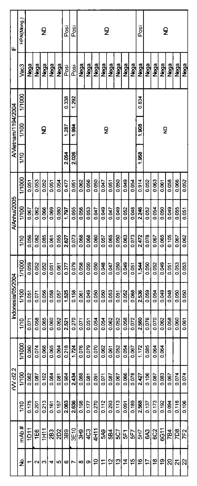

「3E10」 :NITE BP−01216
「7G10」 :NITE BP−01217
「8C1」 :NITE ABP−01737
「14A7」 :NITE ABP−01738
GANP H5モノクローナル抗体特異性評価
1.高病原性トリインフルエンザウイルスH5亜型のイムノクロマト診断開発に向けたモノクローナル抗体評価
<方法>
本実施例では、実施例1で作製したモノクローナル抗体の反応性評価を行った。
ウイルスに対する評価は、高病原性、低病原性H5N1ウイルスの感染細胞に対する蛍光免疫染色を行い、他の株種についてはHA組み換えタンパク質を用いELISA法にて検討した。
・蛍光免疫染色方法:細胞にウイルスを感染させ、PFA固定後、抗体培養上清を反応させ蛍光顕微鏡にて染色像を観察。
・ELISA法:H5HA組み換えタンパク質をプレートへ固相化、抗体培養上清を反応させた。二次抗体としてHRP標識抗マウスIgG抗体(SouthernBiotech)を使用し、化学発色後、吸光度を測定した。
1.高病原性トリインフルエンザウイルスH5亜型のイムノクロマト診断開発に向けたモノクローナル抗体評価
<方法>
本実施例では、実施例1で作製したモノクローナル抗体の反応性評価を行った。
ウイルスに対する評価は、高病原性、低病原性H5N1ウイルスの感染細胞に対する蛍光免疫染色を行い、他の株種についてはHA組み換えタンパク質を用いELISA法にて検討した。
・蛍光免疫染色方法:細胞にウイルスを感染させ、PFA固定後、抗体培養上清を反応させ蛍光顕微鏡にて染色像を観察。
・ELISA法:H5HA組み換えタンパク質をプレートへ固相化、抗体培養上清を反応させた。二次抗体としてHRP標識抗マウスIgG抗体(SouthernBiotech)を使用し、化学発色後、吸光度を測定した。
<評価結果>
表1に示す35クローンのうち、10クローンにおいてH5ウイルスMongoliaへ陽性反応を示し、その他はクローンにより異なる反応性が示された。
5H7−11−11−1のみH3N2ウイルス感染細胞での弱い反応性が認められた(表2)。
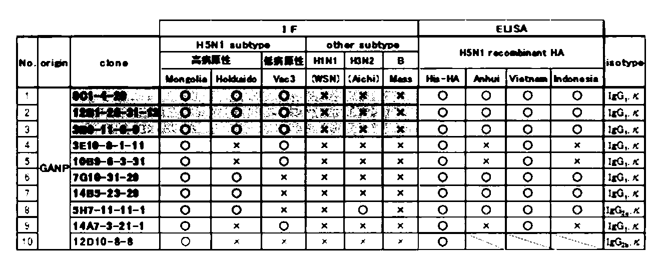
表2において、丸印(○)は陽性であることを、バツ印(×)は陰性であることを意味する。
表1に示す35クローンのうち、10クローンにおいてH5ウイルスMongoliaへ陽性反応を示し、その他はクローンにより異なる反応性が示された。
5H7−11−11−1のみH3N2ウイルス感染細胞での弱い反応性が認められた(表2)。
また、各抗体の解離定数をProteON XPR36(Bio−Rad)を用いて測定した。結果を表3に示す。
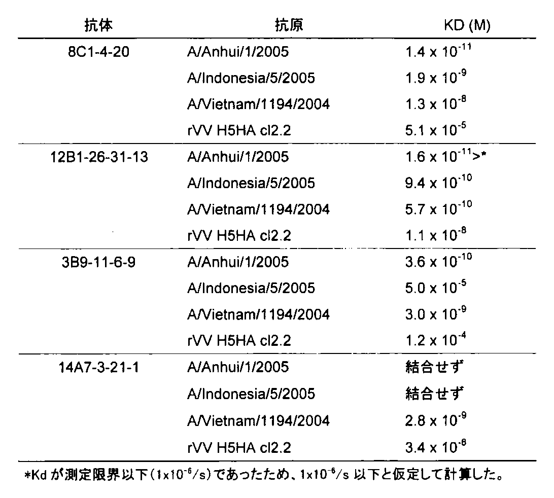
2.イムノクロマト上での抗体組み合わせ評価
<方法>
表2に記載の10クローンのうち9クローン、並びに表1に記載の7A5−1及び29.18クローン(Balb/c由来の対照クローン)の11クローンを用いて、イムノクロマトグラフィーストリップ上での抗体組み合わせ評価を行った。
抗原として、高病原性H5N1ウイルスであるMongolia及びHokkaido、並びに低病原性H5N1ウイルスであるVac3を用い、それぞれ1×10⁵~1×10⁷(pfu/ml)の濃度でアドテック社製ウイルス抽出液に溶解し、添加した。15分後に目視にて陽性ラインを評価した。
<方法>
表2に記載の10クローンのうち9クローン、並びに表1に記載の7A5−1及び29.18クローン(Balb/c由来の対照クローン)の11クローンを用いて、イムノクロマトグラフィーストリップ上での抗体組み合わせ評価を行った。
抗原として、高病原性H5N1ウイルスであるMongolia及びHokkaido、並びに低病原性H5N1ウイルスであるVac3を用い、それぞれ1×10⁵~1×10⁷(pfu/ml)の濃度でアドテック社製ウイルス抽出液に溶解し、添加した。15分後に目視にて陽性ラインを評価した。
<評価結果>
各抗体クローンと各ウイルス株との組み合わせ(90通り)を検討した結果、Mongoliaに対する強い陽性反応が認められるとともに弱いながらもHokkaidoへの反応性を示した7キットを、固相化抗体と金コロイド標識抗体との候補抗体ペアとして選択した(表4)。
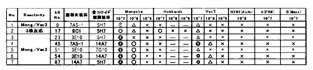
表4において、二重丸(◎)は色見本と比較して、強い反応を表し、丸(○)は同等の反応を表し、三角(△)は弱い反応を表し、バツ印(×)は反応しなかったことを表す。
各抗体クローンと各ウイルス株との組み合わせ(90通り)を検討した結果、Mongoliaに対する強い陽性反応が認められるとともに弱いながらもHokkaidoへの反応性を示した7キットを、固相化抗体と金コロイド標識抗体との候補抗体ペアとして選択した(表4)。
2−1 リコンビナントHAタンパク質を用いた特異性評価
<方法>
上記選択した7キットのうち、反応性の高い候補4キットを用い、H5亜型とその他の亜型リコンビナントHAタンパク質(Sino Biological Inc.)に対する特異性評価を行った。評価方法は、反応性がない0から8段階とし、数字が大きくなるほど陽性強度が増すものとした。また、色見本と比較して、強い反応を二重丸(◎)、弱い反応を三角(△)、反応しなかったことをバツ印(×)とした。
<方法>
上記選択した7キットのうち、反応性の高い候補4キットを用い、H5亜型とその他の亜型リコンビナントHAタンパク質(Sino Biological Inc.)に対する特異性評価を行った。評価方法は、反応性がない0から8段階とし、数字が大きくなるほど陽性強度が増すものとした。また、色見本と比較して、強い反応を二重丸(◎)、弱い反応を三角(△)、反応しなかったことをバツ印(×)とした。
<評価結果>
H5亜型に対して最も広い反応性を示すのは17番(固相8C1−金コロイド標識5H7)であった。
候補イムノクロマトキットを用いたH5亜型特異性評価結果を表5に示す。
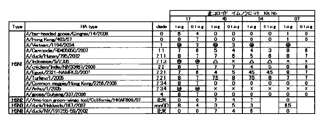
表5において、丸や三角等の記号は前記と同様である。また数字は色見本と比較したときのシグナルレベルを示す。
H5亜型に対して最も広い反応性を示すのは17番(固相8C1−金コロイド標識5H7)であった。
候補イムノクロマトキットを用いたH5亜型特異性評価結果を表5に示す。
また、候補イムノクロマトキットを用いた交差性評価結果を表6に示す。
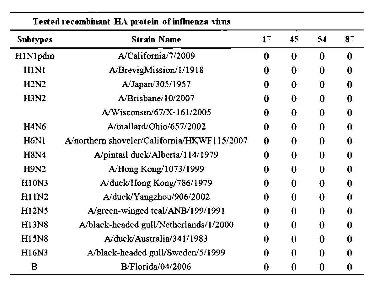
2−2 蛍光標識を用いたイムノクロマト法によるH5亜型の認識
方法
反応性の特に高い二種類の組み合わせに対して、蛍光ラテックスビーズ(藤倉化成3602−613)を金コロイドの代わりに標識し、陽性シグナルを蛍光イムノクロマトリーダー(コニカミノルタ)によって判定した。測定対象はH5亜型の複数の種類のウイルスを用いた。
方法
反応性の特に高い二種類の組み合わせに対して、蛍光ラテックスビーズ(藤倉化成3602−613)を金コロイドの代わりに標識し、陽性シグナルを蛍光イムノクロマトリーダー(コニカミノルタ)によって判定した。測定対象はH5亜型の複数の種類のウイルスを用いた。
標識法
0.3mgの抗体を0.4%の蛍光ビーズ懸濁液1.3mlと混ぜ合わせ、室温で1時間反応させた。遠心分離によって上清を破棄後、1%BSA溶液を加え、1時間ブロッキングした。遠心分離によって上清を破棄後、pH7.7の0.1%Trion X−100溶液で洗浄し、pH7.7の1%BSA溶液に懸濁し、37℃で48時間インキュベートした。これによってできた蛍光ビーズ標識済み抗体を金コロイドの標識抗体の代わりに用いた。
0.3mgの抗体を0.4%の蛍光ビーズ懸濁液1.3mlと混ぜ合わせ、室温で1時間反応させた。遠心分離によって上清を破棄後、1%BSA溶液を加え、1時間ブロッキングした。遠心分離によって上清を破棄後、pH7.7の0.1%Trion X−100溶液で洗浄し、pH7.7の1%BSA溶液に懸濁し、37℃で48時間インキュベートした。これによってできた蛍光ビーズ標識済み抗体を金コロイドの標識抗体の代わりに用いた。
イムノクロマト法
100μlのH5亜型ウイルス懸濁液(105pfu/反応)をストリップに滴下し、イムノクロマトリーダーにセットした。陽性反応はイムノクロマトリーダーによって15分と20分に測定される。シグナルとバックグラウンドの比(S/B値)が1を超した場合を陽性反応として測定した。
100μlのH5亜型ウイルス懸濁液(105pfu/反応)をストリップに滴下し、イムノクロマトリーダーにセットした。陽性反応はイムノクロマトリーダーによって15分と20分に測定される。シグナルとバックグラウンドの比(S/B値)が1を超した場合を陽性反応として測定した。
結果
No.17はユーラシアタイプのH5亜型を網羅して反応したが、北米タイプのH5亜型は認識できなかった(表7)。一方、No.45はユーラシアタイプのH5亜型の一部は認識できなかったが、北米型は認識可能であった(表7)。
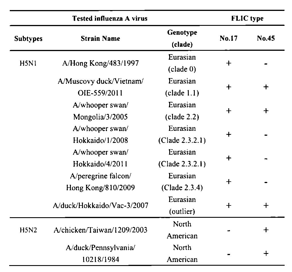
+は陽性、−は陰性を示す。
No.17はユーラシアタイプのH5亜型を網羅して反応したが、北米タイプのH5亜型は認識できなかった(表7)。一方、No.45はユーラシアタイプのH5亜型の一部は認識できなかったが、北米型は認識可能であった(表7)。
2−3 蛍光イムノクロマトを用いたH5亜型最小検出感度試験
方法
前述の蛍光イムノクロマト法でウイルスの最小検出量を測定した。
イムノクロマト法
多段階希釈した100μlのH5亜型ウイルス懸濁液をストリップに滴下し、イムノクロマトリーダーにセットした。陽性反応はイムノクロマトリーダーによって15分と20分に測定される。シグナルとバックグラウンドの比(S/B値)が1を超した場合を陽性反応として測定した。
測定限界
多段階希釈状態のウイルス量に対してのS/B値をプロットし近似線を引いた。これがカットオフ値であるS/B値=1との交点を計算し、それを測定限界とした。
方法
前述の蛍光イムノクロマト法でウイルスの最小検出量を測定した。
イムノクロマト法
多段階希釈した100μlのH5亜型ウイルス懸濁液をストリップに滴下し、イムノクロマトリーダーにセットした。陽性反応はイムノクロマトリーダーによって15分と20分に測定される。シグナルとバックグラウンドの比(S/B値)が1を超した場合を陽性反応として測定した。
測定限界
多段階希釈状態のウイルス量に対してのS/B値をプロットし近似線を引いた。これがカットオフ値であるS/B値=1との交点を計算し、それを測定限界とした。
結果
それぞれのキットの検出限界は表8の通りとなった。この感度は金コロイド法と比較して数十倍の感度上昇が見られた。
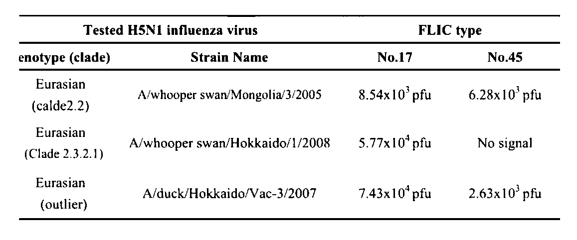
表中の数字は、ヒト反応に必要な最少感染価(pfu)を表す。
それぞれのキットの検出限界は表8の通りとなった。この感度は金コロイド法と比較して数十倍の感度上昇が見られた。
2−4 蛍光イムノクロマトを用いた非H5亜型ウイルスに対しての交差反応性試験
方法
前述の蛍光イムノクロマト法で非H5亜型ウイルスに対しての交差性を検討した。
イムノクロマト法
多段階希釈した100μlの非H5亜型ウイルス懸濁液(107pfu/反応)をストリップに滴下し、イムノクロマトリーダーにセットした。陽性反応はイムノクロマトリーダーによって15分と20分に測定される。シグナルとバックグラウンドの比(S/B値)が1を超した場合を陽性反応として測定した。
方法
前述の蛍光イムノクロマト法で非H5亜型ウイルスに対しての交差性を検討した。
イムノクロマト法
多段階希釈した100μlの非H5亜型ウイルス懸濁液(107pfu/反応)をストリップに滴下し、イムノクロマトリーダーにセットした。陽性反応はイムノクロマトリーダーによって15分と20分に測定される。シグナルとバックグラウンドの比(S/B値)が1を超した場合を陽性反応として測定した。
結果
非H5亜型のウイルスをH1からH16亜型まで検討したが、交差性は見られなかった。本方法の特異性の高さが示された(表9)。

+は陽性、−は陰性を示す。
非H5亜型のウイルスをH1からH16亜型まで検討したが、交差性は見られなかった。本方法の特異性の高さが示された(表9)。
2−5 蛍光イムノクロマトを用いた非H5亜型HAタンパクに対しての交差反応性試験
方法
前述の蛍光イムノクロマト法で非H5亜型ウイルス精製HAタンパクに対しての交差性を検討した。
イムノクロマト法
多段階希釈した100μlの非H5亜型ウイルス懸濁液(107pfu/反応)をストリップに滴下し、イムノクロマトリーダーにセットした。陽性反応はイムノクロマトリーダーによって15分と20分に測定される。シグナルとバックグラウンドの比(S/B値)が1を超した場合を陽性反応として測定した。
方法
前述の蛍光イムノクロマト法で非H5亜型ウイルス精製HAタンパクに対しての交差性を検討した。
イムノクロマト法
多段階希釈した100μlの非H5亜型ウイルス懸濁液(107pfu/反応)をストリップに滴下し、イムノクロマトリーダーにセットした。陽性反応はイムノクロマトリーダーによって15分と20分に測定される。シグナルとバックグラウンドの比(S/B値)が1を超した場合を陽性反応として測定した。
結果
非H5亜型のウイルスのHAタンパクを複数検討したが、交差性は見られなかった。本方法の特異性の高さが示された(表10)。

以上のことから、本発明の抗体は、組み合わせ如何によってすべてのH5亜型を診断し得ることが示された。
非H5亜型のウイルスのHAタンパクを複数検討したが、交差性は見られなかった。本方法の特異性の高さが示された(表10)。
H5N1インフルエンザウイルスHA蛋白質に対するGANP−Tgマウス由来ハイブリドーマクローンの中和・阻害活性の評価(1)
本実施例では、GANP−Tgマウス由来ハイブリドーマ培養上清の低病原性インフルエンザウイルスH5N1−Vac3株に対する中和・阻害活性の評価を行った。
<方法>
インフルエンザウイルスH5N1−Vac3株に対する阻害試験を以下の手順で行った。
1)アッセイ3日前にMDCK細胞を6wellプレートへ2×10⁵cells/wellで播いた。
2)1%BSAを含有するMEM培地で希釈したインフルエンザウイルスH5N1(1×10³PFU/ml)ウイルス液0.1mlと、100倍、1000倍、10000倍に希釈したハイブリドーマ培養上清0.1mlとを混和し、37℃で1時間中和反応を行った。
3)インフルエンザウイルス液とハイブリドーマ培養上清の混液0.1mlを各wellに加え、全体に行き渡らせた。
4)37℃,1時間吸着した。15分おきにプレートを揺らした。
5)ウイルス液を取り除き、BSA(−)/MEMで1回洗浄した。
6)ハイブリドーマ培養上清1/100、1/1000、1/10000量添加,10μg/mlアセチルトリプシン,0.8%アガロース加1%BSA/MEM 2mlを各wellに加えた。
7)アガロース凝固後、プレートの上下を反転させ、CO2インキュベーターへ入れた。
8)37℃,2日培養した。
9)10%ホルマリンを加えた。室温,1時間固定した。
10)ホルマリンとアガロース培地を取り除き、水洗した。
11)1%クリスタルバイオレットで1時間染色した。
12)水洗、乾燥後にプラーク数を判定した。
本実施例では、GANP−Tgマウス由来ハイブリドーマ培養上清の低病原性インフルエンザウイルスH5N1−Vac3株に対する中和・阻害活性の評価を行った。
<方法>
インフルエンザウイルスH5N1−Vac3株に対する阻害試験を以下の手順で行った。
1)アッセイ3日前にMDCK細胞を6wellプレートへ2×10⁵cells/wellで播いた。
2)1%BSAを含有するMEM培地で希釈したインフルエンザウイルスH5N1(1×10³PFU/ml)ウイルス液0.1mlと、100倍、1000倍、10000倍に希釈したハイブリドーマ培養上清0.1mlとを混和し、37℃で1時間中和反応を行った。
3)インフルエンザウイルス液とハイブリドーマ培養上清の混液0.1mlを各wellに加え、全体に行き渡らせた。
4)37℃,1時間吸着した。15分おきにプレートを揺らした。
5)ウイルス液を取り除き、BSA(−)/MEMで1回洗浄した。
6)ハイブリドーマ培養上清1/100、1/1000、1/10000量添加,10μg/mlアセチルトリプシン,0.8%アガロース加1%BSA/MEM 2mlを各wellに加えた。
7)アガロース凝固後、プレートの上下を反転させ、CO2インキュベーターへ入れた。
8)37℃,2日培養した。
9)10%ホルマリンを加えた。室温,1時間固定した。
10)ホルマリンとアガロース培地を取り除き、水洗した。
11)1%クリスタルバイオレットで1時間染色した。
12)水洗、乾燥後にプラーク数を判定した。
<結果>
図3に培養後のプレートの写真を示す。陰性コントロール(無添加)と比較して、3E10,10B9,14A7及び3B9では、プラークの大きさ及び数に明らかに差が見られた。なお、使用したウイルスの量を、Back−titration(無添加)を行って確認したが、所定のウイルス量であった。
図3に培養後のプレートの写真を示す。陰性コントロール(無添加)と比較して、3E10,10B9,14A7及び3B9では、プラークの大きさ及び数に明らかに差が見られた。なお、使用したウイルスの量を、Back−titration(無添加)を行って確認したが、所定のウイルス量であった。
H5N1インフルエンザウイルスHA蛋白質に対するGANP−Tgマウス由来ハイブリドーマクローンの中和・阻害活性の評価(2)
本実施例では、抗体作製時に使用した抗原と同じH5N1クレード(clade 2.2)の高病原性トリインフルエンザウイルスを用いて、中和・阻害活性の評価を行った。実施例2又は3で中和活性を示したかあるいは免疫蛍光染色(IF),ELISAで強い結合性を示した9種類(表2中、12B1−26−31−13を除く9種類)のGANP−Tgマウス由来ハイブリドーマ培養上清の、高病原性トリインフルエンザウイルス(H5N1 A/whooper swan/Mongolia/3/05)株に対する中和・阻害活性について、in vitroでの評価を行った。
本実施例では、抗体作製時に使用した抗原と同じH5N1クレード(clade 2.2)の高病原性トリインフルエンザウイルスを用いて、中和・阻害活性の評価を行った。実施例2又は3で中和活性を示したかあるいは免疫蛍光染色(IF),ELISAで強い結合性を示した9種類(表2中、12B1−26−31−13を除く9種類)のGANP−Tgマウス由来ハイブリドーマ培養上清の、高病原性トリインフルエンザウイルス(H5N1 A/whooper swan/Mongolia/3/05)株に対する中和・阻害活性について、in vitroでの評価を行った。
<方法>
実験はP3施設内で行った。ウイルス感染価はプラーク形成能によって決定した。
インフルエンザウイルスH5N1 A/whooper swan/Mongolia/3/05株に対する阻害試験を以下の手順で行った。
1)アッセイ3日前にMDCK細胞を6wellプレートへ2×10⁵cells/wellで播いた。
2)1%BSAを含有するMEM培地で希釈したインフルエンザウイルスH5N1(1×10³PFU/ml)ウイルス液0.1mlと、100倍、1000倍又は10000倍に希釈したハイブリドーマ培養上清0.1mlとを混和し、37℃で1時間中和反応を行った。
3)インフルエンザウイルス液とハイブリドーマ培養上清の混液0.1mlを各wellに加え、全体に行き渡らせた。
4)37℃,1時間吸着した。15分おきにプレートを揺らした。
5)ウイルス液を取り除き、BSA(−)/MEMで1回洗浄した。
6)ハイブリドーマ培養上清1/100、1/1000、1/10000量添加,10μg/mlアセチルトリプシン,0.8%アガロース加1%BSA/MEM 2mlを各wellに加えた。
7)アガロース凝固後、プレートの上下を反転させ、CO2インキュベーターへ入れた。
8)37℃,3日培養した。
9)10%ホルマリンを加えた。室温,1時間固定した。
10)ホルマリンとアガロース培地を取り除き、水洗した。
11)1%クリスタルバイオレットで1時間染色した。
12)水洗、乾燥後にプラーク数を判定した。
H5N1のプラーク形成価を測定し、1プレート当り50~60プラークを形成する希釈条件を決定した後、その条件でハイブリドーマ培養上清100μlをプレートに添加し、ウイルス感染を阻止する活性を中和活性として測定した。
実験はP3施設内で行った。ウイルス感染価はプラーク形成能によって決定した。
インフルエンザウイルスH5N1 A/whooper swan/Mongolia/3/05株に対する阻害試験を以下の手順で行った。
1)アッセイ3日前にMDCK細胞を6wellプレートへ2×10⁵cells/wellで播いた。
2)1%BSAを含有するMEM培地で希釈したインフルエンザウイルスH5N1(1×10³PFU/ml)ウイルス液0.1mlと、100倍、1000倍又は10000倍に希釈したハイブリドーマ培養上清0.1mlとを混和し、37℃で1時間中和反応を行った。
3)インフルエンザウイルス液とハイブリドーマ培養上清の混液0.1mlを各wellに加え、全体に行き渡らせた。
4)37℃,1時間吸着した。15分おきにプレートを揺らした。
5)ウイルス液を取り除き、BSA(−)/MEMで1回洗浄した。
6)ハイブリドーマ培養上清1/100、1/1000、1/10000量添加,10μg/mlアセチルトリプシン,0.8%アガロース加1%BSA/MEM 2mlを各wellに加えた。
7)アガロース凝固後、プレートの上下を反転させ、CO2インキュベーターへ入れた。
8)37℃,3日培養した。
9)10%ホルマリンを加えた。室温,1時間固定した。
10)ホルマリンとアガロース培地を取り除き、水洗した。
11)1%クリスタルバイオレットで1時間染色した。
12)水洗、乾燥後にプラーク数を判定した。
H5N1のプラーク形成価を測定し、1プレート当り50~60プラークを形成する希釈条件を決定した後、その条件でハイブリドーマ培養上清100μlをプレートに添加し、ウイルス感染を阻止する活性を中和活性として測定した。
<結果>
結果を図4に示す。
図4において、各プレートの左上に記載の数字は、ハイブリドーマ培養上清(1~4番)の10−2,10−3,10−4希釈添加時のプラーク数を示している。コントロール群はBack titration(Back)とし、中和価測定に使用したウイルス液を原液(1倍)及び10倍希釈で添加した。
また抗ウイルス効果のコントロールとしてリレンザ(グラクソ・スミスクライン)を1μM,0.1μM,0.01μMの濃度で添加し、プラークの出現が出来ることを確認した。
図4に示したように、3B9−11−6−9,3E10−8−1−11,10B9−6−3−31,14A7−3−21−1の4種類のクローンが高病原性トリインフルエンザウイルス(H5N1 A/whooper swan/Mongolia/3/05)株に対する中和・増殖阻害活性を示した。
結果を図4に示す。
図4において、各プレートの左上に記載の数字は、ハイブリドーマ培養上清(1~4番)の10−2,10−3,10−4希釈添加時のプラーク数を示している。コントロール群はBack titration(Back)とし、中和価測定に使用したウイルス液を原液(1倍)及び10倍希釈で添加した。
また抗ウイルス効果のコントロールとしてリレンザ(グラクソ・スミスクライン)を1μM,0.1μM,0.01μMの濃度で添加し、プラークの出現が出来ることを確認した。
図4に示したように、3B9−11−6−9,3E10−8−1−11,10B9−6−3−31,14A7−3−21−1の4種類のクローンが高病原性トリインフルエンザウイルス(H5N1 A/whooper swan/Mongolia/3/05)株に対する中和・増殖阻害活性を示した。
H5N1インフルエンザウイルスHA蛋白質に対するGANP−Tgマウス由来ハイブリドーマクローンの中和・阻害活性の評価(3)
本実施例では、抗体作製時に使用した抗原と異なったH5N1クレード(clade 2.3.2)の高病原性トリインフルエンザウイルスを用いて実施した。実施例4で中和活性を示した3B9−11−6−9,3E10−8−1−11,10B9−6−3−31,14A7−3−21−1の4種類のGANP−Tgマウス由来ハイブリドーマ腹水の高病原性トリインフルエンザウイルス(H5N1 A/whooper swan/Hokkaido/1/08)株に対する中和・阻害活性の評価を行った。
本実施例では、抗体作製時に使用した抗原と異なったH5N1クレード(clade 2.3.2)の高病原性トリインフルエンザウイルスを用いて実施した。実施例4で中和活性を示した3B9−11−6−9,3E10−8−1−11,10B9−6−3−31,14A7−3−21−1の4種類のGANP−Tgマウス由来ハイブリドーマ腹水の高病原性トリインフルエンザウイルス(H5N1 A/whooper swan/Hokkaido/1/08)株に対する中和・阻害活性の評価を行った。
<方法>
インフルエンザウイルスH5N1 A/whooper swan/Hokkaido/1/08株に対する阻害試験を以下の手順で行った。
1)アッセイ3日前にMDCK細胞を6wellプレートへ2×10⁵cells/wellで播いた。
2)1%BSAを含有するMEM培地で希釈したインフルエンザウイルスH5N1(1×10³PFU/ml)ウイルス液0.1mlと、1000倍、10000倍、100000倍に希釈したハイブリドーマ培養上清0.1mlとを混和し、37℃で1時間中和反応を行った。
3)インフルエンザウイルス液とハイブリドーマ培養上清の混液0.1mlを各wellに加え、全体に行き渡らせた。
4)37℃,1時間吸着した。15分おきにプレートを揺らした。
5)ウイルス液を取り除き、BSA(−)/MEMで1回洗浄した。
6)ハイブリドーマ培養上清1/1000、1/10000、1/100000量添加,10μg/mlアセチルトリプシン,0.8%アガロース加1%BSA/MEM 2mlを各wellに加えた。
7)アガロース凝固後、プレートの上下を反転させ、CO2インキュベーターへ入れた。
8)37℃,3日培養した。
9)10%ホルマリンを加えた。室温,1時間固定した。
10)ホルマリンとアガロース培地を取り除き、水洗した。
11)1%クリスタルバイオレットで1時間染色した。
12)水洗、乾燥後にプラーク数を判定した。
インフルエンザウイルスH5N1 A/whooper swan/Hokkaido/1/08株に対する阻害試験を以下の手順で行った。
1)アッセイ3日前にMDCK細胞を6wellプレートへ2×10⁵cells/wellで播いた。
2)1%BSAを含有するMEM培地で希釈したインフルエンザウイルスH5N1(1×10³PFU/ml)ウイルス液0.1mlと、1000倍、10000倍、100000倍に希釈したハイブリドーマ培養上清0.1mlとを混和し、37℃で1時間中和反応を行った。
3)インフルエンザウイルス液とハイブリドーマ培養上清の混液0.1mlを各wellに加え、全体に行き渡らせた。
4)37℃,1時間吸着した。15分おきにプレートを揺らした。
5)ウイルス液を取り除き、BSA(−)/MEMで1回洗浄した。
6)ハイブリドーマ培養上清1/1000、1/10000、1/100000量添加,10μg/mlアセチルトリプシン,0.8%アガロース加1%BSA/MEM 2mlを各wellに加えた。
7)アガロース凝固後、プレートの上下を反転させ、CO2インキュベーターへ入れた。
8)37℃,3日培養した。
9)10%ホルマリンを加えた。室温,1時間固定した。
10)ホルマリンとアガロース培地を取り除き、水洗した。
11)1%クリスタルバイオレットで1時間染色した。
12)水洗、乾燥後にプラーク数を判定した。
<結果>
図5に示したように3B9−11−6−9クローンが高病原性トリインフルエンザウイルス(H5N1 A/whooper swan/Hokkaido/1/08)株に対する中和・増殖阻害活性を示した。
図5に示したように3B9−11−6−9クローンが高病原性トリインフルエンザウイルス(H5N1 A/whooper swan/Hokkaido/1/08)株に対する中和・増殖阻害活性を示した。
HA蛋白/GANP−Tgマウス由来ハイブリドーマIgGのマウスへの高病原性トリインフルエンザウイルス感染に対する防御効果
in vitro試験(実施例5)において、3B9−11−6−9クローンに、H5N1 A/whooper swan/Mongolia/3/05(clade2.2)株及びH5N1 A/whooper swan/Hokkaido/1/08株(clade2.3.2)の高病原性トリインフルエンザウイルスに対する中和・増殖阻害活性が認められたので、本実施例では、高病原性トリインフルエンザウイルス感染マウスに対する治療効果(in vivoにおける試験)を検討した。
in vitro試験(実施例5)において、3B9−11−6−9クローンに、H5N1 A/whooper swan/Mongolia/3/05(clade2.2)株及びH5N1 A/whooper swan/Hokkaido/1/08株(clade2.3.2)の高病原性トリインフルエンザウイルスに対する中和・増殖阻害活性が認められたので、本実施例では、高病原性トリインフルエンザウイルス感染マウスに対する治療効果(in vivoにおける試験)を検討した。
<方法>
ハイブリドーマの投与7日前に Crlj:CD1−Fxn1 nu/nu(7週令)マウスをプリスタンで処理し、7日後にマウス各4匹に5x106個のハイブリドーマ細胞(3B9−11−6−9)を腹腔内注入した。2週間後に腹水35ml~50mlを得て、IgG抗体精製を行った。HiTrap Protein G HPカラムを用いて腹水から12~30mgのIgG抗体を精製した。
攻撃感染ウイルスとしてH5N1 A/whooper swan/Hokkaido/1/08(200 PFU/mouse;3.3MLD50)を用い、動物としてBalb/cマウス(8~10週令、♀)を用いた。実験群としてコントロールのVehicle処理群は抗体精製時に用いた試薬を使用し(93.5mM Glycine−HCl,65.4mM Tris−HCl:PBS(−)=0.836:1.164)、被検抗体は各群8匹を用いた。なお、感染3日後に4匹/群ずつ剖検し、実験終了時に4匹/群ずつ剖検した。
非感染(希釈液)接種群(一群4匹)は体重変化のモニタリングに用いた。抗体量は15mg/kg/マウスとし、Day0(ウイルス感染直前)とDay2に眼窩静脈巣より投与した。図6パネルAに方法を示した。
ハイブリドーマの投与7日前に Crlj:CD1−Fxn1 nu/nu(7週令)マウスをプリスタンで処理し、7日後にマウス各4匹に5x106個のハイブリドーマ細胞(3B9−11−6−9)を腹腔内注入した。2週間後に腹水35ml~50mlを得て、IgG抗体精製を行った。HiTrap Protein G HPカラムを用いて腹水から12~30mgのIgG抗体を精製した。
攻撃感染ウイルスとしてH5N1 A/whooper swan/Hokkaido/1/08(200 PFU/mouse;3.3MLD50)を用い、動物としてBalb/cマウス(8~10週令、♀)を用いた。実験群としてコントロールのVehicle処理群は抗体精製時に用いた試薬を使用し(93.5mM Glycine−HCl,65.4mM Tris−HCl:PBS(−)=0.836:1.164)、被検抗体は各群8匹を用いた。なお、感染3日後に4匹/群ずつ剖検し、実験終了時に4匹/群ずつ剖検した。
非感染(希釈液)接種群(一群4匹)は体重変化のモニタリングに用いた。抗体量は15mg/kg/マウスとし、Day0(ウイルス感染直前)とDay2に眼窩静脈巣より投与した。図6パネルAに方法を示した。
<結果>
結果を図6に示す。
図6パネルBには、インフルエンザウイルスによる攻撃感染後の体重の変化を示した。14A7−3−21−1クローンを投与した感染マウス群(●)及びVehicle(▲)を投与した感染マウス群では、感染6日以降に顕著な体重減少が認められた。これに対し、3B9−11−6−9クローンを投与した感染マウス群(■)及び非感染マウス群(◆)では体重の低下は認められなかった。
図6パネルCには、攻撃感染後の生存率の変化を示した。感染11日において、14A7−3−21−1クローンを投与した感染マウス群(●)では25%の生存率、及びVehicleを投与した感染マウス群(▲)では50%の生存率となった。これに対し、3B9−11−6−9クローンを投与した感染マウス群(■)及び非感染マウス群(◆)では生存率の低下は認められなかった。
以上の結果から、H5N1高病原性トリインフルエンザウイルスに対して幅広い結合性を有する3B9−11−6−9クローンは、高病原性トリインフルエンザウイルス感染に対しても顕著な治療効果を示した。
結果を図6に示す。
図6パネルBには、インフルエンザウイルスによる攻撃感染後の体重の変化を示した。14A7−3−21−1クローンを投与した感染マウス群(●)及びVehicle(▲)を投与した感染マウス群では、感染6日以降に顕著な体重減少が認められた。これに対し、3B9−11−6−9クローンを投与した感染マウス群(■)及び非感染マウス群(◆)では体重の低下は認められなかった。
図6パネルCには、攻撃感染後の生存率の変化を示した。感染11日において、14A7−3−21−1クローンを投与した感染マウス群(●)では25%の生存率、及びVehicleを投与した感染マウス群(▲)では50%の生存率となった。これに対し、3B9−11−6−9クローンを投与した感染マウス群(■)及び非感染マウス群(◆)では生存率の低下は認められなかった。
以上の結果から、H5N1高病原性トリインフルエンザウイルスに対して幅広い結合性を有する3B9−11−6−9クローンは、高病原性トリインフルエンザウイルス感染に対しても顕著な治療効果を示した。
HA蛋白/GANP−Tgマウス由来ハイブリドーマIgGのマウスへの高病原性トリインフルエンザウイルス感染に対する防御効果
in vitro試験(実施例6)において、3B9−11−6−9クローンにH5N1 A/whooper swan/Hokkaido/1/08株(clade2.3.2)の高病原性トリインフルエンザウイルスに対する治療効果を示したことが認められたので、本実施例では、高病原性トリインフルエンザウイルス感染後、4,6,8日後に抗体を投与してマウスに対する治療効果(in vivoにおける試験)を検討した。
in vitro試験(実施例6)において、3B9−11−6−9クローンにH5N1 A/whooper swan/Hokkaido/1/08株(clade2.3.2)の高病原性トリインフルエンザウイルスに対する治療効果を示したことが認められたので、本実施例では、高病原性トリインフルエンザウイルス感染後、4,6,8日後に抗体を投与してマウスに対する治療効果(in vivoにおける試験)を検討した。
<方法>
ハイブリドーマの投与7日前にCrlj:CD1−Fxn1 nu/nu(7週令)マウスをプリスタンで処理し、7日後にマウス各4匹に5x106個のハイブリドーマ細胞(3B9−11−6−9)を腹腔内注入した。2週間後に腹水35ml~50mlを得て、IgG抗体精製を行った。HiTrap Protein G HPカラムを用いて腹水から12~30mgのIgG抗体を精製した。
攻撃感染ウイルスとしてH5N1 A/whooper swan/Hokkaido/1/08(200 PFU/mouse;3.3MLD50)を用い、動物としてBalb/cマウス(9週令、♀)を用いた。実験群としてコントロールのVehicle処理群は抗体精製時に用いた試薬を使用し(93.5mM Glycine−HCl,65.4mM Tris−HCl:PBS(−)=0.836:1.164)、被検抗体は各群5匹を用いた。実験終了時に生存個体は剖検した。
非感染(希釈液)接種群(一群4匹)は体重変化のモニタリングに用いた。抗体量は15mg/kg/マウスとし、Day0、Day4、Day6、Day8に眼窩静脈巣より投与した。
ハイブリドーマの投与7日前にCrlj:CD1−Fxn1 nu/nu(7週令)マウスをプリスタンで処理し、7日後にマウス各4匹に5x106個のハイブリドーマ細胞(3B9−11−6−9)を腹腔内注入した。2週間後に腹水35ml~50mlを得て、IgG抗体精製を行った。HiTrap Protein G HPカラムを用いて腹水から12~30mgのIgG抗体を精製した。
攻撃感染ウイルスとしてH5N1 A/whooper swan/Hokkaido/1/08(200 PFU/mouse;3.3MLD50)を用い、動物としてBalb/cマウス(9週令、♀)を用いた。実験群としてコントロールのVehicle処理群は抗体精製時に用いた試薬を使用し(93.5mM Glycine−HCl,65.4mM Tris−HCl:PBS(−)=0.836:1.164)、被検抗体は各群5匹を用いた。実験終了時に生存個体は剖検した。
非感染(希釈液)接種群(一群4匹)は体重変化のモニタリングに用いた。抗体量は15mg/kg/マウスとし、Day0、Day4、Day6、Day8に眼窩静脈巣より投与した。
<結果>
結果を図7に示す。
図7は、抗HAモノクローナル抗体投与による治療効果の検討結果を示す図である(BALB/c mice(Female,9−week−old)、H5N1 HPAIV 300 PFU、15mg/kg 3B9投与群)。
図7パネルAには、インフルエンザウイルスによる攻撃感染後の体重の変化を示した。3B9−11−6−9クローンをDay8に投与した感染マウス群(●)及び非投与を感染マウス群(□)では、感染6日以降に顕著な体重減少が認められた。これに対し、3B9−11−6−9クローンをDay0に投与した感染マウス群(△)及び非感染マウス群(◆)では体重の低下は認められなかった。3B9−11−6−9クローンをDay4(▲)及びにDay6(○)に投与した感染マウス群では一時的な体重の低下は認められなかった。
図7パネルBには、攻撃感染後の生存率の変化を示した。感染14日において、3B9−11−6−9クローンを投与した感染マウス群Day0(△)、Day4(▲)、Day6(○)及びDay8に投与した感染マウス群(●)を示した。Day4(▲)75%、Day6(○)50%及びDay8(●)0%の生存率であった。これに対し、3B9−11−6−9クローンをDay0(△)に投与した感染マウス群(■)及び非感染マウス群(◆)では生存率の低下は認められなかった。
以上の結果から、H5N1高病原性トリインフルエンザウイルスに対して幅広い結合性を有する3B9−11−6−9クローンは、高病原性トリインフルエンザウイルス感染後の投与に対しても顕著な治療効果を示した。
結果を図7に示す。
図7は、抗HAモノクローナル抗体投与による治療効果の検討結果を示す図である(BALB/c mice(Female,9−week−old)、H5N1 HPAIV 300 PFU、15mg/kg 3B9投与群)。
図7パネルAには、インフルエンザウイルスによる攻撃感染後の体重の変化を示した。3B9−11−6−9クローンをDay8に投与した感染マウス群(●)及び非投与を感染マウス群(□)では、感染6日以降に顕著な体重減少が認められた。これに対し、3B9−11−6−9クローンをDay0に投与した感染マウス群(△)及び非感染マウス群(◆)では体重の低下は認められなかった。3B9−11−6−9クローンをDay4(▲)及びにDay6(○)に投与した感染マウス群では一時的な体重の低下は認められなかった。
図7パネルBには、攻撃感染後の生存率の変化を示した。感染14日において、3B9−11−6−9クローンを投与した感染マウス群Day0(△)、Day4(▲)、Day6(○)及びDay8に投与した感染マウス群(●)を示した。Day4(▲)75%、Day6(○)50%及びDay8(●)0%の生存率であった。これに対し、3B9−11−6−9クローンをDay0(△)に投与した感染マウス群(■)及び非感染マウス群(◆)では生存率の低下は認められなかった。
以上の結果から、H5N1高病原性トリインフルエンザウイルスに対して幅広い結合性を有する3B9−11−6−9クローンは、高病原性トリインフルエンザウイルス感染後の投与に対しても顕著な治療効果を示した。
本発明により、強毒性インフルエンザであるインドネシア株、安徽株、ベトナム株の全てを認識することのできる高親和性モノクローナル抗体(KD=1x10?10Mレベル)が提供される。本発明の抗体により、超高感度の診断キットを作製することが可能となる。また、様々なクレードのウイルスの検出のみならず、ウイルスの増殖阻害にも有効であることから、医療及び臨床検査に極めて有用である。
ハイブリドーマ「3E10(rVV cl2.2)」及び「7G10(rVV cl2.2)」は、いずれも、独立行政法人製品評価技術基盤機構特許微生物寄託センター(〒292−0818 千葉県木更津市かずさ鎌足2−5−8 NITEバイオテクノロジー本部 特許微生物寄託センター)にブダペスト条約に基づき国際寄託した(原寄託日:2012年2月1日)。
受託番号は、「3E10(rVV cl2.2)」についてはNITE BP−01216であり、「7G10(rVV cl2.2)」についてはNITE BP−01217である。
ハイブリドーマ「8C1(rVV cl2.2)」及び「14A7(rVV cl2.2)」は、いずれも、上記特許微生物寄託センターにブダペスト条約に基づき国際寄託した(原寄託日:2013年10月18日)。
受領番号は、「8C1(rVV cl2.2)」についてはNITE ABP−01737であり、「14A7(rVV cl2.2)」についてはNITE ABP−01738である。
受託番号は、「3E10(rVV cl2.2)」についてはNITE BP−01216であり、「7G10(rVV cl2.2)」についてはNITE BP−01217である。
ハイブリドーマ「8C1(rVV cl2.2)」及び「14A7(rVV cl2.2)」は、いずれも、上記特許微生物寄託センターにブダペスト条約に基づき国際寄託した(原寄託日:2013年10月18日)。
受領番号は、「8C1(rVV cl2.2)」についてはNITE ABP−01737であり、「14A7(rVV cl2.2)」についてはNITE ABP−01738である。
配列番号1:合成DNA
配列番号2:合成DNA
配列番号2:合成DNA
Claims (27)
- 少なくとも1×10⁻¹⁰Mの解離定数を有する、インフルエンザウイルスのHAタンパク質に対する抗体。
- 解離定数が1×10⁻¹⁰M~1×10⁻¹³Mである、請求項1に記載の抗体。
- インフルエンザウイルスがインフルエンザH5亜型ウイルスである請求項1に記載の抗体。
- インフルエンザH5亜型ウイルスがインフルエンザH5N1ウイルスである請求項3に記載の抗体。
- インフルエンザH5N1ウイルスが、インドガン青海湖株(clade 2.2)、インドネシア株(clade 2.1)、安徽株(clade 2.3)、ベトナム株(clade 1)、モンゴル株(A/whooper swan/Mongolia/3/2005:clade 2.2)及び北海道株(A/whooper swan/Hokkaido/1/2008:clade 2.3.2)からなる群から選ばれる少なくとも1種である請求項4に記載の抗体。
- 抗体がモノクローナル抗体である請求項1に記載の抗体。
- 受託番号がNITE BP−01216若しくはNITE BP−01217、又は受領番号がNITE ABP−01737若しくはNITE ABP−01738であるにより産生される、請求項6に記載の抗体。
- 抗体が、キメラ抗体、ヒト型化抗体又はヒト化抗体である、請求項1に記載の抗体。
- 請求項1に記載の抗体が結合する部位に結合する抗体。
- 請求項1~9のいずれか1項に記載の抗体の断片。
- 請求項6に記載のモノクローナル抗体を産生するハイブリドーマ。
- 受託番号がNITE BP−01216若しくはNITE BP−01217、又は受領番号がNITE ABP−01737若しくはNITE ABP−01738であるハイブリドーマ。
- 請求項1に記載の抗体の製造方法であって、以下の工程:
(a)組換えワクシニアウイルスを用いて発現させたインフルエンザウイルスのHAタンパク質を用いて非ヒト哺乳動物を免疫する工程、及び
(b)当該非ヒト哺乳動物から抗体を採取する工程
を含む、前記方法。 - 請求項6又は7に記載のモノクローナル抗体の製造方法であって、以下の工程:
(a)組換えワクシニアウイルスを用いて発現させたインフルエンザウイルスのHAタンパク質を用いて非ヒト哺乳動物を免疫する工程、
(b)前記免疫された非ヒト哺乳動物から抗体産生細胞を採取する工程、
(c)工程(b)で得られた抗体産生細胞とミエローマ細胞とを融合させる工程、及び
(d)工程(c)で得られた融合細胞から抗体を採取する工程
を含む、前記方法。 - 非ヒト哺乳動物が、GANP(登録商標)トランスジェニック非ヒト哺乳動物である請求項13又は14に記載の方法。
- 請求項1~9のいずれか1項に記載の抗体又は請求項10に記載の断片と生体試料とを接触させて、インフルエンザウイルスを検出することを特徴とする、インフルエンザウイルスの検出方法。
- インフルエンザウイルスがインフルエンザH5亜型ウイルスである請求項16に記載の方法。
- インフルエンザH5亜型ウイルスがインフルエンザH5N1ウイルスである請求項17に記載の方法。
- インフルエンザH5N1ウイルスが、インドガン青海湖株(clade 2.2)、インドネシア株(clade 2.1)、安徽株(clade 2.3)、ベトナム株(clade 1)、モンゴル株(A/whooper swan/Mongolia/3/2005:clade 2.2)及び北海道株(A/whooper swan/Hokkaido/1/2008:clade 2.3.2)からなる群から選ばれる少なくとも1種である請求項18に記載の方法。
- 請求項1~9のいずれか1項に記載の抗体又は請求項10に記載の断片を含む、インフルエンザウイルス検出用試薬。
- インフルエンザウイルスがインフルエンザH5亜型ウイルスである請求項20に記載の試薬。
- インフルエンザH5亜型ウイルスがインフルエンザH5N1ウイルスである請求項21に記載の試薬。
- インフルエンザH5N1ウイルスが、インドガン青海湖株(clade 2.2)、インドネシア株(clade 2.1)、安徽株(clade 2.3)、ベトナム株(clade 1)、モンゴル株(A/whooper swan/Mongolia/3/2005:clade 2.2)及び北海道株(A/whooper swan/Hokkaido/1/2008:clade 2.3.2)からなる群から選ばれる少なくとも1種である請求項22に記載の試薬。
- 請求項1~9のいずれか1項に記載の抗体又は請求項10に記載の断片を含む、抗インフルエンザウイルス用医薬組成物。
- インフルエンザウイルスがインフルエンザH5亜型ウイルスである請求項24に記載の医薬組成物。
- インフルエンザH5亜型ウイルスがインフルエンザH5N1ウイルスである請求項25に記載の医薬組成物。
- インフルエンザH5N1ウイルスが、インドガン青海湖株(clade 2.2)、インドネシア株(clade 2.1)、安徽株(clade 2.3)、ベトナム株(clade 1)、モンゴル株(A/whooper swan/Mongolia/3/2005:clade 2.2)及び北海道株(A/whooper swan/Hokkaido/1/2008:clade 2.3.2)からなる群から選ばれる少なくとも1種である請求項26に記載の医薬組成物。
Priority Applications (1)
| Application Number | Priority Date | Filing Date | Title |
|---|---|---|---|
| JP2014553012A JPWO2014097762A1 (ja) | 2012-12-20 | 2013-10-30 | 高病原性トリインフルエンザに対する抗体 |
Applications Claiming Priority (2)
| Application Number | Priority Date | Filing Date | Title |
|---|---|---|---|
| JP2012-278228 | 2012-12-20 | ||
| JP2012278228 | 2012-12-20 |
Publications (1)
| Publication Number | Publication Date |
|---|---|
| WO2014097762A1 true WO2014097762A1 (ja) | 2014-06-26 |
Family
ID=50978108
Family Applications (1)
| Application Number | Title | Priority Date | Filing Date |
|---|---|---|---|
| PCT/JP2013/080060 Ceased WO2014097762A1 (ja) | 2012-12-20 | 2013-10-30 | 高病原性トリインフルエンザに対する抗体 |
Country Status (2)
| Country | Link |
|---|---|
| JP (1) | JPWO2014097762A1 (ja) |
| WO (1) | WO2014097762A1 (ja) |
Cited By (1)
| Publication number | Priority date | Publication date | Assignee | Title |
|---|---|---|---|---|
| CN111647610A (zh) * | 2020-06-02 | 2020-09-11 | 扬州大学 | 一种互换ha和ns1缺失基因包装信号的h9n2亚型禽流感病毒及其构建方法和应用 |
Citations (3)
| Publication number | Priority date | Publication date | Assignee | Title |
|---|---|---|---|---|
| JPS63245670A (ja) * | 1986-09-23 | 1988-10-12 | アプライド バイオテクノロジー,インコーポレーテッド | 偽狂犬病ワクチン |
| JP2006115845A (ja) * | 1994-05-20 | 2006-05-11 | Genelabs Technol Inc | G型肝炎ウイルスおよびその分子クローニング |
| WO2011068143A1 (ja) * | 2009-12-03 | 2011-06-09 | 独立行政法人科学技術振興機構 | H5亜型インフルエンザウイルスに対する抗体およびその利用 |
Family Cites Families (4)
| Publication number | Priority date | Publication date | Assignee | Title |
|---|---|---|---|---|
| US6165460A (en) * | 1995-07-10 | 2000-12-26 | Therion Biologics Corporation | Generation of immune responses to prostate-specific antigen (PSA) |
| JP2007517767A (ja) * | 2003-03-11 | 2007-07-05 | ダナ ファーバー キャンサー インスティテュート | ウイルスの病原性の阻害方法 |
| DK2352834T3 (en) * | 2008-11-21 | 2017-03-13 | Bavarian Nordic As | VECTOR INCLUDING MULTIPLE HOMOLOGICAL NUCLEOTIDE SEQUENCES |
| JP5716257B2 (ja) * | 2009-02-06 | 2015-05-13 | 国立大学法人 熊本大学 | 胆管癌特異的糖鎖エピトープを認識するモノクローナル抗体 |
-
2013
- 2013-10-30 JP JP2014553012A patent/JPWO2014097762A1/ja active Pending
- 2013-10-30 WO PCT/JP2013/080060 patent/WO2014097762A1/ja not_active Ceased
Patent Citations (3)
| Publication number | Priority date | Publication date | Assignee | Title |
|---|---|---|---|---|
| JPS63245670A (ja) * | 1986-09-23 | 1988-10-12 | アプライド バイオテクノロジー,インコーポレーテッド | 偽狂犬病ワクチン |
| JP2006115845A (ja) * | 1994-05-20 | 2006-05-11 | Genelabs Technol Inc | G型肝炎ウイルスおよびその分子クローニング |
| WO2011068143A1 (ja) * | 2009-12-03 | 2011-06-09 | 独立行政法人科学技術振興機構 | H5亜型インフルエンザウイルスに対する抗体およびその利用 |
Non-Patent Citations (1)
| Title |
|---|
| DU ANARIWA ET AL.: "Detection of circulating Asian H5N1 viruses by a newly established monoclonal antibody", BIOCHEMICAL AND BIOPHYSICAL RESEARCH COMMUNICATIONS, vol. 378, 2009, pages 197 - 202 * |
Cited By (1)
| Publication number | Priority date | Publication date | Assignee | Title |
|---|---|---|---|---|
| CN111647610A (zh) * | 2020-06-02 | 2020-09-11 | 扬州大学 | 一种互换ha和ns1缺失基因包装信号的h9n2亚型禽流感病毒及其构建方法和应用 |
Also Published As
| Publication number | Publication date |
|---|---|
| JPWO2014097762A1 (ja) | 2017-01-12 |
Similar Documents
| Publication | Publication Date | Title |
|---|---|---|
| EP3011968B1 (en) | Composition comprising at least two influenza a virus-neutralizing-binding molecules | |
| US9340603B2 (en) | Neutralizing anti-influenza A virus antibodies and uses thereof | |
| CN102264896B (zh) | 抗人流感病毒人型抗体 | |
| CA2841551C (en) | Neutralizing anti-influenza a virus antibodies and uses thereof | |
| JP6018916B2 (ja) | 抗インフルエンザウイルス中和抗体およびそのスクリーニング方法 | |
| US8871207B2 (en) | Neutralizing anti-influenza A virus antibodies and uses thereof | |
| US20130289246A1 (en) | Influenza virus antibodies and immunogens and uses therefor | |
| JP5998222B2 (ja) | ヒトb細胞で生産されたインフルエンザaウイルス中和活性を持つ結合分子 | |
| CN108610416A (zh) | 登革热病毒中和抗体及其用途 | |
| JP6423550B2 (ja) | 広域スペクトルモノクローナル抗FluB抗体およびその使用 | |
| JPWO2014115893A1 (ja) | ヒト・メタニューモウイルスに特異的なヒト抗体もしくはその抗原結合性断片 | |
| US20230250157A1 (en) | Antibodies for coronavirus and uses thereof | |
| WO2014097762A1 (ja) | 高病原性トリインフルエンザに対する抗体 | |
| WO2026021428A1 (en) | Anti-neuraminidase antibodies and uses thereof | |
| US20240209065A1 (en) | Secretory iga antibodies against covid infection | |
| WO2025117384A1 (en) | Broadly neutralizing influenza hemagglutinin stem-directed antibodies | |
| TW202309069A (zh) | 糖鏈修飾rbd及其用途 |
Legal Events
| Date | Code | Title | Description |
|---|---|---|---|
| 121 | Ep: the epo has been informed by wipo that ep was designated in this application |
Ref document number: 13864834 Country of ref document: EP Kind code of ref document: A1 |
|
| DPE1 | Request for preliminary examination filed after expiration of 19th month from priority date (pct application filed from 20040101) | ||
| ENP | Entry into the national phase |
Ref document number: 2014553012 Country of ref document: JP Kind code of ref document: A |
|
| NENP | Non-entry into the national phase |
Ref country code: DE |
|
| 122 | Ep: pct application non-entry in european phase |
Ref document number: 13864834 Country of ref document: EP Kind code of ref document: A1 |