WO2012135815A2 - Methods and kits for detecting cell-free pathogen-specific nucleic acids - Google Patents
Methods and kits for detecting cell-free pathogen-specific nucleic acids Download PDFInfo
- Publication number
- WO2012135815A2 WO2012135815A2 PCT/US2012/031814 US2012031814W WO2012135815A2 WO 2012135815 A2 WO2012135815 A2 WO 2012135815A2 US 2012031814 W US2012031814 W US 2012031814W WO 2012135815 A2 WO2012135815 A2 WO 2012135815A2
- Authority
- WO
- WIPO (PCT)
- Prior art keywords
- nucleic acid
- target nucleic
- double stranded
- stranded dna
- subject
- Prior art date
Links
Classifications
-
- C—CHEMISTRY; METALLURGY
- C12—BIOCHEMISTRY; BEER; SPIRITS; WINE; VINEGAR; MICROBIOLOGY; ENZYMOLOGY; MUTATION OR GENETIC ENGINEERING
- C12Q—MEASURING OR TESTING PROCESSES INVOLVING ENZYMES, NUCLEIC ACIDS OR MICROORGANISMS; COMPOSITIONS OR TEST PAPERS THEREFOR; PROCESSES OF PREPARING SUCH COMPOSITIONS; CONDITION-RESPONSIVE CONTROL IN MICROBIOLOGICAL OR ENZYMOLOGICAL PROCESSES
- C12Q1/00—Measuring or testing processes involving enzymes, nucleic acids or microorganisms; Compositions therefor; Processes of preparing such compositions
- C12Q1/68—Measuring or testing processes involving enzymes, nucleic acids or microorganisms; Compositions therefor; Processes of preparing such compositions involving nucleic acids
- C12Q1/6876—Nucleic acid products used in the analysis of nucleic acids, e.g. primers or probes
- C12Q1/6888—Nucleic acid products used in the analysis of nucleic acids, e.g. primers or probes for detection or identification of organisms
- C12Q1/689—Nucleic acid products used in the analysis of nucleic acids, e.g. primers or probes for detection or identification of organisms for bacteria
Definitions
- the invention relates generally to methods and kits useful for detecting pathogen-specific nucleic acids in a subject.
- Tuberculosis is a common infectious disease caused by various strains of mycobacteria, usually Mycobacterium tuberculosis. In many cases, it is lethal. Tuberculosis is diagnosed definitively by identifying Mycobacterium tuberculosis in a clinical sample (e.g., sputum or pus) by microbiological culturing the sample. An inconclusive diagnosis may be made using other tests such as radiology (e.g ., chest x-rays), a tuberculin skin test, and an interferon Gamma Release Assays (IGRA).
- radiology e.g ., chest x-rays
- tuberculin skin test e.g., a tuberculin skin test
- IGRA interferon Gamma Release Assays
- PCR Polymer chain reaction
- Mycobacterium tuberculosis in samples for example, sputum, urine, gastric aspirate, cerebrospinal fluid, pleural fluid, blood, and materials from abscesses, bone marrow, biopsy samples, resected tissues, or transbronchial biopsies, to provide early TB diagnosis. It has been reported that detection of TB DNA in a leukocyte fraction of peripheral blood from all 8 confirmed pulmonary TB patients in one study and 39 of 41 confirmed TB patients in another study. Schluger et al., Lancet 344: 232-3 (1994); Cordos et al. Lancet 347: 1082-5 (1996). However, these results were criticized by other researchers exploring blood-based PCR TB diagnosis. Kolk et al. Lancet., 344: 694 (1994); Palenque et al. Lancet. 344: 1021 ( 1994); Aguado et al. Lancet.
- nucleic acids e.g., DNA and RNA
- DNA and RNA nucleic acids in the body are located within cells, but a small amount of nucleic acids are found circulating freely in the plasma of individuals. These DNA and RNA molecules are believed to come from dying cells that release their contents into the blood as they break down.
- Detection of a target RNA derived from a DNA pathogen may be used to differentiate active infection from latent infection.
- detection of a target RNA derived from Mycobacterium tuberculosis (TB) may be used to differentiate active TB infection from latent TB infection and useful for TB diagnosis.
- Circulating nucleic acids (CNA) are DNA or RNA found in the bloodstream. Since the detection of fetus DNA from maternal peripheral blood, cell-free DNA and RNA from tumors, xenographs, transplants, and parasites have been found in host peripheral blood. CNA detection has been explored as a non-invasive diagnosis of a variety of clinical conditions. Unfortunately, it has not been successfully adopted for detecting pathogen-specific circulating nucleic acids with high sensitivity and high specificity.
- the present invention relates to detection of cell-free pathogen-specific nucleic acids in a subject, and related detection kits.
- a method for detecting a target nucleic acid derived from a pathogen in a subject comprises amplifying the nucleic acid sequence of the target nucleic acid, which is obtained from a cell-free fraction of a blood sample from the subject. A double stranded DNA is thereby produced . The method further comprises detecting the double stranded DNA. The presence of the double stranded DNA indicates the presence of the target nucleic acid in the subject.
- the cell-free fraction is preferably blood serum, blood plasma, pleural fluid, or CSF, more preferably blood serum or blood plasma.
- the pathogen may be selected from the group consisting of bacteria, fungi and parasites.
- the pathogen is Mycobacterium Tuberculosis (TB).
- the target nucleic acid may be DNA or RNA.
- the nucleic acid sequence of the target nucleic acid may be derived from a DNA sequence of Mycobacterium Tuberculosis (TB) H37Rv, for example, selected from the group consisting of IS6110, IS1084, MPT 64, rrs, esat6, esat6-like, MDR, rpoB, katG, iniB and fragments thereof.
- TB Mycobacterium Tuberculosis
- the double stranded DNA may have fewer than 100 bp, preferably 40-60 bp.
- the blood sample from the subject may be in the amount of 0.2-10 ml, preferably 2-5 ml.
- the nucleic acid sequence of the target nucleic acid may be amplified by polymer chain reaction (PCR), reverse transcription polymerase chain reaction (RT- PCR), transcription-mediated amplification (TMA), or ligase chain reaction (LCR).
- PCR polymer chain reaction
- RT- PCR reverse transcription polymerase chain reaction
- TMA transcription-mediated amplification
- LCR ligase chain reaction
- the nucleic acid sequence is amplified by PCR.
- the double stranded DNA may be detected by a detecting agent.
- the detecting agent may be a fluorescence labeled probe (e.g., a Taqman probe,
- the detecting agent is an intercalating fluorescence dye.
- the intercalating fluorescence dye may be selected from the group consisting of SYBR green, CytoGreen, Eva Green, BOXTO and SYT09.
- the method may further comprise concentrating the target nucleic acid in the cell-free fraction.
- the method may further comprise preparing the cell-free fraction from the blood sample.
- the method may further comprise diagnosing TB infection in the subject.
- TB infection may be active or latent.
- a kit for detecting a target nucleic acid derived from a pathogen in a subject comprises one or more reagents or materials for amplifying the nucleic acid sequence of the target nucleic acid, which may be DNA or RNA, obtained from a cell-free fraction of a blood sample from the subject to produce a double stranded DNA.
- the kit further comprises one or more reagents or materials for detecting the double stranded DNA.
- the pathogen may be selected from the group consisting of bacteria, fungi and parasites, preferably Mycobacterium Tuberculosis (TB).
- the nucleic acid sequence may be derived from a DNA sequence of Mycobacterium Tuberculosis (TB) H37Rv selected from the group consisting of IS6110, IS1084, MPT 64, rrs, esat6, esat6-like, MDR, rpoB, katG, iniB and fragments thereof.
- the one or more reagents or materials for amplifying the target nucleic acid sequence may comprise a pair of primers, and the double stranded DNA may have 40-60 nucleotides.
- the pair of primers may have sequences of
- the one or more reagents or materials for detecting the double stranded DNA comprises a fluorescence labeled probe (e.g., a Taqman probe, Molecular beacon, or Scorpin), an intercalating fluorescence dye or a primer of Light Upon Extension (LUX), preferably an intercalating fluorescence dye.
- a fluorescence labeled probe e.g., a Taqman probe, Molecular beacon, or Scorpin
- an intercalating fluorescence dye or a primer of Light Upon Extension (LUX) preferably an intercalating fluorescence dye.
- the intercalating fluorescence dye may be selected from the group consisting of SYBR green, CytoGreen, Eva Green, BOXTO and SYT09.
- Figure 1 shows (A) amplification curves and (B) melting curves for short qPCR products using TB genomic DNA as templates.
- Figure 2 shows (A) amplification curves and (B) melting curves for short qPCR products for TB detection in plasma of monkeys.
- Figure 3 shows (A) amplification curves and (B) melting curves for short qPCR products for TB detection in human individuals using plasma fractions from 6 individuals clinically diagnosed with TB (TB, arrow A) or from 2 individuals not clinically diagnosed with TB (non-TB, arrow B).
- Figure 4 shows (A) amplification curves and (B) melting curves for short qPCR products for TB detection in a human individual clinically diagnosed with TB using a cell-free fraction of a pleural effusion sample from the individual (arrow A) and a sediment fraction of the same pleural effusion sample (arrow B).
- Figure 5 shows (A) amplification curves and (B) melting curves for short qPCR products for TB detection in two human individuals, A and B, who were clinically diagnosed with TB, using cell free fractions of plasma (PS) and CSF samples from each individual.
- PS cell free fractions of plasma
- the present invention is based on the discovery of a novel nucleic acid amplification test (NAAT) for detecting target nucleic acids derived from pathogens such as Mycobacterium tuberculosis in a subject.
- NAAT nucleic acid amplification test
- the present invention provides a method for detecting a target nucleic acid derived from a pathogen in a subject.
- the method comprises amplifying the nucleic acid sequence of the target nucleic acid, which is obtained from a cell-free fraction of a biological sample from the subject. A double stranded DNA is thereby produced.
- the method further comprises detecting the double stranded DNA. The presence of the double stranded DNA indicates the presence of the target nucleic acid in subject.
- a subject may be an animal, including a mammal, for example, a human, a mouse, a cow, a horse, a chicken, a dog, a cat, and a rabbit.
- the animal may be an agricultural animal (e.g., horse, cow and chicken) or a pet (e.g., dog and cat).
- the subject is preferably a human or a mouse, more preferably a human.
- the subject may be a male or female.
- the subject may also be a newborn, child or adult.
- the subject may have suffered or predisposed to a disease or medical condition.
- a pathogen may be selected from the group consisting of a bacterium, a parasite and a fungus.
- the bacterium may be Brucella, Treponema, Mycobacterium, Listeria, Legionella, Helicobacter, Streptococcus, Neisseria, Clostridium,
- Mycobacterium tuberculosis Mycobacterium leprae, Listeria monocytogenes,
- the parasite may be Trichomonas, Toxoplasma, Giardia, Cryptosporidium, Plasmodium, Leishmania, Trypanosoma, Entamoeba, Schistosoma, Filariae, Ascaria, or Fasciola; and more preferably Trichomonas vaginalis, Toxoplasma gondii, Giardia intestinalis,
- Cryptosporidium parva Plasmodium, Leishmania, Trypanosoma cruzi, Entamoeba histolytica, Schistosoma, Filariae, Ascaria, and Fasciola hepatica.
- nucleic acid refers to a polynucleotide comprising two or more nucleotides. It may be DNA or RNA.
- a "variant" nucleic acid is a
- polynucleotide having a nucleotide sequence identical to that of its original nucleic acid except having at least one nucleotide modified, for example, deleted, inserted, or replaced, respectively.
- the variant may have a nucleotide sequence at least about 80%, 90%, 95%, or 99%, preferably at least about 90%, more preferably at least about 95%, identical to the nucleotide sequence of the original nucleic acid.
- nucleic acid derived from an original nucleic acid may comprise the original nucleic acid, in part or in whole, and may be a fragment or variant of the original nucleic acid .
- a “target nucleic acid” in the method according to the present invention is a nucleic acid, DIMA or RNA, to be detected .
- a target nucleic acid derived from an organism is a polynucleotide that has a sequence derived from that of the organism and is specific to the organism.
- a target nucleic acid derived from a pathogen refers to a polynucleotide having a polynucleotide seq uence derived from that specific the pathogen.
- a target nucleic acid may be derived from Mycobacterium Tuberculosis (TB) H37Rv strain, and comprises a sequence specific to H 37Rv strain.
- TB H37Rv strain specific sequences include sequences of IS6110, IS1084, MPT 64, rrs, esat6, esat6-like, MDR, rpoB, katG, iniB, and fragments thereof.
- a target nucleic acid may be of any length, preferably having about 30- 150
- nucleotides preferably about 40-100 nucleotides.
- a biological sample may be any sample obtained from the subject.
- the biological samples include bodily fluid, cells and tissues.
- the bodily fluid may be blood serum or plasma, mucus (including nasal drainage and phlegm), peritoneal fluid, pleural fluid, chest fluid, saliva, urine, synovial fluid, cerebrospinal fluid (CSF), thoracentesis fluid, abdominal fluid, ascites, or pericardial fluid .
- the biological sample is a blood sample.
- the biological sample from the subject may be of any volume, for example, about 0.2- 10 ml, preferably about 0.5- 10 ml, more preferably about 2-10 ml, most preferably about 2-5 ml.
- the cell-free fraction is preferably blood serum, blood plasma, pleural fluid, or CSF, more preferably blood serum or blood plasma.
- cell-free fraction of a biological sample used herein refers to a fraction of the biological sample that is substantially free of cells.
- substantially free of cells refers to a preparation from the biological sample comprising fewer than about 20,000 cells per ml, preferably fewer than about 2,000 cells per ml, more preferably fewer than about 200 cells per ml, most preferably fewer than about 20 cells per ml.
- the cell-free fraction may be
- the cell-free fraction of a biological sample from a subject may comprise less than about 1 ,000 ng per ml, preferably less than about 100 ng per ml, more preferably less than about 10 ng per ml, most preferably less than about 1 ng per ml, of host genomic DNA.
- the method of the present invention may further comprise preparing a cell- free fraction from a biological sample.
- the cell-free fraction may be prepared using conventional techniques known in the art.
- a cell-free fraction of a blood sample may be obtained by centrifuging the blood sample for about 3-30 min, preferably about 3-15 min, more preferably about 3-10 min, most preferably about 3- 5 min, at a low speed of about 200-20,000 g, preferably about 200-10,000 g, more preferably about 200-5,000 g, most preferably about 350-4,500 g.
- the biological sample may be obtained by ultrafiltration in order to separate the cells and their fragments from a cell-free fraction comprising soluble DNA or RNA. Conventionally, ultrafiltration is carried out using a 0.22 ⁇ membrane filter.
- the method of the present invention may further comprise concentrating (or enriching) the target nucleic acid in the cell-free fraction of the biological sample.
- the target nucleic acid may be concentrated using conventional techniques known in the art, such as solid phase absorption in the presence of a high salt concentration, organic extraction by phenol-chloroform followed by precipitation with ethanol or isopropyl alcohol, or direct precipitation in the presence of a high salt concentration or 70-80% ethanol or isopropyl alcohol.
- the concentrated target nucleic acid may be at least about 2, 5, 10, 20 or 100 times more concentrated than that in the cell-free fraction.
- the target nucleic acid, whether or not concentrated may be used for amplification according to the method of the present invention.
- the sequence of the target nucleic acid may be amplified to produce a double stranded DNA using various methods known in the art.
- the sequence may be amplified by polymerase chain reaction (PCR), reverse transcription
- the sequence of the target nucleic acid is amplified by quantitative real-time PCR (qPCR).
- a pair of primers may be designed to amplify a desirable sequence of the target nucleic acid to produce a double stranded DNA of a desirable length.
- the pair of primers may have sequences of GGTCAGCACGATTCGGAG (SEQ ID NO: 1) and GCCAACACCAAGTAGACGG (SEQ ID NO: 2).
- the double stranded DNA may have fewer than about 100, 90, 80, 70, 60, 50, 40 or 30 nucleotides.
- the double stranded DNA may have about 30-70 bp, preferably about 40-60 bp.
- the double stranded DNA may be detected by various techniques known in the art.
- the double stranded DNA may be detected by a detecting agent.
- the detecting agent may be selected from the group consisting of a fluorescence labeled probe (e.g., a Taqman probe, Molecular beacon, or Scorpin), an intercalating fluorescence dye, or a primer for Light Upon Extension (LUX).
- the detecting agent is an intercalating fluorescence dye.
- the intercalating fluorescence dye may be SYBR green, CytoGreen, LC Green, Eva Green, BOXTO or SYT09.
- the method of the present invention may further comprise quantifying the copy number of the target nucleic acid in the subject.
- the sequence of the target nucleic acid may be amplified by real time PCR (qPCR).
- qPCR real time PCR
- a standard curve may be established for a standard nucleic acid with known number of copies and the detected fluorescence. Based on the standard curve, the copy number of a target nucleic acid may be determined based on the level of fluorescence after qPCR.
- the method of the present invention may further comprise diagnosis of infection by the pathogen in the subject.
- the pathogenic infection e.g. , TB infection
- the pathogenic infection may be active or latent.
- Detection of RNA derived from a pathogen e.g., a bacterium, a parasite or a fungus
- detection of a target RNA derived from Mycobacterium tuberculosis (TB) may be used to differentiate active TB infection from latent TB infection, and thus contribute to diagnosis of active or latent TB infection.
- TB Mycobacterium tuberculosis
- the method may provide a high sensitivity of, for example, at least about 50%, 60%, 70%, 80%, 90%, 95% or 99%, preferably at least about 80%, more preferably at least bout 90%, most preferably at least about 95%.
- the method may provide a high specificity of, for example, at least about 50%, 60%, 70%, 80%, 90%, 95% or 99%, preferably at least about 80%, more preferably at least bout 90%, most preferably at least about 95%.
- kits for detecting a target nucleic acid derived from a pathogen in a subject.
- the kit comprises (a) one or more reagents or materials for amplifying the nucleic acid sequence of the target nucleic acid obtained from a cell- free fraction of a biological sample from the subject to produce a double stranded DNA, and (b) one or more reagents or materials for detecting the double stranded DNA.
- the biological sample is preferably a blood sample.
- the one or more amplifying reagents or materials may comprise a pair of primers suitable for producing a double stranded nucleic acid having fewer than about 100, 90, 80, 70, 60, 50, 40 or 30 nucleotides.
- the double stranded DNA may have about 30-70 base pairs (bp), preferably 40-60 bp.
- the primers may be designed to amplify a target sequence specific to the pathogen.
- the target sequence may be a sequence specific to Mycobacterium Tuberculosis (TB) H37Rv, for example, selected from the group consisting of IS6110, IS1084, MPT 64, rrs, esat6, esat6-like, MDR, rpoB, katG, iniB and fragments thereof.
- the pair of primers may have sequences of GGTCAGCACGATTCGGAG (SEQ ID NO: 1) and GCCAACACCAAGTAGACGG (SEQ ID NO: 2).
- the one or more detecting reagents or materials may comprise a detecting agent selected from the group consisting of a fluorescence labeled probe (e.g., a Taqman probe, Molecular beacon or Scorpin), an intercalating fluorescence dye, and a primer with LUX.
- a fluorescence labeled probe e.g., a Taqman probe, Molecular beacon or Scorpin
- the detecting agent is an intercalating fluorescence dye.
- the intercalating fluorescence dye may be SYBR Green, CytoGreen, LC Green, Eva Green, BOXTO or SYT09.
- the kit of the present invention may further comprise one or more reagents or materials for preparing the cell-free fraction from the biological sample (e.g., blood sample) in an amount of, for example, about 0.2-10 ml, preferably about 0.5-10 ml, more preferably about 2-10 ml, most preferably about 2-5 ml.
- the cell-free fraction may be substantially free of cells comprising, for example, fewer than about 20,000 cells per ml, preferably fewer than about 2,000 cells per ml, more preferably fewer than about 200 cells per ml, most preferably fewer than about 20 cells per ml.
- the cell-free fraction may be substantially free of host genomic DNA.
- Host genomic DNA are large pieces of DNA (e.g., longer than about 10, 20, 30, 40, 50, 100 or 200 kb) derived from the subject.
- the cell-free fraction of a biological sample from a subject may comprise less than about 1,000 ng per ml, preferably less than about 100 ng per ml, more preferably less than about 10 ng per ml, most preferably less than about 1.0 ng per ml, of host genomic DNA.
- the kit of the present invention may further comprise one or more reagents or materials for isolating or purifying the target nucleic acid from the cell-free fraction.
- the target nucleic acid may be concentrated by at least about 2, 5, 10, 20 or 100 times more concentrated than that in the cell-free fraction.
- the target nucleic acid, whether or not concentrated, may be used for amplification according to the method of the present invention.
- the primer design program Primer3 http://frodo.wi.mit.edu/) was used for the design of all primers for TB detection. To design primers specifically
- Mycobacterium tuberculosis H37Rv strain (GenBank Accession No. NC_000962) was used as a reference.
- human genome was used as reference sequence from Gene Bank database.
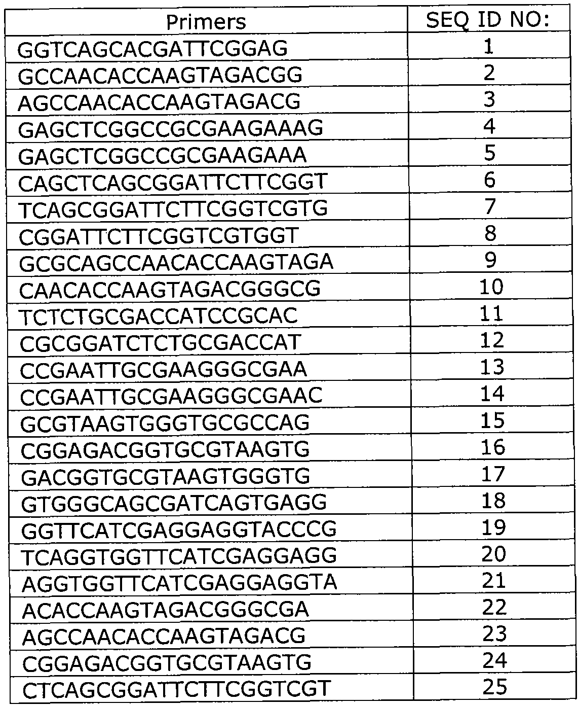
- Primers of a variety of amplicon sizes designed to amplify nucleic acids specific to TB H37rv strain were optimized using SYBR qPCR reaction followed by a melting curve analysis. They may be further validated by Agarose gel (3%) electrophoresis as evidenced by DNA bands of correct sizes without non-specific DNA products or primer-dimers.
- Exemplary TB primers are set forth in Table 1.
- a serial of 10-fold dilutions of TB H37Rv genomic DNA were used as templates in real time qPCR reaction.
- a pair of primers having sequences of GGTCAGCACGATTCGGAG (SEQ ID NO: 1) and GCCAACACCAAGTAGACGG (SEQ ID NO: 2) was used to amplify a target sequence, an IS6110 insertion sequence, in the TB H37Rv genomic DNA.
- the PCR reaction program used included 95°C 3 min, followed by 40 cycles of "94°C 10 sec, 60°C 10 sec. 72°C 30 sec. with fluorescent detection" and a melting phase from 60°C to 95°C.
- Amplification curves Fig.
- tuberculin test (Tuberculin OT, Synbiotics Corp. CA)
- immunoassays for TB antibodies release of cytokines, stimulated IFN-gamma were periodically performed.
- samples were collected from the monkeys for pathological examinations and TB cultures. Whole blood samples were also collected biweekly.
- PWBC Peripheral white blood cells
- Clinical samples (which were ready to be discarded after routine clinical lab tests) were collected from 92 individuals. Among them, 74 individuals were clinically diagnosed of TB, and 18 individuals were not clinically diagnosed for TB. Among these 18 individuals, 15 were diagnosed of other diseases.
- the clinical samples included blood samples, pleural effusion and cerebrospinal fluids (CSF). About 5 ml peripheral blood samples were collected into serum collection tubes or plasma collection tubes with anticoagulants EDTAK2. Both serum and plasma were separated by centrifugation at 1,600 g for 10 min. Serum and plasma aliquots were immediately frozen at -20 °C. Pleural effusion and CSF were collected in tubes with or without anticoagulant EDTAK2, and separated into cell-free fractions and sediments after centrifugation at 5,000g for 10 minutes.
- CSF cerebrospinal fluids
- PS serum-derived protein
- TB plasma fractions, arrow A 6 individuals clinically diagnosed of TB
- non-TB plasma fractions, arrow B 2 individuals not clinically diagnosed for TB
- Fig. 3B The TB specific short nucleic acid fragments of IS6110 (Fig. 3B) in the cell-free fractions of the blood samples were quantified using a standard curve described in Example 2 to have about 20-40 copies per ml of TB plasma fractions and 0 copy per ml of non-TB plasma fractions.
- TB specific nucleic acids were detected in a cell-free fraction of pleural effusion of an individual clinically diagnosed with TB (Fig. 4A, arrow A), but not in the sediment fraction of the same pleural effusion sample (Fig. 4A, arrow B). In addition, the sediment fraction show strong non-specific PCR products (Fig. 4B, arrow B).
- Fig. 5A shows the comparable levels of TB-derived DNA fragments detected in the cell-free fractions (PS vs, CSF) from individuals A and B.
- Fig. 5B shows the specific melting peaks of the IS6110 amplicon of TB DNA fragments, indicating no non-specific PCR products.
- the target TB specific nucleic acid was quantified. A sample having a Ct value greater than 40 was considered as having 0 copy of the target TB specific nucleic acid. A sample having a Ct of 36-40 was considered to have one copy of the target TB specific nucleic acid.
- the copy number of the target TB specific nucleic acid was determined using a standard curve as described in Example 1.
- the average copy number of the target TB specific nucleic acid was 242.6 ⁇ 531.8 per ml of the fraction
- the average copy number of the target TB specific nucleic acid was 16.2 ⁇ 16.2 per ml of the fraction
Abstract
Description

Claims
Priority Applications (9)
| Application Number | Priority Date | Filing Date | Title |
|---|---|---|---|
| US14/009,199 US20140147851A1 (en) | 2011-04-01 | 2012-04-02 | Methods and kits for detecting cell-free pathogen-specific nucleic acids |
| JP2014502902A JP6430826B2 (en) | 2011-04-01 | 2012-04-02 | Method and kit for detecting cell-free pathogen-specific nucleic acid |
| CN201280026842.3A CN103814139B (en) | 2011-04-01 | 2012-04-02 | Method and kit for detecting acellular pathogen specific nucleic acid |
| EP12765867.2A EP2694680A4 (en) | 2011-04-01 | 2012-04-02 | Methods and kits for detecting cell-free pathogen-specific nucleic acids |
| RU2013148806A RU2644672C2 (en) | 2011-04-01 | 2012-04-02 | Methods and kits for detection of cell-free nucleic acids specific for pathogenes |
| AU2012236109A AU2012236109A1 (en) | 2011-04-01 | 2012-04-02 | Methods and kits for detecting cell-free pathogen-specific nucleic acids |
| AU2017203071A AU2017203071A1 (en) | 2011-04-01 | 2017-05-09 | Methods and kits for detecting cell-free pathogen-specific nucleic acids |
| US15/664,074 US20170369932A1 (en) | 2011-04-01 | 2017-07-31 | Methods and kits for detecting cell-free pathogen-specific nucleic acids |
| AU2019204844A AU2019204844A1 (en) | 2011-04-01 | 2019-07-05 | Methods and kits for detecting cell-free pathogen-specific nucleic acids |
Applications Claiming Priority (2)
| Application Number | Priority Date | Filing Date | Title |
|---|---|---|---|
| US201161470774P | 2011-04-01 | 2011-04-01 | |
| US61/470,774 | 2011-04-01 |
Related Child Applications (2)
| Application Number | Title | Priority Date | Filing Date |
|---|---|---|---|
| US14/009,199 A-371-Of-International US20140147851A1 (en) | 2011-04-01 | 2012-04-02 | Methods and kits for detecting cell-free pathogen-specific nucleic acids |
| US15/664,074 Continuation US20170369932A1 (en) | 2011-04-01 | 2017-07-31 | Methods and kits for detecting cell-free pathogen-specific nucleic acids |
Publications (2)
| Publication Number | Publication Date |
|---|---|
| WO2012135815A2 true WO2012135815A2 (en) | 2012-10-04 |
| WO2012135815A3 WO2012135815A3 (en) | 2012-12-27 |
Family
ID=46932429
Family Applications (1)
| Application Number | Title | Priority Date | Filing Date |
|---|---|---|---|
| PCT/US2012/031814 WO2012135815A2 (en) | 2011-04-01 | 2012-04-02 | Methods and kits for detecting cell-free pathogen-specific nucleic acids |
Country Status (7)
| Country | Link |
|---|---|
| US (2) | US20140147851A1 (en) |
| EP (1) | EP2694680A4 (en) |
| JP (2) | JP6430826B2 (en) |
| CN (2) | CN103814139B (en) |
| AU (3) | AU2012236109A1 (en) |
| RU (1) | RU2644672C2 (en) |
| WO (1) | WO2012135815A2 (en) |
Cited By (10)
| Publication number | Priority date | Publication date | Assignee | Title |
|---|---|---|---|---|
| WO2015013486A1 (en) * | 2013-07-26 | 2015-01-29 | General Electric Company | Method and device for collection and amplification of circulating nucleic acids |
| CN104894226A (en) * | 2014-03-07 | 2015-09-09 | 复旦大学 | Method for detecting laboratory mycobacterium tuberculosis micro-environment surface contamination and detection kit |
| EP3141612A1 (en) | 2015-09-10 | 2017-03-15 | Fraunhofer-Gesellschaft zur Förderung der angewandten Forschung e.V. | Method and device for nucleic acid based diagnostic approaches including the determination of a deviant condtion, especially a health condition and/or pathogenic condition of a sample |
| CN108165561A (en) * | 2017-12-01 | 2018-06-15 | 北京蛋白质组研究中心 | Mycobacterium tuberculosis H37Rv encoding gene and its application |
| CN108165560A (en) * | 2017-12-01 | 2018-06-15 | 北京蛋白质组研究中心 | Mycobacterium tuberculosis H37Rv encoding gene and its application |
| CN108165562A (en) * | 2017-12-01 | 2018-06-15 | 北京蛋白质组研究中心 | Mycobacterium tuberculosis H37Rv encoding gene and its application |
| CN108165563A (en) * | 2017-12-01 | 2018-06-15 | 北京蛋白质组研究中心 | Mycobacterium tuberculosis H37Rv encoding gene and its application |
| US10370650B2 (en) | 2013-05-24 | 2019-08-06 | Occam Biolabs, Inc. | System and method for collecting a sample of nucleic acid |
| US11111520B2 (en) | 2015-05-18 | 2021-09-07 | Karius, Inc. | Compositions and methods for enriching populations of nucleic acids |
| US11266337B2 (en) | 2015-09-09 | 2022-03-08 | Drawbridge Health, Inc. | Systems, methods, and devices for sample collection, stabilization and preservation |
Families Citing this family (14)
| Publication number | Priority date | Publication date | Assignee | Title |
|---|---|---|---|---|
| EP2694680A4 (en) * | 2011-04-01 | 2014-09-10 | Occam Biolabs Inc | Methods and kits for detecting cell-free pathogen-specific nucleic acids |
| CN106783971A (en) * | 2011-09-30 | 2017-05-31 | 英特尔公司 | For the tungsten grid of non-planar transistors |
| KR102205950B1 (en) | 2013-11-07 | 2021-01-22 | 더 보드 어브 트러스티스 어브 더 리랜드 스탠포드 주니어 유니버시티 | Cell-free nucleic acids for the analysis of the human microbiome and components thereof |
| RU2020124757A (en) | 2014-11-21 | 2021-06-30 | Оккам Байолэбс, Инк. | SYSTEM AND METHOD FOR COLLECTING NUCLEIC ACID SAMPLE |
| WO2017077999A1 (en) * | 2015-11-06 | 2017-05-11 | 公立大学法人横浜市立大学 | Method for detecting tuberculosis complex-derived dna |
| CN105296661A (en) * | 2015-12-02 | 2016-02-03 | 北京市结核病胸部肿瘤研究所 | Kit for diagnosing tuberculosis by detecting free nucleic acid and application of kit |
| BR112018069557A2 (en) | 2016-03-25 | 2019-01-29 | Karius Inc | synthetic nucleic acid spike-ins |
| JP6812431B2 (en) * | 2016-07-05 | 2021-01-13 | 株式会社日立製作所 | DNA detection method and equipment for that |
| CN110312800A (en) | 2016-11-11 | 2019-10-08 | 生物辐射实验室股份有限公司 | The method for handling sample of nucleic acid |
| MX2019008941A (en) * | 2017-01-30 | 2019-09-11 | Safeguard Biosystems Holdings Ltd | Bead beating tube and method for extracting deoxyribonucleic acid and/or ribonucleic acid from microorganisms. |
| EP3610034B1 (en) | 2017-04-12 | 2022-06-08 | Karius, Inc. | Sample preparation methods, systems and compositions |
| EP3765592A4 (en) | 2018-03-16 | 2021-12-08 | Karius Inc. | Sample series to differentiate target nucleic acids from contaminant nucleic acids |
| CN111876411A (en) * | 2019-09-06 | 2020-11-03 | 深圳微伴生物有限公司 | Primer group for obtaining cfDNA standard substance, PCR amplification positive standard substance, preparation method, kit and application thereof |
| CN114480691A (en) * | 2022-01-24 | 2022-05-13 | 广州迪澳基因科技有限公司 | Method and kit for detecting mycobacterium tuberculosis complex flora based on melting curve |
Family Cites Families (13)
| Publication number | Priority date | Publication date | Assignee | Title |
|---|---|---|---|---|
| US6458366B1 (en) * | 1995-09-01 | 2002-10-01 | Corixa Corporation | Compounds and methods for diagnosis of tuberculosis |
| DE69836012T2 (en) * | 1997-05-02 | 2007-04-05 | Gen-Probe Inc., San Diego | TWO-STEP HYBRIDIZATION AND INJECTION OF A POLYNUCLEOTIDE |
| DE19752898A1 (en) * | 1997-11-28 | 1999-08-05 | Centeon Pharma Gmbh | Method for the detection of high concentrations of four in blood plasma and / or blood serum by means of the polymerase chain reaction |
| US6951722B2 (en) * | 1999-03-19 | 2005-10-04 | Takara Bio Inc. | Method for amplifying nucleic acid sequence |
| TWI237695B (en) * | 1999-12-14 | 2005-08-11 | Joy Biomedical Corp | Helicobacter pylori antigens in blood |
| AU2002224556A1 (en) * | 2000-07-24 | 2002-02-05 | Inpharmatica Limited | Adhesion molecules |
| CN1388378A (en) * | 2002-06-17 | 2003-01-01 | 四川大学 | Tubercle mycobaterium detecting reagent |
| WO2005075683A1 (en) * | 2004-02-03 | 2005-08-18 | Postech Foundation | High throughput device for performing continuous-flow reactions |
| EA014648B1 (en) * | 2005-11-15 | 2010-12-30 | Генойд Кфт. | Method of detecting pathogens |
| US8741565B2 (en) * | 2005-12-30 | 2014-06-03 | Honeywell International Inc. | Oligonucleotide microarray for identification of pathogens |
| US20090142757A1 (en) * | 2007-11-30 | 2009-06-04 | Asiagen Corporation | Strip and method for detecting nucleotide amplification products of mycobacterium tuberculosis and non-tuberculous mycobacterium |
| CA2748822C (en) * | 2009-01-06 | 2016-09-06 | Qimin You | Cross priming amplification of target nucleic acids |
| EP2694680A4 (en) * | 2011-04-01 | 2014-09-10 | Occam Biolabs Inc | Methods and kits for detecting cell-free pathogen-specific nucleic acids |
-
2012
- 2012-04-02 EP EP12765867.2A patent/EP2694680A4/en not_active Withdrawn
- 2012-04-02 US US14/009,199 patent/US20140147851A1/en not_active Abandoned
- 2012-04-02 AU AU2012236109A patent/AU2012236109A1/en not_active Abandoned
- 2012-04-02 WO PCT/US2012/031814 patent/WO2012135815A2/en active Application Filing
- 2012-04-02 CN CN201280026842.3A patent/CN103814139B/en not_active Expired - Fee Related
- 2012-04-02 RU RU2013148806A patent/RU2644672C2/en not_active IP Right Cessation
- 2012-04-02 CN CN201811203829.6A patent/CN109280713A/en active Pending
- 2012-04-02 JP JP2014502902A patent/JP6430826B2/en not_active Expired - Fee Related
-
2017
- 2017-05-09 AU AU2017203071A patent/AU2017203071A1/en not_active Abandoned
- 2017-07-31 US US15/664,074 patent/US20170369932A1/en not_active Abandoned
-
2018
- 2018-05-10 JP JP2018091537A patent/JP2018117645A/en active Pending
-
2019
- 2019-07-05 AU AU2019204844A patent/AU2019204844A1/en not_active Abandoned
Non-Patent Citations (6)
| Title |
|---|
| AGUADO ET AL., LANCET, vol. 347, 1996, pages 1836 - 7 |
| CORDOS ET AL., LANCET, vol. 347, 1996, pages 1082 - 5 |
| KOLK ET AL., LANCET, vol. 344, 1994, pages 694 |
| PALENQUE ET AL., LANCET, vol. 344, 1994, pages 1021 |
| SCHLUGER ET AL., LANCET, vol. 344, 1994, pages 232 - 3 |
| See also references of EP2694680A4 |
Cited By (21)
| Publication number | Priority date | Publication date | Assignee | Title |
|---|---|---|---|---|
| US10370650B2 (en) | 2013-05-24 | 2019-08-06 | Occam Biolabs, Inc. | System and method for collecting a sample of nucleic acid |
| US9644232B2 (en) | 2013-07-26 | 2017-05-09 | General Electric Company | Method and device for collection and amplification of circulating nucleic acids |
| CN105392900A (en) * | 2013-07-26 | 2016-03-09 | 通用电气公司 | Method and device for collection and amplification of circulating nucleic acids |
| JP2016530878A (en) * | 2013-07-26 | 2016-10-06 | ゼネラル・エレクトリック・カンパニイ | Method and apparatus for recovery and amplification of circulating nucleic acids |
| US11142758B2 (en) | 2013-07-26 | 2021-10-12 | Global Life Sciences Solutions Operations UK Ltd | Method and device for collection and amplification of circulating nucleic acids |
| WO2015013486A1 (en) * | 2013-07-26 | 2015-01-29 | General Electric Company | Method and device for collection and amplification of circulating nucleic acids |
| CN104894226A (en) * | 2014-03-07 | 2015-09-09 | 复旦大学 | Method for detecting laboratory mycobacterium tuberculosis micro-environment surface contamination and detection kit |
| US11111520B2 (en) | 2015-05-18 | 2021-09-07 | Karius, Inc. | Compositions and methods for enriching populations of nucleic acids |
| US11266337B2 (en) | 2015-09-09 | 2022-03-08 | Drawbridge Health, Inc. | Systems, methods, and devices for sample collection, stabilization and preservation |
| EP3141612A1 (en) | 2015-09-10 | 2017-03-15 | Fraunhofer-Gesellschaft zur Förderung der angewandten Forschung e.V. | Method and device for nucleic acid based diagnostic approaches including the determination of a deviant condtion, especially a health condition and/or pathogenic condition of a sample |
| WO2017042287A1 (en) | 2015-09-10 | 2017-03-16 | Fraunhofer-Gesellschaft zur Förderung der angewandten Forschung e.V. | Method and device for nucleic acid based diagnostic approaches including the determination of a deviant condition, especially a health condition and/or a pathogenic condition of a sample |
| US10910088B2 (en) | 2015-09-10 | 2021-02-02 | Fraunhofer-Gesellschaft Zur Förderung Der | Method and device for nucleic acid based diagnostic approaches including the determination of a deviant condition, especially a health condition and/or a pathogenic condition of a sample |
| CN108431237A (en) * | 2015-09-10 | 2018-08-21 | 弗劳恩霍夫应用研究促进协会 | For the unusual condition including determining sample, the especially method and apparatus of the diagnostic method based on nucleic acid of health status and/or pathogenic situation |
| CN108165563A (en) * | 2017-12-01 | 2018-06-15 | 北京蛋白质组研究中心 | Mycobacterium tuberculosis H37Rv encoding gene and its application |
| CN108165563B (en) * | 2017-12-01 | 2021-02-19 | 北京蛋白质组研究中心 | Mycobacterium tuberculosis H37Rv encoding gene and application thereof |
| CN108165562B (en) * | 2017-12-01 | 2021-06-08 | 北京蛋白质组研究中心 | Mycobacterium tuberculosis H37Rv encoding gene and application thereof |
| CN108165560B (en) * | 2017-12-01 | 2021-06-08 | 北京蛋白质组研究中心 | Mycobacterium tuberculosis H37Rv encoding gene and application thereof |
| CN108165561B (en) * | 2017-12-01 | 2021-06-18 | 北京蛋白质组研究中心 | Mycobacterium tuberculosis H37Rv encoding gene and application thereof |
| CN108165562A (en) * | 2017-12-01 | 2018-06-15 | 北京蛋白质组研究中心 | Mycobacterium tuberculosis H37Rv encoding gene and its application |
| CN108165560A (en) * | 2017-12-01 | 2018-06-15 | 北京蛋白质组研究中心 | Mycobacterium tuberculosis H37Rv encoding gene and its application |
| CN108165561A (en) * | 2017-12-01 | 2018-06-15 | 北京蛋白质组研究中心 | Mycobacterium tuberculosis H37Rv encoding gene and its application |
Also Published As
| Publication number | Publication date |
|---|---|
| CN109280713A (en) | 2019-01-29 |
| US20170369932A1 (en) | 2017-12-28 |
| JP2014510538A (en) | 2014-05-01 |
| CN103814139A (en) | 2014-05-21 |
| CN103814139B (en) | 2018-11-13 |
| EP2694680A2 (en) | 2014-02-12 |
| JP2018117645A (en) | 2018-08-02 |
| WO2012135815A3 (en) | 2012-12-27 |
| RU2013148806A (en) | 2015-05-10 |
| US20140147851A1 (en) | 2014-05-29 |
| AU2019204844A1 (en) | 2019-07-25 |
| AU2017203071A1 (en) | 2017-06-01 |
| JP6430826B2 (en) | 2018-11-28 |
| RU2644672C2 (en) | 2018-02-13 |
| EP2694680A4 (en) | 2014-09-10 |
| AU2012236109A1 (en) | 2013-11-21 |
Similar Documents
| Publication | Publication Date | Title |
|---|---|---|
| US20170369932A1 (en) | Methods and kits for detecting cell-free pathogen-specific nucleic acids | |
| CN110541022B (en) | Mycobacterium tuberculosis complex detection kit based on CRISPR-Cas12a system | |
| US7803929B2 (en) | Kits for diagnosis and monitoring of pathogenic infection by analysis of cell-free pathogenic nucleic acids in urine | |
| Deshpande et al. | Evaluation of the IS 6110 PCR assay for the rapid diagnosis of tuberculous meningitis | |
| US5942394A (en) | Detection of protozoan parasites | |
| AU2018383191B2 (en) | A non-invasive method for monitoring transplanted organ status in organ-transplant recipients | |
| US20150152485A1 (en) | Mycobacterium tuberculosis detection using transrenal dna | |
| CN114410836A (en) | Kit and method for detecting human parvovirus B19 by integrating sample collection treatment, nucleic acid extraction and multiple isothermal amplification | |
| CN110462064A (en) | The method and its application of microorganism detection are carried out based on excretion body nucleic acid | |
| JP3194943B2 (en) | Nucleic acid probe and method for detecting Cryptococcus neoformans | |
| US9593384B2 (en) | Metronidazole resistance in trichomonas vaginalis and single nucleotide polymorphisms | |
| KR101912488B1 (en) | Molecular detection assay | |
| WO2017073753A1 (en) | Method for measuring ability to produce equol | |
| WO2013049822A2 (en) | Diagnostic method for determining animals persistently infected (pi) with bovine viral diarrhea virus (bvdv) | |
| KR101395938B1 (en) | Pcr diagnosis using specific primer for bacteria that cause diseases of allomyrina dichotoma | |
| Khosla et al. | DIAGNOSTIC EVALUATION OF 38 kDa GENE BASED PCR ASSAY IN DIAGNOSING SMEAR NEGATIVE PULMONARY TUBERCULOSIS | |
| Polley | Parasite Genomes and Diagnostics | |
| KR20220110152A (en) | Method for diagnosing of infectious respiratoy diseases using extracellular vesicles | |
| CN115725608A (en) | Pathogen specific nucleic acid gene and acquisition and detection method | |
| CN115961089A (en) | Primer and kit for detecting human parvovirus B19 | |
| CN115948611A (en) | Detection reagent for coronavirus pathogen nucleic acid and detection method and application thereof | |
| Edwards | The Application of Loop Mediated Isothermal Amplification for the Detection of the Sexually Transmitted Pathogens Chlamydia trachomatis, Neisseria gonorrhoeae, Mycoplasma genitalium, and Trichomonas vaginalis, at the Point of Care | |
| CN116855642A (en) | Real-time fluorescent nucleic acid isothermal amplification detection kit for detecting 14 high-risk HPVs | |
| WO2009035955A1 (en) | Methods for detecting enterobacter sakazakii | |
| CN115896316A (en) | Tuberculosis detection method |
Legal Events
| Date | Code | Title | Description |
|---|---|---|---|
| 121 | Ep: the epo has been informed by wipo that ep was designated in this application |
Ref document number: 12765867 Country of ref document: EP Kind code of ref document: A2 |
|
| ENP | Entry into the national phase |
Ref document number: 2014502902 Country of ref document: JP Kind code of ref document: A |
|
| NENP | Non-entry into the national phase |
Ref country code: DE |
|
| WWE | Wipo information: entry into national phase |
Ref document number: 2012765867 Country of ref document: EP |
|
| ENP | Entry into the national phase |
Ref document number: 2013148806 Country of ref document: RU Kind code of ref document: A |
|
| ENP | Entry into the national phase |
Ref document number: 2012236109 Country of ref document: AU Date of ref document: 20120402 Kind code of ref document: A |
|
| WWE | Wipo information: entry into national phase |
Ref document number: 14009199 Country of ref document: US |