以下、本発明を詳細に記載する。
本発明は、核酸と検出試薬とを閉鎖系内で接触させる工程、および核酸および/または検出試薬の呈色を可視光で判定する工程を含む、核酸の検出方法に関する。
Hereinafter, the present invention will be described in detail.
The present invention relates to a method for detecting a nucleic acid, comprising the steps of bringing a nucleic acid and a detection reagent into contact in a closed system, and determining the coloration of the nucleic acid and / or the detection reagent with visible light.
本発明における核酸とは、核酸増幅法により得られた増幅核酸、二本鎖DNA、二本鎖RNA、DNAとRNAのハイブリッド鎖、およびPNAなどの人工核酸の二本鎖が含まれるが、一本鎖核酸であっても良い。例えば、一本鎖RNAを逆転写酵素によりDNAに逆転写し、このDNAを鋳型として二本鎖核酸を増幅すれば、間接的にRNAを検出できるからである。
The nucleic acid in the present invention includes an amplified nucleic acid obtained by a nucleic acid amplification method, double-stranded DNA, double-stranded RNA, a hybrid strand of DNA and RNA, and a double strand of artificial nucleic acid such as PNA. It may be a double-stranded nucleic acid. This is because, for example, RNA can be indirectly detected by reversely transcribing single-stranded RNA to DNA using reverse transcriptase and amplifying double-stranded nucleic acid using this DNA as a template.
核酸は、試料に含まれているものであってもよい。試料には、核酸増幅法により得られた増幅核酸、核酸を含む生体成分、例えば微生物や動物細胞、植物細胞からの抽出液、あるいは食品からの抽出液が含まれる。生体成分から抽出されたDNAやプラスミドDNAも含まれる。
The nucleic acid may be contained in a sample. The sample includes an amplified nucleic acid obtained by the nucleic acid amplification method, a biological component containing the nucleic acid, for example, an extract from microorganisms, animal cells, plant cells, or an extract from food. DNA extracted from biological components and plasmid DNA are also included.
核酸と検出試薬との接触は、核酸および/または検出試薬を呈色させるものであればよい。例えば、核酸を含む溶液と液体の検出試薬を混合させてもよいし、核酸を含む溶液と個体の検出試薬を接触させてもよい。
The contact between the nucleic acid and the detection reagent may be anything that causes the nucleic acid and / or the detection reagent to be colored. For example, a solution containing a nucleic acid and a liquid detection reagent may be mixed, or a solution containing a nucleic acid may be brought into contact with an individual detection reagent.
閉鎖系とは、核酸の増幅及び検出の各工程間で核酸を含有する試料の添加や除去が行われず、一貫して閉ざされた系を意味する。ただし、完全な機密状態である必要はなく、例えば容器内で核酸と検出試薬とを接触させるために容器を上下反転等した場合に、核酸や検出試薬が漏出しなければ、本発明における閉鎖系に該当する。閉鎖系内で核酸と検出試薬を接触させることにより、異なる核酸同士の混入による誤検出や核酸の飛散による汚染を防止できる。さらに、接触工程の前または後に、核酸を増幅する工程を含む場合には、当該増幅工程、接触工程、および可視光で判定する工程を閉鎖系で行うことにより、感度の高い増幅方法を利用した場合でも、誤検出や汚染を防止し、迅速に可視光で判定することができる。
A closed system means a system that is consistently closed without the addition or removal of a sample containing nucleic acid between nucleic acid amplification and detection steps. However, it is not necessary to be in a completely confidential state. For example, when the container is turned upside down to bring the nucleic acid and the detection reagent into contact with each other in the container, if the nucleic acid or the detection reagent does not leak, the closed system according to the present invention. It corresponds to. By contacting the nucleic acid and the detection reagent in a closed system, it is possible to prevent erroneous detection due to mixing of different nucleic acids and contamination due to scattering of the nucleic acid. Further, when a nucleic acid amplification step is included before or after the contact step, a highly sensitive amplification method is used by performing the amplification step, the contact step, and the step of determining with visible light in a closed system. Even in this case, it is possible to prevent erroneous detection and contamination, and to quickly determine with visible light.
核酸および/または検出試薬の呈色を可視光で判定する工程では、核酸と検出試薬との接触により生じた物質を、紫外線のような可視光ではない光を照射することなく、一般的な実験室における照明のような可視光下で観察し、核酸の存在を判定する。可視光であれば、紫外線照射する場合のように特別な装置は必要でなく、簡便に観察することができる。
In the step of determining the coloration of the nucleic acid and / or detection reagent with visible light, a general experiment can be performed without irradiating light that is not visible light such as ultraviolet rays to the substance generated by contact between the nucleic acid and the detection reagent. Observation under visible light such as lighting in a room to determine the presence of nucleic acids. In the case of visible light, a special apparatus is not required as in the case of ultraviolet irradiation, and observation can be performed easily.
呈色の可視光での判定は目視により行うことができる。色素と核酸が結合した場合の呈色の変化、核酸と結合していない色素の呈色の変化を、可視光で目視で観察し、色素の呈色の有無に基づき、核酸の存在の有無を確認できる。
The determination of the coloration with visible light can be made visually. The change in coloration when the dye and nucleic acid are combined, and the change in coloration of the dye that is not bonded to the nucleic acid are visually observed with visible light, and the presence or absence of nucleic acid is determined based on the presence or absence of coloration of the dye. I can confirm.
ここで、呈色の変化として、可視光下で色の種類が変化すること(可視光波長)、又は、色の濃淡が変化すること(反射率)などが挙げられるが、可視光で判定できるものであればこれらに限定されない。色の種類の変化としては、例えば、赤紫色から水色への変化、及び黄色から赤紫色への変化が挙げられる。
Here, examples of the color change include a change in color type under visible light (visible light wavelength), a change in color shade (reflectance), and the like, which can be determined with visible light. If it is a thing, it will not be limited to these. Examples of the color type change include a change from red purple to light blue and a change from yellow to red purple.
また、呈色の可視光での判定は、試料溶液の可視光領域の吸光度を測定することによっても行える。判定を目視により行うか吸光度を測定するかにかかわらず、核酸と検出試薬に照射する光、及び観察する光の波長は、いずれも可視光の波長である。可視光の波長は380nm~800nmであることが好ましく、400nm~780nmであることがより好ましく、450nm~750nmであることがさらに好ましい。測定波長は可視光の波長の範囲内で、使用する色素によって適宜設定すればよい。また、吸光度測定により、試料中の核酸濃度を定量することも可能である。
Moreover, the determination with the visible light of coloring can also be performed by measuring the light absorbency of the visible light region of a sample solution. Regardless of whether the determination is made visually or the absorbance is measured, the wavelengths of the light irradiated to the nucleic acid and the detection reagent and the wavelength of the light to be observed are both visible light wavelengths. The wavelength of visible light is preferably 380 to 800 nm, more preferably 400 to 780 nm, and further preferably 450 to 750 nm. What is necessary is just to set a measurement wavelength suitably with the pigment | dye to be used within the range of the wavelength of visible light. It is also possible to quantify the nucleic acid concentration in the sample by measuring absorbance.
その他、色素と核酸による不溶性の沈殿物を生じる場合、フィルターやメンブレンを用いて、又は遠心分離により沈殿物を回収し、沈殿物の量から核酸の量を判断することも可能である。
In addition, when an insoluble precipitate due to the dye and the nucleic acid is generated, the amount of the nucleic acid can be determined from the amount of the precipitate by collecting the precipitate using a filter or a membrane or by centrifugation.
本発明において、核酸検出後の着色した検体液を他の分子生物学的な操作にそのまま使用することも可能である。そのような分子生物学的な操作には、制限酵素反応、シーケンス反応、PCRの様な酵素反応や、電気泳動による確認操作等が含まれる。
In the present invention, it is also possible to use the colored sample solution after nucleic acid detection as it is for other molecular biological operations. Such molecular biological operations include restriction enzyme reactions, sequence reactions, enzymatic reactions such as PCR, confirmation operations by electrophoresis, and the like.
核酸と検出試薬との接触工程の前または後に、核酸を増幅する工程を含むことが好ましい。核酸の増幅は、PCR法に代表される核酸配列を増幅させる手法であればよく、特に限定されるものではない。核酸配列を増幅させる手法としては、例えば、PCR法以外に、LCR(Ligase Chain Reaction)法、SDA(Strand Displacement Amplification)法、RCA(Rolling Circle Amplification)法、CPT(Cycling Probe Technology)法、Q-Beta Replicase Amplification Technology法、ICAN(Isothermal and Chimeric primer-initiated Amplification of Nucleic Acids)法、LAMP(Loop-Mediated Isothermal Amplificaton of DNA)法、NASBA(Nucleic acid Sequence-based Amplification method)法、及びTMA(Transcription mediated amplification method)法等の公知の方法が例示できるが、これらに限定されるものではない。
It is preferable to include a step of amplifying the nucleic acid before or after the step of contacting the nucleic acid with the detection reagent. Nucleic acid amplification is not particularly limited as long as it is a technique for amplifying a nucleic acid sequence represented by PCR. As a method for amplifying a nucleic acid sequence, for example, in addition to the PCR method, the LCR (Ligase Chain Reaction) method, the SDA (Strand Displacement Amplification) method, the RCA (Rolling Circle Amplification) method, and the CPT (CyclingProbing Method) Beta Replication Amplification Technology Method, ICAN (Isothermal and Chimeric Primer-Initiated Amplified Of Nucleic Acids Method), LAMP (Loop-MediatedDiffusionID) ) Method, NASBA (Nucleic acid Sequence-based Amplification method) method, and TMA (Transcription mediated amplification method) method is a known method can be exemplified in, but not limited thereto.
Q-Beta Replicase Amplification Technology法、RCA法、NASBA法、SDA法、TMA法、LAMP法、ICAN法などは一定温度で増幅反応を行う方法であり、その他のPCR法やLCR法などは温度サイクリングで増幅反応を行うものである。
Q-Beta Replication Amplification Technology method, RCA method, NASBA method, SDA method, TMA method, LAMP method, ICAN method, etc. are methods for performing amplification reaction at a constant temperature, and other PCR methods and LCR methods are temperature cycling. An amplification reaction is performed.
また、RNAを逆転写酵素等によりDNAに逆転写し、このDNAを鋳型として二本鎖核酸を増幅すれば、間接的にRNAを検出することも可能となる。
In addition, RNA can be indirectly detected by reversely transcribing RNA to DNA using reverse transcriptase or the like and amplifying double-stranded nucleic acid using this DNA as a template.
核酸増幅方法としてLAMP法を用いた場合、反応に温度サイクルが不要で簡便であり、増幅核酸量が多いというメリットがある。また、LAMP法において反応効率を向上させる目的でピロリン酸除去酵素の存在下で増幅反応を行うことも可能である。ピロリン酸除去酵素としては、ピロリン酸をモノリン酸に分解するピロホスファターゼや、ピロリン酸とアデノシン5’-ホスフォリン酸を結合するATPスルフリラーゼが挙げられる。本発明では、特に、耐熱性のピロホスファターゼが好ましい。
When the LAMP method is used as the nucleic acid amplification method, there is an advantage that the reaction does not require a temperature cycle and is simple and the amount of amplified nucleic acid is large. In the LAMP method, an amplification reaction can be performed in the presence of a pyrophosphate removing enzyme for the purpose of improving the reaction efficiency. Examples of the pyrophosphate removing enzyme include pyrophosphatase that decomposes pyrophosphate into monophosphate, and ATP sulfurylase that binds pyrophosphate and adenosine 5'-phosphophosphate. In the present invention, thermostable pyrophosphatase is particularly preferable.
検出試薬とは、核酸の検出試薬であり、核酸に結合する色素を含む。さらに、一本鎖核酸に結合した色素および核酸と結合していない色素と優先的に反応する物質を含んでいてもよい。
The detection reagent is a nucleic acid detection reagent and contains a dye that binds to the nucleic acid. Furthermore, a substance that preferentially reacts with a dye bound to a single-stranded nucleic acid and a dye not bound to a nucleic acid may be included.
色素とは、可視光の吸収により呈色する物質のことを指す。核酸に結合する色素としては、核酸に結合できれば特に制限されないが、例えば、トリフェニルメタン系色素、チアジン系色素、オキサジン系色素、アジン系色素、フェナジン系色素、キサンテン系色素、フェナントリジウム系色素、アゾ系色素、ラクトン系色素、サルトン系色素、インジゴイド系色素、シアニン系色素、オキソノール系色素、スチリル系色素、ポリフィリン系色素、チオキサンテン系色素、スクワリリウム系色素、クロコニウム系色素、アズレニウム系色素、ジチオール金属塩系色素、ナフトキノン系色素、アントラキノン系色素、インドフェノール系色素、クマリン系色素、ケトクマリン系色素、ピリリウム塩系色素、チオピリリウム塩系色素、チアゾール系色素、キノリン系色素、ベンゾフェノン系色素、チオベンゾフェノン系色素およびこれらの混合物が挙げられる。
A pigment | dye refers to the substance colored by absorption of visible light. The dye that binds to nucleic acid is not particularly limited as long as it can bind to nucleic acid. For example, triphenylmethane dye, thiazine dye, oxazine dye, azine dye, phenazine dye, xanthene dye, phenanthridium dye Azo dyes, lactone dyes, sultone dyes, indigoid dyes, cyanine dyes, oxonol dyes, styryl dyes, porphyrin dyes, thioxanthene dyes, squarylium dyes, croconium dyes, azurenium dyes, Dithiol metal salt dyes, naphthoquinone dyes, anthraquinone dyes, indophenol dyes, coumarin dyes, ketocoumarin dyes, pyrylium salt dyes, thiopyrylium salt dyes, thiazole dyes, quinoline dyes, benzophenone dyes, thiobenzo Enon based dyes and mixtures thereof.
好ましくはトリフェニルメタン系色素、チアジン系色素、オキサジン系色素、アジン系色素、フェナジン系色素、キサンテン系色素、フェナントリジウム系色素、アゾ系色素、インジゴイド系色素、ラクトン系色素、サルトン系色素およびこれらの混合物である。
Preferably triphenylmethane dyes, thiazine dyes, oxazine dyes, azine dyes, phenazine dyes, xanthene dyes, phenanthridium dyes, azo dyes, indigoid dyes, lactone dyes, sultone dyes and It is a mixture of these.
トリフェニルメタン系色素としては、例えば、メチルグリーン、マラカイトグリーン、クリスタルバイオレット、パラロザニリン、ゲンチアナバイオレットB、ゲンチアナバイオレットR、ナイトブルー、ビクトリアブルーB、ビクトリアブルーR、ビクトリアブルー4R、ビクトリアブルーBO、ロゾール酸、フクシン、酸性フクシン、塩基性フクシン、ニューフクシン、ブロモチモールブルー、ブロモクレゾールグリーンフェノールフタレイン、ブロモフェノールブルー、パテントブルーバイオレット、フェノールレッド、ピグメントブルー1、ピグメントバイオレット3、ベンジルバイオレット、ペンタメチルパラロザニリン、ファストグリーンFCF、エチルバイオレット、グリーンS、ローズアニリン、アシッドブルー7、アズールブルーG、ソロクロムシアニンR、アシッドブルー147、ライトグリーンSFイエロー、ライトグリーンSF、エチルグリーン、アニリンルー、メチルバイオレット、クロムバイオレットCGが挙げられる。
Examples of triphenylmethane dyes include methyl green, malachite green, crystal violet, pararosaniline, gentian violet B, gentian violet R, night blue, Victoria blue B, Victoria blue R, Victoria blue 4R, Victoria blue BO, and rosoleic acid. , Fuchsin, acid fuchsin, basic fuchsin, new fuchsin, bromothymol blue, bromocresol green phenolphthalein, bromophenol blue, patent blue violet, phenol red, pigment blue 1, pigment violet 3, benzyl violet, pentamethylpararosa Nilin, Fast Green FCF, Ethyl Violet, Green S, Rose Aniline, Acid Blue 7, Azure Blue G, solo chromium cyanine R, Acid Blue 147, Light Green SF Yellow, Light Green SF, ethyl green, Anirinru, methyl violet, and chromium violet CG.
チアジン系色素としては、例えば、トルイジンブルーO、メチレンブルー、チオニン、アズールA、チオールCが挙げられる。
Examples of thiazine dyes include toluidine blue O, methylene blue, thionine, azure A, and thiol C.
オキサジン系色素としては、例えば、ブリリアントクレシルブルー、ナイルブルー、ガロシアニン、ベーシックブルー3が挙げられる。
Examples of oxazine dyes include brilliant cresyl blue, nile blue, galocyanine, and basic blue 3.
アジン系色素としては、例えば、アニリンブラック、アセチレンブラック等が挙げられる。
Examples of azine dyes include aniline black and acetylene black.
フェナジン系色素としては、例えば、ニュートラルレッド、ヤヌスグリーンB、ベーシックレッド2、サフラニンBが挙げられる。
Examples of phenazine dyes include neutral red, Janus green B, basic red 2, and safranin B.
キサンテン系色素としては、例えば、フルオレセイン、ローダミン、ピロニンYが挙げられる。
Examples of the xanthene dye include fluorescein, rhodamine, and pyronin Y.
フェナントリジウム系色素としては、例えば、エチジウムブロマイドが挙げられる。
Examples of phenanthridium dyes include ethidium bromide.
アゾ色素としては、例えば、ビスマルクブラウン、ニューコクシン、ベーシックレッド29が挙げられる。インジゴイド系色素としては、例えば、インジゴカルミンなどが挙げられる。
Examples of the azo dye include Bismarck Brown, New Coxin, and Basic Red 29. Examples of indigoid pigments include indigo carmine.
検体への検出試薬の添加量は、呈色を可視光で判定できれば特に限定されるものではないが、通常10%以下であり、好ましくは1%以下であり、さらに好ましくは0.1%以下である。
The addition amount of the detection reagent to the sample is not particularly limited as long as the coloration can be determined with visible light, but is usually 10% or less, preferably 1% or less, more preferably 0.1% or less. It is.
これらの色素のなかでも、核酸との結合により色調が変化する性質を有し、かつ、構造変化に伴い色調が変化する性質を有していることが好ましいが、いずれか一方のみを有していてもよい。また、これらの色素は一種でも使用でき、二種以上を組み合わせても使用できる。
Among these dyes, it is preferable that they have a property that the color tone is changed by binding to a nucleic acid, and a property that the color tone changes with a structural change, but only one of them is included. May be. These dyes can be used alone or in combination of two or more.
核酸との結合により色調が変化する性質を有する色素としては、例えば、トルイジンブルーOやメチルグリーン、ブリリアントクレシルブルー、ゲンチアナバイオレットB、ビクトリアブルーB等が挙げられる。具体的にはトルイジンブルーOは二本鎖核酸が存在しない場合は濃紺であるが、核酸と結合することで水色へと変化し、メチルグリーンは核酸が存在しない場合は水色であるが、核酸と結合することで青緑色へと変化する。また、ブリリアントクレシルブルーは核酸が存在しない場合は濃青色であるが、核酸と結合することで青緑色へと変化する。
Examples of the dye having the property of changing the color tone by binding to nucleic acid include toluidine blue O, methyl green, brilliant cresyl blue, gentian violet B, and Victoria blue B. Specifically, toluidine blue O is dark blue when no double-stranded nucleic acid is present, but changes to light blue by binding to nucleic acid, and methyl green is light blue when no nucleic acid is present. When combined, it turns blue-green. Brilliant cresyl blue is dark blue when no nucleic acid is present, but changes to blue-green when bound to nucleic acid.
一方、構造変化に伴って色調が変化する色素としては、プロトン化状態の違いや酸化還元状態の違いなどで異なる色調に変化するpH応答性色素や酸化還元応答性色素などが挙げられる。構造変化には共役構造の変化や、置換基の導入や変換、プロトン化状態の違いなどが含まれる。
On the other hand, examples of the dye whose color tone changes with the structural change include a pH-responsive dye or a redox-responsive dye that changes to a different color tone due to a difference in protonated state or a difference in redox state. Structural changes include changes in conjugated structures, introduction and conversion of substituents, differences in protonation states, and the like.
pH応答性色素とは、pH指示薬等にみられるような、溶液中の水素イオン濃度(pH)に応じて色調を変える色素を意味する。これらの中には、酸性からアルカリ性にすることで、フェノールフタレインのように無色から赤紫へと変化するものや、メチルオレンジのように赤色から橙色へと変化するものが含まれる。
The pH-responsive dye means a dye that changes color tone according to the hydrogen ion concentration (pH) in the solution, as found in a pH indicator or the like. These include those that change from colorless to reddish violet, such as phenolphthalein, and those that change from red to orange, such as methyl orange, by changing from acidic to alkaline.
酸化還元応答性色素とは、例えば、酸塩基指示薬や酸化還元指示薬に用いられるような色素が挙げられ、酸化状態と還元状態で色調が変化する色素を意味する。例えば、メチレンブルーやメチルグリーンは酸化型では青色を呈するが、還元型では無色となる。一方、トルイジンブルーOは酸化型では青色を呈するが、還元型では、赤紫色を呈する。
The redox-responsive dye includes, for example, a dye used for an acid-base indicator or a redox indicator, and means a dye whose color tone changes between an oxidized state and a reduced state. For example, methylene blue and methyl green are blue in the oxidized form, but are colorless in the reduced form. On the other hand, toluidine blue O exhibits blue in the oxidized form, but exhibits reddish purple in the reduced form.
構造変化に伴い色調が変化する性質を有する色素を用いた場合、二本鎖核酸に結合した色素と比較して、一本鎖核酸に結合した色素および核酸と結合していない色素と優先的に反応する物質を添加する工程を追加することで、核酸の有無による色調のコントラストを増強し可視光による検出を容易にすることが可能である。つまり、二本鎖核酸と結合した色素に由来する色調以外を変化させ、又は消して無色にすることで、二本鎖核酸の有無による色調のコントラストが増強される。上記工程により、二本鎖核酸と結合はするが、二本鎖核酸との結合による色調の変化が少ない、或いは、結合はするが色調の変化が全くない色素を使用する場合でも、色素がpH応答性あるいは酸化還元応答性を有していれば、二本鎖核酸増幅の判定が可能となる。
When using a dye that has the property of changing its color tone as the structure changes, it is preferential to dyes that are bound to single-stranded nucleic acids and dyes that are not bound to nucleic acids, compared to dyes that are bound to double-stranded nucleic acids. By adding a step of adding a reacting substance, it is possible to enhance the contrast of the color tone depending on the presence or absence of a nucleic acid and facilitate detection by visible light. That is, by changing the color tone other than the color tone derived from the dye bound to the double-stranded nucleic acid, or by eliminating the color tone, the contrast of the color tone depending on the presence or absence of the double-stranded nucleic acid is enhanced. Even if a dye that binds to a double-stranded nucleic acid but binds to a double-stranded nucleic acid with little or no change in color tone due to the above-mentioned process or has a binding but no color change at all is used, the dye has a pH of If it has responsiveness or redox responsiveness, determination of double-stranded nucleic acid amplification becomes possible.
例えば、メチルグリーンは二本鎖核酸と結合した際の色調の変化を目視で判別しにくい。しかし、一本鎖核酸と結合したメチルグリーンおよび核酸と結合していないメチルグリーンと優先的に反応する物質を添加することで、二本鎖核酸と結合していないメチルグリーン由来の水色を消すことができる。この方法により、着色されている場合は二本鎖核酸が存在し、無色の場合は二本鎖核酸が存在しないというように、核酸の有無を容易に判定することが可能となる。
For example, methyl green is difficult to visually distinguish changes in color when bound to double-stranded nucleic acids. However, by adding a substance that preferentially reacts with methyl green bound to single-stranded nucleic acid and methyl green not bound to nucleic acid, the light blue derived from methyl green not bound to double-stranded nucleic acid is eliminated. Can do. By this method, it is possible to easily determine the presence or absence of a nucleic acid, such that a double-stranded nucleic acid is present when colored, and a double-stranded nucleic acid is absent when colorless.
もう一つの例として、トルイジンブルーOは二本鎖核酸と結合すると、濃紺から青色へと変化する。さらに、一本鎖核酸と結合したトルイジンブルーO、および核酸と結合していないトルイジンブルーOと優先的に反応する物質を添加することで、二本鎖核酸と結合していないトルイジンブルー由来の濃紺色を赤紫色へと変化させ、青色の場合は二本鎖核酸が存在し、赤紫色の場合は二本鎖核酸が存在しないというように、核酸の有無を容易に判定することが可能となる。
As another example, when toluidine blue O binds to a double-stranded nucleic acid, it changes from dark blue to blue. Further, by adding a toluidine blue O bound to a single-stranded nucleic acid and a substance that preferentially reacts with toluidine blue O not bound to a nucleic acid, a dark blue derived from toluidine blue not bound to a double-stranded nucleic acid By changing the color to magenta, it is possible to easily determine the presence or absence of nucleic acid, such as double-stranded nucleic acid is present in blue and no double-stranded nucleic acid is present in red-purple. .
さらにもう一つの例として、ゲンチアナバイオレットBは二本鎖核酸と結合した際の色調の変化を目視で判別しにくい。しかし、一本鎖核酸と結合したゲンチアナバイオレットB、および核酸と結合していないゲンチアナバイオレットBと優先的に反応する物質を添加することで、二本鎖核酸と結合していないゲンチアナバイオレットB由来の青紫色を消すことができ、青紫色の場合は二本鎖核酸が存在し、無色の場合は二本鎖核酸が存在しないというように、核酸の有無を容易に判定することが可能となる。
As yet another example, gentian violet B is difficult to visually distinguish changes in color when bound to double-stranded nucleic acids. However, by adding a substance that preferentially reacts with gentian violet B bound to single-stranded nucleic acid and gentian violet B not bound to nucleic acid, it is derived from gentian violet B not bound to double-stranded nucleic acid. It is possible to erase the blue-violet color, and it is possible to easily determine the presence or absence of a nucleic acid, such that a double-stranded nucleic acid is present when the color is blue-violet, and a double-stranded nucleic acid is not present when the color is colorless.
さらにもう一つの例として、ビクトリアブルーBは中性溶液中では二本鎖核酸と結合した際の色調の変化を判別しにくい。しかし、中性以上のアルカリ領域のpH環境下では二本鎖核酸と結合していないビクトリアブルーB由来の青色が淡赤色へと変化するので、青色の場合は二本鎖核酸が存在し、淡赤色の場合は二本鎖核酸が存在しないというように、核酸の有無を容易に判定することが可能となる。
As yet another example, Victoria Blue B is difficult to distinguish a change in color tone when bound to a double-stranded nucleic acid in a neutral solution. However, since the blue color derived from Victoria Blue B, which is not bound to the double-stranded nucleic acid, changes to a light red color in a pH environment of a neutral or higher alkaline region, the double-stranded nucleic acid is present in the blue color. In the case of red, it is possible to easily determine the presence or absence of nucleic acid, such as no double-stranded nucleic acid.
本工程により色調のコントラストの増強効果を得ようとする場合、使用する色素は特に限定されるものではないが、二本鎖核酸と結合しない場合に、色素の構造変化に依存して呈色が有色から無色へ、又は無色から有色へと変化する色素であってもよい。また、例えば青色から赤色へというように、色調が変化する色素であってもよい。このうちでも、核酸の有無の判定が容易であるという理由から、色素の構造変化に依存して呈色が有色から無色へ、又は無色から有色へと変化する色素であることが好ましい。
When trying to obtain a contrast enhancement effect of color tone by this step, the dye to be used is not particularly limited, but when it does not bind to a double-stranded nucleic acid, the coloration depends on the structural change of the dye. It may be a pigment that changes from colored to colorless or from colorless to colored. Moreover, the pigment | dye from which a color tone changes, for example from blue to red, may be sufficient. Among these, a dye whose coloration changes from colored to colorless or from colorless to colored depending on the structural change of the dye is preferable because it is easy to determine the presence or absence of nucleic acid.
一本鎖核酸に結合した色素、および核酸と結合していない色素と優先的に反応する物質は、色素の構造を変化し呈色を変化させる物質であることが好ましく、例えば、求核剤、酸化剤、還元剤、酸、塩基、pH緩衝剤が挙げられる。
The substance that preferentially reacts with a dye that is bound to a single-stranded nucleic acid and a dye that is not bound to a nucleic acid is preferably a substance that changes the structure of the dye and changes color, for example, a nucleophile, Examples include oxidizing agents, reducing agents, acids, bases, and pH buffering agents.
酸化還元応答性色素を用いる際は、一本鎖核酸に結合した色素、および核酸と結合していない色素と優先的に反応して色調を変化させる物質として、酸化剤、及び還元剤等が挙げられる。
When using a redox-responsive dye, examples of the substance that preferentially reacts with a dye bound to a single-stranded nucleic acid and a dye not bound to the nucleic acid to change the color tone include an oxidizing agent and a reducing agent. It is done.
上記酸化剤は、酸化作用を有するものであれば特に制限はないが、過酸化水素、過マンガン酸カリウム、塩素酸カリウム、二クロム酸カリウム、臭素酸ナトリウム、臭素酸カリウム、ハロゲン、濃硫酸、硝酸、次亜塩素酸ナトリウム、二酸化塩素、クロラミン、四酸化オスミウム、ジメチルスルホキシド、メタクロロ過安息香酸などが含まれる。
The oxidizing agent is not particularly limited as long as it has an oxidizing action, but hydrogen peroxide, potassium permanganate, potassium chlorate, potassium dichromate, sodium bromate, potassium bromate, halogen, concentrated sulfuric acid, Nitric acid, sodium hypochlorite, chlorine dioxide, chloramine, osmium tetroxide, dimethyl sulfoxide, metachloroperbenzoic acid and the like are included.
同様に還元剤は、還元作用を有するものであれば特に制限はないが、水素化ホウ素ナトリウム、シアン化水素化ホウ素ナトリウム、亜硫酸水素ナトリウム、亜硫酸ナトリウム、次亜硫酸ナトリウム、ピロ亜硫酸カリウム、チオ硫酸ナトリウム、グルタチオン、アスコルビン酸、2-メルカプトエタノール、DL-ジチオスレイトール、1-チオグリセロール、システイン、トリブチルホスフィン、アミノエタンチオール、トリス2-カルボキシエチルホスフィン、およびその誘導体などが含まれる。
Similarly, the reducing agent is not particularly limited as long as it has a reducing action, but sodium borohydride, sodium cyanoborohydride, sodium hydrogen sulfite, sodium sulfite, sodium hyposulfite, potassium pyrosulfite, sodium thiosulfate, glutathione. , Ascorbic acid, 2-mercaptoethanol, DL-dithiothreitol, 1-thioglycerol, cysteine, tributylphosphine, aminoethanethiol, tris2-carboxyethylphosphine, and derivatives thereof.
また、pH応答性色素を用いる際は、一本鎖核酸に結合した色素および核酸と結合していない色素と優先的に反応して色調を変化させる物質として、酸、および塩基を使用することができる。
In addition, when using a pH-responsive dye, an acid and a base may be used as a substance that preferentially reacts with a dye bound to a single-stranded nucleic acid and a dye not bound to a nucleic acid to change the color tone. it can.
酸としては、塩酸、硫酸、硝酸等の鉱酸や酢酸、ギ酸、シュウ酸、クエン酸、乳酸等の有機酸が好適に用いられるが、これに限定されるものではない。好ましくは塩酸、硫酸、酢酸、クエン酸であり、特に好ましくは塩酸、酢酸である。
As the acid, mineral acids such as hydrochloric acid, sulfuric acid and nitric acid and organic acids such as acetic acid, formic acid, oxalic acid, citric acid and lactic acid are preferably used, but are not limited thereto. Hydrochloric acid, sulfuric acid, acetic acid and citric acid are preferred, and hydrochloric acid and acetic acid are particularly preferred.
塩基としては、水酸化ナトリウム、水酸化カリウム、炭酸水素ナトリウム、炭酸カリウム、アンモニア、トリエチルアミン等が好適に用いられるが、これに限定されるものではない。好ましくは水酸化ナトリウム、水酸化カリウム、炭酸水素ナトリウムであり、特に好ましくは水酸化ナトリウム、炭酸水素ナトリウムである。
As the base, sodium hydroxide, potassium hydroxide, sodium hydrogen carbonate, potassium carbonate, ammonia, triethylamine and the like are preferably used, but are not limited thereto. Sodium hydroxide, potassium hydroxide and sodium hydrogen carbonate are preferred, and sodium hydroxide and sodium hydrogen carbonate are particularly preferred.
求核剤としては、例えば、亜硫酸ナトリウム等の亜硫酸イオン含有物、亜硫酸水素ナトリウム等の亜硫酸水素イオン含有物、硝酸ナトリウム等の硝酸イオン含有物、亜硝酸ナトリウム等の亜硝酸イオン含有物、ナトリウムシアニド等のシアニドイオン含有物、ハロゲン化物イオン、窒素求核剤、硫黄求核剤、アルカリ金属アルコキシド、アルカリ金属水酸化物、ヒドリド求核剤等の求核剤が使用可能である。
Examples of the nucleophilic agent include sulfite ion-containing substances such as sodium sulfite, hydrogen sulfite ion-containing substances such as sodium hydrogen sulfite, nitrate ion-containing substances such as sodium nitrate, nitrite ion-containing substances such as sodium nitrite, sodium Nucleophiles such as cyanide ion-containing materials such as nido, halide ions, nitrogen nucleophiles, sulfur nucleophiles, alkali metal alkoxides, alkali metal hydroxides, and hydride nucleophiles can be used.
上記の「ハロゲン化物イオン」としては、例えば、フッ化物イオン、塩化物イオン、臭化物イオン、ヨウ化物イオン等が挙げられる。
Examples of the “halide ion” include fluoride ion, chloride ion, bromide ion, iodide ion and the like.
上記の「窒素求核剤」としては、例えば、アンモニア、メチルアミン、n-プロピルアミン、ジメチルアミン、ベンジルアミン、N-メチルベンジルアミン、アニリン、n-ヘプチルアミン、1-アミノデカン、1,3-ジアミノプロパンなどのアミン系求核剤;アセチルアミド等のアミド系求核剤;ジアセチルイミド、ジホルミルイミド、フタルイミド、フタルイミドの金属塩等のイミド系求核剤;ベンゼンスルホニルアミド、p-ニトロベンゼンスルホニルアミド、o-ニトロベンゼンスルホニルアミド、m-ニトロベンゼンスルホニルアミド、p-トルエンスルホニルアミド等のスルホニルアミド系求核剤等が挙げられる。
Examples of the “nitrogen nucleophile” include ammonia, methylamine, n-propylamine, dimethylamine, benzylamine, N-methylbenzylamine, aniline, n-heptylamine, 1-aminodecane, 1,3- Amide nucleophiles such as diaminopropane; Amide nucleophiles such as acetylamide; Imide nucleophiles such as diacetylimide, diformylimide, phthalimide, and metal salts of phthalimide; benzenesulfonylamide, p-nitrobenzenesulfonylamide, o Examples thereof include sulfonylamide nucleophiles such as -nitrobenzenesulfonylamide, m-nitrobenzenesulfonylamide, p-toluenesulfonylamide and the like.
上記の「硫黄求核剤」としては、例えば、チオール、ジスルフィド、またはチオ尿素が挙げられる。
Examples of the “sulfur nucleophile” include thiol, disulfide, or thiourea.
上記の「アルカリ金属アルコキシド」としては、例えば、ナトリウムメトキシド、ナトリウムエトキシド、カリウム第3級ブトキシド等が挙げられる。
Examples of the “alkali metal alkoxide” include sodium methoxide, sodium ethoxide, potassium tertiary butoxide and the like.
上記の「アルカリ金属水酸化物」としては、例えば、水酸化ナトリウム、水酸化カリウム等が挙げられる。
Examples of the “alkali metal hydroxide” include sodium hydroxide and potassium hydroxide.
上記の「ヒドリド求核剤」とは、求核剤として水素供与することができる試薬を意味し、例えば、トリアセトキシヒドロほう酸ナトリウム、水素化ほう素ナトリウム、テトラヒドロほう酸リチウム、ピリジンボラン錯体、テトラヒドロフランボラン錯体、2-ピコリンボラン錯体、硫化ジメチル-ボラン錯体、シアノ水素化ほう素ナトリウム、水素化トリエチルほう素リチウム、水素化リチウムアルミニウム、Red-Al〔水素化ビス(2-メトキシエトキシ)アルミニウムナトリウム〕、L-Selectride〔水素化トリ(sec-ブチル)ほう素リチウム〕、K-Selectride〔水素化トリ(sec-ブチル)ほう素カリウム〕、DIBAL-H(水素化ジイソブチルアルミニウム)等が挙げられる。
The above-mentioned “hydrido nucleophile” means a reagent capable of donating hydrogen as a nucleophile, such as sodium triacetoxyhydroborate, sodium borohydride, lithium tetrahydroborate, pyridine borane complex, tetrahydrofuran borane. Complex, 2-picoline borane complex, dimethyl sulfide-borane complex, sodium cyanoborohydride, lithium triethylborohydride, lithium aluminum hydride, Red-Al [bis (2-methoxyethoxy) aluminum sodium hydride], L-Selectride [lithium tri (sec-butyl) borohydride], K-Selectride [potassium tri (sec-butyl) borohydride], DIBAL-H (diisobutylaluminum hydride), and the like.
求核剤としては、具体的には、水素化ホウ素ナトリウム、シアン化水素化ホウ素ナトリウム、亜硫酸水素ナトリウム、亜硫酸ナトリウム、次亜硫酸ナトリウム、ピロ亜硫酸カリウム、チオ硫酸ナトリウム、グルタチオン、アスコルビン酸、2-メルカプトエタノール、DL-ジチオスレイトール、1-チオグリセロール、システイン、トリブチルホスフィン、アミノエタンチオール、およびトリス2-カルボキシエチルホスフィンからなる群から選ばれる1以上の求核剤が好ましい。
Specific examples of the nucleophilic agent include sodium borohydride, sodium cyanoborohydride, sodium hydrogen sulfite, sodium sulfite, sodium hyposulfite, potassium pyrosulfite, sodium thiosulfate, glutathione, ascorbic acid, 2-mercaptoethanol, One or more nucleophiles selected from the group consisting of DL-dithiothreitol, 1-thioglycerol, cysteine, tributylphosphine, aminoethanethiol, and tris2-carboxyethylphosphine are preferred.
また、一本鎖核酸に結合した色素および核酸と結合していない色素と優先的に反応して色調を変化させる物質としてpH緩衝剤を使用し、検体のpH変化により色調を変化させることもできる。
It is also possible to use a pH buffer as a substance that changes the color tone by reacting preferentially with a dye bound to a single-stranded nucleic acid and a dye not bound to a nucleic acid, and the color tone can be changed by changing the pH of the specimen. .
pH緩衝剤としては、検体のpHを変化させるものであれば特に限定されないが、MES、Bis-Tris、ADA、PIPES、ACES、MOPSO、BES、MOPS、TES、HEPES、DIPSO、TAPSO、POPSO、HEPPSO、EPPS、Tricine、Tris、Bicine、TAPS、CHES、CAPSO、CAPS等のグッド緩衝液、グリシン、リン酸、フタル酸、クエン酸、バルビツール酸、コハク酸、クエン酸、酢酸、炭酸が好適に用いられる。
The pH buffer is not particularly limited as long as it changes the pH of the specimen, but MES, Bis-Tris, ADA, PIPES, ACES, MOPSO, BES, MOPS, TES, HEPES, DIPSO, TAPSO, POPSO, HEPPSO , EPPS, Tricine, Tris, Bicine, TAPS, CHES, CAPSO, CAPS, etc. It is done.
一本鎖核酸に結合した色素および核酸と結合していない色素と優先的に反応して色調を変化させる物質の具体的な添加量は、色素及び前記物質の種類によって変わるので一律に定めることはできないが、当業者が、可視光での判定が最も容易となる添加量を実験的に決定することができる。例えば、色素と反応する物質の量は、色素量の1000モル当量以下が好ましく、100モル当量以下がより好ましい。
The specific addition amount of a substance that preferentially reacts with a dye that binds to a single-stranded nucleic acid and a dye that does not bind to a nucleic acid and changes the color tone varies depending on the kind of the dye and the substance, so it can be determined uniformly. Although not possible, one skilled in the art can experimentally determine the amount of addition that is most easily determined with visible light. For example, the amount of the substance that reacts with the dye is preferably 1000 molar equivalents or less, and more preferably 100 molar equivalents or less of the dye amount.
本発明の方法により核酸増幅産物を検出する場合には、色素、及び一本鎖核酸に結合した色素および核酸と結合していない色素と優先的に反応する物質を添加してコントラストを増強してもよい。また、該色素を予め添加した状態で増幅反応を行い、反応後に閉鎖空間にて本発明の検出デバイスを用いて、一本鎖核酸に結合した色素および核酸と結合していない色素と優先的に反応する物質を添加し、コントラストを増強することも可能である。
When nucleic acid amplification products are detected by the method of the present invention, the contrast is enhanced by adding a dye and a substance that preferentially reacts with a dye bound to a single-stranded nucleic acid and a dye not bound to the nucleic acid. Also good. In addition, an amplification reaction is performed in a state where the dye is added in advance, and after the reaction, the detection device of the present invention is used in a closed space to preferentially bind the dye bound to the single-stranded nucleic acid and the dye not bound to the nucleic acid. It is also possible to add a reacting substance to enhance the contrast.
また、一本鎖核酸に結合した色素および核酸と結合していない色素と優先的に反応する物質は1種のみでも使用でき、2種以上組み合わせても使用できる。さらには、一本鎖核酸に結合した色素および核酸と結合していない色素と優先的に反応する物質が、核酸を含む検体に当初から含まれる場合には、色素との反応工程のみでも核酸検出が可能である。
Moreover, the substance which reacts preferentially with the pigment | dye couple | bonded with the single strand nucleic acid and the pigment | dye which is not couple | bonded with a nucleic acid can be used only by 1 type, and can also be used in combination of 2 or more types. Furthermore, when a substance that reacts preferentially with a dye bound to a single-stranded nucleic acid and a dye not bound to a nucleic acid is originally included in a sample containing nucleic acid, the nucleic acid detection is performed only by the reaction step with the dye. Is possible.
また、色素、及び一本鎖核酸に結合した色素および核酸と結合していない色素と優先的に反応する物質は混合した状態で使用することも可能である。
It is also possible to use a dye and a substance that reacts preferentially with a dye bound to a single-stranded nucleic acid and a dye not bound to a nucleic acid in a mixed state.
検出試薬は、ロイコ型色素を含むものであってもよい。ロイコ型色素とは、無色または淡色の色素であり、顕色剤との反応や、光等の物理刺激によりその構造を変化し呈色を示す色素を意味する。ロイコ型色素の顕色剤としては、酸化剤やアルコールなどが知られているが、本発明の検出方法では、多重鎖核酸が顕色剤として機能する。
The detection reagent may contain a leuco dye. The leuco dye is a colorless or light-colored dye, and means a dye that changes its structure due to a reaction with a developer or a physical stimulus such as light and exhibits coloration. As a developer for a leuco dye, an oxidizing agent, alcohol, and the like are known. In the detection method of the present invention, a multi-stranded nucleic acid functions as a developer.
ロイコ型色素としては、核酸との相互作用により実質的に呈色する性質を有する色素であれば特に限定されず、例えば、トリアリールメタン系色素、キサンテン系色素、キノリン系色素、フェノチアジン系色素、フェノキサジン系色素およびこれらの混合物が挙げられる。
The leuco dye is not particularly limited as long as it is a dye that has a property of being substantially colored by the interaction with a nucleic acid. For example, a triarylmethane dye, a xanthene dye, a quinoline dye, a phenothiazine dye, Mention may be made of phenoxazine dyes and mixtures thereof.
本発明において核酸検出に使用可能なロイコ型色素としては、トリアリールメタン系色素、フェノチアジン系色素、フェノキサジン系色素が挙げられ、このうちトリアリールメタン系色素を使用することが好ましい。
Examples of leuco dyes that can be used for nucleic acid detection in the present invention include triarylmethane dyes, phenothiazine dyes, and phenoxazine dyes. Of these, triarylmethane dyes are preferably used.
トリアリールメタン系色素の具体例として、メチルグリーン、マラカイトグリーン、クリスタルバイオレット、パラロザニリン、ゲンチアナバイオレットB、ゲンチアナバイオレットR、ナイトブルー、ビクトリアブルーB、ビクトリアブルーR、キノリン系の4-(p-ジメチルアミノスチリル)キノリンが挙げられる。このうち、クリスタルバイオレット、およびゲンチアナバイオレットBのロイコ型誘導体がより好ましい。なお、ロイコ型色素としてゲンチアナバイオレットを使用する場合には、非共役型のゲンチアナバイオレットのみが、本発明におけるロイコ型色素に該当する。求核剤とは別に核酸と接触する、共役型のゲンチアナバイオレットは、ロイコ型色素には該当しない。
Specific examples of triarylmethane dyes include methyl green, malachite green, crystal violet, pararosaniline, gentian violet B, gentian violet R, night blue, Victoria blue B, Victoria blue R, and quinoline 4- (p-dimethylamino). Styryl) quinoline. Among these, crystal violet and leuco derivatives of gentian violet B are more preferable. When gentian violet is used as the leuco dye, only non-conjugated gentian violet corresponds to the leuco dye in the present invention. Conjugated gentian violet, which is in contact with nucleic acid separately from the nucleophile, is not a leuco dye.
フェノチアジン系色素の具体例として、フェノチアジン、ベンゾイルロイコメチレンブルーが挙げられる。
Specific examples of phenothiazine dyes include phenothiazine and benzoylleucomethylene blue.
フェノキサジン系色素の具体例として、フェノキサジンなどのロイコ型誘導体が挙げられる。
Specific examples of phenoxazine dyes include leuco derivatives such as phenoxazine.
本発明において、ロイコ型色素は発色型色素から変換することも可能である。発色型色素とは、ロイコ型色素に顕色剤を反応させた際の発色状態にある色素を意味する。例えば、トリアリールメタン系の発色型色素に求核剤を反応させることで、ロイコ型色素に変換できる。この場合、単一のトリアリールメタン系色素を用いても、使用する求核剤を変えれば、異なる構造を有するロイコ型色素が得られる。
In the present invention, the leuco dye can be converted from the coloring dye. The color-forming dye means a dye that is in a colored state when a developer is reacted with the leuco dye. For example, it can be converted into a leuco dye by reacting a nucleophilic agent with a triarylmethane-based coloring dye. In this case, even if a single triarylmethane dye is used, a leuco dye having a different structure can be obtained if the nucleophile used is changed.
増幅された核酸をロイコ型色素を用いて検出する際は、核酸増幅反応前の反応液にロイコ型色素を添加しておくことが好ましいが、核酸増幅反応後の反応液に混合してもよい。
When detecting amplified nucleic acid using a leuco dye, it is preferable to add a leuco dye to the reaction solution before the nucleic acid amplification reaction, but it may be mixed with the reaction solution after the nucleic acid amplification reaction. .
また、核酸増幅を行う検体にロイコ型色素を単独で添加しても良いし、ロイコ型色素と安定化剤との混合物を添加することも可能である。安定化剤とは、ロイコ型色素が発色することを防ぐ化合物を意味し、例えば、アミン、チオール化合物、αシクロデキストリン、βシクロデキストリン、γシクロデキストリン等の包接化合物、アスコルビン酸等の酸化防止剤等が挙げられるが、発色を防止できればこれらに限定されない。安定化剤は単独で添加しても良いし、複数を組み合わせて添加しても良い。
In addition, a leuco dye may be added alone to a sample for nucleic acid amplification, or a mixture of a leuco dye and a stabilizer may be added. Stabilizer means a compound that prevents leuco dyes from developing color, for example, inclusion compounds such as amines, thiol compounds, α cyclodextrins, β cyclodextrins, γ cyclodextrins, and antioxidants such as ascorbic acid Examples thereof include, but are not limited to, as long as color development can be prevented. Stabilizers may be added singly or in combination.
さらに、ロイコ型色素を直接多重鎖核酸に添加するのではなく、発色型色素を求核剤等で処理しロイコ型色素に変更後、検体に添加しても良いし、発色型色素と求核剤の混合物を検体に添加してもよい。例えば、クリスタルバイオレットと亜硫酸ナトリウムとを混合してロイコ型に変換後、この混合物を用いて多重鎖核酸を検出可能である。発色型色素と求核剤等の発色型をロイコ型に変換する薬剤との混合液の使用は、ロイコ型色素が不安定で単離困難である場合に特に有効である。
Furthermore, instead of adding the leuco dye directly to the multi-stranded nucleic acid, the chromogenic dye may be treated with a nucleophile to change it to a leuco dye, and then added to the sample. A mixture of agents may be added to the specimen. For example, after mixing crystal violet and sodium sulfite to convert to leuco form, multi-stranded nucleic acid can be detected using this mixture. The use of a mixture of a coloring dye and a drug that converts a coloring type such as a nucleophile into a leuco type is particularly effective when the leuco dye is unstable and difficult to isolate.
検出試薬は固体であってもよい。検出試薬が固体であるとは、検出に用いる試薬が熱風、真空、蒸気、バレル、スピン、吸引等の手法で水分を飛ばすことによって固化されていることを意味し、ハンドリングのし易さでメリットを有することもある。検出試薬に液体を使用すると遮蔽等に困難を伴う場合も有るからである。
The detection reagent may be a solid. The detection reagent is solid means that the reagent used for detection is solidified by removing moisture by hot air, vacuum, steam, barrel, spin, suction, etc., and it is easy to handle. May be included. This is because if a liquid is used as the detection reagent, it may be difficult to shield.
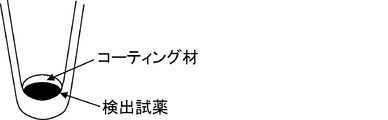
検出試薬はコーティング材で核酸から遮蔽されていてもよい。核酸から遮蔽されているとは、検出試薬が一時的に核酸を含有する試料等から遮蔽されている状態を意味する。核酸検出時に熱や圧力などの物理的刺激によって検出試薬が核酸に露出されることで、核酸と検出試薬が接触し、呈色を生じることが可能になる。
The detection reagent may be shielded from the nucleic acid with a coating material. Being shielded from nucleic acid means a state in which the detection reagent is temporarily shielded from a sample or the like containing nucleic acid. When the detection reagent is exposed to the nucleic acid by a physical stimulus such as heat or pressure at the time of detecting the nucleic acid, the nucleic acid and the detection reagent come into contact with each other, and coloration can be generated.
なお、コーティング材としては、検出試薬を一時的に遮蔽できるものであれば特に制限はないが、例えば、ポリビニルアルコール、ポリエチレングリコールなどのポリマー樹脂、パラフィンなどの油脂、アガロース、ジェランガム、カラギーナン、キサンタンガム、クラスターデキストリン、ヒアルロン酸などの糖類、ゼラチンやコラーゲンなどのタンパク質などが挙げられる。
The coating material is not particularly limited as long as the detection reagent can be temporarily shielded. For example, polymer resins such as polyvinyl alcohol and polyethylene glycol, oils and fats such as paraffin, agarose, gellan gum, carrageenan, xanthan gum, Examples thereof include saccharides such as cluster dextrin and hyaluronic acid, and proteins such as gelatin and collagen.
本発明は、また、検出試薬を保持する検出試薬保持部、および、前記試薬保持部を備えた蓋部および/または反応容器を有する、核酸の検出用デバイスまたはキットに関する。
The present invention also relates to a nucleic acid detection device or kit having a detection reagent holding part for holding a detection reagent, and a lid part and / or a reaction container provided with the reagent holding part.
検出デバイスを構成する蓋部および/または反応容器は閉鎖系を形成し得るものであれば特に制限はない。例えば、反応容器の開口部を密閉する蓋部と、反応容器の上部と蓋部を結合する接合部とを、一体に成型してなるもの(蓋型-反応容器一体型検出デバイス及び反応容器型検出デバイス)、もしくは反応容器の蓋部(蓋型検出デバイス)が挙げられる。
There is no particular limitation on the lid and / or reaction vessel constituting the detection device as long as they can form a closed system. For example, a cover part that seals the opening of the reaction container and a joint part that joins the upper part of the reaction container and the cover part are integrally molded (cover type-reaction container integrated detection device and reaction container type). Detection device), or a lid of the reaction vessel (lid-type detection device).
反応容器は、液体を保持でき、蓋等で密閉可能なものであれば形態に特に制限はないが、例えば、PCRや遺伝子操作等で用いられるチューブ型、プレート型、フィルム型、チップ型であってよく、市販のものを使用することも可能である。反応容器の口径、大きさや肉厚等は、核酸の増幅反応等の機構、或いは、使用する装置の大きさに合わせて適宜調整してもよい。また、反応容器は単独で使用してもよいが、複数本、例えば4本、8本、12本など、複数本が連結されていてもよい。
The reaction vessel is not particularly limited in form as long as it can hold a liquid and can be sealed with a lid or the like. For example, the reaction vessel may be a tube type, a plate type, a film type, or a chip type used in PCR or genetic manipulation. A commercially available product may be used. The diameter, size, wall thickness, etc. of the reaction vessel may be appropriately adjusted according to the mechanism such as nucleic acid amplification reaction or the size of the apparatus used. Moreover, although the reaction container may be used independently, multiple pieces, such as multiple pieces, for example, 4, 8, 12, etc., may be connected.
蓋部は、対応する反応容器の開口部を密閉できるものであれば特に制限はなく、押し込んで密閉できるタイプのもの、シール、フィルム、スクリューキャップなどが挙げられ、市販のものを使用することも可能である。
The lid is not particularly limited as long as the opening of the corresponding reaction vessel can be sealed, and includes a type that can be pushed in and sealed, a seal, a film, a screw cap, etc., and a commercially available one can also be used. Is possible.
蓋部は、反応容器の開口部に適合し、開口部の周縁より下方に続くチューブの内壁面に対して好適な密閉性が得られるように形成された筒状のシール部と、この筒状のシール部の端部に外周全体にわたって形成され、前記反応容器の縁部と密に当接し、それに対して好適に密閉し、閉鎖系が得られるように形成された外周部とを有することが好ましい。筒状のシール部は、反応チューブの開口部と接合させた際に密着性が高まるようにデザインされることが好ましい。
The lid portion is adapted to the opening of the reaction vessel and has a cylindrical seal portion formed so as to obtain a suitable hermetic seal against the inner wall surface of the tube extending below the periphery of the opening. Formed at the end of the seal portion over the entire outer periphery, closely contacting the edge of the reaction vessel, suitably sealed against it, and having an outer periphery formed so as to obtain a closed system. preferable. It is preferable that the cylindrical seal portion is designed so that the adhesion is enhanced when it is joined to the opening of the reaction tube.
蓋部や反応容器の材質は、特に制限されないが、プラスチック製であることが好ましい。プラスチックは射出成型などの公知慣用の方法により、チューブ容器を成型することができ、安価に大量生産することができる。プラスチックとして、例えば、ポリエチレン系樹脂、ポリプロピレン系樹脂、その他ポリオレフィン系樹脂、ポリエステル系樹脂、フッ素系樹脂、シリコン系樹脂等が挙げられる。
The material for the lid and the reaction vessel is not particularly limited, but is preferably made of plastic. Plastic can be molded into a tube container by a known and commonly used method such as injection molding, and can be mass-produced at low cost. Examples of the plastic include polyethylene resins, polypropylene resins, other polyolefin resins, polyester resins, fluorine resins, and silicon resins.
検出試薬保持部は、蓋部および/または反応容器の内側に固定され、核酸を検出するための検出試薬を保持する。検出試薬保持部は、蓋部と反応容器のいずれかに固定されていてもよく、両方に固定されていてもよい。また、反応容器と蓋部における、検出試薬保持部の固定位置は、核酸溶液に接触でき得る位置であれば特に制限はなく、使用する反応容器や蓋部の構造にあわせて、適宜調整されることが望ましい。
The detection reagent holding unit is fixed inside the lid and / or the reaction container, and holds a detection reagent for detecting nucleic acid. The detection reagent holding part may be fixed to either the lid part or the reaction container, or may be fixed to both. Further, the fixing position of the detection reagent holding part in the reaction container and the lid part is not particularly limited as long as it can contact the nucleic acid solution, and is appropriately adjusted according to the structure of the reaction container and the lid part to be used. It is desirable.
蓋部および/または反応容器は、核酸増幅試薬を保持することが好ましい。核酸増幅試薬としては、LAMP法やPCR法等の核酸増幅に使用される試薬が挙げられる。例えば、蓋部に検出試薬を保持し、反応容器にPCR試薬(ポリメラーゼ等)を保持するというように、検出試薬と核酸増幅試薬を別の位置に保持することも可能である。
The lid and / or reaction vessel preferably holds a nucleic acid amplification reagent. Examples of the nucleic acid amplification reagent include reagents used for nucleic acid amplification such as LAMP method and PCR method. For example, the detection reagent and the nucleic acid amplification reagent can be held at different positions, such as holding the detection reagent in the lid and holding the PCR reagent (polymerase or the like) in the reaction vessel.
検出試薬保持部は、検出時に閉鎖系内で核酸と検出試薬が接触できれば、どのような形状で形成されていても良い。例えば、蓋部または反応容器の形状を変えることで試薬を保持するための構造を設けることが挙げられ、その場合、反応容器または蓋部の内壁からチューブ容器内部に向けて張り出して形成される場合(張出しタイプ)、あるいは、内壁から内壁内部に向かって凹部が形成されている場合(凹タイプ)などが挙げられる。また、形状を変化させないフラットな表面をもつタイプも挙げられる。また、反応容器または蓋部に検出試薬を一時的に遮蔽する構造(一時遮蔽タイプ)を設けることも可能であり、この場合、熱などの物理的刺激によって遮蔽を解除し、検出試薬を核酸と接触させることができる。またフィルター、ろ紙、カプセル、リングなど、蓋部や反応容器以外の材料に検出試薬を保持させることも可能である。
The detection reagent holding part may be formed in any shape as long as the nucleic acid and the detection reagent can be brought into contact in the closed system at the time of detection. For example, it is possible to provide a structure for holding the reagent by changing the shape of the lid or the reaction container. In this case, the structure is formed by projecting from the inner wall of the reaction container or the lid toward the inside of the tube container. (Overhang type) or a case where a concave portion is formed from the inner wall toward the inner wall (concave type). In addition, there is a type having a flat surface that does not change its shape. In addition, it is possible to provide a structure (temporary shielding type) for temporarily shielding the detection reagent in the reaction container or lid. In this case, the shielding is released by a physical stimulus such as heat, and the detection reagent is separated from the nucleic acid. Can be contacted. It is also possible to hold the detection reagent on a material other than the lid and the reaction container, such as a filter, filter paper, capsule, and ring.
張出しタイプとは、蓋部または反応容器の内壁面より内部に向かって検出試薬保持部が張り出したものである。張出し部の数は、特に制限はないが、好ましくは1、もしくは2個配置されていることが望ましい。また張出しタイプの上から見た形は、特に制限はないが好ましくは円形であり、蓋部または反応容器の横断面からみた形状は、例えば短冊状、容器内部に向かって張り出した先端がとがっている三角形状、先端が平らである台形状、先端が丸みを帯びているドーム状、あるいは先端が容器内壁に向かってくびれている凹面状などが挙げられる。また、蓋部または反応容器の内部を分割するような構造をとることも可能である。検出試薬保持部の大きさは使用する蓋部や反応容器の構造に合わせて、適宜調整されることが望ましい。
The overhanging type is a type in which the detection reagent holding unit projects from the lid or the inner wall surface of the reaction container toward the inside. The number of overhangs is not particularly limited, but preferably 1 or 2 is preferably arranged. The shape seen from the top of the overhang type is not particularly limited, but is preferably circular, and the shape seen from the cross-section of the lid or the reaction vessel is, for example, a strip shape, with a tip protruding toward the inside of the vessel. A triangular shape having a flat tip, a dome shape having a round tip, or a concave shape having a tip narrowed toward the inner wall of the container. It is also possible to take a structure that divides the inside of the lid or the reaction vessel. It is desirable that the size of the detection reagent holding part is appropriately adjusted according to the structure of the lid part and reaction container to be used.
凹タイプとは、蓋部または反応容器の内壁上に検出試薬保持部として窪みを形成させたものである。窪みの数は特に制限はないが、好ましくは1、もしくは2個配置されていることが望ましい。また凹タイプの上から見た形は、特に制限はないが好ましくは円形であり、蓋部または反応容器の横断面からみたとき、例えば短冊状、容器内部に向かって張り出した先端がとがっている三角形状、先端が平らである台形状、先端が丸みを帯びているドーム状、あるいは先端が容器内壁に向かってくびれている凹面状などが挙げられる。またこの凹タイプは前述の張り出しタイプと組み合わせて利用することも可能である。
In the concave type, a depression is formed as a detection reagent holding part on the lid or the inner wall of the reaction container. The number of the recesses is not particularly limited, but preferably 1 or 2 is desirable. The shape seen from the top of the concave type is not particularly limited, but is preferably circular, and when viewed from the cross section of the lid or the reaction vessel, for example, a strip shape, the tip protruding toward the inside of the vessel is sharp Examples include a triangular shape, a trapezoidal shape with a flat tip, a dome shape with a rounded tip, or a concave shape with a tip narrowed toward the inner wall of the container. This concave type can also be used in combination with the above-described overhang type.
検出試薬保持部の形状、大きさや数は、蓋部や反応容器の製造の容易さとともに、乾燥試薬の接着のしやすさや、試薬を容器内で乾燥・固形化させた際の凹凸形状への入り込みやすさ、挟まれやすさ等を様々に考慮して決定される。
The shape, size, and number of detection reagent holders are not only easy to manufacture the lid and reaction container, but also easy to attach the dry reagent, and to the uneven shape when the reagent is dried and solidified in the container. It is determined in consideration of various factors such as ease of entry and ease of pinching.
一時遮蔽タイプは、蓋部、または、反応容器に、検出試薬を一時的に核酸から遮蔽する構造を有する。遮蔽するための構造は、検出試薬を一時的に遮蔽できれば特に制限はないが、例えば、ポリビニルアルコール、ポリエチレングリコールなどのポリマー樹脂、パラフィンなどの油脂、アガロース、ジェランガム、カラギーナン、キサンタンガム、クラスターデキストリン、ヒアルロン酸などの糖類、ゼラチンやコラーゲンなどのタンパク質などをコーティング材として使用する構造が挙げられる。核酸検出前には検出試薬を核酸から遮蔽し、核酸検出時に熱などの物理的刺激によって検出試薬を核酸に露出させ、核酸と接触させる構造が挙げられる。なお、この一時遮蔽タイプは前記の張出しタイプまたは凹タイプの構造と組み合わせた試薬保持部とすることも可能であり、また蓋部や反応容器にそれらの構造を持たないものでも設置することができる。
The temporary shielding type has a structure in which the detection reagent is temporarily shielded from the nucleic acid in the lid or the reaction container. The structure for shielding is not particularly limited as long as the detection reagent can be temporarily shielded. For example, polymer resins such as polyvinyl alcohol and polyethylene glycol, oils and fats such as paraffin, agarose, gellan gum, carrageenan, xanthan gum, cluster dextrin, hyaluron A structure using a saccharide such as an acid or a protein such as gelatin or collagen as a coating material may be mentioned. A structure in which the detection reagent is shielded from the nucleic acid before detection of the nucleic acid, the detection reagent is exposed to the nucleic acid by physical stimulation such as heat at the time of detecting the nucleic acid, and brought into contact with the nucleic acid. This temporary shielding type can also be a reagent holding unit combined with the above-described overhang type or concave type structure, and a lid or reaction vessel having no such structure can also be installed. .
以下、図面を参照して本発明の検出デバイスのいくつかの形態を説明する。
Hereinafter, several forms of the detection device of the present invention will be described with reference to the drawings.
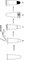
図1に、蓋型-張り出しタイプIである検出デバイスの外観(a)、(a)の開口部側からみた平面図(b)、及び(b)のX-X’線に沿って切断した縦断面図(c)を示す。蓋型-張り出しタイプIは底部1、開口部2と筒状の胴部3及び試薬保持部4を形成する壁部5からなる。胴部は反応容器に差し込む際の密閉性が高まるように底部から開口部にかけて径が広くなっており、その高さは反応容器の長さ以下であることが好ましい。また、検出試薬保持部の形状は、検出試薬を保持できる構造であれば特に制限はないが、前述のように例えば短冊状、反応容器内部に向かって張り出した先端がとがっている三角形状、先端が平らである台形状、先端が丸みを帯びているドーム状、あるいは先端が容器内壁に向かってくびれている凹面状などが挙げられる。壁部や径は検出試薬を保持できる高さであれば特に制限はないが、長さは胴部の長さ以下、径は開口部の径以下が望ましい。
FIG. 1 shows an external appearance of a detection device of lid type-extrusion type I (a), a plan view (b) viewed from the opening side of (a), and a cut along line XX ′ in (b). A longitudinal sectional view (c) is shown. The lid-overhang type I includes a bottom portion 1, an opening portion 2, a cylindrical body portion 3, and a wall portion 5 that forms a reagent holding portion 4. The body portion has a diameter that increases from the bottom portion to the opening portion so as to enhance the sealing property when inserted into the reaction vessel, and the height is preferably equal to or less than the length of the reaction vessel. The shape of the detection reagent holding part is not particularly limited as long as it is a structure that can hold the detection reagent. However, as described above, for example, a strip shape, a triangular shape with a sharpened tip protruding toward the inside of the reaction container, a tip A flat trapezoidal shape, a dome shape with a rounded tip, or a concave shape with a tip narrowed toward the inner wall of the container. The wall and diameter are not particularly limited as long as they can hold the detection reagent, but the length is preferably equal to or less than the length of the body and the diameter is preferably equal to or less than the diameter of the opening.
図2に、蓋型-張り出しタイプIIである検出デバイスの外観(a)、(a)の開口部側からみた平面図(b)、及び(b)のX-X’線に沿って切断した縦断面図(c)を示す。蓋型-張り出しタイプIIは図1の蓋型-張り出しタイプIと同じく、底部、開口部、胴部及び試薬保持部を形成する壁部からなる。ただし、試薬保持部は蓋型-張り出しタイプIとは異なり、壁部5は胴部内部を少なくとも2分割するように設けられており、その高さは検出試薬を保持できる高さであれば特に制限はないが、胴部の長さ以下であることが好ましい。
FIG. 2 shows the appearance of the detection device of the lid type-extrusion type II (a), a plan view from the opening side of (a) (b), and cut along the line XX ′ of (b). A longitudinal sectional view (c) is shown. The lid-overhang type II is composed of a wall portion that forms a bottom portion, an opening, a body portion, and a reagent holding portion, similar to the lid-overhang type I in FIG. However, unlike the lid type-extrusion type I, the reagent holding part is provided so that the wall part 5 is divided into at least two parts inside the body part, and the height is particularly high as long as it can hold the detection reagent. Although there is no restriction | limiting, it is preferable that it is below the length of a trunk | drum.
図3に、蓋型-凹タイプである検出デバイスの外観(a)、(a)の開口部側からみた平面図(b)、及び(b)のX-X’線に沿って切断した縦断面図(c)を示す。蓋型-凹タイプは図1~2に示した検出デバイスと同じく、底部、開口部、胴部及び試薬保持部を形成する壁部からなる。試薬保持部4は開口部から底部に向かって窪みを形成しており、その深さや径は検出試薬を保持できる長さであれば特に制限はないが、深さは底部の厚さ以下であることが好ましく、径は開口部の長さ以下であることが好ましい。
FIG. 3 shows the appearance of the lid-concave type detection device (a), a plan view (b) from the opening side of (a), and a longitudinal section cut along the line XX ′ of (b). A surface view (c) is shown. Similar to the detection device shown in FIGS. 1 and 2, the lid type-concave type includes a bottom part, an opening part, a body part, and a wall part that forms a reagent holding part. The reagent holding part 4 forms a recess from the opening toward the bottom, and the depth and diameter of the reagent holding part 4 are not particularly limited as long as the detection reagent can be held, but the depth is equal to or less than the thickness of the bottom. The diameter is preferably equal to or less than the length of the opening.
さらには、試薬保持部の張出しタイプと凹タイプを組み合わせた試薬保持部を有する蓋型検出デバイスも作製可能である。
Furthermore, a lid-type detection device having a reagent holding unit that combines a protruding type and a recessed type of reagent holding unit can also be manufactured.
図4に、蓋型-一時遮蔽タイプである検出デバイスの断面図を示す。この場合、検出試薬はコーティング材によってコーティングされている。また、一時遮蔽タイプの試薬保持は張出しタイプや凹タイプと組み合わせて形成させることも可能である。
FIG. 4 shows a cross-sectional view of a detection device of the lid type-temporary shielding type. In this case, the detection reagent is coated with a coating material. The temporary shielding type reagent holding can be formed in combination with the overhang type or the concave type.
図7に、蓋型-張り出しタイプIである検出デバイスの外観を示す。図1~図3では反応容器とは独立した、蓋部からなる検出デバイスを示したが、図7のように試薬保持部を有する蓋部と反応容器が一体化したものであってもよい。この一体型を連結することもでき、連結した場合には複数検体の同時並行処理が簡便となる。
FIG. 7 shows the external appearance of a detection device of the lid type-extrusion type I. Although FIGS. 1 to 3 show a detection device having a lid that is independent of the reaction vessel, the lid having the reagent holding portion and the reaction vessel may be integrated as shown in FIG. This integrated type can also be connected, and when connected, simultaneous parallel processing of a plurality of samples is simplified.
図9に、反応容器型Iである検出デバイスの外観(a)、(a)のY-Y’線に沿って切断した縦断面図(b)、凹タイプの試薬保持部を有する反応容器型デバイスの縦断面図(c)、及び張出しタイプの試薬保持部を有する反応容器型デバイスの縦断面図(d)を示す。凹タイプ(c)は反応容器の底方向に向かって窪みを形成しており、その深さや径は検出試薬を保持できる長さであれば特に制限はないが、深さは反応容器厚以下、径は反応容器の底の径以下であることが好ましい。また張り出しタイプ(d)は反応容器の開口面方向に向かって壁部が形成され、検出試薬を保持できる高さであれば特に制限はないが、反応容器の長さ以下であることが望ましい。
FIG. 9 shows an external appearance (a) of a detection device which is a reaction vessel type I, a longitudinal sectional view (b) cut along the line YY ′ of (a), and a reaction vessel type having a concave type reagent holding part. The longitudinal cross-sectional view (c) of a device and the longitudinal cross-sectional view (d) of the reaction container type | mold device which has an overhang type reagent holding part are shown. The concave type (c) forms a dent toward the bottom of the reaction vessel, and the depth and diameter are not particularly limited as long as the detection reagent can be held, but the depth is equal to or less than the thickness of the reaction vessel, The diameter is preferably equal to or less than the diameter of the bottom of the reaction vessel. Further, the overhang type (d) is not particularly limited as long as the wall portion is formed in the direction of the opening surface of the reaction vessel and can hold the detection reagent, but it is preferably less than the length of the reaction vessel.
図10に、反応容器型-一時遮蔽タイプである検出デバイスの断面図を示す。この場合、検出試薬は反応容器のフラットな内面の試薬保持部に前記のコーティング材によってコーティングされている。また、一時遮蔽タイプの試薬保持は張出しタイプや凹タイプと組み合わせて形成させることも可能である。
FIG. 10 shows a cross-sectional view of a detection device of a reaction vessel type-temporary shielding type. In this case, the detection reagent is coated on the reagent holding part on the flat inner surface of the reaction container with the coating material. The temporary shielding type reagent holding can be formed in combination with the overhang type or the concave type.
検出デバイスは、1つで使用することも可能であり、連結部を介して2つ以上連結した状態で使用することも可能である。連結する場合、複数の核酸増幅反応を並行して行った後の増幅産物の検出が簡便にできる。連結する個数は、使用の場面に応じて自由に選ぶことが可能で、一例としては、図5に、図1の検出デバイスの蓋部を8つ連結した蓋型検出デバイスの外観を示している。連結部は、複数の蓋部同士、接合部同士、反応容器同士、あるいはそれらの組み合わせを連結するように形成されているが、好ましくは蓋部同士、もしくは反応容器同士を連結するように形成されてなることが望ましい。このとき連結部は、反応容器の開口部に設けられた縁部で連結するように形成されてよく、あるいは、反応容器の筒状の胴部で連結するように形成されていてもよい。
One detection device can be used, or two or more detection devices can be used in a state of being connected via a connecting portion. In the case of ligation, detection of amplification products after performing a plurality of nucleic acid amplification reactions in parallel can be easily performed. The number of connections can be freely selected according to the situation of use. As an example, FIG. 5 shows the appearance of a lid-type detection device in which eight lids of the detection device of FIG. 1 are connected. . The connecting portion is formed so as to connect a plurality of lid portions, joint portions, reaction vessels, or a combination thereof, but preferably formed so as to connect the lid portions or reaction vessels. It is desirable that At this time, the connecting portion may be formed so as to be connected by an edge provided at the opening of the reaction vessel, or may be formed so as to be connected by a cylindrical body portion of the reaction vessel.
試薬保持部への検出試薬、色素、及び修飾物質の固定化は以下の方法で行う。なお、修飾物質は、検出試薬に含まれる色素以外の成分であって、求核剤の他に、安定化剤、及びコーティング剤等を意味する。求核剤としては、コントラスト増強のための酸化剤、還元剤、酸、塩基、pH緩衝剤、或いは発色型色素をロイコ型に変換する物質が挙げられる。
The detection reagent, the dye, and the modifying substance are immobilized on the reagent holding unit by the following method. The modifier is a component other than the dye contained in the detection reagent, and means a stabilizer, a coating agent, and the like in addition to the nucleophile. Examples of the nucleophilic agent include an oxidizing agent, a reducing agent, an acid, a base, a pH buffer, or a substance that converts a coloring dye into a leuco type for contrast enhancement.
まず、修飾物質を溶媒に溶かした溶液を試薬保持部に添加し、乾燥させ、固定する。乾燥方法は特に限定されず、風乾や加熱、減圧乾燥等が適応可能であり、これらの方法を組み合わせて実施することもできる。なお、この際の溶媒は修飾物質が溶解できるものであれば良く、例えば、水、エタノール、メタノール、イソプロパノール等が挙げられる。また、これに乾燥後の修飾物質の剥離や脱落を防止する目的でポリマー等の基材を添加することもでき、例えばポリビニルアルコールなどを添加しても良い。この際のポリマー等の基材の量は、乾燥時に核酸の呈色が確認できる量で調整することができ、好ましくは0.1mg以下であり、より好ましくは0.01mg以下である。さらには粘性をもった液状のゾル、固体となったゲルにして保持させることもできる。また、乾燥後の修飾物質の溶解性を向上させる目的で、溶解補助剤等を添加しても良く、例えば、TritonX-100やTween20などの界面活性剤やシクロデキストリン、β-グルカン等が挙げられる。この際の修飾物質量は、乾燥時に核酸の呈色が確認できる量で調整することができ、例えば色素量の1000モル当量以下が好ましく、100モル当量以下がより好ましい。
First, a solution in which the modifying substance is dissolved in a solvent is added to the reagent holding unit, dried, and fixed. The drying method is not particularly limited, and air drying, heating, drying under reduced pressure, and the like can be applied, and these methods can also be implemented in combination. In addition, the solvent in this case should just be a thing which can melt | dissolve a modifier, For example, water, ethanol, methanol, isopropanol etc. are mentioned. In addition, a base material such as a polymer can be added to this for the purpose of preventing peeling and dropping off of the modifying substance after drying. For example, polyvinyl alcohol or the like may be added. In this case, the amount of the substrate such as a polymer can be adjusted so that the coloration of the nucleic acid can be confirmed upon drying, and is preferably 0.1 mg or less, more preferably 0.01 mg or less. Furthermore, it can be held in the form of a viscous liquid sol or a solid gel. In addition, for the purpose of improving the solubility of the modified substance after drying, a solubilizing agent may be added, and examples thereof include surfactants such as Triton X-100 and Tween 20, cyclodextrins, β-glucan and the like. . In this case, the amount of the modifying substance can be adjusted so that the coloration of the nucleic acid can be confirmed at the time of drying. For example, the molar amount of the dye is preferably 1000 molar equivalents or less, and more preferably 100 molar equivalents or less.
なお、核酸を含む溶液に修飾物質が含まれている場合には色素のみを固定する場合もある。また修飾物質と色素の固定の順番に制限はない。さらには修飾物質溶液と色素溶液の混合液を使用し、一度に固定することも可能である。
In addition, when the modifying substance is contained in the solution containing the nucleic acid, only the dye may be fixed. Moreover, there is no restriction | limiting in the order of fixation of a modifier and a pigment | dye. Furthermore, it is possible to fix at once using a mixed solution of a modifying substance solution and a dye solution.
一時遮蔽タイプの試薬保持部をもつ検出デバイスを作製する際のコーティングは以下の方法で行う。
Coating for producing a detection device having a temporary shielding type reagent holding unit is performed by the following method.
まず、前述のコーティング材を溶融し、乾燥後の検出試薬上に添加し、冷却して固定する。または、コーティング材を溶媒に溶解し、乾燥後の検出試薬上に添加し、乾燥により固定する。この際のコーティング材は検出試薬を安定にカバーでき、加熱などの物理的処理により検出試薬を放出可能であれば特に制限はないが、例えば固形パラフィン、ポリビニルアルコール、ポリエチレングリコール、アガロース・ジェランガム・カラギーナン・キサンタンガム・ヒアルロン酸などであり、好ましくはパラフィン、ポリビニルアルコールである。
First, the above-described coating material is melted, added onto the detection reagent after drying, and then cooled and fixed. Alternatively, the coating material is dissolved in a solvent, added onto the detection reagent after drying, and fixed by drying. The coating material in this case is not particularly limited as long as the detection reagent can be stably covered and can be released by physical treatment such as heating. For example, solid paraffin, polyvinyl alcohol, polyethylene glycol, agarose / gellan gum / carrageenan Xanthan gum, hyaluronic acid and the like, preferably paraffin and polyvinyl alcohol.
なお、この試薬保持部には、検出試薬と共に核酸増幅に必要な各種試薬を保持させてもよい。各種試薬には前述の核酸増幅法を行うための目的核酸を増幅するためのプライマー、DNAポリメラーゼ、核酸増幅反応に用いられる緩衝液や制限酵素等が含まれる。この場合、検出試薬と増幅用試薬は蓋部と反応容器別々に保持させることができ、また試薬保持部の壁部や一時遮蔽タイプでそれぞれを遮蔽することもできる。
The reagent holder may hold various reagents necessary for nucleic acid amplification together with the detection reagent. The various reagents include a primer for amplifying the target nucleic acid for performing the above-described nucleic acid amplification method, a DNA polymerase, a buffer used for nucleic acid amplification reaction, a restriction enzyme, and the like. In this case, the detection reagent and the amplification reagent can be separately held by the lid and the reaction container, and each can be shielded by the wall of the reagent holding part or the temporary shielding type.
次に、図面を参照して、検出デバイスを用いた核酸検出法について説明する。
Next, a nucleic acid detection method using a detection device will be described with reference to the drawings.
図6は、蓋型検出デバイス(a)を用いた核酸検出工程を示している。核酸溶液又は核酸増幅反応液を反応容器に添加し、検出試薬を保持した検出デバイスで蓋をした後(b)、必要に応じて核酸増幅反応などの反応を行う(c)。その後、検出デバイスを上下反転などさせるなどして、検出試薬と核酸を含む溶液と接触させ、試薬を核酸溶液に溶解させ、核酸を着色する(d)及び(e)。その際、ボルテックスなどの攪拌処理を行うことで効率よく溶解させることも可能である。また、掌サイズの超小型PCR装置を用いてPCR反応を行った場合、増幅反応後に装置自体を逆さにすることで、反応溶液と検出試薬を接触させ、検出することも可能である。
FIG. 6 shows a nucleic acid detection step using the lid-type detection device (a). A nucleic acid solution or a nucleic acid amplification reaction solution is added to the reaction vessel, and the reaction device is capped with a detection device holding a detection reagent (b), and then a reaction such as a nucleic acid amplification reaction is performed as necessary (c). Thereafter, the detection device is turned upside down to bring it into contact with the solution containing the detection reagent and the nucleic acid, so that the reagent is dissolved in the nucleic acid solution and the nucleic acid is colored (d) and (e). In that case, it is also possible to dissolve efficiently by performing a stirring process such as vortexing. In addition, when a PCR reaction is performed using a palm-sized ultra-compact PCR apparatus, the reaction solution and the detection reagent can be brought into contact with each other and detected by inverting the apparatus itself after the amplification reaction.
図8は、蓋型-反応容器一体型検出デバイス(a)を用いた核酸検出工程を示している。検出試薬を保持した検出デバイスに核酸溶液又は核酸増幅反応液を添加し蓋をする(b)。その後は、前述の蓋型検出デバイスと同様な処理を行う。
FIG. 8 shows a nucleic acid detection step using the lid-reaction vessel integrated detection device (a). A nucleic acid solution or a nucleic acid amplification reaction solution is added to the detection device holding the detection reagent and covered (b). Thereafter, processing similar to that of the lid-type detection device described above is performed.
図11は、一時遮蔽タイプの試薬保持部を持つ反応容器型検出デバイス(a)を用いた工程を示している。核酸溶液又は核酸増幅反応液を検出試薬を保持した反応容器に添加した後(b)、蓋をする。必要に応じて核酸増幅反応などの反応を行った後(c)、加熱などの処理によりコーティング材内の検出試薬を核酸溶液に溶解させる。この際の加熱温度は、コーティング材が物理的に変化し検出試薬が核酸溶液と接触できれば特に制限はないが、好ましくは40℃以上であり、より好ましくは60℃以上である。
FIG. 11 shows a process using a reaction container type detection device (a) having a temporary shielding type reagent holding unit. After the nucleic acid solution or the nucleic acid amplification reaction solution is added to the reaction vessel holding the detection reagent (b), the lid is capped. After performing a reaction such as a nucleic acid amplification reaction as necessary (c), the detection reagent in the coating material is dissolved in the nucleic acid solution by a treatment such as heating. The heating temperature at this time is not particularly limited as long as the coating material is physically changed and the detection reagent can come into contact with the nucleic acid solution, but is preferably 40 ° C. or higher, more preferably 60 ° C. or higher.
図12は、蓋型検出デバイス(a)を用いた核酸検出工程を示している。容器内で増幅反応液を調製し(b)、検出試薬を保持した蓋をした後(c)、検出デバイスを上下反転などさせるなどして、検出試薬と反応液を接触させ、試薬を溶解させる(d)。その際、ボルテックスなどの攪拌処理を行うことで効率よく溶解させることも可能である。その後、遠心機等を用いてスピンダウンを行い、蓋部に付着した溶液を容器内に集め、増幅反応を行う。
FIG. 12 shows a nucleic acid detection step using the lid-type detection device (a). Prepare the amplification reaction solution in the container (b), close the lid holding the detection reagent (c), and turn the detection device upside down to bring the detection reagent into contact with the reaction solution and dissolve the reagent. (D). In that case, it is also possible to dissolve efficiently by performing a stirring process such as vortexing. Thereafter, spin down is performed using a centrifuge or the like, and the solution adhering to the lid is collected in a container to carry out an amplification reaction.
図13は、反応容器型検出デバイス(a)を用いた工程を示している。増幅反応溶液を容器内で調製し、反応容器内に保持した検出試薬を溶解させる(b)。または、別の容器内で調製した反応溶液を、検出デバイスに添加してもよい。その後、熱サイクル等により増幅反応を行う。
FIG. 13 shows a process using the reaction vessel type detection device (a). An amplification reaction solution is prepared in a container, and the detection reagent held in the reaction container is dissolved (b). Alternatively, a reaction solution prepared in another container may be added to the detection device. Thereafter, an amplification reaction is performed by a thermal cycle or the like.
次に図6、8、11、12および13の工程(e)以降の可視光での検出工程について述べる。
Next, the detection process with visible light after the process (e) in FIGS. 6, 8, 11, 12 and 13 will be described.
工程(e)での反応により生じた物質を可視光下で観察し、核酸の存在を可視光で判定する際には、紫外線のような可視光ではない光を照射することなく、一般的な実験室における照明のような可視光下で観察する。可視光であれば、紫外線照射する場合のように特別な装置は必要でなく、簡便に観察することができる。
When the substance produced by the reaction in the step (e) is observed under visible light and the presence of nucleic acid is determined with visible light, it is not necessary to irradiate light that is not visible light such as ultraviolet rays. Observe under visible light like lighting in the laboratory. In the case of visible light, a special apparatus is not required as in the case of ultraviolet irradiation, and observation can be performed easily.
なお、該検出デバイスを使用すれば図6、8、11、12および13の(c)から(e)及び検出工程を全自動で行うことができる。全自動とは、本発明の検出デバイスで反応と検出段階を自動で行う工程を含むことを示す。
In addition, if this detection device is used, (c) to (e) of FIGS. 6, 8, 11, 12 and 13 and the detection process can be performed fully automatically. Fully automatic means that it includes the step of automatically performing the reaction and detection steps with the detection device of the present invention.
例えば図6の蓋型検出デバイス及び図8の蓋型-反応容器一体型検出デバイスでは、核酸溶液又は核酸増幅反応液を検出試薬を保持した反応容器に添加した後(b)、蓋をする。これ以降の工程は温度調節ができ、必要であれば振動や振とう、反応容器の反転ができる装置を用いて自動で実施することができる。必要に応じて核酸増幅反応などの反応を行った後(c)、検出デバイスを振とうや反転などをすることによって、蓋部の検出試薬と核酸を含む溶液を接触させ、着色させることができる。さらに必要に応じて装置に組み込まれた可視光によって着色の有無や着色度を計測、場合によってはそのデータを解析することができる。
For example, in the lid-type detection device of FIG. 6 and the lid-reaction vessel integrated detection device of FIG. 8, the nucleic acid solution or the nucleic acid amplification reaction solution is added to the reaction vessel holding the detection reagent (b) and then the lid is closed. The subsequent steps can be temperature-controlled, and can be automatically performed using an apparatus capable of reversing the reaction vessel if necessary. After performing a reaction such as a nucleic acid amplification reaction as necessary (c), the detection device on the lid and the solution containing the nucleic acid can be brought into contact with each other and colored by shaking or inverting the detection device. Furthermore, if necessary, the presence or absence of coloring and the degree of coloring can be measured by visible light incorporated in the apparatus, and the data can be analyzed in some cases.
図11の一時遮蔽タイプの試薬保持部を持つ反応容器型検出デバイスを用いた工程において、核酸溶液又は核酸増幅反応液を検出試薬を保持した反応容器に添加した後(b)、蓋をする。これ以降の工程は前述の装置で自動で実施する。必要に応じて核酸増幅反応などの反応を行った後(c)、加熱などの処理によりコーティング材内の検出試薬を核酸溶液に溶解させて着色させることができる。さらに必要に応じて可視光によって着色の有無や着色度を計測することができる。
In the process using the reaction vessel type detection device having the temporary shielding type reagent holding unit of FIG. 11, the nucleic acid solution or the nucleic acid amplification reaction solution is added to the reaction vessel holding the detection reagent (b), and then the lid is closed. The subsequent steps are automatically performed by the above-described apparatus. After performing a reaction such as a nucleic acid amplification reaction as necessary (c), the detection reagent in the coating material can be dissolved in the nucleic acid solution and colored by a treatment such as heating. Furthermore, the presence or absence of coloring and the degree of coloring can be measured by visible light as necessary.
本発明において、核酸検出後の着色した検体液を他の分子生物学的な操作にそのまま使用することも可能である。そのような分子生物学的な操作には、制限酵素反応、シーケンス反応、PCRの様な酵素反応や、電気泳動による確認操作等が含まれる。
In the present invention, it is also possible to use the colored sample solution after nucleic acid detection as it is for other molecular biological operations. Such molecular biological operations include restriction enzyme reactions, sequence reactions, enzymatic reactions such as PCR, confirmation operations by electrophoresis, and the like.
本発明には、前述の可視光での検出用デバイスと、試薬や器具等を組み合わせたキットも含まれる。キットとは、前述の検出デバイスに加え、各種試薬や器具等を備えた形態を指す。各種試薬には前述の核酸増幅法を行うための目的核酸を増幅するためのプライマー、DNAポリメラーゼ、核酸増幅反応に用いられる緩衝液や増幅した核酸を処理するための制限酵素、シークエンス用の試薬等が含まれる。また抗原、抗体、色素、酵素、基質等を使用することにより、本発明のキットを免疫反応や生化学反応の測定、免疫蛍光法による微量検出にも用いることができる。
The present invention also includes a kit in which the above-described detection device for visible light is combined with reagents, instruments and the like. The kit refers to a form provided with various reagents and instruments in addition to the above-described detection device. Various reagents include primers for amplifying the target nucleic acid for performing the above-described nucleic acid amplification method, DNA polymerase, buffers used for nucleic acid amplification reactions, restriction enzymes for processing amplified nucleic acids, sequencing reagents, etc. Is included. In addition, by using an antigen, an antibody, a dye, an enzyme, a substrate, etc., the kit of the present invention can be used for measurement of an immune reaction or biochemical reaction, or for a trace amount detection by an immunofluorescence method.
本発明には、前記の可視光での検出用デバイスまたはキットを有する装置も含まれる。装置は、検体を自動的に処理し塩基配列等を解析する装置であって、本発明の検出デバイスを含む装置である。核酸精製装置や核酸増幅装置に本発明の可視光での検出用デバイスを組み込んで核酸の有無を判定したり、DNA自動解析装置に組み込むことによって、PCRで増幅・ラベルされたサンプルのみを選別して解析を行う等、操作の確実性向上が期待できる。
The present invention also includes an apparatus having the above-described detection device or kit for visible light. The apparatus is an apparatus that automatically processes a sample and analyzes a base sequence and the like, and includes the detection device of the present invention. By incorporating the detection device for visible light of the present invention into a nucleic acid purification device or nucleic acid amplification device to determine the presence or absence of nucleic acid, or by incorporating it into an automatic DNA analysis device, only samples amplified and labeled by PCR are selected. The reliability of the operation can be expected to improve.
以下、実施例により本発明を具体的に説明する。但し、本発明はこれらの実施例に限定されるものではない。
Hereinafter, the present invention will be described specifically by way of examples. However, the present invention is not limited to these examples.
(実施例1)
(i)蓋型-凹タイプの試薬保持部を持った蓋型検出デバイスの作製
回転ヤスリを使用して、0.2mlのPCR用チューブ(アイビス社製)の蓋部の内側に直径0.5mmの窪みを2箇所作製し、試薬保持部とした。一方の試薬保持部に、色素溶液(0.2% ゲンチアナバイオレットBのエタノール溶液)を、他方に還元剤溶液(3.2% 亜硫酸ナトリウム/1% ポリビニルアルコール水溶液)をそれぞれ1μl添加後、減圧下乾燥することで、核酸検出試薬を保持した反応容器とした。
Example 1
(I) Production of a lid-type detection device having a lid-concave type reagent holding part Using a rotating file, a diameter of 0.5 mm is placed inside the lid part of a 0.2 ml PCR tube (manufactured by Ibis). Two recesses were prepared as reagent holding parts. Add 1 μl of the dye solution (0.2% gentian violet B ethanol solution) to one reagent holding part and 1 μl of the reducing agent solution (3.2% sodium sulfite / 1% polyvinyl alcohol aqueous solution) to the other, and then reduce the pressure. By drying, a reaction container holding the nucleic acid detection reagent was obtained.
(ii)PCR増幅産物の検出
鋳型としてpUC19(タカラバイオ社製)を用い、PCR増幅により約330塩基対が増幅するように以下のプライマーF:5’-GGAAACAGCTATGACCATGA-3’およびプライマーR:5’-CTATGCGGCATCAGAGCAG-3’を設計した。
(Ii) Using pUC19 (manufactured by Takara Bio Inc.) as a detection template for PCR amplification products, the following primer F: 5′-GGAAACAGCTATGACCATGA-3 ′ and primer R: 5 ′ so that about 330 base pairs are amplified by PCR amplification. -CTATGCGGGCATCAGAGCAG-3 'was designed.
プライマーFとプライマーRを各15pmolと、10ngのpUC19とを実施例1で作製した反応容器に入れ、ExTaq PCRキット(タカラバイオ社製)の説明書に従い、50μlのPCR反応液を調製した。その後、チューブをサーマルサイクラー(GeneAmp PCR System(アプライドバイオシステム社製))にセットし、95℃で5分間熱処理後、95℃で30秒、55℃で30秒、72℃で30秒のサイクルを35回行い、目的の約330bpの増幅を行い、ポジティブコントロールとした。また、ExTaq DNAポリメラーゼを添加しないで同様の反応を行い、ネガティブコントロールとした。
Primer F and Primer R of 15 pmol each and 10 ng of pUC19 were put into the reaction vessel prepared in Example 1, and 50 μl of a PCR reaction solution was prepared according to the instructions of ExTaq PCR kit (manufactured by Takara Bio Inc.). Then, the tube was set in a thermal cycler (GeneAmp PCR System (Applied Biosystems)), heat treated at 95 ° C for 5 minutes, then cycled at 95 ° C for 30 seconds, 55 ° C for 30 seconds, and 72 ° C for 30 seconds. This was performed 35 times, and the target amplification of about 330 bp was performed as a positive control. Moreover, the same reaction was performed without adding ExTaq DNA polymerase, and used as a negative control.
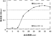
PCR反応後の容器を転倒混和し、反応溶液にて蓋部の検出試薬を溶解後、反応溶液の色調を目視にて確認した。その結果、ネガティブコントロールは無色であったのに対し、ポジティブコントロールは青色の呈色を示し、反応容器の蓋を開閉することなく核酸増幅の有無を確認することが可能であった。
The container after the PCR reaction was mixed by inversion, the detection reagent on the lid was dissolved with the reaction solution, and the color tone of the reaction solution was visually confirmed. As a result, the negative control was colorless, while the positive control showed a blue color, and it was possible to confirm the presence or absence of nucleic acid amplification without opening and closing the lid of the reaction vessel.
(iii)AMP法増幅産物の検出
「Loopamp(R)サルモネラ検出試薬キット」(栄研化学社製)を使用し、上記工程(i)で作製した反応容器内にLAMP法による核酸増幅反応液を調製した。10μlのControl DNA Salと40μlのマスターMix(Reaction Mix.SalとBst DNA Polymeraseを容量比で20:1の割合で別途混合したもの)を混合し、65℃で1時間反応後、さらに、85℃で20分反応することで、核酸が増幅された反応液(ポジティブコントロール)を調製した。また、10μlの蒸留水と40μlのReaction Mix.Salを混合し、65℃で1時間反応後、さらに、85℃で20分反応することで、核酸が増幅されていない反応液(ネガティブコントロール)を調製した。
(Iii) Detection of AMP method amplification product Using a “Loopamp® Salmonella detection reagent kit” (manufactured by Eiken Chemical Co., Ltd.), a nucleic acid amplification reaction solution by the LAMP method is placed in the reaction vessel prepared in the above step (i). Prepared. 10 μl of Control DNA Sal and 40 μl of Master Mix (reaction mix. Sal and Bst DNA Polymerase mixed separately at a volume ratio of 20: 1) were mixed, reacted at 65 ° C. for 1 hour, and further reacted at 85 ° C. Was reacted for 20 minutes to prepare a reaction solution (positive control) in which the nucleic acid was amplified. Also, 10 μl of distilled water and 40 μl of Reaction Mix. Sal was mixed, reacted at 65 ° C. for 1 hour, and further reacted at 85 ° C. for 20 minutes to prepare a reaction solution (negative control) in which nucleic acid was not amplified.
反応後の容器を転倒混和し、反応溶液にて蓋部の検出試薬を溶解後、反応溶液の色調を目視にて確認した。その結果、ネガティブコントロールは無色であったのに対し、ポジティブコントロールは青色の呈色を示した。このことから反応容器の蓋を開閉することなく核酸増幅の有無を確認することが可能であることが確認できた。
The container after the reaction was mixed by inversion, the detection reagent on the lid was dissolved with the reaction solution, and the color tone of the reaction solution was visually confirmed. As a result, the negative control was colorless, while the positive control showed a blue coloration. From this, it was confirmed that the presence or absence of nucleic acid amplification could be confirmed without opening and closing the lid of the reaction vessel.
(実施例2)
(i)凹タイプの試薬保持部をもった反応容器型検出デバイスの作製
回転ヤスリを使用して、0.2 mlのPCR用チューブ(アイビス社製)の蓋部の内側に直径0.5mmの窪みを1箇所作製し、試薬保持部とした。試薬保持部に還元剤溶液(3.2% 亜硫酸ナトリウム/1% ポリビニルアルコール水溶液)を1μl添加後、減圧乾燥した。さらに、色素溶液(0.2% ゲンチアナバイオレットBのエタノール溶液)1μlを、試薬保持部に添加後、減圧乾燥することで核酸検出試薬を保持した反応容器とした。
(Example 2)
(I) Preparation of a reaction vessel type detection device having a concave type reagent holding part Using a rotating file, a diameter of 0.5 mm is formed inside a lid part of a 0.2 ml PCR tube (manufactured by Ibis). One recess was made and used as a reagent holding part. After adding 1 μl of a reducing agent solution (3.2% sodium sulfite / 1% polyvinyl alcohol aqueous solution) to the reagent holding part, it was dried under reduced pressure. Furthermore, 1 μl of a dye solution (0.2% gentian violet B ethanol solution) was added to the reagent holding part and then dried under reduced pressure to obtain a reaction container holding the nucleic acid detection reagent.
(ii)PCRおよびLAMP法増幅産物の検出
上記工程(i)で作製した反応容器を使用した以外は、実施例1の工程(ii)~(iii)と同様の方法で、PCR増幅産物およびLAMP法増幅産物を調製した。反応後の容器を転倒混和し、反応溶液にて蓋部の検出試薬を溶解後、反応溶液の色調を目視にて確認した。その結果、PCRおよびLAMP法による核酸増幅産物が有る場合のみ、反応溶液は青色に着色し、増幅の有無を容易に目視で確認することが可能であった。
(Ii) Detection of PCR and LAMP amplification products The PCR amplification product and LAMP were detected in the same manner as in steps (ii) to (iii) of Example 1 except that the reaction vessel prepared in the above step (i) was used. Method amplification products were prepared. The container after the reaction was mixed by inversion, the detection reagent on the lid was dissolved with the reaction solution, and the color tone of the reaction solution was visually confirmed. As a result, only when there was a nucleic acid amplification product by PCR and LAMP method, the reaction solution was colored blue, and it was possible to easily visually confirm the presence or absence of amplification.
(実施例3)
(i)凹タイプの試薬保持部をもった反応容器型検出デバイスの作製
回転ヤスリを使用して、0.2mlのPCR用チューブ(アイビス社製)の蓋部の内側に直径0.5mmの窪みを1箇所作製し、試薬保持部とした。試薬保持部に核酸検出試薬(0.1% ゲンチアナバイオレットB/0.8% 亜硫酸ナトリウム/0.75% ベータシクロデキストリン/35% イソプロパノール)を2μl添加後、減圧乾燥することで核酸検出試薬を保持した反応容器とした。
(Example 3)
(I) Preparation of reaction vessel type detection device having a concave type reagent holding part Using a rotating file, a hollow with a diameter of 0.5 mm inside the lid part of a 0.2 ml PCR tube (manufactured by Ibis) Was prepared as a reagent holding part. Nucleic acid detection reagent (0.1% gentian violet B / 0.8% sodium sulfite / 0.75% betacyclodextrin / 35% isopropanol) is added to the reagent holding part and then dried under reduced pressure to hold the nucleic acid detection reagent. A reaction vessel was prepared.
(ii)PCRおよびLAMP法増幅産物の検出
上記工程(i)で作製した反応容器を使用した以外は、実施例1の工程(ii)~(iii)と同様の方法で、PCR増幅産物およびLAMP法増幅産物を調製した。反応後の容器を転倒混和し、反応溶液にて蓋部の検出試薬を溶解後、反応溶液の色調を目視にて確認した。その結果、PCRおよびLAMP法による核酸増幅産物が有る場合のみ、反応溶液は青色に着色し、増幅の有無を容易に目視で確認することが可能であった。
(Ii) Detection of PCR and LAMP amplification products The PCR amplification product and LAMP were detected in the same manner as in steps (ii) to (iii) of Example 1 except that the reaction vessel prepared in the above step (i) was used. Method amplification products were prepared. The container after the reaction was mixed by inversion, the detection reagent on the lid was dissolved with the reaction solution, and the color tone of the reaction solution was visually confirmed. As a result, only when there was a nucleic acid amplification product by PCR and LAMP method, the reaction solution was colored blue, and it was possible to easily visually confirm the presence or absence of amplification.
(実施例4)
(i)一時遮蔽タイプの試薬保持部をもった蓋型検出デバイスの作製
回転ヤスリを使用して、0.2 mlのPCR用チューブ(アイビス社製)の蓋部の内側に直径0.5mmの窪みを2箇所作製し、試薬保持部とした。一方の窪みに、色素溶液(0.2% ゲンチアナバイオレットBのエタノール溶液)を、他方に還元剤溶液(3.2% 亜硫酸ナトリウム/1% ポリビニルアルコール水溶液)をそれぞれ1μl添加後、減圧下乾燥した。次に、溶融したパラフィンワックス(融点70℃~80℃)をその上に滴下し、冷却固化することで、蓋部の検出試薬にパラフィンコートを施した反応容器を作製した。
Example 4
(I) Production of a lid-type detection device having a temporary shielding type reagent holding part Using a rotating file, a 0.5 mm diameter inside the lid part of a 0.2 ml PCR tube (manufactured by Ibis) Two depressions were produced and used as a reagent holding part. A dye solution (0.2% gentian violet B ethanol solution) was added to one recess, and 1 μl of a reducing agent solution (3.2% sodium sulfite / 1% polyvinyl alcohol aqueous solution) was added to the other, followed by drying under reduced pressure. . Next, melted paraffin wax (melting point: 70 ° C. to 80 ° C.) was dropped thereon and solidified by cooling to prepare a reaction vessel in which the detection reagent on the lid was coated with paraffin.
(ii)LAMP法増幅産物の検出
上記工程(i)で作製した反応容器を使用して、実施例1の工程(iii)と同様方法でLAMP法反応液(ポジティブコントロール、ネガティブコントロール)を作製した。その後、それぞれのサンプルを65℃で1時間反応後、さらに85℃で20分反応した。反応終了後、蓋部を98℃で1分間加温し、パラフィンを溶解した。次いで、反応容器を転倒混和し、反応溶液にて蓋部の検出試薬を溶解後の反応溶液の色調を目視にて確認した。その結果、ネガティブコントロールは無色であったのに対し、ポジティブコントロールは青色の呈色を示した。このことから反応容器の蓋を開閉することなく核酸増幅の有無を確認することが可能であることが確認できた。
(Ii) Detection of LAMP amplification product Using the reaction vessel prepared in the above step (i), a LAMP reaction solution (positive control, negative control) was prepared in the same manner as in step (iii) of Example 1. . Thereafter, each sample was reacted at 65 ° C. for 1 hour, and further reacted at 85 ° C. for 20 minutes. After completion of the reaction, the lid was heated at 98 ° C. for 1 minute to dissolve the paraffin. Next, the reaction vessel was mixed by inversion, and the color tone of the reaction solution after dissolving the detection reagent on the lid with the reaction solution was visually confirmed. As a result, the negative control was colorless, while the positive control showed a blue coloration. From this, it was confirmed that the presence or absence of nucleic acid amplification could be confirmed without opening and closing the lid of the reaction vessel.
(実施例5)
(i)一時遮蔽タイプの試薬保持部をもった反応容器型検出デバイスの作製
0.2mlのPCR用チューブ(アイビス社製)の本体底部に還元剤溶液(3.2% 亜硫酸ナトリウム/1% ポリビニルアルコール水溶液)を1μl添加後、減圧乾燥した。さらに、色素溶液(0.2% ゲンチアナバイオレットBのエタノール溶液)1μlを、試薬保持部に添加後、減圧乾燥した。次に、溶融したパラフィンワックス(融点70℃~80℃)を滴下し、冷却固化することで、パラフィンコートを施した反応容器を作製した。
(Example 5)
(I) Production of reaction vessel type detection device having a temporary shielding type reagent holding part A reducing agent solution (3.2% sodium sulfite / 1% polyvinyl at the bottom of the main body of a 0.2 ml PCR tube (manufactured by Ibis) After adding 1 μl of an alcohol aqueous solution, the solution was dried under reduced pressure. Further, 1 μl of a dye solution (0.2% gentian violet B ethanol solution) was added to the reagent holding part and then dried under reduced pressure. Next, melted paraffin wax (melting point: 70 ° C. to 80 ° C.) was dropped and solidified by cooling to prepare a reaction vessel coated with paraffin.
(ii)LAMP法増幅産物の検出
上記工程(i)で作製した反応容器を使用して、実施例1の工程(iii)と同様の方法でLAMP法反応液(ポジティブコントロール、ネガティブコントロール)を作製した。その後、それぞれのサンプルを65℃で1時間反応後、さらに85℃で20分反応した。なお、この65℃と85℃の処理は同一の装置を用いてプログラミングにより自動で行った。反応後の反応溶液の着色を観察したところ、ネガティブコントロールは無色であったのに対し、ポジティブコントロールは青色の呈色を示しており、LAMP法による核酸増幅の有無を蓋の開閉をすることなく、容易に目視で確認することが可能であった。
(Ii) Detection of LAMP amplification product Using the reaction vessel prepared in the above step (i), a LAMP reaction solution (positive control, negative control) is prepared in the same manner as in step (iii) of Example 1. did. Thereafter, each sample was reacted at 65 ° C. for 1 hour, and further reacted at 85 ° C. for 20 minutes. In addition, this 65 degreeC and 85 degreeC process was automatically performed by programming using the same apparatus. When the coloring of the reaction solution after the reaction was observed, the negative control was colorless, whereas the positive control showed a blue coloration, and the presence or absence of nucleic acid amplification by the LAMP method was confirmed without opening and closing the lid. It was possible to easily confirm visually.
(実施例6)
(i)核酸検出試薬を保持したプレートシールの作製
96ウェルPCRプレート用のプレートシール(サーモフィッシャーサイエンティフィック社製)の各ウェルの中心部に相当する位置に、還元剤溶液(3.2% 亜硫酸ナトリウム/1% ポリビニルアルコール水溶液)を各1μl添加後、減圧乾燥した。さらに、色素溶液(0.2% ゲンチアナバイオレットBのエタノール溶液)を各1μl添加後、減圧乾燥した。
(Example 6)
(I) Preparation of plate seal holding nucleic acid detection reagent At the position corresponding to the center of each well of a 96-well PCR plate seal (manufactured by Thermo Fisher Scientific), a reducing agent solution (3.2% 1 μl of each (sodium sulfite / 1% polyvinyl alcohol aqueous solution) was added, followed by drying under reduced pressure. Further, 1 μl each of a dye solution (0.2% gentian violet B ethanol solution) was added and dried under reduced pressure.
(ii)PCR増幅産物の検出
96ウェルPCRプレートと上記工程(i)のプレートシールを利用したこと以外、実施例1の工程(ii)と同様の方法で、PCR反応液を調製した。
(Ii) Detection of PCR amplification product A PCR reaction solution was prepared in the same manner as in step (ii) of Example 1 except that a 96-well PCR plate and the plate seal in the above step (i) were used.
PCR反応後のプレートを転倒混和し、反応溶液にて蓋部の検出試薬を溶解後、反応溶液の色調を目視にて確認した。その結果、ネガティブコントロールは無色であったのに対し、ポジティブコントロールは青色の呈色を示し、反応容器の蓋を開閉することなく核酸増幅の有無を確認することが可能であった。
The plate after the PCR reaction was mixed by inversion, the detection reagent on the lid was dissolved in the reaction solution, and the color tone of the reaction solution was visually confirmed. As a result, the negative control was colorless, while the positive control showed a blue color, and it was possible to confirm the presence or absence of nucleic acid amplification without opening and closing the lid of the reaction vessel.
(実施例7)
(i)一時遮蔽タイプの試薬保持部をもった反応容器型検出デバイスの作製(PCR試薬)
0.2mlのPCR用チューブ(アイビス社製)の反応容器底部に実施例1の工程(i)で用いたPCR用の試薬(鋳型のpUC19を除く)を添加し、減圧乾燥した。次に、溶融したパラフィンワックス(融点70℃~80℃)を滴下し、冷却固化することで、乾燥PCR試薬をパラフィンコートでカバーを施した反応容器を作製した。またその蓋部に還元剤溶液(3.2% 亜硫酸ナトリウム/1% ポリビニルアルコール水溶液)を1μl添加後、減圧乾燥した。さらに、その上に色素溶液(0.2% ゲンチアナバイオレットBのエタノール溶液)1μlを添加後、減圧乾燥した。次に、溶融したパラフィンワックス(融点70℃~80℃)をその上に滴下し、冷却固化することで、蓋部の検出試薬にパラフィンコートを施した反応容器を作製した。結果、反応容器にはパラフィンコート済みのPCR試薬、蓋部にはパラフィンコート済みの検出試薬を保持させた検出デバイスを作製した。
(Example 7)
(I) Production of a reaction vessel type detection device having a temporary shielding type reagent holder (PCR reagent)
The PCR reagents (excluding pUC19 as a template) used in step (i) of Example 1 were added to the bottom of the reaction vessel of a 0.2 ml PCR tube (manufactured by Ibis) and dried under reduced pressure. Next, melted paraffin wax (melting point: 70 ° C. to 80 ° C.) was dropped and solidified by cooling to prepare a reaction vessel in which the dry PCR reagent was covered with a paraffin coat. Further, 1 μl of a reducing agent solution (3.2% sodium sulfite / 1% polyvinyl alcohol aqueous solution) was added to the lid, and then dried under reduced pressure. Further, 1 μl of a dye solution (ethanol solution of 0.2% gentian violet B) was added thereon, followed by drying under reduced pressure. Next, melted paraffin wax (melting point: 70 ° C. to 80 ° C.) was dropped thereon and solidified by cooling to prepare a reaction vessel in which the detection reagent on the lid was coated with paraffin. As a result, a detection device was prepared in which the reaction container was held with a paraffin-coated PCR reagent, and the lid was held with a paraffin-coated detection reagent.
(ii)PCR増幅産物の検出
上記工程(i)で作製した反応容器にpUC19を含む50μlの水溶液を添加し、実施例1の工程(ii)と同様方法で増幅反応を実施した。PCR反応後の容器を転倒混和し、反応溶液にて蓋部の検出試薬を溶解後、反応溶液の色調を目視にて確認した。その結果、ネガティブコントロールは無色であったのに対し、ポジティブコントロールは青色の呈色を示し、反応容器の蓋を開閉することなく核酸増幅の有無を確認することが可能であった。
(Ii) Detection of PCR amplification product A 50 μl aqueous solution containing pUC19 was added to the reaction vessel prepared in the above step (i), and an amplification reaction was carried out in the same manner as in step (ii) of Example 1. The container after the PCR reaction was mixed by inversion, the detection reagent on the lid was dissolved with the reaction solution, and the color tone of the reaction solution was visually confirmed. As a result, the negative control was colorless, while the positive control showed a blue color, and it was possible to confirm the presence or absence of nucleic acid amplification without opening and closing the lid of the reaction vessel.
(実施例8)市販チューブを用いた蓋型検出デバイスの作製
(i)0.2mlの蓋部が丸みを帯びたPCRチューブドームキャップ(ワトソン社製)の蓋部の底に還元剤溶液(3.2% 亜硫酸ナトリウム/1% ポリビニルアルコール水溶液)を1μl添加後、減圧乾燥した。さらに、色素溶液(0.2% ゲンチアナバイオレットBのエタノール溶液)1μlを、試薬保持部に添加後、減圧乾燥した。次に、溶融したパラフィンワックス(融点70℃~80℃)を滴下し、冷却固化することで、パラフィンコートを施した反応容器を作製した。
(Example 8) Production of lid-type detection device using commercially available tube (i) Reductant solution (3) on the bottom of the lid of a PCR tube dome cap (manufactured by Watson) with a round 0.2 ml lid Then, 1 μl of 2% sodium sulfite / 1% polyvinyl alcohol aqueous solution) was added and dried under reduced pressure. Further, 1 μl of a dye solution (0.2% gentian violet B ethanol solution) was added to the reagent holding part and then dried under reduced pressure. Next, melted paraffin wax (melting point: 70 ° C. to 80 ° C.) was dropped and solidified by cooling to prepare a reaction vessel coated with paraffin.
(ii)LAMP法増幅産物の検出
上記工程(i)で作製した反応容器を使用して、実施例1の工程(iii)と同様方法でLAMP法反応液(ポジティブコントロール、ネガティブコントロール)を作製した。その後、それぞれのサンプルを65℃で1時間反応後、さらに85℃で20分反応した。なお、この65℃と85℃の処理は同一の装置を用いてプログラミングにより自動で行った。反応後の反応溶液の着色を観察したところ、ネガティブコントロールは無色であったのに対し、ポジティブコントロールは青色の呈色を示しており、LAMP法による核酸増幅の有無を蓋の開閉をすることなく、容易に目視で確認することが可能であった。
(Ii) Detection of LAMP amplification product Using the reaction vessel prepared in the above step (i), a LAMP reaction solution (positive control, negative control) was prepared in the same manner as in step (iii) of Example 1. . Thereafter, each sample was reacted at 65 ° C. for 1 hour, and further reacted at 85 ° C. for 20 minutes. In addition, this 65 degreeC and 85 degreeC process was automatically performed by programming using the same apparatus. When the coloring of the reaction solution after the reaction was observed, the negative control was colorless, whereas the positive control showed a blue coloration, and the presence or absence of nucleic acid amplification by the LAMP method was confirmed without opening and closing the lid. It was possible to easily confirm visually.
(実施例9)
(i)一時遮蔽タイプの試薬保持部をもった反応容器型検出デバイスの作製
8連のPCR用チューブ(アイビス社製)の蓋部に核酸検出試薬(0.068% ゲンチアナバイオレットB/0.4% 亜硫酸ナトリウム/0.24% ベータシクロデキストリン/1.2% ポリビニルアルコール(重合度500)/1.2% クラスターデキストリン/20% エタノール)を5μl添加後、減圧乾燥した。
Example 9
(I) Production of reaction vessel type detection device having a temporary shielding type reagent holding part A nucleic acid detection reagent (0.068% Gentianana violet B / 0.4) is placed on the lid of an 8-series PCR tube (manufactured by Ibis). 5 μl of% sodium sulfite / 0.24% betacyclodextrin / 1.2% polyvinyl alcohol (degree of polymerization 500) /1.2% cluster dextrin / 20% ethanol) was added and dried under reduced pressure.
(ii)LAMP法増幅産物の検出
上記工程(i)で作製した反応容器を使用して、実施例1の工程(iii)と同様方法でLAMP法反応液(ポジティブコントロール、ネガティブコントロール)を作製した。その後、それぞれのサンプルを65℃で1時間反応後、さらに85℃で20分反応した。なお、この65℃と85℃の処理は同一の装置を用いてプログラミングにより自動で行った。反応後の反応溶液の着色を観察したところ、ネガティブコントロールは無色であったのに対し、ポジティブコントロールは青色の呈色を示しており、LAMP法による核酸増幅の有無を蓋の開閉をすることなく、容易に目視で確認することが可能であった。
(Ii) Detection of LAMP amplification product Using the reaction vessel prepared in the above step (i), a LAMP reaction solution (positive control, negative control) was prepared in the same manner as in step (iii) of Example 1. . Thereafter, each sample was reacted at 65 ° C. for 1 hour, and further reacted at 85 ° C. for 20 minutes. In addition, this 65 degreeC and 85 degreeC process was automatically performed by programming using the same apparatus. When the coloring of the reaction solution after the reaction was observed, the negative control was colorless, whereas the positive control showed a blue coloration, and the presence or absence of nucleic acid amplification by the LAMP method was confirmed without opening and closing the lid. It was possible to easily confirm visually.
(実施例10)
(i)一時遮蔽タイプの試薬保持部をもった反応容器型検出デバイスの作製
8連のPCR用チューブの蓋(アイビス社製)に核酸検出試薬(0.1% ゲンチアナバイオレットB/2% 亜硫酸ナトリウム/1.2% ベータシクロデキストリン/40% エタノール)を1μl添加後、減圧乾燥した。次に、2%のポリビニルアルコール(重合度1000)の40%エタノール溶液をその上に滴下し、減圧乾燥することで、蓋部の検出試薬にポリビニルアルコールコートを施した反応容器を作製した。
(Example 10)
(I) Preparation of reaction vessel type detection device having a temporary shielding type reagent holding part Nucleic acid detection reagent (0.1% gentian violet B / 2% sodium sulfite) on the lid of eight PCR tubes (manufactured by Ibis) 1 μl of /1.2% betacyclodextrin / 40% ethanol) was added, followed by drying under reduced pressure. Next, a 40% ethanol solution of 2% polyvinyl alcohol (polymerization degree 1000) was dropped thereon and dried under reduced pressure to prepare a reaction vessel in which the detection reagent on the lid was coated with polyvinyl alcohol.
8連のPCR用チューブ(アイビス社製)の反応容器底部に実施例1の工程(iii)で用いたLAMP用のマスターMix(Reaction Mix.SalとBst DNA Polymeraseを容量比で20:1の割合で混合したもの)を添加し、減圧乾燥した。結果、反応容器にはLAMP試薬、蓋部にはポリビニルアルコールコート済みの検出試薬を保持させた8連型の検出デバイスを作製した。
A ratio of 20: 1 by volume ratio of Master Mix (Reaction Mix. Sal and Bst DNA Polymerase for LAMP used in step (iii) of Example 1 at the bottom of the reaction vessel of 8 PCR tubes (manufactured by ibis) And mixed under reduced pressure). As a result, an LAMP reagent was prepared in the reaction vessel, and a detection device of an 8-series type was prepared in which the detection reagent coated with polyvinyl alcohol was held in the lid.
(ii)LAMP法増幅産物の検出
上記工程(i)で作製した反応容器を使用して、実施例1の工程(iii)と同様方法でLAMP法反応液(ポジティブコントロール、ネガティブコントロール)を作製した。その後、それぞれのサンプルを65℃で1時間反応後、さらに85℃で20分反応した。なお、この65℃と85℃の処理は同一の装置を用いてプログラミングにより自動で行った。反応後の反応溶液の着色を観察したところ、ネガティブコントロールは無色であったのに対し、ポジティブコントロールは青色の呈色を示しており、LAMP法による核酸増幅の有無を蓋の開閉をすることなく、容易に目視で確認することが可能であった。
(Ii) Detection of LAMP amplification product Using the reaction vessel prepared in the above step (i), a LAMP reaction solution (positive control, negative control) was prepared in the same manner as in step (iii) of Example 1. . Thereafter, each sample was reacted at 65 ° C. for 1 hour, and further reacted at 85 ° C. for 20 minutes. In addition, this 65 degreeC and 85 degreeC process was automatically performed by programming using the same apparatus. When the coloring of the reaction solution after the reaction was observed, the negative control was colorless, whereas the positive control showed a blue coloration, and the presence or absence of nucleic acid amplification by the LAMP method was confirmed without opening and closing the lid. It was possible to easily confirm visually.
(実施例11)
LAMP法の検出
「Loopamp DNA増幅試薬キット」(栄研化学社製)を使用し、添付のプロトコールを一部改変して、次の通りLAMP法による核酸増幅反応液を調製した。0.2mlのマイクロチューブに、12.5μlのReaction Mix、2.5μlのPrimer Mix、1μlのBst DNA Polymerase、6μlの蒸留水、および1μlの核酸検出試薬(0.1%ゲンチアナバイオレットB、2.1%亜硫酸ナトリウム、1.5%βシクロデキストリン)を加えた。さらに、陽性コントロールとして2μlのPositive Control DNAを、陰性コントロールとして2μlの蒸留水を添加して全量を25μlとした。反応液をボルテックスにより良く混合し、スピンダウンを行った。マイクロチューブを63℃で60分インキュベートし増幅反応を行った後、80℃で5分間インキュベートし反応を停止した。
(Example 11)
Detection of LAMP method A “Loopamp DNA amplification reagent kit” (manufactured by Eiken Chemical Co., Ltd.) was used, a part of the attached protocol was modified, and a nucleic acid amplification reaction solution by the LAMP method was prepared as follows. In a 0.2 ml microtube, 12.5 μl Reaction Mix, 2.5 μl Primer Mix, 1 μl Bst DNA Polymerase, 6 μl distilled water, and 1 μl nucleic acid detection reagent (0.1% gentian violet B, 2. 1% sodium sulfite, 1.5% β cyclodextrin) was added. Furthermore, 2 μl of Positive Control DNA was added as a positive control, and 2 μl of distilled water was added as a negative control to make a total volume of 25 μl. The reaction solution was mixed well by vortexing and spin down was performed. The microtube was incubated at 63 ° C. for 60 minutes to carry out an amplification reaction, and then incubated at 80 ° C. for 5 minutes to stop the reaction.
増幅反応後の溶液の色調を目視にて確認した結果、陰性コントロールはほぼ無色であったのに対し、陽性コントロールは青色の呈色を示した。このことから、本検出試薬はLAMP法の反応時に、反応液中に共存しても反応を大きく阻害することなく、反応終了後の溶液を観察するだけで核酸増幅の有無を判別可能であることが確認できた。
As a result of visually confirming the color tone of the solution after the amplification reaction, the negative control was almost colorless, whereas the positive control showed a blue coloration. Therefore, this detection reagent can determine the presence or absence of nucleic acid amplification only by observing the solution after completion of the reaction without greatly inhibiting the reaction even if it coexists in the reaction solution during the reaction of the LAMP method. Was confirmed.
(実施例12)
ピロホスファターゼを添加したLAMP法の検出
ピロホスファターゼにより増幅反応の副生成物であるピロリン酸の生成を抑えた核酸増幅反応として、実施例11のReaction Mixの替わりに「Isothermal Master Mix」(OptiGene社製)を使用してLAMP法の反応液を調製した。陽性コントロール、陰性コントロールを63℃で30分インキュベートし増幅反応を行った後、80℃で5分間インキュベートし反応を停止した。
(Example 12)
Detection of LAMP method with addition of pyrophosphatase As a nucleic acid amplification reaction in which pyrophosphatase suppresses the generation of pyrophosphate, which is a byproduct of the amplification reaction, “Isomal Master Mix” (manufactured by OptiGene) is used instead of Reaction Mix in Example 11. ) Was used to prepare a reaction solution for the LAMP method. A positive control and a negative control were incubated at 63 ° C. for 30 minutes to carry out an amplification reaction, and then incubated at 80 ° C. for 5 minutes to stop the reaction.
増幅反応後の溶液の色調を目視にて確認した結果、陰性コントロールはほぼ無色であったのに対し、陽性コントロールは青色の呈色を示した。また、ピロホスファターゼによりピロリン酸を分解することで反応性が向上し、着色に要する時間を短縮することが可能であった。
As a result of visually confirming the color tone of the solution after the amplification reaction, the negative control was almost colorless, whereas the positive control showed a blue coloration. Moreover, the pyrophosphoric acid was decomposed by pyrophosphatase, so that the reactivity was improved and the time required for coloring could be shortened.
(実施例13)
蓋型検出デバイスの作製とLAMP反応の検出
回転ヤスリを使用して、0.2mlのPCR用チューブ(アイビス社製)の蓋部の内側に直径0.5mmの窪みを作製し、試薬保持部とした。試薬保持部に検出試薬(0.1%ゲンチアナバイオレットB、2.1%亜硫酸ナトリウム、1.5%βシクロデキストリン)を1μl添加後、減圧下乾燥することで、核酸検出試薬を保持した反応容器とした。
(Example 13)
Preparation of lid-type detection device and detection of LAMP reaction Using a rotating file, a recess with a diameter of 0.5 mm is prepared inside the lid of a 0.2 ml PCR tube (manufactured by ibis). did. A reaction container holding a nucleic acid detection reagent by adding 1 μl of a detection reagent (0.1% gentian violet B, 2.1% sodium sulfite, 1.5% β cyclodextrin) to the reagent holding part and then drying under reduced pressure It was.
この反応容器を使用して、検出試薬を添加する以外は実施例11と同様の方法でLAMP反応溶液を調製した。反応容器を転倒混和することで、反応液と試薬保持部に保持した検出試薬を接触させて、完全に溶解した。スピンダウンを行った後、反応容器を63℃で60分インキュベートし増幅反応を行った後、80℃で5分間インキュベートし反応を停止した。
Using this reaction vessel, a LAMP reaction solution was prepared in the same manner as in Example 11 except that the detection reagent was added. By mixing the reaction vessel by inversion, the reaction solution and the detection reagent held in the reagent holding part were brought into contact with each other and completely dissolved. After the spin down, the reaction vessel was incubated at 63 ° C. for 60 minutes to carry out an amplification reaction, and then incubated at 80 ° C. for 5 minutes to stop the reaction.
増幅反応後の溶液の色調を目視にて確認した結果、陰性コントロールはほぼ無色であったのに対し、陽性コントロールは青色の呈色を示した。
As a result of visually confirming the color tone of the solution after the amplification reaction, the negative control was almost colorless, whereas the positive control showed a blue coloration.
(実施例14)
(i)蓋型検出デバイスの作製
LAMP法の反応試薬(14mM dNTPs、4μM FIP、4μM BIP、0.5μM F3、0.5μM B3、40U Bst DNA Polymerase)を調製した。調製した反応試薬を0.2mlのPCR用チューブ(アイビス社製)の本体部に10μlずつ分注した。同じチューブの蓋部の内側に検出試薬(0.1%ゲンチアナバイオレットB、2.1%亜硫酸ナトリウム、1.5%βシクロデキストリン、0.15%アスコルビン酸)を1μlずつ分注した。分注した反応試薬と検出試薬を減圧下乾燥することで、LAMP反応試薬と検出試薬を保持した反応容器とした。
(Example 14)
(I) Preparation of lid-type detection device Reaction reagents (14 mM dNTPs, 4 μM FIP, 4 μM BIP, 0.5 μM F3, 0.5 μM B3, 40 U Bst DNA Polymerase) were prepared. 10 μl of the prepared reaction reagent was dispensed into the main body of a 0.2 ml PCR tube (manufactured by Ibis). 1 μl of a detection reagent (0.1% gentian violet B, 2.1% sodium sulfite, 1.5% β cyclodextrin, 0.15% ascorbic acid) was dispensed inside the lid of the same tube. The dispensed reaction reagent and detection reagent were dried under reduced pressure to obtain a reaction container holding the LAMP reaction reagent and detection reagent.
(ii)反応容器型検出デバイスの作製
上記工程(i)の反応試薬と検出試薬を予め混合した溶液10μlを、PCR用チューブの本体部に分注し、減圧下乾燥することでLAMP反応試薬と検出試薬を保持した反応容器とした。
(Ii) Preparation of reaction vessel type detection device 10 μl of the solution prepared by mixing the reaction reagent and detection reagent in the above step (i) in advance is dispensed into the main body of the PCR tube and dried under reduced pressure to obtain the LAMP reaction reagent A reaction vessel holding the detection reagent was used.
(iii)LAMP法の検出
上記工程(i)~(ii)で作製した反応容器内に、緩衝液(20mM Tris-HCl(pH 8.8)、10mM 塩化カリウム、10mM 硫酸アンモニウム、8mM 硫酸マグネシウム、0.1% Tween20)を加えることでLAMP反応溶液とし、M13mp18DNAを100コピー/チューブとなるように加えた物を陽性コントロール、添加していないものを陰性コントロールとした。反応溶液を転倒混和することで良く混合し、スピンダウンした後、63℃で60分間LAMP反応を行った。
(Iii) Detection of LAMP method In the reaction vessel prepared in the above steps (i) to (ii), a buffer solution (20 mM Tris-HCl (pH 8.8), 10 mM potassium chloride, 10 mM ammonium sulfate, 8 mM magnesium sulfate, 0 .1% Tween 20) was added to give a LAMP reaction solution, and M13mp18 DNA added at 100 copies / tube was used as a positive control, and one not added as a negative control. The reaction solution was mixed well by inversion, and after spinning down, a LAMP reaction was performed at 63 ° C. for 60 minutes.
いずれの反応容器を用いた場合にも、陽性コントロールのみが着色し、標的の遺伝子を検出することが可能であった。
In any reaction vessel, only the positive control was colored and the target gene could be detected.
(実施例15)
LAMP反応による吸光度の経時的変化(リアルタイム測定)
反応容器として石英セル(光路長1cm)を用い、以下の通りLAMP反応液を調製した。50μlのReaction Mix、10μlのPrimer Mix、4μlのBst DNA Polymerase、1μlのピロホスファターゼ、30μlの蒸留水、および4μlの核酸検出試薬(0.1%ゲンチアナバイオレットB、2.1%亜硫酸ナトリウム、1.5%βシクロデキストリン)を石英セルに加え良く撹拌した。さらに、陽性コントロールとして1μlのPositive Control DNAを、陰性コントロールとして1μlの蒸留水を添加して全量を100μlとした。石英セルを63℃で60分加温し、この間、経時的に590nmの吸光度を測定した。また、バックグラウンド値として、蒸留水に検出試薬のみを添加して同様の測定を行った。
(Example 15)
Change in absorbance over time due to LAMP reaction (real-time measurement)
A quartz cell (optical path length: 1 cm) was used as a reaction vessel, and a LAMP reaction solution was prepared as follows. 50 μl Reaction Mix, 10 μl Primer Mix, 4 μl Bst DNA Polymerase, 1 μl pyrophosphatase, 30 μl distilled water, and 4 μl nucleic acid detection reagent (0.1% gentian violet B, 2.1% sodium sulfite, 1.% 5% β cyclodextrin) was added to the quartz cell and stirred well. Furthermore, 1 μl of Positive Control DNA was added as a positive control, and 1 μl of distilled water was added as a negative control to make a total volume of 100 μl. The quartz cell was heated at 63 ° C. for 60 minutes, and during this time, absorbance at 590 nm was measured over time. Moreover, as a background value, the same measurement was performed by adding only the detection reagent to distilled water.
図14にバックグランド値を差し引いたそれぞれの結果を示す。その結果、陰性コントロールでは吸光度に変化が見られなかったが、陽性コントロールでは約10分より吸光度の上昇が見られ、約25分でプラトーに達した。以上の結果から、本検出試薬の吸光度を経時的に観察することにより、LAMP反応による核酸増幅をモニタリングできることが明らかとなった。
FIG. 14 shows the results of subtracting the background value. As a result, no change was observed in absorbance in the negative control, but an increase in absorbance was observed in about 10 minutes in the positive control, and a plateau was reached in about 25 minutes. From the above results, it was revealed that nucleic acid amplification by the LAMP reaction can be monitored by observing the absorbance of this detection reagent over time.