WO2012086197A1 - 認知機能障害疾患のバイオマーカーおよび該バイオマーカーを用いる認知機能障害疾患の検出方法 - Google Patents
認知機能障害疾患のバイオマーカーおよび該バイオマーカーを用いる認知機能障害疾患の検出方法 Download PDFInfo
- Publication number
- WO2012086197A1 WO2012086197A1 PCT/JP2011/007150 JP2011007150W WO2012086197A1 WO 2012086197 A1 WO2012086197 A1 WO 2012086197A1 JP 2011007150 W JP2011007150 W JP 2011007150W WO 2012086197 A1 WO2012086197 A1 WO 2012086197A1
- Authority
- WO
- WIPO (PCT)
- Prior art keywords
- seq
- amino acid
- acid sequence
- sequence represented
- peptide
- Prior art date
Links
Images


Classifications
-
- G—PHYSICS
- G01—MEASURING; TESTING
- G01N—INVESTIGATING OR ANALYSING MATERIALS BY DETERMINING THEIR CHEMICAL OR PHYSICAL PROPERTIES
- G01N33/00—Investigating or analysing materials by specific methods not covered by groups G01N1/00 - G01N31/00
- G01N33/48—Biological material, e.g. blood, urine; Haemocytometers
- G01N33/50—Chemical analysis of biological material, e.g. blood, urine; Testing involving biospecific ligand binding methods; Immunological testing
- G01N33/68—Chemical analysis of biological material, e.g. blood, urine; Testing involving biospecific ligand binding methods; Immunological testing involving proteins, peptides or amino acids
- G01N33/6893—Chemical analysis of biological material, e.g. blood, urine; Testing involving biospecific ligand binding methods; Immunological testing involving proteins, peptides or amino acids related to diseases not provided for elsewhere
- G01N33/6896—Neurological disorders, e.g. Alzheimer's disease
-
- C—CHEMISTRY; METALLURGY
- C07—ORGANIC CHEMISTRY
- C07K—PEPTIDES
- C07K16/00—Immunoglobulins [IGs], e.g. monoclonal or polyclonal antibodies
- C07K16/40—Immunoglobulins [IGs], e.g. monoclonal or polyclonal antibodies against enzymes
-
- G—PHYSICS
- G01—MEASURING; TESTING
- G01N—INVESTIGATING OR ANALYSING MATERIALS BY DETERMINING THEIR CHEMICAL OR PHYSICAL PROPERTIES
- G01N2333/00—Assays involving biological materials from specific organisms or of a specific nature
- G01N2333/90—Enzymes; Proenzymes
- G01N2333/914—Hydrolases (3)
- G01N2333/948—Hydrolases (3) acting on peptide bonds (3.4)
- G01N2333/974—Thrombin
-
- G—PHYSICS
- G01—MEASURING; TESTING
- G01N—INVESTIGATING OR ANALYSING MATERIALS BY DETERMINING THEIR CHEMICAL OR PHYSICAL PROPERTIES
- G01N2800/00—Detection or diagnosis of diseases
- G01N2800/28—Neurological disorders
- G01N2800/2814—Dementia; Cognitive disorders
-
- G—PHYSICS
- G01—MEASURING; TESTING
- G01N—INVESTIGATING OR ANALYSING MATERIALS BY DETERMINING THEIR CHEMICAL OR PHYSICAL PROPERTIES
- G01N2800/00—Detection or diagnosis of diseases
- G01N2800/28—Neurological disorders
- G01N2800/2814—Dementia; Cognitive disorders
- G01N2800/2821—Alzheimer
-
- G—PHYSICS
- G01—MEASURING; TESTING
- G01N—INVESTIGATING OR ANALYSING MATERIALS BY DETERMINING THEIR CHEMICAL OR PHYSICAL PROPERTIES
- G01N2800/00—Detection or diagnosis of diseases
- G01N2800/60—Complex ways of combining multiple protein biomarkers for diagnosis
Definitions
- the present invention relates to a biomarker that is a novel protein and peptide that can be used for detection of cognitive dysfunction diseases including mild cognitive impairment and Alzheimer's disease, and a method for detecting cognitive dysfunction disease using the biomarker.
- a technique that is generally used in an in vitro diagnostic agent is a main conventional technique.
- the most common in vitro diagnostic agents are those that perform diagnostic tests by analyzing blood components as biomarkers.
- the presence of a single specific protein in blood or a so-called peptide having a molecular weight of 10,000 or less, or an enzyme protein is measured for activity, and a normal (healthy person) sample and a disease sample are measured. It has helped diagnosis with the obvious difference.
- a specific primary antibody labeled with an enzyme that develops color when a sample is directly or diluted in advance and reacted with a substrate with a single or a plurality of specific proteins or peptides Enzyme-linked immunosorbent assay (ELISA; Enzyme Linked Immunosorbent Assay) or chemiluminescence assay (CLIA; ChemiLuminescent Immunoassay) that measures the color development amount of the sample using a secondary antibody and binds to the primary antibody or secondary antibody
- RIA RadioImmunoassay
- an enzyme activity assay that measures a product by color development by directly giving a substrate when the protein is an enzyme.
- the inventor binds an antibody against a protein or peptide of interest to beads (including magnetic beads), captures the protein or peptide to be measured by this, and then elutes from the beads to measure by immunoassay.
- beads including magnetic beads
- immunoassay for the purpose of analyzing intact proteins, a method of performing mass spectrometry using the above method after digestion with trypsin or the like has also been reported (see Patent Document 1).
- utilizing the properties of intact proteins fractionation is performed as it is, or protein molecules that are specifically adsorbed are selected and analyzed by mass spectrometry.
- Cognitive dysfunction diseases mainly Alzheimer's disease
- the number was about 1.3 million in 1995, but about 1.9 million in 2005 and is expected to reach about 3 million in 2020.
- Alzheimer's disease is said to account for 60-90% of cognitive impairment disorders. This disease is not only losing the patient's memory, but also destroying the personality and losing the patient's social life function, so it is becoming a social problem.
- Donepezil hydrochloride an anti-acetylcholinesterase inhibitor, was approved at the end of 1999, and if it was administered early, it became possible to “delay” the decline in cognitive function with a high probability.
- early diagnosis is the most important issue in order to increase the effects of current therapies and therapeutic drugs to be developed.
- DSM IV The main diagnostic criteria for Alzheimer's disease by the American Psychiatric Association are shown below.
- AD Alzheimer's disease
- MCI mild cognitive impairment
- FTD includes Pick's disease, which histologically shows the presence of Pick spheres in the cerebral cortex.
- Lewy body dementia (DLB) is characterized by progressive memory impairment and visual cognitive impairment such as hallucinations. In clinical diagnosis, DLB accounts for 10-30% of dementia, and the second most common degenerative dementia in the elderly after Alzheimer-type dementia (AD). Histologically, it is characterized by the presence of Lewy bodies in the cerebrum. Since FTD and DLB have dementia and are dementia-type, they are also called dementia-type neurological diseases (Non-patent Document 1).
- MCI cognitive dysfunction diseases
- AD dementia type neurological diseases
- HDS-R Hasegawa Intelligence Rating Scale
- MMSE Mini-Mental State Examination
- HDS was revised in 1991 and is now called HDS-R. It consists of nine questions and tests for orientation, memorization, computing power, memory / recollection and common sense. Defects are suspected of being 30 out of 30 and below 23.
- MMSE was devised in the United States for the diagnosis of dementia and covers orientation, memory, computational power, linguistic skills, and graphic skills. It consists of 11 questions with a maximum score of 30. Like HDS-R, it has a score of 23 or less and is suspected of having dementia. The results of both tests agree well with the proportions.
- the diagnostic imaging includes CT / MRI for cerebral morphological abnormalities such as brain atrophy and cerebral cerebral ventricular enlargement, cerebral blood flow scintigraphy (SPECT) for cerebral blood flow, and oxygen consumption / glucose consumption.
- SPECT cerebral blood flow scintigraphy
- PET positron tomography
- SPECT and PET are nuclear medicine methods that can detect abnormalities before morphological abnormalities occur (Non-patent Document 1).
- image diagnosis requires special equipment, it has a drawback that it cannot be performed in all medical institutions. In addition, judgment may be different depending on the doctor who views the image, and the objectivity is lacking.
- the diagnosis of dementia including AD
- AD currently depends on methods that are not objective and require the use of expensive devices, and screening for disease detection is impossible.
- a biomarker is found here that enables objective diagnosis using easily obtained patient samples such as blood (including serum and plasma), screening will be the most important issue at present. It becomes possible to detect early cognitive dysfunction diseases.
- the present invention provides such a novel biomarker and a method for detecting a cognitive dysfunction disease using the biomarker.
- the present invention matched non-cognitive impairment subjects (including healthy individuals and cognitive impairment subjects who may suffer from any disease but do not suffer from mental disorders including cognitive impairment diseases)
- Non-demented control hereinafter abbreviated as “NDC”
- cognitive dysfunction diseases including mild cognitive impairment and Alzheimer's disease using proteins and their partial peptides that differ in the presence or absence of cognitive impairment patients
- the present invention also provides a biomarker for detecting mild cognitive impairment and cognitive dysfunction diseases including Alzheimer's disease comprising the protein and the partial peptide.
- the inventor has intensively studied a method for detecting a cognitive dysfunction disease, and found in the serum a peptide capable of detecting a cognitive dysfunction disease including mild cognitive impairment and Alzheimer's disease.
- the peptide found in the present invention has significance as a biomarker when it is detected not only in serum but also in other biological samples such as blood, plasma, cerebrospinal fluid, and urine.
- proteins or peptides that are the origin of these peptides (hereinafter referred to as intact proteins or peptides) also have significance as biomarkers.
- Complement C3 consisting of the amino acid sequence represented by SEQ ID NO: 1, Transcription factor AP-2 gamma consisting of the amino acid sequence represented by SEQ ID NO: 3, amino acid represented by SEQ ID NO: 5 Synapsin-3 comprising the sequence, Oxytocin receptor comprising the amino acid sequence represented by SEQ ID NO: 7, Inter-alpha-trypsin inhibitor heavy chain H5-like protein comprising the amino acid sequence represented by SEQ ID NO: 9, represented by SEQ ID NO: 11 E3 ubiquitin-protein ligase HERC2, consisting of the amino acid sequence shown below, Prothrombin consisting of the amino acid sequence represented by SEQ ID NO: 13, Transthyretin consisting of the amino acid sequence represented by SEQ ID NO: 15, and the amino acid sequence represented by SEQ ID NO: 17 Tumor necrosis factor receptor superfamily member 16, Complement C4-A consisting of the amino acid sequence represented by SEQ ID NO: 19, represented by SEQ ID NO: 21 Complement C4-B consisting of the amino acid sequence
- Complement C3-derived peptide CO3 consisting of the amino acid sequence represented by SEQ ID NO: 2
- Transcription factor AP-2 gamma-derived peptide AP2C consisting of the amino acid sequence represented by SEQ ID NO: 4
- from the amino acid sequence represented by SEQ ID NO: 6 Synapsin-3 derived peptide SYN3,
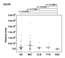
- Oxytocin receptor-derived peptide OXYR consisting of the amino acid sequence represented by SEQ ID NO: 8
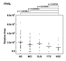
- Inter-alpha-trypsin inhibitor heavy chain H5-like protein derived peptide consisting of the amino acid sequence represented by SEQ ID NO: 10 ITH5L
- E3 ubiquitin-protein ligase HERC2-derived peptide HERC2 consisting of the amino acid sequence represented by SEQ ID NO: 12
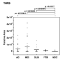
- Prothrombin-derived peptide THRB consisting of the amino acid sequence represented by SEQ ID NO: 14
- amino acid sequence represented by SEQ ID NO: 16 Tumor necrosi
- the present inventors used these proteins and peptides or peptide fragments by using two-dimensional liquid chromatography (2D-LC), MALDI TOF-MS method (mass spectrometry) and immunoMS method, so that many proteins can be obtained at once.
- the present inventors have succeeded in measuring peptides or peptide fragments and have completed the present invention.
- Complement C3 comprising the amino acid sequence represented by SEQ ID NO: 1, Transcription factor AP-2 gamma comprising the amino acid sequence represented by SEQ ID NO: 3, Synapsin-3 comprising the amino acid sequence represented by SEQ ID NO: 5, Oxytocin receptor consisting of the amino acid sequence represented by SEQ ID NO: 7, Inter-alpha-trypsin inhibitor heavy chain H5-like protein consisting of the amino acid sequence represented by SEQ ID NO: 9, E3 consisting of the amino acid sequence represented by SEQ ID NO: 11 ubiquitin-protein ligase HERC2, Prothrombin consisting of the amino acid sequence represented by SEQ ID NO: 13, Transthyretin consisting of the amino acid sequence represented by SEQ ID NO: 15, Tumor necrosis factor receptor superfamily member 16 consisting of the amino acid sequence represented by SEQ ID NO: 17
- Complement C4-A consisting of the amino acid sequence represented by SEQ ID NO: 19, Complement C4-B consisting
- Cognitive impairment disorder selected from peptides consisting of amino acid sequences represented by SEQ ID NOs: 2, 4, 6, 8, 10, 12, 14, 16, 18, 20, 22, 24, 26 and 27
- a biomarker for a cognitive dysfunction disease that appears or increases in a patient biological sample as compared to a biological sample of a subject not suffering from a mental illness.
- Alzheimer's disease patient living body selected from peptides consisting of amino acid sequences represented by SEQ ID NOs: 2, 4, 6, 8, 10, 12, 14, 16, 18, 20, 22, 24, 26 and 27
- An Alzheimer's disease biomarker that appears or increases in a sample compared to a biological sample of a subject not suffering from a mental illness.
- Patients with mild cognitive impairment selected from peptides consisting of amino acid sequences represented by SEQ ID NOs: 2, 4, 6, 8, 10, 12, 14, 16, 18, 20, 22, 24, 26 and 27
- a mild cognitive impairment biomarker that appears or increases in a biological sample compared to a biological sample of a subject not suffering from a mental illness.
- a method for detecting a cognitive dysfunction disease comprising measuring at least one biomarker for diagnosing a cognitive dysfunction disease according to any one of [1] to [5] in a biological sample.
- Detection is performed by immunoblotting method or Western blotting method, enzyme or fluorescent or radioactive substance-labeled antibody method, mass spectrometry method, immunoMS method or surface plasmon resonance method. Detection method.
- a kit for detecting a cognitive dysfunction disease for measuring at least one biomarker according to any one of [1] to [5].
- kits for detecting a cognitive dysfunction disease comprising an antibody or aptamer to at least one biomarker according to any one of [1] to [5].
- Complement C3 consisting of the amino acid sequence represented by SEQ ID NO: 1 in a biological sample derived from a subject, Transcription factor AP-2 gamma consisting of the amino acid sequence represented by SEQ ID NO: 3, represented by SEQ ID NO: 5 Synapsin-3 consisting of amino acid sequence, Oxytocin receptor consisting of amino acid sequence represented by SEQ ID NO: 7, Inter-alpha-trypsin inhibitor heavy chain H5-like protein consisting of amino acid sequence represented by SEQ ID NO: 9, SEQ ID NO: E3 ubiquitin-protein ligase HERC2 consisting of the amino acid sequence represented by 11, Prothrombin consisting of the amino acid sequence represented by SEQ ID NO: 13, Transthyretin consisting of the amino acid sequence represented by SEQ ID NO: 15, and amino acid represented by SEQ ID NO: 17 Tumor necrosis factor receptor superfamily member 16 consisting of the sequence, Complement C4-A consisting of the amino acid sequence represented by SEQ ID NO: 19, Complement C
- the complement C3-derived peptide CO3 consisting of the amino acid sequence represented by SEQ ID NO: 2 in the biological sample derived from the subject, the Transcription factor AP-2 gamma consisting of the amino acid sequence represented by SEQ ID NO: 4 Peptide AP2C, Synapsin-3 derived peptide SYN3 comprising the amino acid sequence represented by SEQ ID NO: 6, Oxytocin receptor derived peptide OXYR comprising the amino acid sequence represented by SEQ ID NO: 8, Inter comprising the amino acid sequence represented by SEQ ID NO: 10 -alpha-trypsin inhibitor heavy chain H5-like protein-derived peptide ITH5L, E3 ubiquitin-protein ligase HERC2-derived peptide HERC2, consisting of amino acid sequence represented by SEQ ID NO: 12, Prothrombin-derived peptide consisting of amino acid sequence represented by SEQ ID NO: 14 THRB, Transthyretin-derived peptide TTHY
- the present invention also provides a diagnostic system with extremely high accuracy and specificity. According to the present invention, it becomes possible for the first time to make a highly accurate diagnosis for a cognitive dysfunction disease for which there is no specific test method for a biological sample such as blood. Furthermore, the biomarker of the present invention is highly useful in determining drug effects.
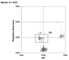
- the points in the rectangle indicated by (A) are the retention times (Retention Time) of mass peaks in Marker A's C18 reverse phase chromatography (second dimension) detected in the serum of individual subjects. And m / z.
- the points in the cluster can be regarded as having the same m / z and the same retention time within the error range, and are defined as originating from the same peptide.
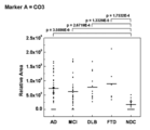
- Results of difference analysis for Marker A (dot diagram).
- Marker A is a complement C3-derived peptide CO3 as shown in the amino acid sequence obtained as a result of MS / MS analysis in FIG.
- FIG. 2 shows a comparison between CO3 and cognitive impairment disorders (AD, MCI, DLB, FTD) and NDC.
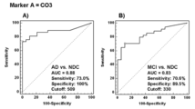
- Sequence number 2 CO3 ROC curve. See the Examples and Results section for ROC curve definitions.
- A) is an AD vs. NDC
- B) is an ROC curve obtained by analysis with a receiver operating characteristic curve in a comparison of MCI vs. NDC.
- the upper part of FIG. 4 shows the amino acid sequence of CO3 and b ions and y ions appearing in the MS / MS spectrum.
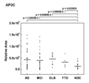
- SEQ ID NO: 4 Result of AP2C difference analysis. Comparison between subjects without mental illness (NDC) and cognitive impairment patients (AD, MCI, DLB, FTD). The result of the difference analysis of SEQ ID NO: 6 SYN3.
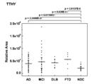
- SEQ ID NO: 16 TTHY difference analysis result Comparison between subjects without mental illness (NDC) and cognitive impairment patients (AD, MCI, DLB, FTD). The result of the difference analysis of SEQ ID NO: 18 TNR16. Comparison between subjects without mental illness (NDC) and cognitive impairment patients (AD, MCI, DLB, FTD). SEQ ID NO: 20 Result of CO4-1 difference analysis. Comparison between subjects without mental illness (NDC) and cognitive impairment patients (AD, MCI, DLB, FTD). The result of the difference analysis of SEQ ID NO: 22 CO4-2. Comparison between subjects without mental illness (NDC) and cognitive impairment patients (AD, MCI, DLB, FTD). Sequence number 24 Results of FIBA-1 difference analysis.
- SEQ ID NO: 26 Difference analysis result of FIBA-2. Comparison between subjects without mental illness (NDC) and cognitive impairment patients (AD, MCI, DLB, FTD).
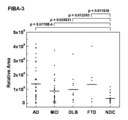
- SEQ ID NO: 27 Difference analysis result of FIBA-3. Comparison between subjects without mental illness (NDC) and cognitive impairment patients (AD, MCI, DLB, FTD).
- the present invention detects the type and amount of an intact protein and / or its partial peptide and simultaneously measures the variation of the type and amount of the intact protein and its partial peptide when the subject suffers from a cognitive impairment disease.
- This is a method for diagnosing whether or not a subject suffers from a cognitive dysfunction disease.
- the peptide generally refers to a peptide in which amino acids having a molecular weight of 10,000 or less are linked, or the number of amino acid residues is from several to about 50 or less.
- a partial peptide of an intact protein can be used as a biomarker for detecting a cognitive dysfunction disease.
- the intact peptide partial peptide refers to a peptide having a partial amino acid sequence of the amino acid sequence of the intact protein, and is generated as a partial peptide in the expression synthesis process by transcription and translation; After being synthesized as an intact protein, it may be digested and decomposed in vivo to produce a digested degradation product peptide. This is because protein synthesis and control mechanisms are deregulated when the living body is in a state other than normal, such as a cognitive impairment disorder.
- the present invention determines whether a subject is in a normal state or suffers from a cognitive dysfunction disease by using expression synthesis and / or digestion degradation of protein in vivo as an index, and also suffers from a cognitive dysfunction disease. It is also a method for evaluating and discriminating the degree of progression of the disease in the case of being present.
- the detection of a cognitive dysfunction disease refers to evaluation discrimination, that is, diagnosis, whether or not a subject suffers from a cognitive dysfunction disease. It may also include an assessment of the risk that the subject will suffer from more severe cognitive impairment.
- Complement C3 consisting of the amino acid sequence represented by SEQ ID NO: 1 and SEQ ID NO: 3 From Transcription factor AP-2 gamma consisting of amino acid sequence, Synapsin-3 consisting of amino acid sequence represented by SEQ ID NO: 5, Oxytocin receptor consisting of amino acid sequence represented by SEQ ID NO: 7, from amino acid sequence represented by SEQ ID NO: 9 Inter-alpha-trypsin inhibitor heavy chain H5-like protein, E3 ubiquitin-protein ligase HERC2 consisting of the amino acid sequence represented by SEQ ID NO: 11, Prothrombin consisting of the amino acid sequence represented by SEQ ID NO: 13, represented by SEQ ID NO: 15 Transthyretin consisting of the amino acid sequence expressed, and Tumor necrosis factor re consisting of the amino acid sequence represented by SEQ ID NO: 17.
- ceptor superfamily member 16 Complement C4-A consisting of the amino acid sequence represented by SEQ ID NO: 19
- Complement C4-B consisting of the amino acid sequence represented by SEQ ID NO: 21
- Fibrinogen alpha consisting of the amino acid sequence represented by SEQ ID NO: 23
- Examples include a chain (isoform 1) and a fibrinogen alpha chain (isoform 2) consisting of the amino acid sequence represented by SEQ ID NO: 25. The same applies to peptide fragments of 5 or more amino acid residues that are partial peptides of these intact proteins. Can be used for purposes.
- Complement C3-derived peptide CO3 consisting of the amino acid sequence represented by SEQ ID NO: 2 and Transcription factor AP consisting of the amino acid sequence represented by SEQ ID NO: 4 -2 gamma derived peptide AP2C, Synapsin-3 derived peptide SYN3 consisting of amino acid sequence represented by SEQ ID NO: 6, Oxytocin receptor derived peptide OXYR consisting of amino acid sequence represented by SEQ ID NO: 8, amino acid represented by SEQ ID NO: 10 Inter-alpha-trypsin inhibitor heavy chain H5-like protein-derived peptide ITH5L consisting of the sequence E3 ubiquitin-protein ligase HERC2-derived peptide HERC2 consisting of the amino acid sequence represented by SEQ ID NO: 12, from the amino acid sequence represented by SEQ ID NO: 14 Prothrombin-derived peptide THRB,
- the above intact protein and peptide are used as markers, and the amino acid sequence represented by SEQ ID NOs: 1 to 27 comprises an amino acid sequence in which one or several amino acids are deleted, substituted, or added.
- Proteins and peptides are also included, and these proteins or peptides can also be used as biomarkers in the methods of the invention.
- “one or several” means “one or three”, “one or two” or “one”.
- these peptides that can be used as biomarkers for the detection of cognitive dysfunction diseases also include peptide fragments having 5 or more amino acid residues resulting from the amino acid sequences represented by SEQ ID NOs: 1 to 27.
- Non-Patent Document 2 The reason why the peptide fragment is 5 or more amino acid residues is due to the following description in Non-Patent Document 2. That is, the peptide CGGGERA in which R is replaced with K for the amino acid residue sequence IRGERA at the C-terminus (130-135) of histone H3 and IR is deleted, and peptide CGERGA in which CGG is bound to GERA is used instead of peptide IRGERA. It was reported that it was recognized by the antibody obtained. This indicates that antigenic recognition is made by a peptide consisting of 4 or more amino acid residues.
- the number of residues is increased by 1 to 5 or more, but such low molecular peptides are also targeted. It is important when using a method for detection and fractionation using immunological techniques such as immunoblotting, ELISA, and immunoMS.
- a sugar chain may be added to an intact protein or a partial peptide thereof. Proteins and partial peptides to which these sugar chains are added can also be used as biomarkers for detecting cognitive impairment disorders.
- the biomarker may be quantified or the presence or absence may be determined by qualitative.
- two-dimensional electrophoresis or two-dimensional chromatography can be used as a method for separating biomarkers in a biological sample such as serum.
- the two types of chromatography in this case may be selected from known chromatography such as ion exchange chromatography, reverse phase chromatography, gel filtration chromatography and the like. It can also be quantified by the SRM / MRM method using the LC-MS / MS method.
- an immunoMS that binds an antibody against the protein or peptide of interest to the beads (including magnetic beads) developed by the inventor, captures the protein or peptide to be measured, and then elutes from the bead and measures by mass spectrometry
- the presence or amount of the target protein, protein fragment, or peptide can be easily evaluated without using two-dimensional electrophoresis or chromatography.
- a subject's cognitive dysfunction can be evaluated at a mild stage, which is useful for preventive medicine. Furthermore, when psychotherapy or pharmacotherapy is performed on a patient suffering from a cognitive dysfunction disease, if the progression of the disorder is suppressed, it is also reflected in the amount of protein / partial peptide in a biological sample such as serum. By measuring this, it is possible to evaluate and determine the therapeutic effect.
- the type and amount of protein in a biological sample can be measured by various methods.
- a target protein including a protein fragment and a partial peptide
- an antibody against it primary antibody
- Immunoblot method The simplest method. Prepare test serum diluted in several stages, drop a certain amount (around 1 microliter) onto a suitable membrane such as nitrocellulose membrane, and air dry. After treatment with a blocking solution containing a protein such as BSA, washing and reacting with the primary antibody, and after washing, a labeled secondary antibody for detecting the primary antibody is reacted. After washing the membrane, the label is visualized and the concentration is measured.
- ELISA method An antibody against a protein or its partial peptide is bound to a carrier such as a microtiter plate that has been subjected to special chemical modification in advance. After serial dilution of the sample, an appropriate amount is added to the microtiter plate to which the antibody is bound and incubated. . Thereafter, washing is performed to remove uncaptured proteins and partial peptides. Next, a secondary antibody conjugated with a fluorescent or chemiluminescent substance or enzyme is added and incubated. For detection, evaluation is performed by adding each substrate and then measuring visible light by fluorescent or chemiluminescent substances or enzymatic reactions. A substance capable of binding to a protein or a partial peptide thereof may be used instead of an antibody. For example, an aptamer or the like can be used.
- microarray is a generic term for devices in which substances that can be bound to substances to be measured are arranged (arrayed) and immobilized on a carrier (substrate).
- antibodies or aptamers against proteins and partial peptides may be used after being aligned and immobilized.
- add a biological sample to the immobilized antibody, etc. bind the protein or partial peptide to be measured on the microarray, and then add the secondary antibody to which the fluorescent or chemiluminescent substance or enzyme is bound.
- Detection may be performed by adding each substrate and then measuring the fluorescent or chemiluminescent substance or visible light from the enzyme reaction.
- Mass Spectrometry for example, an antibody against a specific protein and its partial peptide is bound to microbeads or substrates (protein chips) that have been specially modified in advance.
- the microbead may be a magnetic bead. Any material can be used for the substrate.
- the antibodies used are (1) an antibody that recognizes only the full length of a specific protein, (2) an antibody that recognizes only a partial peptide, (3) an antibody that recognizes both a specific protein and its partial peptide, or A combination of (1) and (2), (1) and (3), or (2) and (3) may be used.
- a sample is serially diluted with a stock solution or a buffer solution, and an appropriate amount thereof is added to a microbead or a substrate to which an antibody is bound, and incubated. Thereafter, washing is performed to remove uncaptured proteins and partial peptides.
- the protein and partial peptide captured on the microbead or substrate are then analyzed by mass spectrometry using MALDI TOF-MS, SELDI TOF-MS, etc., and the mass number and peak intensity of the protein, protein fragment and partial peptide peaks are analyzed. Measure.
- a certain amount of an appropriate internal standard substance is added to the original biological sample, its peak intensity is measured, and the ratio to the peak intensity of the target substance is determined to obtain the concentration in the original biological sample. I can know.
- the sample can be diluted with a stock solution or a buffer solution or a part of protein can be removed, and then separated by HPLC and quantified by mass spectrometry using an electrospray ionization (ESI) method.
- ESI electrospray ionization
- the concentration in the data can be known by absolute quantification by SRM / MRM method using an isotope-labeled internal standard peptide.
- proteins and partial peptides can be analyzed by a method using two-dimensional electrophoresis, a method using surface plasmon resonance, or the like.
- the present invention also includes a method in which a biological sample collected from a subject is subjected to two-dimensional electrophoresis or surface plasmon resonance, and a cognitive impairment disorder is detected using the presence or absence or amount of the biomarker as an index.
- SCX 1 was a flow-through fraction
- SCX 2 was a fraction eluted at a salt concentration of 100%.
- the two fractions fractionated by SCX were fractionated into 191 fractions by C18 reverse phase column chromatography, respectively.
- One fraction was eluted in 6 seconds, and the retention time (Retention Time) was obtained by multiplying the number of eluted fractions minus 1 by 6 seconds.
- MALDI target plates MTP AnchorChip (TM) 600/384) for MALDI TOF / TOF mass spectrometer (ultraflex TOF / TOF, BRUKER DALTONICS) using an online spotting robot (AccuSpot, SHIMADZU) plate well (BRUKER DALTONICS)) in the matrix solution ( ⁇ -cyano-hydroxycinnamic acid, Spot and co-crystallize while mixing with ⁇ -CHCA).
- matrix solution ⁇ -cyano-hydroxycinnamic acid, Spot and co-crystallize while mixing with ⁇ -CHCA.
- the peak area was normalized with 250 fmole of bradykinin 1-7 per well previously added to the matrix solution to obtain an Area value. That is, the area value was obtained by dividing the peak area at a specific mass of the sample by the peak area obtained from bradykinin 1-7. This Area value corresponds to 25 ⁇ l of sample serum.
- Detection of peptides with differences in serum abundance between groups was performed using Parnassum (TM) (MCBI), a multigroup statistical analysis software developed by us. Peptides in which a difference in abundance was observed were determined by MS / MS using ultraflex TOF / TOF, and the intact protein or peptide that originated the peptide was identified.
- Each point in the figure is a TOF-MS peak derived from an individual subject, and the part where the points are collected is a peak derived from one peptide having the same m / z and the same retention time within the error range. It is defined and shown as a cluster.
- the set of points in the rectangle shown in FIG. 1A is a Marker A cluster.
- FIG. 2 shows the results of the difference analysis for Marker A.
- Marker A is a complement C3-derived peptide CO3 as shown in FIG. 4 later.
- FIG. 2 is a comparison between cognitive impairment disorders (AD, MCI, DLB, FTD) and subjects not suffering from mental disorders (NDC). Area values were statistically significantly higher (p ⁇ 0.05) in cognitive impairment disorders (AD, MCI, DLB, FTD) as a result of t-test compared to NDC.
- A) and B) in FIG. 3 are ROC curves in comparison of AD vs. NDC and MCI vs. NDC, respectively.
- AUC value area value of ROC
- Typical values of sensitivity and specificity are A) and B) in Fig. 3.When a straight line is drawn from the 100% point on the y-axis to the ROC curve, the ROC that minimizes the distance The value of the coordinates of the point on the curve (open square in the figure).
- the cut-off value giving this point becomes a useful threshold for classification between different groups, and the sensitivity and specificity (that is, the above-mentioned representative values) at that time become an index of the usefulness of the biomarker together with the AUC value.
- sensitivity as a typical value in AD vs NDC is 73.0%, specificity is 100%, AUC value is 0.88, and B) is typical in MCI vs NDC.
- the sensitivity (sensitivity) was 70.6%, the specificity was 89.5%, and the AUC value was 0.83.
- Marker A is useful for distinguishing AD patients and MCI patients from subjects not suffering from mental illness (NDC).
- NDC mental illness
- Marker A is considered to be extremely useful as a marker for early diagnosis and detection of subjects who may migrate to AD.
- Fig. 4 shows the result of MS / MS analysis of Marker A using an ultraflex TOF / TOF mass spectrometer. Signals indicating y-ion, b-ion, and a-ion appeared sufficiently, and the amino acid sequence could be easily determined. This result was searched by Mascot, and the protein or peptide (hereinafter referred to as intact protein or peptide) as the origin was identified as Complement C3, and the detected peptide was derived from this protein. Amino acid sequence APVIHQEMIGGLRN (SEQ ID NO: 2) It became clear that. For the detected peptide, UniProt Entry Name CO3 is used as an abbreviation for the peptide name. The same applies to peptides other than CO3.
- MS / MS was performed by ultraflex TOF / TOF for peptides with different abundance in serum between groups, including Marker A, and the amino acid sequence was determined and the intact protein or peptide was identified. The results are shown below. Signals indicating y-ion and b-ion also appeared sufficiently for peptides other than Marker A, and the amino acid sequence could be easily determined.
- the following amino acid sequences show two sets of sequences as one set, the first of which is the intact protein amino acid sequence and the second is the sequence of the detected peptide. The underlined portion of the first sequence corresponds to the detected peptide sequence.
- the amino acid sequence starting from (0001) in the sequence represents the N-terminal side.
- Complement C3-derived peptide CO3 CO3 of SEQ ID NO: 2 formed the cluster of FIG. 1 by clustering using Parnassum (TM). As shown in FIG. 2, the Area value is statistically significantly higher (p ⁇ 0.05) in the cognitive dysfunction disease (AD, MCI, DLB, FTD) than the NDC, as a result of the t-test, It has been found that CO3 of SEQ ID NO: 2 is useful in distinguishing between patients with cognitive dysfunction disease (AD, MCI, DLB, FTD) and subjects not suffering from mental illness (NDC). Analysis by the receiver operating characteristic curve clearly shows that it is useful for distinguishing between AD and MCI patients and subjects not suffering from mental illness (FIG. 3A), B), Table 1. ).
- AP2C of SEQ ID NO: 4 is statistically significantly (p ⁇ 0.05) higher in cognitive dysfunction diseases (AD, MCI, DLB, FTD) as a result of t-test compared to NDC (Fig. 5) It has been found that AP2C of SEQ ID NO: 4 is useful in distinguishing between patients with cognitive impairment disorders (AD, MCI, DLB, FTD) and subjects who do not suffer from mental disorders (NDC). Analysis by receiver operating characteristic curves clearly proves useful in distinguishing AD and MCI patients from subjects not suffering from mental illness (Table 1).
- Transcription factor AP-2 gamma-derived peptide AP2C PGRQSQEGAGLPSHHG (SEQ ID NO: 4)
- SYN3 The area value of SYN3 of SEQ ID NO: 6 is statistically significantly (p ⁇ 0.05) higher in cognitive impairment disorders (AD, MCI, DLB, FTD) as a result of t-test compared to NDC (Fig. 6) It was found that SYN3 of SEQ ID NO: 6 is useful in distinguishing between patients with cognitive impairment disorders (AD, MCI, DLB, FTD) and subjects not suffering from mental disorders (NDC). Analysis by receiver operating characteristic curves clearly proves useful in distinguishing AD and MCI patients from subjects not suffering from mental illness (Table 1).
- Oxytocin receptor-derived peptide OXYR The area value of OXYR of SEQ ID NO: 8 is statistically significantly (p ⁇ 0.05) higher in cognitive dysfunction diseases (AD, MCI, DLB, FTD) as a result of t-test compared to NDC (Fig. 7) OXYR of SEQ ID NO: 8 was found to be useful in distinguishing between patients with cognitive dysfunction disorders (AD, MCI, DLB, FTD) and subjects not suffering from mental disorders (NDC). Analysis by receiver operating characteristic curves clearly proves useful in distinguishing AD and MCI patients from subjects not suffering from mental illness (Table 1).
- Oxytocin receptor-derived peptide OXYR AAPPGAEGNRT (SEQ ID NO: 8)
- ITH5L of SEQ ID NO: 10 The area value of ITH5L of SEQ ID NO: 10 is statistically significantly (p ⁇ 0.05) higher in cognitive dysfunction diseases (AD, MCI, DLB) than in NDC as a result of t-test (FIG. 8). It has been found that ITH5L of SEQ ID NO: 10 is useful in distinguishing between patients with cognitive dysfunction disease (AD, MCI, DLB) and subjects not suffering from mental illness (NDC). Analysis by receiver operating characteristic curves clearly proves useful in distinguishing AD and MCI patients from subjects not suffering from mental illness (Table 1).
- Inter-alpha-trypsin inhibitor heavy chain H5-like protein derived peptide ITH5L RVSLFSLAFGDDAD (SEQ ID NO: 10)
- E3 ubiquitin-protein ligase HERC2-derived peptide HERC2 The area value of HERC2 of SEQ ID NO: 12 is statistically significantly (p ⁇ 0.05) higher in cognitive dysfunction diseases (AD, MCI, DLB) than in NDC as a result of t-test (FIG. 9).
- the HERC2 of SEQ ID NO: 12 was found to be useful in distinguishing between patients with cognitive dysfunction disease (AD, MCI, DLB) and subjects who do not suffer from mental illness (NDC). Analysis by receiver operating characteristic curves clearly proves useful in distinguishing AD and MCI patients from subjects not suffering from mental illness (Table 1).
- THRB Prothrombin-derived peptide THRB
- the area value of THRB of SEQ ID NO: 14 is statistically significantly (p ⁇ 0.05) higher in cognitive dysfunction diseases (AD, MCI, DLB, FTD) as a result of t-test compared to NDC (Fig. 10)
- the THRB of SEQ ID NO: 14 was found to be useful in distinguishing between patients with cognitive dysfunction disorders (AD, MCI, DLB, FTD) and subjects not suffering from mental disorders (NDC). Analysis by receiver operating characteristic curves clearly proves useful in distinguishing AD and MCI patients from subjects not suffering from mental illness (Table 1).
- Prothrombin-derived peptide THRB TATSEYQTFFNPRTFGSGEAD (SEQ ID NO: 14)
- TTHY Transthyretin-derived peptide TTHY
- AD cognitive dysfunction diseases
- MCI cognitive dysfunction diseases
- DLB cognitive impairment disorders
- NDC mental disorders
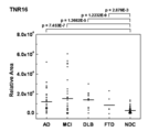
- TNR16 of SEQ ID NO: 18 is statistically significantly (p ⁇ 0.05) higher in cognitive dysfunction diseases (AD, MCI, DLB, FTD) as a result of t-test compared to NDC (Fig. 12) It was found that TNR16 of SEQ ID NO: 18 is useful in distinguishing between patients with cognitive impairment (AD, MCI, DLB, FTD) and subjects not suffering from mental illness (NDC). Analysis by receiver operating characteristic curves clearly proves useful in distinguishing AD and MCI patients from subjects not suffering from mental illness (Table 1).
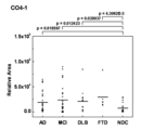
- CO4-1 of SEQ ID NO: 20 is statistically significantly (p ⁇ 0.05) higher in cognitive dysfunction diseases (AD, MCI, DLB, FTD) as a result of t-test than NDC (FIG. 13), CO4-1 of SEQ ID NO: 20 is useful in distinguishing between cognitive dysfunction disease (AD, MCI, DLB, FTD) patients and subjects not suffering from mental illness (NDC) I understood. Analysis by receiver operating characteristic curves clearly proves useful in distinguishing AD and MCI patients from subjects not suffering from mental illness (Table 1).
- Complement C4-A protein is separated into Complement C4 beta chain, Complement C4-A alpha chain and Complement C4 gamma chain by biosynthesis and processing.
- SEQ ID NO: 19 is the amino acid sequence of intact protein Complement C4-A, which includes all of these.
- Complement C4-B derived peptide CO4-1 Peptide CO4-1 of SEQ ID NO: 20 is an amino acid sequence present in the topological region common to the proteins of Complement C4-A (SEQ ID NO: 19) and Complement C4-B based on MS / MS and MASCOT database search results. .
- Complement C4-B protein is divided into Complement C4 beta chain, Complement C4-B alpha chain, and Complement C4 gamma chain by processing after biosynthesis.
- SEQ ID NO: 21 is the amino acid sequence of intact protein Complement C4-B, which includes all of these.
- CO4-2 of SEQ ID NO: 22 is statistically significantly (p ⁇ 0.05) higher in cognitive dysfunction diseases (AD, MCI, DLB, FTD) as a result of t-test than NDC (FIG. 14), CO4-2 of SEQ ID NO: 22 is useful in distinguishing between patients with cognitive impairment disorders (AD, MCI, DLB, FTD) and subjects who do not suffer from mental disorders (NDC) I understood. Analysis by receiver operating characteristic curves clearly proves useful in distinguishing AD and MCI patients from subjects not suffering from mental illness (Table 1).
- Complement C4-A protein is separated into Complement C4 beta chain, Complement C4-A alpha chain and Complement C4 gamma chain by biosynthesis and processing.
- SEQ ID NO: 19 is the amino acid sequence of intact protein Complement C4-A, which includes all of these.
- Complement C4-B-derived peptide CO4-2 Peptide CO4-2 of SEQ ID NO: 22 is an amino acid sequence present in the topological region portion common to the proteins of Complement C4-A (SEQ ID NO: 19) and Complement C4-B based on MS / MS and MASCOT database search results. .
- Complement C4-B protein is divided into Complement C4 beta chain, Complement C4-B alpha chain, and Complement C4 gamma chain by processing after biosynthesis.
- SEQ ID NO: 21 is the amino acid sequence of intact protein Complement C4-B, which includes all of these.
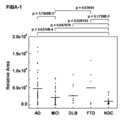
- Fibrinogen alpha chain (isoform 1) derived peptide FIBA-1 The area value of FIBA-1 of SEQ ID NO: 24 is statistically significantly (p ⁇ 0.05) higher in cognitive dysfunction diseases (AD, MCI, DLB, FTD) as a result of t-test than NDC (FIG. 15), FIBA-1 of SEQ ID NO: 24 is useful in distinguishing between cognitive dysfunction disease (AD, MCI, DLB, FTD) patients and subjects not suffering from mental illness (NDC) I understood. Analysis by receiver operating characteristic curves clearly proves useful in distinguishing AD and MCI patients from subjects not suffering from mental illness (Table 1).
- Fibrinogen alpha chain-derived peptide FIBA-1 SSSYSKQFTSSTSYNRGDSTFES (SEQ ID NO: 24)
- Fibrinogen alpha chain (isoform 2) derived peptide FIBA-1 Peptide FIBA-1 of SEQ ID NO: 24 is a topological region portion common to the proteins of Fibrinogen alpha chain (isoform 1) (SEQ ID NO: 23) and Fibrinogen alpha chain (isoform 2) from MS / MS and MASCOT database search results Is the amino acid sequence present in The amino acid sequence of the fibrinogen alpha chain (isoform 2) intact protein is shown below as SEQ ID NO: 25.
- Fibrinogen alpha chain-derived peptide FIBA-1 SSSYSKQFTSSTSYNRGDSTFES (SEQ ID NO: 24)
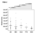
- Fibrinogen alpha chain (isoform 1) derived peptide FIBA-2 The area value of FIBA-2 of SEQ ID NO: 26 is statistically significantly (p ⁇ 0.05) higher in cognitive impairment disorders (AD, MCI, DLB, FTD) as a result of t-test than NDC. (FIG. 16), FIBA-2 of SEQ ID NO: 26 is useful in distinguishing between patients with cognitive dysfunction disorders (AD, MCI, DLB, FTD) and subjects not suffering from mental disorders (NDC) I understood. Analysis by receiver operating characteristic curves clearly proves useful in distinguishing AD and MCI patients from subjects not suffering from mental illness (Table 1).
- Fibrinogen alpha chain derived peptide FIBA-2 SSSYSKQFTSSTSYNRGDSTFESKS (SEQ ID NO: 26)
- Fibrinogen alpha chain (isoform 2) derived peptide FIBA-2 Peptide FIBA-2 of SEQ ID NO: 26 is a topological region part common to the proteins of Fibrinogen alpha chain (isoform 1) (SEQ ID NO: 23) and Fibrinogen alpha chain (isoform 2) based on MS / MS and MASCOT database search results Is the amino acid sequence present in The amino acid sequence of the fibrinogen alpha chain (isoform 2) intact protein is shown below as SEQ ID NO: 25.
- Fibrinogen alpha chain derived peptide FIBA-2 SSSYSKQFTSSTSYNRGDSTFESKS (SEQ ID NO: 26)
- Fibrinogen alpha chain (isoform 1) derived peptide FIBA-3 The area value of FIBA-3 of SEQ ID NO: 27 is statistically significantly (p ⁇ 0.05) higher in cognitive impairment disorders (AD, MCI, DLB, FTD) as a result of t-test than NDC (FIG. 17), FIBA-3 of SEQ ID NO: 27 is useful in distinguishing between patients with cognitive impairment disorders (AD, MCI, DLB, FTD) and subjects who do not suffer from mental disorders (NDC) I understood. Analysis by receiver operating characteristic curves clearly proves useful in distinguishing AD and MCI patients from subjects not suffering from mental illness (Table 1).
- Fibrinogen alpha chain (isoform 2) derived peptide FIBA-3 Peptide FIBA-3 of SEQ ID NO: 26 is a topological region portion common to the proteins of Fibrinogen alpha chain (isoform 1) (SEQ ID NO: 23) and Fibrinogen alpha chain (isoform 2) from the results of MS / MS and MASCOT database searches Is the amino acid sequence present in The amino acid sequence of the fibrinogen alpha chain (isoform 2) intact protein is shown below as SEQ ID NO: 25.
- Fibrinogen alpha chain-derived peptide FIBA-3 SSSYSKQFTSSTSYNRGDSTFESKSY (SEQ ID NO: 27)
- Table 1 shows the AUC values obtained by the analysis using the receiver operation characteristic curve in the detection of cognitive impairment disease of each marker peptide.
- biomarker disclosed in the present invention can be used to detect cognitive impairment disorders including mild cognitive impairment and Alzheimer's disease, it can be applied to applications in the diagnostic field including diagnostic agents.
Landscapes
- Health & Medical Sciences (AREA)
- Life Sciences & Earth Sciences (AREA)
- Engineering & Computer Science (AREA)
- Chemical & Material Sciences (AREA)
- Biomedical Technology (AREA)
- Molecular Biology (AREA)
- Immunology (AREA)
- Hematology (AREA)
- Urology & Nephrology (AREA)
- Proteomics, Peptides & Aminoacids (AREA)
- Biochemistry (AREA)
- Medicinal Chemistry (AREA)
- General Health & Medical Sciences (AREA)
- Organic Chemistry (AREA)
- Biotechnology (AREA)
- General Physics & Mathematics (AREA)
- Microbiology (AREA)
- Physics & Mathematics (AREA)
- Analytical Chemistry (AREA)
- Cell Biology (AREA)
- Neurosurgery (AREA)
- Food Science & Technology (AREA)
- Pathology (AREA)
- Neurology (AREA)
- Genetics & Genomics (AREA)
- Biophysics (AREA)
- Peptides Or Proteins (AREA)
- Other Investigation Or Analysis Of Materials By Electrical Means (AREA)
- Investigating Or Analysing Biological Materials (AREA)
Priority Applications (13)
| Application Number | Priority Date | Filing Date | Title |
|---|---|---|---|
| EP11851822.4A EP2657706B1 (de) | 2010-12-22 | 2011-12-21 | Biomarker für krankheiten im zusammenhang mit kognitiver dysfunktion und verfahren zur erkennung von krankheiten im zusammenhang mit kognitiver dysfunktion mittels dieses biomarkers |
| EP18163415.5A EP3422008A1 (de) | 2010-12-22 | 2011-12-21 | Neuartige biomarker zur kognitiven beeinträchtigung und verfahren zur erkennung von kognitiver beeinträchtigung mithilfe solcher biomarker |
| EP17178674.2A EP3260866B1 (de) | 2010-12-22 | 2011-12-21 | Neuartige biomarker zur kognitiven beeinträchtigung und verfahren zur erkennung von kognitiver beeinträchtigung mithilfe solcher biomarker |
| EP17178675.9A EP3255434B1 (de) | 2010-12-22 | 2011-12-21 | Neuartige biomarker zur kognitiven beeinträchtigung und verfahren zur erkennung von kognitiver beeinträchtigung mithilfe solcher biomarker |
| EP18163418.9A EP3422009A1 (de) | 2010-12-22 | 2011-12-21 | Neuartige biomarker zur kognitiven beeinträchtigung und verfahren zur erkennung von kognitiver beeinträchtigung mithilfe solcher biomarker |
| US13/995,682 US20130337479A1 (en) | 2010-12-22 | 2011-12-21 | Biomarker for cognitive dysfunction diseases, and method for detection of cognitive dysfunction diseases using the biomarker |
| EP18163419.7A EP3422010A1 (de) | 2010-12-22 | 2011-12-21 | Neuartige biomarker zur kognitiven beeinträchtigung und verfahren zur erkennung von kognitiver beeinträchtigung mithilfe solcher biomarker |
| EP18163414.8A EP3422007A1 (de) | 2010-12-22 | 2011-12-21 | Neuartige biomarker zur kognitiven beeinträchtigung und verfahren zur erkennung von kognitiver beeinträchtigung mithilfe solcher biomarker |
| US14/582,778 US20150125876A1 (en) | 2010-12-22 | 2014-12-24 | Biomarker for cognitive dysfunction diseases, and method for detection of cognitive dysfunction diseases using the biomarker |
| US15/467,646 US20170269105A1 (en) | 2010-12-22 | 2017-03-23 | Biomarker for cognitive dysfunction diseases, and method for detection of cognitive dysfunction diseases using the biomarker |
| US15/639,559 US20170299615A1 (en) | 2010-12-22 | 2017-06-30 | Biomarker for cognitive dysfunction diseases, and method for detection of cognitive dysfunction diseases using the biomarker |
| US15/639,735 US20170299616A1 (en) | 2010-12-22 | 2017-06-30 | Novel biomarkers for cognitive impairment and methods for detecting cognitive impairment using such biomarkers |
| US16/791,984 US11307208B2 (en) | 2010-12-22 | 2020-02-14 | Biomarkers for cognitive impairment and methods for detecting cognitive impairment using such biomarkers |
Applications Claiming Priority (2)
| Application Number | Priority Date | Filing Date | Title |
|---|---|---|---|
| JP2010-285726 | 2010-12-22 | ||
| JP2010285726A JP6012923B2 (ja) | 2010-12-22 | 2010-12-22 | 認知機能障害疾患のバイオマーカーおよび該バイオマーカーを用いる認知機能障害疾患の検出方法 |
Related Child Applications (2)
| Application Number | Title | Priority Date | Filing Date |
|---|---|---|---|
| US13/995,682 A-371-Of-International US20130337479A1 (en) | 2010-12-22 | 2011-12-21 | Biomarker for cognitive dysfunction diseases, and method for detection of cognitive dysfunction diseases using the biomarker |
| US14/582,778 Continuation US20150125876A1 (en) | 2010-12-22 | 2014-12-24 | Biomarker for cognitive dysfunction diseases, and method for detection of cognitive dysfunction diseases using the biomarker |
Publications (1)
| Publication Number | Publication Date |
|---|---|
| WO2012086197A1 true WO2012086197A1 (ja) | 2012-06-28 |
Family
ID=46313488
Family Applications (1)
| Application Number | Title | Priority Date | Filing Date |
|---|---|---|---|
| PCT/JP2011/007150 WO2012086197A1 (ja) | 2010-12-22 | 2011-12-21 | 認知機能障害疾患のバイオマーカーおよび該バイオマーカーを用いる認知機能障害疾患の検出方法 |
Country Status (4)
| Country | Link |
|---|---|
| US (6) | US20130337479A1 (de) |
| EP (7) | EP3260866B1 (de) |
| JP (1) | JP6012923B2 (de) |
| WO (1) | WO2012086197A1 (de) |
Cited By (3)
| Publication number | Priority date | Publication date | Assignee | Title |
|---|---|---|---|---|
| CN105659093A (zh) * | 2013-06-28 | 2016-06-08 | 株式会社Mcbi | 认知功能障碍疾病的生物标记物及使用该生物标记物的认知功能障碍疾病的检测方法 |
| WO2019012667A1 (ja) | 2017-07-13 | 2019-01-17 | 株式会社Mcbi | 認知機能障害疾患のバイオマーカー及び該バイオマーカーを用いる認知機能障害疾患の検出方法 |
| JP7107614B1 (ja) * | 2020-12-28 | 2022-07-27 | 株式会社Mcbi | 判定支援情報生成方法、判定支援情報生成システム、及び情報処理装置 |
Families Citing this family (13)
| Publication number | Priority date | Publication date | Assignee | Title |
|---|---|---|---|---|
| JP6012923B2 (ja) | 2010-12-22 | 2016-10-25 | 株式会社Mcbi | 認知機能障害疾患のバイオマーカーおよび該バイオマーカーを用いる認知機能障害疾患の検出方法 |
| GB201310203D0 (en) * | 2013-06-07 | 2013-07-24 | Electrophoretics Ltd | Materials and methods relating to Alzheimer's disease |
| WO2016044697A1 (en) * | 2014-09-19 | 2016-03-24 | The Johns Hopkins University | Biomarkers of cognitive dysfunction |
| KR102569559B1 (ko) | 2016-09-16 | 2023-08-23 | 다케다 파머수티컬 컴패니 리미티드 | 유전성 혈관부종에 대한 rna 바이오마커 |
| CN107238711B (zh) * | 2017-05-18 | 2019-07-23 | 无锡市精神卫生中心 | 一种检测阿尔茨海默病外周血蛋白标志物的诊断试剂盒及其检测方法 |
| WO2019012671A1 (ja) * | 2017-07-13 | 2019-01-17 | 株式会社Mcbi | 認知機能障害疾患のバイオマーカーおよび該バイオマーカーを用いる認知機能障害疾患の検出方法 |
| US11592452B2 (en) * | 2017-07-14 | 2023-02-28 | Mcbi Inc. | Disease detection method |
| JP2019020206A (ja) * | 2017-07-14 | 2019-02-07 | 株式会社島津製作所 | 質量分析を用いた認知機能障害疾患バイオマーカーの定量方法及び質量分析装置 |
| JP6950952B2 (ja) * | 2017-12-20 | 2021-10-13 | 国立大学法人三重大学 | 脳アミロイド血管症の末梢血バイオマーカー |
| JP7457300B2 (ja) * | 2018-08-29 | 2024-03-28 | 国立大学法人 岡山大学 | 神経変性疾患の診断用ペプチドマーカー |
| JPWO2022102654A1 (de) * | 2020-11-10 | 2022-05-19 | ||
| JP7332249B2 (ja) | 2021-12-10 | 2023-08-23 | 防衛装備庁長官 | 移行先決定プログラム、装置、及び方法 |
| WO2024004944A1 (ja) * | 2022-06-28 | 2024-01-04 | 株式会社Mcbi | 判定支援情報生成方法、判定支援情報生成システム、及び情報処理装置 |
Citations (6)
| Publication number | Priority date | Publication date | Assignee | Title |
|---|---|---|---|---|
| JPH11507821A (ja) * | 1995-06-07 | 1999-07-13 | アセナ ニューロサイエンシーズ,インコーポレイテッド | トランスジェニック動物モデルを用いてアルツハイマー病治療薬を同定する方法 |
| JP2004333274A (ja) | 2003-05-07 | 2004-11-25 | Japan Found Cancer Res | 血清アポリポタンパク質a−ii量変化の検出方法 |
| JP2005511063A (ja) * | 2001-11-28 | 2005-04-28 | ビオ・ビジョン・アーゲー | アルツハイマー病を検出する方法およびアルツハイマー病と他の痴呆疾患とを区別する方法、関連のペプチドおよびその使用 |
| JP2005523420A (ja) * | 2001-11-23 | 2005-08-04 | シン.クス ファーマ、インコーポレイテッド | アルツハイマー病を示す補体c3前駆体バイオポリマーマーカー |
| JP2006308533A (ja) | 2005-05-02 | 2006-11-09 | Mcbi:Kk | 新規肝がんバイオマーカーおよび該バイオマーカーを用いた肝がんの検出方法 |
| JP2008514946A (ja) * | 2004-09-29 | 2008-05-08 | プロティオーム・サイエンシィズ・ピーエルシー | アルツハイマー病に関する方法及び組成物 |
Family Cites Families (12)
| Publication number | Priority date | Publication date | Assignee | Title |
|---|---|---|---|---|
| EP0582450A3 (de) * | 1992-08-03 | 1994-11-23 | Rohto Pharma | Antikörper gegen Oxytocin-Rezeptor und deren Herstellung. |
| US6717031B2 (en) | 1995-06-07 | 2004-04-06 | Kate Dora Games | Method for selecting a transgenic mouse model of alzheimer's disease |
| US20040265825A1 (en) * | 2001-01-19 | 2004-12-30 | Boris Tartakovsky | Methods and compositions for diagnosing and treating a subject having depression |
| AU2003229735A1 (en) | 2002-05-02 | 2003-11-17 | Bayer Aktiengesellschaft | Diagnostics and therapeutics for diseases associated with oxytocin receptor (oxtr) |
| EP2369348A1 (de) * | 2003-11-07 | 2011-09-28 | Ciphergen Biosystems, Inc. | Biomarker für Morbus Alzheimer |
| DE602005021509D1 (de) * | 2004-08-17 | 2010-07-08 | Csl Behring Gmbh | Modifizierte vitamin-k-abhängige polypeptide |
| WO2006108051A2 (en) | 2005-04-05 | 2006-10-12 | Neurodx, Llc | Compositions and methods relating to alzheimer's disease |
| WO2007132291A2 (en) * | 2006-05-15 | 2007-11-22 | Digilab, Inc. | Biomarkers for pre-form of type 2 diabetes and methods for detecting the presence of absence of a pre-form of type 2 diabetes |
| US20100167937A1 (en) * | 2008-07-08 | 2010-07-01 | Power3 Medical Products, Inc. | Multiple forms of Alzheimer's disease based on differences in concentrations of protein biomarkers in blood serum |
| WO2010084327A2 (en) * | 2009-01-26 | 2010-07-29 | Electrophoretics Limited | Methods |
| JP2010271078A (ja) * | 2009-05-19 | 2010-12-02 | Mcbi:Kk | 認知機能障害疾患を含む精神疾患のバイオマーカーおよび該バイオマーカーを用いた認知機能障害疾患を含む精神疾患の検出方法 |
| JP6012923B2 (ja) | 2010-12-22 | 2016-10-25 | 株式会社Mcbi | 認知機能障害疾患のバイオマーカーおよび該バイオマーカーを用いる認知機能障害疾患の検出方法 |
-
2010
- 2010-12-22 JP JP2010285726A patent/JP6012923B2/ja active Active
-
2011
- 2011-12-21 EP EP17178674.2A patent/EP3260866B1/de active Active
- 2011-12-21 EP EP11851822.4A patent/EP2657706B1/de not_active Not-in-force
- 2011-12-21 EP EP18163419.7A patent/EP3422010A1/de not_active Withdrawn
- 2011-12-21 EP EP18163415.5A patent/EP3422008A1/de not_active Withdrawn
- 2011-12-21 EP EP18163418.9A patent/EP3422009A1/de not_active Withdrawn
- 2011-12-21 EP EP18163414.8A patent/EP3422007A1/de not_active Withdrawn
- 2011-12-21 EP EP17178675.9A patent/EP3255434B1/de not_active Not-in-force
- 2011-12-21 WO PCT/JP2011/007150 patent/WO2012086197A1/ja active Application Filing
- 2011-12-21 US US13/995,682 patent/US20130337479A1/en not_active Abandoned
-
2014
- 2014-12-24 US US14/582,778 patent/US20150125876A1/en not_active Abandoned
-
2017
- 2017-03-23 US US15/467,646 patent/US20170269105A1/en not_active Abandoned
- 2017-06-30 US US15/639,559 patent/US20170299615A1/en not_active Abandoned
- 2017-06-30 US US15/639,735 patent/US20170299616A1/en not_active Abandoned
-
2020
- 2020-02-14 US US16/791,984 patent/US11307208B2/en active Active
Patent Citations (6)
| Publication number | Priority date | Publication date | Assignee | Title |
|---|---|---|---|---|
| JPH11507821A (ja) * | 1995-06-07 | 1999-07-13 | アセナ ニューロサイエンシーズ,インコーポレイテッド | トランスジェニック動物モデルを用いてアルツハイマー病治療薬を同定する方法 |
| JP2005523420A (ja) * | 2001-11-23 | 2005-08-04 | シン.クス ファーマ、インコーポレイテッド | アルツハイマー病を示す補体c3前駆体バイオポリマーマーカー |
| JP2005511063A (ja) * | 2001-11-28 | 2005-04-28 | ビオ・ビジョン・アーゲー | アルツハイマー病を検出する方法およびアルツハイマー病と他の痴呆疾患とを区別する方法、関連のペプチドおよびその使用 |
| JP2004333274A (ja) | 2003-05-07 | 2004-11-25 | Japan Found Cancer Res | 血清アポリポタンパク質a−ii量変化の検出方法 |
| JP2008514946A (ja) * | 2004-09-29 | 2008-05-08 | プロティオーム・サイエンシィズ・ピーエルシー | アルツハイマー病に関する方法及び組成物 |
| JP2006308533A (ja) | 2005-05-02 | 2006-11-09 | Mcbi:Kk | 新規肝がんバイオマーカーおよび該バイオマーカーを用いた肝がんの検出方法 |
Non-Patent Citations (3)
| Title |
|---|
| "The better understanding of Alzheimer's disease.", 2004, NAGAI SHOTEN CO., LTD. |
| BENKIRANE, N. ET AL., J. BIOL. CHEM., vol. 268, 1993, pages 26279 - 26285 |
| See also references of EP2657706A4 |
Cited By (5)
| Publication number | Priority date | Publication date | Assignee | Title |
|---|---|---|---|---|
| CN105659093A (zh) * | 2013-06-28 | 2016-06-08 | 株式会社Mcbi | 认知功能障碍疾病的生物标记物及使用该生物标记物的认知功能障碍疾病的检测方法 |
| EP3015865A4 (de) * | 2013-06-28 | 2017-06-07 | Mcbi Inc. | Biomarker für kognitiver dysfunktionserkrankungen und verfahren zur erkennung von kognitiven dysfunktionserkrankungen unter verwendung von biomarkern |
| CN111426848A (zh) * | 2013-06-28 | 2020-07-17 | 株式会社 Mcbi | 认知功能障碍疾病的生物标记物及使用该生物标记物的认知功能障碍疾病的检测方法 |
| WO2019012667A1 (ja) | 2017-07-13 | 2019-01-17 | 株式会社Mcbi | 認知機能障害疾患のバイオマーカー及び該バイオマーカーを用いる認知機能障害疾患の検出方法 |
| JP7107614B1 (ja) * | 2020-12-28 | 2022-07-27 | 株式会社Mcbi | 判定支援情報生成方法、判定支援情報生成システム、及び情報処理装置 |
Also Published As
| Publication number | Publication date |
|---|---|
| US20200200768A1 (en) | 2020-06-25 |
| JP6012923B2 (ja) | 2016-10-25 |
| EP3422008A1 (de) | 2019-01-02 |
| EP3260866B1 (de) | 2019-03-06 |
| US20170269105A1 (en) | 2017-09-21 |
| EP3255434A1 (de) | 2017-12-13 |
| EP2657706A1 (de) | 2013-10-30 |
| JP2012132808A (ja) | 2012-07-12 |
| US20170299615A1 (en) | 2017-10-19 |
| EP3422009A1 (de) | 2019-01-02 |
| EP3422007A1 (de) | 2019-01-02 |
| EP3255434B1 (de) | 2019-03-13 |
| EP3260866A1 (de) | 2017-12-27 |
| US20150125876A1 (en) | 2015-05-07 |
| EP2657706A4 (de) | 2015-11-04 |
| EP3422010A1 (de) | 2019-01-02 |
| EP2657706B1 (de) | 2018-04-11 |
| US11307208B2 (en) | 2022-04-19 |
| US20170299616A1 (en) | 2017-10-19 |
| US20130337479A1 (en) | 2013-12-19 |
Similar Documents
| Publication | Publication Date | Title |
|---|---|---|
| US11307208B2 (en) | Biomarkers for cognitive impairment and methods for detecting cognitive impairment using such biomarkers | |
| JP6265507B2 (ja) | 認知機能障害疾患のバイオマーカー及び当該バイオマーカーを用いる認知機能障害疾患の検出方法 | |
| US11726099B2 (en) | Biomarker for mental disorders including cognitive disorders, and method using said biomarker to detect mental disorders including cognitive disorders | |
| JP6113798B2 (ja) | 認知機能障害疾患のバイオマーカーおよび該バイオマーカーを用いる認知機能障害疾患の検出方法 | |
| JP6408087B2 (ja) | 認知機能障害疾患のバイオマーカーおよび該バイオマーカーを用いる認知機能障害疾患の検出方法 | |
| JP6967206B2 (ja) | 認知機能障害疾患のバイオマーカー及び該バイオマーカーを用いる認知機能障害疾患の検出方法 | |
| WO2019012671A1 (ja) | 認知機能障害疾患のバイオマーカーおよび該バイオマーカーを用いる認知機能障害疾患の検出方法 | |
| JP6359158B2 (ja) | 認知機能障害疾患のバイオマーカーおよび該バイオマーカーを用いる認知機能障害疾患の検出方法 | |
| JP6359160B2 (ja) | 認知機能障害疾患のバイオマーカーおよび該バイオマーカーを用いる認知機能障害疾患の検出方法 | |
| WO2022102654A1 (ja) | バイオマーカーの組合せ、及び、当該組合せを用いた認知機能障害又はそのリスクの検出方法 | |
| JP6193942B2 (ja) | 認知機能障害疾患のバイオマーカーおよび該バイオマーカーを用いる認知機能障害疾患の検出方法 | |
| JP6359159B2 (ja) | 認知機能障害疾患のバイオマーカーおよび該バイオマーカーを用いる認知機能障害疾患の検出方法 | |
| JP2012037349A (ja) | 認知機能障害疾患のバイオマーカーおよび該バイオマーカーを用いた認知機能障害疾患の検出方法 |
Legal Events
| Date | Code | Title | Description |
|---|---|---|---|
| 121 | Ep: the epo has been informed by wipo that ep was designated in this application |
Ref document number: 11851822 Country of ref document: EP Kind code of ref document: A1 |
|
| NENP | Non-entry into the national phase |
Ref country code: DE |
|
| WWE | Wipo information: entry into national phase |
Ref document number: 2011851822 Country of ref document: EP |
|
| WWE | Wipo information: entry into national phase |
Ref document number: 13995682 Country of ref document: US |