WO2012033002A1 - X線ct装置 - Google Patents
X線ct装置 Download PDFInfo
- Publication number
- WO2012033002A1 WO2012033002A1 PCT/JP2011/069910 JP2011069910W WO2012033002A1 WO 2012033002 A1 WO2012033002 A1 WO 2012033002A1 JP 2011069910 W JP2011069910 W JP 2011069910W WO 2012033002 A1 WO2012033002 A1 WO 2012033002A1
- Authority
- WO
- WIPO (PCT)
- Prior art keywords
- image
- dose
- ray
- irradiation
- subject
- Prior art date
Links
Images


Classifications
-
- A—HUMAN NECESSITIES
- A61—MEDICAL OR VETERINARY SCIENCE; HYGIENE
- A61B—DIAGNOSIS; SURGERY; IDENTIFICATION
- A61B6/00—Apparatus for radiation diagnosis, e.g. combined with radiation therapy equipment
- A61B6/10—Application or adaptation of safety means
-
- A—HUMAN NECESSITIES
- A61—MEDICAL OR VETERINARY SCIENCE; HYGIENE
- A61B—DIAGNOSIS; SURGERY; IDENTIFICATION
- A61B6/00—Apparatus for radiation diagnosis, e.g. combined with radiation therapy equipment
- A61B6/02—Devices for diagnosis sequentially in different planes; Stereoscopic radiation diagnosis
- A61B6/03—Computerised tomographs
- A61B6/032—Transmission computed tomography [CT]
-
- A—HUMAN NECESSITIES
- A61—MEDICAL OR VETERINARY SCIENCE; HYGIENE
- A61B—DIAGNOSIS; SURGERY; IDENTIFICATION
- A61B6/00—Apparatus for radiation diagnosis, e.g. combined with radiation therapy equipment
- A61B6/40—Apparatus for radiation diagnosis, e.g. combined with radiation therapy equipment with arrangements for generating radiation specially adapted for radiation diagnosis
- A61B6/4064—Apparatus for radiation diagnosis, e.g. combined with radiation therapy equipment with arrangements for generating radiation specially adapted for radiation diagnosis specially adapted for producing a particular type of beam
- A61B6/4078—Fan-beams
-
- A—HUMAN NECESSITIES
- A61—MEDICAL OR VETERINARY SCIENCE; HYGIENE
- A61B—DIAGNOSIS; SURGERY; IDENTIFICATION
- A61B6/00—Apparatus for radiation diagnosis, e.g. combined with radiation therapy equipment
- A61B6/46—Apparatus for radiation diagnosis, e.g. combined with radiation therapy equipment with special arrangements for interfacing with the operator or the patient
- A61B6/461—Displaying means of special interest
-
- A—HUMAN NECESSITIES
- A61—MEDICAL OR VETERINARY SCIENCE; HYGIENE
- A61B—DIAGNOSIS; SURGERY; IDENTIFICATION
- A61B6/00—Apparatus for radiation diagnosis, e.g. combined with radiation therapy equipment
- A61B6/52—Devices using data or image processing specially adapted for radiation diagnosis
- A61B6/5205—Devices using data or image processing specially adapted for radiation diagnosis involving processing of raw data to produce diagnostic data
-
- G—PHYSICS
- G06—COMPUTING; CALCULATING OR COUNTING
- G06T—IMAGE DATA PROCESSING OR GENERATION, IN GENERAL
- G06T11/00—2D [Two Dimensional] image generation
- G06T11/003—Reconstruction from projections, e.g. tomography
- G06T11/005—Specific pre-processing for tomographic reconstruction, e.g. calibration, source positioning, rebinning, scatter correction, retrospective gating
Definitions
- the present invention relates to an X-ray CT apparatus that obtains an X-ray CT by irradiating a subject with X-rays, and particularly relates to a technique for measuring an irradiation dose to a subject and an exposure dose of the subject.
- Patent Document 1 information related to an exposure dose when a predetermined amount of X-rays are irradiated for a predetermined time is stored in a storage unit, and each part of the subject corresponding to each position in a reconstructed X-ray tomogram is stored.
- the approximate constant X-ray dose reached is calculated by the X-ray dose calculation unit, and based on the information on the exposure dose, the calculated X-ray dose reaching each part, and the X-ray irradiation time, Since the exposure dose distribution of the subject by the X-rays irradiated when reconstructing the line tomographic image is calculated by the exposure dose distribution calculation unit, the exposure dose distribution for each part of the subject is calculated.
- An object of the present invention is to provide an X-ray CT apparatus capable of quickly evaluating the exposure dose of a subject.
- the present invention acquires an irradiation X-ray image that is a distribution of irradiation intensity of X-rays irradiated to a subject by an X-ray irradiation unit according to imaging conditions, and the irradiation X-rays
- the image and the reconstructed image are projected and converted, and the projected reconstructed image and the irradiation X-ray image corresponding to the reconstructed image creation region are used to indicate the exposure dose distribution of the subject.
- An image reconstruction unit that creates an exposure dose image and calculates the exposure dose is provided.
- the exposure dose of the subject can be quickly evaluated.
- the figure explaining the irradiation dose image creation processing in 1st Embodiment The flowchart which shows the flow of the exposure dose image creation process in 1st Embodiment
- the figure explaining the exposure dose image creation process in 1st Embodiment The figure which shows an example of the screen which displays dose evaluation information
- the figure explaining the irradiation dose image creation processing in 2nd Embodiment The flowchart which shows the flow of the exposure dose image creation process in 2nd Embodiment
- the figure explaining the exposure dose image creation process in 2nd Embodiment The figure explaining the exposure dose image creation process in 2nd Embodiment
- the figure explaining the cumulative irradiation dose image creation process in 3rd Embodiment The figure explaining the accumulated exposure dose image creation process in 4th Embodiment
- the present invention evaluates an exposure dose in an image diagnosis performed using an X-ray CT apparatus.
- the X-ray CT apparatus includes an imaging unit 1, an operation unit 2, and the like.
- the photographing means 1 includes a gantry 100 that houses a scanner main body and a bed apparatus 101.
- the operating means 2 operates and controls the photographing means 1.
- the operation means 2 performs input of shooting conditions, image processing, and the like.
- the gantry 100 includes an X-ray generator 102 that irradiates X-rays from around the subject 3, a collimator device 104 that narrows a bundle of X-rays generated from the X-ray generator 102, and the subject.
- X-ray detection device 103 for detecting X-rays transmitted through 3
- high-voltage generation device 105 for applying a high voltage to the X-ray generation device 102
- data collection device 106 for collecting data detected by the X-ray detection device 103
- scanner Is composed of a drive device 107 and the like that rotate the patient 3 around the subject 3.
- the collimator device 104 may include a compensation filter for adjusting the X-ray irradiation distribution.
- the operation means 2 includes an input / output device 201, a calculation device 202, a central control device 200, and the like.
- the input / output device 201 includes a display device 211 that displays data such as images, an input device 212 for an operator to input shooting conditions, a storage device 213 that stores data necessary for shooting such as programs and device parameters, and the like. Consists of
- the computing device 202 includes an image reconstruction device 221 that creates a reconstructed image of the subject 3 based on data obtained from the imaging means 1, an image processing device 222 that analyzes image data, and the like.
- the central control device 200 controls each of the photographing unit 1 and the operation unit 2 in accordance with an operation instruction from the operator.
- Imaging with the X-ray CT apparatus is a rotating imaging in which the X-ray generator 102 and the data collection device 106 rotate while taking the image in the gantry 100, and the X-ray generation device 102 and the data collection device 106 are stationary in the gantry 100. There is still shooting for shooting.
- Tomographic imaging for obtaining a tomographic image of the subject 3 is based on rotational imaging.
- Scanogram imaging for determining the tomographic imaging position is based on still imaging.
- the tomographic imaging trajectory may be any of a circular trajectory, a spiral trajectory, a combination of a circular trajectory and a spiral trajectory, and is not particularly limited.
- the X-ray CT apparatus comprising the imaging means 1 and the operation means 2 shown in FIGS. It can be understood as an X-ray CT device that is electrically connected by, for example.
- a data processing device such as a workstation or a desktop computer as shown in the operation means 2 in FIG. 1, such a device is replaced as follows. That is, the input / output device 201 constituting the operation means 2 is an input / output unit, the central control device 200 is a control unit, and the calculation device 202 is a calculation unit.
- a data processing device is the same as that of an integrated stand-alone type device.
- the X-ray generator 102 is an X-ray generator
- the collimator device 104 is a collimator unit
- the X-ray generator and the collimator unit are configured.
- the X-ray detection device 103 is an X-ray detection unit
- the high voltage generation device 105 is a high voltage generation unit
- the data collection device 106 is a data collection unit
- the drive device 107 is a drive unit. That is, the photographing means 1 includes these parts, the gantry 100, and the bed apparatus 101.
- Each part of the imaging means 1 and the operating means 2 has the same function as the above-described apparatus.
- the “X-ray irradiation unit”, “X-ray detection unit”, “image reconstruction unit”, and “display unit” are defined as follows.
- the “X-ray irradiation unit” irradiates X-rays from around the subject.
- the X-ray generation unit 102 irradiates X-rays from around the subject 3 and the X-ray generation unit 102 And a collimator unit 104 for narrowing a bundle of lines.
- the “X-ray detection unit” detects X-rays transmitted through the subject as X-ray information, and corresponds to the X-ray detection device 103.
- the “image reconstruction unit” creates a reconstructed image of the subject from the X-ray information detected by the X-ray detection unit. Acquire an irradiation X-ray image that is the distribution of the irradiation intensity of the irradiated X-ray, project the irradiation X-ray image and the reconstructed image, and correspond to the reconstructed image and the reconstructed image creation area
- the image reconstruction device 221 corresponds to an exposure dose image that is an image showing the distribution of the exposure dose of the subject using the irradiated X-ray image, and calculates the exposure dose.
- the “display unit” displays an exposure dose image and an exposure dose, and corresponds to the display device 211.
- the image reconstruction unit image reconstruction device 221) evaluates the exposure dose of the subject 3 by executing a process flow to be described later.
- the “image” is not limited to what is visually displayed on the display device, but also means pixel value aggregate data. Then, “creating an image” means calculating each pixel value.
- “Irradiated X-ray information” is a distribution of irradiation intensity of X-rays irradiated to the subject 3 by the X-ray generator 102.
- the “irradiation X-ray image” and “irradiation dose image” are images showing the distribution of the X-ray irradiation dose irradiated to the subject 3.
- an “attenuation coefficient image” is an image showing the distribution of attenuation coefficients.
- the “irradiation dose correction image” is an image showing a reduction result of the attenuation amount with respect to the irradiation dose image. That is, the irradiation dose correction image is an image showing the result of reducing the attenuation by the subject 3 from the irradiation dose image.
- the “absorbed dose image” is an image showing the distribution of the absorbed dose of X-rays absorbed by the subject 3. Absorbed dose is the basic dosimetric measure in radiation protection and is defined as the energy absorbed per unit mass. The unit of absorbed dose is J / kg (special unit is Gy (gray)).
- “Equivalent dose image” is an image showing the distribution of equivalent dose.
- the equivalent dose is the product of the average absorbed dose in each organ or tissue and the radiation weighting factor.
- the unit of equivalent dose is J / kg (special unit is Sv (sievert)).
- the radiation weighting factor for photons is 1, not limited to the energy range, so the absorbed dose is equivalent to the equivalent dose in CT.
- Effective dose image is an image showing the effective dose. Effective doses are weighted by the International Commission on Radiological Protection (ICRP) by weighting the tissue weighting factors that represent different radiation sensitivities for organs and / or tissues (hereinafter referred to as organs) with respect to equivalent doses. It is defined as the total value of etc.
- the unit of effective dose is J / kg (special unit is Sv (sievert)).
- the “organ discrimination image” is an image for identifying a region such as an organ included in the reconstructed image.
- a “tissue load coefficient image” is an image obtained by weighting an equivalent dose image with a tissue load coefficient for each region such as an organ identified by an organ discrimination image.
- the “exposure dose image” is an image showing the distribution of the exposure dose of the subject 3.
- the exposure dose image is calculated as an effective dose image, for example.
- the image reconstruction device 221 of the X-ray CT apparatus acquires a subject image (S101).
- the subject image in the first embodiment is a tomographic image 5 (reconstructed image) of the subject 3 shown in FIG.
- a tomographic plane of the tomographic image 5 is a region 4a indicated by a dotted line.
- the tomographic image 5 may be either a single image or a plurality of images, and is not particularly limited.
- the subject image in the first embodiment is not only a cross-sectional image (axial image) perpendicular to the body axis of the subject 3 as in the tomographic image 5, but also the body axis of the subject 3.
- the image reconstruction device 221 of the X-ray CT apparatus acquires irradiation X-ray information from the storage device 213 according to the imaging conditions (S102).
- Information is acquired from the storage device 213.
- the image reconstruction device 221 acquires irradiation X-ray information corresponding to the imaging conditions at predetermined time intervals (for example, for each view).
- the irradiation X-ray information is the three-dimensional irradiation intensity distribution data 1001 shown in FIG.
- the image reconstruction device 221 stores the three-dimensional irradiation intensity distribution data 1001 in the storage device 213 for each imaging condition before S102.
- the three-dimensional irradiation intensity distribution data 1001 includes the X-ray beam spread (fan angle) in the slice plane direction (xy plane direction) and the X-ray beam in the body axis direction (z axis direction). Of spread (cone angle).
- the three-dimensional irradiation intensity distribution data 1001 is not limited to the example shown in FIG.
- the three-dimensional irradiation intensity distribution data 1001 may be any of data measured in advance before capturing the tomographic image 5, data obtained during capturing of the tomographic image 5, or data obtained by numerical simulation.
- the three-dimensional irradiation intensity distribution data 1001 may be data obtained by modeling or parameterizing actual measurement data or simulation data.
- the three-dimensional irradiation intensity distribution data 1001 varies depending on the voltage and current (tube voltage, tube current) applied to the X-ray generator 102. Further, it varies depending on the degree of opening of the collimator portion of the collimator device 104. It also depends on the geometric characteristics of the gantry 100.
- the three-dimensional irradiation intensity distribution data 1001 is not particularly limited to imaging conditions, and can be defined according to various imaging conditions.
- the image reconstruction device 221 of the X-ray CT apparatus creates an irradiation dose image (S103).
- the image reconstruction device 221 creates three-dimensional irradiation dose data 2000 by time-integrating a set 1000 of a plurality of three-dimensional irradiation intensity distribution data acquired in S102.
- the image reconstruction device 221 uses a region 4b having the same z position as the region 4a indicating the tomographic plane of the tomographic image 5 as the cutout plane, and uses the three-dimensional irradiation dose data 2000 on the cutout plane. These pixel values are extracted as an irradiation dose image 6.
- the irradiation dose image 6 shown in FIG. 4 is expressed so that the pixel value of the circular region is constant, it actually has a different pixel value for each pixel. That is, the irradiation dose image 6 is an image having a distribution.
- the region of the three-dimensional irradiation dose data 2000 has, for example, an inner peripheral surface of the gantry 100 (an opening of the gantry 100) as a cross section, and X-rays reach the body axis direction (z-axis direction).
- the shape of the region of the three-dimensional irradiation dose data 2000 is not limited to this, and may include at least an X-ray irradiation range that may be irradiated to the subject 3 at the time of imaging.
- the cross section of the region of the three-dimensional irradiation dose data 2000 is not limited to a circle but may be an ellipse or a rectangle. Moreover, the cross section of the region of the three-dimensional irradiation dose data 2000 may have different areas in the body axis direction (z-axis direction).
- the image reconstruction device 221 of the X-ray CT apparatus creates an exposure dose image (S104).
- the image reconstruction device 221 acquires the tomographic image 5 shown in FIG. 4 (S201), and based on the information indicating the relationship between the CT value and the linear attenuation coefficient, the attenuation coefficient image from the tomographic image 5 Conversion to 7 (S202).
- CT value ( ⁇ t ⁇ w) / ⁇ w ⁇ K” ( ⁇ t: subject 3 linear attenuation coefficient, ⁇ w: linear attenuation coefficient of water, K: constant) It is expressed by a relational expression.
- the X-ray attenuation coefficient is a physical coefficient determined by the energy distribution of the irradiated X-rays and the substances constituting the subject 3.
- Information indicating the relationship between the CT value and the attenuation coefficient depends on imaging conditions, characteristics of the X-ray CT apparatus, and the like.
- the conversion data may be data measured in advance before the tomographic image 5 is taken, data obtained by numerical simulation, or data obtained by modeling or parameterizing the measured data or simulation data.
- the image reconstruction device 221 performs forward projection (also referred to as reprojection) processing on the attenuation coefficient image 7 and converts it into attenuation coefficient projection data 50 (S203).
- the forward projection process is a process of converting the distribution of the linear attenuation coefficient in the orthogonal coordinate system into the distribution of the linear attenuation coefficient in the projection coordinate system (so-called radon conversion).
- the orthogonal coordinate system is a coordinate system in which the subject 3 is fixed.
- the projected coordinate system is a coordinate system that represents the position when the X-ray generator 102 and the data collection device 106 are rotated by ⁇ with respect to the orthogonal coordinate system.
- the image reconstruction device 221 acquires the irradiation dose image 6 created in S103 of FIG. 3 (S204), forward-projects the irradiation dose image 6, and converts it into irradiation dose projection data 51 (S205). ).
- the forward projection process in S205 is the same as the forward projection process in S203.
- an exposure dose image that is an image showing the distribution of the exposure dose of the subject is created using the reconstructed image and the irradiated X-ray image corresponding to the creation area of the reconstructed image.
- the image reconstruction device 221 performs reverse attenuation correction processing on the irradiation dose projection data 51 using the attenuation coefficient projection data 50 (S206). Specifically, the image reconstruction device 221 generates attenuation correction irradiation dose projection data 52 according to the following equation (1).
- correction in consideration of the influence of X-ray scattering, refraction, and diffraction by the subject 3 and X-ray directivity may be added.
- the image reconstruction device 221 performs back projection processing on the attenuation correction irradiation dose projection data 52 to create an irradiation dose correction image 8 (S207).
- the back projection process is a process of converting the distribution of the linear attenuation coefficient in the projection coordinate system into the distribution of the linear attenuation coefficient in the orthogonal coordinate system (so-called inverse radon conversion).
- the irradiation dose correction image 8 includes essential information for evaluating the exposure of the subject 3.
- S208 and S209, which will be described later, are processes for converting the irradiation dose correction image 8 into existing dose units.
- the image reconstruction device 221 obtains a difference between the irradiation dose correction image 8 from the irradiation dose image 6 and creates an absorbed dose image based on the obtained difference (S208).
- the difference between the irradiation dose image 6 and the irradiation dose correction image 8 indicates the amount of attenuation by the subject 3.
- the attenuation amount by the subject 3 corresponds to the absorbed dose of the subject 3.
- the absorbed dose image is an image having a distribution similar to the irradiation dose image 6.
- the image reconstruction device 221 creates an equivalent dose image by multiplying the absorbed dose image by the radiation load coefficient (S209).
- the ICRP recommendation is that the radiation weighting factor in photons is 1, not limited to the energy range, so the absorbed dose image and the equivalent dose image are the same.
- the image reconstruction device 221 performs region determination based on the CT value of the tomographic image 5, identifies a region such as an organ, and creates an organ discrimination image (S210).
- the organ discrimination image is a map of an organ or the like, for example, a label image with a different label for each organ or the like.
- the region determination may be performed based on characteristics of an anatomical shape such as an organ instead of the CT value.
- the image reconstruction device 221 may not only identify a region such as an organ but also identify a part of a human body and create a part-specific image.
- the image reconstruction device 221 creates a tissue load coefficient image using a tissue load coefficient corresponding to an organ or the like (S211).
- the tissue load coefficient for example, the value shown in the recommendation of ICRP can be used.
- the image reconstruction unit calculates the exposure dose. That is, the image reconstruction device 221 collates the organ discrimination image and the equivalent dose image (or absorbed dose image), and creates a tissue load coefficient image.
- the tissue load coefficient image shows the calculation result of the exposure dose for each organ or the like. With the tissue load coefficient image, it becomes possible to evaluate the exposure dose for each organ or the like of the subject 3.
- the image reconstruction device 221 may collate the region-specific image with the equivalent dose image (or absorbed dose image) to create a region load coefficient image.
- the part load coefficient image shows the calculation result of the exposure dose for each part of the human body (head, chest, abdomen, limbs, etc.).
- the image reconstruction device 221 converts the equivalent dose image into an effective dose image using the tissue load coefficient image, and sets the effective dose image as the exposure dose image (S212).
- the effective dose image is an image obtained by weighting the tissue load coefficient with respect to the equivalent dose image.
- the image reconstruction device 221 may convert the absorbed dose image into an exposure dose image using a dose equivalent conversion coefficient.
- the dose equivalent conversion factor for example, the value shown in the recommendation of ICRP can be used.
- the display device 211 displays a dose management screen 700 as shown in FIG. 7 based on the calculation result of the image reconstruction device 221.
- the dose management screen 700 shown in FIG. 7 includes a coronal image exposure image 9a, an axial image exposure image 9b, an irradiation range image 701 in which the irradiation image 6 is superimposed on the virtual image 3a of the subject 3, Includes exposure dose 702, exposure dose index conversion button 703, exposure dose by imaging 704, exposure dose by region 705, exposure dose by organ 706, exposure dose 707 within the ROI (region of interest), and ON / OFF button 708 for image display It is.
- the exposure dose images 9a and 9b include partial information on the region-specific dose 705, organ-specific dose 706, and ROI internal dose 707. Is displayed.
- the lung exposure dose 706a, the ROI 707a, and the ROI exposure dose 707b are superimposed and displayed.
- an abdominal exposure dose 705a, a liver exposure dose 706b, and a spine exposure dose 706c are superimposed and displayed.
- irradiation range image 701 in addition to the virtual image 3a of the subject 3 and the irradiation dose image 6, a cylindrical three-dimensional irradiation dose data 2000 and a circular region 4b indicating a cut-out plane of the irradiation dose image 6 are displayed. Has been.
- the irradiation range image 701 allows the user to roughly grasp the X-ray irradiation range for the subject 3.
- the exposure dose by imaging 702 displays the exposure dose by tomography, the exposure dose by simple imaging (scanogram imaging), and the cumulative exposure dose by tomography and simple imaging.
- the calculation of the exposure dose by simple imaging and the cumulative exposure dose by tomographic imaging and simple imaging will be described in an embodiment described later.
- the exposure dose index conversion button 703 is a button for converting an exposure dose index into another index.
- Other indexes may include existing indexes such as CTDI (Computed Tomography Dose Index), DLP (Dse Length Product), CTDIw (weighted CTDI), CTDIvol (volumetric CTDI), and user-defined indexes.
- the image reconstruction device 221 calculates CTDI, DLP, CTDIw, and CTDIvol based on the irradiation dose image 6.
- an irradiation dose by rotational imaging (corresponding to tomographic imaging), an irradiation dose by still imaging (corresponding to scanogram imaging), and a cumulative irradiation dose by rotating imaging and static imaging are displayed.
- calculation of the exposure dose by still photography and the cumulative irradiation dose by rotation photography and still photography will be described in an embodiment described later.
- the site-specific exposure dose 705 displays the exposure dose of the head, chest, abdomen, and extremities.
- the organ-specific exposure dose 706 displays the exposure dose of bone, brain, lung, stomach, intestine, liver, breast, bladder and the like.
- the ROI exposure dose 707 the exposure dose in the ROI is displayed.
- tomographic imaging rotational imaging
- the exposure dose of the subject 3 and the irradiation dose to the subject 3 can be evaluated and managed in high speed, high accuracy and in detail.
- the physical characteristics reflecting the morphological information included in the reconstructed image (clinical image information) and the substance configuration of the subject 3 are used, those different from human tissues such as indwelling materials and contrast agents, and irradiation X Applicable to CT tomographic images where there is a subject that significantly attenuates the line as subject 3 or a radiography method that modulates the intensity and energy of irradiated X-rays at short time intervals during radiography. Yes, the exposure dose of the subject can be evaluated.
- the exposure dose can be evaluated even for reconstructed images that have undergone image reconstruction methods or image processing methods that change the image characteristics. it can.
- the three-dimensional distribution of the irradiation X-ray dose is considered for each pixel in the image for evaluating the exposure dose by the processing shown in S201 to S207 shown in FIG. Since the attenuation amount in consideration of the shape of the person 3 can be calculated, the exposure dose of the subject 3 can be evaluated with high accuracy.
- the calculation process for each organ or the like of the subject 3 (for example, the process for calculating the X-ray transmission length for each organ or the like) is not performed. Even if there are many organs or the like in the image for evaluating the dose, the calculation amount is small, and the calculation time can be shortened.
- the exposure dose can be calculated for each organ or the like by using the values shown in the recommendation of ICRP by S210 to S211 shown in FIG. This is because the absorbed dose can be calculated with high accuracy in units of pixels by the processing shown in S201 to S207 shown in FIG.
- Scanogram imaging is still imaging, and is imaging for obtaining a scanogram image of the subject 3 (a moving image in which the movement of the organ or the like of the subject 3 can be confirmed).
- the subject 3 image in the second embodiment is the scanogram image 10 of the subject 3 shown in FIG.
- the area 4c in the imaging range of the scanogram image 10 is an area parallel to the YZ plane.
- the scanogram image 10 may be either a single image or a plurality of images, and is not particularly limited. Further, the scanogram image 10 may be taken from the front, the plane, the left side, the right side, or any other direction of the subject 3, or may be taken multiple times by combining a plurality of directions. You may do it.
- the image reconstruction device 221 of the X-ray CT apparatus acquires the scanogram image 10 shown in FIG. 4 (S301), and converts the scanogram image 10 to the attenuation ratio image 11 based on the attenuation reference value. (S302).
- the attenuation ratio image 11 is an image showing the distribution of the attenuation ratio.
- the attenuation ratio is a value (relative value) indicating the ratio of each pixel value with reference to the attenuation reference value.
- the attenuation reference value may be data measured in advance, data obtained by numerical simulation, or an average pixel value of a partial region 12 (see FIG. 10) of the scanogram image.
- the partial area 12 of the scanogram image is preferably an air area, but may be an area of a substance other than that.
- the image reconstruction device 221 acquires irradiation X-ray information from the storage device 213 according to the imaging conditions.
- Information is acquired from the storage device 213.
- the irradiation X-ray information is the three-dimensional irradiation intensity distribution data 1101 shown in FIG.
- the three-dimensional irradiation intensity distribution data 1101 includes the X-ray beam spread (fan angle) in the slice plane direction (xy plane direction) and the X-ray beam in the body axis direction (z axis direction). Of spread (cone angle).
- the three-dimensional irradiation intensity distribution data 1101 is not limited to the example shown in FIG.
- the image reconstruction device 221 time-integrates a set 1100 of 3D irradiation intensity distribution data at each time, and performs a projective transformation from 3D to 2D according to the scanogram image 10 (in the example shown in FIG. 8).
- two-dimensional irradiation dose data 2100 is created by performing projection conversion to the yz plane.
- the image reconstruction device 221 creates an irradiation dose image 6 (stationary imaging irradiation dose image, irradiation X-ray image) having the same region 4d as the imaging region 4c of the scanogram image 10 (S303).
- the image reconstruction device 221 calculates the attenuation amount by the subject 3 by performing reverse attenuation correction processing on the irradiation dose image 6 using the attenuation ratio image 11 (S304), and the subject 3 Assuming that the attenuation amount corresponds to the amount absorbed by the subject 3, an absorbed dose image 16 (stationary imaging absorbed dose image) is created (S305).
- the image reconstruction device 221 creates an absorbed dose image according to the following equation (2).
- the image reconstruction device 221 converts the absorbed dose image 16 into an exposure dose image using a dose equivalent conversion coefficient (S306).
- the dose equivalent conversion factor for example, the value shown in the recommendation of ICRP can be used.
- the display device 211 of the X-ray CT apparatus displays dose information.
- scanogram imaging still imaging
- the exposure dose of the subject 3 and the irradiation dose to the subject 3 can be evaluated and managed with high accuracy and detail.
- scanogram imaging is still imaging
- exposure dose and irradiation dose can be evaluated and managed in the same manner as scanogram imaging.
- the physical information reflecting the morphological information included in the subject image (clinical image information) and the substance configuration of the subject 3 is used, it is different from the human tissue such as an indwelling object or a contrast agent or irradiation. Even if there is something that significantly attenuates X-rays as the subject 3, the exposure dose can be evaluated.
- the calculation process for each organ of the subject 3 (for example, the process for calculating the X-ray transmission length for each organ, etc.) is not performed. Even if there are many organs or the like in the image for evaluating the dose, the calculation amount is small, and the calculation time can be shortened.
- the X-ray CT apparatus performs scanogram imaging and tomographic imaging based on the set imaging conditions, and acquires a scanogram image and a tomographic image.
- the image reconstruction device 221 of the X-ray CT apparatus acquires irradiation X-ray information in scanogram imaging from the storage device 213 according to the imaging conditions.
- Irradiation X-ray information is acquired from the storage device 213.
- the irradiation X-ray information in the scanogram imaging is a set 1100 of three-dimensional irradiation intensity distribution data shown in FIG.
- the image reconstruction device 221 acquires irradiation X-ray information in tomography from the storage device 213 according to the imaging conditions.
- Irradiation X-ray information is acquired from the storage device 213.
- the irradiation X-ray information in tomography is a set 1000 of three-dimensional irradiation intensity distribution data shown in FIG.
- the image reconstruction device 221 synthesizes both a set 1100 of 3D irradiation intensity distribution data in scanogram imaging and a set 1000 of 3D irradiation intensity distribution data in tomography at each time, and combines the combined 3D Cumulative three-dimensional irradiation dose data 2200 is created by time integration of a set 1200 of irradiation intensity distribution data.
- the image reconstruction device 221 extracts the pixel value of the accumulated 3D irradiation data 2200 in the cutout plane, using the area 4e at the same z position as the area indicating the tomographic plane of the evaluation tomographic image as the cutout plane.
- the cumulative irradiation dose image 15 is obtained.
- the cumulative irradiation dose image 15 shown in FIG. 11 is expressed as if the pixel value is constant, but actually has a different pixel value for each pixel. That is, the cumulative irradiation dose image 15 is an image having a distribution.
- the image reconstruction device 221 creates a cumulative exposure dose image using the tomographic image to be evaluated and the cumulative irradiation dose image 15. Then, the display device 211 of the X-ray CT apparatus displays dose information as in the first embodiment.
- both tomographic imaging (rotational imaging) and scanogram imaging (still imaging) are performed, and when there are overlapping areas in the imaging range, the subject according to the imaging conditions
- the cumulative exposure dose of 3 and the cumulative exposure dose to the subject 3 can be quickly evaluated.
- the accumulated dose of the subject 3 and the accumulated dose to the subject 3 can be evaluated and managed with high accuracy and detail.
- the 3D irradiation intensity distribution data in the scanogram imaging and the 3D irradiation intensity distribution data in the tomography are synthesized and the cumulative exposure dose and the cumulative irradiation dose are evaluated.
- synthesizing an exposure dose image will be described.
- the X-ray CT apparatus performs scanogram imaging and tomographic imaging based on the set imaging conditions, and acquires a scanogram image and a tomographic image.
- the image reconstruction device 221 of the X-ray CT apparatus creates a tomographic exposure dose image 9c.
- FIG. 12 shows tomographic exposure dose images 9c at a plurality of slice positions (z positions).
- the image reconstruction device 221 creates a scanogram imaging exposure dose image 9d.
- the scanogram imaging exposure dose image 9d shown in FIG. 12 is an image showing the exposure dose of the scanogram imaging captured from the front direction of the subject 3.
- the image reconstruction device 221 projects the tomographic exposure dose image 9c in the imaging direction of the scanogram imaging, and generates the exposure dose data 13 per unit thickness (thickness means slice thickness).
- the horizontal axis indicates the position in the body width direction (x direction) of the subject 3, and the vertical axis indicates the exposure dose per unit thickness.
- the image reconstruction device 221 adds each exposure dose data 13 to the corresponding slice position 14 of the scanogram imaging exposure dose image 9d to create a cumulative exposure dose image. Then, the display device 211 of the X-ray CT apparatus displays dose information as in the first embodiment.
- the fourth embodiment it is possible to synthesize a plurality of exposure dose images and quickly evaluate the cumulative exposure dose of the subject 3 according to the imaging conditions. Furthermore, the accumulated dose of the subject 3 can be evaluated and managed with high accuracy and detail.
- the image reconstruction device 221 of the X-ray CT apparatus creates an irradiation dose image, an exposure dose image, and the like, but the present invention is not limited to this.
- a CPU (control unit) of a computer (medical image processing device) that performs medical image processing may create an irradiation dose image, an exposure dose image, or the like.
- the medical image processing apparatus acquires from the X-ray CT apparatus imaging conditions, information that is the basis of the reconstructed image of the subject, and the like.
- the X-ray CT apparatus may transmit such information to the medical image processing apparatus via a network.
- the X-ray CT apparatus may store these information in a storage medium, and the medical image processing apparatus may read out the information from the storage medium.
- the medical image processing apparatus stores three-dimensional irradiation intensity distribution data (irradiation X-ray information) in a storage device (storage unit) for each imaging condition.
- the control unit of the medical image processing apparatus acquires irradiation X-ray information from the storage unit according to the imaging conditions, and creates a dose image based on the irradiation X-ray information and the reconstructed image of the subject.
- the display device (display unit) of the medical image processing apparatus displays dose information such as an exposure dose image.
- Imaging means 2 Operating means, 3 Subject, 4a, 4b, 4c, 4d, 4e area, 5 Tomographic image, 6 Irradiation dose image, 7 Attenuation coefficient image, 8 Irradiation dose correction image, 9a Coronal image exposure dose Image, 9b Axial exposure image, 9c Tomographic exposure image, 9d Scanogram exposure image, 10 Scanogram image, 11 Attenuation ratio image, 12 Partial area of scanogram image, 13 Exposure data, 14 Slice position , 15 Cumulative dose image, 16 Absorbed dose image, 50 Attenuation coefficient projection data, 51 Irradiation dose projection data, 52 Attenuation corrected dose projection data, 100 Gantry, 101 Couch, 102 X-ray generator, 103 X-ray detector, 104 Collimator device, 105 High voltage generator, 106 Data collection device, 107 Drive device, 200 Central control device, 201 Input / output device, 202 Arithmetic device, 211 Display device, 212 Input device, 213 Storage device, 221 Image reconstruction Device,
Abstract
Description
それ故、被曝線量算出の演算処理時間が長くなり、被検者の被曝線量の評価に時間を要するという解決すべき課題が依然として残っている。
本発明の目的は、被検者の被曝線量を迅速に評価可能なX線CT装置を提供することにある。
最初に、図1、図2を参照し、本発明に係るX線CT装置の構成について説明する。
図1に示すように、X線CT装置は、撮影手段1、操作手段2等から構成される。撮影手段1は、スキャナ本体を収めるガントリ100、寝台装置101を含む。操作手段2は、撮影手段1を操作、制御する。また、操作手段2は、撮影条件の入力、画像処理などを行う。
入出力装置201は、画像等のデータを表示する表示装置211、操作者が撮影条件等を入力するための入力装置212、プログラムや装置パラメータ等の撮影に必要なデータを記憶する記憶装置213等から構成される。
さらに、「X線照射部」、「X線検出部」、「画像再構成部」及び「表示部」は次のように定義される。
本実施の形態では、画像再構成部(画像再構成装置221)が、後述する処理の流れを実行することにより、被検者3の被曝線量の評価を行う。
「画像」とは、表示装置に可視表示されるものに限らず、画素値の集合データも意味するものとする。そして、「画像の作成」とは、各画素値を算出することを意味する。
また、被検者3に対して照射されるX線の線量(照射線量)、被検者3の被曝量を示すX線の線量(被曝線量)などを区別しない場合、単に「線量」と記載することとする。
「照射X線情報」とは、X線発生装置102によって被検者3に照射されるX線の照射強度の分布である。
「照射X線画像」、「照射線量画像」とは、被検者3に照射されるX線の照射線量の分布を示す画像である。
「照射線量補正画像」とは、照射線量画像に対する減弱量の削減結果を示す画像である。すなわち、照射線量補正画像は、照射線量画像から被検者3による減弱分を削減した結果を示す画像である。
「組織荷重係数画像」とは、臓器弁別画像によって識別される臓器等の領域ごとに、等価線量画像に対して組織荷重係数を重み付けした画像である。
図3に示すように、X線CT装置の画像再構成装置221は、被検者画像を取得する(S101)。
第1の実施の形態における被検者画像は、図4に示す被検者3の断層画像5(再構成画像)である。断層画像5の断層平面は、点線にて示す領域4aである。断層画像5は、1枚、複数枚のいずれでも良く、特に限定されるものではない。また、第1の実施の形態における被検者画像は、断層画像5のように、被検者3の体軸と垂直の断面像(アキシャル画像)だけでなく、被検者3の体軸と平行かつ被検者3の側面方向の断面像(コロナル画像)や、被検者3の体軸と平行かつ被検者3の正面から背面への方向の断面像(サジタル画像)等であっても良い。
画像再構成装置221は、X線の照射開始(t=t0)から照射終了(t=tn)までの所定の時間間隔ごとの各時刻(t=t0、・・・、tn)における照射X線情報を記憶装置213から取得する。すなわち、画像再構成装置221は、所定の時間間隔ごとに(例えば、ビューごとに)、撮影条件に応じた照射X線情報を取得する。
画像再構成装置221は、S102にて取得した複数の3次元照射強度分布データの集合1000を時間積分することで、3次元照射線量データ2000を作成する。3次元照射線量データ2000は、X線の照射開始(t=t0)から照射終了(t=tn)までに照射されるX線の線量の総和である。そして、画像再構成装置221は、再構成画像の作成領域、即ち断層画像5の断層平面を示す領域4aと同一のz位置である領域4bを切り出し平面とし、切り出し平面における3次元照射線量データ2000の画素値を抽出して、照射線量画像6とする。
図3の説明に戻る。次に、X線CT装置の画像再構成装置221は、被曝線量画像を作成する(S104)。
図5に示すように、画像再構成装置221は、図4に示す断層画像5を取得し(S201)、CT値と線減弱係数の関係を示す情報に基づいて、断層画像5から減弱係数画像7に変換する(S202)。

gT(X,θ):減弱係数投影データ
g0(X,θ):照射線量投影データ
通常の減弱補正は、補正対象データ(通常は、被検者3を透過したX線の線量を示すデータ)に対して減弱分を元に戻す作用がある。一方、S206の処理は、補正対象データ(S206では、被検者3に照射したX線の線量を示すデータ)に対して減弱分を削減する作用がある為、「逆」減弱補正と呼ぶことにする。
尚、領域判定は、CT値に代えて、臓器等の解剖学的な形状の特徴に基づいて行っても良い。
図3の説明に戻る。次に、X線CT装置の表示装置211は、線量情報を表示する(S105)。
表示装置211は、画像再構成装置221の算出結果に基づいて、図7に示すように、線量管理画面700を表示する。
臓器別被曝線量706には、骨、脳、肺、胃、腸、肝臓、乳房、膀胱等の被曝線量が表示されている。
ROI内被曝線量707には、ROI内の被曝線量が表示されている。
第1の実施の形態では、断層撮影(回転撮影)による被曝線量の評価について説明したが、第2の実施の形態では、スキャノグラム撮影(静止撮影)による被曝線量の評価について説明する。
第2の実施の形態における被検者3画像は、図8に示す被検者3のスキャノグラム画像10である。スキャノグラム画像10の撮影範囲の領域4cは、YZ平面に平行な領域である。スキャノグラム画像10は、1枚、複数枚のいずれでも良く、特に限定されるものではない。また、スキャノグラム画像10は、被検者3の正面、平面、左側面、右側面、またはその他あらゆる方向から撮影されたものであっても良いし、複数の方向を組み合わせて、複数回の撮影をしても良い。
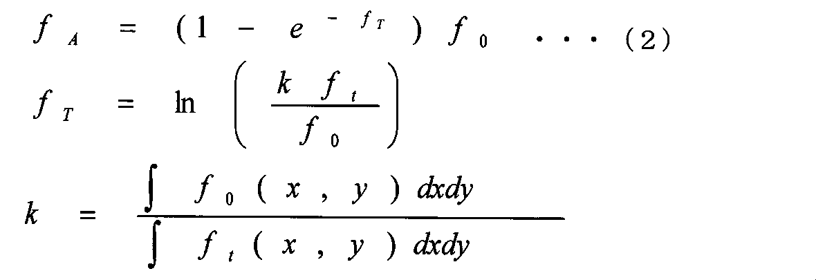
fT:減弱比画像 ft:スキャノグラム画像
k:減弱基準値
次に、画像再構成装置221は、線量当量換算係数を用いて、吸収線量画像16を被曝線量画像に変換する(S306)。線量当量換算係数は、例えば、ICRPの勧告に示される値を用いることができる。
以上、第2の実施の形態によれば、スキャノグラム撮影(静止撮影)において、撮影条件に応じた被検者3の被曝線量、および被検者3に対する照射線量を迅速に評価できる。
さらに、被検者3の被曝線量、および被検者3に対する照射線量を高精度かつ詳細に評価、管理することができる。なお、、スキャノグラム撮影も静止撮影であるから、スキャノグラム撮影と同様に、被曝線量と照射線量を評価、管理することができる。
第1の実施の形態と第2の実施の形態では、断層撮影(回転撮影)とスキャノグラム撮影(静止撮影)について、それぞれの被曝線量の評価について説明したが、第3の実施の形態では、断層撮影(回転撮影)とスキャノグラム撮影(静止撮影)の両方を実施し、撮影範囲に重複する領域がある場合について説明する。
X線CT装置は、設定された撮影条件に基づいて、スキャノグラム撮影と断層撮影を行い、スキャノグラム画像と断層画像を取得する。
そして、X線CT装置の表示装置211は、第1の実施の形態と同様、線量情報を表示する。
第3の実施の形態では、スキャノグラム撮影における3次元照射強度分布データと断層撮影における3次元照射強度分布データを合成し、累積被曝線量と累積照射線量を評価することについて説明したが、第4の実施の形態では、被曝線量画像を合成することについて説明する。
X線CT装置は、設定された撮影条件に基づいて、スキャノグラム撮影と断層撮影を行い、スキャノグラム画像と断層画像を取得する。
そして、X線CT装置の表示装置211は、第1の実施の形態と同様、線量情報を表示する。
さらに、被検者3の累積被曝線量を高精度かつ詳細に評価、管理することができる。
52 減弱補正照射線量投影データ、100 ガントリ、101 寝台装置、102 X線発生装置、103 X線検出装置、104 コリメータ装置、105 高電圧発生装置、106 データ収集装置、107 駆動装置、200 中央制御装置、201 入出力装置、202 演算装置、211 表示装置、212 入力装置、213 記憶装置、221 画像再構成装置、222 画像処理装置、700 線量管理画面、701 照射範囲画像、702 撮影別被曝線量、703 被曝線量指標変換ボタン、704 撮影別照射線量、705 部位別被曝線量、705a 腹部の被曝線量、706 臓器別被曝線量、706a 肺の被曝線量、706b 肝臓の被曝線量、706c 脊椎の被曝線量、707a ROI、707b ROI内被曝線量、707 ROI内被曝線量、708 画像上表示のON/OFFボタン、1000、1100 3次元照射強度分布データの集合、1001、1101 3次元照射強度分布データ、1200 合成された3次元照射強度分布データの集合、2000 3次元照射線量データ、2100 2次元照射線量データ、2200 累積3次元照射線量データ
Claims (15)
- 被検者の周囲からX線を照射するX線照射部と、
被検者を透過するX線をX線情報として検出するX線検出部と、
前記X線情報から被検者の再構成画像を作成するものであって、撮影条件に応じて、前記X線照射部によって被検者に照射されるX線の照射強度の分布である照射X線画像を取得し、前記照射X線画像と前記再構成画像を投影変換し、投影変換された前記再構成画像と前記再構成画像の作成領域に対応する照射X線画像を用いて当該被検者の被曝線量の分布を示す画像である被曝線量画像を作成すると共に、前記被曝線量を算出する画像再構成部と、
前記被曝線量画像及び前記被曝線量を表示する表示部と、を備えたことを特徴とするX線CT装置。 - 前記画像再構成部は、複数の前記被曝線量画像に基づいて、当該被検者の累積被曝線量の分布を示す画像である累積被曝線量画像を作成する請求項1に記載のX線CT装置。
- 前記画像再構成部は、前記照射X線画像を照射開始から照射終了までの所定の時間間隔ごとの各時刻において、時間積分することで照射線量データを生成し、前記照射線量データに基づいて当該被検者に照射されるX線の線量の分布を示す画像として作成する請求項1に記載のX線CT装置。
- 前記画像再構成部は、CT値と減弱係数の関係を示す情報に基づいて前記再構成画像のCT値を減弱係数に変換することで、減弱係数の分布を示す画像である減弱係数画像を作成する請求項3に記載のX線CT装置。
- 前記画像再構成部は、
前記減弱係数画像と前記照射線量画像とに対してそれぞれ直交座標系から投影座標系への変換である順投影処理を行うことで前記減弱係数画像の投影データと前記照射線量画像の投影データを生成し、前記減弱係数画像の投影データを用いて前記照射線量画像の投影データに対して逆減弱補正処理を行い、逆減弱補正処理後の前記照射線量画像の投影データに対して投影座標系から直交座標系への変換である逆投影処理を行うことで、前記照射線量画像に対する減弱量の削減結果を示す画像である照射線量補正画像を作成する請求項4に記載のX線CT装置。 - 前記表示部は、被検者の仮想画像と重畳して前記照射線量画像を表示する請求項4に記載のX線CT装置。
- 前記画像再構成部は、前記照射X線情報の回転撮影および静止撮影に関する前記照射X線情報を時間積分することで、回転撮影および静止撮影において当該被検者に照射されるX線の線量の分布を示す画像である累積照射線量画像を作成する請求項4に記載のX線CT装置。
- 前記画像再構成部は、前記照射X線情報の静止撮影に関する前記照射X線情報を時間積分することで、静止撮影において当該被検者に照射されるX線の線量の分布を示す画像である静止撮影照射線量画像を作成する請求項4に記載のX線CT装置。
- 前記画像再構成部は、減弱基準値を示す情報に基づいて静止撮影画像から減弱比の分布を示す画像である減弱比画像を作成し、
前記減弱比画像を用いて前記静止撮影照射線量画像に対して逆減弱補正処理を行い、静止撮影において当該被検者に吸収されるX線の線量の分布を示す画像である静止撮影吸収線量画像を作成する請求項8に記載のX線CT装置。 - 前記画像再構成部は、前記照射線量画像に基づいて、CTDI、DLP、CTDIw、またはCTDIvolのいずれかを算出する請求項4に記載のX線CT装置。
- 前記画像再構成部は、前記照射線量画像と前記照射線量補正画像との差分に基づき前記被検者に吸収されるX線の線量の分布を示す画像である吸収線量画像を作成する請求項10に記載のX線CT装置。
- 前記画像再構成部は、前記再構成画像における臓器および/または組織の領域を同定し、前記吸収線量画像と照合することで、臓器および/または組織ごとに被曝線量を算出する請求項11に記載のX線CT装置。
- 前記表示部は、臓器および/または組織ごとの被曝線量を表示する請求項12に記載のX線CT装置。
- 前記画像再構成部は、前記再構成画像における各部位の領域を同定し、前記吸収線量画像と照合することで、部位ごとに被曝線量を算出する請求項11に記載のX線CT装置。
- 前記表示部は、部位ごとの被曝線量を表示する請求項14に記載のX線CT装置。
Priority Applications (3)
| Application Number | Priority Date | Filing Date | Title |
|---|---|---|---|
| US13/819,196 US9119560B2 (en) | 2010-09-07 | 2011-09-01 | X-ray CT apparatus |
| JP2012532949A JP5898081B2 (ja) | 2010-09-07 | 2011-09-01 | X線ct装置 |
| CN201180043051.7A CN103096802B (zh) | 2010-09-07 | 2011-09-01 | X射线ct装置 |
Applications Claiming Priority (2)
| Application Number | Priority Date | Filing Date | Title |
|---|---|---|---|
| JP2010-199516 | 2010-09-07 | ||
| JP2010199516 | 2010-09-07 |
Publications (1)
| Publication Number | Publication Date |
|---|---|
| WO2012033002A1 true WO2012033002A1 (ja) | 2012-03-15 |
Family
ID=45810604
Family Applications (1)
| Application Number | Title | Priority Date | Filing Date |
|---|---|---|---|
| PCT/JP2011/069910 WO2012033002A1 (ja) | 2010-09-07 | 2011-09-01 | X線ct装置 |
Country Status (4)
| Country | Link |
|---|---|
| US (1) | US9119560B2 (ja) |
| JP (1) | JP5898081B2 (ja) |
| CN (1) | CN103096802B (ja) |
| WO (1) | WO2012033002A1 (ja) |
Cited By (4)
| Publication number | Priority date | Publication date | Assignee | Title |
|---|---|---|---|---|
| CN103110423A (zh) * | 2013-01-31 | 2013-05-22 | 深圳先进技术研究院 | 一种估计成像剂量的方法和系统 |
| JP2015019856A (ja) * | 2013-07-19 | 2015-02-02 | 株式会社東芝 | 医用画像処理装置及び医用画像処理システム |
| CN104755029A (zh) * | 2012-10-19 | 2015-07-01 | 皇家飞利浦有限公司 | 确定身体中的剂量的分布的方法 |
| JP2021029639A (ja) * | 2019-08-26 | 2021-03-01 | コニカミノルタ株式会社 | 医用画像処理装置及びプログラム |
Families Citing this family (11)
| Publication number | Priority date | Publication date | Assignee | Title |
|---|---|---|---|---|
| US20140253544A1 (en) * | 2012-01-27 | 2014-09-11 | Kabushiki Kaisha Toshiba | Medical image processing apparatus |
| US9125572B2 (en) | 2012-06-22 | 2015-09-08 | University Of Utah Research Foundation | Grated collimation system for computed tomography |
| US9125286B2 (en) * | 2012-12-28 | 2015-09-01 | General Electric Company | X-ray dose estimation technique |
| CN104510486B (zh) * | 2013-09-30 | 2021-04-20 | Ge医疗系统环球技术有限公司 | 计算机化断层扫描设备及其机架旋转控制装置和方法 |
| US10219763B2 (en) * | 2014-09-11 | 2019-03-05 | Hitachi, Ltd. | Photon counting CT device and estimated exposure level computation method |
| CN104287768A (zh) * | 2014-09-30 | 2015-01-21 | 沈阳东软医疗系统有限公司 | 一种ct扫描剂量控制方法及系统 |
| KR101725099B1 (ko) * | 2014-12-05 | 2017-04-26 | 삼성전자주식회사 | 컴퓨터 단층 촬영장치 및 그 제어방법 |
| WO2017103238A1 (en) | 2015-12-17 | 2017-06-22 | Koninklijke Philips N.V. | Method for estimating the radiation dose received by an organ during a computed tomography scan |
| US10888296B2 (en) | 2018-06-29 | 2021-01-12 | Shanghai United Imaging Healthcare Co., Ltd. | Methods and systems for modulating radiation dose |
| CN109124666A (zh) * | 2018-06-29 | 2019-01-04 | 上海联影医疗科技有限公司 | 一种确定辐射剂量调制线的方法、系统和装置 |
| JP7304765B2 (ja) * | 2019-08-06 | 2023-07-07 | キヤノンメディカルシステムズ株式会社 | X線診断装置および医用画像処理装置 |
Citations (2)
| Publication number | Priority date | Publication date | Assignee | Title |
|---|---|---|---|---|
| JP2005143759A (ja) * | 2003-11-13 | 2005-06-09 | Hitachi Medical Corp | X線ct装置 |
| JP2010269048A (ja) * | 2009-05-25 | 2010-12-02 | Ge Medical Systems Global Technology Co Llc | X線ct装置 |
Family Cites Families (11)
| Publication number | Priority date | Publication date | Assignee | Title |
|---|---|---|---|---|
| US4670892A (en) * | 1977-11-15 | 1987-06-02 | Philips Medical Systems, Inc. | Method and apparatus for computed tomography of portions of a body plane |
| JP2004173924A (ja) * | 2002-11-27 | 2004-06-24 | Ge Medical Systems Global Technology Co Llc | X線制御方法およびx線画像撮影装置 |
| JP4393105B2 (ja) * | 2003-05-14 | 2010-01-06 | キヤノン株式会社 | 放射線撮像装置及びその作動方法 |
| WO2005070296A1 (ja) * | 2004-01-22 | 2005-08-04 | Canon Kabushiki Kaisha | X線撮影装置及びx線撮影方法 |
| JP2009514559A (ja) * | 2005-07-22 | 2009-04-09 | トモセラピー・インコーポレーテッド | 線量体積ヒストグラムを用いて輪郭構造を生成するシステムおよび方法 |
| JP5192372B2 (ja) * | 2006-05-25 | 2013-05-08 | 株式会社日立メディコ | X線ct装置 |
| CA2905989C (en) * | 2006-05-25 | 2017-01-24 | Di Yan | Real-time, on-line and offline treatment dose tracking and feedback process for volumetric image guided adaptive radiotherapy |
| CN101299068A (zh) * | 2007-04-30 | 2008-11-05 | 上海西门子医疗器械有限公司 | 计算机断层扫描辐射剂量显示系统和方法 |
| JP5105589B2 (ja) * | 2007-07-11 | 2012-12-26 | 株式会社日立メディコ | X線ct装置 |
| US8107589B2 (en) * | 2007-12-21 | 2012-01-31 | Kabushiki Kaisha Toshiba | Radiotherapeutic system and radiotherapeutic dose distribution measuring method |
| JP5238242B2 (ja) * | 2007-12-21 | 2013-07-17 | 株式会社東芝 | 放射線治療用線量分布測定装置及び放射線治療用線量分布測定プログラム |
-
2011
- 2011-09-01 WO PCT/JP2011/069910 patent/WO2012033002A1/ja active Application Filing
- 2011-09-01 CN CN201180043051.7A patent/CN103096802B/zh not_active Expired - Fee Related
- 2011-09-01 JP JP2012532949A patent/JP5898081B2/ja active Active
- 2011-09-01 US US13/819,196 patent/US9119560B2/en not_active Expired - Fee Related
Patent Citations (2)
| Publication number | Priority date | Publication date | Assignee | Title |
|---|---|---|---|---|
| JP2005143759A (ja) * | 2003-11-13 | 2005-06-09 | Hitachi Medical Corp | X線ct装置 |
| JP2010269048A (ja) * | 2009-05-25 | 2010-12-02 | Ge Medical Systems Global Technology Co Llc | X線ct装置 |
Cited By (4)
| Publication number | Priority date | Publication date | Assignee | Title |
|---|---|---|---|---|
| CN104755029A (zh) * | 2012-10-19 | 2015-07-01 | 皇家飞利浦有限公司 | 确定身体中的剂量的分布的方法 |
| CN103110423A (zh) * | 2013-01-31 | 2013-05-22 | 深圳先进技术研究院 | 一种估计成像剂量的方法和系统 |
| JP2015019856A (ja) * | 2013-07-19 | 2015-02-02 | 株式会社東芝 | 医用画像処理装置及び医用画像処理システム |
| JP2021029639A (ja) * | 2019-08-26 | 2021-03-01 | コニカミノルタ株式会社 | 医用画像処理装置及びプログラム |
Also Published As
| Publication number | Publication date |
|---|---|
| CN103096802B (zh) | 2015-12-16 |
| US9119560B2 (en) | 2015-09-01 |
| US20130156149A1 (en) | 2013-06-20 |
| JPWO2012033002A1 (ja) | 2014-01-20 |
| CN103096802A (zh) | 2013-05-08 |
| JP5898081B2 (ja) | 2016-04-06 |
Similar Documents
| Publication | Publication Date | Title |
|---|---|---|
| JP5898081B2 (ja) | X線ct装置 | |
| US7269241B2 (en) | Method and arrangement for medical X-ray imaging and reconstruction from sparse data | |
| KR101728046B1 (ko) | 단층 영상 복원 장치 및 그에 따른 단층 영상 복원 방법 | |
| JP5696305B2 (ja) | 放射線撮像装置及び放射線による撮像方法 | |
| US20100119033A1 (en) | Intensity-modulated, cone-beam computed tomographic imaging system, methods, and apparatus | |
| US20130202079A1 (en) | System and Method for Controlling Radiation Dose for Radiological Applications | |
| JP6066596B2 (ja) | X線撮像における散乱補正の方法及びシステム | |
| WO2007074772A1 (ja) | X線ct装置 | |
| JP2005312970A (ja) | コンピュータ断層撮影における線量低減された部分的スパイラル走査時の投影データセットの再構成方法 | |
| US7787669B2 (en) | Reconstruction of local patient doses in computed tomography | |
| CN105962959A (zh) | 对于虚拟x射线量子能量分布产生图像的方法和拍摄装置 | |
| KR20200095740A (ko) | 의료 영상 장치 및 그 제어방법 | |
| Fahrig et al. | Flat-panel conebeam CT in the clinic: history and current state | |
| CN108601571B (zh) | Ct成像系统和用于ct成像系统的方法 | |
| CN113613561A (zh) | 处理x射线检测数据的数据处理装置及数据处理方法及搭载有该装置或方法的x射线检查装置 | |
| JP2007125174A (ja) | 放射線画像撮影方法及び装置 | |
| US9848846B2 (en) | Systems and methods for determining radiation dose in computed tomography scans | |
| JP5027909B2 (ja) | X線ct装置 | |
| US7068752B2 (en) | Method and arrangement for medical X-ray imaging | |
| JP4644292B2 (ja) | X線ct装置とその画像表示方法 | |
| Swennen et al. | From 3-D volumetric computer tomography to 3-D cephalometry | |
| JP5384293B2 (ja) | X線ct装置 | |
| Svalkvist et al. | Investigation of the dosimetry of chest tomosynthesis | |
| Kara et al. | Estımatıon of Absorbed Dose Dıstrıbutıon in Dıfferent Organs durıng the CT Scan: Monte Carlo Study | |
| Lintz | Computed Tomography |
Legal Events
| Date | Code | Title | Description |
|---|---|---|---|
| WWE | Wipo information: entry into national phase |
Ref document number: 201180043051.7 Country of ref document: CN |
|
| 121 | Ep: the epo has been informed by wipo that ep was designated in this application |
Ref document number: 11823478 Country of ref document: EP Kind code of ref document: A1 |
|
| WWE | Wipo information: entry into national phase |
Ref document number: 2012532949 Country of ref document: JP |
|
| WWE | Wipo information: entry into national phase |
Ref document number: 13819196 Country of ref document: US |
|
| NENP | Non-entry into the national phase |
Ref country code: DE |
|
| 122 | Ep: pct application non-entry in european phase |
Ref document number: 11823478 Country of ref document: EP Kind code of ref document: A1 |