WO2011125648A1 - 歯科画像処理装置、歯科用測色装置、歯科画像処理方法および歯科画像処理プログラム - Google Patents
歯科画像処理装置、歯科用測色装置、歯科画像処理方法および歯科画像処理プログラム Download PDFInfo
- Publication number
- WO2011125648A1 WO2011125648A1 PCT/JP2011/057802 JP2011057802W WO2011125648A1 WO 2011125648 A1 WO2011125648 A1 WO 2011125648A1 JP 2011057802 W JP2011057802 W JP 2011057802W WO 2011125648 A1 WO2011125648 A1 WO 2011125648A1
- Authority
- WO
- WIPO (PCT)
- Prior art keywords
- color information
- tooth
- image
- information
- dental
- Prior art date
- Legal status (The legal status is an assumption and is not a legal conclusion. Google has not performed a legal analysis and makes no representation as to the accuracy of the status listed.)
- Ceased
Links
Images


Classifications
-
- A—HUMAN NECESSITIES
- A61—MEDICAL OR VETERINARY SCIENCE; HYGIENE
- A61C—DENTISTRY; APPARATUS OR METHODS FOR ORAL OR DENTAL HYGIENE
- A61C19/00—Dental auxiliary appliances
- A61C19/04—Measuring instruments specially adapted for dentistry
-
- A—HUMAN NECESSITIES
- A61—MEDICAL OR VETERINARY SCIENCE; HYGIENE
- A61C—DENTISTRY; APPARATUS OR METHODS FOR ORAL OR DENTAL HYGIENE
- A61C13/00—Dental prostheses; Making same
- A61C13/08—Artificial teeth; Making same
- A61C13/082—Cosmetic aspects, e.g. inlays; Determination of the colour
-
- G—PHYSICS
- G01—MEASURING; TESTING
- G01J—MEASUREMENT OF INTENSITY, VELOCITY, SPECTRAL CONTENT, POLARISATION, PHASE OR PULSE CHARACTERISTICS OF INFRARED, VISIBLE OR ULTRAVIOLET LIGHT; COLORIMETRY; RADIATION PYROMETRY
- G01J3/00—Spectrometry; Spectrophotometry; Monochromators; Measuring colours
- G01J3/46—Measurement of colour; Colour measuring devices, e.g. colorimeters
- G01J3/463—Colour matching
-
- G—PHYSICS
- G01—MEASURING; TESTING
- G01J—MEASUREMENT OF INTENSITY, VELOCITY, SPECTRAL CONTENT, POLARISATION, PHASE OR PULSE CHARACTERISTICS OF INFRARED, VISIBLE OR ULTRAVIOLET LIGHT; COLORIMETRY; RADIATION PYROMETRY
- G01J3/00—Spectrometry; Spectrophotometry; Monochromators; Measuring colours
- G01J3/46—Measurement of colour; Colour measuring devices, e.g. colorimeters
- G01J3/50—Measurement of colour; Colour measuring devices, e.g. colorimeters using electric radiation detectors
- G01J3/508—Measurement of colour; Colour measuring devices, e.g. colorimeters using electric radiation detectors measuring the colour of teeth
Definitions
- the present invention relates to a dental image processing device, a dental colorimetric device, a dental image processing method, and a dental image processing program.
- a dental colorimetric device that acquires accurate color information of a tooth from an image acquired by a digital camera is known (see, for example, Patent Document 1).
- This dental colorimetric device acquires color information at a predetermined position of a tooth, and corrects color information in a color image of a tooth corresponding to the position where the color information is acquired by using the acquired color information. By doing so, a color image subjected to highly accurate color correction can be acquired.
- the dental colorimetric device disclosed in Patent Document 1 can perform color correction with high accuracy because the position where the color information is acquired matches the corresponding position in the color image. Even if color information and a color image are acquired for the same tooth, if color information and a color image are acquired on different sides such as the cheek side and the occlusal side, there is no corresponding position. There is an inconvenience that high-precision color correction cannot be performed.
- the present invention has been made in view of the above-described circumstances, and is a dental image capable of performing color correction with high accuracy so that color information matches even on different side surfaces of the same tooth such as the cheek side surface and the occlusal surface. It is an object of the present invention to provide a processing device, a dental colorimetric device, a dental image processing method, and a dental image processing program.
- a first aspect of the present invention is a reference region on an original image that is a correction target acquired for either the occlusal surface or the cheek side of a tooth, and is either the occlusal surface or the cheek side
- a storage unit for storing position information of a reference area having color correlation with reference color information that is color information acquired in a reference area that is one area, and a reference color that is color information in the reference area in the original image
- a reference color information extraction unit for extracting information, a correction coefficient calculation unit for calculating a correction coefficient for matching the reference color information extracted by the reference color information extraction unit with the reference color information, and the correction coefficient calculation unit
- the dental image processing apparatus includes a color information correction unit that corrects color information of the original image using the correction coefficient calculated by the above.
- the reference color Based on the position information of the reference region stored in the storage unit as a region having color correlation with the information, the reference color information in the reference region on the original image is extracted by the reference color information extraction unit.
- a correction coefficient is calculated by the correction coefficient calculation unit using the extracted reference color information and standard color information, and the color information of the entire original image is corrected by the color information correction unit using the calculated correction coefficient.
- the reference color information of the original image acquired for either the occlusal surface or the cheek side surface is changed to either the occlusal surface or the cheek side surface.
- the color information of the original image can be corrected so as to match the reference color information acquired for the other with high accuracy.
- high-accuracy color information can be acquired only for either the cheek side or the occlusal surface for the same tooth, it is not necessary to acquire an image having high-accuracy color information for the other tooth.
- the color information can be corrected with high accuracy.
- an input unit for inputting identification information of a tooth as a reference is provided, and the storage unit associates the identification information of the tooth with the positional information of the reference area for each tooth.
- the reference color information extraction unit extracts the reference image from the original image based on the positional information of the reference area stored in the storage unit corresponding to the identification information of the tooth input by the input unit. Reference color information may be extracted.
- the position information of the reference region in the storage unit stored in association with the identification information is referred to, and the position of the corresponding reference region from the original image
- the reference color information is extracted by the reference color information extraction unit.
- the positional relationship between the area for acquiring the reference color information and the reference area for the image information having color correlation with the area differs depending on the type of tooth such as anterior teeth, premolars, molars, etc. Only by inputting, reference color information in a reference area appropriate for each tooth can be extracted. Accordingly, it is possible to calculate an appropriate correction coefficient based on the reference color information and the standard color information having color correlation with each other regardless of the type of tooth, and obtain an image in which the color information is corrected with high accuracy.
- the storage unit stores position information of a reference region as relative position information with respect to a tooth contour shape
- the reference color information extraction unit extracts the tooth contour shape. Then, the color information of the reference area may be extracted based on the extracted contour shape and the position information of the reference area stored in the storage unit. In this way, even if the position accuracy of the acquisition of the tooth image is low, the position of the reference region can be accurately determined based on the contour shape of the tooth to be extracted, and the color information of the image is corrected.
- the correction coefficient can be accurately calculated.
- the color information extracting unit includes a storage unit that stores position information of a reference area on the original image as relative position information with respect to the contour shape of the tooth.
- the tooth outline shape may be extracted, the tooth type may be specified based on the extracted outline shape, and the reference color information may be extracted using position information of a reference region according to the specified type. In this way, it is possible to easily extract appropriate reference color information corresponding to the type of tooth, and to obtain an image in which the color information is corrected with high accuracy.
- a reference color information extracting unit that extracts the reference color information that is color information in the reference region in a reference image acquired for either the occlusal surface or the cheek side is provided.
- the storage unit may further store the identification information of the tooth and the position information of the reference area in association with each tooth.
- the reference area and reference area position information in the storage unit stored in association with the identification information is referenced, and the reference color information is the reference color.
- the reference color information is extracted by the information extraction unit, and the reference color information at the position of the corresponding reference region is extracted from the original image by the reference color information extraction unit.
- the color information in the reference region and the standard region appropriate for each tooth can be extracted by simply inputting the identification information from the input unit. Accordingly, it is possible to calculate an appropriate correction coefficient based on the reference color information and the standard color information having color correlation with each other regardless of the type of tooth, and obtain an image in which the color information is corrected with high accuracy.
- the second aspect of the present invention is an image acquisition unit that acquires the original image of either the occlusal surface or the cheek side of a tooth, and color information in one region of the other of the occlusal surface or the cheek side.
- a dental colorimetric device including a reference color information acquisition unit that acquires certain reference color information and any one of the dental image processing devices described above.
- the reference color information is stored in the dental image processing apparatus. Extraction is performed, a correction coefficient is calculated, and color information of the original image is corrected.
- the reference color information acquisition unit may acquire a colorimetric image of either the occlusal surface or the cheek side surface of the tooth. In this way, accurate reference color information can be acquired based on the colorimetric information obtained from the colorimetric image, so that the color information of the image to be corrected can be accurately corrected.
- the image acquisition unit may acquire a colorimetric image of either the occlusal surface or the cheek side surface of the tooth. Even if the image to be corrected is a colorimetric image, accurate color information may not be obtained for reasons such as the incidence of ambient light. Even in such a case, the color information is accurately obtained by correction. It can be corrected.
- an original image to be corrected acquired for either the occlusal surface or the cheek side surface of a tooth and reference color information that is color information in one of the other regions are input.
- the reference color information which is color information in the reference area, is obtained based on the position information of the reference area in the original image stored as an area having a color correlation with the reference color information.
- the calculation step for calculating the correction coefficient for matching the reference color information extracted in the extraction step with the reference color information, and the correction coefficient calculated in the calculation step And a correction step of correcting color information of the original image.
- an original image to be corrected acquired for either the occlusal surface or the cheek side surface of the tooth and reference color information that is color information in one of the other regions are input.
- the reference color information which is color information in the reference area, is obtained based on the position information of the reference area in the original image stored as an area having a color correlation with the reference color information.
- the calculation step for calculating the correction coefficient for matching the reference color information extracted in the extraction step with the reference color information, and the correction coefficient calculated in the calculation step A dental image processing program for causing a computer to execute a correction step for correcting color information of an original image.
- FIG. 1 It is a block diagram which shows the dental colorimetric apparatus which concerns on one Embodiment of this invention. It is a perspective view of the incisor explaining the acquisition direction of the image by the dental colorimetric device of FIG. It is a figure which shows an example of the image of the cheek side surface of the incisor acquired by the dental colorimetry apparatus of FIG. It is a figure which shows an example of the image of the occlusal surface of the incisor acquired by the dental colorimetry apparatus of FIG. It is a perspective view of the molar which explains the acquisition direction of the image by the dental colorimetric device of FIG. It is a figure which shows an example of the image of the buccal side surface of the molar acquired by the dental colorimetry apparatus of FIG.
- the dental colorimetric apparatus 1 is a first image acquisition unit (image acquisition unit) that acquires an image (original image) G 1 of an occlusal surface A 1 of a tooth A.
- a second image acquisition unit (reference color information acquisition unit) 3 that acquires an image (reference image) G 2 of the buccal side surface A 2 of the tooth A, and the image G 1 ,
- the input unit 4 for inputting the tooth number (identification information) N of the tooth A from which G 2 has been acquired, and the images G 1 and G 2 acquired by the two image acquisition units 2 and 3
- the dental image processing apparatus 5 is provided.
- the image G 1 of the occlusal surface A 1 of the tooth A is an image G 1 as shown in FIG. 3B and FIG. 5B obtained by photographing the tooth A from the direction indicated by the arrow P in FIG. 2 and
- FIG. image G 2 in buccal surface a 2 of the tooth a is an image G 2 as shown in FIGS. 3A and 5A were taken tooth a from the direction indicated by arrow Q in FIGS.
- the dental image processing apparatus 5 includes an image G 1 of the occlusal surface A 1 of the tooth A acquired by the first image acquisition unit 2 and a dental formula number N of the tooth A input from the input unit 4. And the image G 2 of the buccal side surface A 2 of the tooth A acquired by the second image acquisition unit 3 and the tooth A input from the input unit 4. And a second image storage unit 7 which stores the tooth number N in association with each other.
- the dental image processing apparatus 5 associates the tooth number N of each tooth A with the positional information of the regions R 1 and R 2 having a color correlation between the occlusal surface A 1 and the cheek side surface A 2 of the tooth A.
- An area information storage unit (storage unit) 8 for storing the information is also provided.
- the dental image processing apparatus 5 has the occlusal surface A 1 of the tooth A having the same tooth number N stored in the first image storage unit 6 and the second image storage unit 7.
- a color information extracting unit (reference color information extracting unit, standard color information extracting unit) 9 for extracting color information in the regions R 1 and R 2 searched in the images G 1 and G 2 and the color A correction coefficient calculation unit 10 that calculates a correction coefficient using the color information of the two regions R 1 and R 2 extracted by the information extraction unit 9, and an image using the correction coefficient calculated by the correction coefficient calculation unit 10 and an image correcting unit 11 for correcting the G 1.
- the first image acquisition unit 2 is, for example, a digital camera that acquires RGB images.
- the second image acquisition unit 3 is, for example, an imaging device that acquires a multiband image as a colorimetric image that can acquire color information with higher color reproducibility than an RGB image.
- the second image acquisition unit 3 sequentially emits a plurality of light sources that emit light in different wavelength bands, for example, as disclosed in Japanese Patent Application Laid-Open No. 2007-190371. A subject is sequentially irradiated with light of different wavelength bands, and a plurality of RGB images are acquired.
- the second image acquisition unit 3 selects the B image from the RGB image acquired when the light source emitting light belonging to the blue wavelength band of the center wavelength is emitted, and sets the center wavelength to the green wavelength band.
- the G image is selected from the RGB image acquired when the light source that emits the light belonging to is emitted, and the RGB image acquired when the light source that emits the light belonging to the wavelength band whose center wavelength is red is emitted. Select an R image.
- the second image acquisition unit 3 generates a multiband image by synthesizing the selected R image, G image, and B image.
- the number of wavelength bands of light emitted from a plurality of light sources is preferably 4 or more.
- the second image acquisition unit 3 is not limited to the above-described configuration, and sequentially irradiates the subject with light having different wavelength bands, and acquires a monochrome image of the subject for each wavelength band. May be.
- the second image acquisition unit 3 includes four or more types of filters having different transmission wavelength bands, irradiates the subject with white light, and acquires a plurality of subject images transmitted through the filters simultaneously or sequentially. It may be. In other words, the second image acquisition unit 3 accurately measures the color of the patient's tooth A, that is, performs tooth colorimetry.
- the input unit 4 includes an arbitrary input device such as a mouse or a keyboard.
- the area information storage unit 8 stores the position information of one area (reference area) R 1 in the image G 1 of the occlusal surface A 1 for each tooth number N of the tooth A, and the cheek and positional information of the side surface a region of the image G 2 of a 2 (reference region) R 2 is stored.
- the reference region R 1 and the reference region R 2 are selected as regions R 1 and R 2 having a color correlation that will have colors close to each other if measured accurately.
- the reference region R 1 and the reference region R 2 having color correlation with each other are set for each tooth A or for each type of tooth A. Yes.
- the reference region R 1 is defined in the image G 1 of the occlusal surface A 1
- the reference region R 2 is defined in the image G 2 of the cheek side surface A 2 .
- the reference positions and dimensions of the reference area R 1 and the reference area R 2 are set based on the contour shape of the tooth A in each of the images G 1 and G 2 .
- the reference region R 1 includes a straight line L 1 connecting the centers of gravity of the two regular reflection regions Ra generated in the contour of the tooth A in the image G 1 of the occlusal surface A 1 , and has a center of gravity at the intersection of the center line L 2 in the width direction is defined as a rectangular area having the same dimensional ratios as the reference region R 2 with respect to the entire width dimension.
- the teeth of the tooth type number # 6 is a molar
- the reference region R 2 is the image G 2 in buccal surface A 2, through the center of gravity of the contour of the tooth A divided by a straight line L 3, include the center of gravity of the region of the mesial (or any distal sides), 10% of the width W6 of the width of the mesial region, therewith the height of the same size W6 Is defined as a rectangular region having
- the reference region R 1 is the buccal mesial side (or either the buccal side distal side) divided by vertical and horizontal straight lines L 4 and L 5 passing through the center of gravity of the tooth A in the image G 1 of the occlusal plane A 1.
- the center of gravity of the area is defined as a rectangular area having the same dimensional ratios as the reference region R 2 with respect to the total width dimension of the tooth a.
- These regions R 1 and R 2 have a color difference ⁇ E of 3 or less and have a color correlation with each other.
- Color information extraction unit 9 the first image from the storage unit 6 and the second image storage unit 7, the same teeth type number N of the image G 1 and the buccal surface A 2 of the occlusal surface A 1 which is associated image
- the region information storage unit 8 is searched using the tooth number N as a key, and the position information of the reference region R 1 and the reference region R 2 stored in association with the tooth number N is obtained. It is designed to read.
- the color information extraction unit 9, based on the position information of the reference area R 1 read the color of the reference area R 1 in the image G 1 of the occlusal surface A 1 received from the first image storage section 6 Information is extracted.
- the color information extraction unit 9 determines the color in the reference region R 2 in the image G 2 of the cheek side surface A 2 received from the second image storage unit 7 based on the read position information of the reference region R 2 . Information is extracted.
- Correction coefficient calculating unit 10 by dividing by the average value of the color information of the color information in the extracting unit 9 reference region R 2, which are extracted in, likewise extracted average value of the color information in the reference area R 1, A correction coefficient for multiplying the color information in the reference region R 1 to match the color information in the reference region R 2 is calculated.
- Image correcting unit 11 to correct the image G 1 by multiplying the image G 1 of the occlusal surface A 1 which is stored the correction coefficient calculated by the correction coefficient calculation unit 10 in the first image storage section 6 It has become.
- the first image acquisition unit 2 uses the occlusal surface of the tooth A.
- An image G 1 of A 1 is acquired, and an image G 2 of the cheek side surface A 2 of the tooth A is acquired by the second image acquisition unit 3.
- the input unit 4 is operated to input the tooth number N, and together with the input tooth number N, the image G 1 of the occlusal surface A 1 of the tooth A acquired by the first image acquiring unit 2 and The image G 2 of the cheek side surface A 2 acquired by the second image acquisition unit 3 is input to the dental image processing device 5.
- the tooth number N input to the dental image processing apparatus 5 and the image G 1 of the occlusal surface A 1 are stored in the first image storage unit 6 in association with each other.
- the second image G 2 is stored in the second image storage unit 7 in association with each other.
- occlusal surface stored in the second image storage unit 6, 7 A 1 and the image G 1 of buccal surface A 2, G 2 is, color information together with the teeth expression number N
- the color information extraction unit 9 reads the position information of the reference region R 1 and the reference region R 2 stored in the region information storage unit 8 using the tooth number N as a key. Then, the color information extraction unit 9, according to the position information read, the color information of the reference area R 2 on the image G 2 of the reference region on the image G 1 of the occlusal surface A 1 R 1 and buccal surfaces A 2 Extraction (extraction step S1).
- the extracted color information of the reference region R 1 and the color information of the reference region R 2 are input to the correction coefficient calculation unit 10, and the average value of the color information in each region R 1 , R 2 is calculated.
- the average value of the reference color information is color information of the reference area R 2 is divided by the average value of the reference color information is color information of the reference area R 1, correction coefficient is calculated (calculating step S2) .
- a correction coefficient calculated in the calculation step S2 the image G 1 of the occlusal surface A 1 stored in the first image storage unit 6 is inputted to the image correcting unit 11, the occlusal surface A 1 by multiplying the correction coefficient to the image G 1, the entire image G 1 of the occlusal surface a 1 is corrected (correction step S3), and the corrected image is output.
- the correction coefficient is the reference color. This is a coefficient for making the information coincide with the reference color information.
- the dental colorimetric apparatus 1 and the dental image processing method according to the present embodiment the images G 1 and G 2 of the occlusal surface A 1 and the buccal side surface A 2 of the same tooth A are obtained.
- the dental image processing apparatus 5 the dental colorimetric apparatus 1 and the dental image processing method according to the present embodiment, the images G 1 and G 2 of the occlusal surface A 1 and the buccal side surface A 2 of the same tooth A are obtained.
- the dental image processing apparatus 5 the dental colorimetric apparatus 1 and the dental image processing method according to the present embodiment, the images G 1 and G 2 of the occlusal surface A 1 and the buccal side surface A 2 of the same tooth A are obtained.
- the buccal surface A 2 can be easily obtained a multi-band image of high accuracy, may be difficult to get the multi-band image for occlusal surface A 1, in such a case, the occlusal surface A 1 Has an advantage that even if an image G 1 that is simpler and less accurate like an RGB image is acquired, a corrected image having color information with high accuracy can be generated based on the image G 2 of the cheek side surface A 2. is there.
- the accuracy of the color information may be reduced.
- the color information of the reference region R 2 on the image G 2 (or G 1 ) on the higher accuracy side is used. There is an advantage that the color information of the low-accuracy image G 1 (or G 2 ) can be corrected.
- the multi-band image for buccal surface A 2 has been described as an example the case of acquiring the RGB image for occlusal surface A 1, instead of this, obtains the RGB image for buccal surfaces A 2 and, a multi-band image may be obtained for occlusal surface a 1.
- the reference area R 1 is set in a region of an image G 2 in buccal surface A 2
- the reference region R 2 may be be set in one region of the image G 1 of the occlusal surface A 1.
- color information (L * a * b *, L * u * v) obtained by color conversion from a multiband image is used as a colorimetric image acquired by the second image acquisition unit 3. *, XYZ, RGB, CMYK, spectral reflection spectrum, etc.) may be employed.
- the position information of the reference region R 1 and the position information of the reference region R 2 are associated with each tooth number N and stored in the region information storage unit 8.
- the position information of the reference region R 1 and the reference region R 2 is associated with each type of tooth A, for example, incisor and molar, and stored in the region information storage unit 8. Good.
- the type of the tooth A is determined based on the tooth number, and the reference region R 1 and the reference region R 2 stored in association with the type are determined. The position information is read out.
- an image is acquired and stored for each single tooth A.
- a dentition image including a plurality of adjacent teeth is acquired, and the first image storage unit 6 is used. And / or it may be stored in the second image storage unit 7.
- the color information extraction unit 9 may extract the contour shape of each tooth A from the dentition image, and detect the type of tooth A and the tooth number N based on the contour shape.
- each extracted tooth A may be associated with the dental formula number N input from the input unit 4.
- the color information extraction unit 9 includes the reference region R in the image G 2 of the cheek side surface A 2 received from the second image storage unit 7. There is no need to extract the color information in 2 . Therefore, the color information extraction unit 9 refers to the image G 1 of the occlusal surface A 1 received from the first image storage unit 6 based on the position information of the reference region R 1 read from the region information storage unit 8. it is sufficient to extract the color information in the region R 1. Further, as shown in FIGS.
- the acquired images G 1 and G 2 may include a high luminance region Ra due to regular reflection of illumination at the time of shooting, and such a high luminance region Ra.
- the correction coefficient cannot be calculated with high accuracy.
- FIG. 6 an example in which the positional information of the reference region R 1 and the reference region R 2 is defined is shown, but the present invention is not limited to this, and the color difference ⁇ E ⁇ 3. it may be that stores an association position information of an arbitrary reference region R 1 and the reference area R 2 made. In this case, the position of the reference region R 1 and the reference area R 2, size and shape can be chosen arbitrarily.
- the dental image processing method may be realized not only by hardware, but also by a dental image processing program that can be executed by a computer.
Landscapes
- Health & Medical Sciences (AREA)
- Spectroscopy & Molecular Physics (AREA)
- Physics & Mathematics (AREA)
- Life Sciences & Earth Sciences (AREA)
- Oral & Maxillofacial Surgery (AREA)
- Animal Behavior & Ethology (AREA)
- Veterinary Medicine (AREA)
- Public Health (AREA)
- General Physics & Mathematics (AREA)
- Dentistry (AREA)
- Epidemiology (AREA)
- General Health & Medical Sciences (AREA)
- Engineering & Computer Science (AREA)
- Biophysics (AREA)
- Biomedical Technology (AREA)
- Dental Tools And Instruments Or Auxiliary Dental Instruments (AREA)
- Spectrometry And Color Measurement (AREA)
- Color Television Image Signal Generators (AREA)
- Image Processing (AREA)
Abstract
頬側面と咬合面のように同一歯牙の異なる側面においても色情報が一致するように高精度の色補正を行う。歯牙の咬合面または頬側面のいずれか一方について取得された補正の対象となる画像(G1)上の参照領域であって、咬合面または頬側面のいずれか他方の一領域において取得された色情報である基準色情報と色相関を有する参照領域の位置情報を記憶する記憶部(8)と、画像(G1)において参照領域内の色情報である参照色情報を抽出する参照色情報抽出部(9)と、参照色情報抽出部(9)により抽出された参照色情報を基準色情報に一致させるための補正係数を算出する補正係数算出部(10)と、補正係数算出部(10)により算出された補正係数を用いて、画像(G1)の色情報を補正する色情報補正部(11)とを備える歯科画像処理装置(5)を提供する。
Description
本発明は、歯科画像処理装置、歯科用測色装置、歯科画像処理方法および歯科画像処理プログラムに関する。
従来、デジタルカメラにより取得した画像から、歯牙の正確な色情報を取得する歯科用測色装置が知られている(例えば、特許文献1参照。)。
この歯科用測色装置は、歯牙の所定位置おける色彩情報を取得しておき、該色彩情報を取得した位置に対応する歯牙のカラー画像中の色情報を、取得しておいた色彩情報によって補正することにより、高精度の色補正を行ったカラー画像を取得できる。
この歯科用測色装置は、歯牙の所定位置おける色彩情報を取得しておき、該色彩情報を取得した位置に対応する歯牙のカラー画像中の色情報を、取得しておいた色彩情報によって補正することにより、高精度の色補正を行ったカラー画像を取得できる。
しかしながら、特許文献1に開示されている歯科用測色装置では、色彩情報を取得する位置と、カラー画像中の対応位置とが一致しているために高精度の色補正を行うことができるが、同一歯牙について色彩情報とカラー画像とを取得した場合であっても、頬側面と咬合面のように異なる側面において色彩情報とカラー画像とをそれぞれ取得した場合には、対応位置が存在せず、高精度の色補正を行うことができないという不都合がある。
本発明は上述した事情に鑑みてなされたものであって、頬側面と咬合面のように同一歯牙の異なる側面においても色情報が一致するように高精度の色補正を行うことができる歯科画像処理装置、歯科用測色装置、歯科画像処理方法および歯科画像処理プログラムを提供することを目的としている。
上記目的を達成するために、本発明は以下の手段を提供する。
本発明の第一の態様は、歯牙の咬合面または頬側面のいずれか一方について取得された補正の対象となる原画像上の参照領域であって、前記咬合面または前記頬側面のいずれか他方の一領域である基準領域において取得された色情報である基準色情報と色相関を有する参照領域の位置情報を記憶する記憶部と、前記原画像において前記参照領域内の色情報である参照色情報を抽出する参照色情報抽出部と、該参照色情報抽出部により抽出された参照色情報を前記基準色情報に一致させるための補正係数を算出する補正係数算出部と、該補正係数算出部により算出された補正係数を用いて、前記原画像の色情報を補正する色情報補正部とを備える歯科画像処理装置である。
本発明の第一の態様は、歯牙の咬合面または頬側面のいずれか一方について取得された補正の対象となる原画像上の参照領域であって、前記咬合面または前記頬側面のいずれか他方の一領域である基準領域において取得された色情報である基準色情報と色相関を有する参照領域の位置情報を記憶する記憶部と、前記原画像において前記参照領域内の色情報である参照色情報を抽出する参照色情報抽出部と、該参照色情報抽出部により抽出された参照色情報を前記基準色情報に一致させるための補正係数を算出する補正係数算出部と、該補正係数算出部により算出された補正係数を用いて、前記原画像の色情報を補正する色情報補正部とを備える歯科画像処理装置である。
上記本発明の第一の態様によれば、歯牙の咬合面または頬側面のいずれか一方について取得された原画像と、他方の一領域において取得された基準色情報とが与えられると、基準色情報と色相関を有する領域として記憶部に記憶されている参照領域の位置情報に基づいて、参照色情報抽出部により、原画像上の参照領域内の参照色情報が抽出される。抽出された参照色情報と、基準色情報とを用いて、補正係数算出部により補正係数が算出され、算出された補正係数を用いて、色情報補正部により原画像全体の色情報が補正される。
このように、相互に色相関を有する基準色情報と参照色情報とを用いることで、咬合面または頬側面のいずれか一方について取得された原画像の参照色情報が咬合面または頬側面のいずれか他方について取得した基準色情報に精度よく一致するように、原画像の色情報を補正することができる。その結果、同一の歯牙に対して、頬側面または咬合面のいずれか一方についてのみ高精度の色情報を取得することができれば、他方については高精度の色情報を有する画像を取得しなくても、その色情報も精度よく補正することができる。
上記本発明の第一の態様において、基準とする歯牙の識別情報を入力する入力部を備え、前記記憶部が、歯牙毎に、前記歯牙の識別情報と前記参照領域の位置情報とを対応づけて記憶し、前記参照色情報抽出部が、前記入力部により入力された歯牙の識別情報に対応して前記記憶部に記憶されている前記参照領域の位置情報に基づいて、前記原画像から前記参照色情報を抽出してもよい。
このようにすることで、入力部から歯牙の識別情報を入力すると、該識別情報に対応づけて記憶されている記憶部内の参照領域の位置情報が参照され、原画像から該当する参照領域の位置における参照色情報が参照色情報抽出部により抽出される。基準色情報を取得する領域と、これに色相関を有する画像情報の参照領域との位置関係は、前歯、小臼歯、大臼歯等の歯牙の種別毎に相違するが、入力部から識別情報を入力するだけで、各歯牙に適正な参照領域における参照色情報を抽出することができる。これにより、歯牙の種別によらず、相互に色相関を有する参照色情報と基準色情報とに基づいて適正な補正係数を算出し、色情報を精度よく補正した画像を得ることができる。
上記本発明の第一の態様において、前記記憶部が、参照領域の位置情報を歯牙の輪郭形状に対する相対的な位置情報として記憶し、前記参照色情報抽出部が、前記歯牙の輪郭形状を抽出し、抽出された輪郭形状と、前記記憶部に記憶されている参照領域の位置情報とに基づいて、前記参照領域の色情報を抽出してもよい。
このようにすることで、歯牙の画像の取得の位置精度が低くても、抽出する歯牙の輪郭形状を基準として参照領域の位置を精度よく決定することができ、画像の色情報を補正するための補正係数を正確に算出することができる。
このようにすることで、歯牙の画像の取得の位置精度が低くても、抽出する歯牙の輪郭形状を基準として参照領域の位置を精度よく決定することができ、画像の色情報を補正するための補正係数を正確に算出することができる。
上記本発明の第一の態様において、歯牙の種別毎に、前記原画像上における参照領域の位置情報を前記歯牙の輪郭形状に対する相対位置情報として記憶する記憶部を備え、前記色情報抽出部が、前記歯牙の輪郭形状を抽出し、抽出された輪郭形状に基づいて該歯牙の種別を特定し、特定された種別に従う参照領域の位置情報を用いて前記参照色情報を抽出してもよい。
このようにすることで、歯牙の種別に応じた適正な参照色情報を簡易に抽出することができ、色情報を精度よく補正した画像を得ることができる。
このようにすることで、歯牙の種別に応じた適正な参照色情報を簡易に抽出することができ、色情報を精度よく補正した画像を得ることができる。
上記本発明の第一の態様において、前記咬合面または前記頬側面のいずれか他方について取得された基準画像において前記基準領域内の色情報である前記基準色情報を抽出する基準色情報抽出部を備え、前記記憶部が、さらに、歯牙毎に、前記歯牙の識別情報と前記基準領域の位置情報とを対応づけて記憶してもよい。
このようにすることで、入力部から歯牙の識別情報を入力すると、該識別情報に対応づけて記憶されている記憶部内の参照領域及び基準領域の位置情報が参照され、参照色情報が参照色情報抽出部により抽出されるとともに、原画像から該当する基準領域の位置における基準色情報が基準色情報抽出部により抽出される。これにより、入力部から識別情報を入力するだけで、各歯牙に適正な参照領域及び基準領域における色情報をそれぞれ抽出することができる。これにより、歯牙の種別によらず、相互に色相関を有する参照色情報と基準色情報とに基づいて適正な補正係数を算出し、色情報を精度よく補正した画像を得ることができる。
本発明の第二の態様は、歯牙の咬合面または頬側面のいずれか一方の前記原画像を取得する画像取得部と、前記咬合面または前記頬側面のいずれか他方の一領域における色情報である基準色情報を取得する基準色情報取得部と、上記いずれかの歯科画像処理装置とを備える歯科用測色装置である。
上記本発明の第二の態様によれば、画像取得部により取得された原画像と、基準色情報取得部により取得された基準色情報とに基づいて、歯科画像処理装置において、参照色情報が抽出され、補正係数が算出され、原画像の色情報の補正が行われる。これにより、同一の歯牙に対して、頬側面または咬合面のいずれか一方についてのみ高精度の色情報を取得することができれば、他方については高精度の色情報を有する画像を取得しなくても、その色情報も精度よく補正することができる。
上記本発明の第二の態様によれば、画像取得部により取得された原画像と、基準色情報取得部により取得された基準色情報とに基づいて、歯科画像処理装置において、参照色情報が抽出され、補正係数が算出され、原画像の色情報の補正が行われる。これにより、同一の歯牙に対して、頬側面または咬合面のいずれか一方についてのみ高精度の色情報を取得することができれば、他方については高精度の色情報を有する画像を取得しなくても、その色情報も精度よく補正することができる。
上記本発明の第二の態様において、前記基準色情報取得部が、前記歯牙の前記咬合面または前記頬側面のいずれかの測色画像を取得してもよい。
このようにすることで、測色画像から得られる測色情報に基づいて正確な基準色情報を取得することができるので、補正すべき画像の色情報を精度良く補正することができる。
このようにすることで、測色画像から得られる測色情報に基づいて正確な基準色情報を取得することができるので、補正すべき画像の色情報を精度良く補正することができる。
上記本発明の第二の態様において、前記画像取得部が、前記歯牙の前記咬合面または前記頬側面のいずれかの測色画像を取得してもよい。
補正すべき画像が測色画像である場合であっても、環境光の入射等の理由によって正確な色情報が得られない場合があり、そのような場合にも補正によりその色情報を精度良く補正することができる。
補正すべき画像が測色画像である場合であっても、環境光の入射等の理由によって正確な色情報が得られない場合があり、そのような場合にも補正によりその色情報を精度良く補正することができる。
本発明の第三の態様は、歯牙の咬合面または頬側面のいずれか一方について取得された補正の対象となる原画像と、いずれか他方の一領域における色情報である基準色情報とが入力されたときに、該基準色情報と色相関を有する領域として記憶されている前記原画像内の参照領域の位置情報に基づいて、該参照領域内の色情報である参照色情報を前記原画像から抽出する抽出ステップと、該抽出ステップにおいて抽出された参照色情報を前記基準色情報に一致させるための補正係数を算出する算出ステップと、該算出ステップにおいて算出された補正係数を用いて、前記原画像の色情報を補正する補正ステップとを含む歯科画像処理方法である。
本発明の第四の態様は、歯牙の咬合面または頬側面のいずれか一方について取得された補正の対象となる原画像と、いずれか他方の一領域における色情報である基準色情報とが入力されたときに、該基準色情報と色相関を有する領域として記憶されている前記原画像内の参照領域の位置情報に基づいて、該参照領域内の色情報である参照色情報を前記原画像から抽出する抽出ステップと、該抽出ステップにおいて抽出された参照色情報を前記基準色情報に一致させるための補正係数を算出する算出ステップと、該算出ステップにおいて算出された補正係数を用いて、前記原画像の色情報を補正する補正ステップとをコンピュータに実行させる歯科画像処理プログラムである。
本発明によれば、頬側面と咬合面のように同一歯牙の異なる側面においても色情報が一致するように高精度の色補正を行うことができるという効果を奏する。
本発明の一実施形態に係る歯科画像処理装置、歯科用測色装置、歯科画像処理方法および歯科画像処理プログラムについて、図面を参照して以下に説明する。
本実施形態に係る歯科用測色装置1は、図1に示されるように、歯牙Aの咬合面A1の画像(原画像)G1を取得する第1の画像取得部(画像取得部)2と、歯牙Aの頬側面A2の画像(基準画像)G2を取得する第2の画像取得部(基準色情報取得部)3と、これらの画像取得部2,3により画像G1,G2が取得された歯牙Aの歯式番号(識別情報)Nを入力する入力部4と、2つの画像取得部2,3により取得された画像G1,G2を処理する本実施形態に係る歯科画像処理装置5とを備えている。
本実施形態に係る歯科用測色装置1は、図1に示されるように、歯牙Aの咬合面A1の画像(原画像)G1を取得する第1の画像取得部(画像取得部)2と、歯牙Aの頬側面A2の画像(基準画像)G2を取得する第2の画像取得部(基準色情報取得部)3と、これらの画像取得部2,3により画像G1,G2が取得された歯牙Aの歯式番号(識別情報)Nを入力する入力部4と、2つの画像取得部2,3により取得された画像G1,G2を処理する本実施形態に係る歯科画像処理装置5とを備えている。
上記において、歯牙Aの咬合面A1の画像G1は、図2および図4に矢印Pで示される方向から歯牙Aを撮影した図3Bおよび図5Bに示されるような画像G1であり、歯牙Aの頬側面A2の画像G2は、図2および図4に矢印Qで示される方向から歯牙Aを撮影した図3Aおよび図5Aに示されるような画像G2である。
本実施形態に係る歯科画像処理装置5は、第1の画像取得部2により取得された歯牙Aの咬合面A1の画像G1と入力部4から入力されたその歯牙Aの歯式番号Nとを対応づけて記憶する第1の画像記憶部6と、第2の画像取得部3により取得された歯牙Aの頬側面A2の画像G2と入力部4から入力されたその歯牙Aの歯式番号Nとを対応づけて記憶する第2の画像記憶部7とを備えている。また、歯科画像処理装置5は、各歯牙Aの歯式番号Nと、当該歯牙Aにおいて咬合面A1と頬側面A2とで色相関を有する領域R1,R2の位置情報とを対応づけて記憶する領域情報記憶部(記憶部)8を備えている。
さらに、本実施形態に係る歯科画像処理装置5は、第1の画像記憶部6および第2の画像記憶部7に記憶されている同一の歯式番号Nを有する歯牙Aの咬合面A1の画像G1と頬側面A2の画像G2とが入力されると、その歯式番号Nを用いて領域情報記憶部8に記憶されている色相関を有する領域R1,R2の位置情報を検索し、各画像G1,G2において検索された領域R1,R2内の色情報を抽出する色情報抽出部(参照色情報抽出部、基準色情報抽出部)9と、該色情報抽出部9により抽出された2つの領域R1,R2の色情報を用いて補正係数を算出する補正係数算出部10と、該補正係数算出部10により算出された補正係数を用いて画像G1の補正を行う画像補正部11とを備えている。
第1の画像取得部2は、例えば、RGB画像を取得するデジタルカメラである。
第2の画像取得部3は、例えば、RGB画像より色再現性の高い色情報を取得可能な測色画像としてのマルチバンド画像を取得する撮像装置である。この場合、第2の画像取得部3は、例えば、特開2007-190371号公報に開示されているのと同様に、互いに異なる波長帯域の光を発する複数の光源を順次発光させることにより、互いに異なる波長帯域の光を被写体に順次照射し、複数枚のRGB画像を取得するようになっている。
第2の画像取得部3は、例えば、RGB画像より色再現性の高い色情報を取得可能な測色画像としてのマルチバンド画像を取得する撮像装置である。この場合、第2の画像取得部3は、例えば、特開2007-190371号公報に開示されているのと同様に、互いに異なる波長帯域の光を発する複数の光源を順次発光させることにより、互いに異なる波長帯域の光を被写体に順次照射し、複数枚のRGB画像を取得するようになっている。
この第2の画像取得部3は、中心波長が青色の波長帯域に属する光を発する光源を発光させたときに取得された上記RGB画像からB画像を選択し、中心波長が緑色の波長帯域に属する光を発する光源を発光させたときに取得された上記RGB画像からG画像を選択し、中心波長が赤色の波長帯域に属する光を発する光源を発光させたときに取得された上記RGB画像からR画像を選択する。そして、第2の画像取得部3は、選択したR画像、G画像、B画像を合成することにより、マルチバンド画像を作成するようになっている。ここで、複数の光源から発せられる光の波長帯域の数は、4以上であることが好ましい。
なお、第2の画像取得部3は、上記の構成に限られるものではなく、互いに異なる波長帯域の光を被写体に順次照射し、それぞれの波長帯域について被写体のモノクロ画像を取得するようになっていてもよい。或いは、第2の画像取得部3は、透過する波長帯域が互いに異なる4種類以上のフィルタを備え、白色光を被写体に照射し、各フィルタを透過した複数の被写体画像を同時に又は順次取得するようになっていてもよい。
すなわち、第2の画像取得部3は、患者の歯牙Aの色を正確に計測する、つまり、歯の測色を行うようになっている。
すなわち、第2の画像取得部3は、患者の歯牙Aの色を正確に計測する、つまり、歯の測色を行うようになっている。
入力部4は、マウスまたはキーボード等、任意の入力装置により構成されている。
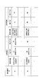
領域情報記憶部8には、図6に示されるように、歯牙Aの歯式番号N毎に、咬合面A1の画像G1内の一領域(参照領域)R1の位置情報と、頬側面A2の画像G2内の一領域(基準領域)R2の位置情報とが記憶されている。参照領域R1と基準領域R2とは、精度よく測定したならば相互に近い色を有することとなるような色相関を有する領域R1,R2として選択されている。
領域情報記憶部8には、図6に示されるように、歯牙Aの歯式番号N毎に、咬合面A1の画像G1内の一領域(参照領域)R1の位置情報と、頬側面A2の画像G2内の一領域(基準領域)R2の位置情報とが記憶されている。参照領域R1と基準領域R2とは、精度よく測定したならば相互に近い色を有することとなるような色相関を有する領域R1,R2として選択されている。
図3A及び図3B並びに図5A及び図5Bに示されるように、相互に色相関を有する参照領域R1と基準領域R2とは、歯牙A毎に、あるいは歯牙Aの種別毎に設定されている。図3A及び図3B並びに図5A及び図5Bに示す例では、咬合面A1の画像G1において参照領域R1が、頬側面A2の画像G2において基準領域R2がそれぞれ定義されている。参照領域R1および基準領域R2は、各画像G1,G2内の歯牙Aの輪郭形状を基準として、その基準位置および寸法が設定されている。
例えば、歯式番号N=#1の歯牙Aは切歯であって、図3Aに示されるように、その基準領域R2は、歯牙Aの輪郭内の切縁端部に位置し、歯牙Aの幅方向の中央位置を基準位置として、歯牙Aの幅寸法の25%の幅寸法W1と歯牙Aの高さ寸法の10%の高さ寸法H1を有する矩形領域として定義されている。また、その参照領域R1は、図3Bに示されるように、咬合面A1の画像G1において、歯牙Aの輪郭内に生じる2つの正反射領域Raの重心どうしを結ぶ直線L1と、幅方向の中心線L2との交点に重心を有し、全幅寸法に対して基準領域R2と同じ寸法比率を有する矩形領域として定義されている。
また、図5A及び図5Bに示す例では、歯式番号#6の歯牙は臼歯であって、その基準領域R2は、頬側面A2の画像G2において、歯牙Aの輪郭の重心を通る直線L3で分割した、近心側(または遠心側のいずれか)の領域の重心を含み、近心側の領域の幅寸法の10%の幅寸法W6と、それと同じ寸法の高さ寸法W6を有する矩形領域として定義されている。また、参照領域R1は、咬合面A1の画像G1において、歯牙Aの重心を通る縦横の直線L4,L5で分割された頬側近心側(または頬側遠心側のいずれか)の領域の重心を含み、歯牙Aの全幅寸法に対して基準領域R2と同じ寸法比率を有する矩形領域として定義されている。
これらの領域R1,R2は、色差ΔEが3以下であり、相互に色相関を有している。
これらの領域R1,R2は、色差ΔEが3以下であり、相互に色相関を有している。
色情報抽出部9は、第1の画像記憶部6および第2の画像記憶部7から、同じ歯式番号Nが対応づけられている咬合面A1の画像G1および頬側面A2の画像G2を受け取ると、その歯式番号Nをキーとして領域情報記憶部8内を検索し、歯式番号Nに対応づけて記憶されている参照領域R1および基準領域R2の位置情報とを読み出すようになっている。そして、色情報抽出部9は、読み出された参照領域R1の位置情報に基づいて、第1の画像記憶部6から受け取った咬合面A1の画像G1において参照領域R1内の色情報を抽出するようになっている。さらに、色情報抽出部9は、読み出された基準領域R2の位置情報に基づいて、第2の画像記憶部7から受け取った頬側面A2の画像G2において基準領域R2内の色情報を抽出するようになっている。
補正係数算出部10は、色情報抽出部9において抽出された基準領域R2内の色情報の平均値を、同じく抽出された参照領域R1内の色情報の平均値によって除算することにより、参照領域R1内の色情報を基準領域R2内の色情報に一致させるために乗算する補正係数を算出するようになっている。
画像補正部11は、補正係数算出部10により算出された補正係数を第1の画像記憶部6に記憶されている咬合面A1の画像G1に乗算することにより画像G1を補正するようになっている。
画像補正部11は、補正係数算出部10により算出された補正係数を第1の画像記憶部6に記憶されている咬合面A1の画像G1に乗算することにより画像G1を補正するようになっている。
このように構成された本実施形態に係る歯科画像処理装置5および歯科用測色装置1による歯科画像処理方法について以下に説明する。
本実施形態に係る歯科用測色装置1を用いて、高い精度の色情報を有する歯牙Aの画像G1,G2を取得するには、第1の画像取得部2によって歯牙Aの咬合面A1の画像G1を取得し、第2の画像取得部3によって歯牙Aの頬側面A2の画像G2を取得する。
本実施形態に係る歯科用測色装置1を用いて、高い精度の色情報を有する歯牙Aの画像G1,G2を取得するには、第1の画像取得部2によって歯牙Aの咬合面A1の画像G1を取得し、第2の画像取得部3によって歯牙Aの頬側面A2の画像G2を取得する。
そして、入力部4を操作して、歯式番号Nを入力し、入力された歯式番号Nとともに、第1の画像取得部2によって取得された歯牙Aの咬合面A1の画像G1および第2の画像取得部3によって取得された頬側面A2の画像G2が歯科画像処理装置5に入力される。
歯科画像処理装置5に入力された歯式番号Nと咬合面A1の画像G1とは、相互に対応づけられて第1の画像記憶部6に記憶され、歯式番号Nと頬側面A2の画像G2とは、相互に対応づけられて第2の画像記憶部7に記憶される。
歯科画像処理装置5に入力された歯式番号Nと咬合面A1の画像G1とは、相互に対応づけられて第1の画像記憶部6に記憶され、歯式番号Nと頬側面A2の画像G2とは、相互に対応づけられて第2の画像記憶部7に記憶される。
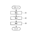
図7に示されるように、第1,第2の画像記憶部6,7に記憶された咬合面A1および頬側面A2の画像G1,G2が、その歯式番号Nとともに色情報抽出部9に入力されると、色情報抽出部9が、歯式番号Nをキーとして、領域情報記憶部8に記憶されている参照領域R1および基準領域R2の位置情報を読み出す。そして、色情報抽出部9は、読み出された位置情報に従って、咬合面A1の画像G1上の参照領域R1および頬側面A2の画像G2上の基準領域R2の色情報を抽出する(抽出ステップS1)。
次いで、抽出された参照領域R1の色情報および基準領域R2の色情報は、補正係数算出部10に入力され、各領域R1,R2内の色情報の平均値が算出された後、基準領域R2の色情報である基準色情報の平均値が参照領域R1の色情報である参照色情報の平均値によって除算されることにより、補正係数が算出される(算出ステップS2)。
最後に、算出ステップS2において算出された補正係数と、第1の画像記憶部6に記憶されている咬合面A1の画像G1とが、画像補正部11に入力され、咬合面A1の画像G1に補正係数を乗算することにより、咬合面A1の画像G1全体が補正され(補正ステップS3)、補正画像が出力される。
この場合に、領域情報記憶部8に、歯式番号N毎に記憶されている参照領域R1と基準領域R2とは、相互に色相関を有する領域であるため、補正係数は、参照色情報を基準色情報に一致させるための係数となる。
この場合に、領域情報記憶部8に、歯式番号N毎に記憶されている参照領域R1と基準領域R2とは、相互に色相関を有する領域であるため、補正係数は、参照色情報を基準色情報に一致させるための係数となる。
このように、本実施形態に係る歯科画像処理装置5、歯科用測色装置1および歯科画像処理方法によれば、同一歯牙Aの咬合面A1および頬側面A2の画像G1,G2を取得する場合に、咬合面A1および頬側面A2のいずれか一方についてのみ高精度の色情報を取得することができれば、他方については高精度の色情報を有する画像を取得しなくても、その色情報も精度よく補正することができる。例えば、頬側面A2については、高い精度のマルチバンド画像を容易に取得できるが、咬合面A1についてはマルチバンド画像を取得し難い場合があり、そのような場合に、咬合面A1については、RGB画像のようにより簡易で精度の低い画像G1を取得したとしても、頬側面A2の画像G2に基づいて精度の高い色情報を有する補正画像を生成することができるという利点がある。
また、頬側面A2のみならず咬合面A1についてもマルチバンド画像が取得できる場合であっても、外来光のような環境光によって頬側面A2あるいは咬合面A1のいずれかの画像G1(またはG2)については色情報の精度が低下する場合があり、そのような場合にも精度の高い側の画像G2(またはG1)上の基準領域R2の色情報を用いて、精度の低い側の画像G1(またはG2)の色情報を補正することができるという利点がある。
なお、本実施形態においては、頬側面A2についてマルチバンド画像、咬合面A1についてRGB画像を取得する場合を例示して説明したが、これに代えて、頬側面A2についてRGB画像を取得し、咬合面A1についてマルチバンド画像を取得してもよい。この場合、参照領域R1が、頬側面A2の画像G2の一領域において設定され、基準領域R2が、咬合面A1の画像G1の一領域において設定されることとしてもよい。
また、第2の画像取得部3が取得する測色画像として、マルチバンド画像に代えて、マルチバンド画像から色変換して得られた色情報(L*a*b*、L*u*v*、XYZ、RGB、CMYK、分光反射スペクトル等)を有する画像を採用してもよい。
また、第2の画像取得部3が取得する測色画像として、マルチバンド画像に代えて、マルチバンド画像から色変換して得られた色情報(L*a*b*、L*u*v*、XYZ、RGB、CMYK、分光反射スペクトル等)を有する画像を採用してもよい。
また、本実施形態において、歯式番号Nごとに参照領域R1の位置情報と基準領域R2の位置情報とを対応づけて領域情報記憶部8に記憶しておくこととしたが、これに代えて、歯牙Aの種別、例えば、切歯と臼歯のような種別毎に参照領域R1と基準領域R2の位置情報を対応づけて領域情報記憶部8に記憶しておくことにしてもよい。その場合には、歯式番号Nが入力されると、該歯式番号に基づいて歯牙Aの種別が判定され、その種別に対応づけて記憶されている参照領域R1と基準領域R2の位置情報が読み出されることになる。
また、本実施形態において、単一の歯牙Aごとに画像を取得し記憶することとしたが、これに代えて、複数の隣接歯牙を含む歯列画像を取得し、第1の画像記憶部6および/または第2の画像記憶部7に記憶することにしてもよい。この場合には、色情報抽出部9において、歯列画像から各歯牙Aの輪郭形状を抽出し、その輪郭形状に基づいて歯牙Aの種別や歯式番号Nを検出することにしてもよい。または、抽出された各歯牙Aに対して、入力部4から入力した歯式番号Nを対応づけることにしてもよい。
また、本実施形態において、第2の画像記憶部7に精度の高い色情報を有する歯牙全体の画像を記憶することとしたが、基準領域R2の色情報のみを記憶してもよい。この場合には、基準領域R2の色情報は一つに特定されるので、色情報抽出部9は、第2の画像記憶部7から受け取った頬側面A2の画像G2において基準領域R2内の色情報を抽出する必要がない。そのため、色情報抽出部9は、領域情報記憶部8から読み出された参照領域R1の位置情報に基づいて、第1の画像記憶部6から受け取った咬合面A1の画像G1において参照領域R1内の色情報を抽出すれば足りる。
また、図3および図5に示されるように、取得された画像G1,G2には撮影時の照明の正反射による高輝度領域Raが存在することがあり、そのような高輝度領域Raが基準領域R2あるいは参照領域R1内に存在すると、補正係数を精度よく算出することができなくなる。このため、そのような高輝度領域Raについては、補正係数の算出前に基準領域R2および参照領域R1内から除去して、色情報の平均値を算出することが好ましい。
また、図3および図5に示されるように、取得された画像G1,G2には撮影時の照明の正反射による高輝度領域Raが存在することがあり、そのような高輝度領域Raが基準領域R2あるいは参照領域R1内に存在すると、補正係数を精度よく算出することができなくなる。このため、そのような高輝度領域Raについては、補正係数の算出前に基準領域R2および参照領域R1内から除去して、色情報の平均値を算出することが好ましい。
また、本実施形態において、図6に示されるように、参照領域R1および基準領域R2の位置情報を定義した一例を示したが、これに限定されるものではなく、色差ΔE≦3となる任意の参照領域R1および基準領域R2の位置情報を対応づけて記憶することにしてもよい。この場合に、参照領域R1および基準領域R2の位置、大きさおよび形状は任意に選択することができる。
また、本実施形態において、歯科画像処理方法をハードウェアによって実行する場合のみならず、コンピュータにより実行可能な歯科画像処理プログラムによって実現することにしてもよい。
A 歯牙
A1 咬合面
A2 頬側面
G1,G2 画像
N 歯式番号(識別情報)
R1 参照領域
R2 基準領域(一領域)
S1 抽出ステップ
S2 算出ステップ
S3 補正ステップ
1 歯科用測色装置
2 第1の画像取得部(画像取得部)
3 第2の画像取得部(基準色情報取得部)
4 入力部
5 歯科画像処理装置
8 領域情報記憶部(記憶部)
9 色情報抽出部(参照色情報抽出部)
10 補正係数算出部
11 画像補正部(色情報補正部)
A1 咬合面
A2 頬側面
G1,G2 画像
N 歯式番号(識別情報)
R1 参照領域
R2 基準領域(一領域)
S1 抽出ステップ
S2 算出ステップ
S3 補正ステップ
1 歯科用測色装置
2 第1の画像取得部(画像取得部)
3 第2の画像取得部(基準色情報取得部)
4 入力部
5 歯科画像処理装置
8 領域情報記憶部(記憶部)
9 色情報抽出部(参照色情報抽出部)
10 補正係数算出部
11 画像補正部(色情報補正部)
Claims (10)
- 歯牙の咬合面または頬側面のいずれか一方について取得された補正の対象となる原画像上の参照領域であって、前記咬合面または前記頬側面のいずれか他方の一領域である基準領域において取得された色情報である基準色情報と色相関を有する参照領域の位置情報を記憶する記憶部と、
前記原画像において前記参照領域内の色情報である参照色情報を抽出する参照色情報抽出部と、
該参照色情報抽出部により抽出された参照色情報を前記基準色情報に一致させるための補正係数を算出する補正係数算出部と、
該補正係数算出部により算出された補正係数を用いて、前記原画像の色情報を補正する色情報補正部とを備える歯科画像処理装置。 - 基準とする歯牙の識別情報を入力する入力部を備え、
前記記憶部が、歯牙毎に、前記歯牙の識別情報と前記参照領域の位置情報とを対応づけて記憶し、
前記参照色情報抽出部が、前記入力部により入力された歯牙の識別情報に対応して前記記憶部に記憶されている前記参照領域の位置情報に基づいて、前記原画像から前記参照色情報を抽出する請求項1に記載の歯科画像処理装置。 - 前記記憶部が、参照領域の位置情報を歯牙の輪郭形状に対する相対的な位置情報として記憶し、
前記参照色情報抽出部が、前記歯牙の輪郭形状を抽出し、抽出された輪郭形状と、前記記憶部に記憶されている参照領域の位置情報とに基づいて、前記参照領域の色情報を抽出する請求項2に記載の歯科画像処理装置。 - 歯牙の種別毎に、前記原画像上における参照領域の位置情報を前記歯牙の輪郭形状に対する相対位置情報として記憶する記憶部を備え、
前記参照色情報抽出部が、前記歯牙の輪郭形状を抽出し、抽出された輪郭形状に基づいて該歯牙の種別を特定し、特定された種別に従う参照領域の位置情報を用いて前記参照色情報を抽出する請求項1に記載の歯科画像処理装置。 - 前記咬合面または前記頬側面のいずれか他方について取得された基準画像において前記基準領域内の色情報である前記基準色情報を抽出する基準色情報抽出部を備え、
前記記憶部が、さらに、歯牙毎に、前記歯牙の識別情報と前記基準領域の位置情報とを対応づけて記憶する請求項2に記載の歯科画像処理装置。 - 歯牙の咬合面または頬側面のいずれか一方の前記原画像を取得する画像取得部と、
前記咬合面または前記頬側面のいずれか他方の一領域における色情報である基準色情報を取得する基準色情報取得部と、
請求項1から請求項5のいずれかに記載の歯科画像処理装置とを備える歯科用測色装置。 - 前記基準色情報取得部が、前記歯牙の前記咬合面または前記頬側面のいずれかの測色画像を取得する請求項6に記載の歯科用測色装置。
- 前記画像取得部が、前記歯牙の前記咬合面または前記頬側面のいずれかの測色画像を取得する請求項6または請求項7に記載の歯科用測色装置。
- 歯牙の咬合面または頬側面のいずれか一方について取得された補正の対象となる原画像と、いずれか他方の一領域における色情報である基準色情報とが入力されたときに、該基準色情報と色相関を有する領域として記憶されている前記原画像内の参照領域の位置情報に基づいて、該参照領域内の色情報である参照色情報を前記原画像から抽出する抽出ステップと、
該抽出ステップにおいて抽出された参照色情報を前記基準色情報に一致させるための補正係数を算出する算出ステップと、
該算出ステップにおいて算出された補正係数を用いて、前記原画像の色情報を補正する補正ステップとを含む歯科画像処理方法。 - 歯牙の咬合面または頬側面のいずれか一方について取得された補正の対象となる原画像と、いずれか他方の一領域における色情報である基準色情報とが入力されたときに、該基準色情報と色相関を有する領域として記憶されている前記原画像内の参照領域の位置情報に基づいて、該参照領域内の色情報である参照色情報を前記原画像から抽出する抽出ステップと、
該抽出ステップにおいて抽出された参照色情報を前記基準色情報に一致させるための補正係数を算出する算出ステップと、
該算出ステップにおいて算出された補正係数を用いて、前記原画像の色情報を補正する補正ステップとをコンピュータに実行させる歯科画像処理プログラム。
Applications Claiming Priority (2)
| Application Number | Priority Date | Filing Date | Title |
|---|---|---|---|
| JP2010083475A JP2011212234A (ja) | 2010-03-31 | 2010-03-31 | 歯科画像処理装置、歯科用測色装置、歯科画像処理方法および歯科画像処理プログラム |
| JP2010-083475 | 2010-03-31 |
Publications (1)
| Publication Number | Publication Date |
|---|---|
| WO2011125648A1 true WO2011125648A1 (ja) | 2011-10-13 |
Family
ID=44762595
Family Applications (1)
| Application Number | Title | Priority Date | Filing Date |
|---|---|---|---|
| PCT/JP2011/057802 Ceased WO2011125648A1 (ja) | 2010-03-31 | 2011-03-29 | 歯科画像処理装置、歯科用測色装置、歯科画像処理方法および歯科画像処理プログラム |
Country Status (2)
| Country | Link |
|---|---|
| JP (1) | JP2011212234A (ja) |
| WO (1) | WO2011125648A1 (ja) |
Families Citing this family (3)
| Publication number | Priority date | Publication date | Assignee | Title |
|---|---|---|---|---|
| KR101806889B1 (ko) | 2016-12-22 | 2017-12-08 | 오스템임플란트 주식회사 | 치아 수복물 설계를 위한 그래픽 제공 방법, 이를 위한 장치 및 이를 기록한 기록매체 |
| JP2022508923A (ja) * | 2018-10-16 | 2022-01-19 | キュウティーティー カンパニー | 機械学習アルゴリズムを用いた口腔健康予測装置及び方法 |
| JP7220569B2 (ja) | 2019-01-08 | 2023-02-10 | 株式会社モリタ製作所 | 撮像支援装置、スキャナシステム、および撮像支援方法 |
Citations (4)
| Publication number | Priority date | Publication date | Assignee | Title |
|---|---|---|---|---|
| JP2002135790A (ja) * | 2000-10-24 | 2002-05-10 | Mitsubishi Electric Corp | 画像補正システム及び画像補正方法 |
| JP2007190371A (ja) * | 2005-12-19 | 2007-08-02 | Olympus Corp | 歯科用測色装置、システム、方法、およびプログラム |
| JP2007318331A (ja) * | 2006-05-24 | 2007-12-06 | Olympus Corp | 顕微鏡用撮像装置 |
| JP2010029645A (ja) * | 2008-06-30 | 2010-02-12 | Olympus Corp | 歯科用測色装置、システム、方法、及びプログラム |
-
2010
- 2010-03-31 JP JP2010083475A patent/JP2011212234A/ja not_active Withdrawn
-
2011
- 2011-03-29 WO PCT/JP2011/057802 patent/WO2011125648A1/ja not_active Ceased
Patent Citations (4)
| Publication number | Priority date | Publication date | Assignee | Title |
|---|---|---|---|---|
| JP2002135790A (ja) * | 2000-10-24 | 2002-05-10 | Mitsubishi Electric Corp | 画像補正システム及び画像補正方法 |
| JP2007190371A (ja) * | 2005-12-19 | 2007-08-02 | Olympus Corp | 歯科用測色装置、システム、方法、およびプログラム |
| JP2007318331A (ja) * | 2006-05-24 | 2007-12-06 | Olympus Corp | 顕微鏡用撮像装置 |
| JP2010029645A (ja) * | 2008-06-30 | 2010-02-12 | Olympus Corp | 歯科用測色装置、システム、方法、及びプログラム |
Also Published As
| Publication number | Publication date |
|---|---|
| JP2011212234A (ja) | 2011-10-27 |
Similar Documents
| Publication | Publication Date | Title |
|---|---|---|
| US12150836B2 (en) | Focus scanning apparatus recording color | |
| US8208704B2 (en) | Dental shade mapping | |
| EP2476373B1 (en) | Endoscope system and processor apparatus thereof | |
| US8768025B2 (en) | Dental shade mapping | |
| US7957573B2 (en) | Dental image processing device | |
| JP6817154B2 (ja) | プロセッサ装置及びその作動方法 | |
| TW201338764A (zh) | 牙齒比色儀 | |
| EP3318175A1 (en) | Image processing apparatus and imaging system | |
| KR102565592B1 (ko) | 휘도 및 색도 측정 시스템 및 방법 | |
| JP5074066B2 (ja) | 画像処理装置、及び、画像処理方法 | |
| CN101828404B (zh) | 分光特性校正装置、分光特性校正方法 | |
| WO2011125648A1 (ja) | 歯科画像処理装置、歯科用測色装置、歯科画像処理方法および歯科画像処理プログラム | |
| JP4174707B2 (ja) | 分光測定システム、色再現システム | |
| KR100904559B1 (ko) | 스테레오 영상을 이용한 안면 진단 방법 | |
| US20240212832A1 (en) | Systems and methods for color filter array image enhancement | |
| JP3960989B2 (ja) | 色推定システム及び色推定方法 | |
| WO2016047739A1 (ja) | 口腔内3次元形状測定装置 | |
| US12511824B2 (en) | Three-dimensional modeling method and apparatus using same | |
| CN112019833B (zh) | 数字取像装置及荧幕的色差校正方法及其装置 | |
| WO2025173551A1 (ja) | 情報処理装置、情報処理方法及びプログラム | |
| JP2007315808A (ja) | マルチスペクトル撮像装置 | |
| WO2025173550A1 (ja) | 情報処理装置、情報処理方法及びプログラム | |
| WO2025115505A1 (ja) | 歯列画像生成装置、歯列画像生成方法及びプログラム | |
| BR102021026004A2 (pt) | Método baseado em visão computacional para definição de tonalidades dentárias | |
| Splett | John Penczek, Paul A. Boynton & Jolene |
Legal Events
| Date | Code | Title | Description |
|---|---|---|---|
| 121 | Ep: the epo has been informed by wipo that ep was designated in this application |
Ref document number: 11765548 Country of ref document: EP Kind code of ref document: A1 |
|
| NENP | Non-entry into the national phase |
Ref country code: DE |
|
| 122 | Ep: pct application non-entry in european phase |
Ref document number: 11765548 Country of ref document: EP Kind code of ref document: A1 |