WO2011102532A1 - 誘導肝細胞 - Google Patents
誘導肝細胞 Download PDFInfo
- Publication number
- WO2011102532A1 WO2011102532A1 PCT/JP2011/053875 JP2011053875W WO2011102532A1 WO 2011102532 A1 WO2011102532 A1 WO 2011102532A1 JP 2011053875 W JP2011053875 W JP 2011053875W WO 2011102532 A1 WO2011102532 A1 WO 2011102532A1
- Authority
- WO
- WIPO (PCT)
- Prior art keywords
- cells
- hnf4α
- liver
- cell
- hnf3β
- Prior art date
- Legal status (The legal status is an assumption and is not a legal conclusion. Google has not performed a legal analysis and makes no representation as to the accuracy of the status listed.)
- Ceased
Links
Images





Classifications
-
- C—CHEMISTRY; METALLURGY
- C12—BIOCHEMISTRY; BEER; SPIRITS; WINE; VINEGAR; MICROBIOLOGY; ENZYMOLOGY; MUTATION OR GENETIC ENGINEERING
- C12N—MICROORGANISMS OR ENZYMES; COMPOSITIONS THEREOF; PROPAGATING, PRESERVING, OR MAINTAINING MICROORGANISMS; MUTATION OR GENETIC ENGINEERING; CULTURE MEDIA
- C12N5/00—Undifferentiated human, animal or plant cells, e.g. cell lines; Tissues; Cultivation or maintenance thereof; Culture media therefor
- C12N5/06—Animal cells or tissues; Human cells or tissues
- C12N5/0602—Vertebrate cells
- C12N5/067—Hepatocytes
-
- A—HUMAN NECESSITIES
- A61—MEDICAL OR VETERINARY SCIENCE; HYGIENE
- A61K—PREPARATIONS FOR MEDICAL, DENTAL OR TOILETRY PURPOSES
- A61K35/00—Medicinal preparations containing materials or reaction products thereof with undetermined constitution
- A61K35/12—Materials from mammals; Compositions comprising non-specified tissues or cells; Compositions comprising non-embryonic stem cells; Genetically modified cells
- A61K35/37—Digestive system
- A61K35/407—Liver; Hepatocytes
-
- A—HUMAN NECESSITIES
- A61—MEDICAL OR VETERINARY SCIENCE; HYGIENE
- A61P—SPECIFIC THERAPEUTIC ACTIVITY OF CHEMICAL COMPOUNDS OR MEDICINAL PREPARATIONS
- A61P1/00—Drugs for disorders of the alimentary tract or the digestive system
- A61P1/16—Drugs for disorders of the alimentary tract or the digestive system for liver or gallbladder disorders, e.g. hepatoprotective agents, cholagogues, litholytics
-
- C—CHEMISTRY; METALLURGY
- C07—ORGANIC CHEMISTRY
- C07K—PEPTIDES
- C07K14/00—Peptides having more than 20 amino acids; Gastrins; Somatostatins; Melanotropins; Derivatives thereof
- C07K14/435—Peptides having more than 20 amino acids; Gastrins; Somatostatins; Melanotropins; Derivatives thereof from animals; from humans
- C07K14/46—Peptides having more than 20 amino acids; Gastrins; Somatostatins; Melanotropins; Derivatives thereof from animals; from humans from vertebrates
- C07K14/47—Peptides having more than 20 amino acids; Gastrins; Somatostatins; Melanotropins; Derivatives thereof from animals; from humans from vertebrates from mammals
- C07K14/4701—Peptides having more than 20 amino acids; Gastrins; Somatostatins; Melanotropins; Derivatives thereof from animals; from humans from vertebrates from mammals not used
- C07K14/4702—Regulators; Modulating activity
-
- C—CHEMISTRY; METALLURGY
- C12—BIOCHEMISTRY; BEER; SPIRITS; WINE; VINEGAR; MICROBIOLOGY; ENZYMOLOGY; MUTATION OR GENETIC ENGINEERING
- C12N—MICROORGANISMS OR ENZYMES; COMPOSITIONS THEREOF; PROPAGATING, PRESERVING, OR MAINTAINING MICROORGANISMS; MUTATION OR GENETIC ENGINEERING; CULTURE MEDIA
- C12N2501/00—Active agents used in cell culture processes, e.g. differentation
- C12N2501/10—Growth factors
- C12N2501/11—Epidermal growth factor [EGF]
-
- C—CHEMISTRY; METALLURGY
- C12—BIOCHEMISTRY; BEER; SPIRITS; WINE; VINEGAR; MICROBIOLOGY; ENZYMOLOGY; MUTATION OR GENETIC ENGINEERING
- C12N—MICROORGANISMS OR ENZYMES; COMPOSITIONS THEREOF; PROPAGATING, PRESERVING, OR MAINTAINING MICROORGANISMS; MUTATION OR GENETIC ENGINEERING; CULTURE MEDIA
- C12N2501/00—Active agents used in cell culture processes, e.g. differentation
- C12N2501/10—Growth factors
- C12N2501/12—Hepatocyte growth factor [HGF]
-
- C—CHEMISTRY; METALLURGY
- C12—BIOCHEMISTRY; BEER; SPIRITS; WINE; VINEGAR; MICROBIOLOGY; ENZYMOLOGY; MUTATION OR GENETIC ENGINEERING
- C12N—MICROORGANISMS OR ENZYMES; COMPOSITIONS THEREOF; PROPAGATING, PRESERVING, OR MAINTAINING MICROORGANISMS; MUTATION OR GENETIC ENGINEERING; CULTURE MEDIA
- C12N2501/00—Active agents used in cell culture processes, e.g. differentation
- C12N2501/60—Transcription factors
-
- C—CHEMISTRY; METALLURGY
- C12—BIOCHEMISTRY; BEER; SPIRITS; WINE; VINEGAR; MICROBIOLOGY; ENZYMOLOGY; MUTATION OR GENETIC ENGINEERING
- C12N—MICROORGANISMS OR ENZYMES; COMPOSITIONS THEREOF; PROPAGATING, PRESERVING, OR MAINTAINING MICROORGANISMS; MUTATION OR GENETIC ENGINEERING; CULTURE MEDIA
- C12N2506/00—Differentiation of animal cells from one lineage to another; Differentiation of pluripotent cells
- C12N2506/13—Differentiation of animal cells from one lineage to another; Differentiation of pluripotent cells from connective tissue cells, from mesenchymal cells
- C12N2506/1307—Differentiation of animal cells from one lineage to another; Differentiation of pluripotent cells from connective tissue cells, from mesenchymal cells from adult fibroblasts
-
- C—CHEMISTRY; METALLURGY
- C12—BIOCHEMISTRY; BEER; SPIRITS; WINE; VINEGAR; MICROBIOLOGY; ENZYMOLOGY; MUTATION OR GENETIC ENGINEERING
- C12N—MICROORGANISMS OR ENZYMES; COMPOSITIONS THEREOF; PROPAGATING, PRESERVING, OR MAINTAINING MICROORGANISMS; MUTATION OR GENETIC ENGINEERING; CULTURE MEDIA
- C12N2510/00—Genetically modified cells
Definitions
- the present invention relates to a method for producing liver cells from various cells.
- the present invention is useful in fields related to life science, medicine, and pharmacology.
- hepatocyte transplantation which is expected to be a new treatment for liver disease that can replace liver transplantation with severe donor organ shortage, artificial liver that is being developed for effective treatment of fulminant hepatitis, and the effects and side effects of drugs
- hepatocytes are an essential cellular material.
- the number of hepatocytes that can be collected directly from living tissue is limited, and it is difficult to proliferate in vitro, so the step to medical application using hepatocytes is out of the experimental stage. I can't.
- ES cells embryonic stem cells
- iPS cells induced pluripotent stem cells
- HNF hepatocyte nuclear factor
- HNF1 ⁇ , HNF1 ⁇ (vHNF1), HNF3 ⁇ , HNF3 ⁇ , HNF3 ⁇ , HNF4 ⁇ , HNF6, C / EBP family, and GATA family have been identified so far.
- Hlx gene, Hex gene, and Prox1 gene have been identified as gene groups involved in liver development and hepatoblasts.
- Non-patent Document 1 bone marrow-derived human mesenchymal stem cells expressed HNF3 ⁇ and differentiated into hepatocyte-like cells
- Non-patent Document 2 a report that cells have been induced to differentiate.
- the present inventors have also reported on the differentiation mechanism of hepatocytes from mouse fetal liver stem cells (hepatoblasts) (Non-patent Document 3).
- hepatocytes are extremely useful cells, there is no established method for obtaining a safe and simple method that satisfies the quality and quantity available for medical use, which is a major problem for medical applications of hepatocytes. Yes.
- innovative new technologies such as producing hepatocytes from cells obtained at a low invasive and low cost. Conceivable.
- the present invention provides the following. 1) A method for producing induced hepatocytes from non-liver cells, comprising a step of introducing HNF3 ⁇ , HNF3 ⁇ , or HNF3 ⁇ and HNF4 ⁇ (preferably HNF3 ⁇ and HNF4 ⁇ ) into non-liver cells. 2) A step of introducing a gene encoding HNF3 ⁇ , HNF3 ⁇ , or HNF3 ⁇ and a gene encoding HNF4 ⁇ (preferably a gene encoding HNF3 ⁇ and a gene encoding HNF4 ⁇ ) into a non-liver cell. Production method. 3) The production method according to 1) or 2), wherein the non-liver cells are derived from human.
- a factor set for example, a kit for use in a method for producing induced hepatocytes from non-liver cells consisting of HNF3 ⁇ , HNF3 ⁇ , or HNF3 ⁇ and HNF4 ⁇ (preferably consisting of HNF3 ⁇ and HNF4 ⁇ ).
- a gene encoding HNF3 ⁇ , HNF3 ⁇ , or HNF3 ⁇ and a gene encoding HNF4 ⁇ (preferably a gene encoding HNF3 ⁇ and a gene encoding HNF4 ⁇ ).
- a gene set eg, a kit for use.
- Non-liver cells into which a gene encoding HNF3 ⁇ , HNF3 ⁇ , or HNF3 ⁇ and a gene encoding HNF4 ⁇ (preferably a gene encoding HNF3 ⁇ and a gene encoding HNF4 ⁇ ) are introduced in the presence of a growth factor A method for producing induced hepatocytes comprising a step of culturing on an extracellular matrix, if desired.
- a method for transplanting cells into the liver comprising the step of transplanting the cells according to 5).
- a method for treating a disease comprising the step of using the cell according to 5).
- a method for evaluating drug reactivity comprising the step of using the cell according to 5).
- liver cells can be produced from somatic cells that are not liver cells, such as skin cells. Establish and develop hepatocyte transplantation, artificial liver, and drug reactivity tests that could not be generalized due to difficulty in procuring cells as general medicine by using the liver cells obtained by the present invention. Can do.
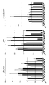
- FIG. 1 is a graph showing the gene expression inducing effect of 12 transcription factors related to selected hepatocyte differentiation.
- MEF into which all 12 were introduced gene expression of albumin, ⁇ -fetoprotein and E-cadherin was observed.
- the gene expression of albumin decreased in the group excluding HNF4 ⁇ or HNF6, and the gene expression of ⁇ -fetoprotein was decreased in the group excluding HNF4 ⁇ or HNF1 ⁇ . There was no group that significantly reduced E-cadherin gene expression.
- FIG. 1 is a graph showing the gene expression inducing effect of 12 transcription factors related to selected hepatocyte differentiation.
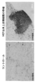
- FIG. 3 is a photograph of induced epithelial cells.
- MEF into which HNF4 ⁇ and HNF3 ⁇ were introduced was transferred to a type I collagen-coated dish and cultured using a medium for hepatic stem cells, epithelial cells completely different in cell morphology from MEF appeared.
- FIG. 4 is a fluorescent staining photograph of epithelial cells into which HNF4 ⁇ and HNF3 ⁇ have been introduced. is there. All of the epithelial cells derived from MEF were E-cadherin positive, and E-cadherin was localized in the cell adhesion region. In addition, many E-cadherin positive cells expressed albumin and cytokeratin 8-18. In addition, expression of ⁇ -1-antitrypsin was also observed (graph). (Reconstruction of liver tissue with MEF-derived epithelial cells) FIG.
- FIG. 5 is a photograph of one month after transplanting epithelial cells derived from MEF into the liver of FAH knockout mice. It was found that the cells were engrafted in the liver tissue of FAH knockout mice as FAH-positive mature hepatocytes, and the liver tissue was reconstructed. From these results, it was revealed that the epithelial cells derived from MEF by introduction of HNF4 ⁇ and HNF3 ⁇ are liver epithelial cells having the ability to reconstruct liver tissue. In the control, MEF was transplanted.
- FIG. 6A shows the nucleotide sequence of the gene (mouse) encoding each of HNF4 ⁇ , HNF3 ⁇ , HNF3 ⁇ , and HNF3 ⁇ .
- FIG. 6B shows the nucleotide sequences of genes (human) encoding HNF4 ⁇ , HNF3 ⁇ , HNF3 ⁇ , and HNF3 ⁇ .
- FIG. 7 shows the amino acid sequences (mouse and human) of HNF4 ⁇ , HNF3 ⁇ , HNF3 ⁇ , and HNF3 ⁇ , respectively.
- FIG. 8 is a graph showing the gene expression induction effect by simultaneous introduction of HNF4 ⁇ and HNF3 ⁇ on human skin-derived fibroblasts. Similar to mice, strong expression induction of albumin, ⁇ -fetoprotein and E-cadherin was observed.
- the present invention can produce liver cells by introducing a specific set of factors into various cells.
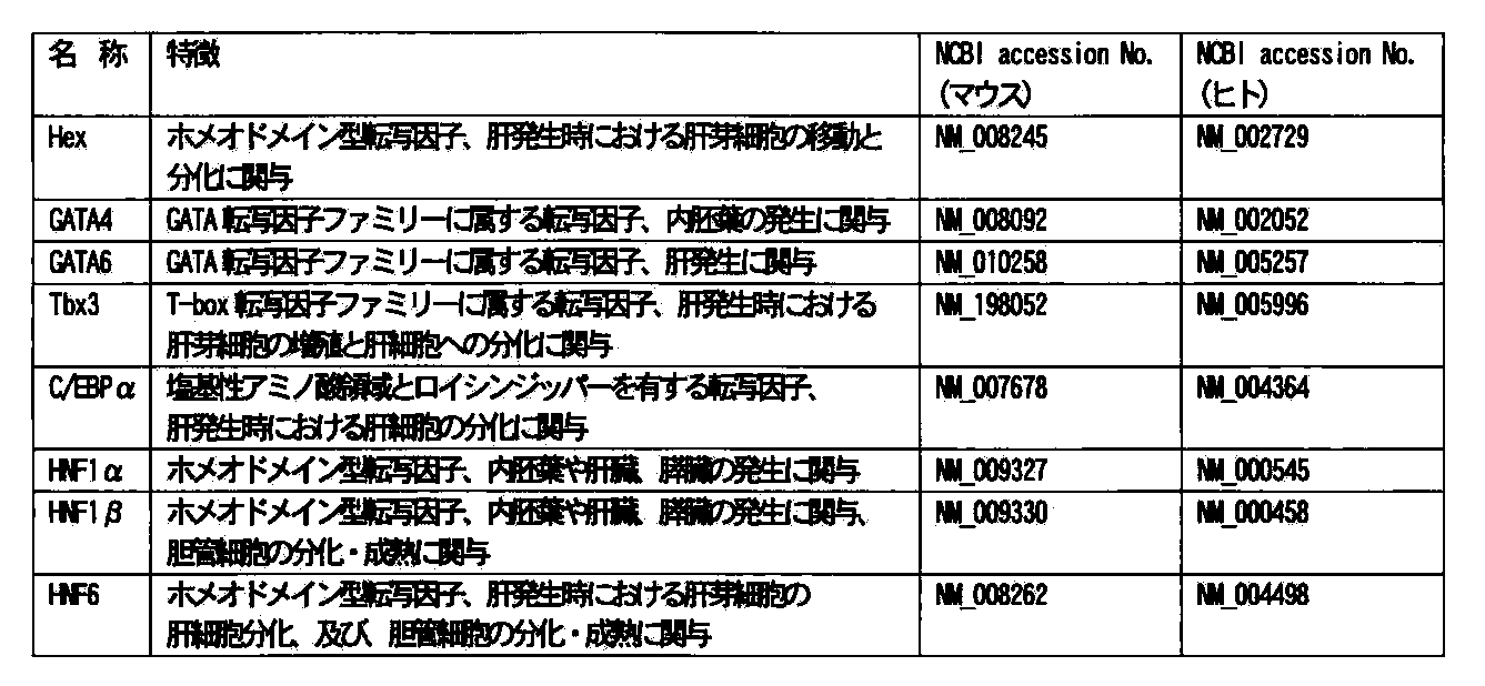
- Factor sets for producing the liver cells of the present invention are expressed by the inventor in hepatoblasts (hepatic stem / progenitor cells) or endoderm cells, and may or may be related to hepatocyte differentiation. Are selected from the group consisting of Hex, GATA4, GATA6, Tbx3, C / EBP ⁇ , HNF1 ⁇ , HNF1 ⁇ , HNF3 ⁇ , HNF3 ⁇ , HNF3 ⁇ , HNF4 ⁇ and HNF6.
- the factor set of the present invention contains at least HNF4 ⁇ .
- a preferred embodiment is characterized by comprising HNF3 ⁇ , HNF3 ⁇ , or HNF3 ⁇ and HNF4 ⁇ , and in a particularly preferred embodiment, HNF4 ⁇ and HNF3 ⁇ are included.
- the factor used may be derived from any mammal.
- factors derived from, for example, mouse, rat, cow, sheep, horse, monkey and human can be used.
- the factor used in the present invention is not limited to a specific isoform.
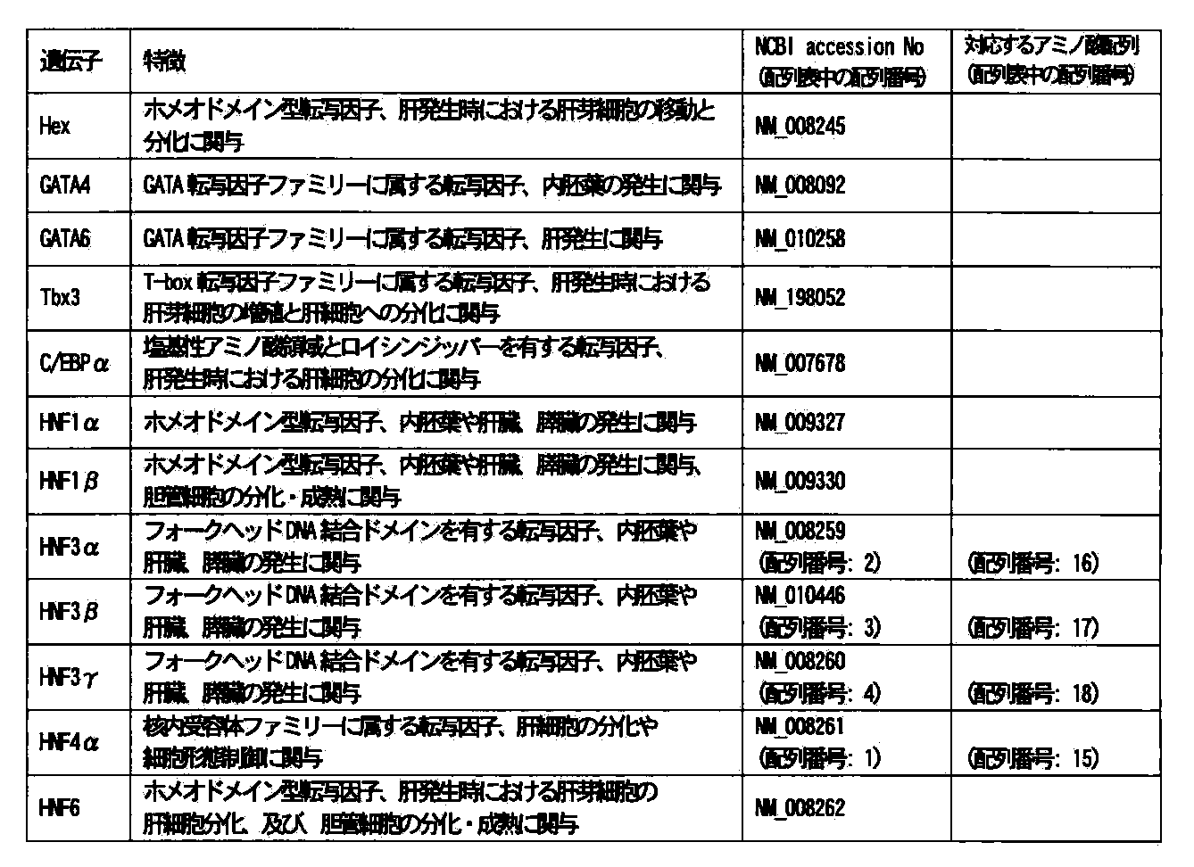
- the table below shows the factors particularly relevant to the present invention and the NCBI accession numbers of the nucleotide sequences of mouse and human genes encoding the factors.
- the nucleotide sequences of the mouse HNF4 ⁇ , HNF3 ⁇ , HNF3 ⁇ , and HNF3 ⁇ genes are shown as SEQ ID NOS: 1 to 4, respectively. Furthermore, as SEQ ID NOs: 5 to 10, six isoforms of human HNF4 ⁇ , SEQ ID NO: 11 as human HNF3 ⁇ , SEQ ID NOS: 12 and 13 as two isoforms of human HNF3 ⁇ , and SEQ ID NO: 14 as the nucleotide sequence of the human HNF3 ⁇ gene It is shown.
- the amino acid sequence of each factor can be derived from the gene sequence.
- amino acid sequences of mouse HNF4 ⁇ , HNF3 ⁇ , HNF3 ⁇ , and HNF3 ⁇ are shown as SEQ ID NOS: 15 to 18, respectively.
- SEQ ID NOS: 19 to 24 six isoforms of human HNF4 ⁇ (transscript variants 1-6), SEQ ID NO: 25 as human HNF3 ⁇ , SEQ ID NOS: 26 and 27 as two isoforms of human HNF3 ⁇ , SEQ ID NO: 28 The amino acid sequence of human HNF3 ⁇ is shown.
- HNF4 ⁇ + HNF3 ⁇ and HNF4 ⁇ + HNF3 ⁇ induce gene expression, but the combination of HNF4 ⁇ and HNF3 ⁇ tended to have the greatest gene expression induction effect.
- gene expression induction was also obtained in human-derived cells with HNF4 ⁇ + HNF3 ⁇ and HNF4 ⁇ + HNF3 ⁇ , but the combination of HNF4 ⁇ and HNF3 ⁇ tended to be most effective, as in mice.
- the final cells obtained are different in albumin secretion ability and gene expression profile, regardless of the combination of factors introduced above, There was no significant difference.
- the results of testing HNF4 ⁇ transcript variants 1 to 5 using human-derived cells seemed to be highly effective for transcript variants 1 to 3, particularly transcript variant 3.
- the scope of the present invention includes the use of the above-mentioned factors and genes encoding the factors, as well as mutants thereof. That is, in the present invention, when referring to a certain factor, unless otherwise specified, in any case, (a) a protein comprising the amino acid sequence of the corresponding SEQ ID No. shown in the sequence listing, (b) the amino acid sequence A protein having an amino acid sequence in which one or more amino acids are substituted, deleted, inserted, and / or added, and having the function of the factor; and (c) at least 90% (preferably 95%) of the amino acid sequence. , More preferably 98%), and a protein having the function of the factor.
- a gene encoding a certain factor when referring to a gene encoding a certain factor, unless otherwise specified, all are (d) a gene consisting of the nucleotide sequence of the corresponding sequence number in the sequence listing; (e) the nucleotide sequence A gene that hybridizes under stringent conditions with a polynucleotide comprising a sequence complementary to and that encodes a protein having the function of the factor; (f) at least 90% (preferably 95%, more than the nucleotide sequence; Preferably 98%) and a gene encoding a protein having the function of the factor; and a gene encoding any one of the proteins (a) to (c).
- stringent conditions refers to conditions of 6M urea, 0.4% SDS, 0.5 ⁇ SSC or equivalent hybridization conditions. Higher stringency conditions such as 6M urea, 0.4% SDS, 0.1 ⁇ SSC or equivalent hybridization conditions may be applied. Under each condition, the temperature can be about 40 ° C. or higher, and if higher stringency conditions are required, for example, about 50 ° C. or about 65 ° C. may be used. Amino acid or nucleotide sequence homology is determined using the algorithm BLAST (Proc. Natl. Acad. Sci.
- the factor set of the present invention can include other factors in addition to the above-described factors. NCBI accession numbers of the nucleotide sequences of other factors and mouse and human genes encoding the factors are shown in the table below.
- the factor set for producing the liver cell of the present invention can contain one or more other factors in addition to the factors specifically described above.
- factors include, for example, introduction of genes encoding various candidate factors into somatic cells expressing one or more selected from the group consisting of HNF3 ⁇ , HNF3 ⁇ , HNF3 ⁇ and HNF4 ⁇ , and liver cells with higher probability. It is possible to select cells by selecting cells that have been able to induce.
- the factor used in the present invention may be in the form of a fusion protein of the protein and another protein or peptide in addition to the protein itself produced from the gene encoding the factor.
- it may be a fusion protein with a green fluorescent protein (GFP) or a fusion protein with a peptide such as a histidine tag.
- GFP green fluorescent protein
- PTD protein transmission domain
- the method for preparing liver cells of the present invention includes a step of introducing a specific factor into the target cells, but the specific means for performing this step is not particularly limited.
- Each factor is originally transcribed from a gene in a cell, translated, and considered to function as a protein (transcription factor). Therefore, by directly introducing the factor into the cell or by physically transferring the gene encoding the factor.
- the desired effect can be achieved by introduction into cells using chemical or viral vectors.
- a typical example of means for directly introducing a factor into a cell is by binding a PTD peptide having a membrane permeation function (for example, AntP, HIV / TAT, HSV / VP-22).
- a PTD peptide having a membrane permeation function for example, AntP, HIV / TAT, HSV / VP-22.
- AntP a membrane permeation function
- This method is excellent in that it can be applied to an in vivo individual and can be introduced into almost all cells including cells in which gene transfer is difficult by existing transfection.
- a factor can be introduced simply by adding a fusion protein to a culture environment while avoiding complicated gene introduction operations on cells.
- a method for preparing a fusion protein of a target protein and a PTD, conditions for introducing the fusion protein into a target cell, and the like are well known to those skilled in the art.
- Examples of means for introducing genes into cells physicochemically are the calcium phosphate method / lipofection method, electroporation method, ultrasonic gene introduction method, gene gun introduction method, and recombinant immunogene method. Both of these methods are well known to those skilled in the art.
- viral vectors used when introducing genes into cells using viral vectors are retrovirus vectors, lentivirus vectors, Sendai virus vectors, and helper-dependent adenovirus vectors. Preparation of vectors incorporating genes encoding factors, methods for infecting cells, and the like are well known to those skilled in the art. From the viewpoint that gene transfer can be performed with high efficiency even in cells that are not highly proliferative, a method using a lentiviral vector would be preferable.
- a marker gene (a gene encoding a fluorescent protein, a drug resistance gene, etc.) is connected on the vector with an IRES (internal ribosome entry site) downstream of the gene to be introduced, and a marker is simultaneously formed with the target gene product. So that cells into which the gene of interest has been introduced can be selected or visualized. Whether two target genes are introduced at the same time can be determined by creating a construct in which each marker gene, for example, one target gene is connected to GFP (green) and the other target gene is connected to RFP1 (red). I can confirm. Moreover, if the cell into which the gene has been introduced can be cloned as a cell line, the presence or absence of the introduced gene can be confirmed by PCR or Southern blotting on the whole cell.
- IRES internal ribosome entry site
- the target cell into which the factor is introduced is a non-hepatic cell.
- the term “non-liver cell” refers to a cell other than the liver cell described later unless otherwise specified.
- the target cell into which the factor is introduced may be derived from any animal.
- the present invention can be directed to cells derived from, for example, mouse, rat, cow, sheep, horse, monkey, or human.
- the target cells into which the factor is introduced may be embryonic stem cells (ES cells) or induced pluripotent stem cells (iPS cells), or other somatic cells.
- An embryonic stem cell (ES cell) refers to a stem cell cell line made from an inner cell mass belonging to a part of an embryo at the blastocyst stage, which is an early stage of animal development.
- iPS cells induced pluripotent stem cells
- iPS cells are the pluripotency that can differentiate into so many cells like ES cells by introducing specific factors into somatic cells, A cell that has the ability to maintain self-replication.
- Somatic cells include stem cells that have the ability to differentiate into cells with several different functions (eg, mesenchymal stem cells) and differentiated cells that specialize in certain functions and do not become other cells (eg, , Fibroblasts, and adult skin cells).
- the target cell into which the factor is introduced may be a germ line cell, a cell derived from a germ line cell, or an amniotic cell (including a placenta cell).
- the target cells to be used are not particularly limited, and may be fetal cells or mature adult cells.
- a gene When a gene is introduced by retrovirus, it may be preferable to select cells that can actively proliferate. Examples of this include fetal fibroblasts and mesenchymal stem cells derived from adult bone marrow.
- the target cells to be used are preferably human (adult) -derived cells, for example, fibroblasts obtained from skin cells, It may be preferable to use cells isolated from the patient himself or from a person who is syngeneic with the patient.
- hepatic cell refers to a fetal or adult cell constituting the liver, hepatic progenitor cell (also referred to as hepatic stem cell), unless otherwise specified. , And partially differentiated into hepatocytes.
- Cells constituting the liver include hepatocytes (hepatocytes) and non-hepatocytes (sinusoidal endothelial cells, Kupffer cells, stellate cells, pit cells, bile duct epithelial cells, etc.).
- induced hepatocytes include hepatocyte-like cells, hepatic progenitor cell-like cells, or cell-like cells partially differentiated into hepatocytes.
- Whether or not a cell into which a factor has been introduced has become an induced hepatocyte can be determined by those skilled in the art with or without the expression of albumin, a hepatocyte differentiation marker, or the expression of ⁇ -1-antitrypsin, a medium-term differentiation marker of hepatocytes.
- the presence or absence of glycogen, the presence or absence of glycogen accumulation ability can be used as an index and / or by morphological observation. In some cases, the expression of ⁇ -fetoprotein may be confirmed.
- CYP7A1 cholesterol 7a-hydroxylase catalyzes the presence or absence of expression of cytokeratin 8 and / or cytokeratin 18 (CK8 / 18) and the initial stage reaction of the biosynthetic pathway from cholesterol to bile acids.
- the presence / absence of the expression of E-cadherin, which is a typical adhesion molecule of epithelial cells, may be used as indicators.
- cells into which HNF3 ⁇ , HNF3 ⁇ , or HNF3 ⁇ and HNF4 ⁇ have been introduced can be used for several hours to several weeks in a standard medium (for example, SCM (see References 1 to 4 below)), if necessary.
- a standard medium for example, SCM (see References 1 to 4 below)
- the cells can be induced into cells having the function of liver cells by culturing under appropriate culture conditions.
- Appropriate conditions for culturing refer to culturing under culturing conditions in which growth factors are included in the environment in which the cells are maintained, and optionally an extracellular matrix is present.
- the medium the standard medium described in Reference Document 2 below can be preferably used.
- growth factors examples include cytokines such as hepatocyte growth factor (HGF) and epidermal growth factor (EGF).
- HGF hepatocyte growth factor
- EGF epidermal growth factor
- a medium containing HGF and EGF is preferred.
- the concentration of the growth factor in the medium is not particularly limited as long as induction is possible, and it varies depending on the type of growth factor used.
- EGF it is about 10 to 40 ng / mL, preferably about 20 ng / mL.
- HGF it is 10 to 50 ng / mL, preferably 20 to 40 ng / mL.
- the origin of the growth factor is not particularly limited and may be a human recombinant.
- extracellular matrix examples include collagen (for example, type I collagen, type IV collagen) and laminin. It is preferable to use type I collagen.
- the extracellular matrix is preferably used as a coating agent for the culture substrate.
- the coating of the culture vessel with the extracellular matrix depends on the type of extracellular matrix used.
- the culture medium for this purpose may use human recombinant growth factor and can also be applied to human-derived cells.
- the cells are preferably induced by a culture system that is serum-free and excludes substances derived from other organisms.
- the culture vessel is not particularly limited, and for example, a flask, a dish, a multi-dish, a microwell plate, a culture bag, and a roller bottle can be used.
- various conditions known for culturing liver cells can be applied.
- the culture temperature is 30 to 40 ° C., preferably 37 ° C.
- the CO 2 concentration is, for example, 1 to 10%, preferably 2 to 5%.
- Cultivation for induction can be performed continuously usually for 1 to several weeks, preferably 1 to 3 weeks, for example 2 weeks.
- the cells obtained by introducing HNF4 ⁇ and HNF3 ⁇ into MEF become a cell population that continues to grow in an epithelial form that expresses a hepatocyte marker by culturing under appropriate conditions. Subsequent passage and cryopreservation were possible. This indicates that the obtained cell population contains a mixture of differentiated cells and more undifferentiated progenitor cells.
- the use of the cells produced by the method of the present invention is not particularly limited, and can be used for all tests / research conducted using cells, treatment of diseases using liver cells, and the like.
- the induced hepatocytes obtained according to the present invention or their progeny can constitute artificial liver tissue in vitro or in vivo.
- Cell transplantation into the liver can be achieved by administering the induced hepatocytes obtained by the present invention or progeny thereof to a patient by an appropriate method.
- Such transplantation techniques include various liver diseases including those that can be treated by partial resection of the liver and / or liver transplantation, such as cirrhosis, primary biliary cirrhosis, viral hepatitis, alcoholic hepatitis, autoimmune hepatitis Diseases such as liver cancer, hepatocellular carcinoma, cholangiocellular carcinoma, metastatic liver cancer, liver abscess, inborn errors of metabolism (eg hereditary hypertyrosineemia type I (fumaryl acetoacetate hydrolase deficiency, hepatorenal tyrosineemia)) Or it may be effective for the treatment of a condition.
- the present invention provides a method for treating liver disease using HNF3 ⁇ , HNF3 ⁇ , or HNF3 ⁇ and HNF4 ⁇ .
- MEF Cell culture Mouse embryonic fibroblasts
- C57BL / 6E13.5 10% fetal bovine serum (FBS), L-glutamine (2 mmol / L) and DME medium (Dulbecco's modified Eagle medium) containing penicillin / streptomycin.
- FBS fetal bovine serum
- L-glutamine 2 mmol / L
- DME medium Dulbecco's modified Eagle medium
- the SCM medium is obtained by adding the following to a 1: 1 mixture of DMEM and F-12 (Nacalai Tesque). 10% FBS insulin (1 ⁇ g / mL) (Wako, Tokyo, Japan) dexamethasone (1 ⁇ 107 mol / L) (Sigma) nicotinamide (10 mmol / L) (Sigma) L-glutamine (2 mmol / L), ⁇ -mercaptoethanol (50 ⁇ mol / L) (Sigma) penicillin / streptomycin
- HGF hepatocyte growth factor
- EGF epidermal growth factor
- RNA analysis Total RNA was obtained using RNeasy Mini Kit (QIAGEN, Tokyo, Japan) according to the manual attached to the kit. Quantitative PCR (qPCR) was performed using TaqMan Universal PCR Master Mix (Applied Biosystems, Japan) and Applied Biosystems 7300 real-time PCR system (Applied Biosystems). Except for E-cadherin (TaqMan Gene Expression Assay ID: Mm00486909_g1) (Applied Biosystems), information on PCR primers and probes is shown in References 1-3.
- a retrovirus-producing retrovirus vector pGCsam (murine stem cell virus, MSCV) (see Reference 1) was used.
- Each of the following mouse-derived genes encoding factor 12 was subcloned into a vector.
- pCMV-VSV-G VSV-G env expression plasmid
- PEI polyethyleneimine
- FAH-deficient mice (FAH ⁇ / ⁇ , Cells were transplanted into the liver of Reference Document 4) (1 ⁇ 10 7 cells / mouse).
- FAH ⁇ / ⁇ mice are known as models of hereditary hypertyrosinemia type I and are usually 7.5 mg / l 2- (2-nitro-4-trifluoromethylbenzoyl) -1,3-cyclohexaneedione (NTBC). ) (Swedish International International) was given and maintained, but the treatment was stopped after cell transplantation.
- hepatic epithelial cells by fibroblast reprogramming After introducing HNF4 ⁇ and HNF3 ⁇ into MEF, the cells were transferred to a type I collagen-coated dish, and a medium for hepatic stem cells containing HGF and EGF was used. Subsequently, the culture was performed. As a result, it was found that epithelial-like cells whose cell morphology was completely different from MEF appeared (FIG. 3). As a result of immunostaining, it was found that all of these epithelial cells were E-cadherin positive and also acquired epithelial cell-specific morphology such that E-cadherin was localized in the cell adhesion region (FIG. 4). ).
- E-cadherin positive cells expressed hepatocyte differentiation markers albumin, cytokeratin 8, and cytokeratin 18 (FIG. 4). Furthermore, in the induced epithelium-like cells, expression of ⁇ -1-antitrypsin, a medium differentiation marker for hepatocytes, was also observed (FIG. 4).
- the epithelial cells born from MEF by introduction of HNF4 ⁇ and HNF3 ⁇ are liver epithelial cells.
- the MEF in culture ages early and undergoes cell death, but the newly born epithelial-like cell population continues to proliferate and can then be passaged or cryopreserved. Therefore, it is considered that E-cadherin-positive liver epithelial cell population contains more undifferentiated hepatic progenitor cells in addition to differentiated hepatocytes. That is, it is thought that many hepatocytes are supplied over a long period from hepatic progenitor cells having a high proliferation ability.
- liver tissue Reconstruction of liver tissue with epithelial cells derived from fibroblasts
- epithelial cells derived from MEF by introduction of HNF4 ⁇ and HNF3 ⁇ are hepatic epithelial cells
- the obtained epithelial cells were treated with FAH.
- the epithelial cells induced from MEF by introduction of HNF4 ⁇ and HNF3 ⁇ are liver epithelial cells having the ability to reconstruct liver tissue.
- Human skin-derived fibroblasts purchased from Cell Applications are cultured according to the attached protocol, and the induction medium and vector are the same as in Example 1, and the gene expression induction effect by introduction of HNF4 ⁇ and HNF3 ⁇ in the table below is used. It was confirmed.
- FIG. 8 shows the results when transscript variant 3 was used.
- transcrib variants 1-3 As a result of trying HNF4 ⁇ transscript variants 1-5, it seems that the effects of transcrib variants 1-3, especially trans- spect variant 3, are high.
- human-derived cells When human-derived cells were used, it was confirmed that gene expression induction was also obtained with HNF4 ⁇ + HNF3 ⁇ and HNF4 ⁇ + HNF3 ⁇ .
- HNF4 ⁇ and HNF3 ⁇ tended to be most effective.
- Reference 1 Suzuki A.I. , Iwama A .; , Miyashita H .; Nakauchi H .; , Taniguchi H .; Role for growth factors and extracellular matrix in controlling differential of hepatic stem cells. Development 130, 2513-2524, 2003.
- Reference 2 Suzuki A. et al. , Zheng Y. et al. W. Kondo R .; Kusakabe M .; Takada Y .; , Fukao K .; Nakauchi H .; , Taniguchi H .; Flow cytometric separation and enrichment of hepatic producer cells in the developing mouse liver.
Landscapes
- Health & Medical Sciences (AREA)
- Life Sciences & Earth Sciences (AREA)
- Chemical & Material Sciences (AREA)
- Engineering & Computer Science (AREA)
- Organic Chemistry (AREA)
- Biomedical Technology (AREA)
- Zoology (AREA)
- Genetics & Genomics (AREA)
- General Health & Medical Sciences (AREA)
- Biotechnology (AREA)
- Bioinformatics & Cheminformatics (AREA)
- Gastroenterology & Hepatology (AREA)
- Wood Science & Technology (AREA)
- Medicinal Chemistry (AREA)
- Biochemistry (AREA)
- Cell Biology (AREA)
- Microbiology (AREA)
- General Engineering & Computer Science (AREA)
- Veterinary Medicine (AREA)
- Public Health (AREA)
- Animal Behavior & Ethology (AREA)
- Pharmacology & Pharmacy (AREA)
- Proteomics, Peptides & Aminoacids (AREA)
- Molecular Biology (AREA)
- Biophysics (AREA)
- Toxicology (AREA)
- Virology (AREA)
- Immunology (AREA)
- Epidemiology (AREA)
- Developmental Biology & Embryology (AREA)
- Physiology (AREA)
- Nutrition Science (AREA)
- General Chemical & Material Sciences (AREA)
- Chemical Kinetics & Catalysis (AREA)
- Nuclear Medicine, Radiotherapy & Molecular Imaging (AREA)
- Micro-Organisms Or Cultivation Processes Thereof (AREA)
- Medicines Containing Material From Animals Or Micro-Organisms (AREA)
- Measuring Or Testing Involving Enzymes Or Micro-Organisms (AREA)
- Materials For Medical Uses (AREA)
- Investigating Or Analysing Biological Materials (AREA)
Abstract
Description
1)非肝臓細胞から誘導肝細胞を製造する方法であって、HNF3α、HNF3β、又はHNF3γ及びHNF4α(好ましくは、HNF3γ及びHNF4α)を、非肝臓細胞に導入する工程を含む、方法。
2)HNF3α、HNF3β、又はHNF3γをコードする遺伝子及びHNF4αをコードする遺伝子(好ましくは、HNF3γをコードする遺伝子及びHNF4αをコードする遺伝子)を、非肝臓細胞に導入する工程を含む、1)記載の製造方法。
3)非肝臓細胞が、ヒト由来である、1)又は2)に記載の製造方法。
4)1)~3)のいずれか一に記載の製造方法で製造された誘導肝細胞又はその子孫を含む、人工肝臓組織。
5)非肝臓細胞に由来し、HNF3α、HNF3β、又はHNF3γをコードする遺伝子及びHNF4αをコードする遺伝子(好ましくは、HNF3γをコードする遺伝子及びHNF4αをコードする遺伝子)が導入されている細胞。
6)HNF3α、HNF3β又はHNF3γをコードする遺伝子及びHNF4αをコードする遺伝子(好ましくは、HNF3γをコードする遺伝子及びHNF4αをコードする遺伝子)が導入された非肝臓細胞から誘導された、E−cadherin陽性であり、アルブミン、サイトケラチン−8、サイトケラチン−18又はα−1−アンチトリプシンを発現可能である細胞。
7)HNF3α、HNF3β、又はHNF3γとHNF4αとからなる(好ましくは、HNF3γとHNF4αとからなる)、非肝臓細胞から誘導肝細胞を製造する方法において使用するための、因子セット(例えば、キット)。
8)HNF3α、HNF3β、又はHNF3γをコードする遺伝子とHNF4αをコードする遺伝子(好ましくは、HNF3γをコードする遺伝子及びHNF4αをコードする遺伝子)とからなる、非肝臓細胞から誘導肝細胞を製造する方法において使用するための、遺伝子セット(例えば、キット)。
9)HNF3α、HNF3β、又はHNF3γをコードする遺伝子とHNF4αをコードする遺伝子(好ましくは、HNF3γをコードする遺伝子及びHNF4αをコードする遺伝子)が導入された非肝臓細胞を、成長因子の存在下で(所望により、細胞外マトリクス上で)、培養する工程を含む、誘導肝細胞の製造方法。
10)5)に記載の細胞を移植する工程を含む、肝臓への細胞移植方法。
11)5)に記載の細胞を用いる工程を含む、疾患の処置方法。
12)5)に記載の細胞を用いる工程を含む、薬剤反応性の評価方法。
本発明の肝臓細胞を製造するための因子セットは、本発明者が、肝芽細胞(肝幹/前駆細胞)又は内胚葉細胞で発現し、肝細胞分化に関係するか又はその可能性があると考えた12の因子、具体的にはHex、GATA4、GATA6、Tbx3、C/EBPα、HNF1α、HNF1β、HNF3α、HNF3β、HNF3γ、HNF4α及びHNF6からなる群から選択されたものである。

(http://www.ncbi.nlm.nih.gov/)。
本発明の、肝臓細胞を調製する方法は、特定の因子を対象細胞に導入する行程を含むが、この行程を実施するための具体的な手段は、特に限定されない。各因子は、本来、細胞内で遺伝子から転写され、翻訳され、タンパク質(転写因子)として機能していると考えられるので、因子を細胞へ直接導入することにより、又は因子をコードする遺伝子を物理化学的又はウイルスベクターを用いて細胞内に導入することにより、目的の効果を達成しうる。
本発明においては、因子が導入される対象細胞は、非肝臓細胞(non−hepatic cell)である。本発明で「非肝臓細胞」というときは、特に記載した場合を除き、後述する肝臓細胞以外の細胞をいう。
本発明で「肝臓細胞(hepatic cell)」というときは、特に記載した場合を除き、胎児又は成体の、肝臓を構成する細胞、肝前駆細胞(hepatic progenitor cell)(肝幹細胞ということもある。)、及び部分的に肝細胞へ分化した細胞を含む。肝臓を構成する細胞は、肝実質細胞(肝細胞(hepatocyte))及び肝非実質細胞(類洞内皮細胞、クッパー細胞、星細胞、ピット細胞、胆管上皮細胞、等)を含む。
本発明においては、HNF3α、HNF3β、又はHNF3γ及びHNF4αを導入された細胞は、必要であれば標準的な培地(例えば、SCM(後掲参照文献1~4参照))で数時間~数週間、例えば2週間前後培養した後、適当な培養条件で培養することにより、肝臓細胞の機能を有する細胞へと誘導されうる。適当な培養のための条件は、細胞を維持する環境に成長因子が含まれ、所望により細胞外マトリクスが存在する培養条件下での培養を意味する。培地は、後掲参照文献2に記載の標準培地を好ましく用いることができる。
本発明の方法により製造された細胞の用途は特に限定されず、細胞を利用して行われているあらゆる試験・研究、肝臓細胞を用いた疾病の治療などに使用することができる。例えば、本発明により得られた誘導肝細胞は又はその子孫は、in vitro又はin vivoで、人工肝臓組織を構成しうる。本発明により得られた誘導肝細胞又はその子孫を適切な方法で患者に投与することにより、肝臓への細胞移植を達成しうる。このような移植技術は、肝臓の一部切除及び/又は肝臓移植によって処置可能なものを含む各種肝臓疾患、例えば、肝硬変、原発性胆汁性肝硬変、ウイルス性肝炎、アルコール性肝炎、自己免疫性肝炎、肝癌、肝細胞癌、胆管細胞癌、転移性肝癌、肝膿瘍、先天代謝異常症候群(例えば、遺伝性高チロシン血症I型(フマリルアセト酢酸ヒドロラーゼ欠損症,肝腎型チロシン血症))等の疾患又は状態の処置のために有効であろう。本発明は、HNF3α、HNF3β、又はHNF3γとHNF4αとを用いる、肝臓疾患の処置方法を提供する。
細胞培養
マウス胎仔繊維芽細胞(Mouse embryonic fibroblasts(MEF))をマウス胎仔(C57BL/6E13.5)から得て、10% ウシ胎児血清(fetal bovine serum(FBS))、L−glutamine(2mmol/L)及びpenicillin/streptomycinを含むDME培地(Dulbecco’s modified Eagle medium)で培養した。増殖中のMEFに対し、レトロウイルスによる遺伝子導入を5回繰り返した。
10% FBS
insulin(1μg/mL)(Wako,Tokyo,Japan)
dexamethasone(1x10⁷mol/L)(Sigma)
nicotinamide(10mmol/L)(Sigma)
L−glutamine(2mmol/L),
β−mercaptoethanol(50μmol/L)(Sigma)
penicillin/streptomycin
RNeasy Mini Kit(QIAGEN,Tokyo,Japan)を用いて、キット付属のマニュアルに従い、total RNAを得た。TaqMan Universal PCR Master Mix(Applied Biosystems,Japan)及びApplied Biosystems 7300リアルタイムPCRシステム(Applied Biosystems)を用い、Quantitative PCR(qPCR)を実施した。E−cadherin(TaqMan Gene Expression Assay ID:Mm00486909_g1)(Applied Biosystems)を除き、PCRプライマー及びプローブの情報は文献1~3に示されている。
レトロウイルスベクターpGCsam(murine stem cell virus,MSCV)(文献1参照)を用いた。下記の、12因子をコードするマウス由来の遺伝子各々を、ベクターにサブクローニングした。
組織及び培養した細胞を固定し、抗アルブミン抗体(Biogenesis,Poole,UK)、抗E−cadherin抗体(BD Biosciences)、抗Cytokeratin(CK)8−18抗体(Progen,Heidelberg,Germany、CK8とCK18の両分子を検出することができる。)、及び抗fumarylacetoacetate hydrolase(FAH)抗体(R.M.Tanguay氏より供与)を一次抗体としてインキュベートした。洗浄後、組織及び細胞に、HRP(horse radish peroxidase)標識二次抗体(1:500;Dako)、又はAlexa488−及び/若しくはAlexa555−標識二次抗体(1:200;Molecular Probes,Eugene,OR)を加え、4’,6−diamino−2−phenylindole(DAPI)とともにインキュベートした。
MEFから誘導された上皮様細胞(又はコントロールではMEF)をトリプシン処理し、洗浄し、200μlのSCMに懸濁して、門脈経由で注入することにより、FAH欠損マウス(FAH−/−、参照文献4)の肝臓に細胞を移植した(1x107 cells/mouse)。FAH−/−マウスは、遺伝性高チロシン血症I型のモデルとして知られており、通常は7.5mg/lの2−(2−nitro−4−trifluoromethylbenzoyl)−1,3−cyclohexanedione(NTBC)(Swedish Orphan International)を含む水を与えて維持しているが、細胞移植後は、この処置を停止した。
繊維芽細胞を肝細胞に変化させるリプログラミング因子の同定
肝臓の発生過程において肝細胞分化に関連する12個の転写因子をコードする遺伝子をレトロウイルスに組み込み、マウス胎仔繊維芽細胞(MEF)に感染させた。
MEFに対してHNF4αとHNF3βを導入してから2週間培養後、I型コラーゲンコートディッシュに細胞を移し、HGF及びEGFを含む肝幹細胞用培地を用いて引き続き培養を行った。その結果、細胞形態がMEFとは完全に異なる上皮様の細胞が出現することが判明した(図3)。免疫染色の結果、これら上皮様細胞のすべてがE−cadherin陽性であり、かつ、E−cadherinが細胞接着領域に局在するといった上皮細胞特有の形態も獲得していることが判明した(図4)。また、E−cadherin陽性細胞の多くが、肝細胞分化マーカーのアルブミンやサイトケラチン8、サイトケラチン18を発現していることも明らかとなった(図4)。さらに、誘導された上皮様細胞では、肝細胞の中期分化マーカーであるα−1−アンチトリプシンの発現も認められた(図4)。
HNF4αとHNF3βの導入によってMEFから誘導された上皮様細胞が肝上皮細胞であることを確かめるために、得られた上皮様細胞をFAHノックアウトマウスの肝臓へ移植し、肝臓組織の再構築能を解析した。

参照文献1:Suzuki A.,Iwama A.,Miyashita H.,Nakauchi H.,Taniguchi H.Role for growth factors and extracellular matrix in controlling differentiation of prospectively isolated hepatic stem cells.Development 130,2513−2524,2003.
参照文献2:Suzuki A.,Zheng Y.W.,Kondo R.,Kusakabe M.,Takada Y.,Fukao K.,Nakauchi H.,Taniguchi H.Flow cytometric separation and enrichment of hepatic progenitor cells in the developing mouse liver.Hepatology 32,1230−1239,2000.
参照文献3:Suzuki A.,Zheng Y.W.,Kaneko S.,Onodera M.,Fukao K.,Nakauchi H.,Taniguchi H.Clonal identification and characterization of self−renewing pluripotent stem cells in the developing liver.J Cell Biol 156,173−184,2002.
参照文献4:Suzuki A.,Sekiya S.,Onishi M.,Oshima N.,Kiyonari H.,Nakauchi H.,Taniguchi H.Flow cytometric isolation and clonal identification of self−renewing bipotent hepatic progenitor cells in adult mouse liver.Hepatology 48,1964−1978,2008.
参照文献5:Kaneko S,Onodera M,Fujiki Y,Nagasawa T,Nakauchi H.Simplified retroviral vector gcsap with murine stem cell virus long terminal repeat allows high and continued expression of enhanced green fluorescent protein by human hematopoietic progenitors engrafted in nonobese diabetic/severe combined immunodeficient mice.Hum Gene Ther 12,35−44,2001.
Claims (12)
- 非肝臓細胞から誘導肝細胞を製造する方法であって、HNF3α、HNF3β、又はHNF3γ及びHNF4αを、非肝臓細胞に導入する工程を含む、方法。
- HNF3α、HNF3β、又はHNF3γをコードする遺伝子及びHNF4αをコードする遺伝子を、非肝臓細胞に導入する工程を含む、請求項1に記載の方法。
- 非肝臓細胞が、ヒト由来である、請求項1又は2に記載の方法。
- 請求項1~3のいずれか1項に記載の製造方法で製造された誘導肝細胞又はその子孫を含む、人工肝臓組織。
- 非肝臓細胞に由来し、HNF3α、HNF3β、又はHNF3γをコードする遺伝子及びHNF4αをコードする遺伝子が導入されている細胞。
- HNF3α、HNF3β又はHNF3γをコードする遺伝子及びHNF4αをコードする遺伝子が導入された非肝臓細胞から誘導された、E−cadherin陽性であり、アルブミン、サイトケラチン−8、サイトケラチン−18又はα−1−アンチトリプシンを発現可能である細胞。
- HNF3α、HNF3β、又はHNF3γをコードする遺伝子とHNF4αをコードする遺伝子とからなる、因子セット。
- HNF3α、HNF3β、又はHNF3γをコードする遺伝子とHNF4αをコードする遺伝子とからなる、遺伝子セット。
- HNF3α、HNF3β、又はHNF3γをコードする遺伝子とHNF4αをコードする遺伝子が導入された非肝臓細胞を、成長因子の存在下で培養する工程を含む、誘導肝細胞の製造方法。
- 請求項5に記載の細胞を移植する工程を含む、肝臓への細胞移植方法。
- 請求項5に記載の細胞を用いる工程を含む、疾患の処置方法。
- 請求項5に記載の細胞を用いる工程を含む、薬剤反応性の評価方法。
Priority Applications (2)
| Application Number | Priority Date | Filing Date | Title |
|---|---|---|---|
| JP2012500694A JPWO2011102532A1 (ja) | 2010-02-16 | 2011-02-16 | 誘導肝細胞 |
| US13/579,038 US20130071365A1 (en) | 2010-02-16 | 2011-02-16 | Induced hepatocytes |
Applications Claiming Priority (2)
| Application Number | Priority Date | Filing Date | Title |
|---|---|---|---|
| JP2010031718 | 2010-02-16 | ||
| JP2010-031718 | 2010-02-16 |
Publications (1)
| Publication Number | Publication Date |
|---|---|
| WO2011102532A1 true WO2011102532A1 (ja) | 2011-08-25 |
Family
ID=44483115
Family Applications (1)
| Application Number | Title | Priority Date | Filing Date |
|---|---|---|---|
| PCT/JP2011/053875 Ceased WO2011102532A1 (ja) | 2010-02-16 | 2011-02-16 | 誘導肝細胞 |
Country Status (3)
| Country | Link |
|---|---|
| US (1) | US20130071365A1 (ja) |
| JP (1) | JPWO2011102532A1 (ja) |
| WO (1) | WO2011102532A1 (ja) |
Cited By (6)
| Publication number | Priority date | Publication date | Assignee | Title |
|---|---|---|---|---|
| WO2012058868A1 (en) * | 2010-11-04 | 2012-05-10 | Shanghai Institutes For Biological Sciences, Chinese Academy Of Sciences | Hepatocyte-like cells and uses thereof |
| WO2014039768A1 (en) * | 2012-09-07 | 2014-03-13 | Genentech, Inc. | Methods and compositions for producing induced hepatocytes |
| CN103981147A (zh) * | 2013-02-08 | 2014-08-13 | 中国科学院上海生命科学研究院 | 一种新的制备肝实质细胞的方法 |
| JP2017086031A (ja) * | 2015-11-16 | 2017-05-25 | 国立大学法人宇都宮大学 | 血中尿酸値低下作用を有する物質のスクリーニング法 |
| US9709554B2 (en) | 2013-01-31 | 2017-07-18 | Rutgers, The State University Of New Jersey | In vitro model of macrosteatotic (fatty) liver |
| WO2020138208A1 (ja) * | 2018-12-26 | 2020-07-02 | 国立大学法人京都大学 | 肝細胞の製造方法 |
Families Citing this family (8)
| Publication number | Priority date | Publication date | Assignee | Title |
|---|---|---|---|---|
| US8497124B2 (en) | 2011-12-05 | 2013-07-30 | Factor Bioscience Inc. | Methods and products for reprogramming cells to a less differentiated state |
| CN104830906B (zh) | 2014-02-12 | 2018-09-04 | 北京维通达生物技术有限公司 | 一种重编程获得功能性人肝脏实质细胞的方法 |
| CA2976376A1 (en) | 2015-02-13 | 2016-08-18 | Factor Bioscience Inc. | Nucleic acid products and methods of administration thereof |
| KR101967435B1 (ko) * | 2016-07-28 | 2019-04-09 | 울산과학기술원 | 체세포로부터 간 줄기세포, 간세포, 또는 담관세포로의 직접교차분화 유도용 조성물 |
| IL308824A (en) | 2016-08-17 | 2024-01-01 | Factor Bioscience Inc | Nucleic acid products and methods of administration thereof |
| CA3145425A1 (en) | 2019-07-03 | 2021-01-07 | Factor Bioscience Inc. | Cationic lipids and uses thereof |
| US10501404B1 (en) | 2019-07-30 | 2019-12-10 | Factor Bioscience Inc. | Cationic lipids and transfection methods |
| US20250102522A1 (en) * | 2021-10-07 | 2025-03-27 | Hepatx Corporation | Methods of tracking donor cells in a recipient |
Family Cites Families (12)
| Publication number | Priority date | Publication date | Assignee | Title |
|---|---|---|---|---|
| US5861313A (en) * | 1995-06-07 | 1999-01-19 | Ontogeny, Inc. | Method of isolating bile duct progenitor cells |
| ATE371018T1 (de) * | 2001-06-22 | 2007-09-15 | Stemcells Inc | Le-zellen (liver engrafting cells), assays und verwendungen davon |
| GB0329449D0 (en) * | 2003-12-19 | 2004-01-28 | Omnicyte Ltd | Stem cells |
| US8617815B2 (en) * | 2004-08-05 | 2013-12-31 | The Regents Of The University Of California | Molecules with effects on cellular development and function |
| US20070105224A1 (en) * | 2005-11-07 | 2007-05-10 | Industrial Technology Research Institute | Genetic modification of liver cells to enhance metabolic and physiological efficacy |
| AU2006315163A1 (en) * | 2005-11-16 | 2007-05-24 | University Of North Carolina At Chapel Hill | Extracellular matrix components for expansion or differentiation of hepatic progenitors |
| US8673635B2 (en) * | 2005-12-21 | 2014-03-18 | Universite Catholique De Louvain | Isolated liver stem cells |
| US20070148141A1 (en) * | 2005-12-22 | 2007-06-28 | Vesta Therapeutics Inc. | Method of using hepatic progenitors in treating liver dysfunction |
| AU2007256393A1 (en) * | 2006-06-04 | 2007-12-13 | Cellartis Ab | Novel hepatocyte-like cells and hepatoblast-like cells derived from hBS cells |
| MX2009000023A (es) * | 2006-06-28 | 2009-04-06 | Univ Kansas | Diferenciacion de celulas madre de matriz de cordon umbilical en celulas de linaje hepatocitico. |
| WO2010014949A2 (en) * | 2008-07-31 | 2010-02-04 | The General Hospital Corporation | Compositions comprising hepatocyte-like cells and uses thereof |
| CN102666853B (zh) * | 2009-10-28 | 2014-12-03 | 财团法人日本健康科学振兴财团 | 由干细胞向肝细胞的分化诱导方法 |
-
2011
- 2011-02-16 JP JP2012500694A patent/JPWO2011102532A1/ja active Pending
- 2011-02-16 WO PCT/JP2011/053875 patent/WO2011102532A1/ja not_active Ceased
- 2011-02-16 US US13/579,038 patent/US20130071365A1/en not_active Abandoned
Non-Patent Citations (4)
| Title |
|---|
| BAILLY, A ET AL.: "Phenotypic effects of the forced expression of HNF4 and HNFlalpha are conditioned by properties of the recipient cell.", J CELL SCI., vol. 111, no. 16, August 1998 (1998-08-01), pages 2411 - 2421 * |
| CHEN, ML ET AL.: "HNF-4a determines hepatic differentiation of human mesenchymal stem cells from bone marrow.", WORLD J GASTROENTEROL., vol. 16, no. 40, 28 October 2010 (2010-10-28), pages 5092 - 5103 * |
| ISHIZAKA, S ET AL.: "Development of hepatocytes from ES cells after transfection with the HNF- 3beta gene.", FASEB J., vol. 16, no. 11, September 2002 (2002-09-01), pages 1444 - 1446 * |
| KHURANA, S ET AL.: "Hepatocyte nuclear factor- 4alpha induces transdifferentiation of hematopoietic cells into hepatocytes.", J BIOL CHEM., vol. 285, no. 7, 12 February 2010 (2010-02-12), pages 4725 - 4731 * |
Cited By (12)
| Publication number | Priority date | Publication date | Assignee | Title |
|---|---|---|---|---|
| WO2012058868A1 (en) * | 2010-11-04 | 2012-05-10 | Shanghai Institutes For Biological Sciences, Chinese Academy Of Sciences | Hepatocyte-like cells and uses thereof |
| US9416348B2 (en) | 2010-11-04 | 2016-08-16 | Shanghai Institutes For Biological Sciences | Hepatocyte-like cells and uses thereof |
| WO2014039768A1 (en) * | 2012-09-07 | 2014-03-13 | Genentech, Inc. | Methods and compositions for producing induced hepatocytes |
| CN104781393A (zh) * | 2012-09-07 | 2015-07-15 | 弗·哈夫曼-拉罗切有限公司 | 用于生产诱导肝细胞的方法和组合物 |
| JP2015527084A (ja) * | 2012-09-07 | 2015-09-17 | ジェネンテック, インコーポレイテッド | 誘導肝細胞を作製するための方法及び組成物 |
| US9709554B2 (en) | 2013-01-31 | 2017-07-18 | Rutgers, The State University Of New Jersey | In vitro model of macrosteatotic (fatty) liver |
| US10495631B2 (en) | 2013-01-31 | 2019-12-03 | Rutgers, The State University Of New Jersey | In vitro model of macrosteatotic (fatty) liver |
| CN103981147A (zh) * | 2013-02-08 | 2014-08-13 | 中国科学院上海生命科学研究院 | 一种新的制备肝实质细胞的方法 |
| JP2017086031A (ja) * | 2015-11-16 | 2017-05-25 | 国立大学法人宇都宮大学 | 血中尿酸値低下作用を有する物質のスクリーニング法 |
| WO2020138208A1 (ja) * | 2018-12-26 | 2020-07-02 | 国立大学法人京都大学 | 肝細胞の製造方法 |
| JPWO2020138208A1 (ja) * | 2018-12-26 | 2021-11-18 | 国立大学法人京都大学 | 肝細胞の製造方法 |
| JP7452799B2 (ja) | 2018-12-26 | 2024-03-19 | 国立大学法人京都大学 | 肝細胞の製造方法 |
Also Published As
| Publication number | Publication date |
|---|---|
| JPWO2011102532A1 (ja) | 2013-06-17 |
| US20130071365A1 (en) | 2013-03-21 |
Similar Documents
| Publication | Publication Date | Title |
|---|---|---|
| WO2011102532A1 (ja) | 誘導肝細胞 | |
| JP6023862B2 (ja) | 誘導肝幹細胞及びその製造方法、並びに、該細胞の応用 | |
| Egusa et al. | Gingival fibroblasts as a promising source of induced pluripotent stem cells | |
| CA2823265C (en) | Method of generating induced pluripotent stem cells and differentiated cells | |
| Montserrat et al. | Generation of induced pluripotent stem cells from human renal proximal tubular cells with only two transcription factors, OCT4 and SOX2 | |
| Qu et al. | Induced pluripotent stem cells generated from human adipose-derived stem cells using a non-viral polycistronic plasmid in feeder-free conditions | |
| JP2016508726A (ja) | 組み合わせた遺伝的および化学的操作によるフォワードプログラミングを介した肝細胞の産生 | |
| US20210147869A1 (en) | Reprogramming vectors | |
| EP2481795A1 (en) | Method of generating induced pluripotent stem cells and differentiated cells | |
| EP2907870A1 (en) | Reprogramming peptide and use thereof | |
| CN101497872A (zh) | 诱导人胚胎干细胞定向分化为肝细胞的方法及专用培养基 | |
| JP6095272B2 (ja) | 上皮系体性幹細胞の製造方法 | |
| Wu et al. | Generation of hepatocyte-like cells from human urinary epithelial cells and the role of autophagy during direct reprogramming | |
| KR20140113954A (ko) | 인간 신장-유래된 세포로부터 제조된 유도 만능 줄기 세포 | |
| WO2011145615A1 (ja) | 多能性幹細胞の製造のための核酸 | |
| JP5913984B2 (ja) | 多能性幹細胞の製造のための核酸 | |
| KR101587231B1 (ko) | 체세포에서 간세포로의 직접교차분화 촉진용 조성물 및 방법 | |
| KR20220054315A (ko) | 요로 상피 세포로의 유도제 및 요로 상피 세포의 유도 방법 | |
| Thowfeequ et al. | Reprogramming of liver to pancreas | |
| CN110628824A (zh) | Dj-1功能丧失的细胞模型的构建方法与应用 | |
| JP7452799B2 (ja) | 肝細胞の製造方法 | |
| JPWO2019009419A1 (ja) | 脱分化誘導剤及びその使用 | |
| JP2010051326A (ja) | 中胚葉幹細胞を神経系細胞に分化誘導する方法 | |
| KR20160126734A (ko) | 자가면역질환 환자 특이적 유도만능줄기세포의 제조방법 및 이의 이용 | |
| JP2008119003A (ja) | 中胚葉幹細胞もしくはes細胞、または不死化した中胚葉幹細胞から神経系細胞への分化誘導方法 |
Legal Events
| Date | Code | Title | Description |
|---|---|---|---|
| 121 | Ep: the epo has been informed by wipo that ep was designated in this application |
Ref document number: 11744818 Country of ref document: EP Kind code of ref document: A1 |
|
| WWE | Wipo information: entry into national phase |
Ref document number: 2012500694 Country of ref document: JP |
|
| NENP | Non-entry into the national phase |
Ref country code: DE |
|
| WWE | Wipo information: entry into national phase |
Ref document number: 13579038 Country of ref document: US |
|
| 122 | Ep: pct application non-entry in european phase |
Ref document number: 11744818 Country of ref document: EP Kind code of ref document: A1 |