WO2011021706A1 - Sheet for corneal transplants - Google Patents
Sheet for corneal transplants Download PDFInfo
- Publication number
- WO2011021706A1 WO2011021706A1 PCT/JP2010/064125 JP2010064125W WO2011021706A1 WO 2011021706 A1 WO2011021706 A1 WO 2011021706A1 JP 2010064125 W JP2010064125 W JP 2010064125W WO 2011021706 A1 WO2011021706 A1 WO 2011021706A1
- Authority
- WO
- WIPO (PCT)
- Prior art keywords
- corneal
- sheet
- gelatin hydrogel
- gelatin
- cells
- Prior art date
Links
Images


Classifications
-
- A—HUMAN NECESSITIES
- A61—MEDICAL OR VETERINARY SCIENCE; HYGIENE
- A61L—METHODS OR APPARATUS FOR STERILISING MATERIALS OR OBJECTS IN GENERAL; DISINFECTION, STERILISATION OR DEODORISATION OF AIR; CHEMICAL ASPECTS OF BANDAGES, DRESSINGS, ABSORBENT PADS OR SURGICAL ARTICLES; MATERIALS FOR BANDAGES, DRESSINGS, ABSORBENT PADS OR SURGICAL ARTICLES
- A61L27/00—Materials for grafts or prostheses or for coating grafts or prostheses
- A61L27/14—Macromolecular materials
- A61L27/22—Polypeptides or derivatives thereof, e.g. degradation products
- A61L27/222—Gelatin
-
- A—HUMAN NECESSITIES
- A61—MEDICAL OR VETERINARY SCIENCE; HYGIENE
- A61L—METHODS OR APPARATUS FOR STERILISING MATERIALS OR OBJECTS IN GENERAL; DISINFECTION, STERILISATION OR DEODORISATION OF AIR; CHEMICAL ASPECTS OF BANDAGES, DRESSINGS, ABSORBENT PADS OR SURGICAL ARTICLES; MATERIALS FOR BANDAGES, DRESSINGS, ABSORBENT PADS OR SURGICAL ARTICLES
- A61L27/00—Materials for grafts or prostheses or for coating grafts or prostheses
- A61L27/28—Materials for coating prostheses
- A61L27/34—Macromolecular materials
-
- A—HUMAN NECESSITIES
- A61—MEDICAL OR VETERINARY SCIENCE; HYGIENE
- A61L—METHODS OR APPARATUS FOR STERILISING MATERIALS OR OBJECTS IN GENERAL; DISINFECTION, STERILISATION OR DEODORISATION OF AIR; CHEMICAL ASPECTS OF BANDAGES, DRESSINGS, ABSORBENT PADS OR SURGICAL ARTICLES; MATERIALS FOR BANDAGES, DRESSINGS, ABSORBENT PADS OR SURGICAL ARTICLES
- A61L27/00—Materials for grafts or prostheses or for coating grafts or prostheses
- A61L27/36—Materials for grafts or prostheses or for coating grafts or prostheses containing ingredients of undetermined constitution or reaction products thereof, e.g. transplant tissue, natural bone, extracellular matrix
- A61L27/38—Materials for grafts or prostheses or for coating grafts or prostheses containing ingredients of undetermined constitution or reaction products thereof, e.g. transplant tissue, natural bone, extracellular matrix containing added animal cells
- A61L27/3804—Materials for grafts or prostheses or for coating grafts or prostheses containing ingredients of undetermined constitution or reaction products thereof, e.g. transplant tissue, natural bone, extracellular matrix containing added animal cells characterised by specific cells or progenitors thereof, e.g. fibroblasts, connective tissue cells, kidney cells
- A61L27/3808—Endothelial cells
-
- A—HUMAN NECESSITIES
- A61—MEDICAL OR VETERINARY SCIENCE; HYGIENE
- A61L—METHODS OR APPARATUS FOR STERILISING MATERIALS OR OBJECTS IN GENERAL; DISINFECTION, STERILISATION OR DEODORISATION OF AIR; CHEMICAL ASPECTS OF BANDAGES, DRESSINGS, ABSORBENT PADS OR SURGICAL ARTICLES; MATERIALS FOR BANDAGES, DRESSINGS, ABSORBENT PADS OR SURGICAL ARTICLES
- A61L27/00—Materials for grafts or prostheses or for coating grafts or prostheses
- A61L27/50—Materials characterised by their function or physical properties, e.g. injectable or lubricating compositions, shape-memory materials, surface modified materials
- A61L27/58—Materials at least partially resorbable by the body
-
- A—HUMAN NECESSITIES
- A61—MEDICAL OR VETERINARY SCIENCE; HYGIENE
- A61P—SPECIFIC THERAPEUTIC ACTIVITY OF CHEMICAL COMPOUNDS OR MEDICINAL PREPARATIONS
- A61P27/00—Drugs for disorders of the senses
- A61P27/02—Ophthalmic agents
-
- A—HUMAN NECESSITIES
- A61—MEDICAL OR VETERINARY SCIENCE; HYGIENE
- A61L—METHODS OR APPARATUS FOR STERILISING MATERIALS OR OBJECTS IN GENERAL; DISINFECTION, STERILISATION OR DEODORISATION OF AIR; CHEMICAL ASPECTS OF BANDAGES, DRESSINGS, ABSORBENT PADS OR SURGICAL ARTICLES; MATERIALS FOR BANDAGES, DRESSINGS, ABSORBENT PADS OR SURGICAL ARTICLES
- A61L2430/00—Materials or treatment for tissue regeneration
- A61L2430/16—Materials or treatment for tissue regeneration for reconstruction of eye parts, e.g. intraocular lens, cornea
Definitions
- the present invention relates to a corneal transplant sheet using a gelatin hydrogel as a support. More specifically, the present invention relates to a highly transparent corneal transplant sheet prepared by culturing corneal endothelial cells on gelatin hydrogel.
- the cornea is composed of five layers from the outside: a corneal epithelial layer, a Bowman membrane, a parenchymal corneal layer, a Descemet's membrane, and a corneal endothelial layer.
- the innermost corneal endothelium layer is a single cell layer, taking in substances necessary for the cornea from the anterior aqueous humor, draining the corneal water into the anterior aqueous humor, keeping the corneal thickness constant, Maintains corneal transparency.
- the number of corneal endothelial cells decreases, water drainage becomes insufficient, which causes corneal opacity and corneal endothelial diseases such as bullous keratopathy. Since human corneal endothelial cells do not proliferate in vivo, once they are damaged, there is no method other than transplantation for the fundamental treatment.
- DSEK Decemet stripping endothelial keratoplasty
- cultured cells are usually detached and collected from the culture vessel by treatment with an enzyme such as trypsin or chemicals, but the desmosome structure is destroyed by the treatment, and the engraftment, strength, and function of the cell sheet after transplantation There is also a problem that remarkably decreases.
- an enzyme such as trypsin or chemicals
- the inventors have developed a culture corneal endothelial cell sheet transplantation method using a culture dish to which a temperature-responsive polymer whose hydration power changes within a temperature range of 0-80 ° C. is used. .
- the cell sheet can be recovered simply by lowering the temperature without using an enzyme, and has been experimentally successfully transplanted to a rabbit cornea.
- Patent Literature 1 and Non-Patent Literature 1 are examples of a carrier.
- Non-patent Document 2 a method of using atelocollagen as a carrier for transplanting cultured corneal endothelial cells is also known.
- atelocollagen has poor bioadhesiveness and biodegradability and falls off the endothelial surface after transplantation and remains in the anterior chamber.
- the endothelial cells are stimulated as a result and the density thereof is reduced.
- Non-patent Document 3 a method has been reported in which cultured corneal endothelial cells are placed on a gelatin disk processed into a cylindrical shape and transplanted together with the disk (Non-patent Document 3), but the gelatin disk has a thickness of about 800 ⁇ m and takes time to biodegrade. In addition, there is concern about the occurrence of inflammation due to degradation.
- a method in which cells and tissues are cultured on a biodegradable culture support and transplanted directly to the affected area together with the support. It is also known to use a water-soluble polymer such as polyvinyl alcohol or a hydrogel such as collagen as a constituent material of the support (Patent Document 2).
- Gelatin hydrogel is a hydrogel obtained by forming crosslinks between gelatin molecules by thermal reaction or the like, and has an excellent sustained release effect in addition to high bioabsorbability and biocompatibility. Therefore, utilization as cytokines such as bFGF and BMP and sustained-release bases of poorly water-soluble drugs (Patent Documents 3 to 5 and the like) has been reported. However, it has not been known so far that gelatin hydrogel is used for corneal cell culture as a carrier for transplantation of corneal endothelium.
- the substrate for transplantation used for corneal transplantation requires the requirement of transparency.
- An object of the present invention is to determine the best base material for corneal endothelium transplantation that satisfies these conditions, and to provide a corneal transplantation sheet that can be applied as it is to an affected part using the base material.
- a material satisfying the following conditions As a transplant base material used for corneal transplantation and selected gelatin hydrogel.
- gelatin was processed into a sheet shape and subjected to crosslinking treatment to produce a gelatin hydrogel sheet, and corneal endothelial cells were cultured using this as a support. Furthermore, this sheet was transplanted to the anterior segment of a bullous keratosis model mouse, and its degradability and therapeutic effect were evaluated. As a result, it was confirmed that the corneal transplant sheet using gelatin hydrogel as a support showed high cell engraftment and transparency, and the progress after transplantation was very good. That is, the present invention relates to a corneal transplant sheet comprising corneal endothelial cells in gelatin hydrogel.
- the thickness of the gelatin hydrogel is preferably 10 to 200 ⁇ m, more preferably 30 to 100 ⁇ m.
- the water content of the gelatin hydrogel is preferably 92 to 99%, more preferably 95 to 98%.
- the corneal endothelial cells used are ZO-1 or Na + / K + -ATPase positive cells.
- the gelatin hydrogel used in the present invention is prepared by subjecting gelatin to a heat crosslinking treatment.
- Gelatin hydrogel is degraded in vivo after transplantation, and the degradation rate can be adjusted by the degree of crosslinking and the like. That is, the gelatin hydrogel may be adjusted so that it degrades and disappears in vivo within 1 to 6 months after transplantation, or it is adjusted so that it engrafts in the real tissue without biodegrading for more than one year. Also good.
- the corneal endothelial cell used in the present invention is preferably prepared from a patient-derived cell.
- the corneal transplant sheet of the present invention can be obtained by seeding and culturing corneal endothelial cells on a gelatin hydrogel sheet coated with collagen.
- the coating of the collagen is performed by a method of applying the collagen solution to the sheet surface, not a conventional method of immersing in a collagen solution.
- the cell adhesiveness and barrier function of the sheet are remarkably improved.
- the temperature, humidity, and time during the coating process There are no particular limitations on the temperature, humidity, and time during the coating process.
- the collagen solution concentration and type are not limited.
- corneal endothelial cells are seeded and cultured on a gelatin hydrogel sheet coated with collagen, and the gelatin hydrogel sheet containing corneal endothelial cells is formed on or after the seventh day after the corneal endothelial cells become confluent. It is obtained by peeling from the culture vessel.
- the cell density in the gelatin hydrogel in the obtained corneal transplant sheet is 2000 cells / mm 2 or more, preferably 2500 cells / mm 2 or more, more preferably 3000 cells / mm 2 or more.
- the gelatin hydrogel used in the present invention has appropriate strength and excellent transparency, and when used as a transplant carrier, the strength and operability of the corneal transplant sheet are significantly improved. Moreover, since gelatin hydrogel is decomposed
- FIG. 1 shows photographic images comparing the transparency of gelatin hydrogel sheets and atelocollagen sheets.
- FIG. 2 shows the evaluation results of the mechanical properties of the gelatin hydrogel sheet and the atelocollagen sheet (A: strength against tensile test of each sheet (from the graph left, 48 hr, 24 hr, 12 hr, atelo collagen, 6 hr), B: each sheet Strength against tensile test, C: break point in tensile test of each sheet, D: elastic modulus (Elastic modulus) of each sheet.
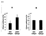
- FIG. 3 shows the substance permeability test results of atelocollagen and gelatin hydrogel sheets (A: albumin, B: glucose).
- FIG. 1 shows photographic images comparing the transparency of gelatin hydrogel sheets and atelocollagen sheets.
- FIG. 2 shows the evaluation results of the mechanical properties of the gelatin hydrogel sheet and the atelocollagen sheet (A: strength against tensile test of each sheet (from the graph left, 48 h
- FIG. 4 shows the evaluation results of cell adhesion (phase contrast microscopic image) and barrier function improvement (ZO-1 expression) due to different coating methods on gelatin hydrogel sheets cultured with corneal endothelial cells (from the left, ( a) Phase contrast microscopic image (Day 1), (b) ZO-1 immunofluorescence staining (Day 10); A: Sheet immersed in 0.15 mg / ml collagen solution (conventional method), B: 3.0 mg / ml collagen Sheet coated with solution, C: atelocollagen (comparative control)).
- FIG. 5 shows the evaluation results of the transparency of gelatin hydrogel sheets cultured with corneal endothelial cells (left: atelocollagen, right: gelatin hydrogel sheet).
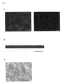
- FIG. 6 shows evaluation of corneal endothelial cells cultured on a gelatin hydrogel sheet (A: Na + / K + -ATPase and ZO-1 expression analysis (immunostaining image), B: HE staining image, C: scanning microscope Image, each scale represents 200 ⁇ m).
- FIG. 7 shows HE-stained images of gelatin hydrogel sheets before (a) transplantation and after (b) transplantation in the rabbit anterior chamber (each scale shows 200 ⁇ m).
- FIG. 8 shows (a) 7 days after transplantation and (b) 21 days after transplantation of the corneal thickness of a cultured corneal endothelial cell sheet using a gelatin hydrogel sheet for a rabbit bullous keratopathy model. Results of comparison between eyes and normal eyes are shown.
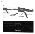
- FIG. 9 shows a HE-stained image (A) and a DiI-stained image (B) of 21 after transplantation of a cultured corneal endothelial cell sheet using a gelatin hydrogel sheet for a rabbit vesicular keratopathy model.
- the right figure is an enlarged view of the box part of the left figure.
- “Gelatin hydrogel” is a gelatin reaction between gelatin molecules by applying chemical reaction, thermal dehydration treatment, radiation, ultraviolet rays, electron beam irradiation, etc. to gelatin. It is a hydrogel obtained by forming a crosslink.
- gelatin means denatured collagen that is irreversibly changed to a water-soluble protein by cleavage of salt bonds or hydrogen bonds between peptide chains of collagen by acid or alkali, or enzymatic treatment.
- the gelatin used in the present invention may be either acidic gelatin or basic gelatin.
- “acidic gelatin” means gelatin having an isoelectric point prepared by alkali treatment of collagen and having an isoelectric point of less than 7.0 and not less than 2.0, preferably not more than 6.5 and not less than 4.0, more preferably 5. 5 or more and 4.5 or more are intended.
- the “basic gelatin” is gelatin having an isoelectric point of 7.0 or more and 13.0 or less prepared by acid treatment of collagen, preferably 7.5 or more and 10.0 or less, more preferably 8.5 or more. Less than 9.5 is contemplated.
- IEP Nitta Gelatin's sample isoelectric point
- Nitta Gelatin's sample IEP 9.0 or the like can be used as basic gelatin. can do.
- gelatin is used is appropriately selected according to the medicinal component to be blended and the use.
- bFGF has an IEP of 4.6, so acidic gelatin is used when blending such drugs.
- basic gelatin is used when a protein drug having an IEP of 7 or less is blended.
- the degree of crosslinking of gelatin can be appropriately selected according to the desired level of biodegradability, moisture content, and bioabsorbability.
- the cross-linking may be any part of the collagen constituting the gelatin, but it is particularly preferable to cross-link the carboxyl group and the hydroxyl group, the carboxyl group and the ⁇ -amino group, or the ⁇ -amino group.
- the composite has the desired mechanical strength properties.
- the degradation rate (remaining period) in the living body can also be controlled by the introduction rate of crosslinking.
- concentration of gelatin and the crosslinking agent and the crosslinking time increase, the degree of crosslinking of the hydrogel increases and the bioabsorbability decreases.
- Cross-linking of gelatin is performed by methods such as thermal reaction (thermal dehydration, etc.), cross-linking by chemical methods using cross-linking agents and condensing agents, and cross-linking by physical methods using gamma rays, ultraviolet rays, electron beams, etc. Can do.
- examples of the crosslinking agent used include aldehyde-based crosslinking agents such as glutaraldehyde and formaldehyde; isocyanate-based crosslinking agents such as hexamethylene diisocyanate; 1-ethyl-3- (3-dimethylaminopropyl) Examples thereof include carbozide-based crosslinking agents such as carbodiimide hydrochloride; polyepoxy-based crosslinking agents such as ethylene glycol diethyl ether; transglutaminase and the like.
- the amount of the crosslinking agent to be added is appropriately set depending on the crosslinking agent to be used.
- the degree of crosslinking can be appropriately selected according to the desired water content, that is, the level of bioabsorbability of the hydrogel.
- the preferred range of the gelatin and cross-linking agent concentration when preparing the hydrogel is a gelatin concentration of 1 to 20 w / w% and a cross-linking agent concentration of 0.01 to 1 w / w%. is there.
- the crosslinking reaction conditions are not particularly limited, and can be carried out, for example, at 0 to 40 ° C., preferably 25 to 30 ° C., for 1 to 48 hours, preferably 12 to 24 hours.
- Crosslinking may be performed by any method, but in the present invention, it is preferable to perform thermal crosslinking (thermal dehydration treatment or the like). This is because, when chemical crosslinking is performed, the crosslinking agent may remain in the hydrogel, or depending on the conditions, only the hydrogel surface may be crosslinked and uniform crosslinking may not be possible. On the other hand, in the case of thermal cross-linking, uniform cross-linking is formed throughout the gelatin hydrogel, and the desired biodegradability is achieved.
- thermal crosslinking uniform cross-linking is formed throughout the gelatin hydrogel, and the desired biodegradability is achieved.
- the crosslinking is formed by a thermal reaction, specifically, it can be carried out under a vacuum condition at 140 ° C. to 160 ° C. under conditions ranging from 6 hours to 72 hours. It is possible to control the degradation rate (remaining period) in the body.
- gelatin hydrogel prepared by vacuum heating at 160 ° C. and 0.01 Torr for 72 hours was used.
- the degradability of gelatin hydrogel can be adjusted by the degree of crosslinking and the thickness of the sheet. Degradability can be controlled within the range of 14 days to 1 year or more in an animal body, for example, the anterior eye segment. In the present invention, if the degradability is too early, it cannot play a role as a scaffold for corneal endothelial cells, and corneal endothelial cell engraftment is considered to be poor. Accordingly, the degradability is preferably set to 14 days to 6 months, particularly about 1 to 3 months. Alternatively, if the gelatin hydrogel has a good engraftment property and does not drop off, the degradation time may be set to a period in which almost no biodegradation occurs for one year or longer.
- the degree of crosslinking of gelatin can be evaluated using the water content as an index.
- the water content is the weight percent of water in the hydrogel relative to the weight of the swollen hydrogel. If the water content is large, the degree of crosslinking of the hydrogel is low and it is easily decomposed.
- the water content showing preferable decomposability is about 92 to 99 w / w%, and more preferably about 95 to 98 w / w%.
- the gelatin hydrogel sheet of the present invention can be molded into any shape, but is molded on the sheet in the present invention for application to a carrier for transplanting cultured corneal endothelial cells.
- the thickness of the sheet is preferably about 10 to 200 ⁇ m, more preferably about 30 to 100 ⁇ m, considering application to corneal endothelial transplantation.
- gelatin hydrogel of the present invention has excellent transparency, and its transparency is much higher than atelocollagen or the like conventionally used as a carrier. Moreover, gelatin hydrogel can maintain its transparency even after application to the affected area.
- the corneal endothelial cell used in the present invention includes cells contained in the corneal endothelial cell layer, and includes corneal endothelial stem cells and corneal endothelial progenitor cells. .
- the corneal endothelial cell may be a donor other than a patient, for example, a corneal endothelium of a human imported eye bank cornea, or a corneal endothelial cell derived from a patient receiving a transplant.
- corneal endothelial cells do not proliferate in vivo as described above, so it is technically possible to collect corneal endothelial cells from healthy patient eyes However, it is difficult in practice.
- Corneal endothelial cells induced to differentiate from stem cells Corneal endothelial cells are corneal endothelial cells, tissue stem cells other than corneal endothelial stem cells or embryonic stem cells (ES cells), and corneal endothelial cells derived from induced pluripotent stem cells. May be.
- the method for preparing corneal endothelial stem cells and induced pluripotent stem cells to be used is not particularly limited, but is preferably a cell derived from a patient who needs treatment.
- Artificial pluripotent stem cells are cells that have been reprogrammed (initialized) to have the same differentiation pluripotency as ES cells by introducing specific factors into mammalian somatic cells or undifferentiated stem cells.
- “Artificial pluripotent stem cells” were first established by Yamanaka et al. By introducing four factors Oct3 / 4, Sox2, Klf4, and c-Myc into mouse fibroblasts.
- IPS cells induced Pluripotent Stem Cell
- iPS cells established by introducing similar four factors into human fibroblasts (Takahashi K, Yamanaka S., et al. Cell, (2007) 131: 861-872.), Furthermore, iPS cells (Nakagawa M, Yamanaka S., et al. Nature Biotechnology, (2008) 26, 101-106) produced by a method not containing c-Myc can also be used.
- an artificial pluripotent stem cell (Yu J., Thomson JA. Et al., Produced by introducing 4 genes of OCT3 / 4, SOX2, NANOG, and LIN28 into human fibroblasts by Thomson et al. Of University of Wisconsin. Science (2007) 318: 1917-1920.), Daley et al. Of Harvard University, and introduced artificial genes produced by introducing 6 genes of OCT3 / 4, SOX2, KLF4, C-MYC, hTERT, SV40 large T into skin cells.
- Ability stem cells (Park IH, Daley GQ.
- artificial pluripotent stem cells (Shi Y., Ding S., et al., Cell Stem Cell, (2008)) produced by introducing OCT3 / 4, KLF4, low molecular weight compounds into mouse neural progenitor cells, etc. Vol3, Issue 5, 568-574), induced pluripotent stem cells (Kim JB., Produced by introducing OCT3 / 4, KLF4 into mouse neural stem cells endogenously expressing SOX2, C-MYC.
- Induction of differentiation of corneal endothelial cells from tissue stem cells, ES cells, or induced pluripotent stem cells may be direct, or may be indirect via neural crest cells or corneal parenchymal stem cells. That is, tissue stem cells, ES cells, and induced pluripotent stem cells are once induced to differentiate into neural crest cells or corneal parenchymal stem cells, and further differentiated from these neural crest cells or corneal parenchymal stem cells with TGFb2 or the like according to a known technique. Endothelial cells are induced (Japanese Patent Application No. 2008-123562).
- the SDIA method is a method of inducing differentiation of neural crest cells from ES cells by using mouse-derived stromal cells (PA6 cells) as feeder cells (Kawasaki, H., Sasai, Y. et al. (2000). ) Neuron 28, 31-40., Kawasaki, H., Sasai, Y. et al. (2002) Proc. Natl. Acad. Sci. USA 99, 1580-1585, Mizuseki, K., Sasai, Y. et al.
- PA6 cells mouse-derived stromal cells
- SFEB Serum-free Floating culture of Embryoid Body-like aggregates
- the corneal transplant sheet of the present invention is coated with the aforementioned gelatin hydrogel sheet with collagen (type I or type IV) and seeded with corneal endothelial cells and cultured. It is produced by this.
- Collagen coating on gelatin hydrogel is performed for the purpose of improving cell adhesion and corneal endothelial function.
- the collagen used is preferably type I or type IV, and is preferably atelocollagen from which antigenicity has been removed.
- Collagen coding can be performed by diluting collagen 10 times with dilute hydrochloric acid (pH 3.0), spreading thinly on gelatin hydrogel, and drying according to a conventional method.
- the coated sheet is preferably washed with Phosphate-Buffered Salines (PBS) (Invitrogen) before use.
- PBS Phosphate-Buffered Salines
- composition of the aqueous solution can be used in any kinds of things.
- the coating of collagen is performed by a method of applying (coating) the collagen solution to the sheet surface, not by a conventional method of immersing in a collagen solution.
- the cell adhesiveness and barrier function of the sheet are remarkably improved.
- the coated sheet is dried to fix the coated collagen in the vicinity of the gelatin sheet surface. This fixation to the surface is important for cell adhesion and function expression. Without coating, it is thought that cell adhesion and endothelial function are reduced, so that it is desirable to carry out coating.
- a medium usually used for culturing adherent cells can be performed using a medium usually used for culturing adherent cells.
- a medium usually used for culturing adherent cells For example, DMEM medium, BME medium, ⁇ MEM medium, Dulbecco MEM medium, BGJb medium, CMRL 1066 medium, Glasgow MEM medium, Improved MEM Zinc Option medium, IMDM medium, Medium 199 medium, Eagle MEM medium, RPMI medium 16, RPM medium 16 Any medium that can be used for culturing animal cells, such as Fischer's medium, McCoy's medium, Williams E medium, and mixed medium thereof, can be used.
- nutrient sources include serum, basic fibroblast growth factor (bFGF), epidermal growth factor (EGF), glycerol, glucose, fructose, sucrose, lactose, honey, starch, dextrin and other carbon sources, Fatty acids, fats and oils, lecithins, alcohols and other hydrocarbons, ammonium sulfate, ammonium nitrate, ammonium chloride, urea, sodium nitrate and other nitrogen sources, salt, potassium salts, phosphates, magnesium salts, calcium salts, iron salts, manganese salts Inorganic salts such as monopotassium phosphate, dipotassium phosphate, magnesium sulfate, sodium chloride, ferrous sulfate, sodium molybdate, sodium tungstate and manganese sulfate, various vitamins, amino acids and the like.
- bFGF basic fibroblast growth factor
- EGF epidermal growth factor
- glycerol glucose
- amino acid reducing agents such as pyruvic acid and ⁇ -mercaptoethanol, serum substitutes, etc.
- serum substitutes include albumin (for example, lipid-rich albumin), transferrin, fatty acid, insulin, collagen precursor, trace element, ⁇ -mercaptoethanol or 3 ′ thiol glycerol, commercially available Knockout Serum Replacement (KSR), and Chemically.
- KSR Knockout Serum Replacement
- the pH of the medium obtained by blending these components is in the range of 5.5 to 9.0, preferably 6.0 to 8.0, and more preferably 6.5 to 7.5.
- the gelatin hydrogel comprising 3.3 culture conditions the above medium, the seeding density of the corneal endothelial cells 500 ⁇ 4500cells / mm 2, preferably seeded with a seeding density of 1500 ⁇ 3500cells / mm 2, 36 °C ⁇ 38 °C,
- the culture is preferably performed at 36.5 ° C. to 37.5 ° C. under the conditions of 1% to 25% O 2 and 1% to 15% CO 2 .
- the number of culture days is at least 3 days, preferably 7 days or more after the cells are confluent.
- the cell density in the finally obtained gelatin hydrogel is 2000 cells / mm 2 or more, preferably 2500 cells / mm 2 or more, more preferably 3000 cells / mm 2 or more.
- Corneal Transplant Sheet Since the corneal transplant sheet of the present invention uses gelatin hydrogel as a support, the strength and operability of the corneal endothelial cell layer having a thickness of only about 10 ⁇ m are remarkably improved. Corneal endothelial cells on the sheet express a tight junction protein ZO-1 that exhibits a barrier function, and the adhesion of the cells to the sheet is good. Since the sheet can be peeled off from the culture vessel without applying an enzyme treatment such as trypsin or dispase, the desmosome structure of the cells is maintained, and the original structure and strength of the corneal endothelial cell layer are maintained.
- an enzyme treatment such as trypsin or dispase
- the corneal transplant sheet of the present invention has much higher transparency than the conventionally known corneal transplant sheet using atelocollagen as a support. Is maintained after transplantation.
- the corneal transplant sheet of the present invention can contain a medicinal component as necessary.
- medicinal components include antitumor agents, antibacterial agents, anti-inflammatory agents, antiviral agents, anti-AIDS agents, low molecular drugs such as hormones, bioactive peptides and proteins containing bone morphogenetic or bone growth factors , Glycoproteins, polysaccharides, nucleic acids and the like.
- These medicinal ingredients may be natural substances or synthetically produced substances.
- epidermal growth factor EGF
- fibroblast growth factor FGF
- platelet-derived growth factor PDGF
- HGF hepatocyte growth factor
- TGF insulin-like growth factor
- BGF-2, BMP-4, BMP-5, BMP-6, BMP-7 (OP-1) and BMP-8 (OP-2) and other bone growth factors such as IGF
- BMP Forming protein
- GDNF glial-induced neurotrophic factor
- NF neurotrophic factor
- PRP platelet rich plasma
- Anticancer drugs such as ifosfamide, antibiotics such as streptomycin, gentamicin, gatifloxacin, atorvastatin (ato Cholesterol lowering agents such as vastatin, pravastatin, simvastatin, lidocaine, protamine sulfate, sodium iodohypurate, iodosulfopro
- the time when the gelatin hydrogel is impregnated with the above-mentioned medicinal component can be before culturing corneal endothelial cells on the gel, during culturing, or before or after preparing the transplantation sheet.
- the compounding ratio of the medicinal component to the gelatin hydrogel is preferably about 5 times or less by molar ratio. More preferably, the molar ratio is about 5 to about 1/10 4 times.
- This impregnation operation is usually completed at 4 to 37 ° C. for 15 minutes to 1 hour, preferably at 4 to 25 ° C. for 15 to 30 minutes, during which time the gelatin hydrogel swells with a solution containing a medicinal component, It is combined with the gel by physicochemical interaction and fixed in the hydrogel.
- the medicinal component and gelatin hydrogel can be bonded between the functional group of the drug or the metal and the functional group on the hydrogel. It is considered that a coordination bond of is involved alone or in combination.
- the medicinal component is gradually released to the outside of the sheet as the gelatin hydrogel is decomposed in vivo and the gelatin molecules are water-solubilized.
- This release rate is determined by the degree of degradation and absorption in the living body of the gelatin hydrogel used, and the degree and stability of the bond strength between the medicinal ingredient and the gelatin hydrogel in the complex.
- the degree of degradation and absorption of gelatin hydrogel in the living body can be adjusted by adjusting the degree of crosslinking during the preparation of the hydrogel.
- the gelatin hydrogel when a negatively charged substance such as nucleic acid is used as the medicinal component, the gelatin hydrogel is positively charged so that a stable complex of the medicinal component and the gelatin hydrogel is formed. It is preferable.
- a stable gelatin hydrogel complex is formed by a strong bond (ionic bond) between the negative charge of the medicinal component and the positive charge of the gelatin hydrogel.
- it can be cationized by previously introducing an amino group or the like into the gelatin hydrogel. This increases the binding force between the gelatin hydrogel and the medicinal component, and a more stable gelatin hydrogel complex can be formed.
- the medicinal component is a substance having a positive charge
- a stable complex of the medicinal component and gelatin hydrogel is formed, and gelatin is anionized into an interaction.
- the medicinal component interacts with gelatin and is stably complexed and fixed in the hydrogel.
- the cationization step is not particularly limited as long as it is a method capable of introducing a functional group that can be cationized under physiological conditions, but a 1, 2 or tertiary amino group or ammonium group is added to the hydroxyl group or carboxyl group of gelatin. A method of introducing under mild conditions is preferred.
- alkyldiamines such as ethylenediamine, N, N-dimethyl-1,3-diaminopropane, trimethylammonium acetohydrazide, spermine, spermidine, diethylamide chloride, and the like can be used with various condensing agents such as 1-ethyl-3- (3 -Dimethylaminopropyl) carbodiimide hydrochloride, cyanuric chloride, N, N'-carbodiimidazole, cyanogen bromide, diepoxy compound, tosyl chloride, diethyltriamine-N, N, N ', N ", N" -pentane
- dianhydride compounds such as an acid dianhydride, a trisyl chloride, etc.
- the method of reacting ethylenediamine is preferred because it is simple and versatile.
- the medicinal component-containing corneal transplant sheet has a sustained release effect and a stabilizing effect of the medicinal component, and thus can release the medicinal component over a long period of time in a controlled direction at a desired site. Therefore, the action of the medicinal component is effectively exhibited within the lesion site.
- corneal transplant recipients There are 1 million corneal transplant recipients worldwide, and tens of thousands in Japan. Among them, bullous keratopathy patients caused by corneal endothelium disorders account for about 80% of all corneal transplant diseases. Since the corneal endothelium does not regenerate once damaged, there is currently no effective treatment method other than corneal transplantation.
- the corneal transplant sheet of the present invention has a simple manufacturing method, excellent operability, engraftment, and degradability, and also has a sustained drug release effect as described above. If a patient-derived corneal endothelial cell is used, the problem of rejection is avoided and the corneal endothelium can be rapidly regenerated. Furthermore, since gelatin hydrogel also has a function of slowly releasing medicinal ingredients, it can also contain a medicinal ingredient having biological activity and release it for a necessary period of time to further promote the healing of the disease.
- Example 1 Preparation of gelatin hydrogel sheet Extracted from collagen by pork skin or cow bone by alkali treatment (molecular weight 98,000 isoelectric point 5.0). A 10 WT% aqueous solution of gelatin was prepared. This was poured into a plastic petri dish and left at room temperature for several days to evaporate water and obtain a gelatin sheet. This was subjected to thermal dehydration treatment at 160 ° C. and 0.01 Torr for 72 hours to chemically crosslink between gelatin molecules. The obtained gelatin sheet had a water content of 97% and a thickness of 100 ⁇ m. The thickness can be adjusted from 30 ⁇ m to depending on the purpose.
- Substance permeability The substance permeability of the gelatin hydrogel sheet (48 hours cross-linked) was compared with that of the atelocollagen sheet. The substance permeability was evaluated by determining the diffusion coefficient for albumin and glucose according to a conventional method.
- the gelatin hydrogel sheet showed significantly higher albumin permeability than the atelocollagen sheet.
- the gelatin hydrogel sheet was equivalent to the atelocollagen sheet for glucose permeability. From this, the gelatin hydrogel sheet was considered useful as a carrier sheet.
- Example 2 Cell culture on gelatin hydrogel sheet Preparation of Corneal Endothelial Cells
- a cornea piece was prepared from a rabbit eyeball and the Descemet's membrane was peeled off. Descemet's membrane was incubated with 0.25% Trypsin-EDTA at 37 ° C. for 10 minutes to isolate cells.
- the cell mass was suspended in the same medium.
- the gelatin hydrogel sheet prepared in Example 1 was coated with collagen type I or type IV (Nitta gelatin).
- a 3.0 mg / ml collagen solution stock solution (pH 3.0) is directly applied onto the gelatin hydrogel sheet prepared in Example 1 using a cell scraper or the like, and diluted 10-fold with dilute hydrochloric acid.
- Coating was performed by immersing a gelatin hydrogel sheet in the collagen solution thus prepared, and allowing it to stand for 30 minutes or more in a clean bench.
- the coated sheet was washed 3 times with Phosphate-Buffered Salines (PBS) (Invitrogen) before use.
- PBS Phosphate-Buffered Salines
- corneal endothelial cells isolated from Descemet's membrane were seeded at a seeding density of 1.5 to 4.5 ⁇ 10 3 cells / mm 2 , 37 ° C., 10% CO 2 , containing serum used in the previous section. For 14 days. For comparison, corneal endothelial cells were seeded and cultured on atelocollagen under the same conditions.
- Example 3 Rabbit anterior chamber transplantation of gelatin hydrogel sheet (examination of biodegradability) After the rabbit lens extraction (ultrasonic emulsification), the Descemet's membrane was peeled off. Thereafter, only the gelatin hydrogel sheet punched with 8 mm trepan was transplanted into the anterior chamber.
- Example 4 Transplantation of cultured corneal endothelial cell sheet using gelatin hydrogel sheet to rabbit bullous keratopathy model Rabbit-derived corneal endothelial cells were cultured on gelatin hydrogel and transplanted based on the method of Example 2 A cultured corneal endothelial cell sheet was prepared. The prepared cultured corneal endothelial cell sheet was transplanted into a rabbit bullous keratopathy model from which the corneal endothelium had been completely removed. After transplantation, corneal thickness measurement and anterior ocular segment observation using a pachymeter were performed.
- HE staining FIG. 9A
- DiI staining FIG. 9B
- the corneal transplant sheet using the gelatin hydrogel of the present invention as a support can be transplanted without impairing the function and form of the endothelial cells.
- the sheet for corneal transplantation of the present invention is excellent in transparency after transplantation and engraftment in tissue. Moreover, the biodegradation rate in the living body can be adjusted according to the production conditions, and there is no problem such as an inflammatory reaction.
- gelatin hydrogel as a material has already been used clinically. Therefore, it is extremely useful as a treatment method for corneal endothelial disease as an alternative to conventional full-thickness corneal transplantation. Since the number of patients with bullous keratopathy and the like that are the target of treatment using the corneal transplant sheet of the present invention is expected to reach tens of thousands of years in Japan, the treatment method is highly valuable for commercialization.
Landscapes
- Health & Medical Sciences (AREA)
- Life Sciences & Earth Sciences (AREA)
- Chemical & Material Sciences (AREA)
- Veterinary Medicine (AREA)
- Public Health (AREA)
- General Health & Medical Sciences (AREA)
- Animal Behavior & Ethology (AREA)
- Medicinal Chemistry (AREA)
- Transplantation (AREA)
- Epidemiology (AREA)
- Oral & Maxillofacial Surgery (AREA)
- Dermatology (AREA)
- Engineering & Computer Science (AREA)
- Biomedical Technology (AREA)
- Cell Biology (AREA)
- Chemical Kinetics & Catalysis (AREA)
- Zoology (AREA)
- Urology & Nephrology (AREA)
- Botany (AREA)
- Nuclear Medicine, Radiotherapy & Molecular Imaging (AREA)
- Ophthalmology & Optometry (AREA)
- Pharmacology & Pharmacy (AREA)
- General Chemical & Material Sciences (AREA)
- Organic Chemistry (AREA)
- Bioinformatics & Cheminformatics (AREA)
- Materials For Medical Uses (AREA)
- Medicinal Preparation (AREA)
Abstract
Description
1)培養角膜内皮細胞を移植する際に内皮細胞のもつ機能・形態を損なうことなく移植することが可能。
2)実質への生着性が良好でありシートの脱落が起きない、又は、速やかに生分解されるためシートが前房内に脱落しないため移植した角膜内皮への傷害も起きない。
3)移植後の透明性・生体適合性も良好であり、炎症反応等が無く、角膜透明性も維持されている。 The inventors searched for a material satisfying the following conditions as a transplant base material used for corneal transplantation and selected gelatin hydrogel.
1) When transplanting cultured corneal endothelial cells, it can be transplanted without impairing the functions and morphology of the endothelial cells.
2) It has good adherence to the substance, and the sheet does not fall off, or because it is rapidly biodegraded, the sheet does not fall into the anterior chamber, so that the transplanted corneal endothelium is not damaged.
3) Transparency and biocompatibility after transplantation are good, there is no inflammatory reaction, and corneal transparency is maintained.
すなわち、本発明は、ゼラチンハイドロゲルに角膜内皮細胞を含んでなる角膜移植用シートに関する。 Then, gelatin was processed into a sheet shape and subjected to crosslinking treatment to produce a gelatin hydrogel sheet, and corneal endothelial cells were cultured using this as a support. Furthermore, this sheet was transplanted to the anterior segment of a bullous keratosis model mouse, and its degradability and therapeutic effect were evaluated. As a result, it was confirmed that the corneal transplant sheet using gelatin hydrogel as a support showed high cell engraftment and transparency, and the progress after transplantation was very good.
That is, the present invention relates to a corneal transplant sheet comprising corneal endothelial cells in gelatin hydrogel.
本発明で用いられる角膜内皮細胞は、患者由来の細胞から調製されたものであることが好ましい。 The gelatin hydrogel used in the present invention is prepared by subjecting gelatin to a heat crosslinking treatment. Gelatin hydrogel is degraded in vivo after transplantation, and the degradation rate can be adjusted by the degree of crosslinking and the like. That is, the gelatin hydrogel may be adjusted so that it degrades and disappears in vivo within 1 to 6 months after transplantation, or it is adjusted so that it engrafts in the real tissue without biodegrading for more than one year. Also good.
The corneal endothelial cell used in the present invention is preferably prepared from a patient-derived cell.
1.1 ゼラチンハイドロゲルとは
本発明にかかる「ゼラチンハイドロゲル」とは、ゼラチンに化学反応、熱脱水処理、放射線、紫外線、あるいは電子線照射等を与えることによりゼラチン分子間に架橋を形成させて得られるハイドロゲルのことである。 1. Seratin hydrogel 1.1 What is gelatin hydrogel? “Gelatin hydrogel” according to the present invention is a gelatin reaction between gelatin molecules by applying chemical reaction, thermal dehydration treatment, radiation, ultraviolet rays, electron beam irradiation, etc. to gelatin. It is a hydrogel obtained by forming a crosslink.
ゼラチンの架橋度は、所望の生体内分解性や含水率、生体吸収性のレベルに応じて適宜選択することができる。架橋は、ゼラチンを構成するコラーゲンのどの部分を架橋するものであってもよいが、特にカルボキシル基と水酸基、カルボキシル基とε−アミノ基、ε−アミノ基同士を架橋することが好ましい。こうして架橋を導入することにより、複合体は所望の機械的強度特性を有するようになる。また、架橋の導入率によって、生体内での分解速度(残存期間)も制御することができる。一般に、ゼラチン及び架橋剤の濃度、架橋時間が増大するとともにハイドロゲルの架橋度は増加し、生体吸収性は低くなる。 1.2 Crosslinking of gelatin The degree of crosslinking of gelatin can be appropriately selected according to the desired level of biodegradability, moisture content, and bioabsorbability. The cross-linking may be any part of the collagen constituting the gelatin, but it is particularly preferable to cross-link the carboxyl group and the hydroxyl group, the carboxyl group and the ε-amino group, or the ε-amino group. By introducing cross-linking in this way, the composite has the desired mechanical strength properties. Further, the degradation rate (remaining period) in the living body can also be controlled by the introduction rate of crosslinking. In general, as the concentration of gelatin and the crosslinking agent and the crosslinking time increase, the degree of crosslinking of the hydrogel increases and the bioabsorbability decreases.
ゼラチンハイドロゲルの分解性は、前述した架橋度とシートの厚さによって調整することができる。分解性は動物体内、例えば前眼部であれば、14日~1年以上の範囲でコントロールが可能である。本発明では分解性が早すぎると角膜内皮細胞の足場としての役割が果たせず、角膜内皮細胞の生着が不良になると考えられる。そこで分解性としては14日~6ヶ月、特に1~3ヶ月程度で設定することが好ましい。又は、ゼラチンハイドロゲルの実質への生着性が良好であり脱落が起きなければ、分解時間を1年以上のほぼ生分解が起きない期間に設定することも考えられる。 1.3 Biodegradability The degradability of gelatin hydrogel can be adjusted by the degree of crosslinking and the thickness of the sheet. Degradability can be controlled within the range of 14 days to 1 year or more in an animal body, for example, the anterior eye segment. In the present invention, if the degradability is too early, it cannot play a role as a scaffold for corneal endothelial cells, and corneal endothelial cell engraftment is considered to be poor. Accordingly, the degradability is preferably set to 14 days to 6 months, particularly about 1 to 3 months. Alternatively, if the gelatin hydrogel has a good engraftment property and does not drop off, the degradation time may be set to a period in which almost no biodegradation occurs for one year or longer.
本発明のゼラチンハイドロゲルシートはいずれの形状にも成型可能であるが、培養角膜内皮細胞移植用キャリアへの適用のため、本発明においてはシート上に成型する。シートの厚さは、角膜内皮移植への適用を考慮すると、10~200μm程度が好ましく、30~100μm程度がより好ましい。 1.4 Shape of gelatin hydrogel The gelatin hydrogel sheet of the present invention can be molded into any shape, but is molded on the sheet in the present invention for application to a carrier for transplanting cultured corneal endothelial cells. The thickness of the sheet is preferably about 10 to 200 μm, more preferably about 30 to 100 μm, considering application to corneal endothelial transplantation.
本発明のゼラチンハイドロゲルは、優れた透明性を有し、従来キャリアとして用いられているアテロコラーゲン等に比較して、その透明度は格段に高い。また、ゼラチンハイドロゲルは、患部に適用後においても、その透明性を維持しうる。 1.5 Transparency of gelatin hydrogel The gelatin hydrogel of the present invention has excellent transparency, and its transparency is much higher than atelocollagen or the like conventionally used as a carrier. Moreover, gelatin hydrogel can maintain its transparency even after application to the affected area.
2.1 ドナーあるいは患者由来の角膜内皮細胞
本発明で用いられる角膜内皮細胞は、角膜内皮細胞層に含まれる細胞を包括したものであり、角膜内皮幹細胞、角膜内皮前駆細胞をも含む。また角膜内皮細胞は、患者以外のドナー、例えばヒト輸入アイバンク角膜の角膜内皮を用いてもよいし、移植を受ける患者由来の角膜内皮細胞を用いてもよい。拒絶反応を防止する点では患者由来の細胞を用いることが望ましいが、前述のように角膜内皮細胞は生体内では増殖しないため、患者健常眼から角膜内皮細胞を採取することは技術的には可能であるが、現実的には困難である。 2. Corneal Endothelial Cell 2.1 Corneal Endothelial Cell Derived from Donor or Patient The corneal endothelial cell used in the present invention includes cells contained in the corneal endothelial cell layer, and includes corneal endothelial stem cells and corneal endothelial progenitor cells. . The corneal endothelial cell may be a donor other than a patient, for example, a corneal endothelium of a human imported eye bank cornea, or a corneal endothelial cell derived from a patient receiving a transplant. Although it is desirable to use patient-derived cells in terms of preventing rejection, corneal endothelial cells do not proliferate in vivo as described above, so it is technically possible to collect corneal endothelial cells from healthy patient eyes However, it is difficult in practice.
角膜内皮細胞は、角膜内皮幹細胞、角膜内皮幹細胞以外の組織幹細胞あるいは胚性幹細胞(ES細胞)、人工多能性幹細胞から誘導された角膜内皮細胞を利用してもよい。用いられる角膜内皮幹細胞や人工多能性幹細胞の調製方法は特に限定されないが、治療を必要とする患者自身に由来する細胞であることが好ましい。 2.2 Corneal endothelial cells induced to differentiate from stem cells Corneal endothelial cells are corneal endothelial cells, tissue stem cells other than corneal endothelial stem cells or embryonic stem cells (ES cells), and corneal endothelial cells derived from induced pluripotent stem cells. May be. The method for preparing corneal endothelial stem cells and induced pluripotent stem cells to be used is not particularly limited, but is preferably a cell derived from a patient who needs treatment.
本発明の角膜移植用シートは、前述のゼラチンハイドロゲルシートをコラーゲン(タイプIあるいはタイプIV)でコーティングし、その上に角膜内皮細胞を播種して培養する
ことにより作製される。 3. Culture of Corneal Endothelial Cells on Gelatin Hydrogel The corneal transplant sheet of the present invention is coated with the aforementioned gelatin hydrogel sheet with collagen (type I or type IV) and seeded with corneal endothelial cells and cultured. It is produced by this.
ゼラチンハイドロゲル上へのコラーゲンのコーティングは、細胞接着性及び角膜内皮機能の改善を目的として行う。用いるコラーゲンはタイプIあるいはタイプIVが好ましく、抗原性を除去したアテロコラーゲンであることが好ましい。コラーゲンコーディングは、常法にしたがって、コラーゲンを希塩酸(pH3.0)で10倍希釈しゼラチンハイドロゲル上に薄く塗り広げ、乾燥させることで実施できる。コーティングしたシートは、使用前にPhosphate−Buffered Salines(PBS)(Invitrogen)で洗浄することが好ましい。 3.1 Collagen coating Collagen coating on gelatin hydrogel is performed for the purpose of improving cell adhesion and corneal endothelial function. The collagen used is preferably type I or type IV, and is preferably atelocollagen from which antigenicity has been removed. Collagen coding can be performed by diluting
ゼラチンハイドロゲルシート上での培養は、接着細胞の培養に通常用いられる培地を用いて行うことができる。例えば、DMEM培地、BME培地、α MEM培地、Dulbecco MEM培地、BGJb培地、CMRL 1066培地、Glasgow MEM培地、Improved MEM Zinc Option培地、IMDM培地、Medium 199培地、Eagle MEM培地、ハム培地、RPMI 1640培地、Fischer’s培地、McCoy’s培地、ウイリアムスE培地、及びこれらの混合培地など、動物細胞の培養に用いることのできる培地であればいずれも用いることができる。 3.2 Medium Culture on a gelatin hydrogel sheet can be performed using a medium usually used for culturing adherent cells. For example, DMEM medium, BME medium, α MEM medium, Dulbecco MEM medium, BGJb medium, CMRL 1066 medium, Glasgow MEM medium, Improved MEM Zinc Option medium, IMDM medium, Medium 199 medium, Eagle MEM medium, RPMI medium 16, RPM medium 16 Any medium that can be used for culturing animal cells, such as Fischer's medium, McCoy's medium, Williams E medium, and mixed medium thereof, can be used.
上記の培地を含むゼラチンハイドロゲルに、角膜内皮細胞を500~4500cells/mm2の播種密度、好ましくは1500~3500cells/mm2の播種密度で播種し、36℃~38℃、好ましくは36.5℃~37.5℃で、1%~25%O2、1%~15%CO2の条件下で培養する。培養日数は、細胞がコンフルエントになってから少なくとも3日以上、好ましくは7日以上であることがよい。最終的に得られるゼラチンハイドロゲル内の細胞密度は、2000cells/mm2以上、好ましくは2500cells/mm2以上、より好ましくは3000cells/mm2以上である。 The gelatin hydrogel comprising 3.3 culture conditions the above medium, the seeding density of the corneal endothelial cells 500 ~ 4500cells / mm 2, preferably seeded with a seeding density of 1500 ~ 3500cells / mm 2, 36 ℃ ~ 38 ℃, The culture is preferably performed at 36.5 ° C. to 37.5 ° C. under the conditions of 1% to 25% O 2 and 1% to 15% CO 2 . The number of culture days is at least 3 days, preferably 7 days or more after the cells are confluent. The cell density in the finally obtained gelatin hydrogel is 2000 cells / mm 2 or more, preferably 2500 cells / mm 2 or more, more preferably 3000 cells / mm 2 or more.
本発明の角膜移植用シートは、ゼラチンハイドロゲルを支持体としているため、単層で厚さ10μm程度しかない角膜内皮細胞層の強度と操作性を格段に向上させる。シート上の角膜内皮細胞は、バリア機能を示すタイトジャンクションタンパクZO−1を発現し、細胞のシートへの接着性は良好である。シートは、トリプシンやディスパーゼ等の酵素処理を施すことなく培養容器から剥離可能であるため、細胞のデスモソーム構造が維持され、角膜内皮細胞層本来の構造と強度が維持される。 4). Corneal Transplant Sheet Since the corneal transplant sheet of the present invention uses gelatin hydrogel as a support, the strength and operability of the corneal endothelial cell layer having a thickness of only about 10 μm are remarkably improved. Corneal endothelial cells on the sheet express a tight junction protein ZO-1 that exhibits a barrier function, and the adhesion of the cells to the sheet is good. Since the sheet can be peeled off from the culture vessel without applying an enzyme treatment such as trypsin or dispase, the desmosome structure of the cells is maintained, and the original structure and strength of the corneal endothelial cell layer are maintained.
豚皮もしくは牛骨よりアルカリ処理によってコラーゲンから抽出した(分子量98,000等電点5.0)。ゼラチンの10WT%水溶液を調整した。これをプラスチックシャーレに流し込み室温で数日放置することで水を蒸発させ、ゼラチンシートを得た。これを160℃0.01Torrで72時間熱脱水処理を行い、ゼラチン分子間を化学架橋した。得られたゼラチンシートは、含水率97%、厚さ:100μm、であった。なお、厚さは目的に応じて、30μm~で調整可能である。 Example 1: Preparation of gelatin hydrogel sheet Extracted from collagen by pork skin or cow bone by alkali treatment (molecular weight 98,000 isoelectric point 5.0). A 10 WT% aqueous solution of gelatin was prepared. This was poured into a plastic petri dish and left at room temperature for several days to evaporate water and obtain a gelatin sheet. This was subjected to thermal dehydration treatment at 160 ° C. and 0.01 Torr for 72 hours to chemically crosslink between gelatin molecules. The obtained gelatin sheet had a water content of 97% and a thickness of 100 μm. The thickness can be adjusted from 30 μm to depending on the purpose.
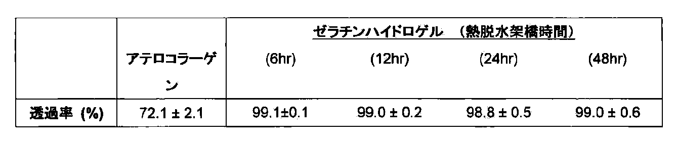
上記のようにして調製したゼラチンハイドロゲルシート(48時間架橋)と、アテロコラーゲンシート(株式会社高研)の透明度を目視で比較した(図1)。さらに、ゼラチンハイドロゲルシートの架橋時間を変えて(6~48時間)、その光透過率を比較した。表1に示すように、いずれの架橋時間においても、ゼラチンハイドロゲルの光透過率はアテロコラーゲンより高かった。
ゼラチンハイドロゲルシートの力学特性をアテロコラーゲンと比較評価した。
架橋時間の異なる(6~48時間架橋)ゼラチンハイドロゲルシート、及びアテロコラーゲンシートの引っ張り試験に対する強度(図2A及びB)とブレークポイント(図2C)を測定するとともに、弾性係数(Elastic modulus)を算出した(図2D)。
ゼラチンハイドロゲルシートは架橋時間依存的に引っ張り強度が増加し、逆に弾性係数は低下する傾向が認められた。また、ゼラチンハイドロゲルシートは、いずれの架橋時間についても、アテロコラーゲンと同等かそれ以上の強度及び弾性を有することが確認された。 (1) Transparency The transparency of the gelatin hydrogel sheet (48 hours cross-linking) prepared as described above and the atelocollagen sheet (Koken Co., Ltd.) were visually compared (FIG. 1). Further, the light transmittance was compared by changing the crosslinking time of the gelatin hydrogel sheet (6 to 48 hours). As shown in Table 1, the light transmittance of gelatin hydrogel was higher than that of atelocollagen at any crosslinking time.
The strength (FIGS. 2A and B) and the breakpoint (FIG. 2C) of the gelatin hydrogel sheet and the atelocollagen sheet with different crosslinking times (crosslinked for 6 to 48 hours) and the breakpoint (FIG. 2C) were measured, and the elastic modulus (Elastic modulus) was calculated. (FIG. 2D).
In the gelatin hydrogel sheet, the tensile strength increased depending on the crosslinking time, whereas the elastic modulus tended to decrease. In addition, it was confirmed that the gelatin hydrogel sheet has strength and elasticity equal to or higher than that of atelocollagen for any cross-linking time.
ゼラチンハイドロゲルシート(48時間架橋)の物質透過性をアテロコラーゲンシートと比較評価した。物質透過性は、常法にしたがいアルブミンとグルコースに対する拡散係数を求めることで評価した。 (3) Substance permeability The substance permeability of the gelatin hydrogel sheet (48 hours cross-linked) was compared with that of the atelocollagen sheet. The substance permeability was evaluated by determining the diffusion coefficient for albumin and glucose according to a conventional method.
1.角膜内皮細胞の調製
家兎眼球より強角膜片を作成し、デスメ膜を剥離した。デスメ膜は、0.25%Trypsin−EDTAと37℃10分インキュベートして、細胞を単離した。2ng/ml bFGF(R&D systems)及び10%血清(Japan bio serum)入りのDMEM培地(Low glucose、日研生物医学研究所)を添加し、300g x 5min遠心分離し、上清を吸引し、残った細胞塊を同様の培地に懸濁させた。 Example 2: Cell culture on gelatin hydrogel sheet Preparation of Corneal Endothelial Cells A cornea piece was prepared from a rabbit eyeball and the Descemet's membrane was peeled off. Descemet's membrane was incubated with 0.25% Trypsin-EDTA at 37 ° C. for 10 minutes to isolate cells. Add 2 ng / ml bFGF (R & D systems) and DMEM medium with 10% serum (Japan bio serum) (Low glucose, Nikken Biomedical Laboratories), centrifuge at 300
実施例1で調製したゼラチンハイドロゲルシートをコラーゲンタイプIもしくはタイプIV(新田ゼラチン)にてコーティングした。コーティング方法としては、3.0mg/mlコラーゲン溶液原液(pH3.0)を、実施例1で作製したゼラチンハイドロゲルシート上に、セルスクレイパー等を用いて直接塗布する方法と、希塩酸で10倍に希釈したコラーゲン溶液中にゼラチンハイドロゲルシートを浸漬する方法で、ともにクリーンベンチ内で30分以上置き静置させることでコーティングした。コーティングしたシートは、使用前にPhosphate−Buffered Salines(PBS)(Invitrogen)で3回洗浄した。 2. Corneal Endothelial Cell Culture and Examination of Coating Conditions The gelatin hydrogel sheet prepared in Example 1 was coated with collagen type I or type IV (Nitta gelatin). As a coating method, a 3.0 mg / ml collagen solution stock solution (pH 3.0) is directly applied onto the gelatin hydrogel sheet prepared in Example 1 using a cell scraper or the like, and diluted 10-fold with dilute hydrochloric acid. Coating was performed by immersing a gelatin hydrogel sheet in the collagen solution thus prepared, and allowing it to stand for 30 minutes or more in a clean bench. The coated sheet was washed 3 times with Phosphate-Buffered Salines (PBS) (Invitrogen) before use.
コラーゲンを塗布したゼラチンハイドロゲルを支持体として培養した角膜内皮細胞シートと、アテロコラーゲンを支持体として培養した角膜内皮細胞シートの透明度を目視で比較した。培養後のアテロコラーゲンは白濁し、透明性が低下しているのに比較して、ゼラチンハイドロゲルシートは培養による透明性の低下は認められなかった(図5)。
ゼラチンハイドロゲル上に培養した角膜内皮細胞は内皮ポンプマーカーNa+/K+−ATPase(図6A)およびタイトジャンクションマーカーであるZO−1(図6B)を発現していた。また走査顕微鏡観察により、内皮細胞に特有の微絨毛が細胞表面に観察された(図6C)。以上のことからゼラチンハイドロゲルシートは角膜内皮細胞の足場として正常な生育をサポートすることが示された。 3. Evaluation of Corneal Endothelial Cell Sheet The transparency of a corneal endothelial cell sheet cultured using a gelatin hydrogel coated with collagen as a support and a corneal endothelial cell sheet cultured using atelocollagen as a support were visually compared. The gelatin hydrogel sheet did not show a decrease in transparency as a result of culturing, compared to that after culturing atelocollagen became cloudy and reduced in transparency (FIG. 5).
Corneal endothelial cells cultured on gelatin hydrogel expressed endothelial pump marker Na + / K + -ATPase (FIG. 6A) and tight junction marker ZO-1 (FIG. 6B). Further, microvilli peculiar to the endothelial cells were observed on the cell surface by scanning microscope observation (FIG. 6C). These results indicate that gelatin hydrogel sheets support normal growth as a scaffold for corneal endothelial cells.
家兎水晶体摘出術(超音波乳化吸引術)後、デスメ膜を剥離した。その後8mmトレパンにて打ち抜いたゼラチンハイドロゲルシートのみを前房内に移植した。 Example 3: Rabbit anterior chamber transplantation of gelatin hydrogel sheet (examination of biodegradability)
After the rabbit lens extraction (ultrasonic emulsification), the Descemet's membrane was peeled off. Thereafter, only the gelatin hydrogel sheet punched with 8 mm trepan was transplanted into the anterior chamber.
移植後28日目では、前房内に移植したシートの透明性及び角膜透明性は共に保たれており、虹彩が透見可能であった。また、炎症や浮腫等も認められなかった。 (1) Ocular surface observation image On the 28th day after transplantation, the transparency and corneal transparency of the sheet transplanted into the anterior chamber were both maintained, and the iris could be seen through. Neither inflammation nor edema was observed.
術後28日目にて安楽死させ、眼球摘出後10%中性緩衝ホルマリン液にて化学固定を行った。その後ヘマトキシリン&エオジン染色により角膜組織を観察した(図7(a)移植前、(b)移植後)。角膜組織とゼラチンハイドロゲルシートは良好に接着しており、ゼラチンハイドロゲルによる炎症細胞の遊走・浸潤は認められず、角膜透明性も良好であった。移植前ゼラチンハイドロゲルシートの厚さは約192.7±3.2μm(N=9)であったのに対して、移植後28日目では163.8μm(N=9)に減少し、良好な分解性が確認された。 (2) Gelatin hydrogel sheet after transplantation: Euthanized on the 28th day after the operation, and after eyeball extraction, chemical fixation was performed with 10% neutral buffered formalin solution. Thereafter, the corneal tissue was observed by hematoxylin & eosin staining (FIG. 7 (a) before transplantation, (b) after transplantation). The corneal tissue and the gelatin hydrogel sheet adhered well, no migration or infiltration of inflammatory cells by the gelatin hydrogel was observed, and the corneal transparency was also good. The thickness of the gelatin hydrogel sheet before transplantation was about 192.7 ± 3.2 μm (N = 9), whereas it decreased to 163.8 μm (N = 9) on the 28th day after transplantation. Degradability was confirmed.
実施例2の方法に基づき、家兎由来角膜内皮細胞をゼラチンハイドロゲル上で培養し、移植用の培養角膜内皮細胞シートを作製した。作製した培養角膜内皮細胞シートを、角膜内皮を完全に除去した家兎水疱性角膜症モデルに移植を行った。移植後、パキメーターを用いた角膜厚測定及び前眼部観察を行った。 Example 4: Transplantation of cultured corneal endothelial cell sheet using gelatin hydrogel sheet to rabbit bullous keratopathy model Rabbit-derived corneal endothelial cells were cultured on gelatin hydrogel and transplanted based on the method of Example 2 A cultured corneal endothelial cell sheet was prepared. The prepared cultured corneal endothelial cell sheet was transplanted into a rabbit bullous keratopathy model from which the corneal endothelium had been completely removed. After transplantation, corneal thickness measurement and anterior ocular segment observation using a pachymeter were performed.
移植後28日目では、前房内に移植したシートの透明性及び角膜透明性は共に良好で、虹彩が透見可能である。前眼部スリット観察により炎症や浮腫等は認められず、またシートの脱落も認められなかった。 (1) Ocular surface observation image On the 28th day after transplantation, the transparency and corneal transparency of the sheet transplanted into the anterior chamber are both good, and the iris can be seen through. Inflammation and edema were not observed by observation of the anterior segment slit, and the sheet was not dropped.
移植後7日及び21日における移植眼、非移植眼、正常眼の角膜厚を比較した結果、ゼラチンシートをキャリアに用いて移植した培養角膜内皮細胞は、術後21日において機能しており、角膜厚及び透明性の改善が認められた(図8)。 (2) Corneal thickness of transplanted eyes, non-transplanted eyes, and normal eyes at 7 and 21 days after transplantation As a result of comparing the corneal thicknesses of transplanted eyes, non-transplanted eyes, and normal eyes at 7 and 21 days after transplantation, gelatin sheets The cultured corneal endothelial cells transplanted using as a carrier functioned 21 days after the operation, and improvement in corneal thickness and transparency were observed (FIG. 8).
Claims (16)
- ゼラチンハイドロゲルに角膜内皮細胞を含んでなる、角膜移植用シート。 A sheet for corneal transplantation comprising corneal endothelial cells in gelatin hydrogel.
- ゼラチンハイドロゲルが、厚さ10~200μmである、請求項1に記載の角膜移植用シート。 The corneal transplant sheet according to claim 1, wherein the gelatin hydrogel has a thickness of 10 to 200 µm.
- ゼラチンハイドロゲルが、厚さ30~100μmである、請求項1又は2に記載の角膜移植用シート。 The corneal transplant sheet according to claim 1 or 2, wherein the gelatin hydrogel has a thickness of 30 to 100 µm.
- ゼラチンハイドロゲルの含水率が92~99%である、請求項1~3のいずれか1項に記載の角膜移植用シート。 The corneal transplant sheet according to any one of claims 1 to 3, wherein the water content of the gelatin hydrogel is 92 to 99%.
- ゼラチンハイドロゲル内の細胞密度が2000cells/mm2以上である、請求項1~4のいずれか1項に記載の角膜移植用シート。 The corneal transplant sheet according to any one of claims 1 to 4, wherein the cell density in the gelatin hydrogel is 2000 cells / mm 2 or more.
- ZO−1陽性の角膜内皮細胞を含む、請求項1~5のいずれか1項に記載の角膜移植用シート。 The corneal transplant sheet according to any one of claims 1 to 5, comprising ZO-1 positive corneal endothelial cells.
- ゼラチンハイドロゲルが、ゼラチンを加熱架橋処理して作製されたものである、請求項1~6のいずれか1項に記載の角膜移植用シート。 The corneal transplant sheet according to any one of claims 1 to 6, wherein the gelatin hydrogel is produced by subjecting gelatin to a heat crosslinking treatment.
- ゼラチンハイドロゲルが、移植後生体内で分解されることを特徴とする、請求項1~7のいずれか1項に記載の角膜移植用シート。 The corneal transplant sheet according to any one of claims 1 to 7, wherein the gelatin hydrogel is degraded in vivo after transplantation.
- ゼラチンハイドロゲルが、移植後、1~6ヶ月に生体内で分解消失することを特徴とする、請求項1~8のいずれか1項に記載の角膜移植用シート。 The corneal transplant sheet according to any one of claims 1 to 8, wherein the gelatin hydrogel is decomposed and disappeared in a living body in 1 to 6 months after transplantation.
- ゼラチンハイドロゲルが、生分解せずに実質組織に生着することを特徴とする、請求項1~8のいずれか1項に記載の角膜移植用シート。 The corneal transplant sheet according to any one of claims 1 to 8, wherein the gelatin hydrogel is engrafted in the parenchyma without biodegradation.
- 角膜内皮細胞が患者由来の細胞である、請求項1~10のいずれか1項に記載の角膜移植用シート。 The corneal transplant sheet according to any one of claims 1 to 10, wherein the corneal endothelial cells are patient-derived cells.
- コラーゲンでコーティングをしたゼラチンハイドロゲルシート上に、角膜内皮細胞を播種して培養することにより得られる、請求項1~11のいずれか1項に記載の角膜移植用シート。 The corneal transplant sheet according to any one of claims 1 to 11, which is obtained by seeding and culturing corneal endothelial cells on a gelatin hydrogel sheet coated with collagen.
- コラーゲンでコーティングをしたゼラチンハイドロゲルシート上に、角膜内皮細胞を播種して培養することを特徴とする、角膜移植用シートの製造方法。 A method for producing a sheet for corneal transplantation, which comprises seeding and culturing corneal endothelial cells on a gelatin hydrogel sheet coated with collagen.
- コラーゲンをゼラチンハイドロゲルシート表面上に塗布する工程を含む、請求項13に記載の方法。 The method of Claim 13 including the process of apply | coating collagen on the surface of a gelatin hydrogel sheet.
- 角膜内皮細胞がコンフルエントになってから7日目以降に、角膜内皮細胞を含むゼラチンハイドロゲルシートを培養容器から剥離することを特徴とする、請求項13又は14に記載の方法。 15. The method according to claim 13 or 14, wherein the gelatin hydrogel sheet containing corneal endothelial cells is peeled from the culture vessel after 7 days from the time when the corneal endothelial cells become confluent.
- 得られた角膜移植用シートにおけるゼラチンハイドロゲル内の細胞密度が2000cells/mm2以上であることを特徴とする、請求項13~15のいずれか1項に記載の方法。 The method according to any one of claims 13 to 15, wherein the cell density in the gelatin hydrogel in the obtained corneal transplant sheet is 2000 cells / mm 2 or more.
Priority Applications (3)
| Application Number | Priority Date | Filing Date | Title |
|---|---|---|---|
| JP2011527722A JP5709015B2 (en) | 2009-08-19 | 2010-08-16 | Corneal transplant sheet |
| EP10810047.0A EP2468312A4 (en) | 2009-08-19 | 2010-08-16 | Sheet for corneal transplants |
| US13/391,530 US20120282318A1 (en) | 2009-08-19 | 2010-08-16 | Sheet for corneal transplants |
Applications Claiming Priority (2)
| Application Number | Priority Date | Filing Date | Title |
|---|---|---|---|
| JP2009190415 | 2009-08-19 | ||
| JP2009-190415 | 2009-08-19 |
Publications (1)
| Publication Number | Publication Date |
|---|---|
| WO2011021706A1 true WO2011021706A1 (en) | 2011-02-24 |
Family
ID=43607154
Family Applications (1)
| Application Number | Title | Priority Date | Filing Date |
|---|---|---|---|
| PCT/JP2010/064125 WO2011021706A1 (en) | 2009-08-19 | 2010-08-16 | Sheet for corneal transplants |
Country Status (4)
| Country | Link |
|---|---|
| US (1) | US20120282318A1 (en) |
| EP (1) | EP2468312A4 (en) |
| JP (1) | JP5709015B2 (en) |
| WO (1) | WO2011021706A1 (en) |
Cited By (12)
| Publication number | Priority date | Publication date | Assignee | Title |
|---|---|---|---|---|
| JP2013194084A (en) * | 2012-03-16 | 2013-09-30 | Kawamura Institute Of Chemical Research | Organic/inorganic composite hydrogel |
| JP2013215562A (en) * | 2012-03-12 | 2013-10-24 | Fujifilm Corp | Method for producing tissue repair material |
| JP2013226166A (en) * | 2012-04-24 | 2013-11-07 | Gunze Ltd | Bioabsorbable adhesion preventing material |
| WO2014104366A1 (en) | 2012-12-27 | 2014-07-03 | 新田ゼラチン株式会社 | Human corneal endothelial cell sheet |
| WO2014141877A1 (en) * | 2013-03-12 | 2014-09-18 | 富士フイルム株式会社 | Tissue repair material |
| JP2015035978A (en) * | 2013-08-13 | 2015-02-23 | 独立行政法人農業生物資源研究所 | Producing method of vitrified hydrogel membrane, producing method of hydrogel material, vitrified hydrogel membrane, dried product of vitrified hydrogel membrane, cell sheet, and producing device of vitrified hydrogel membrane |
| KR20160040288A (en) * | 2013-08-06 | 2016-04-12 | 고쿠리쓰 겐큐 가이하쓰 호징 리가가쿠 겐큐소 | Method for producing anterior eye segment tissue |
| WO2017183655A1 (en) * | 2016-04-20 | 2017-10-26 | 京都府公立大学法人 | Method for producing cultivated epithelial cell sheet |
| RU2646804C1 (en) * | 2016-12-28 | 2018-03-07 | федеральное государственное автономное образовательное учреждение высшего образования "Казанский (Приволжский) федеральный университет" (ФГАОУ ВО КФУ) | Ophthalmic agent for regeneration of cornea of the eye |
| JP2018512930A (en) * | 2015-03-26 | 2018-05-24 | フラウンホファー ゲセルシャフト ツール フェールデルンク ダー アンゲヴァンテン フォルシュンク エー.ファオ. | Artificial desme membrane |
| JP2020525232A (en) * | 2017-07-03 | 2020-08-27 | ビスコファン,エセ.アー | Patch for regenerating biological tissue and method of making the same |
| WO2021065395A1 (en) * | 2019-10-01 | 2021-04-08 | 国立大学法人大阪大学 | Method for producing fibrin sheet |
Families Citing this family (4)
| Publication number | Priority date | Publication date | Assignee | Title |
|---|---|---|---|---|
| PT2788472T (en) * | 2011-12-06 | 2019-04-01 | Astellas Inst For Regenerative Medicine | Method of directed differentiation producing corneal endothelial cells, compositions thereof, and uses thereof |
| IL269671B2 (en) * | 2019-09-25 | 2024-01-01 | Precise Bio Inc | Artificial endothelial keratoplasty graft and methods of preparation thereof |
| EP4056206A1 (en) * | 2021-03-11 | 2022-09-14 | Precise Bio Inc. | Artificial endothelial keratoplasty graft and methods of preparation thereof |
| KR102442292B1 (en) * | 2021-10-12 | 2022-09-13 | 한국과학기술원 | Hydrogel comprising coating layer and manufacturing method thereof |
Citations (16)
| Publication number | Priority date | Publication date | Assignee | Title |
|---|---|---|---|---|
| JP2002186847A (en) | 2000-12-19 | 2002-07-02 | National Institute Of Agrobiological Sciences | Method for preparing hydrogel and support for culturing cell |
| JP2004203829A (en) | 2002-12-26 | 2004-07-22 | Medgel Corp | Sustained release pharmaceutical preparation having bmp |
| WO2004073761A1 (en) | 2003-02-20 | 2004-09-02 | Cellseed Inc. | Endothelial cell sheet for cornea regeneration, method of producing the same and method of using the same |
| WO2007043255A1 (en) * | 2005-09-13 | 2007-04-19 | Arblast Co., Ltd. | Cultured corneal endothelial sheet and method of producing the same |
| WO2007069666A1 (en) | 2005-12-13 | 2007-06-21 | Kyoto University | Nuclear reprogramming factor |
| WO2007083685A1 (en) * | 2006-01-19 | 2007-07-26 | Senju Pharmaceutical Co., Ltd. | Corneal endothelial preparation which enables cells to grow in vivo |
| JP2007332106A (en) | 2006-06-16 | 2007-12-27 | Okayama Univ | Sustained release pharmaceutical composition for osteogenesis |
| JP2008123562A (en) | 2008-02-22 | 2008-05-29 | Casio Comput Co Ltd | Code reader |
| JP2008137975A (en) | 2006-12-05 | 2008-06-19 | Medgel Corp | Sustained-release preparation of hardly water-soluble substance |
| US20080233610A1 (en) | 2007-03-23 | 2008-09-25 | Thomson James A | Somatic cell reprogramming |
| WO2008118220A2 (en) | 2006-11-28 | 2008-10-02 | Veritainer Corporation | Radiation detection unit for mounting a radiation sensor to a container crane |
| WO2008124133A1 (en) | 2007-04-07 | 2008-10-16 | Whitehead Institute For Biomedical Research | Reprogramming of somatic cells |
| WO2008151058A2 (en) | 2007-05-30 | 2008-12-11 | The General Hospital Corporation | Methods of generating pluripotent cells from somatic cells |
| JP2008307007A (en) | 2007-06-15 | 2008-12-25 | Bayer Schering Pharma Ag | Human pluripotent stem cell induced from human tissue-originated undifferentiated stem cell after birth |
| US20090047263A1 (en) | 2005-12-13 | 2009-02-19 | Kyoto University | Nuclear reprogramming factor and induced pluripotent stem cells |
| JP2009190415A (en) | 1998-12-14 | 2009-08-27 | Eastman Kodak Co | Droplet generator for inkjet printer |
Family Cites Families (8)
| Publication number | Priority date | Publication date | Assignee | Title |
|---|---|---|---|---|
| US5108428A (en) * | 1988-03-02 | 1992-04-28 | Minnesota Mining And Manufacturing Company | Corneal implants and manufacture and use thereof |
| US5962324A (en) * | 1988-06-30 | 1999-10-05 | The United States Of America As Represented By The Administrator Of The National Aeronautics And Space Adminstration | Three dimensional optic tissue culture and process |
| US5836313A (en) * | 1993-02-08 | 1998-11-17 | Massachusetts Institute Of Technology | Methods for making composite hydrogels for corneal prostheses |
| US6786926B2 (en) * | 2001-11-09 | 2004-09-07 | Minu, L.L.C. | Method and apparatus for alignment of intracorneal inlay |
| US7569222B2 (en) * | 2002-11-18 | 2009-08-04 | Woerly Stephane | Hydrogel membrane composition and use thereof |
| US20060287721A1 (en) * | 2004-10-05 | 2006-12-21 | David Myung | Artificial cornea |
| US20090222086A1 (en) * | 2005-10-12 | 2009-09-03 | Ge Ming Lui | Resorbable Cornea Button |
| US20080050423A1 (en) * | 2006-08-23 | 2008-02-28 | National Tsing Hua University | Biopolymer-bioengineered cell sheet construct |
-
2010
- 2010-08-16 WO PCT/JP2010/064125 patent/WO2011021706A1/en active Application Filing
- 2010-08-16 EP EP10810047.0A patent/EP2468312A4/en not_active Withdrawn
- 2010-08-16 JP JP2011527722A patent/JP5709015B2/en active Active
- 2010-08-16 US US13/391,530 patent/US20120282318A1/en not_active Abandoned
Patent Citations (20)
| Publication number | Priority date | Publication date | Assignee | Title |
|---|---|---|---|---|
| JP2009190415A (en) | 1998-12-14 | 2009-08-27 | Eastman Kodak Co | Droplet generator for inkjet printer |
| JP2002186847A (en) | 2000-12-19 | 2002-07-02 | National Institute Of Agrobiological Sciences | Method for preparing hydrogel and support for culturing cell |
| JP2004203829A (en) | 2002-12-26 | 2004-07-22 | Medgel Corp | Sustained release pharmaceutical preparation having bmp |
| WO2004073761A1 (en) | 2003-02-20 | 2004-09-02 | Cellseed Inc. | Endothelial cell sheet for cornea regeneration, method of producing the same and method of using the same |
| WO2007043255A1 (en) * | 2005-09-13 | 2007-04-19 | Arblast Co., Ltd. | Cultured corneal endothelial sheet and method of producing the same |
| WO2007069666A1 (en) | 2005-12-13 | 2007-06-21 | Kyoto University | Nuclear reprogramming factor |
| US20090047263A1 (en) | 2005-12-13 | 2009-02-19 | Kyoto University | Nuclear reprogramming factor and induced pluripotent stem cells |
| JP2008283972A (en) | 2005-12-13 | 2008-11-27 | Kyoto Univ | Method for producing induced pluripotent stem cell |
| WO2007083685A1 (en) * | 2006-01-19 | 2007-07-26 | Senju Pharmaceutical Co., Ltd. | Corneal endothelial preparation which enables cells to grow in vivo |
| JP2007332106A (en) | 2006-06-16 | 2007-12-27 | Okayama Univ | Sustained release pharmaceutical composition for osteogenesis |
| WO2008118220A2 (en) | 2006-11-28 | 2008-10-02 | Veritainer Corporation | Radiation detection unit for mounting a radiation sensor to a container crane |
| JP2008137975A (en) | 2006-12-05 | 2008-06-19 | Medgel Corp | Sustained-release preparation of hardly water-soluble substance |
| US20080233610A1 (en) | 2007-03-23 | 2008-09-25 | Thomson James A | Somatic cell reprogramming |
| WO2008124133A1 (en) | 2007-04-07 | 2008-10-16 | Whitehead Institute For Biomedical Research | Reprogramming of somatic cells |
| WO2008151058A2 (en) | 2007-05-30 | 2008-12-11 | The General Hospital Corporation | Methods of generating pluripotent cells from somatic cells |
| JP2008307007A (en) | 2007-06-15 | 2008-12-25 | Bayer Schering Pharma Ag | Human pluripotent stem cell induced from human tissue-originated undifferentiated stem cell after birth |
| WO2009006997A1 (en) | 2007-06-15 | 2009-01-15 | Izumi Bio, Inc. | Human pluripotent stem cells and their medical use |
| WO2009006930A1 (en) | 2007-06-15 | 2009-01-15 | Izumi Bio, Inc. | Human pluripotent stem cells induced from undifferentiated stem cells derived from a human postnatal tissue |
| WO2009007852A2 (en) | 2007-06-15 | 2009-01-15 | Izumi Bio, Inc | Multipotent/pluripotent cells and methods |
| JP2008123562A (en) | 2008-02-22 | 2008-05-29 | Casio Comput Co Ltd | Code reader |
Non-Patent Citations (19)
| Title |
|---|
| HSIUE GH ET AL., TRANSPLANTATION, vol. 81, no. 3, 15 February 2006 (2006-02-15), pages 473 - 476 |
| HUANGFU D.; MELTON, DA. ET AL., NATURE BIOTECHNOLOGY, vol. 26, no. 7, 2008, pages 795 - 797 |
| IDE T ET AL., BIOMATERIALS, vol. 27, no. 4, February 2006 (2006-02-01), pages 607 - 514 |
| KAWASAKI, H.; SASAI, Y. ET AL., NEURON, vol. 28, 2000, pages 31 - 40 |
| KAWASAKI, H.; SASAI, Y. ET AL., PROC. NATL. ACAD. SCI. USA, vol. 99, 2002, pages 1580 - 1585 |
| KIM JB.; SCHOLER HR. ET AL., NATURE, vol. 454, 2008, pages 646 - 650 |
| KOIZUMI N., INVEST. OPHTHALMOL VIS. SCI., vol. 48, no. 10, October 2007 (2007-10-01), pages 4519 - 4526 |
| LAI J.Y. ET AL: "Effect of charge and molecular weight on the functionality of gelatin carriers for corneal endothelial cell therapy", BIOMACROMOLECULES, vol. 7, no. 6, 2006, pages 1836 - 1844, XP008152810 * |
| LEE G ET AL., NAT BIOTECHNOL., vol. 25, 2007, pages 1468 - 1475 |
| MIZUSEKI, K.; SASAI, Y. ET AL., PROC. NATL. ACAD. SCI. USA, vol. 100, 2003, pages 5828 - 5833 |
| MOTOHASHI T ET AL., STEM CELLS, vol. 25, 2007, pages 402 - 410 |
| NAKAGAWA M; YAMANAKA S. ET AL., NATURE BIOTECHNOLOGY, vol. 26, 2008, pages 101 - 106 |
| PARK IH; DALEY GQ ET AL., NATURE, vol. 451, 2007, pages 141 - 146 |
| See also references of EP2468312A4 |
| SHI Y.; DING S., CELL STEM CELL, vol. 3, no. 5, 2008, pages 568 - 574 |
| TAKAHASHI K; YAMANAKA S. ET AL., CELL, vol. 131, 2007, pages 861 - 872 |
| TAKAHASHI K; YAMANAKA S., CELL, vol. 126, 2006 |
| WATANABE K ET AL., NATURE NEUROSCIENCE, vol. 8, 2005, pages 288 - 296 |
| YU J.; THOMSON JA. ET AL., SCIENCE, vol. 318, 2007, pages 1917 - 1920 |
Cited By (28)
| Publication number | Priority date | Publication date | Assignee | Title |
|---|---|---|---|---|
| JP2013215562A (en) * | 2012-03-12 | 2013-10-24 | Fujifilm Corp | Method for producing tissue repair material |
| JP2013194084A (en) * | 2012-03-16 | 2013-09-30 | Kawamura Institute Of Chemical Research | Organic/inorganic composite hydrogel |
| JP2013226166A (en) * | 2012-04-24 | 2013-11-07 | Gunze Ltd | Bioabsorbable adhesion preventing material |
| WO2014104366A1 (en) | 2012-12-27 | 2014-07-03 | 新田ゼラチン株式会社 | Human corneal endothelial cell sheet |
| JP5946046B2 (en) * | 2012-12-27 | 2016-07-05 | 新田ゼラチン株式会社 | Human corneal endothelial cell sheet |
| JPWO2014141877A1 (en) * | 2013-03-12 | 2017-02-16 | 富士フイルム株式会社 | Tissue repair material |
| WO2014141877A1 (en) * | 2013-03-12 | 2014-09-18 | 富士フイルム株式会社 | Tissue repair material |
| CN105025941A (en) * | 2013-03-12 | 2015-11-04 | 富士胶片株式会社 | Tissue repair material |
| EP2952214A4 (en) * | 2013-03-12 | 2016-03-30 | Fujifilm Corp | Tissue repair material |
| US10960106B2 (en) | 2013-03-12 | 2021-03-30 | Fujifilm Corporation | Tissue repair material |
| JP5981025B2 (en) * | 2013-03-12 | 2016-08-31 | 富士フイルム株式会社 | Tissue repair material |
| KR102297580B1 (en) | 2013-08-06 | 2021-09-03 | 고쿠리쓰 겐큐 가이하쓰 호징 리가가쿠 겐큐소 | Method for producing anterior eye segment tissue |
| KR20160040288A (en) * | 2013-08-06 | 2016-04-12 | 고쿠리쓰 겐큐 가이하쓰 호징 리가가쿠 겐큐소 | Method for producing anterior eye segment tissue |
| US11274277B2 (en) | 2013-08-06 | 2022-03-15 | Riken | Method for producing anterior eye segment tissue |
| JP2015035978A (en) * | 2013-08-13 | 2015-02-23 | 独立行政法人農業生物資源研究所 | Producing method of vitrified hydrogel membrane, producing method of hydrogel material, vitrified hydrogel membrane, dried product of vitrified hydrogel membrane, cell sheet, and producing device of vitrified hydrogel membrane |
| US11504225B2 (en) | 2015-03-26 | 2022-11-22 | Fraunhofer-Gesellschaft zur Förderung der angewandten Forschung e.V. | Artificial Descemet construct |
| JP2018512930A (en) * | 2015-03-26 | 2018-05-24 | フラウンホファー ゲセルシャフト ツール フェールデルンク ダー アンゲヴァンテン フォルシュンク エー.ファオ. | Artificial desme membrane |
| US11479754B2 (en) | 2016-04-20 | 2022-10-25 | Kyoto Prefectural Public University Corporation | Method for producing cultivated epithelial cell sheet |
| JPWO2017183655A1 (en) * | 2016-04-20 | 2019-02-21 | 京都府公立大学法人 | Method for producing cultured epithelial sheet |
| JP7024974B2 (en) | 2016-04-20 | 2022-02-24 | 京都府公立大学法人 | Method for manufacturing cultured epithelial sheet |
| CN109312301A (en) * | 2016-04-20 | 2019-02-05 | 京都府公立大学法人 | The method for being used to prepare culture epithelial cell piece |
| WO2017183655A1 (en) * | 2016-04-20 | 2017-10-26 | 京都府公立大学法人 | Method for producing cultivated epithelial cell sheet |
| RU2646804C1 (en) * | 2016-12-28 | 2018-03-07 | федеральное государственное автономное образовательное учреждение высшего образования "Казанский (Приволжский) федеральный университет" (ФГАОУ ВО КФУ) | Ophthalmic agent for regeneration of cornea of the eye |
| JP2020525232A (en) * | 2017-07-03 | 2020-08-27 | ビスコファン,エセ.アー | Patch for regenerating biological tissue and method of making the same |
| JP7216029B2 (en) | 2017-07-03 | 2023-01-31 | ビスコファン,エセ.アー | PATCH FOR REGENERATING BIOLOGICAL TISSUE AND METHOD FOR MANUFACTURE THE SAME |
| WO2021065395A1 (en) * | 2019-10-01 | 2021-04-08 | 国立大学法人大阪大学 | Method for producing fibrin sheet |
| JPWO2021065395A1 (en) * | 2019-10-01 | 2021-04-08 | ||
| JP7222567B2 (en) | 2019-10-01 | 2023-02-15 | 国立大学法人大阪大学 | Manufacturing method of fibrin sheet |
Also Published As
| Publication number | Publication date |
|---|---|
| EP2468312A4 (en) | 2014-05-14 |
| US20120282318A1 (en) | 2012-11-08 |
| EP2468312A1 (en) | 2012-06-27 |
| JPWO2011021706A1 (en) | 2013-01-24 |
| JP5709015B2 (en) | 2015-04-30 |
Similar Documents
| Publication | Publication Date | Title |
|---|---|---|
| JP5709015B2 (en) | Corneal transplant sheet | |
| Anjum et al. | Biocomposite nanofiber matrices to support ECM remodeling by human dermal progenitors and enhanced wound closure | |
| EP2157983B1 (en) | Collagen scaffold modified by covalent grafting of adhesion molecules | |
| US10434216B2 (en) | Ultra-thin film silk fibroin/collagen composite implant and manufacturing method therefor | |
| EP3057624A1 (en) | Regenerative prostheses as alternatives to donor corneas for transplantation | |
| Sun et al. | Construction and evaluation of collagen-based corneal grafts using polycaprolactone to improve tension stress | |
| Shi et al. | Alendronate crosslinked chitosan/polycaprolactone scaffold for bone defects repairing | |
| JP2024038337A (en) | Hydrogel for stimulating neurotization, osteogenesis, and angiogenesis | |
| Mishan et al. | Potential of a novel scaffold composed of human platelet lysate and fibrin for human corneal endothelial cells | |
| Millesi et al. | Systematic comparison of commercial hydrogels revealed that a synergy of laminin and strain-stiffening promotes directed migration of neural cells | |
| KR20160140494A (en) | Cell scaffold including collagen and decellularized extraceulluar matrix and artificial cornea sheet | |
| Bosch et al. | Design of functional biomaterials as substrates for corneal endothelium tissue engineering | |
| JP2003126236A (en) | Porous support body prepared from biodegradable polymer for regeneration of damaged ocular tissue | |
| US20230069065A1 (en) | Novel corneal tissues and methods of making the same | |
| Zhang et al. | Chicken utricle stromal cell-derived decellularized extracellular matrix coated nanofibrous scaffolds promote auditory cell production | |
| US20210015974A1 (en) | Biodegradable polymeric film including extracellular matrix and use thereof | |
| Kim et al. | Recent advances in tissue-engineered corneal regeneration | |
| CN111407922B (en) | Black phosphorus composition, biological material containing black phosphorus composition, preparation method and application | |
| CN111214703B (en) | iPS-derived myocardial cell composite patch and preparation and application thereof | |
| RU2464987C1 (en) | Method for making resorbed polylactide matrix for cell culture and implantation for wound healing | |
| WO2019221639A1 (en) | Method for producing a collagen-laminin matrix for healing skin ulcers, burns and wounds in humans | |
| Shved et al. | Interaction of cultured skin cells with the polylactide matrix coved with different collagen structural isoforms | |
| Zhang et al. | Exploration of 2D and 2.5 D conformational designs applied on epoxide/collagen-based integrative biointerfaces with device/tissue heterogeneous affinity | |
| Ra’oh et al. | Recent Approaches of Collagen Biomatrix Modification as a Corneal Biomatrix and its Cellular Interaction | |
| JP2021533908A (en) | Organizational scaffolding |
Legal Events
| Date | Code | Title | Description |
|---|---|---|---|
| 121 | Ep: the epo has been informed by wipo that ep was designated in this application |
Ref document number: 10810047 Country of ref document: EP Kind code of ref document: A1 |
|
| WWE | Wipo information: entry into national phase |
Ref document number: 2011527722 Country of ref document: JP |
|
| NENP | Non-entry into the national phase |
Ref country code: DE |
|
| REEP | Request for entry into the european phase |
Ref document number: 2010810047 Country of ref document: EP |
|
| WWE | Wipo information: entry into national phase |
Ref document number: 2010810047 Country of ref document: EP |
|
| WWE | Wipo information: entry into national phase |
Ref document number: 13391530 Country of ref document: US |