WO2010125762A1 - 抗体性拒絶反応抑制剤 - Google Patents
抗体性拒絶反応抑制剤 Download PDFInfo
- Publication number
- WO2010125762A1 WO2010125762A1 PCT/JP2010/002825 JP2010002825W WO2010125762A1 WO 2010125762 A1 WO2010125762 A1 WO 2010125762A1 JP 2010002825 W JP2010002825 W JP 2010002825W WO 2010125762 A1 WO2010125762 A1 WO 2010125762A1
- Authority
- WO
- WIPO (PCT)
- Prior art keywords
- antibody
- cells
- mouse
- rejection inhibitor
- production
- Prior art date
- Legal status (The legal status is an assumption and is not a legal conclusion. Google has not performed a legal analysis and makes no representation as to the accuracy of the status listed.)
- Ceased
Links
Images




Classifications
-
- C—CHEMISTRY; METALLURGY
- C07—ORGANIC CHEMISTRY
- C07K—PEPTIDES
- C07K16/00—Immunoglobulins [IG], e.g. monoclonal or polyclonal antibodies
- C07K16/18—Immunoglobulins [IG], e.g. monoclonal or polyclonal antibodies against material from animals or humans
- C07K16/34—Immunoglobulins [IG], e.g. monoclonal or polyclonal antibodies against material from animals or humans against blood group antigens
-
- A—HUMAN NECESSITIES
- A61—MEDICAL OR VETERINARY SCIENCE; HYGIENE
- A61P—SPECIFIC THERAPEUTIC ACTIVITY OF CHEMICAL COMPOUNDS OR MEDICINAL PREPARATIONS
- A61P37/00—Drugs for immunological or allergic disorders
- A61P37/02—Immunomodulators
- A61P37/06—Immunosuppressants, e.g. drugs for graft rejection
-
- C—CHEMISTRY; METALLURGY
- C07—ORGANIC CHEMISTRY
- C07K—PEPTIDES
- C07K16/00—Immunoglobulins [IG], e.g. monoclonal or polyclonal antibodies
- C07K16/18—Immunoglobulins [IG], e.g. monoclonal or polyclonal antibodies against material from animals or humans
- C07K16/28—Immunoglobulins [IG], e.g. monoclonal or polyclonal antibodies against material from animals or humans against receptors, cell surface antigens or cell surface determinants
- C07K16/2803—Immunoglobulins [IG], e.g. monoclonal or polyclonal antibodies against material from animals or humans against receptors, cell surface antigens or cell surface determinants against the immunoglobulin superfamily
- C07K16/2833—Immunoglobulins [IG], e.g. monoclonal or polyclonal antibodies against material from animals or humans against receptors, cell surface antigens or cell surface determinants against the immunoglobulin superfamily against MHC-molecules, e.g. HLA-molecules
Definitions
- the present invention relates to an antibody rejection inhibitor.
- organ transplantation is becoming a general treatment due to advances in medical technology.
- donors organ donors
- transplantation from donors with different ABO blood types can be performed safely, it can help resolve donor shortages.
- an antigen having an A-type or B-type blood group sugar chain is targeted and the transplanted organ is abolished by an antigen-antibody reaction (an antigen having these blood group sugar chains is also referred to as a blood group sugar chain antigen).
- lymphocyte cells play an important role in the antigen-antibody reaction.
- anti-HM1.24 antibody suppresses the activation of B cells and T cells
- the lymphocyte activation inhibitor that enables the suppression of T cell blastogenesis and B cell antibody production.
- Patent Document 1 proposes the lymphocyte activation inhibitor that enables the suppression of T cell blastogenesis and B cell antibody production.
- the present inventors have also studied B cells that are reactive with the above-mentioned blood group sugar chains.
- receptors receptors that react with blood group sugar chains are B-1 among B cells. Being attributed to cells, differentiation of this B-1 cell is suppressed by calcineurin inhibitors, calcineurin inhibitors are peptide antigens (corresponding to antigens derived from general external enemies such as microorganisms and viruses) It has been confirmed that it does not affect the differentiation of B-2 cells that react (Non-Patent Documents 1 to 4).
- the present inventors suppressed the cells that had already differentiated into B-1 cells by using an anti-CD20 antibody preparation that specifically inhibits B cells, and differentiated B-1 cells.
- the anti-CD20 antibody preparation acts on all B cells and suppresses not only B-1 cells reactive to blood sugar chains but also B-2 cells reactive to peptide antigens. There is a problem that an increase in infectious diseases is inevitable due to a decrease in immune function.
- an object of the present invention is to provide an antibody rejection inhibitor that is more effective than an anti-CD20 antibody preparation and the like and can reduce a decrease in cellular immune function.
- NKT natural killer T
- the present invention is an antibody rejection inhibitor that suppresses signal production between NKT cells and B cells and suppresses the production of antibodies, and is an antibody rejection inhibitor containing anti-CD1d antibodies.
- the B cell contains B-1 cells, and the antibody whose production is suppressed contains an antibody against a sugar chain antigen. More specifically, blood group sugar chain antigens are included.
- the antibody whose production is suppressed does not include an antibody against a peptide antigen. Therefore, since the production of antibodies against general antigens derived from external enemies is not suppressed, an excessive decrease in immune function can be reduced.
- the anti-CD1d antibody is a monoclonal antibody, antibody rejection can be suppressed more efficiently. If the anti-CD1d antibody is a human-type antibody, transplantation from a donor with an incompatible ABO blood group Even if it is, etc., it will be possible to transplant without trouble, and the donor shortage will be greatly resolved.
- the “NKT cell” is a lymphocyte cell that has both NK (natural killer) cell and T cell markers and is attracting attention as a cell that links innate immunity and acquired immunity.
- NKT cells are known to recognize antigens presented on CD1d of MHC class I-like molecules by their antigen receptors (invariant T cell antigen receptor: iTCR, V ⁇ 24TCR in humans, V ⁇ 14TCR in mice).
- iTCR invariant T cell antigen receptor: iTCR, V ⁇ 24TCR in humans, V ⁇ 14TCR in mice).
- ⁇ -galactosylceramide a chain antigen, has been discovered. When activated, NKT cells are thought to produce a large amount of IFN- ⁇ (interferon-gamma) and IL-4 (interleukin-4) and affect each immune system.
- a “B cell” is a lymphocyte that differentiates and matures in the bone marrow, and is an antibody-producing precursor cell that produces an antibody by a predetermined stimulus. CD19, 20, 21 as surface markers. B cells are classified into normal B cells (B-2 cells) involved in antibody production against protein antigens and B-1 cells involved in sugar chain antigen antibody production. “B-1 cells” are cells that are present mainly in the abdominal cavity, have a surface marker of IgM high CD5 + , and are responsible for production of antibodies against sugar chain antigens independent of T cells. An “anti-CD1d antibody” is an antibody against a CD1d molecule.
- sugar chain antigen is a sugar chain antigen among sugar chain antigens, protein antigens, and lipid antigens. It is thought that antibody production against a sugar chain antigen is mainly performed by B-1 cells, and its production is T cell-independent.
- a “blood group sugar chain antigen” is a sugar chain antigen discovered as an alloantigen on erythrocytes. It is classified into A, B, and O types. The A type is an O type antigen bound to N acetylgalactosamine, and the B type is ⁇ galactose. Blood group sugar chain antigens are also expressed on vascular endothelium in organs other than erythrocytes. Upon organ transplantation, acute antibody rejection is caused by natural antibodies against blood group sugar chain antigens, activated B cells, and antibodies produced from plasma cells. A reaction is triggered.
- “Monoclonal antibody” refers to an antibody against a single antigen.
- One B cell has a B cell receptor for a single antigen, differentiates into a plasma cell after antigen stimulation, and produces only one type of immunoglobulin.
- a monoclonal antibody can be obtained by producing an antibody in a heterologous plasmacytoma by antigen stimulation and then purifying it.
- “Humanized (chimeric) antibodies” are those in which the constant region of a monoclonal antibody produced in a heterologous plasmacytoma is replaced with the constant region of human immunoglobulin, greatly reducing the side effects of administration to humans. is there.
- Such an antibody-like rejection inhibitor obtains a test mouse by administering a target component into the abdominal cavity of a mouse, and obtains a control mouse by administering an antibody component to be inhibited into the abdominal cavity of another mouse. And a step of immunizing each of the test mouse and the control mouse with erythrocytes and measuring the production of anti-blood group antibodies.
- the antibody rejection inhibitor of the present invention for example, it is possible to specifically suppress only the antibody rejection that occurs at the time of transplantation, without significantly impairing the biological defense function. It becomes possible to transplant from a donor who is incompatible with the ABO blood group, to suppress the occurrence of autoimmune diseases, and the like.
- FIG. 1 is a conceptual diagram for explaining the operation in the present embodiment.
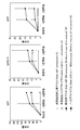
- FIG. 2 is a graph showing test results in Example 1.
- (A) shows before immunization and (b) shows 2 weeks after immunization.
- FIG. 3 is a graph showing test results in Example 1.
- (A) shows before immunization, and (b) shows 6 weeks after immunization.
- FIG. 4 is a graph showing test results in Example 2.
- FIG. 5 is a graph showing test results in Example 3.
- 6 is a graph showing test results in Example 3.
- FIG. (A) shows before immunization, and (b) shows 6 weeks after immunization.
- FIG. 7 is a graph showing the test results in Example 4.
- NKT cells may be involved in the production of antibodies against blood group sugar chain antigens (also referred to as anti-blood group antibodies) in some form and analyzed them. It was. The details will be described in Example 1, but mice lacking the CD1d molecule (also referred to as knockout mice) and normal mice were immunized with human erythrocytes, and the subsequent production of anti-blood group antibodies was compared.
- blood group sugar chain antigens also referred to as anti-blood group antibodies
- NKT cells are involved in the production of anti-blood group antibodies and are essential for antibody class switching.
- NKT cells have little effect on peptide antigens.
- anti-blood group antibodies could be specifically suppressed by controlling the activity of NKT cells. Focusing on the point that the first signal transduction is performed by antigen presentation via CD1d molecule, the production of anti-blood group antibody can be suppressed by inhibiting the signal transduction between NKT cells and B cells with anti-CD1d antibody I thought that.
- Fig. 1 schematically shows the mechanism.
- reference numeral 1 is an NKT cell
- reference numeral 2 is a B-1 cell
- reference numeral 3 is an anti-CD1d antibody.
- B-1 cell 2 has antigen receptor 6 on its surface that binds in response to CD1d molecule 4 and blood group sugar chain antigen 5, while NKT cell 1 It has an antigen receptor (iTCR) 7 that reacts and binds to the CD1d molecule 4 of 1 cell 2 on its surface.
- iTCR antigen receptor
- Signal transmission between the NKT cell 1 and the B-1 cell 2 is performed via the iTCR7 and the CD1d molecule 4.
- CD1d molecule 4 is not unique to B-1 cell 2 but also exists in B-2 cells.
- anti-CD1d antibody 3 is applied and bound to CD1d molecule 4, and signal transduction between NKT cell 1 and B-1 cell 2 is inhibited, anti-blood It should be possible to suppress the production of type 8 antibody.
- isotype ⁇ control mice administered with isotype antibody
- anti-AIgM antibody and anti-AIgG antibody were compared over time (details are given in Example 3).
- mice administered with isotype antibodies As a result, normal antibody production was observed in mice administered with isotype antibodies, and in mice administered with anti-CD1d antibodies, the effect of suppressing the production of anti-blood group antibodies and the suppression of antibody class switching were suppressed as in the case of knockout mice. The effect was recognized (refer FIG. 5, FIG. 6).
- an anti-CD1d antibody inhibitor for mice can be easily obtained by obtaining and appropriately adjusting the anti-CD1d antibody.
- the antibody-based rejection inhibitor for mice when administered, for example, so that the amount of anti-CD1d antibody per mouse is 400 to 600 ⁇ g, preferably 450 ⁇ g, the production of anti-blood group antibodies in the mouse is specifically detected. It is possible to suppress organs and transplant organs regardless of blood type.
- monoclonal human anti-CD1d antibodies may be produced using a series of known techniques for producing human monoclonal antibodies. Then, the prepared monoclonal human anti-CD1d antibody is mixed with other adjuvants such as preservatives and nutrients according to a conventional method, and adjusted as, for example, an injection, thereby suppressing human antibody rejection. An agent can be obtained.
- the prescribed amount for example, 30 g per mouse is converted on the basis of body weight
- the production of anti-blood group antibodies of type A and B can be specifically suppressed, and blood group Regardless, it becomes possible to transplant organs without impairing the normal immune response.
- this anti-CD1d antibody is effective against cells that have already differentiated into B-1 cells, in combination with administration of this anti-CD1d antibody, -1 It is considered to be more effective if a calcineurin inhibitor that suppresses the differentiation of cells is administered.
- the present invention may also be applicable to xenotransplantation, for example, transplantation using a pig organ that is physiologically and anatomically similar to a human in place of a human organ.
- Gal ⁇ 1-3Gal ⁇ 1-4GlcNAc (Gal) sugar chain and N-glycolylneuraminic acid (NeuGc) sugar chain in the porcine organ there are Gal ⁇ 1-3Gal ⁇ 1-4GlcNAc (Gal) sugar chain and N-glycolylneuraminic acid (NeuGc) sugar chain in the porcine organ, and these sugar chain antigens become the target of natural antibodies and cause antibody rejection.
- Gal sugar chains and NeuGc sugar chains are similar to blood group sugar chain antigens, the antibody rejection inhibitor of the present invention is highly likely to be effective for xenotransplantation between humans and pigs. .
- the antibody rejection inhibitor of the present invention can be put into practical use relatively easily and can be used to safely perform organ transplantation even from donors who are incompatible with the ABO blood group. This is expected to help resolve the donor shortage.
- Human type A erythrocytes are immunized by collecting blood from healthy volunteers with blood type A, washing twice with PBS, diluting to 8 ⁇ 10 8 / ml, and administering 1 ml intraperitoneally to the mouse, at 1-week intervals. Immunized twice.
- Detection using anti-A antibody titer using ELISA was performed with 5 ⁇ g / ml A-BSA (bovin ⁇ ⁇ ⁇ ⁇ ⁇ ⁇ serum albumin) (Dextra) in a polystyrene 96-well flat bottom plate (costar) or 5 ⁇ g / ml BSA (Roche) as a background control. Coating was performed at 0 ° C. for 8 hours. After washing the plate, it was blocked with 1% BSA, diluted mouse serum was added to each well and incubated for 2 hours at room temperature.
- A-BSA bivin ⁇ ⁇ ⁇ ⁇ ⁇ ⁇ serum albumin
- the results are shown in FIGS.
- the graph of FIG. 2 shows the change of the antibody amount of the anti-AIgM antibody, (a) is before immunization, and (b) is 2 weeks after immunization.
- the vertical axis of the graph represents the amount of antibody, and the horizontal axis represents the dilution factor of the added serum.
- the graph of FIG. 3 shows the change in the antibody amount of the anti-AIgG antibody, (a) is before immunization, and (b) is 6 weeks after immunization.
- the vertical axis of the graph indicates the amount of antibody as in FIG. 2, and the horizontal axis indicates the dilution factor of the added serum.
- the thymus of C57BL / 6 wild-type mice was removed, the thymus was ground in a dish, and ACK lysing solution (155 mM NH4Cl, 10 mM KHCO3, 1 mM EDTA-2Na, and PBS, pH 7.4)
- ACK lysing solution 155 mM NH4Cl, 10 mM KHCO3, 1 mM EDTA-2Na, and PBS, pH 7.4
- the thymocytes were isolated, diluted to 20 ⁇ 10 6 / ml with 199 medium, 1 ml was intraperitoneally injected into the mouse, and immunized twice at weekly intervals.
- Detection of anti-homogeneous MHC antibody was carried out by isolating thymocytes of C57BL / 6 wild-type mice by the above method and using flow cytometer (FCM) medium (PBS containing 0.1% BSA and 0.1% sodium azide). 10 ⁇ 10 6 / ml. To 1 ⁇ 10 6 cells, 10 ⁇ l of test plasma was added and incubated at 4 ° C. for 1 hour. 10 ⁇ l of anti-mouse IgM or IgG1,2a / b, 3-biotin was added as a secondary antibody and incubated at 4 ° C. for 30 minutes.
- FCM flow cytometer
- streptavidin-PE was added, and the mixture was incubated at 4 ° C. for 15 minutes, analyzed by FACSCalibur flow cytometer (Becton Dickinson), and the amount of the main fluorescein intensity (MFI) was used as the antibody amount.
- mice After 24 hours, each of these mice was immunized twice with a weekly human A-type erythrocyte, and the production of anti-blood group antibodies was measured using ELISA as described above.
- the graph in FIG. 5 shows the change in the antibody amount of the anti-AIgM antibody, (a) is before immunization, and (b) is 6 weeks after immunization.
- the vertical axis of the graph represents the amount of antibody, and the horizontal axis represents the dilution factor of the added serum.
- the graph of FIG. 6 shows the change in the antibody amount of the anti-AIgG antibody, (a) is before immunization, and (b) is 6 weeks after immunization.
- the vertical axis of the graph indicates the amount of antibody as in FIG. 5, and the horizontal axis indicates the dilution factor of the added serum.
- Example 4 In order to confirm that the same effect as in the case of the mouse described above can be obtained in the case of human, a test was conducted using a human lymphocyte chimeric mouse.
- Human lymphocyte chimeric mice were purchased from severe combined immunodeficient mice (NOD.Cg-Prkdc scid Il2rg tm1Sug / Jic ; manufactured by Central Research Institute for Experimental Animals). The collected peripheral blood lymphocytes were prepared by intraperitoneal administration of 20 ⁇ 10 6 mice per mouse.
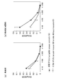
- FIG. 7 shows the abundance ratio of anti-A receptor-expressing B cells in human CD19 + B cells in the spleen of human lymphocyte chimeric mice.
- the black circle is the value of each solid, and the white circle is the average value.
- human B cells recognizing type A antigen in the spleen of test mice administered with anti-CD1d antibody have a significantly lower abundance ratio than in the spleen of control mice administered with control antibody. It was. That is, it was confirmed that the anti-CD1d antibody specifically and significantly suppresses the response of human B cells to blood group antigens.
- mice and humans In addition to organ transplantation and xenotransplantation of mice and humans, it can also be used to suppress the occurrence of autoimmune diseases.
Landscapes
- Health & Medical Sciences (AREA)
- Immunology (AREA)
- Chemical & Material Sciences (AREA)
- Organic Chemistry (AREA)
- General Health & Medical Sciences (AREA)
- Life Sciences & Earth Sciences (AREA)
- Medicinal Chemistry (AREA)
- Biophysics (AREA)
- Genetics & Genomics (AREA)
- Biochemistry (AREA)
- Molecular Biology (AREA)
- Proteomics, Peptides & Aminoacids (AREA)
- Hematology (AREA)
- Engineering & Computer Science (AREA)
- Bioinformatics & Cheminformatics (AREA)
- Chemical Kinetics & Catalysis (AREA)
- Transplantation (AREA)
- General Chemical & Material Sciences (AREA)
- Nuclear Medicine, Radiotherapy & Molecular Imaging (AREA)
- Pharmacology & Pharmacy (AREA)
- Animal Behavior & Ethology (AREA)
- Public Health (AREA)
- Veterinary Medicine (AREA)
- Medicines Containing Antibodies Or Antigens For Use As Internal Diagnostic Agents (AREA)
- Peptides Or Proteins (AREA)
Abstract
Description
NKT細胞の血液型糖鎖抗原に対する抗体産生への影響を解析するため、Balb/c CD1d-/-マウス(iTCRのリガンドであるCD1dを欠き、NKT細胞が顕著に減少していることが知られているマウス;ノックアウトマウス、n=4)と、比較対照のBalb/c野生種マウス(n=5)とをヒトA型赤血球(8×108/マウス)で免疫し、抗血液型A抗体産生をELISAを用いて測定した。
NKT細胞の通常のペプチド抗原に対する抗体産生の影響を解析するため、上記ノックアウトマウスと、Balb/c野生種マウス(MHCハプロタイプd)とをそれぞれC57BL/6野生型マウス(MHCハプロタイプb)の胸腺細胞(20×106/マウス)で免疫し、抗アロMHC抗体の産生量をフローサイトメーターを用いて測定した(n=6、比較対照として未処理群:n=3)。
NKT細胞とB-1細胞との間のシグナル伝達を抗CD1d抗体で阻害することにより抗血液型抗体の産生を抑制できるか否かを解析するため、Balb/c野生型マウスに抗CD1d抗体(rat anti-mouse CD1d monoclonal antibody、clone:1B1)をマウス1匹当たり450μg腹腔内投与した(テストマウス、n=3)。また、同様にrat IgG2b抗体(isotype control)をマウス1匹当たり450μg腹腔内投与した(対照マウス、n=3)。
ヒトの場合においても上述したマウスの場合と同様の効果が得られることを確認するため、ヒトリンパ球キメラマウスを用いて試験を行った。
2 B-1細胞
3 抗CD1d抗体
4 CD1d分子
5 糖鎖抗原
6 抗原レセプター
7 iTCR
8 抗体
Claims (8)
- NKT細胞とB細胞との間のシグナル伝達を阻害して抗体の産生を抑制する抗体性拒絶反応抑制剤であって、
抗CD1d抗体を含む抗体性拒絶反応抑制剤。 - 請求項1に記載の抗体性拒絶反応抑制剤であって、
前記B細胞がB-1細胞を含む抗体性拒絶反応抑制剤。 - 請求項1又は請求項2に記載の抗体性拒絶反応抑制剤であって、
産生が抑制される前記抗体に糖鎖抗原に対する抗体が含まれる抗体性拒絶反応抑制剤。 - 請求項3に記載の抗体性拒絶反応抑制剤であって、
前記糖鎖抗原が血液型糖鎖抗原を含む抗体性拒絶反応抑制剤。 - 請求項4に記載の抗体性拒絶反応抑制剤であって、
産生が抑制される前記抗体にペプチド抗原に対する抗体が含まれない抗体性拒絶反応抑制剤。 - 請求項1に記載の抗体性拒絶反応抑制剤であって、
前記抗CD1d抗体がモノクローナル抗体である抗体性拒絶反応抑制剤。 - 請求項6に記載の抗体性拒絶反応抑制剤であって、
前記抗CD1d抗体がヒト型抗体である抗体性拒絶反応抑制剤。 - 抗体性拒絶反応抑制剤のスクリーニング方法であって、
マウスの腹腔内に対象成分を投与してテストマウスを得るとともに、他のマウスの腹腔内に抑制すべき抗体成分を投与して対照マウスを得る工程と、
前記テストマウス及び前記対照マウスのそれぞれを赤血球で免疫し、抗血液型抗体の産生を測定する工程と、
を含む抗体性拒絶反応抑制剤のスクリーニング方法。
Priority Applications (2)
| Application Number | Priority Date | Filing Date | Title |
|---|---|---|---|
| US13/318,090 US20120046452A1 (en) | 2009-04-30 | 2010-04-19 | Antibody-mediated rejection inhibitor |
| JP2011511287A JP5757483B2 (ja) | 2009-04-30 | 2010-04-19 | 抗体性拒絶反応抑制剤 |
Applications Claiming Priority (2)
| Application Number | Priority Date | Filing Date | Title |
|---|---|---|---|
| JP2009-110887 | 2009-04-30 | ||
| JP2009110887 | 2009-04-30 |
Publications (1)
| Publication Number | Publication Date |
|---|---|
| WO2010125762A1 true WO2010125762A1 (ja) | 2010-11-04 |
Family
ID=43031922
Family Applications (1)
| Application Number | Title | Priority Date | Filing Date |
|---|---|---|---|
| PCT/JP2010/002825 Ceased WO2010125762A1 (ja) | 2009-04-30 | 2010-04-19 | 抗体性拒絶反応抑制剤 |
Country Status (3)
| Country | Link |
|---|---|
| US (1) | US20120046452A1 (ja) |
| JP (1) | JP5757483B2 (ja) |
| WO (1) | WO2010125762A1 (ja) |
Citations (3)
| Publication number | Priority date | Publication date | Assignee | Title |
|---|---|---|---|---|
| JP2001512438A (ja) * | 1997-02-11 | 2001-08-21 | イムノメディクス,インコーポレイテッド | αガラクトシルエピトープで標識された抗体による免疫応答の刺激 |
| JP2004500154A (ja) * | 1999-06-03 | 2004-01-08 | アドバンスト エクストラバスキュラー システムズ | 循環血液からの好ましくない分子の一段階除去 |
| JP2008546647A (ja) * | 2005-06-08 | 2008-12-25 | デューク ユニバーシティ | 移植のための抗−cd19抗体治療 |
-
2010
- 2010-04-19 US US13/318,090 patent/US20120046452A1/en not_active Abandoned
- 2010-04-19 WO PCT/JP2010/002825 patent/WO2010125762A1/ja not_active Ceased
- 2010-04-19 JP JP2011511287A patent/JP5757483B2/ja active Active
Patent Citations (3)
| Publication number | Priority date | Publication date | Assignee | Title |
|---|---|---|---|---|
| JP2001512438A (ja) * | 1997-02-11 | 2001-08-21 | イムノメディクス,インコーポレイテッド | αガラクトシルエピトープで標識された抗体による免疫応答の刺激 |
| JP2004500154A (ja) * | 1999-06-03 | 2004-01-08 | アドバンスト エクストラバスキュラー システムズ | 循環血液からの好ましくない分子の一段階除去 |
| JP2008546647A (ja) * | 2005-06-08 | 2008-12-25 | デューク ユニバーシティ | 移植のための抗−cd19抗体治療 |
Non-Patent Citations (2)
| Title |
|---|
| IREI, T. ET AL.: "The persistent elimination of B cells responding to blood group A carbohydrates by synthetic group A carbohydrates and B-1 cell differentiation blockade: novel concept in preventing antibody- mediated rejection in ABO-incompatible transplantation", BLOOD, vol. 110, no. 13, 2007, pages 4567 - 4575 * |
| LIU, S. ET AL.: "CDld-mediated interaction between activated T cells and B cells is essential to B-cell proliferation and anti- alpha-Gal antibody production", TRANSPLANTATION PROCEEDINGS, vol. 41, no. 1, February 2009 (2009-02-01), pages 398 - 402 * |
Also Published As
| Publication number | Publication date |
|---|---|
| JP5757483B2 (ja) | 2015-07-29 |
| US20120046452A1 (en) | 2012-02-23 |
| JPWO2010125762A1 (ja) | 2012-10-25 |
Similar Documents
| Publication | Publication Date | Title |
|---|---|---|
| JP7363828B2 (ja) | 二重特異性抗体 | |
| WO2020060924A1 (en) | Use of a cd4/cd8 bispecific antibody for the treatment of autoimmune/inflammatory disorders | |
| US20110318340A1 (en) | Deglycosylated antibodies | |
| KR20190117489A (ko) | 항-bcma 중쇄-단독 항체 | |
| WO2018170096A1 (en) | Use of a cd4/cd8 bispecific antibody for the treatment of diabetes | |
| JP2019527678A (ja) | CD38に特異的に結合する抗体によるIgE媒介疾患の治療 | |
| US11059889B2 (en) | Anti-human Igβ antibody | |
| US9718891B2 (en) | Antibodies specific to a novel epitope on CεMX of human membrane-bound IgE and uses thereof in treating IgE-mediated diseases | |
| Ali et al. | Analysis of the capability of IgG antibodies and receptors with their relationships to food tolerance and autoimmune disorders | |
| JP7574692B2 (ja) | 二重特異性抗体 | |
| AU2018350372C1 (en) | Method | |
| JP5757483B2 (ja) | 抗体性拒絶反応抑制剤 | |
| US11084871B2 (en) | Composition with reduced immunogenicity | |
| JP2008530019A (ja) | 死細胞または瀕死細胞に露出する抗原に対する結合タンパク質を用いて自己免疫の症状および炎症性の症状を低減するための方法 | |
| RU2796019C2 (ru) | Биспецифическое антитело | |
| EP4709764A2 (en) | Human monoclonal antibodies for treating subjects suffering from pulmonary fibrosis | |
| EP1675616A2 (fr) | Nouvelles igg3 utiles pour stimuler la phagocytose | |
| CN112867785A (zh) | 通过抗体介导的特定肠道细菌的中和作用治疗免疫性疾病 | |
| HK1235431B (zh) | 新的抗人类IGβ抗体 |
Legal Events
| Date | Code | Title | Description |
|---|---|---|---|
| 121 | Ep: the epo has been informed by wipo that ep was designated in this application |
Ref document number: 10769464 Country of ref document: EP Kind code of ref document: A1 |
|
| DPE1 | Request for preliminary examination filed after expiration of 19th month from priority date (pct application filed from 20040101) | ||
| ENP | Entry into the national phase |
Ref document number: 2011511287 Country of ref document: JP Kind code of ref document: A |
|
| WWE | Wipo information: entry into national phase |
Ref document number: 13318090 Country of ref document: US |
|
| NENP | Non-entry into the national phase |
Ref country code: DE |
|
| 122 | Ep: pct application non-entry in european phase |
Ref document number: 10769464 Country of ref document: EP Kind code of ref document: A1 |