WO2010032786A1 - トランスジェニック動物 - Google Patents
トランスジェニック動物 Download PDFInfo
- Publication number
- WO2010032786A1 WO2010032786A1 PCT/JP2009/066262 JP2009066262W WO2010032786A1 WO 2010032786 A1 WO2010032786 A1 WO 2010032786A1 JP 2009066262 W JP2009066262 W JP 2009066262W WO 2010032786 A1 WO2010032786 A1 WO 2010032786A1
- Authority
- WO
- WIPO (PCT)
- Prior art keywords
- wdr36
- polypeptide
- retinal
- mutant
- retinal disease
- Prior art date
- Legal status (The legal status is an assumption and is not a legal conclusion. Google has not performed a legal analysis and makes no representation as to the accuracy of the status listed.)
- Ceased
Links
Images





Classifications
-
- A—HUMAN NECESSITIES
- A01—AGRICULTURE; FORESTRY; ANIMAL HUSBANDRY; HUNTING; TRAPPING; FISHING
- A01K—ANIMAL HUSBANDRY; AVICULTURE; APICULTURE; PISCICULTURE; FISHING; REARING OR BREEDING ANIMALS, NOT OTHERWISE PROVIDED FOR; NEW BREEDS OF ANIMALS
- A01K67/00—Rearing or breeding animals, not otherwise provided for; New or modified breeds of animals
- A01K67/027—New or modified breeds of vertebrates
- A01K67/0275—Genetically modified vertebrates, e.g. transgenic
-
- A—HUMAN NECESSITIES
- A61—MEDICAL OR VETERINARY SCIENCE; HYGIENE
- A61P—SPECIFIC THERAPEUTIC ACTIVITY OF CHEMICAL COMPOUNDS OR MEDICINAL PREPARATIONS
- A61P27/00—Drugs for disorders of the senses
- A61P27/02—Ophthalmic agents
- A61P27/06—Antiglaucoma agents or miotics
-
- C—CHEMISTRY; METALLURGY
- C07—ORGANIC CHEMISTRY
- C07K—PEPTIDES
- C07K14/00—Peptides having more than 20 amino acids; Gastrins; Somatostatins; Melanotropins; Derivatives thereof
- C07K14/435—Peptides having more than 20 amino acids; Gastrins; Somatostatins; Melanotropins; Derivatives thereof from animals; from humans
- C07K14/46—Peptides having more than 20 amino acids; Gastrins; Somatostatins; Melanotropins; Derivatives thereof from animals; from humans from vertebrates
- C07K14/47—Peptides having more than 20 amino acids; Gastrins; Somatostatins; Melanotropins; Derivatives thereof from animals; from humans from vertebrates from mammals
- C07K14/4701—Peptides having more than 20 amino acids; Gastrins; Somatostatins; Melanotropins; Derivatives thereof from animals; from humans from vertebrates from mammals not used
- C07K14/4702—Regulators; Modulating activity
-
- C—CHEMISTRY; METALLURGY
- C12—BIOCHEMISTRY; BEER; SPIRITS; WINE; VINEGAR; MICROBIOLOGY; ENZYMOLOGY; MUTATION OR GENETIC ENGINEERING
- C12N—MICROORGANISMS OR ENZYMES; COMPOSITIONS THEREOF; PROPAGATING, PRESERVING, OR MAINTAINING MICROORGANISMS; MUTATION OR GENETIC ENGINEERING; CULTURE MEDIA
- C12N15/00—Mutation or genetic engineering; DNA or RNA concerning genetic engineering, vectors, e.g. plasmids, or their isolation, preparation or purification; Use of hosts therefor
- C12N15/09—Recombinant DNA-technology
- C12N15/63—Introduction of foreign genetic material using vectors; Vectors; Use of hosts therefor; Regulation of expression
- C12N15/79—Vectors or expression systems specially adapted for eukaryotic hosts
- C12N15/85—Vectors or expression systems specially adapted for eukaryotic hosts for animal cells
- C12N15/8509—Vectors or expression systems specially adapted for eukaryotic hosts for animal cells for producing genetically modified animals, e.g. transgenic
-
- A—HUMAN NECESSITIES
- A01—AGRICULTURE; FORESTRY; ANIMAL HUSBANDRY; HUNTING; TRAPPING; FISHING
- A01K—ANIMAL HUSBANDRY; AVICULTURE; APICULTURE; PISCICULTURE; FISHING; REARING OR BREEDING ANIMALS, NOT OTHERWISE PROVIDED FOR; NEW BREEDS OF ANIMALS
- A01K2217/00—Genetically modified animals
- A01K2217/05—Animals comprising random inserted nucleic acids (transgenic)
- A01K2217/054—Animals comprising random inserted nucleic acids (transgenic) inducing loss of function
-
- A—HUMAN NECESSITIES
- A01—AGRICULTURE; FORESTRY; ANIMAL HUSBANDRY; HUNTING; TRAPPING; FISHING
- A01K—ANIMAL HUSBANDRY; AVICULTURE; APICULTURE; PISCICULTURE; FISHING; REARING OR BREEDING ANIMALS, NOT OTHERWISE PROVIDED FOR; NEW BREEDS OF ANIMALS
- A01K2227/00—Animals characterised by species
- A01K2227/10—Mammal
- A01K2227/105—Murine
-
- A—HUMAN NECESSITIES
- A01—AGRICULTURE; FORESTRY; ANIMAL HUSBANDRY; HUNTING; TRAPPING; FISHING
- A01K—ANIMAL HUSBANDRY; AVICULTURE; APICULTURE; PISCICULTURE; FISHING; REARING OR BREEDING ANIMALS, NOT OTHERWISE PROVIDED FOR; NEW BREEDS OF ANIMALS
- A01K2267/00—Animals characterised by purpose
- A01K2267/03—Animal model, e.g. for test or diseases
- A01K2267/0306—Animal model for genetic diseases
Definitions
- the present invention relates to a transgenic animal. More specifically, a transgenic animal into which a gene encoding a mutated WDR36 polypeptide and / or a derivative thereof is introduced to express the mutated WDR36 polypeptide and / or a derivative thereof, and used as a retinal disease onset model or the like Related to transgenic animals.
- the retina has a function of receiving light from the outside and plays an important role in terms of visual function.
- it is a tissue having a thickness of 0.1 to 0.5 mm composed of 10 layers such as a retinal pigment epithelium layer, an inner plexiform layer, a ganglion cell layer, and a nerve fiber layer.
- a retinal pigment epithelium layer a tissue having a thickness of 0.1 to 0.5 mm composed of 10 layers such as a retinal pigment epithelium layer, an inner plexiform layer, a ganglion cell layer, and a nerve fiber layer.
- the inner plexiform layer there is a neuron that forms a synapse by pairing with a ganglion cell process called amacrine cell, but it responds well at the beginning and end of light irradiation. It is thought to work as a detector.
- retinal ganglion cell In the ganglion cell layer, there is a retinal ganglion cell (hereinafter also referred to as “RGC”) located on the innermost side of the retina, and is deeply involved in motor vision, peripheral vision, color vision, morphological sense, and the like.
- RRC retinal ganglion cell
- nerve fiber layer retinal blood vessels, which are branches of the central retinal arteriovenous vein, run and supply oxygen and nutrients to retinal neurons.
- Glaucoma is an example of a disease that causes damage to the retina including retinal neurons. Glaucoma is one of the eye diseases that can cause serious visual impairment that can lead to blindness if not properly treated. In glaucoma, retinal ganglion cells, particularly retinal nerve cells, are selectively damaged and cause optic nerve damage, resulting in progression to visual field impairment. Although the detailed mechanism of glaucomatous optic neuropathy is not yet clear, the major cause of optic nerve atrophy is the mechanical disorder theory that the optic nerve is directly compressed by increased intraocular pressure and optic nerve atrophy occurs, and the circulatory disorder of the optic nerve head is the main cause. The theory of circulatory disturbance has been proposed, and it is considered that both mechanisms based on these mechanical disorders and circulatory disorders are involved in a complex manner.
- Non-Patent Document 1 Several animal models for the development of retinal diseases such as glaucoma have been developed in accordance with the pathogenesis.
- Non-patent Document 2 There is a high intraocular pressure model animal that reflects a mechanical disorder, a model that induces high intraocular pressure by increasing aqueous humor outflow resistance by laser irradiation at the corner (non-patent document 2), physiological hypertonicity A model that induces high intraocular pressure by injecting fluid into the blood vessels of the aqueous humor discharge pathway (Non-patent Document 3), and the amount of aqueous humor outflow by cauterizing the vortex vein or superior scleral vein There is a model (Non-patent Document 4) to be reduced.
- Non-patent Document 5 As models that cause direct axonal transport obstacles, an axonal compression model (Non-patent Document 5), an axotomy model (Non-patent Document 6), and the like have been reported. Furthermore, as a model reflecting circulatory disturbance, there is an ischemia reperfusion model (Non-patent Document 7). On the other hand, glutamate is one of the neurotransmitters in the retina, but excessive activation of this glutamate signal cascade for some reason is thought to contribute to retinal ganglion cell damage (non-patented). Reference 8), an NMDA-induced retinal disorder model has been reported as a model animal model for the development of retinal diseases such as glaucoma based on this viewpoint (Non-patent Document 9).
- the WDR36 (WD-repeat-domain-36) gene has been reported as one of the genes associated with glaucoma.
- the WDR36 polypeptide encoded by the WDR36 gene is a polypeptide having a WD-Repeat structure, but its function has not yet been elucidated.
- Patent Document 1 polymorphisms in the WDR36 gene of glaucoma patients were analyzed.
- these glaucoma model animals can be produced based on the prior art, these retinal disorder models reflect only a part of the cause of glaucoma onset, and human glaucoma that has been revealed in recent years. Since it is unrelated to genetic predisposition, it is desirable to create a new disease onset model that reflects the mechanism of glaucoma in humans and / or genetic predisposition.
- An object of the present invention is to provide a disease model related to the genetic predisposition of glaucoma. Still another object of the present invention is to provide a screening method for a prophylactic or therapeutic agent for glaucoma using the model, and a material that can be used for the screening.
- the present inventors focused on the WDR36 gene as a factor that reflects the genetic predisposition of glaucoma patients, and as a result of intensive studies to solve the above-mentioned problems, the present inventors have found that the polymorphism encoded by the human WDR36 gene known to be related to glaucoma is known.
- a transgenic mouse expressing a mutant of a mouse WDR36 polypeptide into which a mutation corresponding to a mutation in which the 658th aspartic acid residue of the peptide is a glycine residue (hereinafter sometimes referred to as a D658G mutation)
- human Expresses a mutant of mouse WDR36 polypeptide introduced with a mutation corresponding to a mutation that deletes the entire WD8 region containing the 658th aspartic acid residue in WDR36 polypeptide (hereinafter sometimes referred to as delWD8 mutation)
- Transgenic mice do not show any phenotype associated with glaucoma, but the 658th aspartate residue in the human WDR36 polypeptide
- Transgenic mice that express a mutant of the mouse WDR36 polypeptide introduced with a mutation corresponding to a mutation that deletes amino acid residues 657 to 659 (hereinafter referred to as the del658 mutation) It was found that a phenotype that reflects glaucom
- the disorder that occurred in the periphery of the retina of the transgenic mouse is not only retinal ganglion cells related to glaucoma, but also surprisingly, it covers the entire retina including photoreceptor cells, retinal pigment epithelium, etc.
- the present invention was completed by finding that it can be used as a disease onset model animal for retinal diseases in general.
- the present invention [1] In a mammalian WDR36 polypeptide, a variant of the WDR36 polypeptide lacking one or several amino acid residues including the amino acid residue corresponding to the 658th aspartic acid residue in human WDR36 polypeptide, or A non-human transgenic animal in which the derivative is expressed, [2] Use of the transgenic animal according to the above [1] as a retinal disease onset model, [3] A retinal disease onset model animal in which the transgenic animal according to [1] has a disorder in the retina, [4] Retinal disease onset model animal according to [3], screening method for preventive or therapeutic agent for retinal disease using the tissue or cell thereof, and [5] aspartic acid residue at position 658 in human WDR36 polypeptide A mutant WDR36 polypeptide lacking one or several amino acid residues containing the amino acid residues corresponding to The present invention relates to a mutant WDR36 polypeptide selected from the group consisting of a mutant WDR36 polypeptide lacking one or
- the transgenic animal of the present invention develops retinal ganglion cell damage in the periphery of the retina and shows a phenotype similar to glaucoma. This is an excellent effect.
- FIG. 1 is a diagram for explaining a pBluescriptII KS (+) vector.
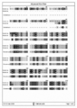
- FIG. 2 is an example for explaining the amino acid sequences and sequence alignment of human WDR36 polypeptide and mouse WDR36 polypeptide.
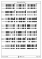
- FIG. 3 is an example for explaining the amino acid sequences and sequence alignment of human WDR36 polypeptide and mouse WDR36 polypeptide.
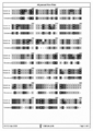
- FIG. 4 is an example for explaining the amino acid sequences and sequence alignment of human WDR36 polypeptide and mouse WDR36 polypeptide.
- FIG. 5 is an example of a diagram illustrating amino acid sequences and sequence alignment of human WDR36 polypeptide and mouse WDR36 polypeptide.
- FIG. 6 is an example for explaining the amino acid sequences and sequence alignment of human WDR36 polypeptide and mouse WDR36 polypeptide.
- FIG. 7 is a diagram for explaining the pCMV-Tag5 vector.
- FIG. 8 is a diagram for explaining the pCAGGS vector.
- FIG. 9 is a photograph showing the intracellular localization of WDR36 polypeptide after introducing an expression vector into NIH3T3-3-4 cells.
- A is a photograph of a cell into which an expression vector containing each cDNA of Mus wild type,
- B is Mus del658,
- C is Mus D658G, and
- D is Mus delWD8.
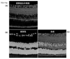
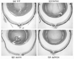
- FIG. 10 is a photograph showing the expression of the WDR36 mutant gene in del658 TGM in the retina.
- FIG. 11 is a tissue stained image of the entire retina of Wild type TMG, del658 TMG, D658G TGM, and delWD8 TGM.
- A Wild type TGM
- B del658 TMG
- C D658G TGM
- D delWD8 TGM
- FIG. 12 is a tissue stained image of the retina of the periphery of Wild type TGM, del658 TMG, D658G TGM, and delWD8 TGM.
- FIG. 13 is a diagram showing the results of evaluating the thickness of the peripheral retina of Wild type TMG, del658 TMG, D658G TGM, and delWD8 TGM.
- the transgenic animal of the present invention is a mutant WDR36 polypeptide that lacks one or several amino acid residues including the amino acid residue corresponding to the 658th aspartic acid residue of the human WDR36 polypeptide in the mammalian WDR36 polypeptide.
- mutant WDR36 polypeptide lacking amino acid residues corresponding to amino acid residues 657 to 659 of human WDR36 polypeptide, or mutant WDR36 polypeptide represented by the amino acid sequence of SEQ ID NO: 1-2 (Hereinafter, when the mutation is not specified and simply referred to as a mutant WDR36 polypeptide, it means a mutant of a mammalian WDR36 polypeptide containing these mutations) or a non-human transgenic animal in which these derivatives are expressed. Thus, it has a great feature of spontaneous development of retinal diseases.
- the WDR36 polypeptide is expressed in many organs such as the eyes (including the retina), liver, heart, kidney, pancreas, etc., regardless of the species of mammal, and the WDR36 polypeptide in the present invention Or a derivative thereof is expressed at least in the retina.
- the mammalian WDR36 polypeptide into which the mutation is introduced includes a polypeptide comprising a known amino acid sequence of mammalian WDR36.
- WDR36 polypeptide includes a polypeptide comprising a known amino acid sequence of mammalian WDR36.
- An example is a polypeptide encoded by a known polynucleotide of mammalian WDR36.
- it is a WDR36 polypeptide derived from human or mouse.
- a polypeptide comprising a sequence exhibiting a certain level of identity to the amino acid sequence of WDR36 is also included.
- the mutant WDR36 polypeptide of the present invention may be any amino acid sequence containing a mutation described below in the amino acid sequence of a known WDR36 polypeptide, preferably the amino acid sequence of a human or mouse WDR36 polypeptide.
- the amino acid sequence of the mutant WDR36 polypeptide of the present invention is one or several, preferably 1 to several, in the amino acid sequence of a known WDR36 polypeptide, preferably the human or mouse WDR36 polypeptide.
- 150 more preferably 1-50, more preferably 1-20, even more preferably 1-10, and even more preferably 1-5 amino acids are deleted, inserted, added or substituted. May be.
- the amino acid sequence of the mutant WDR36 polypeptide of the present invention is preferably 85% or more with respect to the amino acid sequence of a known WDR36 polypeptide, preferably the human or mouse WDR36 polypeptide, as long as it has the mutation. More preferably, it is 95% or more, more preferably 98% or more, more preferably 99.0% or more, and still more preferably 99.5% or more.
- identity is calculated by using a known sequence search program such as BLAST (translated by David W. Mount, written by Koji Okazaki et al., Bioinformatics 2nd edition, Medical Science International). .
- the mutation introduced into the WDR36 polypeptide includes one or several, preferably 1 to 9, more preferably an amino acid residue corresponding to the 658th aspartic acid residue in the human WDR36 polypeptide.
- the mutation introduced into the WDR36 polypeptide is a mutation that lacks an amino acid residue corresponding to amino acid residues 657 to 659 of the human WDR36 polypeptide.
- WDR36 polypeptides exist in various mammalian species. By using the alignment information of these sequences to compare the sequences of the human WDR36 polypeptide and the WDR36 polypeptide of the desired species, the WDR36 polypeptide of the desired species corresponding to the 658th aspartic acid in the human WDR36 polypeptide is obtained. The amino acid residue to be deleted of the peptide can be determined. As will be described in detail in the Examples section, the sequence comparison diagrams (alignment diagrams) described in FIGS.
- 2-6 are sequence comparison diagrams conserved between human and mouse species for the WDR36 polypeptide. is there. 2 to 6, the amino acid residue in mouse WDR36 polypeptide isoform 1 corresponding to the 658th aspartic acid residue of human WDR36 polypeptide is the 606th aspartic acid residue.
- the amino acid residue in mouse WDR36 polypeptide isoform 1 corresponding to the 658th aspartic acid residue of human WDR36 polypeptide is the 606th aspartic acid residue.
- the location to be deleted may be determined using a comparative diagram created based on the alignment information.
- a comparison diagram is, for example, alignment information by a known multiple alignment program such as ClustalW (translated by David W. Mount, written by Koji Okazaki et al., Bioinformatics 2nd edition, Medical Science International), or the aforementioned BLAST. Created based on alignment information.
- the variant of WDR36 polypeptide introduced into an animal as a transgene is a variant WDR36 polypeptide comprising the amino acid sequence set forth in any one of SEQ ID NOs: 1 or 2 or a derivative thereof.
- the amino acid sequence described in SEQ ID NO: 1 is the amino acid sequence of a mutant human WDR36 polypeptide lacking amino acid residues 657 to 659 of the human WDR36 polypeptide
- the amino acid sequence described in SEQ ID NO: 2 is the human WDR36 polypeptide
- the variant of WDR36 polypeptide introduced as a transgene is the amino acid sequence set forth in SEQ ID NO: 2.
- a derivative of a mutant WDR36 polypeptide includes a derivative of a mutant WDR36 polypeptide that has undergone a modification that is generally known to occur in vivo as a modification to a polypeptide, specifically, an acetylated derivative, palmitoyl Derivatives, myristylated derivatives, amidated derivatives, acrylated derivatives, dansylated derivatives, biotinylated derivatives, phosphorylated derivatives, succinylated derivatives, anilylated derivatives, benzyloxycarbonylated derivatives, formylated derivatives, nitrated derivatives, sulfones Derivatives, aldehyde derivatives, cyclized derivatives, glycosylated derivatives, monomethylated derivatives, dimethylated derivatives, trimethylated derivatives, polymethylated derivatives, guanidylated derivatives, amidated derivatives, maleylated derivatives, trifluoroacetylated derivatives, carbamyl
- the derivative includes a label attached to facilitate detection of expression of the introduced polypeptide, that is, a derivative with a so-called tag.
- the tag to be attached may be any tag that can be detected using a labeling compound that can be strongly bound by an antigen-antibody reaction or hydrogen bonding, and is preferably an amino acid or polypeptide that is integrated with the mutant WDR36 polypeptide.
- a tag that can be expressed in Specifically, a biotin tag, a histidine tag, a hemagglutinin epitope tag, a myc tag and the like are exemplified, and among them, a myc tag using a c-myc polypeptide or a partial polypeptide thereof as a tag is preferable.
- the derivative in the present invention may be a derivative that is tagged and further modified that is generally known to occur in vivo.
- the gene of the mutant WDR36 polypeptide and / or its derivative can be prepared by a method known in the art.
- a mammalian WDR36 polypeptide containing a known amino acid sequence is also prepared by designing a polypeptide sequence containing the mutation disclosed in the present invention and chemically synthesizing DNA encoding the polypeptide.
- Chemical synthesis of DNA can be performed using a Shimadzu DNA synthesizer using the thiophosphite method, a Perkin Elmer DNA synthesizer model 1392 using the phosphoramidite method, etc. Can do.
- the gene of the mutant WDR36 polypeptide and / or its derivative can be obtained by a known method such as a site-directed mutagenesis method (site-directed mutagenesis method) with respect to a nucleic acid molecule encoding a mammalian WDR36 polypeptide obtained separately. )
- site-directed mutagenesis method site-directed mutagenesis method
- a commercially available mutagenesis kit based on a known principle is used, and an appropriate template nucleic acid molecule or primer is used. Can be prepared by introducing a desired mutation.
- kits for such a kit include QuikChange (registered trademark) XL Site-directed mutagenesis kit manufactured by Stratagene. This kit produces a supercoiled double-stranded DNA plasmid vector into which the target gene without mutation is inserted, and uses a primer containing the mutation sequence desired to be introduced into the gene in the nucleic acid sequence.
- a kit for introducing a desired mutation into the gene the principle of which is described in U.S. Pat. 6,444,428, 6,489,150, 6,734,293, 7,045,328 and the like.
- methods other than commercially available kits include mutant polypeptides and / or derivatives thereof with reference to books such as Methods Molecular Biology, vol.
- the primer or template nucleic acid molecule used for mutagenesis is a primer or template nucleic acid molecule that can introduce the mutation disclosed in the present invention into the sequence of the WDR36 gene desired to be mutagenized.
- One skilled in the art can design such primers or template nucleic acid molecules.
- the primer capable of introducing the mutation in the present invention is prepared based on the instructions of the kit, for example, for the mouse WDR36 gene
- the primers used are the primers shown in SEQ ID NOs: 5 and 6.
- a gene encoding a mammalian WDR36 polypeptide into which a mutation is introduced can be obtained from an appropriate cDNA library by amplification using appropriate primers. it can.
- the cDNA library can be used without particular limitation as long as it is a species having WDR36, but is preferably derived from a human or mouse, and may be any derived from a tissue expressing a WDR36 polypeptide. An example of such tissue is the heart.
- a commercially available cDNA library can be used as long as it contains the WDR36 gene, or a cDNA library of tissue or cells expressing the WDR36 gene can be prepared and used by a known method.
- Primers for amplifying the WDR36 gene can be designed by those skilled in the art based on the sequence of the nucleic acid molecule encoding the WDR36 polypeptide of the species, and can be prepared, for example, using the aforementioned DNA synthesizer.
- WDR36 gene a cDNA encoding a commercially available WDR36 polypeptide can be used, and examples of such a reagent include ORIGEN TECHNOLOGIES, INC SC314980, etc. .
- the method for introducing a gene encoding a mutant WDR36 polypeptide and / or a derivative thereof into an animal is not particularly limited.
- a DNA fragment containing a gene encoding a mutant WDR36 polypeptide or a derivative thereof, or a mutant WDR36 polypeptide Such as microinjection, electroporation, lipofection, or a method of introducing a gene by introducing a foreign gene into a retroviral vector and using an expression vector into which a gene encoding a peptide or its derivative has been introduced
- a known method includes a method of introducing a gene of a mutant WDR36 polypeptide or a derivative thereof contained in the DNA fragment or expression vector into a fertilized egg cell.
- the DNA fragment is preferably a linear double-stranded DNA and includes a promoter, a gene encoding a mutant WDR36 polypeptide or a derivative thereof, and a poly A terminus.
- the vector preferably contains a promoter, a gene encoding a mutant WDR36 polypeptide or a derivative thereof, and a transcription termination sequence while being capable of autonomous replication in a host.
- the DNA fragment and / or the vector may contain a gene that controls a promoter such as an enhancer.
- a gene encoding a polypeptide for preparing detection of the expression of the introduced gene may be further included downstream of the gene encoding the mutant WDR36 polypeptide or a derivative thereof.
- the tag may be expressed as the same polypeptide molecule as the mutant WDR36 polypeptide or derivative thereof.
- promoters examples include ⁇ -actin promoter and CAG promoter.
- ⁇ -actin is a protein constituting the cytoskeleton and can be expressed in almost all cells into which a gene encoding a mutant WDR36 polypeptide or a derivative thereof has been introduced. Therefore, the ⁇ -actin promoter is preferable. .
- Any known method can be used to include a tag in a DNA fragment containing a gene encoding a mutant WDR36 polypeptide or a derivative thereof.
- a vector in which a desired tag is separately incorporated is prepared, After cleaving with an appropriate restriction enzyme, the gene encoding the mutant WDR36 polypeptide or its derivative is incorporated into a vector, amplified by a known method, the vector is purified, and cleaved with an appropriate restriction enzyme to obtain a DNA fragment be able to.
- Vectors in which a gene encoding the tag is previously incorporated are commercially available, and a desired one may be purchased and used.
- the promoter of the gene encoding the mutant WDR36 polypeptide or a derivative thereof is modified to a desired one, it can be modified by the same method.
- pCMV-Tag5 is preferably used as a vector for introducing a tag
- pCAGGS is preferably used as a vector for modifying the promoter to a ⁇ -actin promoter.
- the above DNA fragment or expression vector can be prepared according to a known method.
- the fertilized egg cell into which the DNA fragment or vector prepared as described above is introduced is transplanted into the uterus of a pseudopregnant animal, and after the fetus is sufficiently grown, it is delivered by natural delivery or cesarean section.
- the individual is transgenic. Whether it is an animal or not can be determined.
- transgenic animal of the present invention Maintenance can be performed.
- mice other than humans are used, preferably rodents such as mice, rats, guinea pigs, rabbits, and the like. Of these mice are preferred. There is no particular limitation on the mouse to be used, and a known mouse can be used. However, it is easy to produce a transgenic mouse, it is versatile, and it has a wealth of information such as gene background. From this point of view, the BDF1 / C57BL / 6N system is preferably used in the present invention. It should be noted that transgenic mice obtained by backcrossing other strains to a mouse into which a gene encoding a mutant WDR36 polypeptide and / or a derivative thereof has already been introduced can also be used in the present invention.
- the transgenic animal of the present invention can also be a so-called knock-in animal in which a gene encoding the mutant WDR36 polypeptide of the present invention or a derivative thereof is integrated in the genome instead of the WDR36 gene originally possessed by the animal.
- Incorporation of a gene encoding a mutant polypeptide in place of the original gene can be performed by a known technique using homologous recombination.
- transgenic animal into which a gene encoding a mutant WDR36 polypeptide and / or a derivative thereof has been introduced can be obtained.
- the transgenic animal has a great feature that it expresses a mutant WDR36 polypeptide and / or a derivative thereof at a site such as the retina and spontaneously develops a retinal disease, and therefore can be used as a retinal disease onset model animal.
- no phenotype associated with retinal disease is observed in the transgenic animals expressing the delWD8 mutant WDR36 polypeptide or derivative thereof, or the D658G mutant WDR36 polypeptide or derivative thereof.
- Transforms expressing the mutant WDR36 polypeptide or derivative thereof of the present invention despite no retinal changes observed in transgenic animals expressing the D658G mutant WDR36 polypeptide or derivative thereof known to be associated with glaucoma It is surprising that there are significant retinal disorders in the transgenic animals.
- the transgenic animal of the present invention spontaneously develops retinal changes at the age of 8 months at the latest and sufficiently develops retinal diseases at the age of 13 months.
- the retinal change of the transgenic animal of the present invention occurs from the peripheral part of the retina, and gradually changes toward the optic nerve head.
- the phenotype of such a transgenic animal of the present invention is that, even in glaucoma patients, there are many cases in which an area of reduced visibility such as a nasal staircase occurs from the periphery, and visual field constriction of moderately advanced glaucoma is It is reported that the peripheral area is dominant and that retinal changes in glaucoma patients also occur from the peripheral area of the retina (Ou Tan et al., Ophthalmology Vol.115, Page 949, 2008). These characteristics in the transgenic animals of the invention well reflect the characteristics of glaucoma in humans.
- the transgenic animal of the present invention is surprisingly provided with a surprising feature that all layers of the retina are damaged. That is, the retinal tissue in which the disorder occurs spans all layers of retinal ganglion cells, photoreceptor cells, pigment epithelial cells, etc., and in the retina having a clear decrease in the number of these cells and / or a layered structure. In addition, the thickness of the layer in which these cells exist is reduced, and the thickness of the entire retina is also reduced. Therefore, the transgenic animal of the present invention can be used as a model animal that spontaneously develops retinal diseases.
- retinal diseases include diseases mainly involving retinal ganglion cells and / or optic nerve damage, which is an axon process of retinal ganglion cells, photoreceptive cell damage and / or retinal pigment epithelium, depending on the location of the damage. Examples include diseases mainly involving cell damage.
- “mainly involved” means causing a disease as a main factor.
- the transgenic animal of the present invention Is suitably used as a model animal that develops a disease mainly involving retinal ganglion cell injury, more specifically as a glaucoma onset model animal or as a label disease or ischemic optic neuropathy model animal, Since it shows the damage of the peripheral part of the retina in common with the glaucoma, it is more preferably used as a glaucoma onset model animal.
- the pattern of reduced visibility in glaucoma patients and the pattern of visual field stenosis in moderately advanced glaucoma and the pattern of retinal damage in the transgenic animals of the present invention are found to match, so that the present invention
- the transgenic animal is preferably used as a model animal for a disease associated with visual field stenosis among diseases that develop retinal ganglion cell disorders.
- diabetic retinopathy, diabetic macular edema, age-related macular degeneration, retinitis pigmentosa, etc. are known as diseases mainly involving damage of photoreceptor cells or pigment epithelial cells in humans.
- the transgenic animal of the present invention is a model animal that develops a disease mainly involving photoreceptor cell damage and / or retinal pigment epithelial cell damage, more specifically, diabetic retinopathy, diabetic macular edema or aging. It is suitably used as a macular degeneration onset model animal or as a retinitis pigmentosa onset model animal.
- the retinal disease onset model animal of the present invention spontaneously develops retinal disease
- the retinal disease onset model animal of the present invention its tissue or cell should be suitably used for screening for preventive or therapeutic agents for retinal disease.
- retinal diseases include diseases mainly involving retinal ganglion cell disorders, and diseases mainly involving photoreceptor cell disorders and / or retinal pigment epithelial cell disorders.
- diseases mainly involving retinal ganglion cell damage include glaucoma, Label disease, and ischemic optic neuropathy. Among these, glaucoma is preferable.
- the preventive or therapeutic agent of the disease which has a visual field constriction among the diseases in which the disorder
- diseases in which photoreceptor cell disorders are mainly involved include diabetic retinopathy, diabetic macular edema and age-related macular degeneration.
- Diseases in which retinal pigment epithelial cell disorders are mainly involved include retinal pigment degeneration. Can be mentioned.
- the screening method is not particularly limited.
- the animal model for retinal disease onset according to the present invention may be administered once or continuously with a drug to be screened before or after the onset of retinal disease. This can be done by evaluating the condition of the retina after the period.
- a method for evaluating the retinal condition for example, the eyeball is removed, fixed by a known method, sliced and stained to obtain a microscopic specimen, and then the degree of retinal thinning, cell loss, or cell death is macroscopically examined.
- OCT optical coherence tomography
- any tissue that expresses a mutant WDR36 polypeptide or a derivative thereof can be used, but preferably a retinal tissue or a nerve tissue is used. More preferably, retinal tissue is used.
- retinal tissue is used.
- retinal tissue excised from the transgenic of the present invention is cultured for a certain period in the presence of the test drug, and retinal morphological changes such as retinal ganglion Drugs can be screened using as an index the suppression of cell number reduction or cell death.
- any cell that expresses a mutant WDR36 polypeptide or a derivative thereof can be used, but preferably, cells derived from blood cells or retinal tissues Alternatively, cells derived from neural tissue can be used, and cells derived from retinal tissue are more preferably used.
- the screening method There is no particular limitation on the screening method, but the case of using cells derived from retinal tissue is exemplified.
- cells are isolated by performing collagenase treatment from retinal tissue extracted from the transgenic of the present invention, and if necessary Purify cells using adhesion or cell surface antigens, etc., and culture for a certain period in the presence of the test drug.
- the preparation of cells derived from this transgenic animal will be described later.
- serum and / or factors for maintaining or growing cells, neurotrophic factors, and the like may be added.
- Examples of cells derived from the retina include retinal ganglion cells, amacrine cells, horizontal cells, bipolar cells, photoreceptor cells, retinal pigment epithelial cells, and Mueller cells.
- mutant WDR36 polypeptide a nucleic acid molecule encoding the mutant WDR36 polypeptide, an antibody against the mutant WDR36 polypeptide, a cell expressing the mutant WDR36 polypeptide, and the like are provided.
- the nucleic acid molecule encoding the mutant WDR36 polypeptide can be prepared by the method described in the section of transgenic animal production. Furthermore, a mutant WDR36 polypeptide can be obtained using this nucleic acid molecule.
- the tissue or cells derived from the above-described transgenic animals or cells expressing the mutant WDR36 polypeptide described below may be used as materials, and purified from these materials by a known method.
- a method for purifying a polypeptide is well known to those skilled in the art, and any method can be used. For example, a method described in a book such as Methods inzymology (Academic Press) can be used.
- the mutant WDR36 polypeptide thus obtained is used to screen for a substance that binds to the mutant WDR36 polypeptide or to produce an antibody that reacts with the mutant WDR36 polypeptide by an antigen-antibody reaction (hereinafter referred to as an anti-mutant WDR36 antibody). Can be used as an antigen.
- mutant WDR36 polypeptide of the present invention a mutant WDR36 polypeptide lacking one or several amino acid residues including the amino acid residue corresponding to the 658th aspartic acid residue of human WDR36 polypeptide, or mouse WDR36 isoform Any mutant WDR36 polypeptide lacking one or several amino acid residues including the amino acid residue corresponding to the 606th amino acid residue of one polypeptide may be used, but preferably a human WDR36 polypeptide.
- Mutant WDR36 polypeptide lacking amino acid residues corresponding to amino acid residues 657 to 659, or mutant WDR36 lacking amino acid residues corresponding to amino acid residues 605 to 607 of mouse WDR36DRisoform 1 polypeptide A polypeptide is used, more preferably, a polypeptide comprising the amino acid sequence set forth in SEQ ID NO: 1 or 2 is used. More preferably, the polypeptide is used comprising an amino acid sequence set forth in SEQ ID NO: 2. Of these mutant WDR36 polypeptides, mouse-derived mutant WDR36 polypeptides are preferred. Among these mutant WDR36 polypeptides, polypeptide fragments containing the mutation site are also included in the scope of the present invention.
- the length of the fragment containing the mutation site includes the mutant WDR36 polypeptide of the present invention and the antigen antibody.
- a length that can be used as an antigen in producing a reacting antibody is preferable, and specifically, 10 to 20 amino acid residues are more preferable.
- any known method can be used to produce an anti-mutant WDR36 antibody.
- a mutant WDR36 polypeptide or a fragment thereof containing a mutation site is immunized with an appropriate animal such as a mouse, rat, rabbit, or guinea pig together with an adjuvant, and the antibody titer of the anti-mutant WDR36 antibody in blood is sufficiently increased.
- the serum of the animal is collected, the immunoglobulin fraction is purified from the serum, and the anti-mutant WDR36 polyclonal antibody is obtained by purifying an antibody having high affinity with the mutant WDR36 polypeptide as necessary. be able to.
- known methods such as gel filtration and / or affinity column chromatography are used.
- a hybridoma can be obtained by collecting spleen cells from the spleen of these animals and fusing them with an appropriate cell line having infinite growth ability, preferably myeloma cells.
- an appropriate cell line having infinite growth ability preferably myeloma cells.
- a hybridoma that produces an anti-mutated WDR36 monoclonal antibody can be obtained.
- the culture supernatant of the hybridoma thus prepared can be purified to obtain an anti-mutated WDR36 monoclonal antibody.
- the anti-mutant WDR36 antibody is preferably an antibody that specifically binds to the mutant WDR36 polypeptide and does not bind to the WDR36 polypeptide without mutation.
- Such an antibody can be obtained by purifying on the basis of affinity as described above, or selecting a hybridoma on the basis of the affinity of the produced antibody.
- an expression vector containing the nucleic acid molecule is prepared, or introduced into a suitable mammalian cell or bacteria such as E. coli as a nucleic acid fragment by a known method. Or by introducing the mutant WDR36 polypeptide directly into the cells or bacteria, etc., to produce a cell into which a gene encoding the mutant WDR36 polypeptide has been introduced or a cell that expresses the mutant WDR36 polypeptide. be able to.
- a method for producing a viral vector containing a gene encoding a WDR36 polypeptide and infecting a target cell can introduce a gene encoding the mutant WDR36 polypeptide into a desired cell to express the mutant WDR36 polypeptide.
- a cell expressing the mutant WDR36 polypeptide can be produced by directly injecting the mutant WDR36 polypeptide into the cell by a method such as microinjection.
- a method such as microinjection.
- the cell into which the gene encoding the mutant WDR36 polypeptide is introduced or the cell into which the mutant WDR36 polypeptide is injected and the species from which the cell is derived is not limited, but when human-derived cells are used. Is preferably not a germ cell such as a fertilized egg cell, and therefore non-human mammalian cells are preferred.
- a nucleic acid molecule encoding a mutant WDR36 polypeptide can be introduced into a fertilized egg cell, and the mutant WDR36 polypeptide can be expressed in a cell line or a cell prepared from blood or tissue.
- the cell prepared from blood or tissue is preferably a cell prepared from the retina or nerve cell, more preferably a cell prepared from the retina.
- Examples of cell lines include cell lines such as RGC-5, ARPE-19, etc. that partially retain the traits of retinal cells, or PC12 that can be cultured into cells that exhibit neuronal morphology in the presence of appropriate stimuli. Examples include cell lines such as cells.
- pluripotent stromal cells or stem cells or cells having these properties for example, bone marrow-derived or umbilical cord blood-derived stem cells, adipose tissue-derived stromal cells, ES cells, or described in WO2007 / 069666 Cells produced by the method, so-called iPS cells, may be used, and isolated human cells that express the mutant WDR36 polypeptide are prepared by introducing nucleic acid molecules encoding the mutant WDR36 polypeptide into the isolated human cells. May be.
- an appropriate vector can be used in the same manner. However, prior to the introduction, it is preferable to destroy the cell wall by enzymatic treatment so that it is in a so-called competent cell state.
- cells derived from the tissue of the transgenic animal of the present invention can be used as non-human mammalian cells that express mutant WDR36 polypeptide.
- Such cells include known methods (Edwards ⁇ RB: ⁇ Methods in Enzymology Vol.81, pp 39-43, ACADEMIC PRESS INC., 1982., Schaeffer JM: Methods in) from the blood or tissue of the transgenic animal of the present invention.
- the cells derived from the transgenic animal of the present invention are retinal tissue-derived cells or neural tissue-derived cells, and more preferably retinal tissue-derived cells.
- Examples of cells derived from the retina include retinal ganglion cells, amacrine cells, horizontal cells, bipolar cells, photoreceptor cells, retinal pigment epithelial cells, and Mueller cells. If necessary, these cells may be immortalized using a known immortalization stimulus such as EB virus, SV40 virus or phorbol ester.
- Cells expressing these mutant WDR36 polypeptides can be used as a material for purifying the mutant WDR36 polypeptide as described above.
- screening for preventive or therapeutic agents for retinal diseases can be performed in vitro using cells expressing the mutant WDR36 polypeptide.
- the cells used for screening are retina-derived cells or nerve cells, cell lines such as RGC-5, ARPE-19 or PC12 cells described above, or cells having other differentiation potential as described above, and Cells expressing the mutant WDR36 polypeptide of the present invention are preferably used.
- coexistence of serum and / or neurotrophic factors such as BDNF or NGF, etc., and cell morphological changes when cultured in the presence or absence of the drug to be evaluated and Screening for preventive or therapeutic agents for retinal diseases can be performed using cell death as an indicator.
- nucleic acid molecule that mediates RNA interference that specifically knocks down the mRNA of the mutant WDR36 polypeptide can be produced.
- nucleic acid molecules include siRNA, miRNA, shRNA and the like, but are not limited thereto as long as RNA interference can be specifically induced in a gene encoding a mutant WDR36 polypeptide.
- a nucleic acid molecule is a nucleic acid molecule containing the periphery of the mutation disclosed in the present invention in the nucleic acid sequence encoding the mutant WDR36 polypeptide.
- siRNA the form is a double-stranded RNA, preferably 21-23 bases long complementary RNA molecules bound to each other and having a 1-3 base overhang at their 3 'end Alternatively, complementary RNA molecules having a length of 19 bases are bound to each other, and both ends thereof are smooth. Nucleic acid molecules that mediate RNA interference with mRNA encoding a mutant WDR36 polypeptide can be used to treat retinal diseases in which mutant WDR36 is involved in the pathogenesis.
- the primers for WDR36 full-length cDNA amplification described below were designed based on the sequence information of mouse WDR36 mRNA (Genbank ID: BC022132) obtained from NCBI Genbank database.
- the sequence recognized by EcoRI bases 3 to 8 from 5 '
- NcoI bases 10 to 15 from 5'
- substitution of the 32nd base (T ⁇ C) was performed, and a sequence recognized by XhoI (4th to 9th bases from 5 ′) was added to primer 2 for Mus wild type.
- the synthesized single-stranded cDNA was mixed with PrimeSTAR (registered trademark) HS DNA Polymerase (Takara Bio) and these primers, and WDR36 full-length cDNA was amplified by PCR.
- DNA fragments containing amplified full-length WDR36 cDNA are separated by agarose gel electrophoresis, agarose gels containing the corresponding fragments are excised and purified with QIAquick® Gel Extraction Kit (Qiagen), followed by EcoRI (Takara Bio) and XhoI ( Cut with Takara Bio). Next, this fragment was ligated to the EcoRI / XhoI site present at the multiple cloning site of the pBluescriptII KS (+) vector (Stratagene), which is the cloning vector shown in FIG. A restriction enzyme map of the multiple cloning site of this cloning vector is shown in FIG.
- the cloned WDR36 full-length cDNA was sequenced by ABI PRISM (registered trademark) 3130xl Genetic Analyzer (Applied Biosystems) using BigDye (registered trademark) Terminator v3.1 Cycle Sequencing Kit (Applied Biosystems). And was confirmed to have the sequence of normal WDR36 cDNA (hereinafter also referred to as “Mus wild type”).
- Mus wild type 1 CGGAATTCACCATGGCGGAGATGGAGAGCGCCGTGGAAGGC (SEQ ID NO: 3)
- Primer 2 for Mus wild type CCGCTCGAGCAAGAAAGCACTTTTGATGTAATTTAAGAC (SEQ ID NO: 4)
- Example 2 Mutation Introduction and Mutant Cloning into WDR36 cDNA Using the Mus wild type clone obtained in Example 1, a mouse corresponding to the 658th aspartic acid residue of WDR36 polypeptide in which mutation is observed in human glaucoma For the aspartic acid residue of WDR36, three types of mutant WDR36 cDNA related to the aspartic acid residue were prepared. First, since the human WDR36 polypeptide and the mouse WDR36 polypeptide differ in the total number of amino acids and the amino acid composition, the respective amino acid sequences were aligned.
- human WDR36 polypeptide (RefSeq ID: NP_644810) encoded by human WDR36 mRNA (RefSeq ID: NM_139281) and mouse WDR36 polypeptide (Genbank ID: BC022132) encoded by mouse WDR36 polypeptide
- An amino acid sequence corresponding to the mouse WDR36 isoform 1 polypeptide (same as RefSeq ID: NP_001103485) was obtained.
- FIGS. 2 to 6 show, as an example, alignment diagrams when these amino acid sequences are aligned using OMIGA ver2.0 (Oxford Molecular).
- mouse WDR36 polypeptide is shorter than the human WDR36 polypeptide, and therefore the 658th aspartic acid residue reported to be a mutation associated with glaucoma in humans corresponds to mouse 606.
- the following three mouse WDR36 mutant cDNAs were prepared in which the amino acid residue containing the 606th aspartic acid residue was mutated.
- WDR36 mutant cDNA encoding a polypeptide lacking the 3 amino acid residues from 605 (isoleucine) to 607 (cysteine) containing the 606th aspartic acid residue (hereinafter also referred to as “Mus del658”)
- WDR36 mutant cDNA encoding a polypeptide in which the 606th aspartic acid residue is replaced with a glycine residue (hereinafter also referred to as “Mus D658G”)
- WDR36 mutant cDNA encoding a polypeptide lacking 40 amino acid residues corresponding to the 601st to 640th WD8 regions (hereinafter also referred to as "Mus delWD8")
- the base part encoding amino acids 605 to 607 of the WDR36 polypeptide (ATCGACTGC of the mouse WDR36 mRNA (BC022132) sequence in the primer for Mus del658 1 and its complementary chain in the primer for Mus del658)
- the base part encoding the amino acid at position 606 of the WDR36 polypeptide in the case of Mus D658G primer 1, the GAC of the mouse WDR36 mRNA (BC022132) sequence is changed to GGC; The complementary strand] was replaced.
- an extension reaction was performed starting from each primer using a circular plasmid containing each WDR36 mutant cDNA as a template.
- ordinary PCR was used for the production of Mus delWD8.
- the primer for Mus delWD8 has a sense strand sequence starting from immediately after the base portion encoding the 640th amino acid of the WDR36 polypeptide.
- the primer 2 for Mus delWD8 was designed to have an antisense strand sequence starting from immediately after the base portion encoding the 601st amino acid of the WDR36 polypeptide.
- cDNA and vector region not containing the 8th WD repeat of WDR36 were amplified by PrimeSTAR (registered trademark) HS DNA Polymerase (Takara Bio) using these primers.
- the obtained linear DNA was self-circularized by DNA Ligation Kit Ver.2.1 (Takara Bio).
- WDR36 mutant cDNA clones were analyzed by ABI PRISM (registered trademark) 3130xl Genetic Analyzer (Applied Biosystems) using BigDye (registered trademark) Terminator v3.1 Cycle Sequencing Kit (Applied Biosystems). The sequence was confirmed.
- Example 3 Preparation of WDR36 Mutant Expression Vector Mus wild type clones prepared in Examples 1 and 2 and three WDR36 mutant clones were cleaved with EcoRI (Takara Bio) and XhoI (Takara Bio) and agarose gel After separation by electrophoresis, an agarose gel containing the corresponding fragment was excised and purified with QIAquick (registered trademark) Gel Extraction Kit (Qiagen). Next, a c-myc tag was added to the purified Mus wild type and the cDNAs of the three WDR36 mutants in order to facilitate confirmation of the expression site of the cDNA introduced during the production of the transgenic mouse.
- QIAquick registered trademark Gel Extraction Kit
- the Mus wild type purified above or the cDNAs of three types of WDR36 mutants were inserted into the EcoRI / XhoI sites present in the multicloning site of pCMV-Tag5 (Stratagene) shown in FIG. .
- a restriction map of the multicloning site of this vector is shown in FIG.
- the above Mus wild type clone and the three WDR36 mutant clones were cut with EcoRI (Takara Bio) and separated by agarose gel electrophoresis, and then the agarose gel containing the corresponding fragment was excised and QIAquick (registered trademark) Purified with Gel Extraction Kit (Qiagen). Further, a vector for expressing a polypeptide encoded by the purified Mus wild type or the cDNA of three types of WDR36 mutants was prepared as follows. In other words, the purified Mus wild type, or three WDR36 mutant cDNAs, was provided by pCAGGS (Osaka University, Dr.
- Junichi Miyazaki was inserted into the EcoRI (Takara Bio) cleavage site and subcloned to obtain an expression vector (pCAGGS-WDR36).
- a restriction enzyme map of this expression vector is shown in FIG.
- Mus wild type expression vector and three WDR36 mutant expression vectors ABI PRISM® 3130xl Genetic Analyzer (Applied) using BigDye (registered trademark) ⁇ Terminator v3.1 Cycle Sequencing Kit (Applied Biosystems) Biosystems) confirmed the sequence.
- the obtained expression vector was cleaved with SalI (Takara Bio) and BamHI (Takara Bio) and purified with QIAquick (registered trademark) Gel Extraction Kit (Qiagen).
- the obtained DNA fragment contains a cytomegatrovirus enhancer, chicken beta actin promoter, Mus wild type or cDNA of any one of the three WDR36 mutants, c-myc tag, and polyA region in this order.
- Example 4 Evaluation of expression of WDR36 mutant polypeptide in NIH3T3-3-4 cells 400 ng of each expression vector prepared in Example 3 was introduced into NIH3T3-3-4 cells cultured in an 8-well chamber (Nalge Nunc) using Lipofectamine (registered trademark) 2000 (Invitrogen) according to the protocol. 48 hours after gene transfer, an anti-myc antibody (clone 4A6) (Upstate) was added, and the localization of each polypeptide in the cell was observed under a fluorescence microscope.
- FIG. 9 shows a fluorescence microscope image of NIH3T3-3-4 cells into which a Mus wild type expression vector or three types of WDR36 mutant expression vectors were introduced.
- Example 5 Establishment of WDR36 Mutant Transgenic Mouse BDF1 female mice were administered pregnant mare serum gonadotropin intraperitoneally, then human chorionic gonadotropin was administered intraperitoneally at 48 hour intervals, and C57BL / 6N Live with male mice. The next day, mated BDF1 female mice were euthanized by cervical dislocation, and the extracted oviducts were perfused to recover pronuclear fertilized eggs. Next, the DNA fragment prepared in Example 3 was injected into the pronucleus of the pronuclear fertilized egg by the microinjection method.
- the number of fertilized eggs injected with the cDNA fragment was 224 for Mus wild type, 241 for Mus del658, 224 for Mus D658G, and 212 for Mus delWD8. Then, 212 fertilized eggs injected with Mus wild type and fertilized eggs injected with Mus del658 were inserted into the oviduct of recipient CD1 female mice that had induced pseudopregnancy by infertile mating with the vas deferens the previous day. 222, 217 fertilized eggs injected with Mus D658G, and 201 fertilized eggs injected with Mus delWD8 were transplanted. The transplanted fertilized eggs were 10-15 per unilateral oviduct per recipient CD1 female mouse, for a total of 20-30 per mouse.
- mice when fractions of less than 15 were generated, transplanted to only one side. Newborns of 34, 54, 74 and 69 mice were born by natural delivery to fertilized eggs injected with Mus wild type, Mus del658, Mus D658G and Mus delWD8, respectively.
- each transgene was confirmed. That is, genotyping by PCR was performed on DNA extracted from the blood of each individual born using a conventional method using the following primers. In order to detect only the transgene, the sequence of each genotyping primer was designed to recognize myc-tag (primer 1) or pCAGGS vector sequence (primer 2). As a result, normal WDR36 Mus wild type, and each WDR36 mutant Mus del658, Mus D658G, and Mus delWD8 each having a transgene sequence (each, ⁇ Wild type TGM '', ⁇ del658 TGM '', "D658G TGM” and "delWD8 TGM”) were 9, 7, 8, and 15 respectively.
- Genotyping primer 1 CAGAAACTCATCTCTGAAGAGGATCTGTAG (SEQ ID NO: 11)
- Genotyping primer 2 TTGTTCATGGCAGCCAGCATATGGCATATG (SEQ ID NO: 12)
- Example 6 Detection of WDR36 mutant polypeptide in the retina of del658 TGM (WDR36 mutant gene expression evaluation method)
- del658 TGM expression of WDR36 mutant polypeptide in the retina was evaluated using myc-tag expression as an index.
- a paraffin-embedded block is sliced horizontally so as to include the optic papilla, and a 5 ⁇ m retinal section is prepared.
- a physiological saline solution containing 10% BSA and 0.1% Tween 20 Time blocked.
- an anti-rabbit myc tag polyclonal antibody (Abcam) diluted 1: 200 as a primary antibody
- Alexa registered trademark
- 568-labeled anti-rabbit IgG antibody diluted 1: 500 as a secondary antibody.
- the obtained immunohistochemically stained image of myc was observed under a fluorescence microscope (Radiance 2100, Bio-Rad) and photographed. A typical example of a stained image of each tissue section is shown in FIG.
- the polypeptide encoded by Mus del658 is expressed in all layers of the retina, and its expression was restricted to the cytoplasm when superimposed with nuclear staining. In addition, the expression of the polypeptide encoded by Mus del658 was particularly remarkable in the retinal ganglion cell layer.
- Example 7 Evaluation of retinal damage in WDR36 mutant transgenic mice (Evaluation method for retinopathy) Retinopathy of wild type TGM and each of WDR36 mutant transgenic mice of del658 TGM, D658G TGM, and delWD8 TGM was evaluated as follows. Each 13-month-old mouse was general anesthetized with mixed anesthesia of ketamine and xylazine, and then fixed by perfusion with 4% paraformaldehyde solution. After removing the eyeball, it was fixed by immersing in Davidson's solution at 4 ° C overnight and embedded in paraffin according to a conventional method.
- the paraffin-embedded block was sliced horizontally so as to include the optic papilla to prepare a 5 ⁇ m retinal section, deparaffinized, and stained with hematoxylin-eosin.
- For the stained section take a picture of the entire retina and the peripheral retina between 0.8 and 1.4 mm from the left or right of the optic nerve head.
- For the peripheral retina measure the thickness of the entire retina from the inner limiting membrane to the photoreceptor layer. It was measured. Each measured value is expressed as the mean ⁇ standard error of 3-4 animals (6-8 eyes).
- FIGS. 11 and 12 The whole retina image and the peripheral retina enlarged images of Wild type TGM, del658 TGM, D658G TGM, and delWD8 TGM are shown in FIGS. 11 and 12, respectively.
- FIG. 11 compared with Wild type TGM, D658G TGM, and delWD8 TGM, in del658 TGM, remarkably thinning of the peripheral region of the retina occurred.
- FIG. 12 the thinning of the peripheral region of the retina in del658 TGM was clearer in the enlarged image, and the number of ganglion cells was significantly smaller than that of other transgenic mice.
- FIG. 13 shows the result of quantifying the thickness of the entire retina in the peripheral region of the retina.
- del658 TGM showed clear peripheral retinal thinning compared to normal (BDF1 / C57BL6 / 6N mice into which no expression vector was introduced), and these changes were not observed in other transgenic mice .
- This thinning of the entire retina is considered to reflect the thinning of the inner and outer reticulated layers, or the decrease in the number of cells in the inner and outer granular layers. Similar changes were also observed in several lines produced. Thus, it was revealed that only the del658 mutation induces damage specifically in the retina of transgenic mice.
- Example 8 Screening of preventive agent or therapeutic agent for retinal disorders using WDR36 mutant transgenic mice Screening for preventive or therapeutic agents for retinal disorders by oral administration using WDR36 mutant transgenic mice of the present invention. The method is illustrated.
- the composition for administering the test drug is prepared by dissolving the test drug in purified water or the like, or pulverizing the test drug in an agate mortar and then dissolving or suspending it in a medium such as 1% methylcellulose. If necessary, it can be diluted with a medium to prepare test drug administration compositions having different concentrations.
- a medium can be used as a composition for administration of the negative control drug.
- del658 TGM prepared in Example 5 is preferably used as the WDR36 mutant transgenic mouse of the present invention used for screening.
- Del658delTGM of about 6-13 weeks old is randomly assigned to each group, and the test drug or negative control drug composition is orally administered once a day for about 1 to 6 months.
- the administration route is administered by local administration to the eye such as instillation, intravitreal administration, subconjunctival administration, subtenon administration, or systemic administration such as intraperitoneal administration, subcutaneous administration, intravenous administration as necessary. Also good.
- the number of administrations can be increased or decreased as necessary.
- the composition for administration can be prepared by any known method.
- subcutaneous administration for topical administration to the eye such as eye drops, intravitreal administration, subconjunctival administration, subtenon administration, or intraperitoneal administration, subcutaneous administration, intravenous administration, dissolve in distilled water, physiological saline for injection, Alternatively, it can be prepared in suspension if acceptable.
- a pH regulator that does not affect the test result, for example, a buffer such as hydrochloric acid, sodium bicarbonate, boric acid, phosphate buffer, and / or an interface such as a solubilizer such as polysorbate 80.
- An appropriate amount of activator may be added.
- the degree of retinal damage is evaluated by the method described in Example 7. Compared with the negative control group, the thickness of the entire retina, the thickness of the nerve fiber layer, the thickness of the inner reticulated layer, the thickness of the outer reticulated layer, the number of cells of the retinal ganglion cell layer, the number of cells of the inner granular layer, the outer granular layer
- evaluation may be made by adding statistical analysis.
- the transgenic animal of the present invention exhibits an excellent effect of causing retinal ganglion cell damage in the periphery of the retina and exhibiting a phenotype similar to glaucoma, and is therefore suitable as a model for developing retinal diseases such as glaucoma. Can be applied.
- SEQ ID NO: 1 in the sequence listing is a mutant human WDR36 polypeptide lacking the amino acid residues 657 to 659 of the human WDR36 polypeptide.
- SEQ ID NO: 2 in the sequence listing is a mutant mouse WDR36 polypeptide isoform 1 polypeptide lacking an amino acid residue corresponding to amino acid residues 657 to 659 in human WDR36 polypeptide.
- Sequence number 3 of a sequence table is a primer for Mus wild type.
- Sequence number 4 of a sequence table is a primer for Mus wild type.
- Sequence number 5 of a sequence table is a primer for Mus del658.
- Sequence number 6 of a sequence table is a primer for Mus del658.
- Sequence number 7 of a sequence table is a primer for Mus D658G.
- Sequence number 8 of a sequence table is a primer for Mus D658G.
- Sequence number 9 of a sequence table is a primer for Mus delWD8.
- Sequence number 10 of a sequence table is a primer for Mus delWD8.
- Sequence number 11 of a sequence table is a primer for genotyping.
- Sequence number 12 of a sequence table is a genotyping primer.
Landscapes
- Health & Medical Sciences (AREA)
- Life Sciences & Earth Sciences (AREA)
- Chemical & Material Sciences (AREA)
- Engineering & Computer Science (AREA)
- Genetics & Genomics (AREA)
- Organic Chemistry (AREA)
- Zoology (AREA)
- General Health & Medical Sciences (AREA)
- Bioinformatics & Cheminformatics (AREA)
- Biotechnology (AREA)
- Veterinary Medicine (AREA)
- General Engineering & Computer Science (AREA)
- Molecular Biology (AREA)
- Environmental Sciences (AREA)
- Wood Science & Technology (AREA)
- Ophthalmology & Optometry (AREA)
- Biomedical Technology (AREA)
- Medicinal Chemistry (AREA)
- Animal Behavior & Ethology (AREA)
- Biochemistry (AREA)
- Biophysics (AREA)
- Gastroenterology & Hepatology (AREA)
- Public Health (AREA)
- Proteomics, Peptides & Aminoacids (AREA)
- Physics & Mathematics (AREA)
- Toxicology (AREA)
- Chemical Kinetics & Catalysis (AREA)
- Nuclear Medicine, Radiotherapy & Molecular Imaging (AREA)
- Plant Pathology (AREA)
- Microbiology (AREA)
- General Chemical & Material Sciences (AREA)
- Animal Husbandry (AREA)
- Biodiversity & Conservation Biology (AREA)
- Pharmacology & Pharmacy (AREA)
- Investigating Or Analysing Biological Materials (AREA)
- Measuring Or Testing Involving Enzymes Or Micro-Organisms (AREA)
- Micro-Organisms Or Cultivation Processes Thereof (AREA)
- Peptides Or Proteins (AREA)
Abstract
哺乳動物のWDR36ポリペプチドにおいて、ヒトWDR36ポリペプチドにおける658番目のアスパラギン酸残基に相当するアミノ酸残基を含む1若しくは数個のアミノ酸残基を欠損する、WDR36ポリペプチドの変異体又はその誘導体が発現する、非ヒトのトランスジェニック動物。本発明のトランスジェニック動物は、網膜の周辺部において網膜神経節細胞の障害を生じ、緑内障と同様の表現型を示すという優れた効果を発揮することから、緑内障等の網膜疾患発症モデルとして好適に適用することができる。
Description
本発明は、トランスジェニック動物に関する。さらに詳しくは、変異を有するWDR36ポリペプチド及び/又はその誘導体をコードする遺伝子を導入し、前記変異WDR36ポリペプチド及び/又はその誘導体が発現するトランスジェニック動物であって、網膜疾患発症モデル等として用いられるトランスジェニック動物に関する。
網膜は、外部からの光を受容する機能を有しており、視機能に関して重要な役割を果たしている。構造的には網膜色素上皮層を始め、内網状層、神経節細胞層、神経線維層等、10層の層から成る、厚さ0.1~0.5mmの組織である。内網状層には、アマクリン細胞という神経節細胞突起と対をなしてシナプスを形成する神経細胞が存在するが、光の照射開始時と終了時によく応答することから、この神経細胞は光強度の検出器として働くと考えられている。神経節細胞層には、網膜のもっとも内側に位置する網膜神経節細胞(以下「RGC」ともいう)が存在しており、運動視、周辺視、色覚、形態覚などに深く関与している。また、神経線維層には、網膜中心動静脈の分枝である網膜血管が走行しており、網膜神経細胞に酸素及び栄養を供給する役割を担っている。
網膜神経細胞を含む網膜に障害を生じる疾患として、緑内障が挙げられる。緑内障は、適切に治療されなければ失明に至る重篤な視機能障害をもたらす眼疾患のひとつである。緑内障においては網膜神経細胞のうち特に網膜神経節細胞が選択的に障害を受け、視神経障害が引き起こされる結果、視野障害へと進行していく。緑内障性視神経障害の詳細な機序は未だに明らかではないが、眼圧上昇により視神経が直接圧迫され、視神経萎縮が生ずるという機械的障害説と、視神経乳頭の循環障害が視神経萎縮の主因であるという循環障害説が提唱されており、これら機械的障害と循環障害に基づく両機序が複雑に関与していると考えられている。そして、機械的障害及び循環障害は、いずれも視神経軸索輸送障害を引き起こし、この軸索輸送障害に伴って神経栄養因子の供給が途絶することが網膜神経節細胞障害の一因となっていると考えられている(非特許文献1)。このような発症機序に応じて、緑内障等の網膜疾患発症モデル動物が幾つか開発されている。機械的障害を反映するモデルとして、高眼圧モデル動物があり、隅角部へのレーザー照射により房水流出抵抗を増大させて高眼圧を誘発するモデル(非特許文献2)、生理的高張液を房水排出経路の血管に注入することにより内皮細胞を破壊して高眼圧を誘発するモデル(非特許文献3)、渦静脈若しくは上強膜静脈を焼灼することにより房水流出量を低下させるモデル(非特許文献4)などがある。また、直接的な軸索輸送障害を生じるモデルとして、軸索圧迫モデル(非特許文献5)や軸索切断モデル(非特許文献6)などが報告されている。さらに循環障害を反映するモデルとしては、虚血再灌流モデル(非特許文献7)がある。一方、グルタミン酸は網膜内の神経伝達物質の一つであるが、何らかの原因によりこのグルタミン酸シグナルカスケードが過度に活性化することも網膜神経節細胞障害の一因であると考えられており(非特許文献8)、この観点に基づいた緑内障等の網膜疾患発症モデル動物として、NMDA誘発網膜障害モデルが報告されている(非特許文献9)。
一方、緑内障と関連する遺伝子の1つとして、WDR36(WD repeat domain 36)遺伝子が報告されている。WDR36遺伝子がコードするWDR36ポリペプチドは、WD Repeat構造を持つポリペプチドであるが、その機能は未だ解明されていない。特許文献1には、緑内障患者のWDR36遺伝子上の多型を解析したところ、ヒトWDR36ポリペプチドの31番目アミノ酸残基PがTに置換した変異、355番目のアミノ酸残基NがSに置換した変異、449番目のアミノ酸残基AがTに置換した変異、505番目のアミノ酸残基CがFに置換した変異、529番目のアミノ酸残基RがQに置換した変異、548番目のアミノ酸残基DがNに置換した変異、及び、658番目のアミノ酸残基DがGに置換した変異が、緑内障との関連が疑われる変異として記載されている。
眼科,44,1413-1416,2002
Invest. Ophthalmol. Vis. Sci. 42, 2849-2855, 2001
Exp. Eye Res. 64, 85-96, 1997
Exp. Eye Res. 77, 27-33, 2003
Rest. Neurol. Neurosci. 2, 31-38, 1990
J. Neurosci. 14, 4368-4374, 1994
Ophthalmologica 203, 138-47, 1991
Surv. Ophthalmol. 48, S38-S46,2003
Invest. Ophthalmol. Vis. Sci. 44, 385-392, 2003
従来技術に拠って、これらの緑内障モデル動物を作製することができるものの、これらの網膜障害モデルは、緑内障の発症原因の一部のみを反映するモデルであり、近年明らかにされつつあるヒト緑内障の遺伝的素因とは無関係であるため、ヒトの緑内障の発症機序及び/又は遺伝的素因を反映する新しい疾患発症モデルの作出が望まれる。
本発明の課題は、緑内障の遺伝的素因に関連した疾患モデルを提供することにある。またさらに、該モデルを用いた緑内障の予防剤又は治療剤のスクリーニング方法、及び前記スクリーニングに使用可能な材料を提供することにある。
本発明者らは、緑内障患者の遺伝的素因を反映する因子としてWDR36遺伝子に着目し、前記課題を解決するために鋭意検討した結果、緑内障との関係が公知であるヒトWDR36遺伝子がコードするポリペプチドの658番目のアスパラギン酸残基がグリシン残基となる変異に相当する変異(以下、D658G変異ということがある)を導入したマウスWDR36ポリペプチドの変異体を発現するトランスジェニックマウス、及び、ヒトWDR36ポリペプチドにおける658番目のアスパラギン酸残基を含むWD8領域全体を欠損するような変異に相当する変異(以下、delWD8変異と呼ぶことがある)を導入したマウスWDR36ポリペプチドの変異体を発現するトランスジェニックマウスは、緑内障に関連する表現型を何ら示さないが、ヒトWDR36ポリペプチドにおける658番目のアスパラギン酸残基を含む657~659番目のアミノ酸残基を欠損するような変異に相当する変異(以下、del658変異と呼ぶことがある)を導入したマウスWDR36ポリペプチドの変異体を発現するトランスジェニックマウスでは、再現良く網膜周辺部に障害が生じるという緑内障を反映する表現型が示されることを見出した。さらに、前記トランスジェニックマウスの網膜周辺部に生じた障害は、緑内障に関連する網膜神経節細胞のみならず、意外にも、光受容細胞、網膜色素上皮等を含む網膜全層に及び、緑内障のみならず網膜疾患全般の疾患発症モデル動物として使用可能であることを見出し、本発明を完成させるに至った。
即ち、本発明は、
〔1〕 哺乳動物のWDR36ポリペプチドにおいて、ヒトWDR36ポリペプチドにおける658番目のアスパラギン酸残基に相当するアミノ酸残基を含む1若しくは数個のアミノ酸残基を欠損する、WDR36ポリペプチドの変異体又はその誘導体が発現する、非ヒトのトランスジェニック動物、
〔2〕 網膜疾患発症モデルとしての前記〔1〕記載のトランスジェニック動物の使用、
〔3〕 前記〔1〕記載のトランスジェニック動物が網膜に障害を生じてなる網膜疾患発症モデル動物、
〔4〕 前記〔3〕記載の網膜疾患発症モデル動物、その組織又は細胞を用いる、網膜疾患の予防剤又は治療剤のスクリーニング方法、ならびに
〔5〕 ヒトWDR36ポリペプチドにおける658番目のアスパラギン酸残基に相当するアミノ酸残基を含む1若しくは数個のアミノ酸残基を欠損する変異WDR36ポリペプチド、及び、
マウスWDR36ポリペプチドにおける606番目のアスパラギン酸残基に相当するアミノ酸残基を含む1若しくは数個のアミノ酸残基を欠損する変異WDR36ポリペプチド
からなる群より選択される、変異WDR36ポリペプチド
に関する。
〔1〕 哺乳動物のWDR36ポリペプチドにおいて、ヒトWDR36ポリペプチドにおける658番目のアスパラギン酸残基に相当するアミノ酸残基を含む1若しくは数個のアミノ酸残基を欠損する、WDR36ポリペプチドの変異体又はその誘導体が発現する、非ヒトのトランスジェニック動物、
〔2〕 網膜疾患発症モデルとしての前記〔1〕記載のトランスジェニック動物の使用、
〔3〕 前記〔1〕記載のトランスジェニック動物が網膜に障害を生じてなる網膜疾患発症モデル動物、
〔4〕 前記〔3〕記載の網膜疾患発症モデル動物、その組織又は細胞を用いる、網膜疾患の予防剤又は治療剤のスクリーニング方法、ならびに
〔5〕 ヒトWDR36ポリペプチドにおける658番目のアスパラギン酸残基に相当するアミノ酸残基を含む1若しくは数個のアミノ酸残基を欠損する変異WDR36ポリペプチド、及び、
マウスWDR36ポリペプチドにおける606番目のアスパラギン酸残基に相当するアミノ酸残基を含む1若しくは数個のアミノ酸残基を欠損する変異WDR36ポリペプチド
からなる群より選択される、変異WDR36ポリペプチド
に関する。
緑内障との関連が公知であるヒトWDR36ポリペプチドの658番目のアスパラギン酸残基のグリシン残基への変異に相当する、WDR36 D658G変異を有するマウスWDR36ポリペプチドの変異体を発現するトランスジェニック動物の網膜が正常型と同様に、緑内障の表現型を示さないにも関わらず、本発明のトランスジェニック動物は、網膜の周辺部において網膜神経節細胞の障害を生じ、緑内障と同様の表現型を示すという、優れた効果を奏するものである。
本発明のトランスジェニック動物は、哺乳動物のWDR36ポリペプチドにおいて、ヒトWDR36ポリペプチドの658番目のアスパラギン酸残基に相当するアミノ酸残基を含む1若しくは数個のアミノ酸残基が欠損する変異WDR36ポリペプチド、ヒトWDR36ポリペプチドの657~659番目のアミノ酸残基に相当するアミノ酸残基が欠損する変異WDR36ポリペプチド、若しくは配列番号1~2のいずれかに記載のアミノ酸配列で示される変異WDR36ポリペプチド(以下、変異が特定されず単に変異WDR36ポリペプチドというときは、これらの変異を含む哺乳動物のWDR36ポリペプチドの変異体を指す)又はこれらの誘導体が発現した、非ヒトのトランスジェニック動物であって、網膜疾患を自然発症するという大きな特徴を有するものである。なお、WDR36ポリペプチドは、哺乳動物の種に関係なく、多くの臓器、たとえば、眼(網膜を含む)、肝臓、心臓、腎臓、すい臓などにおいて発現が認められ、また、本発明におけるWDR36ポリペプチドの変異体又はこれらの誘導体は、少なくとも網膜に発現が認められる。
本発明における変異WDR36ポリペプチドにおいて、変異を導入される哺乳動物のWDR36ポリペプチド(以下、単にWDR36ポリペプチドと記載することもある)としては、哺乳動物のWDR36の公知のアミノ酸配列からなるポリペプチド、哺乳動物のWDR36の公知のポリヌクレオチドによりコードされるポリペプチドが例示できる。好ましくは、ヒト又はマウス由来のWDR36ポリペプチドである。さらに、これらのWDR36のアミノ酸配列に対しある水準以上の同一性を示す配列からなるポリペプチドも含まれる。本発明の変異WDR36ポリペプチドは、公知のWDR36ポリペプチドのアミノ酸配列、好ましくはヒト又はマウスのWDR36ポリペプチドのアミノ酸配列において、後述の変異を含むアミノ酸配列を有するものであればよい。本発明の変異WDR36ポリペプチドのアミノ酸配列は、該変異を有する限り、公知のWDR36ポリペプチドのアミノ酸配列、好ましくはヒト又はマウスのWDR36ポリペプチドのアミノ酸配列において、1若しくは数個、好ましくは1~150個、より好ましくは1~50個、さらに好ましくは1~20個、さらに好ましくは1~10個、さらに好ましくは1~5個のアミノ酸が欠失、挿入、付加若しくは置換されたものであってもよい。また、本発明の変異WDR36ポリペプチドのアミノ酸配列は、該変異を有する限り、公知のWDR36ポリペプチドのアミノ酸配列、好ましくはヒト又はマウスのWDR36ポリペプチドのアミノ酸配列に対して、好ましくは85%以上、より好ましくは95%以上、さらに好ましくは98%以上、さらに好ましくは99.0%以上、さらに好ましくは99.5%以上の同一性を有するものであることが好ましい。なお、本明細書において、同一性は、例えば、BLAST(David W.Mount 著、岡崎康司ら監訳、バイオインフォマティクス第2版 メディカルサイエンスインターナショナル参照)など、公知の配列検索プログラムを用いることにより計算される。
一方、WDR36ポリペプチドに導入される変異としては、ヒトWDR36ポリペプチドにおける、658番目のアスパラギン酸残基に相当するアミノ酸残基を含む、1若しくは数個、好ましくは1~9個、より好ましくは1~3個のアミノ酸残基を欠損するような変異であれば特に限定はない。
好ましくは、WDR36ポリペプチドに導入される変異は、ヒトWDR36ポリペプチドの657~659番目のアミノ酸残基に相当するアミノ酸残基を欠損するような変異である。様々な哺乳動物の種においてWDR36ポリペプチドが存在することが知られている。これらの配列のアラインメント情報を用いてヒトのWDR36ポリペプチドと所望の種のWDR36ポリペプチドの配列を比較することにより、ヒトWDR36ポリペプチドにおける658番目のアスパラギン酸に相当する、所望の種のWDR36ポリペプチドの欠損させるべきアミノ酸残基を決定することができる。詳細には実施例の項で説明するが、図2~6に記載された配列の比較図(アラインメント図)は、WDR36ポリペプチドについて、ヒト及びマウスの種間で保存された配列の比較図である。図2~6より、ヒトWDR36ポリペプチドの658番目のアスパラギン酸残基に相当するマウスのWDR36ポリペプチド isoform 1におけるアミノ酸残基は、606番目のアスパラギン酸残基である。ヒトWDR36ポリペプチドの657~659番目のアミノ酸残基(Ile-Asp-Cys)の欠損体に相当する変異体を作製する場合には、マウスWDR36ポリペプチド isoform 1においては、605~607番目のアミノ酸残基(Ile-Asp-Cys)を欠損させればよい。他の種のWDR36ポリペプチド、例えばラットのWDR36ポリペプチドを用いる場合も同様に考え、アラインメント情報を元に作成した比較図を用いて欠損させる場所を決定すればよい。尚、このような比較図は、例えば、ClustalW(David W.Mount 著、岡崎康司ら監訳、バイオインフォマティクス第2版 メディカルサイエンスインターナショナル参照)などの公知の多重整列プログラムによるアラインメント情報、又は前述のBLASTによるアラインメント情報等を元に作成される。
さらに好ましくは、トランスジーンとして動物に導入されるWDR36ポリペプチドの変異体は、配列番号1~2のいずれかに記載のアミノ酸配列を含む変異体WDR36ポリペプチド又はその誘導体である。配列番号1に記載のアミノ酸配列は、ヒトWDR36ポリペプチドの657~659番目のアミノ酸残基を欠損する変異ヒトWDR36ポリペプチドのアミノ酸配列であり、配列番号2に記載のアミノ酸配列は、ヒトWDR36ポリペプチドにおける657~659番目のアミノ酸残基に相当するアミノ酸残基を欠損する、即ち、マウスWDR36ポリペプチドにおける605~607番目のアミノ酸残基を欠損する変異マウスWDR36ポリペプチド isoform 1のアミノ酸配列である。さらに好ましくは、トランスジーンとして導入されるWDR36ポリペプチドの変異体は、配列番号2に記載のアミノ酸配列である。
変異WDR36ポリペプチドの誘導体とは、ポリペプチドに対する修飾として生体内で一般的に生じることが知られている修飾を受けた変異WDR36ポリペプチドの誘導体を含み、具体的には、アセチル化誘導体、パルミトイル化誘導体、ミリスチル化誘導体、アミド化誘導体、アクリル化誘導体、ダンシル化誘導体、ビオチン化誘導体、リン酸化誘導体、サクシニル化誘導体、アニリド化誘導体、ベンジルオキシカルボニル化誘導体、ホルミル化誘導体、ニトロ化誘導体、スルフォン化誘導体、アルデヒド化誘導体、環状化誘導体、グリコシル化誘導体、モノメチル化誘導体、ジメチル化誘導体、トリメチル化誘導体、ポリメチル化誘導体、グアニジル化誘導体、アミジン化誘導体、マレイル化誘導体、トリフルオロアセチル化誘導体、カルバミル化誘導体、トリニトロフェニル化誘導体、ニトロトロポニル化誘導体、分子内架橋化誘導体、分子間架橋化誘導体等が例示される。これらのなかでも、リン酸化誘導体、グリコシル化誘導体、モノメチル化誘導体、ジメチル化誘導体、トリメチル化誘導体、ポリメチル化誘導体、分子内架橋化誘導体及び分子間架橋化誘導体からなる群より選ばれる少なくとも1種であることが好ましい。また、前記誘導体には、導入されたポリペプチドが発現していることの検出を容易にするために付される標識、いわゆるタグを付された誘導体が含まれる。付されるタグとしては、抗原抗体反応、又は水素結合によって強く結合可能な標識化合物等を用いて検出可能なタグであればよく、好ましくはアミノ酸又はポリペプチドであって前記変異WDR36ポリペプチドと一体に発現可能なタグである。具体的には、ビオチンタグ、ヒスチジンタグ、ヘマグルチニンエピトープタグ、mycタグ等が例示され、中でもc-mycポリペプチド又はその部分ポリペプチドをタグとして利用するmycタグが好ましい。本発明における誘導体は、タグを付され、さらに、生体内で一般的に生じることが知られている修飾を受けた誘導体であっても良い。
変異WDR36ポリペプチド及び/又はその誘導体の遺伝子は、当該分野で公知の方法により作製され得る。例えば、公知のアミノ酸配列を含む哺乳動物のWDR36ポリペプチドにおいて本発明で開示された変異を含むようなポリペプチドの配列を設計し、当該ポリペプチドをコードするDNAを化学合成することによっても調製することができ、DNAの化学合成は、チオホスファイト法を利用した島津製作所社製のDNA合成機、フォスフォアミダイト法を利用したパーキン・エルマー社製のDNA合成機model 1392などを用いて行うことができる。
また、変異WDR36ポリペプチド及び/又はその誘導体の遺伝子は、別途取得した哺乳動物のWDR36ポリペプチドをコードする核酸分子に対し、公知の方法、例えば部位特異的突然変異導入法(site-directed mutagenesis法)により、本発明にて開示された変異を導入することによっても作製できる。本法を用いて所望の変異ポリペプチド及び/又はその誘導体をコードする遺伝子を作製するには、例えば公知の原理にもとづく、市販の突然変異導入キットを用い、適切なテンプレート核酸分子又はプライマーを用いて所望の変異を導入することによって作製できる。このようなキットとしては、例えばStratagene社のQuikChange(登録商標) XL Site-directed mutagenesis kitが挙げられる。当該キットは、目的とする、変異を含まない遺伝子が挿入されたスーパーコイル状態の二本鎖DNAプラスミドベクターを作製し、その遺伝子に対して導入を所望する変異配列を核酸配列に含むプライマーを用い、所望の変異を前記遺伝子に導入するキットであり、その原理は米国特許5,789,166、5,932,419、6,391,548、6,713,285、7,132,265、7,176,004号等に、使用されるポリメラーゼの詳細は米国特許5,545,522、5,866,395、5,948,663、6,183,997、6,444,428、6,489,150、6,734,293、7,045,328号等に記載されている。市販のキット以外の方法としては、例えば、Methods in Molecular Biology, vol. 57 (Trower MK, Humana Press Inc., New Jersey, 1996年) 等の成書を参考に変異ポリペプチド及び/又はその誘導体をコードする遺伝子を作製することができる。これらの方法で作製した変異WDR36ポリペプチド及び/又はその誘導体をコードする核酸分子は、例えばシークエンサーを用いてその配列を決定し、所望する変異を含む配列と比較することにより、正しく変異導入されたか否かを確認することができる。
変異導入に用いるプライマー又はテンプレート核酸分子は、変異導入を所望するWDR36遺伝子の配列に本発明で開示された変異を導入可能なプライマー又はテンプレート核酸分子であり、本発明にて開示された情報により当業者はこのようなプライマー又はテンプレート核酸分子を設計できる。前述のQuikChange(登録商標) XL Site-Directed Mutagenesis Kitを用いて変異を導入する場合、本発明における変異を導入可能なプライマーはキットの説明書に基づいて作製されるが、例えばマウスWDR36遺伝子に対し、ヒトWDR36ポリペプチドの657番目~659番目のアミノ酸残基を欠損する変異に相当する変異、即ち、マウスWDR36 isoform 1ポリペプチドの605番目~607番目のアミノ酸残基を欠損する変異を導入する場合、用いるプライマーは配列番号5及び6で示されるプライマーである。
一方、変異を導入される哺乳動物のWDR36ポリペプチドをコードする遺伝子(以下、WDR36遺伝子と記載することもある)は、適切なcDNAライブラリから、適切なプライマーを使用して増幅し取得することもできる。cDNAライブラリは、由来する種としてはWDR36を有する種であれば特に限定無く使用できるが、ヒト又はマウス由来のものが好ましく、WDR36ポリペプチドを発現する組織に由来するものであれば良い。このような組織の例としては、心臓が例示される。cDNAライブラリはWDR36遺伝子を含む物であれば市販のものを用いることができ、又は、WDR36遺伝子を発現する組織若しくは細胞のcDNAライブラリを公知の方法で調製して用いることができる。WDR36遺伝子を増幅するためのプライマーは当業者であれば当該種のWDR36ポリペプチドをコードする核酸分子の配列に基づいて設計することができ、例えば前述のDNA合成機を用いて作ることができる。
さらに、WDR36遺伝子としては、試薬として市販されているWDR36ポリペプチドをコードするcDNAを使用することができ、このような試薬の例としては、例えばORIGEN TECHNOLOGIES,INCのSC314980等を使用することができる。
変異WDR36ポリペプチド及び/又はその誘導体をコードする遺伝子の動物への導入方法としては、特に限定はなく、例えば、変異WDR36ポリペプチド又はその誘導体をコードする遺伝子を含むDNA断片、あるいは、変異WDR36ポリペプチド又はその誘導体をコードする遺伝子を導入した発現ベクターを用い、マイクロインジェクション法、エレクトロポレーション法、リポフェクション法、又は、レトロウィルスベクターに外来遺伝子を組み込んで感染させることにより遺伝子を導入する方法等の公知の方法により、前記DNA断片又は発現ベクターに含まれる変異WDR36ポリペプチド又はその誘導体の遺伝子を受精卵細胞に導入する方法が挙げられる。
DNA断片は直鎖状の2本鎖DNAであり、かつ、プロモーター、変異WDR36ポリペプチド又はその誘導体をコードする遺伝子及びポリA末端を含むことが好ましい。ベクターを用いる場合、ベクターは宿主において自律複製可能であると同時に、プロモーター、変異WDR36ポリペプチド又はその誘導体をコードする遺伝子、転写終結配列を含むことが好ましい。また、DNA断片及び/又はベクターには、エンハンサー等のプロモーターを制御する遺伝子が含まれていてもよい。
また、変異WDR36ポリペプチド又はその誘導体をコードする遺伝子の下流側に、さらに、導入された遺伝子の発現の検出を用意するためのポリペプチドをコードする遺伝子、所謂タグを含んでも良い。タグは変異WDR36ポリペプチド又はその誘導体と同一ポリペプチド分子として発現されるものであってもよい。
プロモーターとしては、β-アクチンプロモーター、CAGプロモーター等が例示される。これらのなかでも、β-アクチンは細胞骨格を構成するタンパク質であり、変異WDR36ポリペプチド又はその誘導体をコードする遺伝子が導入されたほぼ全ての細胞で発現可能となるので、β-アクチンプロモーターが好ましい。
変異WDR36ポリペプチド又はその誘導体をコードする遺伝子を含むDNA断片にタグを含ませるには、公知のいかなる方法も用いられるが、例えば所望するタグを別に組み込んだベクターを用意しておき、前記ベクターを適当な制限酵素で切断し、変異WDR36ポリペプチド又はその誘導体をコードする遺伝子をベクターに組み込み、公知の方法で増幅した後、当該ベクターを精製し、適当な制限酵素で切断してDNA断片を得ることができる。タグをコードする遺伝子が予め組み込まれたベクターは市販されており、所望のものを購入して用いても良い。変異WDR36ポリペプチド又はその誘導体をコードする遺伝子のプロモーターを所望のものに改変する場合も同様の方法で改変することができる。
本発明において、タグを導入するためのベクターとしてはpCMV-Tag5が好適に使用され、プロモーターをβ-アクチンプロモーターに改変するためのベクターとしては、pCAGGSが好適に使用される。
上記DNA断片又は発現ベクターは、公知の方法に準じて調製することができる。
上記のように調製したDNA断片又はベクターが導入された受精卵細胞を、偽妊娠動物の子宮に移植し、胎仔が充分に生育した後に自然分娩又は帝王切開により出産させる。出生した仔の皮膚や耳介等の体組織の一部又は血液等を採取し、導入した変異WDR36ポリペプチド若しくはその誘導体又はこれらをコードする遺伝子の発現を確認することにより、当該個体がトランスジェニック動物であるか否かを判定することができる。さらにトランスジェニック動物である個体(F0、ファウンダー)を野生型動物又はトランスジェニック動物と交配して生まれたF1からトランスジェニック動物を選別し、これを繁殖させることにより、本発明のトランスジェニック動物の系統維持を行うことができる。
また、変異WDR36ポリペプチド及び/又はその誘導体をコードする遺伝子が導入される動物としては、ヒト以外の哺乳動物が用いられ、好ましくはマウス、ラット、モルモット、ウサギ等のげっ歯類が用いられ、中でもマウスが好ましい。使用するマウスに特に限定はなく、公知のマウスを使用することができるが、トランスジェニックマウスの作製が容易であること、汎用性があること、及び、遺伝子背景等の情報が豊富であること等の観点から、本発明においては、BDF1/C57BL/6N系を好適に使用する。なお、既に変異WDR36ポリペプチド及び/又はその誘導体をコードする遺伝子が導入されたマウスに、他の系統をバッククロスさせることにより得られるトランスジェニックマウスも本発明に用いることができる。
さらに、本発明のトランスジェニック動物は、本来その動物が有するWDR36遺伝子の変わりに本発明における変異WDR36ポリペプチド又はその誘導体をコードする遺伝子がゲノムに組み込まれた、所謂ノックイン動物とすることもできる。本来の遺伝子に変えて変異ポリペプチドをコードする遺伝子を組み込むには、相同組み換えを用いる公知の手法によって行うことができる。
以上のようにして変異WDR36ポリペプチド及び/又はその誘導体をコードする遺伝子が導入されたトランスジェニック動物が得られる。前記トランスジェニック動物は、網膜等の部位に変異WDR36ポリペプチド及び/又はその誘導体を発現し、かつ、網膜疾患を自然発症するという大きな特徴を有するため、網膜疾患発症モデル動物として使用することができる。これに対し、delWD8変異WDR36ポリペプチド若しくはその誘導体、又は、D658G変異WDR36ポリペプチド若しくはその誘導体を発現するトランスジェニック動物では、網膜疾患に関連する表現型は全く認められない。緑内障との関連が公知であるD658G変異WDR36ポリペプチド又はその誘導体を発現するトランスジェニック動物において網膜の変化が全く認められないにも関わらず、本発明の変異WDR36ポリペプチド又はその誘導体を発現するトランスジェニック動物では顕著な網膜の障害が認められることは、驚くべきことである。
本発明のトランスジェニック動物は、遅くとも8ヶ月齢において網膜の変化を自然発症し、13ヶ月齢において充分に網膜疾患を発症する。
本発明のトランスジェニック動物の網膜変化は網膜周辺部より発生し、徐々に視神経乳頭に向かって変化が進行する。このような本発明のトランスジェニック動物の表現型は、緑内障患者においても鼻側階段などの視感度低下領域の拡大が周辺部から発生する例が多いこと、中程度に進行した緑内障の視野狭窄は周辺部が優位であること、緑内障患者の網膜変化も同様に網膜周辺部から生じることが報告されていること(Ou Tan他、Ophthalmology Vol.115, Page 949, 2008)等とも良く一致し、本発明のトランスジェニック動物におけるこれらの特徴はヒトにおける緑内障の特徴を良く反映するものである。
さらに、本発明のトランスジェニック動物は、意外にも、網膜全層に障害が生じるという驚くべき特徴を備える。即ち、障害が生じる網膜組織は、網膜神経節細胞、光受容細胞、色素上皮細胞等の全層に亘っており、明白なこれらの細胞の数の減少、及び/又は、層状構造を持つ網膜において、これらの細胞が存在する層の厚さの減少を伴い、さらには網膜全体の厚さも薄層化するという顕著な特徴を有する。よって、本発明のトランスジェニック動物は網膜疾患を自然発症するモデル動物として用いることができる。ヒトにおいて、網膜疾患としては、障害の発生箇所によって、網膜神経節細胞及び/又は網膜神経節細胞の軸索突起である視神経の障害が主として関与する疾患、光受容細胞障害及び/又は網膜色素上皮細胞障害が主として関与する疾患等が挙げられる。なお、本明細書において「主として関与する」とは、主な要因として疾患を発症させることを意味する。網膜神経節細胞及び/又は網膜神経節細胞の軸索突起である視神経の障害が主として関与する疾患としては、緑内障、レーベル病、虚血性視神経症等が知られており、本発明のトランスジェニック動物は網膜神経節細胞障害が主として関与する疾患を発症するモデル動物として、より具体的には、緑内障発症モデル動物として、又は、レーベル病若しくは虚血性視神経症発症モデル動物として好適に使用され、中でも、上記緑内障と共通する網膜周辺部の障害を示すことから、緑内障発症モデル動物としてより好適に使用される。また、前述のように、緑内障患者の視感度低下領域の拡大パターン及び中程度に進行した緑内障における視野狭窄のパターンと本発明のトランスジェニック動物の網膜障害のパターンは一致を見ることから、本発明のトランスジェニック動物は、網膜神経節細胞障害を発症する疾患のうち、視野狭窄を伴う疾患のモデル動物として好適に使用される。一方、ヒトにおいて、光受容細胞又は色素上皮細胞の障害が主として関与する疾患としては、糖尿病網膜症、糖尿病黄斑浮腫、加齢黄斑変性、網膜色素変性症等が知られている。中でも、糖尿病網膜症、糖尿病黄斑浮腫及び加齢黄斑変性は光受容細胞の障害が主として関与し、網膜色素変性症では網膜色素上皮細胞の障害が主として関与する。よって、本発明のトランスジェニック動物は、光受容細胞障害及び/又は網膜色素上皮細胞障害が主として関与する疾患を発症するモデル動物として、より具体的には、糖尿病網膜症、糖尿病黄斑浮腫若しくは加齢黄斑変性発症モデル動物として、又は、網膜色素変性症発症モデル動物として好適に使用される。
また、本発明の網膜疾患発症モデル動物は網膜疾患を自然発症することから、本発明の網膜疾患発症モデル動物、その組織又は細胞は、網膜疾患の予防剤又は治療剤のスクリーニングに好適に用いることができる。網膜疾患としては、網膜神経節細胞の障害が主として関与する疾患、ならびに、光受容細胞の障害及び/又は網膜色素上皮細胞の障害が主として関与する疾患が挙げられる。網膜神経節細胞の障害が主として関与する疾患としては、緑内障、レーベル病、虚血性視神経症が挙げられ、なかでも、緑内障が好ましい。さらに、網膜神経節細胞の障害が主として関与する疾患のうち、視野狭窄を伴う疾患の予防剤又は治療剤のスクリーニングに用いることができる。一方、光受容細胞の障害が主として関与する疾患としては、糖尿病網膜症、糖尿病黄斑浮腫及び加齢黄斑変性が挙げられ、網膜色素上皮細胞の障害が主として関与する疾患としては、網膜色素変性症が挙げられる。
スクリーニングの方法としては、特に限定はないが、例えば、本発明の網膜疾患発症モデル動物が、網膜疾患を発症する前又は発症した後に、スクリーニングに供する薬物を単回又は連続投与し、一定の実験期間の後に網膜の状態を評価することにより行うことができる。網膜状態の評価方法としては、例えば眼球を摘出し、公知の方法で固定し、薄切、染色して顕微鏡標本とした後に網膜薄層化の程度や細胞の減少若しくは細胞死の程度を肉眼的に観察しスコア化する方法、同様に作製した標本を用いて網膜厚を測定し網膜障害の程度を数値化する方法、光干渉断層撮影(OCT)を用いて網膜を可視化し薄層化の程度をスコア化し、若しくは網膜厚を測定する方法、又は、網膜電図(ERG)を測定する方法など特に限定はない。
スクリーニングに供する薬物を本発明の網膜疾患発症モデル動物に投与する方法に特に限定はなく、経口投与、皮下投与、静脈内投与、腹腔内投与、点眼、結膜下投与、硝子体内投与等が、薬物動態に応じて選択され用いられる。
本発明のトランスジェニック動物の組織を用いてスクリーニングを行う場合、変異WDR36ポリペプチド又はその誘導体を発現する組織であれば如何なる組織でも使用可能であるが、好ましくは、網膜組織又は神経組織を用いることができ、より好ましくは網膜組織を用いる。スクリーニングの方法に特に制限はないが、網膜組織を用いる場合を例示すると、例えば本発明のトランスジェニックより摘出した網膜組織を被験薬物の存在下一定期間培養し、網膜の形態変化、例えば網膜神経節細胞の細胞数の減少又は細胞死を抑制することを指標に薬物をスクリーニングすることができる。
本発明のトランスジェニック動物の細胞を用いてスクリーニングを行う場合、変異WDR36ポリペプチド又はその誘導体を発現する細胞であれば如何なる細胞でも使用可能であるが、好ましくは、血液細胞、網膜組織由来の細胞又は神経組織由来の細胞を用いることができ、より好ましくは網膜組織由来の細胞を用いる。スクリーニングの方法に特に制限はないが、網膜組織由来の細胞を用いる場合を例示すると、例えば本発明のトランスジェニックより摘出した網膜組織からコラゲナーゼ処理等を行うことにより細胞を単離し、必要に応じて付着性あるいは細胞表面抗原等を利用して細胞を精製し、被験薬物の存在下一定期間培養して、適切な指標、例えば培養期間中の細胞数の減少、細胞死の抑制効果、又は網膜神経節細胞等の神経細胞における神経突起の伸張速度抑制を指標に薬効を評価し、薬物をスクリーニングすることができる。尚、本トランスジェニック動物に由来する細胞の調製については後述する。細胞培養に際し、血清及び/又は細胞を維持又は増殖させるための因子、神経栄養性因子等を添加しても良い。網膜由来の細胞としては、網膜神経節細胞、アマクリン細胞、水平細胞、双極細胞、光受容細胞、網膜色素上皮細胞、ミュラー細胞が例示される。
さらに、本発明の別の態様では、変異WDR36ポリペプチド、変異WDR36ポリペプチドをコードする核酸分子、変異WDR36ポリペプチドに対する抗体、変異WDR36ポリペプチドを発現する細胞等が提供される。
変異WDR36ポリペプチドをコードする核酸分子はトランスジェニック動物の作製の項で説明した方法にて作製することができる。さらに、この核酸分子を用いて変異WDR36ポリペプチドを得ることができる。変異WDR36ポリペプチドを得るには、前述のトランスジェニック動物由来の組織若しくは細胞、又は、後述の変異WDR36ポリペプチドを発現する細胞を材料とし、公知の方法でこれらの材料から精製すればよい。ポリペプチドを精製する方法は当業者にとって周知でありいかなる方法も用いられるが、例えばMethods in Enzymology (Academic Press)等の成書に記載された方法を用いることができる。このようにして得られた変異WDR36ポリペプチドは、変異WDR36ポリペプチドに結合する物質をスクリーニングするために、又は、変異WDR36ポリペプチドと抗原抗体反応する抗体(以下、抗変異WDR36抗体)を作製するための抗原として利用することができる。
本発明の変異WDR36ポリペプチドとして、ヒトWDR36ポリペプチドの658番目のアスパラギン酸残基に相当するアミノ酸残基を含む1若しくは数個のアミノ酸残基を欠損する変異WDR36ポリペプチド、又は、マウスWDR36 isoform 1ポリペプチドの606番目のアミノ酸残基に相当するアミノ酸残基を含む1若しくは数個のアミノ酸残基を欠損する変異WDR36ポリペプチドであればいかなるものも用いられるが、好ましくはヒトWDR36ポリペプチドの657~659番目のアミノ酸残基に相当するアミノ酸残基を欠損する変異WDR36ポリペプチド、又は、マウスWDR36 isoform 1ポリペプチドの605~607番目のアミノ酸残基に相当するアミノ酸残基を欠損する変異WDR36ポリペプチドが用いられ、さらに好ましくは、配列番号1又は2に記載のアミノ酸配列を含むポリペプチドが用いられ、さらに好ましくは、配列番号2に記載のアミノ酸配列を含むポリペプチドが用いられる。これらの変異WDR36ポリペプチドのうち、マウスに由来する変異WDR36ポリペプチドが好ましい。また、これらの変異WDR36ポリペプチドのうち、当該変異部位を含むポリペプチド断片も本発明の範囲に含まれ、当該変異部位を含む断片の長さとしては、本発明の変異WDR36ポリペプチドと抗原抗体反応する抗体を製造する際の抗原として使用可能な長さが好ましく、具体的には、10~20アミノ酸残基がより好ましい。
抗変異WDR36抗体を作製するには、公知のいかなる方法も用いられる。例えば、変異WDR36ポリペプチド又は変異箇所を含むその断片をマウス、ラット、ウサギ、モルモット等の適切な動物にアジュバントと共に免疫し、血液中の抗変異WDR36抗体の抗体価が充分に上昇しているのを確認した後に当該動物の血清を採取し、血清からイムノグロブリン分画を精製し、さらに必要に応じて変異WDR36ポリペプチドとの親和性が高い抗体を精製することにより抗変異WDR36ポリクローナル抗体を得ることができる。抗体の精製にはゲルろ過及び/又はアフィニティカラムクロマトグラフィー等の公知の方法が用いられる。さらに、これらの動物の脾臓より脾細胞を採取し、無限増殖能を持つ適切な株化細胞、好ましくはミエローマ細胞と融合させることにより、ハイブリドーマを得ることができる。このようにして作製したハイブリドーマのうち、産生される抗体の変異WDR36ポリペプチドに対する親和性が高いものを選択することにより、抗変異WDR36モノクローナル抗体を産生するハイブリドーマを得ることができる。このようにして作製したハイブリドーマの培養上清を精製し、抗変異WDR36モノクローナル抗体を得ることができる。これらの抗体のうち、抗変異WDR36抗体としては、変異WDR36ポリペプチドと特異的に結合し、かつ、変異のないWDR36ポリペプチドとは結合しない抗体が好ましい。このような抗体は、前述のように親和性を元に精製し、又は、産生される抗体の親和性を元にハイブリドーマを選択することにより得ることができる。
さらに、本発明の変異WDR36ポリペプチドをコードする核酸分子を用い、前記核酸分子を含む発現ベクターを作製し、若しくは、核酸断片として、適切な哺乳動物細胞又は大腸菌等の細菌に公知の方法により導入することにより、又は、変異WDR36ポリペプチドを直接前記細胞又は細菌等に導入することにより、変異WDR36ポリペプチドをコードする遺伝子が導入された細胞、又は、変異WDR36ポリペプチドを発現する細胞を作製することができる。変異WDR36ポリペプチドをコードする遺伝子を哺乳動物細胞に導入し、変異WDR36ポリペプチドを発現させる場合は、例えば、トランスジェニック動物の作製の項で説明した受精卵細胞に導入する場合と同様の方法、変異WDR36ポリペプチドをコードする遺伝子を含むウィルスベクターを作製し対象とする細胞に感染させる方法により、所望の細胞に変異WDR36ポリペプチドをコードする遺伝子を導入し、変異WDR36ポリペプチドを発現させることができる。変異WDR36ポリペプチドを直接細胞内に注入する場合、マイクロインジェクション等の方法により変異WDR36ポリペプチドを直接に細胞内に注入することにより、変異WDR36ポリペプチドを発現する細胞を作製することができる。変異WDR36ポリペプチドをコードする遺伝子を導入する細胞、又は、変異WDR36ポリペプチドを注入する細胞としては特に限定はなく、細胞が由来する種にも制限はないが、ヒト由来の細胞を用いる場合には受精卵細胞等の生殖細胞ではないことが好ましいことから、非ヒト哺乳動物細胞が好適である。なかでも、変異WDR36ポリペプチドをコードする核酸分子は、受精卵細胞に導入することができ、株化細胞、又は、血液若しくは組織から調製した細胞に変異WDR36ポリペプチドを発現させることができる。血液若しくは組織から調製した細胞としては、網膜又は神経細胞から調製した細胞が望ましく、より好ましくは網膜から調製した細胞である。株化細胞としては、例えば、網膜細胞の形質を部分的に保有するRGC-5、ARPE-19などの細胞株、又は、適切な刺激の存在下神経細胞の形態を示す細胞に培養可能なPC12細胞などの細胞株が挙げられる。また、多分化能を持つ間質細胞若しくは幹細胞又はこれらの性質を持つ細胞、例えば骨髄由来若しくは臍帯血由来の幹細胞、脂肪組織由来の間質細胞、ES細胞又はWO2007/069666号公報に記載された方法で作製された細胞、所謂iPS細胞を使用してもよく、変異WDR36ポリペプチドをコードする核酸分子を単離ヒト細胞に導入することにより、変異WDR36ポリペプチドを発現する単離ヒト細胞を調製してもよい。細菌に導入する場合にも適切なベクターを用いて同様に行うことができるが、導入に先立って細胞壁を酵素処理して破壊し、いわゆるコンピテント細胞の状態にしておくことが好ましい。これらの方法は当業者にとって公知であり、Molecular Cloning 第3版(Sambrook J他;Cold Spring Harbor Press, 2001)又はMethods in Molecular Biology等の成書に記載された方法を用いることができる。
また、本発明のトランスジェニック動物の組織に由来する細胞は、変異WDR36ポリペプチドを発現する非ヒト哺乳動物細胞として用いることができる。このような細胞としては、本発明のトランスジェニック動物の血液又は組織から公知の方法(Edwards RB: Methods in Enzymology Vol.81, pp 39-43, ACADEMIC PRESS INC., 1982., Schaeffer JM: Methods in Enzymology Vol.103, pp 362-368, ACADEMIC PRESS INC., 1983., Otori Y et al.: Investigative Ophthalmology and Visual Science Vol.39, 1998, pp 972-981.等を参照)で分離した細胞を用いることができ、好ましくは当該細胞が変異WDR36ポリペプチドを発現することを確認したものを用いることができる。より好ましくは、本発明のトランスジェニック動物由来の細胞は、網膜組織由来細胞又は神経組織由来細胞であり、さらに好ましくは網膜組織由来細胞である。網膜由来の細胞としては、網膜神経節細胞、アマクリン細胞、水平細胞、双極細胞、光受容細胞、網膜色素上皮細胞、ミュラー細胞が例示される。また、必要に応じ、これらの細胞をEBウイルス、SV40ウイルス又はホルボールエステル等の公知の不死化刺激を用いて不死化して使用してもよい。
これらの変異WDR36ポリペプチドを発現する細胞は、前述のように、変異WDR36ポリペプチドを精製するための材料に用いることができる。
さらに、変異WDR36ポリペプチドを発現する細胞を用いて、in vitroで網膜疾患の予防剤又は治療剤のスクリーニングを行うことができる。スクリーニングに用いられる細胞としては、網膜由来細胞若しくは神経細胞、前述のRGC-5、ARPE-19若しくはPC12細胞のような株化細胞、又は、前述の他分化能を持つ細胞であって、かつ、本発明の変異WDR36ポリペプチドを発現する細胞が好ましく用いられる。このような細胞を用い、必要に応じて血清及び/又はBDNF若しくはNGF等の神経栄養性の因子等を共存させ、評価したい薬物の存在下又は非存在下で培養した際の細胞の形態変化及び/又は細胞死を指標に網膜疾患の予防剤又は治療剤のスクリーニングを行うことができる。
また、本発明の変異WDR36ポリペプチドをコードする核酸分子に基づき、変異WDR36ポリペプチドのmRNAを特異的にノックダウンする、RNA干渉を媒介する核酸分子を作製することができる。このような核酸分子にはsiRNA、miRNA、shRNAなどが含まれるが、変異WDR36ポリペプチドをコードする遺伝子に特異的にRNA干渉を誘導することができる限りこれらに限定されない。このような核酸分子は、変異WDR36ポリペプチドをコードする核酸配列のうち、本発明で開示された変異の周辺を含む核酸分子である。siRNAの場合、その形態は二本鎖のRNAで、好ましくは21~23塩基長の相補的なRNA分子が互いに結合しており、それらの3’末端に1~3塩基の突出部を持つもの、又は、19塩基長の相補的なRNA分子が互いに結合しており、それらの両端が平滑であるものである。変異WDR36ポリペプチドをコードするmRNAにRNA干渉を媒介する核酸分子は、変異WDR36が発症に関与する網膜疾患の治療に用いることができる。
実施例1 WDR36全長cDNAの調製
C57BL/6Nマウスの心臓よりTRIzol(登録商標)Reagent(インビトロジェン)のプロトコルに従いtotal RNAを抽出し、得られたtotal RNAからSuperScript(登録商標) II Reverse Transcriptase(インビトロジェン)とOligo(dT)12-18プライマーを用いて逆転写を行い、一本鎖cDNAを合成した。以下に記載するWDR36全長cDNA増幅用プライマーは、NCBIのGenbankデータベースより取得したマウスWDR36 mRNA(Genbank ID: BC022132)の配列情報に基づき設計した。なお、クローニング及びベクターの構築を容易にするため、Mus wild type用プライマー1にはEcoRI(5’より3~8番目の塩基)及びNcoI(5’より10~15番目の塩基)が認識する配列の付加ならびに32番目の塩基の置換(T→C)を行い、Mus wild type用プライマー2にはXhoI(5’より4~9番目の塩基)が認識する配列を付加した。合成した一本鎖cDNAをPrimeSTAR(登録商標) HS DNA Polymerase(タカラバイオ)及びこれらのプライマーと混合し、PCRによりWDR36全長cDNAを増幅させた。増幅したWDR36全長cDNAを含むDNA断片をアガロースゲル電気泳動で分離し、該当フラグメントを含むアガロースゲルを切り出してQIAquick(登録商標) Gel Extraction Kit(キアゲン)で精製後、EcoRI(タカラバイオ)及びXhoI(タカラバイオ)で切断した。次に、この断片を図1に示したクローニングベクターであるpBluescriptII KS (+)ベクター(ストラタジーン)のマルチクローニングサイトに存在するEcoRI/XhoI部位に連結した。このクローニングベクターのマルチクローニングサイトの制限酵素地図を図1に示す。なお、クローニングされたWDR36全長cDNAに対して、BigDye(登録商標) Terminator v3.1 Cycle Sequencing Kit(アプライドバイオシステムズ)を用いて、ABI PRISM(登録商標) 3130xl Genetic Analyzer(アプライドバイオシステムズ)によりそのシーケンスを決定し、正常WDR36 cDNA(以下「Mus wild type」ともいう。)の配列を有することを確認した。
Mus wild type用プライマー1:CGGAATTCACCATGGCGGAGATGGAGAGCGCCGTGGAAGGC(配列番号3)
Mus wild type用プライマー2:CCGCTCGAGCAAGAAAGCACTTTTGATGTAATTTAAGAC(配列番号4)
C57BL/6Nマウスの心臓よりTRIzol(登録商標)Reagent(インビトロジェン)のプロトコルに従いtotal RNAを抽出し、得られたtotal RNAからSuperScript(登録商標) II Reverse Transcriptase(インビトロジェン)とOligo(dT)12-18プライマーを用いて逆転写を行い、一本鎖cDNAを合成した。以下に記載するWDR36全長cDNA増幅用プライマーは、NCBIのGenbankデータベースより取得したマウスWDR36 mRNA(Genbank ID: BC022132)の配列情報に基づき設計した。なお、クローニング及びベクターの構築を容易にするため、Mus wild type用プライマー1にはEcoRI(5’より3~8番目の塩基)及びNcoI(5’より10~15番目の塩基)が認識する配列の付加ならびに32番目の塩基の置換(T→C)を行い、Mus wild type用プライマー2にはXhoI(5’より4~9番目の塩基)が認識する配列を付加した。合成した一本鎖cDNAをPrimeSTAR(登録商標) HS DNA Polymerase(タカラバイオ)及びこれらのプライマーと混合し、PCRによりWDR36全長cDNAを増幅させた。増幅したWDR36全長cDNAを含むDNA断片をアガロースゲル電気泳動で分離し、該当フラグメントを含むアガロースゲルを切り出してQIAquick(登録商標) Gel Extraction Kit(キアゲン)で精製後、EcoRI(タカラバイオ)及びXhoI(タカラバイオ)で切断した。次に、この断片を図1に示したクローニングベクターであるpBluescriptII KS (+)ベクター(ストラタジーン)のマルチクローニングサイトに存在するEcoRI/XhoI部位に連結した。このクローニングベクターのマルチクローニングサイトの制限酵素地図を図1に示す。なお、クローニングされたWDR36全長cDNAに対して、BigDye(登録商標) Terminator v3.1 Cycle Sequencing Kit(アプライドバイオシステムズ)を用いて、ABI PRISM(登録商標) 3130xl Genetic Analyzer(アプライドバイオシステムズ)によりそのシーケンスを決定し、正常WDR36 cDNA(以下「Mus wild type」ともいう。)の配列を有することを確認した。
Mus wild type用プライマー1:CGGAATTCACCATGGCGGAGATGGAGAGCGCCGTGGAAGGC(配列番号3)
Mus wild type用プライマー2:CCGCTCGAGCAAGAAAGCACTTTTGATGTAATTTAAGAC(配列番号4)
実施例2 WDR36cDNAへの変異導入と変異体クローニング
実施例1にて得られたMus wild typeクローンを用いて、ヒト緑内障で変異が認められるWDR36ポリペプチドの658番目のアスパラギン酸残基に相当するマウスWDR36のアスパラギン酸残基について、当該アスパラギン酸残基に関する3種の変異WDR36 cDNAを作製した。まず、ヒトWDR36ポリペプチドとマウスWDR36ポリペプチドでは、全アミノ酸数、及びアミノ酸構成が一部異なるため、各々のアミノ酸配列のアラインメントを行った。NCBIのRefSeqデータベース、若しくはGenbankデータベースより、ヒトWDR36 mRNA(RefSeq ID: NM_139281)がコードするヒトWDR36ポリペプチド(RefSeq ID: NP_644810)及びマウスWDR36 mRNA(Genbank ID: BC022132)がコードするマウスWDR36ポリペプチド〔マウスWDR36 isoform 1のポリペプチド(RefSeq ID: NP_001103485)と同一〕に相当するアミノ酸配列を入手した。図2~6に、一例として、これらのアミノ酸配列に対してOMIGA ver2.0(オックスフォードモレキュラー社)を用いてアラインメントを行った場合のアラインメント図を示す。マウスWDR36ポリペプチドはヒトWDR36ポリペプチドに比べて短く、したがって、ヒトで緑内障と関連する変異であることが報告されている658番目のアスパラギン酸残基はマウスの606番目に相当する。この606番目のアスパラギン酸残基を含むアミノ酸残基を変異させた以下の3種のマウスWDR36変異体cDNAを作製した。
(1) 606番目のアスパラギン酸残基を含む605(イソロイシン)~607番目(システイン)の3アミノ酸残基が欠損したポリペプチドをコードするWDR36変異体cDNA(以下「Mus del658」ともいう)
(2) 606番目のアスパラギン酸残基がグリシン残基に置換されたポリペプチドをコードするWDR36変異体cDNA(以下「Mus D658G」ともいう)
(3) 601番目から640番目のWD8領域に相当する40個のアミノ酸残基が欠損したポリペプチドをコードするWDR36変異体cDNA(以下「Mus delWD8」ともいう)
実施例1にて得られたMus wild typeクローンを用いて、ヒト緑内障で変異が認められるWDR36ポリペプチドの658番目のアスパラギン酸残基に相当するマウスWDR36のアスパラギン酸残基について、当該アスパラギン酸残基に関する3種の変異WDR36 cDNAを作製した。まず、ヒトWDR36ポリペプチドとマウスWDR36ポリペプチドでは、全アミノ酸数、及びアミノ酸構成が一部異なるため、各々のアミノ酸配列のアラインメントを行った。NCBIのRefSeqデータベース、若しくはGenbankデータベースより、ヒトWDR36 mRNA(RefSeq ID: NM_139281)がコードするヒトWDR36ポリペプチド(RefSeq ID: NP_644810)及びマウスWDR36 mRNA(Genbank ID: BC022132)がコードするマウスWDR36ポリペプチド〔マウスWDR36 isoform 1のポリペプチド(RefSeq ID: NP_001103485)と同一〕に相当するアミノ酸配列を入手した。図2~6に、一例として、これらのアミノ酸配列に対してOMIGA ver2.0(オックスフォードモレキュラー社)を用いてアラインメントを行った場合のアラインメント図を示す。マウスWDR36ポリペプチドはヒトWDR36ポリペプチドに比べて短く、したがって、ヒトで緑内障と関連する変異であることが報告されている658番目のアスパラギン酸残基はマウスの606番目に相当する。この606番目のアスパラギン酸残基を含むアミノ酸残基を変異させた以下の3種のマウスWDR36変異体cDNAを作製した。
(1) 606番目のアスパラギン酸残基を含む605(イソロイシン)~607番目(システイン)の3アミノ酸残基が欠損したポリペプチドをコードするWDR36変異体cDNA(以下「Mus del658」ともいう)
(2) 606番目のアスパラギン酸残基がグリシン残基に置換されたポリペプチドをコードするWDR36変異体cDNA(以下「Mus D658G」ともいう)
(3) 601番目から640番目のWD8領域に相当する40個のアミノ酸残基が欠損したポリペプチドをコードするWDR36変異体cDNA(以下「Mus delWD8」ともいう)
具体的には、Mus del658及びMus D658Gの作製にはQuikChange(登録商標)XL Site-Directed Mutagenesis Kit(ストラタジーン)を用いた。以下に記載するMus del658及びMus D658G作製用プライマーは、上記のマウスWDR36ポリペプチド mRNA(BC022132)の配列情報に基づき設計した。なお、Mus del658用プライマーではWDR36ポリペプチドの605~607番目のアミノ酸をコードする塩基部分〔Mus del658用プライマー1ではマウスWDR36 mRNA(BC022132)配列のATCGACTGC、Mus del658用プライマー2ではその相補鎖〕を除外し、また、Mus D658G用プライマーではWDR36ポリペプチドの606番目のアミノ酸をコードする塩基部分〔Mus D658G用プライマー1ではマウスWDR36 mRNA(BC022132)配列のGACをGGCに、Mus D658G用プライマー2ではその相補鎖〕を置換した。プロトコルに従い、各WDR36変異体cDNAを含む環状プラスミドを鋳型として各々のプライマーを起点として伸張反応を行った。一方、Mus delWD8の作製には通常のPCRを用いた。以下に記載するMus delWD8作製用のプライマー配列で示すように、Mus delWD8用プライマー1ではWDR36ポリペプチドの640番目のアミノ酸をコードする塩基部分の直後を起点とするセンス鎖の配列を持つように、また、Mus delWD8用プライマー2ではWDR36ポリペプチドの601番目のアミノ酸をコードする塩基部分の直後を起点とするアンチセンス鎖の配列を持つように設計した。上記と同様の環状プラスミドを鋳型として、WDR36の8番目のWDリピートを含まないcDNA及びベクター領域に対して、これらのプライマーを用いてPrimeSTAR(登録商標) HS DNA Polymerase(タカラバイオ)により増幅した。得られた直鎖状DNAをDNA Ligation Kit Ver.2.1(タカラバイオ)により自己環状化させた。これら3種のWDR36変異体cDNAクローンに対して、BigDye(登録商標) Terminator v3.1 Cycle Sequencing Kit(アプライドバイオシステムズ)を用いてABI PRISM(登録商標) 3130xl Genetic Analyzer(アプライドバイオシステムズ)によりこれらのシーケンスを確認した。
Mus del658用プライマー1:ACCTTCCTTCTGGGTGCCTTTTTTTGTTGGACTCAGCGCC(配列番号5)
Mus del658用プライマー2:GGCGCTGAGTCCAACAAAAAAAGGCACCCAGAAGGAAGGT(配列番号6)
Mus D658G用プライマー1:CTGGGTGCCTTATCGGCTGCTTTTTGTTGGAC(配列番号7)
Mus D658G用プライマー2:GTCCAACAAAAAGCAGCCGATAAGGCACCCAG(配列番号8)
Mus delWD8用プライマー1:AATATTTCCCTCTATTCAGTTGT(配列番号9)
Mus delWD8用プライマー2:AGGAAGGTCCCAAGTCCTAA(配列番号10)
Mus del658用プライマー1:ACCTTCCTTCTGGGTGCCTTTTTTTGTTGGACTCAGCGCC(配列番号5)
Mus del658用プライマー2:GGCGCTGAGTCCAACAAAAAAAGGCACCCAGAAGGAAGGT(配列番号6)
Mus D658G用プライマー1:CTGGGTGCCTTATCGGCTGCTTTTTGTTGGAC(配列番号7)
Mus D658G用プライマー2:GTCCAACAAAAAGCAGCCGATAAGGCACCCAG(配列番号8)
Mus delWD8用プライマー1:AATATTTCCCTCTATTCAGTTGT(配列番号9)
Mus delWD8用プライマー2:AGGAAGGTCCCAAGTCCTAA(配列番号10)
実施例3 WDR36変異体発現ベクターの作製
実施例1及び2で作製したMus wild typeクローン、ならびに3種のWDR36変異体クローンを、EcoRI(タカラバイオ)及びXhoI(タカラバイオ)で切断し、アガロースゲル電気泳動で分離後、該当フラグメントを含むアガロースゲルを切り出しQIAquick(登録商標) Gel Extraction Kit(キアゲン)で精製した。次に、トランスジェニックマウス作製時に導入したcDNAの発現部位の確認を容易にするために、精製したMus wild type、及び3種のWDR36変異体のcDNAにc-mycタグを付加させた。具体的には、上記で精製したMus wild type、又は3種のWDR36変異体のcDNAを、図7に示したpCMV-Tag5(ストラタジーン)のマルチクローニングサイトに存在するEcoRI/XhoI部位に挿入した。このベクターのマルチクローニングサイトの制限酵素地図を図7に示す。
実施例1及び2で作製したMus wild typeクローン、ならびに3種のWDR36変異体クローンを、EcoRI(タカラバイオ)及びXhoI(タカラバイオ)で切断し、アガロースゲル電気泳動で分離後、該当フラグメントを含むアガロースゲルを切り出しQIAquick(登録商標) Gel Extraction Kit(キアゲン)で精製した。次に、トランスジェニックマウス作製時に導入したcDNAの発現部位の確認を容易にするために、精製したMus wild type、及び3種のWDR36変異体のcDNAにc-mycタグを付加させた。具体的には、上記で精製したMus wild type、又は3種のWDR36変異体のcDNAを、図7に示したpCMV-Tag5(ストラタジーン)のマルチクローニングサイトに存在するEcoRI/XhoI部位に挿入した。このベクターのマルチクローニングサイトの制限酵素地図を図7に示す。
続いて、上記のMus wild typeクローン、及び3種のWDR36変異体クローンを、EcoRI(タカラバイオ)で切断し、アガロースゲル電気泳動で分離後、該当フラグメントを含むアガロースゲルを切り出しQIAquick(登録商標) Gel Extraction Kit(キアゲン)で精製した。さらに、精製したMus wild type、又は3種のWDR36変異体のcDNAがコードするポリペプチドを発現させるためのベクターを以下のように作製した。即ち、精製したMus wild type、又は3種のWDR36変異体のcDNAを、ニワトリベータアクチンプロモーターとその上流にサイトメガトロウィルスエンハンサーを有する哺乳動物発現ベクターであるpCAGGS(大阪大学、宮崎純一博士より供与)のEcoRI(タカラバイオ)切断部位に挿入し、サブクローニングして発現ベクター(pCAGGS-WDR36)を得た。この発現ベクターの制限酵素地図を図8に示す。Mus wild type発現ベクター、及び3種のWDR36変異体発現ベクターに対して、BigDye(登録商標) Terminator v3.1 Cycle Sequencing Kit(アプライドバイオシステムズ)を用いてABI PRISM(登録商標) 3130xl Genetic Analyzer(アプライドバイオシステムズ)によりシーケンスを確認した。得られた発現ベクターをSalI(タカラバイオ)とBamHI(タカラバイオ)で切断し、QIAquick(登録商標) Gel Extraction Kit(キアゲン)で精製した。得られたDNA断片は、サイトメガトロウィルスエンハンサー、ニワトリベータアクチンプロモーター、Mus wild type又は3種の内いずれかのWDR36変異体のcDNA、c-mycタグ、及びポリA領域をこの順に含む。
実施例4 NIH3T3-3-4細胞でのWDR36変異体ポリペプチドの発現評価
(細胞内の発現評価方法)
実施例3で作製した発現ベクター各々400ngを、8穴チャンバー(Nalge Nunc)にて培養したNIH3T3-3-4細胞に対し、Lipofectamine(登録商標)2000(インビトロジェン)を用いて、プロトコルに従って導入した。遺伝子導入の48時間後に、抗myc抗体(クローン4A6)(Upstate)を添加し、各ポリペプチドの細胞内での局在を蛍光顕微鏡下にて観察した。Mus wild type発現ベクター、又は3種のWDR36変異体発現ベクターを導入させたNIH3T3-3-4細胞の蛍光顕微鏡像を図9に示した。
(細胞内の発現評価方法)
実施例3で作製した発現ベクター各々400ngを、8穴チャンバー(Nalge Nunc)にて培養したNIH3T3-3-4細胞に対し、Lipofectamine(登録商標)2000(インビトロジェン)を用いて、プロトコルに従って導入した。遺伝子導入の48時間後に、抗myc抗体(クローン4A6)(Upstate)を添加し、各ポリペプチドの細胞内での局在を蛍光顕微鏡下にて観察した。Mus wild type発現ベクター、又は3種のWDR36変異体発現ベクターを導入させたNIH3T3-3-4細胞の蛍光顕微鏡像を図9に示した。
(結果)
図9から明らかなように、Mus wild type、Mus del658、Mus D658G、Mus delWD8のそれぞれのcDNAを含む発現ベクター導入により、NIH3T3-3-4細胞内でそれぞれのWDR36ポリペプチドの発現が認められた。これらのポリペプチドは特に細胞質内に分布しており、この発現分布パターンは正常WDR36ポリペプチドと3種のWDR36変異体ポリペプチド間で違いは無かった。
図9から明らかなように、Mus wild type、Mus del658、Mus D658G、Mus delWD8のそれぞれのcDNAを含む発現ベクター導入により、NIH3T3-3-4細胞内でそれぞれのWDR36ポリペプチドの発現が認められた。これらのポリペプチドは特に細胞質内に分布しており、この発現分布パターンは正常WDR36ポリペプチドと3種のWDR36変異体ポリペプチド間で違いは無かった。
実施例5 WDR36変異体トランスジェニックマウスの樹立
BDF1雌マウスに妊馬血清性性腺刺激ホルモンを腹腔内に投与した後、ヒト絨毛性性腺刺激ホルモンを48時間間隔で腹腔内に投与し、C57BL/6N雄マウスと同居させた。その翌日に交配の成立したBDF1雌マウスを頸椎脱臼により安楽死させ、摘出した卵管を灌流して前核期受精卵を回収した。次いで、前核期受精卵の前核内に、実施例3で作製したDNA断片をマイクロインジェクション法で注入した。cDNA断片を注入された受精卵の個数は、Mus wild typeが224個、Mus del658が241個、Mus D658Gが224個、Mus delWD8が212個であった。その後、前日に精管結紮雄との不妊交尾により偽妊娠を誘起したレシピエントCD1雌マウスの卵管内に、Mus wild typeを注入された受精卵を212個、Mus del658を注入された受精卵を222個、Mus D658Gを注入された受精卵を217個、Mus delWD8を注入された受精卵を201個、それぞれ移植した。移植された受精卵はレシピエントCD1雌マウス1匹の片側卵管あたり10~15個、一匹あたり計20~30個であった。但し、15個未満の端数が生じる場合には片側のみに移植した。Mus wild type、Mus del658、Mus D658G、Mus delWD8を注入した受精卵に対して、各々、34匹、54匹、74匹、69匹のマウスの新生仔を自然分娩により出産させた。
BDF1雌マウスに妊馬血清性性腺刺激ホルモンを腹腔内に投与した後、ヒト絨毛性性腺刺激ホルモンを48時間間隔で腹腔内に投与し、C57BL/6N雄マウスと同居させた。その翌日に交配の成立したBDF1雌マウスを頸椎脱臼により安楽死させ、摘出した卵管を灌流して前核期受精卵を回収した。次いで、前核期受精卵の前核内に、実施例3で作製したDNA断片をマイクロインジェクション法で注入した。cDNA断片を注入された受精卵の個数は、Mus wild typeが224個、Mus del658が241個、Mus D658Gが224個、Mus delWD8が212個であった。その後、前日に精管結紮雄との不妊交尾により偽妊娠を誘起したレシピエントCD1雌マウスの卵管内に、Mus wild typeを注入された受精卵を212個、Mus del658を注入された受精卵を222個、Mus D658Gを注入された受精卵を217個、Mus delWD8を注入された受精卵を201個、それぞれ移植した。移植された受精卵はレシピエントCD1雌マウス1匹の片側卵管あたり10~15個、一匹あたり計20~30個であった。但し、15個未満の端数が生じる場合には片側のみに移植した。Mus wild type、Mus del658、Mus D658G、Mus delWD8を注入した受精卵に対して、各々、34匹、54匹、74匹、69匹のマウスの新生仔を自然分娩により出産させた。
次いでトランスジェニックマウスである個体を選別するため、各トランスジーンの導入を確認した。即ち、出生した各個体の血液から、常法を用いて抽出したDNAに対して、以下のプライマーを用いてPCRによるジェノタイピングを実施した。なお、トランスジーンのみを検出するために、ジェノタイピング用の各プライマーの配列はmyc-tag(プライマー1)あるいはpCAGGSベクター配列(プライマー2)を認識するように設計した。その結果、正常WDR36であるMus wild type、ならびに各WDR36変異体であるMus del658、Mus D658G、及びMus delWD8の各配列をトランスジーンとして持つ個体(各々、「Wild type TGM」、「del658 TGM」、「D658G TGM」、「delWD8 TGM」ともいう)は各々9匹、7匹、8匹、15匹であった。これらの個体よりF1を作出し、その子孫を近親交配して、各WDR36変異体トランスジェニックマウス系統を樹立した。
ジェノタイピング用プライマー1:CAGAAACTCATCTCTGAAGAGGATCTGTAG(配列番号11)
ジェノタイピング用プライマー2:TTGTTCATGGCAGCCAGCATATGGCATATG(配列番号12)
ジェノタイピング用プライマー1:CAGAAACTCATCTCTGAAGAGGATCTGTAG(配列番号11)
ジェノタイピング用プライマー2:TTGTTCATGGCAGCCAGCATATGGCATATG(配列番号12)
実施例6 del658 TGMの網膜におけるWDR36変異体ポリペプチドの検出
(WDR36変異体遺伝子の発現評価方法)
del658 TGMにおいて、WDR36変異体ポリペプチドの網膜での発現をmyc-tagの発現を指標として評価した。13ヶ月齢のdel658 TGMをケタミン、キシラジンの混合麻酔で全身麻酔後、4%パラホルムアルデヒド液で灌流固定した。眼球を摘出後、デビッドソン液に4℃で終夜浸漬して固定し、定法に従いパラフィンで包埋した。続いて、視神経乳頭部が含まれるように水平方向にパラフィン包埋ブロックを薄切して5μmの網膜切片を作製し、脱パラフィン後、10% BSA、0.1% Tween 20を含む生理食塩液で1時間ブロッキングした。次に、一次抗体として1:200で希釈した抗ウサギmyc tagポリクローナル抗体(アブカム)で1時間反応後、二次抗体として1:500で希釈したAlexa(登録商標)568標識抗ウサギIgG抗体(Molecular Probe)で1時間反応させた。得られたmycに対する免疫組織化学染色像を蛍光顕微鏡(Radiance 2100、バイオラッド)下で観察し、写真撮影を行った。各組織切片の染色像の典型例を図10に示した。
(WDR36変異体遺伝子の発現評価方法)
del658 TGMにおいて、WDR36変異体ポリペプチドの網膜での発現をmyc-tagの発現を指標として評価した。13ヶ月齢のdel658 TGMをケタミン、キシラジンの混合麻酔で全身麻酔後、4%パラホルムアルデヒド液で灌流固定した。眼球を摘出後、デビッドソン液に4℃で終夜浸漬して固定し、定法に従いパラフィンで包埋した。続いて、視神経乳頭部が含まれるように水平方向にパラフィン包埋ブロックを薄切して5μmの網膜切片を作製し、脱パラフィン後、10% BSA、0.1% Tween 20を含む生理食塩液で1時間ブロッキングした。次に、一次抗体として1:200で希釈した抗ウサギmyc tagポリクローナル抗体(アブカム)で1時間反応後、二次抗体として1:500で希釈したAlexa(登録商標)568標識抗ウサギIgG抗体(Molecular Probe)で1時間反応させた。得られたmycに対する免疫組織化学染色像を蛍光顕微鏡(Radiance 2100、バイオラッド)下で観察し、写真撮影を行った。各組織切片の染色像の典型例を図10に示した。
(結果)
図10から明らかなように、Mus del658がコードするポリペプチドは網膜全層に発現しており、核染色と重ね合わせると、その発現は細胞質に限局した。また、Mus del658がコードするポリペプチドの発現は、なかでも、網膜神経節細胞層において顕著であった。
図10から明らかなように、Mus del658がコードするポリペプチドは網膜全層に発現しており、核染色と重ね合わせると、その発現は細胞質に限局した。また、Mus del658がコードするポリペプチドの発現は、なかでも、網膜神経節細胞層において顕著であった。
実施例7 WDR36変異体トランスジェニックマウスにおける網膜障害の評価
(網膜障害の評価方法)
Wild type TGM、ならびに、del658 TGM、D658G TGM、及びdelWD8 TGMの各WDR36変異体トランスジェニックマウスの網膜障害を以下のように評価した。13ヶ月齢の各マウスをケタミン、キシラジンの混合麻酔で全身麻酔後、4%パラホルムアルデヒド液で灌流固定した。眼球を摘出後、デビッドソン液4℃で終夜浸漬して固定し、定法に従いパラフィンで包埋した。続いて、視神経乳頭部が含まれるように水平方向にパラフィン包埋ブロックを薄切して5μmの網膜切片を作製し、脱パラフィン後、ヘマトキシリン-エオジンで染色した。染色した切片について、網膜全体、及び視神経乳頭から左右いずれかの0.8~1.4mm間の周辺部網膜の写真撮影を行い、周辺部網膜については内境界膜から視細胞層までの網膜全体の厚みを測定した。なお、各計測値は3~4匹(6~8眼)の平均±標準誤差で表す。
(網膜障害の評価方法)
Wild type TGM、ならびに、del658 TGM、D658G TGM、及びdelWD8 TGMの各WDR36変異体トランスジェニックマウスの網膜障害を以下のように評価した。13ヶ月齢の各マウスをケタミン、キシラジンの混合麻酔で全身麻酔後、4%パラホルムアルデヒド液で灌流固定した。眼球を摘出後、デビッドソン液4℃で終夜浸漬して固定し、定法に従いパラフィンで包埋した。続いて、視神経乳頭部が含まれるように水平方向にパラフィン包埋ブロックを薄切して5μmの網膜切片を作製し、脱パラフィン後、ヘマトキシリン-エオジンで染色した。染色した切片について、網膜全体、及び視神経乳頭から左右いずれかの0.8~1.4mm間の周辺部網膜の写真撮影を行い、周辺部網膜については内境界膜から視細胞層までの網膜全体の厚みを測定した。なお、各計測値は3~4匹(6~8眼)の平均±標準誤差で表す。
(結果)
Wild type TGM、del658 TGM、D658G TGM、及びdelWD8 TGMの網膜全体像、周辺部網膜拡大像をそれぞれ図11、12に示した。図11に示すように、Wild type TGM、D658G TGM、及びdelWD8 TGMに比べ、del658 TGMでは網膜周辺部領域の顕著な薄層化が生じていた。また、図12で示すように、del658 TGMにおける網膜周辺部領域の薄層化は拡大像においてより明確であり、さらに、神経節細胞数も他のトランスジェニックマウスに比べて著しく少なかった。図13には網膜周辺部領域の全層網膜の厚みを定量した結果を示した。del658 TGMでは、normal(発現ベクターが導入されなかったBDF1/C57BL6/6Nマウス)に比べ明らかな周辺部網膜の薄層化が生じており、これらの変化は他のトランスジェニックマウスでは認められなかった。この全層網膜の薄層化は、内網状層及び外網状層の薄層化、あるいは内顆粒層及び外顆粒層の細胞数減少も反映すると考えられる。また、同様の変化が作製した複数の系統で認められた。このように、del658変異のみが特異的にトランスジェニックマウスの網膜に障害を誘発することが明らかとなった。
Wild type TGM、del658 TGM、D658G TGM、及びdelWD8 TGMの網膜全体像、周辺部網膜拡大像をそれぞれ図11、12に示した。図11に示すように、Wild type TGM、D658G TGM、及びdelWD8 TGMに比べ、del658 TGMでは網膜周辺部領域の顕著な薄層化が生じていた。また、図12で示すように、del658 TGMにおける網膜周辺部領域の薄層化は拡大像においてより明確であり、さらに、神経節細胞数も他のトランスジェニックマウスに比べて著しく少なかった。図13には網膜周辺部領域の全層網膜の厚みを定量した結果を示した。del658 TGMでは、normal(発現ベクターが導入されなかったBDF1/C57BL6/6Nマウス)に比べ明らかな周辺部網膜の薄層化が生じており、これらの変化は他のトランスジェニックマウスでは認められなかった。この全層網膜の薄層化は、内網状層及び外網状層の薄層化、あるいは内顆粒層及び外顆粒層の細胞数減少も反映すると考えられる。また、同様の変化が作製した複数の系統で認められた。このように、del658変異のみが特異的にトランスジェニックマウスの網膜に障害を誘発することが明らかとなった。
実施例8 WDR36変異体トランスジェニックマウスを用いる網膜障害の予防剤又は治療剤のスクリーニング
以下に、本発明のWDR36変異体トランスジェニックマウスを用い、経口投与によって網膜障害の予防剤又は治療剤をスクリーニングする方法を例示する。
以下に、本発明のWDR36変異体トランスジェニックマウスを用い、経口投与によって網膜障害の予防剤又は治療剤をスクリーニングする方法を例示する。
被験薬を投与するための組成物の調製は、被験薬を精製水等に溶解し、又は、被験薬をめのう乳鉢で微粉化した後に1%メチルセルロース等の媒体に溶解又は懸濁して調製する。必要に応じ媒体で希釈して異なる濃度の被験薬投与用組成物を調製することができる。陰性対照薬の投与用組成物としては媒体を用いることができる。
スクリーニングに用いる本発明のWDR36変異体トランスジェニックマウスとしては、実施例5で作成されたdel658 TGMが好適に使用される。6~13週齢程度のdel658 TGMを1群あたり数匹ずつ無作為に割り付け、被験薬又は陰性対照薬の投与用組成物を1ヶ月~6ヶ月程度、毎日1回経口投与する。尚、投与経路は、必要に応じ点眼、硝子体内投与、結膜下投与、テノン嚢下投与等の眼に対する局所投与、又は、腹腔内投与、皮下投与、静脈内投与などの全身投与によって投与してもよい。投与回数も必要に応じ増減することができる。経口投与以外の方法によって被験物質を投与する場合、投与するための組成物は、公知の任意の方法で調製することができる。点眼、硝子体内投与、結膜下投与、テノン嚢下投与等の眼に対する局所投与の場合、又は、腹腔内投与、皮下投与、静脈内投与の場合、蒸留水、注射用生理食塩水に溶解させ、又は許容される場合には懸濁させて調製することができる。その際、必要に応じ試験結果に影響のないpH調節剤、例えば塩酸、炭酸水素ナトリウム、ホウ酸、リン酸緩衝剤等の緩衝剤、及び/又は、溶解補助剤、例えば、ポリソルベート80等の界面活性剤を適量添加してもよい。
投与期間終了後に、実施例7に記載の方法で網膜障害の程度を評価する。陰性対照群と比較して、網膜全体の厚み、神経線維層の厚み、内網状層の厚み、外網状層の厚み、網膜神経節細胞層の細胞数、内顆粒層の細胞数、外顆粒層の細胞数の少なくとも1つが、被験薬投与群において陰性対照群よりも改善している場合に、被験薬は網膜障害の予防効果又は治療効果があると判断する。予防効果又は治療効果の判定にあたり、統計学的な解析を加えて評価してもよい。統計学的な解析方法としては、例えばU検定、t検定、クラスカル・ウォリス検定、ダネットの多重比較等が用いられる。これらの解析方法を用い、各群のそれぞれの項目の平均値に統計学的に有意な差が存在するか否かが検定される。陰性対照群に対する被験薬投与群の平均値の差が統計学的に有意である場合、網膜障害の予防又は治療効果は信頼性が高いと考えることができる。
本発明のトランスジェニック動物は、網膜の周辺部において網膜神経節細胞の障害を生じ、緑内障と同様の表現型を示すという優れた効果を発揮することから、緑内障等の網膜疾患発症モデルとして好適に適用することができる。
配列表の配列番号1は、ヒトWDR36ポリペプチドの657~659番目のアミノ酸残基を欠損する変異ヒトWDR36ポリペプチドである。
配列表の配列番号2は、ヒトWDR36ポリペプチドにおける657~659番目のアミノ酸残基に相当するアミノ酸残基を欠損する変異マウスWDR36ポリペプチド isoform 1のポリペプチドである。
配列表の配列番号3は、Mus wild type用プライマーである。
配列表の配列番号4は、Mus wild type用プライマーである。
配列表の配列番号5は、Mus del658用プライマーである。
配列表の配列番号6は、Mus del658用プライマーである。
配列表の配列番号7は、Mus D658G用プライマーである。
配列表の配列番号8は、Mus D658G用プライマーである。
配列表の配列番号9は、Mus delWD8用プライマーである。
配列表の配列番号10は、Mus delWD8用プライマーである。
配列表の配列番号11は、ジェノタイピング用プライマーである。
配列表の配列番号12は、ジェノタイピング用プライマーである。
配列表の配列番号2は、ヒトWDR36ポリペプチドにおける657~659番目のアミノ酸残基に相当するアミノ酸残基を欠損する変異マウスWDR36ポリペプチド isoform 1のポリペプチドである。
配列表の配列番号3は、Mus wild type用プライマーである。
配列表の配列番号4は、Mus wild type用プライマーである。
配列表の配列番号5は、Mus del658用プライマーである。
配列表の配列番号6は、Mus del658用プライマーである。
配列表の配列番号7は、Mus D658G用プライマーである。
配列表の配列番号8は、Mus D658G用プライマーである。
配列表の配列番号9は、Mus delWD8用プライマーである。
配列表の配列番号10は、Mus delWD8用プライマーである。
配列表の配列番号11は、ジェノタイピング用プライマーである。
配列表の配列番号12は、ジェノタイピング用プライマーである。
Claims (42)
- 哺乳動物のWDR36ポリペプチドにおいて、ヒトWDR36ポリペプチドにおける658番目のアスパラギン酸残基に相当するアミノ酸残基を含む1若しくは数個のアミノ酸残基を欠損する、WDR36ポリペプチドの変異体又はその誘導体が発現する、非ヒトのトランスジェニック動物。
- 哺乳動物のWDR36ポリペプチドの変異体が、ヒトWDR36ポリペプチドにおける657~659番目のアミノ酸残基に相当するアミノ酸残基を欠損するポリペプチドである、請求項1記載のトランスジェニック動物。
- 哺乳動物のWDR36ポリペプチドの変異体が、配列番号1又は配列番号2のアミノ酸配列を含むポリペプチドである、請求項1又は2記載のトランスジェニック動物。
- 哺乳動物のWDR36ポリペプチドの変異体が、ヒトWDR36ポリペプチドにおける658番目のアスパラギン酸残基に相当するアミノ酸残基を含む1若しくは数個のアミノ酸残基を欠損し、かつ、ヒトWDR36ポリペプチド又はマウスWDR36 isoform 1ポリペプチドのアミノ酸配列に対して85%以上の同一性を有するアミノ酸配列で示されるポリペプチドである、請求項1記載のトランスジェニック動物。
- トランスジェニック動物の種がマウスである、請求項1~4いずれか記載のトランスジェニック動物。
- 網膜疾患発症モデルとしての請求項1~5いずれか記載のトランスジェニック動物の使用。
- 網膜疾患が網膜神経節細胞障害が主として関与する疾患である、請求項6記載のトランスジェニック動物の使用。
- 網膜疾患が光受容細胞障害及び/又は網膜色素上皮細胞障害が主として関与する疾患である、請求項6記載のトランスジェニック動物の使用。
- 網膜疾患が視野狭窄を伴う疾患である、請求項7記載のトランスジェニック動物の使用。
- 網膜疾患が緑内障である、請求項7記載のトランスジェニック動物の使用。
- 網膜疾患がレーベル病又は虚血性視神経症である、請求項7記載のトランスジェニック動物の使用。
- 網膜疾患が糖尿病網膜症、糖尿病黄斑浮腫、又は加齢黄斑変性である、請求項8記載のトランスジェニック動物の使用。
- 網膜疾患が網膜色素変性症である、請求項8記載のトランスジェニック動物の使用。
- 請求項1~5いずれか記載のトランスジェニック動物が網膜に障害を生じてなる網膜疾患発症モデル動物。
- 網膜疾患を自然発症することを特徴とする、請求項14記載の網膜疾患発症モデル動物。
- 網膜疾患が網膜神経節細胞障害が主として関与する疾患である、請求項14又は15記載の網膜疾患発症モデル動物。
- 網膜疾患が光受容細胞障害及び/又は網膜色素上皮細胞障害が主として関与する疾患である、請求項14又は15記載の網膜疾患発症モデル動物。
- 網膜疾患が視野狭窄を伴う疾患である、請求項16記載の網膜疾患発症モデル動物。
- 網膜疾患が緑内障である、請求項16記載の網膜疾患発症モデル動物。
- 網膜疾患がレーベル病又は虚血性視神経症である、請求項16記載の網膜疾患発症モデル動物。
- 網膜疾患が糖尿病網膜症、糖尿病黄斑浮腫、又は加齢黄斑変性である、請求項17記載の網膜疾患発症モデル動物。
- 網膜疾患が網膜色素変性症である、請求項17記載の網膜疾患発症モデル動物。
- 請求項14記載の網膜疾患発症モデル動物、その組織又は細胞を用いる、網膜疾患の予防剤又は治療剤のスクリーニング方法。
- 網膜疾患が網膜神経節細胞障害が主として関与する疾患である、請求項23記載の網膜疾患の予防剤又は治療剤のスクリーニング方法。
- 網膜疾患が光受容細胞障害及び/又は網膜色素上皮細胞障害が主として関与する疾患である、請求項23記載の網膜疾患の予防剤又は治療剤のスクリーニング方法。
- 網膜疾患が視野狭窄を伴う疾患である、請求項24記載の網膜疾患の予防剤又は治療剤のスクリーニング方法。
- 網膜疾患が緑内障である、請求項24記載の網膜疾患の予防剤又は治療剤のスクリーニング方法。
- 網膜疾患がレーベル病又は虚血性視神経症である、請求項24記載の網膜疾患の予防剤又は治療剤のスクリーニング方法。
- 網膜疾患が糖尿病網膜症、糖尿病黄斑浮腫、又は加齢黄斑変性である、請求項25記載の網膜疾患の予防剤又は治療剤のスクリーニング方法。
- 網膜疾患が網膜色素変性症である、請求項25記載の網膜疾患の予防剤又は治療剤のスクリーニング方法。
- 組織が網膜組織である、請求項23~30いずれか記載のスクリーニング方法。
- 細胞が網膜由来の細胞である、請求項23~30いずれか記載のスクリーニング方法。
- ヒトWDR36ポリペプチドにおける658番目のアスパラギン酸残基に相当するアミノ酸残基を含む1若しくは数個のアミノ酸残基を欠損する変異WDR36ポリペプチド、及び、
マウスWDR36ポリペプチドにおける606番目のアスパラギン酸残基に相当するアミノ酸残基を含む1若しくは数個のアミノ酸残基を欠損する変異WDR36ポリペプチド
からなる群より選択される、変異WDR36ポリペプチド。 - 請求項33記載の変異WDR36ポリペプチドをコードする核酸分子。
- 請求項34記載の核酸分子を含むベクター。
- 請求項34記載の核酸分子が導入された非ヒト哺乳動物細胞。
- 非ヒト哺乳動物細胞が受精卵細胞である、請求項36記載の非ヒト哺乳動物細胞。
- 請求項33記載の変異WDR36ポリペプチドを発現する非ヒト哺乳動物細胞。
- 非ヒト哺乳動物細胞が網膜由来細胞又は神経細胞である請求項38記載の非ヒト哺乳動物細胞。
- 請求項33記載の変異WDR36ポリペプチドを発現する単離ヒト細胞。
- 請求項33記載の変異WDR36ポリペプチドと抗原抗体反応する抗体。
- 請求項33記載の変異WDR36ポリペプチドのmRNAにRNA干渉を誘導可能な核酸分子。
Applications Claiming Priority (2)
| Application Number | Priority Date | Filing Date | Title |
|---|---|---|---|
| JP2008241209 | 2008-09-19 | ||
| JP2008-241209 | 2008-09-19 |
Publications (1)
| Publication Number | Publication Date |
|---|---|
| WO2010032786A1 true WO2010032786A1 (ja) | 2010-03-25 |
Family
ID=42039608
Family Applications (1)
| Application Number | Title | Priority Date | Filing Date |
|---|---|---|---|
| PCT/JP2009/066262 Ceased WO2010032786A1 (ja) | 2008-09-19 | 2009-09-17 | トランスジェニック動物 |
Country Status (2)
| Country | Link |
|---|---|
| JP (1) | JP2010094125A (ja) |
| WO (1) | WO2010032786A1 (ja) |
Families Citing this family (1)
| Publication number | Priority date | Publication date | Assignee | Title |
|---|---|---|---|---|
| JP2013220090A (ja) * | 2012-04-19 | 2013-10-28 | Tohoku Univ | 眼疾患治療に使用する薬剤スクリーニング方法 |
Citations (1)
| Publication number | Priority date | Publication date | Assignee | Title |
|---|---|---|---|---|
| US20070172919A1 (en) * | 2006-01-26 | 2007-07-26 | The University Of Connecticut | WDR36 Gene Alterations and Glaucoma |
-
2009
- 2009-09-17 WO PCT/JP2009/066262 patent/WO2010032786A1/ja not_active Ceased
- 2009-09-17 JP JP2009215614A patent/JP2010094125A/ja active Pending
Patent Citations (1)
| Publication number | Priority date | Publication date | Assignee | Title |
|---|---|---|---|---|
| US20070172919A1 (en) * | 2006-01-26 | 2007-07-26 | The University Of Connecticut | WDR36 Gene Alterations and Glaucoma |
Non-Patent Citations (4)
| Title |
|---|
| HAUSER, M.A. ET AL.: "Distribution of WDR36 DNA sequence variants in patients with primary Open-Angle Glaucoma", INV.OPHTHALMOLOGY VISUAL SCI., vol. 47, no. 6, 2006, pages 2542 - 2546 * |
| HEWITT, A.E. ET AL.: "A glaucoma case-control study of the WDR36 gene D658G sequence variant", AM.J.OPHTHALMOLOGY, vol. 142, 2006, pages 324 - 325 * |
| PASUTTO, F. ET AL.: "Profiling of WDR36 missense variants in German patients with glaucoma", INV.OPHTHALMOLOGY VISUAL SCI., vol. 49, no. 1, January 2008 (2008-01-01), pages 270 - 274 * |
| ZBOU, Y. ET AL.: "Transgenic mice expressing the Tyr437His mutant of human myocilin protein develop glaucoma", INV.OPHTHALMOLOGY VISUAL SCI., vol. 49, no. 5, May 2008 (2008-05-01), pages 1932 - 1939 * |
Also Published As
| Publication number | Publication date |
|---|---|
| JP2010094125A (ja) | 2010-04-30 |
Similar Documents
| Publication | Publication Date | Title |
|---|---|---|
| Hopfer et al. | Targeted disruption of Col8a1 and Col8a2 genes in mice leads to anterior segment abnormalities in the eye | |
| WO1998029544A1 (en) | Novel peptide, novel dna, and novel antibody | |
| JP2001517065A (ja) | トランスジェニック動物モデルを用いてアルツハイマー病治療薬を同定する方法 | |
| JP2008541781A5 (ja) | ||
| Fuerst et al. | Defects in eye development in transgenic mice overexpressing the heparan sulfate proteoglycan agrin | |
| Aísa-Marín et al. | Nr2e3 functional domain ablation by CRISPR-Cas9D10A identifies a new isoform and generates retinitis pigmentosa and enhanced S-cone syndrome models | |
| JP2009178162A (ja) | 神経変性性障害のトランスジェニック動物モデル | |
| JP7603334B2 (ja) | 網膜血管疾患の非ヒトモデル動物とその作製方法、及び、網膜血管疾患の治療又は予防用薬剤のスクリーニング方法 | |
| JP2006525784A (ja) | 膜貫通型タンパク質amigoおよびその用途 | |
| JP2000512484A (ja) | ヒト・ヒアルロナン・レセプター | |
| WO2021218594A1 (zh) | 调控整合素β亚基的试剂和方法 | |
| Muto et al. | Identification and characterization of Veph, a novel gene encoding a PH domain-containing protein expressed in the developing central nervous system of vertebrates | |
| WO2010032786A1 (ja) | トランスジェニック動物 | |
| JP2002500623A (ja) | スプルーティタンパク質およびコーディング配列 | |
| JP2024500153A5 (ja) | ||
| CA2522597C (en) | Mouse deficient in glutamate transporter glast function | |
| JPH09512174A (ja) | 神経受容体チロシンキナーゼ | |
| JP5046413B2 (ja) | ヒトfadプレセニリン突然変異をもつ標的遺伝子組換え非ヒト哺乳動物および生殖による子孫 | |
| US20090162848A1 (en) | Noxin, a novel stress-induced gene involved in cell cycle and apoptosis | |
| US20030165897A1 (en) | Dispatched polypeptides | |
| CN113999873B (zh) | 一种基因修饰的非人动物的构建方法及其应用 | |
| US7488803B2 (en) | Antibodies to the extracellular domain of human Fragilis polypeptide and methods of making said antibodies | |
| JP2008220174A (ja) | 変異型βシヌクレインとαシヌクレインとを共発現する形質転換細胞及びトランスジェニック非ヒト動物 | |
| JP4375993B2 (ja) | Vpe欠損モデル | |
| JPWO2018012497A1 (ja) | 疾患モデル動物および疾患治療剤 |
Legal Events
| Date | Code | Title | Description |
|---|---|---|---|
| 121 | Ep: the epo has been informed by wipo that ep was designated in this application |
Ref document number: 09814630 Country of ref document: EP Kind code of ref document: A1 |
|
| NENP | Non-entry into the national phase |
Ref country code: DE |
|
| 122 | Ep: pct application non-entry in european phase |
Ref document number: 09814630 Country of ref document: EP Kind code of ref document: A1 |