INTRAOCULAR DRUG DELIVERY SYSTEMS CONTAINING A THERAPEUTIC COMPONENT , A CYCLODEX TRIN, AND A POLYMERIC COMPONENT
CROSS-REFERENCE TO RELATED APPLICATIONS
The present application claims the benefit of U.S. Application No. 60/567,423, filed April 30, 2004, the content of which in its entirety is hereby 0 incorporated by reference.
BACKGROUND
The present invention relates to therapeutic drug delivery systems and 5 methods for using such systems to treat diseases or disorders of one or more eyes of an individual. More specifically, the present invention relates to intraocular drug delivery systems, including drug releasing microparticles and implants, structured for placement in the interior of an eye of an individual to treat or reduce one or more symptoms of an ocular condition to improve or maintain0 vision of a patient without causing substantial toxicity, damage, or injury to intraocular tissues.
The retinal pigmented epithelium (RPE) is made up of a monolayer of polarized cells attached on Brϋch's membrane. The RPE sustains photoreceptor5 cell integrity and function through phagocytosis and regeneration of visual pigment, active transport of metabolites, light absorption, and maintenance of outer blood-retina barrier. Alterations in RPE cell functions can cause various pathologies of the retina. RPE phenotype changes are known to result in dysregulation of extracellular matrix synthesis and degradation. In addition, RPE0 cells play a critical role in the metabolism of the retina. RPE cells are responsible for the transport of nutrients to rod and cone photoreceptors and removal of waste products to the blood. RPE cells are part of the outer blood-retinal barrier which confers to the eye an immune privilege (Streilein JW et al., Ocular immune privilege: therapeutic opportunities from an experiment of nature", Nature
Reviews Immunology, 2003, 3:879-89). Therefore, RPE cells are often the targeted cells for therapeutics for example to treat proliferative vitreoretinopathy (PVR) or angiogenesis defect-induced pathologies such as age-related macular degeneration (AMD).
In certain ocular conditions, the retina can change or become damaged and thereby negatively affect vision of an individual. For example, in ocular conditions, such as dry age related macular degeneration (ARMD), lesions form beneath the macula due to RPE changes. These lesions, drusen, comprise lipid- rich extracellular matrix components and may coalesce overtime resulting in a shallow elevation of the RPE cells. The RPE cells begin to clump, aggregate, and atrophy. Degeneration of the RPE cells leads to a secondary degeneration of the overlying photoreceptors. Clearly, anything that can disrupt the RPE can dramatically affect vision.
Many existing therapies for ocular diseases and disorders utilize topical ophthalmic compositions. These treatments often require frequent administration of topical ophthalmic compositions. Typically, less than 5% of a drug or therapeutic agent in topical eye drops reach anterior intraocular tissues. Reasons for low bioavailability include poor penetration across the corneal barrier and rapid loss of the instilled solution from the precomeal area. Very little drug further reaches the posterior segment of the eye; the retina, RPE, optic nerve head and vitreous. The amount reaching the retina from topical ocular dosing typically represents a million fold dilution. Hence, direct intraocular administration is required for many drugs targeting the posterior segment ocular tissues.
Cyclodextrins are cyclic oligosaccharides containing 6, 7, or 8 glucopyranose units, referred to as alpha-cyclodextrin, beta-cyclodextrin, or gamma-cyclodextrin, respectively. Cyclodextrins have been shown to increase aqueous solubility and chemical stability of numerous poorly water-soluble drugs, reduce local irritation, and often enhance bioavailability of the drug to ocular tissues. For example, see U.S. Patent Nos. 4,727,064 (Pitha); 5,324,718 (Loftsson); 5,332,582 (Babcock et al.); 5,494,901 (Javitt et al.); 6,407,079 (Muller et al.); 6,723,353 (Beck et al.); and U.S. Patent Publication Nos. 2002/0198174
(Lyons) and 2004/0152664 (Chang et al.); and Rao et al., "Preparation and evaluation of ocular inserts containing norfloxacin", Turk J Med Sci. 2004, 34:239-246. Thus, cyclodextrins have been used to solubilize and/or stabilize therapeutic agents in topical ophthalmic compositions. However, complexes of a cyclodextrin and a drug do not appear to permeate the cornea.
More recently, intraocular ophthalmic compositions have been developed and utilized to treat ocular diseases and disorders. By administering a therapeutic agent directly into the eye, it is possible to address problems associated with topical administration of drugs.
As one example, among the therapies currently being practiced to treat ocular posterior segment disorders, such as uveitis, macular degeneration, macular edema and the like, intravitreal injection of a corticosteroid, such as triamcinolone acetonide has been employed. See, for example, U.S. Patent No. 5,770,589 (Billson et al.). However, many compounds are known to be toxic to the retina, including pharmaceutically active agents, such as chloroquine and canthanxanthin. In addition to pharmacologically active compounds, an overlooked source of drug induced retinal toxicity includes drug formulation excipients. The importance of understanding retinal toxicity due to therapeutic agents and/or excipients present in ophthalmic compositions becomes clear when compositions are administered into the eye where the components of such compositions can directly interact with retinal cells and tissue. Triamcinolone acetonide has received a lot of attention recently due to its efficacy in treating macular edema. Kenalog®-40 is a commercially available formulation of triamcinolone acetonide, approved for intramuscular and intraarticular administration. Kenalog®-40 is reconstituted and injected directly into the vitreous of an eye. Each milliliter (ml) of the Kenalog® 40 composition includes 40 milligrams (mg) of triamcinolone acetonide, sodium chloride as a tonicity agent, 10 mg of benzyl alcohol as a preservative, and 7.5 mg of carboxymethylcellulose and 0.4 mg of polysorbate 80 as resuspension aids.
Although widely used by ophthalmologists, this commercially available formulation suffers from several important limitations. After intravitreal injection, triamcinolone acetonide and all formulation excipients contact the RPE. The retina does not possess intercellular tight junctions and poses little resistance to molecules diffusing to the level of the RPE. Kenalog®-40 injection, when administered intravitreally, has been implicated in non-bacterial endophthalmitis.
The formulation excipients benzyl alcohol (preservative) and/or polysorbate 80 (surfactant) are thought to be the cause of non-bacterial endophthalmitis associated with intravitreal injection of Kenalog-40. For example, the presence of benzyl alcohol preservative and polysorbate 80 surfactant tends to lead to unnecessary and/or undue cell damage or other toxicities in ocular tissues. Even though some clinicians routinely "wash" the triamcinolone acetonide precipitate several times with saline to reduce the concentration of these undesirable materials, such washing is inconvenient, time consuming, and most importantly, increases the probability of microbial or endoxin contamination that could lead to intraocular infection and inflammation.
Moreover, the triamcinolone acetonide in the Kenalog® 40 tends to rapidly separate and precipitate from the remainder of the composition. For example, this composition, if left standing for 1 to 2 hours, results in a substantial separation of a triamcinolone acetonide precipitate from the remainder of the composition. Thus, if the composition is to be injected into the eye, it must be vigorously shaken and used promptly after being so shaken in order to provide a substantially uniform suspension in the eye. In addition, resuspension processing requires the use of the resuspension aids noted above, at least one of which is less than totally desirable for sensitive ocular tissues, such as the RPE.
In addition, implant elements or implants have been described which can be placed in the interior of an eye to release therapeutic agents from the implant and obtain a therapeutic benefit. For example, U.S. Patent No. 6,713,081 discloses ocular implant devices made from polyvinyl alcohol and used for the delivery of a therapeutic agent to an eye in a controlled and sustained manner. The implants may be placed subconjunctivally or intravitreally in an eye.
Biocompatible implants for placement in the eye have also been disclosed in a number of patents, such as U.S. Pat. Nos. 4,521 ,210; 4,853,224; 4,997,652; 5,164,188; 5,443,505; 5,501 ,856; 5,766,242; 5,824,072; 5,869,079; 6,074,661 ; 6,331 ,313; 6,369,116; and 6,699,493.
Some intraocular implants release a therapeutic agent or drug by diffusion. Many drug compounds or therapeutic agents are pH or water labile. Such compounds become difficult to formulate in long term diffusion controlled delivery systems. The mechanism of drug release from these diffusion controlled delivery systems involves penetration of surrounding water, or water based media, dissolution of the drug, and diffusion out of the system. In addition, water may be present in the polymeric environment of the system and may contribute to the release characteristics of diffusion-based implants. Further, acid or base conditions can prevail in microenvironments of the implant depending on the exterior medium and the nature of polymer monomers. Thus, drug compounds present in certain implants may not be stable in such implants, for example, stable over the life of an implant.
The mass transport of a compound, such as a therapeutic agent, from a diffusion controlled matrix or implant can be described by the following equation (Equation 1):
1/2 Q = [D(2A-Cs)Cst)] where Q is the mass flux, D is the diffusivity of the compound in the implant, A is the surface area of the implant, Cs is the drug activity or compound activity in the implant, and t is time.
The overall flux is determined by the drug activity Cs. For implants which include a biodegradable polymer, such as poly (lactide-co-glycolide) (PLGA) polymers, this relationship continues throughout most of an implant's life, as long as release is controlled by diffusion and not terminal degradation of the polymeric implant. For poorly soluble agents, the net flux will be limited by the Cs.
As discussed herein, cyclodextrins can enhance drug activity in an aqueous phase, such as in an aqueous composition, through complexation of the cyclodextrin with a drug or therapeutic agent. The governing process can be described by the following equation (Equation 2):
Cs = Co + KCo/1+KCo [CD] where Cs is the drug activity, Co is the intrinsic solubility of the drug in the aqueous environment, [CD] is the molar concentration of cyclodextrin (CD), and K is the drug-cyclodextrin stability constant. Based on Equation 2, a drug with sufficient K will complex with cyclodextrins and increase its total activity, Cs.
Thus, there remains a need for new drug delivery systems and methods which may be used to treat ocular conditions by being intraocularly placed in an eye of a patient and which have little or no adverse reactions to the patient receiving the implants.
SUMMARY The present invention addresses this need and provides therapeutic drug delivery systems and methods that provide effective treatment of one or more ocular conditions without causing substantial damage or injury to ocular tissues. Among other things, the present drug delivery systems and compositions containing such systems may be administered into or in the vicinity of an eye of a patient with reduced inflammation resulting from administration of system or composition, but not necessarily caused by the drug itself. The present systems are useful for delivering one or more therapeutic agents to the interior of an eye of a individual, such as a person or animal, with desirable release rates. The drug delivery systems comprise a therapeutic component, an excipient component, and a polymeric component.
The therapeutic component of the present drug delivery systems is present in an amount effective in providing a desired therapeutic effect(s) when administered to the interior of an eye, such as the posterior segment of an eye.
The therapeutic effect(s) can be alleviating one or more symptoms of the ocular condition, or can be preventing the further development of an ocular condition. The therapeutic component may comprise one or more agents selected from the group consisting of anti-angiogenic agents, anti-inflammatory agents, and neuroprotective agents, among others.
The excipient component of the present drug delivery systems may be present in an amount such that when the excipient component is released from the drug delivery system, it is released in an amount with reduced toxicity to RPE cells compared to currently used excipients. The excipient component may comprise one or more inert substances or agents, such as agents selected from the group consisting of viscosing agents (viscosity inducing agents), solubilizing agents, preservative agents, buffer agents, and tensioactive agents, among others.
The polymeric component may comprise one or more polymers associated with the therapeutic component and the efficacy enhancing component to form an element suitable for placement on or in an eye, such as in the vitreous of the eye. The element can be a biodegradable microparticle or population of microparticles, a non-biodegradable microparticle or population of such microparticles, a biodegradable implant, or a non-biodegradable implant and can be placed in an eye by itself or as a component of a composition.
In one detailed embodiment, a therapeutic drug delivery system useful for placement into a posterior segment of an eye of an individual, comprises a therapeutic component; a cyclodextrin component complexed with the therapeutic component to enhance a therapeutic efficacy of the therapeutic component in treating an ocular condition; and a polymeric component associated with the therapeutic component and cyclodextrin component in the form of a drug delivery element structured to be placed in the posterior segment an eye of an individual. The drug delivery element can be a microparticle, a biodegradable implant, or a non-biodegradable implant. The present drug delivery systems can thus comprise any combination of one or more microparticles, biodegradable implants, or non-biodegradable implants.
The cyclodextrin component comprises one or more cyclodextrins or cyclodextrin derivatives. In a specific embodiment, the cyclodextrin component comprises at least one cyclodextrin selected from the group consisting of sulfobutyl ether 4-beta-cyclodextrin, hydroxypropyl beta-cyclodextrin, and hydroxypropyl gamma-cyclodextrin. The cyclodextrin component can be complexed with a drug or therapeutic agent to encapsulate the drug or therapeutic agent. The cyclodextrin component is effective in enhancing the therapeutic efficacy of the therapeutic component by sustaining or controlling the release rate of the therapeutic component from the drug delivery system, by enhancing the stability of the therapeutic component in the drug delivery system and/or in the eye, by enhancing the solubility of the therapeutic component, and/or by enhancing the ocular tolerability of the drug delivery system and/or the therapeutic component.
In another embodiment, a therapeutic drug delivery system comprises a polymeric component effective in forming an implant useful for placement into the posterior segment of an eye of an individual; a therapeutic component present in an amount effective in providing a desired therapeutic effect to an individual when the implant is placed in the posterior segment of the eye; and a cyclodextrin component in an amount from about 0.5% (w/w) to about 25.0% (w/w) of the implant and effective in solubilizing a therapeutic agent of the therapeutic component. In yet another embodiment, a method of treating an ocular condition of an individual person or animal comprises administering the present drug delivery systems to the interior of an eye of the individual, such as the vitreous or posterior segment of the eye. In a further embodiment, a method of manufacturing a cyclodextrin- containing drug delivery system in accordance with the present disclosure comprises encapsulating a therapeutic component in a cyclodextrin component to form complexes, and adding the complexes to the polymeric component prior to formation of the element to form a mixture. The mixture can then be processed,
such as by extrusion, compression, or injection molding, to form a drug delivery system in accordance with the present invention.
Similar methods may be employed to produce drug delivery systems which comprise an excipient component with reduced toxicity, as described herein.
The present invention also provides methods of screening potential ophthalmic excipients for toxicity, such as RPE cell toxicity. The present methods provide for the ability to determine the toxicity of a potential excipient based on standardized values and/or in relation to other excipients in use. Such methods generally comprise a step of contacting cultured retinal pigment epithelial cells with an excipient. The cell viability and/or morphology can be determined. By exposing cultured RPE cells to different concentrations of an excipient, it is possible to evaluate the toxicity of such excipients and determine potentially useful amounts of such excipients for use in the present drug delivery systems.
Each and every feature described herein, and each and every combination of two or more of such features, is included within the scope of the present invention provided that the features included in such a combination are not mutually inconsistent. In addition, any feature or combination of features may be specifically excluded from any embodiment of the present invention.
Additional aspects and advantages of the present invention are set forth in the following description, drawings and claims, particularly when considered in conjunction with the accompanying examples.
BRIEF DESCRIPTION OF THE DRAWINGS FIG. 1 is a graph illustrating cell viability (%) as a function of carboxymethyl cellulose (CMC) concentration.
FIG. 2 is a graph and photographs illustrating cell morphology score as a function of CMC concentration at 24 hour, 48 hour, and 72 hour time points.
FIG. 3 is a graph illustrating cell viability (%) as a function of hydroxypropylmethyl cellulose (HPMC) concentration. FIG. 4 is a graph and photographs illustrating cell morphology score as a function of HPMC concentration at 24 hour, 48 hour, and 72 hour time points.
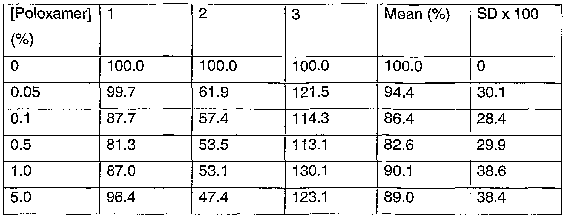
FIG. 5 is a graph illustrating cell viability (%) as a function of poloxamer 407nf (poloxamer) concentration.
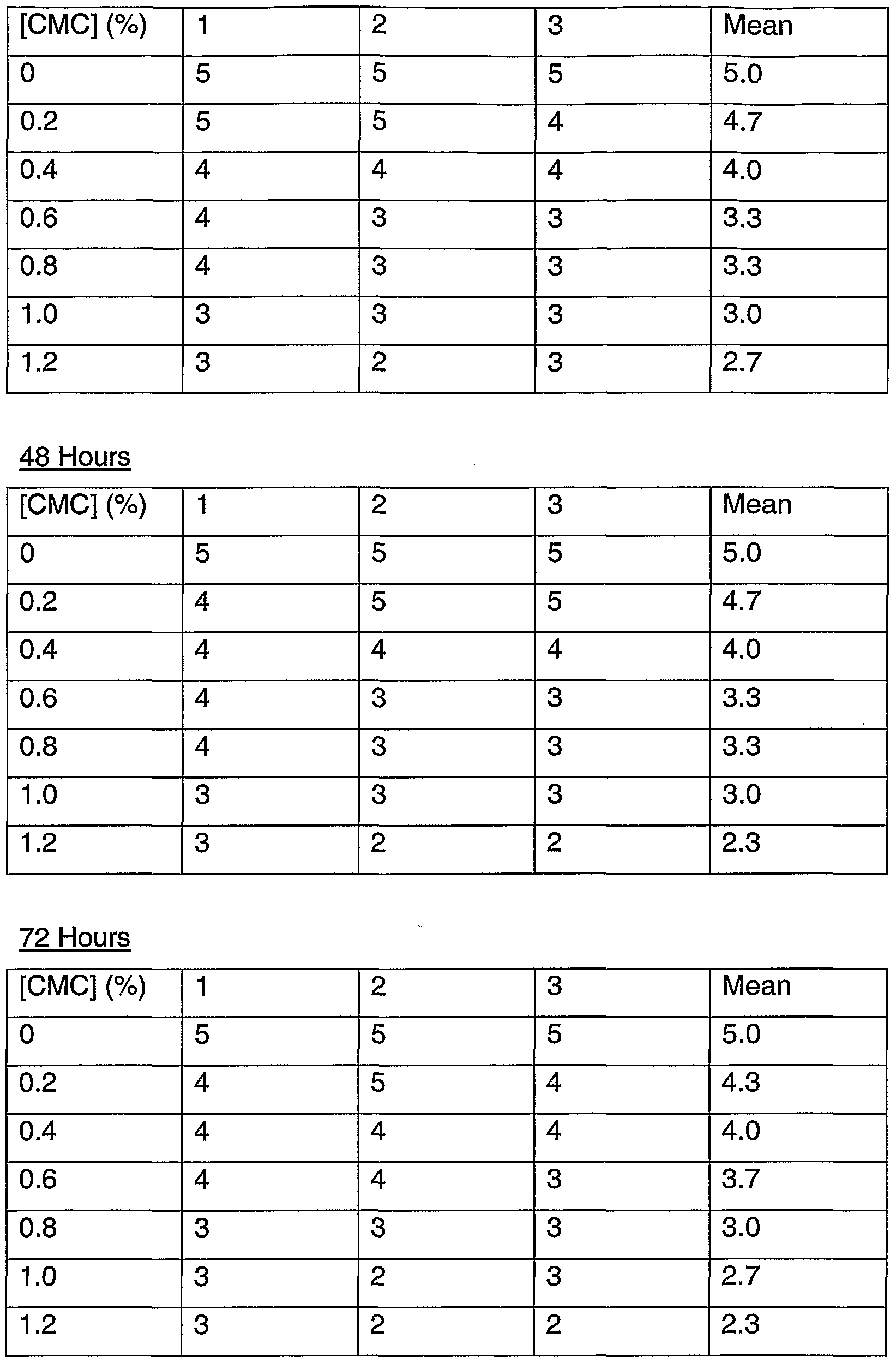
FIG. 6 is a graph and photographs illustrating cell morphology score as a function of poloxamer concentration at 24 hour, 48 hour, and 72 hour time points.
FIG. 7 is a graph illustrating cell viability (%) as a function of hyaluronic acid (HA) concentration.
FIG. 8 is a graph and photographs illustrating cell morphology score as a function of HA concentration at 24 hour, 48 hour, and 72 hour time points. FIG. 9 is a graph illustrating cell viability (%) as a function of hydroxypropyl gamma-cyclodextrin (hydroxypropyl gamma-CD) concentration.
FIG. 10 is a graph and photographs illustrating cell morphology score as a function of hydroxypropyl gamma-CD concentration at 24 hour, 48 hour, and 72 hour time points.
FIG. 11 is a graph illustrating cell viability (%) as a function of sulfobutyl ether 4 beta-cyclodextrin (sulfobuytyl ether 4 beta-CD) concentration. FIG. 12 is a graph and photographs illustrating cell morphology score as a function of sulfobutyl ether 4 beta-CD concentration at 24 hour, 48 hour, and 72 hour time points.
FIG. 13 is a graph illustrating cell viability (%) as a function of hydroxypropyl beta-cyclodextrin (hydroxypropyl beta-CD) concentration.
FIG. 14 is a graph and photographs illustrating cell morphology score as a function of hydroxypropyl beta-CD concentration at 24 hour, 48 hour, and 72 hour time points.
FIG. 15 is a graph illustrating cell viability (%) as a function of benzyl alcohol (benzylOH) concentration.
FIG. 16 is a graph and photographs illustrating cell morphology score as a function of benzylOH concentration at 24 hour, 48 hour, and 72 hour time points.
FIG. 17 is a graph illustrating cell viability (%) as a function of borate buffer (X Eur. Ph. Borate Buffer) concentration.
FIG. 18 is a graph and photographs illustrating cell morphology score as a function of borate buffer concentration at 24 hour, 48 hour, and 72 hour time points.
FIG. 19 is a graph illustrating cell viability (%) as a function of phosphate buffer (X phosphate) concentration.
FIG. 20 is a graph illustrating cell morphology score as a function of phosphate buffer concentration at 24 hour, 48 hour, and 72 hour time points.
FIG. 21 is a graph illustrating cell viability (%) as a function of polysorbate 80 concentration. FIG. 22 is a graph and photographs illustrating cell morphology score as a function of polysorbate 80 concentration at 24 hour, 48 hour, and 72 hour time points.
FIG. 23 is a series of photographs illustrating cell morphology characteristics used in scoring the RPE cell cultures.
DESCRIPTION
Drug delivery systems and methods have been invented which provide effective treatment of ocular conditions, such as disorders or diseases of the posterior segment of an eye of an individual, such as a human or animal. The present systems comprise a therapeutic component, an excipient component, and a polymeric component.
As used herein, a drug delivery system refers to one or more elements, such as a drug delivery element, structured, such as being sized and shaped, for placement in the interior of an eye, such as the posterior segment or vitreous of an eye. The drug delivery elements release a drug or therapeutic agent into the eye for extended time periods relative to liquid therapeutic compositions that may be administered to the interior of an eye. For example, a single administration of a drug delivery system in accordance with the disclosure herein can result in release of therapeutically effective amounts of a drug or therapeutic agent for at least about 30 days and in certain embodiments for a time of about 6 months, 1 year or even 5 or more years.
In the context of the present description, it may be understood that the present drug delivery systems comprise substantially non-liquid drug delivery elements. In other words, the present systems comprise solid or substantially solid or semi-solid polymeric elements. Formulations can also undergo a sol-gel transition upon administration into the eye such that the formulation is liquid prior to administration or dosing and solidifies or gels upon administration. This can be a thermo-gelling system, an in-situ gelling system or other solidifying system. Thus, the elements of the present drug delivery systems refer to a drug- containing bioerodible or biodegradable particles or microparticles, populations of drug-containing particles or microparticles, bioerodible or biodegradable implants (which have a larger size than microparticles), and non-bioerodible or non- biodegradable implants. Although some of the elements are substantially non-
liquid compositions, at least some embodiments may comprise a liquid component, which typically constitutes a minor portion, such as less than 50%, of the element. The present drug delivery systems can be provided in a liquid composition if desired, and thus, the present invention encompasses compositions which may comprise the drug delivery systems disclosed herein.
As discussed herein, the therapeutic component of the present drug delivery systems comprises one or more therapeutic agents or drugs, the excipient component comprises one or more excipient agents or otherwise inert substances, and the polymeric component comprises one or more polymers useful in forming the present drug delivery systems. When the drug delivery systems comprise a cyclodextrin component, the cyclodextrin component may comprise one or more cyclodextrins or cyclodextrin derivatives. The therapeutic component of the present drug delivery systems is typically present in an amount effective in providing a desired therapeutic effect or effects to an individual, such as a human or animal patient, when the system is administered to the interior of an eye of the individual. It may be understood that the present systems are useful for injection or implantation into the interior of an eye of the individual. More specifically, the present systems are useful for injection or implantation or other administration technique into the posterior segment of the eye, such as into the vitreous of an eye.
The therapeutic component of the present compositions comprises one or more therapeutic agents or drugs. Examples of therapeutic agents or drugs include chemical compounds, macromolecules, proteins, and the like, which are effective in treating an ocular condition, such as an ocular condition of the posterior segment of an eye. In certain embodiments, the therapeutic agents are poorly soluble in aqueous environments. Thus, the therapeutic agents may be present as particles in the drug delivery systems.
Therapeutic agents which may be provided in the therapeutic component of the present drug delivery systems may be obtained from public sources or may be synthesized using routine chemical procedures known to persons of ordinary
skill in the art. Agents are screened for therapeutic efficacy using conventional assays known to persons of ordinary skill in the art. For example, agents can be monitored for their effects on reducing intraocular pressure, reducing or preventing neovascularization in the eye, reducing inflammation in the eye, and the like using such conventional assays. Thus, the therapeutic component of the present systems can comprise a variety of therapeutic agents, including anti- angiogenesis agents, anti-inflammatory agents, neuroprotective agents, and the like. For example, the therapeutic component of the present drug delivery systems may comprise one or more of the following: anti-excitotoxic agents, anti- histamine agents, antibiotic agents, beta blocker agents, one or more steroid agents, anti-neoplastic agents, ocular hemorrhage treatment agents, immunosuppressive agents, anti-viral agents, anti-oxidant agents, anti- inflammatory agents, including non-steroidal antiinflammatory agents, adrenergic receptor agonists and antagonists, VEGF inhibitor agents, neuroprotective agents, and any ophthalmically therapeutic macromolecule that can be identified and/or obtained using routine chemical screening and synthesis techniques. The therapeutic component may also include salts of the therapeutic agents. Pharmaceutically acceptable acid addition salts of therapeutic compounds of the present systems are those formed from acids which form non- toxic addition salts containing pharmaceutically acceptable anions, such as the hydrochloride, hydrobromide, hydroiodide, sulfate, or bisulfate, phosphate or acid phosphate, acetate, maleate, fumarate, oxalate, lactate, tartrate, citrate, gluconate, saccharate and p-toluene sulphonate salts. Based on the disclosure herein, it may be understood that the therapeutic component is ophthalmically acceptable. Examples of antihistamines include, and are not limited to, loradatine, hydroxyzine, diphenhydramine, chlorpheniramine, brompheniramine, cyproheptadine, terfenadine, clemastine, triprolidine, carbinoxamine, diphenylpyraline, phenindamine, azatadine, tripelennamine, dexchlorpheniramine, dexbrompheniramine, methdilazine, and trimprazine
doxylamine, pheniramine, pyrilamine, chiorcyclizine, thonzylamine, and derivatives thereof.
As used herein, the term "derivative" refers to any substance which is sufficiently structurally similar to the material of which it is identified as a derivative so as to have substantially similar functionality or activity, for example, therapeutic effectiveness, as the material when the substance is used in place of the material. Useful derivatives of a substance can be routinely determined by conducting one or more conventional assays using the derivatives instead of the substance from which the derivative is derived.
Examples of antibiotics include without limitation, cefazolin, cephradine, cefaclor, cephapirin, ceftizoxime, cefoperazone, cefotetan, cefutoxime, cefotaxime, cefadroxil, ceftazidime, cephalexin, cephalothin,, cefamandole, cefoxitin, cefonicid, ceforanide, ceftriaxone, cefadroxil, cephradine, cefuroxime, cyclosporine, ampicillin, amoxicillin, cyclacillin, ampicillin, penicillin G, penicillin V potassium, piperacillin, oxacillin, bacampicillin, cloxacillin, ticarcillin, azlocillin, carbenicillin, methicillin, nafcillin, erythromycin, tetracycline, doxycycline, minocycline, aztreonam, chloramphenicol, ciprofloxacin hydrochloride, clindamycin, metronidazole, gentamicin, lincomycin, tobramycin, vancomycin, polymyxin B sulfate, colistimethate, colistin, azithromycin, augmentin, sulfamethoxazole, trimethoprim, gatifloxacin, ofloxacin, and derivatives thereof.
Examples of beta blockers include acebutolol, atenolol, labetalol, metoprolol, propranolol, timolol, and derivatives thereof.
Examples of steroids include corticosteroids, such as cortisone, prednisolone, flurometholone, dexamethasone, medrysone, loteprednol, fluazacort, hydrocortisone, prednisone, betamethasone, prednisone, methylprednisolone, triamcinolone hexacatonide, paramethasone acetate, diflorasone, fluocinonide, fluocinolone, triamcinolone, triamcinolone acetonide, derivatives thereof, and mixtures thereof.
Examples of antineoplastic agents include adriamycin, cyclophosphamide, actinomycin, bleomycin, duanorubicin, doxorubicin, epirubicin, mitomycin, methotrexate, fluorouracil, carboplatin, carmustine (BCNU), methyl-CCNU, cisplatin, etoposide, interferons, camptothecin and derivatives thereof, phenesterine, taxol and derivatives thereof, taxotere and derivatives thereof, vinblastine, vincristine, tamoxifen, etoposide, piposulfan, cyclophosphamide, and flutamide, and derivatives thereof.
Examples of immunosuppresive agents include cyclosporine, azathioprine, tacrolimus, and derivatives thereof.
Examples of antiviral agents include interferon gamma, zidovudine, amantadine hydrochloride, ribavirin, acyclovir, valciclovir, dideoxycytidine, phosphonoformic acid, ganciclovir and derivatives thereof.
Examples of antioxidant agents include ascorbate, alpha-tocopherol, mannitol, reduced glutathione, various carotenoids, cysteine, uric acid, taurine, tyrosine, superoxide dismutase, lutein, zeaxanthin, cryotpxanthin, astazanthin, lycopene, N-acetyl-cysteine, carnosine, gamma-glutamylcysteine, quercitin, lactoferrin, dihydrolipoic acid, citrate, Ginkgo Biloba extract, tea catechins, bilberry extract, vitamins E or esters of vitamin E, retinyl palmitate, and derivatives thereof.
Some additional examples of therapeutic agents include anacortave (anti- angiogenesis compound), hyaluronic acid (ocular hemorrhage treatment compound), ketorlac tromethamine (Acular) (non-steroidal anti-inflammatory agent), ranibizumab, pegaptanib (Macugen) (VEGF inhibitors), cyclosporine, gatifloxacin, ofloxacin, epinastine (antibiotics). Macromolecules useful in the present implants may have a molecular weight greater than about 1000 Daltons, for example between about 10,000 and about 1 million Daltons. Examples of suitable macromolecules include large proteins.
Other therapeutic agents include squalamine, carbonic anhydrase inhibitors, brimonidine, prostamides, prostaglandins, antiparasitics, antifungals,
tyrosine kinase inhibitors, glutamate receptor antagonists, including NMDA receptor antagonists, and derivatives thereof.
In view of the foregoing, it can be appreciated that the therapeutic component of the present drug delivery systems can comprise many different types of therapeutic agents, and that such agents are routinely known to or obtained by persons of ordinary skill in the art.
The therapeutic agent may be in a particulate or powder form and may be associated with the polymeric component in a number of different configurations. For example, particles of the therapeutic agent may be entrapped by a polymer matrix, such as a biodegradable polymer matrix. Or, therapeutic agent particles may be encompassed by the polymeric component, such as in the form of a diffusion controlled implant. In certain embodiments, therapeutic agent particles in the present drug delivery systems may have an effective average size less than about 3000 nanometers. In other embodiments, the particles may have an effective average size greater than 3000 nanometers. In certain implants, the particles may have an effective average particle size about an order of magnitude smaller than 3000 nanometers. For example, the particles may have an effective average particle size of less than about 500 nanometers. In additional implants, the particles may have an effective average particle size of less than about 400 nanometers, and in still further embodiments, a size less than about 200 nanometers. The particles of the therapeutic agent may be associated with the polymeric component to form the present microparticles or implants.
The therapeutic agent of the present drug delivery systems is preferably present in an amount from about 1% to 90% by weight of the drug delivery system or drug delivery element. More preferably, the therapeutic agent is present in an amount from about 20% to about 80% by weight of the system or element. In a preferred embodiment, the therapeutic agent comprises about 40% by weight of the system or element (e.g., 30%-50%). In another embodiment, the therapeutic agent comprises about 60% by weight of the system or element. In embodiments comprising water soluble therapeutic components, the water
soluble therapeutic agent may be provided in an amount from about 5% to about 25% by weight.
The present drug delivery systems comprise an excipient component. Any conventional excipient agent which is useful in liquid ophthalmic compositions, such as ophthalmic formulations, suspension, and the like, or is useful in ophthalmic polymeric devices may be used in the present drug delivery systems. Examples of excipient agents include viscosing agents or viscosity inducing agents, solubilizing agents, preservative agents, buffer agents, or tensioactive agents.
Viscosing agents include, without limitation, sodium carboxymethylcellulose (CMC), hydroxypropylmethyl cellulose (HPMC), poloxamer 407nf (Pluronic® F127 Prill), and hyaluronic acid.
Solubilizing agents include without limitation, cyclodextrins (CDs), such as hydroxypropyl gamma-CD (Cavasol®), sulfobutyl ether 4 beta-CD (Captisol®), and hydroxypropyl beta-CD (Kleptose®). Preservative agents may include benzyl alcohol.
Buffer agents may include phosphate buffers, such as dibasic sodium phosphate heptahydrate, monobasic sodium phosphate monohydrate; and/or borate buffers, such as sodium borate, boric acid, sodium chloride (according to Eu. Pharmacopeia).
Resuspension agents may include polysorbate 80 (TweenδO®).
Tensioactive agents may include sodium chloride sugar alcohols, such as mannitol.
The present drug delivery systems comprise an excipient component which comprises one or more excipients. The excipient component is provided in an amount such that as the excipient component is released from the drug
delivery system, the excipient is released in an amount that is less toxic to retinal pigment epithelial cells than an equal amount of benzyl alcohol or polysorbate 80. Thus, the present drug delivery systems may be understood to comprise excipients that are less toxic than excipients currently used in ophthalmic compositions. Administration of the present drug delivery systems to the interior of the eye advantageously provide reduced inflammation compared to existing ophthalmic compositions.
Certain embodiments of the present drug delivery systems comprise a cyclodextrin component associated with a therapeutic component to improve or enhance the therapeutic efficacy and/or bioavailability of the therapeutic cqmponent. For example, the cyclodextrin component may be associated with the therapeutic component to enhance the release profile of the therapeutic component from the drug delivery element or elements, enhance or improve the stability of the therapeutic component in the drug delivery element or in the eye, and/or enhance or improve the ocular tolerability of the element and/or therapeutic component, relative to drug delivery systems which comprise the same therapeutic component and substantially no cyclodextrin component. The cyclodextrin component of the present cyclodextrin-containing drug delivery systems comprises one or more cyclodextrins or cyclodextrin derivatives. As discussed herein, cyclodextrins have been discovered to have a reduced toxicity to retinal cells relative to polysorbate 80 or benzyl alcohol, even at substantially higher concentrations than toxic amounts of polysorbate 80 or benzyl alcohol. Thus, the cyclodextrin component of the present drug delivery systems contribute to the enhanced compatibility and tolerance of the present systems to the tissues in the posterior segment of the eye, for example, the retina of the eye, relative to compositions or drug delivery systems previously proposed for placement into a posterior segment of an eye which contain polysorbate 80 and/or benzyl alcohol, for example, the composition sold under the trademark Kenalog®-40.
The cyclodextrin component of the present systems may comprise a cyclodextrin selected from the group consisting of alpha-cyclodextrin, beta-
cyclodextrin, gamma-cyclodextrin, derivatives thereof, and mixtures thereof. The term "cyclodextrin derivative" has the broadest meaning generally understood in the art, and refers to a compound or a mixture of compounds wherein one or more of the free hydroxyl groups of alpha-, beta-, or gamma-cyclodextrin is replaced with any other group. A "water-soluble" cyclodextrin derivative is soluble at a concentration of at least 300 mg/mL in water. The cyclodextrin derivative used in the systems disclosed herein may vary. Derivatives of alpha- cyclodextrin, beta-cyclodextrin, and gamma-cyclodextrin may be used. In certain systems, a beta-cyclodextrin derivative such as calcium sulfobutylether-beta- cyclodextrin, sodium sulfobutylether-beta-cyclodextrin, and hydroxypropyl-beta- cyclodextrin, may be used. Alternatively, a gamma-cyclodextrin derivative such as calcium sulfobutylether-gamma-cyclodextrin, sodium sulfobutylether-gamma- cyclodextrin, and hydroxypropyl-gamma-cyclodextrin may be used. Some specific derivatives contemplated herein are the hydroxypropyl derivatives of cyclodextrins, such as hydroxypropyl-beta-cyclodextrin or hydroxypropyl-gamma- cyclodextrin.
The amount of the excipient component that is released from the drug delivery systems is preferably an amount that is not substantially toxic to retinal cells, including RPE cells. The exact amounts of different excipient agents that are released into the eye can vary, but overall the amounts may range from about 0.1% to about 5% or 10% concentrations. Understandably, the drug delivery system may comprise a greater amount of the excipient agent to facilitate delivery of these amounts. Examples of specific amounts that may be released into the eye include 0.5 % of a cyclodextrin, 0.5% of a vitamin E agent, 2% hyaluronic acid, 2 % of a vitamin E agent, and 5% of a cyclodextrin. The exact amounts can be determined by measuring the toxicity of such excipient agents in vitro, as described herein, or by administering formulations or drug delivery systems with desired amounts into the interior of the eye and monitoring the effects of such exposure to retinal cells or the eye or individual in general.
For example, an in vivo method that may be useful to determine the desired amount of excipients to provide in the present drug delivery system may comprise inserting a drug delivery system into an eye of the animal. Different
systems comprising different amounts and/or combinations of excipients may be administered to eyes of different animals. The animals and eyes can be monitored and/or examined for viability, clinical effects, and gross ocular effects. In certain methods, the effects can be monitored by slit lamp biomicroscopy, pupillary reflex, ophthalmoscopy, electroretinography (ERG), intraocular pressure (IOP), body weight, macroscopic observations, and microscopic pathololgy of ocular tissues. Dose response curves can be obtained based on the results of such methods, and the desired amounts of the excipient agents can be determined. Results which indicate that systems having a certain amount of an excipient do not produce inflammation, irritation, or other adverse side effects compared to control systems may be indicative that such excipient-containing systems have a low retinal cell toxicity.
The present drug delivery systems also comprise a polymeric component. Suitable polymeric materials or compositions for use in the drug delivery systems include those materials which are compatible, that is biocompatible, with the eye so as to cause no substantial interference with the functioning or physiology of the eye. In certain embodiments, the materials preferably are at least partially and more preferably substantially completely biodegradable or bioerodible. In other embodiments, non-biodegradable polymers are used. Non-biodegradable polymers may be particularly useful in diffusion-based drug delivery systems, such as systems which include a drug-containing core and have a coating with one or more pores to permit the drug to diffuse therefrom. Examples of useful polymeric materials include, without limitation, such materials derived from and/or including organic esters and organic ethers, which when degraded result in physiologically acceptable degradation products, including the monomers. Also, polymeric materials derived from and/or including, anhydrides, amides, orthoesters and the like, by themselves or in combination with other monomers, may also find use. The polymeric materials may be addition or condensation polymers, advantageously condensation polymers. The polymeric materials may be cross-linked or non-cross-linked, for example not more than lightly cross-linked, such as less than about 5%, or less than about 1 % of the polymeric material being cross-linked. For the most part, besides carbon
and hydrogen, the polymers will include at least one of oxygen and nitrogen, advantageously oxygen. The oxygen may be present as oxy, e.g. hydroxy or ether, carbonyl, e.g. non-oxo-carbonyl, such as carboxylic acid ester, and the like. The nitrogen may be present as amide, cyano and amino. The polymers set forth in Heller, Biodegradable Polymers in Controlled Drug Delivery, In: CRC Critical Reviews in Therapeutic Drug Carrier Systems, Vol. 1 , CRC Press, Boca Raton, FL 1987, pp 39-90, which describes encapsulation for controlled drug delivery, may find use in the present implants. Of additional interest are polymers of hydroxyaliphatic carboxylic acids, either homopolymers or copolymers, and polysaccharides. Polyesters of interest include polymers of D-lactic acid, L-lactic acid, racemic lactic acid, glycolic acid, polycaprolactone, and combinations thereof. Generally, by employing the L- lactate or D-lactate, a slowly eroding polymer or polymeric material is achieved, while erosion is substantially enhanced with the lactate racemate. Additionally, the higher the glycolate content the faster the erosion. The higher the crystallinity the slower the erosion and the lower the molecular weight the faster the erosion.
Among the useful polysaccharides are, without limitation, calcium alginate, and functionalized celluloses, particularly carboxymethylcellulose esters characterized by being water insoluble, a molecular weight of about 5 kD to 500 kD, for example.
Other polymers of interest include, without limitation, polyesters, polyethers and combinations thereof which are biocompatible and may be biodegradable and/or bioerodible.
Some preferred characteristics of the polymers or polymeric materials for use in the present invention may include biocompatibility, compatibility with the therapeutic component, ease of use of the polymer in making the drug delivery systems of the present invention, a half-life in the physiological environment of at least about 6 hours, preferably greater than about one day, not significantly increasing the viscosity of the vitreous, and water insolubility.
The biodegradable polymeric materials which are included to form the present elements are desirably subject to enzymatic or hydrolytic instability. Water soluble polymers may be cross-linked with hydrolytic or biodegradable unstable cross-links to provide useful water insoluble polymers. The degree of stability can be varied widely, depending upon the choice of monomer, whether a homopolymer or copolymer is employed, employing mixtures of polymers, and whether the polymer includes terminal acid groups.
Also important to controlling the biodegradation of the polymer and hence the extended release profile of the implant is the relative average molecular weight of the polymeric composition employed in the implant. Different molecular weights of the same or different polymeric compositions may be included in the implant to modulate the release profile. In certain drug delivery systems, the relative average molecular weight of the polymer will range from about 9 to about 64 kD, usually from about 10 to about 54 kD, and more usually from about 12 to about 45 kD.
In some systems, copolymers of glycolic acid and lactic acid are used, where the rate of biodegradation is controlled by the ratio of glycolic acid to lactic acid. The most rapidly degraded copolymer has roughly equal amounts of glycolic acid and lactic acid. Homopolymers, or copolymers having ratios other than equal, are more resistant to degradation. The ratio of glycolic acid to lactic acid will also affect the brittleness of the drug delivery element, where a more flexible element is desirable for larger geometries. The % of polylactic acid in the polylactic acid polyglycolic acid (PLGA) copolymer can be 0-100%, preferably about 15-85%, more preferably about 35-65%. In some elements, a 50/50 PLGA copolymer is used.
The biodegradable polymer matrix of some drug delivery systems may comprise a mixture of two or more biodegradable polymers. For example, the elements of the system may comprise a mixture of a first biodegradable polymer and a different second biodegradable polymer. One or more of the biodegradable polymers may have terminal acid groups.
Release of a drug from an erodible polymer is the consequence of several mechanisms or combinations of mechanisms. Some of these mechanisms include desorption from the implant's surface, dissolution, diffusion through porous channels of the hydrated polymer and erosion. Erosion can be bulk or surface or a combination of both. As discussed herein, a matrix of the drug delivery system may release drug at a rate effective to sustain release of an amount of the therapeutic agent for more than one week after implantation into an eye. In certain systems, therapeutic amounts of the therapeutic agent are released for more than about one month, and even for about six months or more.
The release of the therapeutic agent from a drug delivery system comprising a biodegradable polymer matrix may include an initial burst of release followed by a gradual increase in the amount of the therapeutic agent released, or the release may include an initial delay in release of the therapeutic agent followed by an increase in release. As discussed herein, the rate of release or the release profile can be changed and controlled by the presence of the cyclodextrin component. When the biodegradable system is substantially completely degraded, the percent of the therapeutic agent that has been released is about one hundred. Compared to existing implants, the systems disclosed herein do not completely release, or release about 100% of the therapeutic agent, until after about one week of being placed in an eye.
It may be desirable to provide a relatively constant rate of release of the therapeutic agent from the system over the life of the system. For example, it may be desirable for the therapeutic agent to be released in amounts from about 0.01 μg to about 2 μg per day for the life of the system. However, the release rate may change to either increase or decrease depending on the formulation of the biodegradable polymer matrix. In addition, the release profile of the therapeutic agent may include one or more linear portions and/or one or more non-linear portions. Preferably, the release rate is greater than zero once the implant has begun to degrade or erode.
The present cyclodextrin-containing drug delivery systems have desirable release rates due to the presence of the cyclodextrin component. As discussed
herein, drug delivery implants with no cyclodextrin component may have a noticeable lag time. By associating the cyclodextrin component with the therapeutic component in the present drug delivery systems, the lag time of the release profile of the therapeutic component can be reduced, thereby enhancing the release rate of the therapeutic component from the drug delivery element.
The present drug delivery elements may be monolithic, i.e. having the active agent or agents homogenously distributed through a polymeric matrix, or encapsulated, where a reservoir of active agent is encapsulated by a polymeric matrix. Due to ease of manufacture, monolithic elements are usually preferred over encapsulated forms. However, the greater control afforded by the encapsulated, reservoir-type elements may be of benefit in some circumstances, where the therapeutic level of the drug falls within a narrow window. In addition, the therapeutic component, including the therapeutic agent(s) described herein, may be distributed in a non-homogenous pattern in a polymeric matrix. For example, the drug delivery element may include a portion that has a greater concentration of the therapeutic agent relative to a second portion of the element.
Thus, it may be understood that bioerodible polymers can be used to form monolithic homogeneous or heterogeneous implants and microparticulates, membrane controlled implants or microparticulates, multistage delivery systems, or any combination thereof. The polymers comprising the carrier delivery system can be natural or synthetic polymers. In certain embodiments, examples of polymers include polyesters, poly (ortho esters) or polyanhydrides, as discussed above. Some specific polymers include poly-lactic acid (PLA), poly (lactide-co- glycolide) (PLGA), poly-l-lactic acid (PLLA), polycaprolactone, poly (ortho acetate), and combinations thereof.
Examples of intraocular elements disclosed herein may have a size of between about 5 μm and about 2 mm, or between about 10 μm and about 1 mm for administration with a needle, greater than 1 mm, or greater than 2 mm, such as 3 mm or up to 10 mm, for administration by surgical implantation. The vitreous chamber in humans is able to accommodate relatively large elements of varying geometries, having lengths of, for example, 1 to 10 mm. The element
may ue an implant in tne τorm ot a cylindrical pellet (e. g., rod) with dimensions of about 2 mm x 0.75 mm diameter. Or the element may be an implant in the form of a cylindrical pellet with a length of about 7 mm to about 10 mm, and a diameter of about 0.75 mm to about 1.5 mm.
Polymeric particles of the present drug delivery systems are smaller in size than the implants. The particles may have any desired shape. In certain embodiments, the particles are substantially spherical. In other embodiments, the particles have a non-spherical shape. Particles which comprise complexes of the therapeutic component and the cyclodextrin component may have a dimension from about 1 μ to about 1 mm, for example. In certain particles, the dimension corresponds to a maximum diameter. In further embodiments, the particles may have a maximum diameter from about 3 μm to about 1 mm. In view of the disclosure herein, the polymeric particles may comprise particles of a therapeutic agent complexed or encapsulated with cyclodextrins. Polymeric particles can be produced using conventional methods known by persons of ordinary skill in the art. For example, polymeric particles can be produced by milling the relatively larger implants disclosed herein. The implants of the present systems may also be at least somewhat flexible so as to facilitate both insertion of the implant in the eye, such as in the vitreous, and accommodation of the implant. The total weight of the implant is usually about 250-5000 μg, more preferably about 500-1000 μg. For example, an implant may be about 500 μg, or about 1000μg. For non-human individuals, the dimensions and total weight of the implant(s) may be larger or smaller, depending on the type of individual. For example, humans have a vitreous volume of approximately 3.8 ml, compared with approximately 30 ml for horses, and approximately 60-100 ml for elephants. An implant sized for use in a human may be scaled up or down accordingly for other animals, for example, about 8 times larger for an implant for a horse, or about, for example, 26 times larger for an implant for an elephant.
Non-homogenous implants can be prepared where the center may be of one material and the surface may have one or more layers of the same or a
different composition, where the layers may be cross-linked, or of a different molecular weight, different density or porosity, or the like. For example, where it is desirable to quickly release an initial bolus of drug, the center may be a polylactate coated with a polylactate-polyglycolate copolymer, so as to enhance the rate of initial degradation. Alternatively, the center may be polyvinyl alcohol coated with polylactate, so that upon degradation of the polylactate exterior the center would dissolve and be rapidly washed out of the eye.
The implants may be of any geometry including fibers, sheets, films, spheres, circular discs, plaques and the like. The upper limit for the implant size will be determined by factors such as toleration for the implant, size limitations on insertion, ease of handling, etc. Where sheets or films are employed, the sheets or films will be in the range of at least about 0.5 mm x 0.5 mm, usually about 3-10 mm x 5-10 mm with a thickness of about 0.1-1.0 mm for ease of handling. Where fibers are employed, the fiber diameter will generally be in the range of about 0.05 to 3 mm and the fiber length will generally be in the range of about 0.5-10 mm. Spheres may be in the range of about 5 μm to 4 mm in diameter, with comparable volumes for other shaped particles. The size and form of the implant can also be used to control the rate of release, period of treatment, and drug concentration at the site of implantation. Larger implants will deliver a proportionately larger dose, but depending on the surface to mass ratio, may have a slower release rate. The particular size and geometry of the implant are chosen to suit the site of implantation.
The proportions of therapeutic agent, polymer, excipient agents, and any other modifiers may be empirically determined by formulating several drug delivery elements with varying proportions. A USP approved method for dissolution or release test can be used to measure the rate of release (USP 23; NF 18 (1995) pp. 1790-1798). For example, using the infinite sink method, a weighed sample of the element is added to a measured volume of a solution containing 0.9% NaCI in water, where the solution volume will be such that the drug concentration after release is less than 5% of saturation. The mixture is maintained at 37°C and stirred slowly to maintain the elements in suspension.
The appearance of the dissolved drug as a function of time may be followed by various methods known in the art, such as spectrophotometrically, HPLC, mass spectroscopy, etc. until the absorbance becomes constant or until greater than 90% of the drug has been released.
In addition to the therapeutic component, the intraocular implants disclosed herein may include effective amounts of other excipients in addition to those described above. For example, the present implants may include effective amounts of buffering agents, preservatives and the like, which have a reduced toxicity, such as a reduced toxicity relative to polysorbate 80 or benzyl alcohol. Suitable water soluble buffering agents include, without limitation, alkali and alkaline earth carbonates, phosphates, bicarbonates, citrates, borates, acetates, succinates and the like, such as sodium phosphate, citrate, borate, acetate, bicarbonate, carbonate and the like. These agents are advantageously present in amounts sufficient to maintain a pH of the system of between about 2 to about 9 and more preferably about 4 to about 8, such as about 7.2 to about 7.5. As such the buffering agent may be as much as about 5% by weight of the total implant. Suitable water soluble preservatives include sodium bisulfite, sodium bisulfate, sodium thiosulfate, ascorbate, benzalkonium chloride, chlorobutanol, thimerosal, phenylmercuric acetate, phenylmercuric borate, phenylmercuric nitrate, parabens, methylparaben, polyvinyl alcohol, phenylethanol and the like and mixtures thereof. These agents may be present in amounts of from 0.001 to about 5% by weight and preferably 0.01 to about 2% by weight. In certain embodiments of the present drug delivery systems, the systems comprise substantially no polysorbate 80 or benzyl alcohol. As discussed herein, polysorbate 80 and/or benzyl alcohol are believed to be responsible for retinal pigment epithelial cell toxicity associated with existing intraocular ophthalmic formulations. Thus, embodiments of the present systems comprise a cyclodextrin component and substantially no polysorbate 80 or benzyl alcohol, such as less than 0.05% benzyl alcohol. In other words, and in certain embodiments, the present systems are substantially free of added preservative components, or include effective preservative components which are more compatible with or friendly to the posterior segment, e.g., retina or RPE, of the
eye relative to benzyl alcohol, which is included in the Kenalog®-40 composition as a preservative, as discussed herein. The cyclodextrin component is present in these embodiments of the system in an amount that is less toxic to retinal pigment epithelial cells relative to an equal amount of either polysorbate 80 or benzyl alcohol.
In certain embodiments of the present drug delivery systems, the cyclodextrin component is associated with the therapeutic component in a manner such that the cyclodextrins encapsulate a drug. For example, the cyclodextrins may be complexed with the drug or drugs of the therapeutic component. Drugs, such as chemical compounds, are encapsulated or complexed with cyclodextrins by conventional methods which are routine to persons of ordinary skill in the art. For example, the drug or drugs can be encapsulated or complexed with cyclodextrins by methods which include at least one step of freeze drying, spray drying, solvent evaporation, and the like. In certain embodiments, the drug is complexed with the cyclodextrin prior to fabrication of the drug delivery systems. In other embodiments, the complexation can occur in-situ during the manufacture of the drug delivery system. In embodiments which comprise macromolecule therapeutic agents, the association of the cyclodextrin component with the therapeutic agent may be obtained using other conventional methods.
Based on Equation 1 and Equation 2 and the discussion therewith, it has been discovered that in order for a cyclodextrin component to improve the solubility of a therapeutic component, and subsequently release the therapeutic component from diffusion controlled implants, the therapeutic component must be complexed with the cyclodextrin component. Simply mixing the therapeutic component and the cyclodextrin component together does not enhance solubility. However, it may also be understood that complexation may not be required when the primary goal is to obtain reduced toxicity effects without the desirable release rate of the present drug delivery systems.
As discussed herein, embodiments of the present drug delivery system have augmented release profiles of the therapeutic component compared to
substantially identical drug delivery systems without a cyclodextrin component. For example, implants may have an enhanced release profile of the therapeutic component relative to implants that include substantially no cyclodextrin component. In certain implants, the release lag time of the therapeutic component can be more desirably controlled. The release lag time refers to a period of slow drug release or no drug release preceding a more rapid rate of release of the drug from an implant. This release lag time can be caused by a slow wetting and dissolution of the drug, or a delayed water penetration of the implant. The release lag time represents the non-steady state release from the implant.
Embodiments of the present drug delivery systems which include complexes of a cyclodextrin component and therapeutic component can provide substantial advantages over existing systems. For example, more rapid wetting can be obtained, more rapid dissolution of the drug with mitigation of the lag time can be obtained, and the lag time can be more precisely controlled, compared to systems which do not include a cyclodextrin component. Complexation between a cyclodextrin component and a therapeutic component occurs at a molecular level and variances in particle size or particle size distribution of the drug become insignificant.
In addition, the present drug delivery systems provide enhanced stability of the therapeutic component relative to drug delivery systems without a cyclodextrin component.
The release profile and other characteristics of the present systems can be measured in environments which mimic the vitreous of an eye using conventional methods which are routine to persons of ordinary skill in the art. For example, implants can be immersed in a 3 mL volume of liquid, and the release rate of the therapeutic component can be monitored, as discussed herein.
In certain embodiments of the present drug delivery systems, the therapeutic component comprises, consists essentially of, or consists of, steroids and/or steroid precursors. As used herein, a steroid precursor is understood to
be an agent that can be converted into a therapeutically effective steroid by physiological processes. Steroid precursors may be understood to be steroid prodrugs. An example of a steroid precursor or steroid prodrug is a compound that is converted in vivo into a steroid after the compound is administered into the eye. For example, a prednisolone precursor is a compound that is converted to prednisolone in vivo. A dexamethasone precursor is a compound that is converted to dexamethasone in vivo. A triamcinolone precursor is a compound that is converted to triamcinolone in vivo. Steroids and steroid precursors can be obtained from commercial suppliers, or can be synthesized using routine methods known to persons of ordinary skill in the art, and can be screened using conventional methods known to persons of ordinary skill in the art. The steroid or steroid precursor may be present in the drug delivery system as a plurality of particles. As discussed herein, the therapeutic component may comprise one or more therapeutic agents that are poorly soluble. For example, the therapeutic agent may have a limited solubility in water, for example, at 25 degrees C. In certain embodiments, the therapeutic component comprises a therapeutic agent that has a solubility in water at 25 degrees C of less than 10 mg/ml. The therapeutic component should be ophthalmically acceptable, that is, should have substantially no significant or undue detrimental effect of the eye structures or tissues. Embodiments comprising a corticosteroid component have an ability of such component to reduce inflammation in the posterior segment of the eye into which the drug delivery system is placed caused by the result of one or more diseases and/or conditions in the posterior segment of the eye.
In at least one embodiment of the present drug delivery systems, the therapeutic component comprises, consists essentially of, or consists entirely of at least one steroid selected from the group consisting of cortisone, dexamethasone, fluorometholone, loteprednol, medrysone, prednisolone, prednisolone acetate, triamcinolone, and triamcinolone acetonide.
As discussed herein, the present drug delivery systems may comprise one or more types of cyclodextrins or cyclodextrin derivatives, such as alpha-
cyclodextrins, beta-cyclodextrins, gamma-cyclodextrins, and derivatives thereof. As discussed herein, cyclodextrin derivatives can be understood to be any substituted or otherwise modified compound that has the characteristic chemical structure of a cyclodextrin sufficiently to function as a cyclodextrin, for example, to enhance the solubility and/or stability of therapeutic agents and/or reduce unwanted side effects of the therapeutic agents and/or to form inclusive complexes with the therapeutic agents. In certain embodiments, the cyclodextrin component comprises at least one cyclodextrin selected from the group consisting of sulfobutyl ether 4-beta-cyclodextrin, hydroxypropyl beta- cyclodextrin, and hydroxypropyl gamma-cyclodextrin.
As discussed herein, embodiments of the present drug delivery systems comprise a cyclodextrin component present in an amount that has a reduced toxicity to retinal pigment epithelial cells relative to an equal amount of polysorbate 80 or benzyl alcohol. In certain embodiments of the present systems, the cyclodextrin component comprises an amount of hydroxypropyl gamma-cyclodextrin which is released in an amount from about 0.1% (w/v) to about 10% (w/v) in the eye. Certain embodiments may comprise an amount of sulfobutyl ether 4-beta-cyclodextrin which is released in an amount from about 0.1 % (w/v) to about 10% (w/v) in the eye. Further embodiments may comprise an amount of hydroxypropyl beta-cyclodextrin which is released in an amount from about 0.1% (w/v) to about 5% (w/v) in the eye.
In one embodiment of the present systems, the cyclodextrin component is provided in an effective amount to solubilize a minor amount, that is less than 50%, for example in a range of 1% or about 5% to about 10% or about 20% of a corticosteroid component. For example, the inclusion of a cyclodextrin component, such as beta-cyclodextrin, secondary butylether beta-cyclodextrin, other cyclodextrins and the like and mixtures thereof, at about 0.5 to about 25.% (w/w) solubilizes about 1 to about 10% of the initial dose of triamcinolone acetonide. This presolubilized fraction provides a readily bioavailable loading dose, thereby reducing delay time in therapeutic effectiveness. The use of such a corticosteroid component may provide a relatively quick release of the corticosteroid component into the eye for therapeutic effectiveness.
In view of the disclosure herein, one useful embodiment of the present drug delivery systems comprises a therapeutic component present in an amount effective in providing a desired therapeutic effect to an individual when the composition is administered to the interior of an eye of the individual; and at least one cyclodextrin selected from the group consisting of sulfobutyl ether 4-beta- cyclodextrin, hydroxypropyl beta-cyclodextrin, and hydroxypropyl gamma- cyclodextrin, the cyclodextrin is complexed with the therapeutic component. The complexes are associated with a polymeric component in the form of a drug delivery element structured to be placed in the eye. As discussed herein, the drug delivery system is substantially free of polysorbate 80 or benzyl alcohol.
In another embodiment, a therapeutic drug delivery system useful for injection into a posterior segment of an eye of an individual, comprises a therapeutic component present in an amount effective in providing a desired therapeutic effect to an individual when the system is placed in the interior of an eye of the individual; and a cyclodextrin component present in an amount from about 0.5% (w/w) to about 25% (w/w) of the system and effective in solubilizing a therapeutic agent of the therapeutic component. In additional embodiments, the cyclodextrin component is provided in an amount from about 5.0% (w/w) to about 15% (w/w) of the system. Such an amount of the cyclodextrin component may be effective in solubilizing about 50% or less of the therapeutic agent of the therapeutic component. In accordance with the disclosure herein, such an amount of a cyclodextrin component, at least in certain embodiments, provides a reduced toxicity relative to an equal amount of polysorbate 80 or benzyl alcohol when released from the drug delivery system.
By utilizing amounts of the cyclodextrin component which have a reduced toxicity relative to equal amounts of polysorbate 80 or benzyl alcohol, the present drug delivery systems may be understood to have a reduced toxicity relative to a second substantially identical drug delivery systems which comprises polysorbate 80 or benzyl alcohol, or both, and which is substantially free of a cyclodextrin component.
In addition, other embodiments of the present drug delivery systems may comprise one or more excipients selected from the group consisting of polysorbate 80, benzyl alcohol, poloxamer 407nf, sodium carboxymethylcellulose, hydroxypropylmethyl cellulose, and hyaluronic acid provided that such excipients are present in amounts that have a low toxicity. For example, such drug delivery systems comprise an excipient component in an amount that does not substantially affect cell viability or cell morphology, or both. For example, the effects mediated by such excipients results in a reduction in cell viability and cell morphology less than 50% compared to systems without such excipients.
The toxicity of potentially useful ophthalmic excipients can be determined by contacting cultured RPE cells with an excipient. Some detailed procedures are described in the Examples herein. Broadly, a method of screening excipients in accordance with the present invention may comprise a step of contacting cultured RPE cells with an excipient. Generally, the method can be practiced by contacting cultured RPE cells with different concentrations of an excipient at one or more time points. The cultured cells may be examined to determine the effects, such as toxicity, of the excipients on the cells. For example, the viability of the cells may be examined by evaluating the metabolism of the cells, such as by using a colorometric assay. In addition, and/or alternatively, the morphology of the cells may be examined by scoring the cell cultures based on visual criteria, such as cell size and shape and cell monolayer integrity or modification of confluence.
Suitable methods for screening excipients may include culturing RPE cells (such as ARPE-19 cells) in culture dishes and conducting a dose-response for excipients at different time points, such as 24 hours, 48 hours, and 72 hours. Various properties of excipient-containing incubating solutions, such as pH, osmolarity (mOsm), and viscosity, can be measured. Concentrations of the excipients can be determined using routine methods, and can include concentrations commonly used in ophthalmic formulations, concentrations with desired solubility characteristics, and/or limiting the concentrations with desirable viscosity, osmolarity, and/or pH values. The methods may also comprise one or
more steps of measuring cell proliferation, secretion of pro-inflammatory mediators,, and the like.
In addition, as discussed herein, the present screening methods may comprise a step of placing a drug delivery system in an animal's eye. Dose response curves can be obtained using these in vivo screening procedures. From the dose response curve data, the desired amounts can be determined for the drug delivery systems. Various techniques may be employed to produce the drug delivery systems described herein. Useful techniques include, but are not necessarily limited to, solvent evaporation methods, phase separation methods, interfacial methods, molding methods, injection molding methods, extrusion methods, coextrusion methods, carver press method, die cutting methods, heat compression, combinations thereof and the like.
Specific methods are discussed in U.S. Pat. No. 4,997,652. Extrusion methods may be used to avoid the need for solvents in manufacturing. When using extrusion methods, the polymer and drug are chosen so as to be stable at the temperatures required for manufacturing, usually at least about 85 degrees Celsius. Extrusion methods use temperatures of about 25 degrees C to about 150 degrees C, more preferably about 65 degrees C to about 130 degrees C. A drug delivery system may be produced by bringing the temperature to about 60 degrees C to about 150 degrees C for drug/polymer mixing, such as about 130 degrees C, for a time period of about 0 to 1 hour, 0 to 30 minutes, or 5-15 minutes. For example, a time period may be about 10 minutes, preferably about 0 to 5 min. The systems are then extruded at a temperature of about 60 degrees C to about 130 degrees C, such as about 75 degrees C. In addition, the system may be produced by coextruding so that a coating is formed over a core region during the manufacture of the system.
Compression methods may be used to make the system, and typically yield drug delivery elements with faster release rates than extrusion methods.
Compression methods may use pressures of about 50-150 psi, more preferably about 70-80 psi, even more preferably about 76 psi, and use temperatures of about 0 degrees C to about 115 degrees C, more preferably about 25 degrees C. As discussed herein, in certain embodiments, the cyclodextrin component and therapeutic component are present as complexes in the drug delivery systems or when administered to the interior of an eye. Complexation of the cyclodextrin component and a therapeutic agent of the therapeutic component can occur via routine methods known to persons of ordinary skill in the art. For example, complexation of a cyclodextrin component and a therapeutic agent can be accomplished by ultrasonic processing with a high energy microtip sonicator at ambient temperatures. Such a process is effective for processing small volumes of solution. Larger volumes can be processed by autoclaving the mixture at elevated temperatures, such as about 120 degrees C. Excess uncomplexed therapeutic agent can be removed by centrifugation and filtration. Or, as another example, inclusion complexes can be made by:(i) rapid stirring at 25 degrees C for 72 hrs, (ii) high-shear processing at 60 degrees C with a rotor/stator homogenizer, (iii) brief ultrasonication with a high-energy probe sonicator, and (iv) autoclaving in sealed borosilicate glass vials for 10 min at 121 degrees C. Equimolar concentration of therapeutic agent, such as a steroid, can be added to 10% solutions of cyclodextrin in dilute (20 mM) aqueous buffer prior to complex formation. After processing, aliquots are filtered (0.45 μm) for HPLC analysis of soluble, complexed therapeutic agent and the hydrolytic degradant, non-esterified therapeutic agent. For example, see U.S. Patent Publication No. 2002/0198174 (Lyons). The complexes of the therapeutic agent and cyclodextrin component can be combined with the polymer prior to implant fabrication, such as prior to extrusion, or the therapeutic agent, the cyclodextrin, and the polymer can be combined, and the complexes between the therapeutic agent and the cyclodextrins can form in the produced implant. Thus, the present invention encompasses methods of producing or manufacturing a drug delivery system. The method comprises a step of encapsulating the therapeutic component of a drug delivery system with a cyclodextrin component to form complexes, and a step of combining the complexes with a polymeric component to form a drug delivery element.
The present systems are placeable into the interior of an eye of an individual without causing significant adverse effects related to the presence of the systems. For example, the present systems preferably do not cause substantial changes in intraocular pressure of the eye resulting from the placement of the system into the eye. In addition, the present systems preferably do not interfere with the vision of the individual receiving the systems. For example, the present systems may be optically clear, or may be sized or shaped to be placed in the eye without interfering with the field of vision of the individual.
The drug delivery systems disclosed herein may be placed in the interior of any eye using any suitable device, such as a trocar and the like, or the systems may be administered into the eye in an injectable composition. Therefore, it may be understood that the present invention also encompasses compositions which may contain the present drug delivery systems. The drug delivery systems and/or compositions containing such systems are preferably sterile prior to administration to a patient.
The drug delivery elements of the present systems may be inserted into the eye, for example the vitreous chamber of the eye, by a variety of methods, including placement by forceps or by trocar following making a 2-3 mm incision in the sclera. One example of a device that may be used to insert the elements into an eye is disclosed in U.S. Patent Publication No. 2004/0054374. The method of placement may influence the therapeutic component or drug release kinetics. For example, delivering the element with a trocar may result in placement of the element deeper within the vitreous than placement by forceps, which may result in the element being closer to the edge of the vitreous. The location of the element may influence the concentration gradients of therapeutic component or drug surrounding the element, and thus influence the release rates (e.g., an element placed closer to the edge of the vitreous may result in a slower release rate).
The present elements are configured to release an amount of the therapeutic agent effective to treat or reduce a symptom of an ocular condition,
such as an ocular condition such as glaucoma. More specifically, the elements and the systems comprising such elements, may be used in a method to treat or reduce one or more symptoms of glaucoma or proliferative vitreoretinopathy. The elements and systems disclosed herein may also be configured to release additional therapeutic agents, as described above, which are effective in treating one or more symptoms of an ocular condition or are effective in preventing diseases or conditions of the eye, such as the following: MACULOPATHIES/RETINAL DEGENERATION: Non-Exudative Age
Related Macular Degeneration (ARMD), Exudative Age Related Macular Degeneration (ARMD), Choroidal Neovascularization, Diabetic Retinopathy, Acute Macular Neuroretinopathy, Central Serous Chorioretinopathy, Cystoid Macular Edema, Diabetic Macular Edema.
UVEITIS/RETINITIS/CHOROIDITIS: Acute Multifocal Placoid Pigment Epitheliopathy, Behcet's Disease, Birdshot Retinochoroidopathy, Infectious (Syphilis, Lyme, Tuberculosis, Toxoplasmosis), Intermediate Uveitis (Pars Planitis), Multifocal Choroiditis, Multiple Evanescent White Dot Syndrome (MEWDS), Ocular Sarcoidosis, Posterior Scleritis, Serpignous Choroiditis, Subretinal Fibrosis and Uveitis Syndrome, Vogt-Koyanagi-Harada Syndrome.
VASCULAR DISEASES/EXUDATIVE DISEASES: Retinal Arterial Occlusive Disease, Central Retinal Vein Occlusion, Disseminated Intravascular Coagulopathy, Branch Retinal Vein Occlusion, Hypertensive Fundus Changes, Ocular Ischemic Syndrome, Retinal Arterial Microaneurysms, Coat's Disease, Parafoveal Telangiectasis, Hemi-Retinal Vein Occlusion, Papillophlebitis, Central Retinal Artery Occlusion, Branch Retinal Artery Occlusion, Carotid Artery Disease (CAD), Frosted Branch Angitis, Sickle Cell Retinopathy and other Hemoglobinopathies, Angioid Streaks, Familial Exudative Vitreoretinopathy, Eales Disease.
TRAUMATIC/SURGICAL: Sympathetic Ophthalmia, Uveitic Retinal Disease, Retinal Detachment, Trauma, Laser, PDT, Photocoagulation,
Hypoperfusion During Surgery, Radiation Retinopathy, Bone Marrow Transplant Retinopathy.
PROLIFERATIVE DISORDERS: Proliferative Vitreal Retinopathy and Epiretinal Membranes, Proliferative Diabetic Retinopathy.
INFECTIOUS DISORDERS: Ocular Histoplasmosis, Ocular Toxocariasis, Presumed Ocular Histoplasmosis Syndrome (POHS), Endophthalmitis, Toxoplasmosis, Retinal Diseases Associated with HIV Infection, Choroidal Disease Associated with HIV Infection, Uveitic Disease Associated with HIV Infection, Viral Retinitis, Acute Retinal Necrosis, Progressive Outer Retinal Necrosis, Fungal Retinal Diseases, Ocular Syphilis, Ocular Tuberculosis, Diffuse Unilateral Subacute Neuroretinitis, Myiasis. GENETIC DISORDERS: Retinitis Pigmentosa, Systemic Disorders with
Accosiated Retinal Dystrophies, Congenital Stationary Night Blindness, Cone Dystrophies, Stargardt's Disease and Fundus Flavimaculatus, Best's Disease, Pattern Dystrophy of the Retinal Pigmented Epithelium, X-Linked Retinoschisis, Sorsby's Fundus Dystrophy, Benign Concentric Maculopathy, Bietti's Crystalline Dystrophy, pseudoxanthoma elasticum.
RETINAL TEARS/HOLES: Retinal Detachment, Macular Hole, Giant Retinal Tear. TUMORS: Retinal Disease Associated with Tumors, Congenital
Hypertrophy of the RPE, Posterior Uveal Melanoma, Choroidal Hemangioma, Choroidal Osteoma, Choroidal Metastasis, Combined Hamartoma of the Retina and Retinal Pigmented Epithelium, Retinoblastoma, Vasoproliferative Tumors of the Ocular Fundus, Retinal Astrocytoma, Intraocular Lymphoid Tumors.
MISCELLANEOUS: Punctate Inner Choroidopathy, Acute Posterior Multifocal Placoid Pigment Epitheliopathy, Myopic Retinal Degeneration, Acute Retinal Pigement Epithelitis and the like.
Thus, the present drug delivery systems can be administered to an individual, such as a person or animal, to treat one or more ocular conditions. Thus, the present invention relates to methods of treating a posterior segment ocular condition or conditions.
EXAMPLES Additional aspects of the present invention are provided in the following non-limiting examples which are not intended to limit the scope of the invention.
Example 1 Cytotoxicity of Excipient Agents on Retinal Pigment Epithelial Cells
Cell viability and cell morphology were examined to evaluate the toxicity of excipients on retinal pigment epithelial cells. The human retina cell line used in these experiments is the ARPE-19 cell line (human adult-derived retinal pigmented epithelial cells). The ARPE-19 cell line is non-transformed and displays physiological characteristics close to freshly isolated RPE from donor (Dunn, KG et al., (1996) "ARPE-19, a human retinal pigment epithelial cell line with differentiated properties", Exp. Eye Res, 62:155-69). These cells form stable monolayers, which exhibit morphological and functional polarity. The cells exhibit morphological polarization when plated on laminin-coated filters in medium with a low serum concentration (Dunn KC et al., supra). They form tight-junctions with transepithelial resistance of monolayers (Dunn KC et al., supra). From a molecular standpoint, it appears that ARPE-19 express a huge pattern of genes similar to those expressed by human RPE from fresh explant which could account for their physiological function (Klimanskaya I. et al., "Derivation and comparative assessment of retinal pigment epithelium from human embryonic stem cells using transcriptomics", Cloning and Stem Cells, 2004, 6(3):217-45). ARPE-19 cells express RPE-specific markers such as cellular retinaldehyde- binding protein (CRALBP) (Crabb JW et al., "Cloning of the cDNAs encoding the cellular retinaldehyde-binding protein from bovine and human retina and comparison of the protein structures", J Biol Chem., 1988, 263(35): 18688-92) and
RPE-65 protein (Hamel CP et al., "Molecular cloning and expression of RPE65, a novel retinal pigment epithelium-specific microsomal protein that is post- transcriptionally regulated in vitro, "J Biol Chem., 1993, 268(21)15751-7. Comparison of ARPE-19 cells to the human transformed RPE cell line D407 shows that the latter is unable to maintain a intense polarity like ARPE-19 cells exhibit (Rogojina AT et al., "Comparing the use of Affymetrix to spotted oligonucleotide microarrays using two retinal pigment epithelium cell lines", Molecular Vision, 2003, 9:482-96). Also, ARPE-19 cells are described to possess phagocytosis activity when differentiated.
ARPE-19 cells are widely used as a retinal model that resemble physiological properties of RPE cells. Similar methods of culturing ARPE-19 cells and cytotoxicity assays can be found in Yeung et al., "Cytotoxicity of triamcinolone on cultured human retinal pigment epithelial cells: comparison with dexamethasone and hydrocortisone". Jpn J. Ophthalmol. 2004; 48:236-242. Cell viability and cell morphology were examined using conventional colormetric and visual methods. Viability and morphology measurements were obtained at 24 hours, 48 hours, and 72 hours after exposure to an excipient composition. To assess cell viability, mitochondrial metabolism was quantified through conventional colorometric assays (i.e., the MTT assay). This colorimetric assay utilizes 3-(4,5-dimethylthiazol-2-yl)-2,5-diphenyltetrazolium bromide (MTT) and correlates mitochondrial metabolism and cell viability through measurement of dehydrogenase activity which converts a substrate into crystal. More specifically, the assay measures the activity of living cells through mitochondrial dehydrogenases. When dissolved in culture cell medium, MTT solution appears dark orange. Mitochondrial dehydrogenases of viable cells degrade MTT by cleaving the tetrazolium ring yielding to formation of a purple formazan crystal, which is insoluble in water. Crystals are subsequently dissolved in isopropanol solution. A resulting purple solution is spectrophotometrically measured. Cell viability is relative to amount of formazan and calculated as a percentage of remaining treated compared to non-treated cells.
Results from the MTT assay were expressed as a percentage of cell viability calculated using the following equation : % cell viability = ODtest / ODcontrol X 100. For each experiment, one concentration was performed in triplicate . Each point was spectrophotometrically read twice. Average of readings was calculated, then average from these 3 values was determined.
After 3 experiments were independently completed under the same conditions, a graph was plotted with cell viability expressed as a function of concentration (dose response) and for various incubation period (time course). The IC50 was then estimated based on the graphed data.
More specifically, cell density in 24-multiwell plates was observed on the day of the experiment to check if confluence of the cells was reached. Aliquots of MTT concentrated solutions were removed from a freezer at -20°C and thawed at room temperature. Cell medium was warmed at 37°C before use. Appropriated tubes for diluting compounds were prepared. Higher concentrations of each excipient agent was prepared. Serial dilution was then performed in cell medium (DMEM:F12 + 10% FBS).
Cell culture medium in the 24-well plate was removed using vacuum- pump. Cells were then stimulated with 0.5ml final volume for each concentration of one given excipient agent. Distribution of the excipient-containing solution was performed using automated pipet-aid in sterile environment (under PSM) or not depending on time of compounds and duration of incubation.
After stimulation, cells were replaced in a C02 incubator for the required time of exposure. All remaining solutions were kept in small containers (stored in lab tank) before being disposed (according to current method describing proper use of dangerous substance).
The MTT solution was made by reconstituting MTT powder in PBS 1X at a concentration of 5mg/ml, then aliquoted by 12ml and stored at -20°C until use.
The MTT solution was prepared in culture medium supplemented with FBS to a final concentration of 0.5mg/ml. The solution was kept at 37°C before adding to cells. A stock solubilizing solution was prepared and stored at 4°C for 6 months.
It was composed of 10% TritonX-100, 10% HCI 1N in 80% isopropanol. Before use, the solution was brought to room temperature.
After incubating time, the cells were removed from the incubator to stop the reaction. Medium was discarded for predefined experiment or removed using vacuum-pump. 0.5ml MTT solution was added per well in 24-well plate. Cells were then incubated at 37°C, 10% C02 for 3h. MTT was converted into formazan crystals. Cells were removed from incubator and 0.5ml of solubilizing solution was added. A blank sample was prepared by adding MTT and solubilizing solution to 1 :1 ratio. Plates were placed on rotative shaker to around 300 rpm for 1h to gently mix and enhance dissolution of crystals. If necessary, the solution was pipetted to help dissolution in case of dense cultures.
Samples were analyzed by spectrophotometry using MRXII predefined program. To this end, 200 I from each point of 24-well plates were loaded in duplicate in 96-multiwell plates. Absorbance was measured at 570nm and was compared to 690nm background from plastic of multiwell microplates. Results were automatically printed and raw data archived. After appropriate number of experiments are completed in the same conditions, a graph is plotted expressing % cell viability as a function of both concentrations and time of exposure. When the graph profile allowed for IC50 determination, concentration range is approximately deduced from curve. Care was taken to store the reconstituted MTT solution under conditions which reduce decomposition and erroneous results. In addition, care was taken to reduce microbial contamination, and maintining desirable protein concentrations.
Cell morphology was visualized using light microscopy. Cell morphology observation using light microscopy permits determination of (i) cell number and density and (ii) whether or not cells in contact with an excipient agent display modified phenotype compared to a non-treated population of cells.
Cell morphology was analyzed in parallel by semi-quantitative scoring ranging from 5 to 1 , from basal to lethal phenotype respectively, as shown in FIG. 23. More specifically, cells were removed from an incubator for examination.
Morphological shape of cells was visualized using a light microscope and CCD camera. Every 3 wells of a given concentration was observed through image analysis software. Then, a representative photograph describing the average appearance of cells was saved. The supernatant was either, kept for further predefined experiments to perform, or removed using vacuum-pump.
Morphology semi-quantitative scoring was obtained using the following typical phenotype scale, as shown in FIG. 23. Resulting semi-quantitative scoring is represented as a function of time course and dose response. Score 5 : wild type phenotype of non-treated cells, 100% confluent adherent cells. This phenotype very much vary following post-seeding time. For example, at confluence ARPE-19 cells at 1 day appear well defined with visible outer membrane and dark grey cytoplasm. After 3 days of confluence, limits between cells become less visible and cells adopt a more hexagonal shape and constitute an epithelium-like uniform dense structure. Score 4 : density < 100%. Cell shape has changed, some spaces are present between cells, few cells possibly detached. Score 3 : 80% < Cell density < 40%. Spaces are sometimes present among cells, areas appears confluent while in others, cells detached. General cell shape starts showing non negligible alterations. Score 2 : Cell density < 50%. Cells adopt exclusively stressed appearance, presence of mass dead floating cells. Score 1 : Cell density < 10%. Presence of almost exclusively dead cells.
Based on quotation, each condition of the applied compound was given a score which allow to a morphology evolution profile to be prepared, ie. cell morphology as a function of compound concentration and time of exposure. MTT and morphological quotation results were globally interpreted so to discriminate between excipient agents that do not modify cell parameters from those that affect only one of them or from those affecting both.
By determining both cell viability and cell morphology for treated cells, the effects of excipient agents can be quantified in a reproducible manner. The analysis of compound-induced effects on ARPE-19 cells according these two end-points enables one to discriminate between an agent without any noticeable effect on cells, and agents which irritate cells through morphological modification but without affecting metabolism, irritate cells by affecting metabolism without morphological modification, or irritate cells by affecting both cell shape and viability.
Cell morphology was examined upon treatment with increasing concentrations and times of exposure to excipient agents. In addition, cell viability was measured based on mitochondrial enzyme activity. The physico- chemical properties of excipient-containing incubating solutions such as pH, osmolarity were also determined.
ARPE-19 cells (ATCC CRL-2302) were purchased from LGC Promochem (Molsheim, France). Culture dishes were obtained from BD Falcon (le Pont de Claix, France). DMEM:F12 1 :1 mixture, foetal bovine serum (FBS : USDA approved), penicillin/streptomycin (10000unit/10000 /g) were purchased from Cambrex (Verviers, Belgium). Trypsin/EDTA was purchased from InVitrogen (Cergy Pontoise, France). ProlineXL dispenser was obtained from Biohit (Bonnelles, France). H202, isopropanol, Triton X-100 and MTT lyophilized powder were obtained from from Sigma-Aldrich (St Quentin Fallavier, France).
The MRXII microplate reader (Dynex Technologies) was purchased from ThermoLifeSciences (Cergy Pontoise, France), Z1 Coulter counter was obtained
from Beckman Coulter (Villepinte, France). The orbital shaker (Heidolph Instruments) was obtained from Fisher Bioblock (IIIKirch, France).
The excipients: Cavasol® (Wacker; batch # 83B009), Captisol® (Cydex; batch # CDDR-059-46), Kleptose® (RM #R14080), HA (Hyaluron Inc.; batch # 04-001), HPMC (Methocel F4M premium RM #1018), CMC (type 7H3SXF 10-15 RM #1392), Pluronic® F127 Prill (RM #1230), boric acid (PM #12550) and sodium borate (PM #1980) were provided by Allergan (Irvine, CA). Benzyl alcohol (RM #11006), TweenδO® (RM #1044), sodium chloride (PM #1979), sodium phosphate monobasic monohydrate (PM #1095) and disodium hydrogen phosphate heptahydrate (PM #1116) were purchased from Sigma-Aldrich (St Quentin Fallavier, France).
Borate buffer solution according to European Pharmacopeia was prepared with 2.5g NaCI, 2.85g disodium tetraborate and 10.5g boric acid dissolved in
1000ml of water. Therefore the concentration referenced as X for Borate buffer is 42mM NaCI, 7.5mM disodium tetraborate and 170mM boric acid^A 3X concentrated borate buffer solution was prepared in water. Then, various concentrations were obtained by dilution in culture medium for each condition. . Dilutions applied on experiments are : 0.12X, 0.15X, 0.2X, 0.25X, 0.5X, 0.75X.
Phosphate buffer is composed of dibasic and monobasic phosphate at constant proportion ratio. The resulting concentration of phosphate buffer containing both entities is referenced as X. Phosphate buffer was used in triamcinolone acetonide formulations at X= (0.3% w/v dibasic phosphate - 0.04% w/v monobasic phosphate). Concentrations inferior (0.16X, 0.33X) and superior (1.6X, 3.3X, 5X, 6.6X) to X have been tested in addition to X in order to determine IC50 concentration range. To this end, 20ml of 6.7X (the most concentrated solution) is prepared by weighing 0.4g of dibasic phosphate and 0.053g of monobasic phosphate added in culture medium. Then, subsequent conditions are obtained through serial dilutions in culture medium supplemented with 10% FBS (fetal bovine serum).
Higher concentrated solutions for each excipient were obtained by weighing appropriate amounts or pipet adequate volumes of stock powder or solution and diluting in culture media DMEM:F12 supplemented with 10% FBS. Then, subsequent concentrations were obtained by serial dilution of concentrated solution into the same media.
ARPE-19 cells (passage 9 to 27) were seeded the day prior to experimentation in 24 well-plates at 125,000 cells/well in DMEM:F12 medium supplemented with 10% FBS. Time courses and dose responses were simultaneously performed on ARPE-19 cells. Parameters of incubating solutions were measured such as pH, osmolarity for every concentration of each compound. Viscosity was also determined when applicable. Times of incubation are 24h, 48h, 72h. Negative (non-treated) and positive controls (5mM H202) were included at each time point. Not-treated condition was cell culture medium supplemented with serum. 5mM H2O2 was prepared from 3% H2O2 stock solution (875 mM).
Generally, a first experiment which covers a wide range of concentrations was performed. If preliminary results (see Experiment 1 of Table 1) showed conditions are appropriate to determine IC
50, a second set of experiments was performed to confirm previous data. If not, the concentration range was modified to determine more accurately compound concentrations leading to inhibition of 50% of cell viability (see Experiments 2, 3, and 4 of Table 1). Concentrations of excipient agents applied to cells were determined considering several parameters, such as commonly used concentration in formulations, limiting concentration to excipient agent solubility, limiting concentration to applicable osmolarity and pH values. All ranges of concentrations for each excipient agent were obtained with serial dilution from most concentrated condition into cell culture medium (DMEM:F12 supplemented with 10% FBS).
(1) X: Concentration of borate buffer from European Pharmacopeia (2) X: Concentration of phosphate buffer in tested formulations X=(0.3%(w/v) dibasic phosphate - 0.04%(w/v) monobasic phosphate)
Parameters of incubating solutions such as pH, osmolarity were measured for every concentration of each excipient agent. pH was measured using pHM220 MeterLab (Villeurbanne, France) connected to InLab 427 electrode from Mettler Toledo (Urdorf, Switzerland). Osmolarity was measured using osmometer type 13/13DR from Roebling (Berlin, GR).
The pH of the incubating solutions for the cell cultures was maintained at about 7.6 (range 7.5 to 7.7). The osmolarity of the incubating solutions was maintained within a range from about 300 mOsm to about 700 mOsm (range from 307 mOsm to 710 mOsm). The osmolarity varied as a function of the concentration of the excipient in the incubation solution.
For each concentration of tested compound, pH and osmolarity parameters were measured. If values are far from physiological conditions (pH«7.4 and osmolarity«300mOsm), the corresponding concentration was not further tested although preliminary results display remarkable results so to maintain the condition.
These data allow one to conclude for example that Sulfobutylether 4beta- CD at 20% far exceeds physiological osmolarity range, leading one not to consider this condition further. In comparison, 10% hydroxypropyl gamma-CD solution also displayed high osmolarity values but gave unexpected results using both assay characterizations (see Fig.1 & 2). This means this condition is maintained in the tested concentration range so to determine IC5o value.
Example 2 Sodium Carboxymethylcellulose (CMC)
The cytotoxicity of CMC (low density) on ARPE-19 cells upon various conditions of concentrations and exposure period was studied. Concentration ranges from 0.2 to 1.2% was applied to cells. Then the MTT assay was performed and results are shown in Fig.1. The general profile shows that CMC appears of low cytotoxicity. Addition of 0.2% dose seems to weakly decrease cell viability, but raising content of CMC (up to 1.2%) does not further diminish the number of viable cells. FIG. 1 also shows that longer duration of exposure does not supplementary impact cell capacity to transform MTT into crystals.
Cell morphology using light microscopy was analyzed (Fig.2). From the score results, the addition of CMC is not without consequence on cell shape. At 0.2%, a slight modification is noticeable at all time points. Increasing the concentration of CMC augments the impact on cells. The highest tested concentration (i.e., 1.2%) results in a stressed phenotype (see photographs of Fig.2) although cells still form a monolayer. 0.8% to 1 % appear to be the first concentrations bringing alterations to cell shape. With regards to viability results, it can be concluded that although cells seem affected in their aspect, it does not correlate with striking metabolic changes leading to cell death.
This CMC-induced cytotoxicity could be owed to physical constraints due to osmolarity or pH properties of incubating solutions (see Table 1) or also its viscosity (data not shown). Although physico-chemical constraints do not seem to alter cell metabolism (no IC50 range concentrations are reached), it certainly influences morphological aspect of ARPE-19 cells.
The raw data for cell viability and cell morphology are provided below
Cell Viabilitv 24 Hours
Example 3 Hvdroxypropylmethyl cellulose (HPMQ
HPMC was added to ARPE-19 cultures, as described in Example 1. The cell viability results are shown in FIG. 3 and the morphology data are shown in FIG. 4. As for CMC, HPMC treatment shows a slight decrease in cell viability even at lower concentrations (Fig.3). However, increasing concentrations of HPMC does not yield to additional noticeable dose response effects. While CMC brings percentage of cell viability comprised between 80 and 100% at all tested doses, HPMC diminishes cell viability by about 20-40% to reach 60-80% of viable cells. It is noteworthy that, similar to CMC, increasing time incubation with HPMC does not influence cell viability.
The morphological images from HPMC-treated cells show results generally comparable with those of CMC. But, different from CMC-treated cells, higher doses of HPMC (1.2%) affect cell shape less than CMC, especially for short incubation time periods (24h) (see photographs of Fig.4).
HPMC appears less aggressive than CMC for toxicity, based on morphological consideration, mitochondrial dehydrogenase is influenced to such an extent that overall HPMC treatment affect cells more than CMC.
The raw data for cell viability and cell morphology are provided below
Cell Viabilitv 24 Hours
Example 4 Pluronic F127 Prill - Poloxamer 407nf
ARPE-19 cells were incubated at various time with increasing concentrations of poloxamer 407nf from 0.1 to 10% in preliminary trial. Results are shown in FIG. 5 and FIG. 6. The results showed that 10% dose carried out surprising results probably correlated with technical issues while testing poloxamer. It appeared that even after 24h, an increase in cell viability up to 200% was measured (data not shown).
In the light of such results, 3 hypothesis were raised. First is that cell number could double subsequently to incubation with 10% poloxamer, but considering that at beginning of experiment, cells had already reached confluence, it was rather unlikely they could have double their generation. Second, that mitochondrial metabolism is highly enhanced by treatment, bringing a higher ratio of MTT degradation and crystals formation. Third, poloxamer through non-specific binding to cells, interferes with MTT buffer at some step in the experiment, resulting in artificial coloration of cells. During the expereiments, we noticed that variations in the rinsing step influenced the observed variations, meaning that poloxamer could possibly stick unspecifically to cell membranes or dish plastic and subsequently interact with experimental reagents (3rd hypothesis). Besides, we cannot rule out the possibility that poloxamer has an effect on mitochondrial target (2nd hypothesis), increasing enzymatic activity and directly interfering with the assay. As 10% doses gave irrepressible variations, we decided not to further perform 10% concentration in following experiments. Also, as seen in Table 2, osmolarity (462mOsm) at this concentration far exceeded physiological value. Then, a range from 0.05 to 5% was applied in subsequent assays (Fig.5). Nevertheless, we think that at lower doses, poloxamer also interferes with MTT assay, even to a lesser extent. This leads to high standard variation within experiment.
Cell morphology score is represented in Fig.6. 24h incubation with poloxamer slightly altered cell shape after 1% dose. Longer time of exposure (48
and 72h) showed that 0.5% dose is the first concentration affecting cell shape but again, only minor changes were visible. The 5% dose resulted in a maximum modification of cell shape around 3.3 score after both 48h and 72h exposure.
In summary, if we consider long time exposure (72h) to poloxamer, concentrations up to 0.1% does not show any significant changes. Doses greater than 1% modify morphological appearance of ARPE-19 cells but only to a modest extent.
The raw data for cell viability and cell morphology are provided below
Cell Viabilitv 24 Hours
Cell Morphology 24 Hours
Example 5 Hvaluronic Acid
The results of the MTT assay for hyaluronic acid (Fig.7) displayed a two phase profile. From concentrations between 0.2 to 0.6%, similar results to HPMC were obtained. From concentrations between 0.8 to 1.2%, the cell viability decreased down to nearly 25% after 24h. Surprisingly, from concentrations between 0.8 to 1.2%, as time of exposure increased, the cells seemed to recover from treatment in a time-dependent manner. pH and osmolarity values were very similar for both excipient agents at this dose (Table 1). HA-induced cytotoxicity towards ARPE-19 cells could possibly be due to high viscosity and explain why cells react so strongly to physical constraints.
If concentration ranges for all time points corresponding to 50% cell viability inhibition were considered, it was found that the IC50 was between 0.8 and 1.1%.
Examination of the cell morphology scoring profile in Fig.8, showed about the same results or tendency as CMC and HPMC. The highest dose (1.2%) yielded the same score as CMC. Similar to incubation with CMC, HA-treated cells appeared as a sparse monolayer with space between cell bodies (right panel on Fig.8).
To conclude, hyaluronic acid exerted the strongest effect on ARPE-19 cells compared to all tested viscosing agents (poloxamer 407nf, CMC, HPMC), with an IC5o around 1% for all tested incubation times. Both cell viability and morphology are affected by HA treatment.
The raw data for cell viability and cell morphology are provided below
Cell Viabilitv 24 Hours
Cell Morphology 24 Hours
Example 6 Hydroxypropyl gamma-CD (Cavasol®)
ARPE-19 cells were treated with increasing concentrations of Cavasol® (from 0.05 to 10%) during 24, 48 and 72h. Cell viability results are presented in Fig.9. It was observed that Cavasol® slightly decreased ARPE-19 viability for any tested concentration. A small decrease was noticeable after 24h (~ 80% viable cells), and then, cells seemed to recover to maintain a percentage of living cells greater than approximately 80%. Therefore, incubation time does not seem to influence the cytotoxic effect of Cavasol® on ARPE-19 cells.
ARPE-19 cell shape was visualized using light microscopy and scored semi-quantitatively. Results are presented in Fig.10.
We observed that a 24 hour incubation did not seem to affect cell morphology, even at a 10% concentration. At the 48 hour time point, treatment at 10% cyclodextrin did not greatly alter cell shape. On the contrary, 72h of incubation showed slight to moderate morphological changes from 0.1 to 10% respectively. Therefore, 72h incubation appeared to impact cell shape more than either 24 or 48h time of exposure.
Besides these morphological changes, the MTT assay did not reflect a strong modification in mitochondrial metabolism resulting from contacting the
ARPE-19 cells with Cavasol®. It was concluded that Cavasol® has an overall limited cytotoxicity to ARPE-19 cells since it was not possible to determine the IC50 at the tested concentrations and incubation times.
The raw data for cell viability and cell morphology are provided below
Cell Viability 24 Hours
Cell Morphology 24 Hours
Example 7 Sulfobutyl ether 4 beta-CD (Captisol®)
As with Cavsol®, ARPE-19 cells were incubated at various times with increasing concentrations of Captisol® (from 0.05 to 10%). Fig.11 represents MTT assay results. It was first deduced that increasing incubation time does not appear to enhance Captisol®-induced cytotoxicity, except for 10% concentration. 24h of incubation at this dose resulted in a decrease to only 40% cell viability compared to 48 and 72h treatment which lead to complete lethality. From the , profiles for all the incubation times, the IC50 was deduced and determined to be between 6.5 and 8.5%.
Morphological appearance scoring showed comparable curves at all incubation times (Fig.12). From a 1% dose, an alteration (although slight at 24h) of cell shape was apparent. This alteration correlated with the cell viability assay data. At the 5% dose, the cell shape was moderately altered. The 5% concentration corresponded to the inferior limit concentration bringing to 50% cell death determined through the MTT assay (FIG. 11). On this graph, the apparent critical limit concentration was around 6%.
It was concluded that sulfobutyl ether 4 beta-CD-Captisol® showed a mild cytotoxicity to retinal cells. Although a lethal cytotoxic effect on cells was detected, the effect was noticeable at concentrations greater than 5% which far exceeds usually used cyclodextrin concentration in formulations. Sulfobutyl ether 4 beta-CD appeared to affect cell metabolism and shape in a time-independent manner.
The raw data for cell viability and cell morphology are provided below Cell Viability 24 Hours
Cell Morphology 24 Hours
Example 8 Hydroxypropyl beta-CD (Kleptose®)
Kleptose® was evaluated in the same manner as the previous agents but at lower minimum and maximum doses (0.01% and 5%, respectively) upon results from preliminary studies (10% dose was as lethal as 5% dose). Fig.13 shows the MTT assay results. For concentrations below 1%, cell viability decreased by about 40% (like sulfobutyl ether 4 beta-CD). On the contrary to the cyclodextrins of Examples 6 and 7, the 5% dose of Kleptose® exhibited a harsh cytotoxic effect on ARPE-19 since 24h of incubation lead to complete disappearance of living cells. The IC5o is determined to be about 2.2%.
Cell morphology was followed and scored. As shown in Fig.14, cell shape was not altered for concentrations of Kleptose® below 1%. For greater doses, morphology was slightly altered at 1% and dramatically evolved to lethal phenotype at 5%. Even if mitochondrial metabolism seemed to be sensitive to concentration as low as 0.1% of Kleptose® (see Fig.13), cell shape did not show variations at such concentrations. At 0.5%, cell shape remained visibly normal while cell viability decreased to about 63% viable cells after 24h incubation.
Morphological appearance correlated with cell viability for concentrations over 1%.
From our data, it was concluded that hydroxypropyl beta-CD has an IC50 around 2.5% independently from time of exposure.
The raw data for cell viability and cell morphology are provided below
Cell Viability 24 Hours
Cell Morphology 24 Hours
48 Hours
Example 9 Benzyl Alcohol (BOH)
Benzyl alcohol-induced cytotoxicity on ARPE-19 cells was assessed using the methods described above. Concentrations from 0.05 to 2% were applied to ARPE-19 cell cultures. Fig.15 represents MTT assay results obtained for 24 to 72h time of exposure. The lowest concentration tested (i.e., 0.05%) at all incubation times resulted in a decrease of about 45% in cell viability. This dramatic effect was evident at concentrations up to 0.5% where living cells were not visible. This profile is almost independent of time of exposure, as 24h gives
maximal effect (except for 0.5% at 24h). Based on the cell viability profile shown in FIG. 15, the IC5o for benzyl alcohol was about 0.07%.
The IC50 for benzyl alcohol also appears to be about 0.07% when cell morphology is examined, as shown in FIG. 16. At a 0.1% dose, cells appeared stressed and exhibited long typical phenotypes (see panel on Fig.16). At 0.5% concentrations of benzyl alcohol, no living cells survived.
As a whole these results demonstrate that ARPE-19 cells are particularly sensitive to benzyl alcohol. Even at concentrations as low as 0.05%, an intense impact is measured both on cell viability and morphological aspect. The IC50 deduced from the graphs is approximately around 0.07% for every tested time of incubation.
The raw data for cell viability and cell morphology are provided below
Cell Viability 24 Hours
Cell Morphology 24 Hours
The effects of borate buffer were also tested on ARPE-19 cell viability and cell morphology. ARPE-19 cells were treated with a range of concentration from 0.12 to 0.75 times the concentration of borate buffer prepared according to European Pharmacopeia. Fig.17 illustrates the results obtained from one experiment of an MTT assay for borate buffer. The data demonstrate that borate buffer barely affects mitochondrial metabolism. Low concentrations (0.12-0.25) faintly modified cell metabolism. Higher concentrations (up to 0.75) resulted in a decrease of cell viability less than 20%.
Morphological scoring for borate buffer is shown in Fig.18. FIG. 18 shows that very low concentrations of borate buffer altered cell shape to a small extent 0.12 to 0.2 (below left panel on Fig.18). From 0.25 to 0.5 concentrations, the cell aspect undergoes more changes, and from 0.5 to 0.75 large areas of the culture were devoid of attached cells after 72h incubation (see upper right panel on Fig.18). Although cell metabolism seemed poorly affected by treatment with borate buffer, cell shape exhibited substantial changes as both density and morphology of cells vary upon incubation.
Based on these results, it was concluded that borate buffer seems well accepted by ARPE-19 cells under the experimental conditions in vitro. A time dependent effect was seen in morphology even if cell viability did not seem to be greatly affected at the tested concentrations. In these experimental conditions, it was not possible to presume any IC50 values.
The raw data for cell viability and cell morphology are provided below
Cell Vitality 24 Hours
72 Hours
Cell Morphology 24 Hours
48 Hours
72 Hours
Example 11 Phosphate Buffer
Phosphate buffer was tested on human retinal cells as described above. FIG. 19 shows an almost complete inhibition of formazan crystal formation for 3.3X dose (e.g., a reduction in cell viability about 100%). Cell viability moderately decreased for low concentrations, such as 0.16X and 0.33X. For a 1.6X dose, a significant decrease in cell viability was observed, around 40-60% of viable cells (e.g., a 40%-60% reduction in cell viability). The IC50 was deduced from the data in FIG. 19 and was determined to be between 1.3X and 2X.
Morphological scores for phosphate buffer treatment are shown in FIG. 20. In Fig.20, we observed that doses of 1.6X altered morphology in a non negligible manner, and even after 24h time of exposure. Additional incubations did not affect the outcome. Treatment for 24h with 3.3X phosphate buffer resulted in extreme phenotype where living cells were not observed. Critical limit concentration for morphology integrity was estimated to be about 1.1X.
These results show that the metabolic effects and morphological effects caused by phosphate buffer are similar. The critical limits appeared to be higher
than concentrations of phosphate buffer found in existing ophthalmic formulations, especially concerning cell viability parameter.
The raw data for cell viability and cell morphology are provided below
Cell Viability 24 Hours
72 Hours
Cell Morphology 24 Hours
Example 12 Polysorbate 80 (Tween 80)
ARPE-19 cells were incubated with increasing concentrations of the detergent TweenδO® from 24 to 72h, as described above.
In a first approach (see Table 1), high doses of TweenδO®, such as from 0.1% to 20%, were applied to ARPE-19 cells. Subsequently, the doses were decreased to a range from 0.01% to 0.1%, as 0.1% induced a dramatic decrease in viability of retinal cells. Results from the MTT assay are illustrated in Fig.21. it seemed that, as previously discussed for poloxamer 407nf, the viability of cells increased at concentrations from 0.01 % to 0.06%, then diminished to lethality at 0.1%. As discussed above, causes for the increase in cell proliferation were not considered in this study. Rather, it can be proposed that TweenδO is a surfactant that permeabiiizes cell membranes, and therefore, TweenδO® could possibly increase MTT crossing through the membrane into cells. Therefore, MTT conversion could be enhanced and MTT more rapidly converted into crystals, explaining intense coloration of cells. This situation might occur when cells are still able to convert MTT into crystals, until 0.06%. Raising concentrations (>0.08%) affect cell viability to such a point that even if MTT rapidly crossed the membrane towards mitochondria, it is no longer converted
62
into formazan as viability diminishes and cells are less and less metabolicaly active. This interpretation correlates to the observation of cell morphology.
The cell morphology scoring for TweenδO® is shown in FIG. 22. We can deduce that at concentrations as low as 0.01%, a perceptible change on cell aspect is noticeable, even after 24h incubation. From 0.02 to 0.06% of TweenδO®, cells appeared stressed with a rather long shape, but still form a confluent monolayer. At concentrations greater than 0.06%, the general aspect changed, for example, the cells appeared rather squared and non negligible proportion detached. At 0.1%, over 95% of the cells are floating in a typical round dead shape. From a morphological consideration, TweenδO® appeared fairly aggressive to cells, as even low concentrations exerted an effect at minimum time of exposure (24h) . Cells exhibited an injured aspect from 0.06% which represents critical limit for lethality.
The IC50 was not determined using the cell viability data, but based on the morphology data, the IC5o could be estimated to be about 0.06%.
The raw data for cell viability and cell morphology are provided below
Cell Viability 24 Hours
33
4δ Hours
Cell Morphology 24 Hours
34
48 Hours
In summary, hydroxypropyl gamma-cyclodextrin (Cavasol) was less toxic to ARPE-19 cells than polysorbate 30. For example, 0.1% (w/v) of hydroxypropyl gamma-cyclodextrin resulted in only about a 10% or less reduction of cell survival at 24 hour, 4δ hour, and 72 hour time points. At concentrations of 10% (w/v) of hydroxypropyl gamma-cyclodextrin, cell survival was about δ0% of the initial value. In comparison to polysorbate δO, hydroxypropyl gamma-cyclodextrin exhibited substantially reduced toxicity relative to polysorbate δO at tested concentrations from 0.1% (w/v) to 10% (w/v).
δ5
Sulfobutyl ether 4 beta-cyclodextrin (Captisol) was less toxic to ARPE-19 cells than polysorbate δO. For example, 0.1% (w/v) of sulfobutyl ether 4 beta- cyclodextrin resulted in only about a 10-20% reduction of cell survival at 24 hour, 4δ hour, and 72 hour time points. At concentrations of 5% (w/v) of sulfobutyl ether 4 beta-cyclodextrin, cell survival was about 70-δ0% of the initial value. In comparison to polysorbate δO, sulfobutyl ether 4 beta-cyclodextrin exhibited substantially reduced toxicity relative to polysorbate 80 at tested concentrations from 0.1% (w/v) to 5% (w/v). In addition, at the 24 hour time point, sulfobutyl ether 4 beta-cyclodextrin at a concentration of 10% (w/v) resulted in about 50- 60% cell survival, whereas polysorbate 80 had substantially zero cell survival at a concentration of 0.1% (w/v).
Hydroxypropyl beta-cyclodextrin (Kleptose) was less toxic to ARPE-19 cells than polysorbate 80. For example, 0.1% (w/v) of hydroxypropyl beta- cyclodextrin resulted in only about a 20-30% reduction of cell survival at 24 hour, 43 hour, and 72 hour time points. At concentrations of 1% (w/v) of hydroxypropyl beta-cyclodextrin, cell survival was about 70% of the initial value. In comparison to polysorbate δO, hydroxypropyl beta-cyclodextrin exhibited substantially reduced toxicity relative to polysorbate 80 at tested concentrations from 0.1% (w/v) to 1% (w/v). In addition, at the 24 hour time point, hydroxypropyl beta- cyclodextrin at a concentration of 5% (w/v) resulted in about 20% cell survival, whereas polysorbate 80 had substantially zero cell survival at a concentration of 0.1% (w/v). The nonionic surfactant (Pluronic F127 Prill) was less toxic to ARPE-19 cells than polysorbate 80. For example, 0.1% (w/v) of Pluronic F127 Prill resulted in only about a 0-20% reduction of cell survival at 24 hour, 48 hour, and 72 hour time points. At concentrations of 1% (w/v) of Pluronic F127 Prill, cell survival was about 80-100% of the initial value. In comparison to polysorbate 80, Pluronic F127 Prill exhibited substantially reduced toxicity relative to polysorbate 30 at tested concentrations from 0.1% (w/v) to 1% (w/v).
In view of the above, hydroxypropyl gamma-cyclodextrin exhibited a lower toxicity compared to sulfobutyl ether 4-beta cyclodextrin, which exhibited a lower
66
toxicity compared to hydroxypropyl beta-cyclodextrin, all of which exhibited a lower toxicity compared to polysorbate δO.
Overall results are summarized in Table 3, below.
NA : not applicable; ND : not determined, experimental conditions do not allow to determine IC
50. "Correlation between assay" describes when cell viability data draw a parallel with cell morphology results.
The present results suggest that of 4 candidate viscosing agents (poloxamer 407nf, CMC, HPMC and HA), poloxamer 407nf appears to be less toxic, based on morphological consideration. For example, 24h treatment shows almost no visible effect on cell shape. Carboxymethylcellulose (CMC) appeared to be well tolerated by cells in vitro, in our conditions. Both cell morphology and viability results showed scarce effect on cells (very moderate cytotoxicity). Hydroxypropylmethyl cellulose (HPMC) shows a different profile (particularly
67
regarding mitochondrial metabolism) and was considered of moderate cytotoxicity. In our in vitro testing conditions, hyaluronic acid impacted cells the most, at concentrations over 0.6%. It was suspected that compared to other tested viscosing agents, highly viscous HA conditions, directly dispensed on cells in vitro, generate harsh environmental conditions. However, it is noted that such situations may not occur in vivo after in intravitreal delivery of formulation since the RPE cell layer is located between Brϋch's membrane and photoreceptor cells outer segment. A substantial difference in behaviour between borate buffer and phosphate buffer towards ARPE-19 cells was observed. In our experimental conditions, borate buffer displayed discrete effects on cell metabolism, although provided significant changes in morphology even at the lowest doses tested. On the contrary, phosphate buffer decreased cell viability to a large extent upon increasing concentrations applied to cells. Cells were incubated with concentrations of phosphate buffer more than 6 times the one used in ophthalmic formulations. In Table 2, it can be seen that at the highest tested concentrations, osmolarity reaches values that affect cell integrity and is probably responsible for the damage measured on ARPE-19 cells (Fig.19-20). In comparision, cells were treated with concentrations under the concentrations of borate buffer prepared according the European Pharmacopeia.
The excipient, polysorbateδO, is barely tolerated by cells in vitro, probably due to membrane permeabilization which affect general cell metabolism and isotonicity. In addition, we noted possible cross-reaction between polysorbate δO and the MTT assay reagent, and favor the idea that membrane disorganization enhances MTT penetration within the cells, when cells are still viable. After a certain concentration threshold, cell viability decreased as cells are no longer able to maintain active metabolism.
Benzyl alcohol is used as preservative in formulations. This excipient showed very aggressive impact on retinal cells. Even minimum doses as low as 0.05% had substantial measurable effects on cell viability and cell morphology. Benzyl alcohol is often used at concentrations as high as about 1 % to prevent
contamination of solution. It can be concluded that concentrations of benzyl alcohol less than 0.05% may be tolerated, but antimicrobial effectiveness may be insufficient. Our results also demonstrate that hydroxypropyl beta-CD showed greater toxicity towards ARPE-19 cells than both sulfobutyl 4 ether beta-CD and hydroxypropyl gamma-CD. Concentrations around 1% often used in ophthalmic formulations appeared of non-negligible effect, at least for sulfobutyl ether 4 beta- CD (6.5%<lC50<8.5%) and hydroxypropyl beta-CD (IC50=2%) on cell metabolism even if cell phenotype did not markedly vary. Under these test conditions, 10% of hydroxypropyl gamma-CD showed no measurable effect on both cell parameters. Therefore hydroxypropyl gamma-CD may provide substantial advantages for drug delivery systems for intravitreal administration. The methods described herein can be used to screen additional excipient agents for use in the present drug delivery systems. In view of the disclosure herein, such methods are routine to persons of ordinary skill in the art. Excipients with reduced toxicity, alone or in combination with other excipients, are selected for the present drug delivery systems so that administration of the drug delivery system to the eye does not cause substantial undersirable effects.
Example 13 Biodegradable Intraocular Implant Containing a Cyclodextrin A therapeutic agent having a molecular weight of 250 is combined with 2.1 mg of hydroxypropyl beta-cyclodextrin on a stochiometric basis to form complexes. The complexes are combined with a poly (lactide-co-glycolide) polymer (PLGA) to form a mixture. The mixture is extruded to form filaments and the filaments are cut to form individual intraocular implants. The therapeutic agent is released from the implants at a rate of about 5 μg/day for about three months. The maximal concentration of hydroxypropyl beta-cyclodextrin released from the implant is about 0.07%, which is not substantially toxic to retinal pigment epithelial cells, as shown in the examples above.
Example 14 Biodegradable Intraocular Implant Containing Triamcinolone and Cyclodextrin
A biodegradable implant is made by combining complexes of triamcinolone and a cyclodextrin with a biodegradable polymer composition in a stainless steel mortar. The combination is mixed via a Turbula shaker set at 96 RPM for 15 minutes. The powder blend is scraped off the wall of the mortar and then remixed for an additional 15 minutes. The mixed powder blend is heated to a semi-molten state at specified temperature for a total of 30 minutes, forming a polymer/drug melt.
Rods are manufactured by pelletizing the polymer/complex melt using a 9 gauge polytetrafluoroethylene (PTFE) tubing, loading the pellet into the barrel and extruding the material at the specified core extrusion temperature into filaments. The filaments are then cut into about 1 mg size implants or drug delivery systems. The rods have dimensions of about 2 mm long x 0.72 mm diameter. The rod implants weigh between about 900 μg and 1100 /g.
Wafers are formed by flattening the polymer melt with a Carver press at a specified temperature and cutting the flattened material into wafers, each weighing about 1 mg. The wafers have a diameter of about 2.5 mm and a thickness of about 0.13 mm. The wafer implants weigh between about 900 μg and 1100 μg. In-vitro release testing can be performed on each lot of implant (rod or wafer). Each implant may be placed into a 24 mL screw cap vial with 10 mL of Phosphate Buffered Saline solution at 37°C and 1 mL aliquots are removed and replaced with equal volume of fresh medium on day 1 , 4, 7, 14, 28, and every two weeks thereafter.
Drug assays may be performed by HPLC, which consists of a Waters 2690 Separation Module (or 2696), and a Waters 2996 Photodiode Array Detector. An Ultrasphere, C-18 (2), 5 μm; 4.6 x 150 mm column heated at 30 ° C
can be used for separation and the detector can be set at 264 nm. The mobile phase can be (10:90) MeOH - buffered mobile phase with a flow rate of 1 mL/min and a total run time of 12 min per sample. The buffered mobile phase may comprise (68:0.75:0.25:31) 13 mM 1 -Heptane Sulfonic Acid, sodium salt - glacial acetic acid - triethylamine - Methanol. The release rates can be determined by calculating the amount of drug being released in a given volume of medium over time in μg/day.
The polymers chosen for the implants can be obtained from Boehringer Ingelheim or Purac America, for example. Examples of polymers include:
RG502, RG752, R202H, R203 and R206, and Purac PDLG (50/50). RG502 is (50:50) poly(D,L-lactide-co-glycolide), RG752 is (75:25) poly(D,L-lactide-co- glycolide), R202H is 100% poly(D, L-lactide) with acid end group or terminal acid groups, R203 and R206 are both 100% poly(D, L-lactide). Purac PDLG (50/50) is (50:50) poly(D,L-lactide-co-glycolide). The inherent viscosity of RG502,
RG752, R202H, R203, R206 , and Purac PDLG are 0.2, 0.2, 0.2, 0.3, 1.0, and 0.2 dl_/g, respectively. The average molecular weight of RG502, RG752, R202H, R203, R206, and Purac PDLG are, 11700, 11200, 6500, 14000, 63300, and 9700 daltons, respectively.
Example 15 Biodegradable Particles Containing Triamcinolone and Cyclodextrin
The implants produced in Example 4 are processed through a milling machine to produce a population of PLGA microparticles. The microparticles have a substantially spherical configuration. A population of PLGA microparticles have an average maximum diameter of 4 μm +/- 10%. The triamcinolone is released from the microparticles at a substantially constant rate for more than one week in a aqueous environment.
Example 16 Biodegradable Intraocular Implant Containing Dexamethasone and Cyclodextrin
Biodegradable implants are produced as described in Example 4 except dexamethasone is substituted for triamcinolone.
Example 17 Use of Intraocular Implants to Treat an Ocular Condition
The implant of Example 4 is placed in the vitreous of an eye of a patient with macular degeneration to reduce inflammation in the patient's eye. The implant is inserted into the vitreous using a trocar. The implant releases a therapeutic amount of the triamcinolone for an extended period of time to thereby treat a symptom of the ocular condition and improve the patient's visual health.
Although the present invention has been described in detail with regard to certain preferred systems and methods, other embodiments, versions, and modifications within the scope of the present invention are possible. For example, combination therapies are also provided with the present systems. As one example, the present systems may comprise a combination of an anti- inflammatory agent, such as a steroid, and an intraocular pressure reducing agent, such as an alpha-2-adrenergic agonist, to reduce inflammation and intraocular pressure substantially at the same time. Another example, includes a composition comprising an anti-excitotoxic agent which may be used as a neuroprotectant, and an anti-inflammatory agents. Combination therapies may use any and all possible combinations of therapeutic agents disclosed herein so long as such combinations are not mutually exclusive.
In addition, although certain preferred drug delivery systems have been described which include a cyclodextrin component. Additional drug delivery systems can be made in a suitable manner which include one or more excipients selected from the group consisting of CMC, HPMC, and boric acid.
The present invention also includes within its scope the use of a therapeutic component, such as one or more therapeutic agents, and one or more excipient agents in the preparation of a drug delivery systems for the
treatment of an ocular condition, such as a disease or disorder of the posterior segment of an eye, by administration of the system to the interior of an eye.
All references, articles, patents, applications and publications set forth above are incorporated herein by reference in their entireties.
While this invention has been described with respect to various specific examples and embodiments, it is to be understood that the invention is not limited thereto and that it can be variously practiced within the scope of the following claims.