WO2004016176A2 - Device and method for measuring elasticity of a human or animal organ - Google Patents
Device and method for measuring elasticity of a human or animal organ Download PDFInfo
- Publication number
- WO2004016176A2 WO2004016176A2 PCT/FR2003/002243 FR0302243W WO2004016176A2 WO 2004016176 A2 WO2004016176 A2 WO 2004016176A2 FR 0302243 W FR0302243 W FR 0302243W WO 2004016176 A2 WO2004016176 A2 WO 2004016176A2
- Authority
- WO
- WIPO (PCT)
- Prior art keywords
- elasticity
- ultrasonic
- calculating
- calculation
- low frequency
- Prior art date
Links
Classifications
-
- A—HUMAN NECESSITIES
- A61—MEDICAL OR VETERINARY SCIENCE; HYGIENE
- A61B—DIAGNOSIS; SURGERY; IDENTIFICATION
- A61B8/00—Diagnosis using ultrasonic, sonic or infrasonic waves
- A61B8/48—Diagnostic techniques
- A61B8/485—Diagnostic techniques involving measuring strain or elastic properties
-
- A—HUMAN NECESSITIES
- A61—MEDICAL OR VETERINARY SCIENCE; HYGIENE
- A61B—DIAGNOSIS; SURGERY; IDENTIFICATION
- A61B8/00—Diagnosis using ultrasonic, sonic or infrasonic waves
-
- A—HUMAN NECESSITIES
- A61—MEDICAL OR VETERINARY SCIENCE; HYGIENE
- A61B—DIAGNOSIS; SURGERY; IDENTIFICATION
- A61B5/00—Measuring for diagnostic purposes; Identification of persons
- A61B5/0048—Detecting, measuring or recording by applying mechanical forces or stimuli
- A61B5/0051—Detecting, measuring or recording by applying mechanical forces or stimuli by applying vibrations
-
- A—HUMAN NECESSITIES
- A61—MEDICAL OR VETERINARY SCIENCE; HYGIENE
- A61B—DIAGNOSIS; SURGERY; IDENTIFICATION
- A61B5/00—Measuring for diagnostic purposes; Identification of persons
- A61B5/42—Detecting, measuring or recording for evaluating the gastrointestinal, the endocrine or the exocrine systems
- A61B5/4222—Evaluating particular parts, e.g. particular organs
- A61B5/4244—Evaluating particular parts, e.g. particular organs liver
-
- A—HUMAN NECESSITIES
- A61—MEDICAL OR VETERINARY SCIENCE; HYGIENE
- A61B—DIAGNOSIS; SURGERY; IDENTIFICATION
- A61B8/00—Diagnosis using ultrasonic, sonic or infrasonic waves
- A61B8/08—Detecting organic movements or changes, e.g. tumours, cysts, swellings
-
- A—HUMAN NECESSITIES
- A61—MEDICAL OR VETERINARY SCIENCE; HYGIENE
- A61B—DIAGNOSIS; SURGERY; IDENTIFICATION
- A61B8/00—Diagnosis using ultrasonic, sonic or infrasonic waves
- A61B8/42—Details of probe positioning or probe attachment to the patient
- A61B8/4245—Details of probe positioning or probe attachment to the patient involving determining the position of the probe, e.g. with respect to an external reference frame or to the patient
- A61B8/4254—Details of probe positioning or probe attachment to the patient involving determining the position of the probe, e.g. with respect to an external reference frame or to the patient using sensors mounted on the probe
-
- A—HUMAN NECESSITIES
- A61—MEDICAL OR VETERINARY SCIENCE; HYGIENE
- A61B—DIAGNOSIS; SURGERY; IDENTIFICATION
- A61B8/00—Diagnosis using ultrasonic, sonic or infrasonic waves
- A61B8/42—Details of probe positioning or probe attachment to the patient
- A61B8/4272—Details of probe positioning or probe attachment to the patient involving the acoustic interface between the transducer and the tissue
- A61B8/4281—Details of probe positioning or probe attachment to the patient involving the acoustic interface between the transducer and the tissue characterised by sound-transmitting media or devices for coupling the transducer to the tissue
-
- G—PHYSICS
- G01—MEASURING; TESTING
- G01N—INVESTIGATING OR ANALYSING MATERIALS BY DETERMINING THEIR CHEMICAL OR PHYSICAL PROPERTIES
- G01N2291/00—Indexing codes associated with group G01N29/00
- G01N2291/02—Indexing codes associated with the analysed material
- G01N2291/024—Mixtures
- G01N2291/02475—Tissue characterisation
-
- G—PHYSICS
- G01—MEASURING; TESTING
- G01N—INVESTIGATING OR ANALYSING MATERIALS BY DETERMINING THEIR CHEMICAL OR PHYSICAL PROPERTIES
- G01N2291/00—Indexing codes associated with group G01N29/00
- G01N2291/04—Wave modes and trajectories
- G01N2291/042—Wave modes
- G01N2291/0421—Longitudinal waves
-
- G—PHYSICS
- G01—MEASURING; TESTING
- G01N—INVESTIGATING OR ANALYSING MATERIALS BY DETERMINING THEIR CHEMICAL OR PHYSICAL PROPERTIES
- G01N2291/00—Indexing codes associated with group G01N29/00
- G01N2291/04—Wave modes and trajectories
- G01N2291/042—Wave modes
- G01N2291/0422—Shear waves, transverse waves, horizontally polarised waves
Definitions
- the present invention relates to a device and a method for measuring the elasticity of a human or animal organ, or more generally any viscoelastic medium presenting an ultrasonic signal after ultrasonic illumination. It applies in particular, but not exclusively, to the measurement of the elasticity of the liver of a human or an animal, the advantage of this measurement being that the latter is correlated with the amount of fibrosis present in the liver.
- chronic hepatitis which can be of alcoholic, viral or other origin have a fibrosing effect which it is important to know to appreciate the best time to treat these hepatitis.
- US Pat. No. 5,882,302 which describes a transducer attached to a motor.
- the motor allows the transducer to be moved so as to obtain images of different zones of the medium.
- the motor is therefore used to modify the imaged area and absolutely not to generate a low frequency blow.
- the displacement is absolutely not parallel to the axis of the ultrasonic beam.
- US patent 5099848 discloses an ultrasonic device associated with a vibrator used in monochromatic mode with fixed frequency at 50 Hz. Furthermore, in this device, the transducer is not carried by the actuator and therefore cannot be used to generate a low frequency blow.
- the actual shape of the low frequency pulse depends on the pressure applied by the operator.
- the pressure exerted by the operator is a parameter which is not taken into account while it disturbs the measurement of elasticity.
- the amplitude of the displacements is not linked to the depth in the medium but to the phenomena observed, for example the displacements linked to the flow of blood are more important in center of the artery only at its edges.
- the algorithm used to measure the displacements is therefore the same whatever the depth.
- the amplitude of the displacements depends on the distance at which the low frequency vibration was given. When the vibration is given from the surface, the amplitude of the displacements generated by the low frequency blow decreases as the wave penetrates deep into the tissue.
- the use of a conventional algorithm is not favorable for measuring displacements over the entire depth range.
- the invention therefore more particularly aims to remedy the drawbacks of the systems of the prior art.
- a device for measuring the elasticity of a human or animal organ, in particular of a liver, or more generally all viscoelastic media presenting an ultrasonic signal after ultrasonic illumination comprising at least one probe comprising a transducer ultrasonic, at least one position sensor, an actuator for triggering said device, connected by wire connection to a source of electrical energy, characterized in that it comprises a controlled electrodynamic actuator, fixed to the ultrasonic transducer, capable of generating a transient low frequency blow having a frequency range between 1 Hz and 5000 Hz.
- transient low frequency blow means a mechanical stress of fixed duration whose frequency is between 1 Hz and 5000 Hz and whose peak-to-peak amplitude is between 10 ⁇ m and 20 millimeters, preferably between 500 ⁇ m and 5 mm.
- the duration of this request is between 100 ⁇ s and 20 seconds, preferably between 5 ms and 40 ms (millisecond).
- the invention makes it possible to propose a device which makes it possible to obtain a vibration, or solicitation, low frequency perfectly controlled in time and in amplitude.
- Knowledge of the exact displacement makes it possible to compensate in better conditions and in a minimum of time for the relative displacement of the vibrator.
- the shape of the stroke is better controlled, which makes it possible to obtain more reliable measurements and therefore to increase the reproducibility of the system.
- the device according to the invention has a reduced volume and weight.
- the presence of a control loop makes it possible to know the pressure applied by the operator.
- this device will include a protection device intended to ensure the protection of the aforesaid ultrasonic transducer.
- the device according to the invention will be controlled by at least one control means, for example a computer, a microcomputer or a central unit.
- control means for example a computer, a microcomputer or a central unit.
- the probe according to the invention will include a flexible and waterproof membrane.
- this device for measuring the elasticity of a human or animal organ will be associated with a servo module and an ultrasonic acquisition module capable of communicating with one 'other; the control means being able to communicate with the servo module and the ultrasonic acquisition module.
- the control means and the user interface will be supplied with electrical energy by means of at least one battery.
- this device will include a user interface, for example a display screen, connected to the control means.
- the device will be associated with at least one ultrasound system; the images and information obtained being viewable on a screen, ideally that of said ultrasound system.
- the device can be adapted around an ultrasound bar.
- the ultrasound module will be able to carry out the elasticity measurement itself provided that it is equipped with a controlled vibratory system.
- the device for measuring the elasticity of a human or animal organ may comprise an elastic intermediate medium transparent to ultrasound and for the low frequency wave, such as for example a synthetic polymer of polyacrylamide type.
- At least the end of the ultrasonic transducer will have an elongated shape, for example an oblong, rectangular, ellipsoidal shape, with a length between 2 and 20 millimeters, preferably about 11 millimeters, and a width between 1 and 10 millimeters, preferably about 5 millimeters.
- the ultrasonic transducer may have a conical or frustoconical shape having an angle between 10 and 80 degrees.
- the invention also relates to a method for calculating an elasticity by virtue of the above device, characterized in that it comprises the following steps:
- the acquisition of the ultrasonic signals that is to say for the echo lines, which can take place at a rate of about 50 lines per second
- the method will include a prior step of localization by image mode of the desired area, the acquisition of the ultrasonic signals, that is to say for the echo lines, taking place for example at a rate of about 50 lines per second.
- the result obtained by the elasticity calculation step will be superimposed on the echo lines, for example in the form of a different color level.
- the method will include a step of automatic recognition of the organ examined / studied by the calculation of tissue parameters such as for example the ultrasonic retrograde coefficient.
- Automatic recognition is based on the calculation of tissue parameters of the organ studied and on the comparison of its parameters with the values presented in the litterature.
- the tissue parameter could be the ultrasonic backscattering coefficient measured in real time from the echo lines.
- the low frequency hit, or signal will have a frequency between 1 Hz and 5000 Hz and a duration varying from l / 2f to 20 / f.
- FIG. 1 illustrates an example of a device for measuring the elasticity of a human or animal organ according to the invention
- FIG. 2 illustrates said device provided with a roller and a low frequency ultrasonic positioning means consisting of at least three ultrasonic receivers;
- FIG. 3 illustrates a device according to the invention associated with an ultrasound system
- Figure 4 illustrates the device shown in Figure 3 associated with the probe which is placed on the side of an ultrasound bar used to obtain the image of the liver and thus localized the areas analyzed.
- FIG. 5a to 5d illustrate elasticity measurements superimposed on the ultrasound image, in the case where the device according to the invention is associated with an ultrasound scanner, the ultrasound probe being superimposed on the ultrasound image;
- FIG. 6 illustrates a device according to the invention with an elastic intermediate medium transparent to ultrasound
- FIG. 7a and 7b respectively illustrate the shape of a low frequency stroke of peak to peak amplitude 2 millimeters and the frequency spectrum of the low frequency stroke whose central frequency is 50 Hz and whose half-width bandwidth ranges from 18 Hz to 100 Hz, the bandwidth reaching 82 Hz at -6 dB (Decibels).
- the device comprises a probe 1 comprising at least one ultrasonic transducer 2, an electrodynamic actuator 3, a position sensor 4, a flexible and waterproof membrane 5, a protective cap 6, a press button 7 for triggering the operation of said device, the electronic equipment 8 of the position sensor 4, a cable 9, an alphanumeric display 10.
- the probe 1 is controlled by control means constituted here by a microcomputer or by a central unit, not shown in the different figures, which can for example be a card embedded in a box connected by a flexible cable to the probe 1.
- a display also known as the user interface, allows the user or operator to read the information provided by the system.
- a servo module and an ultrasonic acquisition module are both connected to the 1.
- the two modules communicate with each other; the acquisition module sending a synchronization signal when an ultrasonic acquisition is triggered. The corresponding position is then recorded so that it can be communicated to a compensation algorithm.
- the central unit communicates with the ultrasonic acquisition module and the servo module.
- the user interface consists of a possibly touch screen, a keyboard, possibly cursors.
- the image of the medium to be measured can be displayed on the screen in order to help the user locate the zone in which he wants to take the elasticity measurement.
- the sensor 4 is then used in standard ultrasound mode so as to typically acquire 50 ultrasonic lines per second of the medium.
- the envelope of these ultrasonic lines is displayed on the screen.
- the lines are coded in gray level and in logarithmic scale and placed side by side so as to constitute an image.
- the probe 1 can be provided with a positioning system in order to know the positions at which the lines are acquired and thus reconstitute the image of the medium to be measured when the user, practitioner or operator, walks the probe 1 on the surface of the tissues human or animal.
- N ultrasonic acquisitions are carried out at a l / T rate typically between 100 Hz and 10,000 Hz.
- a low frequency signal is transmitted to the vibratory system, preferably just after the start of ultrasonic acquisitions.
- This signal has a frequency f, between 5 Hz and 1000 Hz and a duration varying from l / 2f to 20 / f.
- the low frequency vibration causes the propagation in the tissues of an elastic wave whose speed depends on the elasticity of the medium.
- the acquisition of the ultrasonic data is done by emitting with the ultrasonic transducer 2 an ultrasonic pulse which is reflected by the particles contained in the medium.
- the ultrasonic signal called "speckle" is recorded by the same ultrasonic transducer 2 over a period which can vary between 1 ⁇ s and 10 ms. This operation is repeated a number N of times at the rate l / T.
- the transducer is fixed to the vibrator, or the slave actuator, or conversely the actuator is fixed to the transducer.
- the displacement of the tissue slices between two ultrasonic acquisitions d (z, t)
- d d (z, t)
- the displacements measured experimentally are equal to the absolute displacements.
- the transducer is used to generate the low frequency wave
- account must be taken of the displacement of the transducer because the displacements measured experimentally are no longer equal to the absolute displacements. The exact movement of the vibrator must be subtracted from the measured movements to obtain the absolute movements.
- the noise can become important. Indeed, the bypass is very sensitive to noise. It therefore seems important to compensate in good conditions for the moving the vibrator.
- the presence of a position sensor 4 makes it possible to measure D (t) faithfully and directly.
- the compensation (or readjustment) of the ultrasonic lines can for example be carried out in the Fourier domain.
- the displacements are measured either by intercorrelation, by Doppler, or by autocorrelation and more generally by any other technique for measuring displacements.
- the tissue strain rate is obtained by deriving v ( ⁇ , t) from the depth:
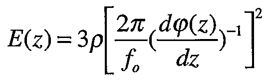
- the elasticity in soft media such as biological tissues and more generally solid media mainly consisting of water in liquid form, the elasticity (Young's modulus) is expressed as a function of the shear rate which we will denote by V s and the density p.
- the device for measuring the elasticity of a human or animal organ provides either an average value of the elasticity between two depths indicated by the user, i.e. the variations in elasticity as a function of depth.
- the probe 1 can comprise several transducers which can be positioned in an arbitrary manner, for example linearly (type echographic strip) or in honeycomb. In this way, the elasticity can be measured in different zones of the medium to be analyzed.
- the device according to the invention acquires ultrasonic lines at a typical rate of 50 lines per second. These lines are treated as in standard ultrasound so as to keep only the signal envelope. The lines are then displayed on the screen of the device in gray level and in logarithmic scale one after the other and one next to the other so as to form an image.
- the image can be obtained by moving the probe 1 at approximately constant speed over the surface of the liver, the user then having a distorted image of the area he is observing.
- the image is distorted because it is not possible for the user to move the probe 1 at constant speed. This image allows him to determine the zone in which the measurement is taken.
- the image distortion is significantly reduced by measuring the position of the probe 1 on the surface of the medium.
- the lines are displayed on the screen according to the abscissa of the sensor on the middle.
- the position of the probe 1 on the surface of the medium can be obtained using a measurement system which can be of different types: - position sensor of the type used in microcomputer mice, one can then choose a system using a wheel 11, an optical system as on so-called “optical” mice, - low frequency ultrasonic positioning system 12 (typically 100 kHz) consisting of at least three ultrasonic receivers 13 placed on the patient's body and at least one transmitter 14 placed on the probe (the position is obtained by triangulation) - or any other displacement measurement system; the system being connected to the central unit.
- a measurement system which can be of different types: - position sensor of the type used in microcomputer mice, one can then choose a system using a wheel 11, an optical system as on so-called “optical” mice, - low frequency ultrasonic positioning system 12 (typically 100 kHz) consisting of at least three ultrasonic receivers 13 placed on the patient's body and at least one transmitter 14 placed on the probe (the position is obtained by triangulation) - or any
- the device for measuring the elasticity of a human or animal organ according to the invention can be associated with a standard ultrasound system.
- the echograph not only provides morphological information on the organs but also a quantitative parameter of elasticity.
- the ultrasound system can then present, in addition to standard ultrasound probes 16, a probe of the type
- the probe 17 can be adapted around an ultrasound bar, not shown in the figures, in the manner of certain guidance systems for biopsy or of old continuous Doppler systems, as illustrated in FIG. 4.
- the ultrasonic bar itself carries out the acquisition of the ultrasonic signals used for the elastography algorithm.
- the device according to the invention may be portable, whether it is connected to the mains or supplied with batteries.
- One scan for measurement elasticity can be operated manually to obtain an image of elasticity.
- the scanning can be carried out by means of a stepping motor or any other controlled electromagnetic actuators.
- the system can possibly share the electronic modules of the ultrasound system 15 since the standard ultrasound systems 15 are a priori provided with signal processing units capable of rotating or calculating the algorithms necessary for measuring elasticity.
- the bar can then possibly generate the low frequency blow itself by a vibration movement which can be perpendicular to the surface of the medium.
- the acquisition can be carried out on the central line of the ultrasound image, as illustrated in FIG. 5a. It is possible to change the acquisition line and reproduce the low frequency shot so as to scan the entire surface of the image, as illustrated in FIGS. 5b to 5d.
- several lines can be produced at the same time using advanced ultrasonic focusing techniques such as:
- this device can be used in conjunction with the ultrafast imaging techniques described in the documents cited above so as to obtain an image of the elasticity.
- the device according to the invention will use an elastic intermediate medium 18 transparent to ultrasound.
- This medium 18 may for example be a synthetic polymer of the polyacrylamide type.
- An adhesive material or glue can be placed between the intermediate medium 18 and the medium studied so as to obtain either a sliding interface or a bonded interface.
- the intermediate medium 18 is innovative because it is not only transparent for ultrasound but also for the low frequency wave.
- the intermediate medium 18 is chosen so as to have an elasticity close to that of the medium studied so as to adapt the impedance and thus allow a maximum of energy to be transmitted to the medium studied.
- the intermediate medium 18 can also be compressed so that its modulus of elasticity which varies non-linearly becomes close to that of the medium studied. This latter proposition is, moreover, an original technique for measuring the elasticity of the medium: it consists in modifying the elasticity of the intermediate medium 18 until a maximum of energy is transmitted. The elasticity reached is then close to that of the middle.
- the device and method of the invention has an algorithm or means for calculating the displacements which are adapted according to the depth in the environment.
- the algorithm compares the successive lines with each other.
- the correlation is made between the line m and the line m + ⁇ with ⁇ > 1.
Abstract
Description


Claims
Priority Applications (9)
| Application Number | Priority Date | Filing Date | Title |
|---|---|---|---|
| BRPI0313214A BRPI0313214A2 (en) | 2002-08-08 | 2003-07-16 | device and procedure for measuring the elasticity of a human or animal organ |
| AU2003271812A AU2003271812B2 (en) | 2002-08-08 | 2003-07-16 | Device and method for measuring elasticity of a human or animal organ |
| CA 2494828 CA2494828A1 (en) | 2002-08-08 | 2003-07-16 | Device and method for measuring elasticity of a human or animal organ |
| ES03753649T ES2394961T3 (en) | 2002-08-08 | 2003-07-16 | Device and procedure for measuring the elasticity of a human or animal organ |
| EP03753649A EP1531733B1 (en) | 2002-08-08 | 2003-07-16 | Device and method for measuring elasticity of a human or animal organ |
| BRPI0313214A BRPI0313214B8 (en) | 2002-08-08 | 2003-07-16 | device and procedure for measuring the elasticity of a human or animal organ |
| JP2004528572A JP4451309B2 (en) | 2002-08-08 | 2003-07-16 | Apparatus and method for measuring elasticity of human or animal organs |
| IL16653603A IL166536A0 (en) | 2002-08-08 | 2003-07-16 | Device and method for measuring elasticity of a human or animal organ |
| US11/049,400 US7578789B2 (en) | 2002-08-08 | 2005-02-02 | Device and method for measuring the elasticity of a human or animal organ |
Applications Claiming Priority (2)
| Application Number | Priority Date | Filing Date | Title |
|---|---|---|---|
| FR0210104A FR2843290B1 (en) | 2002-08-08 | 2002-08-08 | DEVICE AND METHOD FOR MEASURING THE ELASTICITY OF A HUMAN OR ANIMAL ORGAN |
| FR02/10104 | 2002-08-08 |
Related Child Applications (1)
| Application Number | Title | Priority Date | Filing Date |
|---|---|---|---|
| US11/049,400 Continuation US7578789B2 (en) | 2002-08-08 | 2005-02-02 | Device and method for measuring the elasticity of a human or animal organ |
Publications (2)
| Publication Number | Publication Date |
|---|---|
| WO2004016176A2 true WO2004016176A2 (en) | 2004-02-26 |
| WO2004016176A3 WO2004016176A3 (en) | 2004-04-08 |
Family
ID=30471020
Family Applications (1)
| Application Number | Title | Priority Date | Filing Date |
|---|---|---|---|
| PCT/FR2003/002243 WO2004016176A2 (en) | 2002-08-08 | 2003-07-16 | Device and method for measuring elasticity of a human or animal organ |
Country Status (11)
| Country | Link |
|---|---|
| EP (1) | EP1531733B1 (en) |
| JP (1) | JP4451309B2 (en) |
| KR (1) | KR20050054916A (en) |
| CN (1) | CN100438833C (en) |
| AU (1) | AU2003271812B2 (en) |
| BR (2) | BRPI0313214A2 (en) |
| CA (1) | CA2494828A1 (en) |
| ES (1) | ES2394961T3 (en) |
| FR (1) | FR2843290B1 (en) |
| IL (1) | IL166536A0 (en) |
| WO (1) | WO2004016176A2 (en) |
Cited By (12)
| Publication number | Priority date | Publication date | Assignee | Title |
|---|---|---|---|---|
| FR2869521A1 (en) * | 2004-05-03 | 2005-11-04 | Echosens Sa | Instrument for measuring prostate gland elasticity through a duct has vibration axis of ultrasound transducer angled relative to penetration axis of connecting arm |
| JP2007044231A (en) * | 2005-08-10 | 2007-02-22 | Hitachi Medical Corp | Ultrasonic diagnostic equipment |
| JP2008514252A (en) * | 2004-09-28 | 2008-05-08 | エコサンス | Instrument of the type having a centering means for measuring organ elasticity |
| JP2009504217A (en) * | 2005-08-12 | 2009-02-05 | エショザン | Human or animal tissue imaging system that can be used to measure tissue elasticity |
| FR2928077A1 (en) * | 2008-02-29 | 2009-09-04 | Echosens Sa | DEVICE AND METHOD FOR MICROELASTOGRAPHY |
| JP2011067670A (en) * | 2011-01-07 | 2011-04-07 | Hitachi Medical Corp | Ultrasonic diagnostic apparatus |
| JP2011083645A (en) * | 2011-02-02 | 2011-04-28 | Toshiba Corp | Ultrasonograph and ultrasonic signal processing device |
| JP2011177535A (en) * | 2004-08-24 | 2011-09-15 | General Hospital Corp | Process, system, and software for measuring mechanical strain and elastic properties of sample |
| US11246570B2 (en) | 2016-10-28 | 2022-02-15 | Echosens | Probe for transient elastography |
| US11331073B2 (en) | 2017-03-27 | 2022-05-17 | Echosens | Device and method for measuring the viscoelastic properties of a viscoelastic medium |
| US11457895B2 (en) | 2016-07-25 | 2022-10-04 | Echosens | Method for measuring a viscoelastic parameter of a human or animal organ |
| US11759178B2 (en) | 2018-09-18 | 2023-09-19 | Echosens | Transient elastography probe with sealing membrane integrated to the ultrasound transducer |
Families Citing this family (38)
| Publication number | Priority date | Publication date | Assignee | Title |
|---|---|---|---|---|
| US8721573B2 (en) | 2003-09-04 | 2014-05-13 | Simon Fraser University | Automatically adjusting contact node for multiple rib space engagement |
| US8734368B2 (en) | 2003-09-04 | 2014-05-27 | Simon Fraser University | Percussion assisted angiogenesis |
| US8870796B2 (en) | 2003-09-04 | 2014-10-28 | Ahof Biophysical Systems Inc. | Vibration method for clearing acute arterial thrombotic occlusions in the emergency treatment of heart attack and stroke |
| CA2439667A1 (en) | 2003-09-04 | 2005-03-04 | Andrew Kenneth Hoffmann | Low frequency vibration assisted blood perfusion system and apparatus |
| FR2902308B1 (en) * | 2006-06-15 | 2009-03-06 | Echosens Sa | METHOD FOR MEASURING VISCOELASTIC PROPERTIES OF BIOLOGICAL TISSUES USING AN ULTRASONIC TRANSDUCER |
| FR2913875B1 (en) * | 2007-03-21 | 2009-08-07 | Echosens Sa | DEVICE FOR MEASURING VISCOELASTIC PROPERTIES OF BIOLOGICAL TISSUES AND METHOD USING THE DEVICE |
| JP5252985B2 (en) * | 2008-05-02 | 2013-07-31 | 花王株式会社 | Elasticity measurement method inside skin |
| FR2939512B1 (en) | 2008-12-04 | 2012-07-27 | Echosens | DEVICE AND METHOD FOR ELASTOGRAPHY |
| CN101803933B (en) * | 2009-02-13 | 2012-11-07 | 深圳市一体医疗科技股份有限公司 | Liver fibrosis detection device |
| WO2010143555A1 (en) * | 2009-06-11 | 2010-12-16 | 株式会社 日立メディコ | Excitation unit, ultrasonic probe, and ultrasonic diagnostic equipment |
| RU2559910C2 (en) * | 2009-06-30 | 2015-08-20 | Конинклейке Филипс Электроникс Н.В. | Propagation/tracking sequences for vibrometry of dispersion transverse waves |
| CN101933819B (en) * | 2009-07-02 | 2013-06-19 | 深圳市一体医疗科技股份有限公司 | Composite probe for cirrhosis detector |
| JPWO2011001776A1 (en) * | 2009-07-02 | 2012-12-13 | 株式会社日立メディコ | Ultrasonic diagnostic apparatus, shear wave propagation image generation method |
| CN102469989B (en) * | 2009-07-07 | 2014-04-16 | 株式会社日立医疗器械 | Ultrasonic diagnosis apparatus and ultrasonic measurement method |
| CN102548496B (en) * | 2009-09-15 | 2015-04-29 | 皇家飞利浦电子股份有限公司 | Medical ultrasound device with force detection |
| FR2949965B1 (en) | 2009-09-17 | 2012-09-28 | Echosens | PROCESS FOR MEASURING AT LEAST ONE PROPERTY OF BIOLOGICAL TISSUE |
| CN101699280B (en) | 2009-10-15 | 2011-08-17 | 北京索瑞特医学技术有限公司 | Method and device for ultrasonic and nondestructive detection of elasticity of viscoelastic medium |
| RU2453268C2 (en) * | 2010-09-20 | 2012-06-20 | Государственное бюджетное образовательное учреждение высшего профессионального образования "Северо-Западный государственный медицинский университет им. И.И. Мечникова" Министерства здравоохранения и социального развития Российской Федерации | Method of estimating liver fibrosis |
| WO2013160468A1 (en) | 2012-04-27 | 2013-10-31 | Echosens | Device for measuring an ultrasonic or biomechanical parameter of a viscoelastic medium |
| EP2959312B1 (en) | 2013-02-19 | 2017-12-13 | Echosens | Multipulse elastography method |
| CN104644209B (en) * | 2013-11-21 | 2017-06-20 | 通用电气公司 | Ultrasound Instrument and the vibrating device being applied thereon |
| CN103720490A (en) * | 2013-12-31 | 2014-04-16 | 无锡海斯凯尔医学技术有限公司 | Instantaneous elasticity detecting device |
| CN204274404U (en) * | 2014-09-12 | 2015-04-22 | 无锡海斯凯尔医学技术有限公司 | A kind of elastomeric check probe |
| CN107106120B (en) * | 2014-10-29 | 2021-11-16 | 梅约医学教育与研究基金会 | Method for ultrasound elastography by continuous vibration of an ultrasound transducer |
| CN104359972A (en) * | 2014-10-31 | 2015-02-18 | 杨松 | Method and device for detecting physical property of object |
| RU2565845C1 (en) * | 2014-10-31 | 2015-10-20 | Государственное бюджетное образовательное учреждение высшего профессионального образования "Смоленский государственный медицинский университет Министерства здравоохранения Российской Федерации (ГБОУ ВПО СГМУ Минздрава России) | Method for prediction of clinical course of diffuse liver conditions |
| CN105640593B (en) * | 2014-11-18 | 2019-05-28 | 爱科森股份有限公司 | It is a kind of for measuring the elasticity measuring device of viscoelastic medium |
| EP3095387A1 (en) * | 2015-05-22 | 2016-11-23 | Echosens | Interchangeable tip for ultrasound probe housing |
| FR3036943A1 (en) | 2015-06-02 | 2016-12-09 | Echosens | NON-INVASIVE HEPATIC LESION DETECTION DEVICE |
| CN105395218B (en) * | 2015-11-10 | 2019-02-15 | 中国科学院声学研究所 | Ultrasonic elastograph imaging system and method |
| CN105395217A (en) * | 2015-11-10 | 2016-03-16 | 中国科学院声学研究所 | Probe |
| CN106344070B (en) * | 2016-11-03 | 2023-09-22 | 乐普(北京)医疗器械股份有限公司 | Probe and detection device |
| JP6831696B2 (en) * | 2016-12-27 | 2021-02-17 | ゼネラル・エレクトリック・カンパニイ | Ultrasonic diagnostic system |
| FR3078484A1 (en) | 2018-03-02 | 2019-09-06 | Echosens | METHOD FOR MEASURING A GUIDED ULTRASONIC ATTENUATION PARAMETER BY HARMONIC ELASTOGRAPHY, PROBE AND DEVICE FOR IMPLEMENTING THE METHOD |
| CN209574737U (en) * | 2018-11-09 | 2019-11-05 | 无锡海斯凯尔医学技术有限公司 | Shell, probe and detection device |
| CN111000627B (en) * | 2019-12-04 | 2020-11-20 | 南京航空航天大学 | Microwave ablation tissue Young modulus real-time evaluation method and device based on reduced scattering coefficient |
| JP7077433B1 (en) | 2021-02-10 | 2022-05-30 | ジーイー・プレシジョン・ヘルスケア・エルエルシー | Control program for ultrasonic diagnostic equipment, ultrasonic diagnostic system and ultrasonic diagnostic equipment |
| CN113081040A (en) * | 2021-04-06 | 2021-07-09 | 无锡海斯凯尔医学技术有限公司 | Elasticity measurement method, device, system and storage medium |
Citations (5)
| Publication number | Priority date | Publication date | Assignee | Title |
|---|---|---|---|---|
| US5099848A (en) * | 1990-11-02 | 1992-03-31 | University Of Rochester | Method and apparatus for breast imaging and tumor detection using modal vibration analysis |
| US5882302A (en) * | 1992-02-21 | 1999-03-16 | Ths International, Inc. | Methods and devices for providing acoustic hemostasis |
| EP0920833A1 (en) * | 1997-12-05 | 1999-06-09 | Helmut Institut für Hochfrequenztechnik Ermert | Sonographic elastography system |
| WO2000055616A1 (en) * | 1999-03-15 | 2000-09-21 | Societe D'elastographie Impulsionnelle Pour Les Systemes De Mesure De L'elasticite (Seisme) | Imaging method and device using shearing waves |
| US6277074B1 (en) * | 1998-10-02 | 2001-08-21 | University Of Kansas Medical Center | Method and apparatus for motion estimation within biological tissue |
-
2002
- 2002-08-08 FR FR0210104A patent/FR2843290B1/en not_active Expired - Fee Related
-
2003
- 2003-07-16 KR KR1020057002337A patent/KR20050054916A/en not_active Application Discontinuation
- 2003-07-16 CA CA 2494828 patent/CA2494828A1/en not_active Abandoned
- 2003-07-16 EP EP03753649A patent/EP1531733B1/en not_active Expired - Lifetime
- 2003-07-16 BR BRPI0313214A patent/BRPI0313214A2/en active IP Right Grant
- 2003-07-16 JP JP2004528572A patent/JP4451309B2/en not_active Expired - Lifetime
- 2003-07-16 BR BRPI0313214A patent/BRPI0313214B8/en unknown
- 2003-07-16 CN CNB038191326A patent/CN100438833C/en not_active Expired - Lifetime
- 2003-07-16 IL IL16653603A patent/IL166536A0/en active IP Right Grant
- 2003-07-16 WO PCT/FR2003/002243 patent/WO2004016176A2/en active Application Filing
- 2003-07-16 AU AU2003271812A patent/AU2003271812B2/en not_active Ceased
- 2003-07-16 ES ES03753649T patent/ES2394961T3/en not_active Expired - Lifetime
Patent Citations (5)
| Publication number | Priority date | Publication date | Assignee | Title |
|---|---|---|---|---|
| US5099848A (en) * | 1990-11-02 | 1992-03-31 | University Of Rochester | Method and apparatus for breast imaging and tumor detection using modal vibration analysis |
| US5882302A (en) * | 1992-02-21 | 1999-03-16 | Ths International, Inc. | Methods and devices for providing acoustic hemostasis |
| EP0920833A1 (en) * | 1997-12-05 | 1999-06-09 | Helmut Institut für Hochfrequenztechnik Ermert | Sonographic elastography system |
| US6277074B1 (en) * | 1998-10-02 | 2001-08-21 | University Of Kansas Medical Center | Method and apparatus for motion estimation within biological tissue |
| WO2000055616A1 (en) * | 1999-03-15 | 2000-09-21 | Societe D'elastographie Impulsionnelle Pour Les Systemes De Mesure De L'elasticite (Seisme) | Imaging method and device using shearing waves |
Cited By (15)
| Publication number | Priority date | Publication date | Assignee | Title |
|---|---|---|---|---|
| FR2869521A1 (en) * | 2004-05-03 | 2005-11-04 | Echosens Sa | Instrument for measuring prostate gland elasticity through a duct has vibration axis of ultrasound transducer angled relative to penetration axis of connecting arm |
| JP2011177535A (en) * | 2004-08-24 | 2011-09-15 | General Hospital Corp | Process, system, and software for measuring mechanical strain and elastic properties of sample |
| JP2008514252A (en) * | 2004-09-28 | 2008-05-08 | エコサンス | Instrument of the type having a centering means for measuring organ elasticity |
| JP2007044231A (en) * | 2005-08-10 | 2007-02-22 | Hitachi Medical Corp | Ultrasonic diagnostic equipment |
| JP2009504217A (en) * | 2005-08-12 | 2009-02-05 | エショザン | Human or animal tissue imaging system that can be used to measure tissue elasticity |
| FR2928077A1 (en) * | 2008-02-29 | 2009-09-04 | Echosens Sa | DEVICE AND METHOD FOR MICROELASTOGRAPHY |
| WO2009122024A3 (en) * | 2008-02-29 | 2009-11-26 | Echosens Sa | Device and method for micro-elastography |
| JP2011067670A (en) * | 2011-01-07 | 2011-04-07 | Hitachi Medical Corp | Ultrasonic diagnostic apparatus |
| JP2011083645A (en) * | 2011-02-02 | 2011-04-28 | Toshiba Corp | Ultrasonograph and ultrasonic signal processing device |
| US11457895B2 (en) | 2016-07-25 | 2022-10-04 | Echosens | Method for measuring a viscoelastic parameter of a human or animal organ |
| US11246570B2 (en) | 2016-10-28 | 2022-02-15 | Echosens | Probe for transient elastography |
| US11331073B2 (en) | 2017-03-27 | 2022-05-17 | Echosens | Device and method for measuring the viscoelastic properties of a viscoelastic medium |
| US11690592B2 (en) | 2017-03-27 | 2023-07-04 | Echosens | Device and method for measuring the viscoelastic properties of a viscoelastic medium |
| US11730448B2 (en) | 2017-03-27 | 2023-08-22 | Echosens | Device and method for measuring the viscoelastic properties of a viscoelastic medium |
| US11759178B2 (en) | 2018-09-18 | 2023-09-19 | Echosens | Transient elastography probe with sealing membrane integrated to the ultrasound transducer |
Also Published As
| Publication number | Publication date |
|---|---|
| CN100438833C (en) | 2008-12-03 |
| AU2003271812B2 (en) | 2009-04-09 |
| FR2843290B1 (en) | 2005-06-24 |
| BRPI0313214A2 (en) | 2016-11-08 |
| IL166536A0 (en) | 2006-01-15 |
| ES2394961T3 (en) | 2013-02-07 |
| CA2494828A1 (en) | 2004-02-26 |
| BRPI0313214B1 (en) | 2018-01-02 |
| JP2005534455A (en) | 2005-11-17 |
| AU2003271812A1 (en) | 2004-03-03 |
| WO2004016176A3 (en) | 2004-04-08 |
| EP1531733B1 (en) | 2012-09-12 |
| KR20050054916A (en) | 2005-06-10 |
| JP4451309B2 (en) | 2010-04-14 |
| FR2843290A1 (en) | 2004-02-13 |
| EP1531733A2 (en) | 2005-05-25 |
| CN1674827A (en) | 2005-09-28 |
| BRPI0313214B8 (en) | 2021-06-22 |
Similar Documents
| Publication | Publication Date | Title |
|---|---|---|
| EP1531733B1 (en) | Device and method for measuring elasticity of a human or animal organ | |
| EP1169636B1 (en) | Imaging method and device using shear waves | |
| EP1546757B1 (en) | Imaging method and device employing shear waves | |
| EP1866667B1 (en) | Imaging method and device using shearing waves | |
| EP1538987B1 (en) | Device and method for measuring elasticity of a human or animal organ and for two- or three-dimensional representation thereof | |
| EP3824280B1 (en) | Methods and systems for non-invasively characterising a heterogeneous medium using ultrasound | |
| EP0751743B1 (en) | Method and device for estimating and characterising bone properties | |
| EP2084702B1 (en) | Method for generating mechanical waves by creating an interfacial acoustic radiation force | |
| FR3047405A1 (en) | ||
| CA2939013A1 (en) | Ultrasonic method and device for characterising weak anisotropic media, and ultrasonic probe assembly for such a characterisation device | |
| WO2019166395A1 (en) | Hybrid elastography method, probe, and device for hybrid elastography | |
| EP1473058B1 (en) | Apparatus for analysis of the skin comprising an ultrasonic probe | |
| CN111449629B (en) | Optical coherence elastography method and device | |
| Thompson et al. | Laser-induced synthetic aperture ultrasound imaging | |
| Ashkenazi et al. | High frequency ultrasound imaging using Fabry-Perot optical etalon | |
| FR3065079B1 (en) | ULTRASONIC SURVEY METHOD AND DEVICE FOR OBTAINING DISPERSION CURVES FROM A PROBE ENVIRONMENT | |
| FR3016791B1 (en) | METHOD AND DEVICE FOR CHARACTERIZING THE SKIN | |
| Ashkenazi et al. | High Frequency ultrasound detection using Fabry-Perot optical etalon | |
| CA2184510C (en) | Method and device for estimating and characterising bone properties | |
| CA3046106A1 (en) | Method of processing signals arising from an acquisition by ultrasound probing, corresponding computer program and ultrasound-based probing device |
Legal Events
| Date | Code | Title | Description |
|---|---|---|---|
| AK | Designated states |
Kind code of ref document: A2 Designated state(s): AE AG AL AM AT AU AZ BA BB BG BR BY BZ CA CH CN CO CR CU CZ DE DK DM DZ EC EE ES FI GB GD GE GH GM HR HU ID IL IN IS JP KE KG KP KR KZ LC LK LR LS LT LU LV MA MD MG MK MN MW MX MZ NI NO NZ OM PG PH PL PT RO RU SC SD SE SG SK SL SY TJ TM TN TR TT TZ UA UG US UZ VC VN YU ZA ZM ZW |
|
| AL | Designated countries for regional patents |
Kind code of ref document: A2 Designated state(s): GH GM KE LS MW MZ SD SL SZ TZ UG ZM ZW AM AZ BY KG KZ MD RU TJ TM AT BE BG CH CY CZ DE DK EE ES FI FR GB GR HU IE IT LU MC NL PT RO SE SI SK TR BF BJ CF CG CI CM GA GN GQ GW ML MR NE SN TD TG |
|
| 121 | Ep: the epo has been informed by wipo that ep was designated in this application | ||
| WWE | Wipo information: entry into national phase |
Ref document number: 55/MUMNP/2005 Country of ref document: IN |
|
| WWE | Wipo information: entry into national phase |
Ref document number: 166536 Country of ref document: IL |
|
| WWE | Wipo information: entry into national phase |
Ref document number: 2003271812 Country of ref document: AU |
|
| WWE | Wipo information: entry into national phase |
Ref document number: 11049400 Country of ref document: US |
|
| ENP | Entry into the national phase |
Ref document number: 2494828 Country of ref document: CA |
|
| WWE | Wipo information: entry into national phase |
Ref document number: 1020057002337 Country of ref document: KR Ref document number: 2004528572 Country of ref document: JP Ref document number: 20038191326 Country of ref document: CN |
|
| WWE | Wipo information: entry into national phase |
Ref document number: 2003753649 Country of ref document: EP |
|
| WWP | Wipo information: published in national office |
Ref document number: 2003753649 Country of ref document: EP |
|
| WWP | Wipo information: published in national office |
Ref document number: 1020057002337 Country of ref document: KR |
|
| ENP | Entry into the national phase |
Ref document number: PI0313214 Country of ref document: BR Kind code of ref document: A2 Effective date: 20050203 |