US9273291B2 - Enzymatic alkene cleavage - Google Patents
Enzymatic alkene cleavage Download PDFInfo
- Publication number
- US9273291B2 US9273291B2 US14/241,210 US201214241210A US9273291B2 US 9273291 B2 US9273291 B2 US 9273291B2 US 201214241210 A US201214241210 A US 201214241210A US 9273291 B2 US9273291 B2 US 9273291B2
- Authority
- US
- United States
- Prior art keywords
- seq
- primer
- enzyme
- pcr
- sequence
- Prior art date
- Legal status (The legal status is an assumption and is not a legal conclusion. Google has not performed a legal analysis and makes no representation as to the accuracy of the status listed.)
- Expired - Fee Related
Links
- 150000001336 alkenes Chemical class 0.000 title description 21
- 238000003776 cleavage reaction Methods 0.000 title description 21
- 230000007017 scission Effects 0.000 title description 18
- 230000002255 enzymatic effect Effects 0.000 title description 9
- 102000004190 Enzymes Human genes 0.000 claims abstract description 70
- 108090000790 Enzymes Proteins 0.000 claims abstract description 70
- 238000000034 method Methods 0.000 claims abstract description 32
- 239000003054 catalyst Substances 0.000 claims abstract description 10
- 229920002554 vinyl polymer Polymers 0.000 claims abstract description 6
- 125000000391 vinyl group Chemical group [H]C([*])=C([H])[H] 0.000 claims abstract description 5
- 241000222357 Trametes hirsuta Species 0.000 claims description 21
- 238000004191 hydrophobic interaction chromatography Methods 0.000 claims description 18
- 239000013598 vector Substances 0.000 claims description 16
- FWMNVWWHGCHHJJ-SKKKGAJSSA-N 4-amino-1-[(2r)-6-amino-2-[[(2r)-2-[[(2r)-2-[[(2r)-2-amino-3-phenylpropanoyl]amino]-3-phenylpropanoyl]amino]-4-methylpentanoyl]amino]hexanoyl]piperidine-4-carboxylic acid Chemical compound C([C@H](C(=O)N[C@H](CC(C)C)C(=O)N[C@H](CCCCN)C(=O)N1CCC(N)(CC1)C(O)=O)NC(=O)[C@H](N)CC=1C=CC=CC=1)C1=CC=CC=C1 FWMNVWWHGCHHJJ-SKKKGAJSSA-N 0.000 claims description 15
- 239000000284 extract Substances 0.000 claims description 14
- 238000005571 anion exchange chromatography Methods 0.000 claims description 9
- QVGXLLKOCUKJST-UHFFFAOYSA-N atomic oxygen Chemical compound [O] QVGXLLKOCUKJST-UHFFFAOYSA-N 0.000 claims description 8
- 229910052760 oxygen Inorganic materials 0.000 claims description 8
- 239000001301 oxygen Substances 0.000 claims description 8
- 241000588724 Escherichia coli Species 0.000 claims description 6
- -1 Mn3+ ions Chemical class 0.000 claims description 5
- 108020004707 nucleic acids Proteins 0.000 claims description 5
- 102000039446 nucleic acids Human genes 0.000 claims description 5
- 150000007523 nucleic acids Chemical class 0.000 claims description 5
- 239000002773 nucleotide Substances 0.000 claims description 5
- 125000003729 nucleotide group Chemical group 0.000 claims description 5
- 239000013600 plasmid vector Substances 0.000 claims description 3
- 241000235058 Komagataella pastoris Species 0.000 claims description 2
- 238000012258 culturing Methods 0.000 claims description 2
- 125000003275 alpha amino acid group Chemical group 0.000 claims 6
- 239000012062 aqueous buffer Substances 0.000 claims 1
- 230000001131 transforming effect Effects 0.000 claims 1
- 238000004519 manufacturing process Methods 0.000 abstract description 2
- 238000007248 oxidative elimination reaction Methods 0.000 abstract 1
- 108090000623 proteins and genes Proteins 0.000 description 89
- 235000018102 proteins Nutrition 0.000 description 50
- 102000004169 proteins and genes Human genes 0.000 description 50
- 108020004414 DNA Proteins 0.000 description 40
- 238000006243 chemical reaction Methods 0.000 description 39
- 239000000047 product Substances 0.000 description 38
- 230000000694 effects Effects 0.000 description 34
- 239000002299 complementary DNA Substances 0.000 description 31
- 239000000872 buffer Substances 0.000 description 28
- 238000000746 purification Methods 0.000 description 23
- 150000001413 amino acids Chemical class 0.000 description 22
- 210000004027 cell Anatomy 0.000 description 21
- 238000000137 annealing Methods 0.000 description 19
- 238000004925 denaturation Methods 0.000 description 16
- 230000036425 denaturation Effects 0.000 description 16
- 238000001962 electrophoresis Methods 0.000 description 16
- IAZDPXIOMUYVGZ-UHFFFAOYSA-N Dimethylsulphoxide Chemical compound CS(C)=O IAZDPXIOMUYVGZ-UHFFFAOYSA-N 0.000 description 15
- 239000000499 gel Substances 0.000 description 15
- MMIPFLVOWGHZQD-UHFFFAOYSA-N manganese(3+) Chemical compound [Mn+3] MMIPFLVOWGHZQD-UHFFFAOYSA-N 0.000 description 15
- 238000012163 sequencing technique Methods 0.000 description 14
- 239000011543 agarose gel Substances 0.000 description 13
- ZRSNZINYAWTAHE-UHFFFAOYSA-N p-methoxybenzaldehyde Chemical compound COC1=CC=C(C=O)C=C1 ZRSNZINYAWTAHE-UHFFFAOYSA-N 0.000 description 12
- RUVINXPYWBROJD-ONEGZZNKSA-N trans-anethole Chemical compound COC1=CC=C(\C=C\C)C=C1 RUVINXPYWBROJD-ONEGZZNKSA-N 0.000 description 12
- XLYOFNOQVPJJNP-UHFFFAOYSA-N water Substances O XLYOFNOQVPJJNP-UHFFFAOYSA-N 0.000 description 12
- 108090000765 processed proteins & peptides Proteins 0.000 description 11
- 238000013519 translation Methods 0.000 description 10
- 235000001014 amino acid Nutrition 0.000 description 9
- 229940024606 amino acid Drugs 0.000 description 9
- 230000000295 complement effect Effects 0.000 description 9
- 239000011541 reaction mixture Substances 0.000 description 9
- 108091028043 Nucleic acid sequence Proteins 0.000 description 8
- 238000013461 design Methods 0.000 description 8
- 238000000605 extraction Methods 0.000 description 8
- 229910052751 metal Inorganic materials 0.000 description 8
- 239000002184 metal Substances 0.000 description 8
- 238000002415 sodium dodecyl sulfate polyacrylamide gel electrophoresis Methods 0.000 description 8
- 125000000539 amino acid group Chemical group 0.000 description 7
- 239000008223 sterile water Substances 0.000 description 7
- 238000007857 nested PCR Methods 0.000 description 6
- 239000013612 plasmid Substances 0.000 description 6
- 238000001228 spectrum Methods 0.000 description 6
- 102000016928 DNA-directed DNA polymerase Human genes 0.000 description 5
- 108010014303 DNA-directed DNA polymerase Proteins 0.000 description 5
- 230000003321 amplification Effects 0.000 description 5
- 238000004458 analytical method Methods 0.000 description 5
- 230000015572 biosynthetic process Effects 0.000 description 5
- 238000010367 cloning Methods 0.000 description 5
- 238000002844 melting Methods 0.000 description 5
- 230000008018 melting Effects 0.000 description 5
- 150000002739 metals Chemical class 0.000 description 5
- 239000000203 mixture Substances 0.000 description 5
- 238000003199 nucleic acid amplification method Methods 0.000 description 5
- 239000000758 substrate Substances 0.000 description 5
- 241000233866 Fungi Species 0.000 description 4
- XEEYBQQBJWHFJM-UHFFFAOYSA-N Iron Chemical compound [Fe] XEEYBQQBJWHFJM-UHFFFAOYSA-N 0.000 description 4
- 102000035195 Peptidases Human genes 0.000 description 4
- 108091005804 Peptidases Proteins 0.000 description 4
- 238000012300 Sequence Analysis Methods 0.000 description 4
- BFNBIHQBYMNNAN-UHFFFAOYSA-N ammonium sulfate Chemical compound N.N.OS(O)(=O)=O BFNBIHQBYMNNAN-UHFFFAOYSA-N 0.000 description 4
- 229910052921 ammonium sulfate Inorganic materials 0.000 description 4
- 235000011130 ammonium sulphate Nutrition 0.000 description 4
- OWMVSZAMULFTJU-UHFFFAOYSA-N bis-tris Chemical compound OCCN(CCO)C(CO)(CO)CO OWMVSZAMULFTJU-UHFFFAOYSA-N 0.000 description 4
- 238000010276 construction Methods 0.000 description 4
- 230000029087 digestion Effects 0.000 description 4
- 239000012634 fragment Substances 0.000 description 4
- 230000003647 oxidation Effects 0.000 description 4
- 238000007254 oxidation reaction Methods 0.000 description 4
- 235000019833 protease Nutrition 0.000 description 4
- 238000002864 sequence alignment Methods 0.000 description 4
- 238000004885 tandem mass spectrometry Methods 0.000 description 4
- QTBSBXVTEAMEQO-UHFFFAOYSA-M Acetate Chemical compound CC([O-])=O QTBSBXVTEAMEQO-UHFFFAOYSA-M 0.000 description 3
- WEVYAHXRMPXWCK-UHFFFAOYSA-N Acetonitrile Chemical compound CC#N WEVYAHXRMPXWCK-UHFFFAOYSA-N 0.000 description 3
- 229920000936 Agarose Polymers 0.000 description 3
- 101100353170 Caenorhabditis elegans pri-2 gene Proteins 0.000 description 3
- 108020004705 Codon Proteins 0.000 description 3
- DHMQDGOQFOQNFH-UHFFFAOYSA-N Glycine Chemical compound NCC(O)=O DHMQDGOQFOQNFH-UHFFFAOYSA-N 0.000 description 3
- QNAYBMKLOCPYGJ-REOHCLBHSA-N L-alanine Chemical compound C[C@H](N)C(O)=O QNAYBMKLOCPYGJ-REOHCLBHSA-N 0.000 description 3
- KZSNJWFQEVHDMF-BYPYZUCNSA-N L-valine Chemical compound CC(C)[C@H](N)C(O)=O KZSNJWFQEVHDMF-BYPYZUCNSA-N 0.000 description 3
- 241001149420 Laccaria bicolor Species 0.000 description 3
- KZSNJWFQEVHDMF-UHFFFAOYSA-N Valine Natural products CC(C)C(N)C(O)=O KZSNJWFQEVHDMF-UHFFFAOYSA-N 0.000 description 3
- 235000004279 alanine Nutrition 0.000 description 3
- 238000003556 assay Methods 0.000 description 3
- 230000003197 catalytic effect Effects 0.000 description 3
- 238000006555 catalytic reaction Methods 0.000 description 3
- 229910052802 copper Inorganic materials 0.000 description 3
- 239000010949 copper Substances 0.000 description 3
- 239000012153 distilled water Substances 0.000 description 3
- 238000007373 indentation Methods 0.000 description 3
- BPHPUYQFMNQIOC-NXRLNHOXSA-N isopropyl beta-D-thiogalactopyranoside Chemical compound CC(C)S[C@@H]1O[C@H](CO)[C@H](O)[C@H](O)[C@H]1O BPHPUYQFMNQIOC-NXRLNHOXSA-N 0.000 description 3
- 238000000816 matrix-assisted laser desorption--ionisation Methods 0.000 description 3
- 238000005457 optimization Methods 0.000 description 3
- 125000001997 phenyl group Chemical group [H]C1=C([H])C([H])=C(*)C([H])=C1[H] 0.000 description 3
- 238000002360 preparation method Methods 0.000 description 3
- 102000004196 processed proteins & peptides Human genes 0.000 description 3
- 239000011535 reaction buffer Substances 0.000 description 3
- 238000011160 research Methods 0.000 description 3
- 150000003839 salts Chemical class 0.000 description 3
- 238000003786 synthesis reaction Methods 0.000 description 3
- 235000019801 trisodium phosphate Nutrition 0.000 description 3
- 239000004474 valine Substances 0.000 description 3
- OEGPRYNGFWGMMV-UHFFFAOYSA-N (3,4-dimethoxyphenyl)methanol Chemical compound COC1=CC=C(CO)C=C1OC OEGPRYNGFWGMMV-UHFFFAOYSA-N 0.000 description 2
- KSFOVUSSGSKXFI-GAQDCDSVSA-N CC1=C/2NC(\C=C3/N=C(/C=C4\N\C(=C/C5=N/C(=C\2)/C(C=C)=C5C)C(C=C)=C4C)C(C)=C3CCC(O)=O)=C1CCC(O)=O Chemical compound CC1=C/2NC(\C=C3/N=C(/C=C4\N\C(=C/C5=N/C(=C\2)/C(C=C)=C5C)C(C=C)=C4C)C(C)=C3CCC(O)=O)=C1CCC(O)=O KSFOVUSSGSKXFI-GAQDCDSVSA-N 0.000 description 2
- 101100521097 Caenorhabditis elegans pri-1 gene Proteins 0.000 description 2
- RYGMFSIKBFXOCR-UHFFFAOYSA-N Copper Chemical compound [Cu] RYGMFSIKBFXOCR-UHFFFAOYSA-N 0.000 description 2
- WHUUTDBJXJRKMK-UHFFFAOYSA-N Glutamic acid Natural products OC(=O)C(N)CCC(O)=O WHUUTDBJXJRKMK-UHFFFAOYSA-N 0.000 description 2
- DCXYFEDJOCDNAF-REOHCLBHSA-N L-asparagine Chemical compound OC(=O)[C@@H](N)CC(N)=O DCXYFEDJOCDNAF-REOHCLBHSA-N 0.000 description 2
- KDXKERNSBIXSRK-UHFFFAOYSA-N Lysine Natural products NCCCCC(N)C(O)=O KDXKERNSBIXSRK-UHFFFAOYSA-N 0.000 description 2
- 108091034117 Oligonucleotide Proteins 0.000 description 2
- 229920002684 Sepharose Polymers 0.000 description 2
- FAPWRFPIFSIZLT-UHFFFAOYSA-M Sodium chloride Chemical compound [Na+].[Cl-] FAPWRFPIFSIZLT-UHFFFAOYSA-M 0.000 description 2
- 244000061458 Solanum melongena Species 0.000 description 2
- 235000002597 Solanum melongena Nutrition 0.000 description 2
- 239000003795 chemical substances by application Substances 0.000 description 2
- 239000013599 cloning vector Substances 0.000 description 2
- OPTASPLRGRRNAP-UHFFFAOYSA-N cytosine Chemical compound NC=1C=CNC(=O)N=1 OPTASPLRGRRNAP-UHFFFAOYSA-N 0.000 description 2
- 238000010586 diagram Methods 0.000 description 2
- 239000012149 elution buffer Substances 0.000 description 2
- 238000002474 experimental method Methods 0.000 description 2
- 238000013467 fragmentation Methods 0.000 description 2
- 238000006062 fragmentation reaction Methods 0.000 description 2
- 230000002538 fungal effect Effects 0.000 description 2
- UYTPUPDQBNUYGX-UHFFFAOYSA-N guanine Chemical compound O=C1NC(N)=NC2=C1N=CN2 UYTPUPDQBNUYGX-UHFFFAOYSA-N 0.000 description 2
- 239000004615 ingredient Substances 0.000 description 2
- 229910052742 iron Inorganic materials 0.000 description 2
- 239000012160 loading buffer Substances 0.000 description 2
- 108020004999 messenger RNA Proteins 0.000 description 2
- 244000005700 microbiome Species 0.000 description 2
- 230000001590 oxidative effect Effects 0.000 description 2
- 239000008188 pellet Substances 0.000 description 2
- 229950003776 protoporphyrin Drugs 0.000 description 2
- 238000001542 size-exclusion chromatography Methods 0.000 description 2
- 239000000243 solution Substances 0.000 description 2
- 238000012360 testing method Methods 0.000 description 2
- RWQNBRDOKXIBIV-UHFFFAOYSA-N thymine Chemical compound CC1=CNC(=O)NC1=O RWQNBRDOKXIBIV-UHFFFAOYSA-N 0.000 description 2
- 230000009466 transformation Effects 0.000 description 2
- 238000011144 upstream manufacturing Methods 0.000 description 2
- 101150028074 2 gene Proteins 0.000 description 1
- VGONTNSXDCQUGY-RRKCRQDMSA-N 2'-deoxyinosine Chemical compound C1[C@H](O)[C@@H](CO)O[C@H]1N1C(N=CNC2=O)=C2N=C1 VGONTNSXDCQUGY-RRKCRQDMSA-N 0.000 description 1
- GOJUJUVQIVIZAV-UHFFFAOYSA-N 2-amino-4,6-dichloropyrimidine-5-carbaldehyde Chemical group NC1=NC(Cl)=C(C=O)C(Cl)=N1 GOJUJUVQIVIZAV-UHFFFAOYSA-N 0.000 description 1
- 229930024421 Adenine Natural products 0.000 description 1
- GFFGJBXGBJISGV-UHFFFAOYSA-N Adenine Chemical compound NC1=NC=NC2=C1N=CN2 GFFGJBXGBJISGV-UHFFFAOYSA-N 0.000 description 1
- 229920001817 Agar Polymers 0.000 description 1
- 239000004475 Arginine Substances 0.000 description 1
- DCXYFEDJOCDNAF-UHFFFAOYSA-N Asparagine Natural products OC(=O)C(N)CC(N)=O DCXYFEDJOCDNAF-UHFFFAOYSA-N 0.000 description 1
- 241000222356 Coriolus Species 0.000 description 1
- 102000053602 DNA Human genes 0.000 description 1
- 102000012410 DNA Ligases Human genes 0.000 description 1
- 108010061982 DNA Ligases Proteins 0.000 description 1
- 238000001712 DNA sequencing Methods 0.000 description 1
- MYMOFIZGZYHOMD-UHFFFAOYSA-N Dioxygen Chemical compound O=O MYMOFIZGZYHOMD-UHFFFAOYSA-N 0.000 description 1
- 241000672609 Escherichia coli BL21 Species 0.000 description 1
- 108010058643 Fungal Proteins Proteins 0.000 description 1
- 239000004471 Glycine Substances 0.000 description 1
- ONIBWKKTOPOVIA-BYPYZUCNSA-N L-Proline Chemical compound OC(=O)[C@@H]1CCCN1 ONIBWKKTOPOVIA-BYPYZUCNSA-N 0.000 description 1
- CKLJMWTZIZZHCS-REOHCLBHSA-N L-aspartic acid Chemical compound OC(=O)[C@@H](N)CC(O)=O CKLJMWTZIZZHCS-REOHCLBHSA-N 0.000 description 1
- AGPKZVBTJJNPAG-WHFBIAKZSA-N L-isoleucine Chemical compound CC[C@H](C)[C@H](N)C(O)=O AGPKZVBTJJNPAG-WHFBIAKZSA-N 0.000 description 1
- ROHFNLRQFUQHCH-YFKPBYRVSA-N L-leucine Chemical compound CC(C)C[C@H](N)C(O)=O ROHFNLRQFUQHCH-YFKPBYRVSA-N 0.000 description 1
- FFEARJCKVFRZRR-BYPYZUCNSA-N L-methionine Chemical compound CSCC[C@H](N)C(O)=O FFEARJCKVFRZRR-BYPYZUCNSA-N 0.000 description 1
- COLNVLDHVKWLRT-QMMMGPOBSA-N L-phenylalanine Chemical compound OC(=O)[C@@H](N)CC1=CC=CC=C1 COLNVLDHVKWLRT-QMMMGPOBSA-N 0.000 description 1
- QIVBCDIJIAJPQS-VIFPVBQESA-N L-tryptophane Chemical compound C1=CC=C2C(C[C@H](N)C(O)=O)=CNC2=C1 QIVBCDIJIAJPQS-VIFPVBQESA-N 0.000 description 1
- OUYCCCASQSFEME-QMMMGPOBSA-N L-tyrosine Chemical compound OC(=O)[C@@H](N)CC1=CC=C(O)C=C1 OUYCCCASQSFEME-QMMMGPOBSA-N 0.000 description 1
- 108010029541 Laccase Proteins 0.000 description 1
- ROHFNLRQFUQHCH-UHFFFAOYSA-N Leucine Natural products CC(C)CC(N)C(O)=O ROHFNLRQFUQHCH-UHFFFAOYSA-N 0.000 description 1
- 108010054320 Lignin peroxidase Proteins 0.000 description 1
- 239000006137 Luria-Bertani broth Substances 0.000 description 1
- 239000004472 Lysine Substances 0.000 description 1
- 102000010750 Metalloproteins Human genes 0.000 description 1
- 108010063312 Metalloproteins Proteins 0.000 description 1
- 244000111261 Mucuna pruriens Species 0.000 description 1
- 235000006161 Mucuna pruriens Nutrition 0.000 description 1
- 101100202924 Neurospora crassa (strain ATCC 24698 / 74-OR23-1A / CBS 708.71 / DSM 1257 / FGSC 987) tsp-2 gene Proteins 0.000 description 1
- 108700020962 Peroxidase Proteins 0.000 description 1
- 102000003992 Peroxidases Human genes 0.000 description 1
- ONIBWKKTOPOVIA-UHFFFAOYSA-N Proline Natural products OC(=O)C1CCCN1 ONIBWKKTOPOVIA-UHFFFAOYSA-N 0.000 description 1
- MTCFGRXMJLQNBG-UHFFFAOYSA-N Serine Natural products OCC(N)C(O)=O MTCFGRXMJLQNBG-UHFFFAOYSA-N 0.000 description 1
- 108091081024 Start codon Proteins 0.000 description 1
- AYFVYJQAPQTCCC-UHFFFAOYSA-N Threonine Natural products CC(O)C(N)C(O)=O AYFVYJQAPQTCCC-UHFFFAOYSA-N 0.000 description 1
- 239000004473 Threonine Substances 0.000 description 1
- 241000222354 Trametes Species 0.000 description 1
- 102000004142 Trypsin Human genes 0.000 description 1
- 108090000631 Trypsin Proteins 0.000 description 1
- QIVBCDIJIAJPQS-UHFFFAOYSA-N Tryptophan Natural products C1=CC=C2C(CC(N)C(O)=O)=CNC2=C1 QIVBCDIJIAJPQS-UHFFFAOYSA-N 0.000 description 1
- 239000006035 Tryptophane Substances 0.000 description 1
- 229960000643 adenine Drugs 0.000 description 1
- 239000008272 agar Substances 0.000 description 1
- 238000003277 amino acid sequence analysis Methods 0.000 description 1
- AVKUERGKIZMTKX-NJBDSQKTSA-N ampicillin Chemical compound C1([C@@H](N)C(=O)N[C@H]2[C@H]3SC([C@@H](N3C2=O)C(O)=O)(C)C)=CC=CC=C1 AVKUERGKIZMTKX-NJBDSQKTSA-N 0.000 description 1
- 229960000723 ampicillin Drugs 0.000 description 1
- 238000005349 anion exchange Methods 0.000 description 1
- 238000013459 approach Methods 0.000 description 1
- ODKSFYDXXFIFQN-UHFFFAOYSA-N arginine Natural products OC(=O)C(N)CCCNC(N)=N ODKSFYDXXFIFQN-UHFFFAOYSA-N 0.000 description 1
- 125000003118 aryl group Chemical group 0.000 description 1
- 235000009582 asparagine Nutrition 0.000 description 1
- 229960001230 asparagine Drugs 0.000 description 1
- 235000003704 aspartic acid Nutrition 0.000 description 1
- OQFSQFPPLPISGP-UHFFFAOYSA-N beta-carboxyaspartic acid Natural products OC(=O)C(N)C(C(O)=O)C(O)=O OQFSQFPPLPISGP-UHFFFAOYSA-N 0.000 description 1
- 230000002210 biocatalytic effect Effects 0.000 description 1
- 239000012620 biological material Substances 0.000 description 1
- 230000036983 biotransformation Effects 0.000 description 1
- 150000001728 carbonyl compounds Chemical class 0.000 description 1
- 230000008859 change Effects 0.000 description 1
- 239000007809 chemical reaction catalyst Substances 0.000 description 1
- 108091036078 conserved sequence Proteins 0.000 description 1
- 239000000287 crude extract Substances 0.000 description 1
- 235000018417 cysteine Nutrition 0.000 description 1
- XUJNEKJLAYXESH-UHFFFAOYSA-N cysteine Natural products SCC(N)C(O)=O XUJNEKJLAYXESH-UHFFFAOYSA-N 0.000 description 1
- 229940104302 cytosine Drugs 0.000 description 1
- VGONTNSXDCQUGY-UHFFFAOYSA-N desoxyinosine Natural products C1C(O)C(CO)OC1N1C(NC=NC2=O)=C2N=C1 VGONTNSXDCQUGY-UHFFFAOYSA-N 0.000 description 1
- 229910001882 dioxygen Inorganic materials 0.000 description 1
- 238000001035 drying Methods 0.000 description 1
- 238000005516 engineering process Methods 0.000 description 1
- 210000003527 eukaryotic cell Anatomy 0.000 description 1
- 238000011156 evaluation Methods 0.000 description 1
- 238000004362 fungal culture Methods 0.000 description 1
- 239000000054 fungal extract Substances 0.000 description 1
- 235000013922 glutamic acid Nutrition 0.000 description 1
- 239000004220 glutamic acid Substances 0.000 description 1
- ZDXPYRJPNDTMRX-UHFFFAOYSA-N glutamine Natural products OC(=O)C(N)CCC(N)=O ZDXPYRJPNDTMRX-UHFFFAOYSA-N 0.000 description 1
- HNDVDQJCIGZPNO-UHFFFAOYSA-N histidine Natural products OC(=O)C(N)CC1=CN=CN1 HNDVDQJCIGZPNO-UHFFFAOYSA-N 0.000 description 1
- 230000002209 hydrophobic effect Effects 0.000 description 1
- 238000011534 incubation Methods 0.000 description 1
- 230000006698 induction Effects 0.000 description 1
- 238000001095 inductively coupled plasma mass spectrometry Methods 0.000 description 1
- 238000007689 inspection Methods 0.000 description 1
- 230000003993 interaction Effects 0.000 description 1
- 230000003834 intracellular effect Effects 0.000 description 1
- IUSBVFZKQJGVEP-SNAWJCMRSA-N isoeugenol acetate Chemical compound COC1=CC(\C=C\C)=CC=C1OC(C)=O IUSBVFZKQJGVEP-SNAWJCMRSA-N 0.000 description 1
- 238000002955 isolation Methods 0.000 description 1
- AGPKZVBTJJNPAG-UHFFFAOYSA-N isoleucine Natural products CCC(C)C(N)C(O)=O AGPKZVBTJJNPAG-UHFFFAOYSA-N 0.000 description 1
- 229960000310 isoleucine Drugs 0.000 description 1
- 108010016488 lignostilbene-alpha,beta-dioxygenase Proteins 0.000 description 1
- 229910052748 manganese Inorganic materials 0.000 description 1
- 239000011572 manganese Substances 0.000 description 1
- 238000013507 mapping Methods 0.000 description 1
- 239000003550 marker Substances 0.000 description 1
- 238000001819 mass spectrum Methods 0.000 description 1
- 239000000463 material Substances 0.000 description 1
- 238000001869 matrix assisted laser desorption--ionisation mass spectrum Methods 0.000 description 1
- 238000001906 matrix-assisted laser desorption--ionisation mass spectrometry Methods 0.000 description 1
- 230000007246 mechanism Effects 0.000 description 1
- 150000001455 metallic ions Chemical class 0.000 description 1
- 229930182817 methionine Natural products 0.000 description 1
- 229910052750 molybdenum Inorganic materials 0.000 description 1
- 230000035772 mutation Effects 0.000 description 1
- 239000013642 negative control Substances 0.000 description 1
- 239000007800 oxidant agent Substances 0.000 description 1
- COLNVLDHVKWLRT-UHFFFAOYSA-N phenylalanine Natural products OC(=O)C(N)CC1=CC=CC=C1 COLNVLDHVKWLRT-UHFFFAOYSA-N 0.000 description 1
- 238000003752 polymerase chain reaction Methods 0.000 description 1
- 230000008569 process Effects 0.000 description 1
- 210000001236 prokaryotic cell Anatomy 0.000 description 1
- 239000012264 purified product Substances 0.000 description 1
- 238000011084 recovery Methods 0.000 description 1
- 230000003252 repetitive effect Effects 0.000 description 1
- 230000027756 respiratory electron transport chain Effects 0.000 description 1
- 108091008146 restriction endonucleases Proteins 0.000 description 1
- 238000010839 reverse transcription Methods 0.000 description 1
- 239000012266 salt solution Substances 0.000 description 1
- 238000010845 search algorithm Methods 0.000 description 1
- 238000011218 seed culture Methods 0.000 description 1
- 239000011780 sodium chloride Substances 0.000 description 1
- 150000001629 stilbenes Chemical class 0.000 description 1
- 235000021286 stilbenes Nutrition 0.000 description 1
- 238000006467 substitution reaction Methods 0.000 description 1
- 239000006228 supernatant Substances 0.000 description 1
- WROMPOXWARCANT-UHFFFAOYSA-N tfa trifluoroacetic acid Chemical compound OC(=O)C(F)(F)F.OC(=O)C(F)(F)F WROMPOXWARCANT-UHFFFAOYSA-N 0.000 description 1
- 229940113082 thymine Drugs 0.000 description 1
- IUSBVFZKQJGVEP-UHFFFAOYSA-N trans-isoeugenol acetate Natural products COC1=CC(C=CC)=CC=C1OC(C)=O IUSBVFZKQJGVEP-UHFFFAOYSA-N 0.000 description 1
- 239000012588 trypsin Substances 0.000 description 1
- 229960004799 tryptophan Drugs 0.000 description 1
- OUYCCCASQSFEME-UHFFFAOYSA-N tyrosine Natural products OC(=O)C(N)CC1=CC=C(O)C=C1 OUYCCCASQSFEME-UHFFFAOYSA-N 0.000 description 1
- 238000002525 ultrasonication Methods 0.000 description 1
- 238000002604 ultrasonography Methods 0.000 description 1
- 238000012795 verification Methods 0.000 description 1
Images




Classifications
-
- C—CHEMISTRY; METALLURGY
- C12—BIOCHEMISTRY; BEER; SPIRITS; WINE; VINEGAR; MICROBIOLOGY; ENZYMOLOGY; MUTATION OR GENETIC ENGINEERING
- C12N—MICROORGANISMS OR ENZYMES; COMPOSITIONS THEREOF; PROPAGATING, PRESERVING, OR MAINTAINING MICROORGANISMS; MUTATION OR GENETIC ENGINEERING; CULTURE MEDIA
- C12N9/00—Enzymes; Proenzymes; Compositions thereof; Processes for preparing, activating, inhibiting, separating or purifying enzymes
- C12N9/0004—Oxidoreductases (1.)
- C12N9/0071—Oxidoreductases (1.) acting on paired donors with incorporation of molecular oxygen (1.14)
- C12N9/0073—Oxidoreductases (1.) acting on paired donors with incorporation of molecular oxygen (1.14) with NADH or NADPH as one donor, and incorporation of one atom of oxygen 1.14.13
-
- C—CHEMISTRY; METALLURGY
- C12—BIOCHEMISTRY; BEER; SPIRITS; WINE; VINEGAR; MICROBIOLOGY; ENZYMOLOGY; MUTATION OR GENETIC ENGINEERING
- C12N—MICROORGANISMS OR ENZYMES; COMPOSITIONS THEREOF; PROPAGATING, PRESERVING, OR MAINTAINING MICROORGANISMS; MUTATION OR GENETIC ENGINEERING; CULTURE MEDIA
- C12N9/00—Enzymes; Proenzymes; Compositions thereof; Processes for preparing, activating, inhibiting, separating or purifying enzymes
- C12N9/0004—Oxidoreductases (1.)
- C12N9/0069—Oxidoreductases (1.) acting on single donors with incorporation of molecular oxygen, i.e. oxygenases (1.13)
-
- C—CHEMISTRY; METALLURGY
- C12—BIOCHEMISTRY; BEER; SPIRITS; WINE; VINEGAR; MICROBIOLOGY; ENZYMOLOGY; MUTATION OR GENETIC ENGINEERING
- C12P—FERMENTATION OR ENZYME-USING PROCESSES TO SYNTHESISE A DESIRED CHEMICAL COMPOUND OR COMPOSITION OR TO SEPARATE OPTICAL ISOMERS FROM A RACEMIC MIXTURE
- C12P7/00—Preparation of oxygen-containing organic compounds
- C12P7/24—Preparation of oxygen-containing organic compounds containing a carbonyl group
-
- C—CHEMISTRY; METALLURGY
- C12—BIOCHEMISTRY; BEER; SPIRITS; WINE; VINEGAR; MICROBIOLOGY; ENZYMOLOGY; MUTATION OR GENETIC ENGINEERING
- C12Y—ENZYMES
- C12Y114/00—Oxidoreductases acting on paired donors, with incorporation or reduction of molecular oxygen (1.14)
- C12Y114/13—Oxidoreductases acting on paired donors, with incorporation or reduction of molecular oxygen (1.14) with NADH or NADPH as one donor, and incorporation of one atom of oxygen (1.14.13)
- C12Y114/13069—Alkene monooxygenase (1.14.13.69)
Definitions
- This application contains a sequence listing, which is submitted electronically via EFS-Web as an ASCII formatted sequence listing with a file name “Substitute_Sequence_Listing.TXT”, creation date of Oct. 14, 2014, and having a size of 86406 bytes.
- the sequence listing submitted via EFS-Web is part of the specification and is herein incorporated by reference in its entirety.
- the invention relates to an enzyme isolated from a fungal culture for the first time, its use in catalyzing alkene cleavage reactions and a method for its preparation.
- WO 2009/006662 A2 disclose a method using enzymatic catalysis by certain peroxidases and laccases, which surprisingly make use oxygen as a substrate
- WO 2010/003161 A1 disclose a similar method using enzymatic catalysis by certain haemins, also in the presence of oxygen.
- the present invention achieves this goal by providing a previously unknown enzyme having or comprising the sequence of SEQ ID NO: 1 or SEQ ID NO: 2, that has been isolated for the first time.
- SEQ ID NO: 1 is equal to the sequence determined by primer walking
- SEQ ID NO: 2 represents the sequence obtained by conclusive sequencing of the enzyme then isolated from Trametes hirsuta , with the two differing only by a single amino acid.
- This difference concerns an alanine in the terminal sequence as opposed to a valine in the one determined by primer walking.
- both amino acids belong to the group of apolar aliphatic amino acids, such replacement is regarded as a “conservative substitution”, which is why an enzyme having the sequence determined by primer walking will, with high probability, have similar characteristics and activities as the isolated amino acid sequence.
- a protein having an amino acid sequence with at least 80%, preferably at least 90%, more preferably at least 95%, particularly at least 99%, identity to SEQ ID NO: 1 or 2, will have at least comparable, or even the same, activity as the isolated enzyme.
- the isolated enzyme having SEQ ID NO: 2 hardly exhibited any effectiveness as a catalyst for cleaving alkenes with oxygen.
- the inventors have found that the catalytic activity of the enzyme could be increased dramatically if Mn 3+ ions are present in the reaction mixture. Accordingly, this enzyme seems to require the presence of manganese(III) as a co-factor in order to be catalytically effective in the above reaction to a satisfying degree, even though, generally, enzymes with comparable activities usually have iron or copper dependencies.
- enzymes with similar amino acid sequences as the enzyme of the invention are usually proteinases, which show no activity whatsoever as catalysts in alkene cleavage reactions. This is why the activity found by the inventors was all the more surprising.
- the invention comprises the use of an enzyme according to the first aspect of the invention—or a protein having at least 80%, %, preferably at least 90%, more preferably at least 95%, and particularly at least 99%, identity to SEQ ID NO: 1 or 2, as a catalyst for cleaving vinyl aromatics with oxygen, with the cleavage being preferably in the presence of Mn 3+ ions in order to increase enzyme activity, as set out above.
- the invention comprises a method of preparing an enzyme of the invention by culturing a culture of Trametes hirsuta and recovering the enzyme from a cell-free extract of the culture, with a culture of Trametes hirsuta G FCC 047 being preferably cultured since the inventors have achieved good results with this strain.
- recovery from the cell-free extract is performed using a combination of hydrophobic interaction chromatography and anion exchange chromatography, in particular using hydrophobic interaction chromatography, anion exchange chromatography and again hydrophobic interaction chromatography, in this order.
- the invention comprises a method of recombinantly producing the enzyme according to the first aspect of the invention using nucleic acid encoding the enzyme, i.e. nucleic acid having a nucleotide sequence according to SEQ ID NO: 3 or 4, with the first corresponding to the nucleotide sequence encoding SEQ ID NO: 1 and the latter corresponding to the nucleotide sequence encoding SEQ ID NO: 2.
- the two sequences were obtained as described in more detail below.
- the nucleic acid encoding the enzyme is preferably ligated into a vector, e.g. a plasmid vector, used to transform cells capable of expressing the enzyme, such as E.
- the enzyme of the invention is recovered from the culture broth by techniques known in the art, preferably as a cell-free extract, and stored, preferably in a lyophilized state.
- FIG. 1 shows the reaction scheme of cleaving trans-anethole using a cell-free extract of Trametes hirsuta as the catalyst.
- FIG. 2 shows the chromatogram of hydrophobic interaction chromatography performed as a first purification step.
- FIG. 3 shows the chromatogram of anion exchange chromatography performed as a second purification step.
- FIG. 4 shows the electropherogram of SDS-PAGE on anion exchange chromatography samples.
- FIG. 5 shows the chromatogram of hydrophobic interaction chromatography performed as a third purification step.
- FIG. 6 is an enlarged view of a part of the chromatogram of FIG. 5 .
- FIG. 7 shows the electropherogram of SDS-PAGE of the hydrophobic interaction chromatography samples of the third purification step.
- FIG. 8 shows the electropherogram of SDS-PAGE of the most active fraction after hydrophobic interaction chromatography.
- FIG. 9 shows an electropherogram of PCR products on agarose gel.
- FIG. 10 shows an electropherogram of the PCR products inserted into a cloning vector.
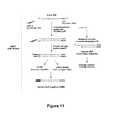
- FIG. 11 shows a flow diagram of the cDNA construction used for de novo sequencing.
- FIG. 12 shows an electropherogram of the PCR products of primer walking 1.
- FIG. 13 shows an electropherogram of the PCR products of primer walking 2.
- FIG. 14 shows ClustaW sequence alignment results of primer walking 3 (SEQ ID NOs: 84-95).
- FIG. 15 shows an electropherogram of the products of optimized PCR reactions of primer walking 3.
- FIG. 16 shows an electropherogram of the products of nested primer PCR reactions of primer walking 3.
- FIG. 17 shows the general procedure of primer walking 4.
- FIG. 18 shows an electropherogram of the products of a third PCR reaction of primer walking 4.
- FIG. 19 shows an electropherogram of the complete gene encoding the enzyme.
- FIG. 20 shows a sequence alignment of the complete gene as isolated (SEQ ID NO: 4) and as assembled from partial primer walking segments (SEQ ID NO: 3) using Clustal 2.1 software.
- FIG. 21 shows a comparison of amino acid sequences encoded by the isolated gene (SEQ ID NO: 2) and by the one assembled from partial primer walking segments (SEQ ID NO: 1).
- FIG. 22 shows a comparison of sequences of the gene obtained from genomic DNA (SEQ ID NO: 96) and the one obtained as a template (SEQ ID NO: 4).
- FIG. 23 shows an electropherogram of three reaction mixtures for restriction digestion of the gene from the pJET1.2 vector.
- nucleotide sequences herein use the usual one-letter code, i.e. A for adenine, C for cytosine, g for guanine and T for thymine (in both upper and lower case).
- Designations of amino acids of amino acid sequences disclosed in the present description and in the sequence listing is in one-letter and three-letter codes as indicated below for overview.
- Trametes hirsuta ( Coriolus hirsuta ) is a fungus which belongs to the white-rot fungal family. Alkenes possessing a C ⁇ C double bond adjacent to an aromatic ring were cleaved to yield the corresponding carbonyl compound by use of molecular oxygen as the sole oxidant and a cell-free extract of Trametes hirsuta G FCC 047 culture as a catalyst. trans-anethole has shown to be the best substrate for the biocatalytic alkene cleavage, affording p-anisaldehyde as the sole product. The conditions required to carry out this reaction were optimized previously and are indicated in FIG. 1 .
- the first step of purification employed was always hydrophobic interaction chromatography (phenyl sepharose CL-4B). After this step, the enzyme was still active. This was a neat process, as it was consistent with the fractions in which the enzyme activity was present and also repeatable.
- size-exclusion chromatography was employed as the second step. Hardly any activity could be obtained after this step. However, based on the few fractions which gave little activity the size of the enzyme was predicted to be between 140 and 160 KDa. Positive fractions from the first step (HIC) were also subjected to ICP-MS studies to detect the metals present.
- the cell-free extract was prepared using the lyophilized T. hirsuta culture. A decent concentration of culture (of 44 mg lyophilized culture/mL of buffer) was subjected to cell disruption by means of ultrasonication. Cell debris was removed from the crude extract, and the supernatant was subjected to purification.
- the column used for the first step was self-packed (Phenylsepharose CL-4B). For the rest of the steps, commercially available pre-packed columns were used.
- the self-packed phenyl sepharose column was used. This step was repeatable, and the enzyme activity was always detected in the fractions corresponding to the second peak, which appeared after the end of the salt (ammonium sulfate) gradient, in water.
- the chromatogram diagram obtained is given in FIG. 2 .
- the circled region in FIG. 2 is where enzyme activity is always found (second peak after the end of the salt gradient). The fractions corresponding to this region were pooled for the next step of purification.
- the commercially available HiTrap® FF Q, anion-exchange column (1 mL) was used in this step.
- the pooled positive fractions from the previous step (45 mL) was loaded onto the column for further purification. Positive fractions were tested by addition of minimum amounts (0.4 mM) of Mn(III) acetate, which did not in itself catalyze the alkene cleavage.
- enzymatic activity was observed for the first time.
- the enzyme was eluted from the flow-through fractions, which means that the enzyme did not bind to the column.
- the other proteins, which did bind to the column were eluted with an increasing linear salt (NaCl) concentration gradient.
- Samples from the fractions were collected for protein concentration evaluation and for SDS-PAGE.
- the positive fractions (flow-through) were then pooled for further purification.
- the chromatogram is shown in FIG. 3 .
- the average protein concentration of the flow-through fractions (2 to 11) were found to be 428 ⁇ g protein/mL.
- the circled area is where enzyme activity occurred (flow-through fractions).
- FIG. 4 shows the electropherogram of an SDS-PAGE of anion exchange chromatography samples. Fractions 8, 9, 10 and 11 were the active fractions (flow-through). Fractions 15 and 16 were negative fractions.
- Steps 1 and 2 were performed twice to get sufficient amounts of sample for step 3 (the final step).
- the pooled sample from the anion exchange chromatography (performed twice, resulting in a volume of about 80 mL with about 428 ⁇ g protein/mL) was loaded onto the HIC column. The first time the experiment was performed all fractions were tested for activity. It was then clear where the enzyme of interest eluted. The region was similar to where it eluted in Step-1 HIC (after the end of the ammonium sulfate gradient, in water).
- the selected protein spot was excised from the gel.
- the gel plug was washed alternately three times with 15 ⁇ L 10 mM NH 4 HCO 3 and 5 mM NH 4 HCO 3 /50% acetonitrile. After drying the gel segment, trypsin solution (33 ng/ ⁇ L in 10 mM NH 4 HCO 3 , pH 7.8) was added to digest the protein for several hours at 37° C.
- the peptides were extracted from the gel segment using 0.1% trifluoroacetic acid (TFA) and purified using C18 material (ZipTipTM, Millipore, Bedford, Mass., USA) before spotting them onto the MALDI target.
- TFA trifluoroacetic acid
- C18 material ZipTipTM, Millipore, Bedford, Mass., USA
- the protein identification was performed using an Ultraflex III TOF/TOF mass spectrometer (Bruker Daltonics). For the acquisition of peptide mass fingerprint spectra (PMF, MS), 200 single shot spectra were averaged and peak picking was performed using the SNAP algorithm. The resulting mass list was sent to the ProteinScapeTM database for protein identification. Peptide fragmentation spectra (PFF, MS/MS) were acquired where possible. The peaks for fragmentation were selected by the ProteinScape database based upon the results of the protein identification by PMF.
- Protein identification was performed by searching the mass spectra against the NCBI protein database (website: www.ncbi.nlm.nih.gov) using several external search algorithms (ProFoundTM, MascotTM, SequestTM). For PMF spectra, the mass tolerance was set to 50 ppm. The protein identification was based on the metascore1 calculated from the individual search results by the ProteinScapeTM database and on manual inspection of the data where needed. PFF spectra were either used to confirm the protein already identified by PMF or for identification of proteins that eluded the PMF identification.
- MS/MS data sets were automatically de-novo sequenced using the PEAKS 4.5 software package.
- the top scoring sequences are reported for each MS/MS spectrum.
- the following protein was identified by database search: gi
- the peptide EPGLAFAFGK (SEQ ID NO: 5) could be identified by database search and could be mapped to this protein.
- SEQ ID NOs: 5-13 The following peptide sequences (SEQ ID NOs: 5-13) were obtained by de novo sequencing. The amino acids, shown in bold, are identified with very high confidence (PEAKS Score of this sequence part >90%).
- PCR using the different combinations of designed forward and reverse degenerated primers were carried out. Based on the mapping of the obtained sequences with the amino acid sequence of aspartic peptidase A1 of Laccaria bicolor , three forward primers and two reverse primers were designed. Using these, six different combinations of forward and reverse primers were made. In order to check which temperature was suitable for the PCR, three temperatures lower than the melting points of the primers were tested. The different combinations of the primers are given in Table 2. The PCR conditions used are given in Table 3. The program of PCR reactions is given in Table 4.
- Fwd primer 1 (SEQ ID NO: 14) AAY CAR GAY TTY GCN GAR GC (SEQ ID NO: 19) NQDFAEA Fwd primer 2: (SEQ ID NO: 15) GAR GAR GAY GGN GGN GAR GCN (SEQ ID NO: 20) EEDGGEA Fwd primer 3: (SEQ ID NO: 16) AAR GCN TAY TGG GAR GTN GA (SEQ ID NO: 21) KAYWEVE Reverse primer 1: (SEQ ID NO: 17) TC NAC YTC CCA RTA NGC YTT (SEQ ID NO: 22 KAYWEVE Reverse primer 2: (SEQ ID NO: 18) C RTG RTC RTA NAC NGT RTA RTA YT (SEQ ID NO: 23) KYYTVYDHG
- the extractions of the DNA from the bands cut out from the gel were done using the QIAGEN QIAquick® (50) Gel Extraction Kit.
- the DNA concentrations of the three samples (50 ⁇ L volume) were measured.
- the elution buffer from the kit was used as a blank.
- the DNA fragments were cloned and then transformed into TOP 10 E. coli cells for amplification of the DNA.
- Cloning of the DNA fragments (1c, 6a and 6b) was done using CloneJETTM PCR Cloning kit. The amount of DNA sample taken for each sample was based on the size of the fragment and the concentration of the purified DNA sample. The procedure followed the kit manual.
- the QIAGEN QIAprep® (250) Spin Miniprep Kit was used for performing a miniprep. The procedure followed the manual.
- the DNA eluted from the minipreps (50 ⁇ L each) were mixed with 6 ⁇ loading buffer (8.3 ⁇ L), and the whole sample was loaded on the agarose gel (1%); see FIG. 10 showing the amplified PCR products in the CloneJETTM cloning vector.
- the bands of the plasmids were excised from each lane and filled into tubes (15 mL). The gel segments were weighed and the DNA was extracted using the QIAGEN QIAquick® (50) Gel Extraction Kit. The DNA concentration of the 9 samples (50 ⁇ L volume) were measured.
- the elution buffer from the kit was used as a blank.
- the sequencing results for 1c1, 6a1 and 6b1 were individually compared with the sequence of the plasmid pJET1.2 using BLAST.
- the non-identical region was taken as the sequence of the insert in the plasmid. This region was isolated and translated into its amino acid sequence.
- the presence of the forward and reverse primers used (Fwd 1 and rev 1 in case of 1c1 and Fwd 3 and rev 2 in case of 6a1 and 6b1) was checked. It was found that both primers were only present in the 6a1 sequence.
- a protein BLAST with this amino acid sequence gave certain similarities to several fungal proteins belonging to the pepsin-retropepsin superfamily (the second best result was aspartic peptidase A1 from Laccaria bicolor ).
- the sequence of sample 6a1 is shown below.
- the criteria for designing the definite primers were that both primers have GC content at the end and the melting point of both the primers should be similar. The melting point and the probability of self-complementarity on behalf of the primers were checked using the Oligo-Calc online program.
- the reverse primer the reverse complement of the original DNA segment was prepared and the length was altered to suit the requirement.
- the primers were designed to comprise the initial degenerate primer region (to double-check the accuracy in primer walking).
- the underlined sequences each represent one of the following primers:
- the attempt to carry out a PCR with just one primer did not yield any PCR product. There had to be a combination of both forward and reverse primers in a PCR.
- the SMARTTM cDNA library construction kit was used to get the cDNA from the mRNA of Trametes hirsuta . Initially, the CDS III primer was used for the first cDNA strand synthesis, starting from the poly A + tail of the mRNA. For the second strand synthesis, the SMART IV oligonucleotide was used.
- every cDNA fragment will contain the sequence of CDSIII primer at the 3′ end and the sequence of SMART IV primer at the 5′ end (see FIG. 11 ).
- a forward primer based on the sequence of SMART IV oligonucleotide and a reverse primer based on CDS III primer were designed to both have melting point similar to that of the definite forward and reverse primers previously designed.
- a flowchart of the cDNA construction from the user manual of the kit is given in FIG. 11 . The new primers were designed on this basis. The two forward and reverse primers designed are given below:
- PCR using the different combinations of forward and reverse primers were carried out.
- the main change made in the PCR program was that of the annealing temperature. Since all the primers had similar melting temperatures, two different temperatures were tested.
- the different primer combinations are given in Table 5.
- the different reaction ingredients are summarized in Table 6.
- the PCR reaction program is given in Table 7.
- the insert 2a had both the PW-I fwd primer sequence and the original degenerate reverse primer sequence. Hence it is part of the protein of interest. The translation of the sequence gave a sequence with a stop codon in between. Hence the end of the protein of interest has been reached.
- the PW-I reverse primer was present but did not comprise the initial degenerate forward primer sequence.
- the designed reverse primer PW-1 rev was not specific enough and had bound to some other part of the cDNA.
- three other reverse primers were designed, which were used for primer walking 2. A total of 84 amino acid residues could be deduced at the end of primer walking 1 and the end of the protein had been reached.
- the sequence of the peptide obtained until this step is given below:
- PW-2 rev Pri-1 (SEQ ID NO: 39) AGCTCAAGCTCGTCGTCTCCG 2) PW-2 rev Pri-2: (SEQ ID NO: 40) AGTGTCGATGGCAGCGCCG 3) PW-2 rev Pri-3: (SEQ ID NO: 41) GATTTGCACATTGAGCATCTCCGCC
- the samples (50 ⁇ L each) were analyzed preparatively on agarose gel (1% agarose). Clear bands (PCR product) were visible (around 400 bp) for samples 3b, 4a, 4b, 5a and 5b. Additionally, distinct bands were visible (around 600 bp) for combinations 5a and 5b. However, no distinct bands were observed in combination no. 2. It is possible that the distance between the 5′ end of the cDNA and the reverse primer was too long. It is also possible that the duration of the DNA strand extension step was too short for the long distance extension to reach completion.
- the DNA sequences obtained were analyzed for identifying the primers and hence the sequence of the inserts. All the inserts resulted in the same sequence. The identities of the sequences obtained were compared. The sequences were found to be completely identical.
- Primer walking 2 using the newly designed reverse primers and the degenerate forward primers was successful.
- the sequence could be easily verified as it comprised several primer sequences which had been previously designed. Additional 89 amino acid residues were determined from primer walking 2. After primer walking 2, 173 amino acid residues of the protein were determined and half the stretch of the protein was achieved (based on BLAST similarity search).
- the protein sequence after primer walking 2 is given below (SEQ ID NO: 50):
- degenerated forward primers were designed in addition to the definite reverse primer.
- the top ten complete consensus full sequence hits related to the obtained sequence were compared and primers were designed from the best conserved regions of these sequences, which were upstream to the position to be resolved.
- the degree of degeneracy was very high.
- the base deoxyinosine which binds to A, T, G and C
- the PW-3 degenerated forward primer 2 has the lowest amount of degeneracy and is the most promising candidate among the designed degenerated primers. Additionally, two reverse primers were designed from the DNA sequence of the currently obtained sequence. A comparison of these sequences and the conserved regions selected for primer design is shown below. Definite reverse primers were also designed. Shorter versions of the reverse1 and reverse2 primers were also designed, in case the temperatures of annealing of the degenerate forward primers would be low. Additionally, a third reverse primer was designed, which comprised a segment of each of primers reverse 1 and reverse 2.
- Fwd Primer 1 (SEQ ID NO: 51) CAN ARR HTIAARYTISANAA (SEQ ID NO: 59) -HRMKLEK PW-3-Degenerate
- Fwd Primer 2 (SEQ ID NO: 52) AAYTWYATGAAYGCNCARTA (SEQ ID NO: 60) -NYMNAQY PW-3-Degenerate
- Fwd Primer 3 (SEQ ID NO: 53) TTYAARGTIRTNYTTIGAYAC (SEQ ID NO: 61) -FKVILDT PW-3-definite Reverse Primer1: (SEQ ID NO: 54) GCCAAAGGCAAATGCGAG (SEQ ID NO: 62) -LAFAFG (rev.
- Pw3-RevPrimer1short (SEQ ID NO: 55) GCCAAAGGCAAATGCG PW-3-definite Reverse Primer2: (SEQ ID NO: 56) CAGGCCGAGGATACCATC (SEQ ID NO: 63) -DGILGL (rev. complement)
- Pw3-RevPrimer2short (SEQ ID NO: 57) CAGGCCGAGGATACC
- Pw3-RevPrimer23x (SEQ ID NO: 58) CATCGAACTTGCCAAAG (SEQ ID NO: 64) -FGKFDX
- FIGS. 14 a and 14 b The ClutaW sequence alignment results of the top pBLAST best ten hits are shown in FIGS. 14 a and 14 b .
- the conserved sequences used for designing the degenerate forward primers for primer walking 3 and the relative positioning of the definite reverse primers are marked.
- the reaction buffer and the temperature of primer annealing had to be optimized for the PCR since degenerate forward primers were used. Different combination of forward and reverse primers were carried out. For optimizing the conditions, a degenerate forward 2 primer was used in combination with the short reverse primers. The nomenclature used for the different primer combinations/annealing temperature and buffer usage are given below. The components of the different PCR reactions carried out are summarized in the Table.
- PCR with primer combination III was performed such that it could be used as a template for nested primer PCR in the next step.
- FIG. 15 is an electropherogram of the PCR reactions on 1% agarose gel optimized concerning buffers and annealing temperatures.
- the samples (30 ⁇ L) were each analyzed on agarose gel.
- the PCR product III was used as template DNA for further nested PCR reactions. Since a PCR product was observed when GC buffer was used but not observed when only HF buffer was used for primer combinations I and II, GC buffer was taken as the preferred buffer for the nested PCR.
- the temperature of annealing was chosen to be 45.4° C. for the nested primer PCR, as product formation could be observed at this temperature.
- the product III from the PCR reaction on the previous day was used as template DNA for the nested PCR. All primer combinations suspected to be lying within the template were tested.
- the best suited reaction buffer for nested primer PCR was also tested for the combination of degenerate PW-3-forward-2 primer (primer with least degree of degeneracy) and PW-3-reverse1-short primer. The nomenclature for the primer combinations and the buffer that were used is given below.
- the PCR reaction components are summarized in Table 13.
- FIG. 16 shows an electropherogram of the nested primer PCR reaction on 1% agarose gel.
- the band from sample V1 was excised for extraction of DNA, amplification of the PCR product by cloning and transformation and sequence analysis.
- the DNA sequences obtained were analyzed for identifying the primers and hence the sequence of the inserts. All inserts resulted in the same sequence. The identities of the sequences obtained were compared. The sequences were found to be completely identical.
- Fw Primer 1 (SEQ ID NO: 51) CANARRHTIAARYTISANAA (SEQ ID NO: 59) -HRMKLEK FwPrimer2: (SEQ ID NO: 52) AAYTWYATGAAYGCNCARTA (SEQ ID NO: 60) -NYMNAQY FwPrimer3: (SEQ ID NO: 53) TTYAARGTIRTNYTTIGAYAC (SEQ ID NO: 61) -FKVILDT RevPrimer1: (SEQ ID NO: 54) GCCAAAGGCAAATGCGAG (SEQ ID NO: 62) -LAFAFG (rev complement) RevPrimer1short: (SEQ ID NO: 55) GCCAAAGGCAAATGCG RevPrimer2: (SEQ ID NO: 56) CAGGCCGAGGATACCATC (SEQ ID NO: 63) -DGILGL (rev complement) RevPrimer2short: (SEQ ID NO: 57) CAGGCCGAGGATACC RevPrimer
- Primer walking 3 using the newly designed reverse primers and the degenerate forward primers was successful. The sequence could be verified, as it comprised the primer sequences previously constructed. Another 102 amino acid residues were determined by primer walking 3. After primer walking 3, 263 amino acid residues of the protein had been determined. The protein sequence after primer walking 3 is given again below in a contiguous manner:
- SMART IV the beginning of every cDNA
- Various conditions concerning buffers, temperatures and extension/annealing times were studied, but while none of the conditions tested resulted in the formation of clear bands, blurring was observed each time. Possibly, SMART IV, which was contained in all DNA segments, bound at several sites and thus underwent extension. Based on the failure of all testing conditions, the SpeedUpTM Premix Kit II for DNA Walking (cat. no. K1503, Seegene, South Korea) was used in the last step of primer walking.
- the kit consists of a PCT master mix an unique DNA walking ACP primers, which are constructed to capture unknown target sites with high specificity.
- One of the four ACP primers and the target-specific primer 1 (TSP1) constructed by the inventors themselves were used to amplify the target region from the template in the first PCR.
- the second PCR the first nested PCR
- the DW-ACPN primer and the target-specific primer 2 (TSP2) constructed by the inventors themselves were then used to amplify the target from the first PCR product.
- TSP3 the third target-specific primer 2
- FIG. 17 shows this general procedure in DNA walking using ACPTM PCR technology.
- TSPs The requirements for constructing TSPs were the following:
- the primers should not form hairpin structures.
- ACP primer binding sites (5′-AACGG; 5′-CTCGA; 5′-CTACG; 5′-ACGTG) should be located upstream of the TSP1 primer.
- the primers should have a length of 18 to 23 bp, a GC content of between 40% and 60% and a Tm of from 60° C. to 65° C.
- TSP1 primers 1.1, 1.2, 1.3
- the target-specific primers are given below.
- the PCR was carried out in a thermal cycler which was pre-heated to 94° C. (Hotstart).
- the PCR program for the first PCR is indicated in Table 15.
- the PCR products obtained were purified using PCR purification kit (QIAGEN, cat. No. 28106).
- the purified products (8 tubes in total) were used as the template DNA for the second PCR.
- the contents for the second PCR carried out are given in Table 16.
- the thermal cycler was pre-heated to 94° C. for carrying out the PCR.
- the PCR program is given in Table 17.
- the third PCR i.e. the second nested PCR, was carried out using the PCR product from the second step as the DNA template and universal primers as primer pairs.
- the PCR tube contents are given in Table 18 and the PCR program is given in Table 19. Again, the thermal cycler was pre-heated to 94° C.
- the sequence of the plasmids carrying the insert was analyzed.
- One of the products did in fact contain the part of the gene of interest.
- the TSP3 primer and the universal primer could be identified in the sequence.
- the translation of the DNA sequence comprised the part of the gene of interest which was previously known, which confirmed that it was contained in the gene. The beginning of the gene of interest was now reached. The obtained DNA sequencing results are indicated below.
- SEQ ID NO: 1 The entire amino acid sequence was designated SEQ ID NO: 1 and is indicated below once again using one-letter code, but in a contiguous manner.
- the complete gene sequence was designated SEQ ID NO: 3 and is given below:
- New primers were now designed for isolating the complete gene from cDNA and genomic DNA.
- the gene also had to be isolated from genomic DNA in order to compare the two sequences and check if there were any mutations when the reverse transcription was conducted.
- the designed primers comprised an NdeI restriction site at the forward primer and a XhoI restriction site at the reverse primer.
- the primer sequences are marked in SEQ ID NO: 2 above.
- Forward primer (SEQ ID NO: 82) AAT TAC AT A TGA TAC TCT CCA GAT TCG CCC CC (NdeI site)
- Reverse primer (SEQ ID NO: 83) AATTCTC GAG TC A CTT GGC AAG AGC GAA GCC (XhoI site)
- FIG. 21 shows a comparison between the amino acid sequences using the Clustal 2.1 software. The difference in amino acids can be discerned from the missing asterisk in position 318.
- the actual amino acid sequence of the isolated enzyme thus determined was designated SEQ ID NO: 2, and the sequence encoding the same was designated SEQ ID NO: 4. Both are given in the attached sequence listing.
- the restricted gene was then ligated into a pET-21(a) vector previously restricted with the same pair of restriction enzymes.
- the ligation reaction mixture is given in Table 1.24.
- the vector was then transformed into E. coli XL1-Blue cells in order to amplify the gene containing the vector.
- the recombinant plasmid was then transformed into E. coli BL(21).pTf16 cells. Certain transformant colonies were picked up, overnight cultures were made therewith. These were then used as the seed culture for a larger culture. Expression of the enzyme was induced by the addition of IPTG. The resulting cell pellet was resuspended in Bis-Tris buffer (pH 6, 50 mM), ultrasonicated in order to break open the cells and then centrifuged in order to separate the soluble PF1 protein fraction, which was then examined for catalytic activity in alkene cleavage.
- the PF1 fraction was tested for alkene cleavage activity in duplicates under standard reaction conditions (6 mM t-anethole substrate, 0.4 mM Mn(III) acetate; 2 bar oxygen pressure, shaking at 170 rpm, room temperature, 36 h). On an average, 38 conversion of the substrate to the corresponding alkene cleavage product, i.e. p-anisaldehyde, were found.
- the gene was again ligated into the pET-21(a) vector and transformed into E. coli BL21 Codon Plus (DE3).
- the cells were cultured with ampicillin (100 ⁇ g/L) in LB media (250 mL in 1 L shaking flasks) at 37° C. and 120 rpm, until the OD 600 reached the value of 0.6.
- IPTG isopropyl- ⁇ -thiogalactoside; 0.3 mM
- the reaction mixture was incubated for 16 h at 20° C.
- the cells were then centrifuged (8,000 rpm, 4° C., 20 min), the cell pellet was resuspended in Bis-Tris buffer (pH 6, 50 mM) and lyophilized.
- Bis-Tris buffer pH 6, 50 mM
- cell-free extracts were used, for which the cells were resuspended in buffer and solubilized in ice using ultrasound (30% amplitude; 1 s pulse on, 4 s pulse off; 2 min 30 s).
- the inventors have thus managed to isolate a new enzyme from Trametes hirsuta , which is effective as a catalyst in alkene cleavage, and to completely clarify (SEQ ID NO: 1) and verify (SEQ ID NO: 2) its structure and amino acid sequence.
- the catalytic effectiveness of the enzyme can be considerably increased by adding Mn 3+ ions, which suggests that manganese(III) is a co-factor for the enzyme.
- the sequence of the gene encoding the enzyme was determined (SEQ ID NO: 3), after which the gene could be isolated from the fungal strain as well and its sequence could be verified (SEQ ID NO: 4). With this enzyme, the present invention is a highly efficient catalyst for the biologically catalyzed cleavage of alkenes.
Landscapes
- Chemical & Material Sciences (AREA)
- Life Sciences & Earth Sciences (AREA)
- Organic Chemistry (AREA)
- Health & Medical Sciences (AREA)
- Zoology (AREA)
- Engineering & Computer Science (AREA)
- Wood Science & Technology (AREA)
- Genetics & Genomics (AREA)
- Bioinformatics & Cheminformatics (AREA)
- Microbiology (AREA)
- Biotechnology (AREA)
- Biochemistry (AREA)
- General Engineering & Computer Science (AREA)
- General Health & Medical Sciences (AREA)
- Biomedical Technology (AREA)
- Molecular Biology (AREA)
- Medicinal Chemistry (AREA)
- Chemical Kinetics & Catalysis (AREA)
- General Chemical & Material Sciences (AREA)
- Enzymes And Modification Thereof (AREA)
Applications Claiming Priority (3)
| Application Number | Priority Date | Filing Date | Title |
|---|---|---|---|
| ATA1221/2011 | 2011-08-26 | ||
| AT12212011 | 2011-08-26 | ||
| PCT/AT2012/050113 WO2013029076A1 (de) | 2011-08-26 | 2012-08-09 | Enzymatische alkenspaltung |
Publications (2)
| Publication Number | Publication Date |
|---|---|
| US20150064756A1 US20150064756A1 (en) | 2015-03-05 |
| US9273291B2 true US9273291B2 (en) | 2016-03-01 |
Family
ID=46880943
Family Applications (1)
| Application Number | Title | Priority Date | Filing Date |
|---|---|---|---|
| US14/241,210 Expired - Fee Related US9273291B2 (en) | 2011-08-26 | 2012-08-09 | Enzymatic alkene cleavage |
Country Status (3)
| Country | Link |
|---|---|
| US (1) | US9273291B2 (de) |
| EP (1) | EP2748310B1 (de) |
| WO (1) | WO2013029076A1 (de) |
Families Citing this family (1)
| Publication number | Priority date | Publication date | Assignee | Title |
|---|---|---|---|---|
| AU2018386331B2 (en) * | 2017-12-15 | 2025-04-10 | Baylor College Of Medicine | Methods and compositions for the amplification of mRNA |
Citations (2)
| Publication number | Priority date | Publication date | Assignee | Title |
|---|---|---|---|---|
| US6146865A (en) | 1998-06-08 | 2000-11-14 | Novo Nordisk A/S | Nucleic acids encoding polypeptides having pyranose oxidase activity |
| WO2009006662A2 (de) | 2007-07-10 | 2009-01-15 | Universität Graz | Verfahren zur spaltung von vinylaromaten unter verwendung von peroxidasen oder laccasen in gegenwart von sauerstoff |
Family Cites Families (2)
| Publication number | Priority date | Publication date | Assignee | Title |
|---|---|---|---|---|
| FR2729661B1 (fr) | 1995-01-19 | 1997-04-04 | Mane Fils V | Procede de preparation de substances aromatiques par voie biochimique |
| AT506983B1 (de) | 2008-07-09 | 2011-11-15 | Univ Graz | Häm-komplexe |
-
2012
- 2012-08-09 EP EP12761701.7A patent/EP2748310B1/de not_active Not-in-force
- 2012-08-09 WO PCT/AT2012/050113 patent/WO2013029076A1/de not_active Ceased
- 2012-08-09 US US14/241,210 patent/US9273291B2/en not_active Expired - Fee Related
Patent Citations (2)
| Publication number | Priority date | Publication date | Assignee | Title |
|---|---|---|---|---|
| US6146865A (en) | 1998-06-08 | 2000-11-14 | Novo Nordisk A/S | Nucleic acids encoding polypeptides having pyranose oxidase activity |
| WO2009006662A2 (de) | 2007-07-10 | 2009-01-15 | Universität Graz | Verfahren zur spaltung von vinylaromaten unter verwendung von peroxidasen oder laccasen in gegenwart von sauerstoff |
Non-Patent Citations (8)
| Title |
|---|
| Aspartic peptidase A1 Nucleotide Sequence, Uniprot database accession No. B0CYE2 (Feb. 26, 2008). |
| Beloqui et al, "Recent trends in industrial microbiology," Current Opinion in Microbiology, vol. 11, pp. 240-248 (2008). |
| Int'l Search Report issued Jan. 21, 2013 in Int'l Application No. PCT/AT2012/050113. |
| Kurlemann et al, "Asymmetric synthesis of chiral 2-hydroxy ketones by coupled biocatalytic alkene oxidation and C?C bond formation," Journal of Molecular Catalysis, vol. 61, No. 1-2, pp. 111-116 (Nov. 1, 2009). |
| Lara et al, "Biocatalytic Cleavage of Alkenes with 0 2 and Trametes hirsuta G FCC 047," European Journal of Organic Chemistry, vol. 2008, No. 21, pp. 3668-72 (Jul. 1, 2008). |
| Mang et al, "Optimization of a biocatalytic single-step alkene cleavage of aryl alkenes," Tetrahedron, vol. 63, No. 16, pp. 3350-3354 (Mar. 15, 2007). |
| Rajagopalan et al, "Biocatalytic C=C double bond cleavage by an enzyme from Trametes hirsuta: Unexpected metal dependence," retrieved from the internet at http://www.biotrans.eu/documents/Poster-BIOTRANS-italy-october2011-biotrains website.pdf (Nov. 1, 2011). |
| Search Report issued Nov. 22, 2011 in AT Application No. 1221/2011. |
Also Published As
| Publication number | Publication date |
|---|---|
| US20150064756A1 (en) | 2015-03-05 |
| WO2013029076A1 (de) | 2013-03-07 |
| EP2748310B1 (de) | 2015-10-07 |
| EP2748310A1 (de) | 2014-07-02 |
Similar Documents
| Publication | Publication Date | Title |
|---|---|---|
| US12365882B2 (en) | P450-BM3 variants with improved activity | |
| US20230250403A1 (en) | Novel p450-bm3 variants with improved activity | |
| CA3110572A1 (en) | Recombinant expression of fumonisin amine oxidase | |
| AU2013227067B2 (en) | Hydrocarbon synthase gene, and use thereof | |
| JP2014506115A (ja) | リグニン由来の化合物からの芳香族化学物質のバイオプロダクション | |
| CN114341350B (zh) | 用于制备熊脱氧胆酸的方法 | |
| Stoorvogel et al. | Characterization of the gene encoding quinohaemoprotein ethanol dehydrogenase of Comamonas testosteroni | |
| AU3798900A (en) | Novel proteins related to gaba metabolism | |
| US20150064756A1 (en) | Enzymatic alkene cleavage | |
| CN103282490B (zh) | 在酵母中产生肠激酶的组合物和方法 | |
| JP4053878B2 (ja) | 催涙成分生成酵素のアイソザイム及びそれをコードする遺伝子 | |
| US20240254456A1 (en) | Novel production method of flavocytochrome b2 | |
| JP2024093820A (ja) | 抗菌ペプチドおよびその製造方法 | |
| JP2013132226A (ja) | (−)−3a,6,6,9a−テトラメチルドデカヒドロナフト[2,1−b]フランの製造方法 | |
| JP2006055005A (ja) | 新規ウレタナーゼ遺伝子 | |
| US9090870B2 (en) | Enzymatic synthesis of nootkatone | |
| JP4122580B2 (ja) | ヘキシュロースリン酸イソメラーゼ遺伝子 | |
| KR101936975B1 (ko) | 활성이 개선된 메틸로시누스 트리코스포리움 유래의 돌연변이 가용성 메탄 일원자산소화효소 수산화효소 및 그 용도 | |
| WO2025033534A1 (ja) | 抗ウイルス性組成物およびその製造方法 | |
| KR102076214B1 (ko) | 피페로날 합성 효소 및 이를 이용한 피페로날의 제조방법 | |
| JP2023168145A (ja) | イソプレン合成酵素、及びそれを用いたイソプレンの製造方法 | |
| WO2005123921A1 (ja) | 新規グリセロール脱水素酵素、その遺伝子、及びその利用法 | |
| WO2025033532A1 (ja) | ポリペプチドおよび抗菌組成物 |
Legal Events
| Date | Code | Title | Description |
|---|---|---|---|
| AS | Assignment |
Owner name: UNIVERSITAT GRAZ, AUSTRIA Free format text: ASSIGNMENT OF ASSIGNORS INTEREST;ASSIGNORS:RAJAGOPALAN, AASHRITA;KROUTIL, WOLFGANG;SCHOBER, MARKUS;SIGNING DATES FROM 20140321 TO 20140327;REEL/FRAME:033697/0873 |
|
| STCF | Information on status: patent grant |
Free format text: PATENTED CASE |
|
| FEPP | Fee payment procedure |
Free format text: MAINTENANCE FEE REMINDER MAILED (ORIGINAL EVENT CODE: REM.); ENTITY STATUS OF PATENT OWNER: SMALL ENTITY |
|
| LAPS | Lapse for failure to pay maintenance fees |
Free format text: PATENT EXPIRED FOR FAILURE TO PAY MAINTENANCE FEES (ORIGINAL EVENT CODE: EXP.); ENTITY STATUS OF PATENT OWNER: SMALL ENTITY |
|
| STCH | Information on status: patent discontinuation |
Free format text: PATENT EXPIRED DUE TO NONPAYMENT OF MAINTENANCE FEES UNDER 37 CFR 1.362 |
|
| FP | Expired due to failure to pay maintenance fee |
Effective date: 20200301 |