CROSS-REFERENCE TO RELATED APPLICATION
The application is a is a Section 371 of International Application No. PCT/AT2012/050113, filed Aug. 9, 2012, which was published in the German language on Mar. 7, 2013, under International Publication No. WO 2013/029076 A1, and the disclosure of which is incorporated herein by reference.
REFERENCE TO SEQUENCE LISTING SUBMITTED ELECTRONICALLY
This application contains a sequence listing, which is submitted electronically via EFS-Web as an ASCII formatted sequence listing with a file name “Substitute_Sequence_Listing.TXT”, creation date of Oct. 14, 2014, and having a size of 86406 bytes. The sequence listing submitted via EFS-Web is part of the specification and is herein incorporated by reference in its entirety.
FIELD OF THE INVENTION
The invention relates to an enzyme isolated from a fungal culture for the first time, its use in catalyzing alkene cleavage reactions and a method for its preparation.
PRIOR ART
Cleaving aliphatic double bonds of vinyl aromatics using enzymatic cleavage has been known for some time. For example, WO 96/22381 A1 and the related U.S. Pat. No. 5,861,286 A describe such oxidations in the presence of proteins, which can optionally be various metalloproteins or enzymes such as haemoglobin or protoporphyrin, or of metallic ions. As the only example that uses a protein having a structure other than that of a protoporphyrin, eggplant pulp is used as a catalyst instead, and while this eggplant pulp is referred to as having “a certain oxidative enzymatic effect” due to its protein content, this is not further specified. Field et al., Eur. J. Biochem. 265, 1008-1014 (1999), describe the oxidation of isoeugenol acetate with lignin peroxidase using H2O2 in the presence of veratryl alcohol. Tadao et al., Bioorg. Med. Chem. Lett. 12(8), 1139-1142 (2002), describe the cleavage of stilbenes by lignostilbene α,β-dioxygenase using oxygen.
Also the present inventors have developed new and efficient enzyme-catalytic methods of cleaving vinyl aromatics in the course of previous research. In WO 2009/006662 A2, for example, they disclose a method using enzymatic catalysis by certain peroxidases and laccases, which surprisingly make use oxygen as a substrate, and in WO 2010/003161 A1, they disclose a similar method using enzymatic catalysis by certain haemins, also in the presence of oxygen.
In addition, they had previously found that adding cells or cell extracts of a certain fungus, i.e. Trametes hirsuta (hairy bracket), can also catalyze such oxidations in the presence of oxygen (Kroutil et al., Angew. Chem. 118, 5325-5328 (2006) and Angew. Chem. Int. Ed. 45, 5201-5203 (2006)), but it could not be clarified which enzyme(s) is/are responsible, as isolating the relevant enzymatic activity has always failed so far.
It is thus an object of the invention to isolate and analyze the enzyme, or enzymes, having this enzymatic activity.
DISCLOSURE OF THE INVENTION
The present invention achieves this goal by providing a previously unknown enzyme having or comprising the sequence of SEQ ID NO: 1 or SEQ ID NO: 2, that has been isolated for the first time. SEQ ID NO: 1 is equal to the sequence determined by primer walking, while SEQ ID NO: 2 represents the sequence obtained by conclusive sequencing of the enzyme then isolated from Trametes hirsuta, with the two differing only by a single amino acid. This difference concerns an alanine in the terminal sequence as opposed to a valine in the one determined by primer walking. As both amino acids belong to the group of apolar aliphatic amino acids, such replacement is regarded as a “conservative substitution”, which is why an enzyme having the sequence determined by primer walking will, with high probability, have similar characteristics and activities as the isolated amino acid sequence.
As is well-known by those skilled in the art, the characteristics of protein homologs are increasingly similar with increasing identity of amino acid sequences. Therefore, the higher the degree of identity of the amino acid sequence of a protein or enzyme is to that of SEQ ID NO: 1 or SEQ ID NO: 2, the better its activity as a reaction catalyst in the alkene cleavage reactions will be. As a result, a protein having an amino acid sequence with at least 80%, preferably at least 90%, more preferably at least 95%, particularly at least 99%, identity to SEQ ID NO: 1 or 2, will have at least comparable, or even the same, activity as the isolated enzyme.
Initially, the isolated enzyme having SEQ ID NO: 2 hardly exhibited any effectiveness as a catalyst for cleaving alkenes with oxygen. Surprisingly, however, the inventors have found that the catalytic activity of the enzyme could be increased dramatically if Mn3+ ions are present in the reaction mixture. Accordingly, this enzyme seems to require the presence of manganese(III) as a co-factor in order to be catalytically effective in the above reaction to a satisfying degree, even though, generally, enzymes with comparable activities usually have iron or copper dependencies. Also, enzymes with similar amino acid sequences as the enzyme of the invention are usually proteinases, which show no activity whatsoever as catalysts in alkene cleavage reactions. This is why the activity found by the inventors was all the more surprising.
In a second aspect, the invention comprises the use of an enzyme according to the first aspect of the invention—or a protein having at least 80%, %, preferably at least 90%, more preferably at least 95%, and particularly at least 99%, identity to SEQ ID NO: 1 or 2, as a catalyst for cleaving vinyl aromatics with oxygen, with the cleavage being preferably in the presence of Mn3+ ions in order to increase enzyme activity, as set out above.
In a third aspect, the invention comprises a method of preparing an enzyme of the invention by culturing a culture of Trametes hirsuta and recovering the enzyme from a cell-free extract of the culture, with a culture of Trametes hirsuta G FCC 047 being preferably cultured since the inventors have achieved good results with this strain. Further, in preferred embodiments, recovery from the cell-free extract is performed using a combination of hydrophobic interaction chromatography and anion exchange chromatography, in particular using hydrophobic interaction chromatography, anion exchange chromatography and again hydrophobic interaction chromatography, in this order.
Finally, in a fourth aspect, the invention comprises a method of recombinantly producing the enzyme according to the first aspect of the invention using nucleic acid encoding the enzyme, i.e. nucleic acid having a nucleotide sequence according to SEQ ID NO: 3 or 4, with the first corresponding to the nucleotide sequence encoding SEQ ID NO: 1 and the latter corresponding to the nucleotide sequence encoding SEQ ID NO: 2. The two sequences were obtained as described in more detail below. The nucleic acid encoding the enzyme is preferably ligated into a vector, e.g. a plasmid vector, used to transform cells capable of expressing the enzyme, such as E. coli cells but also other prokaryotic or eukaryotic cells, e.g. Pichia pastoris cells. Following appropriate incubation of the transformed cells, the enzyme of the invention is recovered from the culture broth by techniques known in the art, preferably as a cell-free extract, and stored, preferably in a lyophilized state.
Examples of the methods according to the invention are described in more detail below.
BRIEF DESCRIPTION OF THE DRAWINGS
The invention is described in further detail below, referring to the accompanying drawings, which show the following:
FIG. 1 shows the reaction scheme of cleaving trans-anethole using a cell-free extract of Trametes hirsuta as the catalyst.
FIG. 2 shows the chromatogram of hydrophobic interaction chromatography performed as a first purification step.
FIG. 3 shows the chromatogram of anion exchange chromatography performed as a second purification step.
FIG. 4 shows the electropherogram of SDS-PAGE on anion exchange chromatography samples.
FIG. 5 shows the chromatogram of hydrophobic interaction chromatography performed as a third purification step.
FIG. 6 is an enlarged view of a part of the chromatogram of FIG. 5.
FIG. 7 shows the electropherogram of SDS-PAGE of the hydrophobic interaction chromatography samples of the third purification step.
FIG. 8 shows the electropherogram of SDS-PAGE of the most active fraction after hydrophobic interaction chromatography.
FIG. 9 shows an electropherogram of PCR products on agarose gel.
FIG. 10 shows an electropherogram of the PCR products inserted into a cloning vector.
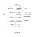
FIG. 11 shows a flow diagram of the cDNA construction used for de novo sequencing.
FIG. 12 shows an electropherogram of the PCR products of primer walking 1.
FIG. 13 shows an electropherogram of the PCR products of primer walking 2.
FIG. 14 shows ClustaW sequence alignment results of primer walking 3 (SEQ ID NOs: 84-95).
FIG. 15 shows an electropherogram of the products of optimized PCR reactions of primer walking 3.
FIG. 16 shows an electropherogram of the products of nested primer PCR reactions of primer walking 3.
FIG. 17 shows the general procedure of primer walking 4.
FIG. 18 shows an electropherogram of the products of a third PCR reaction of primer walking 4.
FIG. 19 shows an electropherogram of the complete gene encoding the enzyme.
FIG. 20 shows a sequence alignment of the complete gene as isolated (SEQ ID NO: 4) and as assembled from partial primer walking segments (SEQ ID NO: 3) using Clustal 2.1 software.
FIG. 21 shows a comparison of amino acid sequences encoded by the isolated gene (SEQ ID NO: 2) and by the one assembled from partial primer walking segments (SEQ ID NO: 1).
FIG. 22 shows a comparison of sequences of the gene obtained from genomic DNA (SEQ ID NO: 96) and the one obtained as a template (SEQ ID NO: 4).
FIG. 23 shows an electropherogram of three reaction mixtures for restriction digestion of the gene from the pJET1.2 vector.
NOMENCLATURE
Designations of nucleotide sequences herein use the usual one-letter code, i.e. A for adenine, C for cytosine, g for guanine and T for thymine (in both upper and lower case). Designations of amino acids of amino acid sequences disclosed in the present description and in the sequence listing is in one-letter and three-letter codes as indicated below for overview.
| |
|
| |
Amino acid |
Three-letter code |
One-letter code |
| |
|
| |
alanine |
Ala |
A |
| |
arginine |
Arg |
R |
| |
asparagine |
Asn |
N |
| |
aspartic acid |
Asp |
D |
| |
cysteine |
Cys |
C |
| |
glutamine |
Gln |
Q |
| |
glutamic acid |
Glu |
E |
| |
glycine |
Gly |
G |
| |
histidine |
His |
H |
| |
isoleucine |
Ile |
I |
| |
leucine |
Leu |
L |
| |
lysine |
Lys |
K |
| |
methionine |
Met |
M |
| |
phenylalanine |
Phe |
F |
| |
proline |
Pro |
P |
| |
serine |
Ser |
S |
| |
threonine |
Thr |
T |
| |
tryptophane |
Trp |
W |
| |
tyrosine |
Tyr |
Y |
| |
valine |
Val |
V |
| |
|
DETAILED DESCRIPTION OF THE INVENTION
1.1 Introduction
Trametes hirsuta (Coriolus hirsuta) is a fungus which belongs to the white-rot fungal family. Alkenes possessing a C═C double bond adjacent to an aromatic ring were cleaved to yield the corresponding carbonyl compound by use of molecular oxygen as the sole oxidant and a cell-free extract of Trametes hirsuta G FCC 047 culture as a catalyst. trans-anethole has shown to be the best substrate for the biocatalytic alkene cleavage, affording p-anisaldehyde as the sole product. The conditions required to carry out this reaction were optimized previously and are indicated in FIG. 1.
The purification of this alkene-cleaving enzyme from the cell-free extract of Trametes hirsuta culture has been attempted several times before. It was always found to be problematic as the activity of the enzyme was already lost after two steps, or even a single step, of purification (hydrophobic interaction chromatography, followed by size exclusion chromatography). The exact cause of this loss of activity was unknown. However, it was suspected that there was a loss of the enzyme co-factor responsible for the reaction, during the column purification. Further research was done to assay the metal dependence of the enzyme.
1.2 Results and Discussion
1.2.1 Mn(III) Dependence of the Alkene-Cleaving Enzyme and Purification of the Enzyme from the Cell-Free Extract
1.2.1.1 Mn(III) Co-Factor Dependency of the Alkene Cleavage Activity
During further research on the intracellular enzymes of Trametes hirsuta G FCC047, surprisingly, an influence of Mn(III) on alkene cleavage activity was found. To find out if the addition of Mn(III) in the fungi culture with poor alkene cleavage activity had any effect on the alkene cleavage activity, Mn(III) salt solution was added to the biotransformation reaction mixture. The reaction was carried out in triplicates using t-anethole as the standard substrate. The sample composition and the obtained conversions (%) are summarized in Table 1.
| TABLE 1 |
| |
| Effect of Mn3+ on t-anethole with and without CFE |
| (cell-free fungal extract) in a three-way approach: conversion |
| of t-anethole to p-anisaldehyde after 10 hours while periodically |
| adding 10 μL of Mn3+ acetate solution (1 mM) |
| 800 μL Bis-tris buffer (pH 6) + 5 μL t-anethole + |
1 |
1 |
1 |
| Mn(III) |
| 800 μL I CFE 1 + 5 μL t-anethole + Mn(III) |
9 |
15 |
11 |
| 800 μL CFE 1 + 5 μL t-anethole |
7 |
7 |
8 |
| 800 μL Bis-tris buffer (pH 6) + 5 μL t-anethole |
0.7 |
0.6 |
0.6 |
| (negative control) |
| |
It could be concluded from the study that the addition of Mn(III) to the cell-free extract was definitely having an influence on the alkene cleavage activity, as there was a considerable increase in the conversion (%) from t-anethole to the p-anisaldehyde.
1.2.1.2 Purification of the Enzyme from the Cell Free Extract of Trametes hirsuta
1.2.1.2.1 Information from Previously Attempted Purification Work
Purification of the enzyme has been attempted several times before. Even though it could not be purified as the activity was always lost after two steps of purification, some valuable information was gathered. The first step of purification employed was always hydrophobic interaction chromatography (phenyl sepharose CL-4B). After this step, the enzyme was still active. This was a neat process, as it was consistent with the fractions in which the enzyme activity was present and also repeatable. Previously, size-exclusion chromatography was employed as the second step. Hardly any activity could be obtained after this step. However, based on the few fractions which gave little activity the size of the enzyme was predicted to be between 140 and 160 KDa. Positive fractions from the first step (HIC) were also subjected to ICP-MS studies to detect the metals present. Only three metals were detected: Mn, Cu and Mo. It was suspected that some key metal/metals were getting lost during the purification process, hence the loss of activity. The study of the mechanism showed that the enzymatic activity involves metal capable of one electron transfer. It was also tested if the activity of the enzyme could be recovered by adding various metals (Cu1+, Cu2+, Mn2+, Mo3+ and Mo5+). All known alkene-cleaving enzymes in literature exhibit either iron or copper. None of the metals tested with the different oxidation states could recover the lost activity. This was another clue that the not previously tested Mn(III) could be the involved co-factor for the alkene cleavage activity.
1.2.1.2.2 Preparation of Cell-Free Extract and Column
The cell-free extract was prepared using the lyophilized T. hirsuta culture. A decent concentration of culture (of 44 mg lyophilized culture/mL of buffer) was subjected to cell disruption by means of ultrasonication. Cell debris was removed from the crude extract, and the supernatant was subjected to purification. The column used for the first step was self-packed (Phenylsepharose CL-4B). For the rest of the steps, commercially available pre-packed columns were used.
1.2.1.2.3 Hydrophobic Interaction Chromatography (Step 1)
The self-packed phenyl sepharose column was used. This step was repeatable, and the enzyme activity was always detected in the fractions corresponding to the second peak, which appeared after the end of the salt (ammonium sulfate) gradient, in water. The chromatogram diagram obtained is given in FIG. 2.
The circled region in FIG. 2 is where enzyme activity is always found (second peak after the end of the salt gradient). The fractions corresponding to this region were pooled for the next step of purification.
1.2.1.2.4 Anion Exchange Chromatography (Step 2)
The commercially available HiTrap® FF Q, anion-exchange column (1 mL) was used in this step. The pooled positive fractions from the previous step (45 mL) was loaded onto the column for further purification. Positive fractions were tested by addition of minimum amounts (0.4 mM) of Mn(III) acetate, which did not in itself catalyze the alkene cleavage. After the second step of purification, enzymatic activity was observed for the first time.
The enzyme was eluted from the flow-through fractions, which means that the enzyme did not bind to the column. The other proteins, which did bind to the column, were eluted with an increasing linear salt (NaCl) concentration gradient. Samples from the fractions were collected for protein concentration evaluation and for SDS-PAGE. The positive fractions (flow-through) were then pooled for further purification. The chromatogram is shown in FIG. 3. The average protein concentration of the flow-through fractions (2 to 11) were found to be 428 μg protein/mL. The circled area is where enzyme activity occurred (flow-through fractions).
FIG. 4 shows the electropherogram of an SDS-PAGE of anion exchange chromatography samples. Fractions 8, 9, 10 and 11 were the active fractions (flow-through). Fractions 15 and 16 were negative fractions.
1.2.1.2.5 Hydrophobic Interaction Chromatography (Step 3)
A commercially available Hitrap® Phenyl HP (1 mL) hydrophobic interaction column was used for the final step of purification. Steps 1 and 2 were performed twice to get sufficient amounts of sample for step 3 (the final step). The pooled sample from the anion exchange chromatography (performed twice, resulting in a volume of about 80 mL with about 428 μg protein/mL) was loaded onto the HIC column. The first time the experiment was performed all fractions were tested for activity. It was then clear where the enzyme of interest eluted. The region was similar to where it eluted in Step-1 HIC (after the end of the ammonium sulfate gradient, in water). When the entire experiment was repeated for the second time, part of the fractions (700 μL) after the ammonium sulfate concentration gradient were collected and tested for activity to find the active fractions. The remaining amount in the fractions was stored for protein concentration estimation and for SDS-PAGE. Fraction 36 showed the highest activity (16% conversion from t-anethole to p-anisaldehyde under reaction conditions). It had a protein concentration of approximately 150 μg protein/mL The chromatogram obtained is given in FIG. 5. Enzyme activity was found in two fractions after the end of the ammonium sulfate concentration gradient. FIG. 6 is a magnified view of the chromatogram in FIG. 5. The hatched area shows the fractions in which enzyme activity was found (fractions 36 and 37).
1.2.1.3 SDS-PAGE of the Active Fraction and Amino Acid Sequence Analysis
SDS-PAGE of the active fraction revealed three main bands. One was in the area of 37 kDa, one was in the area of 25 kDa and the last one was in the area of 20 kDa. FIG. 7 is the electropherogram of fractions 35, 36 and 37 of the purification by hydrophobic interaction chromatography as purification step 3. Fraction 6 was the most active fraction. As the protein concentration of the fractions was low, two indentations in the gel were connected in order to achieve one larger indentation, and 80 μL of sample (fraction/loading buffer=50/50) were filled into each indentation. Comparison between the positive fraction (HIC3-36) and the negative fraction (HIC3-35) suggests that the band at approximately 37 kDa was a unique band for the positive fraction. The band at approximately 37 kDa was excised from the gel and sent to PROTAGEN AG in Dortmund, Germany for MALDI MS-MS analysis and de novo sequencing.
1.2.2 Amino Acid Sequencing of the Obtained SDS Gel Band
This section is partly a copy of the data and report received from PROTAGEN AG.
1.2.2.1 Protein Identification by MALDI-MS/MS and De Novo Sequencing
1.2.2.1.1 In-Gel Digestion
The selected protein spot was excised from the gel. The gel plug was washed alternately three times with 15 μL 10 mM NH4HCO3 and 5 mM NH4HCO3/50% acetonitrile. After drying the gel segment, trypsin solution (33 ng/μL in 10 mM NH4HCO3, pH 7.8) was added to digest the protein for several hours at 37° C.
1.2.2.1.2 MALDI Sample Preparation
The peptides were extracted from the gel segment using 0.1% trifluoroacetic acid (TFA) and purified using C18 material (ZipTip™, Millipore, Bedford, Mass., USA) before spotting them onto the MALDI target.
1.2.2.1.3 MALDI Spectrum Acquisition
The protein identification was performed using an Ultraflex III TOF/TOF mass spectrometer (Bruker Daltonics). For the acquisition of peptide mass fingerprint spectra (PMF, MS), 200 single shot spectra were averaged and peak picking was performed using the SNAP algorithm. The resulting mass list was sent to the ProteinScape™ database for protein identification. Peptide fragmentation spectra (PFF, MS/MS) were acquired where possible. The peaks for fragmentation were selected by the ProteinScape database based upon the results of the protein identification by PMF.
1.2.2.1.4 Protein Identification Using a Public Database
Protein identification was performed by searching the mass spectra against the NCBI protein database (website: www.ncbi.nlm.nih.gov) using several external search algorithms (ProFound™, Mascot™, Sequest™). For PMF spectra, the mass tolerance was set to 50 ppm. The protein identification was based on the metascore1 calculated from the individual search results by the ProteinScape™ database and on manual inspection of the data where needed. PFF spectra were either used to confirm the protein already identified by PMF or for identification of proteins that eluded the PMF identification.
1.2.2.1.5 Automated De Novo Sequencing
The MS/MS data sets were automatically de-novo sequenced using the PEAKS 4.5 software package. The top scoring sequences are reported for each MS/MS spectrum.
1.2.2.1.6 Database Search
The following protein was identified by database search: gi|170091822|ref|XP—001877133.1| aspartic peptidase A1 [Laccaria bicolors S238NH82]. The peptide EPGLAFAFGK (SEQ ID NO: 5) could be identified by database search and could be mapped to this protein.
1.2.2.1.7 De Novo Sequencing
Sample PG379-U11-2010-001
The following peptide sequences (SEQ ID NOs: 5-13) were obtained by de novo sequencing. The amino acids, shown in bold, are identified with very high confidence (PEAKS Score of this sequence part >90%).
| |
| Parent |
|
| mass |
Peptide |
| |
| 1036.5482 |
E P G L A F A F G K* |
| |
| 1166.6215 |
L V D S P V F S F R |
| |
| 1301.6235 |
K Y Y T V Y D H G R |
| |
| 2041.0100 |
N Q D F A E A T K E |
| |
P G L A F A F G K |
| |
| 1173.5321 |
Y Y T V Y D H G R |
| |
| 1366.6850 |
A Y W E V E L E S I |
| |
K |
| |
| 2375.0981 |
L G S S E E D G G E |
| |
A L F G G V D E T A |
| |
Y S G K |
| |
| 1023.4805 |
N Q D F A E A T K |
| |
| 1494.7802 |
K A Y W E V E L E S |
| |
I K |
| |
| *Also found by database searching |
These peptides were identified by de novo sequencing and could be mapped to gi|17009182 by high sequence homology between the peptide identified by de novo sequencing and a sequence region of gi|17009182. Thus, it is assumed that all peptides are part of the same protein.
1.2.3 Identification of the Gene Sequence of the Alkene Cleaving Enzyme
1.2.3.1 PCR Using Designed Degenerate Primers and cDNA of Trametes hirsuta as the Template
1.2.3.1.1 Initial PCR Reaction Optimization
PCR using the different combinations of designed forward and reverse degenerated primers were carried out. Based on the mapping of the obtained sequences with the amino acid sequence of aspartic peptidase A1 of Laccaria bicolor, three forward primers and two reverse primers were designed. Using these, six different combinations of forward and reverse primers were made. In order to check which temperature was suitable for the PCR, three temperatures lower than the melting points of the primers were tested. The different combinations of the primers are given in Table 2. The PCR conditions used are given in Table 3. The program of PCR reactions is given in Table 4.
Key:
| |
Fwd primer 1: |
| |
(SEQ ID NO: 14) |
| |
AAY CAR GAY TTY GCN GAR GC |
| |
|
| |
(SEQ ID NO: 19) |
| |
NQDFAEA |
| |
|
| |
Fwd primer 2: |
| |
(SEQ ID NO: 15) |
| |
GAR GAR GAY GGN GGN GAR GCN |
| |
|
| |
(SEQ ID NO: 20) |
| |
EEDGGEA |
| |
|
| |
Fwd primer 3: |
| |
(SEQ ID NO: 16) |
| |
AAR GCN TAY TGG GAR GTN GA |
| |
|
| |
(SEQ ID NO: 21) |
| |
KAYWEVE |
| |
|
| |
Reverse primer 1: |
| |
(SEQ ID NO: 17) |
| |
TC NAC YTC CCA RTA NGC YTT |
| |
|
| |
(SEQ ID NO: 22 |
| |
KAYWEVE |
| |
|
| |
Reverse primer 2: |
| |
(SEQ ID NO: 18) |
| |
C RTG RTC RTA NAC NGT RTA RTA YT |
| |
|
| |
(SEQ ID NO: 23) |
| |
KYYTVYDHG |
| TABLE 2 |
| |
| Sample numbering for the different combinations of forward and |
| reverse degenerate primers |
| Sample No. |
Primer combination |
| |
| 1 |
Fwd primer 1 and reverse primer 1 |
| 2 |
Fwd primer 1 and reverse primer 2 |
| 3 |
Fwd primer 2 and reverse primer 1 |
| 4 |
Fwd primer 2 and reverse primer 2 |
| 5 |
Fwd primer 3 and reverse primer 1 |
| 6 |
Fwd primer 3 and reverse primer 2 |
| |
| TABLE 3 |
| |
| Reaction components for PCR using the different combinations of |
| primers |
| |
Primer combinations (1-6) |
| |
a |
b |
c |
| Contents |
(47.1° C.) (μL) |
(50.2° C.) (μL) |
(53° C.) (μL) |
| |
| Buffer (5X HF) |
10 |
10 |
10 |
| dNTPs (10 mM) |
1 |
1 |
1 |
| Forward primer |
1 |
1 |
1 |
| ( 1/10 diluted) |
| Reverse primer |
1 |
1 |
1 |
| ( 1/10 diluted) |
| cDNA (180.5 ng/μL) |
1 |
1 |
1 |
| Sterile water |
35.5 |
35.5 |
35.5 |
| DNA polymerase |
0.5 |
0.5 |
0.5 |
| (Phusion) |
| |
| TABLE 4 |
| |
| PCR program used to assay different temperatures with degenerate |
| primers |
| Step |
Temperature (° C.) |
Time |
Number of cycles |
| |
| Initial denaturation |
98 |
2 min |
1 |
| Denaturation |
98 |
30 s |
40 |
| Annealing |
T = 50 |
30 s |
| |
Gradient = 3° C. |
| |
Rate = 3° C./s |
| Extension |
| |
72 |
45 s |
| Final extension |
72 |
7 min |
1 |
| |
After carrying out the PCR, the samples (50 μL each) were analyzed preparatively on agarose gel (1% agarose). Three bands were visible (1c, 6a and 6b). All products were around 200 bp in size. The agarose gel picture is shown in FIG. 9. A PCR product was obtained for combinations 1c, 6a and 6b, as indicated by arrows in FIG. 9.
1.2.3.1.2 Extraction and Amplification of PCR Products
The extractions of the DNA from the bands cut out from the gel were done using the QIAGEN QIAquick® (50) Gel Extraction Kit. The DNA concentrations of the three samples (50 μL volume) were measured. The elution buffer from the kit was used as a blank.
1c—8.9 ng/μL (around 200 bp)
6a—7.6 ng/μL (around 200 bp)
6b—7.8 ng/μL (around 200 bp)
The DNA fragments were cloned and then transformed into TOP 10 E. coli cells for amplification of the DNA. Cloning of the DNA fragments (1c, 6a and 6b) was done using CloneJET™ PCR Cloning kit. The amount of DNA sample taken for each sample was based on the size of the fragment and the concentration of the purified DNA sample. The procedure followed the kit manual.
Overnight cultures (5 mL LB+100 μg/L Amp in each 50 mL tube) for the transformants were started (3 colonies were picked from each transformant plate). A total of 9 tubes were incubated at 37° C. with shaking at 120 rpm overnight. The backup of each of the colonies was also made on agar plates (LB+100 μg/L Amp). The plates were removed the next day and stored at 4° C.
The QIAGEN QIAprep® (250) Spin Miniprep Kit was used for performing a miniprep. The procedure followed the manual. The DNA eluted from the minipreps (50 μL each) were mixed with 6× loading buffer (8.3 μL), and the whole sample was loaded on the agarose gel (1%); see FIG. 10 showing the amplified PCR products in the CloneJET™ cloning vector. The bands of the plasmids were excised from each lane and filled into tubes (15 mL). The gel segments were weighed and the DNA was extracted using the QIAGEN QIAquick® (50) Gel Extraction Kit. The DNA concentration of the 9 samples (50 μL volume) were measured. The elution buffer from the kit was used as a blank.
1c1—20.7 ng/μL, 1c2—12.1 ng/μL, 1c3—22.8 ng/μL, 6a1—16.7 ng/μL, 6a2—14.3 ng/l, 6a3—11.8 ng/μL, 6b1—25.4 ng/μL, 6b2—20 ng/μL, 6b3—61.9 ng/μL.
1.2.3.1.3 Analysis of Insert Sequences and Design of Definite Primers for Primer Walking
The sequencing results for 1c1, 6a1 and 6b1 were individually compared with the sequence of the plasmid pJET1.2 using BLAST. The non-identical region was taken as the sequence of the insert in the plasmid. This region was isolated and translated into its amino acid sequence. Using the different reading frames, the presence of the forward and reverse primers used (Fwd 1 and rev 1 in case of 1c1 and Fwd 3 and rev 2 in case of 6a1 and 6b1) was checked. It was found that both primers were only present in the 6a1 sequence. A protein BLAST with this amino acid sequence gave certain similarities to several fungal proteins belonging to the pepsin-retropepsin superfamily (the second best result was aspartic peptidase A1 from Laccaria bicolor). The sequence of sample 6a1 is shown below. The criteria for designing the definite primers were that both primers have GC content at the end and the melting point of both the primers should be similar. The melting point and the probability of self-complementarity on behalf of the primers were checked using the Oligo-Calc online program. In the case of the reverse primer, the reverse complement of the original DNA segment was prepared and the length was altered to suit the requirement. The primers were designed to comprise the initial degenerate primer region (to double-check the accuracy in primer walking).
| |
>6a1-forward.pJET1.2-F |
| |
(SEQ ID NO: 24) |
| |
TTTTTCAGCAAGAT AAGGCTTATTGGGAGGTGGA GCTGGAATCG |
| |
|
| |
ATCAAACTCGGAGAC GACGAGCTTGAGCTCGATAACACCGGCGCT |
| |
|
| |
GCCATCGACACTGGAACCTCGTTGATTGCTCTCCCCTCCGATCTGG |
| |
|
| |
CGGAGATGCTCAATGTGCAAATCGGTGCCAAGAAGTCCTGGAATGG |
| |
|
| |
TCAGTACACCGTCGACTGCGCGAAGGTCCCTACCCTCCCCGACCTC |
| |
|
| |
ACCTTCTACTTCAGCGGCAAGCCTTACACTCTCAAGGGTACCGACT |
| |
|
| |
ACGTCCTCGAAGTTCAGGGAACTTGCATGTCCTCGTTCACCGGCAT |
| |
|
| |
CGACATCAATCTGCCCGGCGGTGGTGCTC TGTGGATCATTGGTGA |
| |
|
| |
TGTCTTCCTGC GCA AGTACTACACAGTTTACGATCACG AT CT |
| |
|
| |
TTCTAGAAGATCTCC TACAATATTCTCAGCTGCCATGGAAAATCG |
| |
|
| |
ATGTTCTTCTTTTATTCTCTCAAGATTTTCAGGCTGTATATTAAAA |
| |
|
| |
CTTATATTAAGAACTATGCTAACCACCTCATCAGGAACCGTTGTAG |
| |
|
| |
GTGGCGTGGGTTTTCTTGGCAATCGACTCTCATGAAAACTACGAGC |
| |
|
| |
TAAATATTCAATATGTTCCTCTTGACCAACTTTATTCTGCATTTTT |
| |
|
| |
TTTGAACGAGGTTTAGAGCAAGCTTCAGGAAACTGAGACAGGAATT |
| |
|
| |
TTATTAAAAATTTAAATTTTGAAGAAAGTTCAGGGTTAATAGCATC |
| |
|
| |
CATTTTTTGCTTTGCAAGTTCCTCAGCATTCTTAACAAAAGACGTC |
| |
|
| |
TCTTTTGACATGTTTAAAGTTTAAACCTCCTGTGTGAAATTATTAT |
| |
|
| |
CCGCTCATAATTCCACACATTATACGAGCCGGAAGCATAAAGTGTA |
| |
|
| |
AAGCCTGGGGTGCCTAATGAGTGAGCTAACTCACATTAATTGCGTT |
| |
|
| |
GCGCTCACTGCCAATTGCTTTCCAGTCGGGAACCTGTCGTGCCAGC |
| |
|
| |
TGCATTAATG |
Amino acid sequence obtained by translation of the insert DNA sequence:
| |
>EMBOSS_001_3 |
| |
(SEQ ID NO: 25) |
| |
FQQD KAYWEVE LESIKLGD DELELDNTGAAIDTGTSLIALPSD |
| |
|
| |
LAEMLNVQIGAKKSWNGQYTVDCAKVPTLPDLTFYFSGKPYTLKGT |
| |
|
| |
DYVLEVQGTCMSSFTGIDINLPGGGAL WIIGDV FLR KYYTVYD |
| |
|
| |
H D |
The underlined sequences each represent one of the following primers:
forward degenerate Primer
reverse degenerate Primer
start of vector sequence
designed definite forward primer (PW-1 Fwd)
designed definite reverse primer (PW-1 Rev)
1.2.3.2 Primer Walking 1
1.2.3.2.1 Design of Primers for the cDNA End
The attempt to carry out a PCR with just one primer (the definite forward and reverse primers each) did not yield any PCR product. There had to be a combination of both forward and reverse primers in a PCR. In order the design another set of forward and reverse primers, the construction of the cDNA was revisited. The SMART™ cDNA library construction kit was used to get the cDNA from the mRNA of Trametes hirsuta. Initially, the CDS III primer was used for the first cDNA strand synthesis, starting from the poly A+ tail of the mRNA. For the second strand synthesis, the SMART IV oligonucleotide was used. Thus, every cDNA fragment will contain the sequence of CDSIII primer at the 3′ end and the sequence of SMART IV primer at the 5′ end (see FIG. 11). Hence a forward primer based on the sequence of SMART IV oligonucleotide and a reverse primer based on CDS III primer were designed to both have melting point similar to that of the definite forward and reverse primers previously designed. A flowchart of the cDNA construction from the user manual of the kit is given in FIG. 11. The new primers were designed on this basis. The two forward and reverse primers designed are given below:
| |
PW1-Fwd |
| |
(SEQ ID NO: 26) |
| |
TGTGGATCATTGGTGATGTCTTCCTGC |
| |
|
| |
PW1-Rev |
| |
(SEQ ID NO: 27) |
| |
GTCTCCGAGTTTGATCGATTCCAGC |
| |
|
| |
SMART-IV |
| |
(SEQ ID NO: 28) |
| |
GTATCAACGCAGAGTGGCCATTACG |
| |
|
| |
CDS III |
| |
(SEQ ID NO: 29) |
| |
CGAGGCGGCCGACATGTTTTTTTT |
1.2.3.2.2 PCR Primer Walking 1
PCR using the different combinations of forward and reverse primers were carried out. The main change made in the PCR program was that of the annealing temperature. Since all the primers had similar melting temperatures, two different temperatures were tested. The different primer combinations are given in Table 5. The different reaction ingredients are summarized in Table 6. The PCR reaction program is given in Table 7.
| TABLE 5 |
| |
| Different primer combinations and sample numbering |
| Sample No. |
Primer combination |
| |
| 1 |
Pw1-Fwd and SMART IV fwd |
| 2 |
Pw1-fwd and CDS III rev |
| 3 |
Pw1-rev and SMART IV fwd |
| 4 |
Pw1-rev and CDS III rev |
| |
| TABLE 6 |
| |
| Reaction ingredients for PCR using the different combinations of |
| primers for primer walking 1 |
| |
Primer combinations (1-4) |
|
| |
|
a |
b |
| |
Contents |
(59.3° C.) (μL) |
(63.4° C.) (μL) |
| |
|
| |
Buffer (5X HF) |
10 |
10 |
| |
dNTP's (10 mM) |
1 |
1 |
| |
Forward primer ( 1/10 diluted) |
1 |
1 |
| |
Reverse primer( 1/10 diluted) |
1 |
1 |
| |
cDNA (180.5 ng/μL) |
1 |
1 |
| |
Sterile water |
35.5 |
35.5 |
| |
DNA polymerase (Phusion) |
0.5 |
0.5 |
| |
|
| TABLE 7 |
| |
| PCR program used to assay different temperatures for primer walking 1 |
| Step |
Temperature (° C.) |
Duration |
Number of cycles |
| |
| Initial denaturation |
98 |
2 min |
1 |
| Denaturation |
98 |
30 s |
40 |
| Annealing |
T = 60 |
45 s |
|
| |
Gradient = 4° C. |
|
|
| |
Rate = 3° C./s | |
|
| Extension |
| |
72 |
45 s |
|
| Final extension |
72 |
7 min |
1 |
| |
After carrying out the PCR, the samples (50 μL each) were analyzed preparatively on agarose gel (1% agarose). Clear bands were visible (around 300 bp) for combination no. 2 for both the tested temperatures. Faint bands were also visible (around 300 bp) for combination no. 3 for both temperatures. Combinations 2 and 3 were the sensible combinations as they each contained one forward and one reverse primer. Some other products were also obtained, but the sizes were low and hence they were not excised. The agarose gel picture is shown in FIG. 12. Thus, in primer walking 1, PCR products were obtained for products 2a, 2b, 3a and 3b. Extraction and amplification of the PCR products 2a, 2b, 3a and 3b were done in a similar manner as explained in segment 1.2.3.2.3.
1.2.3.2.3 Sequence Analysis for Primer Walking 1
Out of all the samples given for sequencing, clear results could be obtained for one of the samples of 2a insert and two samples of 3b insert. Since sequencing can be in both forward and reverse directions, the sequences were checked for the actual primer sequence and also the reverse complement sequence of the primers.
| PW1-Fwd |
| (SEQ ID NO: 26) |
| TGTGGATCATTGGTGATGTCTTCCTGC |
| |
| CDS III |
| (SEQ ID NO: 29) |
| CGAGGCGGCCGACATGTTTTTTTT |
| |
| PW1-Rev |
| (SEQ ID NO: 27) |
| GTCTCCGAGTTTGATCGATTCCAGC |
| |
| SMART-IV |
| (SEQ ID NO: 28) |
| GTATCAACGCAGAGTGGCCATTACG |
| |
| PW1-Fwd rev |
| (SEQ ID NO: 30) |
| GCAGGAAGACATCACCAATGATCCACA |
| |
| CDS III rev |
| (SEQ ID NO: 31) |
| AAAAAAAACATGTCGGCCGCCTCG |
| |
| PW1-Rev rev |
| (SEQ ID NO: 32) |
| GCTGGAATCGATCAAACTCGGAGAC |
| |
| SMART-IV rev |
| (SEQ ID NO: 33) |
| CGTAATGGCCACTCTGCGTTGATAC |
| |
| >PW-1-2a3.pJET1.2-F |
| (SEQ ID NO: 34) |
| TGTGGATCATTGGTGATGTCTTCCTGC GCAAGTACTACACTGTGTAC |
| |
| GACCATGGTCGCGATGCCGTTGGCTTCGCTCTTGCCAAGTGAAGGCGT |
| |
| AGTGTATCTCCCGAAGACAGTTCTACCGTACGACGCGTCGTGTTACGG |
| |
| TTTCTTGATACCTGCATGTACAATACTTAGTCTCCGTTGGAACCATAC |
| |
| CTTCTGTGTGTTGCCCAAAAAAAAAAAAAAAAAA AAAAAAAACATGT |
| |
| CGGCCGCCTCG ATCTTTCTAAAAAATCTCCTACAATATTCTCAGCTG |
| |
| CCATGGAAAATCGATGTTCTTCTTTTATTCTCTCAAGATTTTCAGGCT |
| |
| GTATATTAAAACTTATATTAAAAACTATGCTAACCACCTCATCAGGAA |
| |
| CCGTTGTAGGTGGCGTGGGTTTTCTTGGCAATCGACTCTCATGAAAAC |
| |
| TACGAGCTAAATATTCAATATGTTCCTCTTGACCAACTTTATTCTGCA |
| |
| TTTTTTTTGAACGAGGTTTAGAGCAAGCTTCAGGAAACTGAGACAGGA |
| |
| ATTTTATTAAAAATTTAAATTTTGAAGAAAGTTCAGGGTTAATAGCAT |
| |
| CCATTTTTTGCTTTGCAAGTTCCTCAGCATTCTTAACAAAAGACGTCT |
| |
| CTTTTGACATGTTTAAAGTTTAAACCTCCTGTGTGAAATTATTATCCG |
| |
| CTCATAATTCCACACATTATACGAGCCGGAAGCATAAAGTGTAAAGCC |
| |
| TGGGGTGCCTAATGAGTGAGCTAACTCACATTAATTGCGTTGCGCTCA |
| |
| CTGCCAATTGCTTTCCAGTCGGGAAACCTGTCGTGCCAGCTGCATTAA |
| |
| TGAATCGGCCAACGCGCGGGGAGAGGCGGTT |
| |
| Translation of Insert 2a3 |
| >EMBOSS_001_3 |
| (SEQ ID NO: 35) |
| WIIGDV FLR KYYTVYDH GRDAVGFALAK*RRSVSPEDSSTVRRVV |
| |
| LRFLDTCMYNT*SPLEPYLLCVAQKKKKKKKKHVGRLX |
| |
| >PW-1-3b2.pJET1.2-F |
| (SEQ ID NO: 36) |
| GTCTCCGAGTTTGATCGATTCCAGC GGGACATGGGGACAGTCAATTA |
| |
| GGCTACGCGGATGTACTGCGCAGCAAGGCATGCCGACCGGCCTTCATC |
| |
| ATGTTATAGCTATAGCTAGAGCAGCGCGAGAGACCCTGTAGAGTCACT |
| |
| GATGAATCACTCGTGCTCCCTTCTGTGCCTTGGCTGAATAAGTTTTCC |
| |
| ACAAGTTGTCGTGGAGAGTCGTGCAGGAGGGAGGCAACTTGCCCCCGG |
| |
| C CGTAATGGCCACTCTGCGTTGATAC ATCTTTCTAGAAGATCTCCT |
| |
| ACAATATTCTCAGCTGCCATGGAAAATCGATGTTCTTCTTTTATTCTC |
| |
| TCAAGATTTTCAGGCTGTATATTAAAACTTATATTAAGAACTATGCTA |
| |
| ACCACCTCATCAGGAACCGTTGTAGGTGGCGTGGGTTTTCTTGGCAAT |
| |
| CGACTCTCATGAAAACTACGAGCTAAATATTCAATATGTTCCTCTTGA |
| |
| CCAACTTTATTCTGCATTTTTTTTGAACGAGGTTTAGAGCAAGCTTCA |
| |
| GGAAACTGAGACAGGAATTTTATTAAAAATTTAAATTTTGAAGAAAGT |
| |
| TCAGGGTTAATAGCATCCATTTTTTGCTTTGCAAGTTCCTCAGCATTC |
| |
| TTAACAAAAGACGTCTCTTTTGACATGTTTAAAGTTTAAACCTCCTGT |
| |
| GTGAAATTATTATCCGCTCATAATTCCACACATTATACGAGCCGGAAG |
| |
| CATAAAGTGTAAAGCCTGGGGTGCCTAATGAGTGAGCTAACTCACATT |
| |
| AATTGCGTTGCGCTCACTGCCAATTGCTTTCCAGTCGGGAAACCTGTC |
| |
| GTGCCAGCTGCATTAATGAATCGGCCAACGCGCGG |
| |
| Translation of Insert 3b1 |
| (SEQ ID NO: 37) |
| YQRRVA ITAGGKLPPSCTTLHDNLWKTYSAKAQKGARVIHQ*LYRVS |
| |
| RAALAIAIT**RPVGMPCCAVHPRSLIDCPHVP LESIKLGD |
Analysis of the sequences showed that the insert 2a had both the PW-I fwd primer sequence and the original degenerate reverse primer sequence. Hence it is part of the protein of interest. The translation of the sequence gave a sequence with a stop codon in between. Hence the end of the protein of interest has been reached. In the case of the sequence of the insert 3b, the PW-I reverse primer was present but did not comprise the initial degenerate forward primer sequence. Hence it was quite clear that the designed reverse primer PW-1 rev was not specific enough and had bound to some other part of the cDNA. Thus, three other reverse primers were designed, which were used for primer walking 2. A total of 84 amino acid residues could be deduced at the end of primer walking 1 and the end of the protein had been reached. The sequence of the peptide obtained until this step is given below:
| |
(SEQ ID NO: 38) |
| |
KAYWEVE LESIKLGD DELELDNTGAAIDTGTSLIALPSDLA |
| |
|
| |
EMLNVQIGAKKSWNGQYTVDCAKVPTLPDLTFYFSGKPYTLKG |
| |
|
| |
TDYVLEVQGTCMSSFTGIDINLPGGGAL WIIGDV FLR KYY |
| |
|
| |
TVYDH GRDAVGFALAK |
1.2.3.3 Primer Walking 2
1.2.3.3.1 Design of Alternative Reverse Primers
Since the reverse primer (PW-1 rev) from primer walking 1 was not specific enough for the PCR, alternative reverse primers had to be designed. Three alternative reverse primers were designed. Further it was also noticed (based on the BLAST search) that the reverse primers were situated at the end of the entire protein sequence. As a consequence, the degenerate primer (Fwd-1) from the first step (expected to be in the middle of the sequence in the BLAST search) was used as the forward primer.
| |
>6a1-forward.pJET1.2-F (result of 2.1) |
| |
(SEQ ID NO: 24) |
| |
TTTTTCAGCAAGAT AAGGCTTATTGGGAGGTGGA GCTGGA |
| |
|
| |
ATCGATCAAACT CGGAGACGACGAGCTTGAGCT CGATAAC |
| |
|
| |
A CCGGCGCTGCCATCGACACT GGAACCTCGTTGATTGCTC |
| |
|
| |
TCCCCTCCGATCT
GGCGGAGATGCTCAATGTGCAAATC
GG
|
| |
|
| |
TGCCAAGAAGTCCTGGAATGGTCAGTACACCGTCGACTGCGC
|
| |
|
| |
GAAGGTCCCTACCCTCCCCGACCTCACCTTCTACTTCAGCGG
|
| |
|
| |
CAAGCCTTACACTCTCAAGGGTACCGACTACGTCCTCGAAGT
|
| |
|
| |
TCAGGGAACTTGCATGTCCTCGTTCACCGGCATCGACATCAA
|
| |
|
| |
TCTGCCCGGCGGTGGTGCTC TGTGGATCATTGGTGATGTCT |
| |
|
| |
TCCTGC GCA AGTACTACACAGTTTACGATCACG ATCTTT |
| |
|
| |
CTAGAAGATCTCCTACAATATTCTCAGCTGCCATGGAAAATC |
| |
|
| |
GATGTTCTTCTTTTATTCTCTCAAGATTTTCAGGCTGTATAT |
| |
|
| |
TAAAACTTATATTAAGAACTATGCTAACCACCTCATCAGGAA |
| |
|
| |
CCGTTGTAGGTGGCGTGGGTTTTCTTGGCAATCGACTCTCAT |
| |
|
| |
GAAAACTACGAGCTAAATATTCAATATGTTCCTCTTGACCAA |
| |
|
| |
CTTTATTCTGCATTTTTTTTGAACGAGGTTTAGAGCAAGCTT |
| |
|
| |
CAGGAAACTGAGACAGGAATTTTATTAAAAATTTAAATTTTG |
| |
|
| |
AAGAAAGTTCAGGGTTAATAGCATCCATTTTTTGCTTTGCAA |
| |
|
| |
GTTCCTCAGCATTCTTAACAAAAGACGTCTCTTTTGACATGT |
| |
|
| |
TTAAAGTTTAAACCTCCTGTGTGAAATTATTATCCGCTCATA |
| |
|
| |
ATTCCACACATTATACGAGCCGGAAGCATAAAGTGTAAAGCC |
| |
|
| |
TGGGGTGCCTAATGAGTGAGCTAACTCACATTAATTGCGTTG |
| |
|
| |
CGCTCACTGCCAATTGCTTTCCAGTCGGGAACCTGTCGTGCC |
| |
|
| |
AGCTGCATTAATG |
The reverse complements of the sequences marked in the above:
| |
1) PW-2 rev Pri-1: |
| |
(SEQ ID NO: 39) |
| |
AGCTCAAGCTCGTCGTCTCCG |
| |
|
| |
2) PW-2 rev Pri-2: |
| |
(SEQ ID NO: 40) |
| |
AGTGTCGATGGCAGCGCCG |
| |
|
| |
3) PW-2 rev Pri-3: |
| |
(SEQ ID NO: 41) |
| |
GATTTGCACATTGAGCATCTCCGCC |
1.2.3.3.2 PCR Primer Walking 2
PCR using the combinations of the three new reverse primers designed with the forward degenerated primer (Fwd-1) was attempted. As a test, one of the reverse primers were also tested with the primer which binds to the end of the cDNA (SMART IV fwd) to check if any clear bands could be obtained (owing to the long distance to the beginning of the protein). As a recheck, the combination of degenerate forward primer (Fwd-1) and degenerate reverse primer (Rev-2) from step 1 was also repeated. The different combinations of primers used are given in Table 8. The PCR components are summarized in Table 9, and the PCR program used is indicated in Table 10.
| TABLE 8 |
| |
| Numbering of the different primer combinations tested for PW-2 |
| Sample no. |
Primer combination |
| |
| 1 |
degenerate Fwd-1 and degenerate Rev-1 |
| 2 |
SMART IV Fwd and PW-2-Rev-Pri-2 |
| 3 |
degenerate Fwd-1 and PW-2-Rev-Pri-1 |
| 4 |
degenerate Fwd-1 and PW-2-Rev-Pri-2 |
| 5 |
degenerate Fwd-1 and PW-2-Rev-Pri-3 |
| |
| TABLE 9 |
| |
| Reaction components for PCR using the different combinations of |
| primers for primer walking 2 |
| |
Primer combinations (1-5) |
| |
a |
b |
c |
| |
(53.2° C.) |
(56.5° C.) |
(59.4° C.) |
| Contents |
(μL) |
(μL) |
(μL) |
| |
| Buffer (5X HF) |
10 |
10 |
10 |
| dNTPs (10 mM) |
1 |
1 |
1 |
| Forward primer |
1 |
1 |
1 |
| (1/10 diluted) |
|
|
|
| Reverse primer |
1 |
1 |
1 |
| (1/10 diluted) |
|
|
|
| cDNA (180.5 ng/μL) |
1 |
1 |
1 |
| Sterile water |
35.5 |
35.5 |
35.5 |
| DNA polymerase |
0.5 |
0.5 |
0.5 |
| (Phusion) |
| |
| TABLE 10 |
| |
| PCR program used for checking different temperatures for primer |
| walking 2 |
| Step |
Temperature (° C.) |
Time |
Number of cycles |
| |
| Initial denaturation |
98 |
2 min |
1 |
| Denaturation |
98 |
30 s |
40 |
| Annealing |
T = 55 |
45 s |
|
| |
Gradient = 4° C. |
|
|
| |
Rate = 3° C./s | |
|
| Extension |
| |
72 |
1 min |
|
| Final extension |
72 |
7 min |
1 |
| |
After carrying out the PCR, the samples (50 μL each) were analyzed preparatively on agarose gel (1% agarose). Clear bands (PCR product) were visible (around 400 bp) for samples 3b, 4a, 4b, 5a and 5b. Additionally, distinct bands were visible (around 600 bp) for combinations 5a and 5b. However, no distinct bands were observed in combination no. 2. It is possible that the distance between the 5′ end of the cDNA and the reverse primer was too long. It is also possible that the duration of the DNA strand extension step was too short for the long distance extension to reach completion.
The extraction and amplification of the PCR products 3b, 4a, 4b, 5a small band, 5b small band, 5a big band and 5b big band were performed in a manner similar to that explained in segment 1.2.3.1.2. Unfortunately, the transformation plates of 3b, 4a and 5a big band were contaminated. As a result, only the samples 4b, 5a small band, 5b small band and 5b big band were sequenced.
1.2.3.3.3 Sequence Analysis for Primer Walking 2
The DNA sequences obtained were analyzed for identifying the primers and hence the sequence of the inserts. All the inserts resulted in the same sequence. The identities of the sequences obtained were compared. The sequences were found to be completely identical.
Key:
| |
PW-2-Reverse primers |
| |
1) PW-2 rev Pri-1: |
| |
(SEQ ID NO: 39) |
| |
AGCTCAAGCTCGTCGTCTCCG |
| |
|
| |
2) PW-2 rev Pri-2: |
| |
(SEQ ID NO: 40) |
| |
AGTGTCGATGGCAGCGCCG |
| |
|
| |
3) PW-2 rev Pri-3: |
| |
(SEQ ID NO: 41) |
| |
GATTTGCACATTGAGCATCTCCGCC |
| |
|
| |
Reverse complement of reverse primers: |
| |
2) PW-2 rev Pri-2: |
| |
(SEQ ID NO: 42) |
| |
CGGCGCTGCCATCGACACT |
| |
|
| |
3) PW-2 rev Pri-3: |
| |
(SEQ ID NO: 43) |
| |
GGCGGAGATGCTCAATGTGCAAATC |
| |
|
| |
Fwd-1 degenerated primer |
| |
(SEQ ID NO: 14) |
| |
AAYCARGAYTTYGCNGARGC |
| |
|
| |
>4b1.pJET1.2-F |
| |
(SEQ ID NO: 44) |
| |
AACCAGGATTTTGCGGAGGC CACCAAGGAGCCCGGCCTCGCATTT |
| |
|
| |
GCCTTTGGCAAGTTTGATGGTATCCTCGGCCTCGGGTATGACACCA |
| |
|
| |
TTTCCGTGAACCACATCACTCCTCCCTTCTACCAGATGATGAACCA |
| |
|
| |
GAAGCTCGTCGATTCTCCTGTGTTCTCTTTCCGCCTCGGTAGCTCG |
| |
|
| |
GAAGAGGACGGTGGTGAAGCCATCTTCGGAGGAGTCGATGAGACCG |
| |
|
| |
CGTACAGTGGCAAGATCGAATACGTCCCTGTCAGGAGGAAGGCGTA |
| |
|
| |
CTGGGAGGTGGAGCTGGAATCGATCAAACTCGGAGACGACGAGCTT |
| |
|
| |
GAGCTCGATAACAC CGGCGCTGCCATCGACACT ATCTTTCTAGA |
| |
|
| |
AGATCTCCTACAATATTCTCAGCTGCCATGGAAAATCGATGTTCTT |
| |
|
| |
CTTTTATTCTCTCAAGATTTTCAGGCTGTATATTAAAACTTATATT |
| |
|
| |
AAGAACTATGCTAACCACCTCATCAGGAACCGTTGTAGGTGGCGTG |
| |
|
| |
GGTTTTCTTGGCAATCGACTCTCATGAAAACTACGAGCTAAATATT |
| |
|
| |
CAATATGTTCCTCTTGACCAACTTTATTCTGCATTTTTTTTGAACG |
| |
|
| |
AGGTTTAGAGCAAGCTTCAGGAAACTGAGACAGGAATTTTATTAAA |
| |
|
| |
AATTTAAATTTTGAAGAAAGTTCAGGGTTAATAGCATCCATTTTTT |
| |
|
| |
GCTTTGCAAGTTCCTCAGCATTCTTAACAAAAGACGTCTCTTTTGA |
| |
|
| |
CATGTTTAAAGTTTAAACCTCCTGTGTGAAATTATTATCCGCTCAT |
| |
|
| |
AATTCCACACATTATACGAGCCGGAAGCATAAAGTGTAAAGCCTGG |
| |
|
| |
GGTGCCTAATGAGTGAGCTAACTCACATTAATTGCGTTGCGCTCAC |
| |
|
| |
TGCCAATTGCTTTCCAGTCGGGAAACCTGTCGTG |
| |
|
| |
>Translation of insert 4b1 |
| |
(SEQ ID NO: 45) |
| |
NQDFAEA TKEPGLAFAFGKFDGILGLGYDTISVNHITPPFYQMMN |
| |
|
| |
QKLVDSPVFSFRLGSSEEDGGEAIFGGVDETAYSGKIEYVPVRR K |
| |
|
| |
AYWEVE LESIKLGDDELELDNT GAAIDT |
| |
|
| |
>5a1smallband.pJET1.2-F |
| |
(SEQ ID NO: 46) |
| |
GATTTGCACATTGAGCATCTCCGCC AGATCGGAGGGGAGAGCAAT |
| |
|
| |
CAACGAGGTTCC AGTGTCGATGGCAGCGCCG GTGTTATCGAGCT |
| |
|
| |
CAAGCTCGTCGTCTCCGAGTTTGATCGATTCCAGCTCCACCTCCCA |
| |
|
| |
GTACGCCTTCCTCCTGACAGGGACGTATTCGATCTTGCCACTGTAC |
| |
|
| |
GCGGTCTCATCGACTCCTCCGAAGATGGCTTCACCACCGTCCTCTT |
| |
|
| |
CCGAGCTACCGAGGCGGAAAGAGAACACAGGAGAATCGACGAGCTT |
| |
|
| |
CTGGTTCATCATCTGGTAGAAGGGAGGAGTGATGTGGTTCACGGAA |
| |
|
| |
ATGGTGTCATACCCGAGGCCGAGGATACCATCAAACTTGCCAAAGG |
| |
|
| |
CAAATGCGAGGCCGGGCTCCTTGGTG GCCTCAGCAAAATCCTGAT |
| |
|
| |
T ATCTTTCTAGAAGATCTCCTACAATATTCTCAGCTGCCATGGAA |
| |
|
| |
AATCGATGTTCTTCTTTTATTCTCTCAAGATTTTCAGGCTGTATAT |
| |
|
| |
TAAAACTTATATTAAGAACTATGCTAACCACCTCATCAGGAACCGT |
| |
|
| |
TGTAGGTGGCGTGGGTTTTCTTGGCAATCGACTCTCATGAAAACTA |
| |
|
| |
CGAGCTAAATATTCAATATGTTCCTCTTGACCAACTTTATTCTGCA |
| |
|
| |
TTTTTTTTGAACGAGGTTTAGAGCAAGCTTCAGGAAACTGAGACAG |
| |
|
| |
GAATTTTATTAAAAATTTAAATTTTGAAGAAAGTTCAGGGTTAATA |
| |
|
| |
GCATCCATTTTTTGCTTTGCAAGTTCCTCAGCATTCTTAACAAAAG |
| |
|
| |
ACGTCTCTTTTGACATGTTTAAAGTTTAAACCTCCTGTGTGAAATT |
| |
|
| |
ATTATCCGCTCATAATTCCACACATTATACGAGCCGGAAGCATAAA |
| |
|
| |
GTGTAAAGCCTGGGGTGCCTAATGAGTGAGCTAACTCACATT |
| |
|
| |
>Translation of insert 5a1 small band |
| |
(SEQ ID NO: 47) |
| |
NQDFAEA TKEPGLAFAFGKFDGILGLGYDTISVNHITPPFYQMMN |
| |
|
| |
QKLVDSPVFSFRLGSSEEDGGEAIFGGVDETAYSGKIEYVPVRR K |
| |
|
| |
AYWEVE LESIKLGDDELELDNT GAAIDT GTSLIALPSDL AEM |
| |
|
| |
LNVQI
|
| |
|
| |
>5b2bigband.pJET1.2-F |
| |
(SEQ ID NO: 48) |
| |
GATTTGCACATTGAGCATCTCCGCC AGATCGGAGGGGAGAGCAAT |
| |
|
| |
CAACGAGGTTCC AGTGTCGATGGCAGCGCCG GTGTTATCGAGCT |
| |
|
| |
CAAGCTCGTCGTCTCCGAGTTTGATCGACTCCAGCTCCACCTCCCA |
| |
|
| |
GTACGCCTTCCTCCTGACAGGGACGTATTCGATCTTGCCACTGTAC |
| |
|
| |
GCGGTCTCATCGACTCCTCCGAAGATGGCTTCACCACCGTCCTCTT |
| |
|
| |
CCGAGCTACCGAGGCGGAAAGAGAACACAGGAGAATCGACGAGCTT |
| |
|
| |
CTGGTTCATCATCTGGTAGAAGGGAGGAGTGATGTGGTTCACGGAA |
| |
|
| |
ATGGTGTCATAACCCAGGCCGAGGATACCATCGAACTT GCCAAAG |
| |
|
| |
GCAAATGCGAG GCCGGGCTCCTTGGTG GCCTCAGCGAAGTCCTG |
| |
|
| |
ATA TCTTTCTAGAAGATCTCCTACAATATTCTCAGCTGCCATGGA |
| |
|
| |
AAATCGATGTTCTTCTTTTATTCTCTCAAGATTTTCAGGCTGTATA |
| |
|
| |
TTAAAACTTATATTAAGAACTATGCTAACCACCTCATCAGGAACCG |
| |
|
| |
TTGTAGGTGGCGTGGGTTTTCTTGGCAATCGACTCTCATGAAAACT |
| |
|
| |
ACGAGCTAAATATTCAATATGTTCCTCTTGACCAACTTTATTCTGC |
| |
|
| |
ATTTTTTTTGAACGAGGTTTAGAGCAAGCTTCAGGAAACTGAGACA |
| |
|
| |
GGAATTTTATTAAAAATTTAAATTTTGAAGAAAGTTCAGGGTTAAT |
| |
|
| |
AGCATCCATTTTTTGCTTTGCAAGTTCCTCAGCATTCTTAACAAAA |
| |
|
| |
GACGTCTCTTTTGACATGTTTAAAGTTTAAACCTCCTGTGTGAAAT |
| |
|
| |
TATTATCCGCTCATAATTCCACACATTATACGAGCCGGAAGCATAA |
| |
|
| |
AGTGTAAAGCCTGGGGTGCCTAATGAGTGAGCTAACT |
| |
|
| |
>Translation of insert 5b2 bigband |
| |
(SEQ ID NO: 49) |
| |
YQDFAEA TKEPGLAFAFGKFDGILGLGYDTISVNHITPPFYQMMN |
| |
|
| |
QKLVDSPVFSFRLGSSEEDGGEAIFGGVDETAYSGKIEYVPVRR K |
| |
|
| |
AYWEVE LESIKLGDDELELDNT GAAIDTGT SLIALPSDL AEM |
| |
|
| |
LNVQI
|
| |
|
| |
Protein sequence after primer walking step 1: |
| |
>EMBOSS_001_3 |
| |
(SEQ ID NO: 38) |
| |
KAYWEVE LESIKLGD DELELDNT GAAIDT GTSLIALPSDL A |
| |
|
| |
EMLNVQI GAKKSWNGQYTVDCAKVPTLPDLTFYFSGKPYTLKGTD |
| |
|
| |
YVLEVQGTCMSSFTGIDINLPGGGAL WIIGDV FLR KYYTVYDH |
| |
|
| |
GRDAVGFALAK* |
| |
|
| |
Protein sequence from primer walking step 2: |
| |
(SEQ ID NO: 50) |
| |
NQDFAEA TKEPGLAFAFGKFDGILGLGYDTISVNHITPPFYQMMN |
| |
|
| |
QKLVDSPVFSFRLGSSEEDGGEAIFGGVDETAYSGKIEYVPVRR K |
| |
|
| |
AYWEVE LESIKLGDDELELDNT GAAIDT GTSLIALPSDLAEML |
| |
|
| |
NVQI GAKKSWNGQYTVDCAKVPTLPDLTFYFSGKPYTLKGTDYVL |
| |
|
| |
EVQGTCMSSFTGIDINLPGGGAL WIIGDV FLR KYYTVYDH GR |
| |
|
| |
DAVGFALAK* |
Primer walking 2 using the newly designed reverse primers and the degenerate forward primers was successful. The sequence could be easily verified as it comprised several primer sequences which had been previously designed. Additional 89 amino acid residues were determined from primer walking 2. After primer walking 2, 173 amino acid residues of the protein were determined and half the stretch of the protein was achieved (based on BLAST similarity search). The protein sequence after primer walking 2 is given below (SEQ ID NO: 50):
| |
NQDFAEA TKEPGLAFAFGKFDGILGLGYDTISVNHITPPFYQ |
| |
|
| |
MMNQKLVDSPVFSFRLGSSEEDGGEAI FGGVDETAYSGKIEY |
| |
|
| |
VPVRR KAYWEVE LESIKLGDDELELDNT GAAIDT GTSLI |
| |
|
| |
ALPSDL AEMLNVQI GAKKSWNGQYTVDCAKVPTLPDLTFYF |
| |
|
| |
SGKPYTLKGTDYVLEVQGTCMSSFTGIDINLPGGGAL WIIGD |
| |
|
| |
V FLR KYYTVYDH GRDAVGFALAK* |
1.2.3.4 Primer Walking 3
1.2.3.4.1 Design of Primers for Primer Walking 3
The first two primer walking steps were successful. The situation in the next step was a little more complicated, as there was a possibility that the 5′ end of the cDNA was still too far away for PCR. Hence, as a second option, degenerated forward primers were designed in addition to the definite reverse primer. For the design of the degenerated primers, the top ten complete consensus full sequence hits related to the obtained sequence were compared and primers were designed from the best conserved regions of these sequences, which were upstream to the position to be resolved. In case of PW-3 degenerated forward primers 1 and 3, the degree of degeneracy was very high. In order to reduce degeneracy, the base deoxyinosine (which binds to A, T, G and C) was used in the synthesis. The PW-3 degenerated forward primer 2 has the lowest amount of degeneracy and is the most promising candidate among the designed degenerated primers. Additionally, two reverse primers were designed from the DNA sequence of the currently obtained sequence. A comparison of these sequences and the conserved regions selected for primer design is shown below. Definite reverse primers were also designed. Shorter versions of the reverse1 and reverse2 primers were also designed, in case the temperatures of annealing of the degenerate forward primers would be low. Additionally, a third reverse primer was designed, which comprised a segment of each of primers reverse 1 and reverse 2.
| |
PW-3-Degenerate Fwd Primer 1: |
| |
(SEQ ID NO: 51) |
| |
CAN ARR HTIAARYTISANAA |
| |
|
| |
(SEQ ID NO: 59) |
| |
-HRMKLEK |
| |
|
| |
PW-3-Degenerate Fwd Primer 2: |
| |
(SEQ ID NO: 52) |
| |
AAYTWYATGAAYGCNCARTA |
| |
|
| |
(SEQ ID NO: 60) |
| |
-NYMNAQY |
| |
|
| |
PW-3-Degenerate Fwd Primer 3: |
| |
(SEQ ID NO: 53) |
| |
TTYAARGTIRTNYTTIGAYAC |
| |
|
| |
(SEQ ID NO: 61) |
| |
-FKVILDT |
| |
|
| |
PW-3-definite Reverse Primer1: |
| |
(SEQ ID NO: 54) |
| |
GCCAAAGGCAAATGCGAG |
| |
|
| |
(SEQ ID NO: 62) |
| |
-LAFAFG |
| |
(rev. complement) |
| |
|
| |
Pw3-RevPrimer1short: |
| |
(SEQ ID NO: 55) |
| |
GCCAAAGGCAAATGCG |
| |
|
| |
PW-3-definite Reverse Primer2: |
| |
(SEQ ID NO: 56) |
| |
CAGGCCGAGGATACCATC |
| |
|
| |
(SEQ ID NO: 63) |
| |
-DGILGL |
| |
(rev. complement) |
| |
|
| |
Pw3-RevPrimer2short: |
| |
(SEQ ID NO: 57) |
| |
CAGGCCGAGGATACC |
| |
|
| |
Pw3-RevPrimer23x: |
| |
(SEQ ID NO: 58) |
| |
CATCGAACTTGCCAAAG |
| |
|
| |
(SEQ ID NO: 64) |
| |
-FGKFDX |
The ClutaW sequence alignment results of the top pBLAST best ten hits are shown in FIGS. 14 a and 14 b. The conserved sequences used for designing the degenerate forward primers for primer walking 3 and the relative positioning of the definite reverse primers are marked.
1.2.3.4.2 Optimization of Temperature and Buffer Conditions
The reaction buffer and the temperature of primer annealing had to be optimized for the PCR since degenerate forward primers were used. Different combination of forward and reverse primers were carried out. For optimizing the conditions, a degenerate forward 2 primer was used in combination with the short reverse primers. The nomenclature used for the different primer combinations/annealing temperature and buffer usage are given below. The components of the different PCR reactions carried out are summarized in the Table.
Numbering of Primer Combinations
Primer combination-I: PW-3-degenerate Fwd-2 and PW-3-Rev1_short(a)
Primer combination-II: PW-3-degenerate Fwd-2 and PW-3-Rev2_short(a)
Primer combination-III: SMART IV Fwd and PW-3-Rev2 (b)
PCR with primer combination III was performed such that it could be used as a template for nested primer PCR in the next step.
Temperature of Annealing
a—45.4° C.
b—59.8° C.
Buffer Usage
1-5×HF buffer
2-5×GC buffer
3-5×HF buffer+DMSO
| TABLE 11 |
| |
| Reaction components for PCR using the different |
| combinations of primers for primer walking 3 |
| |
Reaction components for different |
| |
primer combinations |
| |
I |
II |
III |
| |
(45.4° C.) |
(45.4° C.) |
(59.8° C.) |
| |
(μL) |
(μL) |
(μL) |
| Buffer (5X HF) |
10 |
10 |
10 |
10 |
10 |
10 |
10 |
| dNTP's (10 mM) |
1 |
1 |
1 |
1 |
1 |
1 |
1 |
| Forward primer |
1 |
1 |
1 |
1 |
1 |
1 |
1 |
| (undiluted/ |
| 1/10 dil.) |
| Reverse primer |
1 |
1 |
1 |
1 |
1 |
1 |
1 |
| ( 1/10 diluted) |
| cDNA (350 |
0.5 |
0.5 |
0.5 |
0.5 |
0.5 |
0.5 |
0.5 |
| ng/μL) |
| DMSO |
— |
— |
1.5 |
— |
— |
1.5 |
— |
| Sterile water |
36 |
36 |
34.5 |
36 |
36 |
34.5 |
36 |
| DNA polymerase |
0.5 |
0.5 |
0.5 |
0.5 |
0.5 |
0.5 |
0.5 |
| (Phusion) |
| |
| TABLE 12 |
| |
| PCR program used for the buffer and annealing temperature |
| optimization in primer walking-III |
| Step |
Temperature (° C.) |
Time |
Number of cycles |
| |
| Initial denaturation |
98 |
2 min |
1 |
| Denaturation |
98 |
30 s |
35 |
| Annealing |
T = 50 |
45 s |
|
| |
Gradient = 10° C. |
|
|
| |
Rate = 3° C./s | |
|
| Extension |
| |
72 |
1 min |
|
| Final extension |
72 |
7 min |
1 |
| |
FIG. 15 is an electropherogram of the PCR reactions on 1% agarose gel optimized concerning buffers and annealing temperatures. The samples (30 μL) were each analyzed on agarose gel. The PCR product III was used as template DNA for further nested PCR reactions. Since a PCR product was observed when GC buffer was used but not observed when only HF buffer was used for primer combinations I and II, GC buffer was taken as the preferred buffer for the nested PCR. The temperature of annealing was chosen to be 45.4° C. for the nested primer PCR, as product formation could be observed at this temperature.
1.2.3.4.3 Nested Primer PCR
The product III from the PCR reaction on the previous day was used as template DNA for the nested PCR. All primer combinations suspected to be lying within the template were tested. In addition, the best suited reaction buffer for nested primer PCR was also tested for the combination of degenerate PW-3-forward-2 primer (primer with least degree of degeneracy) and PW-3-reverse1-short primer. The nomenclature for the primer combinations and the buffer that were used is given below. The PCR reaction components are summarized in Table 13.
Primer Combinations/Template DNA
I—Pw3-Fw Primer 1 and Pw3-RevPrimer1short/PCR product III from previous step
II—Pw3-FwPrimer2 and Pw3-RevPrimer1short/PCR product III from previous step
III—Pw3-FwPrimer3 and Pw3-RevPrimer1short/PCR product III from previous step
IV—Pw3-FwPrimer3 and Pw3-RevPrimer1short/PCR product II 2 from previous step
V—Pw3-FwPrimer2 and Pw3-RevPrimer23x/PCR product III from previous step
PCR Reaction Buffer
1—GC buffer
2—GC buffer with DMSO
3—HF buffer
4—HF buffer with DMSO
| TABLE 13 |
| |
| Reaction components for PCR using the different |
| combination of primers for Primer walking-3 |
| |
Reaction components for different |
| |
primer combinations (μL) |
| |
(at 45.4° C. annealing temperature) |
| Contents |
I1 |
II1 |
III1 |
II2 |
II3 |
II4 |
IV1 |
V1 |
| |
| Buffer (5X 1 |
10 |
10 |
10 |
10 |
10 |
10 |
10 |
10 |
| (or) 2) |
| dNTP's |
1 |
1 |
1 |
1 |
1 |
1 |
1 |
1 |
| (10 mM) |
| Forward primer |
1 |
1 |
1 |
1 |
1 |
1 |
1 |
1 |
| (undiluted) |
| Reverse primer |
1 |
1 |
1 |
1 |
1 |
1 |
1 |
1 |
| ( 1/10 diluted) |
| Template DNA |
1 |
1 |
1 |
1 |
1 |
1 |
5 |
1 |
| DMSO |
— |
— |
— |
1.5 |
— |
1.5 |
— |
— |
| Sterile water |
36 |
36 |
36 |
34.5 |
36 |
34.5 |
32 |
36 |
| DNA polymer- |
0.5 |
0.5 |
0.5 |
0.5 |
0.5 |
0.5 |
0.5 |
0.5 |
| ase (Phusion) |
| |
FIG. 16 shows an electropherogram of the nested primer PCR reaction on 1% agarose gel. The band from sample V1 was excised for extraction of DNA, amplification of the PCR product by cloning and transformation and sequence analysis.
1.2.3.4.4 Sequence Analysis for Primer Walking 3
The DNA sequences obtained were analyzed for identifying the primers and hence the sequence of the inserts. All inserts resulted in the same sequence. The identities of the sequences obtained were compared. The sequences were found to be completely identical.
| | Fw Primer 1: |
| | (SEQ ID NO: 51) |
| | CANARRHTIAARYTISANAA |
| | |
| | (SEQ ID NO: 59) |
| | -HRMKLEK |
| | |
| | FwPrimer2: |
| | (SEQ ID NO: 52) |
| | AAYTWYATGAAYGCNCARTA |
| | |
| | (SEQ ID NO: 60) |
| | -NYMNAQY |
| | |
| | FwPrimer3: |
| | (SEQ ID NO: 53) |
| | TTYAARGTIRTNYTTIGAYAC |
| | |
| | (SEQ ID NO: 61) |
| | -FKVILDT |
| | |
| | RevPrimer1: |
| | (SEQ ID NO: 54) |
| | GCCAAAGGCAAATGCGAG |
| | |
| | (SEQ ID NO: 62) |
| | -LAFAFG |
| | (rev complement) |
| | |
| | RevPrimer1short: |
| | (SEQ ID NO: 55) |
| | GCCAAAGGCAAATGCG |
| | |
| | RevPrimer2: |
| | (SEQ ID NO: 56) |
| | CAGGCCGAGGATACCATC |
| | |
| | (SEQ ID NO: 63) |
| | -DGILGL |
| | (rev complement) |
| | |
| | RevPrimer2short: |
| | (SEQ ID NO: 57) |
| | CAGGCCGAGGATACC |
| | |
| | RevPrimer23x: |
| | (SEQ ID NO: 58) |
| | CATCGAACTTGCCAAAG |
| | |
| | (SEQ ID NO: 64) |
| | -FGKFDX |
| | |
| | >PW-3-V2-2.pJET1.2-F |
| | (SEQ ID NO: 65) |
| | CATCGAACTTGCCAAAG GCAAATGCG AGGCCGGGCTCCTTGGT |
| | |
| | GGCCTCTGCGAAATCTTGGTTCTTGATGGTGATGTCACCGATTGT |
| | |
| | CAAGACATCTTGCGAGACGAAGCCCTCCATGGAGCCAGAGCCATA |
| | |
| | CTGGATCGAGAACTCGGAGCCGTTCGCCTTGTATGTCGACGAAGC |
| | |
| | GGTCGAGTCATACTTGGCGTGTAGGAAGCACGCAATGGAGGTACA |
| | |
| | CTTGGTGCTCGGAACCCAGAGGTTGCTCGACCCAGTGTCCAGGAT |
| | |
| | GACCTTGAACGATTGCGGGGGAGTGCCCAAGGTGATTTCAGCGAA |
| | |
| | GTACTGTGCA TTCAT GAAGTTATCTTTCTAGAAGATCTCCTAC |
| | |
| | AATATTCTCAGCTGCCATGGAAAATCGATGTTCTTCTTTTATTCT |
| | |
| | CTCAAGATTTTCAGGCTGTATATTAAAACTTATATTAAGAACTAT |
| | |
| | GCTAACCACCTCATCAGGAACCGTTGTAGGTGGCGTGGGTTTTCT |
| | |
| | TGGCAATCGACTCTCATGAAAACTACGAGCTAAATATTCAATATG |
| | |
| | TTCCTCTTGACCAACTTTATTCTGCATTTTTTTTGAACGAGGTTT |
| | |
| | AGAGCAAGCTTCAGGAAACTGAGACAGGAATTTTATTAAAAATTT |
| | |
| | AAATTTTGAAGAAAGTTCAGGGTTAATAGCATCCATTTTTTGCTT |
| | |
| | TGCAAGTTCCTCAGCATTCTTAACAAAAGACGTCTCTTTTGACAT |
| | |
| | GTTTAAAGTTTAAACCTCCTGTGTGAAATTATTATCCGCTCATAA |
| | |
| | TTCCACACATTATACGAGCCGGAAGCATAAAGTGTAAAGCCTGGG |
| | |
| | GTGCCTAATGAGTGAGCTAACTCACATTAATTGCGTTGCGCTCAC |
| | |
| | TGCCAATTGCTTTCCAGTCGGGAAACCTGTCGTGCCAGCTGC |
| | |
| | >EMBOSS_001_5 |
| | (SEQ ID NO: 66) |
| | NFMNAQY FAEITLGTPPQS FKVILDT GSSNLWVPSTKCTSIA |
| | |
| | CFLHAKYDSTASSTYKANGSEFSIQYGSGSMEGFVSQDVLTIGDI |
| | |
| | TIK NQDFAEA TKEPG LAFAFG KFDX |
Protein Sequence after Primer Walking Step 1:
| | >EMBOSS_001_3 |
| | (SEQ ID NO: 38) |
| | KAYWEVE LESIKLGD DELELDNT GAAIDT GTSLIALPSDL |
| | |
| | AEMLNVQI GAKKSWNGQYTVDCAKVPTLPDLTFYFSGKPYTLK |
| | |
| | GTDYVLEVQGTCMSSFTGIDINLPGGGAL WIIGDV FLR KYY |
| | |
| | TVYDH GRDAVGFALAK* |
Protein Sequence from Primer Walking Step 2:
| | (SEQ ID NO: 50) |
| | NQDFAEA TKEPGLAFAFGKFDGILGLGYDTISVNHITPPFY |
| | |
| | QMMNQKLVDSPVFSFRLGSSEEDGGEAIFGGVDETAYSGKIE |
| | |
| | YVPVRR KAYWEVE LESIKLGDDELELDNT GAAIDT GTS |
| | |
| | LIALPSDL AEMLNVQI GAKKSWNGQYTVDCAKVPTLPDLT |
| | |
| | FYFSGKPYTLKGTDYVLEVQGTCMSSFTGIDINLPGGGAL W |
| | |
| | IIGDV FLR KYYTVYDH GRDAVGFALAK* |
Protein Sequence from Primer Walking Step 3:
| | (SEQ ID NO: 67) |
| | NFMNAQY FAEITLGTPPQS FKVILDT GSSNLWVPSTKCTSIAC |
| | |
| | FLHAKYDSTASSTYKANGSEFSIQYGSGSMEGFVSQDVLTIGDITI |
| | |
| | K NQDFAEA TKEPG LAFAFG KFDX |
Protein Sequence after Primer Walking Step 3:
| |
(SEQ ID NO: 68) |
| |
NFMNAQY FAEITLGTPPQS FKVILDT GSSNLWVPSTKCTSIAC |
| |
|
| |
FLHAKYDSTASSTYKANGSEFSIQYGSGSMEGFVSQDVLTIGDITI |
| |
|
| |
K NQDFAEA TKEPG LAFAFG KFD GILGL GYDTISVNHITPP |
| |
|
| |
FYQMMNQKLVDSPVFSFRLGSSEEDGGEAIFGGVDETAYSGKIEYV |
| |
|
| |
PVRR KAYWEVE LESIKLGDDELELDNT GAAIDT GTSLIALPS |
| |
|
| |
DL AEMLNVQI GAKKSWNGQYTVDCAKVPTLPDLTFYFSGKPYTL |
| |
|
| |
KGTDYVLEVQGTCMSSFTGIDINLPGGGAL WIIGDV FLR KYYT |
| |
|
| |
VYDH GRDAVGFALAK* |
Primer walking 3 using the newly designed reverse primers and the degenerate forward primers was successful. The sequence could be verified, as it comprised the primer sequences previously constructed. Another 102 amino acid residues were determined by primer walking 3. After primer walking 3, 263 amino acid residues of the protein had been determined. The protein sequence after primer walking 3 is given again below in a contiguous manner:
| |
(SEQ ID NO: 68) |
| |
NFMNAQYFAEITLGTPPQSFKVILDTGSSNLWVPSTKCTSIACFLH |
| |
|
| |
AKYDSTASSTYKANGSEFSIQYGSGSMEGFVSQDVLTIGDITIKNQ |
| |
|
| |
DFAEATKEPGLAFAFGKFDGILGLGYDTISVNHITPPFYQMMNQKL |
| |
|
| |
VDSPVFSFRLGSSEEDGGEAIFGGVDETAYSGKIEYVPVRRKAYWE |
| |
|
| |
VELESIKLGDDELELDNTGAAIDTGTSLIALPSDLAEMLNVQIGAK |
| |
|
| |
KSWNGQYTVDCAKVPTLPDLTFYFSGKPYTLKGTDYVLEVQGTCMS |
| |
|
| |
SFTGIDINLPGGGALWIIGDVFLRKYYTVYDHGRDAVGFALAK* |
1.2.3.5 Primer Walking 4
For primer walking 4, new reverse primers were constructed, and PCR was performed using the SMART IV primer (the beginning of every cDNA) as a forward primer. Various conditions concerning buffers, temperatures and extension/annealing times were studied, but while none of the conditions tested resulted in the formation of clear bands, blurring was observed each time. Possibly, SMART IV, which was contained in all DNA segments, bound at several sites and thus underwent extension. Based on the failure of all testing conditions, the SpeedUp™ Premix Kit II for DNA Walking (cat. no. K1503, Seegene, South Korea) was used in the last step of primer walking.
The kit consists of a PCT master mix an unique DNA walking ACP primers, which are constructed to capture unknown target sites with high specificity. One of the four ACP primers and the target-specific primer 1 (TSP1) constructed by the inventors themselves were used to amplify the target region from the template in the first PCR. In the second PCR (the first nested PCR), the DW-ACPN primer and the target-specific primer 2 (TSP2) constructed by the inventors themselves were then used to amplify the target from the first PCR product. In the third PCR (the second nested PCR), universal primers and the third target-specific primer 2 (TSP3) constructed by the inventors themselves were used as was the second PCR product as a template.
FIG. 17 shows this general procedure in DNA walking using ACP™ PCR technology.
1.2.3.5.1 Design of Target-Specific Primers
The requirements for constructing TSPs were the following:
1. The primers should not form hairpin structures.
2. Their 3′ ends should not to base-pair with each other.
3. Repetitive sequences or regions containing segments of the same nucleotide had to be avoided.
4. Overlapping of TSP primers was permitted.
5. No ACP primer binding sites (5′-AACGG; 5′-CTCGA; 5′-CTACG; 5′-ACGTG) should be located upstream of the TSP1 primer.
6. The primers should have a length of 18 to 23 bp, a GC content of between 40% and 60% and a Tm of from 60° C. to 65° C.
In the first trial, three TSP1 primers (1.1, 1.2, 1.3) were constructed to have several of them available in case one or the other would not work. The target-specific primers are given below.
| |
TSP1.3 primer: |
| |
(SEQ ID NO: 69) |
| |
CCC AGT GTC CAG GAT GAC |
| |
|
| |
TSP1.2 primer: |
| |
(SEQ ID NO: 70) |
| |
ACC TTG AAC GAT TGC GGG |
| |
|
| |
TSP1.1 primer: |
| |
(SEQ ID NO: 71) |
| |
GGG AGT GCC CAA GGT GA |
| |
|
| |
TSP2 primer: |
| |
(SEQ ID NO: 72) |
| |
GAG TGC CCA AGG TGA TTT C |
| |
|
| |
TSP3 primer: |
| |
(SEQ ID NO: 73) |
| |
GGT GAT TTC AGC GAA GTA CT |
1.2.3.5.2 Nested Primer PCR
Three PCR reactions were performed successively. The first PCR reaction was performed in four different parallel tubes using primer pairs that were combinations of the TSP1.1 primer with one of the primers DW2-ACP1, -2, -3 and -4, respectively. To save time, the same procedure was chosen for TSP1.2 as well. Altogether, PCR was performed in 8 reaction tubes with the contents given in Table 14 below.
| TABLE 14 |
| |
| Contents of 8 reaction tubes for the first PCR of primer walking 4 |
| 1. |
T. hirsuta cDNA template (350 ng/μL) |
0.2 |
| 2. |
DW2-ACP (one of DW2-ACP1, -2, -3, and -4) (5 uM) |
2 |
| 3. |
TSP1 (1.1 or 1.2) |
1 |
| 4. |
Distilled water |
6.8 |
| 5. |
2X SeeAmp ™ACP ™ Master Mix II |
10 |
| |
The PCR was carried out in a thermal cycler which was pre-heated to 94° C. (Hotstart). The PCR program for the first PCR is indicated in Table 15.
| TABLE 15 |
| |
| PCR program used for the first PCR of primer walking 4 |
| Step |
Temperature (° C.) |
Time |
Number of cycles |
| |
| Initial denaturation |
94 |
5 min |
1 |
| Initial annealing |
42 |
1 min |
1 |
| Initial extension |
72 |
2 min |
1 |
| Denaturation |
94 |
30 s |
30 |
| Annealing |
55 |
30 s |
|
| Extension |
72 |
100 s |
|
| Final extension |
72 |
7 min |
1 |
| |
The PCR products obtained were purified using PCR purification kit (QIAGEN, cat. No. 28106). The purified products (8 tubes in total) were used as the template DNA for the second PCR. The contents for the second PCR carried out are given in Table 16.
| TABLE 16 |
| |
| Contents of the 8 reaction tubes for the second PCR of primer |
| walking 4 |
| 1. Purified first PCR product |
3 |
| 2. DW2-ACPN (5 uM) |
2 |
| 3. TSP 2 |
1 |
| 4. Distilled water |
4 |
| 5. 2X SeeAmp ™ACP ™Master Mix II |
10 |
| |
The thermal cycler was pre-heated to 94° C. for carrying out the PCR. The PCR program is given in Table 17.
| TABLE 17 |
| |
| PCR program used for the second PCR of primer walking 4 |
| Step |
Temperature (° C.) |
Time |
Number of cycles |
| |
| Initial denaturation |
94 |
3 min |
1 |
| Denaturation |
94 |
30 s |
35 |
| Annealing |
60 |
30 s |
|
| Extension |
72 |
100 s |
|
| Final extension |
72 |
7 min |
1 |
| |
The third PCR, i.e. the second nested PCR, was carried out using the PCR product from the second step as the DNA template and universal primers as primer pairs. The PCR tube contents are given in Table 18 and the PCR program is given in Table 19. Again, the thermal cycler was pre-heated to 94° C.
| TABLE 18 |
| |
| Contents of the 8 reaction tubes for the third PCR of primer walking 4 |
| 1. Purified second PCR products |
2 |
| 2. UniP2 (5 uM) |
1 |
| 3. TSP 3 |
1 |
| 4. Distilled water |
6 |
| 5. 2X SeeAmp ™ACP ™ Master Mix II |
10 |
| |
| TABLE 19 |
| |
| PCR program used for the third PCR of primer walking 4 |
| Step |
Temperature (° C.) |
Time |
Number of cycles |
| |
| Initial denaturation |
94 |
3 min |
1 |
| Denaturation |
94 |
30 s |
30 |
| Annealing |
60 |
30 s |
|
| Extension |
72 |
100 s |
|
| Final extension |
72 |
7 min |
1 |
| |
Finally, 10 μL of the sample were analyzed on agarose gel (1%). The results are shown in FIG. 18. The bands in lane 3.1 (around 400 bp), lane 4.1 (around 700 bp), lane 2.2 (around 300 bp) and lane 4.2 (around 500 bp) were excised from the gel, and the DNA was extracted using the QIAGEN gel extraction kit. The excised PCR products had sticky ends, thus, before cloning them into the pJET1.2 vector (blunt end ligation), the ends had to be blunted with a blunting enzyme. The blunting enzyme was provided in the CLONEJET™ PCR cloning kit. The ligation mixture was transformed into competent TOP10 cells. Transformants were picked, overnight cultures were made therewith, the plasmids were isolated and the DNA sequences were analyzed.
1.2.3.5.3 Analysis of DNA Sequences
The sequence of the plasmids carrying the insert was analyzed. One of the products did in fact contain the part of the gene of interest. The TSP3 primer and the universal primer could be identified in the sequence. The translation of the DNA sequence comprised the part of the gene of interest which was previously known, which confirmed that it was contained in the gene. The beginning of the gene of interest was now reached. The obtained DNA sequencing results are indicated below.
| |
>Seegene-2-band-4-1-1-3-forward.pJET1.2-F |
| |
(SEQ ID NO: 74) |
| |
GATGAGTTTAGGTCCAGCGTCCGTGGGGGGGGCGTGCGGACGGATC |
| |
|
| |
TGCGAGCGGAACATATCCTCCGCCATATGGGAGCTTTCTTCGTCTT |
| |
|
| |
GAGAGCTGTACATTATCCACTCCAGCTCCTGCAACTTCGCCCCCGC |
| |
|
| |
CAAAAAAAAAAAAAAAAAAAACATGTCGGCCGCCTCGGCCTCTAGA |
| |
|
| |
ATGGGGAAGCAGTGGTATCAACGCAGAGTGGAAGCAGTGGTATCAA |
| |
|
| |
CGCAGAGTGGCCATTACGGCCGGGAAGCAGTGGTATCAACGCAGAG |
| |
|
| |
TGGCCATTACGAAGCAGTGGTATAAACGCAGAGTGGCCATTAAGCA |
| |
|
| |
GTGGTATCAACGCAGAGTGACCATGATACTCTCCAGATTCGCCCCC |
| |
|
| |
CTCGCCCTGCTCCCCTTCGTGGCCGCCGACGGCGTCCACAAGCTGA |
| |
|
| |
AGCTCACCAAGCTTCCTCCCGCAACTTCCAACCCGTTGTTGGAGAG |
| |
|
| |
TGCTTACCTGGCTGAGAAGTATGGTGGTGGTTCCCAGATGCCCCTT |
| |
|
| |
AGCGCGGGCATTGGCCGCAACGTCCGCGTGTCGCGCCCGACCGTCA |
| |
|
| |
AGGACGGCGAGGAGCTCTTCTGGACTCAGGACGAGTTTTCGACCGA |
| |
|
| |
GGGCGGTCACAACGTTCCCTTGAGTAACTTCATGAACGCTCAGTAC |
| |
|
| |
TTCGCTGAAATCACCATCTTTCTAGAAGATCTCCTACAATATTCTC |
| |
|
| |
AGCTGCCATGGAAAATCGATGTTCTTCTTTTATTCTCTCAAGATTT |
| |
|
| |
TCAGGCTGTATATTAAAACTTATATTAAGAACTATGCTAACCACCT |
| |
|
| |
CATCAGGAACCGTTGTAGGTGGCGTGGGTTTTCTTGGCAATCGACT |
| |
|
| |
CTCATGAAAACTACGAGCTAAATATTCAATATGTTCCTCTTGACCA |
| |
|
| |
ACTTTATTCTGCATTTTTTT |
| |
|
| |
Translation: |
| |
>EMBOSS_001_3 |
| |
(SEQ ID NO: 75) |
| |
V*VQRPWGGRADGSASGTYPPPYGSFLRLESCTLSTPAPATSPPPK |
| |
|
| |
KKKKKTCRPPRPLEWGSSGINAEWKQWYQRRVAITAGKQWYQRRVA |
| |
|
| |
ITKQWYKRRVAIKQWYQRRVTMILSRFAPLALLPFVAADGVHKLKL |
| |
|
| |
TKLPPATSNPLLESAYLAEKYGGGSQMPLSAGIGRNVRVSRPTVKD |
| |
|
| |
GEELFWTQDEFSTEGGHNVPLSNFMNAQYFAEIT |
| |
|
| |
>Seegene-2-band-4-1-1-3-reverse.pJET1.2-R |
| |
(SEQ ID NO: 76) |
| |
GGTGATTTCAGCGAAGTACTGAGCGTTCATGAAGTTACTCAAGGGA |
| |
|
| |
ACGTTGTGACCGCCCTCGGTCGAAAACTCGTCCTGAGTCCAGAAGA |
| |
|
| |
GCTCCTCGCCGTCCTTGACGGTCGGGCGCGACACGCGGACGTTGCG |
| |
|
| |
GCCAATGCCCGCGCTAAGGGGCATCTGGGAACCACCACCATACTTC |
| |
|
| |
TCAGCCAGGTAAGCACTCTCCAACAACGGGTTGGAAGTTGCGGGAG |
| |
|
| |
GAAGCTTGGTGAGCTTCAGCTTGTGGACGCCGTCGGCGGCCACGAA |
| |
|
| |
GGGGAGCAGGGCGAGGGGGGCGAATCTGGAGAGTATCATGGTCACT |
| |
|
| |
CTGCGTTGATACCACTGCTTAATGGCCACTCTGCGTTTATACCACT |
| |
|
| |
GCTTCGTAATGGCCACTCTGCGTTGATACCACTGCTTCCCGGCCGT |
| |
|
| |
AATGGCCACTCTGCGTTGATACCACTGCTTCCACTCTGCGTTGATA |
| |
|
| |
CCACTGCTTCCCCATTCTAGAGGCCGAGGCGGCCGACATGTTTTTT |
| |
|
| |
TTTTTTTTTTTTTTGGCGGGGGCGAAGTTGCAGGAGCTGGAGTGGA |
| |
|
| |
TAATGTACAGCTCTCAAGACGAAGAAAGCTCCCATATGGCGGAGGA |
| |
|
| |
TATGTTCCGCTCGCAGATCCGTCCGCACGCCCCCCCCACGGACGCT |
| |
|
| |
GGACCTAAACTCATCTTGCTGAAAAACTCGAGCCATCCGGAAGATC |
| |
|
| |
TGGCGGCCGCTCTCCCTATAGTGAGTCGTATTACGCCGGATGGATA |
| |
|
| |
TGGTGTTCAGGCACAAGTGTTAAAGCAGTTGATTTTATTCACTATG |
| |
|
| |
ATGAAAAAAACAATGAATGGAACCTGCTCCAAGTTAAAATAGAGAT |
| |
|
| |
AATACCGAAAACTCATCGAGTAGTAAGATTAGAGATAATACAACAA |
| |
|
| |
TAAAAAATGGTTTAGAACTTACTCACAGCGTGATGCTACT |
| |
|
| |
Translation: |
| |
>EMBOSS_001_4 |
| |
(SEQ ID NO: 77) |
| |
V*VQRPWGGRADGSASGTYPPPYGSFLRLESCTLSTPAPATSPPPK |
| |
|
| |
KKKKKTCRPPRPLEWGSSGINAEWKQWYQRRVAITAGKQWYQRRVA |
| |
|
| |
ITKQWYKRRVAIKQWYQRRVTMILSRFAPLALLPFVAADGVHKLKL |
| |
|
| |
TKLPPATSNPLLESAYLAEKYGGGSQMPLSAGIGRNVRVSRPTVKD |
| |
|
| |
GEELFWTQDEFSTEGGHNVPLSNFMNAQYFAEIT |
| |
|
| |
1) Universal primer: |
| |
(SEQ ID NO: 78) |
| |
GAGTTTAGGTCCAGCGTCCGT |
| |
|
| |
Reverse complement: |
| |
(SEQ ID NO: 79) |
| |
ACGGACGCTGGACCTAAACTC |
| |
|
| |
2) PW-4 TSP-3: |
| |
(SEQ ID NO: 73) |
| |
GGTGATTTCAGCGAAGTACT |
| |
|
| |
Reverse complement: |
| |
(SEQ ID NO: 80) |
| |
AGTACTTCGCTGAAATCACC |
The protein sequence obtained in primer walking step 4 (using the Seegene kit) is given below.
Protein Sequence after Primer Walking Step 4:
| |
(SEQ ID NO: 81) |
| |
MILSRFAPLALLPFVAADGVHKLKLTKLPPATSNPLLESAYLAEKY |
| |
|
| |
GGGSQMPLSAGIGRNVRVSRPTVKDGEELFWTQDEFSTEGGHNVPL |
| |
|
| |
S NFMNAQY FAEITLGTPPQS FKVILDT GSSNLWVPSTKCTSI |
| |
|
| |
ACFLHAKYDSTASSTYKANGSEFSIQYGSGSMEGFVSQDVLTIGDI |
| |
|
| |
TIK NQDFAEA TKEPG LAFAFG KFD GILGL GYDTISVNHIT |
| |
|
| |
PPFYQMMNQKLVDSPVFSFRLGSSEEDGGEAIFGGVDETAYSGKIE |
| |
|
| |
YVPVRR KAYWEVE LESIKLGDDELELDNT GAAIDT GTSLIAL |
| |
|
| |
PSDL AEMLNVQI GAKKSWNGQYTVDCAKVPTLPDLTFYFSGKPY |
| |
|
| |
TLKGTDYVLEVQGTCMSSFTGIDINLPGGGAL WIIGDV FLR KY |
| |
|
| |
YTVYDH GRDAVGFALAK* |
After primer walking 4, in which 100 amino acid residues were obtained, the peptide sequence of the gene of interest was complete. The whole protein contained 412 amino acid residues starting with the start codon and ending before the stop codon.
The entire amino acid sequence was designated SEQ ID NO: 1 and is indicated below once again using one-letter code, but in a contiguous manner.
| |
SEQ ID NO: 1 |
| |
MILSRFAPLALLPFVAADGVHKLKLTKLPPATSNPLLESAYLAEKY |
| |
|
| |
GGGSQMPLSAGIGRNVRVSRPTVKDGEELFWTQDEFSTEGGHNVPL |
| |
|
| |
SNFMNAQYFAEITLGTPPQSFKVILDTGSSNLWVPSTKCTSIACFL |
| |
|
| |
HAKYDSTASSTYKANGSEFSIQYGSGSMEGFVSQDVLTIGDITIKN |
| |
|
| |
QDFAEATKEPGLAFAFGKFDGILGLGYDTISVNHITPPFYQMMNQK |
| |
|
| |
LVDSPVFSFRLGSSEEDGGEAIFGGVDETAYSGKIEYVPVRRKAYW |
| |
|
| |
EVELESIKLGDDELELDNTGAAIDTGTSLIALPSDLAEMLNVQIGA |
| |
|
| |
KKSWNGQYTVDCAKVPTLPDLTFYFSGKPYTLKGTDYVLEVQGTCM |
| |
|
| |
SSFTGIDINLPGGGALWIIGDVFLRKYYTVYDHGRDAVGFALAK* |
The corresponding sequence in three-letter code is found in the attached sequence listing.
1.2.3.6 Deducing the Complete Gene Sequence
The portions of the gene obtained from an initial PCR and the different steps of primer walking that followed were put together in order to deduce the complete gene sequence. The complete gene sequence was designated SEQ ID NO: 3 and is given below:
| |
SEQ ID NO: 3 |
| |
ATGATACTCTCCAGATTCGCCCCC CTCGCCCTGCTCCCCTTCGTGG |
| |
|
| |
CCGCCGACGGCGTCCACAAGCTGAAGCTCACCAAGCTTCCTCCCGC |
| |
|
| |
AACTTCCAACCCGTTGTTGGAGAGTGCTTACCTGGCTGAGAAGTAT |
| |
|
| |
GGTGGTGGTTCCCAGATGCCCCTTAGCGCGGGCATTGGCCGCAACG |
| |
|
| |
TCCGCGTGTCGCGCCCGACCGTCAAGGACGGCGAGGAGCTCTTCTG |
| |
|
| |
GACTCAGGACGAGTTTTCGACCGAGGGCGGTCACAACGTTCCCTTG |
| |
|
| |
AGTAACTTCATGAACGCTCAGTACTTCGCTGAAATCACCTTGGGCA |
| |
|
| |
CTCCCCCGCAATCGTTCAAGGTCATCCTGGACACTGGGTCGAGCAA |
| |
|
| |
CCTCTGGGTTCCGAGCACCAAGTGTACCTCCATTGCGTGCTTCCTA |
| |
|
| |
CACGCCAAGTATGACTCGACCGCTTCGTCGACATACAAGGCGAACG |
| |
|
| |
GCTCCGAGTTCTCGATCCAGTATGGCTCTGGCTCCATGGAGGGCTT |
| |
|
| |
CGTCTCGCAAGATGTCTTGACAATCGGTGACATCACCATCAAGAAC |
| |
|
| |
CAAGATTTCGCAGAGGCCACCAAGGAGCCCGGCCTCGCATTTGCCT |
| |
|
| |
TTGGCAAGTTTGATGGTATCCTCGGCCTCGGGTATGACACCATTTC |
| |
|
| |
CGTGAACCACATCACTCCTCCCTTCTACCAGATGATGAACCAGAAG |
| |
|
| |
CTCGTCGATTCTCCTGTGTTCTCTTTCCGCCTCGGTAGCTCGGAAG |
| |
|
| |
AGGACGGTGGTGAAGCCATCTTCGGAGGAGTCGATGAGACCGCGTA |
| |
|
| |
CAGTGGCAAGATCGAATACGTCCCTGTCAGGAGGAAGGCGTACTGG |
| |
|
| |
GAGGTGGAGCTGGAATCGATCAAACTCGGAGACGACGAGCTTGAGC |
| |
|
| |
TCGATAACACCGGCGCTGCCATCGACACTGGAACCTCGTTGATTGC |
| |
|
| |
TCTCCCCTCCGATCTGGCGGAGATGCTCAATGTGCAAATCGGTGCC |
| |
|
| |
AAGAAGTCCTGGAATGGTCAGTACACCGTCGACTGCGCGAAGGTCC |
| |
|
| |
CTACCCTCCCCGACCTCACCTTCTACTTCAGCGGCAAGCCTTACAC |
| |
|
| |
TCTCAAGGGTACCGACTACGTCCTCGAAGTTCAGGGAACTTGCATG |
| |
|
| |
TCCTCGTTCACCGGCATCGACATCAATCTGCCCGGCGGTGGTGCTC |
| |
|
| |
TGTGGATCATTGGTGATGTCTTCCTGCGCAAGTACTACACTGTGTA |
| |
|
| |
CGACCATGGTCGCGATGCCGTT GGCTTCGCTCTTGCCAAGT |
1.2.3.7 Isolation of the Complete Gene from cDNA and Genomic DNA
New primers were now designed for isolating the complete gene from cDNA and genomic DNA. The gene also had to be isolated from genomic DNA in order to compare the two sequences and check if there were any mutations when the reverse transcription was conducted. The designed primers comprised an NdeI restriction site at the forward primer and a XhoI restriction site at the reverse primer. The primer sequences are marked in SEQ ID NO: 2 above.
| |
Forward primer: |
| |
(SEQ ID NO: 82) |
| |
AAT TAC AT A TGA TAC TCT CCA GAT TCG CCC CC |
| |
(NdeI site) |
| |
|
| |
Reverse primer: |
| |
(SEQ ID NO: 83) |
| |
AATTCTC GAG TC A CTT GGC AAG AGC GAA GCC |
| |
(XhoI site) |
Two PCRs were carried out, one with cDNA of T. hirsuta as the template and the other with genomic DNA of T. hirsuta as the template. peqGOLD Tissue DNA Mini Kit (order no. 12-3396-00) was used to isolate the genomic DNA of the fungus. The attached manufacturer's protocol in the kit was accurately followed, with 35 μg of genomic DNA being isolated, starting from 40 mg of lyophilized cells. The composition of the PCR reaction mixtures is given in Table 20, and the PCR program is given in Table 21.
| TABLE 20 |
| |
| PCR reaction mixtures for obtaining the complete gene from |
| cDNA as well as genomic DNA |
| |
|
cDNA |
Genomic DNA |
| |
Contents |
template |
template |
| |
|
| |
Phusion Buffer 5X HF |
10 |
10 |
| |
dNTPs (10 mM) |
1 |
1 |
| |
Forward primer ( 1/10 diluted) |
1 |
1 |
| |
Reverse primer ( 1/10 diluted) |
1 |
1 |
| |
Template DNA (350 ng/μL) |
0.5 |
0.5 |
| |
Sterile water |
36 |
36 |
| |
DNA polymerase (Phusion) |
0.5 |
0.5 |
| |
|
| TABLE 21 |
| |
| PCR program for amplifying the gene from cDNA and genomic DNA |
| Step |
Temperature (° C.) |
Time |
Number of cycles |
| |
| Initial denaturation |
98 |
3 min |
1 |
| Denaturation |
98 |
30 s |
40 |
| Annealing |
65 |
30 s |
|
| Extension |
72 |
100 s |
|
| Final extension |
72 |
7 min |
1 |
| |
After PCR, the samples were analyzed on agarose gel (1%) alongside a marker. The gene could be amplified from both the cDNA template and the genomic DNA template. Analysis results are shown in FIG. 19, where “c” represents cDNA and “g” represents genomic DNA as the template.
1.2.3.8 Sequence Verification of the Obtained Gene
The two bands were cut out from the gel and the DNA was eluted out, the genes were ligated into the pJET 1.2 vectors (CLONEJET™, blunt vector) and transformed into E. coli TOP 10 cells. The overnight cultures of three picked colonies were made and the samples were sequenced to verify the gene sequence. The sequencing results showed that there were seven base pair differences between the final gene isolated and the gene sequence acquired by putting the segments together from the four primer walking steps. Sequence comparison between the two sequences using the Clustal 2.1 multiple sequence alignment software is given in FIG. 20.
These differences, however, resulted in only a single amino acid difference at position 318, where valine is found instead of alanine. FIG. 21 shows a comparison between the amino acid sequences using the Clustal 2.1 software. The difference in amino acids can be discerned from the missing asterisk in position 318. The actual amino acid sequence of the isolated enzyme thus determined was designated SEQ ID NO: 2, and the sequence encoding the same was designated SEQ ID NO: 4. Both are given in the attached sequence listing.
In addition, overlapping of the gene sequence obtained from genomic DNA as the template with the gene sequence obtained from the cDNA template revealed the presence of six intron sequences dispersed within the gene, as this can be well seen in the Clustal 2.1 sequence alignment shown in FIG. 22.
1.2.3.9 Ligation of the Gene into pET-21(a) Vector
Additionally some amount of the gene from the cDNA template were restricted with Nde I and Xho I from the pJET1.2 vector. The reaction mixture for the restriction digestion step is given in Table 22 below.
| TABLE 22 |
| |
| Reaction mixture for the restriction digestion of the gene from cDNA |
| template from pJET1.2 vector |
| Water (nuclease-free) |
13 |
| Fast Digest ® Green buffer (Fermentas) |
7.7 |
| Mini-prep sample (16 ng/μL) |
8 (50 ng) |
| Fast Digest ® Xho I |
1 |
| Fast Digest ® Nde I |
1 |
| |
The samples were then analyzed on agarose gel, and the results are shown in FIG. 23.
The restricted gene was then ligated into a pET-21(a) vector previously restricted with the same pair of restriction enzymes. The ligation reaction mixture is given in Table 1.24. The vector was then transformed into E. coli XL1-Blue cells in order to amplify the gene containing the vector.
| TABLE 23 |
| |
| Subcloning of the gene cleaved with Ndel and Xhol into pET-21(a) |
| |
T4 Ligation buffer (10X) (Fermentas) |
2 |
| |
Gene from cDNA template (12.8 ng/μL) |
4.5 |
| |
pET-21a(+) plasmid vector (17.8 ng/μL) |
2.8 |
| |
Sterile water |
1.3 |
| |
T4 DNA ligase (Fermentas) |
1 |
| |
|
The recombinant plasmid was then transformed into E. coli BL(21).pTf16 cells. Certain transformant colonies were picked up, overnight cultures were made therewith. These were then used as the seed culture for a larger culture. Expression of the enzyme was induced by the addition of IPTG. The resulting cell pellet was resuspended in Bis-Tris buffer ( pH 6, 50 mM), ultrasonicated in order to break open the cells and then centrifuged in order to separate the soluble PF1 protein fraction, which was then examined for catalytic activity in alkene cleavage.
The PF1 fraction was tested for alkene cleavage activity in duplicates under standard reaction conditions (6 mM t-anethole substrate, 0.4 mM Mn(III) acetate; 2 bar oxygen pressure, shaking at 170 rpm, room temperature, 36 h). On an average, 38 conversion of the substrate to the corresponding alkene cleavage product, i.e. p-anisaldehyde, were found.
1.2.3.10 Recombinant Production of the Gene
For expression using a His tag, the gene was again ligated into the pET-21(a) vector and transformed into E. coli BL21 Codon Plus (DE3). The cells were cultured with ampicillin (100 μg/L) in LB media (250 mL in 1 L shaking flasks) at 37° C. and 120 rpm, until the OD600 reached the value of 0.6. Following induction with IPTG (isopropyl-β-thiogalactoside; 0.3 mM), the reaction mixture was incubated for 16 h at 20° C. The cells were then centrifuged (8,000 rpm, 4° C., 20 min), the cell pellet was resuspended in Bis-Tris buffer ( pH 6, 50 mM) and lyophilized. For subsequent catalyzed reaction, cell-free extracts were used, for which the cells were resuspended in buffer and solubilized in ice using ultrasound (30% amplitude; 1 s pulse on, 4 s pulse off; 2 min 30 s).
From this stock, the soluble fraction, PF2, was again purified according to standard protocols, and then the purified enzyme was tested for activity in alkene cleavage as described above for PF2. An average conversion to p-anisaldehyde of 67% could be achieved.
The inventors have thus managed to isolate a new enzyme from Trametes hirsuta, which is effective as a catalyst in alkene cleavage, and to completely clarify (SEQ ID NO: 1) and verify (SEQ ID NO: 2) its structure and amino acid sequence. The catalytic effectiveness of the enzyme can be considerably increased by adding Mn3+ ions, which suggests that manganese(III) is a co-factor for the enzyme. In addition, the sequence of the gene encoding the enzyme was determined (SEQ ID NO: 3), after which the gene could be isolated from the fungal strain as well and its sequence could be verified (SEQ ID NO: 4). With this enzyme, the present invention is a highly efficient catalyst for the biologically catalyzed cleavage of alkenes.
Deposit of Biological Material
According to the provisions of the Budapest Treaty on the International Recognition of the Deposit of Microorganisms for the Purposes of Patent Procedure, a vital sample of the Trametes stain Trametes hirsuta FCC 047 was deposited with the National Collection of Agricultural and Industrial Microorganisms (NCAIM) at the Corvinus University in Budapest, Somlói út 14-16, 1118, Hungary, on Apr. 11, 2012. The stain was assigned the accession number NCAIM (P) F 001404.