US8071740B2 - Promoters exhibiting endothelial cell specificity and methods of using same for regulation of angiogenesis - Google Patents
Promoters exhibiting endothelial cell specificity and methods of using same for regulation of angiogenesis Download PDFInfo
- Publication number
- US8071740B2 US8071740B2 US10/988,487 US98848704A US8071740B2 US 8071740 B2 US8071740 B2 US 8071740B2 US 98848704 A US98848704 A US 98848704A US 8071740 B2 US8071740 B2 US 8071740B2
- Authority
- US
- United States
- Prior art keywords
- cells
- promoter
- expression
- ppe
- nucleic acid
- Prior art date
- Legal status (The legal status is an assumption and is not a legal conclusion. Google has not performed a legal analysis and makes no representation as to the accuracy of the status listed.)
- Expired - Fee Related, expires
Links
- 210000002889 endothelial cell Anatomy 0.000 title abstract description 134
- 238000000034 method Methods 0.000 title abstract description 108
- 230000001747 exhibiting effect Effects 0.000 title abstract description 5
- 230000006884 regulation of angiogenesis Effects 0.000 title description 3
- 108091033319 polynucleotide Proteins 0.000 claims abstract description 71
- 239000002157 polynucleotide Substances 0.000 claims abstract description 71
- 102000040430 polynucleotide Human genes 0.000 claims abstract description 71
- 230000001105 regulatory effect Effects 0.000 claims abstract description 46
- 210000004027 cell Anatomy 0.000 claims description 312
- 101800004490 Endothelin-1 Proteins 0.000 claims description 182
- 108090000623 proteins and genes Proteins 0.000 claims description 172
- 102100033902 Endothelin-1 Human genes 0.000 claims description 154
- IRSCQMHQWWYFCW-UHFFFAOYSA-N ganciclovir Chemical compound O=C1NC(N)=NC2=C1N=CN2COC(CO)CO IRSCQMHQWWYFCW-UHFFFAOYSA-N 0.000 claims description 136
- 229960002963 ganciclovir Drugs 0.000 claims description 131
- 150000007523 nucleic acids Chemical class 0.000 claims description 122
- 102000039446 nucleic acids Human genes 0.000 claims description 89
- 108020004707 nucleic acids Proteins 0.000 claims description 89
- 230000033115 angiogenesis Effects 0.000 claims description 82
- 230000002491 angiogenic effect Effects 0.000 claims description 82
- 206010021143 Hypoxia Diseases 0.000 claims description 56
- 230000003013 cytotoxicity Effects 0.000 claims description 55
- 231100000135 cytotoxicity Toxicity 0.000 claims description 55
- 102000006601 Thymidine Kinase Human genes 0.000 claims description 47
- 108020004440 Thymidine kinase Proteins 0.000 claims description 47
- 230000007954 hypoxia Effects 0.000 claims description 41
- 239000008194 pharmaceutical composition Substances 0.000 claims description 40
- 229940002612 prodrug Drugs 0.000 claims description 34
- 239000000651 prodrug Substances 0.000 claims description 34
- 108091028043 Nucleic acid sequence Proteins 0.000 claims description 28
- 239000002773 nucleotide Substances 0.000 claims description 21
- 125000003729 nucleotide group Chemical group 0.000 claims description 21
- 230000002222 downregulating effect Effects 0.000 claims description 19
- XRECTZIEBJDKEO-UHFFFAOYSA-N flucytosine Chemical compound NC1=NC(=O)NC=C1F XRECTZIEBJDKEO-UHFFFAOYSA-N 0.000 claims description 12
- 229960004413 flucytosine Drugs 0.000 claims description 12
- 102000000311 Cytosine Deaminase Human genes 0.000 claims description 11
- 108010080611 Cytosine Deaminase Proteins 0.000 claims description 11
- 231100000167 toxic agent Toxicity 0.000 claims description 11
- 239000003440 toxic substance Substances 0.000 claims description 11
- 229960004150 aciclovir Drugs 0.000 claims description 10
- MKUXAQIIEYXACX-UHFFFAOYSA-N aciclovir Chemical compound N1C(N)=NC(=O)C2=C1N(COCCO)C=N2 MKUXAQIIEYXACX-UHFFFAOYSA-N 0.000 claims description 10
- IPVFGAYTKQKGBM-BYPJNBLXSA-N 1-[(2r,3s,4r,5r)-3-fluoro-4-hydroxy-5-(hydroxymethyl)oxolan-2-yl]-5-iodopyrimidine-2,4-dione Chemical compound F[C@H]1[C@H](O)[C@@H](CO)O[C@H]1N1C(=O)NC(=O)C(I)=C1 IPVFGAYTKQKGBM-BYPJNBLXSA-N 0.000 claims description 9
- 241000700584 Simplexvirus Species 0.000 claims description 9
- UQQHOWKTDKKTHO-ICQCTTRCSA-N (2r,3s,4s,5r)-2-(hydroxymethyl)-5-(6-methoxypurin-9-yl)oxolane-3,4-diol Chemical compound C1=NC=2C(OC)=NC=NC=2N1[C@@H]1O[C@H](CO)[C@@H](O)[C@@H]1O UQQHOWKTDKKTHO-ICQCTTRCSA-N 0.000 claims description 7
- 210000003527 eukaryotic cell Anatomy 0.000 claims description 7
- 241000894006 Bacteria Species 0.000 claims description 5
- 241000701085 Human alphaherpesvirus 3 Species 0.000 claims description 5
- 239000003937 drug carrier Substances 0.000 claims description 5
- 230000001580 bacterial effect Effects 0.000 claims description 4
- 101900150902 Varicella-zoster virus Thymidine kinase Proteins 0.000 claims description 3
- 239000013543 active substance Substances 0.000 claims description 3
- 108091000080 Phosphotransferase Proteins 0.000 claims description 2
- 102000020233 phosphotransferase Human genes 0.000 claims description 2
- 229940104230 thymidine Drugs 0.000 claims description 2
- 230000000694 effects Effects 0.000 abstract description 149
- 208000037265 diseases, disorders, signs and symptoms Diseases 0.000 abstract description 35
- 238000011282 treatment Methods 0.000 abstract description 35
- 201000010099 disease Diseases 0.000 abstract description 32
- 206010029113 Neovascularisation Diseases 0.000 abstract description 20
- 230000001594 aberrant effect Effects 0.000 abstract description 4
- 230000010261 cell growth Effects 0.000 abstract description 3
- 230000014509 gene expression Effects 0.000 description 247
- 206010028980 Neoplasm Diseases 0.000 description 156
- 210000001519 tissue Anatomy 0.000 description 145
- 241000699670 Mus sp. Species 0.000 description 129
- 108060001084 Luciferase Proteins 0.000 description 116
- 239000005089 Luciferase Substances 0.000 description 116
- 230000003511 endothelial effect Effects 0.000 description 94
- 239000005090 green fluorescent protein Substances 0.000 description 91
- 239000013598 vector Substances 0.000 description 79
- 108010043121 Green Fluorescent Proteins Proteins 0.000 description 78
- 102000004144 Green Fluorescent Proteins Human genes 0.000 description 78
- 238000001727 in vivo Methods 0.000 description 51
- 238000002347 injection Methods 0.000 description 50
- 239000007924 injection Substances 0.000 description 50
- 239000013612 plasmid Substances 0.000 description 49
- 210000004072 lung Anatomy 0.000 description 48
- 210000004204 blood vessel Anatomy 0.000 description 44
- 230000001472 cytotoxic effect Effects 0.000 description 43
- 231100000433 cytotoxic Toxicity 0.000 description 42
- 108091062157 Cis-regulatory element Proteins 0.000 description 40
- 230000006907 apoptotic process Effects 0.000 description 40
- 206010061289 metastatic neoplasm Diseases 0.000 description 40
- 102000004169 proteins and genes Human genes 0.000 description 40
- 208000006552 Lewis Lung Carcinoma Diseases 0.000 description 37
- 108010073929 Vascular Endothelial Growth Factor A Proteins 0.000 description 37
- 102000005789 Vascular Endothelial Growth Factors Human genes 0.000 description 37
- 108010019530 Vascular Endothelial Growth Factors Proteins 0.000 description 37
- 238000010361 transduction Methods 0.000 description 36
- 108090000765 processed proteins & peptides Proteins 0.000 description 35
- 238000001415 gene therapy Methods 0.000 description 34
- 108700028146 Genetic Enhancer Elements Proteins 0.000 description 33
- 241000699666 Mus <mouse, genus> Species 0.000 description 33
- 230000001394 metastastic effect Effects 0.000 description 33
- 241001529936 Murinae Species 0.000 description 32
- 208000015181 infectious disease Diseases 0.000 description 32
- 210000004185 liver Anatomy 0.000 description 32
- 241000701022 Cytomegalovirus Species 0.000 description 31
- 102000004196 processed proteins & peptides Human genes 0.000 description 30
- 241000282414 Homo sapiens Species 0.000 description 29
- 241000700605 Viruses Species 0.000 description 29
- 229920001184 polypeptide Polymers 0.000 description 29
- 230000001640 apoptogenic effect Effects 0.000 description 27
- 238000000338 in vitro Methods 0.000 description 27
- 230000026683 transduction Effects 0.000 description 27
- 108060008682 Tumor Necrosis Factor Proteins 0.000 description 25
- 102000000852 Tumor Necrosis Factor-alpha Human genes 0.000 description 25
- 230000008569 process Effects 0.000 description 25
- 230000001225 therapeutic effect Effects 0.000 description 25
- 201000011510 cancer Diseases 0.000 description 23
- 239000003446 ligand Substances 0.000 description 23
- 206010027458 Metastases to lung Diseases 0.000 description 22
- 238000003556 assay Methods 0.000 description 21
- 108020001756 ligand binding domains Proteins 0.000 description 21
- 238000001959 radiotherapy Methods 0.000 description 21
- 210000004881 tumor cell Anatomy 0.000 description 21
- 108020004414 DNA Proteins 0.000 description 20
- 238000002474 experimental method Methods 0.000 description 20
- 239000000203 mixture Substances 0.000 description 20
- 238000013518 transcription Methods 0.000 description 20
- 230000035897 transcription Effects 0.000 description 20
- 238000011122 anti-angiogenic therapy Methods 0.000 description 19
- 230000000302 ischemic effect Effects 0.000 description 19
- 230000002195 synergetic effect Effects 0.000 description 19
- 239000013603 viral vector Substances 0.000 description 19
- AOJJSUZBOXZQNB-TZSSRYMLSA-N Doxorubicin Chemical compound O([C@H]1C[C@@](O)(CC=2C(O)=C3C(=O)C=4C=CC=C(C=4C(=O)C3=C(O)C=21)OC)C(=O)CO)[C@H]1C[C@H](N)[C@H](O)[C@H](C)O1 AOJJSUZBOXZQNB-TZSSRYMLSA-N 0.000 description 18
- 206010027476 Metastases Diseases 0.000 description 18
- 108700008625 Reporter Genes Proteins 0.000 description 18
- 239000004480 active ingredient Substances 0.000 description 18
- 239000003814 drug Substances 0.000 description 18
- 230000001965 increasing effect Effects 0.000 description 18
- 230000004044 response Effects 0.000 description 18
- 241000701161 unidentified adenovirus Species 0.000 description 18
- 238000002360 preparation method Methods 0.000 description 17
- 239000000047 product Substances 0.000 description 17
- 102000005962 receptors Human genes 0.000 description 17
- 108020003175 receptors Proteins 0.000 description 17
- 230000003612 virological effect Effects 0.000 description 17
- 108700019146 Transgenes Proteins 0.000 description 16
- 239000003623 enhancer Substances 0.000 description 16
- 102100024616 Platelet endothelial cell adhesion molecule Human genes 0.000 description 15
- 230000001772 anti-angiogenic effect Effects 0.000 description 15
- 230000001146 hypoxic effect Effects 0.000 description 15
- 230000001023 pro-angiogenic effect Effects 0.000 description 15
- 230000002062 proliferating effect Effects 0.000 description 15
- 239000011780 sodium chloride Substances 0.000 description 15
- 238000002560 therapeutic procedure Methods 0.000 description 15
- 241000283690 Bos taurus Species 0.000 description 14
- 206010009944 Colon cancer Diseases 0.000 description 14
- 241001465754 Metazoa Species 0.000 description 14
- 230000027455 binding Effects 0.000 description 14
- 230000015572 biosynthetic process Effects 0.000 description 14
- 230000000981 bystander Effects 0.000 description 14
- 230000030833 cell death Effects 0.000 description 14
- 230000000861 pro-apoptotic effect Effects 0.000 description 14
- 102400000686 Endothelin-1 Human genes 0.000 description 13
- 210000000709 aorta Anatomy 0.000 description 13
- 229940079593 drug Drugs 0.000 description 13
- 210000000056 organ Anatomy 0.000 description 13
- 230000008685 targeting Effects 0.000 description 13
- 238000001890 transfection Methods 0.000 description 13
- -1 LDH Proteins 0.000 description 12
- FAPWRFPIFSIZLT-UHFFFAOYSA-M Sodium chloride Chemical compound [Na+].[Cl-] FAPWRFPIFSIZLT-UHFFFAOYSA-M 0.000 description 12
- 238000004113 cell culture Methods 0.000 description 12
- 239000012636 effector Substances 0.000 description 12
- WZUVPPKBWHMQCE-UHFFFAOYSA-N Haematoxylin Chemical compound C12=CC(O)=C(O)C=C2CC2(O)C1C1=CC=C(O)C(O)=C1OC2 WZUVPPKBWHMQCE-UHFFFAOYSA-N 0.000 description 11
- 108091027981 Response element Proteins 0.000 description 11
- 102100033732 Tumor necrosis factor receptor superfamily member 1A Human genes 0.000 description 11
- 238000013459 approach Methods 0.000 description 11
- 239000003795 chemical substances by application Substances 0.000 description 11
- 230000001419 dependent effect Effects 0.000 description 11
- 210000003414 extremity Anatomy 0.000 description 11
- 210000001105 femoral artery Anatomy 0.000 description 11
- 239000012634 fragment Substances 0.000 description 11
- 230000001939 inductive effect Effects 0.000 description 11
- 230000004614 tumor growth Effects 0.000 description 11
- 238000011740 C57BL/6 mouse Methods 0.000 description 10
- 102000016289 Cell Adhesion Molecules Human genes 0.000 description 10
- 108010067225 Cell Adhesion Molecules Proteins 0.000 description 10
- 230000004913 activation Effects 0.000 description 10
- 230000000670 limiting effect Effects 0.000 description 10
- 239000000463 material Substances 0.000 description 10
- 230000001404 mediated effect Effects 0.000 description 10
- 230000002792 vascular Effects 0.000 description 10
- 108050009340 Endothelin Proteins 0.000 description 9
- 108700020796 Oncogene Proteins 0.000 description 9
- 108010053099 Vascular Endothelial Growth Factor Receptor-2 Proteins 0.000 description 9
- 239000002870 angiogenesis inducing agent Substances 0.000 description 9
- 230000002950 deficient Effects 0.000 description 9
- 230000006870 function Effects 0.000 description 9
- 230000012010 growth Effects 0.000 description 9
- 239000012188 paraffin wax Substances 0.000 description 9
- 229920001223 polyethylene glycol Polymers 0.000 description 9
- 230000003389 potentiating effect Effects 0.000 description 9
- 210000000329 smooth muscle myocyte Anatomy 0.000 description 9
- 230000004083 survival effect Effects 0.000 description 9
- 239000003981 vehicle Substances 0.000 description 9
- 108091032973 (ribonucleotides)n+m Proteins 0.000 description 8
- 102000002045 Endothelin Human genes 0.000 description 8
- 101710187743 Tumor necrosis factor receptor superfamily member 1A Proteins 0.000 description 8
- 239000004037 angiogenesis inhibitor Substances 0.000 description 8
- 230000001413 cellular effect Effects 0.000 description 8
- 150000001875 compounds Chemical class 0.000 description 8
- 229960004679 doxorubicin Drugs 0.000 description 8
- 125000005647 linker group Chemical group 0.000 description 8
- 238000004519 manufacturing process Methods 0.000 description 8
- 230000035755 proliferation Effects 0.000 description 8
- 239000000243 solution Substances 0.000 description 8
- 230000009885 systemic effect Effects 0.000 description 8
- 238000012360 testing method Methods 0.000 description 8
- 231100000331 toxic Toxicity 0.000 description 8
- 230000002588 toxic effect Effects 0.000 description 8
- 108091003079 Bovine Serum Albumin Proteins 0.000 description 7
- 101100044298 Drosophila melanogaster fand gene Proteins 0.000 description 7
- 101150064015 FAS gene Proteins 0.000 description 7
- 108010010803 Gelatin Proteins 0.000 description 7
- 101100335198 Pneumocystis carinii fol1 gene Proteins 0.000 description 7
- 239000002202 Polyethylene glycol Substances 0.000 description 7
- 241000700159 Rattus Species 0.000 description 7
- 210000002403 aortic endothelial cell Anatomy 0.000 description 7
- 230000003833 cell viability Effects 0.000 description 7
- ZUBDGKVDJUIMQQ-UBFCDGJISA-N endothelin-1 Chemical compound C([C@@H](C(=O)N[C@@H](CC(C)C)C(=O)N[C@@H](CC(O)=O)C(=O)N[C@@H]([C@@H](C)CC)C(=O)N[C@@H]([C@@H](C)CC)C(=O)N[C@@H](CC=1C2=CC=CC=C2NC=1)C(O)=O)NC(=O)[C@H]1NC(=O)[C@H](CC=2C=CC=CC=2)NC(=O)[C@@H](CC=2C=CC(O)=CC=2)NC(=O)[C@H](C(C)C)NC(=O)[C@H]2CSSC[C@@H](C(N[C@H](CO)C(=O)N[C@@H](CO)C(=O)N[C@H](CC(C)C)C(=O)N[C@@H](CCSC)C(=O)N[C@H](CC(O)=O)C(=O)N[C@@H](CCCCN)C(=O)N[C@@H](CCC(O)=O)C(=O)N2)=O)NC(=O)[C@@H](CO)NC(=O)[C@H](N)CSSC1)C1=CNC=N1 ZUBDGKVDJUIMQQ-UBFCDGJISA-N 0.000 description 7
- 229920000159 gelatin Polymers 0.000 description 7
- 239000008273 gelatin Substances 0.000 description 7
- 235000019322 gelatine Nutrition 0.000 description 7
- 235000011852 gelatine desserts Nutrition 0.000 description 7
- 238000001990 intravenous administration Methods 0.000 description 7
- 208000028867 ischemia Diseases 0.000 description 7
- 210000003734 kidney Anatomy 0.000 description 7
- 230000009401 metastasis Effects 0.000 description 7
- 230000004660 morphological change Effects 0.000 description 7
- 210000003205 muscle Anatomy 0.000 description 7
- 230000017074 necrotic cell death Effects 0.000 description 7
- 230000037311 normal skin Effects 0.000 description 7
- 230000001575 pathological effect Effects 0.000 description 7
- 230000007170 pathology Effects 0.000 description 7
- 230000037361 pathway Effects 0.000 description 7
- 230000002829 reductive effect Effects 0.000 description 7
- 210000000952 spleen Anatomy 0.000 description 7
- 230000001629 suppression Effects 0.000 description 7
- 239000000725 suspension Substances 0.000 description 7
- 230000001988 toxicity Effects 0.000 description 7
- 231100000419 toxicity Toxicity 0.000 description 7
- 238000011144 upstream manufacturing Methods 0.000 description 7
- 210000003462 vein Anatomy 0.000 description 7
- 108010047303 von Willebrand Factor Proteins 0.000 description 7
- 102100024394 Adipocyte enhancer-binding protein 1 Human genes 0.000 description 6
- 101710105604 Adipocyte enhancer-binding protein 1 Proteins 0.000 description 6
- CURLTUGMZLYLDI-UHFFFAOYSA-N Carbon dioxide Chemical compound O=C=O CURLTUGMZLYLDI-UHFFFAOYSA-N 0.000 description 6
- 102000004190 Enzymes Human genes 0.000 description 6
- 108090000790 Enzymes Proteins 0.000 description 6
- 102100026693 FAS-associated death domain protein Human genes 0.000 description 6
- 241000699660 Mus musculus Species 0.000 description 6
- 101100076795 Pasteurella multocida (strain Pm70) metQ gene Proteins 0.000 description 6
- 101100269618 Streptococcus pneumoniae serotype 4 (strain ATCC BAA-334 / TIGR4) aliA gene Proteins 0.000 description 6
- 230000000692 anti-sense effect Effects 0.000 description 6
- 230000008901 benefit Effects 0.000 description 6
- 230000033228 biological regulation Effects 0.000 description 6
- 230000004663 cell proliferation Effects 0.000 description 6
- 238000002648 combination therapy Methods 0.000 description 6
- 239000013078 crystal Substances 0.000 description 6
- 238000003235 crystal violet staining Methods 0.000 description 6
- 230000006378 damage Effects 0.000 description 6
- 238000011161 development Methods 0.000 description 6
- 230000018109 developmental process Effects 0.000 description 6
- 229940088598 enzyme Drugs 0.000 description 6
- 238000009472 formulation Methods 0.000 description 6
- 239000003102 growth factor Substances 0.000 description 6
- 210000003494 hepatocyte Anatomy 0.000 description 6
- 238000012744 immunostaining Methods 0.000 description 6
- 230000006698 induction Effects 0.000 description 6
- 230000003902 lesion Effects 0.000 description 6
- 239000002609 medium Substances 0.000 description 6
- 208000031225 myocardial ischemia Diseases 0.000 description 6
- 239000002245 particle Substances 0.000 description 6
- XYJRXVWERLGGKC-UHFFFAOYSA-D pentacalcium;hydroxide;triphosphate Chemical compound [OH-].[Ca+2].[Ca+2].[Ca+2].[Ca+2].[Ca+2].[O-]P([O-])([O-])=O.[O-]P([O-])([O-])=O.[O-]P([O-])([O-])=O XYJRXVWERLGGKC-UHFFFAOYSA-D 0.000 description 6
- 101150063537 plpA gene Proteins 0.000 description 6
- 230000005855 radiation Effects 0.000 description 6
- 108091008146 restriction endonucleases Proteins 0.000 description 6
- 230000002103 transcriptional effect Effects 0.000 description 6
- 238000011830 transgenic mouse model Methods 0.000 description 6
- 230000005747 tumor angiogenesis Effects 0.000 description 6
- 210000003556 vascular endothelial cell Anatomy 0.000 description 6
- 230000035899 viability Effects 0.000 description 6
- NHBKXEKEPDILRR-UHFFFAOYSA-N 2,3-bis(butanoylsulfanyl)propyl butanoate Chemical compound CCCC(=O)OCC(SC(=O)CCC)CSC(=O)CCC NHBKXEKEPDILRR-UHFFFAOYSA-N 0.000 description 5
- 102400000068 Angiostatin Human genes 0.000 description 5
- 108010079709 Angiostatins Proteins 0.000 description 5
- 208000001382 Experimental Melanoma Diseases 0.000 description 5
- 102000010834 Extracellular Matrix Proteins Human genes 0.000 description 5
- 108010037362 Extracellular Matrix Proteins Proteins 0.000 description 5
- 101000611183 Homo sapiens Tumor necrosis factor Proteins 0.000 description 5
- 206010028851 Necrosis Diseases 0.000 description 5
- 206010061309 Neoplasm progression Diseases 0.000 description 5
- 229920002472 Starch Polymers 0.000 description 5
- 238000004458 analytical method Methods 0.000 description 5
- 230000000259 anti-tumor effect Effects 0.000 description 5
- FZCSTZYAHCUGEM-UHFFFAOYSA-N aspergillomarasmine B Natural products OC(=O)CNC(C(O)=O)CNC(C(O)=O)CC(O)=O FZCSTZYAHCUGEM-UHFFFAOYSA-N 0.000 description 5
- 239000002775 capsule Substances 0.000 description 5
- 229910002092 carbon dioxide Inorganic materials 0.000 description 5
- 238000002512 chemotherapy Methods 0.000 description 5
- 238000010367 cloning Methods 0.000 description 5
- 230000007423 decrease Effects 0.000 description 5
- 238000009826 distribution Methods 0.000 description 5
- YQGOJNYOYNNSMM-UHFFFAOYSA-N eosin Chemical compound [Na+].OC(=O)C1=CC=CC=C1C1=C2C=C(Br)C(=O)C(Br)=C2OC2=C(Br)C(O)=C(Br)C=C21 YQGOJNYOYNNSMM-UHFFFAOYSA-N 0.000 description 5
- 239000012894 fetal calf serum Substances 0.000 description 5
- 238000010914 gene-directed enzyme pro-drug therapy Methods 0.000 description 5
- 230000002068 genetic effect Effects 0.000 description 5
- 210000002216 heart Anatomy 0.000 description 5
- 102000057041 human TNF Human genes 0.000 description 5
- 238000003384 imaging method Methods 0.000 description 5
- 238000011065 in-situ storage Methods 0.000 description 5
- 230000002401 inhibitory effect Effects 0.000 description 5
- 229940079322 interferon Drugs 0.000 description 5
- 210000005228 liver tissue Anatomy 0.000 description 5
- 230000007774 longterm Effects 0.000 description 5
- 238000010369 molecular cloning Methods 0.000 description 5
- 239000000546 pharmaceutical excipient Substances 0.000 description 5
- 230000022532 regulation of transcription, DNA-dependent Effects 0.000 description 5
- 230000010076 replication Effects 0.000 description 5
- 238000012552 review Methods 0.000 description 5
- 108091092562 ribozyme Proteins 0.000 description 5
- 239000000523 sample Substances 0.000 description 5
- 210000001626 skin fibroblast Anatomy 0.000 description 5
- 238000001356 surgical procedure Methods 0.000 description 5
- 238000007910 systemic administration Methods 0.000 description 5
- 238000013519 translation Methods 0.000 description 5
- 230000005751 tumor progression Effects 0.000 description 5
- 102100036537 von Willebrand factor Human genes 0.000 description 5
- 229960001134 von willebrand factor Drugs 0.000 description 5
- 102100022014 Angiopoietin-1 receptor Human genes 0.000 description 4
- 201000001320 Atherosclerosis Diseases 0.000 description 4
- 229920002749 Bacterial cellulose Polymers 0.000 description 4
- 108010081589 Becaplermin Proteins 0.000 description 4
- 208000024172 Cardiovascular disease Diseases 0.000 description 4
- 102100029855 Caspase-3 Human genes 0.000 description 4
- 108090000994 Catalytic RNA Proteins 0.000 description 4
- 102000053642 Catalytic RNA Human genes 0.000 description 4
- 108090000695 Cytokines Proteins 0.000 description 4
- 102000012410 DNA Ligases Human genes 0.000 description 4
- 108010061982 DNA Ligases Proteins 0.000 description 4
- 102000003951 Erythropoietin Human genes 0.000 description 4
- 108090000394 Erythropoietin Proteins 0.000 description 4
- 108090000379 Fibroblast growth factor 2 Proteins 0.000 description 4
- 101000753291 Homo sapiens Angiopoietin-1 receptor Proteins 0.000 description 4
- 101000911074 Homo sapiens FAS-associated death domain protein Proteins 0.000 description 4
- 108010025020 Nerve Growth Factor Proteins 0.000 description 4
- 102000007072 Nerve Growth Factors Human genes 0.000 description 4
- 102100027889 Ninjurin-2 Human genes 0.000 description 4
- 101710091555 Ninjurin-2 Proteins 0.000 description 4
- 229920000954 Polyglycolide Polymers 0.000 description 4
- 108010009341 Protein Serine-Threonine Kinases Proteins 0.000 description 4
- 102000009516 Protein Serine-Threonine Kinases Human genes 0.000 description 4
- 102000016549 Vascular Endothelial Growth Factor Receptor-2 Human genes 0.000 description 4
- 208000027418 Wounds and injury Diseases 0.000 description 4
- 230000002159 abnormal effect Effects 0.000 description 4
- 239000012190 activator Substances 0.000 description 4
- 239000002246 antineoplastic agent Substances 0.000 description 4
- 229940034982 antineoplastic agent Drugs 0.000 description 4
- 206010003246 arthritis Diseases 0.000 description 4
- 230000003143 atherosclerotic effect Effects 0.000 description 4
- QVGXLLKOCUKJST-UHFFFAOYSA-N atomic oxygen Chemical compound [O] QVGXLLKOCUKJST-UHFFFAOYSA-N 0.000 description 4
- 239000005016 bacterial cellulose Substances 0.000 description 4
- 210000002469 basement membrane Anatomy 0.000 description 4
- 239000008280 blood Substances 0.000 description 4
- 238000012761 co-transfection Methods 0.000 description 4
- 210000004351 coronary vessel Anatomy 0.000 description 4
- 229940127089 cytotoxic agent Drugs 0.000 description 4
- 231100000673 dose–response relationship Toxicity 0.000 description 4
- 239000008298 dragée Substances 0.000 description 4
- 229940105423 erythropoietin Drugs 0.000 description 4
- 238000000799 fluorescence microscopy Methods 0.000 description 4
- 238000001476 gene delivery Methods 0.000 description 4
- 238000002695 general anesthesia Methods 0.000 description 4
- 230000035876 healing Effects 0.000 description 4
- 229910052588 hydroxylapatite Inorganic materials 0.000 description 4
- 230000028993 immune response Effects 0.000 description 4
- 238000007901 in situ hybridization Methods 0.000 description 4
- 230000002458 infectious effect Effects 0.000 description 4
- 230000002601 intratumoral effect Effects 0.000 description 4
- 238000010253 intravenous injection Methods 0.000 description 4
- 238000002372 labelling Methods 0.000 description 4
- 210000003141 lower extremity Anatomy 0.000 description 4
- 239000012528 membrane Substances 0.000 description 4
- 238000000386 microscopy Methods 0.000 description 4
- 239000013642 negative control Substances 0.000 description 4
- 235000015097 nutrients Nutrition 0.000 description 4
- 239000001301 oxygen Substances 0.000 description 4
- 229910052760 oxygen Inorganic materials 0.000 description 4
- 230000010412 perfusion Effects 0.000 description 4
- 239000002504 physiological saline solution Substances 0.000 description 4
- 229920001420 poly(caprolactone-co-lactic acid) Polymers 0.000 description 4
- 239000004633 polyglycolic acid Substances 0.000 description 4
- 235000013855 polyvinylpyrrolidone Nutrition 0.000 description 4
- 239000001267 polyvinylpyrrolidone Substances 0.000 description 4
- 229920000036 polyvinylpyrrolidone Polymers 0.000 description 4
- OXCMYAYHXIHQOA-UHFFFAOYSA-N potassium;[2-butyl-5-chloro-3-[[4-[2-(1,2,4-triaza-3-azanidacyclopenta-1,4-dien-5-yl)phenyl]phenyl]methyl]imidazol-4-yl]methanol Chemical compound [K+].CCCCC1=NC(Cl)=C(CO)N1CC1=CC=C(C=2C(=CC=CC=2)C2=N[N-]N=N2)C=C1 OXCMYAYHXIHQOA-UHFFFAOYSA-N 0.000 description 4
- 108020003519 protein disulfide isomerase Proteins 0.000 description 4
- 238000000746 purification Methods 0.000 description 4
- 239000003381 stabilizer Substances 0.000 description 4
- 235000019698 starch Nutrition 0.000 description 4
- UCSJYZPVAKXKNQ-HZYVHMACSA-N streptomycin Chemical compound CN[C@H]1[C@H](O)[C@@H](O)[C@H](CO)O[C@H]1O[C@@H]1[C@](C=O)(O)[C@H](C)O[C@H]1O[C@@H]1[C@@H](NC(N)=N)[C@H](O)[C@@H](NC(N)=N)[C@H](O)[C@H]1O UCSJYZPVAKXKNQ-HZYVHMACSA-N 0.000 description 4
- 239000000126 substance Substances 0.000 description 4
- 229920001059 synthetic polymer Polymers 0.000 description 4
- 239000003826 tablet Substances 0.000 description 4
- 230000029663 wound healing Effects 0.000 description 4
- 108020005345 3' Untranslated Regions Proteins 0.000 description 3
- 108020003589 5' Untranslated Regions Proteins 0.000 description 3
- 108010048154 Angiopoietin-1 Proteins 0.000 description 3
- 102100034594 Angiopoietin-1 Human genes 0.000 description 3
- 201000004569 Blindness Diseases 0.000 description 3
- 101100481403 Bos taurus TIE1 gene Proteins 0.000 description 3
- 108090000397 Caspase 3 Proteins 0.000 description 3
- 102000000844 Cell Surface Receptors Human genes 0.000 description 3
- 108010001857 Cell Surface Receptors Proteins 0.000 description 3
- 108091026890 Coding region Proteins 0.000 description 3
- 108010035532 Collagen Proteins 0.000 description 3
- 102000008186 Collagen Human genes 0.000 description 3
- 102000004127 Cytokines Human genes 0.000 description 3
- FBPFZTCFMRRESA-FSIIMWSLSA-N D-Glucitol Natural products OC[C@H](O)[C@H](O)[C@@H](O)[C@H](O)CO FBPFZTCFMRRESA-FSIIMWSLSA-N 0.000 description 3
- FBPFZTCFMRRESA-JGWLITMVSA-N D-glucitol Chemical compound OC[C@H](O)[C@@H](O)[C@H](O)[C@H](O)CO FBPFZTCFMRRESA-JGWLITMVSA-N 0.000 description 3
- 102000053602 DNA Human genes 0.000 description 3
- 206010012689 Diabetic retinopathy Diseases 0.000 description 3
- SHIBSTMRCDJXLN-UHFFFAOYSA-N Digoxigenin Natural products C1CC(C2C(C3(C)CCC(O)CC3CC2)CC2O)(O)C2(C)C1C1=CC(=O)OC1 SHIBSTMRCDJXLN-UHFFFAOYSA-N 0.000 description 3
- 239000006144 Dulbecco’s modified Eagle's medium Substances 0.000 description 3
- 108010024212 E-Selectin Proteins 0.000 description 3
- 102100023471 E-selectin Human genes 0.000 description 3
- 102000012085 Endoglin Human genes 0.000 description 3
- 108010036395 Endoglin Proteins 0.000 description 3
- 102100024785 Fibroblast growth factor 2 Human genes 0.000 description 3
- 101710082961 GATA-binding factor 2 Proteins 0.000 description 3
- 101150066002 GFP gene Proteins 0.000 description 3
- PEDCQBHIVMGVHV-UHFFFAOYSA-N Glycerine Chemical compound OCC(O)CO PEDCQBHIVMGVHV-UHFFFAOYSA-N 0.000 description 3
- 206010019851 Hepatotoxicity Diseases 0.000 description 3
- 206010061218 Inflammation Diseases 0.000 description 3
- 108010064593 Intercellular Adhesion Molecule-1 Proteins 0.000 description 3
- 102000015271 Intercellular Adhesion Molecule-1 Human genes 0.000 description 3
- 108010050904 Interferons Proteins 0.000 description 3
- 102000014150 Interferons Human genes 0.000 description 3
- GUBGYTABKSRVRQ-QKKXKWKRSA-N Lactose Natural products OC[C@H]1O[C@@H](O[C@H]2[C@H](O)[C@@H](O)C(O)O[C@@H]2CO)[C@H](O)[C@@H](O)[C@H]1O GUBGYTABKSRVRQ-QKKXKWKRSA-N 0.000 description 3
- 206010058467 Lung neoplasm malignant Diseases 0.000 description 3
- 102000004083 Lymphotoxin-alpha Human genes 0.000 description 3
- 108090000542 Lymphotoxin-alpha Proteins 0.000 description 3
- 101100444898 Mus musculus Egr1 gene Proteins 0.000 description 3
- NWIBSHFKIJFRCO-WUDYKRTCSA-N Mytomycin Chemical compound C1N2C(C(C(C)=C(N)C3=O)=O)=C3[C@@H](COC(N)=O)[C@@]2(OC)[C@@H]2[C@H]1N2 NWIBSHFKIJFRCO-WUDYKRTCSA-N 0.000 description 3
- 229930040373 Paraformaldehyde Natural products 0.000 description 3
- 229930182555 Penicillin Natural products 0.000 description 3
- JGSARLDLIJGVTE-MBNYWOFBSA-N Penicillin G Chemical compound N([C@H]1[C@H]2SC([C@@H](N2C1=O)C(O)=O)(C)C)C(=O)CC1=CC=CC=C1 JGSARLDLIJGVTE-MBNYWOFBSA-N 0.000 description 3
- 108010069381 Platelet Endothelial Cell Adhesion Molecule-1 Proteins 0.000 description 3
- 201000004681 Psoriasis Diseases 0.000 description 3
- 229930006000 Sucrose Natural products 0.000 description 3
- CZMRCDWAGMRECN-UGDNZRGBSA-N Sucrose Chemical compound O[C@H]1[C@H](O)[C@@H](CO)O[C@@]1(CO)O[C@@H]1[C@H](O)[C@@H](O)[C@H](O)[C@@H](CO)O1 CZMRCDWAGMRECN-UGDNZRGBSA-N 0.000 description 3
- 102000004357 Transferases Human genes 0.000 description 3
- 108090000992 Transferases Proteins 0.000 description 3
- 206010054094 Tumour necrosis Diseases 0.000 description 3
- 108010000134 Vascular Cell Adhesion Molecule-1 Proteins 0.000 description 3
- 108010053096 Vascular Endothelial Growth Factor Receptor-1 Proteins 0.000 description 3
- 102100023543 Vascular cell adhesion protein 1 Human genes 0.000 description 3
- 102100033178 Vascular endothelial growth factor receptor 1 Human genes 0.000 description 3
- 206010052428 Wound Diseases 0.000 description 3
- 239000011543 agarose gel Substances 0.000 description 3
- 238000010171 animal model Methods 0.000 description 3
- 229940120638 avastin Drugs 0.000 description 3
- 210000004369 blood Anatomy 0.000 description 3
- 230000008081 blood perfusion Effects 0.000 description 3
- GJPICJJJRGTNOD-UHFFFAOYSA-N bosentan Chemical compound COC1=CC=CC=C1OC(C(=NC(=N1)C=2N=CC=CN=2)OCCO)=C1NS(=O)(=O)C1=CC=C(C(C)(C)C)C=C1 GJPICJJJRGTNOD-UHFFFAOYSA-N 0.000 description 3
- 229960003065 bosentan Drugs 0.000 description 3
- 210000004556 brain Anatomy 0.000 description 3
- 239000000969 carrier Substances 0.000 description 3
- 230000030570 cellular localization Effects 0.000 description 3
- 230000008045 co-localization Effects 0.000 description 3
- 229920001436 collagen Polymers 0.000 description 3
- 230000000295 complement effect Effects 0.000 description 3
- 239000002299 complementary DNA Substances 0.000 description 3
- 238000010276 construction Methods 0.000 description 3
- 239000002254 cytotoxic agent Substances 0.000 description 3
- 231100000599 cytotoxic agent Toxicity 0.000 description 3
- 206010012601 diabetes mellitus Diseases 0.000 description 3
- QONQRTHLHBTMGP-UHFFFAOYSA-N digitoxigenin Natural products CC12CCC(C3(CCC(O)CC3CC3)C)C3C11OC1CC2C1=CC(=O)OC1 QONQRTHLHBTMGP-UHFFFAOYSA-N 0.000 description 3
- SHIBSTMRCDJXLN-KCZCNTNESA-N digoxigenin Chemical compound C1([C@@H]2[C@@]3([C@@](CC2)(O)[C@H]2[C@@H]([C@@]4(C)CC[C@H](O)C[C@H]4CC2)C[C@H]3O)C)=CC(=O)OC1 SHIBSTMRCDJXLN-KCZCNTNESA-N 0.000 description 3
- 208000035475 disorder Diseases 0.000 description 3
- 239000002552 dosage form Substances 0.000 description 3
- 210000003038 endothelium Anatomy 0.000 description 3
- 210000003743 erythrocyte Anatomy 0.000 description 3
- 238000011156 evaluation Methods 0.000 description 3
- 231100001014 gastrointestinal tract lesion Toxicity 0.000 description 3
- 239000000499 gel Substances 0.000 description 3
- ZDXPYRJPNDTMRX-UHFFFAOYSA-N glutamine Natural products OC(=O)C(N)CCC(N)=O ZDXPYRJPNDTMRX-UHFFFAOYSA-N 0.000 description 3
- 239000001963 growth medium Substances 0.000 description 3
- 231100000304 hepatotoxicity Toxicity 0.000 description 3
- 230000007686 hepatotoxicity Effects 0.000 description 3
- 230000005745 host immune response Effects 0.000 description 3
- 230000002519 immonomodulatory effect Effects 0.000 description 3
- 239000000411 inducer Substances 0.000 description 3
- 230000002757 inflammatory effect Effects 0.000 description 3
- 230000004054 inflammatory process Effects 0.000 description 3
- 239000003112 inhibitor Substances 0.000 description 3
- 230000005764 inhibitory process Effects 0.000 description 3
- 230000003834 intracellular effect Effects 0.000 description 3
- 230000002147 killing effect Effects 0.000 description 3
- 239000008101 lactose Substances 0.000 description 3
- 239000007788 liquid Substances 0.000 description 3
- 201000005296 lung carcinoma Diseases 0.000 description 3
- 230000003211 malignant effect Effects 0.000 description 3
- 230000007246 mechanism Effects 0.000 description 3
- 230000004048 modification Effects 0.000 description 3
- 238000012986 modification Methods 0.000 description 3
- 238000010172 mouse model Methods 0.000 description 3
- 230000035772 mutation Effects 0.000 description 3
- 231100000252 nontoxic Toxicity 0.000 description 3
- 230000003000 nontoxic effect Effects 0.000 description 3
- 229920002866 paraformaldehyde Polymers 0.000 description 3
- 238000007911 parenteral administration Methods 0.000 description 3
- 229940049954 penicillin Drugs 0.000 description 3
- 230000000144 pharmacologic effect Effects 0.000 description 3
- 229920002791 poly-4-hydroxybutyrate Polymers 0.000 description 3
- 239000000843 powder Substances 0.000 description 3
- 230000003362 replicative effect Effects 0.000 description 3
- 150000003839 salts Chemical class 0.000 description 3
- 239000000600 sorbitol Substances 0.000 description 3
- 238000010186 staining Methods 0.000 description 3
- 239000008107 starch Substances 0.000 description 3
- 238000007920 subcutaneous administration Methods 0.000 description 3
- 239000005720 sucrose Substances 0.000 description 3
- 235000000346 sugar Nutrition 0.000 description 3
- 206010043554 thrombocytopenia Diseases 0.000 description 3
- 238000012546 transfer Methods 0.000 description 3
- 210000003606 umbilical vein Anatomy 0.000 description 3
- 210000005166 vasculature Anatomy 0.000 description 3
- 230000009385 viral infection Effects 0.000 description 3
- CQOQDQWUFQDJMK-SSTWWWIQSA-N 2-methoxy-17beta-estradiol Chemical compound C([C@@H]12)C[C@]3(C)[C@@H](O)CC[C@H]3[C@@H]1CCC1=C2C=C(OC)C(O)=C1 CQOQDQWUFQDJMK-SSTWWWIQSA-N 0.000 description 2
- 102000013918 Apolipoproteins E Human genes 0.000 description 2
- 108010025628 Apolipoproteins E Proteins 0.000 description 2
- 206010006187 Breast cancer Diseases 0.000 description 2
- 208000026310 Breast neoplasm Diseases 0.000 description 2
- 102100025752 CASP8 and FADD-like apoptosis regulator Human genes 0.000 description 2
- 101710100501 CASP8 and FADD-like apoptosis regulator Proteins 0.000 description 2
- 108010025714 CD146 Antigen Proteins 0.000 description 2
- VTYYLEPIZMXCLO-UHFFFAOYSA-L Calcium carbonate Chemical compound [Ca+2].[O-]C([O-])=O VTYYLEPIZMXCLO-UHFFFAOYSA-L 0.000 description 2
- 102000004091 Caspase-8 Human genes 0.000 description 2
- 108090000538 Caspase-8 Proteins 0.000 description 2
- 102000004039 Caspase-9 Human genes 0.000 description 2
- 108090000566 Caspase-9 Proteins 0.000 description 2
- 102100023126 Cell surface glycoprotein MUC18 Human genes 0.000 description 2
- 229930182843 D-Lactic acid Natural products 0.000 description 2
- JVTAAEKCZFNVCJ-UWTATZPHSA-N D-lactic acid Chemical compound C[C@@H](O)C(O)=O JVTAAEKCZFNVCJ-UWTATZPHSA-N 0.000 description 2
- 102000004594 DNA Polymerase I Human genes 0.000 description 2
- 108010017826 DNA Polymerase I Proteins 0.000 description 2
- 230000006820 DNA synthesis Effects 0.000 description 2
- 101150021185 FGF gene Proteins 0.000 description 2
- 108010077716 Fas-Associated Death Domain Protein Proteins 0.000 description 2
- 108010049003 Fibrinogen Proteins 0.000 description 2
- 102000008946 Fibrinogen Human genes 0.000 description 2
- GHASVSINZRGABV-UHFFFAOYSA-N Fluorouracil Chemical compound FC1=CNC(=O)NC1=O GHASVSINZRGABV-UHFFFAOYSA-N 0.000 description 2
- 102000004862 Gastrin releasing peptide Human genes 0.000 description 2
- 108090001053 Gastrin releasing peptide Proteins 0.000 description 2
- 102000004457 Granulocyte-Macrophage Colony-Stimulating Factor Human genes 0.000 description 2
- 108010017213 Granulocyte-Macrophage Colony-Stimulating Factor Proteins 0.000 description 2
- 208000000616 Hemoptysis Diseases 0.000 description 2
- 101000746373 Homo sapiens Granulocyte-macrophage colony-stimulating factor Proteins 0.000 description 2
- 101100369992 Homo sapiens TNFSF10 gene Proteins 0.000 description 2
- 241001135569 Human adenovirus 5 Species 0.000 description 2
- 102000016878 Hypoxia-Inducible Factor 1 Human genes 0.000 description 2
- 108010028501 Hypoxia-Inducible Factor 1 Proteins 0.000 description 2
- DGAQECJNVWCQMB-PUAWFVPOSA-M Ilexoside XXIX Chemical compound C[C@@H]1CC[C@@]2(CC[C@@]3(C(=CC[C@H]4[C@]3(CC[C@@H]5[C@@]4(CC[C@@H](C5(C)C)OS(=O)(=O)[O-])C)C)[C@@H]2[C@]1(C)O)C)C(=O)O[C@H]6[C@@H]([C@H]([C@@H]([C@H](O6)CO)O)O)O.[Na+] DGAQECJNVWCQMB-PUAWFVPOSA-M 0.000 description 2
- 108010078049 Interferon alpha-2 Proteins 0.000 description 2
- 208000032382 Ischaemic stroke Diseases 0.000 description 2
- FBOZXECLQNJBKD-ZDUSSCGKSA-N L-methotrexate Chemical compound C=1N=C2N=C(N)N=C(N)C2=NC=1CN(C)C1=CC=C(C(=O)N[C@@H](CCC(O)=O)C(O)=O)C=C1 FBOZXECLQNJBKD-ZDUSSCGKSA-N 0.000 description 2
- 102000009664 Microtubule-Associated Proteins Human genes 0.000 description 2
- 108010020004 Microtubule-Associated Proteins Proteins 0.000 description 2
- 241000714177 Murine leukemia virus Species 0.000 description 2
- 108700019961 Neoplasm Genes Proteins 0.000 description 2
- 102000048850 Neoplasm Genes Human genes 0.000 description 2
- MWUXSHHQAYIFBG-UHFFFAOYSA-N Nitric oxide Chemical compound O=[N] MWUXSHHQAYIFBG-UHFFFAOYSA-N 0.000 description 2
- 102000043276 Oncogene Human genes 0.000 description 2
- 229930012538 Paclitaxel Natural products 0.000 description 2
- QGMRQYFBGABWDR-UHFFFAOYSA-M Pentobarbital sodium Chemical compound [Na+].CCCC(C)C1(CC)C(=O)NC(=O)[N-]C1=O QGMRQYFBGABWDR-UHFFFAOYSA-M 0.000 description 2
- 208000018262 Peripheral vascular disease Diseases 0.000 description 2
- 102000004160 Phosphoric Monoester Hydrolases Human genes 0.000 description 2
- 108090000608 Phosphoric Monoester Hydrolases Proteins 0.000 description 2
- 108020004511 Recombinant DNA Proteins 0.000 description 2
- 241000219061 Rheum Species 0.000 description 2
- 241000714474 Rous sarcoma virus Species 0.000 description 2
- OTKJDMGTUTTYMP-ROUUACIJSA-N Safingol ( L-threo-sphinganine) Chemical compound CCCCCCCCCCCCCCC[C@H](O)[C@@H](N)CO OTKJDMGTUTTYMP-ROUUACIJSA-N 0.000 description 2
- 108020004682 Single-Stranded DNA Proteins 0.000 description 2
- 108700012411 TNFSF10 Proteins 0.000 description 2
- 102000007000 Tenascin Human genes 0.000 description 2
- 108010008125 Tenascin Proteins 0.000 description 2
- GWEVSGVZZGPLCZ-UHFFFAOYSA-N Titan oxide Chemical compound O=[Ti]=O GWEVSGVZZGPLCZ-UHFFFAOYSA-N 0.000 description 2
- 108010018242 Transcription Factor AP-1 Proteins 0.000 description 2
- 102100023132 Transcription factor Jun Human genes 0.000 description 2
- 108060008683 Tumor Necrosis Factor Receptor Proteins 0.000 description 2
- 108700025716 Tumor Suppressor Genes Proteins 0.000 description 2
- 102000044209 Tumor Suppressor Genes Human genes 0.000 description 2
- 108091008605 VEGF receptors Proteins 0.000 description 2
- 102000009484 Vascular Endothelial Growth Factor Receptors Human genes 0.000 description 2
- 108010065472 Vimentin Proteins 0.000 description 2
- 102100035071 Vimentin Human genes 0.000 description 2
- 238000010317 ablation therapy Methods 0.000 description 2
- SMPZPKRDRQOOHT-UHFFFAOYSA-N acronycine Chemical compound CN1C2=CC=CC=C2C(=O)C2=C1C(C=CC(C)(C)O1)=C1C=C2OC SMPZPKRDRQOOHT-UHFFFAOYSA-N 0.000 description 2
- RJURFGZVJUQBHK-UHFFFAOYSA-N actinomycin D Natural products CC1OC(=O)C(C(C)C)N(C)C(=O)CN(C)C(=O)C2CCCN2C(=O)C(C(C)C)NC(=O)C1NC(=O)C1=C(N)C(=O)C(C)=C2OC(C(C)=CC=C3C(=O)NC4C(=O)NC(C(N5CCCC5C(=O)N(C)CC(=O)N(C)C(C(C)C)C(=O)OC4C)=O)C(C)C)=C3N=C21 RJURFGZVJUQBHK-UHFFFAOYSA-N 0.000 description 2
- 230000002411 adverse Effects 0.000 description 2
- 239000000443 aerosol Substances 0.000 description 2
- 150000001413 amino acids Chemical class 0.000 description 2
- 230000003466 anti-cipated effect Effects 0.000 description 2
- 230000000340 anti-metabolite Effects 0.000 description 2
- 230000002001 anti-metastasis Effects 0.000 description 2
- 230000000118 anti-neoplastic effect Effects 0.000 description 2
- 229940100197 antimetabolite Drugs 0.000 description 2
- 239000002256 antimetabolite Substances 0.000 description 2
- 239000000074 antisense oligonucleotide Substances 0.000 description 2
- 238000012230 antisense oligonucleotides Methods 0.000 description 2
- 230000005775 apoptotic pathway Effects 0.000 description 2
- 239000007864 aqueous solution Substances 0.000 description 2
- 210000001367 artery Anatomy 0.000 description 2
- 230000003190 augmentative effect Effects 0.000 description 2
- 230000003305 autocrine Effects 0.000 description 2
- 108010005774 beta-Galactosidase Proteins 0.000 description 2
- 230000004071 biological effect Effects 0.000 description 2
- 230000017531 blood circulation Effects 0.000 description 2
- 229940098773 bovine serum albumin Drugs 0.000 description 2
- 210000000621 bronchi Anatomy 0.000 description 2
- 239000000872 buffer Substances 0.000 description 2
- 230000003197 catalytic effect Effects 0.000 description 2
- 230000021164 cell adhesion Effects 0.000 description 2
- 230000025084 cell cycle arrest Effects 0.000 description 2
- 230000006037 cell lysis Effects 0.000 description 2
- 238000002659 cell therapy Methods 0.000 description 2
- 229920002678 cellulose Polymers 0.000 description 2
- 239000001913 cellulose Substances 0.000 description 2
- 239000000919 ceramic Substances 0.000 description 2
- 230000008859 change Effects 0.000 description 2
- 208000037976 chronic inflammation Diseases 0.000 description 2
- 208000037893 chronic inflammatory disorder Diseases 0.000 description 2
- 230000004087 circulation Effects 0.000 description 2
- 229960004316 cisplatin Drugs 0.000 description 2
- DQLATGHUWYMOKM-UHFFFAOYSA-L cisplatin Chemical compound N[Pt](N)(Cl)Cl DQLATGHUWYMOKM-UHFFFAOYSA-L 0.000 description 2
- 238000000576 coating method Methods 0.000 description 2
- 230000002435 cytoreductive effect Effects 0.000 description 2
- OPTASPLRGRRNAP-UHFFFAOYSA-N cytosine Chemical compound NC=1C=CNC(=O)N=1 OPTASPLRGRRNAP-UHFFFAOYSA-N 0.000 description 2
- 230000003247 decreasing effect Effects 0.000 description 2
- 238000013461 design Methods 0.000 description 2
- 238000001493 electron microscopy Methods 0.000 description 2
- 230000004528 endothelial cell apoptotic process Effects 0.000 description 2
- 210000003989 endothelium vascular Anatomy 0.000 description 2
- 230000002708 enhancing effect Effects 0.000 description 2
- 210000002919 epithelial cell Anatomy 0.000 description 2
- 210000000981 epithelium Anatomy 0.000 description 2
- 239000013613 expression plasmid Substances 0.000 description 2
- 239000013604 expression vector Substances 0.000 description 2
- 238000000605 extraction Methods 0.000 description 2
- 239000010685 fatty oil Substances 0.000 description 2
- 229940012952 fibrinogen Drugs 0.000 description 2
- 239000000945 filler Substances 0.000 description 2
- MHMNJMPURVTYEJ-UHFFFAOYSA-N fluorescein-5-isothiocyanate Chemical compound O1C(=O)C2=CC(N=C=S)=CC=C2C21C1=CC=C(O)C=C1OC1=CC(O)=CC=C21 MHMNJMPURVTYEJ-UHFFFAOYSA-N 0.000 description 2
- 229960002949 fluorouracil Drugs 0.000 description 2
- 239000001530 fumaric acid Substances 0.000 description 2
- PUBCCFNQJQKCNC-XKNFJVFFSA-N gastrin-releasingpeptide Chemical compound C([C@@H](C(=O)N[C@@H](CC(C)C)C(=O)N[C@@H](CCSC)C(N)=O)NC(=O)CNC(=O)[C@@H](NC(=O)[C@H](C)NC(=O)[C@H](CC=1C2=CC=CC=C2NC=1)NC(=O)[C@H](CC=1N=CNC=1)NC(=O)[C@H](CC(N)=O)NC(=O)CNC(=O)[C@H](CCCN=C(N)N)NC(=O)[C@H]1N(CCC1)C(=O)[C@H](CC=1C=CC(O)=CC=1)NC(=O)[C@H](CCSC)NC(=O)[C@H](CCCCN)NC(=O)[C@@H](NC(=O)[C@H](CC(C)C)NC(=O)[C@@H](NC(=O)[C@@H](NC(=O)CNC(=O)CNC(=O)CNC(=O)[C@H](C)NC(=O)[C@H]1N(CCC1)C(=O)[C@H](CC(C)C)NC(=O)[C@H]1N(CCC1)C(=O)[C@@H](N)C(C)C)[C@@H](C)O)C(C)C)[C@@H](C)O)C(C)C)C1=CNC=N1 PUBCCFNQJQKCNC-XKNFJVFFSA-N 0.000 description 2
- SDUQYLNIPVEERB-QPPQHZFASA-N gemcitabine Chemical compound O=C1N=C(N)C=CN1[C@H]1C(F)(F)[C@H](O)[C@@H](CO)O1 SDUQYLNIPVEERB-QPPQHZFASA-N 0.000 description 2
- 230000002414 glycolytic effect Effects 0.000 description 2
- 210000003128 head Anatomy 0.000 description 2
- 102000046157 human CSF2 Human genes 0.000 description 2
- 238000009169 immunotherapy Methods 0.000 description 2
- 238000002513 implantation Methods 0.000 description 2
- 238000000099 in vitro assay Methods 0.000 description 2
- 238000010348 incorporation Methods 0.000 description 2
- 238000011534 incubation Methods 0.000 description 2
- 239000012678 infectious agent Substances 0.000 description 2
- 230000000977 initiatory effect Effects 0.000 description 2
- 208000014674 injury Diseases 0.000 description 2
- 238000007689 inspection Methods 0.000 description 2
- 206010022498 insulinoma Diseases 0.000 description 2
- 230000010354 integration Effects 0.000 description 2
- 102000006495 integrins Human genes 0.000 description 2
- 108010044426 integrins Proteins 0.000 description 2
- 230000003993 interaction Effects 0.000 description 2
- 230000000968 intestinal effect Effects 0.000 description 2
- 238000007918 intramuscular administration Methods 0.000 description 2
- 238000007913 intrathecal administration Methods 0.000 description 2
- 238000007914 intraventricular administration Methods 0.000 description 2
- 230000002427 irreversible effect Effects 0.000 description 2
- 208000023589 ischemic disease Diseases 0.000 description 2
- 201000002364 leukopenia Diseases 0.000 description 2
- 231100001022 leukopenia Toxicity 0.000 description 2
- 201000002818 limb ischemia Diseases 0.000 description 2
- 210000005229 liver cell Anatomy 0.000 description 2
- 230000005923 long-lasting effect Effects 0.000 description 2
- 239000012139 lysis buffer Substances 0.000 description 2
- HQKMJHAJHXVSDF-UHFFFAOYSA-L magnesium stearate Chemical compound [Mg+2].CCCCCCCCCCCCCCCCCC([O-])=O.CCCCCCCCCCCCCCCCCC([O-])=O HQKMJHAJHXVSDF-UHFFFAOYSA-L 0.000 description 2
- 239000011159 matrix material Substances 0.000 description 2
- 230000006510 metastatic growth Effects 0.000 description 2
- 229960000485 methotrexate Drugs 0.000 description 2
- 239000003226 mitogen Substances 0.000 description 2
- 229960004857 mitomycin Drugs 0.000 description 2
- 230000014399 negative regulation of angiogenesis Effects 0.000 description 2
- 230000007959 normoxia Effects 0.000 description 2
- 238000011903 nutritional therapy Methods 0.000 description 2
- 108700025694 p53 Genes Proteins 0.000 description 2
- 238000004806 packaging method and process Methods 0.000 description 2
- 229960001592 paclitaxel Drugs 0.000 description 2
- 210000000496 pancreas Anatomy 0.000 description 2
- 208000021255 pancreatic insulinoma Diseases 0.000 description 2
- 230000008529 pathological progression Effects 0.000 description 2
- 239000008363 phosphate buffer Substances 0.000 description 2
- 238000002428 photodynamic therapy Methods 0.000 description 2
- 238000001126 phototherapy Methods 0.000 description 2
- 230000004962 physiological condition Effects 0.000 description 2
- 108010049148 plastin Proteins 0.000 description 2
- 239000005014 poly(hydroxyalkanoate) Substances 0.000 description 2
- 229920002627 poly(phosphazenes) Polymers 0.000 description 2
- 230000002265 prevention Effects 0.000 description 2
- 238000012545 processing Methods 0.000 description 2
- 230000000750 progressive effect Effects 0.000 description 2
- 230000000644 propagated effect Effects 0.000 description 2
- QQONPFPTGQHPMA-UHFFFAOYSA-N propylene Natural products CC=C QQONPFPTGQHPMA-UHFFFAOYSA-N 0.000 description 2
- 125000004805 propylene group Chemical group [H]C([H])([H])C([H])([*:1])C([H])([H])[*:2] 0.000 description 2
- RXWNCPJZOCPEPQ-NVWDDTSBSA-N puromycin Chemical compound C1=CC(OC)=CC=C1C[C@H](N)C(=O)N[C@H]1[C@@H](O)[C@H](N2C3=NC=NC(=C3N=C2)N(C)C)O[C@@H]1CO RXWNCPJZOCPEPQ-NVWDDTSBSA-N 0.000 description 2
- 108091008598 receptor tyrosine kinases Proteins 0.000 description 2
- 102000027426 receptor tyrosine kinases Human genes 0.000 description 2
- 230000007115 recruitment Effects 0.000 description 2
- 210000005084 renal tissue Anatomy 0.000 description 2
- 238000011160 research Methods 0.000 description 2
- 230000000284 resting effect Effects 0.000 description 2
- 210000001525 retina Anatomy 0.000 description 2
- 230000001177 retroviral effect Effects 0.000 description 2
- 206010039073 rheumatoid arthritis Diseases 0.000 description 2
- 230000028327 secretion Effects 0.000 description 2
- 239000011734 sodium Substances 0.000 description 2
- 229910052708 sodium Inorganic materials 0.000 description 2
- 238000002415 sodium dodecyl sulfate polyacrylamide gel electrophoresis Methods 0.000 description 2
- 230000000392 somatic effect Effects 0.000 description 2
- 230000004936 stimulating effect Effects 0.000 description 2
- PVYJZLYGTZKPJE-UHFFFAOYSA-N streptonigrin Chemical compound C=1C=C2C(=O)C(OC)=C(N)C(=O)C2=NC=1C(C=1N)=NC(C(O)=O)=C(C)C=1C1=CC=C(OC)C(OC)=C1O PVYJZLYGTZKPJE-UHFFFAOYSA-N 0.000 description 2
- 238000010254 subcutaneous injection Methods 0.000 description 2
- 239000007929 subcutaneous injection Substances 0.000 description 2
- 150000008163 sugars Chemical class 0.000 description 2
- 239000006228 supernatant Substances 0.000 description 2
- 201000004595 synovitis Diseases 0.000 description 2
- 231100000057 systemic toxicity Toxicity 0.000 description 2
- 239000000454 talc Substances 0.000 description 2
- 229910052623 talc Inorganic materials 0.000 description 2
- 235000012222 talc Nutrition 0.000 description 2
- RCINICONZNJXQF-MZXODVADSA-N taxol Chemical compound O([C@@H]1[C@@]2(C[C@@H](C(C)=C(C2(C)C)[C@H](C([C@]2(C)[C@@H](O)C[C@H]3OC[C@]3([C@H]21)OC(C)=O)=O)OC(=O)C)OC(=O)[C@H](O)[C@@H](NC(=O)C=1C=CC=CC=1)C=1C=CC=CC=1)O)C(=O)C1=CC=CC=C1 RCINICONZNJXQF-MZXODVADSA-N 0.000 description 2
- 238000011285 therapeutic regimen Methods 0.000 description 2
- 210000000779 thoracic wall Anatomy 0.000 description 2
- 238000011277 treatment modality Methods 0.000 description 2
- 238000011269 treatment regimen Methods 0.000 description 2
- 102000003298 tumor necrosis factor receptor Human genes 0.000 description 2
- 230000001173 tumoral effect Effects 0.000 description 2
- 241001529453 unidentified herpesvirus Species 0.000 description 2
- 229920006305 unsaturated polyester Polymers 0.000 description 2
- 230000003827 upregulation Effects 0.000 description 2
- 210000005048 vimentin Anatomy 0.000 description 2
- 210000002845 virion Anatomy 0.000 description 2
- XLYOFNOQVPJJNP-UHFFFAOYSA-N water Substances O XLYOFNOQVPJJNP-UHFFFAOYSA-N 0.000 description 2
- 230000004580 weight loss Effects 0.000 description 2
- 238000001262 western blot Methods 0.000 description 2
- 239000002759 woven fabric Substances 0.000 description 2
- PAPBSGBWRJIAAV-UHFFFAOYSA-N ε-Caprolactone Chemical compound O=C1CCCCCO1 PAPBSGBWRJIAAV-UHFFFAOYSA-N 0.000 description 2
- WZUVPPKBWHMQCE-XJKSGUPXSA-N (+)-haematoxylin Chemical compound C12=CC(O)=C(O)C=C2C[C@]2(O)[C@H]1C1=CC=C(O)C(O)=C1OC2 WZUVPPKBWHMQCE-XJKSGUPXSA-N 0.000 description 1
- HZSBSRAVNBUZRA-RQDPQJJXSA-J (1r,2r)-cyclohexane-1,2-diamine;tetrachloroplatinum(2+) Chemical compound Cl[Pt+2](Cl)(Cl)Cl.N[C@@H]1CCCC[C@H]1N HZSBSRAVNBUZRA-RQDPQJJXSA-J 0.000 description 1
- MNHVIVWFCMBFCV-AVGNSLFASA-N (2S)-2-[[(2S)-2-[[(4S)-4-amino-4-carboxybutanoyl]amino]-6-diazo-5-oxohexanoyl]amino]-6-diazo-5-oxohexanoic acid Chemical compound OC(=O)[C@@H](N)CCC(=O)N[C@@H](CCC(=O)C=[N+]=[N-])C(=O)N[C@@H](CCC(=O)C=[N+]=[N-])C(O)=O MNHVIVWFCMBFCV-AVGNSLFASA-N 0.000 description 1
- PAYBYKKERMGTSS-MNCSTQPFSA-N (2r,3r,3as,9ar)-7-fluoro-2-(hydroxymethyl)-6-imino-2,3,3a,9a-tetrahydrofuro[1,2][1,3]oxazolo[3,4-a]pyrimidin-3-ol Chemical compound N=C1C(F)=CN2[C@@H]3O[C@H](CO)[C@@H](O)[C@@H]3OC2=N1 PAYBYKKERMGTSS-MNCSTQPFSA-N 0.000 description 1
- LNAZSHAWQACDHT-XIYTZBAFSA-N (2r,3r,4s,5r,6s)-4,5-dimethoxy-2-(methoxymethyl)-3-[(2s,3r,4s,5r,6r)-3,4,5-trimethoxy-6-(methoxymethyl)oxan-2-yl]oxy-6-[(2r,3r,4s,5r,6r)-4,5,6-trimethoxy-2-(methoxymethyl)oxan-3-yl]oxyoxane Chemical compound CO[C@@H]1[C@@H](OC)[C@H](OC)[C@@H](COC)O[C@H]1O[C@H]1[C@H](OC)[C@@H](OC)[C@H](O[C@H]2[C@@H]([C@@H](OC)[C@H](OC)O[C@@H]2COC)OC)O[C@@H]1COC LNAZSHAWQACDHT-XIYTZBAFSA-N 0.000 description 1
- NAALWFYYHHJEFQ-ZASNTINBSA-N (2s,5r,6r)-6-[[(2r)-2-[[6-[4-[bis(2-hydroxyethyl)sulfamoyl]phenyl]-2-oxo-1h-pyridine-3-carbonyl]amino]-2-(4-hydroxyphenyl)acetyl]amino]-3,3-dimethyl-7-oxo-4-thia-1-azabicyclo[3.2.0]heptane-2-carboxylic acid Chemical compound N([C@@H](C(=O)N[C@H]1[C@H]2SC([C@@H](N2C1=O)C(O)=O)(C)C)C=1C=CC(O)=CC=1)C(=O)C(C(N1)=O)=CC=C1C1=CC=C(S(=O)(=O)N(CCO)CCO)C=C1 NAALWFYYHHJEFQ-ZASNTINBSA-N 0.000 description 1
- MZOFCQQQCNRIBI-VMXHOPILSA-N (3s)-4-[[(2s)-1-[[(2s)-1-[[(1s)-1-carboxy-2-hydroxyethyl]amino]-4-methyl-1-oxopentan-2-yl]amino]-5-(diaminomethylideneamino)-1-oxopentan-2-yl]amino]-3-[[2-[[(2s)-2,6-diaminohexanoyl]amino]acetyl]amino]-4-oxobutanoic acid Chemical compound OC[C@@H](C(O)=O)NC(=O)[C@H](CC(C)C)NC(=O)[C@H](CCCN=C(N)N)NC(=O)[C@H](CC(O)=O)NC(=O)CNC(=O)[C@@H](N)CCCCN MZOFCQQQCNRIBI-VMXHOPILSA-N 0.000 description 1
- SWXOGPJRIDTIRL-DOUNNPEJSA-N (4r,7s,10s,13r,16s,19r)-10-(4-aminobutyl)-n-[(2s)-1-amino-3-(1h-indol-3-yl)-1-oxopropan-2-yl]-19-[[(2r)-2-amino-3-phenylpropanoyl]amino]-16-[(4-hydroxyphenyl)methyl]-13-(1h-indol-3-ylmethyl)-6,9,12,15,18-pentaoxo-7-propan-2-yl-1,2-dithia-5,8,11,14,17-pent Chemical compound C([C@H]1C(=O)N[C@H](CC=2C3=CC=CC=C3NC=2)C(=O)N[C@@H](CCCCN)C(=O)N[C@H](C(N[C@@H](CSSC[C@@H](C(=O)N1)NC(=O)[C@H](N)CC=1C=CC=CC=1)C(=O)N[C@@H](CC=1C2=CC=CC=C2NC=1)C(N)=O)=O)C(C)C)C1=CC=C(O)C=C1 SWXOGPJRIDTIRL-DOUNNPEJSA-N 0.000 description 1
- MWWSFMDVAYGXBV-FGBSZODSSA-N (7s,9s)-7-[(2r,4s,5r,6s)-4-amino-5-hydroxy-6-methyloxan-2-yl]oxy-6,9,11-trihydroxy-9-(2-hydroxyacetyl)-4-methoxy-8,10-dihydro-7h-tetracene-5,12-dione;hydron;chloride Chemical compound Cl.O([C@H]1C[C@@](O)(CC=2C(O)=C3C(=O)C=4C=CC=C(C=4C(=O)C3=C(O)C=21)OC)C(=O)CO)[C@H]1C[C@H](N)[C@@H](O)[C@H](C)O1 MWWSFMDVAYGXBV-FGBSZODSSA-N 0.000 description 1
- RCFNNLSZHVHCEK-YGCMNLPTSA-N (7s,9s)-7-[(2s,4r,6s)-4-amino-6-methyloxan-2-yl]oxy-6,9,11-trihydroxy-9-(2-hydroxyacetyl)-4-methoxy-8,10-dihydro-7h-tetracene-5,12-dione;hydrochloride Chemical compound Cl.O([C@H]1C[C@@](O)(CC=2C(O)=C3C(=O)C=4C=CC=C(C=4C(=O)C3=C(O)C=21)OC)C(=O)CO)[C@H]1C[C@H](N)C[C@H](C)O1 RCFNNLSZHVHCEK-YGCMNLPTSA-N 0.000 description 1
- FDKXTQMXEQVLRF-ZHACJKMWSA-N (E)-dacarbazine Chemical compound CN(C)\N=N\c1[nH]cnc1C(N)=O FDKXTQMXEQVLRF-ZHACJKMWSA-N 0.000 description 1
- LKJPYSCBVHEWIU-KRWDZBQOSA-N (R)-bicalutamide Chemical compound C([C@@](O)(C)C(=O)NC=1C=C(C(C#N)=CC=1)C(F)(F)F)S(=O)(=O)C1=CC=C(F)C=C1 LKJPYSCBVHEWIU-KRWDZBQOSA-N 0.000 description 1
- OJRZEKJECRTBPJ-NGAMADIESA-N (z,5s)-5-acetamido-1-diazonio-6-hydroxy-6-oxohex-1-en-2-olate Chemical compound CC(=O)N[C@H](C(O)=O)CC\C([O-])=C\[N+]#N OJRZEKJECRTBPJ-NGAMADIESA-N 0.000 description 1
- DDMOUSALMHHKOS-UHFFFAOYSA-N 1,2-dichloro-1,1,2,2-tetrafluoroethane Chemical compound FC(F)(Cl)C(F)(F)Cl DDMOUSALMHHKOS-UHFFFAOYSA-N 0.000 description 1
- FONKWHRXTPJODV-DNQXCXABSA-N 1,3-bis[2-[(8s)-8-(chloromethyl)-4-hydroxy-1-methyl-7,8-dihydro-3h-pyrrolo[3,2-e]indole-6-carbonyl]-1h-indol-5-yl]urea Chemical compound C1([C@H](CCl)CN2C(=O)C=3NC4=CC=C(C=C4C=3)NC(=O)NC=3C=C4C=C(NC4=CC=3)C(=O)N3C4=CC(O)=C5NC=C(C5=C4[C@H](CCl)C3)C)=C2C=C(O)C2=C1C(C)=CN2 FONKWHRXTPJODV-DNQXCXABSA-N 0.000 description 1
- OUPZKGBUJRBPGC-HLTSFMKQSA-N 1,5-bis[[(2r)-oxiran-2-yl]methyl]-3-[[(2s)-oxiran-2-yl]methyl]-1,3,5-triazinane-2,4,6-trione Chemical compound O=C1N(C[C@H]2OC2)C(=O)N(C[C@H]2OC2)C(=O)N1C[C@H]1CO1 OUPZKGBUJRBPGC-HLTSFMKQSA-N 0.000 description 1
- UOAFGUOASVSLPK-UHFFFAOYSA-N 1-(2-chloroethyl)-3-(2,2-dimethylpropyl)-1-nitrosourea Chemical compound CC(C)(C)CNC(=O)N(N=O)CCCl UOAFGUOASVSLPK-UHFFFAOYSA-N 0.000 description 1
- JQJSFAJISYZPER-UHFFFAOYSA-N 1-(4-chlorophenyl)-3-(2,3-dihydro-1h-inden-5-ylsulfonyl)urea Chemical compound C1=CC(Cl)=CC=C1NC(=O)NS(=O)(=O)C1=CC=C(CCC2)C2=C1 JQJSFAJISYZPER-UHFFFAOYSA-N 0.000 description 1
- SNYUHPPZINRDSG-UHFFFAOYSA-N 1-(oxiran-2-ylmethyl)-4-[1-(oxiran-2-ylmethyl)piperidin-4-yl]piperidine Chemical compound C1CC(C2CCN(CC3OC3)CC2)CCN1CC1CO1 SNYUHPPZINRDSG-UHFFFAOYSA-N 0.000 description 1
- ZKFNOUUKULVDOB-UHFFFAOYSA-N 1-amino-1-phenylmethyl phosphonic acid Chemical compound OP(=O)(O)C(N)C1=CC=CC=C1 ZKFNOUUKULVDOB-UHFFFAOYSA-N 0.000 description 1
- IXPNQXFRVYWDDI-UHFFFAOYSA-N 1-methyl-2,4-dioxo-1,3-diazinane-5-carboximidamide Chemical compound CN1CC(C(N)=N)C(=O)NC1=O IXPNQXFRVYWDDI-UHFFFAOYSA-N 0.000 description 1
- OOMDVERDMZLRFX-UHFFFAOYSA-N 2,2-bis(aminomethyl)propane-1,3-diol;cyclobutane-1,1-dicarboxylic acid;platinum Chemical compound [Pt].NCC(CN)(CO)CO.OC(=O)C1(C(O)=O)CCC1 OOMDVERDMZLRFX-UHFFFAOYSA-N 0.000 description 1
- VKDGNNYJFSHYKD-UHFFFAOYSA-N 2,5-diamino-2-(difluoromethyl)pentanoic acid;hydron;chloride Chemical compound Cl.NCCCC(N)(C(F)F)C(O)=O VKDGNNYJFSHYKD-UHFFFAOYSA-N 0.000 description 1
- NJWBUDCAWGTQAS-UHFFFAOYSA-N 2-(chrysen-6-ylmethylamino)-2-methylpropane-1,3-diol;methanesulfonic acid Chemical compound CS(O)(=O)=O.C1=CC=C2C(CNC(CO)(CO)C)=CC3=C(C=CC=C4)C4=CC=C3C2=C1 NJWBUDCAWGTQAS-UHFFFAOYSA-N 0.000 description 1
- QXLQZLBNPTZMRK-UHFFFAOYSA-N 2-[(dimethylamino)methyl]-1-(2,4-dimethylphenyl)prop-2-en-1-one Chemical compound CN(C)CC(=C)C(=O)C1=CC=C(C)C=C1C QXLQZLBNPTZMRK-UHFFFAOYSA-N 0.000 description 1
- KPRFMAZESAKTEJ-UHFFFAOYSA-N 2-[1-amino-4-[2,5-dioxo-4-(1-phenylethyl)pyrrolidin-3-yl]-1-oxobutan-2-yl]-5-carbamoylheptanedioic acid;azane Chemical compound [NH4+].[NH4+].C=1C=CC=CC=1C(C)C1C(CCC(C(CCC(CC([O-])=O)C(N)=O)C([O-])=O)C(N)=O)C(=O)NC1=O KPRFMAZESAKTEJ-UHFFFAOYSA-N 0.000 description 1
- QCXJFISCRQIYID-IAEPZHFASA-N 2-amino-1-n-[(3s,6s,7r,10s,16s)-3-[(2s)-butan-2-yl]-7,11,14-trimethyl-2,5,9,12,15-pentaoxo-10-propan-2-yl-8-oxa-1,4,11,14-tetrazabicyclo[14.3.0]nonadecan-6-yl]-4,6-dimethyl-3-oxo-9-n-[(3s,6s,7r,10s,16s)-7,11,14-trimethyl-2,5,9,12,15-pentaoxo-3,10-di(propa Chemical compound C[C@H]1OC(=O)[C@H](C(C)C)N(C)C(=O)CN(C)C(=O)[C@@H]2CCCN2C(=O)[C@H](C(C)C)NC(=O)[C@H]1NC(=O)C1=C(N=C2C(C(=O)N[C@@H]3C(=O)N[C@H](C(N4CCC[C@H]4C(=O)N(C)CC(=O)N(C)[C@@H](C(C)C)C(=O)O[C@@H]3C)=O)[C@@H](C)CC)=C(N)C(=O)C(C)=C2O2)C2=C(C)C=C1 QCXJFISCRQIYID-IAEPZHFASA-N 0.000 description 1
- HZLCGUXUOFWCCN-UHFFFAOYSA-N 2-hydroxynonadecane-1,2,3-tricarboxylic acid Chemical compound CCCCCCCCCCCCCCCCC(C(O)=O)C(O)(C(O)=O)CC(O)=O HZLCGUXUOFWCCN-UHFFFAOYSA-N 0.000 description 1
- DSWLRNLRVBAVFC-UHFFFAOYSA-N 2-methylsulfinyl-1-pyridin-2-ylethanone Chemical compound CS(=O)CC(=O)C1=CC=CC=N1 DSWLRNLRVBAVFC-UHFFFAOYSA-N 0.000 description 1
- NDMPLJNOPCLANR-UHFFFAOYSA-N 3,4-dihydroxy-15-(4-hydroxy-18-methoxycarbonyl-5,18-seco-ibogamin-18-yl)-16-methoxy-1-methyl-6,7-didehydro-aspidospermidine-3-carboxylic acid methyl ester Natural products C1C(CC)(O)CC(CC2(C(=O)OC)C=3C(=CC4=C(C56C(C(C(O)C7(CC)C=CCN(C67)CC5)(O)C(=O)OC)N4C)C=3)OC)CN1CCC1=C2NC2=CC=CC=C12 NDMPLJNOPCLANR-UHFFFAOYSA-N 0.000 description 1
- GRLUHXSUZYFZCW-UHFFFAOYSA-N 3-(8,8-diethyl-2-aza-8-germaspiro[4.5]decan-2-yl)-n,n-dimethylpropan-1-amine;dihydrochloride Chemical compound Cl.Cl.C1C[Ge](CC)(CC)CCC11CN(CCCN(C)C)CC1 GRLUHXSUZYFZCW-UHFFFAOYSA-N 0.000 description 1
- GTJXPMSTODOYNP-BTKVJIOYSA-N 3-[(e)-1-[4-[2-(dimethylamino)ethoxy]phenyl]-2-phenylbut-1-enyl]phenol;2-hydroxypropane-1,2,3-tricarboxylic acid Chemical compound OC(=O)CC(O)(C(O)=O)CC(O)=O.C=1C=CC=CC=1C(/CC)=C(C=1C=C(O)C=CC=1)\C1=CC=C(OCCN(C)C)C=C1 GTJXPMSTODOYNP-BTKVJIOYSA-N 0.000 description 1
- UZFPOOOQHWICKY-UHFFFAOYSA-N 3-[13-[1-[1-[8,12-bis(2-carboxyethyl)-17-(1-hydroxyethyl)-3,7,13,18-tetramethyl-21,24-dihydroporphyrin-2-yl]ethoxy]ethyl]-18-(2-carboxyethyl)-8-(1-hydroxyethyl)-3,7,12,17-tetramethyl-22,23-dihydroporphyrin-2-yl]propanoic acid Chemical compound N1C(C=C2C(=C(CCC(O)=O)C(C=C3C(=C(C)C(C=C4N5)=N3)CCC(O)=O)=N2)C)=C(C)C(C(C)O)=C1C=C5C(C)=C4C(C)OC(C)C1=C(N2)C=C(N3)C(C)=C(C(O)C)C3=CC(C(C)=C3CCC(O)=O)=NC3=CC(C(CCC(O)=O)=C3C)=NC3=CC2=C1C UZFPOOOQHWICKY-UHFFFAOYSA-N 0.000 description 1
- QNKJFXARIMSDBR-UHFFFAOYSA-N 3-[2-[bis(2-chloroethyl)amino]ethyl]-1,3-diazaspiro[4.5]decane-2,4-dione Chemical compound O=C1N(CCN(CCCl)CCCl)C(=O)NC11CCCCC1 QNKJFXARIMSDBR-UHFFFAOYSA-N 0.000 description 1
- WUIABRMSWOKTOF-OYALTWQYSA-N 3-[[2-[2-[2-[[(2s,3r)-2-[[(2s,3s,4r)-4-[[(2s,3r)-2-[[6-amino-2-[(1s)-3-amino-1-[[(2s)-2,3-diamino-3-oxopropyl]amino]-3-oxopropyl]-5-methylpyrimidine-4-carbonyl]amino]-3-[(2r,3s,4s,5s,6s)-3-[(2r,3s,4s,5r,6r)-4-carbamoyloxy-3,5-dihydroxy-6-(hydroxymethyl)ox Chemical compound OS([O-])(=O)=O.N([C@H](C(=O)N[C@H](C)[C@@H](O)[C@H](C)C(=O)N[C@@H]([C@H](O)C)C(=O)NCCC=1SC=C(N=1)C=1SC=C(N=1)C(=O)NCCC[S+](C)C)[C@@H](O[C@H]1[C@H]([C@@H](O)[C@H](O)[C@H](CO)O1)O[C@@H]1[C@H]([C@@H](OC(N)=O)[C@H](O)[C@@H](CO)O1)O)C=1NC=NC=1)C(=O)C1=NC([C@H](CC(N)=O)NC[C@H](N)C(N)=O)=NC(N)=C1C WUIABRMSWOKTOF-OYALTWQYSA-N 0.000 description 1
- CLPFFLWZZBQMAO-UHFFFAOYSA-N 4-(5,6,7,8-tetrahydroimidazo[1,5-a]pyridin-5-yl)benzonitrile Chemical compound C1=CC(C#N)=CC=C1C1N2C=NC=C2CCC1 CLPFFLWZZBQMAO-UHFFFAOYSA-N 0.000 description 1
- AKJHMTWEGVYYSE-AIRMAKDCSA-N 4-HPR Chemical compound C=1C=C(O)C=CC=1NC(=O)/C=C(\C)/C=C/C=C(C)C=CC1=C(C)CCCC1(C)C AKJHMTWEGVYYSE-AIRMAKDCSA-N 0.000 description 1
- FWMNVWWHGCHHJJ-SKKKGAJSSA-N 4-amino-1-[(2r)-6-amino-2-[[(2r)-2-[[(2r)-2-[[(2r)-2-amino-3-phenylpropanoyl]amino]-3-phenylpropanoyl]amino]-4-methylpentanoyl]amino]hexanoyl]piperidine-4-carboxylic acid Chemical compound C([C@H](C(=O)N[C@H](CC(C)C)C(=O)N[C@H](CCCCN)C(=O)N1CCC(N)(CC1)C(O)=O)NC(=O)[C@H](N)CC=1C=CC=CC=1)C1=CC=CC=C1 FWMNVWWHGCHHJJ-SKKKGAJSSA-N 0.000 description 1
- 108020005029 5' Flanking Region Proteins 0.000 description 1
- PXLPCZJACKUXGP-UHFFFAOYSA-N 5-(3,4-dichlorophenyl)-6-ethylpyrimidine-2,4-diamine Chemical compound CCC1=NC(N)=NC(N)=C1C1=CC=C(Cl)C(Cl)=C1 PXLPCZJACKUXGP-UHFFFAOYSA-N 0.000 description 1
- IDPUKCWIGUEADI-UHFFFAOYSA-N 5-[bis(2-chloroethyl)amino]uracil Chemical compound ClCCN(CCCl)C1=CNC(=O)NC1=O IDPUKCWIGUEADI-UHFFFAOYSA-N 0.000 description 1
- XAUDJQYHKZQPEU-KVQBGUIXSA-N 5-aza-2'-deoxycytidine Chemical compound O=C1N=C(N)N=CN1[C@@H]1O[C@H](CO)[C@@H](O)C1 XAUDJQYHKZQPEU-KVQBGUIXSA-N 0.000 description 1
- NMUSYJAQQFHJEW-KVTDHHQDSA-N 5-azacytidine Chemical compound O=C1N=C(N)N=CN1[C@H]1[C@H](O)[C@H](O)[C@@H](CO)O1 NMUSYJAQQFHJEW-KVTDHHQDSA-N 0.000 description 1
- DQOGWKZQQBYYMW-LQGIGNHCSA-N 5-methyl-6-[(3,4,5-trimethoxyanilino)methyl]quinazoline-2,4-diamine;(2s,3s,4s,5r,6s)-3,4,5,6-tetrahydroxyoxane-2-carboxylic acid Chemical compound O[C@H]1O[C@H](C(O)=O)[C@@H](O)[C@H](O)[C@H]1O.COC1=C(OC)C(OC)=CC(NCC=2C(=C3C(N)=NC(N)=NC3=CC=2)C)=C1 DQOGWKZQQBYYMW-LQGIGNHCSA-N 0.000 description 1
- WYWHKKSPHMUBEB-UHFFFAOYSA-N 6-Mercaptoguanine Natural products N1C(N)=NC(=S)C2=C1N=CN2 WYWHKKSPHMUBEB-UHFFFAOYSA-N 0.000 description 1
- OTSZCHORPMQCBZ-UHFFFAOYSA-N 6-[(3-chlorophenyl)-imidazol-1-ylmethyl]-1h-benzimidazole;hydron;chloride Chemical compound Cl.ClC1=CC=CC(C(C=2C=C3NC=NC3=CC=2)N2C=NC=C2)=C1 OTSZCHORPMQCBZ-UHFFFAOYSA-N 0.000 description 1
- KXBCLNRMQPRVTP-UHFFFAOYSA-N 6-amino-1,5-dihydroimidazo[4,5-c]pyridin-4-one Chemical compound O=C1NC(N)=CC2=C1N=CN2 KXBCLNRMQPRVTP-UHFFFAOYSA-N 0.000 description 1
- ZNTIXVYOBQDFFV-UHFFFAOYSA-N 6-amino-1,5-dihydroimidazo[4,5-c]pyridin-4-one;methanesulfonic acid Chemical compound CS(O)(=O)=O.O=C1NC(N)=CC2=C1N=CN2 ZNTIXVYOBQDFFV-UHFFFAOYSA-N 0.000 description 1
- FVFVNNKYKYZTJU-UHFFFAOYSA-N 6-chloro-1,3,5-triazine-2,4-diamine Chemical group NC1=NC(N)=NC(Cl)=N1 FVFVNNKYKYZTJU-UHFFFAOYSA-N 0.000 description 1
- KABRXLINDSPGDF-UHFFFAOYSA-N 7-bromoisoquinoline Chemical compound C1=CN=CC2=CC(Br)=CC=C21 KABRXLINDSPGDF-UHFFFAOYSA-N 0.000 description 1
- ZCYVEMRRCGMTRW-UHFFFAOYSA-N 7553-56-2 Chemical compound [I] ZCYVEMRRCGMTRW-UHFFFAOYSA-N 0.000 description 1
- LPDLEICKXUVJHW-QJILNLRNSA-N 78nz2pmp25 Chemical compound OS(O)(=O)=O.O([C@]12[C@H](OC(C)=O)[C@]3(CC)C=CCN4CC[C@@]5([C@H]34)[C@H]1N(C)C1=C5C=C(C(=C1)OC)[C@]1(C(=O)OC)C3=C(C4=CC=CC=C4N3)CCN3C[C@H](C1)C[C@@](C3)(O)CC)C(=O)N(CCCl)C2=O LPDLEICKXUVJHW-QJILNLRNSA-N 0.000 description 1
- 101150037123 APOE gene Proteins 0.000 description 1
- 244000215068 Acacia senegal Species 0.000 description 1
- 208000012260 Accidental injury Diseases 0.000 description 1
- 108010085238 Actins Proteins 0.000 description 1
- 102000007469 Actins Human genes 0.000 description 1
- 229920001817 Agar Polymers 0.000 description 1
- GUBGYTABKSRVRQ-XLOQQCSPSA-N Alpha-Lactose Chemical compound O[C@@H]1[C@@H](O)[C@@H](O)[C@@H](CO)O[C@H]1O[C@@H]1[C@@H](CO)O[C@H](O)[C@H](O)[C@H]1O GUBGYTABKSRVRQ-XLOQQCSPSA-N 0.000 description 1
- 206010002383 Angina Pectoris Diseases 0.000 description 1
- 102100034608 Angiopoietin-2 Human genes 0.000 description 1
- 108010048036 Angiopoietin-2 Proteins 0.000 description 1
- 108020004491 Antisense DNA Proteins 0.000 description 1
- 108020000948 Antisense Oligonucleotides Proteins 0.000 description 1
- 108020005544 Antisense RNA Proteins 0.000 description 1
- 102100021569 Apoptosis regulator Bcl-2 Human genes 0.000 description 1
- 108010024976 Asparaginase Proteins 0.000 description 1
- 102000015790 Asparaginase Human genes 0.000 description 1
- 108700032558 Aspergillus restrictus MITF Proteins 0.000 description 1
- 241000416162 Astragalus gummifer Species 0.000 description 1
- 208000037260 Atherosclerotic Plaque Diseases 0.000 description 1
- 108010077805 Bacterial Proteins Proteins 0.000 description 1
- 241001160050 Bat mastadenovirus Species 0.000 description 1
- 102100026189 Beta-galactosidase Human genes 0.000 description 1
- 108010006654 Bleomycin Proteins 0.000 description 1
- 206010065553 Bone marrow failure Diseases 0.000 description 1
- 238000009010 Bradford assay Methods 0.000 description 1
- CIUUIPMOFZIWIZ-UHFFFAOYSA-N Bropirimine Chemical compound NC1=NC(O)=C(Br)C(C=2C=CC=CC=2)=N1 CIUUIPMOFZIWIZ-UHFFFAOYSA-N 0.000 description 1
- COVZYZSDYWQREU-UHFFFAOYSA-N Busulfan Chemical compound CS(=O)(=O)OCCCCOS(C)(=O)=O COVZYZSDYWQREU-UHFFFAOYSA-N 0.000 description 1
- FVLVBPDQNARYJU-XAHDHGMMSA-N C[C@H]1CCC(CC1)NC(=O)N(CCCl)N=O Chemical compound C[C@H]1CCC(CC1)NC(=O)N(CCCl)N=O FVLVBPDQNARYJU-XAHDHGMMSA-N 0.000 description 1
- 108050007957 Cadherin Proteins 0.000 description 1
- 102000000905 Cadherin Human genes 0.000 description 1
- 101100381481 Caenorhabditis elegans baz-2 gene Proteins 0.000 description 1
- 241000282465 Canis Species 0.000 description 1
- 206010007275 Carcinoid tumour Diseases 0.000 description 1
- 201000009030 Carcinoma Diseases 0.000 description 1
- DLGOEMSEDOSKAD-UHFFFAOYSA-N Carmustine Chemical compound ClCCNC(=O)N(N=O)CCCl DLGOEMSEDOSKAD-UHFFFAOYSA-N 0.000 description 1
- 102000011727 Caspases Human genes 0.000 description 1
- 108010076667 Caspases Proteins 0.000 description 1
- 102100025064 Cellular tumor antigen p53 Human genes 0.000 description 1
- JWBOIMRXGHLCPP-UHFFFAOYSA-N Chloditan Chemical compound C=1C=CC=C(Cl)C=1C(C(Cl)Cl)C1=CC=C(Cl)C=C1 JWBOIMRXGHLCPP-UHFFFAOYSA-N 0.000 description 1
- PPASFTRHCXASPY-UHFFFAOYSA-N Cl.Cl.NCCCNc1ccc2c3c(nn2CCNCCO)c4c(O)ccc(O)c4C(=O)c13 Chemical compound Cl.Cl.NCCCNc1ccc2c3c(nn2CCNCCO)c4c(O)ccc(O)c4C(=O)c13 PPASFTRHCXASPY-UHFFFAOYSA-N 0.000 description 1
- PTOAARAWEBMLNO-KVQBGUIXSA-N Cladribine Chemical compound C1=NC=2C(N)=NC(Cl)=NC=2N1[C@H]1C[C@H](O)[C@@H](CO)O1 PTOAARAWEBMLNO-KVQBGUIXSA-N 0.000 description 1
- 102000047200 Collagen Type XVIII Human genes 0.000 description 1
- 108010001463 Collagen Type XVIII Proteins 0.000 description 1
- 206010010144 Completed suicide Diseases 0.000 description 1
- 229920002261 Corn starch Polymers 0.000 description 1
- 208000001778 Coronary Occlusion Diseases 0.000 description 1
- 206010011086 Coronary artery occlusion Diseases 0.000 description 1
- 241000699802 Cricetulus griseus Species 0.000 description 1
- CMSMOCZEIVJLDB-UHFFFAOYSA-N Cyclophosphamide Chemical compound ClCCN(CCCl)P1(=O)NCCCO1 CMSMOCZEIVJLDB-UHFFFAOYSA-N 0.000 description 1
- UHDGCWIWMRVCDJ-CCXZUQQUSA-N Cytarabine Chemical compound O=C1N=C(N)C=CN1[C@H]1[C@@H](O)[C@H](O)[C@@H](CO)O1 UHDGCWIWMRVCDJ-CCXZUQQUSA-N 0.000 description 1
- FBPFZTCFMRRESA-KVTDHHQDSA-N D-Mannitol Chemical compound OC[C@@H](O)[C@@H](O)[C@H](O)[C@H](O)CO FBPFZTCFMRRESA-KVTDHHQDSA-N 0.000 description 1
- SPKNARKFCOPTSY-UHFFFAOYSA-N D-asperlin Natural products CC1OC1C1C(OC(C)=O)C=CC(=O)O1 SPKNARKFCOPTSY-UHFFFAOYSA-N 0.000 description 1
- 230000012746 DNA damage checkpoint Effects 0.000 description 1
- 229940123014 DNA polymerase inhibitor Drugs 0.000 description 1
- 102000004163 DNA-directed RNA polymerases Human genes 0.000 description 1
- 108090000626 DNA-directed RNA polymerases Proteins 0.000 description 1
- 108010092160 Dactinomycin Proteins 0.000 description 1
- 101100481408 Danio rerio tie2 gene Proteins 0.000 description 1
- 102000009058 Death Domain Receptors Human genes 0.000 description 1
- 108010049207 Death Domain Receptors Proteins 0.000 description 1
- 102000010170 Death domains Human genes 0.000 description 1
- 108050001718 Death domains Proteins 0.000 description 1
- 229920002307 Dextran Polymers 0.000 description 1
- 239000004338 Dichlorodifluoromethane Substances 0.000 description 1
- MWWSFMDVAYGXBV-RUELKSSGSA-N Doxorubicin hydrochloride Chemical compound Cl.O([C@H]1C[C@@](O)(CC=2C(O)=C3C(=O)C=4C=CC=C(C=4C(=O)C3=C(O)C=21)OC)C(=O)CO)[C@H]1C[C@H](N)[C@H](O)[C@H](C)O1 MWWSFMDVAYGXBV-RUELKSSGSA-N 0.000 description 1
- ZQZFYGIXNQKOAV-OCEACIFDSA-N Droloxifene Chemical compound C=1C=CC=CC=1C(/CC)=C(C=1C=C(O)C=CC=1)\C1=CC=C(OCCN(C)C)C=C1 ZQZFYGIXNQKOAV-OCEACIFDSA-N 0.000 description 1
- 206010059866 Drug resistance Diseases 0.000 description 1
- KCXVZYZYPLLWCC-UHFFFAOYSA-N EDTA Chemical compound OC(=O)CN(CC(O)=O)CCN(CC(O)=O)CC(O)=O KCXVZYZYPLLWCC-UHFFFAOYSA-N 0.000 description 1
- 102000001301 EGF receptor Human genes 0.000 description 1
- 108060006698 EGF receptor Proteins 0.000 description 1
- 238000002965 ELISA Methods 0.000 description 1
- 108010051542 Early Growth Response Protein 1 Proteins 0.000 description 1
- 102100023226 Early growth response protein 1 Human genes 0.000 description 1
- LVGKNOAMLMIIKO-UHFFFAOYSA-N Elaidinsaeure-aethylester Natural products CCCCCCCCC=CCCCCCCCC(=O)OCC LVGKNOAMLMIIKO-UHFFFAOYSA-N 0.000 description 1
- 102400001047 Endostatin Human genes 0.000 description 1
- 108010079505 Endostatins Proteins 0.000 description 1
- 108010009900 Endothelial Protein C Receptor Proteins 0.000 description 1
- 102000009839 Endothelial Protein C Receptor Human genes 0.000 description 1
- 102000010180 Endothelin receptor Human genes 0.000 description 1
- 108050001739 Endothelin receptor Proteins 0.000 description 1
- 229940118365 Endothelin receptor antagonist Drugs 0.000 description 1
- 241000792859 Enema Species 0.000 description 1
- NBEALWAVEGMZQY-UHFFFAOYSA-N Enpromate Chemical compound C=1C=CC=CC=1C(C#C)(C=1C=CC=CC=1)OC(=O)NC1CCCCC1 NBEALWAVEGMZQY-UHFFFAOYSA-N 0.000 description 1
- 102000010911 Enzyme Precursors Human genes 0.000 description 1
- 108010062466 Enzyme Precursors Proteins 0.000 description 1
- 241000588724 Escherichia coli Species 0.000 description 1
- 206010063560 Excessive granulation tissue Diseases 0.000 description 1
- 102000018233 Fibroblast Growth Factor Human genes 0.000 description 1
- 108050007372 Fibroblast Growth Factor Proteins 0.000 description 1
- 102000003971 Fibroblast Growth Factor 1 Human genes 0.000 description 1
- 108090000386 Fibroblast Growth Factor 1 Proteins 0.000 description 1
- 102000003974 Fibroblast growth factor 2 Human genes 0.000 description 1
- 102100037362 Fibronectin Human genes 0.000 description 1
- 108010067306 Fibronectins Proteins 0.000 description 1
- 230000010337 G2 phase Effects 0.000 description 1
- 230000004668 G2/M phase Effects 0.000 description 1
- 208000031448 Genomic Instability Diseases 0.000 description 1
- WQZGKKKJIJFFOK-GASJEMHNSA-N Glucose Natural products OC[C@H]1OC(O)[C@H](O)[C@@H](O)[C@@H]1O WQZGKKKJIJFFOK-GASJEMHNSA-N 0.000 description 1
- SXRSQZLOMIGNAQ-UHFFFAOYSA-N Glutaraldehyde Chemical compound O=CCCCC=O SXRSQZLOMIGNAQ-UHFFFAOYSA-N 0.000 description 1
- 102000004269 Granulocyte Colony-Stimulating Factor Human genes 0.000 description 1
- 108010017080 Granulocyte Colony-Stimulating Factor Proteins 0.000 description 1
- 206010053759 Growth retardation Diseases 0.000 description 1
- NYHBQMYGNKIUIF-UUOKFMHZSA-N Guanosine Chemical class C1=NC=2C(=O)NC(N)=NC=2N1[C@@H]1O[C@H](CO)[C@@H](O)[C@H]1O NYHBQMYGNKIUIF-UUOKFMHZSA-N 0.000 description 1
- 229920000084 Gum arabic Polymers 0.000 description 1
- 239000012981 Hank's balanced salt solution Substances 0.000 description 1
- 208000032843 Hemorrhage Diseases 0.000 description 1
- HTTJABKRGRZYRN-UHFFFAOYSA-N Heparin Chemical compound OC1C(NC(=O)C)C(O)OC(COS(O)(=O)=O)C1OC1C(OS(O)(=O)=O)C(O)C(OC2C(C(OS(O)(=O)=O)C(OC3C(C(O)C(O)C(O3)C(O)=O)OS(O)(=O)=O)C(CO)O2)NS(O)(=O)=O)C(C(O)=O)O1 HTTJABKRGRZYRN-UHFFFAOYSA-N 0.000 description 1
- 102400001369 Heparin-binding EGF-like growth factor Human genes 0.000 description 1
- 101800001649 Heparin-binding EGF-like growth factor Proteins 0.000 description 1
- 102000005548 Hexokinase Human genes 0.000 description 1
- 102100029242 Hexokinase-2 Human genes 0.000 description 1
- 101710198385 Hexokinase-2 Proteins 0.000 description 1
- 108700040460 Hexokinases Proteins 0.000 description 1
- 241000282412 Homo Species 0.000 description 1
- 101000971171 Homo sapiens Apoptosis regulator Bcl-2 Proteins 0.000 description 1
- 101000793880 Homo sapiens Caspase-3 Proteins 0.000 description 1
- 101000721661 Homo sapiens Cellular tumor antigen p53 Proteins 0.000 description 1
- 101100232904 Homo sapiens IL2 gene Proteins 0.000 description 1
- 101001040800 Homo sapiens Integral membrane protein GPR180 Proteins 0.000 description 1
- 101001002657 Homo sapiens Interleukin-2 Proteins 0.000 description 1
- 101000651178 Homo sapiens Striated muscle preferentially expressed protein kinase Proteins 0.000 description 1
- 101000800463 Homo sapiens Transketolase Proteins 0.000 description 1
- 101000782195 Homo sapiens von Willebrand factor Proteins 0.000 description 1
- VSNHCAURESNICA-UHFFFAOYSA-N Hydroxyurea Chemical compound NC(=O)NO VSNHCAURESNICA-UHFFFAOYSA-N 0.000 description 1
- XDXDZDZNSLXDNA-TZNDIEGXSA-N Idarubicin Chemical compound C1[C@H](N)[C@H](O)[C@H](C)O[C@H]1O[C@@H]1C2=C(O)C(C(=O)C3=CC=CC=C3C3=O)=C3C(O)=C2C[C@@](O)(C(C)=O)C1 XDXDZDZNSLXDNA-TZNDIEGXSA-N 0.000 description 1
- 238000012404 In vitro experiment Methods 0.000 description 1
- 208000026350 Inborn Genetic disease Diseases 0.000 description 1
- 108090000723 Insulin-Like Growth Factor I Proteins 0.000 description 1
- 102000004218 Insulin-Like Growth Factor I Human genes 0.000 description 1
- 102100021244 Integral membrane protein GPR180 Human genes 0.000 description 1
- 108010054698 Interferon Alfa-n3 Proteins 0.000 description 1
- 108010002350 Interleukin-2 Proteins 0.000 description 1
- 102000000588 Interleukin-2 Human genes 0.000 description 1
- 108090001005 Interleukin-6 Proteins 0.000 description 1
- 102000004889 Interleukin-6 Human genes 0.000 description 1
- 108090001007 Interleukin-8 Proteins 0.000 description 1
- 102000004890 Interleukin-8 Human genes 0.000 description 1
- 208000005168 Intussusception Diseases 0.000 description 1
- 241000581650 Ivesia Species 0.000 description 1
- 108010000817 Leuprolide Proteins 0.000 description 1
- 102000003960 Ligases Human genes 0.000 description 1
- 108090000364 Ligases Proteins 0.000 description 1
- GQYIWUVLTXOXAJ-UHFFFAOYSA-N Lomustine Chemical compound ClCCN(N=O)C(=O)NC1CCCCC1 GQYIWUVLTXOXAJ-UHFFFAOYSA-N 0.000 description 1
- 235000019759 Maize starch Nutrition 0.000 description 1
- 241000124008 Mammalia Species 0.000 description 1
- 229930195725 Mannitol Natural products 0.000 description 1
- 229930126263 Maytansine Natural products 0.000 description 1
- 208000037196 Medullary thyroid carcinoma Diseases 0.000 description 1
- 229930192392 Mitomycin Natural products 0.000 description 1
- HRHKSTOGXBBQCB-UHFFFAOYSA-N Mitomycin E Natural products O=C1C(N)=C(C)C(=O)C2=C1C(COC(N)=O)C1(OC)C3N(C)C3CN12 HRHKSTOGXBBQCB-UHFFFAOYSA-N 0.000 description 1
- 108010085220 Multiprotein Complexes Proteins 0.000 description 1
- 102000007474 Multiprotein Complexes Human genes 0.000 description 1
- 101100481410 Mus musculus Tek gene Proteins 0.000 description 1
- USVMJSALORZVDV-SDBHATRESA-N N(6)-(Delta(2)-isopentenyl)adenosine Chemical compound C1=NC=2C(NCC=C(C)C)=NC=NC=2N1[C@@H]1O[C@H](CO)[C@@H](O)[C@H]1O USVMJSALORZVDV-SDBHATRESA-N 0.000 description 1
- BKAYIFDRRZZKNF-VIFPVBQESA-N N-acetylcarnosine Chemical compound CC(=O)NCCC(=O)N[C@H](C(O)=O)CC1=CN=CN1 BKAYIFDRRZZKNF-VIFPVBQESA-N 0.000 description 1
- ZDZOTLJHXYCWBA-VCVYQWHSSA-N N-debenzoyl-N-(tert-butoxycarbonyl)-10-deacetyltaxol Chemical compound O([C@H]1[C@H]2[C@@](C([C@H](O)C3=C(C)[C@@H](OC(=O)[C@H](O)[C@@H](NC(=O)OC(C)(C)C)C=4C=CC=CC=4)C[C@]1(O)C3(C)C)=O)(C)[C@@H](O)C[C@H]1OC[C@]12OC(=O)C)C(=O)C1=CC=CC=C1 ZDZOTLJHXYCWBA-VCVYQWHSSA-N 0.000 description 1
- LYPFDBRUNKHDGX-SOGSVHMOSA-N N1C2=CC=C1\C(=C1\C=CC(=N1)\C(=C1\C=C/C(/N1)=C(/C1=N/C(/CC1)=C2/C1=CC(O)=CC=C1)C1=CC(O)=CC=C1)\C1=CC(O)=CC=C1)C1=CC(O)=CC=C1 Chemical compound N1C2=CC=C1\C(=C1\C=CC(=N1)\C(=C1\C=C/C(/N1)=C(/C1=N/C(/CC1)=C2/C1=CC(O)=CC=C1)C1=CC(O)=CC=C1)\C1=CC(O)=CC=C1)C1=CC(O)=CC=C1 LYPFDBRUNKHDGX-SOGSVHMOSA-N 0.000 description 1
- 206010029098 Neoplasm skin Diseases 0.000 description 1
- 206010029155 Nephropathy toxic Diseases 0.000 description 1
- 206010029260 Neuroblastoma Diseases 0.000 description 1
- 239000000020 Nitrocellulose Substances 0.000 description 1
- KYRVNWMVYQXFEU-UHFFFAOYSA-N Nocodazole Chemical compound C1=C2NC(NC(=O)OC)=NC2=CC=C1C(=O)C1=CC=CS1 KYRVNWMVYQXFEU-UHFFFAOYSA-N 0.000 description 1
- KGTDRFCXGRULNK-UHFFFAOYSA-N Nogalamycin Natural products COC1C(OC)(C)C(OC)C(C)OC1OC1C2=C(O)C(C(=O)C3=C(O)C=C4C5(C)OC(C(C(C5O)N(C)C)O)OC4=C3C3=O)=C3C=C2C(C(=O)OC)C(C)(O)C1 KGTDRFCXGRULNK-UHFFFAOYSA-N 0.000 description 1
- 108091034117 Oligonucleotide Proteins 0.000 description 1
- 241000283973 Oryctolagus cuniculus Species 0.000 description 1
- 238000012408 PCR amplification Methods 0.000 description 1
- 238000010222 PCR analysis Methods 0.000 description 1
- 208000007542 Paresis Diseases 0.000 description 1
- 108010057150 Peplomycin Proteins 0.000 description 1
- 102000013566 Plasminogen Human genes 0.000 description 1
- 108010051456 Plasminogen Proteins 0.000 description 1
- 102100035182 Plastin-2 Human genes 0.000 description 1
- HFVNWDWLWUCIHC-GUPDPFMOSA-N Prednimustine Chemical compound O=C([C@@]1(O)CC[C@H]2[C@H]3[C@@H]([C@]4(C=CC(=O)C=C4CC3)C)[C@@H](O)C[C@@]21C)COC(=O)CCCC1=CC=C(N(CCCl)CCCl)C=C1 HFVNWDWLWUCIHC-GUPDPFMOSA-N 0.000 description 1
- 206010060862 Prostate cancer Diseases 0.000 description 1
- 208000000236 Prostatic Neoplasms Diseases 0.000 description 1
- 102000004022 Protein-Tyrosine Kinases Human genes 0.000 description 1
- 108090000412 Protein-Tyrosine Kinases Proteins 0.000 description 1
- XESARGFCSKSFID-UHFFFAOYSA-N Pyrazofurin Natural products OC1=C(C(=O)N)NN=C1C1C(O)C(O)C(CO)O1 XESARGFCSKSFID-UHFFFAOYSA-N 0.000 description 1
- 206010037660 Pyrexia Diseases 0.000 description 1
- 239000012980 RPMI-1640 medium Substances 0.000 description 1
- 238000010240 RT-PCR analysis Methods 0.000 description 1
- 108091036333 Rapid DNA Proteins 0.000 description 1
- 101100383168 Rattus norvegicus Cdkn2b gene Proteins 0.000 description 1
- 101100372762 Rattus norvegicus Flt1 gene Proteins 0.000 description 1
- 108700005075 Regulator Genes Proteins 0.000 description 1
- 206010038923 Retinopathy Diseases 0.000 description 1
- 230000018199 S phase Effects 0.000 description 1
- 241000719193 Seriola rivoliana Species 0.000 description 1
- BQCADISMDOOEFD-UHFFFAOYSA-N Silver Chemical compound [Ag] BQCADISMDOOEFD-UHFFFAOYSA-N 0.000 description 1
- 208000000453 Skin Neoplasms Diseases 0.000 description 1
- 229920002125 Sokalan® Polymers 0.000 description 1
- 102100027659 Striated muscle preferentially expressed protein kinase Human genes 0.000 description 1
- 238000000692 Student's t-test Methods 0.000 description 1
- RCTGMCJBQGBLKT-UHFFFAOYSA-N Sudan IV Chemical compound CC1=CC=CC=C1N=NC(C=C1C)=CC=C1N=NC1=C(O)C=CC2=CC=CC=C12 RCTGMCJBQGBLKT-UHFFFAOYSA-N 0.000 description 1
- 102000003673 Symporters Human genes 0.000 description 1
- 108090000088 Symporters Proteins 0.000 description 1
- 210000001744 T-lymphocyte Anatomy 0.000 description 1
- 108700026226 TATA Box Proteins 0.000 description 1
- 108010017842 Telomerase Proteins 0.000 description 1
- FOCVUCIESVLUNU-UHFFFAOYSA-N Thiotepa Chemical compound C1CN1P(N1CC1)(=S)N1CC1 FOCVUCIESVLUNU-UHFFFAOYSA-N 0.000 description 1
- 102100026966 Thrombomodulin Human genes 0.000 description 1
- 108010079274 Thrombomodulin Proteins 0.000 description 1
- 108060008245 Thrombospondin Proteins 0.000 description 1
- 102000002938 Thrombospondin Human genes 0.000 description 1
- IWEQQRMGNVVKQW-OQKDUQJOSA-N Toremifene citrate Chemical compound OC(=O)CC(O)(C(O)=O)CC(O)=O.C1=CC(OCCN(C)C)=CC=C1C(\C=1C=CC=CC=1)=C(\CCCl)C1=CC=CC=C1 IWEQQRMGNVVKQW-OQKDUQJOSA-N 0.000 description 1
- 229920001615 Tragacanth Polymers 0.000 description 1
- 108700009124 Transcription Initiation Site Proteins 0.000 description 1
- 102000004338 Transferrin Human genes 0.000 description 1
- 108090000901 Transferrin Proteins 0.000 description 1
- 102000004887 Transforming Growth Factor beta Human genes 0.000 description 1
- 108090001012 Transforming Growth Factor beta Proteins 0.000 description 1
- 206010052779 Transplant rejections Diseases 0.000 description 1
- 108010050144 Triptorelin Pamoate Proteins 0.000 description 1
- GLNADSQYFUSGOU-GPTZEZBUSA-J Trypan blue Chemical compound [Na+].[Na+].[Na+].[Na+].C1=C(S([O-])(=O)=O)C=C2C=C(S([O-])(=O)=O)C(/N=N/C3=CC=C(C=C3C)C=3C=C(C(=CC=3)\N=N\C=3C(=CC4=CC(=CC(N)=C4C=3O)S([O-])(=O)=O)S([O-])(=O)=O)C)=C(O)C2=C1N GLNADSQYFUSGOU-GPTZEZBUSA-J 0.000 description 1
- 102000004142 Trypsin Human genes 0.000 description 1
- 108090000631 Trypsin Proteins 0.000 description 1
- VGQOVCHZGQWAOI-UHFFFAOYSA-N UNPD55612 Natural products N1C(O)C2CC(C=CC(N)=O)=CN2C(=O)C2=CC=C(C)C(O)=C12 VGQOVCHZGQWAOI-UHFFFAOYSA-N 0.000 description 1
- 208000025865 Ulcer Diseases 0.000 description 1
- 229910052770 Uranium Inorganic materials 0.000 description 1
- JXLYSJRDGCGARV-WWYNWVTFSA-N Vinblastine Natural products O=C(O[C@H]1[C@](O)(C(=O)OC)[C@@H]2N(C)c3c(cc(c(OC)c3)[C@]3(C(=O)OC)c4[nH]c5c(c4CCN4C[C@](O)(CC)C[C@H](C3)C4)cccc5)[C@@]32[C@H]2[C@@]1(CC)C=CCN2CC3)C JXLYSJRDGCGARV-WWYNWVTFSA-N 0.000 description 1
- 108010067390 Viral Proteins Proteins 0.000 description 1
- 108020000999 Viral RNA Proteins 0.000 description 1
- 208000036142 Viral infection Diseases 0.000 description 1
- VUPBDWQPEOWRQP-RTUCOMKBSA-N [(2R,3S,4S,5R,6R)-2-[(2R,3S,4S,5S,6S)-2-[(1S,2S)-3-[[(2R,3S)-5-[[(2S,3R)-1-[[2-[4-[4-[[4-amino-6-[3-(4-aminobutylamino)propylamino]-6-oxohexyl]carbamoyl]-1,3-thiazol-2-yl]-1,3-thiazol-2-yl]-1-[(2S,3R,4R,5S,6S)-5-amino-3,4-dihydroxy-6-methyloxan-2-yl]oxy-2-hydroxyethyl]amino]-3-hydroxy-1-oxobutan-2-yl]amino]-3-hydroxy-5-oxopentan-2-yl]amino]-2-[[6-amino-2-[(1S)-3-amino-1-[[(2S)-2,3-diamino-3-oxopropyl]amino]-3-oxopropyl]-5-methylpyrimidine-4-carbonyl]amino]-1-(1H-imidazol-5-yl)-3-oxopropoxy]-4,5-dihydroxy-6-(hydroxymethyl)oxan-3-yl]oxy-3,5-dihydroxy-6-(hydroxymethyl)oxan-4-yl] carbamate Chemical compound C[C@@H](O)[C@H](NC(=O)C[C@H](O)[C@@H](C)NC(=O)[C@@H](NC(=O)c1nc(nc(N)c1C)[C@H](CC(N)=O)NC[C@H](N)C(N)=O)[C@H](O[C@@H]1O[C@@H](CO)[C@@H](O)[C@H](O)[C@@H]1O[C@H]1O[C@H](CO)[C@@H](O)[C@H](OC(N)=O)[C@@H]1O)c1cnc[nH]1)C(=O)NC(O[C@@H]1O[C@@H](C)[C@@H](N)[C@@H](O)[C@H]1O)C(O)c1nc(cs1)-c1nc(cs1)C(=O)NCCCC(N)CC(=O)NCCCNCCCCN VUPBDWQPEOWRQP-RTUCOMKBSA-N 0.000 description 1
- SPKNARKFCOPTSY-XWPZMVOTSA-N [(2r,3s)-2-[(2s,3r)-3-methyloxiran-2-yl]-6-oxo-2,3-dihydropyran-3-yl] acetate Chemical compound C[C@H]1O[C@@H]1[C@H]1[C@@H](OC(C)=O)C=CC(=O)O1 SPKNARKFCOPTSY-XWPZMVOTSA-N 0.000 description 1
- IVCRCPJOLWECJU-XQVQQVTHSA-N [(7r,8r,9s,10r,13s,14s,17s)-7,13-dimethyl-3-oxo-2,6,7,8,9,10,11,12,14,15,16,17-dodecahydro-1h-cyclopenta[a]phenanthren-17-yl] acetate Chemical compound C1C[C@]2(C)[C@@H](OC(C)=O)CC[C@H]2[C@@H]2[C@H](C)CC3=CC(=O)CC[C@@H]3[C@H]21 IVCRCPJOLWECJU-XQVQQVTHSA-N 0.000 description 1
- KMLCRELJHYKIIL-UHFFFAOYSA-N [1-(azanidylmethyl)cyclohexyl]methylazanide;platinum(2+);sulfuric acid Chemical compound [Pt+2].OS(O)(=O)=O.[NH-]CC1(C[NH-])CCCCC1 KMLCRELJHYKIIL-UHFFFAOYSA-N 0.000 description 1
- ODEDPKNSRBCSDO-UHFFFAOYSA-N [2-(hexadecylsulfanylmethyl)-3-methoxypropyl] 2-(trimethylazaniumyl)ethyl phosphate Chemical compound CCCCCCCCCCCCCCCCSCC(COC)COP([O-])(=O)OCC[N+](C)(C)C ODEDPKNSRBCSDO-UHFFFAOYSA-N 0.000 description 1
- NAFFDQVVNWTDJD-UHFFFAOYSA-L [4-(azanidylmethyl)oxan-4-yl]methylazanide;cyclobutane-1,1-dicarboxylate;platinum(4+) Chemical compound [Pt+4].[NH-]CC1(C[NH-])CCOCC1.[O-]C(=O)C1(C([O-])=O)CCC1 NAFFDQVVNWTDJD-UHFFFAOYSA-L 0.000 description 1
- JURAJLFHWXNPHG-UHFFFAOYSA-N [acetyl(methylcarbamoyl)amino] n-methylcarbamate Chemical compound CNC(=O)ON(C(C)=O)C(=O)NC JURAJLFHWXNPHG-UHFFFAOYSA-N 0.000 description 1
- 230000003187 abdominal effect Effects 0.000 description 1
- 235000010489 acacia gum Nutrition 0.000 description 1
- 239000000205 acacia gum Substances 0.000 description 1
- 238000009825 accumulation Methods 0.000 description 1
- RUGAHXUZHWYHNG-NLGNTGLNSA-N acetic acid;(4r,7s,10s,13r,16s,19r)-10-(4-aminobutyl)-n-[(2s,3r)-1-amino-3-hydroxy-1-oxobutan-2-yl]-19-[[(2r)-2-amino-3-naphthalen-2-ylpropanoyl]amino]-16-[(4-hydroxyphenyl)methyl]-13-(1h-indol-3-ylmethyl)-6,9,12,15,18-pentaoxo-7-propan-2-yl-1,2-dithia-5, Chemical compound CC(O)=O.CC(O)=O.CC(O)=O.CC(O)=O.CC(O)=O.C([C@H]1C(=O)N[C@H](CC=2C3=CC=CC=C3NC=2)C(=O)N[C@@H](CCCCN)C(=O)N[C@H](C(N[C@@H](CSSC[C@@H](C(=O)N1)NC(=O)[C@H](N)CC=1C=C2C=CC=CC2=CC=1)C(=O)N[C@@H]([C@@H](C)O)C(N)=O)=O)C(C)C)C1=CC=C(O)C=C1.C([C@H]1C(=O)N[C@H](CC=2C3=CC=CC=C3NC=2)C(=O)N[C@@H](CCCCN)C(=O)N[C@H](C(N[C@@H](CSSC[C@@H](C(=O)N1)NC(=O)[C@H](N)CC=1C=C2C=CC=CC2=CC=1)C(=O)N[C@@H]([C@@H](C)O)C(N)=O)=O)C(C)C)C1=CC=C(O)C=C1 RUGAHXUZHWYHNG-NLGNTGLNSA-N 0.000 description 1
- IGCAUIJHGNYDKE-UHFFFAOYSA-N acetic acid;1,4-bis[2-(2-hydroxyethylamino)ethylamino]anthracene-9,10-dione Chemical compound CC([O-])=O.CC([O-])=O.O=C1C2=CC=CC=C2C(=O)C2=C1C(NCC[NH2+]CCO)=CC=C2NCC[NH2+]CCO IGCAUIJHGNYDKE-UHFFFAOYSA-N 0.000 description 1
- DPXJVFZANSGRMM-UHFFFAOYSA-N acetic acid;2,3,4,5,6-pentahydroxyhexanal;sodium Chemical compound [Na].CC(O)=O.OCC(O)C(O)C(O)C(O)C=O DPXJVFZANSGRMM-UHFFFAOYSA-N 0.000 description 1
- QAWIHIJWNYOLBE-OKKQSCSOSA-N acivicin Chemical compound OC(=O)[C@@H](N)[C@@H]1CC(Cl)=NO1 QAWIHIJWNYOLBE-OKKQSCSOSA-N 0.000 description 1
- 229950008427 acivicin Drugs 0.000 description 1
- USZYSDMBJDPRIF-SVEJIMAYSA-N aclacinomycin A Chemical compound O([C@H]1[C@@H](O)C[C@@H](O[C@H]1C)O[C@H]1[C@H](C[C@@H](O[C@H]1C)O[C@H]1C[C@]([C@@H](C2=CC=3C(=O)C4=CC=CC(O)=C4C(=O)C=3C(O)=C21)C(=O)OC)(O)CC)N(C)C)[C@H]1CCC(=O)[C@H](C)O1 USZYSDMBJDPRIF-SVEJIMAYSA-N 0.000 description 1
- 229960004176 aclarubicin Drugs 0.000 description 1
- 208000037919 acquired disease Diseases 0.000 description 1
- 229950000616 acronine Drugs 0.000 description 1
- RJURFGZVJUQBHK-IIXSONLDSA-N actinomycin D Chemical compound C[C@H]1OC(=O)[C@H](C(C)C)N(C)C(=O)CN(C)C(=O)[C@@H]2CCCN2C(=O)[C@@H](C(C)C)NC(=O)[C@H]1NC(=O)C1=C(N)C(=O)C(C)=C2OC(C(C)=CC=C3C(=O)N[C@@H]4C(=O)N[C@@H](C(N5CCC[C@H]5C(=O)N(C)CC(=O)N(C)[C@@H](C(C)C)C(=O)O[C@@H]4C)=O)C(C)C)=C3N=C21 RJURFGZVJUQBHK-IIXSONLDSA-N 0.000 description 1
- 230000003213 activating effect Effects 0.000 description 1
- 108010084938 adenovirus receptor Proteins 0.000 description 1
- 230000001464 adherent effect Effects 0.000 description 1
- 239000000853 adhesive Substances 0.000 description 1
- 230000001070 adhesive effect Effects 0.000 description 1
- 239000002671 adjuvant Substances 0.000 description 1
- 229950004955 adozelesin Drugs 0.000 description 1
- BYRVKDUQDLJUBX-JJCDCTGGSA-N adozelesin Chemical compound C1=CC=C2OC(C(=O)NC=3C=C4C=C(NC4=CC=3)C(=O)N3C[C@H]4C[C@]44C5=C(C(C=C43)=O)NC=C5C)=CC2=C1 BYRVKDUQDLJUBX-JJCDCTGGSA-N 0.000 description 1
- 229940009456 adriamycin Drugs 0.000 description 1
- 239000008272 agar Substances 0.000 description 1
- 235000010419 agar Nutrition 0.000 description 1
- 229940040563 agaric acid Drugs 0.000 description 1
- 108700025316 aldesleukin Proteins 0.000 description 1
- 229960005310 aldesleukin Drugs 0.000 description 1
- 235000010443 alginic acid Nutrition 0.000 description 1
- 239000000783 alginic acid Substances 0.000 description 1
- 229920000615 alginic acid Polymers 0.000 description 1
- 229960001126 alginic acid Drugs 0.000 description 1
- 150000004781 alginic acids Chemical class 0.000 description 1
- 230000004075 alteration Effects 0.000 description 1
- 229960000473 altretamine Drugs 0.000 description 1
- 229950004821 ambomycin Drugs 0.000 description 1
- 229960003437 aminoglutethimide Drugs 0.000 description 1
- ROBVIMPUHSLWNV-UHFFFAOYSA-N aminoglutethimide Chemical compound C=1C=C(N)C=CC=1C1(CC)CCC(=O)NC1=O ROBVIMPUHSLWNV-UHFFFAOYSA-N 0.000 description 1
- 230000003321 amplification Effects 0.000 description 1
- 238000002266 amputation Methods 0.000 description 1
- 229960001220 amsacrine Drugs 0.000 description 1
- XCPGHVQEEXUHNC-UHFFFAOYSA-N amsacrine Chemical compound COC1=CC(NS(C)(=O)=O)=CC=C1NC1=C(C=CC=C2)C2=NC2=CC=CC=C12 XCPGHVQEEXUHNC-UHFFFAOYSA-N 0.000 description 1
- 230000003444 anaesthetic effect Effects 0.000 description 1
- 238000000540 analysis of variance Methods 0.000 description 1
- 230000003872 anastomosis Effects 0.000 description 1
- 229960002932 anastrozole Drugs 0.000 description 1
- YBBLVLTVTVSKRW-UHFFFAOYSA-N anastrozole Chemical compound N#CC(C)(C)C1=CC(C(C)(C#N)C)=CC(CN2N=CN=C2)=C1 YBBLVLTVTVSKRW-UHFFFAOYSA-N 0.000 description 1
- 208000036878 aneuploidy Diseases 0.000 description 1
- 231100001075 aneuploidy Toxicity 0.000 description 1
- 229940121369 angiogenesis inhibitor Drugs 0.000 description 1
- 230000006427 angiogenic response Effects 0.000 description 1
- 210000004102 animal cell Anatomy 0.000 description 1
- 239000005557 antagonist Substances 0.000 description 1
- 229940045799 anthracyclines and related substance Drugs 0.000 description 1
- VGQOVCHZGQWAOI-HYUHUPJXSA-N anthramycin Chemical compound N1[C@@H](O)[C@@H]2CC(\C=C\C(N)=O)=CN2C(=O)C2=CC=C(C)C(O)=C12 VGQOVCHZGQWAOI-HYUHUPJXSA-N 0.000 description 1
- 230000003527 anti-angiogenesis Effects 0.000 description 1
- 230000002424 anti-apoptotic effect Effects 0.000 description 1
- 230000001093 anti-cancer Effects 0.000 description 1
- 239000000427 antigen Substances 0.000 description 1
- 230000000890 antigenic effect Effects 0.000 description 1
- 108091007433 antigens Proteins 0.000 description 1
- 102000036639 antigens Human genes 0.000 description 1
- 229940041181 antineoplastic drug Drugs 0.000 description 1
- 239000003816 antisense DNA Substances 0.000 description 1
- 210000000702 aorta abdominal Anatomy 0.000 description 1
- 210000002376 aorta thoracic Anatomy 0.000 description 1
- 238000003782 apoptosis assay Methods 0.000 description 1
- 239000008135 aqueous vehicle Substances 0.000 description 1
- 229960003272 asparaginase Drugs 0.000 description 1
- DCXYFEDJOCDNAF-UHFFFAOYSA-M asparaginate Chemical compound [O-]C(=O)C(N)CC(N)=O DCXYFEDJOCDNAF-UHFFFAOYSA-M 0.000 description 1
- 239000012131 assay buffer Substances 0.000 description 1
- 208000006673 asthma Diseases 0.000 description 1
- 229960002756 azacitidine Drugs 0.000 description 1
- VSRXQHXAPYXROS-UHFFFAOYSA-N azanide;cyclobutane-1,1-dicarboxylic acid;platinum(2+) Chemical compound [NH2-].[NH2-].[Pt+2].OC(=O)C1(C(O)=O)CCC1 VSRXQHXAPYXROS-UHFFFAOYSA-N 0.000 description 1
- HRXVDDOKERXBEY-UHFFFAOYSA-N azatepa Chemical compound C1CN1P(=O)(N1CC1)N(CC)C1=NN=CS1 HRXVDDOKERXBEY-UHFFFAOYSA-N 0.000 description 1
- 229950004295 azotomycin Drugs 0.000 description 1
- 230000004888 barrier function Effects 0.000 description 1
- XFILPEOLDIKJHX-QYZOEREBSA-N batimastat Chemical compound C([C@@H](C(=O)NC)NC(=O)[C@H](CC(C)C)[C@H](CSC=1SC=CC=1)C(=O)NO)C1=CC=CC=C1 XFILPEOLDIKJHX-QYZOEREBSA-N 0.000 description 1
- 229950001858 batimastat Drugs 0.000 description 1
- 230000009286 beneficial effect Effects 0.000 description 1
- 229950005567 benzodepa Drugs 0.000 description 1
- MMIMIFULGMZVPO-UHFFFAOYSA-N benzyl 3-bromo-2,6-dinitro-5-phenylmethoxybenzoate Chemical compound [O-][N+](=O)C1=C(C(=O)OCC=2C=CC=CC=2)C([N+](=O)[O-])=C(Br)C=C1OCC1=CC=CC=C1 MMIMIFULGMZVPO-UHFFFAOYSA-N 0.000 description 1
- VFIUCBTYGKMLCM-UHFFFAOYSA-N benzyl n-[bis(aziridin-1-yl)phosphoryl]carbamate Chemical compound C=1C=CC=CC=1COC(=O)NP(=O)(N1CC1)N1CC1 VFIUCBTYGKMLCM-UHFFFAOYSA-N 0.000 description 1
- WQZGKKKJIJFFOK-FPRJBGLDSA-N beta-D-galactose Chemical compound OC[C@H]1O[C@@H](O)[C@H](O)[C@@H](O)[C@H]1O WQZGKKKJIJFFOK-FPRJBGLDSA-N 0.000 description 1
- 229960000397 bevacizumab Drugs 0.000 description 1
- 229960000997 bicalutamide Drugs 0.000 description 1
- 230000001588 bifunctional effect Effects 0.000 description 1
- 239000011230 binding agent Substances 0.000 description 1
- 239000012620 biological material Substances 0.000 description 1
- 229950008548 bisantrene Drugs 0.000 description 1
- 229950006844 bizelesin Drugs 0.000 description 1
- 208000034158 bleeding Diseases 0.000 description 1
- 230000000740 bleeding effect Effects 0.000 description 1
- 229960004395 bleomycin sulfate Drugs 0.000 description 1
- 210000000601 blood cell Anatomy 0.000 description 1
- 230000036770 blood supply Effects 0.000 description 1
- 230000037396 body weight Effects 0.000 description 1
- 210000000988 bone and bone Anatomy 0.000 description 1
- 210000001185 bone marrow Anatomy 0.000 description 1
- 201000008274 breast adenocarcinoma Diseases 0.000 description 1
- PZOHOALJQOFNTB-UHFFFAOYSA-M brequinar sodium Chemical compound [Na+].N1=C2C=CC(F)=CC2=C(C([O-])=O)C(C)=C1C(C=C1)=CC=C1C1=CC=CC=C1F PZOHOALJQOFNTB-UHFFFAOYSA-M 0.000 description 1
- 210000000424 bronchial epithelial cell Anatomy 0.000 description 1
- 229950009494 bropirimine Drugs 0.000 description 1
- 108700002839 cactinomycin Proteins 0.000 description 1
- 229950009908 cactinomycin Drugs 0.000 description 1
- RYYVLZVUVIJVGH-UHFFFAOYSA-N caffeine Chemical compound CN1C(=O)N(C)C(=O)C2=C1N=CN2C RYYVLZVUVIJVGH-UHFFFAOYSA-N 0.000 description 1
- 229910000019 calcium carbonate Inorganic materials 0.000 description 1
- 239000001506 calcium phosphate Substances 0.000 description 1
- 229910000389 calcium phosphate Inorganic materials 0.000 description 1
- 235000011010 calcium phosphates Nutrition 0.000 description 1
- IVFYLRMMHVYGJH-PVPPCFLZSA-N calusterone Chemical compound C1C[C@]2(C)[C@](O)(C)CC[C@H]2[C@@H]2[C@@H](C)CC3=CC(=O)CC[C@]3(C)[C@H]21 IVFYLRMMHVYGJH-PVPPCFLZSA-N 0.000 description 1
- 229950009823 calusterone Drugs 0.000 description 1
- 210000001043 capillary endothelial cell Anatomy 0.000 description 1
- 229950009338 caracemide Drugs 0.000 description 1
- 229950005155 carbetimer Drugs 0.000 description 1
- 239000001569 carbon dioxide Substances 0.000 description 1
- 229960004424 carbon dioxide Drugs 0.000 description 1
- 229960004562 carboplatin Drugs 0.000 description 1
- 239000001768 carboxy methyl cellulose Substances 0.000 description 1
- 208000002458 carcinoid tumor Diseases 0.000 description 1
- 210000004413 cardiac myocyte Anatomy 0.000 description 1
- XREUEWVEMYWFFA-CSKJXFQVSA-N carminomycin Chemical compound C1[C@H](N)[C@H](O)[C@H](C)O[C@H]1O[C@@H]1C2=C(O)C(C(=O)C3=C(O)C=CC=C3C3=O)=C3C(O)=C2C[C@@](O)(C(C)=O)C1 XREUEWVEMYWFFA-CSKJXFQVSA-N 0.000 description 1
- 229960005243 carmustine Drugs 0.000 description 1
- 210000000845 cartilage Anatomy 0.000 description 1
- 229950001725 carubicin Drugs 0.000 description 1
- BBZDXMBRAFTCAA-AREMUKBSSA-N carzelesin Chemical compound C1=2NC=C(C)C=2C([C@H](CCl)CN2C(=O)C=3NC4=CC=C(C=C4C=3)NC(=O)C3=CC4=CC=C(C=C4O3)N(CC)CC)=C2C=C1OC(=O)NC1=CC=CC=C1 BBZDXMBRAFTCAA-AREMUKBSSA-N 0.000 description 1
- 229950007509 carzelesin Drugs 0.000 description 1
- 150000003943 catecholamines Chemical class 0.000 description 1
- 230000022534 cell killing Effects 0.000 description 1
- 210000000170 cell membrane Anatomy 0.000 description 1
- 230000005859 cell recognition Effects 0.000 description 1
- 230000033077 cellular process Effects 0.000 description 1
- 230000036755 cellular response Effects 0.000 description 1
- 210000003169 central nervous system Anatomy 0.000 description 1
- 238000012512 characterization method Methods 0.000 description 1
- OWSKEUBOCMEJMI-KPXOXKRLSA-N chembl2105946 Chemical compound [N-]=[N+]=CC(=O)CC[C@H](NC(=O)[C@@H](N)C)C(=O)N[C@H](CCC(=O)C=[N+]=[N-])C(O)=O OWSKEUBOCMEJMI-KPXOXKRLSA-N 0.000 description 1
- 238000006243 chemical reaction Methods 0.000 description 1
- 230000000973 chemotherapeutic effect Effects 0.000 description 1
- 210000000038 chest Anatomy 0.000 description 1
- 229960004630 chlorambucil Drugs 0.000 description 1
- JCKYGMPEJWAADB-UHFFFAOYSA-N chlorambucil Chemical compound OC(=O)CCCC1=CC=C(N(CCCl)CCCl)C=C1 JCKYGMPEJWAADB-UHFFFAOYSA-N 0.000 description 1
- 229950011359 cirolemycin Drugs 0.000 description 1
- 229960002436 cladribine Drugs 0.000 description 1
- 238000003776 cleavage reaction Methods 0.000 description 1
- 238000012411 cloning technique Methods 0.000 description 1
- 238000011260 co-administration Methods 0.000 description 1
- 229940110456 cocoa butter Drugs 0.000 description 1
- 235000019868 cocoa butter Nutrition 0.000 description 1
- 239000003184 complementary RNA Substances 0.000 description 1
- 108091036078 conserved sequence Proteins 0.000 description 1
- 230000001276 controlling effect Effects 0.000 description 1
- 230000002079 cooperative effect Effects 0.000 description 1
- 208000029078 coronary artery disease Diseases 0.000 description 1
- 230000000875 corresponding effect Effects 0.000 description 1
- 229940021392 cubicin Drugs 0.000 description 1
- 239000002875 cyclin dependent kinase inhibitor Substances 0.000 description 1
- 229940043378 cyclin-dependent kinase inhibitor Drugs 0.000 description 1
- 229960004397 cyclophosphamide Drugs 0.000 description 1
- UFULAYFCSOUIOV-UHFFFAOYSA-N cysteamine Chemical compound NCCS UFULAYFCSOUIOV-UHFFFAOYSA-N 0.000 description 1
- 229960000684 cytarabine Drugs 0.000 description 1
- 229940104302 cytosine Drugs 0.000 description 1
- 210000004292 cytoskeleton Anatomy 0.000 description 1
- 210000001151 cytotoxic T lymphocyte Anatomy 0.000 description 1
- 239000003145 cytotoxic factor Substances 0.000 description 1
- YCWXIQRLONXJLF-PFFGJIDWSA-N d06307 Chemical compound OS(O)(=O)=O.C([C@]1([C@@H]2O1)CC)N(CCC=1C3=CC=CC=C3NC=11)C[C@H]2C[C@]1(C(=O)OC)C1=CC([C@]23[C@H]([C@@]([C@H](OC(C)=O)[C@]4(CC)C=CCN([C@H]34)CC2)(O)C(=O)OC)N2C)=C2C=C1OC.C([C@]1([C@@H]2O1)CC)N(CCC=1C3=CC=CC=C3NC=11)C[C@H]2C[C@]1(C(=O)OC)C1=CC([C@]23[C@H]([C@@]([C@H](OC(C)=O)[C@]4(CC)C=CCN([C@H]34)CC2)(O)C(=O)OC)N2C)=C2C=C1OC YCWXIQRLONXJLF-PFFGJIDWSA-N 0.000 description 1
- 229960003901 dacarbazine Drugs 0.000 description 1
- 229960000640 dactinomycin Drugs 0.000 description 1
- 229960003109 daunorubicin hydrochloride Drugs 0.000 description 1
- 230000034994 death Effects 0.000 description 1
- 229960003603 decitabine Drugs 0.000 description 1
- 230000006735 deficit Effects 0.000 description 1
- 238000001514 detection method Methods 0.000 description 1
- VPOCYEOOFRNHNL-RQDPQJJXSA-J dexormaplatin Chemical compound Cl[Pt](Cl)(Cl)Cl.N[C@@H]1CCCC[C@H]1N VPOCYEOOFRNHNL-RQDPQJJXSA-J 0.000 description 1
- 229950001640 dexormaplatin Drugs 0.000 description 1
- 229950010621 dezaguanine Drugs 0.000 description 1
- 239000010432 diamond Substances 0.000 description 1
- WVYXNIXAMZOZFK-UHFFFAOYSA-N diaziquone Chemical compound O=C1C(NC(=O)OCC)=C(N2CC2)C(=O)C(NC(=O)OCC)=C1N1CC1 WVYXNIXAMZOZFK-UHFFFAOYSA-N 0.000 description 1
- 229950002389 diaziquone Drugs 0.000 description 1
- PXBRQCKWGAHEHS-UHFFFAOYSA-N dichlorodifluoromethane Chemical compound FC(F)(Cl)Cl PXBRQCKWGAHEHS-UHFFFAOYSA-N 0.000 description 1
- 229940042935 dichlorodifluoromethane Drugs 0.000 description 1
- 235000019404 dichlorodifluoromethane Nutrition 0.000 description 1
- 229940087091 dichlorotetrafluoroethane Drugs 0.000 description 1
- 235000014113 dietary fatty acids Nutrition 0.000 description 1
- 230000004069 differentiation Effects 0.000 description 1
- 238000009792 diffusion process Methods 0.000 description 1
- OTKJDMGTUTTYMP-UHFFFAOYSA-N dihydrosphingosine Natural products CCCCCCCCCCCCCCCC(O)C(N)CO OTKJDMGTUTTYMP-UHFFFAOYSA-N 0.000 description 1
- 239000003085 diluting agent Substances 0.000 description 1
- 238000010790 dilution Methods 0.000 description 1
- 239000012895 dilution Substances 0.000 description 1
- 239000001177 diphosphate Substances 0.000 description 1
- XPPKVPWEQAFLFU-UHFFFAOYSA-J diphosphate(4-) Chemical compound [O-]P([O-])(=O)OP([O-])([O-])=O XPPKVPWEQAFLFU-UHFFFAOYSA-J 0.000 description 1
- 235000011180 diphosphates Nutrition 0.000 description 1
- 235000021186 dishes Nutrition 0.000 description 1
- CZLKTMHQYXYHOO-QTNFYWBSSA-L disodium;(2s)-2-[(2-phosphonatoacetyl)amino]butanedioic acid Chemical compound [Na+].[Na+].OC(=O)C[C@@H](C(O)=O)NC(=O)CP([O-])([O-])=O CZLKTMHQYXYHOO-QTNFYWBSSA-L 0.000 description 1
- SVJSWELRJWVPQD-KJWOGLQMSA-L disodium;(2s)-2-[[4-[2-[(6r)-2-amino-4-oxo-5,6,7,8-tetrahydro-1h-pyrido[2,3-d]pyrimidin-6-yl]ethyl]benzoyl]amino]pentanedioate Chemical compound [Na+].[Na+].C([C@@H]1CC=2C(=O)N=C(NC=2NC1)N)CC1=CC=C(C(=O)N[C@@H](CCC([O-])=O)C([O-])=O)C=C1 SVJSWELRJWVPQD-KJWOGLQMSA-L 0.000 description 1
- 239000002270 dispersing agent Substances 0.000 description 1
- 238000004090 dissolution Methods 0.000 description 1
- 229960003668 docetaxel Drugs 0.000 description 1
- 230000003828 downregulation Effects 0.000 description 1
- 229960002918 doxorubicin hydrochloride Drugs 0.000 description 1
- 229950004203 droloxifene Drugs 0.000 description 1
- NOTIQUSPUUHHEH-UXOVVSIBSA-N dromostanolone propionate Chemical compound C([C@@H]1CC2)C(=O)[C@H](C)C[C@]1(C)[C@@H]1[C@@H]2[C@@H]2CC[C@H](OC(=O)CC)[C@@]2(C)CC1 NOTIQUSPUUHHEH-UXOVVSIBSA-N 0.000 description 1
- 229950004683 drostanolone propionate Drugs 0.000 description 1
- 238000012377 drug delivery Methods 0.000 description 1
- 229950005133 duazomycin Drugs 0.000 description 1
- 229930192837 duazomycin Natural products 0.000 description 1
- 230000004064 dysfunction Effects 0.000 description 1
- 230000003503 early effect Effects 0.000 description 1
- FSIRXIHZBIXHKT-MHTVFEQDSA-N edatrexate Chemical compound C=1N=C2N=C(N)N=C(N)C2=NC=1CC(CC)C1=CC=C(C(=O)N[C@@H](CCC(O)=O)C(O)=O)C=C1 FSIRXIHZBIXHKT-MHTVFEQDSA-N 0.000 description 1
- 229950006700 edatrexate Drugs 0.000 description 1
- 230000002526 effect on cardiovascular system Effects 0.000 description 1
- 229960002046 eflornithine hydrochloride Drugs 0.000 description 1
- 238000000635 electron micrograph Methods 0.000 description 1
- 238000004520 electroporation Methods 0.000 description 1
- MGQRRMONVLMKJL-KWJIQSIXSA-N elsamitrucin Chemical compound O1[C@H](C)[C@H](O)[C@H](OC)[C@@H](N)[C@H]1O[C@@H]1[C@](O)(C)[C@@H](O)[C@@H](C)O[C@H]1OC1=CC=CC2=C(O)C(C(O3)=O)=C4C5=C3C=CC(C)=C5C(=O)OC4=C12 MGQRRMONVLMKJL-KWJIQSIXSA-N 0.000 description 1
- 229950002339 elsamitrucin Drugs 0.000 description 1
- 210000002257 embryonic structure Anatomy 0.000 description 1
- 230000001804 emulsifying effect Effects 0.000 description 1
- 239000000839 emulsion Substances 0.000 description 1
- 239000002308 endothelin receptor antagonist Substances 0.000 description 1
- 239000007920 enema Substances 0.000 description 1
- 229940079360 enema for constipation Drugs 0.000 description 1
- 229950010625 enloplatin Drugs 0.000 description 1
- 229950001022 enpromate Drugs 0.000 description 1
- 230000007613 environmental effect Effects 0.000 description 1
- 229950004926 epipropidine Drugs 0.000 description 1
- 229960003265 epirubicin hydrochloride Drugs 0.000 description 1
- 229950001426 erbulozole Drugs 0.000 description 1
- KLEPCGBEXOCIGS-QPPBQGQZSA-N erbulozole Chemical compound C1=CC(NC(=O)OCC)=CC=C1SC[C@@H]1O[C@@](CN2C=NC=C2)(C=2C=CC(OC)=CC=2)OC1 KLEPCGBEXOCIGS-QPPBQGQZSA-N 0.000 description 1
- HCZKYJDFEPMADG-UHFFFAOYSA-N erythro-nordihydroguaiaretic acid Natural products C=1C=C(O)C(O)=CC=1CC(C)C(C)CC1=CC=C(O)C(O)=C1 HCZKYJDFEPMADG-UHFFFAOYSA-N 0.000 description 1
- 150000002159 estradiols Chemical class 0.000 description 1
- 229960001842 estramustine Drugs 0.000 description 1
- FRPJXPJMRWBBIH-RBRWEJTLSA-N estramustine Chemical compound ClCCN(CCCl)C(=O)OC1=CC=C2[C@H]3CC[C@](C)([C@H](CC4)O)[C@@H]4[C@@H]3CCC2=C1 FRPJXPJMRWBBIH-RBRWEJTLSA-N 0.000 description 1
- 229960001766 estramustine phosphate sodium Drugs 0.000 description 1
- IIUMCNJTGSMNRO-VVSKJQCTSA-L estramustine sodium phosphate Chemical compound [Na+].[Na+].ClCCN(CCCl)C(=O)OC1=CC=C2[C@H]3CC[C@](C)([C@H](CC4)OP([O-])([O-])=O)[C@@H]4[C@@H]3CCC2=C1 IIUMCNJTGSMNRO-VVSKJQCTSA-L 0.000 description 1
- WCDWBPCFGJXFJZ-UHFFFAOYSA-N etanidazole Chemical compound OCCNC(=O)CN1C=CN=C1[N+]([O-])=O WCDWBPCFGJXFJZ-UHFFFAOYSA-N 0.000 description 1
- 229950006566 etanidazole Drugs 0.000 description 1
- HZQPPNNARUQMJA-IMIWJGOWSA-N ethyl n-[4-[[(2r,4r)-2-(2,4-dichlorophenyl)-2-(imidazol-1-ylmethyl)-1,3-dioxolan-4-yl]methylsulfanyl]phenyl]carbamate;hydrochloride Chemical compound Cl.C1=CC(NC(=O)OCC)=CC=C1SC[C@@H]1O[C@@](CN2C=NC=C2)(C=2C(=CC(Cl)=CC=2)Cl)OC1 HZQPPNNARUQMJA-IMIWJGOWSA-N 0.000 description 1
- LVGKNOAMLMIIKO-QXMHVHEDSA-N ethyl oleate Chemical compound CCCCCCCC\C=C/CCCCCCCC(=O)OCC LVGKNOAMLMIIKO-QXMHVHEDSA-N 0.000 description 1
- 229940093471 ethyl oleate Drugs 0.000 description 1
- 229960005420 etoposide Drugs 0.000 description 1
- VJJPUSNTGOMMGY-MRVIYFEKSA-N etoposide Chemical compound COC1=C(O)C(OC)=CC([C@@H]2C3=CC=4OCOC=4C=C3[C@@H](O[C@H]3[C@@H]([C@@H](O)[C@@H]4O[C@H](C)OC[C@H]4O3)O)[C@@H]3[C@@H]2C(OC3)=O)=C1 VJJPUSNTGOMMGY-MRVIYFEKSA-N 0.000 description 1
- 229960000752 etoposide phosphate Drugs 0.000 description 1
- LIQODXNTTZAGID-OCBXBXKTSA-N etoposide phosphate Chemical compound COC1=C(OP(O)(O)=O)C(OC)=CC([C@@H]2C3=CC=4OCOC=4C=C3[C@@H](O[C@H]3[C@@H]([C@@H](O)[C@@H]4O[C@H](C)OC[C@H]4O3)O)[C@@H]3[C@@H]2C(OC3)=O)=C1 LIQODXNTTZAGID-OCBXBXKTSA-N 0.000 description 1
- 230000000763 evoking effect Effects 0.000 description 1
- 238000011124 ex vivo culture Methods 0.000 description 1
- 210000002744 extracellular matrix Anatomy 0.000 description 1
- 229950011548 fadrozole Drugs 0.000 description 1
- 229960004396 famciclovir Drugs 0.000 description 1
- GGXKWVWZWMLJEH-UHFFFAOYSA-N famcyclovir Chemical compound N1=C(N)N=C2N(CCC(COC(=O)C)COC(C)=O)C=NC2=C1 GGXKWVWZWMLJEH-UHFFFAOYSA-N 0.000 description 1
- 108010052621 fas Receptor Proteins 0.000 description 1
- 102000018823 fas Receptor Human genes 0.000 description 1
- 239000000194 fatty acid Substances 0.000 description 1
- 229930195729 fatty acid Natural products 0.000 description 1
- NMUSYJAQQFHJEW-ARQDHWQXSA-N fazarabine Chemical compound O=C1N=C(N)N=CN1[C@H]1[C@@H](O)[C@H](O)[C@@H](CO)O1 NMUSYJAQQFHJEW-ARQDHWQXSA-N 0.000 description 1
- 229950005096 fazarabine Drugs 0.000 description 1
- 229950003662 fenretinide Drugs 0.000 description 1
- 239000000835 fiber Substances 0.000 description 1
- 210000002950 fibroblast Anatomy 0.000 description 1
- 229940126864 fibroblast growth factor Drugs 0.000 description 1
- 229960000961 floxuridine Drugs 0.000 description 1
- ODKNJVUHOIMIIZ-RRKCRQDMSA-N floxuridine Chemical compound C1[C@H](O)[C@@H](CO)O[C@H]1N1C(=O)NC(=O)C(F)=C1 ODKNJVUHOIMIIZ-RRKCRQDMSA-N 0.000 description 1
- GIUYCYHIANZCFB-FJFJXFQQSA-N fludarabine phosphate Chemical compound C1=NC=2C(N)=NC(F)=NC=2N1[C@@H]1O[C@H](COP(O)(O)=O)[C@@H](O)[C@@H]1O GIUYCYHIANZCFB-FJFJXFQQSA-N 0.000 description 1
- 239000012530 fluid Substances 0.000 description 1
- 229950005682 flurocitabine Drugs 0.000 description 1
- 239000011888 foil Substances 0.000 description 1
- 230000037406 food intake Effects 0.000 description 1
- UXTSQCOOUJTIAC-UHFFFAOYSA-N fosquidone Chemical compound C=1N2CC3=CC=CC=C3C(C)C2=C(C(C2=CC=C3)=O)C=1C(=O)C2=C3OP(O)(=O)OCC1=CC=CC=C1 UXTSQCOOUJTIAC-UHFFFAOYSA-N 0.000 description 1
- 229950005611 fosquidone Drugs 0.000 description 1
- 230000004927 fusion Effects 0.000 description 1
- 108020001507 fusion proteins Proteins 0.000 description 1
- 102000037865 fusion proteins Human genes 0.000 description 1
- 108010009507 ganciclovir kinase Proteins 0.000 description 1
- 239000007789 gas Substances 0.000 description 1
- 230000002496 gastric effect Effects 0.000 description 1
- 229960005277 gemcitabine Drugs 0.000 description 1
- 229960005144 gemcitabine hydrochloride Drugs 0.000 description 1
- 230000030279 gene silencing Effects 0.000 description 1
- 238000010363 gene targeting Methods 0.000 description 1
- 208000016361 genetic disease Diseases 0.000 description 1
- 239000008103 glucose Substances 0.000 description 1
- 230000006377 glucose transport Effects 0.000 description 1
- 125000005456 glyceride group Chemical group 0.000 description 1
- 230000034659 glycolysis Effects 0.000 description 1
- 239000008187 granular material Substances 0.000 description 1
- 210000001126 granulation tissue Anatomy 0.000 description 1
- 238000000227 grinding Methods 0.000 description 1
- 231100000001 growth retardation Toxicity 0.000 description 1
- 238000003306 harvesting Methods 0.000 description 1
- 230000036541 health Effects 0.000 description 1
- 201000010235 heart cancer Diseases 0.000 description 1
- 208000024348 heart neoplasm Diseases 0.000 description 1
- 201000011066 hemangioma Diseases 0.000 description 1
- 210000003958 hematopoietic stem cell Anatomy 0.000 description 1
- 210000000777 hematopoietic system Anatomy 0.000 description 1
- 229960002897 heparin Drugs 0.000 description 1
- 229920000669 heparin Polymers 0.000 description 1
- 210000005161 hepatic lobe Anatomy 0.000 description 1
- 206010073071 hepatocellular carcinoma Diseases 0.000 description 1
- UUVWYPNAQBNQJQ-UHFFFAOYSA-N hexamethylmelamine Chemical compound CN(C)C1=NC(N(C)C)=NC(N(C)C)=N1 UUVWYPNAQBNQJQ-UHFFFAOYSA-N 0.000 description 1
- 238000000265 homogenisation Methods 0.000 description 1
- 238000002744 homologous recombination Methods 0.000 description 1
- 230000006801 homologous recombination Effects 0.000 description 1
- 229940088597 hormone Drugs 0.000 description 1
- 239000005556 hormone Substances 0.000 description 1
- 230000009215 host defense mechanism Effects 0.000 description 1
- SOCGJDYHNGLZEC-UHFFFAOYSA-N hydron;n-methyl-n-[4-[(7-methyl-3h-imidazo[4,5-f]quinolin-9-yl)amino]phenyl]acetamide;chloride Chemical compound Cl.C1=CC(N(C(C)=O)C)=CC=C1NC1=CC(C)=NC2=CC=C(NC=N3)C3=C12 SOCGJDYHNGLZEC-UHFFFAOYSA-N 0.000 description 1
- 229960001330 hydroxycarbamide Drugs 0.000 description 1
- 239000001866 hydroxypropyl methyl cellulose Substances 0.000 description 1
- 235000010979 hydroxypropyl methyl cellulose Nutrition 0.000 description 1
- 229920003088 hydroxypropyl methyl cellulose Polymers 0.000 description 1
- UFVKGYZPFZQRLF-UHFFFAOYSA-N hydroxypropyl methyl cellulose Chemical compound OC1C(O)C(OC)OC(CO)C1OC1C(O)C(O)C(OC2C(C(O)C(OC3C(C(O)C(O)C(CO)O3)O)C(CO)O2)O)C(CO)O1 UFVKGYZPFZQRLF-UHFFFAOYSA-N 0.000 description 1
- 229960001176 idarubicin hydrochloride Drugs 0.000 description 1
- 229960001101 ifosfamide Drugs 0.000 description 1
- HOMGKSMUEGBAAB-UHFFFAOYSA-N ifosfamide Chemical compound ClCCNP1(=O)OCCCN1CCCl HOMGKSMUEGBAAB-UHFFFAOYSA-N 0.000 description 1
- 229950006905 ilmofosine Drugs 0.000 description 1
- 210000001822 immobilized cell Anatomy 0.000 description 1
- 230000008105 immune reaction Effects 0.000 description 1
- 230000037451 immune surveillance Effects 0.000 description 1
- 210000000987 immune system Anatomy 0.000 description 1
- 238000003018 immunoassay Methods 0.000 description 1
- 238000010166 immunofluorescence Methods 0.000 description 1
- 238000003364 immunohistochemistry Methods 0.000 description 1
- 230000003308 immunostimulating effect Effects 0.000 description 1
- 239000007943 implant Substances 0.000 description 1
- 238000010874 in vitro model Methods 0.000 description 1
- 238000001802 infusion Methods 0.000 description 1
- 239000004615 ingredient Substances 0.000 description 1
- 238000011081 inoculation Methods 0.000 description 1
- 238000003780 insertion Methods 0.000 description 1
- 230000037431 insertion Effects 0.000 description 1
- 229960003521 interferon alfa-2a Drugs 0.000 description 1
- 229960003507 interferon alfa-2b Drugs 0.000 description 1
- 229960004061 interferon alfa-n1 Drugs 0.000 description 1
- 108010006088 interferon alfa-n1 Proteins 0.000 description 1
- 229940109242 interferon alfa-n3 Drugs 0.000 description 1
- 229940047124 interferons Drugs 0.000 description 1
- 210000003963 intermediate filament Anatomy 0.000 description 1
- 238000007912 intraperitoneal administration Methods 0.000 description 1
- 239000007928 intraperitoneal injection Substances 0.000 description 1
- PDWUPXJEEYOOTR-JRGAVVOBSA-N iobenguane (131I) Chemical compound NC(N)=NCC1=CC=CC([131I])=C1 PDWUPXJEEYOOTR-JRGAVVOBSA-N 0.000 description 1
- 229910052740 iodine Inorganic materials 0.000 description 1
- 239000011630 iodine Substances 0.000 description 1
- 229950010897 iproplatin Drugs 0.000 description 1
- 229960000779 irinotecan hydrochloride Drugs 0.000 description 1
- GURKHSYORGJETM-WAQYZQTGSA-N irinotecan hydrochloride (anhydrous) Chemical compound Cl.C1=C2C(CC)=C3CN(C(C4=C([C@@](C(=O)OC4)(O)CC)C=4)=O)C=4C3=NC2=CC=C1OC(=O)N(CC1)CCC1N1CCCCC1 GURKHSYORGJETM-WAQYZQTGSA-N 0.000 description 1
- 230000007794 irritation Effects 0.000 description 1
- FZWBNHMXJMCXLU-BLAUPYHCSA-N isomaltotriose Chemical compound O[C@@H]1[C@@H](O)[C@H](O)[C@@H](CO)O[C@@H]1OC[C@@H]1[C@@H](O)[C@H](O)[C@@H](O)[C@@H](OC[C@@H](O)[C@@H](O)[C@H](O)[C@@H](O)C=O)O1 FZWBNHMXJMCXLU-BLAUPYHCSA-N 0.000 description 1
- 210000003292 kidney cell Anatomy 0.000 description 1
- 239000004922 lacquer Substances 0.000 description 1
- 108010021336 lanreotide Proteins 0.000 description 1
- 229960001739 lanreotide acetate Drugs 0.000 description 1
- 238000013493 large scale plasmid preparation Methods 0.000 description 1
- 238000011031 large-scale manufacturing process Methods 0.000 description 1
- 230000002045 lasting effect Effects 0.000 description 1
- 229960003881 letrozole Drugs 0.000 description 1
- HPJKCIUCZWXJDR-UHFFFAOYSA-N letrozole Chemical compound C1=CC(C#N)=CC=C1C(N1N=CN=C1)C1=CC=C(C#N)C=C1 HPJKCIUCZWXJDR-UHFFFAOYSA-N 0.000 description 1
- 210000000265 leukocyte Anatomy 0.000 description 1
- GFIJNRVAKGFPGQ-LIJARHBVSA-N leuprolide Chemical compound CCNC(=O)[C@@H]1CCCN1C(=O)[C@H](CCCNC(N)=N)NC(=O)[C@H](CC(C)C)NC(=O)[C@@H](CC(C)C)NC(=O)[C@@H](NC(=O)[C@H](CO)NC(=O)[C@H](CC=1C2=CC=CC=C2NC=1)NC(=O)[C@H](CC=1N=CNC=1)NC(=O)[C@H]1NC(=O)CC1)CC1=CC=C(O)C=C1 GFIJNRVAKGFPGQ-LIJARHBVSA-N 0.000 description 1
- 229960004338 leuprorelin Drugs 0.000 description 1
- KDQAABAKXDWYSZ-SDCRJXSCSA-N leurosidine sulfate Chemical compound OS(O)(=O)=O.C([C@H](C[C@]1(C(=O)OC)C=2C(=CC3=C([C@]45[C@H]([C@@]([C@H](OC(C)=O)[C@]6(CC)C=CCN([C@H]56)CC4)(O)C(=O)OC)N3C)C=2)OC)C[C@](C2)(O)CC)N2CCC2=C1NC1=CC=CC=C21 KDQAABAKXDWYSZ-SDCRJXSCSA-N 0.000 description 1
- 150000002632 lipids Chemical class 0.000 description 1
- 238000001638 lipofection Methods 0.000 description 1
- 239000002502 liposome Substances 0.000 description 1
- 229940057995 liquid paraffin Drugs 0.000 description 1
- 230000007762 localization of cell Effects 0.000 description 1
- 230000033001 locomotion Effects 0.000 description 1
- 229960002247 lomustine Drugs 0.000 description 1
- XDMHALQMTPSGEA-UHFFFAOYSA-N losoxantrone hydrochloride Chemical compound Cl.Cl.OCCNCCN1N=C2C3=CC=CC(O)=C3C(=O)C3=C2C1=CC=C3NCCNCCO XDMHALQMTPSGEA-UHFFFAOYSA-N 0.000 description 1
- 239000007937 lozenge Substances 0.000 description 1
- 239000000314 lubricant Substances 0.000 description 1
- 238000003670 luciferase enzyme activity assay Methods 0.000 description 1
- 238000004020 luminiscence type Methods 0.000 description 1
- 239000006166 lysate Substances 0.000 description 1
- 235000019359 magnesium stearate Nutrition 0.000 description 1
- 238000012423 maintenance Methods 0.000 description 1
- 230000014759 maintenance of location Effects 0.000 description 1
- 230000036210 malignancy Effects 0.000 description 1
- 210000004962 mammalian cell Anatomy 0.000 description 1
- 239000000594 mannitol Substances 0.000 description 1
- 235000010355 mannitol Nutrition 0.000 description 1
- 239000003550 marker Substances 0.000 description 1
- 229960003951 masoprocol Drugs 0.000 description 1
- HCZKYJDFEPMADG-TXEJJXNPSA-N masoprocol Chemical compound C([C@H](C)[C@H](C)CC=1C=C(O)C(O)=CC=1)C1=CC=C(O)C(O)=C1 HCZKYJDFEPMADG-TXEJJXNPSA-N 0.000 description 1
- WKPWGQKGSOKKOO-RSFHAFMBSA-N maytansine Chemical compound CO[C@@H]([C@@]1(O)C[C@](OC(=O)N1)([C@H]([C@@H]1O[C@@]1(C)[C@@H](OC(=O)[C@H](C)N(C)C(C)=O)CC(=O)N1C)C)[H])\C=C\C=C(C)\CC2=CC(OC)=C(Cl)C1=C2 WKPWGQKGSOKKOO-RSFHAFMBSA-N 0.000 description 1
- HAWPXGHAZFHHAD-UHFFFAOYSA-N mechlorethamine Chemical class ClCCN(C)CCCl HAWPXGHAZFHHAD-UHFFFAOYSA-N 0.000 description 1
- 229960002868 mechlorethamine hydrochloride Drugs 0.000 description 1
- QZIQJVCYUQZDIR-UHFFFAOYSA-N mechlorethamine hydrochloride Chemical compound Cl.ClCCN(C)CCCl QZIQJVCYUQZDIR-UHFFFAOYSA-N 0.000 description 1
- 208000023356 medullary thyroid gland carcinoma Diseases 0.000 description 1
- 229960004296 megestrol acetate Drugs 0.000 description 1
- RQZAXGRLVPAYTJ-GQFGMJRRSA-N megestrol acetate Chemical compound C1=C(C)C2=CC(=O)CC[C@]2(C)[C@@H]2[C@@H]1[C@@H]1CC[C@@](C(C)=O)(OC(=O)C)[C@@]1(C)CC2 RQZAXGRLVPAYTJ-GQFGMJRRSA-N 0.000 description 1
- 229960003846 melengestrol acetate Drugs 0.000 description 1
- 229960001924 melphalan Drugs 0.000 description 1
- SGDBTWWWUNNDEQ-LBPRGKRZSA-N melphalan Chemical compound OC(=O)[C@@H](N)CC1=CC=C(N(CCCl)CCCl)C=C1 SGDBTWWWUNNDEQ-LBPRGKRZSA-N 0.000 description 1
- 210000004379 membrane Anatomy 0.000 description 1
- LWYJUZBXGAFFLP-OCNCTQISSA-N menogaril Chemical compound O1[C@@]2(C)[C@H](O)[C@@H](N(C)C)[C@H](O)[C@@H]1OC1=C3C(=O)C(C=C4C[C@@](C)(O)C[C@H](C4=C4O)OC)=C4C(=O)C3=C(O)C=C12 LWYJUZBXGAFFLP-OCNCTQISSA-N 0.000 description 1
- 229950002676 menogaril Drugs 0.000 description 1
- GLVAUDGFNGKCSF-UHFFFAOYSA-N mercaptopurine Chemical compound S=C1NC=NC2=C1NC=N2 GLVAUDGFNGKCSF-UHFFFAOYSA-N 0.000 description 1
- 229960001428 mercaptopurine Drugs 0.000 description 1
- 210000003584 mesangial cell Anatomy 0.000 description 1
- 108020004999 messenger RNA Proteins 0.000 description 1
- 230000004060 metabolic process Effects 0.000 description 1
- 229910052751 metal Inorganic materials 0.000 description 1
- 239000002184 metal Substances 0.000 description 1
- KPQJSSLKKBKWEW-RKDOVGOJSA-N methanesulfonic acid;5-nitro-2-[(2r)-1-[2-[[(2r)-2-(5-nitro-1,3-dioxobenzo[de]isoquinolin-2-yl)propyl]amino]ethylamino]propan-2-yl]benzo[de]isoquinoline-1,3-dione Chemical compound CS(O)(=O)=O.CS(O)(=O)=O.[O-][N+](=O)C1=CC(C(N([C@@H](CNCCNC[C@@H](C)N2C(C=3C=C(C=C4C=CC=C(C=34)C2=O)[N+]([O-])=O)=O)C)C2=O)=O)=C3C2=CC=CC3=C1 KPQJSSLKKBKWEW-RKDOVGOJSA-N 0.000 description 1
- BKBBTCORRZMASO-ZOWNYOTGSA-M methotrexate monosodium Chemical compound [Na+].C=1N=C2N=C(N)N=C(N)C2=NC=1CN(C)C1=CC=C(C(=O)N[C@@H](CCC(O)=O)C([O-])=O)C=C1 BKBBTCORRZMASO-ZOWNYOTGSA-M 0.000 description 1
- 229960003058 methotrexate sodium Drugs 0.000 description 1
- 229920000609 methyl cellulose Polymers 0.000 description 1
- DWCZIOOZPIDHAB-UHFFFAOYSA-L methyl green Chemical compound [Cl-].[Cl-].C1=CC(N(C)C)=CC=C1C(C=1C=CC(=CC=1)[N+](C)(C)C)=C1C=CC(=[N+](C)C)C=C1 DWCZIOOZPIDHAB-UHFFFAOYSA-L 0.000 description 1
- 239000001923 methylcellulose Substances 0.000 description 1
- 235000010981 methylcellulose Nutrition 0.000 description 1
- HRHKSTOGXBBQCB-VFWICMBZSA-N methylmitomycin Chemical compound O=C1C(N)=C(C)C(=O)C2=C1[C@@H](COC(N)=O)[C@@]1(OC)[C@H]3N(C)[C@H]3CN12 HRHKSTOGXBBQCB-VFWICMBZSA-N 0.000 description 1
- VQJHOPSWBGJHQS-UHFFFAOYSA-N metoprine, methodichlorophen Chemical compound CC1=NC(N)=NC(N)=C1C1=CC=C(Cl)C(Cl)=C1 VQJHOPSWBGJHQS-UHFFFAOYSA-N 0.000 description 1
- QTFKTBRIGWJQQL-UHFFFAOYSA-N meturedepa Chemical compound C1C(C)(C)N1P(=O)(NC(=O)OCC)N1CC1(C)C QTFKTBRIGWJQQL-UHFFFAOYSA-N 0.000 description 1
- 229950009847 meturedepa Drugs 0.000 description 1
- HPNSFSBZBAHARI-UHFFFAOYSA-N micophenolic acid Natural products OC1=C(CC=C(C)CCC(O)=O)C(OC)=C(C)C2=C1C(=O)OC2 HPNSFSBZBAHARI-UHFFFAOYSA-N 0.000 description 1
- 230000002906 microbiologic effect Effects 0.000 description 1
- 210000004925 microvascular endothelial cell Anatomy 0.000 description 1
- 210000004088 microvessel Anatomy 0.000 description 1
- 230000005012 migration Effects 0.000 description 1
- 238000013508 migration Methods 0.000 description 1
- 230000003278 mimic effect Effects 0.000 description 1
- CFCUWKMKBJTWLW-BKHRDMLASA-N mithramycin Chemical compound O([C@@H]1C[C@@H](O[C@H](C)[C@H]1O)OC=1C=C2C=C3C[C@H]([C@@H](C(=O)C3=C(O)C2=C(O)C=1C)O[C@@H]1O[C@H](C)[C@@H](O)[C@H](O[C@@H]2O[C@H](C)[C@H](O)[C@H](O[C@@H]3O[C@H](C)[C@@H](O)[C@@](C)(O)C3)C2)C1)[C@H](OC)C(=O)[C@@H](O)[C@@H](C)O)[C@H]1C[C@@H](O)[C@H](O)[C@@H](C)O1 CFCUWKMKBJTWLW-BKHRDMLASA-N 0.000 description 1
- DRCJGCOYHLTVNR-ZUIZSQJWSA-N mitindomide Chemical compound C1=C[C@@H]2[C@@H]3[C@H]4C(=O)NC(=O)[C@H]4[C@@H]3[C@H]1[C@@H]1C(=O)NC(=O)[C@H]21 DRCJGCOYHLTVNR-ZUIZSQJWSA-N 0.000 description 1
- 229950001314 mitindomide Drugs 0.000 description 1
- 229950002137 mitocarcin Drugs 0.000 description 1
- 229950000911 mitogillin Drugs 0.000 description 1
- 108010026677 mitomalcin Proteins 0.000 description 1
- 229950007612 mitomalcin Drugs 0.000 description 1
- 229950005715 mitosper Drugs 0.000 description 1
- 229960000350 mitotane Drugs 0.000 description 1
- ZAHQPTJLOCWVPG-UHFFFAOYSA-N mitoxantrone dihydrochloride Chemical compound Cl.Cl.O=C1C2=C(O)C=CC(O)=C2C(=O)C2=C1C(NCCNCCO)=CC=C2NCCNCCO ZAHQPTJLOCWVPG-UHFFFAOYSA-N 0.000 description 1
- 229960004169 mitoxantrone hydrochloride Drugs 0.000 description 1
- 238000002156 mixing Methods 0.000 description 1
- 230000009456 molecular mechanism Effects 0.000 description 1
- 238000009126 molecular therapy Methods 0.000 description 1
- 230000000877 morphologic effect Effects 0.000 description 1
- 210000000663 muscle cell Anatomy 0.000 description 1
- 210000002464 muscle smooth vascular Anatomy 0.000 description 1
- 229960000951 mycophenolic acid Drugs 0.000 description 1
- HPNSFSBZBAHARI-RUDMXATFSA-N mycophenolic acid Chemical compound OC1=C(C\C=C(/C)CCC(O)=O)C(OC)=C(C)C2=C1C(=O)OC2 HPNSFSBZBAHARI-RUDMXATFSA-N 0.000 description 1
- 230000002107 myocardial effect Effects 0.000 description 1
- 208000010125 myocardial infarction Diseases 0.000 description 1
- NKFHKYQGZDAKMX-PPRKPIOESA-N n-[(e)-1-[(2s,4s)-4-[(2r,4s,5s,6s)-4-amino-5-hydroxy-6-methyloxan-2-yl]oxy-2,5,12-trihydroxy-7-methoxy-6,11-dioxo-3,4-dihydro-1h-tetracen-2-yl]ethylideneamino]benzamide;hydrochloride Chemical compound Cl.O([C@H]1C[C@@](O)(CC=2C(O)=C3C(=O)C=4C=CC=C(C=4C(=O)C3=C(O)C=21)OC)C(\C)=N\NC(=O)C=1C=CC=CC=1)[C@H]1C[C@H](N)[C@H](O)[C@H](C)O1 NKFHKYQGZDAKMX-PPRKPIOESA-N 0.000 description 1
- NJSMWLQOCQIOPE-OCHFTUDZSA-N n-[(e)-[10-[(e)-(4,5-dihydro-1h-imidazol-2-ylhydrazinylidene)methyl]anthracen-9-yl]methylideneamino]-4,5-dihydro-1h-imidazol-2-amine Chemical compound N1CCN=C1N\N=C\C(C1=CC=CC=C11)=C(C=CC=C2)C2=C1\C=N\NC1=NCCN1 NJSMWLQOCQIOPE-OCHFTUDZSA-N 0.000 description 1
- WRINSSLBPNLASA-FOCLMDBBSA-N n-methyl-n-[(e)-(n-methylanilino)diazenyl]aniline Chemical compound C=1C=CC=CC=1N(C)\N=N\N(C)C1=CC=CC=C1 WRINSSLBPNLASA-FOCLMDBBSA-N 0.000 description 1
- 239000006199 nebulizer Substances 0.000 description 1
- 230000001338 necrotic effect Effects 0.000 description 1
- 230000010046 negative regulation of endothelial cell proliferation Effects 0.000 description 1
- QZGIWPZCWHMVQL-UIYAJPBUSA-N neocarzinostatin chromophore Chemical compound O1[C@H](C)[C@H](O)[C@H](O)[C@@H](NC)[C@H]1O[C@@H]1C/2=C/C#C[C@H]3O[C@@]3([C@@H]3OC(=O)OC3)C#CC\2=C[C@H]1OC(=O)C1=C(O)C=CC2=C(C)C=C(OC)C=C12 QZGIWPZCWHMVQL-UIYAJPBUSA-N 0.000 description 1
- 230000001613 neoplastic effect Effects 0.000 description 1
- 230000007694 nephrotoxicity Effects 0.000 description 1
- 231100000417 nephrotoxicity Toxicity 0.000 description 1
- 230000004770 neurodegeneration Effects 0.000 description 1
- 208000015122 neurodegenerative disease Diseases 0.000 description 1
- 210000002569 neuron Anatomy 0.000 description 1
- 208000004235 neutropenia Diseases 0.000 description 1
- 229920001220 nitrocellulos Polymers 0.000 description 1
- 229950006344 nocodazole Drugs 0.000 description 1
- KGTDRFCXGRULNK-JYOBTZKQSA-N nogalamycin Chemical compound CO[C@@H]1[C@@](OC)(C)[C@@H](OC)[C@H](C)O[C@H]1O[C@@H]1C2=C(O)C(C(=O)C3=C(O)C=C4[C@@]5(C)O[C@H]([C@H]([C@@H]([C@H]5O)N(C)C)O)OC4=C3C3=O)=C3C=C2[C@@H](C(=O)OC)[C@@](C)(O)C1 KGTDRFCXGRULNK-JYOBTZKQSA-N 0.000 description 1
- 229950009266 nogalamycin Drugs 0.000 description 1
- 231100000065 noncytotoxic Toxicity 0.000 description 1
- 230000002020 noncytotoxic effect Effects 0.000 description 1
- 230000000683 nonmetastatic effect Effects 0.000 description 1
- 231100000956 nontoxicity Toxicity 0.000 description 1
- 229960002748 norepinephrine Drugs 0.000 description 1
- SFLSHLFXELFNJZ-UHFFFAOYSA-N norepinephrine Natural products NCC(O)C1=CC=C(O)C(O)=C1 SFLSHLFXELFNJZ-UHFFFAOYSA-N 0.000 description 1
- 238000010899 nucleation Methods 0.000 description 1
- 238000003199 nucleic acid amplification method Methods 0.000 description 1
- 238000007899 nucleic acid hybridization Methods 0.000 description 1
- 229940127073 nucleoside analogue Drugs 0.000 description 1
- 238000002515 oligonucleotide synthesis Methods 0.000 description 1
- 239000003960 organic solvent Substances 0.000 description 1
- 239000003791 organic solvent mixture Substances 0.000 description 1
- 229950008017 ormaplatin Drugs 0.000 description 1
- 208000002865 osteopetrosis Diseases 0.000 description 1
- 210000001672 ovary Anatomy 0.000 description 1
- 230000002018 overexpression Effects 0.000 description 1
- 229950000370 oxisuran Drugs 0.000 description 1
- 230000003076 paracrine Effects 0.000 description 1
- 208000012318 pareses Diseases 0.000 description 1
- 230000036961 partial effect Effects 0.000 description 1
- 230000004963 pathophysiological condition Effects 0.000 description 1
- 229960001744 pegaspargase Drugs 0.000 description 1
- 108010001564 pegaspargase Proteins 0.000 description 1
- 229950006960 peliomycin Drugs 0.000 description 1
- 229960001412 pentobarbital Drugs 0.000 description 1
- 229960002275 pentobarbital sodium Drugs 0.000 description 1
- 229950003180 peplomycin Drugs 0.000 description 1
- QIMGFXOHTOXMQP-GFAGFCTOSA-N peplomycin Chemical compound N([C@H](C(=O)N[C@H](C)[C@@H](O)[C@H](C)C(=O)N[C@@H]([C@H](O)C)C(=O)NCCC=1SC=C(N=1)C=1SC=C(N=1)C(=O)NCCCN[C@@H](C)C=1C=CC=CC=1)[C@@H](O[C@H]1[C@H]([C@@H](O)[C@H](O)[C@H](CO)O1)O[C@@H]1[C@H]([C@@H](OC(N)=O)[C@H](O)[C@@H](CO)O1)O)C=1NC=NC=1)C(=O)C1=NC([C@H](CC(N)=O)NC[C@H](N)C(N)=O)=NC(N)=C1C QIMGFXOHTOXMQP-GFAGFCTOSA-N 0.000 description 1
- VPAWVRUHMJVRHU-VGDKGRGNSA-N perfosfamide Chemical compound OO[C@@H]1CCO[P@@](=O)(N(CCCl)CCCl)N1 VPAWVRUHMJVRHU-VGDKGRGNSA-N 0.000 description 1
- 229950009351 perfosfamide Drugs 0.000 description 1
- 210000003668 pericyte Anatomy 0.000 description 1
- 230000003836 peripheral circulation Effects 0.000 description 1
- 210000003200 peritoneal cavity Anatomy 0.000 description 1
- 230000002085 persistent effect Effects 0.000 description 1
- 238000002823 phage display Methods 0.000 description 1
- 208000028591 pheochromocytoma Diseases 0.000 description 1
- 230000000865 phosphorylative effect Effects 0.000 description 1
- 239000000049 pigment Substances 0.000 description 1
- 239000006187 pill Substances 0.000 description 1
- 229960000952 pipobroman Drugs 0.000 description 1
- NJBFOOCLYDNZJN-UHFFFAOYSA-N pipobroman Chemical compound BrCCC(=O)N1CCN(C(=O)CCBr)CC1 NJBFOOCLYDNZJN-UHFFFAOYSA-N 0.000 description 1
- NUKCGLDCWQXYOQ-UHFFFAOYSA-N piposulfan Chemical compound CS(=O)(=O)OCCC(=O)N1CCN(C(=O)CCOS(C)(=O)=O)CC1 NUKCGLDCWQXYOQ-UHFFFAOYSA-N 0.000 description 1
- 229950001100 piposulfan Drugs 0.000 description 1
- XESARGFCSKSFID-FLLFQEBCSA-N pirazofurin Chemical compound OC1=C(C(=O)N)NN=C1[C@H]1[C@H](O)[C@H](O)[C@@H](CO)O1 XESARGFCSKSFID-FLLFQEBCSA-N 0.000 description 1
- 230000036470 plasma concentration Effects 0.000 description 1
- 229920003023 plastic Polymers 0.000 description 1
- 239000004033 plastic Substances 0.000 description 1
- 239000004014 plasticizer Substances 0.000 description 1
- 229930192033 plastin Natural products 0.000 description 1
- 229960003171 plicamycin Drugs 0.000 description 1
- JKPDEYAOCSQBSZ-OEUJLIAZSA-N plomestane Chemical compound O=C1CC[C@]2(CC#C)[C@H]3CC[C@](C)(C(CC4)=O)[C@@H]4[C@@H]3CCC2=C1 JKPDEYAOCSQBSZ-OEUJLIAZSA-N 0.000 description 1
- 229950004541 plomestane Drugs 0.000 description 1
- 229920000071 poly(4-hydroxybutyrate) Polymers 0.000 description 1
- 230000008488 polyadenylation Effects 0.000 description 1
- 229920000642 polymer Polymers 0.000 description 1
- 210000003137 popliteal artery Anatomy 0.000 description 1
- 229960004293 porfimer sodium Drugs 0.000 description 1
- 229950004406 porfiromycin Drugs 0.000 description 1
- 239000013641 positive control Substances 0.000 description 1
- 229920001592 potato starch Polymers 0.000 description 1
- 239000002243 precursor Substances 0.000 description 1
- 229960004694 prednimustine Drugs 0.000 description 1
- 239000000955 prescription drug Substances 0.000 description 1
- 239000003755 preservative agent Substances 0.000 description 1
- 230000002335 preservative effect Effects 0.000 description 1
- 229960001586 procarbazine hydrochloride Drugs 0.000 description 1
- 230000005522 programmed cell death Effects 0.000 description 1
- 239000003380 propellant Substances 0.000 description 1
- 238000012514 protein characterization Methods 0.000 description 1
- 238000001742 protein purification Methods 0.000 description 1
- 229950010131 puromycin Drugs 0.000 description 1
- MKSVFGKWZLUTTO-FZFAUISWSA-N puromycin dihydrochloride Chemical compound Cl.Cl.C1=CC(OC)=CC=C1C[C@H](N)C(=O)N[C@H]1[C@@H](O)[C@H](N2C3=NC=NC(=C3N=C2)N(C)C)O[C@@H]1CO MKSVFGKWZLUTTO-FZFAUISWSA-N 0.000 description 1
- 238000011002 quantification Methods 0.000 description 1
- 108700042226 ras Genes Proteins 0.000 description 1
- 230000008707 rearrangement Effects 0.000 description 1
- 229940044551 receptor antagonist Drugs 0.000 description 1
- 239000002464 receptor antagonist Substances 0.000 description 1
- 230000006798 recombination Effects 0.000 description 1
- 238000005215 recombination Methods 0.000 description 1
- 230000009467 reduction Effects 0.000 description 1
- 238000007634 remodeling Methods 0.000 description 1
- 230000020874 response to hypoxia Effects 0.000 description 1
- 230000004043 responsiveness Effects 0.000 description 1
- 238000003757 reverse transcription PCR Methods 0.000 description 1
- 230000002441 reversible effect Effects 0.000 description 1
- 229960004356 riboprine Drugs 0.000 description 1
- 229940100486 rice starch Drugs 0.000 description 1
- QXKJWHWUDVQATH-UHFFFAOYSA-N rogletimide Chemical compound C=1C=NC=CC=1C1(CC)CCC(=O)NC1=O QXKJWHWUDVQATH-UHFFFAOYSA-N 0.000 description 1
- 229950005230 rogletimide Drugs 0.000 description 1
- 229950008902 safingol Drugs 0.000 description 1
- 230000037390 scarring Effects 0.000 description 1
- 230000007017 scission Effects 0.000 description 1
- 238000010187 selection method Methods 0.000 description 1
- 229960003440 semustine Drugs 0.000 description 1
- 230000001235 sensitizing effect Effects 0.000 description 1
- 238000000926 separation method Methods 0.000 description 1
- 239000004017 serum-free culture medium Substances 0.000 description 1
- 235000011803 sesame oil Nutrition 0.000 description 1
- 239000008159 sesame oil Substances 0.000 description 1
- 229910052709 silver Inorganic materials 0.000 description 1
- 239000004332 silver Substances 0.000 description 1
- 229950009089 simtrazene Drugs 0.000 description 1
- 210000002027 skeletal muscle Anatomy 0.000 description 1
- 210000003491 skin Anatomy 0.000 description 1
- 208000017520 skin disease Diseases 0.000 description 1
- 239000002002 slurry Substances 0.000 description 1
- 235000015424 sodium Nutrition 0.000 description 1
- 235000010413 sodium alginate Nutrition 0.000 description 1
- 239000000661 sodium alginate Substances 0.000 description 1
- 229940005550 sodium alginate Drugs 0.000 description 1
- 235000019812 sodium carboxymethyl cellulose Nutrition 0.000 description 1
- 229920001027 sodium carboxymethylcellulose Polymers 0.000 description 1
- XBUIKNRVGYFSHL-IAVQPKKASA-M sodium;[(1e,3r,4r,6r,7z,9z,11e)-3,6,13-trihydroxy-3-methyl-1-[(2r)-6-oxo-2,3-dihydropyran-2-yl]trideca-1,7,9,11-tetraen-4-yl] hydrogen phosphate Chemical compound [Na+].OC/C=C/C=C\C=C/[C@H](O)C[C@@H](OP(O)([O-])=O)[C@@](O)(C)\C=C\[C@H]1CC=CC(=O)O1 XBUIKNRVGYFSHL-IAVQPKKASA-M 0.000 description 1
- MIXCUJKCXRNYFM-UHFFFAOYSA-M sodium;diiodomethanesulfonate;n-propyl-n-[2-(2,4,6-trichlorophenoxy)ethyl]imidazole-1-carboxamide Chemical compound [Na+].[O-]S(=O)(=O)C(I)I.C1=CN=CN1C(=O)N(CCC)CCOC1=C(Cl)C=C(Cl)C=C1Cl MIXCUJKCXRNYFM-UHFFFAOYSA-M 0.000 description 1
- ZIQRIAYNHAKDDU-UHFFFAOYSA-N sodium;hydroiodide Chemical compound [Na].I ZIQRIAYNHAKDDU-UHFFFAOYSA-N 0.000 description 1
- 239000007901 soft capsule Substances 0.000 description 1
- 239000007787 solid Substances 0.000 description 1
- 239000012439 solid excipient Substances 0.000 description 1
- 239000002904 solvent Substances 0.000 description 1
- 238000000638 solvent extraction Methods 0.000 description 1
- 229950009641 sparsomycin Drugs 0.000 description 1
- XKLZIVIOZDNKEQ-CLQLPEFOSA-N sparsomycin Chemical compound CSC[S@](=O)C[C@H](CO)NC(=O)\C=C\C1=C(C)NC(=O)NC1=O XKLZIVIOZDNKEQ-CLQLPEFOSA-N 0.000 description 1
- XKLZIVIOZDNKEQ-UHFFFAOYSA-N sparsomycin Natural products CSCS(=O)CC(CO)NC(=O)C=CC1=C(C)NC(=O)NC1=O XKLZIVIOZDNKEQ-UHFFFAOYSA-N 0.000 description 1
- 230000009870 specific binding Effects 0.000 description 1
- 229950006050 spiromustine Drugs 0.000 description 1
- 229950004330 spiroplatin Drugs 0.000 description 1
- 210000004988 splenocyte Anatomy 0.000 description 1
- 239000007921 spray Substances 0.000 description 1
- 230000006641 stabilisation Effects 0.000 description 1
- 238000011105 stabilization Methods 0.000 description 1
- 230000000087 stabilizing effect Effects 0.000 description 1
- 238000003153 stable transfection Methods 0.000 description 1
- 238000007619 statistical method Methods 0.000 description 1
- 210000000130 stem cell Anatomy 0.000 description 1
- 229960005322 streptomycin Drugs 0.000 description 1
- 229960001052 streptozocin Drugs 0.000 description 1
- ZSJLQEPLLKMAKR-GKHCUFPYSA-N streptozocin Chemical compound O=NN(C)C(=O)N[C@H]1[C@@H](O)O[C@H](CO)[C@@H](O)[C@@H]1O ZSJLQEPLLKMAKR-GKHCUFPYSA-N 0.000 description 1
- 229950007841 sulofenur Drugs 0.000 description 1
- 230000009469 supplementation Effects 0.000 description 1
- 239000000829 suppository Substances 0.000 description 1
- 239000002511 suppository base Substances 0.000 description 1
- 239000000375 suspending agent Substances 0.000 description 1
- 208000024891 symptom Diseases 0.000 description 1
- 238000003786 synthesis reaction Methods 0.000 description 1
- 239000006188 syrup Substances 0.000 description 1
- 235000020357 syrup Nutrition 0.000 description 1
- 238000009121 systemic therapy Methods 0.000 description 1
- 238000012353 t test Methods 0.000 description 1
- 108700003774 talisomycin Proteins 0.000 description 1
- 229950002687 talisomycin Drugs 0.000 description 1
- URLYINUFLXOMHP-HTVVRFAVSA-N tcn-p Chemical compound C=12C3=NC=NC=1N(C)N=C(N)C2=CN3[C@@H]1O[C@H](COP(O)(O)=O)[C@@H](O)[C@H]1O URLYINUFLXOMHP-HTVVRFAVSA-N 0.000 description 1
- 229960001674 tegafur Drugs 0.000 description 1
- WFWLQNSHRPWKFK-ZCFIWIBFSA-N tegafur Chemical compound O=C1NC(=O)C(F)=CN1[C@@H]1OCCC1 WFWLQNSHRPWKFK-ZCFIWIBFSA-N 0.000 description 1
- RNVNXVVEDMSRJE-UHFFFAOYSA-N teloxantrone hydrochloride Chemical compound Cl.Cl.OCCNCCN1NC2=C3C(=O)C=CC(=O)C3=C(O)C3=C2C1=CC=C3NCCNC RNVNXVVEDMSRJE-UHFFFAOYSA-N 0.000 description 1
- 229960002197 temoporfin Drugs 0.000 description 1
- 230000002123 temporal effect Effects 0.000 description 1
- NRUKOCRGYNPUPR-QBPJDGROSA-N teniposide Chemical compound COC1=C(O)C(OC)=CC([C@@H]2C3=CC=4OCOC=4C=C3[C@@H](O[C@H]3[C@@H]([C@@H](O)[C@@H]4O[C@@H](OC[C@H]4O3)C=3SC=CC=3)O)[C@@H]3[C@@H]2C(OC3)=O)=C1 NRUKOCRGYNPUPR-QBPJDGROSA-N 0.000 description 1
- 229960001278 teniposide Drugs 0.000 description 1
- 229950008703 teroxirone Drugs 0.000 description 1
- 210000001550 testis Anatomy 0.000 description 1
- 229960005353 testolactone Drugs 0.000 description 1
- BPEWUONYVDABNZ-DZBHQSCQSA-N testolactone Chemical compound O=C1C=C[C@]2(C)[C@H]3CC[C@](C)(OC(=O)CC4)[C@@H]4[C@@H]3CCC2=C1 BPEWUONYVDABNZ-DZBHQSCQSA-N 0.000 description 1
- ZRKFYGHZFMAOKI-QMGMOQQFSA-N tgfbeta Chemical compound C([C@H](NC(=O)[C@H](C(C)C)NC(=O)CNC(=O)[C@H](CCC(O)=O)NC(=O)[C@H](CCCNC(N)=N)NC(=O)[C@H](CC(N)=O)NC(=O)[C@H](CC(C)C)NC(=O)[C@H]([C@@H](C)O)NC(=O)[C@H](CCC(O)=O)NC(=O)[C@H]([C@@H](C)O)NC(=O)[C@H](CC(C)C)NC(=O)CNC(=O)[C@H](C)NC(=O)[C@H](CO)NC(=O)[C@H](CCC(N)=O)NC(=O)[C@@H](NC(=O)[C@H](C)NC(=O)[C@H](C)NC(=O)[C@@H](NC(=O)[C@H](CC(C)C)NC(=O)[C@@H](N)CCSC)C(C)C)[C@@H](C)CC)C(=O)N[C@@H]([C@@H](C)O)C(=O)N[C@@H](C(C)C)C(=O)N[C@@H](CC=1C=CC=CC=1)C(=O)N[C@@H](C)C(=O)N1[C@@H](CCC1)C(=O)N[C@@H]([C@@H](C)O)C(=O)N[C@@H](CC(N)=O)C(=O)N[C@@H](CCC(O)=O)C(=O)N[C@@H](C)C(=O)N[C@@H](CC=1C=CC=CC=1)C(=O)N[C@@H](CCCNC(N)=N)C(=O)N[C@@H](C)C(=O)N[C@@H](CC(C)C)C(=O)N1[C@@H](CCC1)C(=O)N1[C@@H](CCC1)C(=O)N[C@@H](CCCNC(N)=N)C(=O)N[C@@H](CCC(O)=O)C(=O)N[C@@H](CCCNC(N)=N)C(=O)N[C@@H](CO)C(=O)N[C@@H](CCCNC(N)=N)C(=O)N[C@@H](CC(C)C)C(=O)N[C@@H](CC(C)C)C(O)=O)C1=CC=C(O)C=C1 ZRKFYGHZFMAOKI-QMGMOQQFSA-N 0.000 description 1
- 229940124597 therapeutic agent Drugs 0.000 description 1
- 229960001196 thiotepa Drugs 0.000 description 1
- 210000000115 thoracic cavity Anatomy 0.000 description 1
- 210000001685 thyroid gland Anatomy 0.000 description 1
- 208000013818 thyroid gland medullary carcinoma Diseases 0.000 description 1
- YFTWHEBLORWGNI-UHFFFAOYSA-N tiamiprine Chemical compound CN1C=NC([N+]([O-])=O)=C1SC1=NC(N)=NC2=C1NC=N2 YFTWHEBLORWGNI-UHFFFAOYSA-N 0.000 description 1
- 229950011457 tiamiprine Drugs 0.000 description 1
- 229960003087 tioguanine Drugs 0.000 description 1
- MNRILEROXIRVNJ-UHFFFAOYSA-N tioguanine Chemical compound N1C(N)=NC(=S)C2=NC=N[C]21 MNRILEROXIRVNJ-UHFFFAOYSA-N 0.000 description 1
- 229950002376 tirapazamine Drugs 0.000 description 1
- QVMPZNRFXAKISM-UHFFFAOYSA-N tirapazamine Chemical compound C1=CC=C2[N+]([O-])=NC(=N)N(O)C2=C1 QVMPZNRFXAKISM-UHFFFAOYSA-N 0.000 description 1
- 230000017423 tissue regeneration Effects 0.000 description 1
- 239000002407 tissue scaffold Substances 0.000 description 1
- 239000004408 titanium dioxide Substances 0.000 description 1
- UCFGDBYHRUNTLO-QHCPKHFHSA-N topotecan Chemical compound C1=C(O)C(CN(C)C)=C2C=C(CN3C4=CC5=C(C3=O)COC(=O)[C@]5(O)CC)C4=NC2=C1 UCFGDBYHRUNTLO-QHCPKHFHSA-N 0.000 description 1
- 229960002190 topotecan hydrochloride Drugs 0.000 description 1
- 229960004167 toremifene citrate Drugs 0.000 description 1
- 210000003437 trachea Anatomy 0.000 description 1
- 239000012581 transferrin Substances 0.000 description 1
- 230000009466 transformation Effects 0.000 description 1
- 230000001131 transforming effect Effects 0.000 description 1
- 230000009261 transgenic effect Effects 0.000 description 1
- 238000012250 transgenic expression Methods 0.000 description 1
- 238000003146 transient transfection Methods 0.000 description 1
- 230000007704 transition Effects 0.000 description 1
- 230000032258 transport Effects 0.000 description 1
- 150000003626 triacylglycerols Chemical class 0.000 description 1
- QORWJWZARLRLPR-UHFFFAOYSA-H tricalcium bis(phosphate) Chemical compound [Ca+2].[Ca+2].[Ca+2].[O-]P([O-])([O-])=O.[O-]P([O-])([O-])=O QORWJWZARLRLPR-UHFFFAOYSA-H 0.000 description 1
- CYRMSUTZVYGINF-UHFFFAOYSA-N trichlorofluoromethane Chemical compound FC(Cl)(Cl)Cl CYRMSUTZVYGINF-UHFFFAOYSA-N 0.000 description 1
- 229940029284 trichlorofluoromethane Drugs 0.000 description 1
- 230000001960 triggered effect Effects 0.000 description 1
- 229960001099 trimetrexate Drugs 0.000 description 1
- NOYPYLRCIDNJJB-UHFFFAOYSA-N trimetrexate Chemical compound COC1=C(OC)C(OC)=CC(NCC=2C(=C3C(N)=NC(N)=NC3=CC=2)C)=C1 NOYPYLRCIDNJJB-UHFFFAOYSA-N 0.000 description 1
- 229960000538 trimetrexate glucuronate Drugs 0.000 description 1
- 239000001226 triphosphate Substances 0.000 description 1
- 235000011178 triphosphate Nutrition 0.000 description 1
- UNXRWKVEANCORM-UHFFFAOYSA-N triphosphoric acid Chemical compound OP(O)(=O)OP(O)(=O)OP(O)(O)=O UNXRWKVEANCORM-UHFFFAOYSA-N 0.000 description 1
- VXKHXGOKWPXYNA-PGBVPBMZSA-N triptorelin Chemical compound C([C@@H](C(=O)N[C@H](CC=1C2=CC=CC=C2NC=1)C(=O)N[C@@H](CC(C)C)C(=O)N[C@@H](CCCNC(N)=N)C(=O)N1[C@@H](CCC1)C(=O)NCC(N)=O)NC(=O)[C@H](CO)NC(=O)[C@H](CC=1C2=CC=CC=C2NC=1)NC(=O)[C@H](CC=1N=CNC=1)NC(=O)[C@H]1NC(=O)CC1)C1=CC=C(O)C=C1 VXKHXGOKWPXYNA-PGBVPBMZSA-N 0.000 description 1
- 229960004824 triptorelin Drugs 0.000 description 1
- 239000012588 trypsin Substances 0.000 description 1
- 230000005760 tumorsuppression Effects 0.000 description 1
- 231100000397 ulcer Toxicity 0.000 description 1
- 238000002604 ultrasonography Methods 0.000 description 1
- 230000004222 uncontrolled growth Effects 0.000 description 1
- 241001430294 unidentified retrovirus Species 0.000 description 1
- 210000000689 upper leg Anatomy 0.000 description 1
- 229960001055 uracil mustard Drugs 0.000 description 1
- SPDZFJLQFWSJGA-UHFFFAOYSA-N uredepa Chemical compound C1CN1P(=O)(NC(=O)OCC)N1CC1 SPDZFJLQFWSJGA-UHFFFAOYSA-N 0.000 description 1
- 229950006929 uredepa Drugs 0.000 description 1
- 229960002730 vapreotide Drugs 0.000 description 1
- 108700029852 vapreotide Proteins 0.000 description 1
- 230000006496 vascular abnormality Effects 0.000 description 1
- 230000006444 vascular growth Effects 0.000 description 1
- 230000007995 vascular wound healing Effects 0.000 description 1
- 239000005526 vasoconstrictor agent Substances 0.000 description 1
- 235000015112 vegetable and seed oil Nutrition 0.000 description 1
- 239000008158 vegetable oil Substances 0.000 description 1
- 210000000264 venule Anatomy 0.000 description 1
- 229960004982 vinblastine sulfate Drugs 0.000 description 1
- KDQAABAKXDWYSZ-PNYVAJAMSA-N vinblastine sulfate Chemical compound OS(O)(=O)=O.C([C@H](C[C@]1(C(=O)OC)C=2C(=CC3=C([C@]45[C@H]([C@@]([C@H](OC(C)=O)[C@]6(CC)C=CCN([C@H]56)CC4)(O)C(=O)OC)N3C)C=2)OC)C[C@@](C2)(O)CC)N2CCC2=C1NC1=CC=CC=C21 KDQAABAKXDWYSZ-PNYVAJAMSA-N 0.000 description 1
- AQTQHPDCURKLKT-JKDPCDLQSA-N vincristine sulfate Chemical compound OS(O)(=O)=O.C([C@@H](C[C@]1(C(=O)OC)C=2C(=CC3=C([C@]45[C@H]([C@@]([C@H](OC(C)=O)[C@]6(CC)C=CCN([C@H]56)CC4)(O)C(=O)OC)N3C=O)C=2)OC)C[C@@](C2)(O)CC)N2CCC2=C1NC1=CC=CC=C21 AQTQHPDCURKLKT-JKDPCDLQSA-N 0.000 description 1
- 229960002110 vincristine sulfate Drugs 0.000 description 1
- 229960004355 vindesine Drugs 0.000 description 1
- UGGWPQSBPIFKDZ-KOTLKJBCSA-N vindesine Chemical compound C([C@@H](C[C@]1(C(=O)OC)C=2C(=CC3=C([C@]45[C@H]([C@@]([C@H](O)[C@]6(CC)C=CCN([C@H]56)CC4)(O)C(N)=O)N3C)C=2)OC)C[C@@](C2)(O)CC)N2CCC2=C1N=C1[C]2C=CC=C1 UGGWPQSBPIFKDZ-KOTLKJBCSA-N 0.000 description 1
- 229960005212 vindesine sulfate Drugs 0.000 description 1
- BCXOZISMDZTYHW-IFQBWSDRSA-N vinepidine sulfate Chemical compound OS(O)(=O)=O.C([C@H](C[C@]1(C(=O)OC)C=2C(=CC3=C([C@]45[C@H]([C@@]([C@H](OC(C)=O)[C@]6(CC)C=CCN([C@H]56)CC4)(O)C(=O)OC)N3C=O)C=2)OC)C[C@@H](C2)CC)N2CCC2=C1NC1=CC=CC=C21 BCXOZISMDZTYHW-IFQBWSDRSA-N 0.000 description 1
- 229960002166 vinorelbine tartrate Drugs 0.000 description 1
- GBABOYUKABKIAF-IWWDSPBFSA-N vinorelbinetartrate Chemical compound C1N(CC=2C3=CC=CC=C3NC=22)CC(CC)=C[C@H]1C[C@]2(C(=O)OC)C1=CC(C23[C@H]([C@@]([C@H](OC(C)=O)[C@]4(CC)C=CCN([C@H]34)CC2)(O)C(=O)OC)N2C)=C2C=C1OC GBABOYUKABKIAF-IWWDSPBFSA-N 0.000 description 1
- 230000029812 viral genome replication Effects 0.000 description 1
- 230000000007 visual effect Effects 0.000 description 1
- 210000004127 vitreous body Anatomy 0.000 description 1
- 229960001771 vorozole Drugs 0.000 description 1
- XLMPPFTZALNBFS-INIZCTEOSA-N vorozole Chemical compound C1([C@@H](C2=CC=C3N=NN(C3=C2)C)N2N=CN=C2)=CC=C(Cl)C=C1 XLMPPFTZALNBFS-INIZCTEOSA-N 0.000 description 1
- 229940100445 wheat starch Drugs 0.000 description 1
- 238000011816 wild-type C57Bl6 mouse Methods 0.000 description 1
- DVPVGSLIUJPOCJ-XXRQFBABSA-N x1j761618a Chemical compound OS(O)(=O)=O.OS(O)(=O)=O.OS(O)(=O)=O.C([C@H](C[C@]1(C(=O)OC)C=2C(=CC3=C([C@]45[C@H]([C@@]([C@H](OC(=O)CN(C)C)[C@]6(CC)C=CCN([C@H]56)CC4)(O)C(=O)OC)N3C)C=2)OC)C[C@@](C2)(O)CC)N2CCC2=C1NC1=CC=CC=C21.C([C@H](C[C@]1(C(=O)OC)C=2C(=CC3=C([C@]45[C@H]([C@@]([C@H](OC(=O)CN(C)C)[C@]6(CC)C=CCN([C@H]56)CC4)(O)C(=O)OC)N3C)C=2)OC)C[C@@](C2)(O)CC)N2CCC2=C1NC1=CC=CC=C21 DVPVGSLIUJPOCJ-XXRQFBABSA-N 0.000 description 1
- 210000001325 yolk sac Anatomy 0.000 description 1
- 229950003017 zeniplatin Drugs 0.000 description 1
- 229950009268 zinostatin Drugs 0.000 description 1
Images

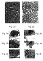


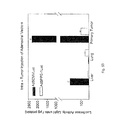


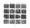


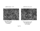


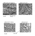
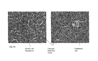

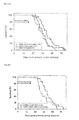


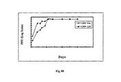


Classifications
-
- C—CHEMISTRY; METALLURGY
- C12—BIOCHEMISTRY; BEER; SPIRITS; WINE; VINEGAR; MICROBIOLOGY; ENZYMOLOGY; MUTATION OR GENETIC ENGINEERING
- C12N—MICROORGANISMS OR ENZYMES; COMPOSITIONS THEREOF; PROPAGATING, PRESERVING, OR MAINTAINING MICROORGANISMS; MUTATION OR GENETIC ENGINEERING; CULTURE MEDIA
- C12N15/00—Mutation or genetic engineering; DNA or RNA concerning genetic engineering, vectors, e.g. plasmids, or their isolation, preparation or purification; Use of hosts therefor
- C12N15/09—Recombinant DNA-technology
- C12N15/63—Introduction of foreign genetic material using vectors; Vectors; Use of hosts therefor; Regulation of expression
- C12N15/79—Vectors or expression systems specially adapted for eukaryotic hosts
- C12N15/85—Vectors or expression systems specially adapted for eukaryotic hosts for animal cells
- C12N15/86—Viral vectors
-
- A—HUMAN NECESSITIES
- A61—MEDICAL OR VETERINARY SCIENCE; HYGIENE
- A61K—PREPARATIONS FOR MEDICAL, DENTAL OR TOILETRY PURPOSES
- A61K48/00—Medicinal preparations containing genetic material which is inserted into cells of the living body to treat genetic diseases; Gene therapy
- A61K48/0008—Medicinal preparations containing genetic material which is inserted into cells of the living body to treat genetic diseases; Gene therapy characterised by an aspect of the 'non-active' part of the composition delivered, e.g. wherein such 'non-active' part is not delivered simultaneously with the 'active' part of the composition
-
- A—HUMAN NECESSITIES
- A61—MEDICAL OR VETERINARY SCIENCE; HYGIENE
- A61K—PREPARATIONS FOR MEDICAL, DENTAL OR TOILETRY PURPOSES
- A61K48/00—Medicinal preparations containing genetic material which is inserted into cells of the living body to treat genetic diseases; Gene therapy
- A61K48/005—Medicinal preparations containing genetic material which is inserted into cells of the living body to treat genetic diseases; Gene therapy characterised by an aspect of the 'active' part of the composition delivered, i.e. the nucleic acid delivered
- A61K48/0058—Nucleic acids adapted for tissue specific expression, e.g. having tissue specific promoters as part of a contruct
-
- A—HUMAN NECESSITIES
- A61—MEDICAL OR VETERINARY SCIENCE; HYGIENE
- A61P—SPECIFIC THERAPEUTIC ACTIVITY OF CHEMICAL COMPOUNDS OR MEDICINAL PREPARATIONS
- A61P17/00—Drugs for dermatological disorders
- A61P17/06—Antipsoriatics
-
- A—HUMAN NECESSITIES
- A61—MEDICAL OR VETERINARY SCIENCE; HYGIENE
- A61P—SPECIFIC THERAPEUTIC ACTIVITY OF CHEMICAL COMPOUNDS OR MEDICINAL PREPARATIONS
- A61P19/00—Drugs for skeletal disorders
- A61P19/02—Drugs for skeletal disorders for joint disorders, e.g. arthritis, arthrosis
-
- A—HUMAN NECESSITIES
- A61—MEDICAL OR VETERINARY SCIENCE; HYGIENE
- A61P—SPECIFIC THERAPEUTIC ACTIVITY OF CHEMICAL COMPOUNDS OR MEDICINAL PREPARATIONS
- A61P27/00—Drugs for disorders of the senses
- A61P27/02—Ophthalmic agents
-
- A—HUMAN NECESSITIES
- A61—MEDICAL OR VETERINARY SCIENCE; HYGIENE
- A61P—SPECIFIC THERAPEUTIC ACTIVITY OF CHEMICAL COMPOUNDS OR MEDICINAL PREPARATIONS
- A61P35/00—Antineoplastic agents
-
- A—HUMAN NECESSITIES
- A61—MEDICAL OR VETERINARY SCIENCE; HYGIENE
- A61P—SPECIFIC THERAPEUTIC ACTIVITY OF CHEMICAL COMPOUNDS OR MEDICINAL PREPARATIONS
- A61P35/00—Antineoplastic agents
- A61P35/04—Antineoplastic agents specific for metastasis
-
- A—HUMAN NECESSITIES
- A61—MEDICAL OR VETERINARY SCIENCE; HYGIENE
- A61P—SPECIFIC THERAPEUTIC ACTIVITY OF CHEMICAL COMPOUNDS OR MEDICINAL PREPARATIONS
- A61P43/00—Drugs for specific purposes, not provided for in groups A61P1/00-A61P41/00
-
- A—HUMAN NECESSITIES
- A61—MEDICAL OR VETERINARY SCIENCE; HYGIENE
- A61P—SPECIFIC THERAPEUTIC ACTIVITY OF CHEMICAL COMPOUNDS OR MEDICINAL PREPARATIONS
- A61P9/00—Drugs for disorders of the cardiovascular system
-
- A—HUMAN NECESSITIES
- A61—MEDICAL OR VETERINARY SCIENCE; HYGIENE
- A61P—SPECIFIC THERAPEUTIC ACTIVITY OF CHEMICAL COMPOUNDS OR MEDICINAL PREPARATIONS
- A61P9/00—Drugs for disorders of the cardiovascular system
- A61P9/10—Drugs for disorders of the cardiovascular system for treating ischaemic or atherosclerotic diseases, e.g. antianginal drugs, coronary vasodilators, drugs for myocardial infarction, retinopathy, cerebrovascula insufficiency, renal arteriosclerosis
-
- A—HUMAN NECESSITIES
- A61—MEDICAL OR VETERINARY SCIENCE; HYGIENE
- A61P—SPECIFIC THERAPEUTIC ACTIVITY OF CHEMICAL COMPOUNDS OR MEDICINAL PREPARATIONS
- A61P9/00—Drugs for disorders of the cardiovascular system
- A61P9/14—Vasoprotectives; Antihaemorrhoidals; Drugs for varicose therapy; Capillary stabilisers
-
- C—CHEMISTRY; METALLURGY
- C07—ORGANIC CHEMISTRY
- C07H—SUGARS; DERIVATIVES THEREOF; NUCLEOSIDES; NUCLEOTIDES; NUCLEIC ACIDS
- C07H21/00—Compounds containing two or more mononucleotide units having separate phosphate or polyphosphate groups linked by saccharide radicals of nucleoside groups, e.g. nucleic acids
- C07H21/04—Compounds containing two or more mononucleotide units having separate phosphate or polyphosphate groups linked by saccharide radicals of nucleoside groups, e.g. nucleic acids with deoxyribosyl as saccharide radical
-
- C—CHEMISTRY; METALLURGY
- C07—ORGANIC CHEMISTRY
- C07K—PEPTIDES
- C07K14/00—Peptides having more than 20 amino acids; Gastrins; Somatostatins; Melanotropins; Derivatives thereof
- C07K14/435—Peptides having more than 20 amino acids; Gastrins; Somatostatins; Melanotropins; Derivatives thereof from animals; from humans
- C07K14/475—Growth factors; Growth regulators
- C07K14/49—Platelet-derived growth factor [PDGF]
-
- C—CHEMISTRY; METALLURGY
- C07—ORGANIC CHEMISTRY
- C07K—PEPTIDES
- C07K14/00—Peptides having more than 20 amino acids; Gastrins; Somatostatins; Melanotropins; Derivatives thereof
- C07K14/435—Peptides having more than 20 amino acids; Gastrins; Somatostatins; Melanotropins; Derivatives thereof from animals; from humans
- C07K14/475—Growth factors; Growth regulators
- C07K14/50—Fibroblast growth factor [FGF]
- C07K14/503—Fibroblast growth factor [FGF] basic FGF [bFGF]
-
- C—CHEMISTRY; METALLURGY
- C07—ORGANIC CHEMISTRY
- C07K—PEPTIDES
- C07K14/00—Peptides having more than 20 amino acids; Gastrins; Somatostatins; Melanotropins; Derivatives thereof
- C07K14/435—Peptides having more than 20 amino acids; Gastrins; Somatostatins; Melanotropins; Derivatives thereof from animals; from humans
- C07K14/475—Growth factors; Growth regulators
- C07K14/515—Angiogenesic factors; Angiogenin
-
- C—CHEMISTRY; METALLURGY
- C07—ORGANIC CHEMISTRY
- C07K—PEPTIDES
- C07K14/00—Peptides having more than 20 amino acids; Gastrins; Somatostatins; Melanotropins; Derivatives thereof
- C07K14/435—Peptides having more than 20 amino acids; Gastrins; Somatostatins; Melanotropins; Derivatives thereof from animals; from humans
- C07K14/52—Cytokines; Lymphokines; Interferons
-
- C—CHEMISTRY; METALLURGY
- C07—ORGANIC CHEMISTRY
- C07K—PEPTIDES
- C07K14/00—Peptides having more than 20 amino acids; Gastrins; Somatostatins; Melanotropins; Derivatives thereof
- C07K14/435—Peptides having more than 20 amino acids; Gastrins; Somatostatins; Melanotropins; Derivatives thereof from animals; from humans
- C07K14/575—Hormones
- C07K14/57536—Endothelin, vasoactive intestinal contractor [VIC]
-
- C—CHEMISTRY; METALLURGY
- C07—ORGANIC CHEMISTRY
- C07K—PEPTIDES
- C07K14/00—Peptides having more than 20 amino acids; Gastrins; Somatostatins; Melanotropins; Derivatives thereof
- C07K14/435—Peptides having more than 20 amino acids; Gastrins; Somatostatins; Melanotropins; Derivatives thereof from animals; from humans
- C07K14/705—Receptors; Cell surface antigens; Cell surface determinants
- C07K14/70578—NGF-receptor/TNF-receptor superfamily, e.g. CD27, CD30, CD40, CD95
-
- C—CHEMISTRY; METALLURGY
- C12—BIOCHEMISTRY; BEER; SPIRITS; WINE; VINEGAR; MICROBIOLOGY; ENZYMOLOGY; MUTATION OR GENETIC ENGINEERING
- C12N—MICROORGANISMS OR ENZYMES; COMPOSITIONS THEREOF; PROPAGATING, PRESERVING, OR MAINTAINING MICROORGANISMS; MUTATION OR GENETIC ENGINEERING; CULTURE MEDIA
- C12N15/00—Mutation or genetic engineering; DNA or RNA concerning genetic engineering, vectors, e.g. plasmids, or their isolation, preparation or purification; Use of hosts therefor
- C12N15/09—Recombinant DNA-technology
- C12N15/63—Introduction of foreign genetic material using vectors; Vectors; Use of hosts therefor; Regulation of expression
- C12N15/79—Vectors or expression systems specially adapted for eukaryotic hosts
- C12N15/85—Vectors or expression systems specially adapted for eukaryotic hosts for animal cells
-
- C—CHEMISTRY; METALLURGY
- C12—BIOCHEMISTRY; BEER; SPIRITS; WINE; VINEGAR; MICROBIOLOGY; ENZYMOLOGY; MUTATION OR GENETIC ENGINEERING
- C12N—MICROORGANISMS OR ENZYMES; COMPOSITIONS THEREOF; PROPAGATING, PRESERVING, OR MAINTAINING MICROORGANISMS; MUTATION OR GENETIC ENGINEERING; CULTURE MEDIA
- C12N5/00—Undifferentiated human, animal or plant cells, e.g. cell lines; Tissues; Cultivation or maintenance thereof; Culture media therefor
-
- A—HUMAN NECESSITIES
- A61—MEDICAL OR VETERINARY SCIENCE; HYGIENE
- A61K—PREPARATIONS FOR MEDICAL, DENTAL OR TOILETRY PURPOSES
- A61K38/00—Medicinal preparations containing peptides
-
- A—HUMAN NECESSITIES
- A61—MEDICAL OR VETERINARY SCIENCE; HYGIENE
- A61K—PREPARATIONS FOR MEDICAL, DENTAL OR TOILETRY PURPOSES
- A61K48/00—Medicinal preparations containing genetic material which is inserted into cells of the living body to treat genetic diseases; Gene therapy
-
- C—CHEMISTRY; METALLURGY
- C07—ORGANIC CHEMISTRY
- C07K—PEPTIDES
- C07K2319/00—Fusion polypeptide
- C07K2319/70—Fusion polypeptide containing domain for protein-protein interaction
-
- C—CHEMISTRY; METALLURGY
- C12—BIOCHEMISTRY; BEER; SPIRITS; WINE; VINEGAR; MICROBIOLOGY; ENZYMOLOGY; MUTATION OR GENETIC ENGINEERING
- C12N—MICROORGANISMS OR ENZYMES; COMPOSITIONS THEREOF; PROPAGATING, PRESERVING, OR MAINTAINING MICROORGANISMS; MUTATION OR GENETIC ENGINEERING; CULTURE MEDIA
- C12N2710/00—MICROORGANISMS OR ENZYMES; COMPOSITIONS THEREOF; PROPAGATING, PRESERVING, OR MAINTAINING MICROORGANISMS; MUTATION OR GENETIC ENGINEERING; CULTURE MEDIA dsDNA viruses
- C12N2710/00011—Details
- C12N2710/10011—Adenoviridae
- C12N2710/10311—Mastadenovirus, e.g. human or simian adenoviruses
- C12N2710/10341—Use of virus, viral particle or viral elements as a vector
- C12N2710/10343—Use of virus, viral particle or viral elements as a vector viral genome or elements thereof as genetic vector
-
- C—CHEMISTRY; METALLURGY
- C12—BIOCHEMISTRY; BEER; SPIRITS; WINE; VINEGAR; MICROBIOLOGY; ENZYMOLOGY; MUTATION OR GENETIC ENGINEERING
- C12N—MICROORGANISMS OR ENZYMES; COMPOSITIONS THEREOF; PROPAGATING, PRESERVING, OR MAINTAINING MICROORGANISMS; MUTATION OR GENETIC ENGINEERING; CULTURE MEDIA
- C12N2800/00—Nucleic acids vectors
- C12N2800/10—Plasmid DNA
- C12N2800/108—Plasmid DNA episomal vectors
-
- C—CHEMISTRY; METALLURGY
- C12—BIOCHEMISTRY; BEER; SPIRITS; WINE; VINEGAR; MICROBIOLOGY; ENZYMOLOGY; MUTATION OR GENETIC ENGINEERING
- C12N—MICROORGANISMS OR ENZYMES; COMPOSITIONS THEREOF; PROPAGATING, PRESERVING, OR MAINTAINING MICROORGANISMS; MUTATION OR GENETIC ENGINEERING; CULTURE MEDIA
- C12N2830/00—Vector systems having a special element relevant for transcription
- C12N2830/001—Vector systems having a special element relevant for transcription controllable enhancer/promoter combination
- C12N2830/002—Vector systems having a special element relevant for transcription controllable enhancer/promoter combination inducible enhancer/promoter combination, e.g. hypoxia, iron, transcription factor
-
- C—CHEMISTRY; METALLURGY
- C12—BIOCHEMISTRY; BEER; SPIRITS; WINE; VINEGAR; MICROBIOLOGY; ENZYMOLOGY; MUTATION OR GENETIC ENGINEERING
- C12N—MICROORGANISMS OR ENZYMES; COMPOSITIONS THEREOF; PROPAGATING, PRESERVING, OR MAINTAINING MICROORGANISMS; MUTATION OR GENETIC ENGINEERING; CULTURE MEDIA
- C12N2830/00—Vector systems having a special element relevant for transcription
- C12N2830/008—Vector systems having a special element relevant for transcription cell type or tissue specific enhancer/promoter combination
-
- C—CHEMISTRY; METALLURGY
- C12—BIOCHEMISTRY; BEER; SPIRITS; WINE; VINEGAR; MICROBIOLOGY; ENZYMOLOGY; MUTATION OR GENETIC ENGINEERING
- C12N—MICROORGANISMS OR ENZYMES; COMPOSITIONS THEREOF; PROPAGATING, PRESERVING, OR MAINTAINING MICROORGANISMS; MUTATION OR GENETIC ENGINEERING; CULTURE MEDIA
- C12N2830/00—Vector systems having a special element relevant for transcription
- C12N2830/15—Vector systems having a special element relevant for transcription chimeric enhancer/promoter combination
-
- C—CHEMISTRY; METALLURGY
- C12—BIOCHEMISTRY; BEER; SPIRITS; WINE; VINEGAR; MICROBIOLOGY; ENZYMOLOGY; MUTATION OR GENETIC ENGINEERING
- C12N—MICROORGANISMS OR ENZYMES; COMPOSITIONS THEREOF; PROPAGATING, PRESERVING, OR MAINTAINING MICROORGANISMS; MUTATION OR GENETIC ENGINEERING; CULTURE MEDIA
- C12N2830/00—Vector systems having a special element relevant for transcription
- C12N2830/30—Vector systems having a special element relevant for transcription being an enhancer not forming part of the promoter region
-
- C—CHEMISTRY; METALLURGY
- C12—BIOCHEMISTRY; BEER; SPIRITS; WINE; VINEGAR; MICROBIOLOGY; ENZYMOLOGY; MUTATION OR GENETIC ENGINEERING
- C12N—MICROORGANISMS OR ENZYMES; COMPOSITIONS THEREOF; PROPAGATING, PRESERVING, OR MAINTAINING MICROORGANISMS; MUTATION OR GENETIC ENGINEERING; CULTURE MEDIA
- C12N2830/00—Vector systems having a special element relevant for transcription
- C12N2830/80—Vector systems having a special element relevant for transcription from vertebrates
- C12N2830/85—Vector systems having a special element relevant for transcription from vertebrates mammalian
-
- C—CHEMISTRY; METALLURGY
- C12—BIOCHEMISTRY; BEER; SPIRITS; WINE; VINEGAR; MICROBIOLOGY; ENZYMOLOGY; MUTATION OR GENETIC ENGINEERING
- C12N—MICROORGANISMS OR ENZYMES; COMPOSITIONS THEREOF; PROPAGATING, PRESERVING, OR MAINTAINING MICROORGANISMS; MUTATION OR GENETIC ENGINEERING; CULTURE MEDIA
- C12N2840/00—Vectors comprising a special translation-regulating system
- C12N2840/20—Vectors comprising a special translation-regulating system translation of more than one cistron
- C12N2840/203—Vectors comprising a special translation-regulating system translation of more than one cistron having an IRES
Definitions
- the present invention relates to nucleic acid constructs, pharmaceutical compositions and methods which can be used to regulate angiogenesis in specific tissue regions of a subject. More particularly, the present invention relates to isolated polynucleotide sequences exhibiting endothelial cell specific promoter activity, and methods of use thereof and, yet more particularly, to a modified-preproendothelin-1 (PPE-1) promoter which exhibits increased activity and specificity in endothelial cells, and nucleic acid constructs, which can be used to either activate cytotoxicity in specific cell subsets, thus, enabling treatment of diseases characterized by aberrant neovasculariza tion or cell growth or induce the growth of new blood vessels, thus, enabling treatment of ischemic diseases.
- the invention further relates to modifications of the PPE promoter, which enhance its expression in response to physiological conditions including hypoxia and angiogenesis, and novel angiogenic endothelial-specific combined therapies.
- Angiogenesis is the growth of new blood vessels, a process that depends mainly on locomotion, proliferation, and tube formation by capillary endothelial cells. During angiogenesis, endothelial cells emerge from their quiescent state and proliferate rapidly. Although the molecular mechanisms responsible for transition of a cell to angiogenic phenotype are not known, the sequence of events leading to the formation of new vessels has been well documented [Hanahan, D., Science 277, 48-50, (1997)]. The vascular growth entails either endothelial sprouting [Risau, W., Nature 386, 671-674, (1997)] or intussusceptions [Patan, S., et al; Microvasc. Res.
- Angiogenesis occurs under conditions of low oxygen concentration (ischemia and tumor metastases etc.) and thus may be an important environmental factor in neovascularization.
- the expression of several genes including erythropoietin, transferrin and its receptor, most of glucose transport and glycolytic pathway genes, LDH, PDGF-BB, endothelin-1 (ET-1), VEGF and VEGF receptors is induced under hypoxic conditions by the specific binding of the Hypoxia Inducible Factor (HIF-1) to the Hypoxic Response Element (HRE) regulating the transcription of these genes. Expression of these genes in response to hypoxic conditions enables the cell to function under low oxygen conditions.
- HIF-1 Hypoxia Inducible Factor
- HRE Hypoxic Response Element
- regulating or modifying the angiogenic process can have an important therapeutic role in limiting the contributions of this process to pathological progression of an underlying disease state as well as providing a valuable means of studying their etiology.
- proangiogenic therapy is directed not only to restoring required angiogenic factors, but to reestablishing the proper balance between them (Dor, et al, Ann N Y Acad Sci 2003; 995:208-16)(for an extensive review of pro- and antiangiogenic therapies see Zhang et al Acta Bioch and Biophys Cinica, 2003: 35:873-880, and Mariani et al. Med Gen Med 2003, 5:22; and Folkman, Semin. Onc 2002, 29:15-18).
- tumor angiogenesis a process known as tumor angiogenesis.
- Tumor growth and metastasis are angiogenesis-dependent.
- a tumor must continuously stimulate the growth of new capillary blood vessels to deliver nutrients and oxygen for the tumor itself to grow. Therefore, either prevention of tumor angiogenesis or selective destruction of tumor's existing blood vessels (vascular targeting therapy) underlies anti-angiogenic tumor therapy.
- An angiogenic switch may occur via several mechanisms:
- the relative balance between activators and inhibitors of angiogenesis is important for maintaining tumors in a quiescent state. Reducing inhibitors or increasing activator levels alters the balance and leads to tumor angiogenesis and tumor growth.
- Oxygen diffusion to neoplastic tissue is inadequate when tumor tissue thickness exceeds 150-200 ⁇ m from the nearest vessel. So, by definition, all tumors that exceed these dimensions are already angiogenically switched-on.
- the tumor cell proliferation rate is independent of the vascular supply. However, as soon as the angiogenic switch occurs, the rate of apoptosis decreases by 3-4 fold (24). Furthermore, nutrient supply and catabolite release are not the only contribution of angiogenic vessels to the decline in tumor apoptosis.
- Microvasculature endothelial cells also secrete anti-apoptotic factors, mitogens and survival factors such as b-FGF, HB-EGF, IL-6, G-CSF, IGF-1 and PDGF that further suppress tumor cell apoptosis.
- mitogens and survival factors such as b-FGF, HB-EGF, IL-6, G-CSF, IGF-1 and PDGF that further suppress tumor cell apoptosis.
- tissue-specific promoters conjugated to cytotoxic genes For example, endothelial cell targeting of a cytotoxic gene, expressed under the control endothelial-specific promoters has been described by Jagger et al who used the KDR or E-selectin promoter to express TNF ⁇ specifically in endothelial cells [Jaggar RT. Et al. Hum Gene Ther (1997) 8(18):2239-47].
- Ozaki et al used the von-Willebrand factor (vWF) promoter to deliver herpes simplex virus thymidine kinase (HSV-tk) to HUVEC [Hum Gene Ther (1996) 7(13):1483-90].
- vWF von-Willebrand factor
- Bu et al. identified a complex regulatory region in the murine PPE-1 promoter (mET-1) that appears to confer endothelial cell specific transcriptional activity and to bind proteins or protein complexes that are restricted to the endothelial cell.
- This region designated endothelial specific positive transcription element, is composed of at least three functional elements, positioned between the ⁇ 364 bp and ⁇ 320 bp of the murine PPE-1 promoter. All three elements are required for full activity. When one or three copies are constructed into a minimal mET-1 promoter, reporter gene expression in endothelial cells in vitro increased 2-10 times, compared to a minimal promoter with no element.
- HSV-TK/GCV system was first described by Kraiselburd et al. in 1976. Cells transfected with an HSV-TK containing plasmid or transduced with an HSV-TK containing vector, are becoming sensitive for a super family of drugs including aciclovir, ganciclovir (GCV), valciclovir and famciclovir.
- the guanosine analog GCV is the most active drug in the setup of gene therapy.
- HSV-TK positive cells produce a viral TK, which is three orders of magnitude more efficient in phosphorylating GCV into GCV monophosphate (GCV-MP) than the human TK. GCV-MP is subsequently phosphorylated by the native thymidine kinase into GCV diphosphate and finally to GCV triphosphate (GCV-TP).
- Recent studies have characterized the HSV-TK/GCV cell cytotoxicity mechanism. They revealed cell cycle arrest in the late S or G2 phase due to activation of the G2-M DNA damage checkpoint. These events were found to lead to irreversible cell death as well as a bystander effect related to cell death. Profound cell enlargement is a well-known morphological change in cells administered with the HSV-TK/GCV system. These morphological changes are due to specific cytoskeleton rearrangement. Stress actin fibers and a net of thick intermediate filaments appear following cell cycle arrest.
- the HSV-TK/GCV system utilizes an amplification potential designated as the “bystander effect”.
- the bystander effect stands for the phenomenon by which HSV-TK positive cells induce the killing of HSV-TK negative cells.
- Culver et al. were the first to demonstrate a bystander effect in an in vivo model. They demonstrated tumor regression when implanted with HSV-TK positive tumor cells in different ratios. Unlike in vitro models, cell-cell contact was not found to be essential for the bystander effect in vivo. Kianmanesh et al. demonstrated a distant bystander effect by implanting tumor cells in different liver lobes, where only some were HSV-TK positive. Both HSV-TK positive and negative foci regressed. A bystander effect was also demonstrated in vivo between cells from different origins. All in all, HSV-TK and its bystander effect facilitate an effective means for tumor suppression when implemented in gene delivery systems. However, to date, clinical studies have demonstrated only limited results.
- an isolated polynucleotide comprising a cis regulatory element including at least a portion of the sequence set forth in SEQ ID NO:15 covalently linked to at least a portion of the sequence set forth in SEQ ID NO:16, the isolated polynucleotide being capable of directing transcription of a polynucleotide sequence transcriptionally linked thereto in eukaryotic cells.
- nucleic acid constructs comprising the isolated polynucleotide, cells comprising the nucleic acid constructs of the invention, and scaffolds seeded with the cells.
- nucleic acid constructs further comprising a nucleic acid sequence positioned under the regulatory control of the cis regulatory element.
- the nucleic sequence can further encode an angiogenesis regulator.
- the nucleic acid sequence is selected from the group consisting of VEGF, p55, angiopoietin-1, bFGF and PDGF-BB.
- the scaffold is composed of a synthetic polymer, a cell adhesion molecule, or an extracellular matrix protein.
- the extracellular matrix protein is selected from the group consisting of fibrinogen, Collagen, fibronectin, vimentin, microtubule-associated protein 1D, Neurite outgrowth factor (NOF), bacterial cellulose (BC), laminin and gelatin.
- a method of expressing a nucleic acid sequence of interest in eukaryotic cells the method effected by administering to a subject a nucleic acid construct including the nucleic acid sequence of interest positioned under transcriptional control of a cis regulatory element including at least a portion of the sequence set forth in SEQ ID NO:15 covalently linked to at least a portion of the sequence set forth in SEQ ID NO:16.
- a method of regulating angiogenesis in a tissue the method effected by expressing in the tissue a nucleic acid construct including a nucleic acid sequence encoding an angiogenesis regulator, the nucleic acid sequence being under regulatory control of a cis regulatory element including at least a portion of the sequence set forth in SEQ ID NO:15 covalently linked to at least a portion of the sequence set forth in SEQ ID NO:16, thereby regulating angiogenesis in the tissue.
- administering is effected by a method selected from the group consisting of, systemic in-vivo administration, ex-vivo administration to cells removed from a body of a subject and subsequent reintroduction of the cells into the body of the subject; and local in-vivo administration.
- the tissue is a natural or an engineered tissue.
- nucleic acid sequence encodes an inhibitor of angiogenesis and regulating angiogenesis is downregulating angiogenesis.
- a nucleic acid construct comprising: (a) a first polynucleotide region encoding a chimeric polypeptide including a ligand binding domain fused to an effector domain of a cytotoxic molecule; and (b) a second polynucleotide region encoding a cis regulatory element being capable of directing expression of said chimeric polypeptide in a specific tissue or cell.
- the ligand binding domain is selected such that it is capable of binding a ligand present in the specific tissue or cell, and binding of said ligand to the ligand binding domain activates the effector domain of the cytotoxic molecule.
- eukaryotic cells transformed with the nucleic acid construct of the invention.
- the cis regulatory element is an endothelial cell-specific or periendothelial cell-specific promoter selected from the group consisting of the PPE-1 promoter, the PPE-1-3x promoter, the TIE-1 promoter, the TIE-2 promoter, the Endoglin promoter, the von Willerband promoter, the KDR/flk-1 promoter, The FLT-1 promoter, the Egr-1 promoter, the ICAM-1 promoter, the VCAM-1 promoter, the PECAM-1 promoter and the aortic carboxypeptidase-like protein (ACLP) promoter.
- the PPE-1 promoter the PPE-1-3x promoter
- the TIE-1 promoter the TIE-2 promoter
- Endoglin promoter the von Willerband promoter
- the KDR/flk-1 promoter the KDR/flk-1 promoter
- the FLT-1 promoter the Egr-1 promoter
- the ICAM-1 promoter the VCAM-1 promoter
- the PECAM-1 promoter
- the ligand binding domain is a ligand-binding domain of a cell-surface receptor.
- the cell-surface receptor can be selected from the group consisting of a receptor tyrosine kinase, a receptor serine kinase, a receptor threonine kinase, a cell adhesion molecule and a phosphatase receptor.
- the cytotoxic molecule is selected from the group consisting of Fas, TNFR, and TRAIL.
- the ligand binding domain is selected such that it is capable of binding a ligand present in, or provided to, the sub-population of angiogenic cells, and binding of the ligand to the ligand binding domain activates the effector domain of said cytotoxic molecule, thereby down-regulating angiogenesis in the tissue.
- a method of down-regulating angiogenesis in a tissue of a subject the method effected by: (a) expressing in the tissue of the subject a nucleic acid construct designed and configured for generating cytotoxicity in a sub-population of angiogenic cells, the nucleic acid construct including: (i) a first polynucleotide region encoding a chimeric polypeptide including a ligand binding domain fused to an effector domain of a cytotoxic molecule, wherein the effector domain is selected such that it is activated following binding of a ligand to the ligand binding domain; and (ii) a second polynucleotide region encoding a cis acting regulatory element for directing expression of the chimeric polypeptide in the sub-population of angiogenic cells; and (b) administering to the subject the ligand, thereby down-regulating angiogenesis in the tissue.
- a pharmaceutical composition for down regulating angiogenesis in a tissue of a subject including, as an active ingredient, a nucleic acid construct designed and configured for generating cytotoxicity in a subpopulation of angiogenic cells and a pharmaceutical acceptable carrier.
- a method of down-regulating angiogenesis in a tissue of a subject the method effected by: (a) expressing in the tissue a nucleic acid construct designed and configured for cytotoxicity in angiogenic cells, the nucleic acid construct including: (i) a first polynucleotide region encoding a suicide gene and (ii) a second polynucleotide region encoding a cis acting regulatory element capable of directing expression of the suicide gene in the angiogenic cells; and (b) administering to the subject a therapeutic amount of a prodrug sufficient to cause apoptosis of the tissue when the prodrug is converted to a toxic compound by the suicide gene, thereby down-regulating angiogenesis in the tissue.
- a nucleic acid construct including: (a) a first polynucleotide region encoding a suicide gene, the suicide gene being capable of converting a prodrug to a toxic compound, and (b) a second polynucleotide region encoding a cis regulatory element capable of directing expression of the suicide gene in angiogenic cells. Also provided are eukaryotic cells transformed with the nucleic acid construct of the invention.
- a method of treating a tumor in a subject the method effected by administering a therapeutically effective amount of a nucleic acid construct designed and configured for generating cytotoxicity in cells of the tumor, the nucleic acid construct including: (i) a first polynucleotide region encoding a suicide gene; and (ii) a second polynucleotide region encoding a cis acting regulatory element capable of directing expression of the suicide gene in the cells of the tumor, where the suicide gene is selected capable of converting a prodrug to a toxic compound capable of causing cytotoxicity in the cells of the tumor.
- the suicide gene is thymidine kinase of herpes simplex virus and the prodrug is ganciclovir, acyclovir, FIAU or their derivatives.
- the suicide gene is varicella zoster virus thymidine kinase and said prodrug is 6-methoxypurine arabinoside or its derivatives.
- the nucleic acid sequence includes an isolated polynucleotide comprising a cis regulatory element including at least a portion of the sequence set forth in SEQ ID NO:15 covalently linked to at least a portion of the sequence set forth in SEQ ID NO:16, the isolated polynucleotide being capable of directing transcription of a polynucleotide sequence transcriptionally linked thereto in eukaryotic cells.
- the at least a portion of the sequence set forth in SEQ ID NO:15 can be positioned upstream of the at least a portion of the sequence set forth in SEQ ID NO:16 in said cis regulatory element, or the least a portion of the sequence set forth in SEQ ID NO:16 can be positioned upstream of said at least a portion of the sequence set forth in SEQ ID NO:15.
- the cis regulatory element further includes at least one copy of the sequence set forth in SEQ ID NO: 6, or at least two copies of SEQ ID NO:6.
- the at least two copies of SEQ ID NO:6 can be contiguous.
- the at least a portion of the sequence set forth in SEQ ID NO:15 is covalently linked to the at least a portion of the sequence set forth in SEQ ID NO:16 via a linker polynucleotide sequence.
- the linker polynulceotide sequence can be a promoter and/or an enhancer element.
- the isolated polynucleotide further includes a hypoxia response element, the hypoxia response element preferably including at least one copy of the sequence set forth in SEQ ID NO: 5.
- the cis regulatory element is as set forth in SEQ ID NO: 7.
- nucleic acid construct further includes a conditionally replicating adenovirus.
- the cis regulatory element is an endothelial cell-specific or periendothelial cell-specific promoter selected from the group consisting of the PPE-1 promoter, the PPE-1-3x promoter, the TIE-1 promoter, the TIE-2 promoter, the Endoglin promoter, the von Willerband promoter, the KDR/flk-1 promoter, The FLT-1 promoter, the Egr-1 promoter, the ICAM-1 promoter, the VCAM-1 promoter, the PECAM-1 promoter and the aortic carboxypeptidase-like protein (ACLP) promoter.
- the PPE-1 promoter the PPE-1-3x promoter
- the TIE-1 promoter the TIE-2 promoter
- Endoglin promoter the von Willerband promoter
- the KDR/flk-1 promoter the KDR/flk-1 promoter
- the FLT-1 promoter the Egr-1 promoter
- the ICAM-1 promoter the VCAM-1 promoter
- the PECAM-1 promoter
- the present invention successfully addresses the shortcomings of the presently known configurations by providing isolated polynucleotide sequences comprising cis regulating elements with novel enhancer elements, and methods of use thereof.
- the novel enhancer elements can be used to make nucleic acid constructs and pharmaceutical compositions for tissue-specific regulation of transgene expression, and for treating a variety of disorders, diseases and conditions by gene therapy.
- the cis regulatory elements, isolated polynucleotides and pharmaceutical compositions of the present invention can be used, along with selected transgenes, to specifically upregulate and/or downregulate angiogenesis in endothelial cells, thus treating tumors, metastatic disease, and ischemic disease.
- FIGS. 1 a - b are schematic illustrations of Fas chimera gene constructed from the extracellular region of TNFR1 and the trans-membrane and intracellular regions of Fas and cloned into pcDNA3 plasmid (a) or into adenoviral vectors (b).
- FIGS. 2 a - b illustrate apoptotic activity of the pro-apoptotic genes, Fas chimera and TNFR1.
- FIG. 2 a Illustrates Bovine Aortic Endothelial Cells (BAEC) transfected with either pcDNA-3-TNFR1 (lower panel) or control empty vector (upper panel) and an expression plasmid encoding GFP.
- FIG. 2 b Illustrates 293 Cells transfected with either pcDNA-3-Fas-c (lower panel) or control empty vector (upper panel) and an expression plasmid encoding GFP. Transfected cells were visualized using fluorescence microscopy and apoptotic activity was morphologically determined.
- FIGS. 3 a - f are electron microscopy images of BAEC cells transfected with pro-apoptotic genes. 24 hours post transfection, BAEC cells were fixed in 2.5% glutaraldehyde and processed. Presented are cells in successive stages of the apoptotic process.
- FIG. 4 are histograms quantifying apoptotic activity of the indicated pro-apoptotic genes in transfected BAEC and 293 cells.
- FIG. 5 a represents a PCR analysis of AdPPE-Fas-c.
- Lanes 1-2 PCR products obtained using primers encompassing the PPE-1 promoter and Fas-c gene.
- Lanes 3-4 PCR products obtained using Fas-c primers.
- Lanes 5-6 PCR products obtained in the absence of template DNA.
- FIG. 5 b is a western blot analysis of AdPPE-Fas-c transfected BAEC cells. Protein samples were resolved by SDS-PAGE, transferred to nitrocellulose membrane and probed with a polyclonal antibody directed against the extracellular portion of TNFR1. Lane 1-2—pcDNA3-Fas-c BAEC transfected cells (positive control). Lane 3-4—BAEC cells transfected with the indicated MOI of AdPPE-Fas-c viruses. Lane 5—non-transfected cells. Lane 6-7—BAEC cells transfected with the indicated MOI of AdPPE-Luc.
- FIGS. 6 a - d are photomicrographs illustrating the effect of Fas-chimera over-expression on apoptosis of endothelial cells.
- BAEC cells were infected with: Ad-PPE-1-3x-Fas-chimera ( FIG. 6 a ); Ad-PPE-1-3x-luciferase ( FIG. 6 b ); Ad-PPE-1-3x-Fas-chimera and Ad-PPE1-3x-GFP ( FIG. 6 c ); Ad-PPE-1-3x-luciferase and Ad-PPE-1-3x-GFP; each at MOI 1000 ( FIG. 6 d ). Photomicrographs were taken 72 h post infection at ⁇ 10 magnification.
- FIG. 7 is a histogram illustrating apoptotic specific effect of Ad-PPE-1-3x-Fas-chimera on endothelial cells. Viability of endothelial (BAEC, HUVEC) and non-endothelial (Normal skin fibroblasts-NSF) cells was quantified by crystal violet staining 72 h post infection with either Ad-PPE-1-3x-Fas-chimera or control (luciferase) virus.
- FIG. 8 shows a dose response effect of TNF ⁇ administration on Fas-chimera mediated apoptosis.
- BAEC were infected with Ad-PPE-1-3x-Fas-c. 48 h post infection TNF was added to the growth medium (at the indicated dose). Viability was determined by the crystal violet assay 24 h thereafter.
- FIGS. 9 a - e are photomicrographs illustrating an endothelial cell-specific apoptosis mediated by the cooperative action of TNF ⁇ ligand and Fas-c receptor.
- the indicated cells were incubated in the presence or absence of TNF ⁇ (10 ng/ml) 48 h following infection with Ad-PPE-1-3x-Fas-c; crystal violet staining was effected 72 h post infection.
- FIG. 10 a is a dose response curve illustrating the TNF ⁇ -dependent apoptotic effect of Ad-CMV-Fas-c on endothelial cells. Viability of BAEC cells infected with the indicated MOI of Ad-CMV-Fas-chimera was determined following incubation with TNF ⁇ .
- FIGS. 10 b - d illustrate the apoptotic effect of TNF ⁇ ligand and Ad-CMV-Fas-chimera on the non-endothelial cells NSF.
- FIG. 10 b NSF infected with a control virus.
- FIG. 10 c NSF infected with Ad-CMV-Fas-chimera.
- FIG. 10 d NSF infected with Ad-CMV-Fas-chimera and incubated with TNF (10 ng/ml).
- FIGS. 11 a - c illustrate the In-vivo anti-tumoral effect of Ad-PPE-1-3x-Fas-c.
- Mice inoculated with B16 melanoma cells were injected intravenously with Ad-PPE-1-3x-Fas-c, Ad-CMV-Fas-chimera, control virus or saline when tumor was palpable.
- FIG. 11 a tumor areas, measured during treatment period.
- FIG. 11 b tumor weights at end of treatment period.
- FIG. 11 c an image representing the state of the tumor in the Ad-PPE-1-3x-Fas-c treated mouse and the control mouse.
- FIG. 12 is a histogram illustrating the effect of the enhancer element of the present invention on Luciferase expression in both bovine and human endothelial cell lines using the B2B cell line (bronchial cell line that expresses endothelin) as a control.
- FIG. 13 is a histogram illustrating endothelial specificity of a promoter of the present invention in an adenoviral vector on Luciferase expression in various cell lines.
- FIGS. 14A-B are photomicrographs illustrating GFP expression under the control of Ad5PPE-1-3X of the present invention ( 14 A) and an Ad5CMV ( 14 B) control construct in the BAEC cell line.
- FIG. 15 is histogram of % apoptosis induced by pACPPE-1-3Xp55, pACPPE-1-3XLuciferase and pCCMVp55 in endothelial and non-endothelial cells.
- FIG. 16 is a histogram illustrating the effect of introducing an enhancer element according to the present invention into a promoter construct on hypoxia response.
- FIG. 17 is a histogram illustrating the effect of introducing an enhancer element according to the present invention into a promoter of an adenovector construct on hypoxia response.
- FIG. 18 is a histogram illustrating the effect of introducing an enhancer element according to the present invention into a promoter on levels of expression in bovine and human endothelial, endothelin expressing cell lines.
- FIG. 19 is a histogram illustrating levels of expression of a reporter gene observed in various organs following injection of an adenoviral construct containing either an endothelial promoter (PPE-1) or a control (CMV) promoter;
- PPE-1 endothelial promoter
- CMV control
- FIGS. 20A-B are two photomicrographs illustrating cellular expression of an Ad5CMVGFP construct ( FIG. 20A ) and an Ad5PPE-1-GFP construct ( FIG. 20B ) in liver tissue of mice injected with the constructs.
- FIG. 21 is a histogram illustrating the effect of introducing an enhancer element according to the present invention into a promoter on levels of expression in endothelial and non-endothelial cell lines.
- FIG. 22 is a histogram illustrating the effect of introducing an enhancer element according to the present invention into a promoter on levels of expression in endothelial and non-endothelial cell lines.
- FIGS. 23A-C are photomicrographs illustrating GFP expression in Ad5PPE-1-3XGFP transduced cells, Ad5PPE-1 GFP transduced cells and Ad 5CMVGFP transduced cells respectively.
- FIGS. 24A-B illustrate GFP expression in SMC transduced by moi-1 of Ad5PPE-1-3XGFP and Ad5CMVGFP respectively.
- FIGS. 25A-B show results of an experiment similar to that of FIGS. 24A-B conducted in HeLa cells.
- FIGS. 26A-B show results of an experiment similar to that of FIGS. 24A-B conducted in HepG2 cells.
- FIGS. 27A-B show results of an experiment similar to that of FIGS. 24A-B conducted in NSF cells.
- FIGS. 28A-B are photomicrographs illustrating GFP expression in endothelial cells lining a blood vessel of mice injected with the Ad5PPE-1GFP and the Ad5PPE-1-3XGFP constructs respectively.
- FIGS. 30A-C illustrate experiments similar to those depicted in FIGS. 29A-C , conducted on sections of spleen tissue.
- FIGS. 31A-D illustrate GFP expression in metastatic lungs of control mice injected with Saline ( FIG. 31A ), mice injected with Ad5CMVGFP ( FIG. 31B ), mice injected with Ad5PPE-1GFP ( FIG. 31C ) and mice injected with Ad5PPE-1-3XGFP ( FIG. 31D ).
- Anti Cd31 immunostaining (FIGS. 31 C′ to 31 D′) confirm the co-localization of the GFP expression and CD31 expression in each metastatic tissue.
- FIG. 33 is a histogram as in FIG. 32 , except that Ad5PPE-1Luc and Ad5CMVLuc were employed.
- FIG. 34 is a histogram as in FIG. 33 showing the effects of hypoxia in different cell lines.
- FIG. 35 is a histogram illustrating the effect of the 3X sequence of the present invention on the PPE-1 hypoxia response in BAEC cells. Cells were transduced by Ad5PPE-1Luc and Ad5PPE-1-3XLuc.
- FIG. 36 is a histogram showing levels of Luciferase expression in various tissues of PPE-1-Luc transgenic mice following femoral artery ligation.
- FIGS. 37A-B are plasmid maps of constructs employed in conjunction with the present invention.
- FIGS. 38A-F illustrate the effects of Ad5PPE-1-3XVEGF and Ad5CMVVEGF on blood perfusion and angiogenesis in mouse ischemic limbs.
- FIGS. 38A-D are representative ultrasonic (US) angiographic images of perfusion in the ischemic limb of mice from the various treatment groups captured 21 days following ligation. Yellow signal represents intense perfusion. The right side of the image represents the distal end of the limb.
- FIG. 38 A Ad5PPE-1-3XVEGF treated mouse
- FIG. 38 B Ad5CMVVEGF treated mouse
- FIG. 38 C control, saline treated mouse
- FIG. 38 D control, normal limb.
- FIGS. 38E-F are histograms illustrating: mean intensity of signal in the US images of the various treatment groups ( FIG. 38E ); mean capillary density, measured as the number of CD31+ cells/mm 2 in the various treatment groups ( FIG. 38F ).
- FIG. 39 is a histogram illustrating Luciferase activity in proliferating and quiescent Bovine Aortic Endothelial Cells (BAEC) transduced with Ad5PPE-1Luc (open bars) and Ad5CMVLuc (black bars).
- BAEC Bovine Aortic Endothelial Cells
- FIG. 40 is a histogram illustrating Luciferase activity in BAEC transduced with Ad5PPE-1Luc. during normal proliferation, a quiescent state and rapid proliferation following addition of VEGF.
- FIG. 43 is a prior art image depicting an aorta dissected from ApoE deficient mice colored by Sudan-IV.
- the thoracic aorta contains less red stained atherosclerotic lesion while the abdominal region includes many red stained atherosclerotic lesions.
- FIG. 45 is a histogram illustrating absolute Luciferase activity (light units/ ⁇ g protein) 5 days post systemic injections of Ad5PPE-1Luc (black bars) or Ad5CMVLuc (open bars) to healing wound C57BL/6 induced mice.
- FIG. 46 is a histogram illustrating Luciferase activity in normal lung, metastatic lung and primary tumor of Lewis lung carcinoma-induced mice.
- FIGS. 47A-D are photomicrographs illustrating GFP expression and tissue morphology in lungs and tumors of LLC bearing mice following intra-tumoral injection of Ad5PPE-1GFP. Tissue was frozen in OCT and sectioned to 10 ⁇ m by cryostat. All pictures were taken in magnification of 25 ⁇ .
- FIG. 47 A GFP in angiogenic blood vessels of lung metastases
- FIG. 47 B CD31 antibody immunostaining of the section pictured in FIG. 47A
- FIG. 47 C GFP expression in blood vessels of primary tumor
- FIG. 47 D phase contrast of the section of C illustrating blood vessels.
- FIG. 49 is a histogram illustrating Luciferase activity as percentage of liver activity (where the liver is 100%), in normal lung and lung metastasis of Lewis lung carcinoma-induced mice injected with Ad5CMV, Ad5PPE-1Luc and Ad5PPE-1(3X).
- FIGS. 50A-B are photomicrographs illustrating co-localization of GFP expression ( FIG. 50A ) and CD31 immunostaining ( FIG. 50B ) in mice with LLC lung metastases injected with Ad5PPE-1-3X-GFP.
- FIG. 53 is a histogram illustrating Luciferase activity, (light units/ ⁇ g protein detected in the livers, lungs and primary tumors of LLC mice injected in primary tumors with Ad5CMVLuc (black bars) or Ad5PPE-1Luc (open bars).
- FIGS. 54A-H are in-situ hybridization images illustrating tissue distribution of tissue-specific or constitutive expression of various transgenes.
- FIGS. 54A-C illustrate in-situ hybridization with a VEGF specific antisense probe on representative ischemic muscles from: A, Ad5PPE-1-3XVEGF treated mouse; B, Ad5CMVVEGF treated mouse; C, saline treated mouse; D, liver section from Ad5CMVVEGF treated mouse. An arrow indicates positively stained cells.
- 54E-G illustrate in-situ hybridization with a PDGF-B specific antisense probe of representative ischemic muscles from: E, Ad5PPE-1-3XPDGF-B treated mouse; F, Ad5CMVPDGF-B treated mouse; G, saline treated mouse; H, liver section from Ad5CMVPDGF-B treated mouse.
- FIGS. 55A-B are histograms illustrating a long-term effect of Ad5PPE-1-3XVEGF or Ad5CMWEGF on blood perfusion and angiogenesis in mouse ischemic limb.
- A mean intensity of signal in the US images of the various treatment groups, 50 days following femoral artery ligation.
- B mean capillary density, measured as number of CD31+ cells/mm 2 in the various treatment groups, 70 days following femoral artery ligation.
- FIGS. 56A-D are histograms showing early and long term effects of Ad5PPE-1-3XPDGF-B on neovascularization in mouse ischemic limb.
- FIGS. 56 A-B mean perfusion intensity measured by US imaging ( 56 A, 30 days following femoral artery ligation; 56 B, 80 days following femoral artery ligation).
- FIGS. 56 C-D mean capillary density, measured as number of CD31+ cells/mm 2 in the various treatment groups ( 56 C, 35 days following femoral artery ligation; 56 D, 90 days following femoral artery ligation).
- FIGS. 57A-G illustrate long term effects of angiogenic therapy using PDGF-B and VEGF alone or in combination under regulation of an endothelial specific or a constitutive promoter on neovascularization and blood flow in mouse ischemic limb.
- A mean intensity of signal in the US images of the various treatment groups, 80 days following femoral artery ligation.
- B mean capillary density, measured as number of CD31+cells/mm 2 in the various treatment groups, 90 days following femoral artery ligation.
- FIGS. 57 C-G smooth muscle cells recruitment to mature vessels in ischemic limb muscles, 90 days following femoral artery ligation.
- Smooth muscle cells are immunostained with anti- ⁇ -SMactin antibodies (in red, X20).
- FIG. 59 is a schematic representation of the basic principles of gene-directed enzyme prodrug therapy (GDEPT).
- FIGS. 60A-B are a schematic map representing the construction of the plasmid pEL8(3x)-TK.
- FIG. 60A is a schematic map representing the construction of the plasmid pEL8(3x)-TK.
- FIG. 60B is a map of plasmid pACPPE-1(3x)-TK.
- FIG. 61 is an agarose gel separation of PCR products of the AdPPE-1(3x)-TK vector, visualized by UV fluorescence.
- Two primers were used: the forward primer 5′-ctcttgattcttgaactct-3′ (455-474 bp in the pre-proendothelin promoter sequence) (SEQ ID NO:9) and the reverse primer 5′-taaggcatgcccattgttat-3′ (1065-1084 bp in the HSV-TK gene sequence)(SEQ ID NO:10).
- Primers specific for other vectors gave no PCR products.
- Lane 1 100 bp size marker ladder.
- Lane 2 pACPPE-1(3x)-TK plasmid.
- Lane 3 AdPPE-1(3x)-TK virus.
- FIGS. 62A-C show linear, schematic maps of the vectors AdPPE-1(3x)-TK FIG. 62 a ).
- AdPPE-1(3x)-Luc FIG. 62 b
- AdCMV-TK FIG. 62 c ).
- FIG. 65 is a series of photomicrographs illustrating the superior synergy of endothelial cell cytotoxicity of TK under control of the PPE-1 (3x) promoter and ganciclovir administration.
- Bovine aorta endothelial cells (BAECs) were transduced with AdPPE-1(3x)-TK, AdCMV-TK and AdPPE-1(3x)-Luc, as described hereinabove, at multiplicity of infection (m.o.i.) of 10, and exposed to increasing concentrations of GCV (0.001-10 ⁇ g/ml, as indicated), added four hours post-transduction. Controls were cells transduced with the vectors without GCV, or GCV without vectors.
- FIG. 66 is a graph representing synergy of endothelial cell cytotoxicity of TK under control of the PPE-1 (3x) promoter and ganciclovir administration.
- BAECs were prepared in 96-well plates and transduced as in FIG. 65 , followed by the addition of increasing concentrations (0.0001-10 ⁇ g/ml) GCV four hours post-transduction. 10 days post vector addition, cell viability was determined using crystal violet staining. Note the superior cytotoxicity of AdPPE-1 (3x)+GCV at GCV concentrations greater than 0.01 ⁇ g/ml, compared with the strong constitutive TK expression of AdCMV-TK.
- FIG. 67 is a series of photomicrographs illustrating the specific, synergic endothelial cytotoxicity of TK under control of the PPE-1 (3x) promoter and ganciclovir administration.
- Endothelial Bovine aortic endothelial cells (BAEC), Human umbilical vein endothelial cells (HUVEC)] and non-endothelial [Human hepatoma cells (HepG-2), Human normal skin fibroblasts (NSF)] cells were transduced with ADPPE-1(3x)-TK, ADPPE-1(3x)-Luc or AdCMV-TK at m.o.i. of 10, followed by the administration of 1 ⁇ g/ml GCV four hours post-transduction.
- BAEC Bovine aortic endothelial cells
- HUVEC Human umbilical vein endothelial cells
- NSF Human normal skin fibroblasts
- FIG. 68 is a histogram representing the specific, synergic endothelial cell cytotoxicity of TK under control of the PPE-1 (3x) promoter and ganciclovir administration.
- Endothelial (BAEC and HUVEC) and non-endothelial (HepG-2 and NSF) cells were prepared in 96-well plates and transduced as in FIG. 67 , followed by the addition of increasing concentrations (1 ⁇ g/ml) GCV four hours post-transduction. 10 days post vector addition, cell viability was determined using crystal violet staining. Note the superior, endothelial specific cytotoxicity of AdPPE-1 (3x)+GCV, compared with the non-specific cytotoxicity of AdCMV-TK+GCV.
- FIG. 69 is a series of photomicrographs illustrating the endothelial selective cytotoxicity of TK under control of the PPE-1 (3x) promoter and ganciclovir administration at extreme multiplicity of infection.
- NSF Non-endothelial
- Non-endothelial (NSF) cells were transduced with AdPPE-1(3x)-TK, AdPPE-1(3x)-Luc or AdCMV-TK as in FIG. 66 , at the higher m.o.i. of 100, followed by the administration of 1 ⁇ g/ml GCV four hours post-transduction.
- the experiment was performed twice, in 96-well plates, 12 wells for every group. Cytotoxicity and cell morphological changes were detected microscopically four days post-transduction. Note the lack of effect on NSF cell morphology with AdPPE-1(3x)-TK+GCV, compared to the non-specific cytotoxicity of AdCMV-TK+GCV.
- FIG. 70 is a series of photographs illustrating synergic suppression of metastatic growth by in vivo expression of TK under control of the PPE-1 (3x) promoter and ganciclovir (GCV) administration.
- FIG. 71 is a histogram illustrating synergic suppression of metastatic growth by in vivo expression of TK under control of the PPE-1 (3x) promoter and ganciclovir (GCV) administration.
- Lung metastases were induced in C57BL/6 mice, and the mice treated with 10 11 PFUs of the adenoviral vectors [AdPPE-1(3x)-TK+GCV; AdCMV-TK+GCV; AdPPE-1(3x)-TK without GCV] and GCV (100 mg/kg) as described hereinabove.
- the mice were sacrificed on the 24 th day post vector injection, and lungs removed for assessment of the lung metastases. Control mice received saline and GCV.
- FIGS. 72 a and 72 b were sectioned and stained with hematoxylin and eosin. Note the massive central necrosis and numerous clusters of mononuclear infiltrates in metastases from lungs with AdPPE-1 (3x)+GCV administration ( FIGS. 72 a and 72 b ).
- FIGS. 74 a and 74 b are representative histopathology sections of induced LLC lung metastases stained with TUNEL and anti-caspase-3 of lung metastases, illustrating the endothelial-specific, synergic enhancement of tumor apoptosis by in vivo expression of TK under control of the PPE-1 (3x) promoter and ganciclovir (GCV) administration. Sections from LLC lung metastases, induced and prepared as described in FIGS.
- 73 a - 73 b were fixed and embedded in paraffin, and assayed for indicators of apoptosis by the deoxynucleotide transferase-mediated dUTP-nick end-labeling (TUNEL) assay using the Klenow-FragE1 (Oncogene, Cambridge, Mass.) ( FIG. 74 a ) and anti-caspase-3-specific immunohistopathology ( 74 b ).
- Black arrows indicate erythrocytes, red arrows apoptotic endothelial cells, and white arrows apoptotic tumor cells. Note the enhanced apoptosis in the vascular (endothelial) regions of the lung metastases from the mice treated with intravenous AdPPE-1 (3x)-TK+GCV.
- FIGS. 75 a - 75 d are representative immunohistopathology sections of tissue from murine lung carcinoma, illustrating the endothelial-specific, synergic inhibition of angiogenesis by in vivo expression of TK under control of the PPE-1 (3x) promoter and ganciclovir (GCV) administration.
- Sections from LLC lung metastases ( FIG. 75 a ) livers ( FIG. 75 c ) and normal lung tissue ( FIG. 75 b ) induced and prepared as described in FIGS. 73 a - 73 b were fixed and embedded in paraffin, and assayed for indicators of angiogenesis by anti-CD-31 immunofluorescence.
- FIG. 75 d is a histogram showing a computer-based vascular density assessment (Image Pro-Plus, Media Cyberneticks Incorporated) of the lung metastases vascularization (angiogenesis). Left bar: AdPPE-1(3x)TK+GCV; right bar: ADPPE-1(3x)TK no GCV.
- FIG. 76 is a representative histopathology section of tissue from murine liver, illustrating the absence of hepatotoxicity in in-vivo expression of TK under control of the PPE-1 (3x) promoter and ganciclovir (GCV) administration.
- Sections from livers of mice bearing LLC lung metastases induced and prepared as described in FIGS. 73 a - 73 b were fixed and embedded in paraffin, and stained with hematoxylin and eosin. Note the absence of cytotoxic indicators in livers from mice treated with AdPPE-1-TK+GCV (3x) (left panel), compared to the profound cytotoxicity in the livers from mice treated with the constitutively expressed AdCMV-TK+GCV(right panel)
- FIG. 77 depicts a RT-PCR analysis illustrating the organ-specific expression of the expression of TK under control of the PPE-1 (3x) promoter and ganciclovir (GCV) administration. LLC lung metastases were induced and prepared in nine 15 week-old C57BL/6 male mice as described in FIGS. 73 a - 73 b . Adenovirus vectors [AdPPE-1 (3x)-TK and AdCMV-TK], and saline control were delivered intravenously 14 days post primary tumor removal. The mice were sacrificed 6 days post vector injection, and organs harvested.
- AdPPE-1 (3x)-TK and AdCMV-TK AdCMV-TK
- RNA of different organs was extracted, as described hereinbelow, and PPE-1 (3x) and HSV-TK transcripts amplified by RT-PCR PCR with PPE-1(3x) promoter and HSV-TK gene primers. Note the endothelial-specific expression of TK under control of the PPE-1 (3x) promoter (center, bottom panel).
- FIGS. 78 a and 78 b are graphs illustrating a range of sub-therapeutic and non-toxic irradiation in Balb/c murine colon carcinoma tumor model.
- the 5 Gy dose induced only a partial, non-statistically significant delay in tumor progression ( FIG. 78 a ), and no significant weight loss ( FIG. 78 b ).
- FIGS. 79 a - 79 g illustrate synergistic suppression of tumor growth in murine colon carcinoma with combined sub-therapeutic radiotherapy and expression of TK under control of the PPE-1 (3x) promoter and ganciclovir (GCV) administration.
- 100 male Balb/C mice aged 8 weeks were inoculated with CT-26 colon carcinoma tumor cells.
- 10 11 PFUs of the viral vectors [AdPPE-1 (3x)-TK or AdCMV-TK] were injected intravenously into the tail vein followed by 14 days of daily intraperitoneal GCV injection (100 mg/kg body weight), where indicated. 3 days post vector administration, the mice were irradiated with a local 5 Gy dose.
- FIG. 79 a shows mean tumor volume ⁇ S.E. on day 14 post vector injection.
- FIG. 79 b shows mean tumor volume progression over time, in groups treated with radiotherapy.
- FIG. 79 c shows mean tumor volume progression over time, in AdPPE-1(3x)-TK+GCV treated mice.
- FIG. 79 d shows mean tumor volume progression over time, in AdCMV-TK+GCV treated mice.
- FIG. 79 e shows mean tumor volume progression over time, in control saline+GCV treated mice.
- FIG. 79 f shows mean tumor volume progression over time, in AdPPE-1(3x)-TK treated mice without GCV.
- FIGS. 80 a - 80 b are representative histopathology sections of primary CT-26 tumor showing synergistic induction of tumor necrosis in murine colon carcinoma with combined sub-therapeutic radiotherapy and expression of TK under control of the PPE-1 (3x) promoter and ganciclovir (GCV) administration. Sections from CT-26 colon carcinoma tumors induced and prepared as described in FIGS. 79 a - 79 g were fixed and embedded in paraffin, and stained with hematoxylin and eosin. Note the areas of necrosis ( FIG. 80 a ) and granulation tissue ( FIG. 80 b ) with combined AdPPE-1 (3x)+GCV+low dose radiotherapy.
- FIGS. 81 a - 81 b are representative histopathology sections of induced primary colon carcinoma tumors stained with TUNEL and anti-caspase-3, illustrating synergic enhancement of endothelial cell and tumor apoptosis by combined radiotherapy and in vivo expression of TK under control of the PPE-1 (3x) promoter and ganciclovir (GCV) administration. Sections from CT-26 primary colon carcinoma tumors, induced and prepared as described in FIGS.
- FIG. 82 is representative histopathology sections of induced primary colon carcinoma tumors stained with anti-caspase-3, illustrating synergic enhancement of endothelial cell and tumor apoptosis by combined radiotherapy and in vivo expression of TK under control of the PPE-1 (3x) promoter and ganciclovir (GCV) administration.
- Sections from CT-26 primary colon carcinoma tumors, induced and prepared as described in FIGS. 79 a - 79 g were fixed and embedded in paraffin, and assayed for indicators of apoptosis by anti-caspase-3-specific immunohistopathology.
- Black arrow indicates erythrocytes; red arrow indicates apoptotic endothelial cell, and white arrow indicates apoptotic tumor cell. Note the GCV-dependent apoptotic effect.
- FIGS. 83 a and 83 b are representative histopathology sections of liver tissue and induced primary colon carcinoma tumors stained with anti-CD-31, illustrating synergic enhancement of inhibition of tumor vascularization by combined radiotherapy and in vivo expression of TK under control of the PPE-1 (3x) promoter and ganciclovir (GCV) administration.
- Sections from liver tissue( 83 b ) and CT-26 primary colon carcinoma tumors ( 83 a ), induced and prepared as described in FIGS. 79 a - 79 g were fixed and embedded in paraffin, and reacted with endothelial-specific anti-CD-31 for immunohistopathology.
- Black arrow indicates erythrocytes; red arrow indicates apoptotic endothelial cell, and white arrow indicates apoptotic tumor cell. Note the extensive vascular disruption in the tumors from the mice treated with combined radiotherapy and intravenous AdPPE-1 (3x)-TK+GCV ( 83 a ) compared with the normal vasculature in the liver cells ( 83 b ).
- FIG. 84 is representative histopathology sections of mouse liver tissue showing tissue-specific cytotoxicity of radiotherapy and TK expression under control of the PPE-1 (3x) promoter and ganciclovir (GCV) administration. Sections from mouse livers exposed to a vectors (AdPPE-1 (3x)-TK and AdCMV-TK) and GCV, alone and in combination, were fixed and embedded in paraffin, and stained with hematoxylin and eosin. Note the typical mild hepatotoxicity with AdCMV-TK and ganciclovir (left panel), and the absence of vascular abnormalities in the AdPPE-1 (3x)-TK treated liver (right panel).
- FIGS. 85 a and 85 b are graphs illustrating a range of sub-therapeutic and non-toxic irradiation in C57Bl/6 lung carcinoma metastatic model.
- 35 C57Bl/6 male mice aged 8 weeks were inoculated with Lewis Lung Carcinoma (LLC) cells into the left footpad and received irradiation into the chest wall with 0, 5, 10, or 15 Gy under general anesthesia, 8 days following removal of the primary tumor. Mice were sacrificed 28 days post tumor removal. Weight loss indicated metastatic disease.
- the 5 Gy dose was neither therapeutic ( FIG. 85 a ) nor toxic ( FIG. 85 b ).
- FIGS. 86 a - 86 d illustrate synergistic suppression of metastatic disease in murine lung carcinoma with combined sub-therapeutic radiotherapy and expression of TK under control of the PPE-1 (3x) promoter and ganciclovir (GCV) administration.
- 180 male Balb/C mice aged 8 weeks were inoculated with LLC cells into the left footpad. The foot was amputated under general anesthesia as soon as the primary tumor developed.
- 5 days post amputation 10 10 PFUs of vector [AdPPE-1(3x)-TK or AdCMV-TK] were injected into the tail vein, followed by 14 days of daily intraperitoneal injections of GCV (100 mg/kg).
- FIG. 86 a shows survival of irradiated, non-vector treated mice over 55 days.
- FIG. 86 b shows survival of AdPPE-1(3x)-TK treated mice.
- FIG. 86 c shows survival of AdCMV-TK treated mice.
- FIG. 86 d shows survival of saline treated control mice. Note that radiotherapy significantly potentiated only the angiogenic endothelial cell transcription-targeted vector, AdPPE-1(3x)-TK, compared to the non-targeted vector, AdCMV-TK ( FIG. 86 b - 86 d ). Treatment regimens with all virus vectors were ineffective without radiotherapy.
- FIGS. 87 a - 87 c are a series of histograms showing endothelial cell cytotoxicity of Fas-c under control of CMV promoter.
- Bovine aortic endothelial cells (BAEC) were stained with crystal violet 72 hours after transduction with 100 (left), 1000 (right) and 10000 (left bottom) moi's of CMV-FAS (dark) or CMV-LUC (gray, negative control), and 24 hours after the addition of human TNF- ⁇ ligand, in different concentrations. Note the reduced viability of BAE cells at high moi and TNF- ⁇ concentrations.
- FIG. 88 is a graph of plaque development showing enhanced spread of viral replication in 293 cells with CMV-FAS (blue diamonds) compared to CMV-LUC (red squares). Titers of CsCl-banded stocks of CMV-FAS and CMV-LUC were determined by PFU assay, as described hereinbelow. Data is plotted as number of plaques seen on every 2-3 days of the plaque assay in log-scale.
- FIGS. 89 a and 89 b is a series of photographs of 293 cell cultures illustrating the higher rate of cell-to-cell spread of virus infection (plaquing) with CMV-Fas-c as compared with CMV-luciferase. 4 days after infection, photographs of plaques from CMV-FAS (left) and CMV-LUC (right) with identical dilution, were taken. Plaques from CMV-FAS are clearly larger than those of CMV-LUC, indicating a higher rate of cell to cell spread, probably induced by apoptosis.
- FIG. 90 is a histogram illustrating the specific, synergic endothelial cytotoxicity of Fas-c under control of the PPE-1 (3x) promoter and doxorubicin administration.
- BAEcells were exposed to 100 nM of Doxorubicin 48 hours after vector (PPE-1 (3x)-FAS) transduction (10 3 moi).
- Cells were stained with crystal violet 96 hours after vector transduction, and cell viability was assessed microscopically. Note the significant synergy in endothelial cytotoxicity between AdPPE-1 (3x)-Fas-c and doxorubicin.
- FIGS. 91 a - 91 b are a graphic representation illustrating superior induction of angiogenesis in engineered tissue constructs by VEGF under the control of the endothelin (PPE-1 3X).
- Tissue engineered constructs (undisclosed procedure) were grown with or without VEGF supplementation to the medium (50 ng/ml).
- Parallel constructs were infected with Ad5PPEC-1-3x VEGF viruses or control Ad5PPEC-1-3x GFP adenoviruses (control virus) (for 4 hours). Following 2 weeks in culture the constructs were fixed, embedded, sectioned and stained. Vascularization was expressed as the number of vessels per mm 2 , and the percentage of area of sections that was vascularized.
- FIG. 91 a shows that infection of the cells with Ad5PPEC-1-3x VEGF has an inductive effect on number and size of vessels-like structures formed in the engineered constructs. Note the dramatic increase (4-5 ⁇ ) in both parameters of vascularization in the Ad5PPEC-1-3x VEGF-transduced tissue constructs (VEGF virus), compared to constructs exposed to VEGF to the medium (VEGF medium).
- FIG. 91 b is a histogram of LUC luminescence intensity, illustrating the superior survival and vascularization of implanted tissue constructs grown with cells infected with Ad5PPEC-1-3x VEGF, compared with Ad5PPEC-1-3x GFP controls.
- FIG. 92 is the wild-type murine PPE-1 promoter DNA sequence (SEQ ID NO: 1).
- the promoter contains the endogenous endothelial specific positive transcriptional element (black italics), the NF-1 response element (pink italics), the GATA-2 element (red italics), the HIF-1 responsive element (blue italics), the AP-1 site (green italics), the CAAT signal (orange italics) and the TATA box (purple italics).
- FIG. 93 is the sequence of the 3 x fragment of the modified murine pre-proendothelin-1 promoter (SEQ ID NO: 7).
- the fragment contains 2 complete endothelial cell specific positive transcription elements (red) and two portions that are located as inverted halves of the original sequence (blue): SEQ ID NO:15 (nucleotides from the 3′ portion of the transcription elements SEQ ID NO: 6), and SEQ ID NO:16 (nucleotides from the 5′ portion of the transcription elements SEQ ID NO: 6).
- FIG. 94 is a histogram illustrating the tissue specific, Bosentan-induced enhancement of expression of the LUC gene under control of the PPE-1 (3x) promoter in transgenic mice.
- FIGS. 95 a and 95 b are histograms illustrating the lack of host immune response to transgenes expressed under control of the PPE-1(3x) promoter, measured by ELISA. Note the nonspecific anti-adenovector response ( FIG. 95 a ), compared with the minimal anti TNF-R1 response evoked in the PPE-1(3x) Fas-c treated mice ( FIG. 95 b ).
- the present invention is of polynucleotide sequences exhibiting endothelial cell specific promoter activity, and methods of use thereof. More particularly, the present invention relates to a modified-preproendothelin-1 (PPE-1) promoter which exhibits increased activity and specificity in endothelial cells, and nucleic acid constructs, which can be used to activate apoptosis in specific cell subsets, thus, enabling treatment of diseases characterized by aberrant neovascularization or cell growth. The invention further relates to modifications of the PPE promoter, which enhance its expression in response to physiological conditions including hypoxia and angiogenesis, and novel angiogenic endothelial-specific combined therapies.
- PPE-1 modified-preproendothelin-1
- Unbalanced angiogenesis typifies various pathological conditions and often sustains progression of the pathological state.
- vascular endothelial cells divide about 35 times more rapidly than those in normal tissues (Denekamp and Hobson, 1982 Br. J. Cancer 46:711-20).
- Such abnormal proliferation is necessary for tumor growth and metastasis (Folkman, 1986 Cancer Res. 46:467-73).
- Vascular endothelial cell proliferation is also important in chronic inflammatory diseases such as rheumatoid arthritis, psoriasis and synovitis, where these cells proliferate in response to growth factors released within the inflammatory site (Brown & Weiss, 1988, Ann. Rheum. Dis. 47:881-5).
- regulating or modifying the angiogenic process can have an important therapeutic role in limiting the contributions of this process to pathological progression of an underlying disease state, as well as providing a valuable means of studying the etiology of such diseases.
- endothelial regulating agents whether designed to be inhibitory or stimulatory, has been made (for a recent review, see Mariani et al Gen Med Gen 2003, 5:22).
- endothelial regulating agents for pro angiogenic applications and mass formation of long lasting functional blood vessel there is a need for repeated or long term delivery of the above described protein factors, thus limiting their use in clinical settings.
- efficient delivery of these factors requires the use of catheters to be placed in the coronary arteries, which further increases the expense and difficulty of treatment.
- endothelial-specific targeting of therapeutic agents is essential to pro- and anti-angiogenic therapies.
- Endothelial specific promoters have been described in the art, examples include flk-1, Flt-1 Tie-2 VW factor, and endothelin-1 (see U.S. Pat. No. 6,200,751 to Gu et al; U.S. Pat. No. 5,916,763 to Williams et al, and U.S. Pat. No. 5,747,340 to Harats et al, all of which are incorporated by reference herein).
- Endothelial cell targeting of a therapeutic gene, expressed under the control endothelial-specific promoters has also been described in the art.
- Jagger et al used the KDR or E-selectin promoter to express TNF ⁇ specifically in endothelial cells [Jaggar R T. Et al. Hum Gene Ther (1997) 8(18):2239-47] while Ozaki et al used the von-Willebrand factor (vWF) promoter to deliver herpes simplex virus thymidine kinase (HSV-tk) to HUVEC [Hum Gene Ther (1996) 7(13):1483-90].
- vWF von-Willebrand factor
- Enhancer elements specific to endothelial cells have previously been described, for example, by Bu et al. (J. Biol Chem. (1997) 272(19): 32613-32622) who demonstrated that three copies of the enhancer element of PPE-1 (containing elements ETE-C, ETE-D, and ETE-E) endows promoter sequences with endothelial cell specificity in-vitro. However, no in-vivo utility of the enhancer element could be demonstrated.
- the present inventors have proven the enhancer element to be suitable for use in in-vivo therapeutic applications.
- a unique, modified, thrice-repeated (3x) enhancer element SEQ ID NO: 7
- the present inventors have further constructed a highly active enhancer element comprising portions of the 3x enhancer element sequence in a novel, rearranged orientation.
- This modified enhancer element exhibits enhanced specificity to proliferating endothelial cells participating in angiogenesis, and negligible activity in normal endothelial cells in-vivo.
- the present inventors have, for the first time, identified portions of the enhancer element which, when reconfigured, impart superior activity to nearby promoter sequences.
- an isolated polypeptide comprising a cis regulatory element capable of directing transcription of a polynucleotide sequence transcriptionally linked hereto in eukaryotic cells.
- the isolated polynucleotide includes at least a portion of the sequence set forth in SEQ ID NO:15, covalently linked to at least a portion of the sequence as set forth in SEQ ID NO:16.
- the at least a portion of the sequence set forth in SEQ ID NO:15 is positioned upstream of the at least a portion of the sequence set forth in SEQ ID NO:16 in the cis regulatory element.
- the at least a portion of the sequence set forth in SEQ ID NO:16 is positioned upstream of the at least a portion of the sequence set forth in SEQ ID NO:15 in the cis regulatory element.
- SEQ ID NO:15 is a polynucleotide sequence representing nucleotide coordinates 27 to 44 of the murine endothelial specific enhancer element (SEQ ID NO:6), with an additional guanyl nucleotide linked at the 3′ terminus
- SEQ ID NO:16 is a polynucleotide sequence representing nucleotide coordinates 1 to 19 of the murine endothelial specific enhancer element (SEQ ID NO:6).
- tissue specific enhancer refers to an enhancer which increases the transcriptional activity of a promoter in a tissue-or context-dependent manner. It will be appreciated that such a “tissue specific enhancer” reduces, inhibits or even silences the transcriptional activity of a promoter in non-compatible tissue or environment.
- the isolated polynucleotide includes contiguous copies of at least a portion of SEQ ID Nos: 15 and 16. Such sequences are preferably positioned in a head-to tail orientation, although other orientations well known in the art can be constructed, such as inverted orientation (tail to tail, or head to head), complementary orientation (replacing “a” with “t”, “t” with “a”, “g” with “c”, and “c” with “g”), inverted complementary orientation, and the like.
- linker polynucleotide refers to a polynucleotide sequence which is linked between two or more flanking polynucleotides (e.g. SEQ ID Nos: 15 and 16).
- linker sequence is the trinucleotide sequence “cca”, for example, which is the linker sequence as set forth in nucleotides in positions 55-57 of SEQ ID NO:7.
- linker sequences can include entire additional enhancer elements, native or artificial, for example, multiple copies of SEQ ID NO.15, SEQ ID NO:16, the 1 ⁇ enhancer element of PPE-1, additional entire promoters, hypoxia response element (such as SEQ ID NO: 5), and the like.
- the phrase “a portion of the sequence as set forth in SEQ ID NO:15 . . . ” or ““a portion of the sequence as set forth in SEQ ID NO:16 . . . ” is defined as a sequence representing at least 8 contiguous nucleotides of the 5′ terminus, 3′ terminus or any sequence therebetween, of the indicated sequence.
- nucleotide coordinates 1-17 of SEQ ID NO:15 in increments of 1 nucleotide up to nucleotide coordinates 1-17 of SEQ ID NO:15 all constitute a portion of SEQ ID NO:15 according to the present invention, as do all the sequences representing nucleotide coordinates 2-9, 2-10, 2-11, . . . to 2-17 of SEQ ID NO:15, as do all the sequences representing nucleotide coordinates 3-10, 3-11, 3-12, . . . to 3-17 of SEQ ID NO:15, inclusive up to sequences representing nucleotide coordinates 10-17 of SEQ ID NO:15.
- nucleotide coordinates 1-19 of SEQ ID NO:16 in increments of 1 nucleotide up to nucleotide coordinates 1-19 of SEQ ID NO:16 all constitute a portion of SEQ ID NO:16 according to the present invention, as do the sequences representing nucleotide coordinates 2-9, 2-10, . . . , as described hereinabove.
- the modified enhancer PPE-1(3x) includes a sequence as set forth in SEQ ID NO:15 linked to a sequence as set forth in SEQ ID NO:16, flanked Immediately upstream and immediately downstream by a copy of the murine endothelial specific enhancer element (1x) (see SEQ ID NO:7).
- the cis regulatory element of the present invention further includes at least one copy of the sequence as set forth in SEQ ID NO:6.
- the cis regulatory element includes at least two copies of the sequence as set forth in SEQ ID NO:6.
- the cis regulatory element of the present invention is as set forth in SEQ ID NO:7.
- the isolated polynucleotide further includes an endothelial cell-specific promoter sequence element.
- promoter refers to any polynucleotide sequence capable of mediating RNA transcription of a downstream sequence of interest.
- the endothelial specific promoter element may include, for example, at least one copy of the PPE-1 promoter.
- suitable promoters/enhancers which can be utilized by the nucleic acid construct of the present invention include the endothelial-specific promoters: preproendothelin-1, PPE-1 promoter (Harats D, J Clin Invest.
- PECAM-1 [AJ313330 X96849; CD31, Newman P J, Science 1990 Mar. 9;247(4947):1219-22], the vascular smooth-muscle-specific elements: CArG box X53154 and aortic carboxypeptidase-like protein (ACLP) promoter [AF332596;Layne M D, Cire Res. 2002; 90: 728-736] and Aortic Preferentially Expressed Gene-1 [Yen-Hsu Chen J. Biol. Chem., Vol. 276, Issue 50, 47658-47663, Dec. 14, 2001].
- ACLP carboxypeptidase-like protein
- endothelial specific promoters are well known in the art, such as, for example, the EPCR promoter (U.S. Pat. No. 6,200,751 to Gu et al) and the VEGF promoter (U.S. Pat. No. 5,916,763 to Williams et al).
- non-endothelial promoters can also be incorporated into the isolated polynucleotide described above, in order to direct expression of desired nucleic acid sequences in a variety of tissue. Promoters suitable for use with the construct of the present invention are well known in the art.
- viral promoters e.g., retroviral ITRs, LTRs, immediate early viral promoters (IEp) (such as herpesvirus IEp (e.g., ICP4-IEp and ICP0-IEp) and cytomegalovirus (CMV) IEp), and other viral promoters (e.g., late viral promoters, latency-active promoters (LAPs), Rous Sarcoma Virus (RSV) promoters, and Murine Leukemia Virus (MLV) promoters)).
- IEp immediate early viral promoters
- IEp such as herpesvirus IEp (e.g., ICP4-IEp and ICP0-IEp) and cytomegalovirus (CMV) IEp
- CMV cytomegalovirus
- viral promoters e.g., retroviral ITRs, LTRs, immediate early viral promoters (IEp) (such as herpesvirus IEp (e.g.,
- promoters are eukaryotic promoters which contain enhancer sequences (e.g., the rabbit.beta.-globin regulatory elements), constitutively active promoters (e.g., the .beta.-actin promoter, etc.), signal and/or tissue specific promoters (e.g., inducible and/or repressible promoters, such as a promoter responsive to TNF or RU486, the metallothionine promoter, PSA promoter, etc.), and tumor-specific promoters such as the telomerase, plastin and hexokinase promoters.
- enhancer sequences e.g., the rabbit.beta.-globin regulatory elements
- constitutively active promoters e.g., the .beta.-actin promoter, etc.
- signal and/or tissue specific promoters e.g., inducible and/or repressible promoters, such as a promoter responsive to TNF
- the isolated polynucleotide further includes a hypoxia response element, for example at least one copy of the sequence set forth in SEQ ID NO: 5.
- the isolated nucleic acid sequence of the present invention can be used to regulate gene expression in eukaryotic tissue, and in particular, in proliferating endothelial cells, for example endothelial cells involved in angiogenesis, or for silencing (inhibiting) gene expression in resting endothelial cells.
- the isolated polynucleotide sequence of the present invention may be provided, in some cases, as part of a nucleic acid construct further including a nucleic acid sequence positioned under the regulatory control of the isolated polynucleotide of the present invention.
- the nucleic acid construct of the present invention can further include additional polynucleotide sequences such as for example, sequences encoding selection markers or reporter polypeptides, sequences encoding origin of replication in bacteria, sequences that allow for translation of several proteins from a single mRNA (IRES), sequences for genomic integration of the promoter-chimeric polypeptide encoding region and/or sequences generally included in mammalian expression vector such as pcDNA3, pcDNA3.1(+/ ⁇ ), pZeoSV2(+/ ⁇ ), pSecTag2, pDisplay, pEF/myc/cyto, pCMV/myc/cyto, pCR3.1, which are available from Invitrogen, pCI which is available from Promega, pBK-RSV and pBK-CMV which are available from Stratagene, pTRES which is available from Clontech, and their derivatives.
- Such a nucleic acid construct is preferably configured for mammalian cell expression and can
- nucleic acid sequences which may be used by the construct according to the present invention are, for example, positive and negative regulators of angiogenesis such as VEGF, FGF-1, FGF-2, PDGF, angiopoietin-1 and angiopoietin-2, TGF- ⁇ , IL-8 (for an extensive list of regulators of angiogenesis, see Table 1 hereinabove), cytotoxic drugs, reporter genes and the like.
- the nucleic acid sequence is selected from the angiogenesis regulators VEGF, p55, angiopoietin-1, bFGF and PDGF-BB. Additional transcribable nucleic acid sequences suitable for control by the cis regulatory element of the present invention are provided hereinbelow and in the Examples section which follows.
- Examples presented hereinbelow illustrate that the novel cis regulatory elements of the present invention can reliably direct expression of a reporter gene (GFP and LUC) to endothelial tissue following systemic in-vivo administration, in a preferential manner in ischemic and/or angiogenic (proliferating) endothelial tissue. More significantly, the examples further show, that the isolated polynucleotide of the present invention can be used to preferentially express therapeutic genes in tumors, metastases, ischemic and/or angiogenic tissue, thus providing direct evidence as to the importance of the cis regulatory element of the present invention, and its derivatives, in therapeutic applications.
- a reporter gene GFP and LUC
- the nucleic acid construct of the present invention is used in upregulating angiogenesis in a tissue, and treating or preventing a disease or condition associated with ischemia.
- a disease or condition associated with ischemia are well known in the art, for example—wound healing, ischemic stroke, ischemic heart disease and gastrointestinal lesions.
- the phrase “down-regulating angiogenesis” refers to either slowing down or stopping the angiogenic process, which lead to formation of new blood vessels.
- the phrase “upregulating angiogenesis” refers to enhancing the expression of a dormant or minimally-functioning endothelial cell angiogenesis activator.
- ex vivo and (2) in vivo gene therapy Two basic approaches to gene therapy have evolved: (1) ex vivo and (2) in vivo gene therapy.
- ex vivo gene therapy cells are removed from a patient, and while being cultured are treated in vitro.
- a functional replacement gene is introduced into the cell via an appropriate gene delivery vehicle/method (transfection, transduction, homologous recombination, etc.) and an expression system as needed and then the modified cells are expanded in culture and returned to the host/patient.
- These genetically reimplanted cells have been shown to express the transfected, genetic material in situ.
- target cells are not removed from the subject rather the genetic material to be transferred is introduced into the cells of the recipient organism in situ, that is within the recipient.
- the host gene if the host gene is defective, the gene is repaired in situ (Culver, 1998. (Abstract) Antisense DNA & RNA based therapeutics, February 1998, Coronado, Calif.).
- the gene expression vehicle is capable of delivery/transfer of heterologous nucleic acid into a host cell.
- the expression vehicle may include elements to control targeting, expression and transcription of the nucleic acid in a cell selective manner as is known in the art. It should be noted that often the 5′UTR and/or 3′UTR of the gene may be replaced by the 5′UTR and/or 3′UTR of the expression vehicle. Therefore, as used herein the expression vehicle may, as needed, not include the 5′UTR and/or 3′UTR of the actual gene to be transferred and only include the specific amino acid coding region.
- the expression vehicle can include a promoter for controlling transcription of the heterologous material and can be either a constitutive or inducible promoter to allow selective transcription. Enhancers that may be required to obtain necessary transcription levels can optionally be included. Enhancers are generally any nontranslated DNA sequence which works contiguously with the coding sequence (in cis) to change the basal transcription level dictated by the promoter.
- the expression vehicle can also include a selection gene as described herein below.
- DNA viral vector introducing and expressing recombination sequences is the adenovirus-derived vector Adenop53TK.
- This vector expresses a herpes virus thymidine kinase (TK) gene for either positive or negative selection and an expression cassette for desired recombinant sequences.
- TK herpes virus thymidine kinase
- This vector can be used to infect cells that have an adenovirus receptor which includes most cancers of epithelial origin as well as others.
- This vector as well as others that exhibit similar desired functions can be used to treat a mixed population of cells and can include, for example, an in vitro or ex vivo culture of cells, a tissue or a human subject.
- features that limit expression to particular cell types can also be included. Such features include, for example, promoter and regulatory elements that are specific for the desired cell type.
- recombinant viral vectors are useful for in vivo expression of a desired nucleic acid because they offer advantages such as lateral infection and targeting specificity.
- Lateral infection is inherent in the life cycle of, for example, retrovirus and is the process by which a single infected cell produces many progeny virions that bud off and infect neighboring cells. The result is that a large area becomes rapidly infected, most of which was not initially infected by the original viral particles. This is in contrast to vertical-type of infection in which the infectious agent spreads only through daughter progeny.
- Viral vectors can also be produced that are unable to spread laterally. This characteristic can be useful if the desired purpose is to introduce a specified gene into only a localized number of targeted cells.
- viruses are very specialized infectious agents that have evolved, in may cases, to elude host defense mechanisms.
- viruses infect and propagate in specific cell types.
- the targeting specificity of viral utilizes its natural specificity of viral vectors utilizes its natural specificity to specifically target predetermined cell types and thereby introduce a recombinant gene into the infected cell.
- the vector to be used in the methods of the invention will depend on desired cell type to be targeted and will be known to those skilled in the art. For example, if breast cancer is to be treated then a vector specific for such epithelial cells would be used. Likewise, if diseases or pathological conditions of the hematopoietic system are to be treated, then a viral vector that is specific for blood cells and their precursors, preferably for the specific type of hematopoietic cell, would be used.
- Retroviral vectors can be constructed to function either as infectious particles or to undergo only a single initial round of infection.
- the genome of the virus is modified so that it maintains all the necessary genes, regulatory sequences and packaging signals to synthesize new viral proteins and RNA. Once these molecules are synthesized, the host cell packages the RNA into new viral particles which are capable of undergoing further rounds of infection.
- the vector's genome is also engineered to encode and express the desired recombinant gene.
- the vector genome is usually mutated to destroy the viral packaging signal that is required to encapsulate the RNA into viral particles. Without such a signal, any particles that are formed will not contain a genome and therefore cannot proceed through subsequent rounds of infection.
- the specific type of vector will depend upon the intended application.
- the actual vectors are also known and readily available within the art or can be constructed by one skilled in the art using well-known methodology.
- the recombinant vector can be administered in several ways. If viral vectors are used, for example, the procedure can take advantage of their target specificity and consequently, do not have to be administered locally at the diseased site. However, local administration can provide a quicker and more effective treatment, administration can also be performed by, for example, intravenous or subcutaneous injection into the subject. Injection of the viral vectors into a spinal fluid can also be used as a mode of administration, especially in the case of neuro-degenerative diseases. Following injection, the viral vectors will circulate until they recognize host cells with appropriate target specificity for infection.
- Such therapeutic applications include both the enhancement, and inhibition of angiogenesis in the target tissue.
- proliferation of endothelial cells, leading to enhanced angiogenesis, or inhibition of endothelial cell proliferation, leading to reduced angiogenesis and ischemia can result.
- nucleic acid sequence the expression of which is cytotoxic in the nucleic acid construct of the invention provides a method of targeting cell death to rapidly proliferating endothelial cells in angiogenic vessels of, for example, tumors. Because such a vector may be administered systemically, it can be employed to effectively induce cell death in developing metastatic foci, in advance of any presently available ability to identify and locate such foci of metastatic spread.
- Such therapeutic nucleic acid sequences that can be used with the constructs of the present invention for cancer gene therapy are often classified as either corrective gene therapy, aimed at restoring mutant gene activity and control, immuno-modulatory gene therapy, aimed at sensitizing the immune system against cancer cells, and cytoreductive gene therapy, aimed at killing cancer cells by a prodrug or toxic agent (suicide gene therapy), pro-apoptotic gene, anti-angiogenic genes or enhancement of chemotherapy or radiotherapy.
- Nucleic acid sequences suited for corrective gene therapy with the cis regulatory element of the present invention include, but are not limited to, the p53 gene (GenBank Access. No.
- BC018819 an anti-neoplastic, DNA-stabilizing gene whose expression is suppressed in cancer cells; Cip/Kip (p21, GenBank Acces. No. NM000389; and p27, GenBank Accesss. No. NM004064) and Ink4 (p14, GenBank Access. No. NM058197), cyclin-dependent kinase inhibitors.
- Nucleic acid sequences suited for suppression of oncogene function with the cis regulatory element of the present invention include, but are not limited to, antisense oligonucleotides that interfere with the transcription and translation of oncogenes such as ras, myc, erbB2 and bcl-2, and catalytic ribozymes that interfere with their translation.
- antisense oligonucleotides that interfere with the transcription and translation of oncogenes such as ras, myc, erbB2 and bcl-2
- catalytic ribozymes that interfere with their translation.
- the nucleic acid sequence expressed under control of the cis regulatory element of the present invention is directed to immunomodulation gene therapy, designed to prevent avoidance of immune surveillance by tumor and metastatic cells.
- Nucleic acid sequences encoding immunomodulatory factors suitable for use with the cis regulatory element of the present invention are cytokine genes, intracellular molecule genes for augmenting cytotoxic T cell recognition of Tumor Antigen and exogenous foreign immunogens (in order to induce a non-specific local immune reaction).
- sequences of human .beta.-interferon, human GM-CSF, human TNF and human lymphotoxin are known and are available.
- sequence of human gamma.-interferon is known (Fiers et al. (1982) Philos. Trans. R. Soc. Lond., B, Biol. Sci. 299:29-38) and has been deposited with GenBank under Accession No. M25460.
- the sequence of human GM-CSF is known (Wong et al. (1985) Science 228:810-815) and has been deposited with GenBank under Accession No. M10663.
- sequence of human TNF has been described (Wang et al.
- the nucleic acid sequence expressed under control of the cis regulatory element of the present invention can be directed to genetic radioisotopic therapy: Uptake of the radio-labeled catecholamine I131-metaiodobenzyl-guanidine into cells which express the noradrenaline receptor (NAT) is an established treatment modality for pheochromocytoma, neuroblastoma, carcinoid tumor and medullary thyroid carcinoma. Alternatively, sodium iodine symporter (NIS) mediates the uptake of iodine into normal and malignant thyroid cells. The NIS gene, as a transgene, has been reported to suppress prostate cancer in in vitro and in vivo models.
- NAT noradrenaline receptor
- AdPPE-1-3X-GF where GF is a growth factor (e.g., cytokine) or modificants thereof (e.g., AdPPE-1-SEQ ID NO:7-GF), can be employed.
- GF is a growth factor (e.g., cytokine) or modificants thereof (e.g., AdPPE-1-SEQ ID NO:7-GF)
- Suitable growth factors for use in this context include, but are not limited to, VEGF (GenBank accession M95200) and rat PDGF-BB (GenBank accession; 99% identity to mus-AF162784) and EGR-1 (GenBank accession M22326) FGFs (including, but not limited to, GenBank accession XM 003306) and combinations thereof.
- a hypoxia response element e.g. SEQ ID NO: 5
- a hypoxia response element within the promoter sequence of the present invention
- the hypoxia response element ceases to be induced, GF levels decline and the neo-vascularization process is halted.
- the cis regulatory element comprising the novel enhancer element of the present invention [for example, PPE-1 (3x)] directs increased expression of recombinant genes specifically in tissues undergoing vascular proliferation, while preventing recombinant gene expression in other, non-angiogenic tissues (see Examples 12, 14, 16, 19, 20, 23, 27, 29, 34 and 35).
- a construct including the cis regulatory element of the present invention in a gene therapy context can be expected to maximize delivery to tumors while minimizing toxic effects on surrounding normal tissue.
- the surrounding tissue contains an endothelial component, as illustrated in the Examples section that follows. This is because, as demonstrated in Example 16, the cis regulatory element of the present invention greatly increases the level of expression in rapidly proliferating endothelial tissue, even in the context of the PPE-1 promoter.
- enhancer elements related to those of the present invention for example, enhancers including SEQ ID Nos. 15 and 16, or SEQ ID NO: 6 may be used with constitutive promoters, for example the SV-40 promoter.
- constructs containing, methods employing and isolated polynucleotides including a eukaryotic promoter modified to include the enhancer sequence of the present invention are well within the scope of the claimed invention.
- an enhancer element is an isolated polynucleotide including at least a portion of the sequence set forth in SEQ ID NO:15 covalently linked to at least a portion of the sequence set forth in SEQ ID NO:16.
- This enhancer is anticipated to function with a wide variety of promoters, including but not limited to endothelial specific promoters (e.g. PPE-1; SEQ ID NO.: 1) and constitutive promoters, for example viral promoters such as those derived from CMV and SV-40.
- This enhancer should be capable of imparting endothelial specificity to a wide variety of promoters.
- the enhancer element may be augmented, for example by addition of one or more copies of the sequence set forth in SEQ ID NO:6. These additional sequences may be added contiguously or non-contiguously to the sequence of SEQ ID NO.: 8.
- the present invention further includes a method of expressing a nucleic acid sequence of interest in endothelial cells employing a construct which relies upon an enhancer element including at least at least a portion of the sequence set forth in SEQ ID NO:15 covalently linked to at least a portion of the sequence set forth in SEQ ID NO:16 and a promoter to direct high level expression of the sequence of interest specifically to endothelial cells.
- a human polypeptide (TNF-R1) expressed as a portion of the Ad5PPE-1(3x) nucleic acid construct (Example 41, FIG. 95 b ) lacks antigenicity in mice, and does not induce a significant immunological response in the host, despite the clear anti-TNF-R1 response to administration of the Fas-c chimera gene under control of the CMV promoter ( FIG. 95 ).
- the isolated polypeptide of the present invention can be used for reducing or eliminating a host immune response to an endogenously expressed recombinant transgene product or products, effected by expressing within a cell the recombinant transgene (or genes), under transcriptional control of the cis regulatory element of the present invention.
- the cis regulatory element is the PPE-1 (3x) promoter.
- a construct including the cis regulatory element of the present invention is administered in combination with an adjunct anti-angiogenic therapy, the anti-angiogenic therapy selected capable of inducing an endogenous enhancer of endothelial-specific promoter activity.
- Anti-angiogenic therapy well known in the art includes, but is not limited to endothelin receptor antagonists such as Bosentan, VEGF-receptor antagonists, angiostatin and endostatin, and antiangiogenic antibodies such as Bevacizumab and Novast.
- VEGF and PDGF are commonly used to induce vascularization, however methods of administration of these factors in an effective way are still not optimal.
- the growth factors are added in the growth medium. In this method relatively high concentration are needed.
- engineered tissue constructs need to be vascularized rapidly and to induce angiogenesis to the site of implantation.
- the cis regulatory elements and nucleic acid constructs of the present invention can be used for neovascularization of tissue in vivo and ex-vivo, for example, for use in tissue engineering, treatment of wound healing, and the like.
- Infection of the cells with Ad5PPEC-1-3x VEGF has an inductive effect on number and size of vessels-like structures formed in the engineered constructs, resulting in a 4-5 fold increase in the number of vessels and percentages of vessel area in the samples treated with Ad5PPEC-1-3x VEGF virus comparing to addition of VEGF to the medium ( FIG. 91 a ).
- survival, differentiation, integration and vascularization of implanted scaffold-based tissue constructs were analyzed. Constructs infected with Ad5PPEC-1-3x VEGF virus show an increase in vessel structures compared to control constructs.
- the nucleic acid construct of the present invention is used to regulate angiogenesis in a tissue, the tissue being a natural or an engineered tissue.
- a cell comprising the nucleic acid construct of the present invention.
- these cells are used to seed a scaffold to be used, for example, for tissue engineering.
- Methods for tissue engineering using scaffolds are well known in the art (see, for example, U.S. Pat. Nos. 6,753,181; 6,652,583; 6,497,725; 6,479,064; 6,438,802; 6,376,244; 6,206,917, 6,783,776; 6,576,265; 6,521,750; 6,444,803; 6,300,127; 6,183,737; 6,110,480; 6,027,743; and 5,906,827, and U.S.
- Suitable scaffolds can be composed of synthetic polymer, a cell adhesion molecule or an extracellular matrix protein.
- the cell adhesion/ECM protein used by the present invention can be any cell adhesion and/or extracellular matrix protein, including, but not limited to, fibrinogen, Collagen, integrin (Stefanidakis M, et al., 2003; J Biol Chem. 278: 34674-84), intercellular adhesion molecule (ICAM) 1 (van de Stolpe A and van der Saag P T. 1996; J. Mol. Med. 74: 13-33), tenascin, fibrinectin (Joshi P, et al., 1993; J. Cell Sci. 106: 389-400); vimentin, microtubule-associated protein 1D (Theodosis D T.
- the synthetic polymer used by the present invention can be polyethylene glycol (PEG), Hydroxyapatite (HA), polyglycolic acid (PGA) (Freed L E, Biotechnology (N Y). 1994 July;12(7):689-93.), epsilon-caprolactone and 1-lactic acid reinforced with a poly-1-lactide knitted [KN-PCLA] (Ozawa T et al., 2002; J. Thorac. Cardiovasc. Surg.
- a method of down-regulating angiogenesis in a tissue of a subject refers to either slowing down or stopping the angiogenic process, which lead to formation of new blood vessels.
- angiogenic cells refers to any cells, which participate or contribute to the process of angiogenesis.
- angiogenic cells include but are not limited to, endothelial cells, smooth muscle cells.
- cytotoxicity refers to the ability of a compound or process to disrupt the normal metabolism, function and/or structure of a cell, in a potentially irreversible manner, most often leading to cell death.
- a “cytotoxic molecule” is herein defined as a molecule having, under defined conditions, the capability of generating cytotoxicity, or inducing a cytotoxic process or pathway within a cell.
- cytotoxic molecules include cytotoxic drugs such as, but not limited to antimetabolites such as methotrexate, nucleoside analogues, nitrogen mustard compounds, anthracyclines, inducers of apoptosis such as caspase, as well as genes encoding cytotoxic drugs and other inducers of cytotoxic processes, such as the Fas-c chimera gene.
- cytotoxic drugs and molecules may be absolutely cytotoxic, independent of other factors, such as antimetabolite drugs, or conditionally cytotoxic, dependent on the interplay of other, cytotoxic or non-cytotoxic factors.
- a cytotoxic generating domain is defined as a portion of a cytotoxic molecule capable of inducing or initiating cytotoxicity, such as a coding sequence of a cytotoxic gene. Cytotoxic pathways include, inter alia, apoptosis and necrosis.
- the expression of the cytotoxic agent is directed to a subpopulation of angiogenic cells.
- the nucleic acid construct of the present invention includes a first polynucleotide region encoding a chimeric polypeptide including a ligand binding domain which can be, for example, a cell-surface receptor domain of a receptor tyrosine kinase, a receptor serine kinase, a receptor threonine kinase, a cell adhesion molecule or a phosphatase receptor fused to an effector domain of an cytotoxic molecule such as, for example, Fas, TNFR, and TRAIL.
- a ligand binding domain which can be, for example, a cell-surface receptor domain of a receptor tyrosine kinase, a receptor serine kinase, a receptor threonine kinase, a cell adhesion molecule or a phosphatase receptor fused to an effect
- Such a chimeric polypeptide can include any ligand binding domain fused to any cytotoxic domain as long as activation of the ligand binding domain, i.e., via ligand binding, triggers cytotoxicity via the effector domain of the cytotoxic molecule.
- the chimeric polypeptide when targeting specific subset of endothelial cells (e.g., proliferating endothelial cells, or endothelial cells exhibiting a tumorous phenotype), the chimeric polypeptide includes a ligand binding domain capable of binding a ligand naturally present in the environment of such endothelial cells and preferably not present in endothelial cells of other non-targeted tissues (e.g., TNF, VEGF ).
- a ligand can be secreted by endothelial cells (autocrine), secreted by neighboring tumor cells (paracrine) or specifically targeted to these endothelial cells.
- chimeric polypeptides are provided hereinabove, and in Examples 7 and 33-36 of the Examples section which follows.
- the chimeric polypeptide is the Fas-c chimera which is described in detail in Examples 7-9 of the Examples section which follows, or the HSV-TK gene described in Examples 33-36.
- Expression of the Fas-c chimera has been shown to induce apoptosis via FADD-mediated activation of the Fas death pathway.
- Expression of the HSV-TK transgene results in hypersusceptibility of the transduced cells to drugs such as ganciclovir and aciclovir, leading to apoptosis and necrotic cell death.
- nucleic acid constructs of the present invention including the HSV-TK gene under transcriptional control of the PPE-1 (3x) promoter element, produced superior, ganciclovir-dependent endothelial cell cytotoxicity. Cytotoxicity was restricted to angiogenic endothelial cells, producing selective apoptotic and necrotic cell death in tumors and metastases.
- the nucleic acid construct of the present invention can be used to deliver a suicide gene, capable of converting a prodrug to a toxic compound.
- the nucleic acid construct includes a first polynucleotide region encoding such a suicide gene, and a second polynucleotide region encoding a cis acting regulatory element capable of directing expression of the suicide gene in angiogenic cells.
- the therapeutic nucleic acid sequence or “suicide gene” is a nucleic acid coding for a product, wherein the product causes cell death by itself or in the presence of other compounds. It will be appreciated that the above described construct represents only one example of a suicide construct. Additional examples are thymidine kinase of varicella zoster virus and the bacterial gene cytosine deaminase which can convert 5-fluorocytosine to the highly toxic compound 5-fluorouracil.
- prodrug means any compound useful in the methods of the present invention that can be converted to a toxic product, i.e. toxic to tumor cells.
- the prodrug is converted to a toxic product by the gene product of the therapeutic nucleic acid sequence (suicide gene) in the vector useful in the method of the present invention.
- Representative examples of such a prodrug is ganciclovir which is converted in vivo to a toxic compound by HSV-thymidine kinase. The ganciclovir derivative subsequently is toxic to tumor cells.
- prodrugs include aciclovir, FIAU [1-(2-deoxy-2-fluoro-.beta.-D-arabinofuranosyl)-5-iodouracil], 6-methoxypurine arabinoside for VZV-TK, and 5-fluorocytosine for cytosine deambinase.
- Preferred suicide gene/prodrug combinations are bacteria cytosine deaminase and 5-fluorocytosine and its derivatives, varicella zoster virus TK and 6-methylpurine arabinoside and its derivatives, HSV-TK and ganciclovir, aciclovir, FIAU or their derivatives.
- the cis acting regulatory element is an endothelial or periendothelial specific promoter. Since transduction of cells with conditionally replicating adenoviral vectors is significantly more effective in target cell lysis and spread of viral infection, the nucleic acid construct can preferably include a conditionally replicating adenovirus.
- CRAD constructs of the present invention are described in detail in the Examples section here which follows.
- the nucleic acid construct of the present invention is administered to the subject via, for example, systemic administration routes or via oral, rectal, transmucosal (especially transnasal), intestinal or parenteral administration routes.
- Systemic administration includes intramuscular, subcutaneous and intramedullary injections as well as intrathecal, direct intraventricular, intravenous, inrtaperitoneal, intranasal, intraocular injections or intra-tumoral.
- the subject is a mammal, more preferably, a human being, most preferably, a human being suffering from diseases characterized by excessive or abnormal neovascularization such as that characterizing tumor growth, proliferating diabetic retinopathy, arthritis and the like.
- nucleic acid constructs of the present invention can be administered to the subject per se or as part (active ingredient) of a pharmaceutical composition.
- a “pharmaceutical composition” refers to a preparation of one or more of the active ingredients or agents described herein with other chemical components such as physiologically suitable carriers and excipients.
- the purpose of a pharmaceutical composition is to facilitate administration of a compound to an organism.
- physiologically acceptable carrier and “pharmaceutically acceptable carrier” which may be interchangeably used refer to a carrier or a diluent that does not cause significant irritation to an organism and does not abrogate the biological activity and properties of the administered nucleic acid construct.
- An adjuvant is included under these phrases.
- excipient refers to an inert substance added to a pharmaceutical composition to further facilitate administration of an active ingredient.
- excipients include calcium carbonate, calcium phosphate, various sugars and types of starch, cellulose derivatives, gelatin, vegetable oils and polyethylene glycols.
- Suitable routes of administration may, for example, include oral, rectal, transmucosal, especially transnasal, intestinal or parenteral delivery, including intramuscular, subcutaneous and intramedullary injections as well as intrathecal, direct intraventricular, intravenous, inrtaperitoneal, intranasal, or intraocular injections.
- administration directly into tumor tissue is a relevant example of local administration.
- compositions of the present invention may be manufactured by processes well known in the art, e.g., by means of conventional mixing, dissolving, granulating, dragee-making, levigating, emulsifying, encapsulating, entrapping or lyophilizing processes.
- compositions for use in accordance with the present invention thus may be formulated in conventional manner using one or more physiologically acceptable carriers comprising excipients and auxiliaries, which facilitate processing of the active ingredients into preparations which can be used pharmaceutically. Proper formulation is dependent upon the route of administration chosen.
- the active ingredient of the pharmaceutical composition may be formulated in aqueous solutions, preferably in physiologically compatible buffers such as Hank's solution, Ringer's solution, or physiological salt buffer.
- physiologically compatible buffers such as Hank's solution, Ringer's solution, or physiological salt buffer.
- penetrants appropriate to the barrier to be permeated are used in the formulation. Such penetrants are generally known in the art.
- the pharmaceutical composition can be formulated readily by combining the active compounds with pharmaceutically acceptable carriers well known in the art.
- Such carriers enable the pharmaceutical composition to be formulated as tablets, pills, dragees, capsules, liquids, gels, syrups, slurries, suspensions, and the like, for oral ingestion by a patient.
- Pharmacological preparations for oral use can be made using a solid excipient, optionally grinding the resulting mixture, and processing the mixture of granules, after adding suitable auxiliaries if desired, to obtain tablets or dragee cores.
- Suitable excipients are, in particular, fillers such as sugars, including lactose, sucrose, mannitol, or sorbitol; cellulose preparations such as, for example, maize starch, wheat starch, rice starch, potato starch, gelatin, gum tragacanth, methyl cellulose, hydroxypropylmethyl-cellulose, sodium carbomethylcellulose; and/or physiologically acceptable polymers such as polyvinylpyrrolidone (PVP).
- disintegrating agents may be added, such as cross-linked polyvinyl pyrrolidone, agar, or alginic acid or a salt thereof such as sodium alginate.
- Dragee cores are provided with suitable coatings.
- suitable coatings For this purpose, concentrated sugar solutions may be used which may optionally contain gum arabic, talc, polyvinyl pyrrolidone, carbopol gel, polyethylene glycol, titanium dioxide, lacquer solutions and suitable organic solvents or solvent mixtures.
- Dyestuffs or pigments may be added to the tablets or dragee coatings for identification or to characterize different combinations of active compound doses.
- compositions which can be used orally include push-fit capsules made of gelatin as well as soft, sealed capsules made of gelatin and a plasticizer, such as glycerol or sorbitol.
- the push-fit capsules may contain the active ingredients in admixture with filler such as lactose, binders such as starches, lubricants such as talc or magnesium stearate and, optionally, stabilizers.
- the active ingredients may be dissolved or suspended in suitable liquids, such as fatty oils, liquid paraffin, or liquid polyethylene glycols.
- stabilizers may be added. All formulations for oral administration should be in dosages suitable for the chosen route of administration.
- compositions may take the form of tablets or lozenges formulated in conventional manner.
- the active ingredients for use according to the present invention are conveniently delivered in the form of an aerosol spray presentation from a pressurized pack or a nebulizer with the use of a suitable propellant, e.g., dichlorodifluoromethane, trichlorofluoromethane, dichlorotetrafluoroethane or carbon dioxide.
- a suitable propellant e.g., dichlorodifluoromethane, trichlorofluoromethane, dichlorotetrafluoroethane or carbon dioxide.
- the dosage unit may be determined by providing a valve to deliver a metered amount.
- Capsules and cartridges of, e.g., gelatin for use in a dispenser may be formulated containing a powder mix of the compound and a suitable powder base such as lactose or starch.
- compositions described herein may be formulated for parenteral administration, e.g., by bolus injection or continuous infusion.
- Formulations for injection may be presented in unit dosage form, e.g., in ampoules or in multidose containers with optionally, an added preservative.
- the compositions may be suspensions, solutions or emulsions in oily or aqueous vehicles, and may contain formulatory agents such as suspending, stabilizing and/or dispersing agents.
- compositions for parenteral administration include aqueous solutions of the active preparation in water-soluble form. Additionally, suspensions of the active ingredients may be prepared as appropriate oily or water based injection suspensions. Suitable lipophilic solvents or vehicles include fatty oils such as sesame oil, or synthetic fatty acids esters such as ethyl oleate, triglycerides or liposomes. Aqueous injection suspensions may contain substances, which increase the viscosity of the suspension, such as sodium carboxymethyl cellulose, sorbitol or dextran. Optionally, the suspension may also contain suitable stabilizers or agents which increase the solubility of the active ingredients to allow for the preparation of highly concentrated solutions.
- the active ingredient may be in powder form for constitution with a suitable vehicle, e.g., sterile, pyrogen-free water based solution, before use.
- a suitable vehicle e.g., sterile, pyrogen-free water based solution
- compositions of the present invention may also be formulated in rectal compositions such as suppositories or retention enemas, using, e.g., conventional suppository bases such as cocoa butter or other glycerides.
- compositions suitable for use in context of the present invention include compositions wherein the active ingredients are contained in an amount effective to achieve the intended purpose. More specifically, a therapeutically effective amount means an amount of active ingredients (e.g. antisense oligonucleotide) effective to prevent, alleviate or ameliorate symptoms of a disorder (e.g., progressive loss of bone mass) or prolong the survival of the subject being treated.
- active ingredients e.g. antisense oligonucleotide
- the therapeutically effective amount or dose can be estimated initially from in vitro and cell culture assays.
- a dose can be formulated in an animal model, such as the murine Src deficient model of osteopetrosis (Boyce et al. (1992) J. Clin. Invest. 90, 1622-1627; Lowe et al. (1993) Proc. Natl. Acad. Sci. USA 90, 4485-4489; Soriano et al. (1991) Cell 64, 693-702), to achieve a desired concentration or titer.
- Such information can be used to more accurately determine useful doses in humans.
- Toxicity, cytotoxicity and therapeutic efficacy of the active ingredients described herein can be determined by standard pharmaceutical procedures in vitro, in cell cultures or experimental animals.
- the data obtained from these in vitro and cell culture assays and animal studies can be used in formulating a range of dosage for use in human.
- the dosage may vary depending upon the dosage form employed and the route of administration utilized. The exact formulation, route of administration and dosage can be chosen by the individual physician in view of the patient's condition. (See e.g., Fingl, et al., 1975, in “The Pharmacological Basis of Therapeutics”, Ch. 1 p.1).
- Dosage amount and interval may be adjusted individually to levels of the active ingredient are sufficient to retard tumor progression (minimal effective concentration, MEC).
- MEC minimum effective concentration
- the MEC will vary for each preparation, but can be estimated from in vitro data. Dosages necessary to achieve the MEC will depend on individual characteristics and route of administration. Detection assays can be used to determine plasma concentrations.
- dosing can be of a single or a plurality of administrations, with course of treatment lasting from several days to several weeks or diminution of the disease state is achieved.
- compositions to be administered will, of course, be dependent on the subject being treated, the severity of the affliction, the manner of administration, the judgment of the prescribing physician, etc.
- compositions of the present invention may, if desired, be presented in a pack or dispenser device, such as an FDA approved kit, which may contain one or more unit dosage forms containing the active ingredient.
- the pack may, for example, comprise metal or plastic foil, such as a blister pack.
- the pack or dispenser device may be accompanied by instructions for administration.
- the pack or dispenser may also be accommodated by a notice associated with the container in a form prescribed by a governmental agency regulating the manufacture, use or sale of pharmaceuticals, which notice is reflective of approval by the agency of the form of the compositions or human or veterinary administration. Such notice, for example, may be of labeling approved by the U.S. Food and Drug Administration for prescription drugs or of an approved product insert.
- Compositions comprising a preparation of the invention formulated in a compatible pharmaceutical carrier may also be prepared, placed in an appropriate container, and labeled for treatment of an indicated condition, as if further detailed above.
- compositions of the present invention may further include any additional ingredients which may improve the uptake of the nucleic acid construct by the cells, expression of the chimeric polypeptide or suicide gene encoded by the nucleic acid construct in the cells, or the activity of the expressed chimeric polypeptide or suicide gene product.
- adenoviral vectors into EC cells can be enhanced by treating the vectors with engineered antibodies or small peptides.
- Such “adenobody” treatment was shown effective in directing adenovirus constructs to EGF receptors on cells (Watkins et al 1997, Gene Therapy 4:1004-1012).
- Nicklin et al have shown that a small peptide, isolated via phage display, increased specificity and efficiency of vectors in endothelial cells and decreased the expression in liver cells in culture (Nicklin et al 2000, Circulation 102:231-237).
- an FGF retargeted adenoviral vector reduced the toxicity of tk in mice (Printz et al 2000, Human Gene Therapy 11:191-204).
- Low dose radiation has been shown to cause breaks in DNA strands primarily in the G2/M phase, cell membrane damage enhancing the bystander effect, and thus may potentiate other cytotoxic and anti-neoplastic therapies, when administered in combination.
- Vascular endothelial cells may be particularly suitable to such combination, or adjunct, therapies, since it has been demonstrated that low dose radiation specifically targets the apoptotic system of the microvascular endothelial cells (Kolesnick et al., Oncogene 2003; 22:5897-906).
- Angiostatin has been shown to potentiate the therapeutic effects of low dose radiation (Gorski et al. Can Res 1998;58:5686-89).
- low dose radiation treatment has a clear synergistic effect on the anti-tumor and anti metastatic effectiveness of nucleic acid constructs including TK under control of PPE-1 (3x) and ganciclovir administration (Examples 35 and 36 FIGS. 79-86 ).
- This is of specific relevance in the context of the present invention, since it has been demonstrated that such low dose radiation can activate TK expression and therapeutic effect, can specifically potentiate doxorubicin chemotherapeutic effect, and is known to activate the FADD/MORT-1 apoptotic pathway (Kim et al, JBC 2002;277:38855-62).
- nucleic acid constructs and the pharmaceutical compositions comprising same of the present invention can be used to treat diseases or conditions associated with aberrant angiogenesis alone or in combination with one or more other established or experimental therapeutic regimen for such disorders.
- Therapeutic regimen for treatment of cancer suitable for combination with the nucleic acid constructs of the present invention or polynucleotide encoding same include, but are not limited to chemotherapy, radiotherapy, phototherapy and photodynamic therapy, surgery, nutritional therapy, ablative therapy, combined radiotherapy and chemotherapy, brachiotherapy, proton beam therapy, immunotherapy, cellular therapy and photon beam radiosurgical therapy.
- Anti-cancer drugs that can be co-administered with the compounds of the invention include, but are not limited to Acivicin; Aclarubicin; Acodazole Hydrochloride; Acronine; Adriamycin; Adozelesin; Aldesleukin; Altretamine; Ambomycin; Ametantrone Acetate; Aminoglutethimide; Amsacrine; Anastrozole; Anthramycin; Asparaginase; Asperlin; Azacitidine; Azetepa; Azotomycin; Batimastat; Benzodepa; Bicalutamide; Bisantrene Hydrochloride; Bisnafide Dimesylate; Bizelesin; Bleomycin Sulfate; Brequinar Sodium; Bropirimine; Busulfan; Cactinomycin; Calusterone; Caracemide; Carbetimer; Carboplatin; Carmustine; Carubicin Hydrochloride; Carzelesin; Cedefm
- Additional antineoplastic agents include those disclosed in Chapter 52, Antineoplastic Agents (Paul Calabresi and Bruce A. Chabner), and the introduction thereto, 1202-1263, of Goodman and Gilman's “The Pharmacological Basis of Therapeutics”, Eighth Edition, 1990, McGraw-Hill, Inc. (Health Professions Division).
- the present invention also envisages expression of the nucleic acid construct of the present invention in cells which are not exposed to, or naturally affected by the ligand or cytotoxic prodrug.
- the method of the present invention includes the step of administering such a ligand, or prodrug, to the cells transformed.
- administration can be effected by using any of the above described administration methods.
- the ligand or prodrug is administrated in a cell targeted manner, using for example antibody conjugated targeting, such that activation of cytotoxicity is highly specific. This approach of cytotoxic or apoptotic activation is described in more detail in the Examples section which follows.
- the present invention provides nucleic acid constructs, pharmaceutical compositions including such constructs and methods of utilizing such constructs for down-regulating angiogenesis in tissue regions characterized by excessive or abnormal angiogenesis.
- the present invention enables targeted expression in specific cell subsets, it can also be modified and used in for treating various tumors.
- the method according to this aspect of the present invention is effected by expressing in tumor cells the chimeric polypeptide or suicide gene described above.
- expression of the polypeptide chimera or suicide gene is directed by a tumor specific element, such as, but not limited to, the gastrin-releasing peptide (GRP) promoter [AF293321S3; Morimoto E Anticancer Res 2001 January-February;21(1A):329-31], the hTERT promoter [AH007699; Gu J , Gene Ther 2002 January;9 (1):30-7], the Hexokinase type II promoter [AF148512; Katabi M M, Hum Gene Ther. 1999 Jan. 20;10(2):155-64.], or the L-plastin promoter [L05490, AH002870, MMU82611; Peng X Y, Cancer Res. Jun. 1, 2001;61(11):4405-13].
- GRP gastrin-releasing peptide
- polypeptide chimera e.g., Fas-c
- suicide gene in tumor cells activates cytotoxicity and/or apoptosis in these cells and thus leads to cell death, which in turn causes tumor growth slowdown or arrest, and possibly tumor shrinkage.
- Lewis Lung Carcinoma—(D122-96), Human Embryonic Kidney (293) and HeLa cells were grown in 4.5 gr/l DMEM, supplemented with 10% fetal calf serum (FCS), 50 U/ml penicillin, 50 ⁇ g/ml streptomycin and 2 mM glutamine (Biological industries, Beit-Haemek, Israel).
- FCS fetal calf serum
- penicillin 50 ⁇ g/ml streptomycin
- 2 mM glutamine Biological industries, Beit-Haemek, Israel.
- Bovine Aortic Endothelial Cells BAEC, Normal Skin Fibroblasts—NSF, HepG2 and Human Umbilical Endothelial Cells—HUVEC-304 (ATCC, USA) were grown in 1.0 gr/l DMEM (Biological industries, Beit-Haemek, Israel), supplemented with 5% FCS, 50 U/ml penicillin, 50 ⁇ g/ml streptomycine and 2 mM glutamine. The BAEC cells were supplemented with complete fibroblast growth factor (Sigma, St. Louis. Mo.).
- RINr1046-38 (RIN-38) were grown in 199 Earle's salts (5.5 mM glucose) medium supplemented with 5% FCS (Biological Industries, Beit-Haemek, Israel), 50 U penicillin/ml, 50 ⁇ g streptomycine/ml and 2 mM glutamine.
- HepG2 as used herein refers to ATCC-HB-8065.
- HeLa as used herein refers to ATCC-CCL-2.
- Human Bronchial Epithelial cells and “B2B” as used herein refers to ATCC-CRL-9609.
- VEC Human Umbilical Vein Endothelial Cells
- Luciferase gene expression system kit was employed (Promega Corp., Madison, Wis.). Forty eight hours post transfection or transduction the cells were washed and 200 ⁇ l lysis buffer was added for 15 minutes. Cells lysates were collected and centrifuged for 15 minutes (14,000 rpm) at 4° C. Subsequently, 10 ⁇ l of the supernatant was added to 50 ⁇ l Luciferase assay buffer. The activity was measured in Luminometer over a 20 second period.
- Luciferase activity in solid tissue a 20 mg sample was excised and homogenized in 1 ml of the homogenization solution and centrifuged for 15 minutes (14,000 rpm) at 4° C., and 10 ml of the supernatant were assayed for Luciferase activity, as described above. Results were expressed as Luciferase light units per 1 ⁇ g protein. Protein was measured using the Bradford assay with bovine serum albumin (BSA) as a standard.
- BSA bovine serum albumin
- tissues were fixed in freshly made 4% paraformaldehyde in 0.1 M phosphate buffer for 6 hours at 4° C., soaked overnight in 30% sucrose at 4° C. and frozen in OCT compound (Sakura, USA).
- OCT compound Sakura, USA.
- the tissue blocks were sliced by a cryostat at 10 ⁇ m thickness and observed directly under fluorescence microscopy (FITC filter).
- proliferating cells growing and infecting in 10% FCS media.
- quiescent cells growing and infected in serum free media started in 72 hours prior to the transduction.
- Several recombinant replication deficient adenoviruses (type 5) were constructed.
- the GFP gene (originated from pEGFP, GenBank accession number AAB02572) was ligated to the PPE-1 promoter at the NotI restriction site.
- Ad5PPE-1Luc or Ad5PPE-1GFP The replication deficient recombinant adenoviruses termed Ad5PPE-1Luc or Ad5PPE-1GFP were prepared by co-transfection of pPACPPE-1Luc or Ad5PPE-1GFP with adenovirus plasmid pJM17 as described by Becker, T. C. et al. (Methods Cell biol. 43, Roth M. (ed). New York. Academic Press, 1994, pp. 161-189) followed by harvest of recombinant virions.
- Viruses were prepared for large-scale production.
- the viral stocks were stored at 4° C. at concentration of 10 9 -10 12 plaque-forming units/ml (pfu/ml).
- the viruses Ad5CMV-Luc and Ad5CMV-GFP (Quantum biotechnologies, Carlsbad, Canada) containing the cytomegalovirus (CMV) immediate early promoter (GenBank Accession number U47119) were prepared for large scale preparation as described for the PPE-1 viral vectors and were used as a non-tissue specific control.
- CMV cytomegalovirus immediate early promoter
- the modified murine PPE-1 promoter was developed by inserting three copies of the positive transcription element discovered by Bu et al (J. Biol Chem. (1997) 272(19): 32613-32622) into the NheI restriction enzyme site located downstream ( ⁇ 286 bp) to the 43 base pairs endogenous positive element ( ⁇ 364 to ⁇ 320 bp).
- the enhancer fragment termed herein “3X” is a triplicate copy of an endogenous sequence element (nucleotide coordinates 407-452 of SEQ ID NO:1) present in the murine PPE-1 promoter. It has been previously shown that induction of PPE-1 promoter activity in vascular endothelial cells depends on the presence of this element Bu et al (J. Biol Chem. (1997) 272(19): 32613-32622). The 3X fragment was synthesized by using two complementary single stranded DNA strands 96 base pares in length (BioTechnology industries; Nes Tziona, Israel), (SEQ ID NO: 2 and 3). The two single stranded DNA fragment were annealed and filled using Klenow fragment (NEB); the resulting double stranded DNA was 145 base pairs long and included Nhe-1 restriction sites (SEQ ID NO: 4).
- the 3X fragment was ligated into the murine PPE-1 promoter down stream of endogenous Nhe-1 site using T4 Ligase.
- the resulting construct was propagated in DH5 alpha compatent cells and a large-scale plasmid preparation was produced using the maxi-prep Qiagene kit.
- the PPE-1-Luciferase cassette (5249 bp) containing 1.4 kb of the murine preproendothelin-1 (PPE-1) promoter, the Luciferase gene with an SV40 polyA signal (GenBank Accession number X 65323) site and the first intron of the murine ET-1 gene is originated from the pEL8 plasmid (8848 bp) used by Harats et al (J. Clin. Inv. (1995) 95: 1335-1344).
- the PPE-1-Luciferase cassette was extracted from the pEL8 plasmid by using the BamHI restriction enzyme, following by extraction of the DNA fragment from a 1% agarose gel using an extraction kit (Qiagen, Hilden, Germany).
- the promoter-less pPAC.plpA plasmid (7594 bp) containing sequences of the adenovirus type 5 was originated from the pPACCMV.pLpA (8800 bp).
- the CMV promoter, the multiple cloning site and the SV40 polyadenylation site (1206 bp) were eliminated by NotI restriction enzyme,
- the fragmented DNA was extracted from 1% agarose gel.
- the linear plasmid (7594 bp) was filled-in by Klenow fragment and BamHI linker was ligated by rapid DNA ligation kit to both cohesive ends.
- the linear plasmid was re-ligated by T4 DNA ligase and transformed into DH5 ⁇ competent cells, in order to amplify the pPAC.plpA with the BamHI restriction sites.
- the plasmid was prepared for large-scale preparation and purified by maxi prep DNA purification kit.
- the pPACPPE-1Luciferase plasmid was constructed by inserting the PPE-1-Luciferase cassette into the BamHI restriction site of the pPAC.plpA plasmid, by using T4 DNA ligase. The plasmid was subsequently used to transform DH5 ⁇ competent cells.
- the plasmid (12843 bp) was prepared for large-scale preparation and purified by maxi prep DNA purification kit.
- the pPACPPE-1GFP plasmid was constructed by sub-cloning the GFP gene (originated from pEGFP, GenBank accession number AAB02572) downstream to the PPE-1 promoter into the NotI restriction site, by T4 DNA ligase.
- the plasmid was subsequently used to transform DH5 ⁇ competent cells.
- the plasmid (11,801 bp) was prepared for large-scale preparation and purified by maxi prep DNA purification kit.
- the pPACPPE-1-3XLuciferase and pPACPPE-1-3XGFP were constructed by inserting the PPE-1-3XLuc or PPE-1-3XGFP cassette digested by BamHI restriction enzyme from pEL8-3X ( FIG. 26B ) containing Luc or GFP into the BamHI restriction site of the pPAC.plpA plasmid.
- pEL8-3X contains the modified murine PPE-1 promoter (1.55 kb) (red)—located between BamHI and NotI that contains the triplicate endothelial specific enhancer 3X (as set forth in SEQ ID NO.: 7) located between two NheI site.
- the promoter, the Luciferase or GFP gene, the SV40 poly A sites and the first intron of the endothelin-1 gene, all termed the PPE-1 modified promoter cassette was digested and extracted by BamHI restriction enzyme as described in materials and methods.
- the plasmids (12843 bp) were prepared for large-scale preparation and purified by maxi prep DNA purification kit.
- MOI multiplicity of infection
- mice Male and female 6 month old ApoE gene deficient mice hybrids of C57BL/6xSJ129 mice (Plump A S. et al. Cell (1991) 71:343-353).
- mice All mice were grown in the Lipids and Atherosclerosis Research Institute.
- Ad5PPE1Luc or Ad5CMVLuc were suspended in 100 ⁇ l of physiological saline and injected into the tail vein of mice as described hereinabove. Luciferase activity was assayed 1, 5, 14, 30 and 90 days post-injection.
- Ad5PPE-1GFP or Ad5CMVGFP (10 10 pfu/ml in 100 ⁇ l physiological saline) were injected into the tail vein of normal 3 month old, male C57BL/6 mice. GFP expression was detected five days post-injection. All mice appeared healthy and no toxicity or inflammation was noted in the liver or other tissue.
- tissue samples from injected mice were fixed in freshly made 4% paraformaldehyde in 0.1 M phosphate buffer for 6 hours at 4° C., soaked overnight in 30% sucrose at 4° C. and frozen in OCT compound (Sakura, Calif., USA).
- OCT compound Sakura, Calif., USA.
- the tissue blocks were sliced at 10 ⁇ m thickness and observed directly under fluorescence microscopy (FITC filter).
- LLC Lewis Lung Carcinoma cells
- the tumor tissue reached a size of 0.7 mm in diameter, the foot pad (with the primary tumor) was resected under anaesthetic and sterile conditions.
- the viruses Ad5PPE-1, Ad5PPE-1GFP, Ad5CMVLuc or Ad5CMVGFP were injected to the mouse tail vein.
- mice were sacrificed 5 days post viral injection, their tissues were excised and tested for Luciferase or GFP activities.
- mice Male 3 month old C57BL/6 mice were anaesthetized by subcutaneous injection of sodium pentobarbital (6 mg/kg). Their backs were shaved and 5 cm of straight incisions was made. The incisions were immediately sutured by 4/0 sterile silk thread. The angiogenic process in the healing wound was examined every two days by H&E and anti von-Willebrand antibody immunohistochemistry staining.
- Plasmids and adenoviral vectors for VEGF and PDGF-B transgenic expression were constructed as described in Varda-Bloom, N. et al. [Tissue-specific gene therapy directed to tumor angiogenesis. (2001) Gene Ther 8, 819-27]. Briefly, pACCMV.pLpA plasmid was modified to include either the cDNA for murine VEGF 165 (GenBank Accession number M95200) or rat PDGF-B (GenBank Accession number AF162784), under the regulation of the cytomegalovirus (CMV) immediate early promoter.
- CMV cytomegalovirus
- the pACPPE-1-3X plasmids in which the CMV promoter was replaced by the modified murine preproendothelin-1 (PPE-1-3X) promoter, were constructed with the same cDNA sequences. Each of the plasmids was co-transfected with pJM17 plasmid into HEK293 cells, to generate the various recombinant adenoviruses. The viruses were propagated in HEK293 cells and reduced to a concentration of 10 10 PFUs/ml. Control vectors were generated similarly.
- Ultrasonic imaging Ultrasonic imaging was performed at 7 days intervals following ligation using Synergy ultrasound system (General Electric, USA) at 7.5 MHz in angiographic mode. Animals were awake and restrained while imaging. Animals were accommodated under conventional conditions for up to 90 days.
- In-situ hybridization 5 ⁇ m skeletal muscle sections were prepared from both hind limbs of ischemic animals. In-situ hybridization with either sense or antisense DIG-labeled probes to VEGF 165 or PDGF-B was performed, and digoxigenin (DIG) was detected by anti-DIG-AP conjugate (Roche Molecular Biochemicals, Mannheim, Germany). Background was stained with methyl green.
- DIG digoxigenin
- Image processing Ultrasonic images were processed using the Image-Pro Plus software tools (Media Cybernetics, Silver Spring, Md.). Number of colored pixels indicating the most intensive perfusion was calculated for each image.
- pro-apoptotic genes including MORT1 (FADD—Fas associated death domain protein, GenBank Accession number NM — 003824), RIP (receptor-interacting-protein, GenBank Accession number U25995), CASH (c-FLIP, GenBank Accession number AF010127), MACH (caspase 8 GenBank Accession number X98172), CPP32 (caspase 3, GenBank Accession number U13737), caspase 9 (U60521) and Fas-chimera (Fas-c), a previously described fusion of two “death receptors”, constructed from the extracellular region of TNFR1 and the trans-membrane and intracellular regions of Fas [Boldin M P et al.
- MORT1 FADD—Fas associated death domain protein, GenBank Accession number NM — 003824
- RIP receptor-interacting-protein, GenBank Accession number U25995
- CASH c-FLIP, GenBank Accession number AF0
- pro-apoptotic gene constructs were co-expressed along with pGFP in BAEC (Bovine Aortic Endothelial Cells) and 293 cells, which were used as non-endothelial control cells. 24 hours post transfection, cells were analyzed using fluorescent microscopy. Apoptotic cells were identified based on typical morphology, (i.e., small and round shape) using fluorescence microscopy ( FIGS. 2 a - b ). Further assessment of the apoptotic phenotype was effected using electron microscopy ( FIGS. 3 a - f ).
- Fas-chimera (Fas-c) gene was selected for the generation of an adenoviral-vector to be used in anti-angiogenic therapy.
- a cDNA encoding a full length Fas-chimera was subcloned into the plasmid pPACPPE1-3x containing the modified pre-proendothelin1 promoter (see FIG. 1 b ).
- Recombinant adenoviruses were produced by co-transfection of this plasmid with pJM17 plasmid into human embryonic kidney 293 cells. Successful viral cloning was verified via PCR amplification ( FIG. 5 a ).
- endothelial BAEC cells were transduced with the indicated titer of Ad-PPE-1(3x)-Fas-c.
- 72h post transduction cells were lysed and cellular proteins resolved using a non-reducing SDS-PAGE gel.
- Western blot analysis was performed using anti-TNFR1 antibody (Sc-7895, Santa-Cruz Biotech).
- a prominent band migrating at 45 kD was clearly evident and its expression was dose-dependent, suggesting correct folding and expression of the chimeric protein.
- no corresponding bands were evident in non-transduced endothelial cells or in cells transduced with control empty viral vector.
- Ad-PPE-1(3x)-Fas-c Expression Induces Apoptosis in Endothelial Cells
- Ad-PPE-1(3x)-Fas chimera The ability of Ad-PPE-1(3x)-Fas chimera to induce apoptosis of endothelial cells was determined. As shown in FIGS. 6 a - b , pre-proendothelin directed, adenovirus-mediated transduction of endothelial cells resulted in an evident and massive cell death; HUVEC and BAEC infected with Ad-PPE-1(3x)-Fas-c (10 3 MOI) had morphological features of adherent cells undergoing apoptosis including membrane blebbing, rounding and shrinking and detachment from the culture dish. In contrast, cells infected with control viruses at the same MOI, maintained normal appearance and growth rate. Cells transduced with 100 MOI presented only a minimal degree of cell death (data not shown).
- Ad-PPE-1(3x)-Fas-c Further assessment of the cytotoxic properties of Ad-PPE-1(3x)-Fas-c was effected by expressing this virus in cells expressing the reporter gene GFP under the control of the PPE-1 promoter. As is evident from FIGS. 6 c - d , most of the transduced cells acquired a typical apoptotic appearance 72 hours following transduction, whereas cells co-transduced with control virus and Ad-PPE-GFP appeared normal.
- the cytotoxic effect of Fas-c was quantified using crystal violet staining. As shown in FIG. 7 , infection of BAEC and HUVEC with Ad-PPE-Fas-c resulted in mortality rates of 57% and 65%, respectively, while the control virus did not affect cell viability.
- Ad-PPE-Fas- The endothelial cell specificity of the pro-apoptotic vector Ad-PPE-Fas- was demonstrated by infecting NSF (normal skin fibroblasts) with this vector. These cells, which express low levels of PPE-1 [Varda-Bloom, N. et al. Gene Ther 8, 819-27. (2001)] were not affected by infection with Ad-PPE-Fas-c. In contrast, the recombinant vector Ad-CMV-Fas-c induced apoptotic in these cells.
- TNF ⁇ The ability of TNF ⁇ to augment the apoptotic effect in Fas-c expressing cells was investigated.
- Human TNF ⁇ was added to an endothelial cell culture 48 h-post virus infection with Ad-PPE-Fas-c (MOI of 100). Cell viability was assayed 24 h later.
- TNF ⁇ (10 ng/ml) induced a 73% decrease in viabilty of Ad-PPE-1(3x)-Fas-c infected cells, whereas no significant mortality was effected by TNF ⁇ alone or in cells infected with control virus (Ad-Luc).
- NSF normal skin fibroblasts
- DA3 mammary adenocarcinoma
- D122 Lewis lung carcinoma
- B16 melanoma cells were infected with Ad-PPE-Fas-c or a control virus. 48 hours later, culture was supplemented with TNF ⁇ and cell morphology was assessed following staining with crystal violet. As shown in FIGS. 9 a - e , non-endothelial cells infected with Ad-PPE-Fas-c displayed normal appearance and were not affected by TNF.
- FIG. 10 a illustrates the TNF-dependent apoptotic effect of Ad-CMV-Fas-c on endothelial cells. Viability of BAEC cells infected with the indicated MOI of Ad-CMV-Fas-chimera was determined following incubation with TNF.
- Ad-PPE1(3x)-Fas-c Induces In-vivo Growth Retardation of B16 Melanoma in Mice
- the B16 melanoma mouse model was used in order to test the anti-tumoral effect of Fas-c expressed from the PPE1-3x promoter.
- B16 melanoma cells (8 ⁇ 10 5 ) were injected subcutaneously to the flank region of 40 C57bl/6 mice.
- mice When the tumor was palpable ( ⁇ 5 ⁇ 5 mm), the mice were randomized into 4 groups as follows: (i) control—saline injection; (ii) control virus (Adeno virus containing luciferase controlled by PPE promoter); (iii) Ad-PPE1-3x-Fas-c-virus containing the Fas-TNF receptor chimeric gene controlled by the preproendothelin (PPE) promoter; and (iv) Ad-CMV-Fas-c-virus containing the Fas-TNF receptor chimeric gene controlled by the non-endothelial specific, CMV promoter.
- control virus Ad-PPE1-3x-Fas-c-virus containing the Fas-TNF receptor chimeric gene controlled by the preproendothelin (PPE) promoter
- PPE preproendothelin
- Ad-CMV-Fas-c-virus containing the Fas-TNF receptor chimeric gene controlled by the non-endotheli
- Tumor size (length and width) was measured using a hand caliper. As shown in FIG. 11 a , tumor size was lower for mice treated with Ad-PPE1-3x-Fas-c or Ad-CMV-Fas-c as compared to control mice. Tumor weights at the end of the treatment period was also lower in the Ad-PPE1-3x-Fas-c treated mice ( FIG. 11 b ). Mice injected with Ad-PPE1-3x-Fas-c showed marked necrosis of their tumor ( FIG. 11 c ).
- Lewis Lung Carcinoma model Specificity of expression and efficacy of inhibition of tumor growth with PPE-1(3x)-Fas-c chimera was further tested in the metastatic Lewis Lung Carcinoma model.
- Lung LLC metastases were induced in male C57BL/6J as described in detail hereinbelow, and mice were injected with the viral vectors AdPPe-1 (3x) LUC, AdPPE-1 (3x)-Fas-c, and AdCMV-Fas-c twice, at 9 day intervals (Greenberger et al, J Clin Invest 2004;1 13:1017-1024).
- Luciferase activity in cells transfected with PPE-1 and PPE-1-3X Luciferase constructs non endothelial Luciferase activity in: endothelial cell lines cell lines Plasmid HUVAC BAEC RIN PPE-1 135.12 1121.3 0.73 PPE-1-3X 768 18331.7 0.32
- PPE-1/Luciferase, PPE-1-3X/Luciferase, PPE-1/GFP and PPE-1-3X/GFP were also ligated into the Ad5 plasmid to produce Ad5PPE-1/Luc and Ad5PPE-1-3X/luc, Ad5PPE-1/GFP and Ad5PPE-1-3X/GFP (Varda-Bloom et al., (2001) Gene therapy 8:819-827). These constructs were assayed separately as detailed hereinbelow.
- B2B Human bronchial epithelial
- BAEC Bovine Aortic Endothelial Cells
- HUVEC Human Umbilical Vein Endothelial Cells
- FIG. 13 clearly illustrates that higher Luciferase expression was achieved in endothelial BAEC and HUVEC cell lines with the PPE-1 promoter than with the CMV promoter.
- the CMV promoter produced more Luciferase activity than the PPE-1 promoter.
- Ad5PPE-3X/Luciferase and Ad5PPE-3X/GFP constructs were used to transfect the cell lines described hereinabove in Example 7 in order to ascertain the impact of the 3X element on specificity and expression levels.
- Ad5CMVLuc was used as a non-endothelial-specific control.
- Higher Luciferase expression in BAEC and HUVEC cell lines was detected under the control of the PPE-3X promoter as compared to the CMV promoter.
- FIG. 14A is a photomicrograph illustrating GFP expression under the control of Ad5PPE-1-3X in the BAEC cell line.
- FIG. 14B is a photomicrograph illustrating GFP expression of Ad5CMV in the BAEC line.
- the PPE-1-3X promoter is more active in endothelial cells. These results clearly indicate that the 3X element does not detract from the endothelial specificity of the PPE-1 promoter. Relative activities of the PPE-1 and PPE-1-3X promoters in cell culture are presented in Example 11 hereinbelow.
- Hypoxia Responsive Element can Enhance Target Gene Expression in Hypoxic Sensitive Endothelial Cells
- hypoxia is an important regulator of blood vessels' tone and structure. It has also been shown to be a potent stimulus of angiogenesis (in both ischemic heart diseases and cancer (Semenza, G. L. et al. (2000) Adv Exp Med Biol.; 475:123-30; Williams, K. J. (2001) Breast Cancer Res. 2001: 3;328-31 and Shimo, T. (2001) Cancer Lett. 174;57-64). Further, hypoxia has been reported to regulate the expression of many genes including erythropoietin, VEGF, glycolytic enzymes and ET-1. These genes are controlled by a common oxygen-sensing pathway, an inducible transcription complex termed hypoxia inducible factor-1 (HIF-1).
- HIF-1 hypoxia inducible factor-1
- the HIF-1 complex mediates transcriptional responses to hypoxia by binding the cis acting hypoxia responsive element (HRE) of target genes.
- HRE hypoxia responsive element
- the HRE is a conserved sequence located in the promoters of few genes that respond to hypoxia including: VEGF, Nitric Oxide Syntase-2, erythropoietin and others including endothelin-1, ET-1.
- the ET-1 promoter contains an inverted hypoxia response element at position ⁇ 118 bp upstream of the transcription start site, the element contain 7 base pairs and is located between the GATA-2 and AP1 sites 5′ GCACGTT 3′-50 base-pairs. (SEQ ID NO: 5.)
- the preproendothelin-1 (PPE-1) promoter contains an hypoxia responsive element (HRE) that has the potential to increase its expression in the hypoxic microenvironment of tumor or ischemic tissues, thus making it “tumoral tissue specific” and/or “ischemic tissue specific”.
- HRE hypoxia responsive element
- assays of the PPE-1 promoter and PPE-1-3X promoter in conjunction with a Luciferase or GFP reporter gene and delivered by an adenoviral vector were undertaken.
- Luciferase activity under the control of the PPE-1 promoter or the PPE-1-3X promoter was compared in BAEC cells under normoxic and hypoxic conditions (0.5% O 2 for 16 h).
- the Luciferase activity under the control of PPE-1 promoter was 5 times higher when exposed to hypoxia ( FIGS. 16 and 17 ).
- the Luciferase activity under the control of PPE-1-3X promoter was 2.5 times higher under hypoxic conditions.
- introduction of the 3X element into the PPE 1 promoter is till capable of increasing expression levels of a downstream gene in response to hypoxia, even though the normoxic levels of expression with the PPE-1-3X gene are higher than those observed with the unmodified PPE-1 promoter.
- FIG. 18 summarizes the results from B2B, HUVEC and BAEC transfection experiments using pPPE-1/Luciferase and pPPE-1-3X/Luciferase.
- Higher Luciferase expression (30, 8.5 and 1.5 times more) was observed under the control of the PPE-1-3X promoter than under the PPE-1 promoter in B2B, HUVEC and BAEC, respectively.
- Ad5PPE-1/Luciferase construct was injected into C57BL/6 mice as described hereinabove in “Tissue gene expression in normal mice”. As in the in-vitro studies, Ad5CMV/Luciferase was employed as a negative control.
- Luciferase activity was found in the liver. Luciferase activity controlled by the PPE-1 promoter was lower in the liver (37-54% of the total body expression).
- the PPE-1 derived expression was much higher in the aorta (23-33% of the total body expression 5 and 14 days post injection, respectively), compared to Ad5CMV/Luciferase. treated mice (up to 1.8% of total body expression; Table 2). These results confirm the endothelial specificity observed in cell culture. It should be remembered that the liver is a highly vascularized organ. Therefore examination of cellular expression within organs was undertaken, as detailed hereinbelow.
- the results in the aorta represent the promoters (PPE-1 or CMV) activity mostly in endothelial cells, while the results in the livers represent their activity mostly in hepatocytes.
- tissue specificity of the PPE-1 promoter is sufficiently strong to effectively eliminate hepatocyte expression, despite preferential uptake of injected DNA by hepatocytes.
- GFP Green Fluorescent Protein
- Ad5PPE-1 and Ad5PPE-1-3X In order to determine the relative efficacy of Ad5PPE-1 and Ad5PPE-1-3X in driving expression of the reporter genes Luciferase and green fluorescent protein (GFP) in cells, specific activity in endothelial cells was tested in-vitro using cell lines described hereinabove. Ad5CMVLuc and Ad5CMVGFP were employed as non-tissue specific controls. Ad5PPE-1Luc and Ad5PPE-1GFP were employed to ascertain the relative change in expression level caused by addition of the 3X sequence.
- Results summarized in FIGS. 21 and 22 , indicate that Luciferase activities under the control of the PPE-1-3X promoter were 5-10 times higher in EC lines (Bovine Aortic Endothelial Cells—BAEC) compared to activity in non-endothelial cells—Rat Insulinoma—RIN, HeLA, HePG2 and normal skin fibroblasts (NSF) ( FIGS. 21 and 22 ).
- BAEC Bovine Aortic Endothelial Cells
- NSF normal skin fibroblasts
- FIG. 21 shows Luciferase activity as light units/ ⁇ g protein in B2B, BAEC and RIN cells transduced by Ad5PPE-1Luc, Ad5PPE-1-3XLuc, and Ad5CMVLuc Highest Luciferase expression was observed in RIN cells transduced by Ad5CMVLuc, however this construct was poorly expressed in BAEC and B2B cells. The next highest level of Luciferase expression was observed in BAEC cells transduced by Ad5PPE-1-3XLuc. Ad5PPE-1Luc was expressed at lower levels in BAEC cells. In the B2B cell line Ad5PPE-1Luc and Ad5PPE-1-3XLuc were expressed at nearly identical levels.
- FIG. 22 shows Luciferase activity as light units/ ⁇ g protein in HeLA, HepG2, NSF and BAEC cells transduced by Ad5PPE-1Luc, Ad5PPE-1-3XLuc and Ad5CMVLuc. Transduction with Ad5CMVLuc caused high levels of Luciferase expression in HeLA, HepG2 and NSF cells. These cell lines failed to express Luciferase under the control of PPE-1 and expressed Luciferase at low levels with the PPE-1-3X promoter. As expected, BAEC cells transduced with Ad5PPE-1Luc or Ad5PPE-1-3XLuc exhibited high Luciferase expression.
- panel A indicates Ad5PPE-1-3XGFP transduced cells
- panel B indicates Ad5PPE-1GFP transduced cells
- panel C indicates Ad5CMVGFP.
- Ad5PPE-1-3X-GFP and Ad5PPE-1GFP transduction resulted in no GFP expression in non-endothelial cells SMC, HeLa, HePG2 and normal skin fibroblasts (NSF) compared to the high expression under the CMV promoter as summarized in FIGS. 24-27 .
- FIG. 25 shows results of a similar experiment conducted in HeLa cells.
- panel A indicates cells transduced with Ad5PPE-1-3XGFP and panel B indicates cells transduced with Ad5CMVGFP.
- panel B indicates cells transduced with Ad5CMVGFP.
- FIG. 26 shows results of a similar experiment conducted in HepG2 cells.
- panel A indicates cells transduced with Ad5PPE-1(3X)GFP and panel B indicates cells transduced with Ad5CMVGFP.
- panel B indicates cells transduced with Ad5CMVGFP.
- FIG. 27 shows results of a similar experiment conducted in NSF cells.
- panel A indicates cells transduced with Ad5PPE-1-3XGFP and panel B indicates cells transduced with Ad5CMVGFP.
- panel B indicates cells transduced with Ad5CMVGFP.
- Ad5PPE-1-3XGFP and Ad5PPE-1GFP were injected into mice as described hereinabove. Five days post-intravenous injection, the mice were sacrificed and their tissues were analyzed by a fluorescent microscopy.
- FIGS. 28 A-B show representative results.
- FIG. 28A shows low level GFP expression in endothelial cells lining a blood vessel of a mouse injected with the Ad5PPE-1GFP.
- FIG. 28B shows the much higher level of GFP expression resulting from addition of the 3 X sequence to the construct.
- FIGS. 18 and 19 Despite the high expression in the lining of the blood vessels, no expression was detected in the hepatocytes, glomeruli, epithelial cells and splenocytes ( FIGS. 18 and 19 ).
- FIG. 29 shows representative results from kidney tissue of injected mice.
- Ad5CMVGFP injected mice FIG. 29A
- Ad5PPE-1GFP FIG. 29 b
- Ad5PPE-1-3 ⁇ GFP FIG. 29C
- FIG. 29B slightly higher GFP expression is visible in the blood vessel wall (indicated by arrow).
- FIG. 30 shows representative results from spleen tissue of injected mice.
- Ad5CMVGFP injected mice (FIG. 30A), Ad5PPE-1GFP injected mice ( FIG. 30B ) and Ad5PPE-1-3 ⁇ GFP injected mice ( FIG. 30C ) all exhibited low level GFP activity in cells of the spleen. Higher GFP activity is visible in the blood vessels of Ad5PPE-1-3 ⁇ GFP injected mice (indicated by arrow).
- both the PPE-1 and the PPE-1-3 ⁇ promoter are endothelial cell specific in-vivo. They further suggest that activity of both promoters was limited in non-proliferating endothelial tissue (i.e. blood vessels of healthy organs. Therefore, assays in a tumor angiogenic model were undertaken.
- Luciferase expression in tumor neovascularization was tested five days post systemic injections of Ad5PPE-1Luc or Ad5CMVLuc (10 10 pfu/ml each).
- the Luciferase expression in non-metastatic tissues such as the liver, kidney, heart and pancreas was minimal.
- the expression level in the aorta was about 30% of the levels in the metastatic lungs.
- Ad5PPE-1GFP and Ad5CMVGFP constructs were employed to localize reporter gene expression in the primary tumor and metastatic lungs.
- Ad5PPE-1GFP injected mice showed high levels of GFP specific expression in the blood vessels of the primary tumor ( FIG. 47C ), although no expression was detected in the tumor cells themselves. This observation is consistent with the results of the LLC cell culture model presented in example 20.
- high levels of GFP expression were detected in both big arteries and small angiogenic vessels of the metastatic foci ( FIG. 47A ). No expression was detected in the normal lung tissue.
- the endothelial cell localization was demonstrated by co-localization of the GFP expression ( FIG. 47A ) and the CD31 antibody immunostaining ( FIG. 47B ).
- Ad5CMVGFP injected mice no GFP activity was detectable in both the primary tumor and lung metastasis.
- FIG. 47C illustrates GFP expression in blood vessels of a primary tumor following intra tumoral injection of Ad5PPE-1GFP.
- FIG. 47D is a phase contrast image of the same filed as panel C illustrating the tumor and its blood vessels.
- LLC Lewis Lung Carcinoma
- FIGS. 31A-D summarize the GFP expression in metastatic lungs of control mice injected with Saline ( FIG. 31A ), mice injected with Ad5CMVGFP ( FIG. 31 B), mice injected with Ad5PPE-1GFP ( FIG. 31C ) and mice injected with Ad5PPE-1-3 ⁇ GFP ( FIG. 31D ).
- Anti-CD31 immunostaining (FIGS. 31 C′ to 20 D′) confirm the location of the GFP expression in each metastatic tissue. The results show that while no GFP expression was detected in control—saline injected mice ( FIG. 31A ), there was a slight expression around the epithelial bronchi of the CMV injected mice, but not in the angiogenic blood vessels of the metastatic lung of these mice ( FIG.
- the LLC metastases model was employed. Five days post i.v. injection of 10 10 pfu/ml of Ad5PPE-1Luc, Ad5PPE-1-3 ⁇ Luc, Ad5CMVLuc, Ad5PPE-1GFP, Ad5PPE-1-3 ⁇ -GFP or Ad5CMVGFP, the mice were sacrificed and their tissues were analyzed for Luciferase or GFP expression as described hereinabove.
- Luciferase expression under the control of the PPE-1-3 ⁇ promoter was 35 fold greater in the metastatic lungs relative to its activity in normal lungs and 3.5 fold higher than expression driven by the PPE-1 promoter without the 3 ⁇ element (p ⁇ 0.001). Very low Luciferase activity was detected in other tissues of mice injected with Ad5PPE-1-3 ⁇ Luc. Calculating the Luciferase expression in the lungs as percentage from the liver of each injected animal revealed that the activity increased 10 fold in the metastatic lung compared to the activity in normal lung ( FIG. 49 ).
- FIG. 50A-B show the GFP expression ( FIG. 50A ) in metastatic lungs of Ad5PPE-1-3 ⁇ GFP injected mice. Immunostaining by CD31 antibody ( FIG. 50B ) confirm the location of the GFP expression in the new blood vessels. No GFP expression was detected in control—saline injected mice. Low level expression around the epithelial bronchi of the CMV injected mice, but not in the angiogenic-blood vessels of the metastatic lung. In summary, these results indicate that large increases in expression level resulted from introduction of a 3 ⁇ element into Ad5PPE-1 constructs and that this increased expression was specific to the angiogenic blood vessels of tumors. Potentially, the observed effect may be coupled with the hypoxia response described hereinabove to further boost expression levels of a sequence of interest.
- bovine aortic endothelial cells were transfected by a DNA plasmid (pEL8; FIG. 37A ).
- the pEL8 plasmid contains the murine PPE-1 promoter (1.4 kb) (red), the luciferase gene (1842 bp), the SV40 poly A sites and the first intron of the endothelin-1 gene, all termed the PPE-1 promoter cassette was digested and extracted by BamHI restriction enzyme as described in material and methods. Following transfection, cells were subjected to hypoxic conditions.
- Luciferase expression in transfected BAEC subjected to 18 hours of hypoxia (0.5% O2) was eight times higher than Luciferase expression in cells grown in a normoxic environment ( FIG. 32 ).
- FIG. 32 shows that Luciferase activity (light units/g protein) in BAEC transfected by a plasmid containing the murine PPE-1 promoter was significantly higher when transfected cells were incubated in a hypoxic environment. Equivalent transfection efficiencies were confirmed by co-transfection with a ⁇ -galactosidase reporter vector and assays of LacZ activity.
- mice In order to examine the murine PPE-1 promoter activity in tissues subjected to regional hypoxia/ischemia, mPPE-1-Luc transgenic mice, described hereinabove in materials and methods, were employed. The mice were induced to regional hind limb ischemia as previously described (Couffinhal T. et al. (1998) Am. J. Pathol. 152;1667-1679). In brief, animals were anesthetized with pentobarbital sodium (40 mg/kg, IP). Unilateral ischemia of the hind limb was induced by ligation of the right femoral artery, approx. 2 mm proximal to the bifurcation of the saphenous and popliteal arteries.
- Luciferase expression was assayed 2, 5, 10 and 18 days post ligation in the ischemic muscle, in the normal non-ligated muscle, in the liver, lung, and aorta.
- Luciferase in other non-ischemic tissues including liver, lungs and aorta of the transgenic mice subjected to regional ischemia revealed no significant changes within 18 days post ischemic induction in the Luciferase expression in these tissues ( FIG. 52 ).
- BAEC endothelial cells
- Luciferase expression under the control of PPE-1 promoter (open bars; FIG. 28 ) was 4 times higher in normal proliferating BAEC than in quiescent cells, and 25 times higher in normal proliferating BAEC than Luciferase expression under control of the CMV promoter (Black bars; FIG. 28 ). Further, in proliferating cells, the activity under the control of PPE-1 promoter was 10 times higher than that under the CMV promoter control.
- Ad5PPE-1Luc activity was tested in BAEC induced to rapid proliferation by addition of 40 ng/ml vascular endothelial growth factor (VEGF). Activity under these conditions was compared activity in normal proliferating cells and quiescent cells as described hereinabove. Luciferase expression in BAEC induced to cell proliferation with VEGF was 44 times higher than in normal proliferating cells, and 83 times higher than in quiescent cells ( FIG. 40 ).
- VEGF vascular endothelial growth factor
- FIG. 43 is a picture of an aorta dissected from an ApoE deficient mouse colored by Sudan-IV. Note that the thoracic aorta contains less red stained atherosclerotic lesions while the abdominal region is highly atherosclerotic. ( FIG. 43 adapted from Imaging of Aortic atherosclerotic lesions by 1251-HDL and 1251-BSA. A. Shaish et al, Pathobiology-Pathobiol 2001;69:225-9).
- Luciferase expression controlled by the PPE-1 promoter was 6 fold higher in the highly atherosclerotic abdominal, and 1.6 fold higher in the slightly atherosclerotic thoracic aorta as compared to expression under the control CMV promoter.
- CMV constitutive promoter
- Ad5CMVLuc was used as a non-tissue specific control. Luciferase activity under the PPE-1 promoter ( FIG. 45 ; open bars) control was higher both in the normal (6.8 ⁇ 3.2) and in healing wound region (5 ⁇ 1.6) compared to the activity observed under the CMV control ( FIG. 45 ; black bars).
- the modified preproendothelin-1 promoter, PPE-1-3 ⁇ was used to express in the endothelium of ischemic limb muscles either VEGF or PDGF-B, an endothelial secreted factor which recruits smooth muscle cells towards the origin of secretion thereby preventing hyper permeability of newly formed vessels.
- VEGF and PDGF-B in-situ hybridization was performed. As shown in FIGS. 54A-C , while a significant expression of VEGF mRNA could be detected in ischemic muscle sections from Ad5PPE-1-3 ⁇ VEGF treated mice, essentially no signal could be seen in muscle sections of Ad5CMVVEGF or saline-treated mice. Similarly, the presence of mRNA of PDGF-B was detected in ischemic limb muscles of mice treated with Ad5PPE-1-3 ⁇ PDGF-B, but not in Ad5CMVPDGF-B or saline-treated mice ( FIGS. 54E-G ). Interestingly, the pattern of the signal in FIGS.
- FIGS. 54D and 54H representative liver sections from the various treatment groups demonstrated massive expression of VEGF or PDGF-B in Ad5CMV treated animals ( FIGS. 54D and 54H ), while no expression was detected in the livers of Ad5PPE-1-3 ⁇ vectors treated mice (data not shown).
- the assay indicates that the Ad5PPE-1-3 ⁇ vectors mediate measurable expression of angiogenic factors in a target organ, while the constitutive Ad5CMV vectors expressed their transgene almost exclusively in hepatic tissues.
- Ad5PPE-1-3 ⁇ VEGF The therapeutic effect of Ad5PPE-1-3 ⁇ VEGF was compared to that of previously reported Ad5CMVVEGF.
- 10 9 PFUs of either therapeutic vectors, as well as reporter vector Ad5CMVluciferase and equivalent volume of saline as control were systemically administered to mice, 5 days following femoral artery ligation.
- Ultrasonic (US) images of the medial aspect of both limbs were taken in angiographic mode. As shown in FIGS. 38A-D , 21 days following ligation, the signal of perfusion was diminished and truncated in the control animals; however, continuous, enhanced signal was seen in the US images of both Ad5PPE-1-3 ⁇ VEGF and Ad5CMVVEGF treated mice.
- the mean intensity of perfusion on the 21 st day in the two VEGF treatment groups was over 3 times higher than that of the control group (p ⁇ 0.01), and similar to that recorded from the normal, contralateral limbs of the animals ( FIG. 38E ).
- Tissue specific expression versus constitutive expression of pro-angiogenic factors was addressed with respect to the induction of angiogenesis.
- the effects of PPE-regulated and CMV-regulated VEGF expression on perfusion and angiogenesis were tested in 70 days long experiments. Mice with ischemic limb were treated as above (see Example 28). US imaging revealed significant improvement in perfusion in both treatment groups beginning 1-2 weeks following virus administration, while minor changes were detected in the control group (data not shown).
- the long-term effect of the Ad5PPE-1-3 ⁇ VEGF treatment was detected 50 and 60 days following femoral artery ligation. Perfusion was significantly increased in the Ad5PPE-1-3 ⁇ VEGF treated mice, as compared to Ad5CMVVEGF or saline-treated mice.
- PDGF-B is a paracrine endothelial secreted factor, which has been shown to be involved in vessel maturation by recruitment of smooth muscle cells, and probably also in angiogenesis [Edelberg, J. M. et al. Circulation 105, 608-13. (2002); Hsu et al. J Cell Physiol 165, 239-45. (1995); Koyama, N. et al. J Cell Physiol 158, 1-6. (1994)]. It has also been shown that PDGF-B is involved in intimal thickening [Sano, H. et al. Circulation 103, 2955-60. (2001); Kaiser, M., et al. Arthritis Rheum 41, 623-33.
- Ad5PPE-1-3 ⁇ PDGF-B vector induced angiogenic changes in endothelial cells in-vitro like Ad5PPE-1-3 ⁇ VEGF (data not shown).
- Transduction of endothelial cells cultured on fibrin coated cultureware with 10 MOI of Ad5PPE-1-3 ⁇ PDGF-B resulted in the formation of 2-dimensional circular structures and fibrin degradation.
- mice were systemically treated with 10 9 PFUs of Ad5PPE-1-3 ⁇ PDGF-B, 5 days following femoral artery ligation. 30 days following ligation the mean intensity of perfusion in the Ad5PPE-1-3 ⁇ PDGF-B treated mice was about 90% higher than that in the control group ( FIG. 56A ). 80 days following ligation the intensity of perfusion in the Ad5PPE-1-3 ⁇ PDGF-B treated group was 60% higher than in the control group ( FIG. 56B )
- Capillary density was measured 35 and 90 days following ligation.
- the mean capillary density in ischemic muscle sections of the Ad5PPE-1-3 ⁇ PDGF-B treated mice was 516 CD31+ cells/mm 2 , while in the saline-treated group it was 439 ( FIG. 56C ).
- 90 days following ligation the mean capillary density in Ad5PPE-1-3 ⁇ PDGF-B treated mice increased slightly to 566 CD31+ cells/mm 2 , while a moderate decrease was detected in the control group (378 CD31+ cells/mm 2 , FIG. 56D )
- Ad5PPE-1-3 ⁇ PDGF-B vector by itself is a potent angiogenic treatment, which not only induces angiogenesis in the short term following administration, but is capable of retaining a therapeutic effect for a long period of time. No chronic changes were detected in the livers of the mice treated with Ad5PPE-1-3 ⁇ PDGF-B.
- the HSV-TK/GCV is the most widely studied and implemented cytoreductive gene-drug combination.
- Cells transfected with an HSV-TK-containing plasmid or transduced with an HSV-TK containing vector are made sensitive to the drug super-family including aciclovir, ganciclovir (GCV), valciclovir and famciclovir.
- the guanosine analog GCV is the most active drug in combination with TK.
- HSV-TK positive cells produce a viral TK, which is three orders of magnitude more efficient in phosphorylating GCV into GCV monophosphate (GCV-MP) than the human TK.
- GCV-MP is subsequently phosphorylated by the native thymidine kinase into GCV diphosphate and finally to GCV triphosphate (GCV-TP).
- One plasmid contains the HSV-TK gene controlled by the modified murine pre-proendothelin-1 (PPE-13 ⁇ ) promoter and was prepared in order to test the efficacy of the gene controlled by the PPE-1(3 ⁇ ) promoter in vitro.
- a larger plasmid containing the HSV-TK gene controlled by the PPE-1(3 ⁇ ) promoter as well as adenoviral sequences was prepared for virus vector preparation by homologous recombination.
- the HSV-TK gene (1190 bp) was digested from the 4348 bp plasmid pORF-HSV1TK by two restriction enzymes.
- the SalI restriction site was positioned against the 5′ end of the HSV-TK gene and the EcoRI site was positioned against the 3′ end.
- the HSV-TK gene was ligated to the multiple cloning site of the 3400 bp plasmid pBluescript-SK that contains a NotI restriction site upstream to the inserted gene (against the 3′ end of the HSV-TK gene).
- the SalI site underwent the Klenow procedure and the NotI linker was ligated to the 5′ end of the HSV-TK gene.
- HSV-TK gene (now outflanked by two NotI restriction sites) was ligated into the NotI restriction site of two plasmids, pEL8(3 ⁇ )-Luc and pACPPE-1(3 ⁇ )-GFP, described hereinabove:
- the 8600 bp plasmid designated pEL8(3 ⁇ )-Luc, instead of the 1842 bp luciferase gene, flanked by two NotI restriction sites.
- the pEL8(3 ⁇ )-TK plasmid contains the PPE-1(3 ⁇ ) promoter, the HSV-TK gene, an SV-40 poly-adenylation site and the first intron of the murine endothelin-1 gene ( FIG. 60 a ).
- AdPPE-1(3 ⁇ )-TK The replication-deficient vector, designated AdPPE-1(3 ⁇ )-TK, was constructed on the basis of a first generation (E1 gene deleted, E3 incomplete) adenovirus-5 vector.
- the recombinant vector was prepared by co-transfection of the plasmids pACPPE-1(3 ⁇ )-TK and pJM-17 (40.3 kb) in human embryonal kidney-293 (HEK-293) using well-known conventional cloning techniques.
- the pJM-17 plasmid contains the entire adenovirus-5 genome except for the E1 gene.
- the HEK-293 cell line substitutes the E1 deletions, since they contain an E1 gene in trans.
- One out of 40 homologous recombinations induced the vector AdPPE-1 (3 ⁇ )-TK.
- the virus was further purified in HEK-293 cells in order to isolate a single viral clone.
- the primers designated 2(SEQ ID NO:11) and 3(SEQ ID NO:12) were used, since no product was obtained by the primers 1(SEQ ID NO:9) and 3(SEQ ID NO:10) alone.
- the result exhibited 99% identity to the Mus musculus Balb/c pre-proendothelin-1 gene, promoter region gi
- the sequence of AdPPE-1(3 ⁇ ) is detailed in FIG. 92 .
- the 3 ⁇ sequence ( FIG. 93 ) contains an additional triplicate repeat of the endothelial specific positive transcription elements.
- this 145 bp sequence there are two complete endothelial specific positive transcription elements and one sequence cut into two fragments in opposite order, as described hereinabove.
- the vector AdCMV-TK (used as a non tissue-specific promoter control) contains the HSV-TK gene controlled by the early cytomegalovirus (CMV) promoter ( FIG. 62 c .
- the vector AdPPE-1(3 ⁇ )-Luc contains the luciferase (Luc) gene controlled by the modified murine pre-proendothelin-1 promoter ( FIG. 62 b ).
- the viruses were grown in scaled up batches and stored at ⁇ 20° C. at a concentration of 10 9 -10 12 particles/ml.
- AdPPE-1(3 ⁇ )-TK Specific endothelial cell-targeted cytotoxicity of AdPPE-1(3 ⁇ )-TK was assessed in-vitro in endothelial cell lines by comparison to control vectors AdCMV-TK and AdPPE-1 (3 ⁇ )-Luc.
- AdPPE-1(3 ⁇ )-TK+GCV is cytotoxic at low multiplicity of infection (moi): Bovine aorta endothelial cells (BAECs) were transduced with AdPPE-1(3 ⁇ )-TK, AdCMV-TK and AdPPE-1(3 ⁇ )-Luc multiplicity of infections (m.o.i.) of 0.1, 1, 10, 100, and 1000. GCV (1 ⁇ g/ml) was added four hours post-transduction. Controls were cells transduced with the vectors without GCV, or GCV without vectors. Both controls did not induce cell death (data not shown).
- AdPPE-1(3 ⁇ )-TK+GCV is cytotoxic at low concentrations of GCV: Bovine aorta endothelial cells (BAECs) were transduced with AdPPE-1(3 ⁇ )-TK, AdCMV-TK and AdPPE-1(3 ⁇ )-Luc, as described hereinabove, at multiplicity of infection (m.o.i.) of 10, and exposed to increasing concentrations of GCV (0.001-10 ⁇ g/ml, as indicated), added four hours post-transduction. Control cells transduced with the vectors without GCV, or receiving GCV without vectors show no indication of cell death (data not shown) at any concentrations.
- BAECs Bovine aorta endothelial cells
- AdPPE-1(3 ⁇ )-TK+GCV cytotoxicity is specific for endothelial cells:
- endothelial Bovine aortic endothelial cells (BAEC), Human umbilical vein endothelial cells (HUVEC)] and non-endothelial [Human hepatoma cells (HepG-2), Human normal skin fibroblasts (NSF)] cells were transduced with AdPPE-1(3 ⁇ )-TK, AdPPE-1(3 ⁇ )-Luc or AdCMV-TK at m.o.i.
- AdPPE-1(3 ⁇ )-Luc+GCV were nontoxic to all cell types ( FIG. 67 ). Assessment of cell viability, determined by staining with crystal violet ( FIG.
- AdCMV-TK+GCV strong constitutive CMV promoter
- AdPPE-1(3 ⁇ )-TK When non-endothelial NSF cells were transduced with AdPPE-1(3 ⁇ )-TK, AdPPE-1(3 ⁇ )-Luc or AdCMV-TK, at the higher m.o.i. of 100, followed by the administration of 1 ⁇ g/ml GCV four hours post-transduction, no effect of AdPPE-1(3 ⁇ )-TK+GCV on cell morphology was observed ( FIG. 69 ). In contrast, cells treated with TK under control of the strong constitutive CMV promoter (AdCMV-TK+GCV) showed strong non-specific cytotoxicity, confirming the endothelial selective cytotoxicity of TK under control of the PPE-1 (3 ⁇ ) promoter and ganciclovir administration, even at extreme multiplicity of infection.
- AdPPE-1(3 ⁇ )-TK The therapeutic efficacy of the specific endothelial cell-targeted cytotoxicity of AdPPE-1(3 ⁇ )-TK was assessed in-vivo by comparison to systemic administration of GCV and control vectors AdCMV-TK and AdPPE-1(3 ⁇ )-Luc, in animal models of cancer tumorigenesis and metastatic growth.
- Lewis Lung Carcinoma Synergic suppression of metastatic growth in Lewis Lung Carcinoma (LLC) by in viva expression of TK under control of the PPE-1 (3 ⁇ ) promoter and ganciclovir (GCV) administration: Lewis Lung Carcinoma is a well-characterized animal model of severely aggressive, malignant cancer with high metastatic potential.
- Adenovirus vectors [AdPPE-1(3 ⁇ )-TK+GCV; AdCMV-TK+GCV; AdPPE-1(3 ⁇ )-TK without GCV] were administered intravenously five days post primary tumor removal followed by daily intraperitoneal GCV administration for 14 days.
- mice exclusion was as follows: 22 mice were excluded since no primary tumor developed, one mouse was excluded due to vector injection failure, 8 mice died without any traces of lung metastasis. Of the excluded mice, 18 mice were excluded before enrollment, 6 were excluded from group 1(AdPPE-1(3 ⁇ )-TK+GCV), 2 from group 2(AdCMV-TK+GCV), 3 from group 3(AdPPE-1(3 ⁇ )-TK without GCV) and 2 from group 4 (Saline+GCV). The mice were sacrificed on the 24 h day post vector injection. On that day 25% of the mice in the control groups (saline+GCV and AdPPE-1(3 ⁇ )-TK without GCV) had died from the spread of lung metastases. FIG.
- 70 shows representative lung tissue from treated and control groups, showing the significantly reduced extent of metastatic spread in the lungs of AdPPE-1 (3 ⁇ ) -TK+GCV treated mice, compared to those from mice treated with AdCMV-TK+GCV, AdPPE-1 (3 ⁇ )-TK without GCV and GCV without adenovirus.
- the mean weight (an indication of extent of metastatic disease) of the metastases of mice treated with AdPPE-1(3 ⁇ )-TK+GCV was 3.3 times lower than that of mice treated with AdPPE-1(3 ⁇ )-TK without GCV (mean ⁇ SE: 0.3g ⁇ 0.04 vs. 0.8g ⁇ 0.2, respectively; p ⁇ 0.05).
- the mean weight of the metastases of mice treated with AdCMV-TK+GCV or with saline+GCV was not statistically different from that of the other groups ( FIG. 71 ).
- Cytotoxic effect of in vivo expression of TK under control of the PPE-1 (3 ⁇ ) promoter and ganciclovir (GCV) on metastatic lung tissue In order to determine the mechanism of the effect of AdPPE-1(3 ⁇ ) and GCV administration on LLC metastatic growth, hematoxylin and eosin staining was performed on lung tissue from metastatic lungs ( FIGS. 72 a - 72 c ). Mild peripheral necrosis was detected in lung metastases taken from mice treated with AdPPE-1(3 ⁇ )-TK without GCV or saline+GCV ( FIG. 72 a ).
- Lung tissue taken from mice treated with AdPPE-1(3 ⁇ )-TK+GCV demonstrated alveolar and peribronchial mononuclear infiltrates, while no infiltrates were detected in lungs taken from mice treated with AdPPE-1(3 ⁇ )-TK without GCV or saline+GCV.
- Lung metastases taken from mice treated with AdPPE-1(3 ⁇ )-TK+GCV demonstrated clusters of mononuclear infiltrates compared to metastases taken from mice treated with AdPPE-1(3 ⁇ )-TK without GCV or saline+GCV ( FIG. 72 b,c ).
- CD-31 is a characteristic endothelial cell marker of angiogenesis. Anti CD-31 staining was performed on metastatic lung tissue in order to determine the involvement of endothelial cells in the anti-metastatic effects of systemic AdPPE-1(3 ⁇ )+GCV.
- FIGS. 75 a - 75 d reveal that angiogenic vessels in lung metastases from AdPPE-1(3 ⁇ )-TK+GCV treated mice were short, without continuity or branching and with indistinct borders ( FIGS. 75 a - 75 c ).
- Angiogenic blood vessels in lung metastases from mice treated with AdPPE-1(3 ⁇ )-TK without GCV or saline+GCV demonstrated long blood vessels with abundant branching and distinct borders ( FIG. 75 a ).
- the specificity of this anti-angiogeneic effect for proliferating endothelial tissue is shown by the absence of effect on hepatic blood vessels ( FIG. 75 c ).
- Systemic AdCMV-TK+GCV administration induces hepatotoxicity in mice bearing LLC lung metastases. Since one of the major side effects of systemic administration of adenovirus vectors is liver toxicity, liver morphology was assayed in C57Bl/6 mice with induced LLC tumors.
- livers from treated mice treated with TK under control of the constitutive promoter AdCMV-TK+GCV exhibited portal and periportal mononuclear infiltrates and small confluent necrotic areas, whereas livers from mice treated with AdPPE-1(3 ⁇ )-TK+GCV, and control groups, exhibited only minimal mononuclear infiltrates and hepatocyte nuclear enlargement ( FIG. 76 ).
- the results demonstrate that while constitutive expression of TK under control of the CMV promoter is clearly hepatotoxic, no adverse side effects on liver morphology were observed with the angiogenesis-specific AdPPE-1(3 ⁇ )-TK+GCV treatment.
- mice Nine C57BL/6 male mice aged 15 weeks were enrolled. LLC lung metastases were induced by inoculation of the left foot with tumor cells and foot amputation as soon as the primary tumor developed. Adenovirus vectors (AdPPE-1(3 ⁇ )-TK and AdCMV-TK) or saline were delivered intravenously 14 days post primary tumor removal. The mice were sacrificed 6 days post vector injection and RNA was extracted from the harvested organs, as described. Reverse transcriptase-PCR was performed on RNA followed by PCR using HSV-TK and ⁇ -actin primers. Positive HSV-TK expression was detected in the lungs of mice treated with AdPPE-1(3 ⁇ )-TK, while no HSV-TK expression was detected in the liver.
- AdPPE-1(3 ⁇ )-TK AdCMV-TK
- HSV-TK expression was detected in the livers of mice treated with AdCMV-TK and no expression was detected in the lungs ( FIG. 77 ).
- Computer based densitometery (Optiquant, Packard-Instruments), corrected for ⁇ -actin, demonstrated lung/liver expression ratio of 11.3 in the AdPPE-1(3 ⁇ )-TK treated mice, compared with the liver/lung expression ratio of 5.8 in the AdCMV-TK treated mice.
- AdCMV-TK treated mice expression of TK under control of the CMV promoter was prominent in Coxsackie adenovirus receptor-rich organs, such as the liver ( FIG. 77 ). Strong positive HSV-TK expression was also detected in testis of AdPPE-1(3 ⁇ )-TK treated mice. While not wishing to be limited by a single hypothesis, it will be appreciated that the positive expression in the AdPPE-1(3 ⁇ )-TK treated mouse is likely explained by a high expression of endothelin promoter in the gonads. The positive expression in the AdCMV-TK treated mouse is explained by the relatively high RNA elution, as mirrored by the highly positive ⁇ -actin band.
- Multiple modality anticancer therapies provide significant advantages over individual therapies, both in terms of reduction in required dosages and duration of treatment, leading to a reduction in undesirable side effects, and in terms of greater efficacy of treatment arising from synergic convergence of different therapeutic mechanisms (for a recent review, see Fang et al, Curr Opin Mol Ther 2003; 5:475-82).
- AdPPE-1(3 ⁇ )-TK+GCV administration in multimodality therapy, the effect of systemic AdPPE-1 (3 ⁇ )-TK+GCV administration on a slow growing primary CT-26 colon carcinoma in Balb/C mice, and metastatic Lewis Lung Carcinoma in C57Bl/6 mice, combined with single-dose radiotherapy was assessed.
- V ganciclovir
- AdPPE-1(3 ⁇ )-TK+GCV+radiotherapy suppressed tumor progression compared to the other treatment regimens.
- the duration of mean tumor suppression was approximately 2 weeks, which is compatible with the duration of adenovirus activity.
- Radiotherapy significantly potentiated only the angiogenic endothelial cell transcription-targeted vector, AdPPE-1(3 ⁇ )-TK, compared to the non-targeted vector, AdCMV-TK (p 0.04) ( FIG. 79 c - f ). Treatment regimens with all virus vectors were ineffective without radiotherapy.
- AdPPE-1(3 ⁇ )-TK+GCV combined with radiotherapy induces massive tumor necrosis:
- hematoxylin and eosin staining was performed on tumor tissue. Tumor tissue was hypercellular, condensed and with a high mitotic index. Two elements were detected in all groups: necrosis and granulation tissue within the necrotic area. Tumors taken from mice treated with regimens that included radiation exhibited larger necrotic areas and granulated tissue than tumors taken from non-irradiated mice. In these groups, necrosis ( FIG. 80 a ) and granulation tissue ( FIG.
- mice treated with AdPPE-1(3 ⁇ )-TK+GCV combined with radiotherapy exhibited the most extensive necrosis and granulation tissue ( FIG. 80 a and 80 b ), estimated at approximately 55%-80% of the specimen area ( FIG. 80 ).
- Tumors taken from mice treated with AdPPE-1(3 ⁇ )-TK+GCV without radiotherapy exhibited a relatively larger necrotic area than the other non-irradiated groups (data not shown). The results suggest that AdPPE-1(3 ⁇ )-TK+GCV+radiotherapy induce massive central tumor necrosis, which is partially replaced by granulation tissue.
- AdPPE-1(3 ⁇ )-TK+GCV combined with radiotherapy induce endothelial cell and massive tumor apoptosis:
- TUNEL and anti-caspase-3 staining were performed on tumor tissues in order to demonstrate apoptotic cells.
- TUNEL staining demonstrated apoptotic tumor cells surrounding a central necrotic area in irradiated groups. More apoptotic tumor cells were detected in tumors taken from mice treated with AdPPE-1(3 ⁇ )-TK+GCV combined with radiotherapy than in any other group ( FIG. 30 a ).
- necrotic areas surrounded by apoptotic tumor cells had a serpentine shape and were unique in containing an increased vascular density ( FIG. 81 b ).
- Endothelial cells of blood vessels within apoptotic areas exhibited positive anti-caspase-3 staining ( FIG. 82 ).
- AdPPE-1(3 ⁇ )-TK+GCV combined with radiotherapy induces massive tumor cell apoptosis surrounding a necrotic area in colon cancer tumors.
- the increased angiogenic vessel density within tumor apoptotic areas, the shape of the necrosis and apoptotic areas and the existence of endothelial cell apoptosis indicate perivascular necrosis secondary to angiogenic tissue damage.
- CD-31 is a characteristic endothelial cell marker of angiogenesis.
- Anti CD-31 immuno-staining was performed on tumor tissues in order to demonstrate direct effects of the combined therapy on endothelial cells.
- Angiogenic vessels in sections of tumors taken from mice treated with a regimen that includes irradiation alone were short, without continuity or branching and with indistinct borders.
- Administration of vector alone, without GCV, caused no abnormalities FIG. 83 a ), while AdPPE-1(3 ⁇ )-TK+GCV combined with radiotherapy demonstrated the most extensive vascular abnormalities ( FIG. 32 a ).
- Hepatic blood vessels were not affected ( FIG. 83 b )
- the results indicate that administration of AdPP1(3 ⁇ )-TK+GCV, combined with radiotherapy induces extensive vascular disruption in angiogenic vessels.
- hepatic endothelial cells were not affected by the vector AdPPE-1(3 ⁇ )-TK+GCV combined with radiotherapy ( FIG. 84 , right panel).
- the results demonstrate that the adenovirus vector expressing HSV-TK controlled by the CMV promoter is relatively hepatotoxic.
- AdPPE-1(3 ⁇ )-TK+GCV is safe for intravenous administration.
- the vector efficiently suppresses slow growing primary tumor progression only in combination with non-toxic, locally-delivered radiotherapy is added. Without wishing to be limited to a single hypothesis, it seems likely that specific suppression of tumor angiogenesis, via apoptosis, appears to be the mechanism for tumor suppression.
- the cytotoxic activity of the AdPPE-1(3 ⁇ )-TK vector is dependent upon administration of GCV and radiotherapy.
- Radiotherapy is non-toxic and sub-therapeutic to C57Bl/6 mice bearing Lewis Lung Carcinoma metastases: 35 C57BL/6 male mice aged 8 weeks were inoculated with LLC cells into the left footpad. The foot was amputated under general anesthesia as soon as the primary tumor developed. 8 days post foot amputation a single dose of radiotherapy aimed at the mouse's chest wall was administered under general anesthesia. Five radiation doses were examined: 0, 2, 50, 10 and 15 Gy. 3-4 weeks post primary tumor removal the non-irradiated mice began to loose weight, which is a sign of metastatic disease. Mouse sacrifice was therefore scheduled for the 28 th day post primary tumor removal. Mouse well-being was monitored daily by observation and weighing.
- mice treated with 15 Gy died within 5 days post-irradiation, without any signs of lung metastasis.
- mice were divided into 6 groups: 1. Ad5PPE-1(3 ⁇ )-TK+GCV. 2. Ad5CMV-TK+GCV, 3. saline+GCV, 4. Ad5PPE-1(3 ⁇ )-TK+GCV+radiotherapy, 5. Ad5CMV-TK+GCV+radiotherapy, and 6. saline+GCV+radiotherapy.
- Mouse exclusion Four mice died soon after leg amputation, four mice were excluded since primary tumor development, 12 mice died without any trace of lung metastasis, 1 mouse was excluded because of bilateral eye discharge. Of the excluded mice, 14 were excluded before enrollment, 2 were excluded from group 1, 2 from group 2, 2 from group 3, 4 from group 4, 3 from group 5 and 1 from group 6.
- radiotherapy significantly potentiated only the angiogenic endothelial cell transcription-targeted vector, AdPPE-1(3 ⁇ )-TK, compared to the non-targeted vector, AdCMV-TK (p 0.04) ( FIG. 86 b - d ).
- the results show that combined treatment of systemically administered AdPPE-1(3 ⁇ )-TK vector+GCV+single dose radiotherapy synergically prolongs survival in metastatic disease.
- AdPPE-1 (3 ⁇ )-Fas-c having a PPE-1(3 ⁇ ) promoter in combination with Fas-chimera (Fas-c, see detailed description hereinabove) was administered to BAEcells alone, and in combination with the anthracycline glycoside doxorubicin (DOX).
- DOX anthracycline glycoside doxorubicin
- Apoptosis as measured by cell survival (% viability, assessed by crystal violet staining) of BAE cells, was significantly greater in mice treated with AdPPE-1 (3 ⁇ ) -Fas-c+DOX than with either AdPPE-1 (3 ⁇ )-Fas-c or DOX alone ( FIG. 91 ).
- Bovine Aortic Endothelial cells BAEC
- Human Normal skin fibroblasts NSF cell-lines are cultured in low glucose DMEM containing 10% heat inactivated FCS, 100 ⁇ g/ml penicillin and 100 ⁇ g/ml streptomycin.
- HeLa Human cervix epithelial adenocarcinoma
- Lewis Lung Carcinoma cells D122-96
- 293 human embryonic kidney cell-lines are cultured in high glucose DMEM containing 10% heat inactivated FCS, 100 ⁇ g/ml penicillin, 100 ⁇ g/ml streptomycin.
- Human Umbilical Endothelial Cells (Cambrex Bio Science Walkersville, Inc.) are cultured in EGM-2 Bullet kit (Clonetics, Bio-Whittaker, Inc., MD, USA).
- Human lung carcinoma cell line (A549) are cultured in MEM containing 10% heat inactivated FCS, 100 ⁇ g/ml penicillin and 100 ⁇ g/ml streptomycin. All cells are grown in 37° C., 5% CO 2 , humidified atmosphere.
- Plasmid cloning The cDNA of firefly luciferase was sub-cloned into the multiple cloning site of pcDNAIII expression plasmid (containing CMV promoter region, Invitrogen), and into pPACPPE-1.plpA, which contains PPE1-3 ⁇ promoter and parts of the adenovirus-5 DNA sequence. A third plasmid was cloned by deleting the first intron of PPE-1 promoter from pPACPPE-1.plpA plasmid. The three plasmids were previously cloned in our lab and were used in cell culture transfections.
- Replication deficient vectors cloning The cDNA of FAS-chimera was sub-cloned into pPACPPE-1.plpA and pPACCMV.plpA plasmids. These plasmids were co-transfected with pJM17 that contains most of the adenovirus-5 genome and were co-transfected with calcium phosphate method into 293 human embryonic kidney cell-line (ATCC). This cell-line was designed to include the E1 gene that is necessary for viral replication but is not included in the pPAC.plpA or pJM17 plasmids.
- the plasmids undergo homologous recombination within the cells, and after approximately two weeks recombinant viruses are formed and start to replicate and finally cause cell lysis. Viral colonies are separated and propagated and their accurate insert orientation is verified by PCR.
- the replication deficient vectors were previously prepared by conventional prior art cloning techniques.
- CRAD Conditionally replicating adenovirus construction: The CRADs were constructed using the AdEasy method (Stratagene, LaJolla Calif.). PShuttle-MK, a plasmid containing parts of the adenovirus-5 DNA sequence, has been modified as follows: the multiple cloning site and right arm in pShuttle (Stratagene, La Jolla, Calif.) were replaced by Midkine (mk) promoter and the consecutive adenoviral E1 region. Later, the MK promoter was replaced by PPE1-3 ⁇ without intron.
- AdEasy method (Stratagene, LaJolla Calif.).
- PShuttle-MK a plasmid containing parts of the adenovirus-5 DNA sequence, has been modified as follows: the multiple cloning site and right arm in pShuttle (Stratagene, La Jolla, Calif.) were replaced by Midkine (mk) promoter and the consecutive adenoviral
- a second plasmid was constructed by subcloning IRES sequence (from p IRES-EYFP plasmid, BD Biosciences) and FAS-chimera cDNA between the promoter and E1. IRES permits translation of two proteins from the same transcript.
- the resultant two shuttles were linearized with Pmel digestion and subsequently transformed into Escherichia coli BJ5183ADEASY-1 (Stratagene). This type of bacteria has already been transformed with pADEASY-1 plasmid, which contains most of the adenovirus-5 sequence, except E1 and E3 gene regions.
- the plasmids undergo homologous recombination within the bacteria (between pShuttle and pADEASY-1), thus creating the complete vector genome.
- the recombinants were later PacI digested and transfected with calcium phosphate method into 293 human embryonic kidney cell-line (ATCC). The rest of the procedure is as described for the replication deficient vectors.
- the positive control virus CMV-E1 was constructed by subcloning the general promoter CMV (cytomegalovirus) before the E1 gene.
- CMV-E1 virus is ubiquitious and has no specificity to endothelial cells.
- PPE-1(3 ⁇ )-FAS CMV-FAS
- CMV-LUC abbreviation for firefly luciferase reporter gene
- Transfection experiments BAEC and HeLa cells were cultured in 24-wells plates to 60-70% confluence. Cotransfection was done using 0.4 ⁇ g/well of expression construct and 0.04 ⁇ g/well of pEGFP-Cl vector (CLONTECH, Palo Alto, Calif.) as a control for transfection efficiency. Lipofectamine and Lipofectamine plus (Invitrogen, Carlsbad, Calif.) were used for transfection. After 3 hours of incubation at 37° c., transfection mixture was replaced with growth medium.
- Vectors (PPE-FAS, CMV-FAS, CMV-LUC, PPE-LUC) were diluted with the infection growth media (Contains 2% FCS instead of 10% in normal growth media) in order to reach to multiplicity of infection (moi) of 10,100,1000,10000.
- the multiplicity of infection was calculated as the number of viruses per target cell.
- Target cells (BAEC and 293) had been seeded 24 hours before transduction.
- cell's growth media was replaced with solution containing the viruses in the desired moi's mixed in 0.1 or 2 ml infection media for 96 wells plate or 60 mm plate, respectively. The cells were incubated for 4 hours, followed by the addition of fresh medium to the transduced cells.
- PFU Proliferative Forming Unit assay
- the viral stocks were titered and stored at ⁇ 80° C.
- Sub-confluent (80%) culture of 293 cells was infected for 2 hours by the viral vector diluted with the infection media for serial dilutions (10 ⁇ 2 -10 ⁇ 13 ). After two hours the medium was washed by PBS and was replaced by agar overlay. The highest dilution in which plaques are apparent after approximately 2 weeks, is considered the concentration in units of PFU/ml (PFU—plaque forming units).
- Cytotoxic gene expression enhances Adenovirus replication In order to test the influence of apoptotic induction on viral replication, CMV-FAS replication was tested in the 293 (human embryonic kidney) cell-line. In this cell-line the virus can induce apoptosis by FAS-c or cell lysis as a result of its replication. Early (a few hours after viral infection) apoptosis might interfere with viral replication, while late apoptosis (a few days after viral infection) might enhance viral spread.
- CMV-FAS In order to test the ability of CMV-FAS to induce apoptosis, BAEC were transduced, and cell apoptosis was evaluated by ELISA-crystal violet assay of viability ( FIG. 89 ).
- CMV-FAS at higher concentration (10000 moi) induced apoptosis without the activating ligand (TNF- ⁇ ), while in lower concentrations there was a need for addition of the ligand in order to induce apoptosis.
- CMV-FAS spread from cell to cell was assayed by plaque development in 293 cells. Plaque development occurred at a higher rate with the CMV-FAS vector, compared to a non-apoptosis inducing vector—CMV-LUC, as observed ( FIGS. 88 and 89 ) according to the rate of plaque development and size of plaques.
- CMV-LUC non-apoptosis inducing vector
- adenovirus vectors such as the angiogenic, endothelial-specific viral construct AdPPE-1(3 ⁇ ) described hereinabove, bearing apoptosis-inducing “killer” genes such as FAS, can be enhanced by the additional apoptotic lysis of the host cells.
- FIG. 91 a shows that infection of the cells with Ad5PPEC-1-3 ⁇ VEGF has an inductive effect on number and size of vessels-like structures formed in the engineered constructs.
- Constructs were grown with or without VEGF supplementation to the medium (50 ng/ml).
- Parallel constructs were infected with Ad5PPEC-1-3 ⁇ VEGF viruses or control GFP adenoviruses (for 4 hours). Following 2 weeks in culture the constructs were fixed, embedded, sectioned and stained.
- constructs were permeated with host blood vessels.
- Constructs infected with Ad5PPEC-1-3 ⁇ VEGF virus showed an increase in vessel structures compared to control constructs.
- the in vivo imaging system works by detecting light generated by the interaction of systemically administered luciferin with locally produced luciferase.
- Constructs were infected with Adeno Associated Virus (AAV) vector encoding luciferase for 48 hours prior to transplantation.
- AAV-Luciferase was injected into the left lower extremity of each mouse at the time of surgery to serve as a positive control.
- the mice received luciferin to assess perfusion to the tissue-engineered construct.
- a common response of many tissues to repression of angiogenesis is the upregulation of endogenous angiogenic pathways, in response to complex signaling generated by the auto-regulated autocrine feedback loops governing vascular homeostasis (see Hahn et al, Am J Med 1993, 94:13S -19S, and Schramek et al, Senim Nephrol 1995;15:195-204).
- Bosentan is a dual endothelin receptor (ETA and ETB) antagonist presently clinically approved for a variety of indications, most importantly pulmonary arterial hypertension and pulmonary fibrosis.
- Transgenic mice bearing the PPE-1(3 ⁇ )-LUC construct of the present invention were produced by cloning methods well known in the art, as described in detail hereinabove.
- Mice were sacrificed on the last day of treatment and organs, the organs of the mice removed for measurement of luminescence intensity, as described in the Methods section hereinabove.
- FIG. 94 shows that the PPE-1 (3 ⁇ ) promoter confers tissue specific over-expression of the recombinant gene in the transgenic mice.
- endothelin receptor antagonists in particular, and inhibitors of angiogenesis in general, can activate the endothelial specific promoters of the present invention, and induce further enhancement of expression of transgenes under PPE-1 (3 ⁇ ) transcriptional control, in a tissue specific manner.
- transgenic protein As described hereinabove, gene therapy, as other long-term therapeutic modalities, is often complicated by endogenous host immune reaction to continued exposure to expressed transgenic protein. Immune stimulation by the transgenic protein can cause reduced efficacy of treatment, inflammation, and sometimes severe side effects.
- antibody titers against adenoviral hexone and TNF-R1 were assayed in mice bearing LLC micrometastases, and treated with vectors bearing the Fas-TNF-R1 chimera (Ad5PPE-1(3 ⁇ ) Fas-c and Ad5CMV Fas-c), or the LUC reporter gene (Ad5PPE-1(3 ⁇ ) Luc) (6 mice per group). Control mice were treated with saline.
- Vectors were injected 3 times, at an interval of 5 days between injection. Mice were sacrificed 10 days after last vector injection, for determination of levels of antibodies against the adenovirus and against human TNF-R1, the protein expressed by the transgene inserted, using an ELISA assay.
Landscapes
- Health & Medical Sciences (AREA)
- Life Sciences & Earth Sciences (AREA)
- Chemical & Material Sciences (AREA)
- Organic Chemistry (AREA)
- Genetics & Genomics (AREA)
- Engineering & Computer Science (AREA)
- General Health & Medical Sciences (AREA)
- Zoology (AREA)
- Medicinal Chemistry (AREA)
- Molecular Biology (AREA)
- Biochemistry (AREA)
- Bioinformatics & Cheminformatics (AREA)
- Biophysics (AREA)
- Biotechnology (AREA)
- Biomedical Technology (AREA)
- Wood Science & Technology (AREA)
- Toxicology (AREA)
- Proteomics, Peptides & Aminoacids (AREA)
- Gastroenterology & Hepatology (AREA)
- Veterinary Medicine (AREA)
- Pharmacology & Pharmacy (AREA)
- Animal Behavior & Ethology (AREA)
- Public Health (AREA)
- General Engineering & Computer Science (AREA)
- Chemical Kinetics & Catalysis (AREA)
- General Chemical & Material Sciences (AREA)
- Nuclear Medicine, Radiotherapy & Molecular Imaging (AREA)
- Microbiology (AREA)
- Vascular Medicine (AREA)
- Physics & Mathematics (AREA)
- Plant Pathology (AREA)
- Epidemiology (AREA)
- Virology (AREA)
- Heart & Thoracic Surgery (AREA)
- Immunology (AREA)
- Cell Biology (AREA)
- Cardiology (AREA)
- Endocrinology (AREA)
- Dermatology (AREA)
- Urology & Nephrology (AREA)
Priority Applications (35)
| Application Number | Priority Date | Filing Date | Title |
|---|---|---|---|
| US10/988,487 US8071740B2 (en) | 2000-11-17 | 2004-11-14 | Promoters exhibiting endothelial cell specificity and methods of using same for regulation of angiogenesis |
| JP2007540833A JP2008519596A (ja) | 2004-11-14 | 2005-11-14 | 内皮細胞特異性を示すプロモーター及びそのプロモーターを使って血管形成を制御する方法 |
| EP10184033.8A EP2301585B1 (en) | 2004-11-14 | 2005-11-14 | Promoters exhibiting endothelial cell specificity and methods of using same for regulation of angiogenesis |
| SG201102612-7A SG171581A1 (en) | 2004-11-14 | 2005-11-14 | Promoters exhibiting endothelial cell specificity and methods of using same for regulation of angiogenesis |
| PCT/IL2005/001195 WO2006051545A2 (en) | 2004-11-14 | 2005-11-14 | Promoters exhibiting endothelial cell specificity and methods of using same for regulation of angiogenesis |
| KR1020137031896A KR101522309B1 (ko) | 2004-11-14 | 2005-11-14 | 내피 세포 특이성을 나타내는 프로모터 및 이를 이용하여 혈관신생을 조절하는 방법 |
| KR1020137013132A KR101396454B1 (ko) | 2004-11-14 | 2005-11-14 | 내피 세포 특이성을 나타내는 프로모터 및 이를 이용하여 혈관신생을 조절하는 방법 |
| NZ580289A NZ580289A (en) | 2004-11-14 | 2005-11-14 | Promoters exhibiting endothelial cell specificity and methods of using same for regulation of angiogenesis |
| CN201210086460.1A CN102698287B (zh) | 2004-11-14 | 2005-11-14 | 表现出内皮细胞特异性的启动子和使用其调节血管生成的方法 |
| CA2587469A CA2587469C (en) | 2004-11-14 | 2005-11-14 | Promoters exhibiting endothelial cell specificity and methods of using same for regulation of angiogenesis |
| KR1020077013464A KR101313257B1 (ko) | 2004-11-14 | 2005-11-14 | 내피 세포 특이성을 나타내는 프로모터 및 이를 이용하여혈관신생을 조절하는 방법 |
| AU2005303385A AU2005303385B2 (en) | 2004-11-14 | 2005-11-14 | Promoters exhibiting endothelial cell specificity and methods of using same for regulation of angiogenesis |
| MX2007005783A MX2007005783A (es) | 2004-11-14 | 2005-11-14 | Promotores que exhiben especificidad celular endotelial y metodo para usarlos para la regulacion de angiogenesis. |
| NZ555612A NZ555612A (en) | 2004-11-14 | 2005-11-14 | Promoters exhibiting endothelial cell specificity and methods of using same for regulation of angiogenesis |
| AT09168899T ATE531392T1 (de) | 2004-11-14 | 2005-11-14 | Promotoren mit endothelzellenspezifität und verfahren zu deren anwendung zur steuerung der angiogenese |
| CNA2005800464128A CN101098882A (zh) | 2004-11-14 | 2005-11-14 | 表现出内皮细胞特异性的启动子和使用其调节血管生成的方法 |
| EP09168899A EP2174668B1 (en) | 2004-11-14 | 2005-11-14 | Promoters exhibiting endothelial cell specificity and methods of using same for regulation of angiogenesis |
| EP10185195A EP2301586B1 (en) | 2004-11-14 | 2005-11-14 | Promoters exhibiting endothelial cell specificity and methods of using same for regulation of angiogenesis |
| CA2913047A CA2913047A1 (en) | 2004-11-14 | 2005-11-14 | Promoters exhibiting endothelial cell specificity and methods of using same for regulation of angiogenesis |
| EP05806361A EP1814897A4 (en) | 2004-11-14 | 2005-11-14 | PROMOTERS HAVING ENDOTHELIAL CELLULAR SPECIFICITY AND METHODS OF USE FOR REGULATING ANGIOGENESIS |
| US11/359,513 US8039261B2 (en) | 2000-11-17 | 2006-05-23 | Promoters exhibiting endothelial cell specificity and methods of using same for regulation of angiogenesis |
| US12/224,178 US20100282634A1 (en) | 2000-11-17 | 2007-02-22 | Promoters Exhibiting Endothelial Cell Specificity and Methods of Using Same for Regulation of Angiogenesis |
| US11/790,992 US20070286845A1 (en) | 2000-11-17 | 2007-04-30 | Promoters exhibiting endothelial cell specificity and methods of using same for regulation of angiogenesis |
| IL183187A IL183187A (en) | 2004-11-14 | 2007-05-14 | Nucleic acid constructs and preparations for inhibiting angiogenesis |
| ZA200704687A ZA200704687B (en) | 2004-11-14 | 2007-06-05 | Promoters exhibiting endothelial cell specificity and methods of using same for regulation of angiogenesis |
| US12/591,252 US20100081193A1 (en) | 2000-11-17 | 2009-11-13 | Promoters exhibiting endothelial cell specificity and methods of using same for regulation of angiogenesis |
| HK10109638.7A HK1143078B (en) | 2004-11-14 | 2010-10-12 | Promoters exhibiting endothelial cell specificity and methods of using same for regulation of angiogenesis |
| IL209034A IL209034A (en) | 2004-11-14 | 2010-10-31 | Promoters with endothelial cell specificity and methods for their use in angiogenesis control |
| US13/018,447 US8206743B2 (en) | 2000-11-17 | 2011-02-01 | Promoters exhibiting endothelial cell specificity and methods of using same for regulation of angiogenesis |
| IL211489A IL211489A (en) | 2004-11-14 | 2011-02-28 | Vectors and pharmaceutical compounds for inhibiting angiogenesis |
| US13/098,512 US20110207985A1 (en) | 2000-11-17 | 2011-05-02 | Promoters exhibiting endothelial cell specificity and methods of using same for regulation of angiogenesis |
| JP2011191492A JP5728337B2 (ja) | 2004-11-14 | 2011-09-02 | 内皮細胞特異性を示すプロモーター及びそのプロモーターを使って血管形成を制御する方法 |
| US13/549,355 US8835398B2 (en) | 2000-11-17 | 2012-07-13 | Promoters exhibiting endothelial cell specificity and methods of using same for regulation of angiogenesis |
| US14/059,426 US20140155467A1 (en) | 2000-11-17 | 2013-10-21 | Promoters exhibiting endothelial cell specificity and methods of using same for regulation of angiogenesis |
| JP2014205463A JP2015057057A (ja) | 2004-11-14 | 2014-10-06 | 内皮細胞特異性を示すプロモーター及びそのプロモーターを使って血管形成を制御する方法 |
Applications Claiming Priority (7)
| Application Number | Priority Date | Filing Date | Title |
|---|---|---|---|
| US24858200P | 2000-11-17 | 2000-11-17 | |
| US33011801P | 2001-10-19 | 2001-10-19 | |
| PCT/IL2001/001059 WO2002040629A2 (en) | 2000-11-17 | 2001-11-15 | Promoters exhibiting endothelial cell specificity and methods of using same |
| US10/135,447 US7067649B2 (en) | 2000-11-17 | 2002-05-01 | Promoters exhibiting endothelial cell specificity and methods of using same |
| PCT/IL2002/000339 WO2003033514A1 (en) | 2001-10-19 | 2002-05-01 | Polynucleotide constructs, pharmaceutical compositions and methods for targeted downregulation of angiogenesis and anticancer therapy |
| US10/490,746 US7585666B2 (en) | 2001-10-19 | 2002-05-01 | Polynucleotide constructs, pharmaceutical compositions and methods for targeted downregulation of angiogenesis and anticancer therapy |
| US10/988,487 US8071740B2 (en) | 2000-11-17 | 2004-11-14 | Promoters exhibiting endothelial cell specificity and methods of using same for regulation of angiogenesis |
Related Parent Applications (4)
| Application Number | Title | Priority Date | Filing Date |
|---|---|---|---|
| US10490746 Continuation-In-Part | 2002-05-01 | ||
| US10/490,746 Continuation-In-Part US7585666B2 (en) | 2000-11-17 | 2002-05-01 | Polynucleotide constructs, pharmaceutical compositions and methods for targeted downregulation of angiogenesis and anticancer therapy |
| PCT/IL2002/000339 Continuation-In-Part WO2003033514A1 (en) | 2000-11-17 | 2002-05-01 | Polynucleotide constructs, pharmaceutical compositions and methods for targeted downregulation of angiogenesis and anticancer therapy |
| US10/135,447 Continuation-In-Part US7067649B2 (en) | 2000-11-17 | 2002-05-01 | Promoters exhibiting endothelial cell specificity and methods of using same |
Related Child Applications (4)
| Application Number | Title | Priority Date | Filing Date |
|---|---|---|---|
| US10/135,447 Continuation-In-Part US7067649B2 (en) | 2000-11-17 | 2002-05-01 | Promoters exhibiting endothelial cell specificity and methods of using same |
| US11/359,513 Continuation-In-Part US8039261B2 (en) | 2000-11-17 | 2006-05-23 | Promoters exhibiting endothelial cell specificity and methods of using same for regulation of angiogenesis |
| US12/591,252 Division US20100081193A1 (en) | 2000-11-17 | 2009-11-13 | Promoters exhibiting endothelial cell specificity and methods of using same for regulation of angiogenesis |
| US13/018,447 Division US8206743B2 (en) | 2000-11-17 | 2011-02-01 | Promoters exhibiting endothelial cell specificity and methods of using same for regulation of angiogenesis |
Publications (2)
| Publication Number | Publication Date |
|---|---|
| US20050186179A1 US20050186179A1 (en) | 2005-08-25 |
| US8071740B2 true US8071740B2 (en) | 2011-12-06 |
Family
ID=36336885
Family Applications (5)
| Application Number | Title | Priority Date | Filing Date |
|---|---|---|---|
| US10/988,487 Expired - Fee Related US8071740B2 (en) | 2000-11-17 | 2004-11-14 | Promoters exhibiting endothelial cell specificity and methods of using same for regulation of angiogenesis |
| US12/591,252 Abandoned US20100081193A1 (en) | 2000-11-17 | 2009-11-13 | Promoters exhibiting endothelial cell specificity and methods of using same for regulation of angiogenesis |
| US13/018,447 Expired - Fee Related US8206743B2 (en) | 2000-11-17 | 2011-02-01 | Promoters exhibiting endothelial cell specificity and methods of using same for regulation of angiogenesis |
| US13/098,512 Abandoned US20110207985A1 (en) | 2000-11-17 | 2011-05-02 | Promoters exhibiting endothelial cell specificity and methods of using same for regulation of angiogenesis |
| US13/549,355 Expired - Fee Related US8835398B2 (en) | 2000-11-17 | 2012-07-13 | Promoters exhibiting endothelial cell specificity and methods of using same for regulation of angiogenesis |
Family Applications After (4)
| Application Number | Title | Priority Date | Filing Date |
|---|---|---|---|
| US12/591,252 Abandoned US20100081193A1 (en) | 2000-11-17 | 2009-11-13 | Promoters exhibiting endothelial cell specificity and methods of using same for regulation of angiogenesis |
| US13/018,447 Expired - Fee Related US8206743B2 (en) | 2000-11-17 | 2011-02-01 | Promoters exhibiting endothelial cell specificity and methods of using same for regulation of angiogenesis |
| US13/098,512 Abandoned US20110207985A1 (en) | 2000-11-17 | 2011-05-02 | Promoters exhibiting endothelial cell specificity and methods of using same for regulation of angiogenesis |
| US13/549,355 Expired - Fee Related US8835398B2 (en) | 2000-11-17 | 2012-07-13 | Promoters exhibiting endothelial cell specificity and methods of using same for regulation of angiogenesis |
Country Status (14)
| Country | Link |
|---|---|
| US (5) | US8071740B2 (enExample) |
| EP (4) | EP2301586B1 (enExample) |
| JP (3) | JP2008519596A (enExample) |
| KR (3) | KR101522309B1 (enExample) |
| CN (2) | CN102698287B (enExample) |
| AT (1) | ATE531392T1 (enExample) |
| AU (1) | AU2005303385B2 (enExample) |
| CA (2) | CA2913047A1 (enExample) |
| IL (1) | IL211489A (enExample) |
| MX (1) | MX2007005783A (enExample) |
| NZ (2) | NZ555612A (enExample) |
| SG (1) | SG171581A1 (enExample) |
| WO (1) | WO2006051545A2 (enExample) |
| ZA (1) | ZA200704687B (enExample) |
Cited By (10)
| Publication number | Priority date | Publication date | Assignee | Title |
|---|---|---|---|---|
| US20090326052A1 (en) * | 2000-11-17 | 2009-12-31 | Vascular Biogenics Ltd. | Promoters exhibiting endothelial cell specificity and methods of using same |
| US20100081193A1 (en) * | 2000-11-17 | 2010-04-01 | Vascular Biogenics Ltd. | Promoters exhibiting endothelial cell specificity and methods of using same for regulation of angiogenesis |
| WO2014118643A2 (en) | 2013-02-04 | 2014-08-07 | Vascular Biogenics Ltd. | Methods of inducing responsiveness to anti-angiogenic agent |
| US8916378B2 (en) | 2001-10-19 | 2014-12-23 | Vascular Biogenics Ltd. | Polynucleotide constructs, pharmaceutical compositions and methods for targeted downregulations of angiogenesis and anticancer therapy |
| US9200056B2 (en) | 2012-10-17 | 2015-12-01 | Vascular Biogenics Ltd. | FAS-chimera adenovirus vector |
| US9567605B2 (en) | 2010-01-05 | 2017-02-14 | Vascular Biogenics Ltd. | Methods for use of a specific anti-angiogenic adenoviral agent |
| WO2019077593A1 (en) | 2017-10-20 | 2019-04-25 | Vascular Biogenics Ltd. | DIAGNOSTIC METHODS FOR ANTI-ANGIOGENIC AGENT TREATMENT |
| US10369196B2 (en) | 2010-01-05 | 2019-08-06 | Vascular Biogenics Ltd. | Compositions and methods for treating glioblastoma GBM |
| WO2020183424A1 (en) | 2019-03-13 | 2020-09-17 | Vascular Biogenics Ltd. | Methods of anti-tumor therapy |
| WO2020208612A1 (en) | 2019-04-12 | 2020-10-15 | Vascular Biogenics Ltd. | Methods of anti-tumor therapy |
Families Citing this family (28)
| Publication number | Priority date | Publication date | Assignee | Title |
|---|---|---|---|---|
| US20100282634A1 (en) * | 2000-11-17 | 2010-11-11 | Dror Harats | Promoters Exhibiting Endothelial Cell Specificity and Methods of Using Same for Regulation of Angiogenesis |
| US20070286845A1 (en) * | 2000-11-17 | 2007-12-13 | Vascular Biogenics Ltd. | Promoters exhibiting endothelial cell specificity and methods of using same for regulation of angiogenesis |
| US8039261B2 (en) * | 2000-11-17 | 2011-10-18 | Vascular Biogenics Ltd. | Promoters exhibiting endothelial cell specificity and methods of using same for regulation of angiogenesis |
| US6838452B2 (en) | 2000-11-24 | 2005-01-04 | Vascular Biogenics Ltd. | Methods employing and compositions containing defined oxidized phospholipids for prevention and treatment of atherosclerosis |
| WO2007035451A2 (en) * | 2005-09-16 | 2007-03-29 | The Board Of Trustees Of The Leland Stanford Junior University | Methods of modulating angiogenesis and screening compounds for activity in modulating angiogenesis |
| WO2008108890A2 (en) * | 2006-10-18 | 2008-09-12 | University Of Rochester | Conditionally replicating viruses for cancer therapy |
| US9320738B2 (en) | 2008-06-30 | 2016-04-26 | Tocagen Inc. | Formulations of 5-fluorocytosine and uses thereof |
| AU2013205004B2 (en) * | 2008-06-30 | 2015-08-27 | Tocagen Inc. | Formulations of 5-fluorocytosine and Uses Thereof |
| EP2310507A4 (en) | 2008-07-08 | 2013-03-20 | David Gladstone Inst | METHOD AND COMPOSITIONS FOR ANGIOGENIC MODULATION |
| SG183498A1 (en) * | 2010-02-25 | 2012-09-27 | Abt Holding Co | Modulation of angiogenesis |
| WO2012006440A2 (en) | 2010-07-07 | 2012-01-12 | Cellular Dynamics International, Inc. | Endothelial cell production by programming |
| WO2012112984A2 (en) * | 2011-02-18 | 2012-08-23 | The Uab Research Foundation | Enhanced therapeutic usage of a purine nucleoside phosphorylase or nucleoside hydrolase prodrug related applications |
| CN107488233A (zh) * | 2011-06-02 | 2017-12-19 | 台北荣民总医院 | 治疗感染性与恶性疾病的标靶化学治疗药物的形成方法 |
| EP2825889A4 (en) * | 2012-03-14 | 2015-10-28 | Salk Inst For Biological Studi | ADENOVIRAL TUMOR DIAGNOSIS |
| EP3049151B1 (en) | 2013-09-27 | 2019-12-25 | Mevion Medical Systems, Inc. | Particle beam scanning |
| US10675487B2 (en) | 2013-12-20 | 2020-06-09 | Mevion Medical Systems, Inc. | Energy degrader enabling high-speed energy switching |
| US9962560B2 (en) | 2013-12-20 | 2018-05-08 | Mevion Medical Systems, Inc. | Collimator and energy degrader |
| US9661736B2 (en) | 2014-02-20 | 2017-05-23 | Mevion Medical Systems, Inc. | Scanning system for a particle therapy system |
| US11007280B2 (en) * | 2015-03-17 | 2021-05-18 | Vrije Universiteit Brussel | Optimized liver-specific expression systems for FVIII and FIX |
| US10786689B2 (en) | 2015-11-10 | 2020-09-29 | Mevion Medical Systems, Inc. | Adaptive aperture |
| US10925147B2 (en) | 2016-07-08 | 2021-02-16 | Mevion Medical Systems, Inc. | Treatment planning |
| US11103730B2 (en) | 2017-02-23 | 2021-08-31 | Mevion Medical Systems, Inc. | Automated treatment in particle therapy |
| JP6940676B2 (ja) | 2017-06-30 | 2021-09-29 | メビオン・メディカル・システムズ・インコーポレーテッド | リニアモーターを使用して制御される構成可能コリメータ |
| EP3760209B1 (en) * | 2018-02-28 | 2023-11-22 | National University Corporation Tokyo Medical and Dental University | Ischemic-lesion-site-specific gene therapy |
| US12331320B2 (en) | 2018-10-10 | 2025-06-17 | The Research Foundation For The State University Of New York | Genome edited cancer cell vaccines |
| EP3934751B1 (en) | 2019-03-08 | 2024-07-17 | Mevion Medical Systems, Inc. | Collimator and energy degrader for a particle therapy system |
| CN110714028B (zh) * | 2019-11-04 | 2021-08-31 | 中国人民解放军第四军医大学 | 可控性上调Ang-(1-7)靶向性防治低氧性肺动脉高压的表达载体 |
| CN116113700A (zh) * | 2020-08-05 | 2023-05-12 | 太空飞船七有限责任公司 | 用于glut1表达的腺相关病毒载体及其用途 |
Citations (46)
| Publication number | Priority date | Publication date | Assignee | Title |
|---|---|---|---|---|
| US4866042A (en) | 1987-11-18 | 1989-09-12 | Neuwelt Edward A | Method for the delivery of genetic material across the blood brain barrier |
| US5464764A (en) | 1989-08-22 | 1995-11-07 | University Of Utah Research Foundation | Positive-negative selection methods and vectors |
| US5635385A (en) | 1993-09-15 | 1997-06-03 | Temple University-Of The Commonwealth System Of Higher Education | Multi-unit ribozyme inhibition of oncogene gene expression |
| US5686281A (en) | 1995-02-03 | 1997-11-11 | Cell Genesys, Inc. | Chimeric receptor molecules for delivery of co-stimulatory signals |
| WO1998000013A1 (en) | 1996-06-28 | 1998-01-08 | The Regents Of The University Of California | Enhancement of cancer cell death |
| US5734039A (en) | 1994-09-15 | 1998-03-31 | Thomas Jefferson University | Antisense oligonucleotides targeting cooperating oncogenes |
| US5747340A (en) | 1994-06-03 | 1998-05-05 | Syntex (U.S.A.) Inc. | Targeted gene expression using preproendothelin-1 promoters |
| US5792453A (en) | 1995-02-28 | 1998-08-11 | The Regents Of The University Of California | Gene transfer-mediated angiogenesis therapy |
| WO1998037901A1 (en) | 1997-02-28 | 1998-09-03 | Boehringer Ingelheim Pharmaceuticals, Inc. | Self-regulated apoptosis of inflammatory cells by gene therapy |
| US5830880A (en) | 1994-08-26 | 1998-11-03 | Hoechst Aktiengesellschaft | Gene therapy of tumors with an endothelial cell-specific, cell cycle-dependent active compound |
| US5882893A (en) | 1997-12-04 | 1999-03-16 | Millennium Pharmaceuticals, Inc. | Nucleic acids encoding muscarinic receptors and uses therefor |
| US5906827A (en) | 1994-06-03 | 1999-05-25 | Creative Biomolecules, Inc. | Matrix for the manufacture of autogenous replacement body parts |
| US5916763A (en) | 1995-11-09 | 1999-06-29 | The Regents Of The University Of California | Promoter for VEGF receptor |
| WO2000006759A2 (en) | 1998-07-27 | 2000-02-10 | Valentis, Inc. | Anti-angiogenesis plasmids and delivery systems, and methods of making and using the same |
| DE19838837A1 (de) | 1998-08-27 | 2000-03-02 | Boehringer Ingelheim Int | Zellspezifische Promoter des Entkopplungsproteins 3 |
| US6110480A (en) | 1997-10-16 | 2000-08-29 | Development Center For Biotechnology | Method for preparing an environmentally compatable porous material comprising beneficial nematodes and the biotic preparations produced therefrom |
| US6180355B1 (en) | 1998-05-07 | 2001-01-30 | University Of Maryland, Baltimore | Method for diagnosing and treating chronic pelvic pain syndrome |
| US6183737B1 (en) | 1997-10-30 | 2001-02-06 | The General Hospital Corporation | Bonding of cartilage pieces using isolated chondrocytes and a biological gel |
| US6200751B1 (en) | 1996-11-08 | 2001-03-13 | Oklahoma Medical Research Foundation | Endothelial specific expression regulated by EPCR control elements |
| US6206917B1 (en) | 1997-05-02 | 2001-03-27 | St. Jude Medical, Inc. | Differential treatment of prosthetic devices |
| US6239151B1 (en) | 1998-06-26 | 2001-05-29 | Hoffmann-La Roche Inc. | Compounds as inhibitor of tumor necrosis factor alpha release |
| US6265216B1 (en) | 2000-01-20 | 2001-07-24 | Isis Pharmaceuticals, Inc. | Antisense modulation of cot oncogene expression |
| US6300490B1 (en) * | 1989-08-30 | 2001-10-09 | Glaxo Wellcome Inc. | Molecular constructs comprising a carcinoembryonic antigen (CEA) transcriptional regulatory region |
| US6300127B1 (en) | 1997-07-30 | 2001-10-09 | Emory University | Bone mineralization proteins, DNA, vectors, expression systems |
| US6348209B2 (en) | 1996-12-30 | 2002-02-19 | Battelle Pulmonary Therapeutics, Inc. | Formulation and method for treating neoplasms by inhalation |
| US6376244B1 (en) | 1999-12-29 | 2002-04-23 | Children's Medical Center Corporation | Methods and compositions for organ decellularization |
| WO2002040629A2 (en) * | 2000-11-17 | 2002-05-23 | Vascular Biogenics Ltd. | Promoters exhibiting endothelial cell specificity and methods of using same |
| US6438802B1 (en) | 2001-06-07 | 2002-08-27 | Randolph Scott Beeman | Locking mechanism and method for securely fastening resilient cords and tubing |
| US6479064B1 (en) | 1999-12-29 | 2002-11-12 | Children's Medical Center Corporation | Culturing different cell populations on a decellularized natural biostructure for organ reconstruction |
| US6503886B1 (en) | 1994-03-15 | 2003-01-07 | Selective Genetics, Inc. | Compositions containing nucleic acids and ligands for therapeutic treatment |
| US6545048B1 (en) | 1999-06-29 | 2003-04-08 | California Institute Of Technology | Compositions and methods of treating cancer using compositions comprising an inhibitor or endothelin receptor activity |
| WO2003033514A1 (en) | 2001-10-19 | 2003-04-24 | Vascular Biogenics Ltd. | Polynucleotide constructs, pharmaceutical compositions and methods for targeted downregulation of angiogenesis and anticancer therapy |
| US6576265B1 (en) | 1999-12-22 | 2003-06-10 | Acell, Inc. | Tissue regenerative composition, method of making, and method of use thereof |
| US6579697B1 (en) | 1995-05-11 | 2003-06-17 | Yeda Research And Development Co. Ltd. | Modulator of TNF/NGF superfamily receptors and soluble oligomeric TNF/NGF superfamily receptors |
| US20030124100A1 (en) | 2000-11-17 | 2003-07-03 | Vascular Biogenics Ltd. | Promoters exhibiting endothelial cell specificity and methods of using same |
| US6627189B1 (en) | 1991-03-06 | 2003-09-30 | Board Of Regents, The University Of Texas Systems | Inhibition of cellular proliferation using ras antisense molecules |
| US6652583B2 (en) | 2000-04-07 | 2003-11-25 | Rhode Island Hospital | Cardiac valve replacement |
| US20040224389A1 (en) | 1994-05-27 | 2004-11-11 | The Regents Of The University Of Colorado | Viral vectors encoding apoptosis-inducing proteins and methods for making and using the same |
| WO2004113497A2 (en) | 2003-06-09 | 2004-12-29 | University Of Florida | Gene delivery to tumors |
| US6866864B2 (en) | 2000-03-20 | 2005-03-15 | Ahmed Mousa | Compositions and methods of use in the treatment of angiogenesis and vascular-related disorders |
| US20050112110A1 (en) | 2000-11-17 | 2005-05-26 | Dror Harats | Promoters exhibiting endothelial cell specificity and methods of using same |
| US20050186179A1 (en) | 2000-11-17 | 2005-08-25 | Dror Harats | Promoters exhibiting endothelial cell specificity and methods of using same for regulation of angiogenesis |
| US20060204478A1 (en) | 2000-11-17 | 2006-09-14 | Dror Harats | Promoters exhibiting endothelial cell specificity and methods of using same for regulation of angiogenesis |
| US20070286845A1 (en) | 2000-11-17 | 2007-12-13 | Vascular Biogenics Ltd. | Promoters exhibiting endothelial cell specificity and methods of using same for regulation of angiogenesis |
| US7625558B2 (en) | 1998-03-04 | 2009-12-01 | The Trustees Of The University Of Pennsylvania | Compositions and methods of treating tumors |
| US20100282634A1 (en) | 2000-11-17 | 2010-11-11 | Dror Harats | Promoters Exhibiting Endothelial Cell Specificity and Methods of Using Same for Regulation of Angiogenesis |
Family Cites Families (146)
| Publication number | Priority date | Publication date | Assignee | Title |
|---|---|---|---|---|
| NL154600B (nl) | 1971-02-10 | 1977-09-15 | Organon Nv | Werkwijze voor het aantonen en bepalen van specifiek bindende eiwitten en hun corresponderende bindbare stoffen. |
| NL154598B (nl) | 1970-11-10 | 1977-09-15 | Organon Nv | Werkwijze voor het aantonen en bepalen van laagmoleculire verbindingen en van eiwitten die deze verbindingen specifiek kunnen binden, alsmede testverpakking. |
| NL154599B (nl) | 1970-12-28 | 1977-09-15 | Organon Nv | Werkwijze voor het aantonen en bepalen van specifiek bindende eiwitten en hun corresponderende bindbare stoffen, alsmede testverpakking. |
| US3901654A (en) | 1971-06-21 | 1975-08-26 | Biological Developments | Receptor assays of biologically active compounds employing biologically specific receptors |
| US3853987A (en) | 1971-09-01 | 1974-12-10 | W Dreyer | Immunological reagent and radioimmuno assay |
| US3867517A (en) | 1971-12-21 | 1975-02-18 | Abbott Lab | Direct radioimmunoassay for antigens and their antibodies |
| NL171930C (nl) | 1972-05-11 | 1983-06-01 | Akzo Nv | Werkwijze voor het aantonen en bepalen van haptenen, alsmede testverpakkingen. |
| US3850578A (en) | 1973-03-12 | 1974-11-26 | H Mcconnell | Process for assaying for biologically active molecules |
| US3935074A (en) | 1973-12-17 | 1976-01-27 | Syva Company | Antibody steric hindrance immunoassay with two antibodies |
| US3996345A (en) | 1974-08-12 | 1976-12-07 | Syva Company | Fluorescence quenching with immunological pairs in immunoassays |
| US4034074A (en) | 1974-09-19 | 1977-07-05 | The Board Of Trustees Of Leland Stanford Junior University | Universal reagent 2-site immunoradiometric assay using labelled anti (IgG) |
| US3984533A (en) | 1975-11-13 | 1976-10-05 | General Electric Company | Electrophoretic method of detecting antigen-antibody reaction |
| US4098876A (en) | 1976-10-26 | 1978-07-04 | Corning Glass Works | Reverse sandwich immunoassay |
| CH642665A5 (en) | 1979-02-08 | 1984-04-30 | Rudolf Berchtold | Process for the preparation of 1-(omega-carboxyalkyl)-2-alkyl- glycero-3-phosphatides |
| US4329302A (en) * | 1980-06-27 | 1982-05-11 | Board Of Regents, The University Of Texas System | Synthetic phosphoglycerides possessing platelet activating properties |
| US4879219A (en) | 1980-09-19 | 1989-11-07 | General Hospital Corporation | Immunoassay utilizing monoclonal high affinity IgM antibodies |
| US4410237A (en) * | 1980-09-26 | 1983-10-18 | Massachusetts Institute Of Technology | Method and apparatus for shaping electromagnetic beams |
| DE3307925A1 (de) | 1983-03-05 | 1984-09-06 | A. Nattermann & Cie GmbH, 5000 Köln | Neue 0-acyl-alkandiol-phospholipide, verfahren zu ihrer herstellung und diese enthaltende pharmazeutische praeparate |
| US4614796A (en) * | 1983-03-25 | 1986-09-30 | Nippon Shinyaku Co., Ltd. | Liposome and method of manufacture therefor |
| JPS60100544A (ja) | 1983-11-08 | 1985-06-04 | Ono Pharmaceut Co Ltd | 新規なグリセリン誘導体,その製造方法及びその誘導体を含有する薬剤 |
| US5011771A (en) | 1984-04-12 | 1991-04-30 | The General Hospital Corporation | Multiepitopic immunometric assay |
| US4666828A (en) | 1984-08-15 | 1987-05-19 | The General Hospital Corporation | Test for Huntington's disease |
| US4710579A (en) | 1984-11-09 | 1987-12-01 | Takeda Chemical Industries, Ltd. | 2-(acetoacetyloxy)-3-(octadecyloxy)propyl-3-trimethylammoniopropyl phosphate or a pharmaceutically acceptable salt thereof |
| US4827011A (en) * | 1984-12-10 | 1989-05-02 | American Cyanamid Company | Antihypertensive phosphate derivatives |
| WO1986005519A1 (en) | 1985-03-15 | 1986-09-25 | James Summerton | Polynucleotide assay reagent and method |
| US4683202A (en) | 1985-03-28 | 1987-07-28 | Cetus Corporation | Process for amplifying nucleic acid sequences |
| US4801531A (en) | 1985-04-17 | 1989-01-31 | Biotechnology Research Partners, Ltd. | Apo AI/CIII genomic polymorphisms predictive of atherosclerosis |
| US4711512A (en) * | 1985-07-12 | 1987-12-08 | Environmental Research Institute Of Michigan | Compact head-up display |
| CA1293663C (en) * | 1986-01-06 | 1991-12-31 | David Christopher Auth | Transluminal microdissection device |
| WO1987005904A1 (en) * | 1986-03-24 | 1987-10-08 | The University Of Sydney | Antigenic analogues of platelet activating factor (paf) |
| US5314407A (en) * | 1986-11-14 | 1994-05-24 | Heart Technology, Inc. | Clinically practical rotational angioplasty system |
| DE3807123A1 (de) | 1988-03-04 | 1989-09-14 | Boehringer Mannheim Gmbh | Substrate fuer phospholipasen |
| DE8904026U1 (de) * | 1988-04-20 | 1989-05-24 | Schneider (Europe) AG, Zürich | Katheter zum Rekanalisieren von verengten Gefäßen |
| DE3821544C2 (de) * | 1988-06-25 | 1994-04-28 | H Prof Dr Med Just | Dilatationskatheter |
| US5272057A (en) | 1988-10-14 | 1993-12-21 | Georgetown University | Method of detecting a predisposition to cancer by the use of restriction fragment length polymorphism of the gene for human poly (ADP-ribose) polymerase |
| US5087244A (en) * | 1989-01-31 | 1992-02-11 | C. R. Bard, Inc. | Catheter and method for locally applying medication to the wall of a blood vessel or other body lumen |
| US4994033A (en) * | 1989-05-25 | 1991-02-19 | Schneider (Usa) Inc. | Intravascular drug delivery dilatation catheter |
| US5192659A (en) | 1989-08-25 | 1993-03-09 | Genetype Ag | Intron sequence analysis method for detection of adjacent and remote locus alleles as haplotypes |
| US5237451A (en) * | 1989-11-17 | 1993-08-17 | Minnesota Mining And Manufacturing Company | Beam shaping system using diffraction |
| US5082629A (en) * | 1989-12-29 | 1992-01-21 | The Board Of The University Of Washington | Thin-film spectroscopic sensor |
| US5049132A (en) * | 1990-01-08 | 1991-09-17 | Cordis Corporation | Balloon catheter for delivering therapeutic agents |
| US5053041A (en) * | 1990-03-12 | 1991-10-01 | Ansari Shapoor S | Vessel holder |
| ES2019552A6 (es) | 1990-04-11 | 1991-06-16 | Menarini Lab | Procedimiento para la preparacion de glicerofosfolipidos. |
| US5364393A (en) * | 1990-07-02 | 1994-11-15 | Heart Technology, Inc. | Tissue dissipative recanalization catheter |
| US5180366A (en) * | 1990-10-10 | 1993-01-19 | Woods W T | Apparatus and method for angioplasty and for preventing re-stenosis |
| US5213576A (en) * | 1991-06-11 | 1993-05-25 | Cordis Corporation | Therapeutic porous balloon catheter |
| US5766151A (en) * | 1991-07-16 | 1998-06-16 | Heartport, Inc. | Endovascular system for arresting the heart |
| US5224198A (en) * | 1991-09-30 | 1993-06-29 | Motorola, Inc. | Waveguide virtual image display |
| US5306250A (en) * | 1992-04-02 | 1994-04-26 | Indiana University Foundation | Method and apparatus for intravascular drug delivery |
| US5368566A (en) * | 1992-04-29 | 1994-11-29 | Cardiovascular Dynamics, Inc. | Delivery and temporary stent catheter having a reinforced perfusion lumen |
| US5281521A (en) | 1992-07-20 | 1994-01-25 | The Trustees Of The University Of Pennsylvania | Modified avidin-biotin technique |
| US5447497A (en) * | 1992-08-06 | 1995-09-05 | Scimed Life Systems, Inc | Balloon catheter having nonlinear compliance curve and method of using |
| US5569189A (en) * | 1992-09-28 | 1996-10-29 | Equidyne Systems, Inc. | hypodermic jet injector |
| US5336178A (en) * | 1992-11-02 | 1994-08-09 | Localmed, Inc. | Intravascular catheter with infusion array |
| US5614502A (en) * | 1993-01-15 | 1997-03-25 | The General Hospital Corporation | High-pressure impulse transient drug delivery for the treatment of proliferative diseases |
| WO1994021320A1 (en) * | 1993-03-15 | 1994-09-29 | Advanced Cardiovascular Systems, Inc. | Fluid delivery catheter |
| JP3623250B2 (ja) * | 1993-06-23 | 2005-02-23 | オリンパス株式会社 | 映像表示装置 |
| DE4324218A1 (de) * | 1993-07-19 | 1995-01-26 | Bavaria Med Tech | Manschettenkatheter |
| US5631236A (en) * | 1993-08-26 | 1997-05-20 | Baylor College Of Medicine | Gene therapy for solid tumors, using a DNA sequence encoding HSV-Tk or VZV-Tk |
| JPH07326065A (ja) | 1994-05-27 | 1995-12-12 | Hitachi Ltd | 光情報処理装置 |
| JP4259611B2 (ja) * | 1994-08-29 | 2009-04-30 | ウェイク フォレスト ユニバーシティ | ウィルス感染を治療するための脂質アナログ |
| US5506929A (en) * | 1994-10-19 | 1996-04-09 | Clio Technologies, Inc. | Light expanding system for producing a linear or planar light beam from a point-like light source |
| US6638762B1 (en) * | 1994-11-28 | 2003-10-28 | Genetic Therapy, Inc. | Tissue-vectors specific replication and gene expression |
| US5599302A (en) * | 1995-01-09 | 1997-02-04 | Medi-Ject Corporation | Medical injection system and method, gas spring thereof and launching device using gas spring |
| US5660855A (en) * | 1995-02-10 | 1997-08-26 | California Institute Of Technology | Lipid constructs for targeting to vascular smooth muscle tissue |
| US5730723A (en) * | 1995-10-10 | 1998-03-24 | Visionary Medical Products Corporation, Inc. | Gas pressured needle-less injection device and method |
| US6254862B1 (en) * | 1997-03-03 | 2001-07-03 | Calydon, Inc. | Adenovirus vectors specific for cells expressing alpha-fetoprotein and methods of use thereof |
| US5746716A (en) * | 1995-07-10 | 1998-05-05 | Interventional Technologies Inc. | Catheter for injecting fluid medication into an arterial wall |
| CA2231547A1 (en) * | 1995-10-11 | 1997-04-17 | Kevin Jon Williams | Liposomal compositions and methods of using them |
| US5761477A (en) * | 1995-12-04 | 1998-06-02 | Microsoft Corporation | Methods for safe and efficient implementations of virtual machines |
| US6825980B2 (en) * | 1995-12-18 | 2004-11-30 | Metrologic Instruments, Inc. | DOE-based systems and devices for producing laser beams having modified beam characteristics |
| US6204055B1 (en) * | 1999-04-12 | 2001-03-20 | Isis Pharmaceuticals, Inc. | Antisense inhibition of Fas mediated signaling |
| US6027514A (en) * | 1997-12-17 | 2000-02-22 | Fox Hollow Technologies, Inc. | Apparatus and method for removing occluding material from body lumens |
| US6699231B1 (en) * | 1997-12-31 | 2004-03-02 | Heartport, Inc. | Methods and apparatus for perfusion of isolated tissue structure |
| ATE254291T1 (de) * | 1998-04-02 | 2003-11-15 | Elop Electrooptics Ind Ltd | Optische holographische vorrichtungen |
| US6364856B1 (en) * | 1998-04-14 | 2002-04-02 | Boston Scientific Corporation | Medical device with sponge coating for controlled drug release |
| US6280411B1 (en) * | 1998-05-18 | 2001-08-28 | Scimed Life Systems, Inc. | Localized delivery of drug agents |
| US6206283B1 (en) * | 1998-12-23 | 2001-03-27 | At&T Corp. | Method and apparatus for transferring money via a telephone call |
| US20030060876A1 (en) * | 2000-09-28 | 2003-03-27 | Amir Loshakove | Graft delivery system |
| US6036700A (en) * | 1998-07-14 | 2000-03-14 | Ethicon Endo-Surgery, Inc. | Surgical anastomosis instrument |
| US6280414B1 (en) * | 1998-09-30 | 2001-08-28 | Medtronic Ave, Inc. | Method and apparatus for local delivery of therapeutic agent |
| JP3622556B2 (ja) | 1999-02-23 | 2005-02-23 | セイコーエプソン株式会社 | 照明光学系および投写型表示装置 |
| US6743196B2 (en) * | 1999-03-01 | 2004-06-01 | Coaxia, Inc. | Partial aortic occlusion devices and methods for cerebral perfusion augmentation |
| WO2000074629A2 (fr) * | 1999-06-08 | 2000-12-14 | Transgene S.A. | Composition destinee a la mise en oeuvre d'un traitement cytotoxique, notamment antitumoral ou antiviral, chez un mammifere |
| IL130608A0 (en) * | 1999-06-23 | 2000-06-01 | Compugen Ltd | Novel nucleic and amino acid sequence |
| US6638233B2 (en) * | 1999-08-19 | 2003-10-28 | Fox Hollow Technologies, Inc. | Apparatus and methods for material capture and removal |
| US6447525B2 (en) * | 1999-08-19 | 2002-09-10 | Fox Hollow Technologies, Inc. | Apparatus and methods for removing material from a body lumen |
| US6299622B1 (en) * | 1999-08-19 | 2001-10-09 | Fox Hollow Technologies, Inc. | Atherectomy catheter with aligned imager |
| US6348583B1 (en) * | 1999-08-30 | 2002-02-19 | Bio-Rad Laboratories, Inc. | Poly(ether-thioether), poly(ether-sulfoxide) and poly(ether-sulfone) nucleic acids |
| US6733513B2 (en) * | 1999-11-04 | 2004-05-11 | Advanced Bioprosthetic Surfaces, Ltd. | Balloon catheter having metal balloon and method of making same |
| JP4829454B2 (ja) | 1999-11-30 | 2011-12-07 | ジ・アリゾナ・ボード・オブ・リージェンツ・オン・ビハーフ・オブ・ザ・ユニバーシティ・オブ・アリゾナ | 放射線感受性リポソーム |
| US6344027B1 (en) * | 1999-12-08 | 2002-02-05 | Scimed Life Systems, Inc. | Needle-less injection apparatus and method |
| US6629953B1 (en) * | 2000-02-18 | 2003-10-07 | Fox Hollow Technologies, Inc. | Methods and devices for removing material from a vascular site |
| CA2403795A1 (en) * | 2000-03-21 | 2001-09-27 | The University Of Virginia Patent Foundation | Osteocalcin promoter directed adenovirus replicaton for therapy |
| US6716190B1 (en) * | 2000-04-19 | 2004-04-06 | Scimed Life Systems, Inc. | Device and methods for the delivery and injection of therapeutic and diagnostic agents to a target site within a body |
| TWI318983B (en) * | 2000-05-02 | 2010-01-01 | Uab Research Foundation | An antibody selective for a tumor necrosis factor-related apoptosis-inducing ligand receptor and uses thereof |
| SE0001894D0 (sv) * | 2000-05-22 | 2000-05-22 | Pharmacia & Upjohn Ab | Medical device |
| WO2001092549A2 (en) * | 2000-05-31 | 2001-12-06 | Genvec, Inc. | Method and composition for targeting an adenoviral vector |
| JP4845327B2 (ja) * | 2000-07-21 | 2011-12-28 | 千葉県 | 腫瘍特異的プロモーター |
| IL138766A0 (en) * | 2000-09-28 | 2001-10-31 | Cyclo Science Ltd | Constant pressure apparatus for the administration of fluids intravenously |
| JP2002122783A (ja) * | 2000-10-13 | 2002-04-26 | Olympus Optical Co Ltd | 観察光学系及び撮像光学系及びそれを用いた装置 |
| US6838452B2 (en) * | 2000-11-24 | 2005-01-04 | Vascular Biogenics Ltd. | Methods employing and compositions containing defined oxidized phospholipids for prevention and treatment of atherosclerosis |
| US6623452B2 (en) * | 2000-12-19 | 2003-09-23 | Scimed Life Systems, Inc. | Drug delivery catheter having a highly compliant balloon with infusion holes |
| US6544223B1 (en) * | 2001-01-05 | 2003-04-08 | Advanced Cardiovascular Systems, Inc. | Balloon catheter for delivering therapeutic agents |
| AU2002230058B2 (en) | 2001-01-29 | 2005-11-17 | Bio-Rad Laboratories, Inc. | Nucleic acid derivatives |
| US20020107504A1 (en) * | 2001-02-06 | 2002-08-08 | Gordon Lucas S. | Apparatus for local drug delivery in limb |
| EP3246396B1 (en) | 2001-02-14 | 2020-01-29 | Celularity, Inc. | Renovation and repopulation of decellularized tissues and cadaveric organs by stem cells |
| KR20020083737A (ko) * | 2001-04-30 | 2002-11-04 | 삼성전자 주식회사 | 착용형 디스플레이 시스템 |
| US20050037445A1 (en) * | 2001-06-25 | 2005-02-17 | Poulsen Hans Skovgaard | Oncology drug innovation |
| CN1195056C (zh) * | 2001-07-12 | 2005-03-30 | 钱其军 | 一种在肿瘤细胞中特异性增殖及高效表达抗癌基因的重组病毒及其构建方法 |
| KR100453877B1 (ko) * | 2001-07-26 | 2004-10-20 | 메덱스젠 주식회사 | 연쇄체화에 의한 면역 글로블린 융합 단백질의 제조 방법 및 이 방법에 의해 제조된 TNFR/Fc 융합 단백질, 상기 단백질을 코딩하는 DNA, 상기 DNA를 포함하는벡터, 및 상기 벡터에 의한 형질전환체 |
| WO2003013434A2 (en) * | 2001-08-06 | 2003-02-20 | Genomed, Llc | Methods and compositions for treating diseases associated with excesses in ace |
| US6833955B2 (en) * | 2001-10-09 | 2004-12-21 | Planop Planar Optics Ltd. | Compact two-plane optical device |
| US20040044403A1 (en) | 2001-10-30 | 2004-03-04 | Joyce Bischoff | Tissue-engineered vascular structures |
| IL161686A0 (en) * | 2001-11-01 | 2004-09-27 | Uab Research Foundation | Combinations of antibodies selective for a tumor necrosis factor-related apoptosis-inducing ligand receptor and other therapeutic agents |
| US20030086903A1 (en) * | 2001-11-02 | 2003-05-08 | Genvec, Inc. | Therapeutic regimen for treating cancer |
| EP2292278B1 (en) | 2001-11-16 | 2013-04-24 | Children's Medical Center Corporation | Augmentation of organ function |
| KR100450815B1 (ko) | 2002-02-01 | 2004-10-01 | 삼성전자주식회사 | 조명계 및 이를 채용한 프로젝션 디스플레이 장치 |
| US20030180268A1 (en) | 2002-02-05 | 2003-09-25 | Anthony Atala | Tissue engineered construct for supplementing or replacing a damaged organ |
| US7247477B2 (en) | 2002-04-16 | 2007-07-24 | Technion Research & Development Foundation Ltd. | Methods for the in-vitro identification, isolation and differentiation of vasculogenic progenitor cells |
| US6757105B2 (en) * | 2002-04-25 | 2004-06-29 | Planop Planar Optics Ltd. | Optical device having a wide field-of-view for multicolor images |
| US7023622B2 (en) * | 2002-08-06 | 2006-04-04 | Dmetrix, Inc. | Miniature microscope objective lens |
| US6805490B2 (en) * | 2002-09-30 | 2004-10-19 | Nokia Corporation | Method and system for beam expansion in a display device |
| WO2004031357A2 (en) | 2002-10-01 | 2004-04-15 | Duke University | Targeted tumor therapy by use of recombinant adenovirus vectors that selectively replicate in hypoxic regions of tumors |
| MXPA05004074A (es) * | 2002-10-15 | 2005-09-20 | Sonne Holm Per | Nuevos adenovirus, acidos nucleicos que los codifican y su uso. |
| EP1577872A4 (en) | 2002-12-26 | 2008-11-26 | Sanyo Electric Co | PROJECTOR-SAME VIDEO DISPLAY DEVICE |
| US7226748B2 (en) * | 2003-01-03 | 2007-06-05 | Aurelium Biopharma, Inc. | HSC70 directed diagnostics and therapeutics for multidrug resistant neoplastic disease |
| US7184617B2 (en) * | 2004-03-12 | 2007-02-27 | Matsushita Electric Industrial Co., Ltd. | Portable device |
| JP2008506447A (ja) * | 2004-07-14 | 2008-03-06 | バイ−パス, インコーポレイテッド | 材料送達システム |
| EP1786904A4 (en) * | 2004-08-09 | 2010-06-16 | Merck Sharp & Dohme | Adenovirus vector COMPOSITIONS |
| US20060056028A1 (en) * | 2004-09-13 | 2006-03-16 | Wildnauer Kenneth R | Apodized diffraction grating with improved dynamic range |
| US20060194765A1 (en) | 2004-11-16 | 2006-08-31 | Garcia Joe G N | Methods and compositions using oxidized phospholipids |
| US7206107B2 (en) * | 2004-12-13 | 2007-04-17 | Nokia Corporation | Method and system for beam expansion in a display device |
| US20090232808A1 (en) * | 2005-01-28 | 2009-09-17 | Apollo Life Sciences Limited | Molecules and chimeric molecules thereof |
| US8137977B2 (en) * | 2006-04-24 | 2012-03-20 | Children's Hospital & Research Center At Oakland | Lipidomic approaches to determining drug response phenotypes in cardiovascular disease |
| EP2051988A4 (en) * | 2006-07-31 | 2010-06-02 | Vascular Biogenics Ltd | POLYPEPTIDES AND THESE CODING POLYNUCLEOTIDES AND THEIR USE IN THE TREATMENT OF MEDICAL DISEASES RELATED TO ISCHEMIA |
| US20080140001A1 (en) * | 2006-12-12 | 2008-06-12 | By-Pass Inc. | Fluid Delivery Apparatus And Methods |
| WO2008152639A2 (en) * | 2007-06-12 | 2008-12-18 | Bypass, Inc. | Pressure pulse actuating device for delivery systems |
| KR20090011223A (ko) | 2007-07-25 | 2009-02-02 | 삼성전자주식회사 | 방송처리장치 및 그 제어방법 |
| CA2702437C (en) * | 2007-11-28 | 2013-06-25 | Teva Pharmaceutical Industries Ltd. | Method of delaying the onset of clinically definite multiple sclerosis |
| TW200935607A (en) * | 2008-02-05 | 2009-08-16 | Univ Yuan Ze | Ceramic MESFET device and a manufacturing method therefor |
| US20110083464A1 (en) * | 2009-10-12 | 2011-04-14 | Craig Kettles | Refrigerator with a Pullout Refrigerator Compartment |
| NZ601324A (en) * | 2010-01-05 | 2014-10-31 | Vascular Biogenics Ltd | Methods for use of a specific anti-angiogenic adenoviral agent |
| AU2011204407B2 (en) * | 2010-01-05 | 2015-05-07 | Vascular Biogenics Ltd. | Compositions and methods for treating glioblastoma GBM |
| JP6009357B2 (ja) * | 2010-01-12 | 2016-10-19 | バスキュラー バイオジェニックス リミテッド | アデノウイルスベクターの生産方法およびそれによって生成されるウイルス調製物 |
| WO2012052878A1 (en) | 2010-10-19 | 2012-04-26 | Vascular Biogenics Ltd. | Isolated polynucleotides and nucleic acid constructs for directing expression of a gene-of-interest in cells |
-
2004
- 2004-11-14 US US10/988,487 patent/US8071740B2/en not_active Expired - Fee Related
-
2005
- 2005-11-14 EP EP10185195A patent/EP2301586B1/en not_active Ceased
- 2005-11-14 CN CN201210086460.1A patent/CN102698287B/zh not_active Expired - Fee Related
- 2005-11-14 EP EP09168899A patent/EP2174668B1/en not_active Ceased
- 2005-11-14 AU AU2005303385A patent/AU2005303385B2/en not_active Ceased
- 2005-11-14 CA CA2913047A patent/CA2913047A1/en not_active Abandoned
- 2005-11-14 AT AT09168899T patent/ATE531392T1/de active
- 2005-11-14 KR KR1020137031896A patent/KR101522309B1/ko not_active Expired - Fee Related
- 2005-11-14 KR KR1020137013132A patent/KR101396454B1/ko not_active Expired - Fee Related
- 2005-11-14 NZ NZ555612A patent/NZ555612A/en not_active IP Right Cessation
- 2005-11-14 EP EP10184033.8A patent/EP2301585B1/en not_active Ceased
- 2005-11-14 JP JP2007540833A patent/JP2008519596A/ja active Pending
- 2005-11-14 CA CA2587469A patent/CA2587469C/en not_active Expired - Fee Related
- 2005-11-14 WO PCT/IL2005/001195 patent/WO2006051545A2/en not_active Ceased
- 2005-11-14 KR KR1020077013464A patent/KR101313257B1/ko not_active Expired - Fee Related
- 2005-11-14 NZ NZ580289A patent/NZ580289A/en not_active IP Right Cessation
- 2005-11-14 SG SG201102612-7A patent/SG171581A1/en unknown
- 2005-11-14 CN CNA2005800464128A patent/CN101098882A/zh active Pending
- 2005-11-14 EP EP05806361A patent/EP1814897A4/en not_active Ceased
- 2005-11-14 MX MX2007005783A patent/MX2007005783A/es active IP Right Grant
-
2007
- 2007-06-05 ZA ZA200704687A patent/ZA200704687B/xx unknown
-
2009
- 2009-11-13 US US12/591,252 patent/US20100081193A1/en not_active Abandoned
-
2011
- 2011-02-01 US US13/018,447 patent/US8206743B2/en not_active Expired - Fee Related
- 2011-02-28 IL IL211489A patent/IL211489A/en active IP Right Grant
- 2011-05-02 US US13/098,512 patent/US20110207985A1/en not_active Abandoned
- 2011-09-02 JP JP2011191492A patent/JP5728337B2/ja not_active Expired - Fee Related
-
2012
- 2012-07-13 US US13/549,355 patent/US8835398B2/en not_active Expired - Fee Related
-
2014
- 2014-10-06 JP JP2014205463A patent/JP2015057057A/ja active Pending
Patent Citations (61)
| Publication number | Priority date | Publication date | Assignee | Title |
|---|---|---|---|---|
| US4866042A (en) | 1987-11-18 | 1989-09-12 | Neuwelt Edward A | Method for the delivery of genetic material across the blood brain barrier |
| US5464764A (en) | 1989-08-22 | 1995-11-07 | University Of Utah Research Foundation | Positive-negative selection methods and vectors |
| US6300490B1 (en) * | 1989-08-30 | 2001-10-09 | Glaxo Wellcome Inc. | Molecular constructs comprising a carcinoembryonic antigen (CEA) transcriptional regulatory region |
| US6627189B1 (en) | 1991-03-06 | 2003-09-30 | Board Of Regents, The University Of Texas Systems | Inhibition of cellular proliferation using ras antisense molecules |
| US5635385A (en) | 1993-09-15 | 1997-06-03 | Temple University-Of The Commonwealth System Of Higher Education | Multi-unit ribozyme inhibition of oncogene gene expression |
| US6503886B1 (en) | 1994-03-15 | 2003-01-07 | Selective Genetics, Inc. | Compositions containing nucleic acids and ligands for therapeutic treatment |
| US20040224389A1 (en) | 1994-05-27 | 2004-11-11 | The Regents Of The University Of Colorado | Viral vectors encoding apoptosis-inducing proteins and methods for making and using the same |
| US5747340A (en) | 1994-06-03 | 1998-05-05 | Syntex (U.S.A.) Inc. | Targeted gene expression using preproendothelin-1 promoters |
| US5906827A (en) | 1994-06-03 | 1999-05-25 | Creative Biomolecules, Inc. | Matrix for the manufacture of autogenous replacement body parts |
| US6027743A (en) | 1994-06-03 | 2000-02-22 | Stryker Corporation | Manufacture of autogenous replacement body parts |
| US5830880A (en) | 1994-08-26 | 1998-11-03 | Hoechst Aktiengesellschaft | Gene therapy of tumors with an endothelial cell-specific, cell cycle-dependent active compound |
| US5734039A (en) | 1994-09-15 | 1998-03-31 | Thomas Jefferson University | Antisense oligonucleotides targeting cooperating oncogenes |
| US5686281A (en) | 1995-02-03 | 1997-11-11 | Cell Genesys, Inc. | Chimeric receptor molecules for delivery of co-stimulatory signals |
| US5792453A (en) | 1995-02-28 | 1998-08-11 | The Regents Of The University Of California | Gene transfer-mediated angiogenesis therapy |
| US6579697B1 (en) | 1995-05-11 | 2003-06-17 | Yeda Research And Development Co. Ltd. | Modulator of TNF/NGF superfamily receptors and soluble oligomeric TNF/NGF superfamily receptors |
| US5916763A (en) | 1995-11-09 | 1999-06-29 | The Regents Of The University Of California | Promoter for VEGF receptor |
| WO1998000013A1 (en) | 1996-06-28 | 1998-01-08 | The Regents Of The University Of California | Enhancement of cancer cell death |
| US6200751B1 (en) | 1996-11-08 | 2001-03-13 | Oklahoma Medical Research Foundation | Endothelial specific expression regulated by EPCR control elements |
| US6348209B2 (en) | 1996-12-30 | 2002-02-19 | Battelle Pulmonary Therapeutics, Inc. | Formulation and method for treating neoplasms by inhalation |
| WO1998037901A1 (en) | 1997-02-28 | 1998-09-03 | Boehringer Ingelheim Pharmaceuticals, Inc. | Self-regulated apoptosis of inflammatory cells by gene therapy |
| US6206917B1 (en) | 1997-05-02 | 2001-03-27 | St. Jude Medical, Inc. | Differential treatment of prosthetic devices |
| US6497725B2 (en) | 1997-05-02 | 2002-12-24 | St. Jude Medical, Inc. | Differential treatment of prosthetic devices |
| US6521750B2 (en) | 1997-07-30 | 2003-02-18 | Univ Emory | Bone mineralization proteins, DNA, vectors, expression systems |
| US6444803B1 (en) | 1997-07-30 | 2002-09-03 | Emory University | Bone mineralization proteins, DNA, vectors, expression systems |
| US6300127B1 (en) | 1997-07-30 | 2001-10-09 | Emory University | Bone mineralization proteins, DNA, vectors, expression systems |
| US6110480A (en) | 1997-10-16 | 2000-08-29 | Development Center For Biotechnology | Method for preparing an environmentally compatable porous material comprising beneficial nematodes and the biotic preparations produced therefrom |
| US6183737B1 (en) | 1997-10-30 | 2001-02-06 | The General Hospital Corporation | Bonding of cartilage pieces using isolated chondrocytes and a biological gel |
| US5882893A (en) | 1997-12-04 | 1999-03-16 | Millennium Pharmaceuticals, Inc. | Nucleic acids encoding muscarinic receptors and uses therefor |
| US7625558B2 (en) | 1998-03-04 | 2009-12-01 | The Trustees Of The University Of Pennsylvania | Compositions and methods of treating tumors |
| US6180355B1 (en) | 1998-05-07 | 2001-01-30 | University Of Maryland, Baltimore | Method for diagnosing and treating chronic pelvic pain syndrome |
| US6239151B1 (en) | 1998-06-26 | 2001-05-29 | Hoffmann-La Roche Inc. | Compounds as inhibitor of tumor necrosis factor alpha release |
| WO2000006759A2 (en) | 1998-07-27 | 2000-02-10 | Valentis, Inc. | Anti-angiogenesis plasmids and delivery systems, and methods of making and using the same |
| DE19838837A1 (de) | 1998-08-27 | 2000-03-02 | Boehringer Ingelheim Int | Zellspezifische Promoter des Entkopplungsproteins 3 |
| US6545048B1 (en) | 1999-06-29 | 2003-04-08 | California Institute Of Technology | Compositions and methods of treating cancer using compositions comprising an inhibitor or endothelin receptor activity |
| US6576265B1 (en) | 1999-12-22 | 2003-06-10 | Acell, Inc. | Tissue regenerative composition, method of making, and method of use thereof |
| US6783776B2 (en) | 1999-12-22 | 2004-08-31 | Acell, Inc. | Tissue regenerative composition, method of making, and method of use thereof |
| US6479064B1 (en) | 1999-12-29 | 2002-11-12 | Children's Medical Center Corporation | Culturing different cell populations on a decellularized natural biostructure for organ reconstruction |
| US6376244B1 (en) | 1999-12-29 | 2002-04-23 | Children's Medical Center Corporation | Methods and compositions for organ decellularization |
| US6265216B1 (en) | 2000-01-20 | 2001-07-24 | Isis Pharmaceuticals, Inc. | Antisense modulation of cot oncogene expression |
| US6866864B2 (en) | 2000-03-20 | 2005-03-15 | Ahmed Mousa | Compositions and methods of use in the treatment of angiogenesis and vascular-related disorders |
| US6652583B2 (en) | 2000-04-07 | 2003-11-25 | Rhode Island Hospital | Cardiac valve replacement |
| US20040048280A1 (en) | 2000-11-17 | 2004-03-11 | Dror Harats | Promoters exhibiting endothelial cell specificity and methods of using same |
| US7067649B2 (en) | 2000-11-17 | 2006-06-27 | Vascular Biogenics Ltd. | Promoters exhibiting endothelial cell specificity and methods of using same |
| US20030124100A1 (en) | 2000-11-17 | 2003-07-03 | Vascular Biogenics Ltd. | Promoters exhibiting endothelial cell specificity and methods of using same |
| WO2002040629A2 (en) * | 2000-11-17 | 2002-05-23 | Vascular Biogenics Ltd. | Promoters exhibiting endothelial cell specificity and methods of using same |
| US20070286845A1 (en) | 2000-11-17 | 2007-12-13 | Vascular Biogenics Ltd. | Promoters exhibiting endothelial cell specificity and methods of using same for regulation of angiogenesis |
| US20090326052A1 (en) | 2000-11-17 | 2009-12-31 | Vascular Biogenics Ltd. | Promoters exhibiting endothelial cell specificity and methods of using same |
| US20060204478A1 (en) | 2000-11-17 | 2006-09-14 | Dror Harats | Promoters exhibiting endothelial cell specificity and methods of using same for regulation of angiogenesis |
| US20050112110A1 (en) | 2000-11-17 | 2005-05-26 | Dror Harats | Promoters exhibiting endothelial cell specificity and methods of using same |
| US20050186179A1 (en) | 2000-11-17 | 2005-08-25 | Dror Harats | Promoters exhibiting endothelial cell specificity and methods of using same for regulation of angiogenesis |
| US20100282634A1 (en) | 2000-11-17 | 2010-11-11 | Dror Harats | Promoters Exhibiting Endothelial Cell Specificity and Methods of Using Same for Regulation of Angiogenesis |
| US6438802B1 (en) | 2001-06-07 | 2002-08-27 | Randolph Scott Beeman | Locking mechanism and method for securely fastening resilient cords and tubing |
| US7585666B2 (en) | 2001-10-19 | 2009-09-08 | Vascular Biogenics Ltd. | Polynucleotide constructs, pharmaceutical compositions and methods for targeted downregulation of angiogenesis and anticancer therapy |
| WO2003033514A1 (en) | 2001-10-19 | 2003-04-24 | Vascular Biogenics Ltd. | Polynucleotide constructs, pharmaceutical compositions and methods for targeted downregulation of angiogenesis and anticancer therapy |
| US20040197860A1 (en) | 2001-10-19 | 2004-10-07 | Dror Harats | Polynucleotide constructs, pharmaceutical compositions and methods for targeted downregulation of angiogenesis and anticancer therapy |
| US20080305088A1 (en) | 2001-10-19 | 2008-12-11 | Vascular Biogenics Ltd. | Polynucleotide constructs, pharmaceutical compositions and methods for targeted downregulation of angiogenesis and anticancer therapy |
| WO2003093409A2 (en) | 2002-05-01 | 2003-11-13 | Vascular Biogenics Ltd. | Promoters exhibiting endothelial cell specificity and methods of using same |
| WO2004113497A2 (en) | 2003-06-09 | 2004-12-29 | University Of Florida | Gene delivery to tumors |
| WO2006051545A2 (en) | 2004-11-14 | 2006-05-18 | Vascular Biogenics Ltd. | Promoters exhibiting endothelial cell specificity and methods of using same for regulation of angiogenesis |
| WO2007096882A2 (en) | 2006-02-23 | 2007-08-30 | Vascular Biogenics Ltd. | Promoters exhibiting endothelial cell specificity and methods of using same for regulation of angiogenesis |
| WO2008132729A2 (en) | 2007-04-30 | 2008-11-06 | Vascular Biogenics Ltd. | Promoters exhibiting endothelial cell specificity and methods of using same for regulation of angiogenesis |
Non-Patent Citations (286)
| Title |
|---|
| Aird et al. "Human von Willebrand Factor Gene Sequences Target Expression to A Subpopulation of Endothelial Cells in Transgenic Mice", Proc. Natl. Acad. Sci. USA, 92: 4567-4571, 1995. |
| Aoki et al. "In Vivo Transfer Efficiency of Antisense Oligonucleotides Into the Myocardium Using HVJ-Liposome Method", Biochemical and Biophysical Research Communications, 231: 540-545, 1997. |
| Araki et al. "Ninjurin2, A Novel Homophilic Adhesion Molecule, Is Expressed in Mature Sensory and Enteric Neurons and Promotes Neurite Outgrowth", The Journal of Neuroscience, 20(1): 187-195, 2000. |
| Arap et al. "Cancer Treatment by Targeted Drug Delivery to Tumor Vasculature in A Mouse Model", Science, New Series, 279(5349): 377-380, 1998. |
| Ausprunk et al. "Migration and Proliferation of Endothelial Cells in Preformed and Newly Formed Blood Vessels During Tumor Angiogenesis", Microvascular Research, 14: 53-65, 1977. |
| Bangari et al. "Current Strategies and Future Directions for Eluding Adenoviral Vector Immunity", Current Gene Therapy, 6(2): 215-226, Apr. 2006. |
| Barcelos et al. "Impaired Inflammatory Angiogenesis, But Not Leukocyte Influx, in Mice Lacking TNFR1", Journal of Leukocyte Biology, 78: 352-358, Aug. 2005. |
| Bobek et al. "Gene Therapy of the Ischemic Lower Limb-Therapeutic Angiogenesis", Vascular Pharmacology, 44: 395-405, 2006. |
| Boldin et al. "A Novel Protein That Interacts With the Death Domain of Fas/APO1 Contains A Sequence Motif Related to the Death Domain", The Journal of Biological Chemistry, 270(14): 7795-7798, 1995. |
| Boldin et al. "A Novel Protein That Interacts With the Death Domain of Fas/APOI Contains A Sequence Motif Related to the Death Domain", The Journal of Biological Chemistry, XP002035462, 270(14): 7795-7798, Apr. 7, 1995. |
| Brown et al. "Neovascularisation and Its Role in the Osteoarthritic Process", Annals of the Rheumatic Diseases, 47: 881-885, 1988. |
| Bu et al. "Identification of an Endothelial Cell-Specific Regulatory Region in the Murine Endothelin-1 Gene", Journal of Biological Chemistry, 272(51): 32613-32622, 1997. |
| Bu et al. "Identification of An Endothelial Cell-Specific Regulatory Region in the Murine Endothelin-1 Gene", The Journal of Biological Chemistry, 272(51): 32613-32611, 1997, Fig. 5A. |
| Carmeliet et al. "Growing Better Blood Vessels", Nature Biotechnology, 19: 1019-1020, 2001. |
| Chen et al. "Upstream Stimulatory Factors Regulate Aortic Preferentially Expressed Gene-1 Expression in Vascular Smooth Muscle Cells", The Journal of Biological Chemistry, 276(50): 47658-47663, 2001. |
| Cho et al. "Development of an Efficient Endothelial Cell Specific Vector Using Promoter and 5′ Untranslated Sequences From the Human Proproendothelin-1 Gene", Experimental and Molecular Medicine, 35(4): 269-274, 2003. |
| Collins et al. "Molecular Cloning of the Human Gene for Von Willebrand Factor and Identification of the Transcription Initiation Site", Proc. Natl. Acad. Sci. USA, 84: 4393-4397, 1987. |
| Collins et al. "Strucutre and Chromosomal Location of the Gene for Endothelial-Leukocyte Adhesion Molecule 1", The Journal of Biological Chemistry, 266(4): 2466-2473, 1991. |
| Communication Pursuant to Article 94(3) EPC Dated Apr. 27, 2007 From the European Patent Office Re.: Application No. 01996590.4. |
| Communication Pursuant to Article 94(3) EPC Dated Feb. 10, 2009 From the European Patent Office Re.: Application No. 02801473.6. |
| Communication Pursuant to Article 94(3) EPC Dated Feb. 16, 2009 From the European Patent Office Re.: Application No. 05806361.1. |
| Communication Pursuant to Article 94(3) EPC Dated Jan. 25, 2011 From the European Patent Office Re. Application No. 09168899.4. |
| Communication Pursuant to Article 94(3) EPC Dated May 29, 2008 From the European Patent Office Re.: Application No. 05806361.1 Mailed by Associate on Jun. 16, 2008. |
| Communication Pursuant to Article 96(2) EPC Dated Apr. 17, 2007 From the European Patent Office Re.: Application No. EP 02801473.6. |
| Communication Pursuant to Article 96(2) EPC Dated Apr. 27, 2007 From the European Patent Office Re.: Application No. EP 01996590.4. Mailed by Associate on May 31, 2007. |
| Communication Pursuant to Article 96(2) EPC Dated Dec. 21, 2006 From the European Patent Office Re.: Application No. 01996590.4. |
| Communication Pursuant to Article 96(2) EPC Dated Jul. 31, 2006 From the European Patent Office Re.: Application No. EP 03717516.3. Mailed by Associate on Aug. 17, 2006. |
| Communication Pursuant to Article 96(2) EPC Dated May 3, 2006 From the European Patent Office Re.: Application No. EP 01996590.4. |
| Communication Pursuant to Rules 70(2) and 70a (2) EPC and Reference to Rule 39(1) EPC Dated Feb. 21, 2011 From the European Patent Office Re. Application No. 10177257.2. |
| Communication Pursuant to Rules 70(2) and 70a(2) EPC and Reference to Rule 39(1) EPC Dated Apr. 4, 2011 From the European Patent Office Re. Application No. 10184033.8. |
| Communication Pursuant to Rules 70(2) and 70a(2) EPC and Reference to Rule 39(1) EPC Dated Apr. 4, 2011 From the European Patent Office Re. Application No. 10185195.4. |
| Communication Pursuant to Rules 70(2) and 70a(2) EPC and Reference to Rule 39(1) EPC Dated Jun. 21, 2010 From the European Patent Office Re. Application No. 09168899.4. |
| Communication Relating to the Results of the Extended International Search Dated Apr. 8, 2010 From the European Patent Office Re.: Application No. 09174998.6. |
| Dancer et al. "Expression of Thymidine Kinase Driven by An Endothelial-Specific Promoter Inhibits Tumor Growth of Lewis Lung Carcinoma Cells in Transgenic Mice", Gene Therapy, XP002522579, 10(14): 1170-1178, Jul. 2003. |
| Davis "The Many Faces of Epidermal Growth Factor Repeats", The New Biologist, 2(5): 410-419, May 1990. |
| Deonarain "Ligand-Targeted Receptor-Mediated Vectors for Gene Delivery", Expert Opinion on Therapeutic Patents, 8(1): 53-69, 1998. |
| Dor et al. "Induction of Vascular Networks in Adult Organs: Implications to Proangiogenic Therapy", Annals of the NY Academy of Sciences, 995: 208-216, 2003. |
| Eck et al. "Gene-Based Therapy", Goodman & Gilman's The Pharmacological Basis of Therapeutics, 9th Ed., Section I(Chap.5): 77-101, 1996. |
| Edelberg et al. "Platelet-Derived Growth Factor-AB Limits the Extent of Myocardial Infarction in a Rat Model: Feasibility of Restoring Impaired Angiogenic Capacity in the Aging Heart", Circulation, 105: 608-613, 2002. |
| Epstein et al. "Therapeutic Interventions for Enhancing Collateral Development by Administration of Growth Factors: Basic Principles, Early Results and Potential Hazards", Cardiovascular Research, 49: 532-542, 2001. * |
| European Search Report and the European Search Opinion Dated Feb. 22, 2011 From the European Patent Office Re. Application No. 10185195.4. |
| European Search Report and the European Search Opinion Dated Feb. 24, 2011 From the European Patent Office Re. Application No. 10184033.8. |
| European Search Report and the European Search Opinion Dated Jan. 19, 2011 From the European Patent Office Re. Application No. 10177257.2. |
| European Search Report and the European Search Opinion Dated Jul. 29, 2010 From the European Patent Office Re. Application No. 09176343.3. |
| European Search Report and the European Search Opinion Dated May 17, 2010 From the European Patent Office Re. Application No. 09168899.4. |
| Examination Report Dated Apr. 13, 2006 From the Government of India, Patent Office Re.: Application No. 743/CHENP/2003—in English only. Mailed by the Associate on Jun. 2, 2006. |
| Examination Report Dated Apr. 3, 2009 From the Intellectual Property Office of New Zealand Re.: Application No. 555612. |
| Examination Report Dated Aug. 3, 2006 From the Intellectual Property Office of New Zealand Re.: Application No. 536578. Mailed by Associate Aug. 6, 2006. |
| Examination Report Dated Aug. 6, 2009 From the Intellectual Property Office of New Zealand Re.: Application No. 555612. |
| Examination Report Dated Jan. 26, 2011 From the Instituto Mexicano de la Propiedad Industrial Re. Application No. PA/a/2004/010711. Eng. Clms 1-13 consider only. |
| Examination Report Dated Jul. 12, 2007 From the Government of India, Patent Office Re.: Application No. 801/CHENP/2004. Mailed by the Associate on Aug. 31, 2007. |
| Examination Report Dated Jun. 20, 2007 From the Government of India, Patent Office Re.: Application No. 2679/CHENP/2004. |
| Examination Report Dated Oct. 1, 2010 From the Intellectual Property Office of New Zealand Re. Application No. 581511. |
| Examination Report Dated Oct. 16, 2009 From the Intellectual Property Office of New Zealand Re.: Application No. 580289. |
| Examination Report Dated Sep. 5, 2005 from the Intellectual Property Office of New Zealand Re.: Application 536578 Mailed by Associate Oct. 12, 2005. |
| Examination Report Dated Sep. 5, 2005 From the Intellectual Property Office of New Zealand Re.: Application 536578. Mailed by Associate Oct. 12, 2005. |
| Examiner's Report Dated Dec. 10, 2008 From the Australian Government, IP Australia Re.: Application No. 2003222427. |
| Faries et al. "Assessing the Role of Gene Therapy in the Treatment of Vascular Disease", Annals of Vascular Surgery, 14(2): 181-188, 2000. * |
| Feldman et al. "Progress in Antiangiogenic Gene Therapy of Cancer", Cancer, 89: 1181-1194, 2000. * |
| Folkman "Angiogenesis and Apoptosis", Seminars in Cancer Biology, 13: 159-167, 2003. |
| Folkman "How Is Blood Vessel Growth Regulated in Normal and Neoplastic Tissue?-G.H.A. Clowes Memorial Award Lecture", Cancer Research, 46: 467-473, 1986. * |
| French Anderson "Human Gene Therapy", Nature, 392: 25-30, 1998. * |
| Garlanda et al. "Heterogeneity of Endothelial Cells. Specific Markers", Arteriosclerosis, Thrombosis, and Vascular Biology, 17(7): 1193-1202, 1997. * |
| Goldman et al. "Influence of Pressure on Permeability of Normal and Diseased Muscular Arteries to Horseradish Peroxidase. A New Catheter Approach", Atherosclerosis, 65: 215-225, 1987. * |
| Goltsev et al. "CASH, A Novel Caspase Homologue With Death Effector Domains", The Journal of Biological Chemistry, 272(32): 19641-19644, Aug. 8, 1997. |
| Górecki "‘Dressed-Up’ Naked Plasmids: Emerging Vectors for Non-Viral Gene Therapy", Discovery Medicine, 6(35): 191-197, Jul. 28, 2008. |
| Gorecki "‘Dressed-Up’ Naked Plasmids: Emerging Vectors for Non-Viral Gene Therapy", Discovery Medicine, 6(35): 191-197, Jul. 28, 2009. |
| Gorecki "Prospects and Problems of Gene Therapy: An Update", Expert Opinion on Emerging Drugs, 6(2): 187-198, 2001. |
| Górecki "Prospects and Problems of Gene Therapy: An Update", Expert Opinion on Emerging Drugs, 6(2): 187-198, 2001. |
| Gorski et al. "Potentiation of the Antitumor Effect of Ionizing Radiation by Brief Concomitant Exposures to Angiostatin", Cancer Research, 58(24): 5686-5689, Dec. 15, 1998. |
| Gorski et al. "Potentiation of the Antitumor Effect of Ionizing Radiation by Brief Concomitant Exposures to Angiostatin", Cancer Research, 58: 5686-5689, 1998. * |
| Gray et al. "Cloning of Human Tumor Necrosis Factor (TNF) Receptor cDNA and Expression of Recombinant Soluble TNF-Binding Protein", Proc. Natl. Acad. Sci. USA, 87: 7380-7384, Oct. 1990. |
| Greenberger et al. "Transcription-Controlled Gene Therapy Against Tumor Angiogenesis", The Journal of Clinical Investigation, XP002349281, 113(7): 1017-1024, Apr. 1, 2004, Abstract, P1018, P.1019, LLC Model, Fig. 2A. |
| Greenberger et al. "Transcription-Controlled Gene Therapy Against Tumor Angiogenesis", The Journal of Clinical Investigations, 113(7): 1017-1024, 2004. |
| Gu et al. "HTERT Promoter Induces Tumor-Specific Bax Gene Expression and Cell Killing in Syngenic Mouse Tumor Model and Prevents Systemic Toxicity", Gene Therapy, 9: 30-37, 2002. * |
| Harada et al. "Basic Fibroblast Growth Factor Improves Myocardial Function in Chronically Ischemic Porcine Hearts", Journal of Clinical Investigation, 94: 623-630, 1994. * |
| Harats et al. "Targeting Gene Expression to the Vascular Wall in Transgenic Mice Using the Murine Preproendothelin-1 Promoter", Journal of Clinical Investigation, 95(3): 1335-1344, 1995. see esp. p. 1336, Fig. 1. |
| Hoefer et al. "Direct Evidence for Tumor Necrosis Factor-? Signaling in Arteriogenesis", Circulation, 105: 1639-1641, Apr. 9, 2002. |
| Hoefer et al. "Direct Evidence for Tumor Necrosis Factor-alpha Signaling in Arteriogenesis", Circulation, 105: 1639-1641, Apr. 9, 2002. * |
| Hoefer et al. "Direct Evidence for Tumor Necrosis Factor-α Signaling in Arteriogenesis", Circulation, 105: 1639-1641, Apr. 9, 2002. * |
| Hu et al. "Hypoxia Regulates Expression of the Endothelial-1 Gene Through A Proximal-Inducible Factor-1 Binding Site on the Antisence Strand", Biochemical and Biophysical Research Communication, 245: 894-899, 1998. |
| International Preliminary Examination Report Dated Jan. 28, 2005 From the International Preliminary Examining Authority Re.: Application No. PCT/IL01/01059. |
| International Preliminary Examination Report Dated Jan. 5, 2005 From the International Preliminary Examining Authority Re.: Application No. PCT/IL02/00339. |
| International Preliminary Examination Report Dated May 26, 2005 From the International Preliminary Examining Authority Re.: Application No. PCT/IL03/00347. |
| International Preliminary Report on Patentability Dated Jan. 22, 2009 From the International Bureau of WIPO Re.: Application No. PCT/IL2007/000242. |
| International Preliminary Report on Patentability Dated May 24, 2007 From the International Bureau of WIPO Re.: Application No. PCT/IL2005/001195. |
| International Preliminary Report on Patentability Dated Nov. 12, 2009 From the International Bureau of WIPO Re.: Application No. PCT/IL2008/000543. |
| International Search Report Dated Apr. 29, 2009 From the International Searching Authority Re.: Application No. PCT/IL08/00543. |
| International Search Report Dated Aug. 4, 2006 From the International Searching Authority Re.: Application No. PCT/IL05/01195. |
| International Search Report Dated Dec. 2, 2002 From the International Searching Authority Re.: Application No. PCT/IL02/00339. |
| International Search Report Dated May 4, 2004 From the International Searching Authority Re.: Application No. PCT/IL01/01059. |
| International Search Report Dated Sep. 18, 2008 From the International Searching Authority Re.: Application No. PCT/IL07/00242. |
| International Search Report or the Declaration Dated Jan. 28, 2005 From the International Searching Authority Re: Application No. PCT/IL03/00347. |
| Invitation to Pay Additional Fees Dated Jun. 1, 2006 From the International Searching Authority Re.: Application No. PCT/IL05/01195. |
| Invitation to Remedy Deficiencies Prusuant to Rule 30(3) EPC/Rule 163(3) EPC Dated Apr. 11, 2011 From the European Patent Office Re: Application No. 10185193.9. |
| Invitation to Remedy Deficiencies Pursuant to Rule 30(3) EPC/Rule 163(3) EPC Dated Mar. 16, 2010 From the European Patent Office Re.: Application No. 09176343.3. |
| Iris et al. "Dense Alu Clustering and A Potential New Member of the NFkappaB Family Within A 90 Kilobase HLA Class III Segment", Nature Genetics, 3: 137-145, 1993. * |
| Iris et al. "Dense Alu Clustering and A Potential New Member of the NFκB Family Within A 90 Kilobase HLA Class III Segment", Nature Genetics, 3: 137-145, 1993. * |
| Itasaka et al. "Endostatin Blocks Endothelial Repopulation After Radiation Therapy", Proceedings of the American Association for Cancer Research, 44: 26, Abstract #115, Mar. 2003. Abstract. |
| Jäger et al. "Endothelial Cell-Specific Transcriptional Targeting From A Hybrid Long Terminal Repeat Retrovirus Vector Containing Human Prepro-Endothelin-1 Promoter Sequences", Journal of Virology, 73(12): 9702-9709, 1999. * |
| Jager et al. "Endothelial Cell-Specific Transcriptional Targeting From A Hybrid Long Terminal Repeat Retrovirus Vector Containing Human Prepro-Endothelin-I Promoter Sequences", Journal of Virology, 73(12): 9702-9709, 1999. |
| Jones et al. "A Portable Regulatory Element Directs Specific Expression of the Caenorhabditis Elegans Ubiquitin Gene UBQ-2 in the Somatic Gonad", Developmental Biology, 171: 60-72, 1995. |
| Joshi et al. "Endothelial Cells Adhere to the RGD Domain and the Fibrinogen-Like Terminal Knob of Tenascin", Journal of Cell Science, 106: 389-400, 1993. |
| Juengst "What Next for Human Gene Therapy? Fene Transfer Often Has Multiple and Unpredictable Effects on Cells", BMJ, 326: 1410-1411, Jun. 28, 2003. |
| Jurnot et al. "N-Acetylsyteine Augments Adenovirus-Mediated Gene Expression in Human Endothelial Cells by Enhancing Tansgen Transcription and Virus Entry", The Journal of Gene Medicine, 4: 54-65, 2002. |
| Kaiser et al. "Platelet-Derived Growth Factor, Intimal Hyperplasia, and Ischemic Complications in Giant Cell Arteritis", Arthritis & Rheumatism, 41(4): 623-633, 1998. |
| Kaito et al. "Potentiation of the Activity of Bone Morphogenetic Protein-2 in Bone Regeneration by A PLA-PEG/Hydroxyapatite Composite", Biomaterials, 26: 73-79, 2005. |
| Kaplan et al. "Fas Ligand (CD95 Ligand) Controls Angiogenesis Beneath the Retina", Nature Medicine, 5(3): 292-297, Mar. 1999. |
| Kay et al. "Viral Vectors for Gene Therapy: The Art of Turning Infectious Agents Into Vehicles of Therapeutics", Nature Medicine, 7(1): 33-40, Jan. 2001. |
| Kaye et al. "A Single Amino Acid Substitution Results in A Retinoblastoma Protein Defective in Phosphorylation and Oncoprotein Binding", Proc. Natl. Acad. Sci. USA, 87: 6922-6926, Sep. 1990. |
| Khan et al. "Gene Therapy Progeress and Prospects: Therapeutic Angiogenesis for Limb and Myocardial Ischemia", Gene Therapy, 10: 285-291, 2003. |
| Kolesnick et al. "Radiation and Ceramide-Induced Apoptosis", Oncogene, 22(37): 5897-5906, Sep. 1, 2003. |
| Kolesnick et al. "Radiation and Ceramide-Induced Apoptosis", Oncogene, 22: 5897-5906, 2003. |
| Kong et al. "Gene Therapy Strategies for Tumor Antiangiogenesis", Journal of the National Cancer Institute, 90(4): 273-286, 1998. |
| Korhonen et al. "Endothelial-Specific Gene Expression Directed by the TIE Gene Promoter in Vivo", Blood, 86(5): 1828-1835, 1995. |
| Koshikawa et al (2000, Cancer Research, vol. 60, pp. 2936-2941). * |
| Koshikawa et al. "Therapeutic Efficacy of the Suicide Gene Driven by the Promoter of Vascular Endothelial Growth Factor Gene Against Hypoxic Tumor Cells", Cancer Research, XP001024127, 60(11): 2936-2941, 2000. |
| Koyama et al. "Migratory and Proliferative Effect of Platelet-Derived Growth Factor in Rabbit Retinal Endothelial Cells: Evidence of an Autocrine Pathway of Platelet-Derived Growth Factor", Journal of Cellular Physiology, 158: 1-6, 1994. |
| Kronenwett et al. "Oligodeoxyribonucleotide Uptake in Primary Human Hematopoietic Cells Is Enhanced by Cationic Lipids and Depends on the Hematopoietic Cell Subset", Blood, 91(3): 852-862, 1998. |
| Lahav et al. "Endothelin Receptor B Inhibition Triggers Apoptosis and Enhances Angiogenesis in Melanomas", Cancer Research, 64:8945-8953, Dec. 16, 2004. |
| Lavigne et al. "Enhanced Antisense Inhibition of Human Immunodeficiency Virus Type 1 in Cell Cultures by DLS Delivery System", Biochemical and Biophysical Research Communications, 237: 566-571, 1997. |
| Layne et al. "Characterization of the Mouse Aortic Carboxypeptidase-Like Protein Promoter Reveals Activity in Differentiated and Dedifferentiatid Vascular Smooth Muscle Cells", Circulation Research, 90: 728-736, 2002. |
| Lebedeva et al. "Restoring Apoptosis as A Strategy for Cancer Gene Therapy: Focus on P53 and MDA-7", Seminars in Cancer Biology, 13: 169-178, 2003. |
| Lee et al. "Functional Analysis of the Endothelin-1 Gene Promoter", Journal of Biological Chemistry, 265(18): 10446-10450, 1990. Fig.1, wherein Nucleotides—124 to—118 Are Identical to SEQ ID No. 5. |
| Li et al. "Transcriptional Regulation of Fas Gene Expression by GA-Binding Protein and AP-1 in T Cells Antigen Receptor CD3 Complex-Stimulated T Cells", The Journal of Biological Chemistry, 274(49): 35203-35210, Dec. 3, 1999. |
| Liu et al. "Restenosis After Coronary Angioplasty. Potential Biologic Determinants and Role of Intimal Hyperplasia", Circulation, 79: 1374-1387, 1989. |
| Lowe et al. "Osteopetrosis in Src-Deficient Mice Is Due to An Autonomous Defect of Osteoclasts", Proc. Natl. Acad. Sci. USA, 90: 4485-4489, 1993. |
| Luft "Making Sense Out of Antisense Oligodeoxynucleotide Delivery: Getting There Is Half the Fun", Journal of Molecular Medicine, 76: 75-76, 1998. |
| Lyden "Impaired Recruitment of Bone-Marrow-Derived Endothelial and Hematopoietic Precursor Cells Blocks Tumor Angiogenesis and Growth", Nature Medicine, 7(11): 1194-1201, 2001. |
| Micheau et al. "STAT-1-Independent Upregulation of FADD and Procaspase-3 and -8 in Cancer Cells Treated With Cytotoxic Drugs", Biochemical and Biophysical Research Communications, 256(3): 603-607, Mar. 24, 1999. |
| Micheau et al. "STAT-1-Independent Upregulation of FADD and Procaspase-3 and -8 in Cancer Cells Treated With Cytotoxic Drugs", Biochemical and Biophysical Research Communications, 256: 603-607, 1999. |
| Minchenko et al. "Regulation of Endothelin-1 Gene Expression in Human Microvascular Endothelial Cells by Hypoxia and Cobalt: Role of Hypoxia Responsive Element", Molecular and Cellular Biochemistry, 208(1-2): 53-62, 2000. p. 57, l-h col., § 2, p. 57, r-h col., Last §, Fig.2. |
| Minchenko et al. Regulation Of endothelin-1 Gene Expression In Human Microvascular Endothelial Cells By Hypoxia and Cobalt: Role Of Hypoxia Responsive Element, 2000. |
| Minchenko et al. Regulation Of endothelin-1 Gene Expression in Human Microvascular Endothelial Cells by Hypoxia and Cobalt: Role of Hypoxia Responsive Element. |
| Modlich et al. "Increasing Endothelial Cell Specific Expression by the Use of Heterologous Hypoxic and Cytokine-Iducible Enhancers", Gene Therapy, 7: 896-902, 2000. |
| Morishita et al. "A Novel Promoter for Vascular Endothelial Growth Factor Receptor (Flt-1) That Confers Endothelial-Specific Gene Expression", The Journal of Biological Chemistry, 270(46): 27948-27953, 1995. |
| Nayak et al. "Progress and Prospects: Immune Responses to Viral Vectors", Gene Therapy, Advance Online Publication on Nov. 12, 2009. Abstract. |
| Newman et al. "PECAM-1 (CD31) Cloning and Relation to Adhesion Molecules of the Immunoglobulin Gene Superfamily", Science, New Series, 247(4947): 1219-1222, 1990. |
| Nicklin et al. "Selective Targeting of Gene Transfer to Vascular Endothelial Cells by Use of Peptides Isolated by Phage Display", Circulation, 102: 231-237, 2000. |
| Notice of Allowance Dated Aug. 14, 2008 From the US Patent and Trademark Office Re.: U.S. Appl. No. 10/075,619. |
| Notice of Allowance Dated Jun. 11, 2009 From the US Patent and Trademark Office Re.: U.S. Appl. No. 10/490,746. |
| Notice of Grant of the Patent Application Dated Aug. 31, 2010 From the Instituto Mexicano de la Propiedad Industrial Re.: Application No. PA/a/2004/003514. |
| Office Action Dated Apr. 27, 2008 From the Korean Intellectual Property Office Re.: Application No. 2004-7005720 and its translation in English. Mailed by Associate May 23, 2008. |
| Office Action Dated Dec. 29, 2009 From the Israel Patent Office Re.: Application No. 183187 and Its Translation Into English. |
| Office Action Dated Jan. 10, 2008 From the Israeli Patent Office Re.: Application No. 155940. |
| Office Action Dated Jul. 31, 2007 From the Japanese Patent Office Re.: Application No. 2002-543626 Mailed by Associate Aug. 23, 2007. |
| Office Action Dated Jul. 31, 2007 From the Japanese Patent Office Re.: Application No. 2004-501545. Mailed by the Associate on Aug. 8, 2007. |
| Office Action Dated Oct. 13, 2006 From the Patent Office of the People's Republic of China Re.: Application No. CN 03815262.2—English Translation only. (mailed by Associate only on Nov. 13, 2006. |
| Office Action Dated Oct. 31, 2010 From the Israel Patent Office Re.: Application No. 183187 and Its Translation Into English. |
| Office Action Dated Sep. 20, 2007, From the Israeli Patent Office Re.: Application No. 155940. |
| Official Action Dated Apr. 10, 2008 From US Patent and Trademark Office Re.: U.S. Appl. No. 10/975,619. |
| Official Action Dated Apr. 6, 2007 From the US Patent and Trademark Office Re.: U.S. Appl. No. 10/975,619. |
| Official Action Dated Apr. 9, 2008 From the US Patent and Trademark Office Re.: U.S. Appl. No. 10/490,746. |
| Official Action Dated Apr. 9, 2008 From the US Patent and Trademark Office Re.: U.S. Appl. No. 10/940,746. |
| Official Action Dated Aug. 31, 2009 From the US Patent and Trademark Office Re.: U.S. Appl. No. 11/790,992. |
| Official Action Dated Aug. 4, 2009 From the US Patent and Trademark Office Re.: U.S. Appl. No. 11/359,513. |
| Official Action Dated Aug. 5, 2010 From the US Patent and Trademark Office Re.: U.S. Appl. No. 11/359,513. |
| Official Action Dated Dec. 15, 2009 From the US Patent and Trademark Office Re.: U.S. Appl. No. 11/790,992. |
| Official Action Dated Dec. 23, 2009 From the US Patent and Trademark Office Re.: U.S. Appl. No. 12/222,439. |
| Official Action Dated Dec. 29, 2006 From the US Patent and Trademark Office Re.: U.S. Appl. No. 10/490,746. |
| Official Action Dated Dec. 6, 2010 From the US Patent and Trademark Office Re.: U.S. Appl. No. 12/224,178. |
| Official Action Dated Jan. 14, 2010 From the US Patent and Trademark Office Re.: U.S. Appl. No. 11/359,513. |
| Official Action Dated Jan. 22, 2010 From the US Patent and Trademark Office Re.: U.S. Appl. No. 12/457,200. |
| Official Action Dated Jan. 26, 2011 From the US Patent and Trademark Office Re.: U.S. Appl. No. 11/359,513. |
| Official Action Dated Jul. 10, 2007 From the US Patent and Trademark Office Re.: U.S. Appl. No. 10/490,746. |
| Official Action Dated Jul. 14, 2004 From the US Patent and Trademark Office Re.: U.S. Appl. No. 10/135,447. |
| Official Action Dated Jul. 29, 2009 From the US Patent and Trademark Office Re.: U.S. Appl. No. 10/490,746. |
| Official Action Dated Jul. 6, 2010 From the US Patent and Trademark Office Re.: U.S. Appl. No. 11/790,992. |
| Official Action Dated Jun. 24, 2009 From the US Patent and Trademark Office Re.: U.S. Appl. No. 12/222,439. |
| Official Action Dated Jun. 4, 2007 From the US Patent and Trademark Office Re.: U.S. Appl. No. 10/975,619. |
| Official Action Dated Jun. 4, 2010 From the US Patent and Trademark Office Re.: U.S. Appl. No. 12/457,200. |
| Official Action Dated May 13, 2010 From the US Patent and Trademark Office Re.: U.S. Appl. No. 12/222,439. |
| Official Action Dated May 28, 2008 From the Japanese Patent Office Re.: Application No. 202-543626 Mailed by Associate Jun. 9, 2008. |
| Official Action Dated May 6, 2009 From the US Patent and Trademark Office Re.: U.S. Appl. No. 11/359,513. |
| Official Action Dated Nov. 10, 2010 From the US Patent and Trademark Office Re.: U.S. Appl. No. 12/457,200. |
| Official Action Dated Nov. 14, 2008 From the US Patent and Trademark Office Re.: U.S. Appl. No. 10/490,746. |
| Official Action Dated Oct. 11, 2006 From the US Patent and Trademark Office Re.: U.S. Appl. No. 10/490,746. |
| Official Action Dated Oct. 13, 2010 From the US Patent and Trademark Office Re.: U.S. Appl. No. 12/222,439. |
| Official Action Dated Oct. 2, 2008 From the US Patent and Trademark Office Re.: U.S. Appl. No. 11/359,513. |
| Official Action Dated Oct. 7, 2010 From the US Patent and Trademark Office Re.: U.S. Appl. No. 12/224,178. |
| Official Communication Dated Feb. 1, 2008 From the Instituto Mexicano de la Propiedad Industrial Re.: Application No. PA/a/2004/003514 and its summary in English by Associate. Mailed by Associate May 7, 2008. |
| Official Communication Dated Feb. 28, 2008 From the Instituto Mexicano de la Propiedad Industrial Re.: Application No. PA/a/2003/004325, and its Summary in English by Associate. Mailed by Associate Apr. 16, 2008. |
| Official Communication Dated Jul. 14, 2010 From the Instituto Mexicano de la Propiedad Industrial Re.: Application No. PA/a/2004/003514 and Its Summary in English by Associate. |
| Official Communication Dated Jul. 18, 2007 From the Instituto Mexicano de la Propiedad Industrial Re.: Application No. PA/a/2004/010711 and its English summary by Associate. Mailed by Associate Sep. 20, 2007. |
| O'Reilly et al. "Endostatin: An Endogenous Inhibitor of Angiogenesis and Tumor Growth", Cell, 88: 277-285, 1997. |
| Ozawa et al. "Histologic Changes of Nonbiodegradable and Biodegradable Biomaterials Used to Repair Right Ventricular Heart Defects in Rats", The Journal of Thoracic and Cardiovascular Surgery, 124(6): 1157-1164, 2002. |
| Palu et al. "In Pursuit of New Development for Gene Therapy of Human Diseases", Journal of Biotechnology, 68: 1-13, 1999. |
| Palù et al. "In Pursuit of New Developments for Gene Therapy of Human Diseases", Journal of Biotechnology, 68: 1-13, 1999. |
| Partial European Search Report Dated Feb. 23, 2010 From the European Patent Office Re.: Application No. 09168899.4. |
| Patan et al. "Intussusceptive Microvascular Growth in A Human Colon Adenocarcinoma Xenograft: A Novel Mechanism of Tumor Angiogenesis", Microvascular Research, 51: 260-272, 1996. |
| Patil et al. "DNA-Based Therapeutics and DNA Delivery Systems: A Comprehensive Review", The AAPS Journal, 7(1): E61-E77, 2005. |
| Peled et al. "Systemic Administration of A Conditionally Replicating Adenovirus, Targeted to Angiogenesis, Reduced Lung Metastasis Burden in Cotton Rats", Clinical Cancer Research, 15(5): 1664-1673, Mar. 1, 2009. |
| Peled et al. "Systemic Administration of A Conditionally Replicating Adenovirus, Targeted to Angiogenesis, Reduced Lung Metastasis Burden in Cotton Rats", Clinical Cancer Research, XP002579967, 15(5): 1664-1673, Mar. 1, 2009. |
| Peng et al. "The Use of the L-Plastin Promoter for Adnoviral-Mediated, Tumor-Specific Gene Expression in Ovarian and Bladder Cancer Cell Lines", Cancer Research, 61: 4405-4413, 2001. |
| Percy et al. "Sequence Analysis of the 3' Hypoxia-Responsive Element of the Human Erythropoietin Gene in Patients With Erythrocytosis", Biochemical and Molecular Medicine, 62: 132-134, 1997. |
| Plump et al. "Severe Hypercholesterolemia and Atherosclerosis in Apolipoprotein E-Deficient Mice Created by Homologous Recombination in ES Cells", Cell, 71: 343-353, 1992. |
| Rajur et al. "Covalent Protein-Oligonucleotide Conjugates for Efficient Delivery of Antisense Molecules", Bioconjugate Chemistry, 8: 935-940, 1997. |
| Requisition by the Examiner Dated Dec. 16, 2008 From the Canadian Intellectual Property Re.: 2,429,342. |
| Requisition by the Examiner Dated Jan. 18, 2010 From the Canadian Intellectual Property Office Re.: Application No. 2,463,816. |
| Requisition by the Examiner Dated May 14, 2010 From the Canadian Intellectual Property Office Re. Application No. 2,483,996. |
| Response Dated Apr. 14, 2010 to Official Action of Dec. 15, 2009 From the US Patent and Treademark Office Re.: U.S. Appl. No. 11/790,992. |
| Response Dated Apr. 14, 2011 to Official Action of Jan. 26, 2011 From the US Patent and Trademark Office Re.: U.S. Appl. No. 11/359,513. |
| Response Dated Apr. 22, 2010 to Official Action of Dec. 23, 2009 From the US Patent and Trademark Office Re.: U.S. Appl. No. 12/222,439. |
| Response Dated Apr. 27, 2011 to Communication Pursuant to Article 94(3) EPC of Jan. 25, 2011 From the European Patent Office Re. Application No. 09168899.4. |
| Response Dated Apr. 29, 2010 to Office Action of Dec. 29, 2009 From the Israel Patent Office Re.: Application No. 183187. |
| Response Dated Apr. 5, 2011 to Search Report and Written Opinion of Nov. 11, 2010 From the Intellectual Property Office of Singapore Issued by the Austrian Patent Office Re. Application No. 200907209.1. |
| Response Dated Aug. 12, 2010 to Official Action of May 13, 2010 From the US Patent and Trademark Office Re.: U.S. Appl. No. 12/222,439. |
| Response Dated Aug. 25, 2010 to Official Action of Jun. 4, 2010 From the US Patent and Trademark Office Re.: U.S. Appl. No. 12/457,200. |
| Response Dated Aug. 25, 2010 to Official Communication of Jul. 14, 2010 From the Instituto Mexicano de la Propiedad Industrial Re.: Application No. PA/a/2004/003514. |
| Response Dated Dec. 15, 2010 to European Search Report and the European Search Opinion of May 17, 2010 From the European Patent Office Re. Application No. 09168899.4. |
| Response Dated Dec. 2, 2010 to Office Action of Jul. 30, 2010 From the State Intellectual Property Office of the People's Republic of China Re. Application No. 200580046412.8. |
| Response Dated Dec. 31, 2010 to Office Action of Sep. 14, 2010 From the State Intellectual Property Office of the People's Republic of China Re. Application No. 200910137707.6. |
| Response Dated Dec. 5, 2010 to Official Action of Aug. 5, 2010 From the US Patent and Trademark Office Re.: U.S. Appl. No. 11/359,513. |
| Response Dated Feb. 15, 2011 to European Search Report and the European Search Opinion of Jul. 29, 2010 From the European Patent Office Re. Application No. 09176343.3. |
| Response Dated Feb. 22, 2010 to Official Action of Jan. 22, 2010 From the US Patent and Trademark Office Re.: U.S. Appl. No. 12/457,200. |
| Response Dated Feb. 28, 2011 to Office Action of Oct. 31, 2010 From the Israel Patent Office Re.: Application No. 183187. |
| Response Dated Feb. 3, 2011 to Official Action of Nov. 10, 2010 From the US Patent and Trademark Office Re.: U.S. Appl. No. 12/457,200. |
| Response Dated Jan. 10, 2010 to Examiner's Report of Dec. 10, 2008 From the Australian Government, IP Australia Re.: Application No. 2003222427. |
| Response Dated Jan. 3, 2011 to Official Action of Oct. 13, 2010 From the US Patent and Trademark Office Re.: U.S. Appl. No. 12/222,439. |
| Response Dated Jul. 14, 2010 to Requisition by the Examiner of Jan. 18, 2010 From the Canadian Intellectual Property Office Re.: Application No. 2,463,816. |
| Response Dated Mar. 15, 2011 to Examination Report of Oct. 1, 2010 From the Intellectual Property Office of New Zealand Re. Application No. 581511. |
| Response Dated May 12, 2010 to Invitation to Remedy Deficiencies Pursuant to Rule 30(3) EPC/Rule 163(3) EPC of Mar. 16, 2010 From the European Patent Office Re.: Application No. 09176343.3. |
| Response Dated May 13, 2010 to Official Action of Jan. 14, 2010 From the US Patent and Trademark Office Re.: U.S. Appl. No. 11/359,513. |
| Response Dated May 20, 2010 to Communication Relating to the Results of the Extended International Search of Apr. 8, 2010 From the European Patent Office Re.: Application No. 09174998.6. |
| Response Dated May 5, 2011 to Official Action of Dec. 6, 2010 From the US Patent and Trademark Office Re.: U.S. Appl. No. 12/224,178. |
| Response Dated Nov. 4, 2009 to Official Action of Aug. 4, 2009 From the US Patent and Trademark Office Re.: U.S. Appl. No. 11/359,513. |
| Response Dated Nov. 7, 2010 to Examination Report of Oct. 16, 2009 From the Intellectual Property Office of New Zealand Re.: Application No. 580289. |
| Response Dated Nov. 8, 2010 to Official Action of Oct. 7, 2010 From the US Patent and Trademark Office Re.: U.S. Appl. No. 12/224,178. |
| Response Dated Oct. 5, 2010 to Official Action of Jul. 6, 2010 From the US Patent and Trademark Office Re.: U.S. Appl. No. 11/790,992. |
| Richardson et al. "Polymeric System for Dual Growth Factor Delivery", Nature Biotechnology, 19: 1029-1034, 2001. |
| Risau "Mechanisms of Angiogenesis", Nature, 386: 671-674, 1997. |
| Ríus et al. "Cloning of the Promoter Region of Human Endoglin, the Target Gene for Hereditary Hemorrhagic Telangiectasia Type 1", Blood, 92(12): 4677-4690, 1998. |
| Sano et al. "Functional Blockade of Platelet-Derived Growth Factor Receptor-? But Not of Receptor-? Prevents Vascular Smooth Muscle Cell Accumulation in Fibrous Cap Lesions in Apolipoprotein E-Deficient Mice", Circulation, 103: 2955-2960, 2001. |
| Sano et al. "Functional Blockade of Platelet-Derived Growth Factor Receptor-beta But Not of Receptor-alpha Prevents Vascular Smooth Muscle Cell Accumulation in Fibrous Cap Lesions in Apolipoprotein E-Deficient Mice", Circulation, 103: 2955-2960, 2001. |
| Sano et al. "Functional Blockade of Platelet-Derived Growth Factor Receptor-β But Not of Receptor-α Prevents Vascular Smooth Muscle Cell Accumulation in Fibrous Cap Lesions in Apolipoprotein E-Deficient Mice", Circulation, 103: 2955-2960, 2001. |
| Sato et al. "Tie-1 and Tie-2 Define Another Class of Putative Receptor Tyrosine Kinase Genes Expressed in Early Embryonic Vascular System", Proc. Natl. Acad. Sci. USA, 90: 9355-9358, 1993. |
| Savontaus et al. "Transcriptional Targeting of Conditionally Replicating Adenovirus to Dividing Endothelial Cells", Gene Therapy, 9: 972-979, 2002. |
| Savontaus et al. "Transcriptional Targeting of Conditionally Replicating Adenovirus to Dividing Endothelial Cells", Gene Therapy, XP002579436, 9(14): 972-979, Jul. 2002. Fig.1. |
| Schlaeger et al. "Vascular Endothelial Cell Lineage-Specific Promoter in Transgenic Mice", Development, 121: 1089-1098, 1995. |
| Search Report and Written Opinion Dated Apr. 21, 2009 From the Intellectual Property Office of Singapore Issued by the Austrian Patent Office Re.: Application No. 200703466-3. |
| Search Report and Written Opinion Dated Oct. 7, 2010 From the Intellectual Property Office of Singapore Issued by the Austrian Patent Office Re. Application No. 200907209-1. |
| Shimo et al. "Connective Tissue Growth Factor as A Major Angiogenic Agent That Is Induced by Hypoxia in A Human Breast Cancer Cell Line", Cancer Letters, 174: 57-64, 2001. |
| Shir et al. "Gene Therapy for Glioblastoma: Future Perspective for Delivery Systems and Molecular Targets", Cellular and Molecular Neurobiology, 21(6): 645-656, 2001. |
| Skolnick "From Genes to Protein Stucture and Function: Novel Applications of Computational Approaches in the Genomic Era", Trends in Biotechnology, TIBTECH, 18: 34-39, Jan. 2000. |
| Smythe et al. "Treatment of Experimental Human Mesothelioma Using Adenovirus Transfer of the Herpes Simplex Thymidine Kinase Gene", Annals of Surgery, 222(1): 78-86, 1995. |
| Soriano et al. "Targeted Disruption of the C-SRC Proto-Oncogene Leads to Osteopetrosis in Mice", Cell, 64: 693-702, 1991. |
| Stefanidakis et al. "Identification of a Negatively Charged Peptide Motif Within the Catalytic Domain of Progelatinases That Mediates Binding to Leukocyte ?2 Integrins", the Journal of Biological Chemistry, 278(36): 34674-34684, 2003. |
| Stefanidakis et al. "Identification of A Negatively Charged Peptide Motif Within the Catalytic Domain of Progelatinases That Mediates Binding to Leukocyte beta2 Integrins", The Journal of Biological Chemistry, 278(36): 34674-34684, 2003. |
| Stefanidakis et al. "Identification of A Negatively Charged Peptide Motif Within the Catalytic Domain of Progelatinases That Mediates Binding to Leukocyte β2 Integrins", The Journal of Biological Chemistry, 278(36): 34674-34684, 2003. |
| Strasser et al. "Apoptosis Signaling", Annual Review of Biochemistry, 69: 217-245, 2000. |
| Strasser et al. "Apoptosis Signaling", Annual Review of Biochemistry, XP002283497, 69: 217-245, 2000. |
| Summons to Attend Oral Proceedings Pursuant to Rule 115(1) EPC Dated Jul. 13, 2009 From the European Patent Office Re.: Application No. 05806361.1. |
| Summons to Attend Oral Proceedings Pursuant to Rule 115(1) EPC Dated Sep. 14, 2009 From the European Patent Office Re.: Application No. 02801473.6. |
| Sun et al. "Functional Analysis of the Preproendothelin-1 Gene Promoter in Pulmonary Epithelial Cells and Monocytes", Biochemical and Biophysical Research Communications, 221(3): 647-652, 1996. p. 649, last §—p. 651, § 1. |
| Supplementary European Search Report and the European Search Opinion Dated Oct. 17, 2007 From the European Patent Office Re.: Application EP 05806361. Mailed Oct. 18, 2007 by Associate. |
| Supplementary European Search Report Dated Mar. 21, 2006 From the European Patent Office Re.: Application No. 02801473.6. |
| Supplementary European Search Report Dated Mar. 21, 2006 From the European Patent Office Re.: Application No. EP 02801473.6. |
| Supplementary European Search Report Dated Oct. 28, 2005 From the European Patent Office Re.: Application No. EP 03717516. |
| Supplementary Partial European Search Report Dated Nov. 15, 2004 From the European Patent Office Re. Application No. 01996590.4. |
| Supplementary Partial European Search Report Dated Nov. 15, 2004 From the European Patent Office Re.: Application EP 01996590. |
| Thomas et al. "Progress and Problems With the Use of Viral Vectors for Gene Therapy", Nature Reviews: Genetics, 4: 346-358, May 2003. |
| Translation of Notice of Reason for Rejection Dated May 28, 2008 From the Japanese Patent Office Re.: Application No. 202-543626 Mailed by Associate Jun. 9, 2008. |
| Translation of Notice of Reason for Rejection Dated Oct. 16, 2007 From the Japanese Patent Office Re.: Application No. 2003-536253 Mailed by Associate on Oct. 25, 2007. |
| Translation of Notice of the Reason for Rejection Dated Jan. 7, 2009 From the Korean Intellectual Property Office Re.: Application No. 2008-7018598. |
| Translation of Office Action Dated Jul. 30, 2010 From the State Intellectual Property Office of the People's Republic of China Re. Application No. 200580046412.8. |
| Translation of Office Action Dated Sep. 14, 2010 From the State Intellectual Property Office of the People's Republic of China Re. Application No. 200910137707.6. |
| Triozzi et al. "A Phase I Study to Assess the Safety and Distribution of GT-111 in Patients With Advanced Metastatic Cancer", Vascular Biogenics, Ltd., Clinical Trials Identifier: NCT00559117, Jun. 9, 2009. |
| Van de Stolpe et al. "Intercellular Adhesion Molecule-1", Journal of Molecular Medicine, 74(1): 13-33, 1996. Abstract. |
| Varda-Bloom et al. "Tissue-Specific Gene Therapy Directed to Tumor Angiogenesis", Gene Therapy, 8(11): 819-827, 2001. p. 822-825. |
| Varda-Bloom et al. "Tissue-Specific Gene Therapy Directed to Tumor Angiogenesis", Gene Therapy, XP002349279, 8(11): 819-827, Jun. 2001. pp. 822-825. |
| Verma et al. "Gene Therapy-Promises, Problems and Prospects", Nature, 389: 239-242, 1997. |
| Wadhwa et al. "Cancer Gene Therapy: Scientific Basis", Annual Review of Medicine, XP002568022, 53: 437-453, 2002. p. 438. |
| Wang et al. "Molecular Cloning of the Complementary DNA for Human Tumor Necrosis Factor", Science, New Series, 228(4696): 149-154, 1985. |
| Watkins et al. "The 'Adenobody' Approach to Viral Targeting: Specific and Enhanced Adenoviral Gene Delivery", Gene Therapy, 4: 1004-1012, 1997. |
| West et al. "Endothelial Cell Proliferation and Diabetic Retinopathy", The Lancet, 1: 715-716, 1988. |
| Whitaker et al. "Induction of Functional Neovascularization by Wisker Stimulation After Focal Ischemia", Abstract Viewer and Itinery Planner, Society for Neuroscience, 2003: Abstract No. 789.12, Nov. 2003. 33rd Annual Meeting of the Society of Neuroscience, New Orleans, LA, USA, Nov. 8-12, 2003. |
| Williams et al. "Hypoxia and Oxidative Stress in Breast Cancer Tumour Hypoxia-Therapeutic Considerations", Breast Cancer Research, 3: 328-331, 2001. |
| Wong et al. "Human GM-CSF: Molecular Cloning of the Complementary DNA and Purification of the Natural and Recombinant Proteins", Science, New Series, 228(4701): 810-815, 1985. |
| Written Opinion Dated Apr. 29, 2009 From the International Searching Authority Re.: Application No. PCT/IL08/00543. |
| Written Opinion Dated Nov. 2, 2004 From the International Preliminary Examining Authority Re.: Application PCT/IL01/01059. |
| Written Opinion Dated Oct. 29, 2003 From the International Preliminary Examining Authority Re.: Application No. PCT/IL02/00339. |
| Written Opinion Dated Sep. 18, 2008 From the International Searching Authority Re.: Application No. PCT/IL07/00242. |
| Written Opinion of the International Searching Authority Dated Aug. 4, 2006 From the International Searching Authority by the Patent Cooperation Treaty Re.: Application No. PCT/IL05/01195. |
| Wu et al. "Chimeric PSA Enhancers Exhibit Augmented Activity in Prostate Cancer Therapy Vectors", Gene Therapy, 8: 1416-1426, 2001. |
| Yanagisawa-Miwa et al. "Salvage of Infarcted Myocardium by Angiogenic Action of Basic Fibroblast Growth Factor", Science, New Series, 257(5075): 1401-1403, 1992. |
| Zou et al. "Antitumor Activity of Free and Liposome-Entrapped Annamycin, A Lipophilic Anthracycline Antibiotic With Non-Cross-Resistance Properties", Cancer Research, 54: 1479-1484, Mar. 15, 1994. |
Cited By (19)
| Publication number | Priority date | Publication date | Assignee | Title |
|---|---|---|---|---|
| US20100081193A1 (en) * | 2000-11-17 | 2010-04-01 | Vascular Biogenics Ltd. | Promoters exhibiting endothelial cell specificity and methods of using same for regulation of angiogenesis |
| US8835398B2 (en) | 2000-11-17 | 2014-09-16 | Vascular Biogenics Ltd. | Promoters exhibiting endothelial cell specificity and methods of using same for regulation of angiogenesis |
| US8846401B2 (en) | 2000-11-17 | 2014-09-30 | Vascular Biogenics Ltd. | Promoters exhibiting endothelial cell specificity and methods of using same |
| US8859747B2 (en) | 2000-11-17 | 2014-10-14 | Vascular Biogenics Ltd. | Promoters exhibiting endothelial cell specificity and methods of using same |
| US8859745B2 (en) | 2000-11-17 | 2014-10-14 | Vascular Biogenics Ltd. | Promoters exhibiting endothelial cell specificity and methods of using same |
| US20090326052A1 (en) * | 2000-11-17 | 2009-12-31 | Vascular Biogenics Ltd. | Promoters exhibiting endothelial cell specificity and methods of using same |
| US8916378B2 (en) | 2001-10-19 | 2014-12-23 | Vascular Biogenics Ltd. | Polynucleotide constructs, pharmaceutical compositions and methods for targeted downregulations of angiogenesis and anticancer therapy |
| US9567605B2 (en) | 2010-01-05 | 2017-02-14 | Vascular Biogenics Ltd. | Methods for use of a specific anti-angiogenic adenoviral agent |
| US10369196B2 (en) | 2010-01-05 | 2019-08-06 | Vascular Biogenics Ltd. | Compositions and methods for treating glioblastoma GBM |
| US9200056B2 (en) | 2012-10-17 | 2015-12-01 | Vascular Biogenics Ltd. | FAS-chimera adenovirus vector |
| EP3441089A1 (en) | 2012-10-17 | 2019-02-13 | Vascular Biogenics Ltd. | Treatment methods using adenovirus |
| US11311583B2 (en) | 2012-10-17 | 2022-04-26 | Vascular Biogenetics Ltd. | Treatment methods using adenovirus |
| US9682154B2 (en) | 2013-02-04 | 2017-06-20 | Vascular Biogenics Ltd. | Methods of inducing responsiveness to anti-angiogenic agent |
| WO2014118643A2 (en) | 2013-02-04 | 2014-08-07 | Vascular Biogenics Ltd. | Methods of inducing responsiveness to anti-angiogenic agent |
| US10383954B2 (en) | 2013-02-04 | 2019-08-20 | Vascular Biogenics Ltd. | Methods of inducing responsiveness to anti-angiogenic agent |
| US11191848B2 (en) | 2013-02-04 | 2021-12-07 | Vascular Biogenics Ltd. | Methods of inducing responsiveness to anti-angiogenic agent |
| WO2019077593A1 (en) | 2017-10-20 | 2019-04-25 | Vascular Biogenics Ltd. | DIAGNOSTIC METHODS FOR ANTI-ANGIOGENIC AGENT TREATMENT |
| WO2020183424A1 (en) | 2019-03-13 | 2020-09-17 | Vascular Biogenics Ltd. | Methods of anti-tumor therapy |
| WO2020208612A1 (en) | 2019-04-12 | 2020-10-15 | Vascular Biogenics Ltd. | Methods of anti-tumor therapy |
Also Published As
Similar Documents
| Publication | Publication Date | Title |
|---|---|---|
| US8206743B2 (en) | Promoters exhibiting endothelial cell specificity and methods of using same for regulation of angiogenesis | |
| US8039261B2 (en) | Promoters exhibiting endothelial cell specificity and methods of using same for regulation of angiogenesis | |
| AU2008243817B2 (en) | Promoters exhibiting endothelial cell specificity and methods of using same for regulation of angiogenesis | |
| US20100282634A1 (en) | Promoters Exhibiting Endothelial Cell Specificity and Methods of Using Same for Regulation of Angiogenesis | |
| AU2011205076B2 (en) | Promoters Exhibiting Endothelial Cell Specificity and Methods of Using Same for Regulation of Angiogenesis | |
| HK1143078B (en) | Promoters exhibiting endothelial cell specificity and methods of using same for regulation of angiogenesis | |
| IL183187A (en) | Nucleic acid constructs and preparations for inhibiting angiogenesis | |
| IL209034A (en) | Promoters with endothelial cell specificity and methods for their use in angiogenesis control | |
| IL201760A (en) | Nucleic acid vectors for inhibiting angiogenesis |
Legal Events
| Date | Code | Title | Description |
|---|---|---|---|
| AS | Assignment |
Owner name: VASCULAR BIOGENICS LTD., ISRAEL Free format text: ASSIGNMENT OF ASSIGNORS INTEREST;ASSIGNORS:HARATS, DROR;GREENBERGER, SHOSHANA;REEL/FRAME:016361/0345;SIGNING DATES FROM 20050203 TO 20050303 Owner name: VASCULAR BIOGENICS LTD., ISRAEL Free format text: ASSIGNMENT OF ASSIGNORS INTEREST;ASSIGNORS:HARATS, DROR;GREENBERGER, SHOSHANA;SIGNING DATES FROM 20050203 TO 20050303;REEL/FRAME:016361/0345 |
|
| AS | Assignment |
Owner name: VASCULAR BIOGENICS LTD., ISRAEL Free format text: ASSIGNMENT OF ASSIGNORS INTEREST;ASSIGNORS:BREITBART, EYAL;BANGIO, LIVNAT;REEL/FRAME:021367/0091;SIGNING DATES FROM 20080212 TO 20080213 Owner name: VASCULAR BIOGENICS LTD., ISRAEL Free format text: ASSIGNMENT OF ASSIGNORS INTEREST;ASSIGNORS:BREITBART, EYAL;BANGIO, LIVNAT;SIGNING DATES FROM 20080212 TO 20080213;REEL/FRAME:021367/0091 |
|
| ZAAA | Notice of allowance and fees due |
Free format text: ORIGINAL CODE: NOA |
|
| ZAAB | Notice of allowance mailed |
Free format text: ORIGINAL CODE: MN/=. |
|
| AS | Assignment |
Owner name: VASCULAR BIOGENICS LTD., ISRAEL Free format text: ASSIGNMENT OF ASSIGNORS INTEREST;ASSIGNOR:PELED, MICHAEL;REEL/FRAME:025806/0975 Effective date: 20110118 |
|
| ZAAA | Notice of allowance and fees due |
Free format text: ORIGINAL CODE: NOA |
|
| ZAAB | Notice of allowance mailed |
Free format text: ORIGINAL CODE: MN/=. |
|
| STCF | Information on status: patent grant |
Free format text: PATENTED CASE |
|
| FEPP | Fee payment procedure |
Free format text: PAYOR NUMBER ASSIGNED (ORIGINAL EVENT CODE: ASPN); ENTITY STATUS OF PATENT OWNER: SMALL ENTITY |
|
| FEPP | Fee payment procedure |
Free format text: PAYOR NUMBER ASSIGNED (ORIGINAL EVENT CODE: ASPN); ENTITY STATUS OF PATENT OWNER: SMALL ENTITY Free format text: PAYER NUMBER DE-ASSIGNED (ORIGINAL EVENT CODE: RMPN); ENTITY STATUS OF PATENT OWNER: SMALL ENTITY |
|
| FPAY | Fee payment |
Year of fee payment: 4 |
|
| MAFP | Maintenance fee payment |
Free format text: PAYMENT OF MAINTENANCE FEE, 8TH YR, SMALL ENTITY (ORIGINAL EVENT CODE: M2552); ENTITY STATUS OF PATENT OWNER: SMALL ENTITY Year of fee payment: 8 |
|
| FEPP | Fee payment procedure |
Free format text: MAINTENANCE FEE REMINDER MAILED (ORIGINAL EVENT CODE: REM.); ENTITY STATUS OF PATENT OWNER: SMALL ENTITY |
|
| LAPS | Lapse for failure to pay maintenance fees |
Free format text: PATENT EXPIRED FOR FAILURE TO PAY MAINTENANCE FEES (ORIGINAL EVENT CODE: EXP.); ENTITY STATUS OF PATENT OWNER: SMALL ENTITY |
|
| STCH | Information on status: patent discontinuation |
Free format text: PATENT EXPIRED DUE TO NONPAYMENT OF MAINTENANCE FEES UNDER 37 CFR 1.362 |
|
| FP | Lapsed due to failure to pay maintenance fee |
Effective date: 20231206 |