US7205117B1 - Cancer detection method and reagents - Google Patents
Cancer detection method and reagents Download PDFInfo
- Publication number
- US7205117B1 US7205117B1 US09/857,739 US85773901A US7205117B1 US 7205117 B1 US7205117 B1 US 7205117B1 US 85773901 A US85773901 A US 85773901A US 7205117 B1 US7205117 B1 US 7205117B1
- Authority
- US
- United States
- Prior art keywords
- cancer
- marker protein
- autoantibodies
- sample
- mammal
- Prior art date
- Legal status (The legal status is an assumption and is not a legal conclusion. Google has not performed a legal analysis and makes no representation as to the accuracy of the status listed.)
- Expired - Fee Related, expires
Links
Images


Classifications
-
- G—PHYSICS
- G01—MEASURING; TESTING
- G01N—INVESTIGATING OR ANALYSING MATERIALS BY DETERMINING THEIR CHEMICAL OR PHYSICAL PROPERTIES
- G01N33/00—Investigating or analysing materials by specific methods not covered by groups G01N1/00 - G01N31/00
- G01N33/48—Biological material, e.g. blood, urine; Haemocytometers
- G01N33/50—Chemical analysis of biological material, e.g. blood, urine; Testing involving biospecific ligand binding methods; Immunological testing
- G01N33/53—Immunoassay; Biospecific binding assay; Materials therefor
- G01N33/575—Immunoassay; Biospecific binding assay; Materials therefor for cancer
- G01N33/5758—Immunoassay; Biospecific binding assay; Materials therefor for cancer involving compounds serving as markers for tumours, cancers or neoplasias, e.g. cellular determinants, receptors, heat shock/stress proteins, A-protein, oligosaccharides or metabolites
-
- G—PHYSICS
- G01—MEASURING; TESTING
- G01N—INVESTIGATING OR ANALYSING MATERIALS BY DETERMINING THEIR CHEMICAL OR PHYSICAL PROPERTIES
- G01N33/00—Investigating or analysing materials by specific methods not covered by groups G01N1/00 - G01N31/00
- G01N33/48—Biological material, e.g. blood, urine; Haemocytometers
- G01N33/50—Chemical analysis of biological material, e.g. blood, urine; Testing involving biospecific ligand binding methods; Immunological testing
- G01N33/53—Immunoassay; Biospecific binding assay; Materials therefor
- G01N33/564—Immunoassay; Biospecific binding assay; Materials therefor for pre-existing immune complex or autoimmune disease, i.e. systemic lupus erythematosus, rheumatoid arthritis, multiple sclerosis, rheumatoid factors or complement components C1-C9
-
- G—PHYSICS
- G01—MEASURING; TESTING
- G01N—INVESTIGATING OR ANALYSING MATERIALS BY DETERMINING THEIR CHEMICAL OR PHYSICAL PROPERTIES
- G01N33/00—Investigating or analysing materials by specific methods not covered by groups G01N1/00 - G01N31/00
- G01N33/48—Biological material, e.g. blood, urine; Haemocytometers
- G01N33/50—Chemical analysis of biological material, e.g. blood, urine; Testing involving biospecific ligand binding methods; Immunological testing
- G01N33/53—Immunoassay; Biospecific binding assay; Materials therefor
- G01N33/575—Immunoassay; Biospecific binding assay; Materials therefor for cancer
Definitions
- the present invention relates to highly sensitive and specific methods for detecting the presence of cancer marker proteins in the bodily fluids of a mammal, to autoantibodies for use in these methods, to immortalised cells for obtaining these autoantibodies and to kits for performing the methods. These methods are useful in the early detection of carcinogenic or pre-neoplastic modifications in asymptomatic patients, in monitoring the progress of cancer, in screening for recurrence of the disease in patients who have previously undergone anti-cancer treatment, in monitoring the efficacy of a systematic treatment in a patient and in determining the most appropriate treatment for a particular patient.
- cancer and pre-neoplastic cells are characterised by the production of cancer-associated marker proteins. These often consist of aberrant forms of wild-type proteins, which are produced by cancer cells as a result of genetic mutations or altered post-translational processing.
- cancer markers can also be proteins that become over-expressed in tumour cells, usually as a result of gene amplification or abnormal transcriptional regulation. In some cases, these two phenomena may occur at the same time leading to an accumulation of modified proteins throughout the development of the disease. For example, modified forms of Ras, p53, c-myc, MUC-1, c-erb ⁇ 2 have been found to be associated with a wide variety of cancers.
- Cancer associated proteins are found both in the tissues and in the bodily fluids of an individual who carries pre-neoplastic or cancer cells. Their levels are very low at the early stages of the carcinogenic process and increases during progression of the disease. The detection of these proteins has advantageously been used in routine tests for the diagnosis of cancer but, unfortunately, these assays have many limitations.
- commercial antibodies available for use in standard tests are usually not sensitive enough to detect the low levels of cancer-associated proteins that are found at the very early stages of the disease, for example in asymptomatic patients, when a treatment would be the most effective.
- most commercial antibodies are not specific for modified forms of cancer-associated markers and cross-react with wild-type forms of these proteins. As a consequence, they are only useful for detecting substantial increases in serum levels of cancer marker proteins, which usually occur at advanced stages of cancer.
- the commercial assay CA15-3 which detects both unmodified and modified forms of MUC1, is useful in the diagnosis of metastatic breast cancers, which are characterised by elevated serum levels of MUC1.
- this assay cannot be used in screening for neoplasia or primary breast cancer because the serum levels of MUC1 at these stages do not differ significantly from those in normal individuals (Robertson et al. (1990), Eur. J. Cancer 26: 1127–1132).
- Other marker proteins such as, for example, carcinoembryonic antigen (CEA) and the marker CA19.9 have been reported to be elevated in the serum of patients with metastatic breast and colorectal cancer but not that of patients with primary cancers (Robertson et al.
- cancer-associated marker protein As used herein the terms “cancer-associated marker protein”, “cancer-associated protein”, “marker protein” or “cancer marker” all refer to cancer-associated modified forms of wild-type proteins.
- Cancer markers often differ from the corresponding wild-type proteins in such a way that they are recognised as foreign molecules by the immune system of an individual, triggering an autoimmune-response.
- the immune-response may be humoral, leading to the production of autoantibodies against the cancer marker protein.
- Autoantibodies are naturally occurring antibodies directed to an antigen that an individual's immune system recognises as foreign even though that antigen actually originated in that individual. For example, modified forms of p53, MUC-1, c-myc, c-erb3 and Ras proteins may elicit production of autoantibodies.
- autoantibody refers to an antibody directed against a self-originating antigen, which antibody is naturally occurring in the circulation of an individual or to an antibody which exhibits the characteristics of the naturally occurring antibody in that it recognises the said self-originated antigen but which is produced outside the body, for example, by an immortalised cell.
- the present inventors have surprisingly found that autoantibodies produced by patients suffering from cancer specifically recognise cancer-associated marker proteins from the same patients or from other patients with cancer and show very low cross-reactivity with wild-type forms of these proteins. Furthermore, the present inventors have found that the above autoantibodies have a much higher sensitivity than the antibodies currently used in routine tests and are therefore unable to detect smaller quantities of cancer-associated marker proteins. Autoantibodies produced by patients with cancer may therefore be used to design alternative, more reliable and sensitive tests to detect pre-neoplastic or carcinogenic modifications in an individual from the very beginning of their occurrence. These assays may also be employed to detect cancer or pre-neoplasia in any other mammal, by utilising autoantibodies produced by a mammal from the same species as the one to be tested or autoantibodies having the same characteristics as such.
- the present invention provides a more sensitive and specific assay system for the detection of pre-neoplasia or cancer in a mammal, which allows the detection of cancer-associated marker proteins from the early stages of the disease.
- the invention provides an in vitro method for detecting a cancer-associated marker protein present in a bodily fluid of a mammal which method comprises the steps of:
- the presence of said complexes is indicative of the presence of cancer associated marker proteins in said mammal.
- derived means an autoantibody or autoantibodies isolated from the said species or an autoantibody or autoantibodies having the characteristics of an autoantibody or autoantibodies isolated from said species.
- the method of the invention may employ a single autoantibody directed against a particular cancer marker protein.
- a panel of autoantibodies recognising a number of cancer-associated proteins may be utilised in order to obtain a profile of cancer markers present in a particular individual. This leads to a more reliable diagnosis and provides information useful in the choice of the most appropriate treatment for an individual.
- the assay method of the invention is performed on a sample of a biological fluid from the patient such as, for example, plasma, serum, whole blood, urine, lymph, faeces, cerebrospinal fluid or nipple aspirate, depending of the nature of the cancer to be detected. Since it is non-invasive the assay can be repeated as often as it is necessary to screen for early neoplastic or carcinogenic modifications, to follow the development of the disease, to test for recurrence of the disease, to verify the efficacy of a treatment or to select the most appropriate treatment for a particular patient.
- a biological fluid from the patient such as, for example, plasma, serum, whole blood, urine, lymph, faeces, cerebrospinal fluid or nipple aspirate, depending of the nature of the cancer to be detected. Since it is non-invasive the assay can be repeated as often as it is necessary to screen for early neoplastic or carcinogenic modifications, to follow the development of the disease, to test for recurr
- the method of the invention can be performed using any immunological technique known to those skilled in the art of immunochemistry.
- ELISA electrospray-based assays
- radio immunoassays or similar techniques may be utilised.
- an appropriate autoantibody is immobilised on a solid surface and the sample to be tested is brought into contact with the autoantibody. If the cancer marker protein recognised by the autoantibody is present in the sample, a complex autoantibody-marker is formed. The complex can then be directed or quantitatively measured using, for example, a labelled secondary antibody which specifically recognises an epitope of the marker protein.
- the secondary antibody may be labelled with biochemical markers such as, for example, horseradish peroxidase (HRP) or alkaline phosphatase (AP), and detection of the complex can be achieved by the addition of a substrate for the enzyme which generates a calorimetric, chemiluminescent or fluorescent product.
- HRP horseradish peroxidase
- AP alkaline phosphatase
- the presence of the complex may be determined by addition of a marker protein labelled with a detectable label, for example an appropriate enzyme.
- the amount of enzymatic activity measured is inversely proportional to the quantity of complex formed and a negative control is needed as a reference to determine the presence of antigen in the sample.
- Another method for detecting the complex may utilise antibodies or antigens that have been labelled with radioisotopes followed by measure of radioactivity.
- the method of the invention can be performed in a qualitative format, which determines the presence or absence of a cancer marker protein in the sample or in a quantitative format, which, in addition, provides a measurement of the quantity of cancer marker protein present in the sample.
- the quantity of marker protein present in a sample may be calculated utilising any of the above described techniques. In this case, prior to performing the assay, it is necessary to draw a standard curve by measuring the signal obtained, using the same detection reaction that will be used for the assay, from a series of standard samples containing known concentrations of the cancer marker protein. The quantity of cancer marker present in a sample to be screened is then interpolated from the standard curve.
- the assay of invention may be performed in a multi-well assay plate where each of the different autoantibodies utilised is placed in a different well.
- the method of the invention can be employed in a variety of clinical situations such as, for example, in the assessment of the predisposition of an individual towards the development of a cancer, in the detection of pre-neoplastic or carcinogenic modifications in asymptomatic patients, in the diagnosis of primary or secondary cancer, in monitoring the progression of the disease in a patient, in screening for recurrence of carcinogenic modifications in a patient who has previously been diagnosed as carrying cancer cells and has undergone a therapy to reduce the number of these cells or in the choice of the more appropriate anti-cancer treatment for a patient suffering from cancer.
- the method of the invention is also suitable for veterinary use in the same clinical situations as the ones described above.
- the assay method of the invention may be employed to detect cancer marker proteins that are associated with a variety of cancers such as, for example, lymphomas, leukaemia, breast cancers, colorectal cancers, lung cancers, pancreatic cancers, prostate cancers, cervical cancers, ovarian cancers, endometrial cancers and cancers of the skin.
- the method of the invention is particularly suitable to detect and monitor primary breast cancer (PBC) and advanced breast cancer (ABC).
- PBC primary breast cancer
- ABSC advanced breast cancer
- the invention provides autoantibodies and reagents comprising said autoantibodies for use in the assay, which specifically recognise at least one epitope of a mammalian cancer-associated marker protein.
- autoantibodies may be isolated from the blood or peripheral blood mononucleocytes of such a mammal, preferably a human.
- the autoantibodies can be produced by immortalised B lymphocytes and directed to an antigen originated in the mammal itself.
- the reagents comprising autoantibodies according to this aspect of the invention are particularly suitable for use in the detection of mammalian cancer-associated marker proteins in body fluids.
- Preferred autoantibodies to use in the assay include those against cancer-associated forms of the glycoprotein MUC1 (Batra, S K. et al. (1992) Int J. Pancreatology 12: 271–283), the signal transduction/cell cycle regulatory protein c-myc (Blackwood, E. M. et al. (1994) Molecular Biology of the Cell 5: 597–609), p53 (Matleashewski, G. et al. (1984) EMBO J. 3: 3257–3262), c-erb ⁇ 2 (Dsouza, B. et al. (1993) Oncogene 8: 1797–1806) and Ras (Gnudi, L. et al. (1997) Mol. Endocrinol. 11: 67–76).
- the glycoprotein MUC1 Botra, S K. et al. (1992) Int J. Pancreatology 12: 271–283
- autoantibodies against any other cancer-associated marker protein may be employed in the assay.
- Particularly suitable for the detection of breast cancers are autoantibodies against a modified MUC1, BRCA1, BRCA2, p53, c-myc, c-erb ⁇ 2 or Ras protein associated with primary breast cancer and autoantibodies against a modified MUC1, BRCA1, BRCA2 p53, c-myc, cerb ⁇ 2 or Ras protein associated with advanced breast cancer.
- These autoantibodies are preferably derived from patients diagnosed with the same type of cancer as the one to which these cancer marker protein are associated.
- the invention also provides immortalised cell populations capable of producing the above autoantibodies.
- the cell populations of the invention may be produced by any method known in the art.
- B cells from patients diagnosed with cancer may be, for example, immortalised with Epstein Barr Virus.
- ELISA or any similar techniques may be performed to screen for the production of autoantibodies, utilising marker proteins obtained from a patient affected from cancer which have been immobilised on a solid support.
- kits for detecting one or more cancer-associated marker proteins in the biological fluids of a mammal include at least mammalian autoantibodies directed against one or more epitopes of a cancer-associated marker protein and means for detecting the formation of complexes between the autoantibodies and the cancer-associated marker protein.
- the autoantibodies are immobilised on a solid surface.
- FIG. 1 shows the results of an ELISA assay to examine the reactivity of autoantibodies produced by B cells derived from six patients diagnosed with breast cancer (1 to 4, with primary breast cancer, 7 and 11 with advanced breast cancer).
- MUC1 protein purified from the same patient from which the B cells were taken, from other patients or from normal subjects was used as immobilised antigens.
- the reactivity of mouse monoclonal B55 anti-MUC1 antibody in a parallel assay is included as a comparative control.
- PBS or antibodies produced by B lymphocytes derived from four healthy subject are used as negative controls.
- MUC1 was eluted from immunoaffinity columns using 0.25 M glycine pH 2.5.
- FIG. 2 shows the results of an ELISA assay to assess the reactivity of autoantibodies obtained from B cells derived from patients diagnosed with primary breast cancer with MUC1 protein from different sources.
- the reactivity of B55 is included as a comparative control.
- PBS is used as a negative control.
- FIG. 3 shows the results of a surface plasmon resonance experiment to measure the binding of autoantibodies produced by B cells derived from patients diagnosed with primary breast cancer to MUC1 protein isolated (a) from the serum of patients with advanced breast cancer or (b) from the urine of normal individuals.
- FIG. 4 shows the sequence of the peptide that was used to immunoaffinity-purify MUC1 antibodies from the sera of patients with advanced breast cancer.
- FIG. 5 shows the results of an ELISA assay employing immobilised autoantibodies from a patient with (2) primary breast cancer or (3) advanced breast cancer to detect MUC1 protein purified from the serum of a patient diagnosed with advanced breast cancer or from the urine of a healthy individual.
- the result of a parallel utilising the anti-MUC1 C595 antibody (1) is included as a comparative example.
- FIG. 6 shows the results of an ELISA assay utilising immobilised autoantibodies from the B cells of patients with primary breast cancer to detect MUC1 protein in serum samples from healthy individuals or from patients diagnosed with primary or advanced breast cancer.
- the results obtained with the C595 antibody in a parallel assay are included as comparative examples.
- FIG. 7 shows the results of an ELISA assay using immobilised autoantibodies from the B cells of patients with primary breast cancer to detect MUC1 protein in sequential serum samples from a patient with advanced breast cancer throughout the progression of the disease.
- the results obtained with the monoclonal C595 antibody in a parallel assay or with the commercial CA15-3 assay are included as comparative examples.
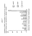
- FIG. 8 shows the results of a number of determinations of the reactivity of sera from breast cancer patients with ABC MUC1 and urinary MUC1.
- Peripheral blood mononucleocytes were purified from a 4 ml sample of heparinised blood from patients or normal individuals using lymphocyte separation medium (ICN flow), as described in detail in the manufacturers instructions. Isolated mononucleocytes were washed in PBS and resuspended in 1 ml of a semipurified preparation of Epstein Barr Virus (EBV) from the B95-8 marmoset transformed leukocyte EBV-producing cell line. The cells were then incubated for 1 hour at 37° C. in 5% CO 2 and centrifuged at 17000 rpm.
- EBV Epstein Barr Virus
- the EBV supernatant was removed and the mononucleocytes were washed three times with RPMI medium, resuspended in RPMI medium supplemented with 10% fetal bovine serum and 5 ⁇ g/ml phytoheamatagglutinin (PHA-P) and seeded in multi-wells tissue culture plates.
- the medium was changed every 3 days and used as a source of autoantibodies.
- MUC1 was purified from the serum of patients diagnosed with primary breast cancer or advanced breast cancer or from the urine of healthy subjects according to the following protocol.
- the mouse monoclonal B55 antibody (also known as NCRC 11 as described by Ellis et al. (1984) Histopathology 8: 501–516 and in International Patent Application No. WO 89/01153) was conjugated to CNBr sepharose beads. Serum or urine samples were diluted 1/10 in PBS and incubated with the antibody conjugated sepharose beads overnight at 4° C. with rolling. The beads were centrifuged and the supernatant removed. In order to remove any molecule non-specifically bound to the beads, these were washed in PBS for 5 times or until the washing buffer showed no absorbance at 280 nm.
- Each wash was performed by resuspending the beads in PBS, rolling for 10 minutes, centrifuging and removing the supernatant.
- the washed beads were resuspended in 0.25 M glycine pH 2.5, rolled at room temperature for 10 minutes and centrifuged.
- the supernatant was removed, adjusted to pH 7 by addition of TRIS and stored at 4° C. labelled “glycine fraction”.
- the beads were then resuspended in 25 mM diethylamine (DEA) pH 11, rolled at room temperature for 10 minutes and centrifuged.
- the supernatant was again removed, adjusted to pH 7 by addition of TRIS and stored at 4° C. labelled “25 DEA fraction”.
- MUC1 preparations obtained as described above, were appropriately diluted with PBS and plated out at 50 ⁇ l per well in a 96 well microtitre assay plate and left to dry overnight. The plate was then washed once with PBS/Tween to remove residual salt crystals, blocked for 60 minutes with a fresh solution of 2% (w/v) polyvinylpyrrolidone (PVP) in PBS and washed three times with PBS/Tween. Culture supernatant of immortalised lymphocytes derived from patients diagnosed with primary or secondary breast cancer were plated out in triplicate, at 50 ⁇ l per well. As a comparative control the mouse monoclonal anti-MUC1 antibody B55 was also plated in triplicate.
- PVP polyvinylpyrrolidone
- the plate was incubated for 60 minutes at room temperature with shaking and washed four times with PNS/Tween. 50 ⁇ l of HRP conjugated anti-human or anti-mouse secondary antibody (obtained from Dako) were added to each well at the dilution recommended by the manufacturer, and incubated for 60 minutes at room temperature with shaking. The plate was then washed again four times with PBS/Tween. 50 ⁇ l of TetraMethylBenzidine (TMB) were added to each well and optical density (OD) at 650 nm for each well of the assay plate was read kinetically over a period of 10 minutes.
- TMB TetraMethylBenzidine
- FIG. 1 shows the result of an ELISA assay to assess the reactivity of autoantibodies produced by lymphocytes derived from six patients diagnosed with breast cancer (1 to 4, with primary breast cancer, 7 and 11 with advanced breast cancer) with MUC1 protein purified from the same patient from which the antibody was taken, from other patients or from healthy subjects.
- the healthy subjects used in this study were women who had no clinical and/or mammographical evidence of breast cancer.
- the reactivity of the monoclonal anti-MUC1 B55 antibody was measured as a comparative control.
- Antibodies produced by lymphocytes from four healthy subjects (N10 to N14) were used as a negative control.
- results presented demonstrate that B lymphocytes derived from patients with breast cancer produce autoantibodies that are able to recognise MUC1 protein isolated both from the same and from different patients.
- these autoantibodies bind with high specificity to MUC1 present in patients with cancer, showing almost no reactivity with MUC1 isolated from healthy individuals.
- results are highly reproducible, since different autoantibodies show a very similar reactivity profile with MUC1 protein purified from different sources.
- results obtained also indicate that the sensitivity of the autoantibodies for cancer-associated MUC1 is much greater than that observed for the monoclonal B55 antibody.
- antibodies produced by lymphocytes from normal patients did not show this profile.
- FIG. 2 shows the reactivity of autoantibodies secreted by immortalised B lymphocytes derived from patients with primary breast cancer with MUC1 protein from different sources, compared with that of B55.
- the profile of reactivity of the different autoantibodies is again very reproducible.
- the autoantibodies show high specificity for MUC1 present in the serum of patients with cancer and have almost no affinity for MUC1 isolated from healthy individuals or from the breast cancer cell line ZR75-1. Furthermore, the affinity of the autoantibodies for MUC1 protein associated with either primary breast cancer or advanced breast cancer is much higher that measured for B55.
- FIG. 3 shows that the autoantibodies produced by B lymphocytes derived from a patient with primary breast cancer bind with a much higher affinity to MUC1 isolated from another patient with breast cancer than MUC1 isolated from a healthy individual.
- the MUC1 peptide TAP2 was conjugated to CNBr-sepharose beads.
- Pooled sera from patients diagnosed with advanced breast cancer were diluted 1/10 in PBS and were incubated with the conjugated sepharose beads overnight at 4° C. with rolling (in the ratio of 25 ml of serum to 1 ml of beads). After centrifugation the supernatant was removed and the beads were washed 5 times with PBS or until absorbance at 280 nm was zero. Each wash was performed by resuspending the beads in PBS, rolling for 10 minutes, centrifuging and removing the supernatant.
- the beads were resuspended in 1 ml of 3M sodium thiocyanate in PBS, rolled at room temperature for 10 minutes and centrifuged. The supernatant was removed and dialysed against PBS at 4° C.
- the anti-MUC1 content was then confirmed by ELISA using as immobilised antigen both MCU1 isolated from patients with advanced breast cancer and a MUC1 peptide, with sequence APDTRTPAPG and conjugated to BSA.
- the autoantibodies obtained as described above were concentrated to a volume of 100 ⁇ l by using centrifugal filters and then diluted to a volume of 1 ml with 0.1 sodium tetraborate buffer pH 8.8. 20 ⁇ g of N-hydroxysuccinimide biotin were added and the autoantibodies/biotin solution was incubated for 4 hours at room temperature with rolling. The reaction was stopped by addition of 10 ⁇ l of 1M NH 4 Cl and incubation for ten minutes. The autoantibodies were then dialysed against PBS for thirty-six hours at 4° C. to remove unbound biotin. Aliquots of the autoantibodies solution were frozen and stored at ⁇ 20° C. in the dark until use.
- Culture supernatant of lymphocytes derived from patients with primary breast cancer or advanced breast cancer or the monoclonal anti-MUC1 C595 antibody were plated out at 50 ⁇ l per well in a 96 well microtitre assay plate and incubated overnight at 4° C. The plate was then washed 4 times with PBS/Tween, blocked for 60 minutes with a fresh solution of 2% (w/v) polyvinylpyrrolidone (PVP) in PBS and washed twice with PBS/Tween. 50 ⁇ l per well of MUC1 from different sources were added. After incubation at room temperature for sixty minutes, the plate was washed again four times with PBS/Tween.
- PVP polyvinylpyrrolidone
- FIG. 5 shows the results of an ELISA assay utilising as immobilised antibodies autoantibodies produced by B lymphocytes derived from patients with primary or advanced breast cancer, compared with those obtained in a parallel assay with the monoclonal anti-MUC1 C595 antibody.
- the data indicate that autoantibodies from patients with breast cancer can be used in ELISA assays to specifically detect modified forms of MUC1 protein associated with cancer. These assays are more sensitive and show higher specificity than those utilising the monoclonal antibody C595.
- An ELISA assay was performed, as described in Example 4, on serum samples from healthy individuals or patients with primary or advanced breast cancer utilising as immobilised antibodies the autoantibodies produced by B lymphocytes derived from patients with primary breast cancer.
- a parallel assay utilising the monoclonal anti-MUC1 antibody C595 was performed on the same samples. The results, shown in FIG. 6 , indicate that the assay employing autoantibodies is able to detect with high sensitivity MUC1 circulating in the blood of patients with breast cancer.
- this assay contrary to utilising the monoclonal antibody C595, this assay has a very high specificity for cancer-associated forms of MUC1.
- An ELISA assay was performed, as described in Example 4, on sequential serum samples from a patient diagnosed with metastatic cancer throughout the progression of the disease, using as immobilised antibodies the autoantibodies produced by B lymphocytes derived from patients with primary breast cancer or the monoclonal anti-MUC1 C595 antibody.
- the commercial assay CA15-3 was also performed on the same samples.
- FIG. 7 shows that the assay employing autoantibodies can be used to follow the progression of cancer in a patient, wherein increasing levels of MUC1 detected in the assay indicate exacerbation of the disease.
- the data also demonstrate that the use of autoantibodies leads to results that better represent the development of the disease than those obtained with either the C959 antibody or the CA15-3 assay.
- FIG. 8 shows the results of a number of determinations of reactivity of sera from breast cancer patients with ABC and urinary MUC1. Sera from the majority of patients clearly exhibit greater specificity for the ABC MUC1 as compared to urinary MUC1.
Landscapes
- Health & Medical Sciences (AREA)
- Life Sciences & Earth Sciences (AREA)
- Immunology (AREA)
- Engineering & Computer Science (AREA)
- Hematology (AREA)
- Molecular Biology (AREA)
- Biomedical Technology (AREA)
- Chemical & Material Sciences (AREA)
- Urology & Nephrology (AREA)
- Food Science & Technology (AREA)
- General Physics & Mathematics (AREA)
- Cell Biology (AREA)
- Biotechnology (AREA)
- Medicinal Chemistry (AREA)
- Physics & Mathematics (AREA)
- Analytical Chemistry (AREA)
- Biochemistry (AREA)
- General Health & Medical Sciences (AREA)
- Microbiology (AREA)
- Pathology (AREA)
- Rehabilitation Therapy (AREA)
- Rheumatology (AREA)
- Peptides Or Proteins (AREA)
- Measuring Or Testing Involving Enzymes Or Micro-Organisms (AREA)
- Investigating Or Analysing Biological Materials (AREA)
- Micro-Organisms Or Cultivation Processes Thereof (AREA)
Priority Applications (1)
| Application Number | Priority Date | Filing Date | Title |
|---|---|---|---|
| US11/681,830 US20080108084A1 (en) | 1998-12-10 | 2007-03-05 | Cancer Detection Methods and Reagents |
Applications Claiming Priority (2)
| Application Number | Priority Date | Filing Date | Title |
|---|---|---|---|
| GBGB9827228.9A GB9827228D0 (en) | 1998-12-10 | 1998-12-10 | Cancer detection method and reagents |
| PCT/GB1999/004182 WO2000034787A1 (en) | 1998-12-10 | 1999-12-10 | Cancer detection method and reagents |
Related Parent Applications (1)
| Application Number | Title | Priority Date | Filing Date |
|---|---|---|---|
| PCT/GB1999/004182 Continuation WO2000034787A1 (en) | 1998-12-10 | 1999-12-10 | Cancer detection method and reagents |
Related Child Applications (1)
| Application Number | Title | Priority Date | Filing Date |
|---|---|---|---|
| US11/681,830 Division US20080108084A1 (en) | 1998-12-10 | 2007-03-05 | Cancer Detection Methods and Reagents |
Publications (1)
| Publication Number | Publication Date |
|---|---|
| US7205117B1 true US7205117B1 (en) | 2007-04-17 |
Family
ID=10844007
Family Applications (2)
| Application Number | Title | Priority Date | Filing Date |
|---|---|---|---|
| US09/857,739 Expired - Fee Related US7205117B1 (en) | 1998-12-10 | 2001-06-08 | Cancer detection method and reagents |
| US11/681,830 Abandoned US20080108084A1 (en) | 1998-12-10 | 2007-03-05 | Cancer Detection Methods and Reagents |
Family Applications After (1)
| Application Number | Title | Priority Date | Filing Date |
|---|---|---|---|
| US11/681,830 Abandoned US20080108084A1 (en) | 1998-12-10 | 2007-03-05 | Cancer Detection Methods and Reagents |
Country Status (13)
| Country | Link |
|---|---|
| US (2) | US7205117B1 (https=) |
| EP (1) | EP1137943B1 (https=) |
| JP (1) | JP4665069B2 (https=) |
| AT (1) | ATE322014T1 (https=) |
| AU (1) | AU1671300A (https=) |
| CA (1) | CA2354702C (https=) |
| CY (1) | CY1105697T1 (https=) |
| DE (1) | DE69930666T2 (https=) |
| DK (1) | DK1137943T3 (https=) |
| ES (1) | ES2257087T3 (https=) |
| GB (1) | GB9827228D0 (https=) |
| PT (1) | PT1137943E (https=) |
| WO (1) | WO2000034787A1 (https=) |
Cited By (16)
| Publication number | Priority date | Publication date | Assignee | Title |
|---|---|---|---|---|
| US20030232399A1 (en) * | 2000-06-14 | 2003-12-18 | Robertson John Forsyth Russell | Cancer detection methods and reagents |
| US20040137538A1 (en) * | 2003-01-10 | 2004-07-15 | Bradford Sherry A. | Cancer comprehensive assay kit for identifying cancer protein patterns |
| US20060014138A1 (en) * | 2004-06-09 | 2006-01-19 | The Regents Of The University Of Michigan | Phage microarray profiling of the humoral response to disease |
| US20080081397A1 (en) * | 2006-09-28 | 2008-04-03 | Philips Lumileds Lighting Company, Llc | Process for Preparing a Semiconductor Structure for Mounting |
| US20080153113A1 (en) * | 1998-05-11 | 2008-06-26 | Robertson John F R | Tumour Markers |
| US20080213921A1 (en) * | 2006-09-13 | 2008-09-04 | Robertson John F R | Immunoassay Methods |
| US20080305476A1 (en) * | 2005-05-27 | 2008-12-11 | Onc-Immune Ltd. | Immunoassay Methods |
| US20090176319A1 (en) * | 2007-12-24 | 2009-07-09 | Onclmmune Limited | Calibrator For Immunoassays |
| US20090197285A1 (en) * | 2005-11-10 | 2009-08-06 | University Of Kentucky | Lung Cancer Diagnostic Assay |
| WO2009137832A3 (en) * | 2008-05-09 | 2010-04-01 | Duke University | Autoantibodies in the detection and treatment of cancer |
| US20100093108A1 (en) * | 2006-07-08 | 2010-04-15 | Khattar Nada H | Lung cancer diagnotic assay |
| US20110045508A1 (en) * | 2008-01-31 | 2011-02-24 | The Brigham And Women's Hospital, Inc. | Urinary CA125 Peptides as Biomarkers of Ovarian Cancer |
| US20110237457A1 (en) * | 2010-03-26 | 2011-09-29 | Armune Biosciences, Inc. | Method and system of particle-coupled phage epitope |
| US20110237461A1 (en) * | 2010-03-17 | 2011-09-29 | The Regents Of The University Of Michigan | Using phage epitopes to profile the immune response |
| US20140072963A1 (en) * | 2011-03-16 | 2014-03-13 | Beijing Unidiag Technology Inc | Electrochemiluminescence immunoassay method |
| US9714938B2 (en) | 2005-05-27 | 2017-07-25 | Oncimmune Ltd. | Immunoassay methods |
Families Citing this family (18)
| Publication number | Priority date | Publication date | Assignee | Title |
|---|---|---|---|---|
| GB2395270B (en) | 2002-11-14 | 2006-08-16 | Univ Nottingham | Tumour marker proteins and uses thereof |
| WO2006081553A2 (en) * | 2005-01-28 | 2006-08-03 | Ramot At Tel Aviv University, Ltd. | ANTI-MUC1 α/ß ANTIBODIES |
| MX2009005466A (es) | 2006-11-22 | 2009-08-17 | Adnexus A Bristol Myers Sqibb | Terapeuticos dirigidos a base de proteinas manipuladas para receptores de tirosina cinasas, incluyendo receptor de factor de crecimiento tipo insulina-i. |
| AU2009213141A1 (en) | 2008-02-14 | 2009-08-20 | Bristol-Myers Squibb Company | Targeted therapeutics based on engineered proteins that bind EGFR |
| WO2009142773A2 (en) | 2008-05-22 | 2009-11-26 | Bristol-Myers Squibb Company | Multivalent fibronectin based scaffold domain proteins |
| US8852870B2 (en) | 2008-06-16 | 2014-10-07 | University Of Louisville Research Foundation, Inc. | Systems and methods for diagnosis and prognosis of cancer |
| TWI496582B (zh) | 2008-11-24 | 2015-08-21 | 必治妥美雅史谷比公司 | 雙重專一性之egfr/igfir結合分子 |
| TW201138808A (en) | 2010-05-03 | 2011-11-16 | Bristol Myers Squibb Co | Serum albumin binding molecules |
| JP6023703B2 (ja) | 2010-05-26 | 2016-11-09 | ブリストル−マイヤーズ スクイブ カンパニーBristol−Myers Squibb Company | 改善された安定性を有するフィブロネクチンをベースとする足場タンパク質 |
| EP3415528B1 (en) | 2011-04-13 | 2021-05-26 | Bristol-Myers Squibb Company | Fc fusion proteins comprising novel linkers or arrangements |
| EP2709669A1 (en) | 2011-05-17 | 2014-03-26 | Bristol-Myers Squibb Company | Methods for maintaining pegylation of polypeptides |
| US20130040839A1 (en) * | 2011-06-27 | 2013-02-14 | Ambergen, Inc. | Method for Diagnosing or Determining the Prognosis of Colorectal Cancer (CRC) Using Novel Autoantigens: Gene Expression Guided Autoantigen Discovery |
| AU2012310086B2 (en) | 2011-09-15 | 2018-05-17 | Vaxil Bio Therapeutics Ltd. | Antibodies directed against signal peptides, methods and uses thereof |
| ES2736127T3 (es) | 2014-03-20 | 2019-12-26 | Bristol Myers Squibb Co | Dominios de tipo III de fibronectina de unión a seroalbúmina |
| GB201501930D0 (en) | 2015-02-05 | 2015-03-25 | Univ London Queen Mary | Biomarkers for pancreatic cancer |
| WO2016164815A1 (en) | 2015-04-10 | 2016-10-13 | Applied Proteomics, Inc. | Protein biomarker panels for detecting colorectal cancer and advanced adenoma |
| CN108290941A (zh) | 2015-09-23 | 2018-07-17 | 百时美施贵宝公司 | 快解离速率的血清白蛋白结合性纤连蛋白iii型结构域 |
| CN109116024B (zh) * | 2018-06-14 | 2021-04-23 | 郑州大学第一附属医院 | 一种肺癌标志物抗-actr3自身抗体及其应用 |
Citations (17)
| Publication number | Priority date | Publication date | Assignee | Title |
|---|---|---|---|---|
| EP0236606A1 (en) | 1986-03-10 | 1987-09-16 | Imre Corporation | Immune complex assay |
| US4898951A (en) | 1984-03-22 | 1990-02-06 | Bresatac Limited | Compounds used as intermediates in the preparations of non-radioactive biological probes |
| US4937185A (en) | 1987-07-31 | 1990-06-26 | The Ohio State University Research Foundation | Detection of circulating antibodies to a cancer marker protein |
| WO1992013065A1 (en) | 1991-01-18 | 1992-08-06 | Novo Nordisk A/S | A method of producing autoantibodies |
| US5157020A (en) * | 1990-05-24 | 1992-10-20 | Research Corporation Tech., Inc. | Synthetic senescent cell antigen |
| WO1993011236A1 (en) | 1991-12-02 | 1993-06-10 | Medical Research Council | Production of anti-self antibodies from antibody segment repertoires and displayed on phage |
| WO1993021529A1 (en) | 1992-04-14 | 1993-10-28 | Duke University | Method of detecting tumors containing complexes of p53 and hsp70 |
| WO1994023728A1 (en) | 1993-04-16 | 1994-10-27 | Northwestern University | Immunogenic cancer proteins and peptides and method of use |
| WO1996000084A1 (en) | 1994-06-24 | 1996-01-04 | Torchilin Vladimir P | Use of autoantibodies for tumor therapy and prophylaxis |
| WO1996003502A2 (en) | 1994-07-26 | 1996-02-08 | Ramot University Authority For Applied Research And Industrial Development Ltd. | Muc1 derived proteins for the diagnosis, imaging and therapy of human cancer |
| WO1997014794A1 (en) | 1995-10-20 | 1997-04-24 | University Of Dundee | ACTIVATION OF p53 PROTEIN |
| JPH09189702A (ja) | 1996-01-09 | 1997-07-22 | Hitachi Chem Co Ltd | 抗ムチン抗体の測定法、癌の測定法及び癌診断薬 |
| US5726023A (en) | 1993-03-17 | 1998-03-10 | University Of Washington | Immune reactivity to HER-2/neu protein for diagnosis and treatment of malignancies in which the HER-2/neu oncogene is associated |
| US5876728A (en) | 1995-02-15 | 1999-03-02 | Howard David Kass | Natural composition extracted from plants used in the treatment of cancer |
| WO1999058978A2 (en) | 1998-05-11 | 1999-11-18 | The University Of Nottingham | Tumour markers |
| WO2000026668A2 (en) | 1998-11-05 | 2000-05-11 | The Regents Of The University Of Michigan | S100 proteins and autoantibodies as serum markers for cancer |
| US6322989B1 (en) | 1991-11-25 | 2001-11-27 | Yoreh Biotechnologies, Ltd. | Whole blood/mitogen assay for the early detection of a subject with ovarian or breast cancer and kit |
Family Cites Families (31)
| Publication number | Priority date | Publication date | Assignee | Title |
|---|---|---|---|---|
| US4241044A (en) * | 1978-12-28 | 1980-12-23 | Abbott Laboratories | Method of diagnosing cancer by detecting elevated anti-T antibody activity |
| US4562160A (en) * | 1983-04-01 | 1985-12-31 | Sloan-Kettering Institute | Melanoma tumor antigen and autologous antibody |
| US5106738A (en) * | 1984-01-31 | 1992-04-21 | Akzo Nv | Tumor specific monoclonal antibodies |
| US7282345B1 (en) * | 1989-08-04 | 2007-10-16 | Schering Ag | C-erbB-2 external domain: GP75 |
| US5110911A (en) * | 1989-11-02 | 1992-05-05 | Biomira, Inc. | Human tumor-associated thomsen-friedenreich antigen |
| DE4120412C1 (https=) * | 1991-06-20 | 1993-01-07 | Henning Berlin Gmbh Chemie- Und Pharmawerk, 1000 Berlin, De | |
| US6280962B1 (en) * | 1991-11-25 | 2001-08-28 | Yoreh Biotechnologies Ltd. | Whole blood/mitogen assay for the early detection of a subject with cancer and kit |
| US5747268A (en) * | 1993-04-22 | 1998-05-05 | Dade International Inc. | Tumor marker control |
| IT1270939B (it) * | 1993-05-11 | 1997-05-26 | Angeletti P Ist Richerche Bio | Procedimento per la preparazione di immunogeni e reagenti diagnostici,e immunogeni e reagenti diagnostici cosi' ottenibili. |
| US5744144A (en) * | 1993-07-30 | 1998-04-28 | University Of Pittsburgh University Patent Committee Policy And Procedures | Synthetic multiple tandem repeat mucin and mucin-like peptides, and uses thereof |
| US5721105A (en) * | 1994-02-20 | 1998-02-24 | B.R.A.H.M.S. Diagnostica Gmbh | Method for the immunological determination of proteins and kit for carrying out the method |
| US5561049A (en) * | 1994-09-21 | 1996-10-01 | Behringwerke Ag | Method for detecting antibodies |
| ATE283069T1 (de) * | 1996-08-16 | 2004-12-15 | Univ Johns Hopkins Med | Melanomzellinien, welche immundominante melanomantigene teilen und verfahren zu deren anwendung |
| DE19805815C1 (de) * | 1998-02-13 | 1999-11-11 | Ansgar W Lohse | Diagnostikum zum Erkennen der autoimmunen Hepatitis |
| US6387639B1 (en) * | 1998-11-10 | 2002-05-14 | Sloan-Kettering Institute For Cancer Research | Ma family polypeptides and anti-Ma antibodies |
| US7807810B2 (en) * | 1999-04-08 | 2010-10-05 | The Ohio State University Research Foundation | Nucleic acids encoding the major outer membrane protein of the causative agent of human granulocytic ehrlichiosis and peptides encoded thereby |
| US6645465B2 (en) * | 1999-08-06 | 2003-11-11 | Michigan, University Of The Regents | Annexin proteins and autoantibodies as serum markers for cancer |
| US20020064801A1 (en) * | 1999-12-01 | 2002-05-30 | Ryan Jeffrey R. | Novel and practical serological assay for the clinical diagnosis of leishmaniasis |
| US6667160B2 (en) * | 2000-03-15 | 2003-12-23 | Kenneth D. Fine | Method for diagnosing immunologic food sensitivity |
| US20030138860A1 (en) * | 2000-06-14 | 2003-07-24 | Robertson John Forsyth Russell | Cancer detection methods and reagents |
| US20030232399A1 (en) * | 2000-06-14 | 2003-12-18 | Robertson John Forsyth Russell | Cancer detection methods and reagents |
| US7745141B2 (en) * | 2001-06-21 | 2010-06-29 | New York University | Mycobacterial proteins as early antigens for serodiagnosis and vaccines |
| US20030049692A1 (en) * | 2002-09-16 | 2003-03-13 | Norman Latov | Detection of anti-glycolipid antibodies by latex agglutination assay |
| GB2395270B (en) * | 2002-11-14 | 2006-08-16 | Univ Nottingham | Tumour marker proteins and uses thereof |
| JP2005352771A (ja) * | 2004-06-10 | 2005-12-22 | Hitachi Software Eng Co Ltd | 発現プロファイルによるパターン認識システム |
| US20060141547A1 (en) * | 2004-11-16 | 2006-06-29 | Das Hasi R | Novel diagnostic marker, a diagnostic kit and a method for diagnosis of rheumatoid arthritis |
| GB2426581A (en) * | 2005-05-27 | 2006-11-29 | Univ Nottingham | Immunoassay methods |
| EP1789435B1 (en) * | 2005-09-12 | 2010-02-24 | Industry Foundation of Chonnam National University | A method for production of mature natural killer cell |
| TWI304443B (en) * | 2005-12-30 | 2008-12-21 | Nat Health Research Institutes | Alpha-enolase specific antibody and method of use |
| CN101632020B (zh) * | 2006-09-13 | 2013-11-27 | 昂西免疫有限公司 | 改进的免疫测定方法 |
| GB0725239D0 (en) * | 2007-12-24 | 2008-02-06 | Oncimmune Ltd | Calibrator for autoantibody assay |
-
1998
- 1998-12-10 GB GBGB9827228.9A patent/GB9827228D0/en not_active Ceased
-
1999
- 1999-12-10 EP EP99959578A patent/EP1137943B1/en not_active Expired - Lifetime
- 1999-12-10 DE DE69930666T patent/DE69930666T2/de not_active Expired - Lifetime
- 1999-12-10 ES ES99959578T patent/ES2257087T3/es not_active Expired - Lifetime
- 1999-12-10 CA CA2354702A patent/CA2354702C/en not_active Expired - Fee Related
- 1999-12-10 WO PCT/GB1999/004182 patent/WO2000034787A1/en not_active Ceased
- 1999-12-10 AT AT99959578T patent/ATE322014T1/de active
- 1999-12-10 DK DK99959578T patent/DK1137943T3/da active
- 1999-12-10 JP JP2000587190A patent/JP4665069B2/ja not_active Expired - Fee Related
- 1999-12-10 PT PT99959578T patent/PT1137943E/pt unknown
- 1999-12-10 AU AU16713/00A patent/AU1671300A/en not_active Abandoned
-
2001
- 2001-06-08 US US09/857,739 patent/US7205117B1/en not_active Expired - Fee Related
-
2006
- 2006-06-08 CY CY20061100772T patent/CY1105697T1/el unknown
-
2007
- 2007-03-05 US US11/681,830 patent/US20080108084A1/en not_active Abandoned
Patent Citations (18)
| Publication number | Priority date | Publication date | Assignee | Title |
|---|---|---|---|---|
| US4898951A (en) | 1984-03-22 | 1990-02-06 | Bresatac Limited | Compounds used as intermediates in the preparations of non-radioactive biological probes |
| EP0236606A1 (en) | 1986-03-10 | 1987-09-16 | Imre Corporation | Immune complex assay |
| US4937185A (en) | 1987-07-31 | 1990-06-26 | The Ohio State University Research Foundation | Detection of circulating antibodies to a cancer marker protein |
| US5157020A (en) * | 1990-05-24 | 1992-10-20 | Research Corporation Tech., Inc. | Synthetic senescent cell antigen |
| WO1992013065A1 (en) | 1991-01-18 | 1992-08-06 | Novo Nordisk A/S | A method of producing autoantibodies |
| US6322989B1 (en) | 1991-11-25 | 2001-11-27 | Yoreh Biotechnologies, Ltd. | Whole blood/mitogen assay for the early detection of a subject with ovarian or breast cancer and kit |
| WO1993011236A1 (en) | 1991-12-02 | 1993-06-10 | Medical Research Council | Production of anti-self antibodies from antibody segment repertoires and displayed on phage |
| US5652115A (en) | 1992-04-14 | 1997-07-29 | Duke University | Method of detecting tumors containing complexes of p53 and HSP70 |
| WO1993021529A1 (en) | 1992-04-14 | 1993-10-28 | Duke University | Method of detecting tumors containing complexes of p53 and hsp70 |
| US5726023A (en) | 1993-03-17 | 1998-03-10 | University Of Washington | Immune reactivity to HER-2/neu protein for diagnosis and treatment of malignancies in which the HER-2/neu oncogene is associated |
| WO1994023728A1 (en) | 1993-04-16 | 1994-10-27 | Northwestern University | Immunogenic cancer proteins and peptides and method of use |
| WO1996000084A1 (en) | 1994-06-24 | 1996-01-04 | Torchilin Vladimir P | Use of autoantibodies for tumor therapy and prophylaxis |
| WO1996003502A2 (en) | 1994-07-26 | 1996-02-08 | Ramot University Authority For Applied Research And Industrial Development Ltd. | Muc1 derived proteins for the diagnosis, imaging and therapy of human cancer |
| US5876728A (en) | 1995-02-15 | 1999-03-02 | Howard David Kass | Natural composition extracted from plants used in the treatment of cancer |
| WO1997014794A1 (en) | 1995-10-20 | 1997-04-24 | University Of Dundee | ACTIVATION OF p53 PROTEIN |
| JPH09189702A (ja) | 1996-01-09 | 1997-07-22 | Hitachi Chem Co Ltd | 抗ムチン抗体の測定法、癌の測定法及び癌診断薬 |
| WO1999058978A2 (en) | 1998-05-11 | 1999-11-18 | The University Of Nottingham | Tumour markers |
| WO2000026668A2 (en) | 1998-11-05 | 2000-05-11 | The Regents Of The University Of Michigan | S100 proteins and autoantibodies as serum markers for cancer |
Non-Patent Citations (64)
| Title |
|---|
| Abstract of Hayes; Anticancer Drugs, 1995, vol. 6, suppl. 2, pp. 26-27. |
| Abstract of Mensdorf-Pouilly et al.; Anticancer Research, Nov.-Dec. 1997, vol. 17, p. 4184. |
| Abstract of Pandha et al.; Cancer Gene Therapy, 1997, vol. 4, No. 5, p. 310. |
| Abstract of Yamamoto et al.; Proc. Amer. Soc. Cancer Res., Mar. 1997, p. 564. |
| Aparecida et al., "Value of CEA Level Determination in Gallbladder Bile in the Diagnosis of Liver Metastases Secondary to Colorectal Adenocarcinoma", Sao Paulo Medical Journal, 2001, vol. 119, No. 3, pp. 110-113. |
| Apostolopoulos et al., Nature Medicine, 1998, vol. 4, pp. 315-320. |
| Baechstrom, et al., "Purification and Characterization of Sialyl-Le-Carrying Mucins of Human Bile; Evidence for the Presence of MUC1 and MUC3 Apoproteins", The Journal of Biological Chemistry, 1994, vol. 269, No. 20, pp. 14430-14437. |
| Beatty, et al., "Biochemical Characterization of the Soluble Form of Tumor Antigen MUC1 Isolated from Sera and Ascites Fluid of Breast and Pancreatic Cancer Patients", Clinical Cancer Research, 2001, vol. 7, pp. 781-787. |
| Bhatti et al.; Journal of Tumor Marker Oncology, Summer-1994, vol. 9, pp. 125-131. |
| Croce et al.; Cancer. Immunol. Immunother., 1995, vol. 40, pp. 132-137. |
| Definition of "moncyte" in On-line Medical Dictionary downloaded on Feb. 5, 2005 from url..cnacerweb.ncl.ac.uk. * |
| Deguchi et al.; Int. Arch. Allergy Appl. Immunol., 1988, vol. 87, pp. 313-316. |
| Denton et al.; Cancer Letters, 1993, vol. 70, pp. 143-150. |
| Disis et al.; Journal of Clinical Oncology, 1997, vol. 15, pp. 3363-3367. |
| Fishman, P., "Application of autoantibodies to cancer therapy: A new concept", The 9<SUP>th</SUP> International Congress of Immunology, 1995, p. 664. |
| Gourevitch et al, Br J Cancer. Oct. 1995;72(4):934-8. * |
| Gourevitch, MM; "Polymorphic epithelial mucin (MUC-1)-containing circulating immune complexes in carcinoma patients"; British Journal of Cancer, 72, pp. 934-938; (1995). |
| Green et al.; European Journal of Cancer, 1994, vol. 30A, pp. 580-584. |
| Güre, "Human Lung Cancer Antigens Recognized by Autologous Antibodies Definition of a Novel cDNA Derived from the Tumor Suppressor Gene Locus on Chromosome 3p21.3", Ludwig Institute for Cancer Research, pp. 1034-1040, 1998, Cancer Res, vol. 58. |
| Hill et al., "Nature of Carcinoembryonic Antigen Purified From Malignant Ascitic Fluid of Serous Cystadenocarcinoma of the Ovary", Molecular Immunology, 1981, vol. 18, No. 7, pp. 647-653. |
| Hinoda, et al., "Detection of a Circulating Antibody Against a Peptide Epitope on a Mucin Core Protein, MUC1, in Ulcerative Colitis", 1991, pp. 163-168. |
| Houghton et al.; "Detection of Cell Surface and Intracellular Antigens by Human Monoclonal Antibodies-Hybrid Cell Lines Derived from Lymphocytes of Patients with Malignant Melanoma*"; J. Exp. Med.; vol. 158, Jul. 1983; pp. 53-65. |
| Janeway et al., (Immunobiology downloaded from url.ncbi.nlm.nih.gov/books, total 2 pages). * |
| Karanikas, et al. J Clin Invest Dec. 1, 1997, vol. 100, No. 11, pp. 2783-2792. |
| Kawahara; Cancer, 1986, vol. 58, pp. 2008-2012. |
| Koterra et al.; Caner Research, 1994, vol. 54, pp. 2856-2860. |
| Kuralay, et al., Diagnostic Usefulness of Tumour Marker Levels in Pleural Effusions of Malignant and Benign Origin, Clinica Chimica Acta, 2000, vol. 300, pp. 43-55. |
| Kutteh, W.H., et al., "Immunologic characterization of tumor markers in human ovarian cancer cell lines"; Journal of the Society for Gynecologic Investigation, 1996, vol. 3, No. 4, pp. 216-222. |
| Laeng, et al., "Anti-Neural Autoantibodies, types 1 and 2: Their Utility in the Study of Tumors of the Nervous System", Acta Neuropathol, 1998, vol. 96, pp. 329-339. |
| Lafond, R.E., et al., "Autoantibodies to c-myc protein: elevated levels in patients with African Burkitt's lymphoma and normal Ghanians", Autoimmunity, vol. 13, No. 3, 1992, pp. 215-224. |
| Lai, et al., "Presence of Serum Anti-P53 Antibodies is Associated with Pleural Effusions and Poor Prognosis in Lung Cancer Patients", Clinical Cancer Research, 1998, vol. 4, pp. 3025-3030. |
| Lawniczak, et al., "The Search for Tumor-Associated Proteins in Pleural Effusions by Means of Monoclonal Antibodies and a Dot Blot Assay", Lung, 1992, vol. 170, pp. 65-74. |
| Luo, et al., "Identification of Heat Shock Protein 90 and Other Proteins as Tumour Antigens by Serological Screening of an Ovarian Carcinoma Expression Library", British Journal of Cancer, 2002, vol. 87, pp. 339-343. |
| Mercer, "Use of Multiple Markers to Enhance Clinical Utility", Immunology Series, vol. 53, 1990, pp. 39-54. |
| Montenarh, et al., "P53 Autoantibodies in the Sera, Cyst and Ascitic Fluids of Patients with Ovarian Cancer", International Journal of Oncology, 1998, vol. 13, pp. 605-610. |
| Nery, et al., "Isolation and Partial Characterization of Macromolecular Urinary Aggregates Containing Carcinoembryonic Antigen-Like Activity", Br J. Cancer, 1974, vol. 29, No. 413. |
| Nustad et al., "Epitopes on CA 125 from Cervical Mucus and Ascites Fluid and Characterization of Six New Antibodies", pp. 303-314. |
| Pavelic et al., Anticancer Reasearch, 1991, vol. 11, pp. 1421-1428. |
| Petrakou et al.; International Journal of oncology, 1997, vol. 11, suppl. p. 902. |
| Petrarca et al, Eur J Cancer. Nov. 1996;32A(12):2155-63. * |
| Petrarca, C., "Human Antibodies Against the Polymorphic Epithelial Mucin in Ovarian Cancer Patients Recognise a Novel Sequence in the Tandem Repeat Region"; European Journal of Cancer, vol. 32A, No. 12, pp. 2155-2163, 1996. |
| Rao et al.; "Detection of human ovarian tumor-associated antigens by antibodies isolated from ovarian carcinoma ascitic fluid"; Am J Obstet Gynecol; Jul. 1988; vol. 159, No. 1; pp. 94-98. |
| Rao, S.G., "Detection of Human Ovarian Tumor Associated Antigens by Autologous Antibodies Isolated from Ovarian Carcinoma Ascites Fluid"; Proceedings of the American Association for Cancer Research Annual Meeting, 1987, vol. 28, p. 358. |
| Rusciano, "Concomitant Purification of Prostatic Carcinoma Tumor Markers from Human Seminal Fluid Under Nondenaturing Conditions", Clinical Chemistry, 1988, vol. 34, No. 12, pp. 2528-2532. |
| Sahin et al.; PNAS, 1995, vol. 92, pp. 11810-11813. |
| Sandrin et al., Glycoconjugate Journal, 1997, vol. 14, pp. 97-105. |
| Scanlan et al.; International Journal of Cancer, 1998, vol. 76, pp. 652-658. |
| Schneider, J. "P53 Protein, EGF Receptor, and Anti-P53 Antibodies in Serum from Patients with Occupationally Derived Lung Cancer", British Journal of Cancer, 1999, vol. 80, No. 12, pp. 1987-1994. |
| Shibata, et al., "Purification and Characterization of Prostate Specific Antigen from Human Urine", Biochimica et Biophysica Acta, 1997, vol. 1336, pp. 425-433. |
| Sokoloff, et al., "A Dual-Monoclonal Sandwich Assay for Prostate-Specific Membrane Antigen: Levels in Tissues, Seminal Fluid and Urine", The Prostate, 2000, vol. 43, pp. 150-157. |
| Stearns et al.; Breast Cancer Research and Treatment, Feb. 8, 1998, vol. 52, pp. 239-259. |
| Stubbs, et al., "Faecal Carcinoembryonic Antigen (CEA) in Patients with Large Bowel Cancer", European Journal of Surgical Oncology, 1987, vol. 13, pp. 433-436. |
| Tondini, et al., "Comparison of CA15-3 and Carcinoembryonic Antigen in Monitoring the Clinical Course of Patients with Metastatic Breast Cancer", Cancer Research, 1988, vol. 48, No. 14, pp. 4107-4112. |
| Toth, et al., "A Carcinoembryonic Antigen (CEA) Binding Protein From Ascites Influences CEA Uptake by Macrophages", 1990, vol. 171, No. 2, pp. 633-640. |
| Venegas, et al., "Purification and Immunochemical Characterization of Ascitic Fluid Glycoproteins Containing Certain Tumor-Associated and Blood Group Antigen Markers", Glycoconjugate Journal, 1989, vol. 6, pp. 511-524. |
| Voet et al., (1990, Biochemistry, p. 78 only). * |
| Voet et al., (1990, Biochemistry, pp. 1096, and 1098 only). * |
| Volkmann, M., et al., "Anti-p53 autoantibodies as serological marker in different tumor-entities", Clinical Chemistry, vol. 41, No. S6 part 2, 1995, pp. S221-S222. |
| von Mensdorff-Pouilly et al, Eur J Cancer. Jul. 1996;32A(8):1325-31. * |
| Von Mensdorff-Pouilly, S., "Humoral Immune Response to Polymorphic Epithelial Mucin (MUC-1) inpatients with Benign and Matignant Breat Tumours"; European Journal of Cancer, vol. 32A, No. 8, pp. 1325-1331, 1996. |
| Wolf, et al., "A Tumour-Associated Antigen from The Pleural Effusion of Patients with Squamous-Cell Carcinoma of Lung", Br. J. Cancer, 1978, vol. 36, pp. 1046-1052. |
| Yamamoto et al., "L-Myc Overexpression and Detection of Auto-Antibodies Against L-Myc in Both the Serum and Pleural Effusion from a Patient with Non-Small Cell Lung Cancer", Internal Medicine, 1997, vol. 36, No. 10, pp. 724-727. |
| Yamauchi, et al., "Autoantibodies to C-MYC Nuclear Protein Products in Autoimmune Disease", 1989, pp. 117-119. |
| Zisman et al.; Journal of Urology, 1995, vol. 154, pp. 1052-1055. |
Cited By (36)
| Publication number | Priority date | Publication date | Assignee | Title |
|---|---|---|---|---|
| US9696319B2 (en) | 1998-05-11 | 2017-07-04 | Oncimmune Ltd. | Tumour markers |
| US8114604B2 (en) | 1998-05-11 | 2012-02-14 | Oncimmune Ltd. | Tumour markers |
| US20080153113A1 (en) * | 1998-05-11 | 2008-06-26 | Robertson John F R | Tumour Markers |
| US20030232399A1 (en) * | 2000-06-14 | 2003-12-18 | Robertson John Forsyth Russell | Cancer detection methods and reagents |
| US20110086061A1 (en) * | 2000-06-14 | 2011-04-14 | Onclmmune Limited | Cancer Detection Methods and Reagents |
| US20040137538A1 (en) * | 2003-01-10 | 2004-07-15 | Bradford Sherry A. | Cancer comprehensive assay kit for identifying cancer protein patterns |
| US10006023B2 (en) | 2004-06-09 | 2018-06-26 | The Regents Of The University Of Michigan | Phage microarray profiling of the humoral response to disease |
| US7597890B2 (en) | 2004-06-09 | 2009-10-06 | The Regents Of The University Of Michigan | Methods and compositions for diagnosing lung cancer |
| US20100009382A1 (en) * | 2004-06-09 | 2010-01-14 | The Regents Of The University Of Michigan | Methods and compositions for diagnosing lung cancer |
| US20080044839A1 (en) * | 2004-06-09 | 2008-02-21 | Regents Of The University Of Michigan | Methods and compositions for diagnosing lung cancer |
| US9267133B2 (en) | 2004-06-09 | 2016-02-23 | The Regents Of The University Of Michigan | Phage microarray profiling of the humoral response to disease |
| US7858323B2 (en) | 2004-06-09 | 2010-12-28 | The Regents Of The University Of Michigan | Phage microarray profiling of the humoral response to disease |
| US20060014138A1 (en) * | 2004-06-09 | 2006-01-19 | The Regents Of The University Of Michigan | Phage microarray profiling of the humoral response to disease |
| US8617547B2 (en) | 2004-06-09 | 2013-12-31 | The Regents Of The University Of Michigan | Methods and compositions for diagnosing prostate cancer |
| US20080305476A1 (en) * | 2005-05-27 | 2008-12-11 | Onc-Immune Ltd. | Immunoassay Methods |
| US9714938B2 (en) | 2005-05-27 | 2017-07-25 | Oncimmune Ltd. | Immunoassay methods |
| US9719984B2 (en) | 2005-05-27 | 2017-08-01 | Oncimmune Ltd. | Immunoassay methods |
| US8722339B2 (en) | 2005-05-27 | 2014-05-13 | Oncimmune Ltd. | Immunoassay methods |
| US20090197285A1 (en) * | 2005-11-10 | 2009-08-06 | University Of Kentucky | Lung Cancer Diagnostic Assay |
| US20100093108A1 (en) * | 2006-07-08 | 2010-04-15 | Khattar Nada H | Lung cancer diagnotic assay |
| US20080213921A1 (en) * | 2006-09-13 | 2008-09-04 | Robertson John F R | Immunoassay Methods |
| US8574848B2 (en) | 2006-09-13 | 2013-11-05 | Oncimmune Ltd. | Immunoassay methods |
| US8927223B2 (en) | 2006-09-13 | 2015-01-06 | Oncimmune Ltd. | Immunoassay methods |
| US20080081397A1 (en) * | 2006-09-28 | 2008-04-03 | Philips Lumileds Lighting Company, Llc | Process for Preparing a Semiconductor Structure for Mounting |
| US9899578B2 (en) | 2006-09-28 | 2018-02-20 | Lumileds Llc | Process for preparing a semiconductor structure for mounting |
| US9111950B2 (en) * | 2006-09-28 | 2015-08-18 | Philips Lumileds Lighting Company, Llc | Process for preparing a semiconductor structure for mounting |
| US20090176319A1 (en) * | 2007-12-24 | 2009-07-09 | Onclmmune Limited | Calibrator For Immunoassays |
| US20110045508A1 (en) * | 2008-01-31 | 2011-02-24 | The Brigham And Women's Hospital, Inc. | Urinary CA125 Peptides as Biomarkers of Ovarian Cancer |
| US8642347B2 (en) | 2008-01-31 | 2014-02-04 | The Brigham And Women's Hospital, Inc. | Urinary CA125 peptides as biomarkers of ovarian cancer |
| WO2009137832A3 (en) * | 2008-05-09 | 2010-04-01 | Duke University | Autoantibodies in the detection and treatment of cancer |
| US9658231B2 (en) | 2010-03-17 | 2017-05-23 | The Regents Of The University Of Michigan | Using phage epitopes to profile the immune response |
| US20110237461A1 (en) * | 2010-03-17 | 2011-09-29 | The Regents Of The University Of Michigan | Using phage epitopes to profile the immune response |
| US11307203B2 (en) | 2010-03-17 | 2022-04-19 | The Regents Of The University Of Michigan | Using phage epitopes to profile the immune response |
| US20110237457A1 (en) * | 2010-03-26 | 2011-09-29 | Armune Biosciences, Inc. | Method and system of particle-coupled phage epitope |
| US20140072963A1 (en) * | 2011-03-16 | 2014-03-13 | Beijing Unidiag Technology Inc | Electrochemiluminescence immunoassay method |
| US10024861B2 (en) * | 2011-03-16 | 2018-07-17 | Beijing Unidiag Technology Inc | Electrochemiluminescence immunoassay method |
Also Published As
| Publication number | Publication date |
|---|---|
| US20080108084A1 (en) | 2008-05-08 |
| WO2000034787A1 (en) | 2000-06-15 |
| CA2354702C (en) | 2013-03-26 |
| DK1137943T3 (da) | 2006-07-31 |
| ATE322014T1 (de) | 2006-04-15 |
| JP2002532686A (ja) | 2002-10-02 |
| CY1105697T1 (el) | 2010-12-22 |
| PT1137943E (pt) | 2006-08-31 |
| GB9827228D0 (en) | 1999-02-03 |
| JP4665069B2 (ja) | 2011-04-06 |
| EP1137943B1 (en) | 2006-03-29 |
| EP1137943A1 (en) | 2001-10-04 |
| DE69930666T2 (de) | 2007-01-11 |
| ES2257087T3 (es) | 2006-07-16 |
| DE69930666D1 (de) | 2006-05-18 |
| AU1671300A (en) | 2000-06-26 |
| CA2354702A1 (en) | 2000-06-15 |
Similar Documents
| Publication | Publication Date | Title |
|---|---|---|
| US7205117B1 (en) | Cancer detection method and reagents | |
| US20030138860A1 (en) | Cancer detection methods and reagents | |
| JP2002532686A5 (https=) | ||
| US8592169B2 (en) | Tumour marker proteins and uses thereof | |
| AU780957B2 (en) | Early cancer tumor marker | |
| US9696319B2 (en) | Tumour markers | |
| JP5736598B2 (ja) | 改善されたイムノアッセイ法 | |
| US20110086061A1 (en) | Cancer Detection Methods and Reagents | |
| EP1678503B1 (en) | Specific method for cancer detection | |
| KR20130081952A (ko) | 암 진단용 바이오마커 및 이를 이용한 암세포 분리 방법 | |
| JP5765635B2 (ja) | Ku86とその自己抗体との複合体の免疫測定方法、それに用いるキット及びそれを用いた癌判定方法 | |
| WO2021070934A1 (ja) | がんの検査方法 | |
| US5601988A (en) | Immunocapture assay for cancer procoagulant antibody complex in biological samples |
Legal Events
| Date | Code | Title | Description |
|---|---|---|---|
| AS | Assignment |
Owner name: UNIVERSITY OF NOTTINGHAM, UNITED KINGDOM Free format text: ASSIGNMENT OF ASSIGNORS INTEREST;ASSIGNORS:ROBERTSON, JOHN RUSSELL;GRAVES, CATHERINE ROSAMUND LOUISE;PRICE, MICHAEL RAWLING (DECEASED) BY FRANCES MARGARET PRICE (EXECUTRIX);REEL/FRAME:012389/0473;SIGNING DATES FROM 20010808 TO 20010904 |
|
| AS | Assignment |
Owner name: NOTTINGHAM, UNIVERSITY OF, UNITED KINGDOM Free format text: ASSIGNMENT OF ASSIGNORS INTEREST;ASSIGNORS:ROBERTSON, JOHN R.;GRAVES, CATHERINE RL;PRICE, MICHAEL R.;REEL/FRAME:012399/0012;SIGNING DATES FROM 20010809 TO 20010904 |
|
| STCF | Information on status: patent grant |
Free format text: PATENTED CASE |
|
| FPAY | Fee payment |
Year of fee payment: 4 |
|
| AS | Assignment |
Owner name: ONCIMMUNE LIMITED, UNITED KINGDOM Free format text: ASSIGNMENT OF ASSIGNORS INTEREST;ASSIGNOR:NOTTINGHAM UNIVERSITY;REEL/FRAME:028406/0897 Effective date: 20051229 |
|
| FPAY | Fee payment |
Year of fee payment: 8 |
|
| FEPP | Fee payment procedure |
Free format text: MAINTENANCE FEE REMINDER MAILED (ORIGINAL EVENT CODE: REM.); ENTITY STATUS OF PATENT OWNER: SMALL ENTITY |
|
| LAPS | Lapse for failure to pay maintenance fees |
Free format text: PATENT EXPIRED FOR FAILURE TO PAY MAINTENANCE FEES (ORIGINAL EVENT CODE: EXP.); ENTITY STATUS OF PATENT OWNER: SMALL ENTITY |
|
| STCH | Information on status: patent discontinuation |
Free format text: PATENT EXPIRED DUE TO NONPAYMENT OF MAINTENANCE FEES UNDER 37 CFR 1.362 |
|
| FP | Lapsed due to failure to pay maintenance fee |
Effective date: 20190417 |