US5218092A - Modified granulocyte-colony stimulating factor polypeptide with added carbohydrate chains - Google Patents
Modified granulocyte-colony stimulating factor polypeptide with added carbohydrate chains Download PDFInfo
- Publication number
- US5218092A US5218092A US07/413,482 US41348289A US5218092A US 5218092 A US5218092 A US 5218092A US 41348289 A US41348289 A US 41348289A US 5218092 A US5218092 A US 5218092A
- Authority
- US
- United States
- Prior art keywords
- dna
- added
- units
- plasmid
- fragment
- Prior art date
- Legal status (The legal status is an assumption and is not a legal conclusion. Google has not performed a legal analysis and makes no representation as to the accuracy of the status listed.)
- Expired - Lifetime
Links
Images


Classifications
-
- C—CHEMISTRY; METALLURGY
- C12—BIOCHEMISTRY; BEER; SPIRITS; WINE; VINEGAR; MICROBIOLOGY; ENZYMOLOGY; MUTATION OR GENETIC ENGINEERING
- C12N—MICROORGANISMS OR ENZYMES; COMPOSITIONS THEREOF; PROPAGATING, PRESERVING, OR MAINTAINING MICROORGANISMS; MUTATION OR GENETIC ENGINEERING; CULTURE MEDIA
- C12N9/00—Enzymes; Proenzymes; Compositions thereof; Processes for preparing, activating, inhibiting, separating or purifying enzymes
- C12N9/14—Hydrolases (3)
- C12N9/48—Hydrolases (3) acting on peptide bonds (3.4)
- C12N9/50—Proteinases, e.g. Endopeptidases (3.4.21-3.4.25)
- C12N9/64—Proteinases, e.g. Endopeptidases (3.4.21-3.4.25) derived from animal tissue
- C12N9/6421—Proteinases, e.g. Endopeptidases (3.4.21-3.4.25) derived from animal tissue from mammals
- C12N9/6424—Serine endopeptidases (3.4.21)
- C12N9/6456—Plasminogen activators
- C12N9/6462—Plasminogen activators u-Plasminogen activator (3.4.21.73), i.e. urokinase
-
- C—CHEMISTRY; METALLURGY
- C07—ORGANIC CHEMISTRY
- C07K—PEPTIDES
- C07K1/00—General methods for the preparation of peptides, i.e. processes for the organic chemical preparation of peptides or proteins of any length
- C07K1/107—General methods for the preparation of peptides, i.e. processes for the organic chemical preparation of peptides or proteins of any length by chemical modification of precursor peptides
- C07K1/1072—General methods for the preparation of peptides, i.e. processes for the organic chemical preparation of peptides or proteins of any length by chemical modification of precursor peptides by covalent attachment of residues or functional groups
- C07K1/1077—General methods for the preparation of peptides, i.e. processes for the organic chemical preparation of peptides or proteins of any length by chemical modification of precursor peptides by covalent attachment of residues or functional groups by covalent attachment of residues other than amino acids or peptide residues, e.g. sugars, polyols, fatty acids
-
- C—CHEMISTRY; METALLURGY
- C07—ORGANIC CHEMISTRY
- C07K—PEPTIDES
- C07K14/00—Peptides having more than 20 amino acids; Gastrins; Somatostatins; Melanotropins; Derivatives thereof
- C07K14/435—Peptides having more than 20 amino acids; Gastrins; Somatostatins; Melanotropins; Derivatives thereof from animals; from humans
- C07K14/52—Cytokines; Lymphokines; Interferons
- C07K14/53—Colony-stimulating factor [CSF]
- C07K14/535—Granulocyte CSF; Granulocyte-macrophage CSF
-
- C—CHEMISTRY; METALLURGY
- C12—BIOCHEMISTRY; BEER; SPIRITS; WINE; VINEGAR; MICROBIOLOGY; ENZYMOLOGY; MUTATION OR GENETIC ENGINEERING
- C12N—MICROORGANISMS OR ENZYMES; COMPOSITIONS THEREOF; PROPAGATING, PRESERVING, OR MAINTAINING MICROORGANISMS; MUTATION OR GENETIC ENGINEERING; CULTURE MEDIA
- C12N15/00—Mutation or genetic engineering; DNA or RNA concerning genetic engineering, vectors, e.g. plasmids, or their isolation, preparation or purification; Use of hosts therefor
- C12N15/09—Recombinant DNA-technology
- C12N15/63—Introduction of foreign genetic material using vectors; Vectors; Use of hosts therefor; Regulation of expression
- C12N15/70—Vectors or expression systems specially adapted for E. coli
-
- C—CHEMISTRY; METALLURGY
- C12—BIOCHEMISTRY; BEER; SPIRITS; WINE; VINEGAR; MICROBIOLOGY; ENZYMOLOGY; MUTATION OR GENETIC ENGINEERING
- C12Y—ENZYMES
- C12Y304/00—Hydrolases acting on peptide bonds, i.e. peptidases (3.4)
- C12Y304/21—Serine endopeptidases (3.4.21)
- C12Y304/21073—Serine endopeptidases (3.4.21) u-Plasminogen activator (3.4.21.73), i.e. urokinase
-
- Y—GENERAL TAGGING OF NEW TECHNOLOGICAL DEVELOPMENTS; GENERAL TAGGING OF CROSS-SECTIONAL TECHNOLOGIES SPANNING OVER SEVERAL SECTIONS OF THE IPC; TECHNICAL SUBJECTS COVERED BY FORMER USPC CROSS-REFERENCE ART COLLECTIONS [XRACs] AND DIGESTS
- Y10—TECHNICAL SUBJECTS COVERED BY FORMER USPC
- Y10S—TECHNICAL SUBJECTS COVERED BY FORMER USPC CROSS-REFERENCE ART COLLECTIONS [XRACs] AND DIGESTS
- Y10S930/00—Peptide or protein sequence
- Y10S930/01—Peptide or protein sequence
- Y10S930/14—Lymphokine; related peptides
- Y10S930/145—Colony stimulating factor
Definitions
- the invention relates to novel polypeptides having an amino acid sequence which allows addition of at least one carbohydrate chain thereto, glycosylated polypeptides derived from said polypeptides, deoxyribonucleic acids (DNA) coding for said polypeptides or glycosylated polypeptides, recombinant plasmids containing said DNA, host cells transformed with said recombinant plasmids, and a method of producing said polypeptides or glycosylated polypeptides which uses the transformant cells.
- DNA deoxyribonucleic acids
- This invention is applicable to each and every polypeptide.
- the polypeptides having a newly added carbohydrate (or oligosaccharide) chain as provided by this invention have diverse carbohydrate chain functions added and are superior in physicochemical properties and/or activities to the corresponding naturally occurring proteins. Therefore, the carbohydrate chain-added polypeptides according to the invention are expected to be useful in a wide range of fields.
- polypeptide or glycosylated polypeptide according to the invention is human granulocyte colony stimulating factor (hG-CSF), for instance, the hG-CSF with an additional carbohydrate chain added at an appropriate site has increased resistance to protease.
- hG-CSF human granulocyte colony stimulating factor
- This novel hG-CSF is fully expected to show slower blood clearance and is expected to be useful as a drug.
- the polypeptide or glycosylated polypeptide according to the invention is urokinase (UK)
- the UK with an additional carbohydrate chain added at an appropriate site is superior in thrombolytic activity to the corresponding UK having no such chain and is expected to be useful as a therapeutic agent for cerebral thrombosis, myocardial infarction and so forth.
- proteins produced in prokaryotes such as Escherichia coli
- proteins produced in eukaryotes such as yeasts, fungi, plant cells or animal cells, have a carbohydrate chain or chains in many instances.
- Carbohydrate chains involved in glycosylation are roughly classifiable into two main groups.
- One group includes N-linked or N-glycosylated carbohydrate chains bound to the asparagine (Asn) residue in proteins and the other includes O-linked or O-glycosylated chains bound to the serine (Ser) or threonine (Thr) residue in proteins.
- N-glycosylated carbohydrate chains have a common basic core structure composed of five monosaccharide residues, namely two N-acetylglucosamine residues and three mannose residues, and are classified into three types: high mannose type, complex type and hybrid type (FIG. 1).
- a precursor to these asparagine-linked carbohydrate chains is the lipid intermediate (Glc- 3 Man 9 GlcNAc 2 -PP-Dol) (FIG.
- dolichol which is polyisoprenoid alcohol comprising 18 to 20 isoprene units, and a carbohydrate chain composed of two N-acetylglucosamine residues, nine mannose residues and three glucose residues and bound to said dolichol via pyrophosphoric acid.
- the carbohydrate chain portion of the lipid intermediate is transferred as a whole to the Asn residue in an amino acid sequence (N-glycosylation site), such as Asn-X-Ser/Thr, in a polypeptide chain under formation within the cisterna of the rough-surfaced endoplasmic reticulum (rER), whereby an N-glycoside linkage is formed.
- N-glycosylation site such as Asn-X-Ser/Thr
- X may be any amino acid other than proline (Pro).
- This reaction is known to be catalyzed by "oligosaccharyl transferase", a kind of membrane enzyme.
- the carbohydrate chain undergoes trimming and processing in the process of passing through the rER and Golgi body, whereby a carbohydrate chain of the high mannose, hybrid or complex type is worked up (FIG. 4). It is known that a number of glycosidases and glycosyl transferases are involved in the process of trimming and processing.
- N-Glycosylated carbohydrate chains are bound to the Asn residue in Asn-X-Ser/Thr (X being any amino acid other than Pro) in polypeptides, as mentioned above.
- X being any amino acid other than Pro
- many proteins contain an unglycosylated Asn-X-Ser/Thr sequence or sequences and the presence of this sequence does not always result in addition of a carbohydrate chain thereto.
- William J. Lennarz et al. suggest that the three-dimensional structure of a protein is important in inducing binding of a carbohydrate chain. Their suggestion is based on the finding that simple tripeptides having the sequence Asn-X-Ser/Thr and denatured proteins free of a complicatedly folded spatial structure such as natural proteins have are comparatively readily glycosylated enzymatically in vitro.
- O-glycosylated carbohydrate chains are bound to the Ser or Thr residue in polypeptides via N-acetylgalactosamine, which is generally followed by galactose, sialic acid, fucose and N-terminal acetylgalactosamine [Suzuki et al.: Tanpakushitsu, Kakusan, Koso, 30, 513 (1985)].
- N-acetylgalactosamine which is generally followed by galactose, sialic acid, fucose and N-terminal acetylgalactosamine
- carbohydrate chains stabilize proteins. Retardation in blood clearance is an example. It is known that human erythropoietin (having no asparagine-linked carbohydrate chain) produced by means of gene introduction into Escherichia coli or human erythropoietin enzymatically treated for carbohydrate chain elimination shows activity in vitro but undergoes rapid clearance and shows decreased activity in vivo [Dordal et al.: Endocrinology, 116, 2293 (1985) and Browne et al.: Cold Spr. Harb. Symp. Quant. Biol., 51, 693 (1986)].
- hGM-CSF human granulocyte macrophage colony stimulating factor
- hGM-CSF human granulocyte macrophage colony stimulating factor
- the rate of clearance from the rat plasma increases in proportion to the reduction in the number of carbohydrate chains [Donahue et al.: Cold Spr. Harb. Symp. Quant. Biol., 51, 685 (1986)].
- the rate of clearance and the site of clearance vary depending on the carbohydrate chain structure as well.
- sialic acid-containing hGM-CSF undergoes clearance in the kidney while hGM-CSF after sialic acid elimination shows an increased rate of clearance and undergoes clearance in the liver.
- ⁇ 1 -acid glycoproteins differing in carbohydrate structure as biosynthesized in a rat liver primary culture system in the presence of different asparagine-linked carbohydrate chain biosynthesis inhibitors were examined for the rate of clearance from the rat plasma and for the rate of clearance from the rat perfusate. It was found that in both fluids the clearance rates were in the following order: high mannose type>carbohydrate chain-deficient type>hybrid type>complex type (natural form) [Gross et al.: Eur. J. Biochem., 162, 83 (1987)].
- carbohydrate chains provide proteins with protease resistance.
- carbohydrate chain-deficient fibronectin In the case of fibronectin, for instance, inhibition of carbohydrate chain formation by means of tunicamycin results in an increased rate of decomposition of the intracellular product protein, i.e. carbohydrate chain-deficient fibronectin [Olden et al.: Cell, 13, 461 (1987)]. It is also known that carbohydrate chain addition increases thermal stability and/or freezing resistance. Furthermore, it is known that carbohydrate chains contribute to increased solubility of proteins, for example in the case of erythropoietin or ⁇ -interferon.
- Carbohydrate chains are helpful for proteins to maintain their proper three-dimentional structure.
- the vesicular stomatitis virus membrane-bound glycoprotein it is known that removal of the two naturally occurring N-glycosylated carbohydrate chains results in inhibition of the transport of the protein to the cell surface and that addition of new carbohydrate chains to the protein results in recovery of this transport.
- carbohydrate chain elimination leads to induction of the aggregation of one protein molecule with another via disulfide bond and, as a result, protein transport is inhibited. It is considered that the newly added carbohydrate chains can inhibit this aggregation and maintain the proper three-dimentional structure of the protein, thus making the protein transport again possible.
- glycoprotein hormones such as luteinizing hormone, follicle-stimulating hormone and chorionic gonadotropin.
- carbohydrate chains are considered to be involved in cell-cell, protein-protein or cell-protein recognition phenomena. That different carbohydrate chain structures may be indicative of different sites of in vivo clearance is an example.
- a method has now been developed of providing physiologically active polypeptides with at least one new or additional carbohydrate chain to thereby accomplish the objects described above.
- the method of modifying the amino acid sequence of a polypeptide is to add at least one new carbohydrate chain to the modified polypeptide at a desired site, for example in the vicinity of a protease cleavage site.
- This method includes constructing a DNA coding for the modified polypeptide using recombinant DNA technology, constructing a recombinant expression vector with this DNA inserted in it, introducing the vector into a microorganism or animal cells and causing the microorganism or cells to express the thus modified polypeptide.
- Upon investigating the properties of several glycosylated polypeptides obtained by this method it was found that these polypeptides had been provided with the properties desired, such as protease resistance.
- the present invention has been completed based on such findings.
- the invention provides novel polypeptides having an amino acid sequence allowing addition of at least one carbohydrate chain, the resultant glycosylated polypeptides, DNAs coding for these polypeptides or glycosylated polypeptides, recombinant plasmids containing these DNAs, microorganism or animal cells harboring these recombinant plasmids, and a method of producing these polypeptides or glycosylated polypeptides which use the microorganism or animal cells are all within this invention.
- FIG. 1 illustrates the classification of N-glycosylated carbohydrate chains.
- the symbols used in the figure respectively have the following meanings and these definitions shall apply throughout the specification and appended claims: Glc: glucose; Man: mannose; GlcNAc: N-acetylglucosamine; Gal: galactose; Sia: sialic acid; Fuc: fucose.
- FIG. 2 illustrates the structures of lipid intermediates.
- PP stands for pyrophosphoric acid.
- FIG. 3 illustrates the dolichol phosphate cycle.
- P dolichol phosphate
- P--P dolichol pyrophosphate
- GDP guanidine diphosphate
- UDP uridine diphosphate
- glucose glucose
- ⁇ mannose
- N-acetylglucosamine N-acetylglucosamine
- FIG. 4 illustrates the system of biosynthesis of N-glycosylated carbohydrate chains.
- the symbols used in the figure respectively have the following meanings: : N-acetylglucosamine; o: mannose; : galactose; : sialic acid; : glucose; ⁇ : fucose.
- FIG. 5 shows the construction scheme for the plasmid pAS28.
- FIG. 6 shows the construction scheme for the plasmid pASN6.
- FIG. 7(1) shows the construction scheme for the plasmid pt19BD28CN145.
- FIG. 7(2) shows the construction scheme for the plasmid pASN145.
- FIG. 8(1) shows the electrophorogram obtained by subjecting hG-CSF and hG-CSF[ND28] each purified following production in Escherichia coli as well as hG-CSF, hG-CSF[ND28], hG-CSF[ND28N6] and hG-CSF[ND28N145] each produced in CHO cells to SDS-polyacrylamide electrophoresis followed by silver staining.
- FIG. 8(2) shows the result of enzyme-labeled antibody staining of proteins on the gel shown in FIG. 8(1) after transfer onto a nitrocellulose membrane.
- An anti-hG-CSF monoclonal antibody was used as the antibody.
- FIG. 8(3) is a schematic representation of FIG. 8(2).
- FIG. 8(4) shows the result of SDS-polyacrylamide gel electrophoresis of hG-CSF[ND28] with an O-glycosylated carbohydrate chain newly added thereto and hG-CSF[ND28] without the carbohydrate chain such as carried out with respect to chymotrypsin resistance, together with a schematic representation of said result.
- FIG. 8(5) shows the result of SDS-polyacrylamide gel electrophoresis of hG-CSF[ND28N6] with an N-glycosylated carbohydrate chain newly added thereto and hG-CSF[ND28N6] without such chain as carried out with respect to chymotrypsin resistance, together with a schematic representation of the same.
- FIG. 8(6) shows the result of SDS-polyacrylamide gel electrophoresis of hG-CSF[ND28N145] with an O-glycosylated carbohydrate chain or N-glycosylated carbohydrate chain newly added thereto and hG-CSF[ND28N145] without such chain as carried out with respect to chymotrypsin resistance, together with a schematic representation of the same.
- FIG. 8(7) shows the result of a comparative test of hG-CSF[ND28N6] with an N-glycosylated carbohydrate chain newly added thereto and hG-CSF[ND28N6] having no such chain (as a result of elimination of such chain) as carried out with respect to thermal stability.
- the symbol o indicates N-glycanase treatment (hG-CSF[ND28N6] after N-glycosylated carbohydrate chain elimination) and the symbol[ indicates the control (N-glycosylated carbohydrate chain-added hG-CSF[ND28N6]).
- FIG. 9 shows the construction scheme for single-strand pUKmpS1.
- FIG. 10 shows the construction scheme for the plasmid pUKS1.
- FIG. 11 shows the construction scheme for the plasmid pSElUKS1-1d.
- FIG. 12 shows the result of SDS-polyacrylamide gel electrophoresis of naturally occurring pro-UK and the UK derivative UK-S1 as carried out for comparing them with respect to thrombin sensitivity.
- FIG. 13 graphically shows the result of comparison of naturally occurring pro-UK and the UK derivative UK-S1 with respect to thrombin sensitivity as expressed in terms of amidolytic activity.
- FIG. 14(1) and FIG. 14(2) show the process for synthesizing a cDNA by the Okayama-Berg method and the construction scheme for a recombinant plasmid containing said cDNA, respectively.
- FIG. 15 shows the construction scheme for the plasmid pCCK1.
- FIG. 16 shows the construction scheme for the plasmid pCCK2.
- FIG. 17 shows the construction scheme for the plasmid pUK11 carrying the human pro-UK cDNA.
- FIG. 18 shows the construction scheme for the plasmid pTrS20.
- FIG. 19 shows the construction scheme for the plasmid pTrS33.
- FIG. 20 shows the construction scheme for the plasmid pTerm2.
- FIG. 21 shows the construction scheme for the plasmid pTSF10.
- FIG. 22 shows the construction scheme for the plasmid pTA4.
- FIG. 23 shows the construction scheme for the plasmid pAGE105M.
- FIG. 24 shows the construction scheme for the plasmid pAGE106.
- FIG. 25 shows the construction scheme for the plasmid pSE1PA1-5.
- FIG. 26 shows the construction scheme for the plasmid pSE1PA1-9.
- FIG. 27 shows the construction scheme for the plasmid pUC19H.
- FIG. 28 shows the construction scheme for the plasmid pSE1PA1-9A.
- FIG. 29 shows the construction scheme for the plasmid pSE1PA1SE1dhfr1-9A.
- FIG. 30 shows the construction scheme for the plasmid pSE1GC3-3.
- FIG. 31 shows the construction scheme for the plasmid pAS3-3.
- FIG. 32 shows the construction scheme for the plasmid pUKA2.
- FIG. 33 shows the construction scheme for the plasmid pUKB101.
- FIG. 34 shows the construction scheme for the plasmid pUKF2.
- FIG. 35 shows the construction scheme for the plasmid pUKFpro.
- FIG. 36 shows the construction scheme for the plasmid pSE1UKpro1-1A.
- FIG. 37 shows the construction scheme for the plasmid pLA1.
- FIG. 38 shows the construction scheme for the plasmid pLSA1.
- FIG. 39 shows the construction scheme for the plasmid pCfTA1.
- FIG. 40 shows the construction scheme for the plasmid pCfTB20.
- FIG. 41 shows the construction scheme for the plasmid pCfTL38.
- FIG. 42 shows the construction scheme for the plasmid pCfWD1.
- FIG. 43 shows the construction scheme for the plasmids pCfT95K19, pCfAA1 and pCfAB5.
- FIG. 44 shows the construction scheme for the plasmids pCfBA8 and pCfBD28.
- FIG. 45 illustrates the changes with time in whole-body fibrinolytic system factor levels during continuous infusion of natural pro-UK and of the carbohydrate chain-added UK-S1.
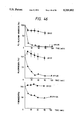
- FIG. 46 illustrates the changes with time in whole-body fibrinolytic system factor levels following rapid bolus intravenous administration of natural pro-UK and of the carbohydrate chain-added UK-S1.
- FIG. 47 shows the construction scheme for the plasmid pUKS3.
- FIG. 48 shows the construction scheme for the plasmid pSEUKS3.
- FIG. 49 graphically illustrates the time courses of thermal deactivation at 70° C. of natural pro-UK and the carbohydrate chain-added modification UK-S3.
- FIG. 50 shows the construction scheme for the plasmid pTkSJ1.
- FIG. 51 shows the construction scheme for the plasmid pTkSR18.
- FIG. 52 shows the construction scheme for the plasmid pTkSD217.
- FIG. 53 shows the construction scheme for the plasmid pTkSL11.
- FIG. 54 shows the construction scheme for the plasmid pTkSS4.
- FIG. 55 shows the construction scheme for the plasmid pTG3.
- FIG. 56 shows the construction scheme for the plasmid phPA2.
- the invention makes it possible to increase their protease resistance through carbohydrate chain addition and thus control their physiological activity.
- amino acid sequence allowing the addition of a new carbohydrate chain in polypeptides can be realized by means such as amino acid substitution in, amino acid deletion from, or amino acid insertion into the polypeptides.
- asparagine (Asn) is known to be an amino acid for linking an N-glycosylated carbohydrate chain and serine (Ser) and threonine (Thr) are each known to be amino acids suited for linking of an O-glycosylated carbohydrate chain, it is suitable for the intended purpose to locate one of these "linking" amino acids at an appropriate position in the polypeptide to be modified.
- Polypeptides modified so that they contain an amino acid to which a carbohydrate chain can be added can be obtained preferably by introducing a tripeptide of the formula Asn-X-Thr/Ser (X being any amino acid other than proline) into the polypeptides to be modified at an appropriate site.
- This tripeptide introduction can be carried out by the site-directed gene mutation technique.
- the site of carbohydrate chain addition is important. As mentioned above, carbohydrate chain addition will not occur at certain sites on polypeptides or, in some instances, addition of a new carbohydrate chain, even if it has occurred, may result in destruction of the appropriate three-dimensional structure of the polypeptide, which may lead to inhibition of membrane transport or loss of activity. Therefore, it is necessary that the site of addition of a new carbohydrate chain should be located at least at a surface site region of the polypeptide. When the three-dimensional structure of a polypeptide is already known, the surface sites of this polypeptide are apparent, so that the site or sites of addition can be readily determined.
- the site or sites of addition should be as remote from the active site as possible.
- the addition site or sites can be selected with due consideration for the above.
- the surface sites can be estimated by calculating the hydrophilicity of the polypeptide on the basis of its primary structure. It is also possible to estimate the locus or loci where a turn structure is likely formed by anticipating the secondary structure based on the primary structure by the method of Chou and Fasman [Biochemistry, 13, 211 (1974); Biochemistry, 13, 222 (1974); Adv.
- a polypeptide glycosylated in the vicinity of a protease cleavage site would be resistant to the relevant protease.
- the vicinity of a protease cleavage site thus may be said to be a very suitable carbohydrate chain addition site for stabilizing the polypeptide. It is desirable and preferable that a carbohydrate chain addition site should be introduced into a polypeptide within the range of 8 amino acid residues from a protease cleavage site.
- the polypeptide which is the subject of the present invention, may be any physiologically active polypeptide.
- colony stimulating factors granulocyte-macrophage colony stimulating factor, granulocyte colony stimulating factor, macrophage colony stimulating factor
- tissue plasminogen activator tissue plasminogen activator (t-PA)
- urokinase UK
- interferon- ⁇ , interferon- ⁇ , interferon- ⁇ , lymphotoxln, lipocortin superoxide dismutase, erythropoietin, interleukin-1, -2, -3, -4, -5, -6 and -7, and the like.
- Other physiologically active peptides amenable to this invention will suggest themselves.
- Polypeptides are glycosylated as follows. First, a DNA coding for a mutant polypeptide so modified that it has, at a desired site, for example in the vicinity of a protease cleavage site, an amino acid sequence which allows new carbohydrate chain addition is constructed by using recombinant DNA techniques. Then, the DNA is inserted into an expression vector and the resulting recombinant is introduced into microbial cells (yeast cells, fungal cells, etc.) or animal cells (CHO cells, Namalwa cells, etc.) and expression is caused, and a newly glycosylated polypeptide can be obtained.
- microbial cells yeast cells, fungal cells, etc.
- animal cells CHO cells, Namalwa cells, etc.
- the DNA For causing addition of an N-glycosylated carbohydrate chain, the DNA should be such that it contains an N-glycosylation site (Asn-X-Ser/Thr; X being any amino acid other than Pro).
- Such DNA coding for a mutant polypeptide can be constructed in the manner of site-directed mutagenesis or by using a synthetic DNA linker.
- the function or functions of a carbohydrate chain greatly depend on the structure thereof. Therefore, it is also important that the structure of the carbohydrate chain to be added should be modified so that the carbohydrate chain selected can add a better property to the glycosylation product.
- the present invention includes the process for such optimization as well.
- the methods for modifying the carbohydrate chain structure there may be mentioned the following, among others: 1) Change of the protein-producing host; 2) Cultivation of microorganism or animal cells harboring the above-mentioned recombinant plasmid in a medium containing an agent (inhibitor) that inhibits an enzyme involved in biosynthesis or processing of carbohydrate chains, such as 1-deoxynojirimycin, 1-deoxymannonojirimycin or swainsonine; and 3) Treatment of glycosylated proteins with various glycosidases, such as sialidase, ⁇ -galactosidase, ⁇ -N-acetylglucosaminidase, ⁇ -mannosidase and endoglycosidase, or glycosyltransferases, such as sialyltransferase.
- various glycosidases such as sialidase, ⁇ -galactosidase, ⁇ -N-acetylgluco
- polypeptide or glycosylated polypeptide according to the invention is hG-CSF, UK or t-PA.
- polypeptide or glycosylated polypeptide according to the invention is hG-CSF:
- hG-CSF[ND28] is more active than mature hG-CSF produced in and purified from Escherichia coli .
- hG-CSF[ND28] a portion close to the N terminus and a portion in the vicinity of the 144th (from the N terminus) amino acid are found on the polypeptide surface. Therefore, an attempt was made to add a carbohydrate chain to hG-CSF[ND28] on the 6th or 145th (from the N terminus) amino acid residue thereof.
- hG-CSF[ND28] derivative which has a carbohydrate chain addition site on the 6th (from the N terminus) amino acid residue is hG-CSF[ND28N6] and a derivative which has a carbohydrate chain addition site on the 145th (from the N terminus) amino acid residue is hG-CSF[ND28N145].
- carbohydrate chain addition can result in development of protease resistance. Retardation of blood clearance can also be expected as a result of stabilization of the polypeptide.
- the hG-CSF, hG-CSF[ND28], hG-CSF[ND28N6] and hG-CSF[ND28N145] can be produced by constructing DNAs respectively coding for hG-CSF, hG-CSF[ND28], hG-CSF[ND28N6] and hG-CSF[ND28N145] by recombinant DNA techniques, inserting them into an appropriate expression vector, introducing the resulting recombinants into animal cells and causing expression thereof in said animal cells.
- hG-CSF[ND28N6] or hG-CSF[ND28N145] among the polypeptides thus obtained a new carbohydrate chain (N-glycosylated carbohydrate chain) is added to about one third of the whole hG-CSF produced.
- a new carbohydrate chain N-glycosylated carbohydrate chain
- Comparison in protease susceptibility between the newly or additionally glycosylated species and new glycosylation-free species of hG-CSF[ND28N6] or hG-CSF[ND28N145] has revealed that the newly glycosylated species is more resistant to protease.
- hG-CSF[ND28N6] it has also been found that the additionally glycosylated species is more stable against heat as compared with the species deprived of the new carbohydrate chain enzymatically. This finding has proved the efficacy of the invention. It has further been revealed that hG-CSF[ND28], when it is an expression product in animal cells, can have an additional O-glycosylated carbohydrate chain added thereto. In this case, too, the additionally O-glycosylated species is more resistant to protease.
- polypeptide or glycosylated polypeptide according to the invention is UK or t-PA:
- Urokinase [UK) and streptokinase (SK) are currently in use as thrombolytic agents. These thrombolytic agents, however, have no affinity for fibrin, which is a thrombus component. Therefore, they must be administered in large quantities for effecting thrombolysis. Furthermore, they activate not only plasminogen adsorbed on thrombin but also plasminogen in blood, causing systemic hyperfibrinolysis and leading to a bleeding tendency. In contrast to these thrombolytic agents, tissue plasminogen activator (t-PA) and pro-urokinase (pro-UK) (inactive precursor to UK), which have affinity for fibrin, have lately attracted much attention.
- t-PA tissue plasminogen activator
- pro-UK pro-urokinase
- t-PA which has affinity for fibrin, is expected to be adsorbed specifically on thrombin and thereby dissolve thrombin efficiently, without causing systemic hyperfibrinolysis.
- t-PA which has affinity for fibrin
- Plasmin converts the single-chain form to the double-chain form.
- the double-chain form of t-PA is the active form, single-chain t-PA, too, can exhibit a fibrinolytic activity equivalent to that of double-chain t-PA when fibrin decomposition products are present.
- the affinity for fibrin it has been revealed that single-chain t-PA has greater affinity for fibrin [Tate et al.: Biochemistry, 26, 338 (1987)]. Therefore, it may be said that single-chain t-PA is higher in specificity to thrombin and is superior in this sense to double-chain t-PA.
- the thrombolytic agents t-PA and pro-UK are readily converted to disadvantageous forms under the action of proteases, such as plasmin or thrombin. Reduction of their protease susceptibility by causing carbohydrate chain addition in the vicinity of their protease cleavage utilizing the method mentioned hereinabove, if attained, would expectedly give better thrombolytic enzymes.
- DNAs respectively coding for pro-UK, UK-S1 and UK-S3 are constructed using recombinant DNA techniques and inserted into an expression vector, each resulting recombinant expression vector is introduced into animal cells, and expression is caused.
- Comparison of pro-UK and UK-S1 and UK-S3 thus obtained has revealed that UK-S1 and UK-S3 are lower in thrombin susceptibility than the natural form pro-UK and that their stability in serum and heat stability are improved, proving the efficacy of the invention also in the case of UK.
- the polypeptide or glycosylated polypeptide according to the invention is hG-CSF, UK or t-PA
- a cDNA obtained by causing reverse transcription of a messenger RNA coding for hG-CSF, UK or t-PA by using appropriate recombinant DNA techniques a DNA coding for hG-CSF, UK or t-PA as obtained from a chromosomal DNA, a synthetic DNA coding for hG-CSF, UK or t-PA, or the like may be used as the DNA coding for hG-CSF, UK or t-PA.
- the hG-CSF cDNA may be any DNA provided that it codes for hG-CSF.
- pCSF2 which has been produced by the present inventors as described in Reference Example 4.
- the hG-CSF cDNA in pCSF2 has been identified by the dideoxy sequencing method using M13 phage [J. Messing et al.: Gene, 19, 269 (1982)].
- the hG-CSF cDNA in pCSF2 contains the whole mature protein portion although a part of the signal sequence is missing therein.
- the base sequence of this mature protein portion is shown in Table 1.
- the human t-PA cDNA or human UK cDNA to be used may be any DNA provided that it codes for human t-PA or human UK.
- ptPA7, pUK1 and pUK11 are the plasmids produced by the present inventors as described in Reference Examples 1, 2 and 3, respectively.
- the human UK cDNA in pUK 1 and that in pUK11 have been sequenced by the dideoxy method using M13 phage.
- An Escherichia coli strain harboring ptPA7 has been deposited, since Sep. 3, 1987, with the Fermentation Research Institute (FRI), Agency of Industrial Science and Technology, under the designation Escherichia coli EtPA7 (deposit number FERM BP-1467), an Escherichia coli strain harboring pUK1 under the designation Escherichia coli EUK1 (FERM BP-1463), and an Escherichia coli strain harboring pUK11 under the designation Escherichia coli EUK11 (FERM BP-1464) in accordance with the Budapest Treaty.
- the plasmid to be used for insertion thereinto of a DNA coding for hG-CSF, UK or t-PA may be any plasmid provided that said DNA can be expressed in Escherichia coli or animal cells.
- Preferred for the expression of hG-CSF, UK or t-PA in Escherichia coli are plasmids which allow insertion of a foreign DNA thereinto at a site downstream from an appropriate promoter, such as a trp or lac promoter, and in which the distance between the Shin-Dalgarno sequence (hereinafter abbreviated as SD sequence) and the initiation codon (ATG) has been adjusted to an appropriate length, for example 6 to 18 bases.
- SD sequence Shin-Dalgarno sequence
- ATG initiation codon
- the plasmid to to be used for the expression of a DNA coding for hG-CSF, UK or t-PA in animal cells may be any plasmid provided that said DNA can be expressed in animal cells.
- Preferred are those plasmids which allow insertion of a foreign DNA thereinto at a site downstream from an appropriate promoter, such as the SV10 early promoter or SV40 later promoter, and which have a poly(A) signal, splicing signal and so forth.
- pAGE103 Minagami et al.: J. Biochem., 101, 1307-1310 (1987)
- pSE1PA1-9A pSE1PA1SE1dhfrl-9A (Reference Example 9)
- pSE1PA1SE1dhfrl-9A Reference Example 9
- the recombination between the DNA coding for the novel polypeptide or glycosylated polypeptide of hG-CSF, UK or t-PA and the vector DNA can be carried out by ordinary recombinant DNA techniques of digesting both DNA with restriction enzymes and conducting ligation using T4 DNA ligase.
- the ligation may also be carried out following filling in of the ends of the DNA fragments obtained by digestion with restriction enzymes using DNA polymerase I Klenow fragment or T4 DNA polymerase, or following paring off of the cohesive ends of such fragments using T4 DNA polymerase, or using a DNA linker.
- recombinant plasmids with a DNA coding for a novel polypeptide or glycosylated polypeptide of hG-CSF or UK being incorporated therein are constructed using pCSF2 as the hG-CSF cDNA-containing plasmid or pUK1 and pUK11 as the pro-UK cDNA-containing plasmids and, if necessary, using a chemically synthesized linker or applying site-directed mutagenesis.
- pAS3-3 (Reference Example 10) is cleaved with MluI. and ApaLI and a DNA fragment about 3.0 kb in size is purified.
- the same plasmid is cleaved with AatII and MluI and a DNA fragment about 6.3 kb in size is purified.
- pCfBD28 (cf. Reference Example 16) is cleaved with AatII and XhoI and a DNA fragment about 0.3 kb in size is purified.
- pAS3-3 is cleaved with MluI and ApaLI and a DNA fragment about 3.0 kb in size is purified.
- pAS28 is cleaved with XhoI and MluI and a DNA fragment about 6.55 kb in size is purified.
- pASN145 The construction of pASN145 is carried out using site-directed mutagenesis. As shown in FIG. 7-(1), pCfBD28 is cleaved with PvuII and BamHI and a DNA fragment about 0.94 kb in size is purified. Separately, the M13 phage vector M13mp19 RF DNA is cleaved with SmaI and BamHI and a DNA fragment about 7.24 kb in size is purified. The thus-obtained two DNA fragments are ligated together in the presence of T4 DNA ligase to give pt19BD28C. Then, this pt19BD28C is used to transfect Escherichia coli JM105.
- Single-stranded pt19BD28C is obtained from the phage obtained above.
- the M13mp19 RF DNA is cleaved with HindIII and EcoRI and a DNA fragment about 7.2 kb in size is purified.
- This DNA fragment (about 7.2 kb) and the single-stranded pt19BD28C obtained above are mixed together and subjected to denaturing treatment, followed by annealing.
- the thus-formed gapped duplex DNA is purified.
- the annealing product is circularized using the Klenow fragment and T4 DNA ligase.
- the resulting circular DNA is used to transfect Escherichia coli JM105 to give pt19BD28CN145 with site-directed mutagenesis introduced therein.
- pt19BD28CN145 is cleaved with BglI and BamHI and a DNA fragment about 0.85 kb in size is purified.
- pCfBD28 is cleaved with BamHI and BglI and a DNA fragment about 3.25 kb in size is purified. The two DNA fragments thus obtained are ligated together in the presence of T4 DNA ligase to give pCfBD28N145.
- pCfBD28N145 is cleaved with BanIII and BamHI and a DNA fragment about 1.3 kb in size is purified.
- the thus-obtained DNA fragment (about 1.3 kb) is cleaved with DdeI, treated with DNA polymerase Klenow fragment for filling in the cohesive ends and then further cleaved with AatII, and a DNA fragment about 0.2 kb in size is purified.
- pAS28 is cleaved with AatII an XhoI and a DNA fragment about 0.8 kb in size is purified.
- pSE1PA1SEldhfr1-9A (Reference Example 9) is cleaved with SmaI and XhoI and a DNA fragment about 8.7 kb in size is purified.
- the thus-obtained three DNA fragments (about 0.2 kb, about 0.8 kb and about 8.7 kb in size) are ligated together in the presence of T4 DNA ligase to give pASN145.
- pUK1 (Reference Example 2) is cleaved with PstI and BamHI and a DNA fragment 890 bp in size is purified.
- the M13mp18 RF DNA [Yanisch-Perron et al.: Gene, 33, 103 (1985)] is cleaved with PstI and BamHI and a DNA fragment about 7.2 kb is purified. Both the DNA fragments thus obtained are ligated together in the presence of T4 DNA ligase to give a plasmid, pUKmpS1, with a part of the UK cDNA subcloned in the M13mp18 vector.
- the single-stranded pUKmpS1 DNA is prepared in the conventional manner and then, as shown in FIG. 10, its base sequence is modified so that an N-glycosylated carbohydrate chain can be added onto the 164th (in UK) amino acid residue.
- the synthetic DNA 5'-GGGGAGAAAACACCACC-3' for substituting Asn for Phe-164 and the synthetic DNA 5'-GTTTTCCCAGTCACGAC-3' for determining the DNA base sequence of M13mp18 as primers
- the single-strand pUKmpS1 DNA is converted to the double-stranded DNA in the presence of Escherichia coli DNA polymerase I Klenow fragment and at the same time mutation is introduced into the desired site.
- the thus-obtained mutant double-stranded DNA is cleaved with PstI and EcoRI and a DNA fragment about 600 bp in size is purified.
- UK cDNA-containing pUK11 (Reference Example 3) is cleaved with AatII and PstI and a DNA fragment about 1.0 kb in size is purified.
- pUKB101 (Reference Example 12) with a KpnI site introduced therein on the 3' terminal side of the UK cDNA is cleaved with AatII and EcoRI and a DNA fragment about 2.9 kb in size is purified.
- the thus-obtained three DNA fragments are ligated together in the presence of T4 DNA ligase to give recombinant plasmid, pUKS1, coding for the UK derivative UK-S1.
- the expression vector for a foreign gene containing the dhfr gene for gene amplification pSE1PA1SEldhfr1-9A (Reference Example 9) is cleaved with KpnI and XhoI and a DNA fragment about 8.6 kb in size is purified.
- the recombinant plasmid for UK expression pSE1UKprol-1A (Reference Example 13) is cleaved with XhoI and BglII and a DNA fragment about 0.75 kb is size is purified.
- pUKS1coding for UK-S1 is cleaved with KpnI and BglII and a DNA fragment about 1.15 kb in size is purified. The thus-obtained three DNA fragments are ligated together using T4 DNA ligase to give a recombinant plasmid, pSE1UKS1-1d, allowing UK-S1 expression.
- Recombinant plasmid pSEUKS3 coding for UK-S3 can be produced by similar method for obtaining pSE1UKS1-1d.
- reaction conditions for use in the above-mentioned recombination techniques are generally as follows.
- the DNA digestion reaction with an restriction enzyme or enzymes is generally carried out in a reaction mixture containing 0.1 to 20 ⁇ g of DNA, 2 to 200 mM (preferably 10 to 40 mM) Tris-HCl (pH 6.0 to 9.5, preferably pH 7.0 to 8.0), 0 to 200 mM NaCl and 2 to 20 mM (preferably 5 to 10 mM) MgCl 2 in the presence of 0.1 to 100 units (preferably 1 to 3 units per microgram of DNA) of each restriction enzyme at 20° to 70° C. (the optimal temperature varying depending on the kind of restriction enzyme) for 15 minutes to 24 hours.
- the reaction is terminated generally by heating at 55° to 75° C. for 5 to 30 minutes. It is also possible to inactivate the restriction enzymes used by means of phenol, diethyl pyrocarbonate or the like reagent.
- LGT method low melting point agarose gel electrophoresis
- AFT method agarose gel freezing-thawing method
- the AFT method comprises adding an equal volume of TE buffer [10 mM Tris-HCl (pH 7.5), 1 mM EDTA] and 2 volumes of phenol (saturated with TE buffer) to a slice of DNA fragment-containing agarose gel (0.7 to 1.5%), achieving admixture, repeating a freezing (-70° C.)-thawing (65° C.) cycle two times, centrifuging, separating the resulting upper aqueous solution layer and recovering the DNA fragment by precipitation with ethanol.
- the DNA fragments can also be eluted and purified from agarose gel or polyacrylamide gel by using an Atto model Maxfield AE-3241 DNA fragment recoverer. The latter method is hereinafter referred to as "electroelution method".
- the ligation of DNA fragments is conducted in a reaction medium containing 2 to 200 mM (preferably 10 to 40 mM) Tris-HCl (pH 6.1 to 9.5, preferably pH 7.0 to 8.0), 2 to 20 mM (preferably 5 to 10 mM) MgCl 2 , 0.1 to 10 mM (preferably 0.5 to 2.0 mM) ATP and 1 to 50 mM (preferably 5 to 10 mM) dithiothreitol (hereinafter sometimes referred to as "DTT”) in the presence of 1° to 1,000 units of T4 DNA ligase at 1 to 37° C. (preferably 3° to 20° C.) for 15 minutes to 72 hours (preferably 2 to 20 hours).
- Tris-HCl pH 6.1 to 9.5, preferably pH 7.0 to 8.0
- 2 to 20 mM preferably 5 to 10 mM
- MgCl 2 preferably 5 to 10 mM
- 0.1 to 10 mM preferably 0.5 to 2.0 mM
- the recombinant plasmid DNA resulting from the ligation reaction is introduced into Escherichia coli using the transformation method of Cohen et al. [S. N. Cohen et al.: Proc. Natl. Acad. Sci. USA, 69, 2110 (1972)] or the transformation method of Hanahan [J. Mol. Biol., 166, 557 (1983)], as necessary.
- the recombinant M13 phage RF DNA resulting from the ligation reaction is introudced into Escherichia coli JM105 [J. Messing et al.: Gene, 33, 103 (1985)] using the known transfection method [Y. Kuchino et al.: Tanpakushitsu, Kakusan, Koso, 29, 294 (1984)] as necessary.
- Single-strand DNA isolation from the recombinant M13 phage is performed by the known method [Y. Kuchino et al.: Tanpakushitsu, Kakusan, Koso, 29, 294 (1984)].
- deoxyoligonucleotides to be used in the practice of the invention can be synthesized by solid-phase synthesis by the phosphoric amidite method [S. L. Beaucage et al.: Tetrahedron Lett., 22, 1859 (1981); L. J. BcBrie et al.: ibid , 24, 245 (1983)] using an Applied Biosystems model 380A DNA synthesizer (Applied Biosystems Inc., Foster City, Calif. 94404).
- T4 kinase buffer 50 mM Tris-HCl (pH 7.6), 10 mM MgCl 2 , 5 mM DTT, 0.1 mM EDTA, 0.5 mM ATP] in the presence of 5 units of T4 DNA kinase.
- the deoxyoligonucleotide is radiolabeled at the 5' end by using 20 to 50 ⁇ Ci of [Y- 32 P]ATP (3,000 Ci/mmol; Amersham, Arlington Heights, II) in place of 0.5 mM ATP in the above-mentioned T4 kinase buffer.
- Structural analysis of plasmid DNAs is performed by digesting each plasmid with 1 to 10 different restriction enzymes and then checking cleavage sites by agarose gel electrophoresis or polyacrylamide gel electrophoresis. Determination of the base sequence of a DNA, if necessary, can be made by the dideoxy sequencing method using M13 phage.
- the polypeptide or glycosylated polypeptide according to the invention can be produced using Escherichia coli or animal cells as hosts.
- Escherichia coli include the strain K-12, NY49, HB101 and C600SF8.
- Examples of the host aminal cells include a Chinese hamster ovary (CHO) cell and a Namalwa cell.
- novel hG-CSF polypeptide of the present invention is produced using Escherichia coli as the host in the following manner.
- Escherichia coli K-12 MM294 [Backman K., et al., Proc. Natl. Acad. Sci. USA., 73, 4174 (1976)] is transformed with a plasmid (e.g. pCfBD28N145) and an Escherichia coli strain harboring pCfBD28N145 is selected from among the ampicillin-resistant (Ap r ; hereinafter the same shall apply) colonies obtained.
- the pCfBD28N145-bearing Escherichia coli strain is cultivated in a medium, whereby the novel hG-CSF polypeptide is produced in the culture.
- the medium to be used here may be either a synthetic one or a nature-derived one provided that it is suited for the growth of Escherichia coli and the production of the novel hG-CSF polypeptide.
- usable as the carbon source are glucose, fructose, lactose, glycerol, mannitol, sorbitol and the like.
- Usable nitrogen sources are NH 4 Cl, (NH 4 ) 2 SO 4 , casamino acids, yeast extract, polypeptone, meat extract, Bacto-tryptone, corn steep liquor and the like.
- Usable as other nutrient sources are K 2 HPO 4 , KH 2 PO 4 , NaCl, MgSO 4 , vitamin B1, MgCl 2 , etc.
- the cultivation is carried out at pH 5.5 to 8.5, at a temperature of 18° to 40° C., with aeration and stirring. After 5 to 90 hours of cultivation, the accumulation of the novel hG-CSF polypeptide in cultured cells becomes substantial. Cells are then harvested from the culture and disrupted by sonication, and the cell detritus mass is recovered by centrifugation.
- the novel hG-CSF polypeptide can be extracted from the cell detritus mass, followed by purification, solubilization and renaturation, by the method of Marston et al. [F. A. O. Marston et al.: BIO/TECHNOLOGY, 2, 800 (1984)] or by the method of Pennica et al. [Nature, 301, 214 (1983)] or by the method of Winkler et al. [BIO/TECHNOLOGY, 3, 990 (1985)].
- the host to be used for the expression of a novel hG-CSF polypeptide or a novel glycosylated hG-CSF polypeptide may be any animal cell line provided that it allows expression of said polypeptide or glycosylated polypeptide.
- preferred specific animal cells there may be mentioned, among others, dhfr-deficient CHO cells [G. Urlaub and L. A. Chasin: Proc. Natl. Acad. Sci. USA, 77, 4216 (1980)].
- the plasmid pASN6 is introduced into dhfr-deficient CHO cells, for example by the calcium phosphate method [Graham and Van der Eb: Virology, 52, 546 (1978)].
- a transformant harboring pASN6 can be selected, for example by using MEM ALPHA medium (ribonucleic acid- and deoxyribonucleic acid-free; Gibco-Oriental) containing G418 and dialyzed fetal calf serum. It is also possible to select a transformant strain in which the novel hG-CSF polypetide gene has been amplified from among transformants by using methotrexate. The transformant strain thus obtained is grown in a medium, whereby the novel hG-CSF polypeptide or novel glycosylated hG-CSF polypeptide is formed in the culture.
- the medium to be used is, for example, HAM F10 medium or HAM F12 medium (Flow Laboratories), Dulbecco's MEM medium or RPMI-1640 medium (Nissui Pharmaceutical) or MEM ALPHA medium, each supplemented with a serum (e.g. fetal bovine serum), or a mixed medium composed of two or more of these.
- a serum e.g. fetal bovine serum
- glutamine 0.5 to 5 mM
- an antibiotic penicillin (25 U/ml), streptomycin (25 ⁇ g/ml), G418 (0.3 mg/ml), etc.
- sodium bicarbonate 0.01%
- the cultivation is generally carried out at a seed cell density of 5 ⁇ 10 4 to 1 ⁇ 10 6 cells/ml and at a temperature of 30° to 40° C. In 2 to 10 days of cultivation, the substance of the invention is secreted for the most part extracellularly.
- Cells are removed from the culture by centrifugation and the novel hG-CSF polypeptide or novel glycosylated polypeptide is extracted from the supernatant obtained by centrigugation.
- novel derivatives of UK or any other proteins can be produced in the same manner.
- the activity measurement of the hG-CSF derivatives obtained in the above manner is performed as follows.
- Bone marrow cells are taken aseptically from the femur of male C3H/He mice (8 to 12 weeks of age; Shizuoka Laboratory Animal Center) and suspended in ⁇ -minimum essential medium (Flow Laboratories; hereinafter abbreviated as " ⁇ -MEM medium") supplemented with 10% fetal bovine serum (FBS).
- ⁇ -MEM medium Flow Laboratories; hereinafter abbreviated as " ⁇ -MEM medium”
- FBS fetal bovine serum
- the bone marrow hematopoietic stem cell colony forming potential is measured by the method of Okabe et al. [T. Okabe et al.: Cancer Research, 44, 4503-4506 (1986)].
- 0.2 ml of the bone marrow cell suspension (2 ⁇ 10 6 cells/ml) prepared as described above is added to a mixture of 0.2 ml of ⁇ -MEM, 0.4 ml of FBS and 0.2 ml of each sample after two serial dilutions. Thereto is added an equal volume (1.0 ml) of a 0.6% solution of agar (Difco, Agar purified #0560-01) maintained at 42° C.
- the potency of each sample is calculated based on the result of counting for the sample after two serial dilutions as obtained in the colony forming test, as follows.
- the activity giving a value half the maximum colony count value found with intact G-CSf used as a standard is defined as 50 units.
- the activity calculated to this scale is multiplied by 20 considering the dilution factor for each sample and for expressing the activity on the per-milliliter basis.
- the product obtained is reported as the potency (in units).
- the specific activity is expressed in terms of potency per unit weight (mg) of protein, namely in units/mg.
- the hG-CSF protein content is determined by enzyme-linked immunosorbent assay ELISA) using anti-hG-CSF monoclonal antibody.
- enzyme-linked immunosorbent assay ELISA enzyme-linked immunosorbent assay ELISA
- standard hG-CSF produced in Escherichia coli and purified and assayed by the Lowry method is used as a standard substance.
- the anti-hG-CSF monoclonal antibody is prepared by the method of Hanai et al. [Cancer Res., 46, 4438 (1986)].
- the t-PA or UK activity can be determined by fibrin plate assay [Granelli-Piperno and Reich: J. Exp. Med., 148, 223 (1978)].
- the cases where the polypeptide or glycosylated polypeptide according to the invention is an hG-CSF or UK are mentioned.
- Examples 1 to 5 are illustrative of the case in which the polypeptide or glycosylated polypeptide according to the invention is an hG-CSF and
- Examples 6 to 8 are illustrative of the case in which said polypeptide or glycosylated polypeptide is a UK.
- a 2 ⁇ g portion of pAS3-3 obtained in Reference Example 10 was dissolved in 20 ⁇ l of 10 mM Tris-HCl buffer (pH 7.5) containing 7 mM MgCl 2 , 6mM 2-mercaptoethanol and 150 mM NaCl (hereinafter such buffer will be referred to as "Y-150 buffer” for short), 10 units of the restriction enzyme MluI (Takara Shuzo; hereinafter, unless otherwise specified, all the restriction enzymes used were obtained from Takara Shuzo) was added, and digestion was carried out at 37° C. for 2 hours. Then, 5 units of ApaLI was added and partial digestion was further effected at 37° C. for 10 minutes. About 0.5 ⁇ g of a DNA fragment about 3.0 kb in size was purified and recovered from the digestion reaction mixture by the LGT method.
- the two single-strand 43-mer DNAs were synthesized using an Applied Biosystems model 380A DNA synthesizer. Then, 20 picomoles each of the synthesized DNAs (two 43-mers) were dissolved in 40 ⁇ l of 50 mM Tris-HCl buffer (pH 7.5) containing 10 mM MgCl 2 , 5 mM dithiothreitol, 0.1 mM EDTA and 1 mM ATP (hereinafter such buffer will be referred to as "T4 kinase buffer” for short), 30 units of T4 polynucleotide kinase (Takara Shuzo; hereinafter the same shall apply) was added, and phosphorylation was carried out at 37° C. for 60 minutes.
- T4 kinase buffer 30 units of T4 polynucleotide kinase
- a 0.5 ⁇ g portion of the pAS3-3-derived MluI-ApaLI fragment (about 3.0 kb), 1.0 ⁇ g of the pAS3-3-derived AatII-MluI fragment (about 6.3 kb) and 0.1 ⁇ g of the pCfBD28-derived AatII-XhoI fragment (about 0.3 kb) were dissolved in 25 ⁇ l of 20 mM Tris-HCl buffer (pH 7.6) containing 10 mM MgCl 2 , 10 mM dithiothreitol and 1 mM ATP (hereinafter such buffer will be referred to as "T4 ligase buffer" for short) and about 1 picomole of the above DNA linker was added to the solution.
- T4 ligase buffer for short
- the recombinant plasmid mixture obtained was used to transform Escherichia coli HB101 [Bolivar et al.: Gene, 2, 75 (1977)] by the method of Cohen et al. [S. N. Cohen et al.: Proc. Natl. Acad. Sci. USA, 69, 2110 (1972)] (hereinafter it is this method that was used for transforming Escherichia coli strains) and ampicillin (Ap)-resistant strains were obtained.
- a plasmid DNA was isolated from one of these transformant strains by the known method [H. C. Birnboim et al.: Nucleic Acids Res., 7, 1513 (1979)] (hereinafter it is this method that was used for isolating plasmid DNAs).
- pAS28 The structure of the plasmid obtained was confirmed by restriction enzyme digestion and by sequencing by the dideoxy method using M13 phage, this plasmid was named pAS28 (cf. FIG. 5).
- a microorganism harboring the plasmid pAS28 has been deposited since Sep. 24, 1988 with the Fermentation Research Institute, Agency of Industrial Science and Technology (FRI) under the designation Escherichia coli EAS28 (deposit number FERM BP-2069) in accordance with the Budapest Treaty.
- the polypeptide (hG-CSF derivative) encoded by the plasmid is distinguished from mature hG-CSF by the following amino acid residue substitutions:
- hG-CSF[ND28] The polypeptide (hG-CSF derivative) encoded by pAS28 is hereinafter referred to as "hG-CSF[ND28]".
- a 2 ⁇ g portion of pAS3-3 obtained in Reference Example 10 was dissolved in 20 ⁇ l of Y-150 buffer, 10 units of the restriction enzyme MluI was added, and digestion was carried out at 37° C. for 2 hours. Then, 5 units of ApaLI was added and partial digestion was further conducted at 37° C. for 10 minutes. About 0.5 ⁇ g of a DNA fragment about 3.0 kb in size was purified and recovered from the reaction mixture by the LGT method.
- Example 2 2 ⁇ g of pAS28 obtained in Example 1 was dissolved in 20 ⁇ l of Y-150 buffer, 10 units each of the restriction enzymes XhoI and MluI were added, and digestion was carried out at 37° C. for 2 hours.
- a DNA fragment (1.0 ⁇ g) about 6.55 kb in size was purified and recovered from the reaction mixture by the LGT method.
- the single-strand DNAs (two 43-mers) were synthesized using an Applied Biosystems model 380A DNA synthesizer.
- the recombinant plasmid-containing ligation reaction mixture was used to transform Escherichia coli HB101 by the method of Cohen et al. (vide supra) and Ap-resistant strains were obtained.
- a plasmid DNA was separated and purified from one of these transformant strains by the known method. The structure of said plasmid DNA was confirmed by restriction enzyme digestion and by the dideoxy sequencing method using M13 phage.
- This plasmid was named pASN6.
- a microorganism harboring the plasmid pASN6 has been deposited since Sep. 24, 1988 with the Fermentation Research Institute under the designation Escherichia coli EASN6 (deposit number FERM BP-2070) in accordance with the Budapest Treaty.
- the polypeptide (hG-CSF derivative) encoded by the plasmid is distinguished from mature hG-CSF by the following amino acid residue substitutions:
- hG-CSF [ND28N6] The polypeptide (hG-CSF derivative) encoded by pASN6 is hereinafter referred to as "hG-CSF [ND28N6]".
- a 3 ⁇ g portion of pCfBD28 (cf. Reference Example 16) was dissolved in 20 ⁇ l of 10 mM Tris-HCl buffer (pH 7.5) containing 7 mM MgCl 2 and 6 mM 2-mercaptoethanol (such buffer is hereinafter referred to as "Y-0 buffer” for short), 10 units of the restriction enzyme PvuII was added, and digestion was carried out at 37° C. for 2 hours. Then, NaCl was added to an NaCl concentration of 100 mM, 10 units of BamHI was added, and reaction was carried out at 37° C. for 2 hours.
- the above ligation reaction mixture was used to transfect Escerichia coli JM105 by the known method [Messing et al.: Methods in Enzymology, 101, 20 (1983)] and recombinant phages were obtained.
- a recombinant 13 phage RF DNA was recovered from cultured cells of one recombinant phage-infected Escherichia coli JM105 strain by the same method as the plasmid DNA recovering method.
- the structure of this RF DNA [named pt19BD28C) was confirmed by cleavage with BamHI, EcoRI and BglI, followed by polyacrylamide gel electrophoresis.
- the single-strand pt19BD28C DNA was recovered from the recombinant phage by the above-mentioned known method and used as a template.
- a 3- ⁇ g portion of the M13mp19 RF DNA was dissolved in 30 ⁇ l of 10 mM Tris-HCl buffer (pH 7.5) containing 7 mM MgCl 2 , 6 mM 2-mercaptoethanol and 100 mM NaCl (hereinafter such buffer is referred to as "Y-100 buffer” for short), 10 units each of the restriction enzymes EcoRI and HindIII, and digestion was carried out at 37° C. for 2 hours. About 2.5 ⁇ g of a DNA fragment (EcoRI-HindIII fragment) about 7.2 kb in size was obtained from the digestion reaction mixture by the LGT method.
- the gapped duplex DNA formed was recovered by the LGT method.
- the single-strand DNA was synthesized using an Applied Biosystems model 380A DNA synthesizer. A 1- ⁇ g portion of the single-strand DNA synthesized was dissolved in 50 ⁇ l of T4 kinase buffer, 30 units of T4 polynucleotide kinase was added, and phosphorylation was carried out at 37° C. for 60 minutes.
- the resultant reaction mixture was used to transfect Escherichia coli JM105 and mutant phages were obtained.
- RF DNAs were recovered from mutant phage-infected Escherichia coli JM105 strains and examined for their structure by restriction enzyme cleavage and by sequencing by the dideoxy method using M13 phage.
- the desired mutant RF DNA was named pt19BD28CN145.
- a 3- ⁇ g portion of pt19BD28CN145 obtained as described in the preceding section was dissolved in 50 ⁇ l of Y-100 buffer, 10 units each of the restriction enzymes BglI (Boehringer Mannheim) and BamHI were added, and digestion was carried out at 37° C. for 2 hours. From the reaction mixture, there was obtained by the LGT method 0.4 ⁇ g of a DNA fragment (BglI-BamHI fragment) about 0.85 kb in size and containing the mutation site introduced as described in the preceding section.
- BglI Boehringer Mannheim
- the ligation reaction mixture was used to transform Escherichia coli HB101 and Ap-resistant strains were obtained. Plasmid DNAs were isolated from the transformants and analyzed for their structure by cleavage with restriction enzymes. The plasmid DNA having the desired structure was named pCfBD28N145.
- DNA was recovered by ethanol precipitation following exatraction with a mixture of equal volumes of phenol and chloroform (hereinafter referred to as "phenol-chloroform extraction") and dissolved in 30 ⁇ l of Klenow buffer, 2 units of DNA polymerase I Klenow fragment was added, and reaction was carried out at 37° C. for 1 hour. After 10-minute treatment at 68° C. for inactivating the DNA polymerase I Klenow fragment, DNA was recovered by ethanol precipitation. The DNA recovered was dissolved in 20 ⁇ l of K-50 buffer, 10 units of the restriction enzyme AatII (Toyobo) was added, and digestion was carried out at 37° C. for 2 hours. About 0.1 ⁇ g of a DNA fragment [DdeI (blunt end)-AatII fragment] about 0.2 kb in size was obtained from the reaction mixture by the LGT method.
- Example 2 2 ⁇ g of pAS28 obtained in Example 1 was dissolved in 20 ⁇ l of K-50 buffer, 10 units of the restriction enzyme AatII (Toyobo) was added, and digestion was conducted at 37° C. for 2 hours. Thereafter, 5 units of the restriction enzyme XhoI was added and partial digestion was further effected at 37° C. for 10 minutes. About 0.1 ⁇ g of a DNA fragment AatII-XhoI fragment) about 0.8 kb in size was obtained from the reaction mixture by the LGT method.
- AatII Toyobo
- the ligation reaction mixture was used to transform Escherichia coli HB101 and Ap-resistant strains were obtained. Plasmids were isolated from the transformants and analyzed for their structure by cleavage with restriction enzymes and, as a result, a plasmid DNA having the desired structure, pASN145, was obtained. A microorganism harboring the plasmid pASN145 has been deposited, since Sep. 24, 1988, with the Fermentation Research Institute under the designation Escherichia coli EASN145 (deposit number FERM BP-2071) in accordance with the Budapest Treaty.
- the polypeptide (hG-CSF derivative) encoded by the plasmid is distinguished from mature hG-CSF by the following amino acid residue substitutions:
- hG-CSF[ND28N145] The polypeptide (hG-CSF derivative) encoded by pASN145 is hereinafter referred to as "hG-CSF[ND28N145]".
- Example 1 pAS28 obtained in Example 1 was introduced into dhfr-deficient CHO cells by the calcium phosphate method.
- MEM ALPHA medium containing ribonucleic acid and deoxyribonucleic acid; Gibco-Oriental
- FCS ribonucleic acid and deoxyribonucleic acid
- 1/50 volume of 7.5% NaHCO 3 solution Flow Laboratories
- MEM ⁇ nonselective medium
- the whole DNA solution was added to dhfr-deficient CHO cells prepared from the above-mentioned culture by discarding the medium, adding a fresh 10-ml portion of MEM ⁇ (nonselective medium) and incubating for 1 hour. The resultant mixture was incubated for 8 hours. Cells were then washed with PBS, 5 ml of MEM ⁇ (nonselective medium) was added, and incubation was continued for 16 hours.
- MEM ALPHA medium ribonucleic aicd- and deoxyribonucleic acid-free supplemented with 10% dialyzed FCS (Gibco-Oriental), 1/50 volume 7.5% NaHCO 3 , 1/100 volume 100 ⁇ nonessential amino acid solution and 0.3 mg/ml G418 (Gibco-Oriental) [hereinafter such medium is referred to as "MEM ⁇ (selective medium)" for short] was added and, after causing the cells to be suspended well in said medium, the cells were cultured in a CO 2 incubator at 37° C. for 5 days using a dish 10 cm in diameter.
- the cells were washed with PBS, then MEM ⁇ (selective medium) was added, and incubation was continues for 5 days. After the same procedure was followed, incubation was further continued for 5 days.
- the cells were washed with PBS, then treated with trypsin, suspended in 10 ml of MEM ⁇ (selective medium) by adding the medium, and cultured in a CO 2 incubator at 37° C. for 3 to 7 days using a dish 6 cm in diameter.
- Colonies that had appeared were treated with trypsin and then inoculated into a dish, 10 cm in diameter, to a concentration of 5 ⁇ 10 4 cells/ml using 10 ml of MEM ⁇ (selective medium) containing 50 nM methotrexate (hereinafter referred to as "MTX" for short).
- MEM ⁇ selective medium
- MTX methotrexate
- Medium exchange was made three times at 5-day intervals using the above-mentioned medium.
- MTX-resistant colonies that had appeared were monoclonally separated and each cultured until confluence using a dish 6 cm in diameter. Thereafter, the medium was replaced with FCS-free MEM ⁇ (selective medium) and, after further 2 days of incubation, the production of hG-CSF[ND28] in the culture fluid was examined.
- the production was highest in clone No. 22, namely 10 ⁇ g hG-CSF[ND28] per 10 6 cells/2 days.
- This clone was cultured in a Falcon 3027 roller bottle containing 100 ml of MEM ⁇ (selective medium) containing 50 nM MTX. After the culture cells became confluent, incubation was continued for 3 days using the above-mentioned FCS-free medium. This 100-ml culture fluid was used in Example 5.
- clone No. 16 showed the highest production of 7 ⁇ g/10 6 cells/2 days.
- This clone was cultured in a Falcon 3027 roller bottle containing 100 ml of MEM ⁇ (selective medium) containing 50 nM MTX and, after confluency was attained, cultivation was continued for 3 days using the above-mentioned FCS-free medium. This 100-ml culture fluid was used in Example 5.
- clone No. 9 showed the highest production of 7 ⁇ g/10 6 cells/2 days.
- This clone was cultured in a Falcon 3027 roller bottle containing 100 ml of MEM ⁇ (selective medium) containing 50 nM MTX and, after the culture cells became confluent, cultivation was continued for 3 days using the above-mentioned FCS-free medium. This 100-m culture fluid was used in Example 5.
- clone No. 5 showed the highest production of 10 ⁇ g/10 6 cells/2 days.
- This clone was cultured in a Falcon 3027 roller bottle containing 100 ml of MEM ⁇ (selective medium) containing 50 nM MTX and, after the culture cells became confluent, cultivation was continued for 3 days using the above-mentioned FCS-free medium.
- the 100-ml culture fluid thus obtained was used in Example 5 as a sample containing natural hG-CSF.
- FIG. 8-(1) The patterns obtained by silver staining (using Wako Pure Chemical Industries' silver staining kit) following electrophoresis are shown in FIG. 8-(1).
- FIG. 8-(2) shows the results of enzyme-labeled antibody staining [K. Tabe: Saibo Kogaku (Cell Technology), 2, 1061 (1983)] using an anti-hG-CSF monoclonal antibody following transfer of the proteins on the same gel to a nitrocellulose membrane.
- the anti-hG-CSF monoclonal antibody was prepared by the method of Hanai et al. [Cancer Res., 46, 4438 (1986)].
- hG-CSF or hG-CSF produced in CHO cells is glycosylated through addition of one O-glycosylated carbohydrate chain to the 133rd (from the N terminus) amino acid Thr residue. It is also known that the carbohydrate chain involved in said addition includes two species, one containing one sialic acid residue and the other containing two sialic acid residues [Oeda et al.: J. Biochem., 103, 544 (1988)]. In the present study, too, hG-CSF produced in CHO cells was found to have one O-glycosylated carbohydrate chain added thereto.
- hG-CSF[ND28] produced in CHO cells was composed of two species, one having one O-glycosylated carbohydrate chain and the other having two such carbohydrate chains.
- hG-CSF[ND28N6] and hG-CSF[ND28N145] differing from hG-CSF[ND28] by having one newly introduced N-glycosylation site, about one third of the whole amount of hG-CSF produced had an N-glycosylated carbohydrate chain added thereto.
- hG-CSF[ND28N145] included four polypeptide species differing in the kind and/or number of carbohydrate chains added: namely one having one O-glycosylated carbohydrate chain, like the natural type; one having two O-glycosylated carbohydrate chains; one having one O-glycosylated carbohydrate chain and one N-glycosylated carbohydrate chain; and one having two O-glycosylated carbohydrate chains and one N-glycosylated carbohydrate chains.
- hG-CSF[ND28N6] showed almost no new O-glycosylated carbohydrate chain addition.
- FIG. 8-(3) is a schematic representation of FIG. 8-(2) and indicates the kind(s) and number of carbohydrate chains for each band.
- hG-CSF[ND28], hG-CSF[ND28N6] and hG-CSF[ND28N145] produced in CHO cells each included a species having an additional carbohydrate chain or chains added thereto and a species having no additional carbohydrate chain. Therefore, chymotrypsin was directly added to the culture supernatant containing both species (one having a new carbohydrate chain or chains and one having no such additional carbohydrate chain) obtained as described in the preceding section and the effects of the presence and absence of such additional carbohydrate chain or chains on protease resistance were compared.
- samples were taken from the mixture each time in an amount of 60 ⁇ l and the reaction was terminated by adding 20 ⁇ l of SDS-polyacrylamide gel electrophoresis buffer [0.25M Tris-HCl (pH 6.8), 8% sodium lauryl sulfate (SDS), 40% glycerol and 0.004% bromphenol blue].
- SDS-polyacrylamide gel electrophoresis buffer [0.25M Tris-HCl (pH 6.8), 8% sodium lauryl sulfate (SDS), 40% glycerol and 0.004% bromphenol blue].
- SDS-polyacrylamide gel electrophoresis buffer 0.25M Tris-HCl (pH 6.8), 8% sodium lauryl sulfate (SDS), 40% glycerol and 0.004% bromphenol blue.
- hG-CSF[ND28] the species having two O-glycosylated carbohydrate chains was more resistant to chymotrypsin than the species (natural type) having one such chain [FIG. 8-(4)].
- hG-CSF[ND28N6] the species having an additional N-glycosylated carbohydrate chain was more resistant to said enzyme than the species having no such carbohydrate chain [FIG. 8-(5)].
- hG-CSF[ND28N145] comprising four kinds of polypeptide, namely the species having one O-glycosylated carbohydrate chain (natural type), that having an additional O-glycosylated carbohydrate chain, that having an additional N-glycosylated carbohydrate chain and that having both additional carbohydrate chains, those having an additional N-glycosylated carbohydrate chain were more resistant than others.
- the species having an additional O-glycosylated carbohydrate chain as well as an additional N-glycosylated carbohydrate chain was the most resistant [FIG. 8-(6)].
- a 5-ml portion of the hG-CSF[ND28N6] -containing, serum-free, culture supernatant obtained in Example 4-(2) was concentrated to 500 ⁇ l using a Molcut-10 membrane (Millipore).
- a 100- ⁇ l portion of the concentrate was applied to a Superose 12 column (Pharmacia) (1 cm ⁇ 30 cm) and the N-glycosylated hG-CSF[ND28N6] species was isolated.
- 0.1M Tris-HCl buffer (pH 8.0) containing 0.2M NaCl and 1 mM EDTA was used. The buffer was passed through the column at a flow rate of 0.5 ml/minute.
- both reaction mixtures were used in a comparative thermal stability test at 56° C. Thus, 60 ⁇ l of each reaction mixture was maintaind at 56° C. After 0, 30, 120, 240 and 360 minutes, sampling (10 ⁇ l per sampling) was made and hG-CSF activity measurement was performed by colony forming ability testing using mouse bone marrow hematopoietic stem cells. The results obtained are shown in FIG. 8-(7). In the figure, the activity is shown in terms of residual activity with the activity after 17.5 hours of incubation at 37° C. being taken as 100%. The activity after 17.5 hours of incubation at 37° C. was 48.1% for the N-glycanase-treated sample and 66.8% in the control when the activity before incubation was taken as 100%.
- M13mp18 RF DNA M13 phage vector; Takara Shuzo
- Y-100 buffer 10 units each of the restriction enzymes PstI and BamHI were added, and digestion was performed at 37° C. for 2 hours.
- PstI and BamHI 10 units each of the restriction enzymes PstI and BamHI were added, and digestion was performed at 37° C. for 2 hours.
- a DNA fragment about 7.2 kb was purified by the AFT method.
- the thus-obtained pUK1-derived 890-bp DNA fragment and M13mp18 RF DNA-derived 7.2-kb DNA fragment were dissolved in a total volume of 20 ⁇ l of T4 ligase buffer, 300 units of T4 DNA ligase was added, and ligation was carried out at 4° C. for 18 hours.
- the above ligation reaction mixture was used to transfect Escherichia coli JM105 by the known method [Messing et al.: Methods in Enzymology, 101, 20 (1983)] and recombinant phages were obtained.
- Escherichia coli JM 105 was infected with one of the recombinant phages by the known method (vide supra) and cultured.
- a single-strand phage DNA was recovered from culture fluid.
- a double-stranded phage DNA was also recovered from cultured cells according to the plasmid DNA recovering method. The structure of this double-stranded phage DNA (pUKmpS1) was confirmed by digestion with restriction enzymes (cf. FIG. 9).
- UK-S1 For the purpose of producing a UK derivative containing Asn in lieu of the 164th amino acid Phe residue of UK and having a carbohydrate chain added hereinafter said UK derivative is referred to as "UK-S1" for short), the 17-base synthetic DNA 5'-GGGGAGAAAACACCACC-3' was synthesized using an Applied Biosystems model 380A DNA synthesizer.
- Klenow fragment Escherichia coli polymerase I Klenow fragment
- Takara Shuzo Escherichia coli polymerase I Klenow fragment
- 1 ⁇ l of 10 times concentrated polymerase buffer 6 ⁇ l of 0.5 picomole/ ⁇ l M13 primer M4 [Takara Shuzo) and 3 units of the Klenow fragment were added to the reaction mixture.
- 2 ⁇ l of 10 mM ATP and 300 units of T4 DNA ligase were added and ligation was carried out at 11° C. for 18 hours.
- the thus-obtained pUK11-derived AatII-PstI fragment (about 0.05 ⁇ g), pUKB101-derived AatII-EcoRI fragment (about 0.1 ⁇ g) and mutant 600 bp PstI-EcoRI fragment were dissolved in 20 ⁇ l of T4 ligase buffer, 300 units of T4 DNA ligase was added, and ligation was carried out at 4° C. for 18 hours.
- the recombinant plasmid mixture obtained was used to transform Escherichia coli C600 SF8 [Proc. Natl. Acad. Sci. USA, 72, 3416 (1975)] and Ap-resistant strains were obtained. From among these transformants, a recombinant plasmid, pUKS1, capable of hybridizing with a probe prepared by labeling the synthetic DNA for mutagenesis (mentioned above) with 32 P at the 5' end was isolated by the colony hybridization method. Structural analysis by restriction enzyme digestion and sequencing by the dideoxy method using M13 phage confirmed that pUKS1 had the desired structure (cf. FIG. 10).
- pSE1PA1SE1dhfr1-9A-derived DNA fragment (about 8.6 kb; about 0.1 ⁇ g), pSE1UKprol-1A-derived DNA fragment (about 0.75 kb; about 0.02 ⁇ g) and pUKS1-derived DNA fragment (about 1.15 kb; about 0.02 ⁇ g) were dissolved in a total volume of 20 ⁇ l of T4 ligase buffer, 100 units of T4 DNA ligase was added, and ligation was carried out at 4° C. for 18 hours.
- the recombinant plasmid mixture obtained was used to transform Escherichia coli MM294 and Ap-resistant strains were obtained.
- a plasmid DNA, pSE1UKS1-1d was isolated from one of the transformants.
- pSE1UKS1-1d had the desired structure (cf. FIG. 11).
- a microorganism harboring the plasmid pSE1UKS1-1d has been deposited since Sep. 24, 1988 with the Fermentation Research Institute under the designation Escherichia coli EUKS1-1d (deposit number FERM BP-2072) in accordance with the Budapest Treaty.
- pSE1UKS1-1d obtained in Example 6 was introduced into dhfr-deficient CHO cells according to the calcium phosphate method.
- 5 ml of MEM ⁇ (nonselective medium) supplemented with 1/10 volume FCS and 1/50 volume 7.5% NaHCO 3 (Flow Laboratories) was inoculated with said cells at an inoculum size of 1 ⁇ 10 5 cells/ml and, using a LUX dish (6 cm in diameter), cultivation was Conducted in a CO 2 incubator at 37° C. for 1 day.
- 10 ⁇ g of the pSE1UKS1-1d DNA was dissolved in 450 ⁇ l of 10 mM Tris-HCl (pH 7.5).
- Cells were washed with PBS, 5 ml of MEM ⁇ (nonselective medium) was added and cultivation was carried out for 16 hours.
- Cells were washed with PBS [8 g/liter NaCl, 0.2 g/liter KCl, 1.15 g/liter Na 2 HPO 4 (anhydrous), 0.2 g/liter KH 2 PO 4 ], 3 ml of a solution containing 0.05% trypsin and 0.2% EDTA [ethylenediaminetetraacetic acid) was added and, after discarding the excess solution, incubation was carried out at 37° C. for 5 minutes (trypsin treatment).
- MEM ⁇ selective medium
- 10% dialyzed FCS Gibco-Oriental
- 1/50 volume 7.5% NaHCO 3 1/100 volume 100 ⁇ nonessential amino acid solution
- 0.3 mg/ml G418 Gibco-Oriental
- cultivation was carried out in a CO 2 incubator at 37° C. for 5 days using a dish 10 cm in diameter.
- Cells were washed with PBS, MEM ⁇ (selective medium) was added, and cultivation was continued for 5 days. Following the same procedure, further cultivation was conducted for 5 days.
- Example 8 After confluence was attained, cultivation was continued for 3 days using the same medium as the above-mentioned one except that it was FCS-free and contained 10 KIU/ml aprotinin (Boehringer Mannheim). The resultant culture fluid (100 ml) was used in Example 8.
- pro-UK-producing cell lines were obtained. Among them, clone No. 5 showed the highest activity and the pro-UK production by said clone amounted to 3 ⁇ g/10 6 cells/day. This clone was cultured in a Falcon 3027 roller bottle containing 100 ml of MEM ⁇ (selective medium) containing 50 nM MTX.
- UK-S1 contained a new N-glycosylated carbohydrate chain was confirmed by the fact that upon analysis by SDS-polyacrylamide gel electrophoresis, UK-S1 showed a greater molecular weight as compared with natural pro-UK and the fact that treatment of natural pro-UK and UK-S1 with N-glycanase resulted in decreases in molecular weight in both cases, giving products almost equal in molecular weight.
- the solution containing natural pro-UK or UK-S1 as obtained as described above was diluted with PBS-TA containing 300 mM NaCl to adjust the urokinase activity to 1,000 IU/ml as determined by the fibrin plate assay method.
- PBS-TA containing 300 mM NaCl
- To 216 ⁇ l of the dilution was added 36 ⁇ l of 24 ⁇ M human thrombin and the mixture was incubated at 37° C.
- the human thrombin used was a product of Sigma.
- the human thrombin was used after addition of 100 IU of aprotinin to 298 IU of thrombin, followed by 1-hour reaction at 37° C. After the lapse of 15, 30, 60 and 120 minutes following thrombin addition, sampling was made (63 ⁇ l per sampling).
- the reaction was terminated by adding 9 ⁇ l of 24 ⁇ m thrombin inhibitor ("Thromstop"; American Diagnostica).
- Thromstop 24 ⁇ m thrombin inhibitor
- a sample was also prepared by adding the thrombin inhibitor immediately after thrombin addition (this was regarded as a sample after the lapse of 0 (zero) minute following thrombin addition).
- incubation was performed at 37° C. without addition of thrombin.
- PPACK D-phenylalanyl-L-prolyl-L-arginine chloromethyl ketone, CALBIOCHEM® Lot No. 86042, Hoechst
- PPACK D-phenylalanyl-L-prolyl-L-arginine chloromethyl ketone, CALBIOCHEM® Lot No. 86042, Hoechst
- Measurements were performed using an Olympus model AU510 autoanalyzer.
- the reagents used were ALP Autocolor Sankyo® (Sankyo) for assaying ⁇ 2 -plasmin inhibitor and plasminogen, and Fibrinogen Reagent® (International Reagent) for assaying fibrinogen.
- Natural pro-UK or the carbohydrate chain-added UK-S1 (0.6 mg/kg) was administered by bolus intravenous injection over 3 minutes and blood samples were collected before administration (0 minutes) and 15, 30, 45, 60, 90 and 120 minutes after initiation of administration.
- the plasma level of natural pro-UK and that of the carbohydrate chain-added UK-S1 were determined.
- the elimination half-life of the carbohydrate chain-added UK-S1 was 48.1 minutes, while that of natural pro-UK was 30.3 minutes, showing a prolongation of the elimination half-life of the former.
- the AUC for UK-S1 was about 5.6 times larger than that for natural pro-UK.
- the plasmid DNA was isolated from each transformant and subjected to structural analysis by restriction enzyme digestion and to base sequence determination by the M13 dideoxy sequencing method.
- a plasmid DNA having the desired structure and including the base substitutions of the 153th Leu with Asn and the 155th Pro with Thr was named pUKS3 (cf. FIG. 47).
- the amino acid sequence of UK-S3 obtained herein is shown in Table 7.
- pSE1PA1SE1dhfr1-9A-derived DNA fragment (about 8.6 kb; about 0.1 ⁇ g), pSE1UKpro1-1A derived DNA fragment (about 0.75 kb; about 0.02 ⁇ g) and pUKS3-derived DNA fragment (about 1.15 kb; about 0.02 ⁇ g) were dissolved in a total of 20 ⁇ l of T4 ligase buffer, 100 units of T4 DNA ligase was added, and ligation was carried out at 4° C. for 18 hours.
- the recombinant plasmid mixture thus obtained was used to transform Escherichia coli MM294 to give Ap-resistant transformants.
- the plasmid DNA pSEUKS3 isolated from one of the transformants was found to have the desired structure (cf. FIG. 48).
- UK-S3-producing cell lines were obtained using the recombinant plasmid pSEUKS3 obtained in Example 11 and dhfr-deficient CHO cells and following the procedure mentioned aboye. Among them, clone No. 13 had the highest activity and produced UK-S3 at a rate of 3 ⁇ g/106 cells/day. This clone was cultured in a Falcon 3027 roller bottle containing 100 ml of MEM ⁇ (selective medium) containing 50 nM MTX.
- MEM ⁇ selective medium
- the carbohydrate chain-added UK-S3 was purified from, the CHO cell culture fluid in the same manner as in Example 8-(1).
- the purified natural pro-UK and carbohydrate chain-added UK-S3 were each dissolved in 50 mM phosphate buffer (pH 7.5) containing 200 mM arginine, 100 mM NaCl, 0.01% Tween 80 and 0.05% sodium azide to give a concentration of 10 ⁇ g/ml and each solution was incubated at 70° C. Samples were collected before incubation and 1, 2, 3 and 4 hours. after initiation of incubation, cooled on ice and immediately assayed for residual activity by the fibrin plate method (cf. FIG. 49).
- a poly(A)-containing RNA was prepared from the human laryngeal cancer cell line Detroit 562 by the guanidine thiocyanate-lithium chloride method [Cathala et al.: DNA, 2, 329 (1983)].
- human laryngeal cancer Detroit 562 cells [Peterson, W. D., Jr. et al.: Proc. Soc. Exp. Biol. Med., 136, 1187 (1971)] were grown in 50 ml of MEM medium (Nissui Pharmaceutical) containing 10% fetal bovine serum, 1/100 volume of 100 ⁇ nonessential amino acid solution (Flow Laboratories), 1 mM sodium pyruvate and 0.1% lactoalbumin hydrolyzate (Gibco-Oriental) in each of six tissue culture flasks (Corning; 150 cm 2 ). After incubation at 37° C.
- phorbol myristate acetate was added in a concentration of 100 ng/ml, 30 ml of the same medium as mentioned above except that it was free from fetal bovine serum was added, and incubation was continued at 37° C. for 24 hours.
- Cells were then treated with 10 ml of a solution containing 0.05% trypsin and 0.02% EDTA to give a cell suspension.
- a total of 1 ⁇ 10 8 cells were obtained from the six tissue culture flasks.
- the cells were harvested from the cell suspension by centrifugation (1,100 ⁇ g, 4° C., 10 minutes), washed with 80 ml of phosphate buffer, and solubilized in 10 ml of a solution containing 5M guanidine thiocyanate, 10 mM EDTA, 50 mM Tris-HCl (pH 7) and 8% (v/v) 2-mercaptoethanol by using a vortex mixer. This solubilization product was transferred to a centrifuge tube, 80 ml of 4M LiCl was added and, after stirring, the mixture was allowed to stand at 4° C. for 20 hours.
- RNA precipitate was suspended in 50 ml of a solution containing 4M urea and 2M lithium chloride, the suspension was centrifuged at 9,000 rpm for 60 minutes by means of a Hitachi RPR 10 rotor and RNA was again recovered as a precipitate.
- the RNA precipitate was dissolved in 10 ml of a solution containing 0.1% sodium lauryl sulfate, 1 mM EDTA and 10 mM Tris-HCl (pH 7.5) and, after phenolchloroform extraction, RNA was recovered by ethanol precipitation.
- RNA obtained (about 2.5 mg) was dissolved in 1 ml of a solution containing 10 mM Tris-HCl (pH 8.0) and 1 mM EDTA. The solution was incubated at 65° C. for 5 minutes and 0.1 ml of 5M NaCl was then added. The mixture was subjected to oligo(dT)-cellulose column (P-L Biochemicals) chromatography (column volume 0.5 ml). Poly(A)-containing mRNA adsorbed was eluted with a solution containing 10 mM Tris-HCl (pH 7.5) and 1 mM EDTA to give about 90 ⁇ g of poly(A)-containing mRNA.
- a cDNA was synthesized and a recombinant plasmid with the same inserted therein was constructed by the Okayama-Berg method [Mol. Cell. Biol., 2, 161 (1982)]. This process is outlined in FIG. 14.
- pCDVl [Okayama & Berg: Mol. Cell. Biol., 3, 280 (1983)] was added to 300 ⁇ l of a solution containing 10 mM Tris-HCl (pH 7.5), 6 mM MgCl 2 and 10 mM NaCl and, after further addition of 500 units of KpnI, the plasmid was cleaved at its KpnI site by 6 hours of reaction at 37° C. After phenol-chloroform extraction, DNA was recovered by ethanol precipitation.
- TdT buffer a buffer containing 40 mM sodium cacodylate, 30 mM Tris-HCl (pH 6.8). 1 mM CaCl 2 and 0.1 mM dithiothreitol (hereinafter, DTT).
- TdT terminal deoxynucleotidyl transferase
- poly(dT) chain-added pCDV1 poly(dT) chain-added pCDV1.
- This DNA was dissolved in 500 ⁇ l of 10 mM Tris-HCl (pH 8.0) containing 1 mM EDTA, the solution was incubated at 65° C. for 5 minutes and then cooled with ice, and 50 ⁇ l of 5M NaCl was added. The mixture was subjected to oligo(dA)-cellulose column (Collaborative Research) chromatography. Fragments sufficient in (dT) chain length were adsorbed on the column.
- vector primer poly(dT) chain-added pCDV1
- This DNA (about 13 ⁇ g) was added to 50 ⁇ l of TdT buffer containing dGTP in a final concentration of 0.25 mM and, after further addition of 54 units of TdT (P-L Biochemicals), the mixture was incubated at 37° C. for 13 minutes, whereby an oligo(dG) chain (about 14-mer) was added to pL1 at its PstI cleavage site 3' terminus. Following phenol-chloroform extraction, DNA was recovered by ethanol precipitation.
- linker DNA This DNA was added to 100 ⁇ l of 10 mM Tris-HCl buffer (pH 7.5) containing 6 mM MgCl 2 and 60 mM NaCl and, after further addition of 80 units of HindIII, the mixture was incubated at 37° C. for 3 hours for cleavage of the Pl1 DNA at the HindIII site.
- the reaction mixture was fractionated by agarose gel electrophoresis and a DNA fragment of about 0.5 kb was recovered by the DEAE paper method [Dretzen et al.: Anal. Biochem., 112, 295 (1981)].
- linker DNA an oligo(dG) chain-added linker DNA
- the reaction mixture was subjected to phenol-chloroform extraction and then to ethanol precipitation, whereby the vector primer DNA with an RNA-DNA double strand added thereto was recovered.
- This DNA was dissolved in 20 ⁇ l of TdT buffer containing 66 ⁇ M dCTP and 0.2 ⁇ g of poly(A) and, after addition of 14 units of TdT P-L Biochemicals), the mixture was incubated at 37° C. for 2 minutes for addition of an oligo(dC) chain (20-mer) to the cDNA 3' terminus.
- the reaction mixture was subjected to phenol-chloroform extraction and then to ethanol precipitation, whereby the cDNA-vector primer DNA with the (dC) chain added was recovered.
- This DNA was dissolved in 400 ⁇ l of 10 mM Tris-HCl (pH 7.5) containing 6 mM MgCl 2 and 60 mM NaCl, 20 units of HindIII was added, and the mixture was incubated at 37° C. for 2 hours for cleavage at the HindIII site.
- the reaction mixture was subjecred to phenol-chloroform extraction and then to ethanol precipitation, whereby 0.5 picomole of a (dC) chain-added cDNA-vector primer DNA was obtained.
- a 0.2 picomole portion of this DNA and 0.4 picomole of the above-mentioned linker DNA were dissolved in 100 ⁇ l of 10 mM Tris-HCl (pH 7.5) containing 0.1M NaCl and 1 mM EDTA and the solution was incubated at 65° C. for 10 minutes, then at 42° C. for 25 minutes and further at 0° C. for 30 minutes.
- the solution was made up to 1,000 ⁇ l so that the resultant solution contained 20 mM Tris-HCl (pH 7.5), 4 mM MgCl 2 , 10 mM (NH 0.1M KCl and 0.1 mM ⁇ -NAD.
- Escherichia coli DNA ligase (New England BioLabs). The mixture was incubated at 11° C. for 18 hours. After addition of each dNTP (40 ⁇ M) and ⁇ -NAD (to a final concentration of 0.15 mM), followed by addition of 10 units of Escherichia coli DNA ligase, 20 units of Escherichia coli DNA polymerase I (P-L Biochemicals) and 10 units of Escherichia coli ribonuclease H (P-L Biochemicals), the resultant solution was incubated at 12° C. for 1 hour and then at 25° C. for 1 hour. The above reaction procedure caused cyclization of the cDNA-containing DNA and substitution of DNA for the RNA portion of the RNA-DNA double strand, giving a recombinant plasmid having a completely double-stranded DNA structure.
- t-PA cDNA was selected as a clone capable of associating with a 32 P-labeled synthetic DNA identical in base sequence with the base sequence of a part of the t-PA signal peptide region of human t-PA cDNA [Pennica et al.: Nature, 301, 214 (1983)], namely 32 P-labeled 5'-ATGGATGCAATGAAGAGAGGGCTCTGCTGT-3', which was used as a probe, in the following manner.
- the recombinant plasmid obtained in (2) was used to tranform Escherichia coli C600 SF8 [Cameron: Proc. Natl. Acad, Sci. USA, 72, 3416 (1975)] by the method of Hanahan [J. Mol. Biol., 166, 557 (1983)]. About 10,000 colonies obtained were immobilized on nitrocellulose filters by the method of Hanahan and Meselson [Methods in Enzymology, 100, 333 (1983)].
- the above-mentioned 32 P-labeled probe was added to this prehybridization solution and allowed to associate with DNA on the filter (65° C., 16 hours or longer).
- the filter was air-dried and then subjected to autoradiography for positive clone identification.
- the base sequence of the cDNA of a plasmid, ptPA7, carried by one positive clone thus identified was determined by the dideoxy sequencing method using M13 phage and found to code for t-PA in complete agreement with the amino acid sequence of t-PA as reported by Pennica et al. [ Nature, 301, 214 (1983)] except for the substitution of GAT and ACC for the codon (GAC) for the 95th amino acid (asparagine) and the codon (ACA) for the 512th amino acid (threonine), respectively, in mature t-PA.
- the Detroit 562 cell cDNA library built up in Reference Example 1 was screened by the colony hybridization method and a human pro-UK cDNA clone was isolated.
- the recombinant plasmid obtained in Reference Example 1 was used to transform Escherichia coli C600 SF8 [Cameron: Proc. Natl. Acad. Sci. USA, 72, 3416 (1975)] by the method of Hanahan [J. Mol. Biol., 166, 557 (1983)].
- About 30,000 colonies obtained were immobilized on nitrocellulose filters by the method of Hanahan and Meselson [Methods in Enzymology, 100, 333 (1983)].
- Prehybridization on each filter was performed in a solution containing 6 ⁇ NET, 10 ⁇ Denhardt solution and 100 ⁇ g/ml of fragmented Escherichia coli chromosome DNA at 65° C. for 4 hours or longer.
- the filter was washed twice with 6 ⁇ SSC (room temperature, 5 minutes/washing), then washed with a solution containing 1 ⁇ SSC and 0.1% SDS at 57° C. for 30 minutes, further washed with a solution containing 1 ⁇ SSC and 0.1% SDS at 57° C. for 15 minutes and, finally, washed twice with 6 ⁇ SSC (room temperature, 5 minutes/washing).
- the filter was air-dried and then subjected to autoradiography for positive clone identification.
- the base sequence of the cDNA of a plasmid, pUK1, carried by one positive clone thus identified was determined by the dideoxy sequencing method using M13 phage (Table 5).
- the cDNA of pUK1 codes for that part of the pro-UK translation region which is downstream from the 41st (according to the numbering of amino acid residues of pro-UK as shown in Table 5) amino acid (Cys) residue of Pro-UK, together with the 3'-nontranslation region.
- the amino acid sequence of pro-UK encoded by the cDNA of pUK1 was in agreement with that reported by Holmes et al. [Bio/Technology, 3, 923 (1985)] except for the third base of each of the codons for the following four amino acids:
- This bacterial strain has been deposited with the Fermentation Research Institute as Escherichia coli EUK1 under the deposit number FERM BP-1463.
- pro-UK cDNA encoded by the plasmid pUK1 obtained in Reference Example 2 does not contain the pro-UK signal region and growth factor domain region, a cDNA containing these regions was cloned following the procedure shown below.
- a vector, pCCK2 for use in cloning the cDNA was constructed in the following manner.
- CCK rat brain cholecystokinin
- the recombinant plasmid mixture obtained was used to transform Escherichia coli MM294 to give Ap-resistant strains.
- the plasmid DNA isolated from one of the transformants was named pCCK1 and structural analysis with restriction enzymes confirmed that the DNA had the desired structure (cf. FIG. 15).
- the recombinant plasmid mixture obtained was used to transform Eacherichia coli MM294 and Ap-resistant strains were obtained.
- a plasmid DNA, pCCK2 was isolated from one of these transformant strains and subjected to structural analysis with restriction enzymes, whereby it was confirmed that said plasmid DNA had the desired structure (cf. FIG. 16).
- mRNA Detroit 562 cell-derived poly(A) RNA
- Reference Example 1 About 8 ⁇ g of the Detroit 562 cell-derived poly(A) RNA (mRNA) prepared in Reference Example 1 [dissolved in 7 ⁇ l of 10 mM Tris-HCl (pH 7.5) containing 0.5 mM EDTA] was heated at 65° C. for 10 minutes and then cooled rapidly on ice.
- This solution was adjusted to a final volume of 80 ⁇ l such that the final solution contained 50 mM Tris-HCl (pH 8.3), 8 mM MgCl 2 , 30 mM KCl, 5 mM DTT, 1 mM each dNTP (dATP, dTTP, dGTP, dCTP), 10 units of ribonuclease inhibitor (P-L Biochemicals) and 5 ⁇ g/ml oligo(dT) 12-18 (Collaborative).
- the resultant solution was incubated at 41° C. for 15 minutes. Then, 20 units of reverse transcriptase (Seikagaku Kogyo) was added and incubation was performed at 41° C.
- the solution was adjusted to a final volume of 40 ⁇ l such that the final solution contained 50 mM Tris-HCl (pH 8.3), 8 mM MgCl 2 , 30 mM KCl, 5 mM DTT, 1 mM each dNTP (dATP, dTTP, dGTP, dCTP) and 2.5 ⁇ g/ml synthetic DNA primer CATGAGAGCCCTGCTGG (in agreement with the base sequence of a part of the human pro-UK signal peptide region).
- the resultant solution was incubated at 65° C. for 10 minutes and then at 41° C. for 30 minutes. Then, 10 units of reverse transcriptase was added and incubation was conducted at 41° C.
- the thus-obtained 1.1-1.4 kb cDNA fragment mixture (about 0.02 ⁇ g) and pCCK2-derived 2.9 kb DNA fragment (about 0.05 ⁇ g) were dissolved in 20 ⁇ l of T4 ligase buffer, 200 units of T4 DNA ligase was added, and ligation was effected at 4° C. for 18 hours.
- the recombinant plasmid mixture obtained was used to transform Escherichia coli C600 SF8.
- about 1,000 positive clones capable of associating with the same probe as used in isolating the pro-UK cDNA in Reference Example 2 were isolated by the colony hybridization method.
- the hybridization and filter washing conditions were the same as those employed in Reference Example 2.
- a plasmid, pUK11 (cf. FIG. 17), harbored by one positive clone thus obtained was isolated and the base sequences of its pro-UK signal peptide, growth factor domain and kringle domain regions were determined by the dideoxy sequencing method using M13 phage. This base sequences were in agreement with those reported by Holmes et al. [Bio/Technology, 3, 923 (1985)].
- Leukocytes obtained from normal human peripheral blood by centrifugation were cultured in a plastic bottle, adherent cells, i.e. macrophages, were isolated by removing nonadherent cells by washing, and a poly(A)-containing RNA was prepared from these macrophages by the guanidine thiocyanate-lithium chloride method [Cathala et al.: DNA, 2, 329 (1983)], as mentioned below.
- Normal human peripheral blood 400 ml was centrifuged at 1,800 rpm for 20 minutes using a Hitachi RPR10 rotor.
- the blood cells sedimented were suspended in 50 ml of phosphate-buffered saline [8 g/liter Nacl, 0.2 g/liter KCl, 1.15 g/liter anhydrous Na 2 HPO 4 and 0.2 g/liter KH 2 PO 4 (pH 7.2); hereinafter referred to as "PBS"].
- PBS phosphate-buffered saline
- a 25-ml portion of this suspension was layered on 25 ml of lymphocyte separating solution (Bionetics) and centrifugation was performed at 1,800 rpm for 30 minutes using a Hitachi RPR10 rotor.
- the middle leukocyte layer was recovered, and the leukocytes were washed with an equal volume of PBS (1,500 rpm, 10 minutes, Hitachi RPR10 rotor), suspended in 20 ml of RPMI 1640 medium (Nissui Pharmaceutical) containing 5% fetal bovine serum and cultured in a flask for tissue culture (Corning). After 1.5 hours of cultivation at 37° C., the culture supernatant was removed together with nonadherent cells. After addition of a fresh 20-ml portion of the same medium and Escherichia coli -derived lipopolysaccharide (LPS) (to a concentration of 0.3 mg/ml), further cultivation was carried out at 37° C. for 4 hours.
- PBS fetal bovine serum
- RNA sediment was dissolved in 10 ml of 10 mM Tris-HCl (pH 7.5) containing 0.1% sodium lauryl sulfate and 1 mM EDTA and, after phenol-chloroform extraction, RNA was recovered by ethanol precipitation.
- the RNA obtained (about 0.8 mg) was dissolved in 1 ml of Tris-HCl (pH 8.0) containing 1 mM EDTA. After 5-minute incubation at 65° C., 0.1 ml of 5M NaCl was added.
- the mixture was subjected to chromatography on an oligo(dT)-cellulose column (P-L Biochemicals) (column volume 0.5 ml). Elution of adsorbed, poly(A)-containing mRNA with 10 mM Tris-HCl (pH 7.5) containing 1 mM EDTA gave about 30 ⁇ g of poly(A)-containing mRNA.
- the reaction mixture was subjected to phenol-chloroform extraction and then to ethanol precipitation, whereby the vector primer DNA with an RNA-DNA double strand added thereto was recovered.
- This DNA was dissolved in 20 ⁇ l of TdT buffer containing 66 ⁇ M dCTP and 0.2 ⁇ g of poly(A) and, after addition of 14 units of TdT (P-L Biochemicals), incubation was conducted at 37° C. for 2 minutes for addition of an oligo(dC) chain (about 20-mer) to the 3' terminus of the cDNA.
- the reaction mixture was subjected to phenol-chloroform extraction and then to ethanol precipitation, whereby the cDNA-vector primer DNA with the (dC) chain added thereto was recovered.
- This DNA was dissolved in 400 ⁇ l of 10 mM Tris-HCl (pH 7.5) containing 6 mM MgCl 2 and 60 mM NaCl and, after addition of 20 units of HindIII, incubation was performed at 37° C. for 2 hours for cleavage at the HindIII site.
- the reaction mixture was subjected to phenolchloroform extraction and then to ethanol precipitation to give 0.5 picomole of a (dC) chain-added cDNA-vector primer DNA.
- a 0.2-picomole portion of the DNA and 0.4 picomole of the above-mentioned linker DNA were dissolved in 100 ⁇ l of 10 mM Tris-HCl pH 7.5) containing 0.1M NaCl and 1 mM EDTA and the solution was incubated at 65° C. for 10 minutes, then at 42° C. for 25 minutes and finally at 0° C. for 30 minutes.
- the reaction mixture was adjusted to a final volume of 1,000 ⁇ l such that the final solution contained 20 mM Tris-HCl (pH 7.5), 4 mM MgCl 2 , 10 mM (NH 4 ) 2 SO 4 , 0.1M KCl and 0.1 mM ⁇ -NAD.
- the recombinant plasmid obtained as described above in (2) was used to transform Escherichia coli C600 SF8 by the method of Scott et al. [K. Shigesada: Saibo Kogaku (Cell Technology), 2, 616 (1983)]. About 9,200 colonies obtained were fixed on nitrocellulose filters. Two strains capable of strongly hybridizing at 60° C. with the 32 P-labeled probe prepared by labeling of a synthetic 27-base DNA 5'-ACCCCCCTGGGCCCTGCCAGCTCCCTG-3' corresponding to the N-terminal nine amino acids of the mature hG-CSF protein isolated by Nagata et al.
- a microorganism harboring the plasmid pCSF1-2 and a microorganism harboring the plasmid pCSF2 have been deposited with the Fermentation Research Institute under the designations of Escherichia coli ECSF1-2 (deposit number FERM BP-1220) and Escherichia coli ECSF2 (deposit number FERM BP-2073) in accordance with the Budapest Treaty.
- An ATG vector, pTrS20, containing the SD sequence and ATG initiation codon separated from each other by 14 bases and having a SacI site immediately behind the ATG codon was constructed by the process shown in FIG. 18.
- pKYP10 prepared by the method described in JP-A-58-110600 or U.S. Pat. No. 4,686,191 was dissolved in 30 ⁇ l of Y-100 buffer, 6 units each of the restriction enzymes BanIII and NruI (new England BioLabs) were added, and cleavage was effected at 37° C. for 3 hours. About 0.5 ⁇ g of a Ptrp-containing DNA fragment about 3.8 kb in size (BanIII-NruI fragment) was recovered from the reaction mixture by the LGT method.
- the following DNA linker was synthesized by the phosphotriester method: ##STR92##
- the synthetic 19-mer and 17-mer DNAs (each 10 picomoles) were dissolved in a total volume of 20 ⁇ l of 50 mM Tris-HCl (pH 7.5) containing 10 mM MgCl 2 , 5 mM dithiothreitol, 0.1 mM EDTA and 1 mM ATP, 3 units of T4 polynucleotide kinase (Takara Shuzo) was added, and phosphorylation was carried out at 37° C. for 60 minutes.
- the thus-obtained recombinant plasmid mixture was used to transform Escherichia coli HB101 [Bolivar et al.: Gene, 2, 75 (1977)] and Ap r colonies were obtained. One of these colonies was cultured and the plasmid DNA was recovered from cultured cells. The structure of the plasmid obtained was confirmed by agarose gel electrophoresis following cleavage with the restriction enzymes EcoRI, BanIII, HindIII, SacI and NruI. This plasmid was named pTrS20 (FIG. 18). The base sequence in the vicinity of the BanIII and HindIII sites of pTrS20 was confirmed by the dideoxy sequencing method using M13 phage to be as folows: ##STR93##
- the thus-obtained pTrS20-derived 1.15 kb DNA fragment (about 0.1 ⁇ g), pKYP26-derived 1.7 kb DNA fragment (about 0.1 ⁇ g), M13mp18-derived 0.65 kb DNA fragment (about 0.05 ⁇ g) and 5'-phosphorylated synthetic DNAs (each 1 picomole) were dissolved in 20 ⁇ l of T4 ligase buffer, 50 units of T4 DNA ligase was added, and ligation was carried out at 4° C. for 18 hours.
- the thus-obtained recombinant plasmid mixture was used to transform Escherichia coli MM294.
- the plasmid pTrS33 was isolated from one of the Ap-resistant transformant strains obtained. Structural analysis by restriction enzyme digestion and sequencing by the dideoxy method confirmed that pTrS33 had the desired structure (cf. FIG. 19).
- the thus-obtained pKYP26-derived 1.7Kb DNA fragment (about 0.1 ⁇ g), pTrS20-derived 1.15 Kb DNA fragment and two 5'-phosphorylated synthetic DNAs (each 1 picomole) were dissolved in 20 ⁇ l of T4 ligase buffer, 50 units of T4 DNA ligase was added, and ligation was carried out at 4° C. for 18 hours.
- the recombinant plasmid mixture obtained was used to transform Escherichia coli MM294 to give Ap-resistant strains.
- the plasmid DNA isolated from one of the transformants and named pTerm2 was examined by structural analysis by restriction enzyme digestion and by sequencing by the dideoxy method. It was confirmed that pTerm2 had the desired structure (cf. FIG. 20).
- ptPA7-derived 2.0 kb DNA fragment (about 0.1 ⁇ g) and pTrS33-derived 2.8 kb DNA fragment (about 0.1 ⁇ g) were dissolved in a total volume of 20 ⁇ l of T4 ligase buffer, 50 units of T4 DNA ligase was added, and ligation was carried out at 4° C. for 18 hours.
- the recombinant plasmid mixture obtained was used to transform Escherichia coli MM294 to give Ap-resistant strains.
- the plasmid DNA isolated from one of the transformants was named pTSF10 and subjected to structural analysis by restriction enzyme digestion. It was confirmed that this plasmid had the desired structure (cf. FIG. 21).
- Escherichia coli IGHA2 (deposited with the Fermentation Research Institute under the deposit number FERM BP-400) was cultured and the pGHA2 plasmid DNA (JP-A-60-221091, U.S. patent application No. 681,292 or EP-A-0 152 613) was prepared from cultured cells by the conventional method. About 2 ⁇ g of the pGHA2 DNA obtained was dissolved in 30 ⁇ l of Y-100 buffer, 8 units of PstI and 8 units of BglII were added, and digestion was effected at 37° C. for 2 hours. After 10-minute heat treatment at 65° C., a DNA fragment about 0.75 kb in size was purified by the AFT method.
- the thus-obtained four DNA fragments (0.03 ⁇ g of the pTSF10-derived fragment, 0.05 ⁇ g of the pGHA2derived fragment, 0.1 ⁇ g of the ptPA7-derived fragment and 0.1 ⁇ g of the pTerm2-derived fragment) were dissolved in 20 ⁇ l of T4 ligase buffer, 200 units of T4 DNA ligase was added, and ligation was carried out at 4° C. for 18 hours.
- the recombinant plasmid mixture obtained was used to transform Escherichia coli MM294 to give Ap-resistant strains.
- the plasmid DNA isolated from one of these transformants was named pTA4 and subjected to structural analysis by restriction enzyme digestion. It was confirmed that pTA4 had the desired structure (cf. FIG. 22).
- Escherichia coli HB101 harboring the plasmid pAGE103 constructed by the present inventors [Mizukami et al.: J. Biochem., 101, 1307-1310 (1987); FERM BP-1312] was cultured and the pAGE103 DNA was prepared from cultured cells by the conventional method. About 3 ⁇ g of the pAGE103 DNA obtained was dissolved in 30 ⁇ l of Y-100 buffer, 10 units of EcoRI was added, and digestion was conducted at 37° C. for 2 hours. A DNA fragment was recovered by ethanol precipitation following phenol-chloroform extraction.
- This DNA fragment was dissolved in a total volume of 40 ⁇ l of polymerase buffer, 6 units of the Klenow fragment was added, and reaction was carried out at 15° C. for 1 hour, whereby the EcoRI cohesive ends were converted to blunt ends. The reaction was terminated by phenolchloroform extraction, the DNA fragment was recovered by ethanol precipitation. This DNA fragment was dissolved in 30 ⁇ l of Y-100 buffer, 12 units of XhoI was added, and digestion was performed at 37° C. for 2 hours. After 10-minute heat treatment at 65° C., a DNA fragment about 0.4 kb in size was purified by the AFT method. Furthermore, Escherichia coli HB101 harboring the plasmid pKCR constructed by O'Hara et al.
- the thus-obtained pAGE28-derived 2.8 kb DNA fragment (about 0.05 ⁇ g), pAGE103-derived 0.4 kb DNA fragment (about 0.03 ⁇ g) and pKCR-derived 1.85 kb DNA fragment (about 0.2 ⁇ g) were dissolved in 20 ⁇ l of T4 ligase buffer, 300 units of T4 DNA ligase was added, and ligation was carried out at 4° C. for 18 hours.
- the thus-obtained recombinant plasmid mixture was used to transform Escherichia coli MM294 and kanamycin (hereinafter briefly referred to as Km)-resistant strains.
- the plasmid isolated from one of these transformant strains was named pAGE105M and subjected to structural analysis by restriction enzyme digestion. It was confirmed that pAGE105M had the desired structure (cf. FIG. 23).
- the thus-obtained pAGE105M-derived 5.0 kb DNA fragment (about 0.1 ⁇ g) and 1 picomole of the 5'-phosphorylated EcoRI linker were dissolved in 20 ⁇ l of T4 ligase buffer, 100 units of T4 DNA ligase was added, and ligation was carried out at 4° C. for 18 hours.
- the recombinant plasmid mixture obtained was used to transform Escherichia coli MM294 and Km-resistant strains were obtained.
- the plasmid DNA isolated from one of these transformant strains was named pAGE106. Structural analysis by restriction enzyme digestion confirmed that pAGE106 had the desired structure (cf. FIG. 24).
- the recombinant plasmid mixture obtained was used to transform Escherichia coli MM294 and Km-resistant strains were obtained.
- the plasmid DNA isolated from one of these transformant strains was named pSE1PA1-5. Structural analysis by restriction enzyme digestion as well as sequencing by the dideoxy method using M13 phage confirmed that pSE1PA11-5 had the desired structure (cf. FIG. 25).
- the recombinant plasmid mixture obtained was used to transform Escherichia coli MM294 to give Km-resistant strains.
- the plasmid DNA isolated from one of these transformant strains was named pSE1PA1-9. Structural analysis by restriction enzyme digestion and sequencing by the dideoxy method using M13 phage confirmed that pSE1PA1-9 had the desired structure (cf. FIG. 26).
- the recombinant plasmid mixture obtained was used to transform Escherichia coli MM294 and Ap-resistant strains were obtained.
- the plasmid DNA isolated from one of these transformants was named pUC19H. Structural analysis by restriction enzyme digestion confirmed that pUC19H had the desired structure (cf. FIG. 27).
- the thus-obtained pUC19H-derived 1.4 kb DNA fragment (about 0.1 ⁇ g), pSE1PA1-9-derived 5.9 kb DNA fragment (about 0.1 ⁇ g) and pAGE28-derived 0.85 kb DNA fragment (about 0.05 ⁇ g) were dissolved in 20 ⁇ l of T4 ligase buffer, 100 units of T4 DNA ligase was added, and ligation was carried out at 4° C. for 18 hours.
- the recombinant plasmid mixture obtained was used to transform Escherichia coli MM294 and strains resistant to both Ap and Km were obtained.
- the plasmid DNA isolated from one of these transformant strains was named pSE1PA1-9A. Structural analysis by restriction enzyme digestion confirmed that it had the desired structure (cf. FIG. 28).
- the DNA fragment was recovered by ethanol precipitation and dissolved in a total volume of 30 ⁇ l of Y-150 buffer, 10 units of MluI was added, and digestion was effected at 37° C. for 2 hours. After 10-minute heat treatment at 65° C., a DNA fragment about 3.3 kb in size was purified by the AFT method.
- the DNA fragment was recovered by ethanol precipitation and dissolved in a total volume of 30 ⁇ l of Y-100 buffer, 12 units of HindIII was added, and digestion was conducted at 37° C. for 2 hours. After 10-minute heat treatment at 65° C., a DNA fragment about 0.75 kb in size was purified by the AFT method. Further, separately, about 3 ⁇ g of the pSE1PA1-9A plasmid DNA obtained as described above was dissolved in 30 ⁇ l of Y-100 buffer, 12 units of HindIII was added, and digestion was carried out at 37° C. for 2 hours.
- pAGE106-derived DNA fragment about 0.1 ⁇ g
- pSV2dhfr-derived DNA fragment about 0.03 ⁇ g
- pSE1PA1-9A-derived DNA fragment about 0.1 ⁇ g
- the recombinant plasmid mixture obtained was used to transform Escherichia coli MM294 and Ap-resistant strains were obtained.
- the plasmid DNA isolated from one of these transformant strains was named pSE1dhfr1A. Structural analysis by restriction enzyme digestion confirmed that it had the desired structure (cf. FIG. 29).
- the DNA was recovered by ethanol precipitation and dissolved in a total volume of 30 ⁇ l of Y-150 buffer, 12 units of MluI was added, and digestion was performed at 37° C. for 2 hours. After 10 minute heat treatment at 65° C., a DNA fragment about 4.4 kb in size was purified by the AFT method.
- the DNA fragment was recovered by ethanol precipitation and dissolved in 30 ⁇ l of Y-150 buffer, 12 units of MluI was added, and digestion was conducted at 37° C. for 2 hours. After 10 minute heat treatment at 65° C., a DNA about 6.75 kb in size was purified by the AFT method.
- pSE1dhfr1A-derived DNA fragment about 0.1 ⁇ g
- pSE1PA1-9A-derived DNA fragment about 0.1 ⁇ g
- the recombinant plasmid mixture obtained was used to transform Escherichia coli MM294 and Ap-resistant strains were obtained.
- the plasmid DNA isolated from one of these transformant strains was named pSE1PA1SE1dhfr1-9A. Structural analysis by restriction enzyme digestion confirmed that it had the desired structure (cf. FIG. 29).
- a 2- ⁇ g portion of pCSF2 obtained in Reference Example 4 was dissolved in 20 ⁇ l of Y-150 buffer, 10 units of the restriction enzyme SalI was added, and digestion was effected at 37° C. for 2 hours. Then, 5 units of the restriction enzyme ApaLI was added and partial cleavage was effected at 37° C. for 10 minutes. About 1.5 ⁇ g of a DNA fragment about 4.0 kb in size (SalI-ApaLI fragment) was isolated from the reaction mixture by the LGT method.
- the above single-strand DNAs namely the 29-mer, 31-mer, 32-mer, 30-mer and 39-mers (two kinds), were synthesized using an Applied Biosystems model 380A DNA synthesizer.
- the 29-mer and 31-mer complementary to each other (each 20 picomoles) were dissolved in 40 ⁇ l of T4 kinase buffer, 6 units of T4 polynucleotide kinase was added, and phosphorylation was effected at 37° C. for 60 minutes.
- the 32-mer and 30-mer complementary to each other as well as the two 39-mers complementary to each other were phosphorylated in the same manner.
- the pCSF2-derived SalI-ApaLI fragment obtained as described above (about 4.0 kb; 0.1 ⁇ g) was dissolved in 30 ⁇ l of T4 DNA ligase buffer, the above three DNA linkers (each 2 picomoles) were added and, after further addition of 400 units of T4 DNA ligase, ligation was conducted at 4° C. for 18 hours.
- the reaction mixture was used to transform Escherichia coli HB101 and Ap-resistant strains were obtained.
- the plasmid DNA isolated from one of the transformant strains was subjected to structural analysis by restriction enzyme cleavage. It was thus confirmed that the plasmid was the desired one, namely pCSF3-3.
- pCSF3-3 (3 ⁇ g) obtained as described in the preceding section was dissolved in 40 ⁇ l of Y-0 buffer, 10 units of the restriction enzyme DraI was added, and digestion was conducted at 37° C. for 2 hours. NaCl was added to an NaCl concentration of 150 mM, 10 units of the restriction enzyme SalI was added, and cleavage was further effected at 37° C. for 2 hours. About 0.6 ⁇ g of a DNA fragment about 0.7 kb in size (DraI-SalI fragment) was isolated from the reaction mixture by the LGT method.
- the reaction mixture thus obtained was used to transform Escherichia coli HB101 and Ap-resistant strains were obtained.
- the plasmid isolated from one of these transformants was subjected to structural analysis by restriction enzyme cleavage. As a result, it was confirmed that it was the desired plasmid DNA, namely pSE1GC3-3.
- pCfTA1 (5 ⁇ g; cf. Reference Example 15) was dissolved in 50 ⁇ l of Y-100 buffer, 10 units each of the restriction enzymes HindIII and BamHI were added, and digestion was conducted at 37° C. for 2 hours. From the reaction mixture was obtained 1 ⁇ g of a DNA fragment about 1.6 kb in size (HindIII-BamHI fragment) by the LGT method. This 1.6 kb DNA fragment [1 ⁇ g) was dissovled in 50 ⁇ l of Y-100 buffer, 10 units of the restriction enzyme DdeI (Toyobo) was added, and digestion was effected at 37° C. for 2 hours. After phenol-chloroform extraction, DNA was recovered by ethanol precipitation.
- DdeI Toyobo
- DNA polymerase I Klenow fragment It was dissolved in 30 ⁇ l of Klenow buffer, 2 units of DNA polymerase I Klenow fragment was added, and reaction was carried out at 37° C. for 1 hour. After inactivation of DNA polymerase I Klenow fragment by 10 minute heat treatment at 68° C., DNA was recovered by ethanol precipitation. The DNA recovered was dissolved in 20 ⁇ l of K-50 buffer, 10 units of the restriction enzyme AatII (Toyobo) was added, and cleavage was carried out at 37° C. for 2 hours. About 0.1 ⁇ g of a DNA fragment about 0.2 kb in size [DdeI (blunt end)-AatII fragment] was obtained from the reaction mixture by the LGT method.
- the resultant reaction mixture was used to transform Escherichia coli HB101 and Ap-resistant strains were obtained.
- the plasmid isolated from one of these transformant strains was analyzed for its structure by restriction enzyme cleavage and, as a result, it was confirmed that it was the plasmid DNA having the desired structure, namely pAS3-3.
- the pUK1 DNA was prepared by a conventional method from Escherichia coli C600 SF8 harboring the human pro-UK cDNA-containing plasmid pUK1 obtained in Reference Example 2. About 2 ⁇ g of the pUK1 DNA obtained was dissolved in 30 ⁇ l of Y-100 buffer, 8 units of the restriction enzyme NcoI and 8 units of StuI were added, and digestion was carried out at 37° C. for 2 hours. After 10-minute heat treatment at 65° C., a DNA fragment about 1.2 kb in size was purified by the AFT method. Separately, about 2 ⁇ g of the pTrS33 plasmid DNA obtained in Reference Example 5 was dissolved in 30 ⁇ l of 10 mM Tris-HCl (pH 7.5)
- K-25 buffer containing 25 mM KCl, 7 mM MgCl 2 and 6 mM 2-mercaptoethanol
- 16 units of the restriction enzyme SmaI was added, and digestion was performed at 30° C. for 2 hours.
- 1.5 ⁇ l of 1M NaCl and 10 units of NcoI were added, and digestion was further conducted at 37° C. for 2 hours.
- 10-minute heat treatment at 65° C., a DNA fragment about 2.85 kb in size was purified by the AFT method.
- the thus-obtained pUK1-derived 1.2 kb DNA fragment (about 0.05 ⁇ g) and pTrS33-derived 2.85 kb DNA fragment (about 0.1 ⁇ g) were dissolved in a total volume of 20 ⁇ l of T4 ligase buffer, 100 units of T4 DNA ligase was added, and ligation was carried out at 4° C. for 18 hours.
- the recombinant plasmid mixture obtained was used to transform Escherichia coli MM294 and Ap-resistant strains were obtained.
- the DNA isolated from one of these transformant strains was named pUKA2 and analyzed for its structure by restriction enzyme digestion and it was confirmed that pUKA2 had the desired structure (cf. FIG. 32).
- the thus-obtained pUKA2-derived 1.2 kb DN fragment (about 0.05 ⁇ g), pTrS33-derived 1.15 kb DNA fragment (about 0.05 ⁇ g), pTerm2-derived 1.7 kb DNA fragment (about 0.05 ⁇ g) and two 5'-phosphorylated synthetic DNAs (1 picomole each) were dissolved in a total volume of 20 ⁇ l of T4 ligase buffer, 300 units of T4 DNA ligase was added, and ligation was carried out at 4° C. for 18 hours.
- the recombinant plasmid mixture obtained was used to transform Escherichia coli MM294 and Ap-resistant strains were obtained.
- the plasmid DNA isolated from one of these transformants were named pUKB101. Structural analysis by restriction enzyme digestion and base sequence determination by the M13 dideoxy sequencing method confirmed that pUKB101 had the desired structure (cf. FIG. 33).
- pUK11-derived 0.45 kb DNA fragment about 0.02 ⁇ g
- pUKA2-derived 1.2 kb DNA fragment about 0.05 ⁇ g
- pTerm2-derived 2.85 kb fragment about 0.05 ⁇ g
- the recombinant plasmid mixture obtained was used to transform Escherichia coli MM294 and Ap-resistant strains were obtained.
- the plasmid DNA isolated from one of these transformants was named pUKF2. Structural analysis by restriction enzyme digestion confirmed that pUKF2 had the desired structure (cf. FIG. 34).
- the thus-obtained pUKF2-derived 4.3 kb DNA fragment (about 0.1 ⁇ g) and six 5'-phosporylated synthetic DNAs (1 picomole each) were dissolved in a total volume of 20 ⁇ l of T4 ligase buffer, 300 units of T4 DNA ligase was added, and ligation was carried out at 4° C. for 18 hours.
- the recombinant plasmid mixture obtained was used to transform Escherichia coli MM294 and Ap-resistant strains were obtained.
- the plasmid DNA isolated from one of these transformants was named pUKFpro. Structural analysis by restriction enzyme digestion as well as base sequnce determination by the M13 dideoxy sequencing method established that pUKFpro had the desired structure [cf. FIG. 35).
- the thus-obtained pSE1PA1-9A-derived 6.3 kb DNA fragment (about 0.1 ⁇ g) and pUKFpro-derived 1.55 kb DNA fragment (about 0.05 ⁇ g) were dissovled in a total volume of 20 ⁇ l of T4 ligase buffer, 100 units of T4 DNA ligase was added, and ligation was carried out at 4° C. for 18 hours.
- the recombinant plasmid mixture obtained was used to transform Escherichia coli MM294 and Ap-resistant strains were obtained.
- the plasmid DNA isolated from one of these transformants was named pSE1UKpro1-1A. It was confirmed by structural analysis by restriction enzyme digestion that pSE1UKpro1-1A had the desired structure (cf. FIG. 36).
- a poly(A)-containing RNA was prepared from the human lymphoblastoid cell line LukII by the guanidine thiocyanate-lithium chloride method [Cathala et al.: DNA, 2, 329 (1983)] in the following manner.
- the human lymphoblastoid cell line LukII [Berish Y. Rubin et al.: Proc. Natl. Acad. Sci. USA, 82, 6637 (1985)] was inoculated into 1 liter of RPMI 1640 medium (Nissui Pharmaceutical) containing 5% fetal bovine serum and 1 mM N-2-hydroxyethylpiperazine-N'-2-ethanesulfonic acid (HEPES) at an inoculum size of 8 ⁇ 10 5 cells/ml and grown therein using a spinner culture bottle. After growing at 37° C.
- cells were collected by centrifugation, transferred to a fresh one-liter portion of RPMI 1640 medium containing 10 ng/ml phorbol myristate acetate (PMA), 5% fetal bovine serum and 1 mM HEPES, and further grown at 37° C. for 48 hours.
- PMA phorbol myristate acetate
- cells were collected from a portion (250 ml) of the resultant cell suspension by centrifugation (1,100 ⁇ g, 4° C., 10 minutes), washed with 80 ml of phosphate buffer, and solubilized in 10 ml of 50 mM Tris-HCl (pH 7) containing 5M guanidine thiocyanate, 10 mM EDTA and 8% (v/v) 2-mercaptoethanol using a vortex mixer. The solubilization product was transferred to a centrifuge tube, 80 ml of 4M LiCl was added and, after stirring, the mixture was allowed to stand at 4° C. for 20 hours.
- RNA sediment was suspended in 50 ml of a solution containing 4M urea and 2M lithium chloride, the suspension was centrifuged at 9,000 rpm for 60 minutes on a Hitachi RPR10 rotor, and an RNA sediment was again recovered.
- the RNA sediment was dissolved in 10 ml of 10 mM Tris-HCl (pH 7.5) containing 0.1% sodium lauryl sulfate and 1 mM EDTA and, after phenol-chloroform extraction, recovered by ethanol precipitation.
- RNA obtained was dissolved in 1 ml of 10 mM Tris-HCl (pH 8.0) containing 1 mM EDTA. After 5 minutes of incubation at 65° C., 0.1 ml of 5M NaCl was added. The mixture was subjected to oligo(dT)-cellulose column (P-L Biochemicals) chromatography (column volume 0.5 ml). The poly(A)-containing mRNA adsorbed was eluted with 10 mM Tris-HCl (pH 7.5) containing 1 mM EDTA to give 100 ⁇ g of poly(A)-containing mRNA.
- the reaction mixture was subjected to phenol-chloroform extraction and then the vector primer DNA with an RNA-DNA double strand added thereto was recovered by ethanol precipitation.
- This DNA was dissolved in 20 ⁇ l of TdT buffer containing 66 ⁇ M dCTP and 0.2 ⁇ g poly(A), 14 units of TdT (P-L Biochemicals) was added, and the mixture was incubated at 37° C. for 2 minutes for the addition of a 20-mer (dC) chain to the 3' end of cDNA.
- the reaction mixture was subjected to phenol-chlorofrom extraction and then the resultant (dC) chain-added cDNA-vector primer DNA was recovered by ethanol precipitation.
- This DNA was dissolved in 400 ⁇ l of 10 mM Tris-HCl (pH 7.5) containing 6 mM MgCl 2 and 60 mM NaCl, 20 units of HindIII was added, and the mixture was incubated at 37° C. for 2 hours for causing cleavage at the HindIII site.
- the reaction mixture was subjected to phenol-chloroform extraction and the subsequent ethanol precipitation gave 0.5 picomole of the resultant (dC) chain-added cDNA-vector primer DNA.
- This DNA (0.2 picomole) and 0.4 picomole of the above-mentioned linker DNA were dissolved in 100 ⁇ l of 10 mM Tris-HCl [pH 7.5) containing 0.1M NaCl and 1 mM EDTA and the solution was incubated at 65° C. for 10 minutes, then at 42° C. for 25 minutes and, finally, at 0° C. for 30 minutes.
- the reaction mixture was made up to a total volume of 100 ⁇ l while the composition was adjusted so that the final solution contained 20 mM Tris-HCl (pH 7.5), 4 mM MgCl 2 , 10 mM (NH 4 ) 2 SO 4 , 0.1M KCl and 0.1 mM ⁇ -NAD.
- Escherichia coli DNA ligase (New England BioLabs; 25 units) was added to the solution and incubation was carried out at 11° C. for 18 hours.
- the reaction mixture was supplemented with dNTPs (each 40 ⁇ M) and ⁇ -NAD (to 0.15 mM), then 10 units of Escherichia coli DNA ligase, 20 units of Escherichia coli DNA polymerase I (P-L Biochemicals) and 10 units of Escherichia coli ribonuclease H (P-L Biochemicals) were added, and the mixture was incubated at 12° C. for 1 hour and then at 25° C. for 1 hour.
- the above reaction procedure resulted in cyclization of the cDNA-containing recombinant DNA and substitution of DNA for the RNA portion of the RNA-DNA double strand to give a completely double-stranded DNA recombinant plasmid.
- the recombinant plasmid obtained in step (2) mentioned above was used to transform Escherichia coli C600 SF8 [Cameron: Proc. Natl. Acad. Sci. USA, 72, 3416 (1975)] by the method of Scott et al. [K. Shigesada: Saibo Kogaku, 2, 616 (1983)]. About 30,000 colonies obtained were immobilized on nitrocellulose filters.
- the whole base sequence of the cDNA of the plasmid pLT1 harbored by this strain was determined by the dideoxy sequencing method using M13 phage. As a result, it was revealed that the cDNA of pLT1 was one coding for human LT.
- pLT1 20 ⁇ g of pLT1 was dissolved in 200 ⁇ l of Y-50 buffer, 40 units of the restriction enzyme HaeIII was added, and cleavage was conducted at 37° C. for 2 hours. Then, NaCl was added to a final concentration of 150 mM, 40 units of NsiI was added, and cleavage was further conducted at 37° C. for 3 hours. About 40 ng of a DNA fragment (HaeIII-NsiI fragment) about 50 bp in size and containing the N-terminus portion of the human LT DNA was recovered from the reaction mixture by polyacrylamide gel electrophoresis.
- pGEL1 (3.4 kb) [Sekine et al., Proc. Natl. Acad. Sci., USA, 82, 4306 (1985), FERM BP-629] was dissolved in a total volume of 30 ⁇ l of Y-100 buffer, 6 units each of the restriction enzymes StuI and BglII were added, and cleavage was effected at 37° C. for 3 hours.
- the thus-obtained recombinant plasmid DNA was used to transform Escherichia coli KM430 by the method of Cohen at al. and Ap r colonies were obtained.
- the plasmid DNA isolated and purified from one of these transformant strains by the known method was subjected to structural analysis by cleaving this plasmid DNA with StuI and other restriction enzymes and, as a result, it was confirmed that it was the desired plasmid.
- This recombinant plasmid was named pLA1.
- Escherichia coli KM430 harboring pLA1 (3.1 kb) obtained as described in the preceding section was cultured and the pLA1 DNA was prepared from cultured cells by the conventional method.
- a 3 ⁇ g portion of the pLA1 DNA obtained was dissolved in 30 ⁇ l of Y-100 buffer, 3 units each of StuI and BglII were added, and cleavage was carried out at 37° C. for 3 hours.
- About 0.5 ⁇ g of a DNA fragment (StuI-BglII fragment) about 790 bp in size and containing most of the human LT gene was obtained from the reaction mixture by the LGT method.
- pKYP10 prepared by the method described in JP-A-58-110600 or U.S. Pat. No. 4,686,191 was dissolved in 30 ⁇ l of Y-100 buffer, 6 units each of the restriction enzymes BanIII and PstI were added, and cleavage was carried out at 37° C. for 3 hours.
- pGEL1 (3.4 kb) was dissolved in 20 ⁇ l of Y-100 buffer, 4 units each of the restriction enzymes HindIII, BamHI and PstI were added, and cleavage was effected at 37° C. for 3 hours.
- the single-strand 27-mer and 25-mer DNAs were synthesized by the ordinary triester method.
- the 27-mer and 25-mer (20 picomoles each) were dissolved in a total volume of 40 ⁇ l of T4 kinase buffer, 6 units of T4 polynucleotide kinase (Takara Shuzo) was added, and phosphorylation was carried out at 37° C. for 60 minutes.
- the recombinant plasmid-containing reaction mixture was used to transform Escherichia coli KM430 and Ap r colonies were obtained.
- a plasmid DNA was recovered from cells obtained by culture of one of the colonies.
- the structure of the plasmid obtained was confirmed by cleavage with the restriction enzymes EcoRI, BanIII, PstI, HindIII and BglII, followed by agarose gel electrophoresis.
- This plasmid was named pLSA1. It was confirmed by the Maxam-Gilbert method [A. M. Maxam et al.: Proc. Natl. Acad. Sci. USA, 74, 560 (1977)] that, in the vicinity of the BanIII and HindIII sites, pLSA1 had the following base sequence: ##STR99##
- the pCSF1-2 DNA obtained in Reference Example 4 (2 ⁇ g) was dissolved in a total volume of 20 ⁇ l of Y-100 buffer, 10 units each of the restriction enzymes ApaI (Boehringer Mannheim) and BamHI were added, and reaction was carried out at 37° C. for 4 hours. A 1.5 kb DNA fragment (0.4 ⁇ g) was purified and recovered from the reaction mixture by the LGT method.
- the single-strand 26-mer and 20-mer DNAs were synthesized by the ordinary triester method [R. Crea et al.: Proc. Natl. Acad. Sci. USA, 75, 5765 (1978)].
- the 26-mer and 20-mer (2 ⁇ g each) were dissolved in T4 kinase buffer, 30 units of T4 polynucleotide kinase was added, and phosphorylation was carried out at 37° C. for 60 minutes.
- the pCSF1-2-derived ApaI-BamHI fragment (1.5 kb; 0.4 ⁇ g) and pLSA1-derived BanIII-BamHI fragment (2.8 kb; 0.2 ⁇ g) obtained as described above were dissolved in 25 ⁇ l of T4 ligase buffer, 0.1 ⁇ g of the above DNA linker was added to the mixture and, after further addition of 6 units of T4 DNA ligase, ligation was carried out at 4° C. for 18 hours.
- the recombinant plasmid mixture obtained was used to transform Escherichia coli HB101 [Bolivar et al: Gene, 2, 75 (1977)] by the method of Cohen et al. [S. N. Cohen et al. : Proc. Natl. Acd. Sci. USA, 69, 2110 (1972)] and Ap r colonies were obtained. A plasmid DNA was recoverd from cells obtained by growing one of the colonies. The structure of the plasmid obtained was confirmed by cleavage with BanIII, RsaI, PstI, HindIII and BglII, followed by agarose gel electrophoresis. This plasmid was named pCfTA1. It was confirmed by the dideoxy sequencing method using M13 phage that, in the vicinity of the BanIII and HindIII sites, pCfTA1 had the following base sequence: ##STR101##
- a 2 ⁇ g portion of the hG-CSF expression plasmid pCfTA1 (4.3 kb) obtained in Reference Example 15 was dissolved in 20 ⁇ l of Y-100 buffer, 4 units of the restriction enzyme BamHI was added, and digestion was effected at 37° C. for 4 hours. After phenol-chloroform extraction, 1.8 ⁇ g of a DNA fragment was recovered by ethanol precipitation. This DNA fragment was dissolved in 20 ⁇ l of Klenow buffer, dATP, dTTP, dCTP and dGTP were added each to a concentration of 1 mM and, after further addition of 4 units of DNA polymerase I Klenow fragment, reaction was carried out at room temperature for 1 hour for converting the cohesive ends to blunt ends.
- pCfTA1 2 ⁇ g was dissolved in 20 ⁇ l of Y-100 buffer, 10 units of EcoRI was added, and cleavage was carried out at 37° C. for 4 hours. Then, NaCl was added to a final NaCl concentration of 150 mM, 10 units of DraI was added, and cleavage was further conducted at 37° C. for 4 hours. After confirmation of completness of digestion by agarose gel electrophoresis, 0.2 ⁇ g of an hG-CSF cDNA-containing 1.0 kb DNA fragment (EcoRI-DraI fragment) was purified and recovered by the LGT method.
- the recombinant plasmid mixture obtained was used to transform Escherichia coli HB101 and Ap r colonies were obtained.
- a plasmid DNA was recovered from cells obtained by cultivating one of the colonies.
- the structure of the plasmid obtained was confirmed by cleavage with HindIII and PstI, followed by agarose gel electrophoresis. This plasmid was named pCfTB20.
- a 3 ⁇ g portion of pCSF1-2 (4.5 kb) obtained by the procedure of Reference Example 4 was dissolved in 60 ⁇ l of Y-100 buffer, 8 units each of the restriction enzymes ApaI (Boehringer Mannheim) and BamHI were added, and cleavage was conducted at 37° C. for 3 hours.
- the single-strand 26-mer and 20-mer DNAs were synthesized by the ordinary triester method. Twenty picomoles each of the 26-mer and 20-mer were dissolved in 40 ⁇ l of T4 kinase buffer, 6 units of T4 polynucleotide kinase was added, and phosphorylation was carried out at 37° C. for 60 minutes.
- 0.3 ⁇ g of the pCSF1-2-derived ApaI-BamHI fragment (about 1.5 kb), 0.2 ⁇ g of the pGEL1-derived PstI-BamHI fragment (about 1.7 kb) and 0.2 ⁇ g of the expression vector pKYP10-derived BanIII-PstI fragment (about 1.1 kb), respectively obtained as described above, were dissolved in a total volume of 30 ⁇ l of T4 ligase buffer, about 1 picomole of the above DNA linker was added to the solution and, after further addition of 6 units of T4 DNA ligase to the solution, ligation was performed at 4° C. for 18 hours.
- the recombinant plasmid-containing reaction mixture was used to transform Escherichia coli C600 SF8 (FERM BP-1070) [Cameron et al.: Proc. Natl. Acad. Sci. USA, 72, 3416 (1975)] and Ap r colonies were obtained.
- a plasmid DNA was isolated and purified from one of the transformants by the known method. The structure of said plasmid DNA was confirmed by cleavage with PstI, EcoRI and BanIII, followed by polyacrylamide gel electrophoresis. This plasmid was named pCfTL38, as shown in FIG. 41. Sequencing by the dideoxy method using M13 phage confirmed that the sequence in the vicinity of the N terminus of the hG-CSF derivative gene in the above polasmid was as follows: ##STR103##
- a 5 ⁇ g portion of pCfTA1 obtained by the method described in Reference Example 15 was dissolved in 50 ⁇ l of Y-100 buffer, 10 units of the restriction enzyme StuI and 10 units of the restriction enzyme BanIII (Toyobo) were added, and digestion was carried out at 37° C. for 1 hour.
- pKYP26 (FERM BP-863) produced by the method of Reference Example 2 was dissolved in 50 ⁇ l of Y-100 buffer, 6 units of BamHI was added, and digestion was carried out at 30° C. for 1 hour.
- This precipitate was dissolved in 30 ⁇ l of Klenow buffer, dATP, dTTP, dCTP and dGTP were added each to a concentration of 100 ⁇ M, 2 units of DNA polymerase I Klenow fragment was added, and reaction was carried out at 17° C. for 15 minutes.
- the DNA polymerase I Klenow fragment was inactivated by 10-minute heat treatment at 68° C., NaCl was then added to a final concentration of 100 mM and, after addition of 5 units of the restriction enzyme PstI, digestion was carried out at 37° C. for 1 hour.
- the reaction mixture was used to transform Escherichia coli HB101 and Ap r colonies were obtained.
- a plasmid DNA was recovered from one of the colonies by the above-mentioned method of Birnboim et al. This was the plasmid pCfWD1 shown in FIG. 42.
- pCfTL38 (5 ⁇ g) as obtained in the above section (2) was dissolved in 50 ⁇ l of Y-100 buffer, 10 units each of the restriction enzymes HindIII and BglII were added, and digestion was carried out at 37° C. for 1 hour. About 0.7 ⁇ g of a tryptophan promoter-containing DNA fragment (HindIII-BglII fragment) about 2.6 kb in size was obtained from the reaction mixture by the LGT method. Separately, 100 ⁇ g of pCfTL38 was dissolved in 1.5 ml of Y-100 buffer, 80 units each of the restriction enzymes BamHI and HindIII were added, and digestion was carried out at 37° C. for 6 hours.
- hG-CSF cDNA-containing DNA fragment was recovered from the reaction mixture by the LGT method and purified using ELUTIPTM-d (Schleicher & Schuell). This DNA was dissolved in 90 ⁇ l of Y-150 buffer, 3 units of the restriction enzyme DpnI (Boehringer Mannheim) was added, and digestion was carried out at 37° C. for 15 minutes. About 1 ⁇ g of an hG-CSF cDNA-containing DNA fragment (HindIII-DpnI fragment) about 300 bp in size was obtained from the reaction mixture by polyacrylamide electrophoresis.
- PCfTB20 obtained in the above section (1) 10 ⁇ g was dissolved in 100 ⁇ l of Y-100 buffer, 10 units of the rescriction enzyme AvaI was added, and digestion was carried out at 37° C. for 1 hour. After phenol-chloroform extraction, the resultant DNA was recovered by ethanol precipitation and dissolved in 30 ⁇ l of Klenow buffer, 2 units of DNA polymerase I Klenow fragment was added, and reaction was carried out at 17° C. for 30 minutes.
- the DNA polymerase I Klenow fragment was inactivated by 10-minute heat treatment at 68° C., NaCl was added to a final concentration of 100 mM and, after further addition of 10 units of the restriction enzyme BglII, digestion was carried out at 37° C. for 1 hour. About 0.3 ⁇ g of an lpp terminator portion-containing DNA fragment [AvaI (blunt end)-BglII fragment] about 480 bp in size was obtained from the reaction mixture by the LGT method.
- a 5 ⁇ g portion of pCfT95K19 obtained as described in the preceding section was dissolved in 50 ⁇ l of Y-100 buffer, 7 units of the restriction enzyme BanIII (Toyobo) and 2 units of BglI (Nippon Gene) were added, and digestion was carried out at 37° C. for 1 hour.
- BanIII Toyobo
- BglI Nippon Gene
- the single-strand 39-mer and 41-mer DNAs were synthesized by the ordinary triester method. Twenty picomoles each of the 39-mer and 41-mer were dissolved in a total volume of 40 ⁇ l of T4 kinase buffer, 6 units of T4 polynucleotide kinase (Takara Shuzo) was added, and phosphorylation was carried out at 37° C. for 60 minutes.
- 0.1 ⁇ g of the pCfT95K19-derived BanIII-BglI fragment (about 1 kb), 0.05 ⁇ g of the pCfT95K19-derived BglI-BglI fragment (about 1.8 kb) and 0.1 ⁇ g of the pCfT95K19-derived BglI-Sau3AI fragment (about 350 bp), respectively obtained as described above, were dissolved in 25 ⁇ l of T4 DNA ligase buffer and about 2 picomoles of the above DNA linker was added to the solution. Further, 6 units of T4 DNA ligase was added and ligation was carried out at 4° C. for 18 hours.
- pCfAA1 shown in FIG. 43.
- Determination of the base sequence of the DNA linker portion of pCfAA1 by the above-mentioned dideoxy sequencing method revealed that the third base of the codon coding for the 4th amino acid Leu was A.
- this pCfAA1 that DNA portion coding for the 10th amino acid Pro to the 23rd amino acid Lys (14 amino acids) of hG-CSF is missing.
- a mutation has been introduced therein so that Asn is encoded in lieu of the 6th amino acid Ala of hG-CSF and an XhoI site newly occurs.
- a 3 ⁇ g portion of pCfAA1 obtained as described in the preceding section was dissolved in 30 ⁇ l of Y-100 buffer, 5 units of the restriction enzyme XhoI was added, and digestion was carried out at 37° C. for 1 hour. After confirmation of completeness of XhoI cleavage by means of agarose gel electrophoresis, 1 unit of the restriction enzyme BglI (Nippon Gene) was added, and digestion was performed at 37° C. for 25 minutes. About 1 ⁇ g of a tryptophan promoter portion- and lpp terminator portion-containing DNA fragment (XhoI-BglI fragment) about 3 kb in size was obtained from the reaction mixture by the LGT method. Separately, the following DNA linker was synthesized: ##STR105## where N stands for G, A, T or C.
- This linker DNA contains that DNA portion coding for the 10th amino acid Pro to the 23rd amino acid Lys of hG-CSF which was missing in the hG-CSF cDNA in pCfAA1.
- the single-strand 27-mer, 25-mer (two kinds) and 23-mer DNAs were synthesized by the ordinary triester method.
- the 27-mer and the 25-mer complementary thereto as well as the 25-mer and the 23-mer complementary thereto were dissolved in a total volume of 40 ⁇ l of T4 kinase buffer each in an amount of 20 picomoles, 6 units of T4 polynucleotide kinase (Takara Shuzo) was added, and phosphorylation was carried out at 37° C. for 60 minutes.
- 0.1 ⁇ g of the pCfAA1-derived XhoI-BglI fragment (about 3 kb) obtained as described above and 0.1 ⁇ g of the pCfT95K19-derived BglI-Sau3AI fragment (about 350 bp) obtained as described in the preceding section were dissolved in 30 ⁇ l of T4 DNA ligase buffer, 2 picomoles each of the above-mentioned DNA linker components were added to the solution and, after further addition of 6 units of T4 DNA ligase, ligation was carried out at 4° C. for 18 hours.
- the reaction mixture was used to transform Escherichia coli HB101 and Ap r colonies were obtained.
- a plasmid DNA was recovered from one of the colonies by the above-mentioned method of Birnboim et al.
- pCfAB5 shown in FIG. 43.
- Determination of the base sequence of the DNA linker portion of pCfAB5 by the above-mentioned dideoxy sequencing method revealed that, in pCfAB5, the first base of the codon coding for the 17th amino acid was A and, hence, that the codon coded for Ser in lieu of the 17th amino acid Cys of mature hG-CSF.
- a 3 ⁇ g portion of pCfAB5 obtained as described in the preceding section was dissolved in 40 ⁇ l of Y-100 buffer, 5 units each of the restriction enzymes AvaI and BglII were added, and digestion was conducted at 37° C. for 1 hour.
- the reaction mixture was used to transform Escherichia coli HB101 and Ap r colonies were obtained.
- a plasmid DNA was recovered from one of these colonies by the above-mentioned method of Birnboim et al. Thus was obtained pCfBA8 shown in FIG. 44.
- the amino acid sequence of the hG-CSF derivative encoded by pCfBA8 contains Asn in lieu of the 6th amino acid Ala of mature hG-CSF and Ser in lieu of the 17th amino acid Cys.
- This DNA linker was obtained as a mixture of a total of 256 DNA linker species since the four bases represented by N in the coding strand each could be any of G, A, T and C.
- the linker designing was such that there were a total of 256 possibilities for the hG-CSF N-terminal amino acid sequence encoded by this DNA linker, namely four amino acids for each of the four relevant codons.
- the single-strand 31-mer and 33-mer DNAs were synthesized by the ordinary triester method.
- the 31-mer and 33-mer (2 ⁇ g each) were dissolved in a total volume of 40 ⁇ l of T4 kinase buffer, 30 units of T4 polynucleotide kinase (Takara Shuzo) was added, and phosphorylation was carried out at 37° C. for 60 minutes.
- 0.1 ⁇ g of the pCfBA8-derived BanIII-BglII fragment (about 2.7 kb) and 0.1 ⁇ g of the pCfBA8-derived XhoI-BglII fragment (about 1.4 kb) were dissolved in 25 ⁇ l of T4 DNA ligase buffer and about 2 picomoles of the above DNA linker was added to the solution. After further addition of 6 units of T4 DNA ligase, ligation was carried out at 4° C. for 18 hours.
- the recombinant plasmid mixture obtained was used to transform Escherichia coli HB101 and Ap r colonies were obtained. Plasmid recovery from one of the colonies gave pCfBD28. Determination of the base sequence of the DNA linker portion by the above-mentioned dideoxy sequencing method revealed that the base sequence on the N-terminal side of the hG-CSF derivative was as follows: ##STR107##
- the hG-CSF derivative encoded by pCfBD28 is distinguished from mature hG-CSF by the following amino acid residue substitutions:
- the hG-CSF derivative encoded by pCfBD28 was named hG-CSF[ND28].
- a pCfBD28-carrying strain of Escherichia coli has been deposited with the Fermentation Research Institute under the designation Escherichia coli ECfBD28 (deposit number FERM BP-1479) in accordance with the Budapest Treaty.
- the thus-obtained pTA4-derived BbeI-EcoRI fragment (about 2.8 kb; about 0.1 ⁇ g), pTA4-derived EcoRI-KpnI fragment (about 1.4 kb; about 0.05 ⁇ g) and two 5'-phosphorylated synthetic DNAs (1 picomole each) were dissolved in 20 ⁇ l of T4 ligase buffer, 50 units of T4 DNA ligase was added, and ligation was carried out at 4° C. for 18 hours.
- the recombinant plasmid mixture thus obtained was used to transform Escherichia coli MM294 to give Ap-resistant transformants.
- the plasmid DNA pTkSJ1 was isolated from one of the transformant strains and subjected to structural analysis by restriction enzyme digestion and to sequencing by the dideoxy method using M13 phage. It was thus confirmed that pTkSJ1 had the desired structure (cf. FIG. 50).
- the thus-obtained pTkSJ1-derived XhoI-ScaI fragment (about 0.5 kb; about 0.05 ⁇ g), pTrS33-derived PvuI-SalI fragment (about 1.0 kb; about 0.1 ⁇ g), pTerm2-derived NsiI-PvuI fragment (about 1.85 kb; about 0.1 ⁇ g) and two 5'-phosphorylated synthetic DNAs (1 picomole each) were dissolved in 20 ⁇ l of T4 ligase buffer, 50 units of T4 DNA ligase was added, and ligation was carried out at 4° C. for 18 hours.
- the recombinant plasmid mixture thus obtained was used to transform Escherichia coli MM294 to give Ap-resistant transformants.
- the plasmid DNA pTkSR18 isolated from one of the transformant strains proved to have the desired structure upon structural analysis by restriction enzyme digestion and sequencing by the dideoxy method using M13 phage (cf. FIG. 51).
- This DNA fragment was dissolved in 30 ⁇ l of Y-100 buffer, 10 units of BamHI was added, and digestion was carried out at 37° C. for 2 hours. After 10-minute heat treatment at 65° C., a DNA fragment about 1.5 kb in size was purified by the AFT method.
- the resultant DNA fragment was recovered by ethanol precipitation and dissolved in a total of 40 ⁇ l of 50 mM Tris-HCl buffer (pH 7.8) containing 7 mM MgCl 2 , 6 mM 2-mercaptoethanol, 0.25 mM dATP, 0.25 mM dCTP, 0.25 mM dGTP and 0.25 mM dTTP (hereinafter referred to as "polymerase buffer” for short), 6 units of the Klenow fragment (Klenow Pol I) (Takara Shuzo) was added, and the reaction was carried out at 15° C. for 1 hour, whereby the SacI cohesive ends were rendered blunt by paring off.
- polymerase buffer 6 units of the Klenow fragment (Klenow Pol I) (Takara Shuzo) was added, and the reaction was carried out at 15° C. for 1 hour, whereby the SacI cohesive ends were rendered blunt by paring off.
- the reaction was terminated by phenol extraction. After further extraction with chloroform, ethanol was added for causing DNA precipitation.
- the DNA fragment thus recovered was dissolved in 30 ⁇ l of Y-100 buffer, 10 units of BamHI was added, and digestion was conducted at 37° C. for 2 hours. After 10-minute heat treatment at 65° C., a DNA fragment about 2.8 kb in size was purified by the AFT method.
- pTA4-derived DNA fragment about 1.5 kb; about 0.2 ⁇ g
- pTrS33-derived DNA fragment about 2.8 kb; about 0.1 ⁇ g
- the recombinant plasmid mixture obtained was used to transform Escherichia coli MM294 to give Ap-resistant transformants.
- the plasmid DNA pTkSD217 isolated from one of the transformants was subjected to structural analysis by restriction enzyme digestion and to base sequence determination of a region downstream from the Escherichia coli tryptophan promoter (Ptrp) by the dideoxy sequence method using M13 phage. As a result, it was confirmed that pTkSD217 had the desired structure and that the base sequence was as follows (cf. FIG. 52): ##STR108##
- pTkSD217-derived DNA fragment (about 0.23 kb; about 0.01 ⁇ g), pTerm2-derived DNA fragment (about 2.8 kb; about 0.1 ⁇ g) and two 5'-phosphorylated synthetic DNAs (1 picomole each) were dissolved in a total of 20 ⁇ l of T4 ligase buffer, 300 units of T4 DNA ligase was added, and ligation was conducted at 4° C. for 18 hours.
- the recombinant plasmid mixture obtained was used to transform Escherichia coli MM294 to give Ap-resistant transformants.
- the plasmid DNA pTkSL11isolated from one of the transformants was subjected to structural analysis by restriction enzyme digestion and to base sequence determination by the M13 dideoxy sequencing method. It was confirmed that pTkSL11had the desired structure (cf. FIG. 53).
- ptPA7-derived DNA fragment about 2.0 kb; about 0.1 ⁇ g
- pTkSL11-derived DNA fragment about 2.0 kb; about 0.1 ⁇ g
- the recombinant plasmid mixture obtained was used to transform Escherichia coli MM294 to give Ap-resistant transformants.
- the plasmid DNA pTkSS4 isolated from one of the transformants was subjected to structural analysis by restriction enzyme digestion and it was confirmed that pTkSS4 had the desired structure (cf. FIG. 54).
- pTkSS4-derived DNA fragment about 3.3 kb; about 0.1 ⁇ g
- pTkSR18-derived DNA fragment about 0.2 kb; about 0.01 ⁇ g
- the recombinant plasmid mixture obtained was used to transform Escherichia coli MM294 to give Ap-resistant transformants.
- the plasmid DNA pTG3 isolated from one of the transformants was subjected to structural analysis by restriction enzyme digestion and it was confirmed that pTG3 had the desired structure (cf. FIG. 55).
- the thus-obtained pTG3-derived DNA fragment (about 1.7 kb; about 0.05 ⁇ g), pUKB101-derived DNA fragment (about 3.0 kb; about 0.05 ⁇ g), pTkSR18-derived DNA fragment (about 0.55 kb; about 0.05 ⁇ g) and 5'-phosphorylated four synthetic DNAs (1 picomole each) were dissolved in a total of 20 ⁇ l of T4 ligase buffer, 300 units of T4 DNA ligase was added, and ligation was carried out at 4° C. for 18 hours.
- the recombinant plasmid mixture obtained was used to transform Escherichia coli MM294 to give Ap-resistant transformants.
- the plasmid DNA phPA2 isolated from one of the transformants was subjected to structural analysis by restriction enzyme digestion and to base sequence determination by the M13 dideoxy sequencing method and it was confirmed that phPA2 had the desired
- the present invention provides polypeptides having such properties as protease resistance and thermal stability on a commercial scale by utilizing recombinant DNA techniques.
Landscapes
- Chemical & Material Sciences (AREA)
- Health & Medical Sciences (AREA)
- Life Sciences & Earth Sciences (AREA)
- Organic Chemistry (AREA)
- Genetics & Genomics (AREA)
- Engineering & Computer Science (AREA)
- Zoology (AREA)
- Wood Science & Technology (AREA)
- Biochemistry (AREA)
- General Health & Medical Sciences (AREA)
- Bioinformatics & Cheminformatics (AREA)
- Biomedical Technology (AREA)
- Molecular Biology (AREA)
- General Engineering & Computer Science (AREA)
- Biophysics (AREA)
- Medicinal Chemistry (AREA)
- Biotechnology (AREA)
- Microbiology (AREA)
- Proteomics, Peptides & Aminoacids (AREA)
- General Chemical & Material Sciences (AREA)
- Toxicology (AREA)
- Immunology (AREA)
- Chemical Kinetics & Catalysis (AREA)
- Gastroenterology & Hepatology (AREA)
- Analytical Chemistry (AREA)
- Physics & Mathematics (AREA)
- Plant Pathology (AREA)
- Peptides Or Proteins (AREA)
- Preparation Of Compounds By Using Micro-Organisms (AREA)
- Medicines That Contain Protein Lipid Enzymes And Other Medicines (AREA)
- Micro-Organisms Or Cultivation Processes Thereof (AREA)
- Saccharide Compounds (AREA)
Applications Claiming Priority (2)
| Application Number | Priority Date | Filing Date | Title |
|---|---|---|---|
| JP24570588 | 1988-09-29 | ||
| JP63-245705 | 1988-09-29 |
Publications (1)
| Publication Number | Publication Date |
|---|---|
| US5218092A true US5218092A (en) | 1993-06-08 |
Family
ID=17137579
Family Applications (1)
| Application Number | Title | Priority Date | Filing Date |
|---|---|---|---|
| US07/413,482 Expired - Lifetime US5218092A (en) | 1988-09-29 | 1989-09-27 | Modified granulocyte-colony stimulating factor polypeptide with added carbohydrate chains |
Country Status (6)
| Country | Link |
|---|---|
| US (1) | US5218092A (de) |
| EP (1) | EP0370205B1 (de) |
| AT (1) | ATE168699T1 (de) |
| CA (1) | CA1341385C (de) |
| DE (1) | DE68928751T2 (de) |
| ES (1) | ES2121734T3 (de) |
Cited By (88)
| Publication number | Priority date | Publication date | Assignee | Title |
|---|---|---|---|---|
| US5416195A (en) * | 1990-04-30 | 1995-05-16 | Zeneca Limited | Polypeptide derivatives of granulocyte colony stimulating factor |
| US5536495A (en) * | 1994-04-15 | 1996-07-16 | Foster; Preston F. | Use of G-CSF to reduce acute rejection |
| US5556941A (en) * | 1992-03-05 | 1996-09-17 | Behringwerke Aktiengesellschaft | Glycopeptide derivatives, a process for their preparation and pharmaceutical agents containing these compounds |
| US5696250A (en) * | 1995-02-15 | 1997-12-09 | Amgen Inc. | DNA encoding megakaryocyte growth and development factor analogs |
| US5718893A (en) * | 1984-04-15 | 1998-02-17 | Foster; Preston F. | Use of G-CSF to reduce acute rejection |
| US5738849A (en) * | 1992-11-24 | 1998-04-14 | G. D. Searle & Co. | Interleukin-3 (IL-3) variant fusion proteins, their recombinant production, and therapeutic compositions comprising them |
| US5772992A (en) * | 1992-11-24 | 1998-06-30 | G.D. Searle & Co. | Compositions for co-administration of interleukin-3 mutants and other cytokines and hematopoietic factors |
| US5989538A (en) * | 1995-02-15 | 1999-11-23 | Amgen Inc. | Mpl ligand analogs |
| US6022535A (en) * | 1993-11-22 | 2000-02-08 | G. D. Searle & Company | Treatment of hematopoietic disorders with fusion proteins comprising multiply mutated interleukin-3 (IL-3) polypeptides and second growth factors |
| US6153183A (en) * | 1992-11-24 | 2000-11-28 | G. D. Searle & Company | Co-administration of interleukin-3 mutant polypeptides with CSF's or cytokines for multi-lineage hematopoietic cell production |
| WO2001004287A1 (en) * | 1999-07-07 | 2001-01-18 | Maxygen Aps | A method for preparing modified polypeptides |
| US6361977B1 (en) | 1992-11-24 | 2002-03-26 | S. Christopher Bauer | Methods of using multivariant IL-3 hematopoiesis fusion protein |
| US6413509B1 (en) | 1992-11-24 | 2002-07-02 | S. Christopher Bauer | Methods of ex-vivo expansion of hematopoietic cells using interleukin-3 mutant polypeptides with other hematopoietic growth factors |
| US20020169290A1 (en) * | 2000-11-02 | 2002-11-14 | Claus Bornaes | New multimeric interferon beta polypeptides |
| US20020177688A1 (en) * | 1988-12-22 | 2002-11-28 | Kirin-Amgen, Inc., | Chemically-modified G-CSF |
| US6491893B1 (en) | 1999-01-19 | 2002-12-10 | Biostream, Inc. | Compounds for targeting and imaging infection and inflammation |
| US20030036181A1 (en) * | 2000-06-30 | 2003-02-20 | Okkels Jens Sigurd | Peptide extended glycosylated polypeptides |
| US6531122B1 (en) | 1999-08-27 | 2003-03-11 | Maxygen Aps | Interferon-β variants and conjugates |
| US20030053982A1 (en) * | 1994-09-26 | 2003-03-20 | Kinstler Olaf B. | N-terminally chemically modified protein compositions and methods |
| US6555660B2 (en) | 2000-01-10 | 2003-04-29 | Maxygen Holdings Ltd. | G-CSF conjugates |
| US20030118612A1 (en) * | 2000-01-10 | 2003-06-26 | Maxygen Holdings Ltd. | G-CSF conjugates |
| US20030130193A1 (en) * | 1999-01-29 | 2003-07-10 | Bailon Pascal Sebastian | GCSF conjugates |
| WO2002020751A3 (en) * | 2000-09-07 | 2003-08-07 | Lilly Co Eli | Hyperglycosylated polypeptides |
| US20030166527A1 (en) * | 2000-09-08 | 2003-09-04 | Sarkar Casim A. | G-CSF analog compositions and methods |
| US20030170206A1 (en) * | 1999-08-27 | 2003-09-11 | Maxygen Aps | Interferon beta-like molecules |
| US6627186B1 (en) * | 1999-01-06 | 2003-09-30 | Xencor | Nucleic acids and protein variants of hG-CSF with granulopoietic activity |
| US20030191291A1 (en) * | 2000-09-08 | 2003-10-09 | Kochendoerfer Gerd G. | Synthetic erythropoiesis stimulating proteins |
| US6646110B2 (en) | 2000-01-10 | 2003-11-11 | Maxygen Holdings Ltd. | G-CSF polypeptides and conjugates |
| US20040013644A1 (en) * | 1999-08-27 | 2004-01-22 | Maxygen Aps | Interferon beta-like molecules |
| US20040096945A1 (en) * | 1999-01-06 | 2004-05-20 | Dahiyat Bassil I. | Nucleic acids and protein variants of hG-CSF with granulopoietic activity |
| US20040181035A1 (en) * | 1994-10-12 | 2004-09-16 | Amgen, Inc. | N-terminally chemically modified protein compositions and methods |
| US20050059149A1 (en) * | 1993-11-22 | 2005-03-17 | Bauer S. Christopher | Methods of ex-vivo expansion of hematopoeitic cells using multivariant IL-3 hematopoiesis chimera proteins |
| US20050063943A1 (en) * | 2001-03-16 | 2005-03-24 | Klaus Sommermeyer | Conjugated of hydroxyalkyl starch and an active agent |
| US20050089507A1 (en) * | 2003-10-27 | 2005-04-28 | Jayesh Mehta | G-CSF therapy as an adjunct to reperfusion therapy in the treatment of acute myocardial infarction |
| US20050181985A1 (en) * | 2002-03-06 | 2005-08-18 | Jurgen Hemberger | Coupling proteins to a modified polysaccharide |
| US20050234230A1 (en) * | 2002-09-11 | 2005-10-20 | Norbert Zander | Hydroxyalkyl starch derivatives |
| US20050260154A1 (en) * | 1993-01-28 | 2005-11-24 | Amgen Inc. | G-CSF analog compositions and methods |
| US20060100163A1 (en) * | 2002-10-08 | 2006-05-11 | Michele Orlando | Pharmaceutically active oligosaccharide conjugates |
| US20060121062A1 (en) * | 2002-09-11 | 2006-06-08 | Wolfram Eichner | Conjugated hydroxyalkyl starch allergen compounds |
| US20060153799A1 (en) * | 2004-11-05 | 2006-07-13 | Northwestern University | Use of SCF and G-CSF in the treatment of cerebral ischemia and neurological disorders |
| US7091319B1 (en) | 1992-11-24 | 2006-08-15 | Bauer S Christopher | IL-3 variant hematopoiesis fusion protein |
| US20060217293A1 (en) * | 2002-03-06 | 2006-09-28 | Michele Orlando | Coupling low-molecular substances to a modified polysaccharide |
| US7118737B2 (en) | 2000-09-08 | 2006-10-10 | Amylin Pharmaceuticals, Inc. | Polymer-modified synthetic proteins |
| US20060286069A1 (en) * | 2000-01-10 | 2006-12-21 | Maxygen Holdings, Ltd. | G-CSF Conjugates |
| US20070014763A1 (en) * | 2005-06-01 | 2007-01-18 | Maxygen Holdings Ltd. | Pegylated g-csf polypeptides and methods of producing same |
| US20070087961A1 (en) * | 2004-03-11 | 2007-04-19 | Wolfram Eichner | Conjugates of hydroxyalkyl starch and erythropoietin |
| US20070092482A1 (en) * | 2005-08-04 | 2007-04-26 | Bossard Mary J | Conjugates of a G-CSF moiety and a polymer |
| WO2003076567A3 (en) * | 2002-03-05 | 2007-05-10 | Lilly Co Eli | Heterologous g-csf fusion proteins |
| US7217689B1 (en) * | 1989-10-13 | 2007-05-15 | Amgen Inc. | Glycosylation analogs of erythropoietin |
| US20070134197A1 (en) * | 2004-03-11 | 2007-06-14 | Wolfram Eichner | Conjugates of hydroxyalkyl starch and a protein, prepared by reductive amination |
| US20070166278A1 (en) * | 2004-04-15 | 2007-07-19 | Veronese Francesco M | Novel g-csf conjugates |
| US20070219357A1 (en) * | 1988-12-22 | 2007-09-20 | Kirin-Amgen, Inc. | Chemically modified G-CSF |
| US7304150B1 (en) | 1998-10-23 | 2007-12-04 | Amgen Inc. | Methods and compositions for the prevention and treatment of anemia |
| US20080207562A1 (en) * | 2005-09-12 | 2008-08-28 | Fresenius Kabi Deutschland Gmbh | Conjugates of Hydroxyalkyl Starch and Active Substance, Prepared by Chemical Ligation Via Thiazolidine |
| US20080206182A1 (en) * | 2003-08-08 | 2008-08-28 | Fresenius Kabi Deutschland Gmbh | Conjugates of a Polymer and a Protein Linked by an Oxime Group |
| US20080274948A1 (en) * | 2003-08-08 | 2008-11-06 | Fresenius Kabi Deutschland Gmbh | Conjugates of Hydroxyalkyl Starch and G-Csf |
| US20090047251A1 (en) * | 2004-03-11 | 2009-02-19 | Wolfram Eichner | Conjugates of hydroxyalkyl starch and a protein |
| EP2080771A2 (de) | 2001-02-27 | 2009-07-22 | Maxygen Aps | Neue Interferon-betaartige Moleküle |
| US20090208454A1 (en) * | 2007-05-02 | 2009-08-20 | Ambrx, Inc. | Modified interferon beta polypeptides and their uses |
| DE202008017456U1 (de) | 2007-08-27 | 2009-08-27 | Biogenerix Ag | Flüssig-Formulierung von G-CSF-Konjugaten |
| EP2133098A1 (de) | 2000-01-10 | 2009-12-16 | Maxygen Holdings Ltd | G-CSF-Konjugate |
| WO2010011735A2 (en) | 2008-07-23 | 2010-01-28 | Ambrx, Inc. | Modified bovine g-csf polypeptides and their uses |
| US20100062486A1 (en) * | 2005-11-29 | 2010-03-11 | Kyowa Hakko Kogyo Co., Ltd. | Novel protein and dna encoding the protein |
| US20100062973A1 (en) * | 2005-03-11 | 2010-03-11 | Fresenius Kabi Deutschland Gmbh | Production of bioactive glycoproteins from inactive starting material |
| EP2186525A2 (de) | 2002-12-31 | 2010-05-19 | SYGNIS Bioscience GmbH & Co KG | G-CSF zur Behandlung der Parkinson-Krankheit |
| US20100143288A1 (en) * | 2005-06-13 | 2010-06-10 | Cj Cheiljedang Corporation | Human granulocyte-colony stimulating factor isoforms |
| US20100297078A1 (en) * | 2007-12-14 | 2010-11-25 | Fresenius Kabi Deutschland Gmbh | Method for producing a hydroxyalkyl starch derivative with two linkers |
| US20100305033A1 (en) * | 2007-12-14 | 2010-12-02 | Fresenius Kabi Deutschland Gmbh | Hydroxyalkyl starch derivatives and process for their preparation |
| US20100322894A1 (en) * | 2007-09-30 | 2010-12-23 | Mark A Atkinson | Combination Therapies for Treating Type 1 Diabetes |
| EP2284191A2 (de) | 2004-12-22 | 2011-02-16 | Ambrx, Inc. | Prozess zur Herstellung von hGH |
| EP2295069A1 (de) | 2004-06-30 | 2011-03-16 | Sygnis Bioscience GmbH & Co. KG | Behandlung von neurologischen Erkrankungen mit hämatopoetischen Wachstumsfaktoren |
| US8012931B2 (en) | 2007-03-30 | 2011-09-06 | Ambrx, Inc. | Modified FGF-21 polypeptides and their uses |
| WO2012000187A1 (zh) * | 2010-06-30 | 2012-01-05 | 泰州贝今生物技术有限公司 | G-csf变体的融合蛋白,其制备方法和应用 |
| US8097702B2 (en) | 2004-02-02 | 2012-01-17 | Ambrx, Inc. | Modified human interferon polypeptides with at least one non-naturally encoded amino acid and their uses |
| WO2012138941A1 (en) | 2011-04-05 | 2012-10-11 | Longevity Biotech, Inc. | Compositions comprising glucagon analogs and methods of making and using the same |
| EP2514757A2 (de) | 2005-01-10 | 2012-10-24 | BioGeneriX AG | Glycopegylierter Granulozyten-Kolonie stimulierende Faktor |
| US8481022B2 (en) | 2007-08-09 | 2013-07-09 | Genzyme Corporation | Method of treating autoimmune disease with mesenchymal stem cells |
| WO2014145718A2 (en) | 2013-03-15 | 2014-09-18 | Longevity Biotech, Inc. | Peptides comprising non-natural amino acids and methods of making and using the same |
| US8889630B2 (en) | 2011-12-23 | 2014-11-18 | Carlos Lopez | Method for hair regrowth using Granulocyte-Colony Stimulating Factor |
| US9290757B2 (en) | 2006-07-05 | 2016-03-22 | Catalyst Biosciences, Inc. | Protease screening methods and proteases identified thereby |
| WO2016065326A2 (en) | 2014-10-24 | 2016-04-28 | Bristol-Myers Squibb Company | Modified fgf-21 polypeptides and uses thereof |
| AU2013202890B2 (en) * | 2006-07-05 | 2016-08-11 | Vertex Pharmaceuticals Incorporated | Protease screening methods and proteases identified thereby |
| EP3103880A1 (de) | 2008-02-08 | 2016-12-14 | Ambrx, Inc. | Modifizierte leptinpolypeptide und deren verwendungen |
| US9782454B2 (en) | 2010-04-22 | 2017-10-10 | Longevity Biotech, Inc. | Highly active polypeptides and methods of making and using the same |
| WO2019051073A1 (en) | 2017-09-08 | 2019-03-14 | Bristol-Myers Squibb Company | MODIFIED FIBROBLAST GROWTH FACTOR 21 (FGF-21) FOR USE IN NON-ALCOHOLIC STÉATOHÉPATITE (NASH) TREATMENT METHODS |
| WO2020010117A2 (en) | 2018-07-03 | 2020-01-09 | Bristol-Myers Squibb Company | Fgf21 formulations |
| WO2020096958A1 (en) | 2018-11-05 | 2020-05-14 | Bristol-Myers Squibb Company | Method for purifying pegylated protein |
| US11273202B2 (en) | 2010-09-23 | 2022-03-15 | Elanco Us Inc. | Formulations for bovine granulocyte colony stimulating factor and variants thereof |
Families Citing this family (65)
| Publication number | Priority date | Publication date | Assignee | Title |
|---|---|---|---|---|
| US5270198A (en) * | 1988-05-20 | 1993-12-14 | Genentech, Inc. | DNA molecules encoding variants of tissue plasminogen activators, vectors, and host cells |
| US5346824A (en) * | 1988-05-20 | 1994-09-13 | Genentech, Inc. | DNA encoding variants of tissue plasminogen activators and expression vectors and hosts thereof |
| US5714145A (en) * | 1988-09-02 | 1998-02-03 | Genentech, Inc. | Tissue plasminogen activator having zymogenic or fibrin specific properties |
| US5262170A (en) * | 1988-09-02 | 1993-11-16 | Genentech, Inc. | Tissue plasminogen activator having zymogenic or fibrin specific properties and substituted at amino acid positions 296-299, DNA molecules encoding them, vectors, and host cells |
| JPH0322979A (ja) * | 1989-06-19 | 1991-01-31 | Kyowa Hakko Kogyo Co Ltd | 新規プラスミノーゲン活性化因子 |
| KR100263845B1 (ko) * | 1989-10-13 | 2000-08-16 | 스튜어트 엘.왓트 | 에리트로포이에틴 동형체와 그의 제조방법 및 그를 포함하는제약학적 조성물 |
| AU664469B2 (en) * | 1992-06-03 | 1995-11-16 | Genentech Inc. | Tissue plasminogen activator glycosylation variants with improved therapeutic properties |
| US5635599A (en) * | 1994-04-08 | 1997-06-03 | The United States Of America As Represented By The Department Of Health And Human Services | Fusion proteins comprising circularly permuted ligands |
| CA2633357A1 (en) | 1997-06-09 | 1998-12-09 | Kyowa Hakko Kogyo Co., Ltd. | Method for producing optically active compound |
| US6747003B1 (en) | 1997-10-23 | 2004-06-08 | Regents Of The University Of Minnesota | Modified vitamin K-dependent polypeptides |
| JP4087563B2 (ja) | 1998-06-15 | 2008-05-21 | ジーティーシー バイオセラピューティックス インコーポレイテッド | エリスロポエチン類似体−ヒト血清アルブミン融合物 |
| SK2942002A3 (en) * | 1999-08-27 | 2002-08-06 | Maxygen Aps | A conjugate exhibiting interferon beta, a nucleotide sequence, an expressing vector, a host cell containing such a nucleotide sequence, method for the preparation thereof, pharmaceutical composition containing the same and its use |
| AU7645700A (en) * | 1999-10-07 | 2001-05-10 | Maxygen Aps | Single-chain antagonist polypeptides |
| US7230081B1 (en) | 1999-11-12 | 2007-06-12 | Maxygen Holdings, Ltd. | Interferon gamma conjugates |
| KR20020065517A (ko) | 1999-11-12 | 2002-08-13 | 맥시겐 홀딩스 리미티드 | 인터페론 감마 접합체 |
| AU2352201A (en) * | 1999-12-30 | 2001-07-16 | Maxygen Aps | Improved lysosomal enzymes and lysosomal enzyme activators |
| CN100488562C (zh) | 2000-02-11 | 2009-05-20 | 拜耳医药保健有限公司 | 凝血因子VⅡ或VⅡa样分子 |
| WO2001058493A1 (en) * | 2000-02-11 | 2001-08-16 | Maxygen Aps | Conjugates of follicle stimulating hormones |
| EP1257574A1 (de) * | 2000-02-11 | 2002-11-20 | Maxygen Aps | Verbessertes interleukin 10 |
| US7220837B1 (en) | 2000-04-28 | 2007-05-22 | Regents Of The University Of Minnesota | Modified vitamin K-dependent polypeptides |
| US7812132B2 (en) | 2000-04-28 | 2010-10-12 | Regents Of The University Of Minnesota | Modified vitamin K-dependent polypeptides |
| JP2004504016A (ja) * | 2000-06-30 | 2004-02-12 | マキシゲン・エイピーエス | ペプチド拡張されたグリコシル化ポリペプチド |
| US20030211094A1 (en) | 2001-06-26 | 2003-11-13 | Nelsestuen Gary L. | High molecular weight derivatives of vitamin k-dependent polypeptides |
| US8435939B2 (en) | 2000-09-05 | 2013-05-07 | Biokine Therapeutics Ltd. | Polypeptide anti-HIV agent containing the same |
| US6933367B2 (en) | 2000-10-18 | 2005-08-23 | Maxygen Aps | Protein C or activated protein C-like molecules |
| US6958388B2 (en) | 2001-04-06 | 2005-10-25 | Maxygen, Aps | Interferon gamma polypeptide variants |
| US7038015B2 (en) | 2001-04-06 | 2006-05-02 | Maxygen Holdings, Ltd. | Interferon gamma polypeptide variants |
| US7173003B2 (en) | 2001-10-10 | 2007-02-06 | Neose Technologies, Inc. | Granulocyte colony stimulating factor: remodeling and glycoconjugation of G-CSF |
| US7214660B2 (en) | 2001-10-10 | 2007-05-08 | Neose Technologies, Inc. | Erythropoietin: remodeling and glycoconjugation of erythropoietin |
| CN105131104B (zh) * | 2001-10-10 | 2018-11-16 | 诺和诺德公司 | 肽的重构和糖缀合 |
| MXPA04010781A (es) | 2002-04-30 | 2005-03-07 | Maxygen Holdings Ltd | Variantes de polipeptido del factor vii o viia. |
| CA2491178A1 (en) | 2002-07-03 | 2004-01-15 | Maxygen Holdings Ltd. | Full-length interferon gamma polypeptide variants |
| ES2403932T3 (es) | 2002-08-27 | 2013-05-22 | Biokine Therapeutics Ltd. | Antagonista de CXCR4 y uso del mismo |
| US7344858B2 (en) * | 2002-08-31 | 2008-03-18 | Cj Corp | Glycosylated human granulocyte colony-stimulating factor (G-CSF) isoform |
| AU2004221824B2 (en) | 2003-03-14 | 2009-08-27 | Ratiopharm Gmbh | Branched water-soluble polymers and their conjugates |
| ES2327044T3 (es) | 2003-03-20 | 2009-10-23 | Bayer Healthcare Llc | Variantes de fvii o fviia. |
| US8791070B2 (en) | 2003-04-09 | 2014-07-29 | Novo Nordisk A/S | Glycopegylated factor IX |
| SG155777A1 (en) | 2003-04-09 | 2009-10-29 | Neose Technologies Inc | Glycopegylation methods and proteins/peptides produced by the methods |
| MXPA05013769A (es) | 2003-06-19 | 2006-03-08 | Maxygen Holdings Ltd | Variantes del dominio gla del factor vii o viia. |
| US9005625B2 (en) | 2003-07-25 | 2015-04-14 | Novo Nordisk A/S | Antibody toxin conjugates |
| US20080305992A1 (en) | 2003-11-24 | 2008-12-11 | Neose Technologies, Inc. | Glycopegylated erythropoietin |
| US8633157B2 (en) | 2003-11-24 | 2014-01-21 | Novo Nordisk A/S | Glycopegylated erythropoietin |
| US20060040856A1 (en) | 2003-12-03 | 2006-02-23 | Neose Technologies, Inc. | Glycopegylated factor IX |
| ES2560657T3 (es) | 2004-01-08 | 2016-02-22 | Ratiopharm Gmbh | Glicosilación con unión en O de péptidos G-CSF |
| WO2006010143A2 (en) | 2004-07-13 | 2006-01-26 | Neose Technologies, Inc. | Branched peg remodeling and glycosylation of glucagon-like peptide-1 [glp-1] |
| EP1799249A2 (de) | 2004-09-10 | 2007-06-27 | Neose Technologies, Inc. | Glycopegyliertes interferon alpha |
| PL2586456T3 (pl) | 2004-10-29 | 2016-07-29 | Ratiopharm Gmbh | Remodeling i glikopegilacja czynnika wzrostu fibroblastów (FGF) |
| EP1871795A4 (de) | 2005-04-08 | 2010-03-31 | Biogenerix Ag | Zusammensetzungen und verfahren zur herstellung von glycosylierungsmutanten eines proteaseresistenten menschlichen wachstumshormons |
| EP1891231A4 (de) | 2005-05-25 | 2011-06-22 | Novo Nordisk As | Glykopegylierter faktor ix |
| US20070105755A1 (en) | 2005-10-26 | 2007-05-10 | Neose Technologies, Inc. | One pot desialylation and glycopegylation of therapeutic peptides |
| WO2007056191A2 (en) | 2005-11-03 | 2007-05-18 | Neose Technologies, Inc. | Nucleotide sugar purification using membranes |
| US20080248959A1 (en) | 2006-07-21 | 2008-10-09 | Neose Technologies, Inc. | Glycosylation of peptides via o-linked glycosylation sequences |
| JP2010505874A (ja) | 2006-10-03 | 2010-02-25 | ノヴォ ノルディスク アー/エス | ポリペプチドコンジュゲートの精製方法 |
| EP2759302A3 (de) | 2006-12-21 | 2014-11-05 | Biokine Therapeutics LTD. | 4F-benzoyl-TN14003 kombiniert mit Rituximab für die Verwendung in der Behandlung eines Tumors |
| BRPI0809670A8 (pt) | 2007-04-03 | 2018-12-18 | Biogenerix Ag | métodos para aumentar a produção de célula tronco, para aumentar o número de granulócitos em um indivíduo, para prevenir, tratar e aliviar a mielossupressão que resulta de uma terapia contra o câncer, para tratar uma condição em um indivíduo, para tratar neutropenia e trombocitopenia em um mamífero, para expandir células tronco hematopoiéticas em cultura, para aumentar a hematopoiese em um indivíduo, para aumentar o número de célulars progenitoras hematopoiéticas em um indivíduo, e para fornecer enxerto estável da medula óssea, e, forma de dosagem oral. |
| MX2009013259A (es) | 2007-06-12 | 2010-01-25 | Novo Nordisk As | Proceso mejorado para la produccion de azucares de nucleotidos. |
| US8207112B2 (en) | 2007-08-29 | 2012-06-26 | Biogenerix Ag | Liquid formulation of G-CSF conjugate |
| ES2476690T3 (es) | 2008-02-27 | 2014-07-15 | Novo Nordisk A/S | Moléculas conjugadas del Factor VIII |
| CN102105582A (zh) | 2008-04-21 | 2011-06-22 | 诺沃-诺迪斯克有限公司 | 高糖基化人凝血因子ix |
| WO2010092571A2 (en) | 2009-02-11 | 2010-08-19 | Yeda Research And Development Co. Ltd. | Short beta-defensin-derived peptides |
| MX2011013459A (es) | 2009-06-14 | 2012-05-08 | Biokine Therapeutics Ltd | Terapia de peptido para incrementar los niveles de plaquetas. |
| EP2841084B1 (de) | 2012-04-24 | 2018-05-30 | Biokine Therapeutics Ltd. | Cxcr4 antagonist peptid zur verwendung in der behandlung von lungenkrebs |
| EP3322431A2 (de) | 2015-07-16 | 2018-05-23 | Biokine Therapeutics Ltd. | Zusammensetzungen und verfahren zur behandlung von krebs |
| KR102033920B1 (ko) | 2016-02-23 | 2019-10-18 | 바이오라인알엑스 리미티드 | 급성 골수성 백혈병을 치료하는 방법 |
| WO2018138267A1 (en) | 2017-01-27 | 2018-08-02 | Cinfa Biotech S.L. | Method for determining the efficacy of a g-csf containing composition |
Citations (12)
| Publication number | Priority date | Publication date | Assignee | Title |
|---|---|---|---|---|
| US4280953A (en) * | 1979-11-08 | 1981-07-28 | The Salk Institute For Biological Studies | Glycosylated analogs of somatostatin |
| US4478746A (en) * | 1982-11-17 | 1984-10-23 | Kim Wan S | Glycosylated insulin derivatives |
| US4478830A (en) * | 1982-11-17 | 1984-10-23 | Kim Wan S | Glycosylated insulin derivatives |
| US4483792A (en) * | 1982-11-17 | 1984-11-20 | Kim Wan S | Glycosylated insulin derivatives |
| US4489064A (en) * | 1982-11-17 | 1984-12-18 | Kim Wan S | Glycosylated insulin derivatives |
| WO1988001647A1 (en) * | 1986-09-01 | 1988-03-10 | Whitbread And Company, Plc | Stabilised polypeptides and their expression and use in fermentation |
| US4751084A (en) * | 1986-02-26 | 1988-06-14 | Monsanto Company | Tissue plasminogen activator from normal human colon cells |
| EP0276846A2 (de) * | 1987-01-29 | 1988-08-03 | Zymogenetics, Inc. | Derivate des koloniestimulierenden Faktors |
| JPS63230083A (ja) * | 1987-03-20 | 1988-09-26 | Eisai Co Ltd | 糖鎖に関する変異tPA |
| US4810643A (en) * | 1985-08-23 | 1989-03-07 | Kirin- Amgen Inc. | Production of pluripotent granulocyte colony-stimulating factor |
| WO1989011531A1 (en) * | 1988-05-20 | 1989-11-30 | Genentech, Inc. | Variants of plasminogen activators and processes for their production |
| US4904584A (en) * | 1987-12-23 | 1990-02-27 | Genetics Institute, Inc. | Site-specific homogeneous modification of polypeptides |
-
1989
- 1989-09-27 US US07/413,482 patent/US5218092A/en not_active Expired - Lifetime
- 1989-09-28 ES ES89117981T patent/ES2121734T3/es not_active Expired - Lifetime
- 1989-09-28 AT AT89117981T patent/ATE168699T1/de not_active IP Right Cessation
- 1989-09-28 DE DE68928751T patent/DE68928751T2/de not_active Expired - Lifetime
- 1989-09-28 EP EP89117981A patent/EP0370205B1/de not_active Expired - Lifetime
- 1989-09-28 CA CA000614003A patent/CA1341385C/en not_active Expired - Fee Related
Patent Citations (12)
| Publication number | Priority date | Publication date | Assignee | Title |
|---|---|---|---|---|
| US4280953A (en) * | 1979-11-08 | 1981-07-28 | The Salk Institute For Biological Studies | Glycosylated analogs of somatostatin |
| US4478746A (en) * | 1982-11-17 | 1984-10-23 | Kim Wan S | Glycosylated insulin derivatives |
| US4478830A (en) * | 1982-11-17 | 1984-10-23 | Kim Wan S | Glycosylated insulin derivatives |
| US4483792A (en) * | 1982-11-17 | 1984-11-20 | Kim Wan S | Glycosylated insulin derivatives |
| US4489064A (en) * | 1982-11-17 | 1984-12-18 | Kim Wan S | Glycosylated insulin derivatives |
| US4810643A (en) * | 1985-08-23 | 1989-03-07 | Kirin- Amgen Inc. | Production of pluripotent granulocyte colony-stimulating factor |
| US4751084A (en) * | 1986-02-26 | 1988-06-14 | Monsanto Company | Tissue plasminogen activator from normal human colon cells |
| WO1988001647A1 (en) * | 1986-09-01 | 1988-03-10 | Whitbread And Company, Plc | Stabilised polypeptides and their expression and use in fermentation |
| EP0276846A2 (de) * | 1987-01-29 | 1988-08-03 | Zymogenetics, Inc. | Derivate des koloniestimulierenden Faktors |
| JPS63230083A (ja) * | 1987-03-20 | 1988-09-26 | Eisai Co Ltd | 糖鎖に関する変異tPA |
| US4904584A (en) * | 1987-12-23 | 1990-02-27 | Genetics Institute, Inc. | Site-specific homogeneous modification of polypeptides |
| WO1989011531A1 (en) * | 1988-05-20 | 1989-11-30 | Genentech, Inc. | Variants of plasminogen activators and processes for their production |
Non-Patent Citations (18)
| Title |
|---|
| Archives of Biochem, LaBranche et al, 276, 1990, pp. 153 159. * |
| Archives of Biochem, LaBranche et al, 276, 1990, pp. 153-159. |
| Biochemical and Biophysical Research Communications, vol. 159, No. 1 1989, Feb. 28, 1989, pp. 103 111, Kuga et al Mutagenesis of Human . . . . * |
| Biochemical and Biophysical Research Communications, vol. 159, No. 1 1989, Feb. 28, 1989, pp. 103-111, Kuga et al "Mutagenesis of Human . . . ". |
| Kavshansky, Biochem 26, 1987, pp. 4861 4867. * |
| Kavshansky, Biochem 26, 1987, pp. 4861-4867. |
| Methods of Enxymology, vol. 138, pp. 418 424 (1987). * |
| Methods of Enxymology, vol. 138, pp. 418-424 (1987). |
| Methods of Enzymology, vol. 138, pp. 409 413 (1987). * |
| Methods of Enzymology, vol. 138, pp. 409-413 (1987). |
| Methods of Enzymology, vol. 138, pp. 413 418, (1987). * |
| Methods of Enzymology, vol. 138, pp. 413-418, (1987). |
| Stanley, TIG 3(3) 1987, pp. 77 81. * |
| Stanley, TIG 3(3) 1987, pp. 77-81. |
| The Journal of Biological Chemistry, vol. 263, No. 12, Apr. 1988, pp. 5948 5954 Influence of New Glycosylation Sites on Expression . . . . * |
| The Journal of Biological Chemistry, vol. 263, No. 12, Apr. 1988, pp. 5948-5954 "Influence of New Glycosylation Sites on Expression . . . ". |
| The Journal of Biological Chemistry, vol. 263, No. 12, Apr. 1988, pp. 5955 5960, Vesicular Stomatitis Virus G Proteins with Altered . . . . * |
| The Journal of Biological Chemistry, vol. 263, No. 12, Apr. 1988, pp. 5955-5960, "Vesicular Stomatitis Virus G Proteins with Altered . . . ". |
Cited By (202)
| Publication number | Priority date | Publication date | Assignee | Title |
|---|---|---|---|---|
| US5718893A (en) * | 1984-04-15 | 1998-02-17 | Foster; Preston F. | Use of G-CSF to reduce acute rejection |
| US20070219357A1 (en) * | 1988-12-22 | 2007-09-20 | Kirin-Amgen, Inc. | Chemically modified G-CSF |
| US20020177688A1 (en) * | 1988-12-22 | 2002-11-28 | Kirin-Amgen, Inc., | Chemically-modified G-CSF |
| US20030204057A1 (en) * | 1988-12-22 | 2003-10-30 | Kirin-Amgen | Chemically modified G-CSF |
| US7217689B1 (en) * | 1989-10-13 | 2007-05-15 | Amgen Inc. | Glycosylation analogs of erythropoietin |
| US5416195A (en) * | 1990-04-30 | 1995-05-16 | Zeneca Limited | Polypeptide derivatives of granulocyte colony stimulating factor |
| US5556941A (en) * | 1992-03-05 | 1996-09-17 | Behringwerke Aktiengesellschaft | Glycopeptide derivatives, a process for their preparation and pharmaceutical agents containing these compounds |
| US6057133A (en) * | 1992-11-24 | 2000-05-02 | G. D. Searle | Multivariant human IL-3 fusion proteins and their recombinant production |
| US5738849A (en) * | 1992-11-24 | 1998-04-14 | G. D. Searle & Co. | Interleukin-3 (IL-3) variant fusion proteins, their recombinant production, and therapeutic compositions comprising them |
| US5858347A (en) * | 1992-11-24 | 1999-01-12 | G. D. Searle & Co. | Therapeutic methods using fusion proteins between interleukin-3 (IL-3) variants and other hematopoietic factors |
| US5772992A (en) * | 1992-11-24 | 1998-06-30 | G.D. Searle & Co. | Compositions for co-administration of interleukin-3 mutants and other cytokines and hematopoietic factors |
| US6030812A (en) * | 1992-11-24 | 2000-02-29 | G. D. Searle & Company | Fusion proteins comprising multiply mutated interleukin-3 (IL-3) polypeptides and second growth factors |
| US6413509B1 (en) | 1992-11-24 | 2002-07-02 | S. Christopher Bauer | Methods of ex-vivo expansion of hematopoietic cells using interleukin-3 mutant polypeptides with other hematopoietic growth factors |
| US6074639A (en) * | 1992-11-24 | 2000-06-13 | G. D. Searle & Co. | Ex vivo expansion of hematopoietic cells using interleukin-3 (IL-3) variant fusion proteins |
| US6132991A (en) * | 1992-11-24 | 2000-10-17 | G. D. Searle & Co. | Human interleukin-3 (IL-3) variant fusion proteins |
| US6153183A (en) * | 1992-11-24 | 2000-11-28 | G. D. Searle & Company | Co-administration of interleukin-3 mutant polypeptides with CSF's or cytokines for multi-lineage hematopoietic cell production |
| US7091319B1 (en) | 1992-11-24 | 2006-08-15 | Bauer S Christopher | IL-3 variant hematopoiesis fusion protein |
| US6361977B1 (en) | 1992-11-24 | 2002-03-26 | S. Christopher Bauer | Methods of using multivariant IL-3 hematopoiesis fusion protein |
| US20050260154A1 (en) * | 1993-01-28 | 2005-11-24 | Amgen Inc. | G-CSF analog compositions and methods |
| US8058398B2 (en) * | 1993-01-28 | 2011-11-15 | Amgen Inc. | Modified G-CSF polypeptide |
| US6022535A (en) * | 1993-11-22 | 2000-02-08 | G. D. Searle & Company | Treatment of hematopoietic disorders with fusion proteins comprising multiply mutated interleukin-3 (IL-3) polypeptides and second growth factors |
| US20050059149A1 (en) * | 1993-11-22 | 2005-03-17 | Bauer S. Christopher | Methods of ex-vivo expansion of hematopoeitic cells using multivariant IL-3 hematopoiesis chimera proteins |
| US5536495A (en) * | 1994-04-15 | 1996-07-16 | Foster; Preston F. | Use of G-CSF to reduce acute rejection |
| US20030053982A1 (en) * | 1994-09-26 | 2003-03-20 | Kinstler Olaf B. | N-terminally chemically modified protein compositions and methods |
| US8258262B2 (en) | 1994-10-12 | 2012-09-04 | Amgen Inc. | N-terminally chemically modified protein compositions and methods |
| US6956027B2 (en) | 1994-10-12 | 2005-10-18 | Amgen Inc. | N-terminally chemically modified protein compositions and methods |
| US20030096400A1 (en) * | 1994-10-12 | 2003-05-22 | Amgen, Inc. | N-terminally chemically modified protein compositions and methods |
| US20100310510A1 (en) * | 1994-10-12 | 2010-12-09 | Amgen Inc. | N-terminally chemically modified protein compositions and methods |
| US20040181035A1 (en) * | 1994-10-12 | 2004-09-16 | Amgen, Inc. | N-terminally chemically modified protein compositions and methods |
| US7662933B2 (en) | 1994-10-12 | 2010-02-16 | Amgen Inc. | N-terminally chemically modified protein compositions and methods |
| US7090835B2 (en) | 1994-10-12 | 2006-08-15 | Amgen, Inc. | N-terminally chemically modified protein compositions and methods |
| US20060233746A1 (en) * | 1994-10-12 | 2006-10-19 | Amgen Inc. | N-terminally chemically modified protein compositions and methods |
| US7399833B1 (en) | 1995-02-15 | 2008-07-15 | Elliott Steven G | MGDF analogs |
| US5756083A (en) * | 1995-02-15 | 1998-05-26 | Amgen Inc. | Mpl ligand analogs |
| US5696250A (en) * | 1995-02-15 | 1997-12-09 | Amgen Inc. | DNA encoding megakaryocyte growth and development factor analogs |
| US5989538A (en) * | 1995-02-15 | 1999-11-23 | Amgen Inc. | Mpl ligand analogs |
| US20090017137A1 (en) * | 1998-10-23 | 2009-01-15 | Amgen Inc. | Methods and compositions for the prevention and treatment of anemia |
| US20080102492A1 (en) * | 1998-10-23 | 2008-05-01 | Amgen Inc. | Methods and compositions for the prevention and treatment of anemia |
| US8268588B2 (en) | 1998-10-23 | 2012-09-18 | Amgen Inc. | Methods and compositions for the prevention and treatment of anemia |
| US7973009B2 (en) | 1998-10-23 | 2011-07-05 | Amgen Inc. | Methods and compositions for the prevention and treatment of anemia |
| US7304150B1 (en) | 1998-10-23 | 2007-12-04 | Amgen Inc. | Methods and compositions for the prevention and treatment of anemia |
| US7977311B2 (en) | 1998-10-23 | 2011-07-12 | Amgen Inc. | Methods and compositions for the prevention and treatment of anemia |
| US20090023636A1 (en) * | 1998-10-23 | 2009-01-22 | Amgen Inc. | Methods and compositions for the prevention and treatment of anemia |
| US7208473B2 (en) | 1999-01-06 | 2007-04-24 | Xencor, Inc. | Nucleic acids and protein variants of hG-CSF with granulopoietic activity |
| US6627186B1 (en) * | 1999-01-06 | 2003-09-30 | Xencor | Nucleic acids and protein variants of hG-CSF with granulopoietic activity |
| US20040096945A1 (en) * | 1999-01-06 | 2004-05-20 | Dahiyat Bassil I. | Nucleic acids and protein variants of hG-CSF with granulopoietic activity |
| US6491893B1 (en) | 1999-01-19 | 2002-12-10 | Biostream, Inc. | Compounds for targeting and imaging infection and inflammation |
| US20050196378A1 (en) * | 1999-01-29 | 2005-09-08 | Bailon Pascal S. | G-CSF conjugates |
| US20080287659A1 (en) * | 1999-01-29 | 2008-11-20 | Amgen Inc. | G-CSF Conjugates |
| US20030130193A1 (en) * | 1999-01-29 | 2003-07-10 | Bailon Pascal Sebastian | GCSF conjugates |
| US20070219356A1 (en) * | 1999-01-29 | 2007-09-20 | Bailon Pascal S | G-CSF conjugates |
| WO2001004287A1 (en) * | 1999-07-07 | 2001-01-18 | Maxygen Aps | A method for preparing modified polypeptides |
| US20090088559A1 (en) * | 1999-07-07 | 2009-04-02 | Torben Halkier | Method for preparing modified polypeptides |
| US6531122B1 (en) | 1999-08-27 | 2003-03-11 | Maxygen Aps | Interferon-β variants and conjugates |
| US20030175240A1 (en) * | 1999-08-27 | 2003-09-18 | Maxygen Aps | Interferon-beta variants and conjugates |
| US7144574B2 (en) | 1999-08-27 | 2006-12-05 | Maxygen Aps | Interferon β variants and conjugates |
| US20040013644A1 (en) * | 1999-08-27 | 2004-01-22 | Maxygen Aps | Interferon beta-like molecules |
| US7431921B2 (en) | 1999-08-27 | 2008-10-07 | Maxygen Aps | Interferon beta-like molecules |
| US7238344B2 (en) | 1999-08-27 | 2007-07-03 | Maxygen, Inc. | Interferon-β variants and conjugates |
| US7338788B2 (en) | 1999-08-27 | 2008-03-04 | Maxygen Aps | Interferon-β variants and conjugates |
| US20030170206A1 (en) * | 1999-08-27 | 2003-09-11 | Maxygen Aps | Interferon beta-like molecules |
| US20030175241A1 (en) * | 1999-08-27 | 2003-09-18 | Maxygen Aps | Interferon-beta variants and conjugates |
| US20030118612A1 (en) * | 2000-01-10 | 2003-06-26 | Maxygen Holdings Ltd. | G-CSF conjugates |
| US6646110B2 (en) | 2000-01-10 | 2003-11-11 | Maxygen Holdings Ltd. | G-CSF polypeptides and conjugates |
| US20050095684A1 (en) * | 2000-01-10 | 2005-05-05 | Maxygen Holdings Ltd. | G-CSF conjugates |
| US20060084793A1 (en) * | 2000-01-10 | 2006-04-20 | Maxygen Holdings, Ltd. | G-CSF polypeptides and conjugates |
| US7696153B2 (en) | 2000-01-10 | 2010-04-13 | Maxygen, Inc. | G-CSF conjugates |
| US20050147587A1 (en) * | 2000-01-10 | 2005-07-07 | Maxygen Holdings Ltd. | G-CSF conjugates |
| US20040241806A1 (en) * | 2000-01-10 | 2004-12-02 | Maxygen Holdings, Ltd. | G-CSF polypeptides and conjugates |
| US7550566B2 (en) | 2000-01-10 | 2009-06-23 | Maxygen Holdings Ltd. | G-CSF conjugates |
| US6555660B2 (en) | 2000-01-10 | 2003-04-29 | Maxygen Holdings Ltd. | G-CSF conjugates |
| US6831158B2 (en) | 2000-01-10 | 2004-12-14 | Maxygen Holdings Ltd. | G-CSF conjugates |
| US20060270703A1 (en) * | 2000-01-10 | 2006-11-30 | Maxygen Holdings, Ltd. | G-csf conjugates |
| US7550565B2 (en) | 2000-01-10 | 2009-06-23 | Maxygen Holdings Ltd. | G-CSF conjugates |
| US20060275257A1 (en) * | 2000-01-10 | 2006-12-07 | Maxygen Holdings, Ltd. | G-CSF Conjugates |
| US20060286069A1 (en) * | 2000-01-10 | 2006-12-21 | Maxygen Holdings, Ltd. | G-CSF Conjugates |
| US20070004904A1 (en) * | 2000-01-10 | 2007-01-04 | Maxygen Holdings, Ltd. | G-csf conjugates |
| US20070010660A1 (en) * | 2000-01-10 | 2007-01-11 | Maxygen Holdings, Ltd. | G-CSF Conjugates |
| US20070123696A1 (en) * | 2000-01-10 | 2007-05-31 | Maxygen Holdings, Ltd. | G-csf conjugates |
| EP2133098A1 (de) | 2000-01-10 | 2009-12-16 | Maxygen Holdings Ltd | G-CSF-Konjugate |
| US20030036181A1 (en) * | 2000-06-30 | 2003-02-20 | Okkels Jens Sigurd | Peptide extended glycosylated polypeptides |
| US20060160177A1 (en) * | 2000-06-30 | 2006-07-20 | Maxygen Aps | Peptide extended glycosylated polypeptides |
| WO2002020751A3 (en) * | 2000-09-07 | 2003-08-07 | Lilly Co Eli | Hyperglycosylated polypeptides |
| US20040254351A1 (en) * | 2000-09-07 | 2004-12-16 | Beals John Michael | Hyperglycosylated polypeptides |
| US20030191291A1 (en) * | 2000-09-08 | 2003-10-09 | Kochendoerfer Gerd G. | Synthetic erythropoiesis stimulating proteins |
| US20030166527A1 (en) * | 2000-09-08 | 2003-09-04 | Sarkar Casim A. | G-CSF analog compositions and methods |
| US20060233747A1 (en) * | 2000-09-08 | 2006-10-19 | Amylin Pharmaceuticals, Inc. | Polymer-modified synthetic proteins |
| US7118737B2 (en) | 2000-09-08 | 2006-10-10 | Amylin Pharmaceuticals, Inc. | Polymer-modified synthetic proteins |
| US7371370B2 (en) | 2000-09-08 | 2008-05-13 | Massachusetts Institute Of Technology | Methods of using G-CSF analog compositions |
| US6790628B2 (en) | 2000-09-08 | 2004-09-14 | Massachusetts Institute Of Technology | Method for screening analogs of G-CSF |
| US20050003453A1 (en) * | 2000-09-08 | 2005-01-06 | Sarkar Casim A. | G-CSF analog compositions and methods |
| US20050123508A1 (en) * | 2000-09-08 | 2005-06-09 | Sarkar Casim A. | G-CSF analog compositions and methods |
| US6946548B2 (en) | 2000-09-08 | 2005-09-20 | Massachusetts Institute Of Technology | G-CSF analog compositions and methods |
| US7402304B2 (en) | 2000-09-08 | 2008-07-22 | Massachusetts Institute Of Technology | Methods of using G-CSF analog compositions |
| US20020169290A1 (en) * | 2000-11-02 | 2002-11-14 | Claus Bornaes | New multimeric interferon beta polypeptides |
| EP2080771A2 (de) | 2001-02-27 | 2009-07-22 | Maxygen Aps | Neue Interferon-betaartige Moleküle |
| US7816516B2 (en) | 2001-03-16 | 2010-10-19 | Fresenius Kabi Deutschland Gmbh | Conjugates of hydroxyalkyl starch and an active agent |
| US20050063943A1 (en) * | 2001-03-16 | 2005-03-24 | Klaus Sommermeyer | Conjugated of hydroxyalkyl starch and an active agent |
| US20060188472A1 (en) * | 2001-03-16 | 2006-08-24 | Fresenius Kabi Deutschland Gmbh, A Germany Corporation | HAS-active ingredient conjugates |
| WO2003076567A3 (en) * | 2002-03-05 | 2007-05-10 | Lilly Co Eli | Heterologous g-csf fusion proteins |
| US7541328B2 (en) | 2002-03-06 | 2009-06-02 | Fresenius Kabi Deutschland Gmbh | Coupling proteins to a modified polysaccharide |
| US20050181985A1 (en) * | 2002-03-06 | 2005-08-18 | Jurgen Hemberger | Coupling proteins to a modified polysaccharide |
| US8466277B2 (en) | 2002-03-06 | 2013-06-18 | Fresenius Kabi Deutschland Gmbh | Coupling low-molecular substances to a modified polysaccharide |
| US20060217293A1 (en) * | 2002-03-06 | 2006-09-28 | Michele Orlando | Coupling low-molecular substances to a modified polysaccharide |
| US8916518B2 (en) | 2002-03-06 | 2014-12-23 | Fresenius Kabi Deutschland Gmbh | Coupling proteins to a modified polysaccharide |
| US20060121062A1 (en) * | 2002-09-11 | 2006-06-08 | Wolfram Eichner | Conjugated hydroxyalkyl starch allergen compounds |
| US20110054152A1 (en) * | 2002-09-11 | 2011-03-03 | Fresenius Kabi Deutschland Gmbh | Hydroxyalkyl Starch Derivatives |
| US20050238723A1 (en) * | 2002-09-11 | 2005-10-27 | Norbert Zander | Method of producing hydroxyalkyl starch derivatives |
| US8618266B2 (en) | 2002-09-11 | 2013-12-31 | Fresenius Kabi Deutschland Gmbh | Hasylated polypeptides |
| US20050234230A1 (en) * | 2002-09-11 | 2005-10-20 | Norbert Zander | Hydroxyalkyl starch derivatives |
| US7815893B2 (en) | 2002-09-11 | 2010-10-19 | Fresenius Kabi Deutschland Gmbh | Hydroxyalkyl starch derivatives |
| US8475765B2 (en) | 2002-09-11 | 2013-07-02 | Fresenius Kabi Deutschland Gmbh | Hydroxyalkyl starch derivatives |
| US20060019877A1 (en) * | 2002-09-11 | 2006-01-26 | Conradt Harald S | Hasylated polypeptides |
| US20060100163A1 (en) * | 2002-10-08 | 2006-05-11 | Michele Orlando | Pharmaceutically active oligosaccharide conjugates |
| US7538092B2 (en) | 2002-10-08 | 2009-05-26 | Fresenius Kabi Deutschland Gmbh | Pharmaceutically active oligosaccharide conjugates |
| EP2311483A1 (de) | 2002-12-31 | 2011-04-20 | SYGNIS Bioscience GmbH & Co KG | GCSF zur Behandlung von Hypoxie oder Ischämie verbundenen neurologischen Erkrankungen |
| EP2186525A2 (de) | 2002-12-31 | 2010-05-19 | SYGNIS Bioscience GmbH & Co KG | G-CSF zur Behandlung der Parkinson-Krankheit |
| US20080206182A1 (en) * | 2003-08-08 | 2008-08-28 | Fresenius Kabi Deutschland Gmbh | Conjugates of a Polymer and a Protein Linked by an Oxime Group |
| US20080274948A1 (en) * | 2003-08-08 | 2008-11-06 | Fresenius Kabi Deutschland Gmbh | Conjugates of Hydroxyalkyl Starch and G-Csf |
| US20050089507A1 (en) * | 2003-10-27 | 2005-04-28 | Jayesh Mehta | G-CSF therapy as an adjunct to reperfusion therapy in the treatment of acute myocardial infarction |
| US20070258945A1 (en) * | 2003-10-27 | 2007-11-08 | Northwestern University | G-GSF therapy as an adjunct to reperfusion therapy in the treatment of acute myocardial infarction |
| US7220407B2 (en) | 2003-10-27 | 2007-05-22 | Amgen Inc. | G-CSF therapy as an adjunct to reperfusion therapy in the treatment of acute myocardial infarction |
| US8097702B2 (en) | 2004-02-02 | 2012-01-17 | Ambrx, Inc. | Modified human interferon polypeptides with at least one non-naturally encoded amino acid and their uses |
| US8119603B2 (en) | 2004-02-02 | 2012-02-21 | Ambrx, Inc. | Modified human interferon polypeptides and their uses |
| US8232371B2 (en) | 2004-02-02 | 2012-07-31 | Ambrx, Inc. | Modified human interferon polypeptides and their uses |
| DE202005021885U1 (de) | 2004-03-11 | 2011-03-03 | Fresenius Kabi Deutschland Gmbh | Hydroxyalkylstärke-Protein-Konjugate, durch reduktive Aminierung hergestellt |
| US20090047251A1 (en) * | 2004-03-11 | 2009-02-19 | Wolfram Eichner | Conjugates of hydroxyalkyl starch and a protein |
| US20110200555A1 (en) * | 2004-03-11 | 2011-08-18 | Fresenius Kabi Deutschland Gmbh | Conjugates of hydroxyalkyl starch and a protein |
| US8287850B2 (en) | 2004-03-11 | 2012-10-16 | Fresenius Kabi Deutschland Gmbh | Conjugates of hydroxyalkyl starch and a protein, prepared by reductive amination |
| US20070087961A1 (en) * | 2004-03-11 | 2007-04-19 | Wolfram Eichner | Conjugates of hydroxyalkyl starch and erythropoietin |
| US8840879B2 (en) | 2004-03-11 | 2014-09-23 | Fresenius Kabi Deutschland Gmbh | Conjugates of hydroxyalkyl starch and a protein |
| EP2336192A1 (de) | 2004-03-11 | 2011-06-22 | Fresenius Kabi Deutschland GmbH | Hydroxyalkylstärke-Protein-Konjugate, durch reduktive Aminierung hergestellt. |
| US8017739B2 (en) | 2004-03-11 | 2011-09-13 | Fresenius Kabi Deutschland Gmbh | Conjugates of hydroxyalkyl starch and a protein |
| US20070134197A1 (en) * | 2004-03-11 | 2007-06-14 | Wolfram Eichner | Conjugates of hydroxyalkyl starch and a protein, prepared by reductive amination |
| US20070166278A1 (en) * | 2004-04-15 | 2007-07-19 | Veronese Francesco M | Novel g-csf conjugates |
| EP2295069A1 (de) | 2004-06-30 | 2011-03-16 | Sygnis Bioscience GmbH & Co. KG | Behandlung von neurologischen Erkrankungen mit hämatopoetischen Wachstumsfaktoren |
| US20060153799A1 (en) * | 2004-11-05 | 2006-07-13 | Northwestern University | Use of SCF and G-CSF in the treatment of cerebral ischemia and neurological disorders |
| US8524655B2 (en) | 2004-11-05 | 2013-09-03 | Northwestern University | Use of SCF and G-CSF in the treatment of cerebral ischemia and neurological disorders |
| EP2284191A2 (de) | 2004-12-22 | 2011-02-16 | Ambrx, Inc. | Prozess zur Herstellung von hGH |
| EP2514757A2 (de) | 2005-01-10 | 2012-10-24 | BioGeneriX AG | Glycopegylierter Granulozyten-Kolonie stimulierende Faktor |
| US20100062973A1 (en) * | 2005-03-11 | 2010-03-11 | Fresenius Kabi Deutschland Gmbh | Production of bioactive glycoproteins from inactive starting material |
| US20070014763A1 (en) * | 2005-06-01 | 2007-01-18 | Maxygen Holdings Ltd. | Pegylated g-csf polypeptides and methods of producing same |
| US20080090764A1 (en) * | 2005-06-01 | 2008-04-17 | Maxygen Holdings Ltd. | Pegylated g-csf polypeptides and methods of producing same |
| US7381805B2 (en) | 2005-06-01 | 2008-06-03 | Maxygen Holdings, Ltd. | Compositions comprising mixtures of positional PEG isomers of PEGylated G-CSF |
| US7655766B2 (en) | 2005-06-01 | 2010-02-02 | Carsten Germansen | Compositions comprising positional isomers of PEGylated G-CSF |
| US20100143288A1 (en) * | 2005-06-13 | 2010-06-10 | Cj Cheiljedang Corporation | Human granulocyte-colony stimulating factor isoforms |
| US8088899B2 (en) | 2005-06-13 | 2012-01-03 | Cj Cheiljedang Corporation | Human granulocyte-colony stimulating factor isoforms |
| US20070092482A1 (en) * | 2005-08-04 | 2007-04-26 | Bossard Mary J | Conjugates of a G-CSF moiety and a polymer |
| US20080207562A1 (en) * | 2005-09-12 | 2008-08-28 | Fresenius Kabi Deutschland Gmbh | Conjugates of Hydroxyalkyl Starch and Active Substance, Prepared by Chemical Ligation Via Thiazolidine |
| US8058037B2 (en) | 2005-11-29 | 2011-11-15 | Kyowa Hakko Bio Co., Ltd. | Protein and DNA encoding the protein |
| US20100062486A1 (en) * | 2005-11-29 | 2010-03-11 | Kyowa Hakko Kogyo Co., Ltd. | Novel protein and dna encoding the protein |
| US9290757B2 (en) | 2006-07-05 | 2016-03-22 | Catalyst Biosciences, Inc. | Protease screening methods and proteases identified thereby |
| AU2013202890B2 (en) * | 2006-07-05 | 2016-08-11 | Vertex Pharmaceuticals Incorporated | Protease screening methods and proteases identified thereby |
| US8383365B2 (en) | 2007-03-30 | 2013-02-26 | Ambrx, Inc. | Methods of making FGF-21 mutants comprising non-naturally encoded phenylalanine derivatives |
| US9975936B2 (en) | 2007-03-30 | 2018-05-22 | Ambrx, Inc. | Nucleic acids encoding modified FGF-21 polypeptides comprising non-naturally occurring amino acids |
| US10961291B2 (en) | 2007-03-30 | 2021-03-30 | Ambrx, Inc. | Modified FGF-21 polypeptides and their uses |
| US8012931B2 (en) | 2007-03-30 | 2011-09-06 | Ambrx, Inc. | Modified FGF-21 polypeptides and their uses |
| US10377805B2 (en) | 2007-03-30 | 2019-08-13 | Ambrx, Inc. | Modified FGF-21 polypeptides comprising non-naturally encoding amino acids and their uses |
| US9079971B2 (en) | 2007-03-30 | 2015-07-14 | Ambrx, Inc. | Modified FGF-21 polypeptides comprising non-naturally occurring amino acids |
| US9517273B2 (en) | 2007-03-30 | 2016-12-13 | Ambrx, Inc. | Methods of treatment using modified FGF-21 polypeptides comprising non-naturally occurring amino acids |
| US11993637B2 (en) | 2007-03-30 | 2024-05-28 | Ambrx, Inc. | Modified FGF-21 polypeptides with non-naturally encoded amino acids |
| US20090208454A1 (en) * | 2007-05-02 | 2009-08-20 | Ambrx, Inc. | Modified interferon beta polypeptides and their uses |
| US8114630B2 (en) | 2007-05-02 | 2012-02-14 | Ambrx, Inc. | Modified interferon beta polypeptides and their uses |
| US8481022B2 (en) | 2007-08-09 | 2013-07-09 | Genzyme Corporation | Method of treating autoimmune disease with mesenchymal stem cells |
| EP2578235A2 (de) | 2007-08-27 | 2013-04-10 | BioGeneriX AG | Flüssige Formulierung von G-CSF-Konjugat |
| DE202008017456U1 (de) | 2007-08-27 | 2009-08-27 | Biogenerix Ag | Flüssig-Formulierung von G-CSF-Konjugaten |
| US8758761B2 (en) | 2007-09-30 | 2014-06-24 | University Of Florida Research Foundation, Inc. | Combination therapies for treating type 1 diabetes |
| US20100322894A1 (en) * | 2007-09-30 | 2010-12-23 | Mark A Atkinson | Combination Therapies for Treating Type 1 Diabetes |
| US20100305033A1 (en) * | 2007-12-14 | 2010-12-02 | Fresenius Kabi Deutschland Gmbh | Hydroxyalkyl starch derivatives and process for their preparation |
| US20100297078A1 (en) * | 2007-12-14 | 2010-11-25 | Fresenius Kabi Deutschland Gmbh | Method for producing a hydroxyalkyl starch derivative with two linkers |
| US8404834B2 (en) | 2007-12-14 | 2013-03-26 | Fresenius Kabi Deutschland Gmbh | Hydroxyalkyl starch derivatives and process for their preparation |
| EP3103880A1 (de) | 2008-02-08 | 2016-12-14 | Ambrx, Inc. | Modifizierte leptinpolypeptide und deren verwendungen |
| US10138283B2 (en) | 2008-07-23 | 2018-11-27 | Ambrx, Inc. | Modified bovine G-CSF polypeptides and their uses |
| US20100035812A1 (en) * | 2008-07-23 | 2010-02-11 | Ambrx, Inc. | Modified Bovine G-CSF Polypeptides And Their Uses |
| WO2010011735A2 (en) | 2008-07-23 | 2010-01-28 | Ambrx, Inc. | Modified bovine g-csf polypeptides and their uses |
| EP3225248A1 (de) | 2008-07-23 | 2017-10-04 | Ambrx, Inc. | Modifizierte bovine g-csf-polypeptide und ihre verwendung |
| US10772934B2 (en) | 2010-04-22 | 2020-09-15 | Longevity Biotech, Inc. | Highly active polypeptides and methods of making and using the same |
| US9782454B2 (en) | 2010-04-22 | 2017-10-10 | Longevity Biotech, Inc. | Highly active polypeptides and methods of making and using the same |
| WO2012000187A1 (zh) * | 2010-06-30 | 2012-01-05 | 泰州贝今生物技术有限公司 | G-csf变体的融合蛋白,其制备方法和应用 |
| US11273202B2 (en) | 2010-09-23 | 2022-03-15 | Elanco Us Inc. | Formulations for bovine granulocyte colony stimulating factor and variants thereof |
| US12138296B2 (en) | 2010-09-23 | 2024-11-12 | Elanco Us Inc. | Formulations for bovine granulocyte colony stimulating factor and variants thereof |
| US9790262B2 (en) | 2011-04-05 | 2017-10-17 | Longevity Biotech, Inc. | Compositions comprising glucagon analogs and methods of making and using the same |
| WO2012138941A1 (en) | 2011-04-05 | 2012-10-11 | Longevity Biotech, Inc. | Compositions comprising glucagon analogs and methods of making and using the same |
| US8889630B2 (en) | 2011-12-23 | 2014-11-18 | Carlos Lopez | Method for hair regrowth using Granulocyte-Colony Stimulating Factor |
| WO2014145718A2 (en) | 2013-03-15 | 2014-09-18 | Longevity Biotech, Inc. | Peptides comprising non-natural amino acids and methods of making and using the same |
| US9789164B2 (en) | 2013-03-15 | 2017-10-17 | Longevity Biotech, Inc. | Peptides comprising non-natural amino acids and methods of making and using the same |
| US11278595B2 (en) | 2013-03-15 | 2022-03-22 | Longevity Biotech, Inc. | Peptides comprising non-natural amino acids and methods of making and using the same |
| US10543255B2 (en) | 2013-03-15 | 2020-01-28 | Longevity Biotech, Inc. | Peptides comprising non-natural amino acids and methods of making and using the same |
| WO2016065326A2 (en) | 2014-10-24 | 2016-04-28 | Bristol-Myers Squibb Company | Modified fgf-21 polypeptides and uses thereof |
| US9631004B2 (en) | 2014-10-24 | 2017-04-25 | Bristol-Myers Squibb Company | Modified FGF-21 polypeptides comprising an internal deletion and uses thereof |
| US12247058B2 (en) | 2014-10-24 | 2025-03-11 | Bristol-Myers Squibb Company | Nucleic acids encoding modified FGF-21 polypeptides, vectors and cells containing, and use thereof |
| US10377806B2 (en) | 2014-10-24 | 2019-08-13 | Bristol-Myers Squibb Company | Methods of treating diseases associated with fibrosis using modified FGF-21 polypeptides and uses thereof |
| US9434778B2 (en) | 2014-10-24 | 2016-09-06 | Bristol-Myers Squibb Company | Modified FGF-21 polypeptides comprising an internal deletion and uses thereof |
| EP3909596A1 (de) | 2014-10-24 | 2021-11-17 | Bristol-Myers Squibb Company | Modifizierte fgf-21-polypeptide und verwendungen davon |
| US11248031B2 (en) | 2014-10-24 | 2022-02-15 | Bristol-Myers Squibb Company | Methods of treating diseases associated with fibrosis using modified FGF-21 polypeptides |
| US10189883B2 (en) | 2014-10-24 | 2019-01-29 | Bristol-Myers Squibb Company | Therapeutic uses of modified FGF-21 polypeptides |
| EP3412302A1 (de) | 2014-10-24 | 2018-12-12 | Bristol-Myers Squibb Company | Modifizierte fgf-21-polypeptide und verwendungen davon |
| US11642395B2 (en) | 2017-09-08 | 2023-05-09 | Bristol-Myers Squibb Company | Modified fibroblast growth factor 21 (FGF-21) for use in methods for treating nonalcoholic steatohepatitis (NASH) |
| WO2019051073A1 (en) | 2017-09-08 | 2019-03-14 | Bristol-Myers Squibb Company | MODIFIED FIBROBLAST GROWTH FACTOR 21 (FGF-21) FOR USE IN NON-ALCOHOLIC STÉATOHÉPATITE (NASH) TREATMENT METHODS |
| WO2020010117A2 (en) | 2018-07-03 | 2020-01-09 | Bristol-Myers Squibb Company | Fgf21 formulations |
| US12226451B2 (en) | 2018-07-03 | 2025-02-18 | Bristol-Myers Squibb Company | FGF-21 formulations |
| WO2020096958A1 (en) | 2018-11-05 | 2020-05-14 | Bristol-Myers Squibb Company | Method for purifying pegylated protein |
Also Published As
| Publication number | Publication date |
|---|---|
| EP0370205A2 (de) | 1990-05-30 |
| DE68928751D1 (de) | 1998-08-27 |
| DE68928751T2 (de) | 1999-04-15 |
| EP0370205A3 (de) | 1990-06-13 |
| CA1341385C (en) | 2002-08-20 |
| ES2121734T3 (es) | 1998-12-16 |
| EP0370205B1 (de) | 1998-07-22 |
| ATE168699T1 (de) | 1998-08-15 |
Similar Documents
| Publication | Publication Date | Title |
|---|---|---|
| US5218092A (en) | Modified granulocyte-colony stimulating factor polypeptide with added carbohydrate chains | |
| JP2928287B2 (ja) | 新規ポリペプチド | |
| US5160735A (en) | Plasminogen activator | |
| FI88932B (fi) | Framstaellning av funktionellt maenskligt urokinasprotein | |
| EP0199574B1 (de) | Plasminogenaktivator-Mutante des menschlichen Gewebes, Verfahren und Zwischenprodukte dafür und diese Mutante verwendende Zusammensetzungen | |
| JP2527454B2 (ja) | 新しい血栓溶解タンパク質 | |
| GB2119804A (en) | Human tissue plasminogen activator pharmaceutical compositions containing it processes for making it and dna and transformed cell intermediates therefor | |
| US5106741A (en) | Tissue plasminogen activator (TPA) analogs | |
| NZ218305A (en) | Modified human t-pa, corresponding recombinant dna, vectors and recombinant processes | |
| EP0297913B1 (de) | Produktion von Kallikrein | |
| CA2139945A1 (en) | Bovine pancreatic trypsin inhibitor derived inhibitors of factor xa | |
| HUT61592A (en) | Process for producing deoxyribonucleic acid molecules and vectors for expressing zymogen forms of human c protein | |
| US5270198A (en) | DNA molecules encoding variants of tissue plasminogen activators, vectors, and host cells | |
| JP2645237B2 (ja) | ハイブリッドプラスミノーゲンアクチベータをコードする遺伝子 | |
| AU624158B2 (en) | Variants of plasminogen activators and processes for their production | |
| HU210863B (en) | Method for expression of zymogen forms of human protein c and for preparation of vectors for these | |
| AU605364B2 (en) | Modified tissue plasminogen activator | |
| RU2046827C1 (ru) | Способ получения активатора плазминогена тканевого типа, штамм культивируемых клеток животных сно-продуцент активатора плазминогена тканевого типа | |
| US5244676A (en) | Modified tissue plasminogen activator with modified glycosylation site | |
| US5242819A (en) | DNA molecules encoding hybrid proteins of tissue plasminogen activator and urokinase | |
| AU3064989A (en) | Modified gene sequences encoding modified tpa and products therefrom | |
| JPH07149798A (ja) | 新規ポリペプチド | |
| JPH05336965A (ja) | ヒトプロウロキナーゼ誘導体 | |
| HK1005037B (en) | Hybrid plasminogen activators | |
| JPH0692994A (ja) | 新規ポリペプチド |
Legal Events
| Date | Code | Title | Description |
|---|---|---|---|
| AS | Assignment |
Owner name: KYOWA HAKKO KOGYO CO., LTD., 6-1, OHTEMACHI 1-CHOM Free format text: ASSIGNMENT OF ASSIGNORS INTEREST.;ASSIGNORS:SASAKI, KATSUTOSHI;NISHI, TATSUNARI;YASUMURA, SHIGEYOSHI;AND OTHERS;REEL/FRAME:005144/0664 Effective date: 19890920 |
|
| FEPP | Fee payment procedure |
Free format text: PAYOR NUMBER ASSIGNED (ORIGINAL EVENT CODE: ASPN); ENTITY STATUS OF PATENT OWNER: LARGE ENTITY |
|
| STCF | Information on status: patent grant |
Free format text: PATENTED CASE |
|
| FPAY | Fee payment |
Year of fee payment: 4 |
|
| FPAY | Fee payment |
Year of fee payment: 8 |
|
| FPAY | Fee payment |
Year of fee payment: 12 |