US10863883B2 - Medical system and treatment tool calibrating method - Google Patents
Medical system and treatment tool calibrating method Download PDFInfo
- Publication number
- US10863883B2 US10863883B2 US15/245,269 US201615245269A US10863883B2 US 10863883 B2 US10863883 B2 US 10863883B2 US 201615245269 A US201615245269 A US 201615245269A US 10863883 B2 US10863883 B2 US 10863883B2
- Authority
- US
- United States
- Prior art keywords
- joint
- image
- treatment tool
- medical system
- controller
- Prior art date
- Legal status (The legal status is an assumption and is not a legal conclusion. Google has not performed a legal analysis and makes no representation as to the accuracy of the status listed.)
- Active, expires
Links
Images


Classifications
-
- A—HUMAN NECESSITIES
- A61—MEDICAL OR VETERINARY SCIENCE; HYGIENE
- A61B—DIAGNOSIS; SURGERY; IDENTIFICATION
- A61B1/00—Instruments for performing medical examinations of the interior of cavities or tubes of the body by visual or photographical inspection, e.g. endoscopes; Illuminating arrangements therefor
- A61B1/00002—Operational features of endoscopes
- A61B1/00004—Operational features of endoscopes characterised by electronic signal processing
- A61B1/00006—Operational features of endoscopes characterised by electronic signal processing of control signals
-
- A—HUMAN NECESSITIES
- A61—MEDICAL OR VETERINARY SCIENCE; HYGIENE
- A61B—DIAGNOSIS; SURGERY; IDENTIFICATION
- A61B1/00—Instruments for performing medical examinations of the interior of cavities or tubes of the body by visual or photographical inspection, e.g. endoscopes; Illuminating arrangements therefor
- A61B1/00002—Operational features of endoscopes
- A61B1/00004—Operational features of endoscopes characterised by electronic signal processing
- A61B1/00009—Operational features of endoscopes characterised by electronic signal processing of image signals during a use of endoscope
-
- A—HUMAN NECESSITIES
- A61—MEDICAL OR VETERINARY SCIENCE; HYGIENE
- A61B—DIAGNOSIS; SURGERY; IDENTIFICATION
- A61B1/00—Instruments for performing medical examinations of the interior of cavities or tubes of the body by visual or photographical inspection, e.g. endoscopes; Illuminating arrangements therefor
- A61B1/00002—Operational features of endoscopes
- A61B1/00043—Operational features of endoscopes provided with output arrangements
- A61B1/00045—Display arrangement
-
- A—HUMAN NECESSITIES
- A61—MEDICAL OR VETERINARY SCIENCE; HYGIENE
- A61B—DIAGNOSIS; SURGERY; IDENTIFICATION
- A61B1/00—Instruments for performing medical examinations of the interior of cavities or tubes of the body by visual or photographical inspection, e.g. endoscopes; Illuminating arrangements therefor
- A61B1/00002—Operational features of endoscopes
- A61B1/00057—Operational features of endoscopes provided with means for testing or calibration
-
- A—HUMAN NECESSITIES
- A61—MEDICAL OR VETERINARY SCIENCE; HYGIENE
- A61B—DIAGNOSIS; SURGERY; IDENTIFICATION
- A61B1/00—Instruments for performing medical examinations of the interior of cavities or tubes of the body by visual or photographical inspection, e.g. endoscopes; Illuminating arrangements therefor
- A61B1/00002—Operational features of endoscopes
- A61B1/00059—Operational features of endoscopes provided with identification means for the endoscope
-
- A—HUMAN NECESSITIES
- A61—MEDICAL OR VETERINARY SCIENCE; HYGIENE
- A61B—DIAGNOSIS; SURGERY; IDENTIFICATION
- A61B1/00—Instruments for performing medical examinations of the interior of cavities or tubes of the body by visual or photographical inspection, e.g. endoscopes; Illuminating arrangements therefor
- A61B1/00064—Constructional details of the endoscope body
- A61B1/00071—Insertion part of the endoscope body
- A61B1/0008—Insertion part of the endoscope body characterised by distal tip features
- A61B1/00087—Tools
-
- A—HUMAN NECESSITIES
- A61—MEDICAL OR VETERINARY SCIENCE; HYGIENE
- A61B—DIAGNOSIS; SURGERY; IDENTIFICATION
- A61B1/00—Instruments for performing medical examinations of the interior of cavities or tubes of the body by visual or photographical inspection, e.g. endoscopes; Illuminating arrangements therefor
- A61B1/005—Flexible endoscopes
- A61B1/0051—Flexible endoscopes with controlled bending of insertion part
-
- A—HUMAN NECESSITIES
- A61—MEDICAL OR VETERINARY SCIENCE; HYGIENE
- A61B—DIAGNOSIS; SURGERY; IDENTIFICATION
- A61B1/00—Instruments for performing medical examinations of the interior of cavities or tubes of the body by visual or photographical inspection, e.g. endoscopes; Illuminating arrangements therefor
- A61B1/04—Instruments for performing medical examinations of the interior of cavities or tubes of the body by visual or photographical inspection, e.g. endoscopes; Illuminating arrangements therefor combined with photographic or television appliances
-
- A—HUMAN NECESSITIES
- A61—MEDICAL OR VETERINARY SCIENCE; HYGIENE
- A61B—DIAGNOSIS; SURGERY; IDENTIFICATION
- A61B34/00—Computer-aided surgery; Manipulators or robots specially adapted for use in surgery
- A61B34/20—Surgical navigation systems; Devices for tracking or guiding surgical instruments, e.g. for frameless stereotaxis
-
- A—HUMAN NECESSITIES
- A61—MEDICAL OR VETERINARY SCIENCE; HYGIENE
- A61B—DIAGNOSIS; SURGERY; IDENTIFICATION
- A61B34/00—Computer-aided surgery; Manipulators or robots specially adapted for use in surgery
- A61B34/30—Surgical robots
-
- A—HUMAN NECESSITIES
- A61—MEDICAL OR VETERINARY SCIENCE; HYGIENE
- A61B—DIAGNOSIS; SURGERY; IDENTIFICATION
- A61B34/00—Computer-aided surgery; Manipulators or robots specially adapted for use in surgery
- A61B34/30—Surgical robots
- A61B34/37—Master-slave robots
-
- B—PERFORMING OPERATIONS; TRANSPORTING
- B25—HAND TOOLS; PORTABLE POWER-DRIVEN TOOLS; MANIPULATORS
- B25J—MANIPULATORS; CHAMBERS PROVIDED WITH MANIPULATION DEVICES
- B25J9/00—Programme-controlled manipulators
- B25J9/16—Programme controls
- B25J9/1679—Programme controls characterised by the tasks executed
- B25J9/1692—Calibration of manipulator
-
- G—PHYSICS
- G02—OPTICS
- G02B—OPTICAL ELEMENTS, SYSTEMS OR APPARATUS
- G02B23/00—Telescopes, e.g. binoculars; Periscopes; Instruments for viewing the inside of hollow bodies; Viewfinders; Optical aiming or sighting devices
- G02B23/24—Instruments or systems for viewing the inside of hollow bodies, e.g. fibrescopes
- G02B23/2476—Non-optical details, e.g. housings, mountings, supports
-
- A—HUMAN NECESSITIES
- A61—MEDICAL OR VETERINARY SCIENCE; HYGIENE
- A61B—DIAGNOSIS; SURGERY; IDENTIFICATION
- A61B1/00—Instruments for performing medical examinations of the interior of cavities or tubes of the body by visual or photographical inspection, e.g. endoscopes; Illuminating arrangements therefor
- A61B1/04—Instruments for performing medical examinations of the interior of cavities or tubes of the body by visual or photographical inspection, e.g. endoscopes; Illuminating arrangements therefor combined with photographic or television appliances
- A61B1/05—Instruments for performing medical examinations of the interior of cavities or tubes of the body by visual or photographical inspection, e.g. endoscopes; Illuminating arrangements therefor combined with photographic or television appliances characterised by the image sensor, e.g. camera, being in the distal end portion
-
- A—HUMAN NECESSITIES
- A61—MEDICAL OR VETERINARY SCIENCE; HYGIENE
- A61B—DIAGNOSIS; SURGERY; IDENTIFICATION
- A61B17/00—Surgical instruments, devices or methods, e.g. tourniquets
- A61B2017/00681—Aspects not otherwise provided for
- A61B2017/00725—Calibration or performance testing
-
- A—HUMAN NECESSITIES
- A61—MEDICAL OR VETERINARY SCIENCE; HYGIENE
- A61B—DIAGNOSIS; SURGERY; IDENTIFICATION
- A61B34/00—Computer-aided surgery; Manipulators or robots specially adapted for use in surgery
- A61B34/20—Surgical navigation systems; Devices for tracking or guiding surgical instruments, e.g. for frameless stereotaxis
- A61B2034/2046—Tracking techniques
- A61B2034/2065—Tracking using image or pattern recognition
-
- A—HUMAN NECESSITIES
- A61—MEDICAL OR VETERINARY SCIENCE; HYGIENE
- A61B—DIAGNOSIS; SURGERY; IDENTIFICATION
- A61B34/00—Computer-aided surgery; Manipulators or robots specially adapted for use in surgery
- A61B34/30—Surgical robots
- A61B2034/301—Surgical robots for introducing or steering flexible instruments inserted into the body, e.g. catheters or endoscopes
-
- A—HUMAN NECESSITIES
- A61—MEDICAL OR VETERINARY SCIENCE; HYGIENE
- A61B—DIAGNOSIS; SURGERY; IDENTIFICATION
- A61B90/00—Instruments, implements or accessories specially adapted for surgery or diagnosis and not covered by any of the groups A61B1/00 - A61B50/00, e.g. for luxation treatment or for protecting wound edges
- A61B90/36—Image-producing devices or illumination devices not otherwise provided for
- A61B2090/364—Correlation of different images or relation of image positions in respect to the body
Definitions
- the present invention relates to a medical system and a treatment tool calibrating method.
- a medical system that drives a treatment tool of a manipulator using a rotational force of a motor via a wire is known.
- calibration is performed by moving one of a pair of gripping members of the manipulator to the other gripping member and measuring the displacement position and the torque value of the gripping member.
- a medical system includes: a treatment tool including a treatment portion that performs treatment on a living body; a joint portion that moves the treatment portion; a flexible tube portion that is connected to the joint portion; and a drive unit that is connected to the flexible tube portion and displaces the joint portion; an endoscope device including a flexible outer sheath that has a distal end and a proximal end, the outer sheath being provided to hold the treatment tool such that the treatment portion protrudes from the distal end, and an imaging unit that is capable of acquiring at least an image including at least the joint portion in an imaging field of view; and a control unit that controls an operation of the treatment tool based on the image; wherein the control unit includes a table that has a parameter for causing the joint portion to move, a controller that issues a command for controlling the drive unit based on the parameter to the drive unit, an image processing unit that calculates at least one of a position and an orientation of the joint portion based on the image, and a compensation value
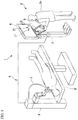
- FIG. 1 is a perspective view schematically showing an entire configuration of a medical system according to a first embodiment.
- FIG. 2 is a perspective view of an endoscope device in the medical system according to the first embodiment.
- FIG. 3 is a partially exploded perspective view of a treatment tool in the medical system according to the first embodiment.
- FIG. 4 is a block diagram showing an example of the medical system according to the first embodiment.
- FIG. 5 is a diagram showing an example of an image of a display device in the medical system according to the first embodiment.
- FIG. 6 is a flowchart showing calibration in the medical system according to the first embodiment.
- FIG. 7 is a flowchart showing an example in which a hysteresis width in the medical system according to the first embodiment is measured.
- FIG. 8 is a flowchart showing an example in which tip angle information of the treatment tool in the medical system according to the first embodiment is detected.
- FIG. 9 is a flowchart showing another example in which the tip angle information of the treatment tool in the medical system according to the first embodiment is detected.
- FIG. 10 is a flowchart showing calibration in a medical system according to a second embodiment.
- FIG. 11 is a flowchart showing calibration in a medical system according to a third embodiment.
- FIG. 12 is a flowchart showing calibration in a medical system according to a fourth embodiment.
- FIG. 13 is a perspective view of a distal portion when calibration is performed in a medical system according to a fifth embodiment.
- FIG. 14 is a flowchart showing an example in which calibration is performed in the medical system according to the fifth embodiment.
- FIG. 15 is a flowchart showing another example in which calibration is performed in the medical system according to the fifth embodiment.
- FIG. 16 is a perspective view of a distal portion when calibration is performed in a medical system according to a sixth embodiment.
- FIG. 17 is a flowchart showing an example in which calibration is performed in the medical system according to the sixth embodiment.
- FIG. 18 is a flowchart showing another example in which calibration is performed in the medical system according to the sixth embodiment.
- FIG. 1 is a perspective view schematically showing an entire configuration of a medical system according to a first embodiment.
- the medical system according to this embodiment is a so-called master-slave system. That is, as shown in FIG. 1 , the medical system 1 includes a master manipulator 2 that is manipulated by an operator Op for treatment, a slave manipulator 4 that is provided with an endoscope device 3 , and a control unit 50 (see FIG. 4 ) that controls the slave manipulator 4 in response to the manipulation of the master manipulator 2 .
- the master manipulator 2 includes a master arm 5 that is manipulated by an operator Op, a display device 6 that displays image information (see FIG. 4 ) 59 such as an image captured using the endoscope device 3 , and a controller 7 of a control unit 50 to be described later.
- the master arm 5 is a manipulation unit that is disposed to actuate the endoscope device 3 . Although details are not shown, two master arms 5 disposed in the master manipulator 2 are provided to correspond to a right hand and a left hand of an operator Op.
- the master arm 5 has a multi-joint structure configured to cause a joint portion 22 of a treatment tool 21 , which is a mechanism disposed in the body with at least one degree of freedom, to operate.
- the display device 6 is a device on which an image of a treatment target part which is captured by an observation device (see FIG. 2 ) attached to the endoscope device 3 is displayed.
- the joint portion 22 of the treatment tool 21 along with the treatment target part is displayed on the display device 6 .
- the controller 7 generates a manipulation command for causing the slave manipulator 4 to operate based on the operation of the master arm 5 .
- the slave manipulator 4 includes a surgical table 8 on which a patient is placed, a multi-joint robot 9 that is disposed around the surgical table 8 , and an endoscope device 3 that is attached to the multi-joint robot 9 .
- the multi-joint robot 9 operates in accordance with a manipulation command issued from the master manipulator 2 .
- FIG. 2 is a perspective view of the endoscope device in the medical system according to the first embodiment.
- the endoscope device 3 includes an insertion portion 24 and an outer sheath driving unit (drive unit) 25 .
- the insertion portion 24 includes an outer sheath 26 and an observation device 23 .
- the outer sheath 26 is a flexible long member that is inserted into the body of a patient.
- the outer sheath 26 has a distal end and a proximal end and also includes a treatment tool channel 29 into which the treatment tool 21 can be inserted.
- the observation device 23 is a device in which an imaging field of view directed to the distal end side from the distal end of the outer sheath 26 is set and can acquire and output an image of a treatment target part and the treatment tool 21 to the display device 6 .
- the observation device 23 is disposed in a distal end portion of the outer sheath 26 .
- the observation device 23 includes an imaging unit 30 and an illumination unit 31 .
- the imaging unit 30 can acquire at least one image including at least the joint portion 22 in the imaging field of view.
- the illumination unit 30 emits illumination light to the imaging field of view of the imaging unit 30 .
- the observation device 23 may be detachable from the outer sheath 26 .
- a known endoscope device may be used as the observation device and an endoscope channel into which the known endoscope device can be inserted may be formed in the outer sheath 26 .
- the treatment tool channel 29 holds the treatment tool 21 such that the treatment portion 27 of the treatment tool 21 protrudes from the distal end of the insertion portion 24 .
- the outer sheath driving unit 25 is disposed on the proximal end side of the outer sheath 26 , and can cause the observation device 23 and the treatment portion 27 of the treatment tool 21 in the body to face a desired direction by curving the distal end portion of the outer sheath 26 by driving.
- FIG. 3 is a partially exploded perspective view of the treatment tool in the medical system according to the first embodiment.
- the treatment tool 21 includes a treatment tool driving unit (drive unit) 32 , a flexible portion 33 which is a flexible tube portion, and a distal portion 34 .
- drive unit treatment tool driving unit
- flexible portion 33 which is a flexible tube portion
- distal portion 34 distal portion
- the treatment tool driving unit 32 includes a motor 35 , an encoder 36 , a driving-side rotary body 37 , a wire 38 , and a driven-side rotary body 39 .
- the motor 35 is disposed for each degree of freedom of the joint portion 22 and the treatment portion 27 . In this embodiment, only one motor 35 for curving one joint portion 22 will be described.
- the treatment tool driving unit 32 can independently drive another joint portion 22 which is not shown and another treatment portion 27 which is not shown using another motor which is not shown.
- a motor shaft of the motor 35 is connected to the driving-side rotary body 37 via a reduction gear which is not shown.
- a stepping motor or the like may be employed as the motor 35 .
- the encoder 36 (see FIG. 4 ) is attached to the motor shaft, which is not shown, of the motor 35 in a non-contact manner.
- the encoder 36 is electrically connected to the control unit 50 .
- An example of the driving-side rotary body 37 is a pulley that rotates with a driving force which the motor 35 generates.
- One end of the wire 38 is suspended on the driving-side rotary body 37 .
- the wire 38 is an annular wire of which one end is suspended on the driving-side rotary body 37 , an intermediate portion is movably accommodated in the flexible portion 33 , and the other end is suspended on the driven-side rotary body 39 .
- the flexible portion 33 has a flexible tubular shape.
- the treatment tool driving unit 32 is disposed at the proximal end of the flexible portion 33
- the treatment portion 27 is disposed at the distal end of the flexible portion 33 .
- the distal portion 34 of the treatment tool 21 includes a joint portion 22 , an arm portion 40 , and a treatment portion 27 .
- the joint portion 22 is coupled to the arm portion 40 .
- the joint portion 22 displaces the arm portion 40 by transmission of an amount of force from the driven-side rotary body 39 .
- a structure with which the joint portion 22 is curved is not limited to the joint structure.
- a joint structure in which a plurality of curving pieces are rotatably coupled may be employed.
- the treatment portion 27 is a forceps or an incision knife for performing treatment on a treatment target.
- the distal portion 34 of the treatment tool 21 has an identification portion X for identifying the type of the treatment tool 21 .
- the identification portion X is disposed in the arm portion 40 , for example, by printing or carving
- the driving-side rotary body 37 rotates with the rotation of the motor 35 and the driven-side rotary body 39 rotates via the wire 38 . Accordingly, the joint portion 22 is curved by the rotation of the driven-side rotary body 39 .
- a rotation signal from the encoder 36 is processed by the control unit 50 and is received as information of a degree of motor drive.
- FIG. 4 is a block diagram showing an example of the medical system according to the first embodiment. This block diagram will also be used in subsequent embodiments.
- the control unit 50 of the medical system 1 includes a master control unit 51 , a slave control unit 52 , a calibration unit 53 , an image processing unit 54 , a compensation value calculating unit 55 , a treatment tool information storage unit 56 , a parameter table 57 , and a controller 7 .
- the master control unit 51 receives and processes a manipulation input (input) 58 in the master manipulator 2 .
- the slave control unit 52 outputs drive signals for the multi-joint robot 9 , the endoscope device 3 , and the treatment tool 21 based on a command from the master control unit 51 .
- the calibration unit 53 generates a parameter for compensating for an operation of the treatment tool 21 .
- the calibration unit 53 may be included in any one of the master control unit 51 and the slave control unit 52 or may be independent from the master control unit 51 and the slave control unit 52 .
- the image processing unit 54 analyzes the image information 59 acquired by the imaging unit 30 .
- the image processing unit 54 calculates at least one of a position and an orientation of the joint portion 22 based on the image information 59 .
- the image processing unit 54 in this embodiment calculates a joint angle of the joint portion 22 through image recognition using the image information 59 .
- the compensation value calculating unit 55 detects displacement of the joint portion 22 based on at least one of the position and the orientation of the joint portion 22 which is calculated by the image processing unit 54 , generates a compensation value for compensating for a difference between the input (command) 58 and the displacement of the joint portion 22 , and incorporates the compensation value into the parameter.
- the compensation value calculating unit 55 incorporates a hysteresis width to be described later as the compensation value into the parameter.
- the compensation value calculating unit 55 calculates the hysteresis width based on an amount of drive of the treatment tool driving unit 32 until displacement of the joint portion 22 is started after the command is issued, and determines and incorporates the compensation value after displacement of the joint portion 22 is started.
- the treatment tool information storage unit 56 stores individual data required for calibration such as individual information of the treatment tool 21 or a pattern matching image.
- the parameter table 57 includes parameters which are referred to by the controller 7 .
- An initial parameter is loaded into the parameter table 57 when the medical system 1 is started.
- the initial parameter loaded into the parameter table 57 is updated with every calibration by the compensation value calculating unit 55 and becomes a parameter which is referred to by the controller 7 .
- the controller 7 issues an output to the motor 35 using the parameter which is updated by the calibration unit 53 and stored in the parameter table 57 .
- the control unit 50 performs calibration through the use of the calibration unit 53 using the image information 59 which is acquired by the imaging unit 30 .
- the control unit 50 controls the position of the treatment tool 21 such that the treatment tool 21 moves to a position at which calibration can be suitably performed on the treatment tool 21 .
- the control unit 50 displaces the joint portion 22 until the joint portion 22 appears in the image information 59 .
- the control unit 50 may determine that the joint portion 22 is located outside the imaging field of view of the imaging unit 30 and may output instruction image information in which a specifiable area for the joint portion 22 is superimposed on the image information 59 to the display device 6 instead of the image information 59 . That is, in this control method, the control unit 50 supports a user's manipulation such that the treatment tool 21 enters a calibratable range by encouraging the user of the medical system 1 to move the treatment tool 21 .
- the instruction image information is received instead of the image information 59 , the instruction image information is displayed on the display device 6 .
- the image processing unit 54 calculates at least one of the position and the orientation of the joint portion 22 using the image information 59 in which the joint portion 22 is located in the specifiable area.
- FIG. 5 is a diagram showing an example of an image on the display device in the medical system according to the first embodiment.
- the calibration unit 53 performs calibration based on the image information 59 displayed on the display device 6 .
- the control unit 50 sets a bent point of the joint portion 22 as a virtual feature point by image recognition from the image information 59 and calculates the bending angle of the joint portion 22 .
- FIG. 6 is a flowchart showing calibration in the medical system according to the first embodiment.
- a hysteresis width ⁇ is measured (MEASUREMENT OF HYSTERESIS WIDTH, step ST 101 ).
- the measurement of the hysteresis width ⁇ is a value based on an amount of drive of the treatment tool driving unit 32 until displacement of the joint portion 22 is started after a command is issued.
- a hysteresis value is updated (UPDATE PARAMETER, step ST 103 ).
- the hysteresis value is defined as a parameter element having a function including the hysteresis width ⁇ x as a variable.
- ⁇ dot over ( ⁇ ) ⁇ ref is a first differential of ⁇ ref .
- Equation (1) denotes the hysteresis value and is the compensation value in this embodiment.
- the hysteresis value is stored in the parameter table 57 and is read as an updated parameter when the controller 7 is activated.
- the symbol sgn denotes a sign function that has +1, 0, or ⁇ 1 depending on plus, 0, or minus of an argument.
- the hysteresis width is acquired through the flowchart shown in FIG. 7 .
- FIG. 7 is a flowchart showing an example in which the hysteresis width in the medical system according to the first embodiment is measured.
- At least one image including at least the joint portion 22 in the imaging field of view is acquired by the imaging unit 30 (START ACQUISITION OF IMAGE, step ST 201 ).
- the image acquired by the imaging unit 30 is displayed on the display device 6 in a state in which the joint portion 22 is included in the image.
- step ST 202 driving of the motor 35 of the treatment tool driving unit 32 is started (START DRIVING OF MOTOR, step ST 202 ). Movement of the joint portion 22 is started with the driving of the motor 35 .
- a delay or other difference may occur between the driving of the motor 35 and the bending of the joint portion 22 due to elongation or looseness of the wire 38 , a frictional force against the outer surface of the wire 38 , or the like.
- the joint portion 22 may not operate at all until the motor 35 reaches a predetermined rotational angle.
- step ST 203 From the time point at which the driving of the motor 35 is started, detection of the treatment tool tip angle information is started (DETECT TREATMENT TOOL TIP ANGLE INFORMATION, step ST 203 ). That is, movement of the moving joint portion 22 is detected.
- the treatment tool tip angle information can be detected by pattern matching or an optical flow to be described later.
- the treatment tool tip angle information is repeatedly detected (DETECT TREATMENT TOOL TIP ANGLE INFORMATION, step ST 203 ).
- the hysteresis width can be calculated by moving the distal portion 34 in one direction.
- the hysteresis width can be calculated with the initial value unknown by causing the distal portion 34 to reciprocate in two opposite directions (for example, right and left directions).
- the motor 35 is driven to move the distal portion 34 in a predetermined direction, the degree of drive of the motor 35 when minute movement of the distal portion 34 is detected is stored, the motor 35 is driven to move the distal portion in the opposite direction of the predetermined direction, and the degree of drive of the motor 35 when minute movement of the distal portion 34 is detected is stored.
- the hysteresis width can be calculated from two pieces of drive information acquired in this process.
- the hysteresis width based on the degree of drive of the treatment tool driving unit 32 is calculated to determine the hysteresis value and is set as the compensation value u by the compensation value calculating unit 55 (CALCULATE HYSTERESIS WIDTH, step ST 206 ). In this way, the motor 35 is driven until the joint portion 22 moves minutely, and the hysteresis width is calculated based on the degree of drive of the treatment tool driving unit 32 .
- FIG. 8 is a flowchart showing an example in which the treatment tool tip angle information in the medical system according to the first embodiment is detected.
- image information 59 is acquired by the imaging unit 30 of the endoscope device 3 (ACQUIRE ENDOSCOPE IMAGE, step ST 301 ).
- image information 59 of the treatment tool 21 is extracted from the acquired image information 59 by pattern matching with reference to a predetermined pattern based on the identification portion X (EXTRACT TREATMENT TOOL FROM IMAGE BY PATTERN MATCHING step ST 302 ).
- a feature point is extracted from the extracted image information 59 (EXTRACT FEATURE POINT, step ST 303 ).
- angle information of the distal portion 34 of the treatment tool 21 is calculated from the extracted feature point (CALCULATE TREATMENT TOOL TIP ANGLE, step ST 304 ).
- FIG. 9 is a flowchart showing another example in which the treatment tool tip angle information in the medical system according to the first embodiment is detected.
- the image information 59 acquired by the endoscope device 3 is processed in an optical flow manner. That is, the image processing unit 54 calculates displacement of the joint portion 22 from a difference between newest image information 59 and image information 59 acquired immediately before the newest image information 59 is acquired in time series in the image information 59 . That is, first, the newest image information 59 of the endoscope device 3 is acquired.
- the previous image information 59 of the endoscope device 3 is acquired and a difference between both pieces of image information 59 is calculated, whereby a moving direction and a moving distance of the endoscope tip are calculated (CALCULATE MOVING DIRECTION AND MOVING DISTANCE OF ENDOSCOPE TIP FROM DIFFERENCE BETWEEN NEWEST ENDOSCOPE IMAGE AND PREVIOUS ENDOSCOPE IMAGE, step ST 401 ).
- control unit 50 since the control unit 50 includes the parameter table 57 , the controller 7 , the image processing unit 54 , and the compensation value calculating unit 55 , calibration is performed in consideration of a variation in operation of the distal portion 34 of the treatment tool 21 due to a variation in characteristics of the wire 38 . Accordingly, it is possible to perform appropriate calibration when the distal portion 34 of the treatment tool 21 is guided to a treatment target part in the endoscope device 3 according to this embodiment.
- the imaging unit 30 essential for observing a treatment target part can be used as an element for calibration. Accordingly, an encoder that detects the joint angle of the joint portion 22 is not necessary and it is possible to perform high-accuracy calibration with a simple configuration.
- the control unit 50 displaces the joint portion 22 until the joint portion 22 appears in the image information 59 . Accordingly, it is possible to avoid a situation in which calibration is not possible.
- the manipulation of the treatment tool 21 is put in the hands of the user of the treatment tool 21 and it is thus possible to move the treatment tool 21 stably.
- the calibration may be automatically started when the treatment tool 21 is moved to an appropriate position by the user. In this case, it is possible to simplify a manipulation to be performed by the user.
- control unit 50 since the control unit 50 specifies the treatment tool 21 based on the identification portion X, it is possible to detect the treatment tool 21 by simple image processing.
- one of the pattern matching and the optical flow is employed, but a configuration in which an image can be processed using both the pattern matching and the optical flow may be employed.
- the pattern matching may be employed when the identification portion X is specifiable and a pattern matching image is present
- the optical flow may be employed when the identification portion X is not specifiable or an appropriate pattern matching image is not present. That is, a configuration capable of selecting one of the pattern matching and the optical flow depending on a situation may be employed.
- the compensation value calculating unit 55 calculates the hysteresis width based on the degree of drive of the treatment tool driving unit 32 until the displacement of the joint portion 22 is started after a command is issued and sets the compensation value, it is possible to acquire an appropriate compensation value using the measured value of the distal portion 34 of the treatment tool 21 .
- control unit 50 since the control unit 50 changes the parameter to correspond to the treatment tool 21 specified based on the identification portion X, it is possible to accurately detect the treatment tool 21 .
- FIG. 10 is a flowchart showing calibration in a medical system according to the second embodiment.
- the same elements as in the first embodiment will be referenced by the same reference signs, a description thereof will not be repeated, and only differences will be described.
- this embodiment is different from the above-mentioned embodiment in the calibration order in the control unit 50 .
- a delay time until the distal portion 34 of the treatment tool 21 starts its turn-back in accordance with a command for turning back the distal portion 34 of the treatment tool 21 after the command is issued is used.
- turn-back refers to switching of a displacement direction such that the distal portion 34 is displaced in the opposite direction in a state in which the distal portion 34 is displaced in a predetermined direction.
- a count value when a turn-back command is issued is acquired (ACQUIRE COUNT WHEN TURN-BACK INPUT IS PERFORMED, step ST 501 ).
- a time point at which the turn-back command is issued is set to zero, for example, by resetting a timer which is not shown to zero and starting count-up.
- a count value when the distal portion 34 of the treatment tool 21 starts the turn-back in accordance with the turn-back command is acquired (ACQUIRE COUNT WHEN TREATMENT TOOL TIP TURNS BACK, step ST 502 ). It can be detected that the distal portion 34 of the treatment tool 21 starts the turn-back by detecting displacement of the distal portion 34 using the pattern matching or the optical flow which is described in the first embodiment.
- a delay time until the distal portion 34 of the treatment tool 21 starts the turn-back after the turn-back command is issued is acquired (ACQUIRE DELAY TIME, step ST 503 ).
- the count value of the timer which is reset to zero when the turn-back command is issued is acquired as the delay time.
- the parameter table 57 is referred to (REFER TO TABLE, step ST 504 ) and then the compensation value of the delay time is updated (UPDATE COMPENSATE VALUE FOR DELAY TIME, step ST 505 ).
- the delay time can be compensated for by a phase advance filter expressed by Equation (2) and control compensation expressed by Equation (3).
- Equation (2) the symbol ⁇ T denotes a time constant and the symbol s denotes a Laplacian operator.
- the phase advance filter may be set from the time constant.
- Equation (3) the symbol u denotes the compensation value.
- the delay time until the distal portion 34 of the treatment tool 21 starts the turn-back after the turn-back command is issued is referred to by the parameter table 57 and the compensation value of the delay time is updated. Accordingly, even when the characteristics of the wire 38 vary, it is possible to perform calibration.
- FIG. 11 is a flowchart showing calibration in a medical system according to a third embodiment. This embodiment is different from the above-mentioned embodiments in the calibration order in the control unit 50 . As shown in FIG. 11 , in the calibration according to this embodiment, the compensation value is changed based on a ratio of the amplitude of displacement of the distal portion 34 of the treatment tool in response to a command and the amplitude of actual displacement of the distal portion 34 of the treatment tool 21 .
- an input value is referred to (REFER TO INPUT VALUE, step ST 601 ).
- the amplitude of the distal portion 34 of the treatment tool 21 is measured (MEASURE AMPLITUDE OF TREATMENT TOOL TIP, step ST 602 ).
- the measured amplitude is referred to by the parameter table 57 (REFER TO TABLE, step ST 603 ) and a compensation coefficient is updated (UPDATE COMPENSATION COEFFICIENT, step ST 604 ).
- Equation (4) the symbol ⁇ denotes the compensation coefficient and is expressed by Equation (5).
- ⁇ ref is an angle command value which is included in a displacement command for the distal portion 34
- the symbol ⁇ ref denotes a response value of the angle of the distal portion 34 to the command.
- the compensation coefficient ⁇ is 2
- the compensation value u is 2 ⁇ ref based on Equation (4).
- the compensation value based on the amplitude ratio of the angle response of the distal portion 34 of the treatment tool 21 to the command, it is possible to perform calibration even when the characteristics of the wire 38 vary.
- FIG. 12 is a flowchart showing calibration in a medical system according to a fourth embodiment. This embodiment is different from the above-mentioned embodiments in the calibration order in the control unit 50 .
- the distal portion 34 has elasticity characteristics due to an elastic restoring force accumulated in the flexible portion 33 or an elastic repulsive force of the wire 38 . Accordingly, the amplitude decreases due to the elasticity characteristics of the flexible portion 33 and the wire 38 . That is, first, the degree of drive of the motor 35 acquired by the encoder 36 is referred to (REFER TO DEGREE OF DRIVE OF MOTOR, step ST 701 ).
- Equation (6) and Equation (7) the symbol ⁇ denotes the angle of the distal portion 34
- the symbol ⁇ m denotes the angle of the motor 35
- the symbol ⁇ ′ denotes the angular velocity of the distal portion 34
- the symbol J denotes the moment of inertia of the distal portion 34
- the symbol J m denotes the moment of inertia of the motor 35
- the symbol F denotes the torque generated from the motor 35
- the symbol k e denotes the environmental stiffness in the rotating direction of the distal portion 34
- the symbol c denotes the viscous frictional coefficient in the rotating direction
- the symbol f d denotes the frictional torque applied to the distal portion 34
- the symbol k denotes the rigidity of the wire 38
- the detected quantity of the distal portion 34 may be a position.
- the symbol ⁇ denotes the position of the distal portion 34
- the symbol ⁇ m denotes the position of the motor 35
- the symbol ⁇ ′ denotes the velocity of the distal portion 34
- the symbol J denotes the mass of the distal portion 34
- the symbol J m denotes the mass of the motor 35
- F denotes the force generated from the motor 35
- the symbol k e denotes the environmental stiffness in the translating direction of the distal portion 34
- the symbol c denotes the viscous frictional coefficient in the translating direction
- the symbol f d denotes the frictional force applied to the distal portion 34
- the symbol k denotes the rigidity of the wire 38 .
- the symbol ⁇ umlaut over ( ⁇ ) ⁇ is a second differential of ⁇
- the symbol ⁇ umlaut over ( ⁇ ) ⁇ is equal to ⁇ ′ and denotes a first differential of ⁇
- a parameter in the model is updated (UPDATE PARAMETER IN MODEL, step ST 704 ).
- the parameter in the model can be acquired by repeating calculation such that the tip angle information acquired from the image information matches the model output.
- the compensation value u can be calculated by using the parameter in the model in Equations (8) and (9).
- Equation (8) the symbol f d denotes the frictional torque applied to the distal portion 34 and the symbol k denotes the rigidity of the wire 38 which is converted into the rotating direction.
- the fourth embodiment by changing the parameter table 57 using the model which is supposed from the input and the angle response, it is possible to perform calibration even when the characteristics of the wire 38 vary.
- Equation (6) and (7) The model expressed by Equations (6) and (7) is an example and the model may be defined using another function.
- FIG. 13 is a perspective view of a distal portion when calibration is performed in a medical system according to the fifth embodiment.
- This embodiment is different from the above-mentioned embodiments in that calibration of a degree of advance and retraction of the distal portion 34 in the axial direction of the treatment tool channel 29 is performed by the control unit 50 .
- the treatment portion 27 is moved to advance and retract by a stroke length L 1 between a position A 1 and a position A 2 by the treatment tool driving unit 32 .
- FIG. 14 is a flowchart showing an example in which calibration is performed in the medical system according to the fifth embodiment. As shown in FIG. 14 , first, image information 59 of the endoscope device 3 is acquired (ACQUIRE ENDOSCOPE IMAGE, step ST 801 ).
- the treatment tool 21 is detected from the image information 59 of the endoscope device 3 by the pattern matching (DETECT TREATMENT TOOL FROM IMAGE BY PATTERN MATCHING step ST 802 ).
- an identification portion (feature point) X is extracted from the detected treatment tool 21 (EXTRACT FEATURE POINT, step ST 803 ).
- the degree of advance and retraction of the treatment tool 21 is calculated using the extracted identification portion X (CALCULATE DEGREE OF ADVANCE AND RETRACTION OF TREATMENT TOOL, step ST 804 ).
- FIG. 15 is a flowchart showing another example in which calibration is performed in the medical system according to the fifth embodiment.
- the calibration according to this embodiment may be performed using the optical flow described in the first embodiment. That is, as shown in FIG. 15 , first, newest image information 59 of the endoscope device 3 is acquired. Then, previous image information 59 of the endoscope device 3 is acquired and a difference between the pieces of image information 59 is calculated, whereby a moving direction and a moving distance of the endoscope tip are calculated (CALCULATE MOVING DIRECTION AND MOVING DISTANCE OF ENDOSCOPE TIP FROM DIFFERENCE BETWEEN NEWEST ENDOSCOPE IMAGE AND PREVIOUS ENDOSCOPE IMAGE, step ST 901 ).
- the fifth embodiment by performing calibration using the degree of advance and retraction of the treatment portion 27 in the image information 59 of the endoscope device 3 , it is possible to perform calibration even when the characteristics of the wire 38 vary.
- FIG. 16 is a perspective view of a distal portion when calibration is performed in a medical system according to the sixth embodiment.
- This embodiment is different from the above-mentioned embodiments in that calibration of a degree of parallel movement of the distal portion 34 in the direction perpendicular to the axis of the treatment tool channel 29 is performed by the control unit 50 .
- the calibration is performed using the degree of parallel movement of the treatment portion 27 in the image information 59 of the endoscope device 3 instead of the joint portion 22 .
- the treatment portion 27 is moved in a parallel style by a stroke length L 2 between a position B 1 and a position B 2 by the treatment tool driving unit 32 .
- FIG. 17 is a flowchart showing an example in which calibration is performed in the medical system according to the sixth embodiment. As shown in FIG. 17 , first, image information 59 of the endoscope device 3 is acquired (ACQUIRE ENDOSCOPE IMAGE, step ST 1101 ).
- the treatment tool 21 is detected from the image information 59 of the endoscope device 3 by the pattern matching (DETECT TREATMENT TOOL FROM IMAGE BY PATTERN MATCHING step ST 1102 ).
- an identification portion (feature point) X is extracted from the detected treatment tool 21 (EXTRACT FEATURE POINT, step ST 1103 ).
- the degree of parallel movement of the treatment tool 21 is calculated using the extracted identification portion X (CALCULATE DEGREE OF PARALLEL MOVEMENT TOOL, step ST 104 ).
- FIG. 18 is a flowchart showing another example in which calibration is performed in the medical system according to the sixth embodiment.
- the calibration according to this embodiment may be performed using the optical flow which is described in the first embodiment. That is, as shown in FIG. 18 , first, newest image information 59 of the endoscope device 3 is acquired. Then, previous image information 59 of the endoscope device 3 is acquired and a difference between the pieces of image information 59 is calculated, whereby a moving direction and a moving distance of the endoscope tip are calculated (CALCULATE MOVING DIRECTION AND MOVING DISTANCE OF ENDOSCOPE TIP FROM DIFFERENCE BETWEEN NEWEST ENDOSCOPE IMAGE AND PREVIOUS ENDOSCOPE IMAGE, step ST 1201 ).
- the degree of parallel movement of the distal portion 34 of the treatment tool 21 is calculated (CALCULATE DEGREE OF PARALLEL MOVEMENT OF TREATMENT TOOL, step ST 1202 ).
- the sixth embodiment by performing calibration using the degree of parallel movement of the treatment portion 27 in the image information 59 of the endoscope device 3 , it is possible to perform calibration even when the characteristics of the wire 38 vary.
Abstract
Description
u=Δθ·sgn({dot over (θ)}ref) Equation (1)
u=α·θ ref Equation (4)
J m{umlaut over (θ)}m =−k(θm−θ)+F Equation (6)
J m{umlaut over (θ)}m =−k(θm−θ)+F Equation (7)
Claims (8)
Applications Claiming Priority (3)
| Application Number | Priority Date | Filing Date | Title |
|---|---|---|---|
| JP2014-036824 | 2014-02-27 | ||
| JP2014036824A JP6270537B2 (en) | 2014-02-27 | 2014-02-27 | Medical system |
| PCT/JP2015/055599 WO2015129802A1 (en) | 2014-02-27 | 2015-02-26 | Medical system and treatment tool calibration method |
Related Parent Applications (1)
| Application Number | Title | Priority Date | Filing Date |
|---|---|---|---|
| PCT/JP2015/055599 Continuation WO2015129802A1 (en) | 2014-02-27 | 2015-02-26 | Medical system and treatment tool calibration method |
Publications (2)
| Publication Number | Publication Date |
|---|---|
| US20160360947A1 US20160360947A1 (en) | 2016-12-15 |
| US10863883B2 true US10863883B2 (en) | 2020-12-15 |
Family
ID=54009116
Family Applications (1)
| Application Number | Title | Priority Date | Filing Date |
|---|---|---|---|
| US15/245,269 Active 2036-08-02 US10863883B2 (en) | 2014-02-27 | 2016-08-24 | Medical system and treatment tool calibrating method |
Country Status (5)
| Country | Link |
|---|---|
| US (1) | US10863883B2 (en) |
| EP (1) | EP3111819B1 (en) |
| JP (1) | JP6270537B2 (en) |
| CN (1) | CN105979848B (en) |
| WO (1) | WO2015129802A1 (en) |
Families Citing this family (60)
| Publication number | Priority date | Publication date | Assignee | Title |
|---|---|---|---|---|
| US8672837B2 (en) | 2010-06-24 | 2014-03-18 | Hansen Medical, Inc. | Methods and devices for controlling a shapeable medical device |
| US20130303944A1 (en) | 2012-05-14 | 2013-11-14 | Intuitive Surgical Operations, Inc. | Off-axis electromagnetic sensor |
| US9452276B2 (en) | 2011-10-14 | 2016-09-27 | Intuitive Surgical Operations, Inc. | Catheter with removable vision probe |
| US20140148673A1 (en) | 2012-11-28 | 2014-05-29 | Hansen Medical, Inc. | Method of anchoring pullwire directly articulatable region in catheter |
| US9057600B2 (en) | 2013-03-13 | 2015-06-16 | Hansen Medical, Inc. | Reducing incremental measurement sensor error |
| US9014851B2 (en) | 2013-03-15 | 2015-04-21 | Hansen Medical, Inc. | Systems and methods for tracking robotically controlled medical instruments |
| US9271663B2 (en) | 2013-03-15 | 2016-03-01 | Hansen Medical, Inc. | Flexible instrument localization from both remote and elongation sensors |
| US9629595B2 (en) | 2013-03-15 | 2017-04-25 | Hansen Medical, Inc. | Systems and methods for localizing, tracking and/or controlling medical instruments |
| US11020016B2 (en) | 2013-05-30 | 2021-06-01 | Auris Health, Inc. | System and method for displaying anatomy and devices on a movable display |
| EP2923669B1 (en) | 2014-03-24 | 2017-06-28 | Hansen Medical, Inc. | Systems and devices for catheter driving instinctiveness |
| EP3200718A4 (en) | 2014-09-30 | 2018-04-25 | Auris Surgical Robotics, Inc | Configurable robotic surgical system with virtual rail and flexible endoscope |
| US10314463B2 (en) * | 2014-10-24 | 2019-06-11 | Auris Health, Inc. | Automated endoscope calibration |
| JP6348854B2 (en) * | 2015-02-03 | 2018-06-27 | 富士フイルム株式会社 | Endoscope processor device, endoscope system, and non-contact power feeding method for endoscope system |
| AU2016323982A1 (en) | 2015-09-18 | 2018-04-12 | Auris Health, Inc. | Navigation of tubular networks |
| US10143526B2 (en) | 2015-11-30 | 2018-12-04 | Auris Health, Inc. | Robot-assisted driving systems and methods |
| WO2017167971A1 (en) | 2016-03-31 | 2017-10-05 | Koninklijke Philips N.V. | Image guided robotic system for tumor aspiration |
| US9931025B1 (en) | 2016-09-30 | 2018-04-03 | Auris Surgical Robotics, Inc. | Automated calibration of endoscopes with pull wires |
| DE102016225613A1 (en) * | 2016-12-20 | 2018-06-21 | Kuka Roboter Gmbh | Method for calibrating a manipulator of a diagnostic and / or therapeutic manipulator system |
| US10244926B2 (en) | 2016-12-28 | 2019-04-02 | Auris Health, Inc. | Detecting endolumenal buckling of flexible instruments |
| KR102558061B1 (en) | 2017-03-31 | 2023-07-25 | 아우리스 헬스, 인코포레이티드 | A robotic system for navigating the intraluminal tissue network that compensates for physiological noise |
| CN110831498B (en) | 2017-05-12 | 2022-08-12 | 奥瑞斯健康公司 | Biopsy device and system |
| US10022192B1 (en) | 2017-06-23 | 2018-07-17 | Auris Health, Inc. | Automatically-initialized robotic systems for navigation of luminal networks |
| WO2019005696A1 (en) | 2017-06-28 | 2019-01-03 | Auris Health, Inc. | Electromagnetic distortion detection |
| WO2019005872A1 (en) | 2017-06-28 | 2019-01-03 | Auris Health, Inc. | Instrument insertion compensation |
| EP3644885B1 (en) | 2017-06-28 | 2023-10-11 | Auris Health, Inc. | Electromagnetic field generator alignment |
| US10426559B2 (en) | 2017-06-30 | 2019-10-01 | Auris Health, Inc. | Systems and methods for medical instrument compression compensation |
| WO2019008942A1 (en) * | 2017-07-03 | 2019-01-10 | 富士フイルム株式会社 | Medical image processing device, endoscope device, diagnostic support device, medical service support device and report generation support device |
| CN107322589B (en) * | 2017-07-14 | 2020-06-19 | 西安交通大学 | Pneumatic control system of flexible operation arm with variable rigidity |
| US10145747B1 (en) | 2017-10-10 | 2018-12-04 | Auris Health, Inc. | Detection of undesirable forces on a surgical robotic arm |
| US11058493B2 (en) | 2017-10-13 | 2021-07-13 | Auris Health, Inc. | Robotic system configured for navigation path tracing |
| US10555778B2 (en) | 2017-10-13 | 2020-02-11 | Auris Health, Inc. | Image-based branch detection and mapping for navigation |
| JP7362610B2 (en) | 2017-12-06 | 2023-10-17 | オーリス ヘルス インコーポレイテッド | System and method for correcting uncommanded instrument rotation |
| AU2018384820A1 (en) | 2017-12-14 | 2020-05-21 | Auris Health, Inc. | System and method for estimating instrument location |
| KR20200101334A (en) | 2017-12-18 | 2020-08-27 | 아우리스 헬스, 인코포레이티드 | Method and system for tracking and navigation of instruments in the luminal network |
| EP3752085A4 (en) | 2018-02-13 | 2021-11-24 | Auris Health, Inc. | System and method for driving medical instrument |
| JP7214747B2 (en) | 2018-03-28 | 2023-01-30 | オーリス ヘルス インコーポレイテッド | System and method for position sensor alignment |
| KR102500422B1 (en) | 2018-03-28 | 2023-02-20 | 아우리스 헬스, 인코포레이티드 | System and method for displaying the estimated position of an instrument |
| US10932812B2 (en) * | 2018-03-30 | 2021-03-02 | Spectranetics Llc | Calibrated power-driven surgical cutting device |
| EP3801190A4 (en) | 2018-05-30 | 2022-03-02 | Auris Health, Inc. | Systems and methods for location sensor-based branch prediction |
| MX2020012898A (en) | 2018-05-31 | 2021-02-26 | Auris Health Inc | Path-based navigation of tubular networks. |
| KR102455671B1 (en) | 2018-05-31 | 2022-10-20 | 아우리스 헬스, 인코포레이티드 | Image-Based Airway Analysis and Mapping |
| EP3801280A4 (en) | 2018-05-31 | 2022-03-09 | Auris Health, Inc. | Robotic systems and methods for navigation of luminal network that detect physiological noise |
| EP3856064A4 (en) | 2018-09-28 | 2022-06-29 | Auris Health, Inc. | Systems and methods for docking medical instruments |
| EP3860497A1 (en) * | 2018-10-04 | 2021-08-11 | Intuitive Surgical Operations, Inc. | Systems and methods for device verification and sensor calibration |
| WO2021038469A1 (en) | 2019-08-30 | 2021-03-04 | Auris Health, Inc. | Systems and methods for weight-based registration of location sensors |
| WO2021038495A1 (en) | 2019-08-30 | 2021-03-04 | Auris Health, Inc. | Instrument image reliability systems and methods |
| CN114641252B (en) | 2019-09-03 | 2023-09-01 | 奥瑞斯健康公司 | Electromagnetic Distortion Detection and Compensation |
| EP4084721A4 (en) | 2019-12-31 | 2024-01-03 | Auris Health Inc | Anatomical feature identification and targeting |
| KR20220123076A (en) | 2019-12-31 | 2022-09-05 | 아우리스 헬스, 인코포레이티드 | Alignment Techniques for Transdermal Access |
| KR20220123087A (en) | 2019-12-31 | 2022-09-05 | 아우리스 헬스, 인코포레이티드 | Alignment interface for transdermal access |
| US20220110703A1 (en) * | 2020-01-17 | 2022-04-14 | Korea Advanced Institute Of Science And Technology | Method of determining hysteresis of surgical robot, method of compensating for the same, and endoscopic surgical apparatus |
| CN112057171B (en) * | 2020-09-04 | 2021-08-17 | 北京科迈启元科技有限公司 | Mechanical arm and operation executor connecting piece |
| EP4247290A1 (en) * | 2020-11-20 | 2023-09-27 | Auris Health, Inc. | Automated procedure evaluation |
| KR102555196B1 (en) * | 2021-05-17 | 2023-07-14 | 주식회사 로엔서지컬 | Surgical tool device having hysteresis compensation and method thereof |
| WO2022269797A1 (en) * | 2021-06-23 | 2022-12-29 | オリンパス株式会社 | Manipulator system, measurement apparatus, and method for controlling manipulator |
| WO2023021538A1 (en) * | 2021-08-16 | 2023-02-23 | オリンパスメディカルシステムズ株式会社 | Manipulator system, control device, and shape estimation method |
| JP2023030681A (en) * | 2021-08-23 | 2023-03-08 | 国立研究開発法人国立がん研究センター | Tip detection device of treatment instrument of endoscopic image, tip detection method of treatment instrument of endoscopic image and tip detection program of treatment instrument of endoscopic image |
| KR102616257B1 (en) * | 2021-10-18 | 2023-12-22 | 주식회사 로엔서지컬 | Hysteresis compensation apparatus of flexible tube and method thereof |
| CN114378819B (en) * | 2022-01-18 | 2022-07-26 | 上海健康医学院 | Master-slave hand control method and device for digestive endoscopy minimally invasive surgery robot |
| KR102478344B1 (en) * | 2022-07-06 | 2022-12-16 | 주식회사 에어스메디컬 | Method, program, and apparatus for mornitoring control of medical robot |
Citations (20)
| Publication number | Priority date | Publication date | Assignee | Title |
|---|---|---|---|---|
| JPS58217015A (en) | 1982-06-11 | 1983-12-16 | Fujitsu Ltd | Correcting method of hysteresis of robot |
| JPH0580842A (en) | 1991-05-10 | 1993-04-02 | Shinko Electric Co Ltd | Control method for moving robot |
| JPH10174686A (en) | 1996-12-17 | 1998-06-30 | Toshiba Corp | Wire drive mechanism and ultrasonic probe |
| JP2004041538A (en) | 2002-07-15 | 2004-02-12 | Hitachi Ltd | Traction and positioning device |
| JP2007029167A (en) | 2005-07-22 | 2007-02-08 | Olympus Medical Systems Corp | Endoscope |
| JP2007260298A (en) | 2006-03-29 | 2007-10-11 | Univ Waseda | Action control system and position detector of operation support robot |
| US20080004603A1 (en) * | 2006-06-29 | 2008-01-03 | Intuitive Surgical Inc. | Tool position and identification indicator displayed in a boundary area of a computer display screen |
| US20080103491A1 (en) | 2006-10-25 | 2008-05-01 | Terumo Kabushiki Kaisha | Manipulator for medical use |
| JP2008104854A (en) | 2006-10-25 | 2008-05-08 | Terumo Corp | Manipulator for medical use |
| US20080114494A1 (en) | 2004-05-04 | 2008-05-15 | Intuitive Surgical, Inc. | Tool grip calibration for robotic surgery |
| GB2448421A (en) | 2007-04-11 | 2008-10-15 | Forth Photonics Ltd | Workstation for a colposcopy |
| US20090112060A1 (en) | 2007-10-25 | 2009-04-30 | Olympus Medical Systems Corp. | Medical apparatus |
| US20090112316A1 (en) | 2007-10-30 | 2009-04-30 | Olympus Medical Systems Corp. | Manipulator apparatus and medical device system |
| CN101444415A (en) | 2007-11-29 | 2009-06-03 | 奥林巴斯医疗株式会社 | Therapeutic device system and manipulator system |
| US20100030023A1 (en) * | 2008-08-04 | 2010-02-04 | Olympus Medical Systems Corp. | Active drive type medical apparatus and drive control method |
| US20100082041A1 (en) | 2008-09-30 | 2010-04-01 | Intuitive Surgical, Inc. | Passive preload and capstan drive for surgical instruments |
| US20100169815A1 (en) * | 2008-12-31 | 2010-07-01 | Intuitive Surgical, Inc. | Visual force feedback in a minimally invasive surgical procedure |
| JP2010214128A (en) | 2010-05-19 | 2010-09-30 | Olympus Medical Systems Corp | Treatment instrument system and manipulator system |
| WO2012153646A1 (en) | 2011-05-12 | 2012-11-15 | オリンパスメディカルシステムズ株式会社 | Medical control device |
| US20130345517A1 (en) * | 2012-06-20 | 2013-12-26 | Fujifilm Corporation | Light source apparatus and endoscope system |
Family Cites Families (1)
| Publication number | Priority date | Publication date | Assignee | Title |
|---|---|---|---|---|
| US10555775B2 (en) * | 2005-05-16 | 2020-02-11 | Intuitive Surgical Operations, Inc. | Methods and system for performing 3-D tool tracking by fusion of sensor and/or camera derived data during minimally invasive robotic surgery |
-
2014
- 2014-02-27 JP JP2014036824A patent/JP6270537B2/en active Active
-
2015
- 2015-02-26 CN CN201580007642.7A patent/CN105979848B/en active Active
- 2015-02-26 EP EP15754565.8A patent/EP3111819B1/en not_active Not-in-force
- 2015-02-26 WO PCT/JP2015/055599 patent/WO2015129802A1/en active Application Filing
-
2016
- 2016-08-24 US US15/245,269 patent/US10863883B2/en active Active
Patent Citations (26)
| Publication number | Priority date | Publication date | Assignee | Title |
|---|---|---|---|---|
| JPS58217015A (en) | 1982-06-11 | 1983-12-16 | Fujitsu Ltd | Correcting method of hysteresis of robot |
| JPH0580842A (en) | 1991-05-10 | 1993-04-02 | Shinko Electric Co Ltd | Control method for moving robot |
| JPH10174686A (en) | 1996-12-17 | 1998-06-30 | Toshiba Corp | Wire drive mechanism and ultrasonic probe |
| JP2004041538A (en) | 2002-07-15 | 2004-02-12 | Hitachi Ltd | Traction and positioning device |
| US20040138530A1 (en) | 2002-07-15 | 2004-07-15 | Olympus Optical Co., Ltd. | Apparatus for traction positional control |
| US20080114494A1 (en) | 2004-05-04 | 2008-05-15 | Intuitive Surgical, Inc. | Tool grip calibration for robotic surgery |
| JP2007029167A (en) | 2005-07-22 | 2007-02-08 | Olympus Medical Systems Corp | Endoscope |
| US20090012365A1 (en) | 2005-07-22 | 2009-01-08 | Olympus Medical Systems Corp. | Endoscope |
| JP2007260298A (en) | 2006-03-29 | 2007-10-11 | Univ Waseda | Action control system and position detector of operation support robot |
| US20080004603A1 (en) * | 2006-06-29 | 2008-01-03 | Intuitive Surgical Inc. | Tool position and identification indicator displayed in a boundary area of a computer display screen |
| US20080103491A1 (en) | 2006-10-25 | 2008-05-01 | Terumo Kabushiki Kaisha | Manipulator for medical use |
| JP2008104854A (en) | 2006-10-25 | 2008-05-08 | Terumo Corp | Manipulator for medical use |
| GB2448421A (en) | 2007-04-11 | 2008-10-15 | Forth Photonics Ltd | Workstation for a colposcopy |
| JP2009101077A (en) | 2007-10-25 | 2009-05-14 | Olympus Medical Systems Corp | Medical apparatus |
| US20090112060A1 (en) | 2007-10-25 | 2009-04-30 | Olympus Medical Systems Corp. | Medical apparatus |
| JP2009107074A (en) | 2007-10-30 | 2009-05-21 | Olympus Medical Systems Corp | Manipulator apparatus and medical device system |
| CN101422901A (en) | 2007-10-30 | 2009-05-06 | 奥林巴斯医疗株式会社 | Manipulator apparatus and medical device system |
| US20090112316A1 (en) | 2007-10-30 | 2009-04-30 | Olympus Medical Systems Corp. | Manipulator apparatus and medical device system |
| CN101444415A (en) | 2007-11-29 | 2009-06-03 | 奥林巴斯医疗株式会社 | Therapeutic device system and manipulator system |
| US20100030023A1 (en) * | 2008-08-04 | 2010-02-04 | Olympus Medical Systems Corp. | Active drive type medical apparatus and drive control method |
| US20100082041A1 (en) | 2008-09-30 | 2010-04-01 | Intuitive Surgical, Inc. | Passive preload and capstan drive for surgical instruments |
| JP2012504016A (en) | 2008-09-30 | 2012-02-16 | インテュイティブ サージカル オペレーションズ, インコーポレイテッド | Passive preload and capstan drive for surgical instruments |
| US20100169815A1 (en) * | 2008-12-31 | 2010-07-01 | Intuitive Surgical, Inc. | Visual force feedback in a minimally invasive surgical procedure |
| JP2010214128A (en) | 2010-05-19 | 2010-09-30 | Olympus Medical Systems Corp | Treatment instrument system and manipulator system |
| WO2012153646A1 (en) | 2011-05-12 | 2012-11-15 | オリンパスメディカルシステムズ株式会社 | Medical control device |
| US20130345517A1 (en) * | 2012-06-20 | 2013-12-26 | Fujifilm Corporation | Light source apparatus and endoscope system |
Non-Patent Citations (5)
| Title |
|---|
| Chinese Office Action dated Jun. 26, 2017 in Chinese Patent Application No. 201580007642.7. |
| Extended Supplementary European Search Report dated Oct. 27, 2017 in European Patent Application No. 15 75 4565.8. |
| International Search Report dated May 19, 2105 issued in PCT/JP2015/055599. |
| Japanese Office Action dated Jun. 6, 2017 in Japanese Patent Application No. 2014-036824. |
| Notice of Allowance dated Dec. 5, 2017 in Japanese Patent Application No. 2014-036824. |
Also Published As
| Publication number | Publication date |
|---|---|
| EP3111819A1 (en) | 2017-01-04 |
| CN105979848A (en) | 2016-09-28 |
| JP2015160278A (en) | 2015-09-07 |
| EP3111819B1 (en) | 2018-12-12 |
| US20160360947A1 (en) | 2016-12-15 |
| WO2015129802A1 (en) | 2015-09-03 |
| CN105979848B (en) | 2018-02-13 |
| JP6270537B2 (en) | 2018-01-31 |
| EP3111819A4 (en) | 2017-11-29 |
Similar Documents
| Publication | Publication Date | Title |
|---|---|---|
| US10863883B2 (en) | Medical system and treatment tool calibrating method | |
| US11406460B2 (en) | Surgery assisting apparatus, method of controlling the same, storage medium, and surgery assisting system | |
| CN110662506B (en) | Tension control in actuation of a combination instrument | |
| US10155316B2 (en) | Manipulator-calibrating method, manipulator, and manipulator system | |
| KR102435989B1 (en) | Systems and methods for medical instrument force sensing | |
| EP2568910B1 (en) | Drive force control in medical instrument providing position measurements | |
| EP2706944B1 (en) | Surgical instrument device | |
| CN109512516B (en) | Robot interface positioning determination system and method | |
| RU2741469C1 (en) | Robotic surgical system | |
| US11666402B2 (en) | End effector force feedback to master controller | |
| JP6149175B1 (en) | Surgery support apparatus, control method thereof, program, and surgery support system | |
| KR101772805B1 (en) | User interface device for surgical robot system | |
| JP6388686B2 (en) | Surgery support apparatus, control method thereof, program, and surgery support system | |
| US20190029762A1 (en) | Medical manipulator system and manipulator curved-shape estimation method | |
| EP3829826B1 (en) | Systems and methods for controlling a robotic manipulator or associated tool | |
| US20220039872A1 (en) | Automated Endoscope Length Detection | |
| JP2015180238A (en) | medical robot |
Legal Events
| Date | Code | Title | Description |
|---|---|---|---|
| AS | Assignment |
Owner name: OLYMPUS CORPORATION, JAPAN Free format text: ASSIGNMENT OF ASSIGNORS INTEREST;ASSIGNORS:IIDA, MASATOSHI;HATAKEYAMA, NAOYA;WAKAI, HIROSHI;REEL/FRAME:039517/0223 Effective date: 20160819 |
|
| STPP | Information on status: patent application and granting procedure in general |
Free format text: NON FINAL ACTION MAILED |
|
| STPP | Information on status: patent application and granting procedure in general |
Free format text: RESPONSE TO NON-FINAL OFFICE ACTION ENTERED AND FORWARDED TO EXAMINER |
|
| STPP | Information on status: patent application and granting procedure in general |
Free format text: FINAL REJECTION MAILED |
|
| STPP | Information on status: patent application and granting procedure in general |
Free format text: DOCKETED NEW CASE - READY FOR EXAMINATION |
|
| STPP | Information on status: patent application and granting procedure in general |
Free format text: NON FINAL ACTION MAILED |
|
| STPP | Information on status: patent application and granting procedure in general |
Free format text: NOTICE OF ALLOWANCE MAILED -- APPLICATION RECEIVED IN OFFICE OF PUBLICATIONS |
|
| STPP | Information on status: patent application and granting procedure in general |
Free format text: PUBLICATIONS -- ISSUE FEE PAYMENT VERIFIED |
|
| STCF | Information on status: patent grant |
Free format text: PATENTED CASE |