US10751413B2 - Anti-pro/latent-Myostatin antibodies and uses thereof - Google Patents
Anti-pro/latent-Myostatin antibodies and uses thereof Download PDFInfo
- Publication number
- US10751413B2 US10751413B2 US15/760,393 US201615760393A US10751413B2 US 10751413 B2 US10751413 B2 US 10751413B2 US 201615760393 A US201615760393 A US 201615760393A US 10751413 B2 US10751413 B2 US 10751413B2
- Authority
- US
- United States
- Prior art keywords
- myostatin
- antibody
- latent
- seq
- pro
- Prior art date
- Legal status (The legal status is an assumption and is not a legal conclusion. Google has not performed a legal analysis and makes no representation as to the accuracy of the status listed.)
- Active, expires
Links
- 108010056852 Myostatin Proteins 0.000 claims abstract description 251
- 102100039939 Growth/differentiation factor 8 Human genes 0.000 claims description 280
- 125000000174 L-prolyl group Chemical group [H]N1C([H])([H])C([H])([H])C([H])([H])[C@@]1([H])C(*)=O 0.000 claims description 202
- 210000003205 muscle Anatomy 0.000 claims description 190
- 238000000034 method Methods 0.000 claims description 124
- 208000021642 Muscular disease Diseases 0.000 claims description 75
- 241000282414 Homo sapiens Species 0.000 claims description 73
- 201000009623 Myopathy Diseases 0.000 claims description 73
- 101100206935 Danio rerio tll1 gene Proteins 0.000 claims description 57
- 101000893545 Homo sapiens Growth/differentiation factor 11 Proteins 0.000 claims description 37
- 102100040898 Growth/differentiation factor 11 Human genes 0.000 claims description 36
- 206010028289 Muscle atrophy Diseases 0.000 claims description 34
- 125000003275 alpha amino acid group Chemical group 0.000 claims description 34
- 201000000585 muscular atrophy Diseases 0.000 claims description 33
- 230000020763 muscle atrophy Effects 0.000 claims description 30
- 241001529936 Murinae Species 0.000 claims description 21
- 230000015572 biosynthetic process Effects 0.000 claims description 18
- 230000002797 proteolythic effect Effects 0.000 claims description 17
- 239000008194 pharmaceutical composition Substances 0.000 claims description 15
- 239000003937 drug carrier Substances 0.000 claims description 7
- 238000001990 intravenous administration Methods 0.000 claims description 7
- 208000030159 metabolic disease Diseases 0.000 claims description 6
- 108010023082 activin A Proteins 0.000 claims description 5
- 238000007920 subcutaneous administration Methods 0.000 claims description 3
- HNDVDQJCIGZPNO-UHFFFAOYSA-N histidine Natural products OC(=O)C(N)CC1=CN=CN1 HNDVDQJCIGZPNO-UHFFFAOYSA-N 0.000 claims description 2
- 102000004472 Myostatin Human genes 0.000 abstract 1
- 230000027455 binding Effects 0.000 description 190
- 102000036639 antigens Human genes 0.000 description 133
- 108091007433 antigens Proteins 0.000 description 133
- 239000000427 antigen Substances 0.000 description 132
- 208000037265 diseases, disorders, signs and symptoms Diseases 0.000 description 118
- 210000004027 cell Anatomy 0.000 description 84
- 241001465754 Metazoa Species 0.000 description 82
- 201000010099 disease Diseases 0.000 description 82
- 238000011282 treatment Methods 0.000 description 67
- 108090000623 proteins and genes Proteins 0.000 description 61
- 238000003556 assay Methods 0.000 description 59
- 238000003776 cleavage reaction Methods 0.000 description 59
- LOKCTEFSRHRXRJ-UHFFFAOYSA-I dipotassium trisodium dihydrogen phosphate hydrogen phosphate dichloride Chemical compound P(=O)(O)(O)[O-].[K+].P(=O)(O)([O-])[O-].[Na+].[Na+].[Cl-].[K+].[Cl-].[Na+] LOKCTEFSRHRXRJ-UHFFFAOYSA-I 0.000 description 59
- 239000002953 phosphate buffered saline Substances 0.000 description 59
- 230000007017 scission Effects 0.000 description 59
- 238000010408 sweeping Methods 0.000 description 58
- 239000000203 mixture Substances 0.000 description 57
- 108010047041 Complementarity Determining Regions Proteins 0.000 description 51
- 102000006437 Proprotein Convertases Human genes 0.000 description 51
- 108010044159 Proprotein Convertases Proteins 0.000 description 51
- 102000004169 proteins and genes Human genes 0.000 description 50
- 235000018102 proteins Nutrition 0.000 description 47
- 239000003102 growth factor Substances 0.000 description 44
- 241000699670 Mus sp. Species 0.000 description 42
- 210000002966 serum Anatomy 0.000 description 42
- 239000012634 fragment Substances 0.000 description 41
- 230000004913 activation Effects 0.000 description 40
- 230000001965 increasing effect Effects 0.000 description 40
- 208000035475 disorder Diseases 0.000 description 36
- 239000000523 sample Substances 0.000 description 35
- 230000008859 change Effects 0.000 description 34
- 101000886562 Homo sapiens Growth/differentiation factor 8 Proteins 0.000 description 33
- 230000000694 effects Effects 0.000 description 31
- 241000700159 Rattus Species 0.000 description 30
- 108090000765 processed proteins & peptides Proteins 0.000 description 30
- 239000007787 solid Substances 0.000 description 29
- 210000001519 tissue Anatomy 0.000 description 29
- 108091033319 polynucleotide Proteins 0.000 description 28
- 102000040430 polynucleotide Human genes 0.000 description 28
- 239000002157 polynucleotide Substances 0.000 description 28
- UREBDLICKHMUKA-CXSFZGCWSA-N dexamethasone Chemical compound C1CC2=CC(=O)C=C[C@]2(C)[C@]2(F)[C@@H]1[C@@H]1C[C@@H](C)[C@@](C(=O)CO)(O)[C@@]1(C)C[C@@H]2O UREBDLICKHMUKA-CXSFZGCWSA-N 0.000 description 27
- 102100026120 IgG receptor FcRn large subunit p51 Human genes 0.000 description 26
- 229960003957 dexamethasone Drugs 0.000 description 26
- 210000004602 germ cell Anatomy 0.000 description 26
- 102000004196 processed proteins & peptides Human genes 0.000 description 26
- 210000002381 plasma Anatomy 0.000 description 25
- 101710177940 IgG receptor FcRn large subunit p51 Proteins 0.000 description 24
- 210000003314 quadriceps muscle Anatomy 0.000 description 24
- 238000012360 testing method Methods 0.000 description 24
- 241000699666 Mus <mouse, genus> Species 0.000 description 23
- 229920001184 polypeptide Polymers 0.000 description 23
- 210000002027 skeletal muscle Anatomy 0.000 description 23
- 102000035195 Peptidases Human genes 0.000 description 22
- 108091005804 Peptidases Proteins 0.000 description 22
- 239000004365 Protease Substances 0.000 description 22
- 235000001014 amino acid Nutrition 0.000 description 21
- 230000003993 interaction Effects 0.000 description 21
- 239000000463 material Substances 0.000 description 21
- 108060003951 Immunoglobulin Proteins 0.000 description 20
- 102000018358 immunoglobulin Human genes 0.000 description 20
- 239000003981 vehicle Substances 0.000 description 20
- 239000012472 biological sample Substances 0.000 description 19
- 238000002474 experimental method Methods 0.000 description 19
- 238000001727 in vivo Methods 0.000 description 19
- 238000007912 intraperitoneal administration Methods 0.000 description 19
- 238000002965 ELISA Methods 0.000 description 18
- 238000006467 substitution reaction Methods 0.000 description 18
- 238000001262 western blot Methods 0.000 description 18
- 150000001413 amino acids Chemical class 0.000 description 17
- 238000004458 analytical method Methods 0.000 description 17
- 230000037396 body weight Effects 0.000 description 17
- 238000001514 detection method Methods 0.000 description 17
- 239000003814 drug Substances 0.000 description 17
- 210000002414 leg Anatomy 0.000 description 17
- 150000007523 nucleic acids Chemical class 0.000 description 17
- 239000013598 vector Substances 0.000 description 17
- 229940024606 amino acid Drugs 0.000 description 16
- 208000020538 atrophic muscular disease Diseases 0.000 description 16
- 230000003442 weekly effect Effects 0.000 description 16
- 102000004961 Furin Human genes 0.000 description 15
- 108090001126 Furin Proteins 0.000 description 15
- 238000006243 chemical reaction Methods 0.000 description 15
- 239000003153 chemical reaction reagent Substances 0.000 description 15
- 230000007423 decrease Effects 0.000 description 15
- 239000000499 gel Substances 0.000 description 15
- 238000000338 in vitro Methods 0.000 description 15
- 206010006895 Cachexia Diseases 0.000 description 14
- 108020003175 receptors Proteins 0.000 description 14
- 102000005962 receptors Human genes 0.000 description 14
- 239000000243 solution Substances 0.000 description 14
- 230000001225 therapeutic effect Effects 0.000 description 14
- -1 PACE7 or PACE3 (e.g. Proteins 0.000 description 13
- 230000018109 developmental process Effects 0.000 description 13
- 229940079593 drug Drugs 0.000 description 13
- 238000002347 injection Methods 0.000 description 13
- 239000007924 injection Substances 0.000 description 13
- 102000039446 nucleic acids Human genes 0.000 description 13
- 108020004707 nucleic acids Proteins 0.000 description 13
- XLYOFNOQVPJJNP-UHFFFAOYSA-N water Substances O XLYOFNOQVPJJNP-UHFFFAOYSA-N 0.000 description 13
- NFGXHKASABOEEW-UHFFFAOYSA-N 1-methylethyl 11-methoxy-3,7,11-trimethyl-2,4-dodecadienoate Chemical compound COC(C)(C)CCCC(C)CC=CC(C)=CC(=O)OC(C)C NFGXHKASABOEEW-UHFFFAOYSA-N 0.000 description 12
- 208000010428 Muscle Weakness Diseases 0.000 description 12
- 206010028372 Muscular weakness Diseases 0.000 description 12
- 125000000539 amino acid group Chemical group 0.000 description 12
- 210000004369 blood Anatomy 0.000 description 12
- 239000008280 blood Substances 0.000 description 12
- 230000003247 decreasing effect Effects 0.000 description 12
- 238000011161 development Methods 0.000 description 12
- 230000002401 inhibitory effect Effects 0.000 description 12
- 239000006166 lysate Substances 0.000 description 12
- 239000002609 medium Substances 0.000 description 12
- 239000012528 membrane Substances 0.000 description 12
- 230000004044 response Effects 0.000 description 12
- 108020004414 DNA Proteins 0.000 description 11
- 101000844802 Lacticaseibacillus rhamnosus Teichoic acid D-alanyltransferase Proteins 0.000 description 11
- 238000011579 SCID mouse model Methods 0.000 description 11
- 206010002026 amyotrophic lateral sclerosis Diseases 0.000 description 11
- 239000000872 buffer Substances 0.000 description 11
- 238000005266 casting Methods 0.000 description 11
- 238000004519 manufacturing process Methods 0.000 description 11
- 230000035772 mutation Effects 0.000 description 11
- 239000002243 precursor Substances 0.000 description 11
- WSFSSNUMVMOOMR-UHFFFAOYSA-N Formaldehyde Chemical compound O=C WSFSSNUMVMOOMR-UHFFFAOYSA-N 0.000 description 10
- 206010028980 Neoplasm Diseases 0.000 description 10
- 102000043168 TGF-beta family Human genes 0.000 description 10
- 108091085018 TGF-beta family Proteins 0.000 description 10
- 231100000673 dose–response relationship Toxicity 0.000 description 10
- 230000006870 function Effects 0.000 description 10
- 238000003032 molecular docking Methods 0.000 description 10
- 238000001543 one-way ANOVA Methods 0.000 description 10
- 239000000546 pharmaceutical excipient Substances 0.000 description 10
- 238000010966 qNMR Methods 0.000 description 10
- 208000020431 spinal cord injury Diseases 0.000 description 10
- 208000024891 symptom Diseases 0.000 description 10
- 208000030507 AIDS Diseases 0.000 description 9
- 102100028728 Bone morphogenetic protein 1 Human genes 0.000 description 9
- 108090000654 Bone morphogenetic protein 1 Proteins 0.000 description 9
- PEDCQBHIVMGVHV-UHFFFAOYSA-N Glycerine Chemical compound OCC(O)CO PEDCQBHIVMGVHV-UHFFFAOYSA-N 0.000 description 9
- 102100036365 Proprotein convertase subtilisin/kexin type 5 Human genes 0.000 description 9
- 101710180648 Proprotein convertase subtilisin/kexin type 5 Proteins 0.000 description 9
- 230000032683 aging Effects 0.000 description 9
- 201000011510 cancer Diseases 0.000 description 9
- 239000003795 chemical substances by application Substances 0.000 description 9
- 238000001114 immunoprecipitation Methods 0.000 description 9
- 230000005764 inhibitory process Effects 0.000 description 9
- 230000000670 limiting effect Effects 0.000 description 9
- 239000011541 reaction mixture Substances 0.000 description 9
- 208000002320 spinal muscular atrophy Diseases 0.000 description 9
- 206010020100 Hip fracture Diseases 0.000 description 8
- 102100038946 Proprotein convertase subtilisin/kexin type 6 Human genes 0.000 description 8
- 230000002638 denervation Effects 0.000 description 8
- 239000003651 drinking water Substances 0.000 description 8
- 235000020188 drinking water Nutrition 0.000 description 8
- 230000002068 genetic effect Effects 0.000 description 8
- 230000008105 immune reaction Effects 0.000 description 8
- 229940072221 immunoglobulins Drugs 0.000 description 8
- 239000002502 liposome Substances 0.000 description 8
- 201000006938 muscular dystrophy Diseases 0.000 description 8
- 229920002981 polyvinylidene fluoride Polymers 0.000 description 8
- 238000002360 preparation method Methods 0.000 description 8
- 230000006337 proteolytic cleavage Effects 0.000 description 8
- 230000001105 regulatory effect Effects 0.000 description 8
- 230000011664 signaling Effects 0.000 description 8
- 238000002560 therapeutic procedure Methods 0.000 description 8
- 108010090804 Streptavidin Proteins 0.000 description 7
- 208000032978 Structural Congenital Myopathies Diseases 0.000 description 7
- 102000004887 Transforming Growth Factor beta Human genes 0.000 description 7
- 108090001012 Transforming Growth Factor beta Proteins 0.000 description 7
- 210000000577 adipose tissue Anatomy 0.000 description 7
- 239000011324 bead Substances 0.000 description 7
- 238000012575 bio-layer interferometry Methods 0.000 description 7
- 230000000903 blocking effect Effects 0.000 description 7
- 239000000839 emulsion Substances 0.000 description 7
- 210000002216 heart Anatomy 0.000 description 7
- 210000004408 hybridoma Anatomy 0.000 description 7
- 210000003141 lower extremity Anatomy 0.000 description 7
- 241000894007 species Species 0.000 description 7
- 210000000952 spleen Anatomy 0.000 description 7
- 230000000087 stabilizing effect Effects 0.000 description 7
- YBJHBAHKTGYVGT-ZKWXMUAHSA-N (+)-Biotin Chemical compound N1C(=O)N[C@@H]2[C@H](CCCCC(=O)O)SC[C@@H]21 YBJHBAHKTGYVGT-ZKWXMUAHSA-N 0.000 description 6
- IJGRMHOSHXDMSA-UHFFFAOYSA-N Atomic nitrogen Chemical compound N#N IJGRMHOSHXDMSA-UHFFFAOYSA-N 0.000 description 6
- 102000004190 Enzymes Human genes 0.000 description 6
- 108090000790 Enzymes Proteins 0.000 description 6
- 102100026818 Inhibin beta E chain Human genes 0.000 description 6
- 241000124008 Mammalia Species 0.000 description 6
- 240000004808 Saccharomyces cerevisiae Species 0.000 description 6
- 230000009471 action Effects 0.000 description 6
- 239000004480 active ingredient Substances 0.000 description 6
- 239000005557 antagonist Substances 0.000 description 6
- 230000001413 cellular effect Effects 0.000 description 6
- 239000003636 conditioned culture medium Substances 0.000 description 6
- 201000011474 congenital myopathy Diseases 0.000 description 6
- 230000009089 cytolysis Effects 0.000 description 6
- 238000010494 dissociation reaction Methods 0.000 description 6
- 230000005593 dissociations Effects 0.000 description 6
- 229940088598 enzyme Drugs 0.000 description 6
- 238000009472 formulation Methods 0.000 description 6
- 239000007788 liquid Substances 0.000 description 6
- 235000006109 methionine Nutrition 0.000 description 6
- 238000005457 optimization Methods 0.000 description 6
- 238000010972 statistical evaluation Methods 0.000 description 6
- 239000000126 substance Substances 0.000 description 6
- 108010059616 Activins Proteins 0.000 description 5
- 239000004475 Arginine Substances 0.000 description 5
- 206010003694 Atrophy Diseases 0.000 description 5
- 238000009007 Diagnostic Kit Methods 0.000 description 5
- 102100026122 High affinity immunoglobulin gamma Fc receptor I Human genes 0.000 description 5
- 101000913074 Homo sapiens High affinity immunoglobulin gamma Fc receptor I Proteins 0.000 description 5
- 102000012745 Immunoglobulin Subunits Human genes 0.000 description 5
- 108010079585 Immunoglobulin Subunits Proteins 0.000 description 5
- 208000010316 Myotonia congenita Diseases 0.000 description 5
- ONIBWKKTOPOVIA-UHFFFAOYSA-N Proline Natural products OC(=O)C1CCCN1 ONIBWKKTOPOVIA-UHFFFAOYSA-N 0.000 description 5
- 208000001647 Renal Insufficiency Diseases 0.000 description 5
- MTCFGRXMJLQNBG-UHFFFAOYSA-N Serine Natural products OCC(N)C(O)=O MTCFGRXMJLQNBG-UHFFFAOYSA-N 0.000 description 5
- ODKSFYDXXFIFQN-UHFFFAOYSA-N arginine Natural products OC(=O)C(N)CCCNC(N)=N ODKSFYDXXFIFQN-UHFFFAOYSA-N 0.000 description 5
- 230000037444 atrophy Effects 0.000 description 5
- 239000011230 binding agent Substances 0.000 description 5
- 230000000747 cardiac effect Effects 0.000 description 5
- 238000012754 cardiac puncture Methods 0.000 description 5
- 239000000969 carrier Substances 0.000 description 5
- 238000012512 characterization method Methods 0.000 description 5
- 210000003527 eukaryotic cell Anatomy 0.000 description 5
- 238000001415 gene therapy Methods 0.000 description 5
- 238000010231 histologic analysis Methods 0.000 description 5
- 238000011534 incubation Methods 0.000 description 5
- 239000003112 inhibitor Substances 0.000 description 5
- 210000003734 kidney Anatomy 0.000 description 5
- 201000006370 kidney failure Diseases 0.000 description 5
- 210000005228 liver tissue Anatomy 0.000 description 5
- 238000013507 mapping Methods 0.000 description 5
- 239000013642 negative control Substances 0.000 description 5
- 230000007170 pathology Effects 0.000 description 5
- 238000010149 post-hoc-test Methods 0.000 description 5
- 230000002829 reductive effect Effects 0.000 description 5
- 238000010561 standard procedure Methods 0.000 description 5
- 239000003826 tablet Substances 0.000 description 5
- 108091032973 (ribonucleotides)n+m Proteins 0.000 description 4
- WQZGKKKJIJFFOK-GASJEMHNSA-N Glucose Natural products OC[C@H]1OC(O)[C@H](O)[C@@H](O)[C@@H]1O WQZGKKKJIJFFOK-GASJEMHNSA-N 0.000 description 4
- 101001098833 Homo sapiens Proprotein convertase subtilisin/kexin type 6 Proteins 0.000 description 4
- 230000004988 N-glycosylation Effects 0.000 description 4
- 108091028043 Nucleic acid sequence Proteins 0.000 description 4
- 101710180552 Proprotein convertase subtilisin/kexin type 6 Proteins 0.000 description 4
- 206010060862 Prostate cancer Diseases 0.000 description 4
- 208000000236 Prostatic Neoplasms Diseases 0.000 description 4
- 208000035977 Rare disease Diseases 0.000 description 4
- 241000283984 Rodentia Species 0.000 description 4
- FAPWRFPIFSIZLT-UHFFFAOYSA-M Sodium chloride Chemical compound [Na+].[Cl-] FAPWRFPIFSIZLT-UHFFFAOYSA-M 0.000 description 4
- 102100031997 Tolloid-like protein 2 Human genes 0.000 description 4
- 101710118138 Tolloid-like protein 2 Proteins 0.000 description 4
- 239000013543 active substance Substances 0.000 description 4
- 239000000488 activin Substances 0.000 description 4
- 150000001412 amines Chemical class 0.000 description 4
- 125000000637 arginyl group Chemical group N[C@@H](CCCNC(N)=N)C(=O)* 0.000 description 4
- 235000009582 asparagine Nutrition 0.000 description 4
- WQZGKKKJIJFFOK-VFUOTHLCSA-N beta-D-glucose Chemical compound OC[C@H]1O[C@@H](O)[C@H](O)[C@@H](O)[C@@H]1O WQZGKKKJIJFFOK-VFUOTHLCSA-N 0.000 description 4
- 229960002685 biotin Drugs 0.000 description 4
- 239000011616 biotin Substances 0.000 description 4
- 208000020832 chronic kidney disease Diseases 0.000 description 4
- 230000009918 complex formation Effects 0.000 description 4
- 230000021615 conjugation Effects 0.000 description 4
- 230000009260 cross reactivity Effects 0.000 description 4
- 230000001419 dependent effect Effects 0.000 description 4
- 239000002552 dosage form Substances 0.000 description 4
- 239000012636 effector Substances 0.000 description 4
- 239000003623 enhancer Substances 0.000 description 4
- 238000011156 evaluation Methods 0.000 description 4
- 230000036541 health Effects 0.000 description 4
- 239000000710 homodimer Substances 0.000 description 4
- 230000005847 immunogenicity Effects 0.000 description 4
- 238000011065 in-situ storage Methods 0.000 description 4
- 238000001802 infusion Methods 0.000 description 4
- 208000014674 injury Diseases 0.000 description 4
- 210000004962 mammalian cell Anatomy 0.000 description 4
- 229930182817 methionine Natural products 0.000 description 4
- 239000003094 microcapsule Substances 0.000 description 4
- 238000011201 multiple comparisons test Methods 0.000 description 4
- 230000004770 neurodegeneration Effects 0.000 description 4
- 208000015122 neurodegenerative disease Diseases 0.000 description 4
- 230000007935 neutral effect Effects 0.000 description 4
- 210000000056 organ Anatomy 0.000 description 4
- 230000037361 pathway Effects 0.000 description 4
- 238000002823 phage display Methods 0.000 description 4
- 239000006187 pill Substances 0.000 description 4
- 239000000843 powder Substances 0.000 description 4
- 125000002924 primary amino group Chemical group [H]N([H])* 0.000 description 4
- 238000012216 screening Methods 0.000 description 4
- 230000000638 stimulation Effects 0.000 description 4
- 208000011580 syndromic disease Diseases 0.000 description 4
- 238000013518 transcription Methods 0.000 description 4
- 230000035897 transcription Effects 0.000 description 4
- 230000008733 trauma Effects 0.000 description 4
- 230000003612 virological effect Effects 0.000 description 4
- WVDDGKGOMKODPV-UHFFFAOYSA-N Benzyl alcohol Chemical compound OCC1=CC=CC=C1 WVDDGKGOMKODPV-UHFFFAOYSA-N 0.000 description 3
- 208000031648 Body Weight Changes Diseases 0.000 description 3
- 208000010392 Bone Fractures Diseases 0.000 description 3
- 206010006187 Breast cancer Diseases 0.000 description 3
- 208000026310 Breast neoplasm Diseases 0.000 description 3
- 241000282472 Canis lupus familiaris Species 0.000 description 3
- 241000699800 Cricetinae Species 0.000 description 3
- BWGNESOTFCXPMA-UHFFFAOYSA-N Dihydrogen disulfide Chemical compound SS BWGNESOTFCXPMA-UHFFFAOYSA-N 0.000 description 3
- LFQSCWFLJHTTHZ-UHFFFAOYSA-N Ethanol Chemical compound CCO LFQSCWFLJHTTHZ-UHFFFAOYSA-N 0.000 description 3
- 241000238631 Hexapoda Species 0.000 description 3
- 241000282412 Homo Species 0.000 description 3
- 101000869690 Homo sapiens Protein S100-A8 Proteins 0.000 description 3
- 108010073807 IgG Receptors Proteins 0.000 description 3
- 102000009490 IgG Receptors Human genes 0.000 description 3
- FFEARJCKVFRZRR-BYPYZUCNSA-N L-methionine Chemical compound CSCC[C@H](N)C(O)=O FFEARJCKVFRZRR-BYPYZUCNSA-N 0.000 description 3
- 238000005481 NMR spectroscopy Methods 0.000 description 3
- 208000034965 Nemaline Myopathies Diseases 0.000 description 3
- KDLHZDBZIXYQEI-UHFFFAOYSA-N Palladium Chemical compound [Pd] KDLHZDBZIXYQEI-UHFFFAOYSA-N 0.000 description 3
- 102000004160 Phosphoric Monoester Hydrolases Human genes 0.000 description 3
- 108090000608 Phosphoric Monoester Hydrolases Proteins 0.000 description 3
- 239000002202 Polyethylene glycol Substances 0.000 description 3
- DNIAPMSPPWPWGF-UHFFFAOYSA-N Propylene glycol Chemical compound CC(O)CO DNIAPMSPPWPWGF-UHFFFAOYSA-N 0.000 description 3
- 102100032442 Protein S100-A8 Human genes 0.000 description 3
- 230000002378 acidificating effect Effects 0.000 description 3
- 230000009824 affinity maturation Effects 0.000 description 3
- 239000012491 analyte Substances 0.000 description 3
- 230000010056 antibody-dependent cellular cytotoxicity Effects 0.000 description 3
- 230000000890 antigenic effect Effects 0.000 description 3
- 238000013459 approach Methods 0.000 description 3
- 230000008901 benefit Effects 0.000 description 3
- 235000020958 biotin Nutrition 0.000 description 3
- 230000004579 body weight change Effects 0.000 description 3
- 210000004899 c-terminal region Anatomy 0.000 description 3
- 125000003178 carboxy group Chemical group [H]OC(*)=O 0.000 description 3
- 230000000295 complement effect Effects 0.000 description 3
- 229920001577 copolymer Polymers 0.000 description 3
- 239000013078 crystal Substances 0.000 description 3
- 230000006240 deamidation Effects 0.000 description 3
- 238000013461 design Methods 0.000 description 3
- 239000000539 dimer Substances 0.000 description 3
- 210000001163 endosome Anatomy 0.000 description 3
- 239000013604 expression vector Substances 0.000 description 3
- 102000037865 fusion proteins Human genes 0.000 description 3
- 108020001507 fusion proteins Proteins 0.000 description 3
- 238000001476 gene delivery Methods 0.000 description 3
- 239000008103 glucose Substances 0.000 description 3
- 210000005260 human cell Anatomy 0.000 description 3
- 230000001900 immune effect Effects 0.000 description 3
- 238000003018 immunoassay Methods 0.000 description 3
- 201000008319 inclusion body myositis Diseases 0.000 description 3
- 239000004615 ingredient Substances 0.000 description 3
- 230000000977 initiatory effect Effects 0.000 description 3
- 238000007918 intramuscular administration Methods 0.000 description 3
- 238000006317 isomerization reaction Methods 0.000 description 3
- 238000012544 monitoring process Methods 0.000 description 3
- 229910052757 nitrogen Inorganic materials 0.000 description 3
- 230000009871 nonspecific binding Effects 0.000 description 3
- 230000003285 pharmacodynamic effect Effects 0.000 description 3
- 239000012071 phase Substances 0.000 description 3
- 150000003904 phospholipids Chemical class 0.000 description 3
- BASFCYQUMIYNBI-UHFFFAOYSA-N platinum Chemical compound [Pt] BASFCYQUMIYNBI-UHFFFAOYSA-N 0.000 description 3
- 238000002264 polyacrylamide gel electrophoresis Methods 0.000 description 3
- 229920001223 polyethylene glycol Polymers 0.000 description 3
- 239000013641 positive control Substances 0.000 description 3
- 238000011533 pre-incubation Methods 0.000 description 3
- 210000001236 prokaryotic cell Anatomy 0.000 description 3
- 125000001500 prolyl group Chemical group [H]N1C([H])(C(=O)[*])C([H])([H])C([H])([H])C1([H])[H] 0.000 description 3
- 230000000069 prophylactic effect Effects 0.000 description 3
- 238000000159 protein binding assay Methods 0.000 description 3
- 230000017854 proteolysis Effects 0.000 description 3
- 238000010298 pulverizing process Methods 0.000 description 3
- 238000000746 purification Methods 0.000 description 3
- 230000002285 radioactive effect Effects 0.000 description 3
- 208000001076 sarcopenia Diseases 0.000 description 3
- 238000002864 sequence alignment Methods 0.000 description 3
- 238000002415 sodium dodecyl sulfate polyacrylamide gel electrophoresis Methods 0.000 description 3
- 238000003860 storage Methods 0.000 description 3
- 239000004094 surface-active agent Substances 0.000 description 3
- 239000000725 suspension Substances 0.000 description 3
- 238000013268 sustained release Methods 0.000 description 3
- 239000012730 sustained-release form Substances 0.000 description 3
- ODHCTXKNWHHXJC-VKHMYHEASA-N 5-oxo-L-proline Chemical group OC(=O)[C@@H]1CCC(=O)N1 ODHCTXKNWHHXJC-VKHMYHEASA-N 0.000 description 2
- 241000251468 Actinopterygii Species 0.000 description 2
- 102100034134 Activin receptor type-1B Human genes 0.000 description 2
- 102100021886 Activin receptor type-2A Human genes 0.000 description 2
- 101710191686 Activin receptor type-2A Proteins 0.000 description 2
- 208000024893 Acute lymphoblastic leukemia Diseases 0.000 description 2
- 208000014697 Acute lymphocytic leukaemia Diseases 0.000 description 2
- 229920000936 Agarose Polymers 0.000 description 2
- 206010002091 Anaesthesia Diseases 0.000 description 2
- 206010002261 Androgen deficiency Diseases 0.000 description 2
- CIWBSHSKHKDKBQ-JLAZNSOCSA-N Ascorbic acid Chemical compound OC[C@H](O)[C@H]1OC(=O)C(O)=C1O CIWBSHSKHKDKBQ-JLAZNSOCSA-N 0.000 description 2
- DCXYFEDJOCDNAF-UHFFFAOYSA-N Asparagine Natural products OC(=O)C(N)CC(N)=O DCXYFEDJOCDNAF-UHFFFAOYSA-N 0.000 description 2
- 102100028726 Bone morphogenetic protein 10 Human genes 0.000 description 2
- 241000283690 Bos taurus Species 0.000 description 2
- 238000011740 C57BL/6 mouse Methods 0.000 description 2
- 206010007558 Cardiac failure chronic Diseases 0.000 description 2
- 241000282693 Cercopithecidae Species 0.000 description 2
- 208000006545 Chronic Obstructive Pulmonary Disease Diseases 0.000 description 2
- 108091026890 Coding region Proteins 0.000 description 2
- 208000017870 Congenital fiber-type disproportion myopathy Diseases 0.000 description 2
- 108091035707 Consensus sequence Proteins 0.000 description 2
- FBPFZTCFMRRESA-FSIIMWSLSA-N D-Glucitol Natural products OC[C@H](O)[C@H](O)[C@@H](O)[C@H](O)CO FBPFZTCFMRRESA-FSIIMWSLSA-N 0.000 description 2
- FBPFZTCFMRRESA-KVTDHHQDSA-N D-Mannitol Chemical compound OC[C@@H](O)[C@@H](O)[C@H](O)[C@H](O)CO FBPFZTCFMRRESA-KVTDHHQDSA-N 0.000 description 2
- FBPFZTCFMRRESA-JGWLITMVSA-N D-glucitol Chemical compound OC[C@H](O)[C@@H](O)[C@H](O)[C@H](O)CO FBPFZTCFMRRESA-JGWLITMVSA-N 0.000 description 2
- 241000702421 Dependoparvovirus Species 0.000 description 2
- 229920002307 Dextran Polymers 0.000 description 2
- KCXVZYZYPLLWCC-UHFFFAOYSA-N EDTA Chemical compound OC(=O)CN(CC(O)=O)CCN(CC(O)=O)CC(O)=O KCXVZYZYPLLWCC-UHFFFAOYSA-N 0.000 description 2
- 241000196324 Embryophyta Species 0.000 description 2
- 241000283074 Equus asinus Species 0.000 description 2
- 241000588724 Escherichia coli Species 0.000 description 2
- 241000282326 Felis catus Species 0.000 description 2
- 208000036119 Frailty Diseases 0.000 description 2
- DHMQDGOQFOQNFH-UHFFFAOYSA-N Glycine Chemical compound NCC(O)=O DHMQDGOQFOQNFH-UHFFFAOYSA-N 0.000 description 2
- 101000799189 Homo sapiens Activin receptor type-1B Proteins 0.000 description 2
- 101000695367 Homo sapiens Bone morphogenetic protein 10 Proteins 0.000 description 2
- 101000635938 Homo sapiens Transforming growth factor beta-1 proprotein Proteins 0.000 description 2
- ONIBWKKTOPOVIA-BYPYZUCNSA-N L-Proline Chemical compound OC(=O)[C@@H]1CCCN1 ONIBWKKTOPOVIA-BYPYZUCNSA-N 0.000 description 2
- DCXYFEDJOCDNAF-REOHCLBHSA-N L-asparagine Chemical compound OC(=O)[C@@H](N)CC(N)=O DCXYFEDJOCDNAF-REOHCLBHSA-N 0.000 description 2
- WHUUTDBJXJRKMK-VKHMYHEASA-N L-glutamic acid Chemical compound OC(=O)[C@@H](N)CCC(O)=O WHUUTDBJXJRKMK-VKHMYHEASA-N 0.000 description 2
- 108010000817 Leuprolide Proteins 0.000 description 2
- 201000009342 Limb-girdle muscular dystrophy Diseases 0.000 description 2
- 108060001084 Luciferase Proteins 0.000 description 2
- 239000005089 Luciferase Substances 0.000 description 2
- KDXKERNSBIXSRK-UHFFFAOYSA-N Lysine Natural products NCCCCC(N)C(O)=O KDXKERNSBIXSRK-UHFFFAOYSA-N 0.000 description 2
- 239000004472 Lysine Substances 0.000 description 2
- 229930195725 Mannitol Natural products 0.000 description 2
- 108700021757 Minicore Myopathy with External Ophthalmoplegia Proteins 0.000 description 2
- 206010056720 Muscle mass Diseases 0.000 description 2
- 201000002481 Myositis Diseases 0.000 description 2
- 108091061960 Naked DNA Proteins 0.000 description 2
- 102400001270 Neuronostatin Human genes 0.000 description 2
- 239000000020 Nitrocellulose Substances 0.000 description 2
- 208000008589 Obesity Diseases 0.000 description 2
- 206010031243 Osteogenesis imperfecta Diseases 0.000 description 2
- 208000031481 Pathologic Constriction Diseases 0.000 description 2
- 241001494479 Pecora Species 0.000 description 2
- ISWSIDIOOBJBQZ-UHFFFAOYSA-N Phenol Natural products OC1=CC=CC=C1 ISWSIDIOOBJBQZ-UHFFFAOYSA-N 0.000 description 2
- 208000006664 Precursor Cell Lymphoblastic Leukemia-Lymphoma Diseases 0.000 description 2
- 241000288906 Primates Species 0.000 description 2
- ODHCTXKNWHHXJC-GSVOUGTGSA-N Pyroglutamic acid Natural products OC(=O)[C@H]1CCC(=O)N1 ODHCTXKNWHHXJC-GSVOUGTGSA-N 0.000 description 2
- 108020004511 Recombinant DNA Proteins 0.000 description 2
- 208000024288 Rotator Cuff injury Diseases 0.000 description 2
- 206010039227 Rotator cuff syndrome Diseases 0.000 description 2
- 208000006011 Stroke Diseases 0.000 description 2
- CZMRCDWAGMRECN-UGDNZRGBSA-N Sucrose Chemical compound O[C@H]1[C@H](O)[C@@H](CO)O[C@@]1(CO)O[C@@H]1[C@H](O)[C@@H](O)[C@H](O)[C@@H](CO)O1 CZMRCDWAGMRECN-UGDNZRGBSA-N 0.000 description 2
- 229930006000 Sucrose Natural products 0.000 description 2
- 102100030742 Transforming growth factor beta-1 proprotein Human genes 0.000 description 2
- 241000251539 Vertebrata <Metazoa> Species 0.000 description 2
- 241000700605 Viruses Species 0.000 description 2
- 208000025033 X-linked centronuclear myopathy Diseases 0.000 description 2
- 239000002253 acid Substances 0.000 description 2
- ODHCTXKNWHHXJC-UHFFFAOYSA-N acide pyroglutamique Natural products OC(=O)C1CCC(=O)N1 ODHCTXKNWHHXJC-UHFFFAOYSA-N 0.000 description 2
- 150000007513 acids Chemical class 0.000 description 2
- 230000037005 anaesthesia Effects 0.000 description 2
- 210000004102 animal cell Anatomy 0.000 description 2
- 208000022531 anorexia Diseases 0.000 description 2
- 229960001230 asparagine Drugs 0.000 description 2
- 150000001508 asparagines Chemical class 0.000 description 2
- 206010003549 asthenia Diseases 0.000 description 2
- 208000013908 autosomal dominant centronuclear myopathy Diseases 0.000 description 2
- 208000013906 autosomal recessive centronuclear myopathy Diseases 0.000 description 2
- 230000004071 biological effect Effects 0.000 description 2
- 238000001574 biopsy Methods 0.000 description 2
- 230000001851 biosynthetic effect Effects 0.000 description 2
- 239000002775 capsule Substances 0.000 description 2
- 150000001720 carbohydrates Chemical class 0.000 description 2
- 235000014633 carbohydrates Nutrition 0.000 description 2
- YCIMNLLNPGFGHC-UHFFFAOYSA-N catechol Chemical compound OC1=CC=CC=C1O YCIMNLLNPGFGHC-UHFFFAOYSA-N 0.000 description 2
- 238000004113 cell culture Methods 0.000 description 2
- 208000013896 centronuclear myopathy X-linked Diseases 0.000 description 2
- 210000004978 chinese hamster ovary cell Anatomy 0.000 description 2
- HVYWMOMLDIMFJA-DPAQBDIFSA-N cholesterol Chemical compound C1C=C2C[C@@H](O)CC[C@]2(C)[C@@H]2[C@@H]1[C@@H]1CC[C@H]([C@H](C)CCCC(C)C)[C@@]1(C)CC2 HVYWMOMLDIMFJA-DPAQBDIFSA-N 0.000 description 2
- 238000000749 co-immunoprecipitation Methods 0.000 description 2
- 239000002299 complementary DNA Substances 0.000 description 2
- 239000000470 constituent Substances 0.000 description 2
- 238000007796 conventional method Methods 0.000 description 2
- 239000003246 corticosteroid Substances 0.000 description 2
- 230000008878 coupling Effects 0.000 description 2
- 238000010168 coupling process Methods 0.000 description 2
- 238000005859 coupling reaction Methods 0.000 description 2
- DDRJAANPRJIHGJ-UHFFFAOYSA-N creatinine Chemical compound CN1CC(=O)NC1=N DDRJAANPRJIHGJ-UHFFFAOYSA-N 0.000 description 2
- 206010061428 decreased appetite Diseases 0.000 description 2
- 230000001934 delay Effects 0.000 description 2
- 238000003745 diagnosis Methods 0.000 description 2
- 238000010790 dilution Methods 0.000 description 2
- 239000012895 dilution Substances 0.000 description 2
- 238000010828 elution Methods 0.000 description 2
- 238000005516 engineering process Methods 0.000 description 2
- 239000012055 enteric layer Substances 0.000 description 2
- LZCLXQDLBQLTDK-UHFFFAOYSA-N ethyl 2-hydroxypropanoate Chemical compound CCOC(=O)C(C)O LZCLXQDLBQLTDK-UHFFFAOYSA-N 0.000 description 2
- 239000012530 fluid Substances 0.000 description 2
- GNBHRKFJIUUOQI-UHFFFAOYSA-N fluorescein Chemical compound O1C(=O)C2=CC=CC=C2C21C1=CC=C(O)C=C1OC1=CC(O)=CC=C21 GNBHRKFJIUUOQI-UHFFFAOYSA-N 0.000 description 2
- 238000001943 fluorescence-activated cell sorting Methods 0.000 description 2
- 229910052731 fluorine Inorganic materials 0.000 description 2
- 230000004907 flux Effects 0.000 description 2
- 238000002825 functional assay Methods 0.000 description 2
- 230000002538 fungal effect Effects 0.000 description 2
- 238000001502 gel electrophoresis Methods 0.000 description 2
- 238000010353 genetic engineering Methods 0.000 description 2
- ZDXPYRJPNDTMRX-UHFFFAOYSA-N glutamine Natural products OC(=O)C(N)CCC(N)=O ZDXPYRJPNDTMRX-UHFFFAOYSA-N 0.000 description 2
- 230000013595 glycosylation Effects 0.000 description 2
- 238000006206 glycosylation reaction Methods 0.000 description 2
- BXWNKGSJHAJOGX-UHFFFAOYSA-N hexadecan-1-ol Chemical compound CCCCCCCCCCCCCCCCO BXWNKGSJHAJOGX-UHFFFAOYSA-N 0.000 description 2
- 102000054677 human MSTN Human genes 0.000 description 2
- 230000003100 immobilizing effect Effects 0.000 description 2
- 210000000987 immune system Anatomy 0.000 description 2
- 238000010348 incorporation Methods 0.000 description 2
- 238000007919 intrasynovial administration Methods 0.000 description 2
- 238000007913 intrathecal administration Methods 0.000 description 2
- 238000002955 isolation Methods 0.000 description 2
- 238000013150 knee replacement Methods 0.000 description 2
- RGLRXNKKBLIBQS-XNHQSDQCSA-N leuprolide acetate Chemical compound CC(O)=O.CCNC(=O)[C@@H]1CCCN1C(=O)[C@H](CCCNC(N)=N)NC(=O)[C@H](CC(C)C)NC(=O)[C@@H](CC(C)C)NC(=O)[C@@H](NC(=O)[C@H](CO)NC(=O)[C@H](CC=1C2=CC=CC=C2NC=1)NC(=O)[C@H](CC=1N=CNC=1)NC(=O)[C@H]1NC(=O)CC1)CC1=CC=C(O)C=C1 RGLRXNKKBLIBQS-XNHQSDQCSA-N 0.000 description 2
- 239000003446 ligand Substances 0.000 description 2
- 150000002632 lipids Chemical class 0.000 description 2
- 239000012669 liquid formulation Substances 0.000 description 2
- RLSSMJSEOOYNOY-UHFFFAOYSA-N m-cresol Chemical compound CC1=CC=CC(O)=C1 RLSSMJSEOOYNOY-UHFFFAOYSA-N 0.000 description 2
- HQKMJHAJHXVSDF-UHFFFAOYSA-L magnesium stearate Chemical compound [Mg+2].CCCCCCCCCCCCCCCCCC([O-])=O.CCCCCCCCCCCCCCCCCC([O-])=O HQKMJHAJHXVSDF-UHFFFAOYSA-L 0.000 description 2
- 238000012423 maintenance Methods 0.000 description 2
- 239000000594 mannitol Substances 0.000 description 2
- 235000010355 mannitol Nutrition 0.000 description 2
- 238000004949 mass spectrometry Methods 0.000 description 2
- 238000005259 measurement Methods 0.000 description 2
- 230000007246 mechanism Effects 0.000 description 2
- 230000010534 mechanism of action Effects 0.000 description 2
- 230000001404 mediated effect Effects 0.000 description 2
- 108020004999 messenger RNA Proteins 0.000 description 2
- 235000020938 metabolic status Nutrition 0.000 description 2
- 229910052751 metal Inorganic materials 0.000 description 2
- 239000002184 metal Substances 0.000 description 2
- 229910021645 metal ion Inorganic materials 0.000 description 2
- 150000002742 methionines Chemical class 0.000 description 2
- 239000004005 microsphere Substances 0.000 description 2
- 230000005012 migration Effects 0.000 description 2
- 238000013508 migration Methods 0.000 description 2
- 238000002156 mixing Methods 0.000 description 2
- 230000004048 modification Effects 0.000 description 2
- 238000012986 modification Methods 0.000 description 2
- 239000000178 monomer Substances 0.000 description 2
- 208000001022 morbid obesity Diseases 0.000 description 2
- 238000010172 mouse model Methods 0.000 description 2
- 238000002703 mutagenesis Methods 0.000 description 2
- 231100000350 mutagenesis Toxicity 0.000 description 2
- 229920001220 nitrocellulos Polymers 0.000 description 2
- 231100000252 nontoxic Toxicity 0.000 description 2
- 230000003000 nontoxic effect Effects 0.000 description 2
- 235000020824 obesity Nutrition 0.000 description 2
- 239000003921 oil Substances 0.000 description 2
- 235000019198 oils Nutrition 0.000 description 2
- 201000008968 osteosarcoma Diseases 0.000 description 2
- 230000003647 oxidation Effects 0.000 description 2
- 238000007254 oxidation reaction Methods 0.000 description 2
- 238000004806 packaging method and process Methods 0.000 description 2
- 239000002245 particle Substances 0.000 description 2
- AQIXEPGDORPWBJ-UHFFFAOYSA-N pentan-3-ol Chemical compound CCC(O)CC AQIXEPGDORPWBJ-UHFFFAOYSA-N 0.000 description 2
- 230000010412 perfusion Effects 0.000 description 2
- 230000000144 pharmacologic effect Effects 0.000 description 2
- 239000013612 plasmid Substances 0.000 description 2
- 229920003023 plastic Polymers 0.000 description 2
- 239000004033 plastic Substances 0.000 description 2
- 238000012545 processing Methods 0.000 description 2
- 230000002035 prolonged effect Effects 0.000 description 2
- QELSKZZBTMNZEB-UHFFFAOYSA-N propylparaben Chemical compound CCCOC(=O)C1=CC=C(O)C=C1 QELSKZZBTMNZEB-UHFFFAOYSA-N 0.000 description 2
- 238000002818 protein evolution Methods 0.000 description 2
- 239000012857 radioactive material Substances 0.000 description 2
- 238000003127 radioimmunoassay Methods 0.000 description 2
- 210000005084 renal tissue Anatomy 0.000 description 2
- GHMLBKRAJCXXBS-UHFFFAOYSA-N resorcinol Chemical compound OC1=CC=CC(O)=C1 GHMLBKRAJCXXBS-UHFFFAOYSA-N 0.000 description 2
- 230000002441 reversible effect Effects 0.000 description 2
- 150000003839 salts Chemical class 0.000 description 2
- 239000011780 sodium chloride Substances 0.000 description 2
- 239000008247 solid mixture Substances 0.000 description 2
- 239000007790 solid phase Substances 0.000 description 2
- 239000000600 sorbitol Substances 0.000 description 2
- 235000010356 sorbitol Nutrition 0.000 description 2
- 239000003549 soybean oil Substances 0.000 description 2
- 235000012424 soybean oil Nutrition 0.000 description 2
- 230000009870 specific binding Effects 0.000 description 2
- 230000006641 stabilisation Effects 0.000 description 2
- 238000011105 stabilization Methods 0.000 description 2
- 239000003381 stabilizer Substances 0.000 description 2
- 230000036262 stenosis Effects 0.000 description 2
- 208000037804 stenosis Diseases 0.000 description 2
- 239000000758 substrate Substances 0.000 description 2
- 239000005720 sucrose Substances 0.000 description 2
- 235000000346 sugar Nutrition 0.000 description 2
- 230000001629 suppression Effects 0.000 description 2
- 230000002459 sustained effect Effects 0.000 description 2
- 230000009885 systemic effect Effects 0.000 description 2
- 229940124597 therapeutic agent Drugs 0.000 description 2
- 230000001296 transplacental effect Effects 0.000 description 2
- 208000001072 type 2 diabetes mellitus Diseases 0.000 description 2
- 241000701161 unidentified adenovirus Species 0.000 description 2
- 241001430294 unidentified retrovirus Species 0.000 description 2
- 230000003827 upregulation Effects 0.000 description 2
- 238000005406 washing Methods 0.000 description 2
- HDTRYLNUVZCQOY-UHFFFAOYSA-N α-D-glucopyranosyl-α-D-glucopyranoside Natural products OC1C(O)C(O)C(CO)OC1OC1C(O)C(O)C(O)C(CO)O1 HDTRYLNUVZCQOY-UHFFFAOYSA-N 0.000 description 1
- XMQUEQJCYRFIQS-YFKPBYRVSA-N (2s)-2-amino-5-ethoxy-5-oxopentanoic acid Chemical compound CCOC(=O)CC[C@H](N)C(O)=O XMQUEQJCYRFIQS-YFKPBYRVSA-N 0.000 description 1
- JLPULHDHAOZNQI-ZTIMHPMXSA-N 1-hexadecanoyl-2-(9Z,12Z-octadecadienoyl)-sn-glycero-3-phosphocholine Chemical compound CCCCCCCCCCCCCCCC(=O)OC[C@H](COP([O-])(=O)OCC[N+](C)(C)C)OC(=O)CCCCCCC\C=C/C\C=C/CCCCC JLPULHDHAOZNQI-ZTIMHPMXSA-N 0.000 description 1
- NVKAWKQGWWIWPM-ABEVXSGRSA-N 17-β-hydroxy-5-α-Androstan-3-one Chemical compound C1C(=O)CC[C@]2(C)[C@H]3CC[C@](C)([C@H](CC4)O)[C@@H]4[C@@H]3CC[C@H]21 NVKAWKQGWWIWPM-ABEVXSGRSA-N 0.000 description 1
- FWMNVWWHGCHHJJ-SKKKGAJSSA-N 4-amino-1-[(2r)-6-amino-2-[[(2r)-2-[[(2r)-2-[[(2r)-2-amino-3-phenylpropanoyl]amino]-3-phenylpropanoyl]amino]-4-methylpentanoyl]amino]hexanoyl]piperidine-4-carboxylic acid Chemical compound C([C@H](C(=O)N[C@H](CC(C)C)C(=O)N[C@H](CCCCN)C(=O)N1CCC(N)(CC1)C(O)=O)NC(=O)[C@H](N)CC=1C=CC=CC=1)C1=CC=CC=C1 FWMNVWWHGCHHJJ-SKKKGAJSSA-N 0.000 description 1
- HIQIXEFWDLTDED-UHFFFAOYSA-N 4-hydroxy-1-piperidin-4-ylpyrrolidin-2-one Chemical compound O=C1CC(O)CN1C1CCNCC1 HIQIXEFWDLTDED-UHFFFAOYSA-N 0.000 description 1
- CJIJXIFQYOPWTF-UHFFFAOYSA-N 7-hydroxycoumarin Natural products O1C(=O)C=CC2=CC(O)=CC=C21 CJIJXIFQYOPWTF-UHFFFAOYSA-N 0.000 description 1
- ZCYVEMRRCGMTRW-UHFFFAOYSA-N 7553-56-2 Chemical compound [I] ZCYVEMRRCGMTRW-UHFFFAOYSA-N 0.000 description 1
- 102000012440 Acetylcholinesterase Human genes 0.000 description 1
- 108010022752 Acetylcholinesterase Proteins 0.000 description 1
- 206010069754 Acquired gene mutation Diseases 0.000 description 1
- 208000031261 Acute myeloid leukaemia Diseases 0.000 description 1
- 206010067484 Adverse reaction Diseases 0.000 description 1
- 108010000239 Aequorin Proteins 0.000 description 1
- 108010088751 Albumins Proteins 0.000 description 1
- 102000009027 Albumins Human genes 0.000 description 1
- 102100036826 Aldehyde oxidase Human genes 0.000 description 1
- 239000012099 Alexa Fluor family Substances 0.000 description 1
- 102000002260 Alkaline Phosphatase Human genes 0.000 description 1
- 108020004774 Alkaline Phosphatase Proteins 0.000 description 1
- 235000019489 Almond oil Nutrition 0.000 description 1
- GUBGYTABKSRVRQ-XLOQQCSPSA-N Alpha-Lactose Chemical compound O[C@@H]1[C@@H](O)[C@@H](O)[C@@H](CO)O[C@H]1O[C@@H]1[C@@H](CO)O[C@H](O)[C@H](O)[C@H]1O GUBGYTABKSRVRQ-XLOQQCSPSA-N 0.000 description 1
- 241000710929 Alphavirus Species 0.000 description 1
- 229920000856 Amylose Polymers 0.000 description 1
- 102100026376 Artemin Human genes 0.000 description 1
- JHFNSBBHKSZXKB-VKHMYHEASA-N Asp-Gly Chemical compound OC(=O)C[C@H](N)C(=O)NCC(O)=O JHFNSBBHKSZXKB-VKHMYHEASA-N 0.000 description 1
- 208000023275 Autoimmune disease Diseases 0.000 description 1
- 108090001008 Avidin Proteins 0.000 description 1
- 244000063299 Bacillus subtilis Species 0.000 description 1
- 235000014469 Bacillus subtilis Nutrition 0.000 description 1
- 241000894006 Bacteria Species 0.000 description 1
- 102100026189 Beta-galactosidase Human genes 0.000 description 1
- 229920002799 BoPET Polymers 0.000 description 1
- 206010005949 Bone cancer Diseases 0.000 description 1
- 102100028727 Bone morphogenetic protein 15 Human genes 0.000 description 1
- 102100024506 Bone morphogenetic protein 2 Human genes 0.000 description 1
- 102100024504 Bone morphogenetic protein 3 Human genes 0.000 description 1
- 102100024505 Bone morphogenetic protein 4 Human genes 0.000 description 1
- 102100022526 Bone morphogenetic protein 5 Human genes 0.000 description 1
- 102100022525 Bone morphogenetic protein 6 Human genes 0.000 description 1
- 102100022544 Bone morphogenetic protein 7 Human genes 0.000 description 1
- 102100022546 Bone morphogenetic protein 8A Human genes 0.000 description 1
- 102100022545 Bone morphogenetic protein 8B Human genes 0.000 description 1
- 208000018084 Bone neoplasm Diseases 0.000 description 1
- 241000701822 Bovine papillomavirus Species 0.000 description 1
- 241000283707 Capra Species 0.000 description 1
- OKTJSMMVPCPJKN-UHFFFAOYSA-N Carbon Chemical compound [C] OKTJSMMVPCPJKN-UHFFFAOYSA-N 0.000 description 1
- 201000003728 Centronuclear myopathy Diseases 0.000 description 1
- KRKNYBCHXYNGOX-UHFFFAOYSA-K Citrate Chemical compound [O-]C(=O)CC(O)(CC([O-])=O)C([O-])=O KRKNYBCHXYNGOX-UHFFFAOYSA-K 0.000 description 1
- 102000014447 Complement C1q Human genes 0.000 description 1
- 108010078043 Complement C1q Proteins 0.000 description 1
- 101710200508 Complement component C1q receptor Proteins 0.000 description 1
- 102100025877 Complement component C1q receptor Human genes 0.000 description 1
- 229920002261 Corn starch Polymers 0.000 description 1
- IGXWBGJHJZYPQS-SSDOTTSWSA-N D-Luciferin Chemical compound OC(=O)[C@H]1CSC(C=2SC3=CC=C(O)C=C3N=2)=N1 IGXWBGJHJZYPQS-SSDOTTSWSA-N 0.000 description 1
- WQZGKKKJIJFFOK-QTVWNMPRSA-N D-mannopyranose Chemical compound OC[C@H]1OC(O)[C@@H](O)[C@@H](O)[C@@H]1O WQZGKKKJIJFFOK-QTVWNMPRSA-N 0.000 description 1
- XPDXVDYUQZHFPV-UHFFFAOYSA-N Dansyl Chloride Chemical compound C1=CC=C2C(N(C)C)=CC=CC2=C1S(Cl)(=O)=O XPDXVDYUQZHFPV-UHFFFAOYSA-N 0.000 description 1
- CYCGRDQQIOGCKX-UHFFFAOYSA-N Dehydro-luciferin Natural products OC(=O)C1=CSC(C=2SC3=CC(O)=CC=C3N=2)=N1 CYCGRDQQIOGCKX-UHFFFAOYSA-N 0.000 description 1
- 235000019739 Dicalciumphosphate Nutrition 0.000 description 1
- 238000012286 ELISA Assay Methods 0.000 description 1
- 102100040897 Embryonic growth/differentiation factor 1 Human genes 0.000 description 1
- 206010066919 Epidemic polyarthritis Diseases 0.000 description 1
- 241000283086 Equidae Species 0.000 description 1
- 241000192125 Firmicutes Species 0.000 description 1
- BJGNCJDXODQBOB-UHFFFAOYSA-N Fivefly Luciferin Natural products OC(=O)C1CSC(C=2SC3=CC(O)=CC=C3N=2)=N1 BJGNCJDXODQBOB-UHFFFAOYSA-N 0.000 description 1
- PXGOKWXKJXAPGV-UHFFFAOYSA-N Fluorine Chemical compound FF PXGOKWXKJXAPGV-UHFFFAOYSA-N 0.000 description 1
- 101150094690 GAL1 gene Proteins 0.000 description 1
- 102100028501 Galanin peptides Human genes 0.000 description 1
- GYHNNYVSQQEPJS-UHFFFAOYSA-N Gallium Chemical compound [Ga] GYHNNYVSQQEPJS-UHFFFAOYSA-N 0.000 description 1
- 108010010803 Gelatin Proteins 0.000 description 1
- 108700039691 Genetic Promoter Regions Proteins 0.000 description 1
- 108091010837 Glial cell line-derived neurotrophic factor Proteins 0.000 description 1
- 102000034615 Glial cell line-derived neurotrophic factor Human genes 0.000 description 1
- 239000004366 Glucose oxidase Substances 0.000 description 1
- 108010015776 Glucose oxidase Proteins 0.000 description 1
- JZNWSCPGTDBMEW-UHFFFAOYSA-N Glycerophosphorylethanolamin Natural products NCCOP(O)(=O)OCC(O)CO JZNWSCPGTDBMEW-UHFFFAOYSA-N 0.000 description 1
- 239000004471 Glycine Substances 0.000 description 1
- 102100040895 Growth/differentiation factor 10 Human genes 0.000 description 1
- 102100040896 Growth/differentiation factor 15 Human genes 0.000 description 1
- 102100040892 Growth/differentiation factor 2 Human genes 0.000 description 1
- 102100035364 Growth/differentiation factor 3 Human genes 0.000 description 1
- 102100035379 Growth/differentiation factor 5 Human genes 0.000 description 1
- 102100035368 Growth/differentiation factor 6 Human genes 0.000 description 1
- 102100035363 Growth/differentiation factor 7 Human genes 0.000 description 1
- 102100035970 Growth/differentiation factor 9 Human genes 0.000 description 1
- 101000928314 Homo sapiens Aldehyde oxidase Proteins 0.000 description 1
- 101000785776 Homo sapiens Artemin Proteins 0.000 description 1
- 101000695360 Homo sapiens Bone morphogenetic protein 15 Proteins 0.000 description 1
- 101000762366 Homo sapiens Bone morphogenetic protein 2 Proteins 0.000 description 1
- 101000762375 Homo sapiens Bone morphogenetic protein 3 Proteins 0.000 description 1
- 101000762379 Homo sapiens Bone morphogenetic protein 4 Proteins 0.000 description 1
- 101000899388 Homo sapiens Bone morphogenetic protein 5 Proteins 0.000 description 1
- 101000899390 Homo sapiens Bone morphogenetic protein 6 Proteins 0.000 description 1
- 101000899361 Homo sapiens Bone morphogenetic protein 7 Proteins 0.000 description 1
- 101000899364 Homo sapiens Bone morphogenetic protein 8A Proteins 0.000 description 1
- 101000899368 Homo sapiens Bone morphogenetic protein 8B Proteins 0.000 description 1
- 101000893552 Homo sapiens Embryonic growth/differentiation factor 1 Proteins 0.000 description 1
- 101100121078 Homo sapiens GAL gene Proteins 0.000 description 1
- 101000893563 Homo sapiens Growth/differentiation factor 10 Proteins 0.000 description 1
- 101000893549 Homo sapiens Growth/differentiation factor 15 Proteins 0.000 description 1
- 101000893585 Homo sapiens Growth/differentiation factor 2 Proteins 0.000 description 1
- 101001023986 Homo sapiens Growth/differentiation factor 3 Proteins 0.000 description 1
- 101001023988 Homo sapiens Growth/differentiation factor 5 Proteins 0.000 description 1
- 101001023964 Homo sapiens Growth/differentiation factor 6 Proteins 0.000 description 1
- 101001023968 Homo sapiens Growth/differentiation factor 7 Proteins 0.000 description 1
- 101001075110 Homo sapiens Growth/differentiation factor 9 Proteins 0.000 description 1
- 101001076604 Homo sapiens Inhibin alpha chain Proteins 0.000 description 1
- 101001054725 Homo sapiens Inhibin beta B chain Proteins 0.000 description 1
- 101001054832 Homo sapiens Inhibin beta C chain Proteins 0.000 description 1
- 101001054830 Homo sapiens Inhibin beta E chain Proteins 0.000 description 1
- 101000967920 Homo sapiens Left-right determination factor 1 Proteins 0.000 description 1
- 101000967918 Homo sapiens Left-right determination factor 2 Proteins 0.000 description 1
- 101100241084 Homo sapiens NRTN gene Proteins 0.000 description 1
- 101001136670 Homo sapiens Persephin Proteins 0.000 description 1
- 101000621344 Homo sapiens Protein Wnt-2 Proteins 0.000 description 1
- 108010001336 Horseradish Peroxidase Proteins 0.000 description 1
- DGAQECJNVWCQMB-PUAWFVPOSA-M Ilexoside XXIX Chemical class C[C@@H]1CC[C@@]2(CC[C@@]3(C(=CC[C@H]4[C@]3(CC[C@@H]5[C@@]4(CC[C@@H](C5(C)C)OS(=O)(=O)[O-])C)C)[C@@H]2[C@]1(C)O)C)C(=O)O[C@H]6[C@@H]([C@H]([C@@H]([C@H](O6)CO)O)O)O.[Na+] DGAQECJNVWCQMB-PUAWFVPOSA-M 0.000 description 1
- 108010054477 Immunoglobulin Fab Fragments Proteins 0.000 description 1
- 102000001706 Immunoglobulin Fab Fragments Human genes 0.000 description 1
- 108010021625 Immunoglobulin Fragments Proteins 0.000 description 1
- 102000008394 Immunoglobulin Fragments Human genes 0.000 description 1
- 102000013463 Immunoglobulin Light Chains Human genes 0.000 description 1
- 108010065825 Immunoglobulin Light Chains Proteins 0.000 description 1
- 108010067060 Immunoglobulin Variable Region Proteins 0.000 description 1
- 102000017727 Immunoglobulin Variable Region Human genes 0.000 description 1
- 208000026350 Inborn Genetic disease Diseases 0.000 description 1
- 206010061218 Inflammation Diseases 0.000 description 1
- 102100025885 Inhibin alpha chain Human genes 0.000 description 1
- 102100027004 Inhibin beta A chain Human genes 0.000 description 1
- 102100027003 Inhibin beta B chain Human genes 0.000 description 1
- 102100026812 Inhibin beta C chain Human genes 0.000 description 1
- 238000012695 Interfacial polymerization Methods 0.000 description 1
- QNAYBMKLOCPYGJ-REOHCLBHSA-N L-alanine Chemical compound C[C@H](N)C(O)=O QNAYBMKLOCPYGJ-REOHCLBHSA-N 0.000 description 1
- ODKSFYDXXFIFQN-BYPYZUCNSA-P L-argininium(2+) Chemical compound NC(=[NH2+])NCCC[C@H]([NH3+])C(O)=O ODKSFYDXXFIFQN-BYPYZUCNSA-P 0.000 description 1
- ZDXPYRJPNDTMRX-VKHMYHEASA-N L-glutamine Chemical compound OC(=O)[C@@H](N)CCC(N)=O ZDXPYRJPNDTMRX-VKHMYHEASA-N 0.000 description 1
- HNDVDQJCIGZPNO-YFKPBYRVSA-N L-histidine Chemical compound OC(=O)[C@@H](N)CC1=CN=CN1 HNDVDQJCIGZPNO-YFKPBYRVSA-N 0.000 description 1
- KDXKERNSBIXSRK-YFKPBYRVSA-N L-lysine Chemical compound NCCCC[C@H](N)C(O)=O KDXKERNSBIXSRK-YFKPBYRVSA-N 0.000 description 1
- GUBGYTABKSRVRQ-QKKXKWKRSA-N Lactose Natural products OC[C@H]1O[C@@H](O[C@H]2[C@H](O)[C@@H](O)C(O)O[C@@H]2CO)[C@H](O)[C@@H](O)[C@H]1O GUBGYTABKSRVRQ-QKKXKWKRSA-N 0.000 description 1
- 102100040508 Left-right determination factor 1 Human genes 0.000 description 1
- 102100040511 Left-right determination factor 2 Human genes 0.000 description 1
- 241000270322 Lepidosauria Species 0.000 description 1
- DDWFXDSYGUXRAY-UHFFFAOYSA-N Luciferin Natural products CCc1c(C)c(CC2NC(=O)C(=C2C=C)C)[nH]c1Cc3[nH]c4C(=C5/NC(CC(=O)O)C(C)C5CC(=O)O)CC(=O)c4c3C DDWFXDSYGUXRAY-UHFFFAOYSA-N 0.000 description 1
- 239000004907 Macro-emulsion Substances 0.000 description 1
- 108010052285 Membrane Proteins Proteins 0.000 description 1
- 102000018697 Membrane Proteins Human genes 0.000 description 1
- 206010068836 Metabolic myopathy Diseases 0.000 description 1
- 201000002169 Mitochondrial myopathy Diseases 0.000 description 1
- ZOKXTWBITQBERF-AKLPVKDBSA-N Molybdenum Mo-99 Chemical compound [99Mo] ZOKXTWBITQBERF-AKLPVKDBSA-N 0.000 description 1
- 102100030173 Muellerian-inhibiting factor Human genes 0.000 description 1
- 101710122877 Muellerian-inhibiting factor Proteins 0.000 description 1
- 208000033776 Myeloid Acute Leukemia Diseases 0.000 description 1
- 239000005041 Mylar™ Substances 0.000 description 1
- 206010028629 Myoglobinuria Diseases 0.000 description 1
- 208000010358 Myositis Ossificans Diseases 0.000 description 1
- 206010061533 Myotonia Diseases 0.000 description 1
- 208000023137 Myotoxicity Diseases 0.000 description 1
- FXHOOIRPVKKKFG-UHFFFAOYSA-N N,N-Dimethylacetamide Chemical compound CN(C)C(C)=O FXHOOIRPVKKKFG-UHFFFAOYSA-N 0.000 description 1
- ZMXDDKWLCZADIW-UHFFFAOYSA-N N,N-Dimethylformamide Chemical compound CN(C)C=O ZMXDDKWLCZADIW-UHFFFAOYSA-N 0.000 description 1
- 241000244206 Nematoda Species 0.000 description 1
- 102000048850 Neoplasm Genes Human genes 0.000 description 1
- 108700019961 Neoplasm Genes Proteins 0.000 description 1
- 208000028389 Nerve injury Diseases 0.000 description 1
- 206010029260 Neuroblastoma Diseases 0.000 description 1
- 102100021584 Neurturin Human genes 0.000 description 1
- 239000004677 Nylon Substances 0.000 description 1
- 108700026244 Open Reading Frames Proteins 0.000 description 1
- 229910019142 PO4 Inorganic materials 0.000 description 1
- 108010033276 Peptide Fragments Proteins 0.000 description 1
- 102000007079 Peptide Fragments Human genes 0.000 description 1
- 102100036660 Persephin Human genes 0.000 description 1
- 229940122907 Phosphatase inhibitor Drugs 0.000 description 1
- 108091000080 Phosphotransferase Proteins 0.000 description 1
- 108010004729 Phycoerythrin Proteins 0.000 description 1
- 206010035226 Plasma cell myeloma Diseases 0.000 description 1
- 241000276498 Pollachius virens Species 0.000 description 1
- 239000004698 Polyethylene Substances 0.000 description 1
- 239000004743 Polypropylene Substances 0.000 description 1
- 229920001214 Polysorbate 60 Polymers 0.000 description 1
- 239000004793 Polystyrene Substances 0.000 description 1
- 229940124158 Protease/peptidase inhibitor Drugs 0.000 description 1
- 102000001708 Protein Isoforms Human genes 0.000 description 1
- 108010029485 Protein Isoforms Proteins 0.000 description 1
- 108010076504 Protein Sorting Signals Proteins 0.000 description 1
- 102100022805 Protein Wnt-2 Human genes 0.000 description 1
- 230000010799 Receptor Interactions Effects 0.000 description 1
- 206010039020 Rhabdomyolysis Diseases 0.000 description 1
- 241000710942 Ross River virus Species 0.000 description 1
- 241000714474 Rous sarcoma virus Species 0.000 description 1
- 241000235070 Saccharomyces Species 0.000 description 1
- 235000019485 Safflower oil Nutrition 0.000 description 1
- 241000293869 Salmonella enterica subsp. enterica serovar Typhimurium Species 0.000 description 1
- 241000710961 Semliki Forest virus Species 0.000 description 1
- 241000607715 Serratia marcescens Species 0.000 description 1
- 108010071390 Serum Albumin Proteins 0.000 description 1
- 102000007562 Serum Albumin Human genes 0.000 description 1
- 229920001800 Shellac Polymers 0.000 description 1
- 241000710960 Sindbis virus Species 0.000 description 1
- 102220497176 Small vasohibin-binding protein_T47D_mutation Human genes 0.000 description 1
- 244000258044 Solanum gilo Species 0.000 description 1
- 235000021355 Stearic acid Nutrition 0.000 description 1
- NINIDFKCEFEMDL-UHFFFAOYSA-N Sulfur Chemical compound [S] NINIDFKCEFEMDL-UHFFFAOYSA-N 0.000 description 1
- 108091008874 T cell receptors Proteins 0.000 description 1
- 102000016266 T-Cell Antigen Receptors Human genes 0.000 description 1
- AYFVYJQAPQTCCC-UHFFFAOYSA-N Threonine Natural products CC(O)C(N)C(O)=O AYFVYJQAPQTCCC-UHFFFAOYSA-N 0.000 description 1
- 239000004473 Threonine Substances 0.000 description 1
- ATJFFYVFTNAWJD-UHFFFAOYSA-N Tin Chemical compound [Sn] ATJFFYVFTNAWJD-UHFFFAOYSA-N 0.000 description 1
- 101710120037 Toxin CcdB Proteins 0.000 description 1
- HDTRYLNUVZCQOY-WSWWMNSNSA-N Trehalose Natural products O[C@@H]1[C@@H](O)[C@@H](O)[C@@H](CO)O[C@@H]1O[C@@H]1[C@H](O)[C@@H](O)[C@@H](O)[C@@H](CO)O1 HDTRYLNUVZCQOY-WSWWMNSNSA-N 0.000 description 1
- 239000007983 Tris buffer Substances 0.000 description 1
- YZCKVEUIGOORGS-NJFSPNSNSA-N Tritium Chemical compound [3H] YZCKVEUIGOORGS-NJFSPNSNSA-N 0.000 description 1
- 241000700618 Vaccinia virus Species 0.000 description 1
- KZSNJWFQEVHDMF-UHFFFAOYSA-N Valine Natural products CC(C)C(N)C(O)=O KZSNJWFQEVHDMF-UHFFFAOYSA-N 0.000 description 1
- 241000710959 Venezuelan equine encephalitis virus Species 0.000 description 1
- FHNFHKCVQCLJFQ-NJFSPNSNSA-N Xenon-133 Chemical compound [133Xe] FHNFHKCVQCLJFQ-NJFSPNSNSA-N 0.000 description 1
- 210000001015 abdomen Anatomy 0.000 description 1
- 238000009825 accumulation Methods 0.000 description 1
- 239000008351 acetate buffer Substances 0.000 description 1
- 229940081735 acetylcellulose Drugs 0.000 description 1
- 229940022698 acetylcholinesterase Drugs 0.000 description 1
- 230000003213 activating effect Effects 0.000 description 1
- 210000000593 adipose tissue white Anatomy 0.000 description 1
- 230000002411 adverse Effects 0.000 description 1
- 230000006838 adverse reaction Effects 0.000 description 1
- 239000007801 affinity label Substances 0.000 description 1
- 235000004279 alanine Nutrition 0.000 description 1
- 238000012867 alanine scanning Methods 0.000 description 1
- 230000001476 alcoholic effect Effects 0.000 description 1
- 125000000217 alkyl group Chemical group 0.000 description 1
- 239000008168 almond oil Substances 0.000 description 1
- HDTRYLNUVZCQOY-LIZSDCNHSA-N alpha,alpha-trehalose Chemical compound O[C@@H]1[C@@H](O)[C@H](O)[C@@H](CO)O[C@@H]1O[C@@H]1[C@H](O)[C@@H](O)[C@H](O)[C@@H](CO)O1 HDTRYLNUVZCQOY-LIZSDCNHSA-N 0.000 description 1
- 230000004075 alteration Effects 0.000 description 1
- 238000012870 ammonium sulfate precipitation Methods 0.000 description 1
- 230000003321 amplification Effects 0.000 description 1
- 238000010171 animal model Methods 0.000 description 1
- 230000003460 anti-nuclear Effects 0.000 description 1
- 229940125644 antibody drug Drugs 0.000 description 1
- 239000003963 antioxidant agent Substances 0.000 description 1
- 235000006708 antioxidants Nutrition 0.000 description 1
- 239000007864 aqueous solution Substances 0.000 description 1
- 239000003125 aqueous solvent Substances 0.000 description 1
- 238000003491 array Methods 0.000 description 1
- 235000010323 ascorbic acid Nutrition 0.000 description 1
- 229960005070 ascorbic acid Drugs 0.000 description 1
- 239000011668 ascorbic acid Substances 0.000 description 1
- 108010047857 aspartylglycine Proteins 0.000 description 1
- 239000005441 aurora Substances 0.000 description 1
- 230000001580 bacterial effect Effects 0.000 description 1
- 229960000686 benzalkonium chloride Drugs 0.000 description 1
- 229960001950 benzethonium chloride Drugs 0.000 description 1
- UREZNYTWGJKWBI-UHFFFAOYSA-M benzethonium chloride Chemical compound [Cl-].C1=CC(C(C)(C)CC(C)(C)C)=CC=C1OCCOCC[N+](C)(C)CC1=CC=CC=C1 UREZNYTWGJKWBI-UHFFFAOYSA-M 0.000 description 1
- 235000019445 benzyl alcohol Nutrition 0.000 description 1
- CADWTSSKOVRVJC-UHFFFAOYSA-N benzyl(dimethyl)azanium;chloride Chemical compound [Cl-].C[NH+](C)CC1=CC=CC=C1 CADWTSSKOVRVJC-UHFFFAOYSA-N 0.000 description 1
- 108010005774 beta-Galactosidase Proteins 0.000 description 1
- 238000012742 biochemical analysis Methods 0.000 description 1
- 238000010256 biochemical assay Methods 0.000 description 1
- 239000012620 biological material Substances 0.000 description 1
- 239000000090 biomarker Substances 0.000 description 1
- 108091006004 biotinylated proteins Proteins 0.000 description 1
- HUTDDBSSHVOYJR-UHFFFAOYSA-H bis[(2-oxo-1,3,2$l^{5},4$l^{2}-dioxaphosphaplumbetan-2-yl)oxy]lead Chemical compound [Pb+2].[Pb+2].[Pb+2].[O-]P([O-])([O-])=O.[O-]P([O-])([O-])=O HUTDDBSSHVOYJR-UHFFFAOYSA-H 0.000 description 1
- 238000004820 blood count Methods 0.000 description 1
- 210000001124 body fluid Anatomy 0.000 description 1
- 239000010839 body fluid Substances 0.000 description 1
- 239000008366 buffered solution Substances 0.000 description 1
- DQXBYHZEEUGOBF-UHFFFAOYSA-N but-3-enoic acid;ethene Chemical compound C=C.OC(=O)CC=C DQXBYHZEEUGOBF-UHFFFAOYSA-N 0.000 description 1
- LRHPLDYGYMQRHN-UHFFFAOYSA-N butyl alcohol Substances CCCCO LRHPLDYGYMQRHN-UHFFFAOYSA-N 0.000 description 1
- 125000000484 butyl group Chemical group [H]C([*])([H])C([H])([H])C([H])([H])C([H])([H])[H] 0.000 description 1
- 239000001506 calcium phosphate Substances 0.000 description 1
- 238000005251 capillar electrophoresis Methods 0.000 description 1
- 229910052799 carbon Inorganic materials 0.000 description 1
- 230000015556 catabolic process Effects 0.000 description 1
- 230000003197 catalytic effect Effects 0.000 description 1
- 238000000423 cell based assay Methods 0.000 description 1
- 230000010261 cell growth Effects 0.000 description 1
- 239000013592 cell lysate Substances 0.000 description 1
- 210000000170 cell membrane Anatomy 0.000 description 1
- 229920002678 cellulose Polymers 0.000 description 1
- 235000010980 cellulose Nutrition 0.000 description 1
- 229920002301 cellulose acetate Polymers 0.000 description 1
- 210000001175 cerebrospinal fluid Anatomy 0.000 description 1
- 229960000541 cetyl alcohol Drugs 0.000 description 1
- 230000005591 charge neutralization Effects 0.000 description 1
- 239000002738 chelating agent Substances 0.000 description 1
- 235000012000 cholesterol Nutrition 0.000 description 1
- 238000007813 chromatographic assay Methods 0.000 description 1
- 238000013375 chromatographic separation Methods 0.000 description 1
- 238000010367 cloning Methods 0.000 description 1
- 238000005354 coacervation Methods 0.000 description 1
- 238000000576 coating method Methods 0.000 description 1
- 238000004440 column chromatography Methods 0.000 description 1
- 230000002860 competitive effect Effects 0.000 description 1
- 238000002967 competitive immunoassay Methods 0.000 description 1
- 150000001875 compounds Chemical class 0.000 description 1
- 238000002591 computed tomography Methods 0.000 description 1
- 230000003750 conditioning effect Effects 0.000 description 1
- 238000012790 confirmation Methods 0.000 description 1
- 108091036078 conserved sequence Proteins 0.000 description 1
- 238000010276 construction Methods 0.000 description 1
- 230000036461 convulsion Effects 0.000 description 1
- 235000005687 corn oil Nutrition 0.000 description 1
- 239000002285 corn oil Substances 0.000 description 1
- 239000008120 corn starch Substances 0.000 description 1
- 235000012343 cottonseed oil Nutrition 0.000 description 1
- 239000002385 cottonseed oil Substances 0.000 description 1
- 229940109239 creatinine Drugs 0.000 description 1
- 238000004132 cross linking Methods 0.000 description 1
- 238000012258 culturing Methods 0.000 description 1
- HPXRVTGHNJAIIH-UHFFFAOYSA-N cyclohexanol Chemical compound OC1CCCCC1 HPXRVTGHNJAIIH-UHFFFAOYSA-N 0.000 description 1
- 238000007405 data analysis Methods 0.000 description 1
- 238000012350 deep sequencing Methods 0.000 description 1
- 230000006735 deficit Effects 0.000 description 1
- 239000003405 delayed action preparation Substances 0.000 description 1
- 230000003111 delayed effect Effects 0.000 description 1
- 230000002939 deleterious effect Effects 0.000 description 1
- 201000001981 dermatomyositis Diseases 0.000 description 1
- 239000008121 dextrose Substances 0.000 description 1
- NEFBYIFKOOEVPA-UHFFFAOYSA-K dicalcium phosphate Chemical compound [Ca+2].[Ca+2].[O-]P([O-])([O-])=O NEFBYIFKOOEVPA-UHFFFAOYSA-K 0.000 description 1
- 229940038472 dicalcium phosphate Drugs 0.000 description 1
- 229910000390 dicalcium phosphate Inorganic materials 0.000 description 1
- 150000002016 disaccharides Chemical class 0.000 description 1
- 208000037765 diseases and disorders Diseases 0.000 description 1
- 238000012377 drug delivery Methods 0.000 description 1
- 239000003596 drug target Substances 0.000 description 1
- 210000001198 duodenum Anatomy 0.000 description 1
- 239000000975 dye Substances 0.000 description 1
- 239000003792 electrolyte Substances 0.000 description 1
- 238000002567 electromyography Methods 0.000 description 1
- 238000010218 electron microscopic analysis Methods 0.000 description 1
- 239000012149 elution buffer Substances 0.000 description 1
- 230000002124 endocrine Effects 0.000 description 1
- 230000012202 endocytosis Effects 0.000 description 1
- 230000002708 enhancing effect Effects 0.000 description 1
- 239000002702 enteric coating Substances 0.000 description 1
- 230000002255 enzymatic effect Effects 0.000 description 1
- 210000003743 erythrocyte Anatomy 0.000 description 1
- 230000010437 erythropoiesis Effects 0.000 description 1
- 125000001495 ethyl group Chemical group [H]C([H])([H])C([H])([H])* 0.000 description 1
- 229940116333 ethyl lactate Drugs 0.000 description 1
- 239000005038 ethylene vinyl acetate Substances 0.000 description 1
- 238000001704 evaporation Methods 0.000 description 1
- 230000028023 exocytosis Effects 0.000 description 1
- 239000000284 extract Substances 0.000 description 1
- 210000003754 fetus Anatomy 0.000 description 1
- 238000001914 filtration Methods 0.000 description 1
- 238000009459 flexible packaging Methods 0.000 description 1
- 238000000684 flow cytometry Methods 0.000 description 1
- MHMNJMPURVTYEJ-UHFFFAOYSA-N fluorescein-5-isothiocyanate Chemical compound O1C(=O)C2=CC(N=C=S)=CC=C2C21C1=CC=C(O)C=C1OC1=CC(O)=CC=C21 MHMNJMPURVTYEJ-UHFFFAOYSA-N 0.000 description 1
- 238000000198 fluorescence anisotropy Methods 0.000 description 1
- 238000002866 fluorescence resonance energy transfer Methods 0.000 description 1
- 239000007850 fluorescent dye Substances 0.000 description 1
- 238000001215 fluorescent labelling Methods 0.000 description 1
- 239000011737 fluorine Substances 0.000 description 1
- NBVXSUQYWXRMNV-UHFFFAOYSA-N fluoromethane Chemical compound FC NBVXSUQYWXRMNV-UHFFFAOYSA-N 0.000 description 1
- 230000005714 functional activity Effects 0.000 description 1
- 238000011990 functional testing Methods 0.000 description 1
- 230000004927 fusion Effects 0.000 description 1
- 229910052733 gallium Inorganic materials 0.000 description 1
- 239000007789 gas Substances 0.000 description 1
- 239000008273 gelatin Substances 0.000 description 1
- 229920000159 gelatin Polymers 0.000 description 1
- 235000019322 gelatine Nutrition 0.000 description 1
- 235000011852 gelatine desserts Nutrition 0.000 description 1
- 229960003297 gemtuzumab ozogamicin Drugs 0.000 description 1
- 238000012252 genetic analysis Methods 0.000 description 1
- 208000016361 genetic disease Diseases 0.000 description 1
- 239000011521 glass Substances 0.000 description 1
- 208000005017 glioblastoma Diseases 0.000 description 1
- 102000018146 globin Human genes 0.000 description 1
- 108060003196 globin Proteins 0.000 description 1
- 239000003862 glucocorticoid Substances 0.000 description 1
- 229940116332 glucose oxidase Drugs 0.000 description 1
- 235000019420 glucose oxidase Nutrition 0.000 description 1
- 229960002989 glutamic acid Drugs 0.000 description 1
- 125000000404 glutamine group Chemical group N[C@@H](CCC(N)=O)C(=O)* 0.000 description 1
- 150000004676 glycans Chemical class 0.000 description 1
- 208000007345 glycogen storage disease Diseases 0.000 description 1
- 108091005608 glycosylated proteins Proteins 0.000 description 1
- 102000035122 glycosylated proteins Human genes 0.000 description 1
- 239000008187 granular material Substances 0.000 description 1
- 244000144993 groups of animals Species 0.000 description 1
- 230000012010 growth Effects 0.000 description 1
- 239000001963 growth medium Substances 0.000 description 1
- 229940093915 gynecological organic acid Drugs 0.000 description 1
- 210000005003 heart tissue Anatomy 0.000 description 1
- 230000001744 histochemical effect Effects 0.000 description 1
- 230000002962 histologic effect Effects 0.000 description 1
- 239000008240 homogeneous mixture Substances 0.000 description 1
- 102000057639 human GDF11 Human genes 0.000 description 1
- 239000000017 hydrogel Substances 0.000 description 1
- 229920001477 hydrophilic polymer Polymers 0.000 description 1
- 229920001600 hydrophobic polymer Polymers 0.000 description 1
- 229920003063 hydroxymethyl cellulose Polymers 0.000 description 1
- 229940031574 hydroxymethyl cellulose Drugs 0.000 description 1
- 238000003384 imaging method Methods 0.000 description 1
- 230000002163 immunogen Effects 0.000 description 1
- 230000016784 immunoglobulin production Effects 0.000 description 1
- 238000003364 immunohistochemistry Methods 0.000 description 1
- 238000013198 immunometric assay Methods 0.000 description 1
- 229910052738 indium Inorganic materials 0.000 description 1
- APFVFJFRJDLVQX-UHFFFAOYSA-N indium atom Chemical compound [In] APFVFJFRJDLVQX-UHFFFAOYSA-N 0.000 description 1
- 230000004054 inflammatory process Effects 0.000 description 1
- 108010019691 inhibin beta A subunit Proteins 0.000 description 1
- 239000007972 injectable composition Substances 0.000 description 1
- 238000001361 intraarterial administration Methods 0.000 description 1
- 230000003834 intracellular effect Effects 0.000 description 1
- 238000007917 intracranial administration Methods 0.000 description 1
- 238000010253 intravenous injection Methods 0.000 description 1
- 239000011630 iodine Substances 0.000 description 1
- 229910052740 iodine Inorganic materials 0.000 description 1
- SZVJSHCCFOBDDC-UHFFFAOYSA-N iron(II,III) oxide Inorganic materials O=[Fe]O[Fe]O[Fe]=O SZVJSHCCFOBDDC-UHFFFAOYSA-N 0.000 description 1
- 238000009533 lab test Methods 0.000 description 1
- 239000008101 lactose Substances 0.000 description 1
- 238000011031 large-scale manufacturing process Methods 0.000 description 1
- 229960004338 leuprorelin Drugs 0.000 description 1
- 230000013190 lipid storage Effects 0.000 description 1
- 239000007791 liquid phase Substances 0.000 description 1
- 210000004185 liver Anatomy 0.000 description 1
- 230000005923 long-lasting effect Effects 0.000 description 1
- 230000004777 loss-of-function mutation Effects 0.000 description 1
- 239000000891 luminescent agent Substances 0.000 description 1
- HWYHZTIRURJOHG-UHFFFAOYSA-N luminol Chemical compound O=C1NNC(=O)C2=C1C(N)=CC=C2 HWYHZTIRURJOHG-UHFFFAOYSA-N 0.000 description 1
- 229940087857 lupron Drugs 0.000 description 1
- 239000008176 lyophilized powder Substances 0.000 description 1
- 125000003588 lysine group Chemical group [H]N([H])C([H])([H])C([H])([H])C([H])([H])C([H])([H])C([H])(N([H])[H])C(*)=O 0.000 description 1
- 239000012139 lysis buffer Substances 0.000 description 1
- 229920002521 macromolecule Polymers 0.000 description 1
- 235000019359 magnesium stearate Nutrition 0.000 description 1
- 230000005291 magnetic effect Effects 0.000 description 1
- 238000013227 male C57BL/6J mice Methods 0.000 description 1
- 239000003550 marker Substances 0.000 description 1
- 229940071648 metered dose inhaler Drugs 0.000 description 1
- 125000002496 methyl group Chemical group [H]C([H])([H])* 0.000 description 1
- 235000010270 methyl p-hydroxybenzoate Nutrition 0.000 description 1
- 239000004292 methyl p-hydroxybenzoate Substances 0.000 description 1
- 229960002216 methylparaben Drugs 0.000 description 1
- 239000004530 micro-emulsion Substances 0.000 description 1
- 238000000386 microscopy Methods 0.000 description 1
- 208000037226 minicore myopathy Diseases 0.000 description 1
- 238000001823 molecular biology technique Methods 0.000 description 1
- 238000010369 molecular cloning Methods 0.000 description 1
- 238000000302 molecular modelling Methods 0.000 description 1
- CQDGTJPVBWZJAZ-UHFFFAOYSA-N monoethyl carbonate Chemical compound CCOC(O)=O CQDGTJPVBWZJAZ-UHFFFAOYSA-N 0.000 description 1
- 150000002772 monosaccharides Chemical class 0.000 description 1
- 208000010924 multiminicore myopathy Diseases 0.000 description 1
- 230000037257 muscle growth Effects 0.000 description 1
- 201000000050 myeloid neoplasm Diseases 0.000 description 1
- 210000004165 myocardium Anatomy 0.000 description 1
- 210000001087 myotubule Anatomy 0.000 description 1
- ZTLGJPIZUOVDMT-UHFFFAOYSA-N n,n-dichlorotriazin-4-amine Chemical compound ClN(Cl)C1=CC=NN=N1 ZTLGJPIZUOVDMT-UHFFFAOYSA-N 0.000 description 1
- 239000002088 nanocapsule Substances 0.000 description 1
- 239000002105 nanoparticle Substances 0.000 description 1
- 108010068617 neonatal Fc receptor Proteins 0.000 description 1
- 230000008764 nerve damage Effects 0.000 description 1
- 230000002232 neuromuscular Effects 0.000 description 1
- 230000036963 noncompetitive effect Effects 0.000 description 1
- 239000002736 nonionic surfactant Substances 0.000 description 1
- 238000003199 nucleic acid amplification method Methods 0.000 description 1
- 229920001778 nylon Polymers 0.000 description 1
- QIQXTHQIDYTFRH-UHFFFAOYSA-N octadecanoic acid Chemical compound CCCCCCCCCCCCCCCCCC(O)=O QIQXTHQIDYTFRH-UHFFFAOYSA-N 0.000 description 1
- OQCDKBAXFALNLD-UHFFFAOYSA-N octadecanoic acid Natural products CCCCCCCC(C)CCCCCCCCC(O)=O OQCDKBAXFALNLD-UHFFFAOYSA-N 0.000 description 1
- 230000003287 optical effect Effects 0.000 description 1
- 150000007524 organic acids Chemical class 0.000 description 1
- 235000005985 organic acids Nutrition 0.000 description 1
- 239000003960 organic solvent Substances 0.000 description 1
- LXCFILQKKLGQFO-UHFFFAOYSA-N p-hydroxybenzoic acid methyl ester Natural products COC(=O)C1=CC=C(O)C=C1 LXCFILQKKLGQFO-UHFFFAOYSA-N 0.000 description 1
- 229910052763 palladium Inorganic materials 0.000 description 1
- 238000004091 panning Methods 0.000 description 1
- 230000005298 paramagnetic effect Effects 0.000 description 1
- 230000001717 pathogenic effect Effects 0.000 description 1
- 239000008188 pellet Substances 0.000 description 1
- 229960002621 pembrolizumab Drugs 0.000 description 1
- 230000000737 periodic effect Effects 0.000 description 1
- 239000008024 pharmaceutical diluent Substances 0.000 description 1
- 229940124531 pharmaceutical excipient Drugs 0.000 description 1
- NBIIXXVUZAFLBC-UHFFFAOYSA-K phosphate Chemical compound [O-]P([O-])([O-])=O NBIIXXVUZAFLBC-UHFFFAOYSA-K 0.000 description 1
- 239000010452 phosphate Substances 0.000 description 1
- WTJKGGKOPKCXLL-RRHRGVEJSA-N phosphatidylcholine Chemical compound CCCCCCCCCCCCCCCC(=O)OC[C@H](COP([O-])(=O)OCC[N+](C)(C)C)OC(=O)CCCCCCCC=CCCCCCCCC WTJKGGKOPKCXLL-RRHRGVEJSA-N 0.000 description 1
- 150000008104 phosphatidylethanolamines Chemical class 0.000 description 1
- LFGREXWGYUGZLY-UHFFFAOYSA-N phosphoryl Chemical group [P]=O LFGREXWGYUGZLY-UHFFFAOYSA-N 0.000 description 1
- 230000026731 phosphorylation Effects 0.000 description 1
- 238000006366 phosphorylation reaction Methods 0.000 description 1
- 102000020233 phosphotransferase Human genes 0.000 description 1
- 230000004962 physiological condition Effects 0.000 description 1
- 229910052697 platinum Inorganic materials 0.000 description 1
- 229920001200 poly(ethylene-vinyl acetate) Polymers 0.000 description 1
- 229920000747 poly(lactic acid) Polymers 0.000 description 1
- 229920002401 polyacrylamide Polymers 0.000 description 1
- 229920000515 polycarbonate Polymers 0.000 description 1
- 239000004417 polycarbonate Substances 0.000 description 1
- 229920000728 polyester Polymers 0.000 description 1
- 229920000573 polyethylene Polymers 0.000 description 1
- 229920002338 polyhydroxyethylmethacrylate Polymers 0.000 description 1
- 229920005862 polyol Polymers 0.000 description 1
- 150000003077 polyols Chemical class 0.000 description 1
- 229920001155 polypropylene Polymers 0.000 description 1
- 229920001282 polysaccharide Polymers 0.000 description 1
- 239000005017 polysaccharide Substances 0.000 description 1
- 229920002223 polystyrene Polymers 0.000 description 1
- 229920002451 polyvinyl alcohol Polymers 0.000 description 1
- 239000004800 polyvinyl chloride Substances 0.000 description 1
- 229920000915 polyvinyl chloride Polymers 0.000 description 1
- 229920000036 polyvinylpyrrolidone Polymers 0.000 description 1
- 239000001267 polyvinylpyrrolidone Substances 0.000 description 1
- 235000013855 polyvinylpyrrolidone Nutrition 0.000 description 1
- 239000011148 porous material Substances 0.000 description 1
- 230000029279 positive regulation of transcription, DNA-dependent Effects 0.000 description 1
- 230000004481 post-translational protein modification Effects 0.000 description 1
- 230000003389 potentiating effect Effects 0.000 description 1
- 239000002244 precipitate Substances 0.000 description 1
- 230000035935 pregnancy Effects 0.000 description 1
- 239000003755 preservative agent Substances 0.000 description 1
- 230000003449 preventive effect Effects 0.000 description 1
- 230000008569 process Effects 0.000 description 1
- 239000000047 product Substances 0.000 description 1
- 230000001737 promoting effect Effects 0.000 description 1
- 235000010232 propyl p-hydroxybenzoate Nutrition 0.000 description 1
- 239000004405 propyl p-hydroxybenzoate Substances 0.000 description 1
- 229960003415 propylparaben Drugs 0.000 description 1
- 238000001742 protein purification Methods 0.000 description 1
- 238000011363 radioimmunotherapy Methods 0.000 description 1
- 238000002708 random mutagenesis Methods 0.000 description 1
- 230000009257 reactivity Effects 0.000 description 1
- 238000011084 recovery Methods 0.000 description 1
- 230000001850 reproductive effect Effects 0.000 description 1
- 238000011160 research Methods 0.000 description 1
- 230000000241 respiratory effect Effects 0.000 description 1
- 230000029058 respiratory gaseous exchange Effects 0.000 description 1
- PYWVYCXTNDRMGF-UHFFFAOYSA-N rhodamine B Chemical compound [Cl-].C=12C=CC(=[N+](CC)CC)C=C2OC2=CC(N(CC)CC)=CC=C2C=1C1=CC=CC=C1C(O)=O PYWVYCXTNDRMGF-UHFFFAOYSA-N 0.000 description 1
- 238000002702 ribosome display Methods 0.000 description 1
- 238000012502 risk assessment Methods 0.000 description 1
- 235000005713 safflower oil Nutrition 0.000 description 1
- 239000003813 safflower oil Substances 0.000 description 1
- 210000003296 saliva Anatomy 0.000 description 1
- 238000009738 saturating Methods 0.000 description 1
- 230000003248 secreting effect Effects 0.000 description 1
- 230000028327 secretion Effects 0.000 description 1
- 238000004062 sedimentation Methods 0.000 description 1
- 238000005204 segregation Methods 0.000 description 1
- 238000000926 separation method Methods 0.000 description 1
- 125000003607 serino group Chemical group [H]N([H])[C@]([H])(C(=O)[*])C(O[H])([H])[H] 0.000 description 1
- 239000008159 sesame oil Substances 0.000 description 1
- 235000011803 sesame oil Nutrition 0.000 description 1
- ZLGIYFNHBLSMPS-ATJNOEHPSA-N shellac Chemical compound OCCCCCC(O)C(O)CCCCCCCC(O)=O.C1C23[C@H](C(O)=O)CCC2[C@](C)(CO)[C@@H]1C(C(O)=O)=C[C@@H]3O ZLGIYFNHBLSMPS-ATJNOEHPSA-N 0.000 description 1
- 239000004208 shellac Substances 0.000 description 1
- 229940113147 shellac Drugs 0.000 description 1
- 235000013874 shellac Nutrition 0.000 description 1
- 230000019491 signal transduction Effects 0.000 description 1
- 230000007781 signaling event Effects 0.000 description 1
- 238000002741 site-directed mutagenesis Methods 0.000 description 1
- 239000002002 slurry Substances 0.000 description 1
- 239000011734 sodium Substances 0.000 description 1
- 229910052708 sodium Inorganic materials 0.000 description 1
- 239000002904 solvent Substances 0.000 description 1
- 230000037439 somatic mutation Effects 0.000 description 1
- 238000001179 sorption measurement Methods 0.000 description 1
- 229940083466 soybean lecithin Drugs 0.000 description 1
- 239000008347 soybean phospholipid Substances 0.000 description 1
- 238000013223 sprague-dawley female rat Methods 0.000 description 1
- 239000007921 spray Substances 0.000 description 1
- 230000005477 standard model Effects 0.000 description 1
- SFVFIFLLYFPGHH-UHFFFAOYSA-M stearalkonium chloride Chemical compound [Cl-].CCCCCCCCCCCCCCCCCC[N+](C)(C)CC1=CC=CC=C1 SFVFIFLLYFPGHH-UHFFFAOYSA-M 0.000 description 1
- 239000008117 stearic acid Substances 0.000 description 1
- 238000011146 sterile filtration Methods 0.000 description 1
- 210000002784 stomach Anatomy 0.000 description 1
- 239000001797 sucrose acetate isobutyrate Substances 0.000 description 1
- 235000010983 sucrose acetate isobutyrate Nutrition 0.000 description 1
- UVGUPMLLGBCFEJ-SWTLDUCYSA-N sucrose acetate isobutyrate Chemical compound CC(C)C(=O)O[C@H]1[C@H](OC(=O)C(C)C)[C@@H](COC(=O)C(C)C)O[C@@]1(COC(C)=O)O[C@@H]1[C@H](OC(=O)C(C)C)[C@@H](OC(=O)C(C)C)[C@H](OC(=O)C(C)C)[C@@H](COC(C)=O)O1 UVGUPMLLGBCFEJ-SWTLDUCYSA-N 0.000 description 1
- 150000008163 sugars Chemical class 0.000 description 1
- 125000000472 sulfonyl group Chemical group *S(*)(=O)=O 0.000 description 1
- 229910052717 sulfur Inorganic materials 0.000 description 1
- 239000011593 sulfur Substances 0.000 description 1
- 239000000829 suppository Substances 0.000 description 1
- 238000002198 surface plasmon resonance spectroscopy Methods 0.000 description 1
- 238000003786 synthesis reaction Methods 0.000 description 1
- 230000009897 systematic effect Effects 0.000 description 1
- 239000000454 talc Substances 0.000 description 1
- 229910052623 talc Inorganic materials 0.000 description 1
- 235000012222 talc Nutrition 0.000 description 1
- 229910052713 technetium Inorganic materials 0.000 description 1
- GKLVYJBZJHMRIY-UHFFFAOYSA-N technetium atom Chemical compound [Tc] GKLVYJBZJHMRIY-UHFFFAOYSA-N 0.000 description 1
- 230000002123 temporal effect Effects 0.000 description 1
- 210000002435 tendon Anatomy 0.000 description 1
- ZRKFYGHZFMAOKI-QMGMOQQFSA-N tgfbeta Chemical compound C([C@H](NC(=O)[C@H](C(C)C)NC(=O)CNC(=O)[C@H](CCC(O)=O)NC(=O)[C@H](CCCNC(N)=N)NC(=O)[C@H](CC(N)=O)NC(=O)[C@H](CC(C)C)NC(=O)[C@H]([C@@H](C)O)NC(=O)[C@H](CCC(O)=O)NC(=O)[C@H]([C@@H](C)O)NC(=O)[C@H](CC(C)C)NC(=O)CNC(=O)[C@H](C)NC(=O)[C@H](CO)NC(=O)[C@H](CCC(N)=O)NC(=O)[C@@H](NC(=O)[C@H](C)NC(=O)[C@H](C)NC(=O)[C@@H](NC(=O)[C@H](CC(C)C)NC(=O)[C@@H](N)CCSC)C(C)C)[C@@H](C)CC)C(=O)N[C@@H]([C@@H](C)O)C(=O)N[C@@H](C(C)C)C(=O)N[C@@H](CC=1C=CC=CC=1)C(=O)N[C@@H](C)C(=O)N1[C@@H](CCC1)C(=O)N[C@@H]([C@@H](C)O)C(=O)N[C@@H](CC(N)=O)C(=O)N[C@@H](CCC(O)=O)C(=O)N[C@@H](C)C(=O)N[C@@H](CC=1C=CC=CC=1)C(=O)N[C@@H](CCCNC(N)=N)C(=O)N[C@@H](C)C(=O)N[C@@H](CC(C)C)C(=O)N1[C@@H](CCC1)C(=O)N1[C@@H](CCC1)C(=O)N[C@@H](CCCNC(N)=N)C(=O)N[C@@H](CCC(O)=O)C(=O)N[C@@H](CCCNC(N)=N)C(=O)N[C@@H](CO)C(=O)N[C@@H](CCCNC(N)=N)C(=O)N[C@@H](CC(C)C)C(=O)N[C@@H](CC(C)C)C(O)=O)C1=CC=C(O)C=C1 ZRKFYGHZFMAOKI-QMGMOQQFSA-N 0.000 description 1
- 229910052716 thallium Inorganic materials 0.000 description 1
- BKVIYDNLLOSFOA-UHFFFAOYSA-N thallium Chemical compound [Tl] BKVIYDNLLOSFOA-UHFFFAOYSA-N 0.000 description 1
- 125000003396 thiol group Chemical group [H]S* 0.000 description 1
- 125000000341 threoninyl group Chemical class [H]OC([H])(C([H])([H])[H])C([H])(N([H])[H])C(*)=O 0.000 description 1
- 238000004448 titration Methods 0.000 description 1
- 230000000699 topical effect Effects 0.000 description 1
- 231100000167 toxic agent Toxicity 0.000 description 1
- 239000003440 toxic substance Substances 0.000 description 1
- 230000005030 transcription termination Effects 0.000 description 1
- 230000002103 transcriptional effect Effects 0.000 description 1
- 230000031998 transcytosis Effects 0.000 description 1
- 238000012546 transfer Methods 0.000 description 1
- 230000001131 transforming effect Effects 0.000 description 1
- 230000009261 transgenic effect Effects 0.000 description 1
- 238000013519 translation Methods 0.000 description 1
- 230000032258 transport Effects 0.000 description 1
- 238000011269 treatment regimen Methods 0.000 description 1
- LENZDBCJOHFCAS-UHFFFAOYSA-N tris Chemical compound OCC(N)(CO)CO LENZDBCJOHFCAS-UHFFFAOYSA-N 0.000 description 1
- 229910052722 tritium Inorganic materials 0.000 description 1
- 229910052721 tungsten Inorganic materials 0.000 description 1
- 229940079023 tysabri Drugs 0.000 description 1
- 238000002604 ultrasonography Methods 0.000 description 1
- ORHBXUUXSCNDEV-UHFFFAOYSA-N umbelliferone Chemical compound C1=CC(=O)OC2=CC(O)=CC=C21 ORHBXUUXSCNDEV-UHFFFAOYSA-N 0.000 description 1
- HFTAFOQKODTIJY-UHFFFAOYSA-N umbelliferone Natural products Cc1cc2C=CC(=O)Oc2cc1OCC=CC(C)(C)O HFTAFOQKODTIJY-UHFFFAOYSA-N 0.000 description 1
- 241000701447 unidentified baculovirus Species 0.000 description 1
- 241001529453 unidentified herpesvirus Species 0.000 description 1
- 241001515965 unidentified phage Species 0.000 description 1
- 210000002700 urine Anatomy 0.000 description 1
- 239000004474 valine Substances 0.000 description 1
- 125000002987 valine group Chemical group [H]N([H])C([H])(C(*)=O)C([H])(C([H])([H])[H])C([H])([H])[H] 0.000 description 1
- 230000002792 vascular Effects 0.000 description 1
- 210000003556 vascular endothelial cell Anatomy 0.000 description 1
- 235000015112 vegetable and seed oil Nutrition 0.000 description 1
- 239000008158 vegetable oil Substances 0.000 description 1
- 210000003501 vero cell Anatomy 0.000 description 1
- 230000007998 vessel formation Effects 0.000 description 1
- 239000013603 viral vector Substances 0.000 description 1
- 239000011534 wash buffer Substances 0.000 description 1
- 239000008215 water for injection Substances 0.000 description 1
- 238000005303 weighing Methods 0.000 description 1
- 235000019786 weight gain Nutrition 0.000 description 1
- 230000029663 wound healing Effects 0.000 description 1
- 229910052727 yttrium Inorganic materials 0.000 description 1
Images


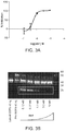


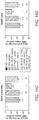


Classifications
-
- A—HUMAN NECESSITIES
- A61—MEDICAL OR VETERINARY SCIENCE; HYGIENE
- A61P—SPECIFIC THERAPEUTIC ACTIVITY OF CHEMICAL COMPOUNDS OR MEDICINAL PREPARATIONS
- A61P21/00—Drugs for disorders of the muscular or neuromuscular system
-
- C—CHEMISTRY; METALLURGY
- C07—ORGANIC CHEMISTRY
- C07K—PEPTIDES
- C07K16/00—Immunoglobulins [IGs], e.g. monoclonal or polyclonal antibodies
- C07K16/18—Immunoglobulins [IGs], e.g. monoclonal or polyclonal antibodies against material from animals or humans
- C07K16/22—Immunoglobulins [IGs], e.g. monoclonal or polyclonal antibodies against material from animals or humans against growth factors ; against growth regulators
-
- A—HUMAN NECESSITIES
- A61—MEDICAL OR VETERINARY SCIENCE; HYGIENE
- A61K—PREPARATIONS FOR MEDICAL, DENTAL OR TOILETRY PURPOSES
- A61K39/00—Medicinal preparations containing antigens or antibodies
- A61K39/395—Antibodies; Immunoglobulins; Immune serum, e.g. antilymphocytic serum
-
- A—HUMAN NECESSITIES
- A61—MEDICAL OR VETERINARY SCIENCE; HYGIENE
- A61K—PREPARATIONS FOR MEDICAL, DENTAL OR TOILETRY PURPOSES
- A61K39/00—Medicinal preparations containing antigens or antibodies
- A61K39/395—Antibodies; Immunoglobulins; Immune serum, e.g. antilymphocytic serum
- A61K39/39533—Antibodies; Immunoglobulins; Immune serum, e.g. antilymphocytic serum against materials from animals
- A61K39/3955—Antibodies; Immunoglobulins; Immune serum, e.g. antilymphocytic serum against materials from animals against proteinaceous materials, e.g. enzymes, hormones, lymphokines
-
- A—HUMAN NECESSITIES
- A61—MEDICAL OR VETERINARY SCIENCE; HYGIENE
- A61P—SPECIFIC THERAPEUTIC ACTIVITY OF CHEMICAL COMPOUNDS OR MEDICINAL PREPARATIONS
- A61P11/00—Drugs for disorders of the respiratory system
-
- A—HUMAN NECESSITIES
- A61—MEDICAL OR VETERINARY SCIENCE; HYGIENE
- A61P—SPECIFIC THERAPEUTIC ACTIVITY OF CHEMICAL COMPOUNDS OR MEDICINAL PREPARATIONS
- A61P13/00—Drugs for disorders of the urinary system
- A61P13/12—Drugs for disorders of the urinary system of the kidneys
-
- A—HUMAN NECESSITIES
- A61—MEDICAL OR VETERINARY SCIENCE; HYGIENE
- A61P—SPECIFIC THERAPEUTIC ACTIVITY OF CHEMICAL COMPOUNDS OR MEDICINAL PREPARATIONS
- A61P15/00—Drugs for genital or sexual disorders; Contraceptives
-
- A—HUMAN NECESSITIES
- A61—MEDICAL OR VETERINARY SCIENCE; HYGIENE
- A61P—SPECIFIC THERAPEUTIC ACTIVITY OF CHEMICAL COMPOUNDS OR MEDICINAL PREPARATIONS
- A61P21/00—Drugs for disorders of the muscular or neuromuscular system
- A61P21/04—Drugs for disorders of the muscular or neuromuscular system for myasthenia gravis
-
- A—HUMAN NECESSITIES
- A61—MEDICAL OR VETERINARY SCIENCE; HYGIENE
- A61P—SPECIFIC THERAPEUTIC ACTIVITY OF CHEMICAL COMPOUNDS OR MEDICINAL PREPARATIONS
- A61P21/00—Drugs for disorders of the muscular or neuromuscular system
- A61P21/06—Anabolic agents
-
- A—HUMAN NECESSITIES
- A61—MEDICAL OR VETERINARY SCIENCE; HYGIENE
- A61P—SPECIFIC THERAPEUTIC ACTIVITY OF CHEMICAL COMPOUNDS OR MEDICINAL PREPARATIONS
- A61P25/00—Drugs for disorders of the nervous system
-
- A—HUMAN NECESSITIES
- A61—MEDICAL OR VETERINARY SCIENCE; HYGIENE
- A61P—SPECIFIC THERAPEUTIC ACTIVITY OF CHEMICAL COMPOUNDS OR MEDICINAL PREPARATIONS
- A61P3/00—Drugs for disorders of the metabolism
-
- A—HUMAN NECESSITIES
- A61—MEDICAL OR VETERINARY SCIENCE; HYGIENE
- A61P—SPECIFIC THERAPEUTIC ACTIVITY OF CHEMICAL COMPOUNDS OR MEDICINAL PREPARATIONS
- A61P3/00—Drugs for disorders of the metabolism
- A61P3/04—Anorexiants; Antiobesity agents
-
- A—HUMAN NECESSITIES
- A61—MEDICAL OR VETERINARY SCIENCE; HYGIENE
- A61P—SPECIFIC THERAPEUTIC ACTIVITY OF CHEMICAL COMPOUNDS OR MEDICINAL PREPARATIONS
- A61P3/00—Drugs for disorders of the metabolism
- A61P3/08—Drugs for disorders of the metabolism for glucose homeostasis
- A61P3/10—Drugs for disorders of the metabolism for glucose homeostasis for hyperglycaemia, e.g. antidiabetics
-
- A—HUMAN NECESSITIES
- A61—MEDICAL OR VETERINARY SCIENCE; HYGIENE
- A61P—SPECIFIC THERAPEUTIC ACTIVITY OF CHEMICAL COMPOUNDS OR MEDICINAL PREPARATIONS
- A61P31/00—Antiinfectives, i.e. antibiotics, antiseptics, chemotherapeutics
- A61P31/12—Antivirals
- A61P31/14—Antivirals for RNA viruses
- A61P31/18—Antivirals for RNA viruses for HIV
-
- A—HUMAN NECESSITIES
- A61—MEDICAL OR VETERINARY SCIENCE; HYGIENE
- A61P—SPECIFIC THERAPEUTIC ACTIVITY OF CHEMICAL COMPOUNDS OR MEDICINAL PREPARATIONS
- A61P35/00—Antineoplastic agents
-
- A—HUMAN NECESSITIES
- A61—MEDICAL OR VETERINARY SCIENCE; HYGIENE
- A61P—SPECIFIC THERAPEUTIC ACTIVITY OF CHEMICAL COMPOUNDS OR MEDICINAL PREPARATIONS
- A61P35/00—Antineoplastic agents
- A61P35/02—Antineoplastic agents specific for leukemia
-
- A—HUMAN NECESSITIES
- A61—MEDICAL OR VETERINARY SCIENCE; HYGIENE
- A61P—SPECIFIC THERAPEUTIC ACTIVITY OF CHEMICAL COMPOUNDS OR MEDICINAL PREPARATIONS
- A61P9/00—Drugs for disorders of the cardiovascular system
-
- A—HUMAN NECESSITIES
- A61—MEDICAL OR VETERINARY SCIENCE; HYGIENE
- A61P—SPECIFIC THERAPEUTIC ACTIVITY OF CHEMICAL COMPOUNDS OR MEDICINAL PREPARATIONS
- A61P9/00—Drugs for disorders of the cardiovascular system
- A61P9/04—Inotropic agents, i.e. stimulants of cardiac contraction; Drugs for heart failure
-
- A—HUMAN NECESSITIES
- A61—MEDICAL OR VETERINARY SCIENCE; HYGIENE
- A61P—SPECIFIC THERAPEUTIC ACTIVITY OF CHEMICAL COMPOUNDS OR MEDICINAL PREPARATIONS
- A61P9/00—Drugs for disorders of the cardiovascular system
- A61P9/12—Antihypertensives
-
- A—HUMAN NECESSITIES
- A61—MEDICAL OR VETERINARY SCIENCE; HYGIENE
- A61K—PREPARATIONS FOR MEDICAL, DENTAL OR TOILETRY PURPOSES
- A61K39/00—Medicinal preparations containing antigens or antibodies
- A61K2039/505—Medicinal preparations containing antigens or antibodies comprising antibodies
-
- A—HUMAN NECESSITIES
- A61—MEDICAL OR VETERINARY SCIENCE; HYGIENE
- A61K—PREPARATIONS FOR MEDICAL, DENTAL OR TOILETRY PURPOSES
- A61K39/00—Medicinal preparations containing antigens or antibodies
- A61K2039/545—Medicinal preparations containing antigens or antibodies characterised by the dose, timing or administration schedule
-
- C—CHEMISTRY; METALLURGY
- C07—ORGANIC CHEMISTRY
- C07K—PEPTIDES
- C07K14/00—Peptides having more than 20 amino acids; Gastrins; Somatostatins; Melanotropins; Derivatives thereof
- C07K14/435—Peptides having more than 20 amino acids; Gastrins; Somatostatins; Melanotropins; Derivatives thereof from animals; from humans
- C07K14/475—Growth factors; Growth regulators
-
- C—CHEMISTRY; METALLURGY
- C07—ORGANIC CHEMISTRY
- C07K—PEPTIDES
- C07K2317/00—Immunoglobulins specific features
- C07K2317/20—Immunoglobulins specific features characterized by taxonomic origin
- C07K2317/24—Immunoglobulins specific features characterized by taxonomic origin containing regions, domains or residues from different species, e.g. chimeric, humanized or veneered
-
- C—CHEMISTRY; METALLURGY
- C07—ORGANIC CHEMISTRY
- C07K—PEPTIDES
- C07K2317/00—Immunoglobulins specific features
- C07K2317/30—Immunoglobulins specific features characterized by aspects of specificity or valency
- C07K2317/33—Crossreactivity, e.g. for species or epitope, or lack of said crossreactivity
-
- C—CHEMISTRY; METALLURGY
- C07—ORGANIC CHEMISTRY
- C07K—PEPTIDES
- C07K2317/00—Immunoglobulins specific features
- C07K2317/50—Immunoglobulins specific features characterized by immunoglobulin fragments
- C07K2317/56—Immunoglobulins specific features characterized by immunoglobulin fragments variable (Fv) region, i.e. VH and/or VL
- C07K2317/565—Complementarity determining region [CDR]
-
- C—CHEMISTRY; METALLURGY
- C07—ORGANIC CHEMISTRY
- C07K—PEPTIDES
- C07K2317/00—Immunoglobulins specific features
- C07K2317/90—Immunoglobulins specific features characterized by (pharmaco)kinetic aspects or by stability of the immunoglobulin
- C07K2317/92—Affinity (KD), association rate (Ka), dissociation rate (Kd) or EC50 value
Definitions
- Embodiments of the present disclosure may include modulators of growth factor activity.
- such modulators may include antibodies and may modulate TGF- ⁇ family member activity and/or biology.
- Myostatin is a secreted growth factor which negatively regulates muscle mass. Loss of function mutations in the Myostatin gene, leading to a hypermuscular phenotype, have been described in cattle, sheep, fish, dogs and humans. Myostatin expression is generally limited to skeletal muscle, with low levels of expression reported in adipose and cardiac tissues. Inhibition of Myostatin signaling leads to an increase in muscle size.
- aspects of the disclosure relate, in some embodiments, to antibodies that bind specifically to forms of Myostatin (e.g., proMyostatin and/or latent Myostatin).
- Myostatin e.g., proMyostatin and/or latent Myostatin.
- antibodies provided herein specifically bind to one or more of a pro-form, and/or a latent-form of Myostatin, such as proMyostatin and/or latent Myostatin.
- the disclosure is based on the surprising discovery of antibodies provided herein that specifically bind pure, or substantially pure, proGDF8 (also referred to as proMyostatin).
- proGDF8 also referred to as proMyostatin
- antibodies provided herein inhibit Myostatin signaling.
- inhibition of Myostatin signaling is useful for increasing muscle mass or preventing muscle atrophy.
- antibodies provided herein bind to and prevent cleavage of Myostatin by a proprotein convertase and/or a tolloid protease. Preventing cleavage of proMyostatin or latent Myostatin, in some embodiments, prevents Myostatin activation. Further aspects of the disclosure relate to antibodies having an affinity to an antigen that is sensitive to pH. In some embodiments, such pH sensitive antibodies are effective for clearing antigens from serum. Furthermore, in some embodiments, antibodies provided herein are sweeping antibodies that can efficiently clear antigens (e.g., proMyostatin and/or latent Myostatin) from serum.
- aspects of the present disclosure include an antibody that comprises a heavy chain variable domain and a light chain variable domain, in which the heavy chain variable domain comprises a complementarity determining region 3 (CDRH3) comprising a sequence as set forth in any one of SEQ ID NOs: 10-11.
- CDRH3 complementarity determining region 3
- an antibody specifically binds to pro/latent-Myostatin.
- the light chain variable domain comprises a complementarity determining region 3 (CDRL3) comprising a sequence as set forth in any one of SEQ ID NO: 22-23.
- said antibody comprises six complementarity determining regions (CDRs): CDRH1, CDRH2, CDRH3, CDRL1, CDRL2, and CDRL3, wherein CDRH1 comprises a sequence as set forth in any one of SEQ ID NOs: 1-3, CDRH2 comprises a sequence as set forth in any one of SEQ ID NOs: 4-9, CDRH3 comprises a sequence as set forth in any one of SEQ ID NOs: 10-11, CDRL1 comprises a sequence as set forth in any one of SEQ ID NOs: 12-17, CDRL2 comprises a sequence as set forth in any one of SEQ ID NOs: 18-21, and CDRL3 comprises a sequence as set forth in any one of SEQ ID NOs: 22-23.
- CDRH1 comprises a sequence as set forth in any one of SEQ ID NOs: 1-3
- CDRH2 comprises a sequence as set forth in any one of SEQ ID NOs: 4-9
- CDRH3 comprises a sequence as set forth in any one of SEQ ID NO
- said CDRH1 comprises a sequence as set forth in SEQ ID NO: 1 or 2
- CDRH2 comprises a sequence as set forth in SEQ ID NO: 4 or 5
- CDRH3 comprises a sequence as set forth in SEQ ID NO: 10
- CDRL1 comprises a sequence as set forth in SEQ ID NO: 12 or 13
- CDRL2 comprises a sequence as set forth in SEQ ID NO: 18 or 19
- CDRL3 comprises a sequence as set forth in SEQ ID NO: 22.
- said CDRH1 comprises a sequence as set forth in SEQ ID NO: 1 or 3
- CDRH2 comprises a sequence as set forth in SEQ ID NO: 6 or 7
- CDRH3 comprises a sequence as set forth in SEQ ID NO: 11
- CDRL1 comprises a sequence as set forth in SEQ ID NO: 14 or 15
- CDRL2 comprises a sequence as set forth in SEQ ID NO: 20 or 21
- CDRL3 comprises a sequence as set forth in SEQ ID NO: 23.
- CDRH1 comprises a sequence as set forth in SEQ ID NO: 1 or 3
- CDRH2 comprises a sequence as set forth in SEQ ID NO: 8 or 9
- CDRH3 comprises a sequence as set forth in SEQ ID NO: 11
- CDRL1 comprises a sequence as set forth in SEQ ID NO: 16 or 17
- CDRL2 comprises a sequence as set forth in SEQ ID NO: 20 or 21
- CDRL3 comprises a sequence as set forth in SEQ ID NO: 23.
- said antibody comprises a heavy chain variable domain sequence as set forth in any one of SEQ ID NOs: 25-29. In some embodiments, said antibody comprises a light chain variable domain sequence of as set forth in any one of SEQ ID NOs: 30-35.
- an antibody that specifically binds to pro/latent-Myostatin and that comprises a heavy chain variable domain and a light chain variable domain, in which the light chain variable domain comprises a complementarity determining region 3 (CDRL3) comprising a sequence as set forth in any one of SEQ ID NO: 22-23.
- said antibody comprises a light chain variable domain sequence of SEQ ID NO: 30.
- polypeptide having a sequence selected from the group consisting of SEQ ID NO: 24, SEQ ID NO: 25, SEQ ID NO: 26, SEQ ID NO: 27, SEQ ID NO: 28, and SEQ ID NO 29.
- the polypeptide is a variable heavy chain domain.
- the polypeptide is at least 75% (e.g., 80%, 85%, 90%, 95%, 98%, or 99%) identical to any one of the amino acid sequences set forth in SEQ ID NO: 24, SEQ ID NO: 25, SEQ ID NO: 26, SEQ ID NO: 27, SEQ ID NO: 28, or SEQ ID NO 29.
- polypeptide having a sequence selected from the group consisting of SEQ ID NO: 30, SEQ ID NO: 31, SEQ ID NO: 32, SEQ ID NO: 33, SEQ ID NO: 34, and SEQ ID NO 35.
- the polypeptide is a variable light chain domain.
- the polypeptide is at least 75% (e.g., 80%, 85%, 90%, 95%, 98%, or 99%) identical to any one of the amino acid sequences set forth in SEQ ID NO: 30, SEQ ID NO: 31, SEQ ID NO: 32, SEQ ID NO: 33, SEQ ID NO: 34, or SEQ ID NO 35.
- Another aspect of the disclosure includes an antibody that competes for binding to pro/latent-Myostatin with an antibody described above.
- said antibody binds to pro/latent-Myostatin at the same epitope as an antibody described above.
- an antibody competes for binding to pro/latent-Myostatin with an equilibrium dissociation constant, Kd, between the antibody and pro/latent-Myostatin of is less than 10 ⁇ 6 M.
- said antibody's Kd is in a range of 10 ⁇ 11 M to 10 ⁇ 6 M.
- an antibody is a humanized antibody, a diabody, a chimeric antibody, a Fab fragment, a F(ab′)2 fragment, or an Fv fragment.
- an antibody is a humanized antibody.
- an antibody is a human antibody.
- an antibody comprises a framework having a human germline sequence.
- an antibody comprises a heavy chain constant domain selected from the group consisting of IgG, IgG1, IgG2, IgG2A, IgG2B, IgG2C, IgG3, IgG4, IgA1, IgA2, IgD, IgM, and IgE constant domains.
- an antibody comprises a constant domain of IgG4.
- an antibody comprises a constant domain of IgG4 having a backbone substitution of Ser to Pro that produces an IgG1-like hinge and permits formation of inter-chain disulfide bonds.
- an antibody is conjugated to an agent selected from the group consisting of a fluorescent agent, a luminescent agent, an enzymatic agent and a radioactive agent.
- an antibody specifically binds to pro/latent-Myostatin compared with mature myostatin. In some embodiments, an antibody specifically binds to pro/latent-Myostatin compared with another member of the transforming growth factor Beta family In another embodiment, said member is GDF11 or Activin.
- a further aspect of the disclosure includes an antibody that specifically binds pro/latent-Myostatin and that inhibits proteolytic formation of mature myostatin by a tolloid protease.
- said antibody inhibits proteolytic formation of mature myostatin by a tolloid protease with an IC50 of less than 1 ⁇ M.
- an antibody is cross-reactive with human and murine pro/latent-Myostatin.
- the antibody specifically binds to pro/latent-Myostatin compared with GDF11 or Activin.
- an antibody specifically binds to pro/latent-Myostatin compared with mature myostatin.
- Another aspect of the disclosure encompasses a method of reducing myostatin receptor activation in cells present in a medium comprising pro/latent-Myostatin, the method comprising delivering to the medium an antibody described above in an amount effective for inhibiting proteolytic activation of the pro/latent-Myostatin.
- the medium further comprises a proprotein convertase.
- the medium further comprises a tolloid protease.
- an antibody is delivered to the medium in an amount effective for inhibiting proteolytic activation of the pro/latent-Myostatin by the tolloid protease.
- the cell is in vitro. In other embodiments, the cell is in vivo.
- the myopathy is a primary myopathy.
- the primary myopathy comprises disuse atrophy.
- the disuse atrophy is associated with hip fracture, elective joint replacement, critical care myopathy, spinal cord injury or stroke.
- the myopathy is a secondary myopathy, in which muscle loss is secondary to a disease pathology.
- the secondary myopathy comprises denervation, genetic muscle weakness or cachexia.
- the secondary myopathy is a denervation associated with amyotrophic lateral sclerosis or spinal muscular atrophy.
- the secondary myopathy is a genetic muscle weakness associated with a muscular dystrophy.
- the secondary myopathy is a cachexia associated with renal failure, AIDS, a cardiac condition, cancer or aging.
- Another aspect of the disclosure includes a method of treating a subject having a disease or condition related to aging.
- diseases and conditions related to ageing include, without limitation, sarcopenia (age-related muscle loss), frailty, and androgen deficiency.
- Another aspect of the disclosure includes a method of treating a subject having a disease or condition related to disuse atrophy/trauma.
- diseases and conditions related to disuse atrophy/trauma include, without limitation, muscle weakness related to time spent in an intensive care unit (ICU), hip/joint replacement, hip fracture, stroke, bed rest, SCI, rotator cuff injury, knee replacement, bone fracture, and burns.
- ICU intensive care unit
- hip/joint replacement hip fracture
- stroke stroke
- bed rest bed rest
- SCI rotator cuff injury
- knee replacement bone fracture
- burns burns.
- Another aspect of the disclosure includes a method of treating a subject having a neurodegenerative disease or condition.
- exemplary neurodegenerative diseases or conditions include, without limitation, spinal muscular atrophy and amyotrophic lateral sclerosis (ALS).
- ALS amyotrophic lateral sclerosis
- Another aspect of the disclosure includes a method of treating a subject having a disease or condition related to Cachexia.
- diseases and conditions related to cachexia include, without limitation, cancer, chronic heart failure, acquired immune deficiency syndrome (AIDS), chronic obstructive pulmonary disease (COPD), and chronic kidney disease (CKD).
- AIDS acquired immune deficiency syndrome
- COPD chronic obstructive pulmonary disease
- CKD chronic kidney disease
- Another aspect of the disclosure includes a method of treating a subject having a disease or condition related to rare diseases.
- rare diseases and conditions include, without limitation, osteogenesis imperfecta, sporadic Inclusion body myositis, and acute lymphoblastic leukemia.
- Another aspect of the disclosure includes a method of treating a subject having a disease or condition related to a metabolic disorder and/or body composition.
- the disease or condition is obesity (e.g., severe obesity), Prader-Willi, type II diabetes, or anorexia.
- additional diseases or conditions related to metabolic disorders and/or body composition are within the scope of this disclosure.
- Another aspect of the disclosure includes a method of treating a subject having a disease or condition related to congenital myopathies.
- congenital myopathies include, without limitation, X-linked myotubular myopathy, autosomal dominant centronuclear myopathy, autosomal recessive centronuclear myopathy, nemaline myopathy, and congenital fiber-type disproportion myopathy.
- Another aspect of the disclosure includes a method of treating a subject having a disease or condition related to muscular dystrophies.
- muscular dystrophies include, without limitation, Duchenne's, Becker's, facioscapulohumeral (FSH), and Limb-Girdle muscular dystrophies.
- Another aspect of the disclosure includes a method of treating a subject having a urogynecological related disease or condition, glottic disorders (stenosis), extraocular myopathy, carpel tunnel, Guillain-Barré, or osteosarcoma.
- glottic disorders stenosis
- extraocular myopathy carpel tunnel
- Guillain-Barré or osteosarcoma
- treatment results in improved muscle strength in the subject. In other embodiments, treatment results in improved metabolic status in the subject.
- an antibody is administered at a dose in a range of 0.1 mg/kg to 100 mg/kg. In another embodiment, an antibody is administered at a dose in a range of 0.3 mg/kg to 30 mg/kg.
- an antibody is administered to the subject intravenously. In other embodiments, an antibody is administered to the subject subcutaneously. In another embodiment, an antibody is administered to the subject on multiple occasions. In some embodiments, said multiple administrations are performed at least monthly. In another embodiment, said multiple administrations are performed at least weekly.
- a further aspect of the disclosure includes a composition comprising any antibody described above and a carrier.
- said carrier is a pharmaceutically acceptable carrier.
- an antibody and carrier are in a lyophilized form.
- an antibody and carrier are in solution.
- an antibody and carrier are frozen.
- an antibody and carrier are frozen at a temperature less than or equal to ⁇ 65° C.
- CDRL1 comprises a sequence as set forth in any one of SEQ ID NOs: 12-17.
- CDRL2 comprises a sequence as set forth in any one of SEQ ID NOs: 18-21.
- nucleic acid comprising a sequence as set forth in any one of SEQ ID NOs: 38-49.
- Another aspect of the disclosure includes an isolated cell comprising an isolated nucleic acid described above.
- the present disclosure includes methods of assessing a biological sample obtained from a subject having a myopathy.
- the method comprises: preparing an immunological reaction mixture that comprises protein of a biological sample obtained from the subject and an antibody that specifically binds pro/latent-Myostatin; maintaining the immunological reaction mixture under conditions that permit binding complexes to form between the antibody and a pro/latent-Myostatin; and determining the extent of binding complex formation.
- the method comprises: preparing an immunological reaction mixture that comprises protein of a biological sample obtained from the subject and an antibody that specifically binds pro-Myostatin; maintaining the immunological reaction mixture under conditions that permit binding complexes to form between the antibody and a pro-Myostatin; and determining the extent of binding complex formation.
- the method comprises: preparing an immunological reaction mixture that comprises protein of a biological sample obtained from the subject and an antibody that specifically binds latent-Myostatin; maintaining the immunological reaction mixture under conditions that permit binding complexes to form between the antibody and a latent-Myostatin; and determining the extent of binding complex formation.
- the method comprises: preparing an immunological reaction mixture that comprises protein of a biological sample obtained from the subject and an antibody that specifically binds mature-Myostatin; maintaining the immunological reaction mixture under conditions that permit binding complexes to form between the antibody and a mature-Myostatin; and determining the extent of binding complex formation.
- an isolated antibody comprising a heavy chain variable region comprising an amino acid sequence of SEQ ID NO:25 and a light chain variable region comprising an amino acid sequence of SEQ ID NO:31.
- the antibody comprises a heavy chain comprising an amino acid sequence of SEQ ID NO:50.
- the antibody comprises a light chain comprising an amino acid sequence of SEQ ID NO:51.
- an isolated antibody comprising a heavy chain variable region comprising a CDRH1 sequence comprising SEQ ID NO:1, a CDRH2 sequence comprising SEQ ID NO:6, and a CDRH3 sequence comprising SEQ ID NO:11; and a light chain variable region comprising a CDRL1 sequence comprising SEQ ID NO:14, a CDRL2 sequence comprising SEQ ID NO:20, and a CDRL3 sequence comprising SEQ ID NO:23.
- the heavy chain variable region comprises a sequence of SEQ ID NO:26. In another embodiment, the light chain variable region comprises a sequence of SEQ ID NO:32.
- the heavy chain variable region comprises a sequence of SEQ ID NO:27. In another embodiment, the light chain variable region comprises a sequence of SEQ ID NO:33
- an isolated antibody comprising a heavy chain variable region comprising a CDRH1 sequence comprising SEQ ID NO:1, a CDRH2 sequence comprising SEQ ID NO:8, and a CDRH3 sequence comprising SEQ ID NO:11; and a light chain variable region comprising a CDRL1 sequence comprising SEQ ID NO:16, a CDRL2 sequence comprising SEQ ID NO:20, and a CDRL3 sequence comprising SEQ ID NO:23.
- the heavy chain variable region comprises a sequence of SEQ ID NO:28. In another embodiment, the light chain variable region comprises a sequence of SEQ ID NO:34.
- the heavy chain variable region comprises a sequence of SEQ ID NO:29. In one embodiment, the light chain variable region comprises a sequence of SEQ ID NO:35.
- the antibody is a human antibody. In one embodiment, the antibody comprises an IgG4 constant domain. In one embodiment, the antibody comprises an IgG4 constant domain having a backbone substitution of Ser to Pro that produces an IgG1-like hinge and permits formation of inter-chain disulfide bonds.
- the antibody specifically binds to pro/latent-myostatin. In one embodiment, the antibody specifically binds to pro-myostatin. In another embodiment, the antibody specifically binds to latent-myostatin. In one embodiment, the antibody does not bind to mature myostatin.
- the antibody inhibits proteolytic formation of mature myostatin by tolloid protease. In one embodiment, the antibody inhibits proteolytic formation of mature myostatin by tolloid protease with an IC50 of less than 1 ⁇ M.
- the antibody is cross-reactive with human and murine pro/latent-myostatin. In another embodiment, the antibody binds to pro/latent-myostatin but does not bind to GDF11 or activin.
- a method of reducing myostatin receptor activation in cells present in a medium comprising pro/latent-myostatin comprising delivering to the medium an antibody described herein in an amount effective for inhibiting proteolytic activation of the pro/latent-myostatin.
- the medium comprises a proprotein convertase.
- the medium comprises a tolloid protease.
- the cell is in vitro. In another embodiment, the cell is in vivo.
- disclosed herein is a method of treating a subject having a myopathy, the method comprising administering to the subject an effective amount of an antibody disclosed herein.
- the myopathy is a primary myopathy.
- the primary myopathy is disuse atrophy.
- the disuse atrophy is associated with hip fracture, elective joint replacement, critical care myopathy, spinal cord injury, and/or stroke.
- the myopathy is a secondary myopathy in which muscle loss is secondary to a disease pathology.
- the secondary myopathy comprises denervation, genetic muscle weakness, or cachexia.
- the secondary myopathy is a denervation associated with amyotrophic lateral sclerosis or spinal muscular atrophy.
- the secondary myopathy is a genetic muscle weakness associated with a muscular dystrophy.
- the secondary myopathy is a cachexia associated with renal failure, AIDS, a cardiac condition, cancer, or aging.
- the administering results in improved muscle strength in the subject. In one embodiment, the administering results in improved metabolic status in the subject.
- the antibody is administered at a dose in a range of 0.1 mg/kg to 100 mg/kg. In another embodiment, the antibody is administered at a dose in a range of 0.3 mg/kg to 30 mg/kg.
- the antibody is administered to the subject intravenously. In another embodiment, the antibody is administered to the subject subcutaneously.
- the antibody is administered to the subject on multiple occasions. In one embodiment, the multiple administrations are performed at least monthly. In another embodiment, the multiple administrations are performed at least weekly.
- a pharmaceutical composition comprising an antibody disclosed herein and a pharmaceutically acceptable carrier.
- the composition is a lyophilized composition.
- the composition is a liquid composition.
- the composition is frozen.
- the composition is frozen at a temperature less than or equal to ⁇ 65° C.
- a syringe comprising a pharmaceutical composition described herein.
- an isolated nucleic acid encoding an antibody comprising a heavy chain variable region comprising a nucleic acid sequence of SEQ ID NO:39 and a light chain variable region comprising a nucleic acid sequence of SEQ ID NO:45.
- an isolated nucleic acid encoding an antibody comprising a heavy chain variable region comprising a CDRH1 sequence comprising SEQ ID NO:1, a CDRH2 sequence comprising SEQ ID NO:6, and a CDRH3 sequence comprising SEQ ID NO:11; and a light chain variable region comprising a CDRL1 sequence comprising SEQ ID NO:14, a CDRL2 sequence comprising SEQ ID NO:20, and a CDRL3 sequence comprising SEQ IDNO:23.
- the heavy chain variable region comprises a sequence of SEQ ID NO:40. In one embodiment, the light chain variable region comprises a sequence of SEQ ID NO:46.
- the heavy chain variable region comprises a sequence of SEQ ID NO:41. In another embodiment, the light chain variable region comprises a sequence of SEQ ID NO:47.
- an isolated nucleic acid encoding an antibody comprising a heavy chain variable region comprising a CDRH1 sequence comprising SEQ ID NO:1, a CDRH2 sequence comprising SEQ ID NO:8, and a CDRH3 sequence comprising SEQ ID NO:11; and a light chain variable region comprising a CDRL1 sequence comprising SEQ ID NO:16, a CDRL2 sequence comprising SEQ ID NO:20, and a CDRL3 sequence comprising SEQ IDNO:23.
- the heavy chain variable region comprises a sequence of SEQ ID NO:42. In another embodiment, the light chain variable region comprises a sequence of SEQ ID NO:48.
- the heavy chain variable region comprises a sequence of SEQ ID NO:43. In another embodiment, the light chain variable region comprises a sequence of SEQ ID NO:49.
- an isolated cell comprising an isolated nucleic acid described herein.
- FIGS. 1A-1B show Myostatin domain structure and proMyostatin assembly.
- FIG. 1A shows Myostatin secreted as a proprotein, with an inhibitory prodomain followed by a C-terminal growth factor domain, which exists as a disulfide-linked dimer.
- FIG. 1B shows precursor protein assembled in an inactive conformation where the prodomain (dark gray) encloses the growth factor (light gray) with a “straightjacket” assembly. This figure is an adaption from the structure of latent TGF ⁇ 1 (Shi et al. Nature 2011).
- FIG. 2 shows the activation of Myostatin involves two distinct protease events, generating three major Myostatin species.
- the biosynthetic precursor protein, proMyostatin is processed by two separate proteases.
- Cleavage of proMyostatin (and proGDF11) is carried out by a proprotein convertase, such as Furin/PACE3 (Paired Basic Amino acid Cleaving Enzyme 3) or PCSK5 (Proprotein Convertase Subtilisin/Kexin type 5), which cuts at a conserved RXXR site between the prodomain and mature growth factor.
- This cleavage produces a latent complex, in which the mature growth factor is shielded from binding to its receptors by the prodomain.
- Activation and release of the active growth factor is accomplished after cleavage by an additional protease from the BMP/tolloid family, such as TLL-2 (Tolloid-like protein 2) or BMP1 (Bone Morphogenetic Protein 1). These cleavage events yield a mature form of Myostatin, which may be referred to as Active Myostatin or Mature Myostatin.
- BMP/tolloid family such as TLL-2 (Tolloid-like protein 2) or BMP1 (Bone Morphogenetic Protein 1).
- FIGS. 3A-3C show that Ab1 blocks cleavage of proMyostatin by members of the tolloid family of proteases.
- Latent Myostatin samples preincubated with increasing amounts of Ab1, were analyzed in a myostatin activation assay.
- myostatin release by reporter assay FIG. 3A
- samples were then run under reducing conditions and probed by western blot with an antibody raised towards the prodomain of Myostatin ( FIG. 3B ).
- An ⁇ 18 kDa band (box) corresponding to the ARM portion of the prodomain generated after tolloid cleavage, decreased proportionally with increasing doses of Ab1.
- the latent and proMyostatin standards show the migration of proMyostatin at ⁇ 50 kDa, and the prodomain at ⁇ 37 kDa.
- FIG. 3C shows the activation of Myostatin involves two distinct protease events, generating three major Myostatin species.
- the biosynthetic precursor protein, proMyostatin is processed by two separate proteases.
- proMyostatin and proGDF11
- Cleavage of proMyostatin is carried out by a proprotein convertase, such as Furin/PACE3 (Paired Basic Amino acid Cleaving Enzyme 3) or PCSK5 (Proprotein Convertase Subtilisin/Kexin type 5), which cuts at a conserved RXXR site between the prodomain and mature growth factor.
- This cleavage produces a latent complex, in which the mature growth factor is shielded from binding to its receptors by the prodomain.
- FIG. 3B which illustrates the potential inhibition of a tolloid protease, blocking further cleavage of proMyostatin.
- Activation and release of the active growth factor is accomplished after cleavage by an additional protease from the BMP/tolloid family, such as TLL-2 (Tolloid-like protein 2) or BMP1 (Bone Morphogenetic Protein 1).
- FIG. 4 shows the performance of the parental Ab1 antibody and other candidates in the cell-based reporter assay. Following an overnight proteolysis reaction with enzymes from both the proprotein-convertase and tolloid protease families, the release of mature growth factor was measured using a CAGA-based reporter assay in 293T cells. Results were compared to control reactions to calculate the fraction of proMyostatin or proGDF11 which was released in the assay. Standard deviation for an average of 3 replicates is shown, but not visible on the graph for most data points due to their low magnitude.
- FIG. 5 shows graphically that Ab1, Ab2, Ab4, and Ab6 antibodies do not inhibit proGDF11 activation.
- FIG. 6 shows results of an assay evaluating mean percent body weight change. Animals were weighed daily and the percent weight change from Day 0 was calculated. Data represent group means ⁇ SEM. The mean percent change data for each group on day 42 of the study were analyzed using a one-way ANOVA followed by Holm-Sidak's post hoc test in comparison to the PBS Control Group, **p ⁇ 0.01.
- FIGS. 7A-7D show results of an assay evaluating tissue weights.
- FIG. 7A shows the mean gastrocnemius weight.
- FIG. 7B shows the mean pectoralis weight.
- FIG. 7C shows the mean soleus weight.
- FIG. 7D shows the mean triceps weight.
- Statistical evaluation was performed using a one-way ANOVA followed by Holm-Sidak's post hoc test in comparison to the Vehicle Control Group (Group 1). Data represent group means ⁇ SEM. **p ⁇ 0.01. Bars indicate from left-to-right Groups 1-5.
- FIGS. 8A-8C show results of an assay evaluating tissue weights.
- FIG. 8A shows the mean tibialis anterior weight.
- FIG. 8B shows the mean diaphragm weight.
- FIG. 8C shows the mean quadriceps weight.
- Statistical evaluation was performed using a one-way ANOVA followed by Holm-Sidak's post hoc test in comparison to the Vehicle Control Group (Group 1). Data represent group means ⁇ SEM. *p ⁇ 0.05. Bars indicate from left-to-right Groups 1-5.
- FIGS. 9A-9B show results of an assay evaluating mean percent body weight and lean mass change.
- FIG. 9A is a graph showing the calculated percent weight change from Day 0 in animals weighed twice weekly throughout the study.
- animals underwent EchoMRI (QNMR) to measure body composition on days ⁇ 4, 7, 14, 21, and 28 and percent lean mass change from Day 0 was calculated.
- Data represent group means ⁇ SEM.
- For both body weight and lean mass the mean percent change data for each group on day 28 of the study were analyzed using a one-way ANOVA followed by Holm-Sidak's post hoc test in comparison to the IgG Control Group (Group 2). ***p ⁇ 0.0005, **p ⁇ 0.005, *p ⁇ 0.05, ns (not significant).
- FIGS. 10A-10D are graphs showing results of an assay evaluating muscle weights.
- FIG. 10A shows the mean quadriceps weight
- FIG. 10B shows the mean gastrocnemius weight
- FIG. 10C shows the mean tibialis anterior weight
- FIG. 10D shows the mean diaphragm weight.
- Percent difference in mean muscle weights of the Ab1 treated groups compared to the IgG control group is noted above each bar.
- Statistical evaluation was performed using a one-way ANOVA followed by Holm-Sidak's post hoc test in comparison to the IgG Control Group (Group 2). Data represent group means ⁇ SEM. ****p ⁇ 0.0001, ***p ⁇ 0.0005, **p ⁇ 0.005, *p ⁇ 0.05, ns (not significant).
- FIGS. 11A-11B show results of an assay evaluating mean percent body weight and lean mass change.
- FIG. 11A shows the percent weight change from Day 0 calculated from animals weighed twice weekly throughout the study.
- FIG. 11B Animals underwent EchoMRI (QNMR) to measure body composition on days ⁇ 1, 6, and 13 and percent lean mass change from Day ⁇ 1 was calculated.
- PBS phosphate buffered saline
- Dex dexamethasone
- IgG (20) IgG control antibody dosed at 20 mg/kg/wk
- Ab1 (20) Ab1 antibody dosed at 20 mg/kg/wk
- Ab1 (2) Ab1 antibody dosed at 2 mg/kg/wk.
- Data represent group means ⁇ SEM.
- FIGS. 12A-12D are graphs showing results of an assay evaluating the weights of different muscles.
- FIG. 12A shows the mean gastrocnemius weight (grams)
- FIG. 12B shows the mean quadriceps weight (grams)
- FIG. 12C shows the mean percent gastrocnemius weight change versus the control animals treated with PBS (IP) and normal drinking water (Group 1)
- FIG. 12D shows the mean percent quadriceps weight change versus the control animals treated with PBS (IP) and normal drinking water (Group 1).
- ns (not significant). Bars indicate from left-to-right, PBS, water; PBS, dex; IgG Control; Ab1(20); and Ab1(2).
- FIGS. 13A-13B show results of an assay evaluating the mean percent body weight and lean mass change.
- FIG. 13A shows the percent weight change from Day 0 calculated for animals who were weighed twice weekly throughout the study.
- FIG. 13B shows the percent lean mass change from Day ⁇ 1 calculated from animals who underwent EchoMRI (QNMR) to measure body composition on days ⁇ 1, 7, and 14.
- PBS phosphate buffered saline
- IgG (20) IgG control antibody dosed at 20 mg/kg/wk
- Ab1 (20) Ab1 antibody dosed at 20 mg/kg/wk
- Ab1 (2) Ab1 antibody dosed at 2 mg/kg/wk.
- Data represent group means ⁇ SEM.
- FIGS. 14A-14D show results of an assay evaluating muscle weights.
- FIG. 14A shows the mean gastrocnemius weight from the casted leg (grams)
- FIG. 14B shows the mean quadriceps weight from the casted leg (grams)
- FIG. 14C shows the mean percent gastrocnemius weight change versus the control animals treated with PBS (IP) and not casted (Group 1)
- FIG. 14D shows the mean percent quadriceps weight change versus the control animals treated with PBS (IP) and not casted (Group 1).
- ns (not significant). Bars indicate from left-to-right, PBS, no cast; PBS, casted; IgG Control(2), casted; Ab1(20), casted; and Ab1(2), casted.
- FIG. 15 shows results of an assay evaluating the lean mass change at Day 21 (top right) and Day 28 (top left). It also depicts the percent change in lean mass at three different doses, 20 mg/kg/wk (bottom left), 2 mg/kg/wk (bottom middle), and 0.5 mg/kg/wk (bottom right) of the tested antibodies, PBS control, and IgG control.
- Statistical evaluation was performed using a one-way ANOVA followed by a Dunnett's multiple comparisons test vs. group 1 (****p ⁇ 0.0001, ***p ⁇ 0.005, **p ⁇ 0.01, *p ⁇ 0.05) and vs. the IgG control.
- bars from left-to-right are: PBS; IgG Ctrl 20 mg/kg/wk; Ab1 20 mg/kg/wk; Ab1 2 mg/kg/wk; Ab1 0.5 mg/kg/wk; Ab2 20 mg/kg/wk; Ab2 2 mg/kg/wk; Ab2 0.5 mg/kg/wk; Ab4 20 mg/kg/wk; Ab4 2 mg/kg/wk; Ab4 0.5 mg/kg/wk; Ab6 20 mg/kg/wk; Ab6 2 mg/kg/wk; and Ab6 0.5 mg/kg/wk.
- the data points corresponding to day 28 post dosing, from top to bottom correspond to Ab1, Ab4, Ab2, Ab6, IgG control, and PBS.
- the data points corresponding to day 28 post dosing, from top to bottom correspond to Ab2, Ab1, Ab6, Ab4, IgG control, and PBS.
- the data points, corresponding to day 28 post dosing, from top to bottom correspond to IgG control Ab1, Ab2, PBS, Ab4, and Ab6.
- FIGS. 16A-16B show the domain structure and evaluation of Myostatin precursor forms.
- FIG. 16A shows the domain structure of proMyostatin and latent Myostatin, with protease cleavage sites indicated.
- FIG. 16B shows partially proprotein convertase cleaved proMyostatin run on an SDS PAGE gel. Under reducing conditions, the protein bands consisted of the proMyostatin monomer ( ⁇ 50 kD), prodomain ( ⁇ 37 kD) and growth factor (12.5 kD).
- FIGS. 17A-17B show Ab1 is specific for Myostatin.
- FIG. 17A shows Ab1 binds specifically to proMyostatin and latent Myostatin, with no binding observed to other members of the TGFB superfamily, most notably the corresponding forms of GDF11.
- Ab1 was administered at a high concentration (50 ug/mL) to Forte-Bio BLI tips coated with the indicated antigen and the on and off rates were measured to obtain an approximate Kd value.
- the magnitude of biosensor response, indicating a binding event is graphically represented by black bars, and the calculated Kd is indicated in orange.
- FIG. 17B shows that Ab1 blocks the activation of proMyostatin, but not proGDF11.
- FIGS. 18A-18C show the SCID dose response with the candidate antibodies.
- FIG. 18A shows the muscle weight of the gastrocnemius and
- FIG. 18B shows the muscle weight of the quadriceps.
- FIG. 18C shows the percent changes in mean muscle weight compared to the PBS control. The bars in FIGS.
- PBS PBS
- IgG Ctrl 20 mg/kg/wk Ab1 20 mg/kg/wk; Ab1 2 mg/kg/wk; Ab1 0.5 mg/kg/wk; Ab2 20 mg/kg/wk; Ab2 2 mg/kg/wk; Ab2 0.5 mg/kg/wk; Ab4 20 mg/kg/wk; Ab4 2 mg/kg/wk; Ab4 0.5 mg/kg/wk; Ab6 20 mg/kg/wk; Ab6 2 mg/kg/wk; and Ab6 0.5 mg/kg/wk.
- FIG. 20 is a schematic illustrating an assay that reconstitutes Myostatin activation in vitro.
- FIGS. 21A-21B show the heavy chain ( FIG. 21A ; SEQ ID NO: 50) and light chain ( FIG. 21B ; SEQ ID NO: 51) of a humanized monoclonal antibody (Ab2) of the IgG4 subtype with Proline substituted for Serine.
- Ab2 humanized monoclonal antibody
- This generates an IgG1-like hinge sequence and minimizes the incomplete formation of inter-chain disulfide bridges which is characteristic of IgG4.
- the complementarity-determining regions (CDRs) are underlined.
- Bolded NST sequence N-linked glycosylation consensus sequence site; Bolded DP sequences are potential cleavage sites; Bolded NX sequences, wherein X can be S, T, or G are potential deamidation sites; Bolded DX sequences, wherein X can be G, S, T, or SDG are potential isomerization sites; Bolded methionines are potential methionine oxidation sites; Bolded Q is an expected N-terminal pyroglutamic acid.
- FIG. 22 is a schematic showing the reduced immunogenicity risk by germlining 24H4 (WT) contains 5 non-germline amino acids within framework regions, as indicated in the schematic.
- FIGS. 24A-24B show sequence alignments of the variable heavy regions ( FIG. 24A ) and variable light regions ( FIG. 24B ) of parental Ab1 with affinity optimized variants, Ab3 and Ab5. Sequence identifiers from top to bottom correspond to SEQ ID NOs.: 24, 26, 28 ( FIG. 24A ). Sequence identifiers from top to bottom correspond to SEQ ID NOs.: 30, 32, 34 ( FIG. 24B ). Complementarity-determining regions (CDRs) are defined using the Kabat (underlined) and IMGT nomenclature (bold). Substitutions from parental Ab1 are shown in light gray.
- CDRs Complementarity-determining regions
- FIG. 26 shows the quantitation of changes in pro and latent Myostatin in muscle and plasma.
- the bars from left to right show proMyostatin, latent myostatin, proMyostatin, latent myostatin, and latent myostatin.
- FIG. 27 shows that Ab2 uniquely recognizes proMyostatin and latent Myostatin, binding to the major forms of Myostatin in both serum and muscle.
- Non-reducing Western blot for prodomain (darker gray) and the mature growth factor (lighter gray).
- Recombinant proMyostatin (rProMyostatin) shows the migration of proMyostatin and myostatin prodomain (latent myostatin) on the gel, highlighted by arrows.
- serum both Ab2 and AbMyo bind to latent Myostatin (prodomain band) and multiple partially processed precursors, however only Ab2 recognized proMyostatin (top band).
- In muscle Ab2 precipitated proMyostatin, with no interaction of AbMyo with proMyostatin in the muscle tissue.
- FIGS. 28A-28B provide a model for Myostatin flux in normal and atrophic muscle.
- proMyostatin is produced in muscle and converted to latent Myostatin through cleavage by Furin protease, which may occur either inside or outside of the cell. Some fraction of the latent Myostatin in muscle is then released into the circulation, forming a circulating pool of latent Myostatin.
- muscle atrophy FIG. 28B
- an increase in the active Myostatin growth factor is caused by upregulation of proMyostatin levels in muscle and increased conversion of latent Myostatin to the active growth factor.
- circulating latent Myostatin is decreased as the muscle pool of latent Myostatin is redirected towards formation of mature Myostatin by mTLL2 cleavage.
- FIG. 29 shows detection of Ab2 (top line) and IgG control (bottom line) antibody in serum from dosed rats.
- Ab2 exhibits elevated levels in the circulation as compared to the IgG control, with an average of 17.1 ⁇ g/ml of Ab2 in serum at the end of the study.
- Ab2 levels determined by human IgG-specific ELISA with known quantities of each antibody used as a reference standard.
- FIGS. 30A-30B shows pharmacodynamic effects of Ab2 in treated rats.
- FIGS. 31A-31B shows pro/latent-Myostatin pro/latent-Myostatin levels in Ab2-treated rats.
- FIG. 31A shows treatment with Ab2 (top line) increases latent myostatin levels in rat serum by ⁇ 20-fold.
- FIG. 31B shows that in rat muscle (rectus femoris), Ab2 treatment leads to a 1.9 ⁇ increase in the latent form of Myostatin.
- the bars from left to right correspond to proMyostatin, latent Myostatin, proMyostatin, and latent Myostatin. No statistically significant change in proMyostatin is observed in rat muscle.
- FIG. 32 shows treatment with Ab2 (Ab2) or with the comparator antibody (AbMyo) leads to increased lean mass by as early 7 days after antibody dosing. Increases in lean mass are equivalent for Ab2 and AbMyo until 21 days after dosing. By 28 days after dosing, however, increases in lean mass are lost in the AbMyo-treated group, while increases in the Ab2-treated group are maintained throughout the duration of the study.
- the top line corresponds to Ab2
- the middle line correspond to AbMyo
- the bottom line corresponds to IgG Control (5 mg/kg).
- FIG. 33 shows that after a single, 5 mg/kg dose of Ab2 (top line) or of comparator antibody (AbMyo; bottom line), serum levels of drug were measured using an anti-human IgG ELISA. Drug is detected in serum as early as 1 hour after dosing, and levels>1 ⁇ g/ml of both antibodies can be detected throughout the study. However, Ab2 exhibits a significantly longer half-life and inferred area under the curve (AUCINF) than AbMyo, suggesting that, at similar doses, Ab2 exhibits significantly greater exposure than AbMyo.
- AUCINF area under the curve
- FIG. 34 shows serum Myostatin was measured in drug treated mice and in controls using fluorescent western blotting. Despite the increased serum exposure of Ab2, serum latent Myostatin levels in both Ab2- and AbMyo-treated mice were similar. These data suggest that the circulating levels of free drug are sufficiently in excess to the level of target that the increased serum exposure of Ab2 does not lead to a greater increase in circulating latent Myostatin than is observed in the AbMyo group. Groups of data from left to right correspond to IgG, Ab2, AbMyo, IgG, Ab2, and AbMyo.
- FIGS. 35A-35B shows relative levels of latent and proMyostatin were measured in mouse muscle lysates by fluorescent western blot.
- FIG. 35A shows latent Myostatin is elevated in both Ab2- and AbMyo-treated muscles. However, elevation of latent Myostatin in AbMyo-treated muscles returns to baseline by day 28, while those in Ab2 treated muscles remain elevated until at least this time (P ⁇ 0.003 vs. AbMyo treatment).
- FIG. 36A shows effects of treatment with Ab2 on muscle mass and function in mice.
- FIG. 36B shows effects of treatment with Ab2 on maximal force generation in mice.
- Myostatin is a member of the TGF ⁇ superfamily, and belongs to a subfamily including two members: Myostatin (also known as GDF8) and GDF11. Like other members of the TGF ⁇ superfamily, Myostatin and GDF11 are both initially expressed as inactive precursor polypeptides (termed proMyostatin and proGDF11, respectively).
- the domain structure and nomenclature are shown in FIG. 1A .
- FIG. 1B illustrates a cartoon model of the overall structure of proMyostatin, where the mature growth factor is held locked in a cage comprised of two alpha helices connected by a loop termed the “latency lasso”.
- Activation and release of the mature growth factor is accomplished by several discrete protease cleavage events, outlined in FIG. 2 .
- the first cleavage step of proMyostatin and proGDF11 is carried out by a proprotein convertase, which cuts at a conserved RXXR site between the prodomain and mature growth factor. This cleavage produces a latent complex, in which the mature growth factor is shielded from binding to its receptors by the prodomain.
- Activation and release of the mature, active Myostatin growth factor is accomplished after cleavage by an additional protease from the BMP/tolloid family, such as mTLL-2 ( FIG. 2 ).
- proGDF8 sequences in the human, rat, mouse and cynomolgus are provided below.
- a proprotein convertase cleavage site is indicated in bold and a tolloid protease site is indicated by underlining.
- the proprotein convertase cleavage site comprises amino acid residues 240 to 243 of SEQ ID NOs: 52-55.
- the tolloid protease site comprises amino acid residues 74-75 of SEQ ID NOs: 52-55.
- the exemplary proGDF8 sequences provided herein are not intended to be limiting and additional proGDF8 sequences from other species, including any isoforms thereof, are within the scope of this disclosure.
- proGDF8 (human): (SEQ ID NO: 52) NENSEQKENVEKEGLCNACTWRQNTKSSRIEAIKIQILSKLRLETAPNIS KDVIRQLLPKAPPLRELIDQYDV QR DDSSDGSLEDDDYHATTETIITMPT ESDFLMQVDGKPKCCFFKFSSKIQYNKVVKAQLWIYLRPVETPTTVFVQI LRLIKPMKDGTRYTGIRSLKLDMNPGTGIWQSIDVKTVLQNWLKQPESNL GIEIKALDENGHDLAVTFPGPGEDGLNPFLEVKVTDTPK RSRR DFGLDCD EHSTESRCCRYPLTVDFEAFGWDWIIAPKRYKANYCSGECEFVFLQKYPH THLVHQANPRGSAGPCCTPTKMSPINMLYFNGKEQIIYGKIPAMVVDRCG CS.
- proGDF8 (rat): (SEQ ID NO: 53) NEDSEREANVEKEGLCNACAWRQNTRYSRIEAIKIQILSKLRLETAPNIS KDAIRQLLPRAPPLRELIDQYDV QR DDSSDGSLEDDDYHATTETIITMPT ESDFLMQADGKPKCCFFKFSSKIQYNKVVKAQLWIYLRAVKTPTTVFVQI LRLIKPMKDGTRYTGIRSLKLDMSPGTGIWQSIDVKTVLQNWLKQPESNL GIEIKALDENGHDLAVTFPGPGEDGLNPFLEVKVTDTPK RSRR DFGLDCD EHSTESRCCRYPLTVDFEAFGWDWIIAPKRYKANYCSGECEFVFLQKYPH THLVHQANPRGSAGPCCTPTKMSPINMLYFNGKEQIIYGKIPAMVVDRCG CS.
- proGDF8 (mouse): (SEQ ID NO: 54) NEGSEREENVEKEGLCNACAWRQNTRYSRIEAIKIQILSKLRLETAPNIS KDAIRQLLPRAPPLRELIDQYDV QR DDSSDGSLEDDDYHATTETIITMPT ESDFLMQADGKPKCCFFKFSSKIQYNKVVKAQLWIYLRPVKTPTTVFVQI LRLIKPMKDGTRYTGIRSLKLDMSPGTGIWQSIDVKTVLQNWLKQPESNL GIEIKALDENGHDLAVTFPGPGEDGLNPFLEVKVTDTPK RSRR DFGLDCD EHSTESRCCRYPLTVDFEAFGWDWIIAPKRYKANYCSGECEFVFLQKYPH THLVHQANPRGSAGPCCTPTKMSPINMLYFNGKEQIIYGKIPAMVVDRCG CS.
- proGDF8 (cynonnolgus): (SEQ ID NO: 55) NENSEQKENVEKEGLCNACTWRQNTKSSRIEAIKIQILSKLRLETAPNIS KDAIRQLLPKAPPLRELIDQYDV QR DDSSDGSLEDDDYHATTETIITMPT ESDFLMQVDGKPKCCFFKFSSKIQYNKVVKAQLWIYLRPVETPTTVFVQI LRLIKPMKDGTRYTGIRSLKLDMNPGTGIWQSIDVKTVLQNWLKQPESNL GIEIKALDENGHDLAVTFPGPGEDGLNPFLEVKVTDTPK RSRR DFGLDCD EHSTESRCCRYPLTVDFEAFGWDWIIA.
- Myostatin and GDF11 share a relatively high degree of conservation between their mature growth factor domains, with ninety percent identity, but are much less well conserved in their prodomain regions with less than fifty percent amino acid identity between the two.
- Myostatin and GDF11 bind and signal through the same receptors consisting of a Type I receptor (ALK4/5) in association with a type II receptor (ACTRIIA/B). Engagement of Myostatin with Type I and Type II receptors initiates a signaling cascade leading to SMAD phosphorylation and transcriptional activation of muscle atrophy genes.
- ALK4/5 Type I receptor
- ACTRIIA/B type II receptor
- pro/latent-Myostatin antibodies are provided herein that specifically bind to a chimeric construct that contains the growth factor domain and N terminal propeptide portion of GDF11 and the C terminal portion of the propeptide of GDF8.
- This chimeric construct as forth below, is referred as GDF11Arm8.
- Skeletal muscle accounts for approximately 40% of body mass and is a dynamic organ, turning over at a rate of 1-2% per day.
- Muscle atrophy is a highly regulated catabolic process which occurs during periods of disuse (e.g. disuse atrophy) and/or in response to heightened systemic inflammation (cachexia).
- disuse atrophy which can occur during prolonged periods of immobilization such as during bed rest, muscle loss occurs rapidly. For example, during a hospital stay of one week, an average patient loses ⁇ 1.3 kg of muscle mass.
- Muscle atrophy causes significant morbidity in a wide range of clinical conditions.
- diseases of denervation like amyotrophic lateral sclerosis (ALS) or spinal muscular atrophy (SMA) and genetic diseases including muscular dystrophies
- loss of muscle strength and function are highly disabling clinical manifestations for which there are no adequate treatments.
- cachexia syndromes due to renal failure, AIDS, cardiac conditions, or cancer
- muscle wasting often undermines successful treatment of the primary condition.
- Muscle loss also results as a natural process of aging and in its most severe form is categorized as sarcopenia, a pervasive condition among the elderly that is increasingly being recognized as a pathology warranting intervention.
- a major driver of muscle atrophy is disuse.
- Immobilization causes rapid and significant muscle loss in a large group of conditions such as hip fracture, elective joint replacement, spinal cord injury, critical care myopathy and stroke. While varied in their cause, these indications share a characteristic of muscle weakness, which leads to significant disability, lengthy physical rehabilitation and recovery times and impairment of quality of life.
- methods are provided herein for treating muscle atrophy.
- methods provided herein relate to the treatment of a primary myopathy.
- methods provided herein relate to the treatment of secondary myopathy, such as, for example, diseases of denervation, genetic muscle weakness and cachexia, conditions in which muscle loss is secondary to the disease pathology.
- methods provided herein for the treatment of primary myopathies such as disuse atrophy (e.g., associated with hip fracture or spinal cord injury (SCI)), result in increase in muscle mass, strength and function in a subject.
- disuse atrophy e.g., associated with hip fracture or spinal cord injury (SCI)
- Myostatin pathway antagonists in various stages of clinical development towards the treatment of muscle-related conditions. Such pathway antagonists target either the mature growth factor or its type II receptor, and most antagonize the signaling of multiple TGF ⁇ family members. For example, a number of current clinical candidates block additional growth factors such as Activin A, GDF11, and BMPs 9 and 10, which are regulators of reproductive biology, wound healing, erythropoiesis and blood vessel formation, respectively. Aspects of this disclosure relate to a recognition that blocking these factors in addition to Myostatin will potentially limit the population of patients who can safely undergo therapy due to unacceptable side-effects.
- Activin A Activin A
- GDF11 GDF11
- BMPs 9 and 10 are regulators of reproductive biology, wound healing, erythropoiesis and blood vessel formation, respectively.
- antibodies capable of binding to proMyostatin and/or latent Myostatin, thereby inhibiting Myostatin activity, and uses thereof for treating diseases and disorders associated with myopathy.
- treatments are provided herein that specifically target more abundant and longer-lived Myostatin precursors e.g., proMyostatin and latent Myostatin, rather than the mature growth factor.
- antibodies provided herein may prevent the proteolytic activation of proMyostatin and/or latent Myostatin into mature Myostatin which is considered the “active” form of Myostatin, capable of activating the Myostatin pathway, e.g., by binding Type I (ALK4/5) and Type II (ACTRIIA/B) receptors.
- pro/latent-Myostatin refers to proMyostatin, latent Myostatin, or both.
- an anti-pro/latent-Myostatin antibody binds specifically to proMyostatin.
- an anti-pro/latent-Myostatin antibody binds specifically to latent Myostatin.
- an anti-pro/latent-Myostatin antibody binds specifically to both latent Myostatin and proMyostatin. It should be appreciated that “latent Myostatin” and “proMyostatin” may also be referred to herein as “latent GDF8” and “proGDF8”, respectively.
- mature myostatin refers to a mature, biologically active form of myostatin.
- mature myostatin is capable of myostatin receptor binding and/or activation. Activation and release of mature myostatin in vivo from its pro-myostatin form is accomplished by several discrete protease cleavage events. To begin with, “pro-myostatin” is cleaved by a proprotein convertase, resulting in “latent-myostatin,” in which the mature myostatin is shielded from binding to its receptors by a portion of the prodomain.
- mature myostatin Activation and release of mature myostatin is accomplished after cleavage of latent-myostatin by an additional protease from the BMP/tolloid family, such as mTLL-2. See, for example, FIGS. 1A, 1B, and 2 .
- the term “mature myostatin” can refer to both full-length mature myostatin, as well as fragments of the full-length mature myostatin which retain biological activity. Exemplary mature myostatin sequences, variants thereof, and methods of generating mature myostatin are well known in the art and described in more detail herein.
- pro-myostatin also known as “proGDF8,” refers to an inactive precursor of mature myostatin which comprises a disulfide-linked homodimer, each molecule of the homodimer comprising the amino terminal prodomain covalently bound to the carboxyl terminal mature myostatin domain.
- pro-myostatin has not been cleaved by either a proprotein convertase, or a protease from the BMP/tolloid family.
- Exemplary pro-myostatin sequences, variants thereof, and methods of generating pro-myostatin are well known in the art and described in more detail herein.
- the term “latent-myostatin” refers to an inactive precursor of mature myostatin which comprises a disulfide-linked homodimer, each molecule of the homodimer comprising the amino terminal prodomain non-covalently bound to the carboxyl terminal mature myostatin domain.
- “latent-myostatin” is generated from a pro-myostatin that has been cleaved by a proprotein convertase, but which has not been cleaved by a protease from the BMP/tolloid family.
- “latent-myostatin” can be generated by combining the prodomain and the carboxy terminal mature myostatin domain in vitro and allowing them to fold properly.
- latent-myostatin sequences, variants thereof, and methods of generating latent-myostatin are well known in the art and described in more detail herein.
- pro/latent-myostatin refers to pro-myostatin, latent-myostatin, or both pro-myostatin and latent-myostatin.
- an antibody disclosed herein binds to pro-myostatin.
- an antibody disclosed herein binds to latent-myostatin.
- an antibody disclosed herein binds to pro-myostatin and latent-myostatin.
- pure pro-myostatin or “pure pro-GDF8” refers to a composition comprising pro-myostatin that is free, or substantially free, of other forms of myostatin, such as latent-myostatin and mature myostatin.
- an antibody disclosed herein specifically binds pure pro-myostatin. In other words, such an antibody binds to pro-myostatin in a composition which lacks the other forms of myostation, latent-myostatin and mature myostatin.
- proprotein convertase cleavage site refers to a site where pro-myostatin is cleaved by a proprotein convertase.
- a proprotein convertase cleavage site is a conserved RXXR site between the prodomain and the biologically active domain, or mature myostatin. See, for example, FIGS. 1A, 1B, and 2 .
- BMP/tolloid protease family cleavage site refers to a site where latent myostatin is cleaved by a BMP/tolloid protease family member.
- a BMP/tolloid protease family member is mTLL-2. See, for example, FIGS. 1A, 1B, and 2 .
- the present disclosure is based, at least in part, on the surprising discovery that certain pro/latent-Myostatin-specific antibodies (e.g., an antibody referred to herein as Ab1), prevented proteolytic activation of pro/latent-Myostatin into mature Myostatin. Furthermore, inhibition of Myostatin activation using such antibodies was effective for increasing muscle mass in both dexamethasone and casting induced muscle atrophy mouse models. Aspects of the disclosure provide antibodies (e.g., antibodies and antigen binding fragments) that bind to pro/latent-Myostatin and inhibit proteolytic activation of pro/latent-Myostatin into mature Myostatin.
- antibodies e.g., antibodies and antigen binding fragments
- An antibody is an immunoglobulin molecule capable of specific binding to a target, such as a carbohydrate, polynucleotide, lipid, polypeptide, etc., through at least one antigen recognition site, located in the variable region of the immunoglobulin molecule.
- antibody encompasses not only intact (e.g., full-length) polyclonal or monoclonal antibodies, but also antigen-binding fragments thereof (such as Fab, Fab′, F(ab′)2, Fv), single chain (scFv), mutants thereof, fusion proteins comprising an antibody portion, humanized antibodies, chimeric antibodies, diabodies, linear antibodies, single chain antibodies, multispecific antibodies (e.g., bispecific antibodies) and any other modified configuration of the immunoglobulin molecule that comprises an antigen recognition site of the required specificity, including glycosylation variants of antibodies, amino acid sequence variants of antibodies, and covalently modified antibodies.
- antigen-binding fragments thereof such as Fab, Fab′, F(ab′)2, Fv), single chain (scFv), mutants thereof, fusion proteins comprising an antibody portion, humanized antibodies, chimeric antibodies, diabodies, linear antibodies, single chain antibodies, multispecific antibodies (e.g., bispecific antibodies) and any other modified configuration of the immuno
- An antibody includes an antibody of any class, such as IgD, IgE, IgG, IgA, or IgM (or sub-class thereof), and the antibody need not be of any particular class.
- immunoglobulins can be assigned to different classes. There are five major classes of immunoglobulins: IgA, IgD, IgE, IgG, and IgM, and several of these may be further divided into subclasses (isotypes), e.g., IgG1, IgG2, IgG3, IgG4, IgA1 and IgA2.
- the heavy-chain constant domains that correspond to the different classes of immunoglobulins are called alpha, delta, epsilon, gamma, and mu, respectively.
- the subunit structures and three-dimensional configurations of different classes of immunoglobulins are well known.
- an “isolated antibody”, as used herein, is intended to refer to an antibody that is substantially free of other antibodies having different antigenic specificities (e.g., an isolated antibody that specifically binds pro/latent-myostatin is substantially free of antibodies that specifically bind antigens other than pro/latent-myostatin).
- An isolated antibody that specifically binds pro/latent-myostatin may, however, have cross-reactivity to other antigens, such as pro/latent-myostatin molecules from other species.
- an isolated antibody may be substantially free of other cellular material and/or chemicals.
- human antibody is intended to include antibodies having variable and constant regions derived from human germline immunoglobulin sequences and fragments thereof.
- the human antibodies of the disclosure may include amino acid residues not encoded by human germline immunoglobulin sequences (e.g., mutations introduced by random or site-specific mutagenesis in vitro or by somatic mutation in vivo), for example in the CDRs and in particular CDR3.
- the term “human antibody”, as used herein, is not intended to include antibodies in which CDR sequences derived from the germline of another mammalian species, such as a mouse, have been grafted onto human framework sequences.
- epitope includes any polypeptide determinant capable of specific binding to an immunoglobulin or T-cell receptor.
- epitope determinants include chemically active surface groupings of molecules such as amino acids, sugar side chains, phosphoryl, or sulfonyl, and, in certain embodiments, may have specific three dimensional structural characteristics, and/or specific charge characteristics.
- An epitope is a region of an antigen that is bound by an antibody.
- an antibody is said to specifically bind an antigen when it preferentially recognizes its target antigen in a complex mixture of proteins and/or macromolecules.
- Antibodies described herein are capable of binding to a pro/latent-Myostatin, thereby inhibiting the proteolytic activation of pro/latent-Myostatin into mature Myostatin.
- antibodies described herein can inhibit the proteolytic activation of pro/latent-Myostatin by at least 20%, e.g., 30%, 40%, 50%, 60%, 70%, 80%, 90%, 95%, or higher.
- antibodies described herein can inhibit the proteolytic cleavage of proMyostatin by a proprotein convertase (e.g., furin) by at least 20%, e.g., 30%, 40%, 50%, 60%, 70%, 80%, 90%, 95%, or higher.
- antibodies described herein can inhibit the proteolytic cleavage of proMyostatin or latent Myostatin by a tolloid protease (e.g., mTLL2) by at least 20%, e.g., 30%, 40%, 50%, 60%, 70%, 80%, 90%, 95%, or higher.
- the inhibitory activity of an anti-pro/latent-Myostatin antibody can be measured by routine methods, for example, by Western blot analysis as described in Example 1 and FIG. 3 .
- additional methods may be used for measuring the inhibitory activity of an anti-pro/latent-Myostatin antibody on proteolytic cleavage of pro/latent-Myostatin.
- inhibition of pro/latent-Myostatin cleavage may be reflected as an inhibition constant (Ki), which provides a measure of inhibitor potency, and which it is the concentration of inhibitor (e.g., an anti-pro/latent-Myostatin antibody) required to reduce protease activity (e.g., of a proprotein convertase or tolloid protease) by half and is not dependent on enzyme or substrate concentrations.
- Ki inhibition constant
- a proprotein convertase comprises (i) a catalytic domain that hydrolyzes a peptide bond of a protein containing a proprotein convertase cleavage site, and (ii) a binding pocket that binds to an rTGF with a proprotein convertase cleavage site.
- proprotein convertases for use in accordance with the present disclosure include, without limitation, PCSK5/6, PACE4, PACE7 and PACE3 (e.g., furin).
- a proprotein convertase in some embodiments, is obtained from any mammal including, without limitation, humans, monkeys or rodents (e.g., mice, rats, hamsters).
- a proprotein convertase is homologous to a proprotein convertase selected from the group consisting of: PCSK5/6, PACE4, PACE7 and PACE3 (e.g., furin).
- a proprotein convertase may be at least 70% identical, at least 80% identical, at least 90% identical, at least 95% identical, at least 96% identical, at least 97% identical, at least 98% identical, at least 99% identical, at least 99.5% identical, or at least about 99.9% identical to PCSK5/6, PACE4, PACE7 or PACE3 (e.g., furin).
- a proprotein convertase cleavage site in some embodiments, is an amino sequence that can be cleaved by a proprotein convertase (e.g., PCSK5/6, PACE4, PACE7 and PACE3).
- the proprotein convertase cleavage site comprises the amino acid sequence R-X-X-R, where R is arginine and X is any amino acid.
- the proprotein convertase cleavage site comprises the amino acid sequence R-X-(K/R)-R, where R is arginine, K is lysine and X is any amino acid.
- the proprotein convertase cleavage site comprises the amino acid sequence is R-V-R-R (SEQ ID NO: 57), where R is arginine and V is valine.
- R is arginine
- V valine.
- Exemplary proprotein convertase cleavage sites for human, rat, mouse, and cynomolgus myostatin are shown, in bold, in SEQ ID NOs: 52-55.
- the proprotein convertase cleavage site comprises the amino acid sequence RSRR (SEQ ID NO: 56).
- tolloid proteases for use in accordance with the present disclosure include, without limitation, BMP-1, mTLL-1 and mTLL-2.
- a tolloid protease may be obtained from any mammal including, without limitation, humans, monkeys, or rodents (e.g., mice, rats, hamsters).
- rodents e.g., mice, rats, hamsters.
- a tolloid protease is homologous to a tolloid protease selected from the group consisting of: BMP-1, mTLL-1 and mTLL-2.
- a tolloid protease may be at least 70% identical, at least 80% identical, at least 90% identical, at least 95% identical, at least 96% identical, at least 97% identical, at least 98% identical, at least 99% identical, at least 99.5% identical, or at least about 99.9% identical to BMP-1, mTLL-1 and mTLL-2.
- a tolloid protease cleavage site in some embodiments, is an amino sequence that can be cleaved by a tolloid (e.g., BMP-1, mTLL-1 and mTLL-2).
- a tolloid e.g., BMP-1, mTLL-1 and mTLL-2.
- Exemplary tolloid protease cleavage sites for human, rat, mouse, and cynomolgus myostatin are shown, in underlining, in SEQ ID NOs: 52-55.
- the tolloid cleavage site comprises the amino acid sequence QR, where Q is glutamine and R is arginine.
- antibodies described herein are capable of binding to a pro/latent-Myostatin, thereby inhibiting Myostatin activity.
- the antibodies described herein can inhibit Myostatin signaling by at least 20%, e.g., 30%, 40%, 50%, 60%, 70%, 80%, 90%, 95%, or higher.
- inhibition of Myostatin signaling can be measured by routine methods, for example, using a Myostatin activation assay as described in Example 1. However, it should be appreciated that additional methods may be used for measuring Myostatin signaling activity.
- the extent of proteolytic cleavage of myostatin can be measured and/or quantified using any suitable method.
- the extent of proteolytic cleavage of myostatin is measured and/or quantified using an enzyme-linked immunosorbent assay (ELISA).
- ELISA enzyme-linked immunosorbent assay
- an ELISA may be used to measure the level of released growth factor (e.g., mature myostatin).
- an antibody that specifically binds to proMyostatin, latent Myostatin and/or mature Myostatin can be used in an ELISA to measure the level of a specific form of myostatin (e.g., pro/latent/mature-Myostatin), to quantify the extent of proteolytic cleavage of myostatin.
- the extent of proteolytic cleavage of myostatin is measured and/or quantified using immunoprecipitation followed by SDS-PAGE or mass spectrometry of tryptic peptides, fluorescence anisotropy-based techniques, FRET assays, hydrogen-deuterium-exchange mass spectrometry, and/or NMR spectroscopy.
- antibodies also known as immunoglobulins
- immunoglobulins are tetrameric glycosylated proteins composed of two light (L) chains of approximately 25 kDa each and two heavy (H) chains of approximately 50 kDa each.
- Two types of light chain, termed lambda and kappa, may be found in antibodies.
- immunoglobulins can be assigned to five major classes: A, D, E, G, and M, and several of these may be further divided into subclasses (isotypes), e.g., IgG 1 , IgG 2 , IgG 3 , IgG 4 , IgA 1 , and IgA 2 .
- Each light chain typically includes an N-terminal variable (V) domain (V L ) and a constant (C) domain (C L ).
- Each heavy chain typically includes an N-terminal V domain (V H ), three or four C domains (C H 1-3), and a hinge region.
- the C H domain most proximal to V H is designated as C H 1.
- the V H and V L domains consist of four regions of relatively conserved sequences called framework regions (FR1, FR2, FR3, and FR4), which form a scaffold for three regions of hypervariable sequences (complementarity determining regions, CDRs).
- the CDRs contain most of the residues responsible for specific interactions of the antibody with the antigen.
- CDRs are referred to as CDR1, CDR2, and CDR3.
- CDRH1, CDRH2, and CDRH3 CDR constituents on the heavy chain are referred to as CDRH1, CDRH2, and CDRH3, while CDR constituents on the light chain are referred to as CDRL1, CDRL2, and CDRL3.
- the CDRs typically refer to the Kabat CDRs, as described in Sequences of Proteins of Immunological Interest, US Department of Health and Human Services (1991), eds. Kabat et al.
- Another standard for characterizing the antigen binding site is to refer to the hypervariable loops as described by Chothia. See, e.g., Chothia, D. et al. (1992) J. Mol. Biol. 227:799-817; and Tomlinson et al. (1995) EMBO J.
- anti-pro/latent-Myostatin antibodies of the present disclosure and the nucleic acid molecules of the present disclosure that encode the antibodies include the CDR amino acid sequences shown in Table 1.
- CDRH1 CDRH2 CDRH3 CDRL1 CDRL2 CDRL3 (SEQ ID (SEQ ID (SEQ ID (SEQ ID (SEQ ID (SEQ ID (SEQ ID (SEQ ID Antibody NOs: 1-3) NOs: 4-9) NOs: 10-11) NOs: 12-17) NOs: 18-21) NOs: 22-23)
- Ab1 SSYGMH (SEQ VISYDGSNKYY DLLVRFLEWSH SGSSSNIGSNTV SDNQRPS (SEQ AAWDDSLNGV Kabat: ID NO: 1) ADSVKG (SEQ YYGMDV (SEQ H (SEQ ID NO: ID NO: 18) (SEQ ID NO: 22) IMGT: GFTFSSYGMH ID NO: 4) ID NO: 10) 12) SDN (SEQ ID (SEQ ID NO: 2) ISYDGSN (SEQ SSNIGSNT (SEQ NO: 19) ID NO:
- anti-pro/latent-Myostatin binding agents e.g., antibodies
- anti-pro/latent-Myostatin binding agents include any antibody (including antigen binding fragments) that includes a CDRH1, CDRH2, CDRH3, CDRL1, CDRL2, or CDRL3, or combinations thereof, as provided for any one of the antibodies shown in Table 1.
- anti-pro/latent-Myostatin binding agents include the CDRH1, CDRH2, CDRH3, CDRL1, CDRL2, and CDRL3 of any one of the antibodies shown in Table 1.
- the disclosure also includes any nucleic acid sequence that encodes a molecule comprising a CDRH1, CDRH2, CDRH3, CDRL1, CDRL2, or CDRL3 as provided for any one of the antibodies shown in Table 1.
- Antibody heavy and light chain CDR3 domains may play a particularly important role in the binding specificity/affinity of an antibody for an antigen. Accordingly, the anti-pro/latent-Myostatin binding agents of the disclosure, or the nucleic acid molecules thereof, may include at least the heavy and/or light chain CDR3s of antibodies as shown in Table 1.
- aspects of the disclosure relate to a monoclonal antibody or antigen binding fragment, that binds to pro/latent-Myostatin protein and that comprises six complementarity determining regions (CDRs): CDRH1, CDRH2, CDRH3, CDRL1, CDRL2, and CDRL3.
- CDRs complementarity determining regions
- CDRH1 comprises a sequence as set forth in any one of SEQ ID NOs: 1-3.
- CDRH2 comprises a sequence as set forth in any one of SEQ ID NOs: 4-9.
- CDRH3 comprises a sequence as set forth in any one of SEQ ID NOs: 10-11.
- CDRL1 comprises a sequence as set forth in any one of SEQ ID NOs: 12-17.
- CDRL2 comprises a sequence as set forth in any one of SEQ ID NOs: 18-21.
- CDRL3 comprises a sequence as set forth in any one of SEQ ID NOs: 22-23.
- CDRH1 comprises a sequence as set forth in SEQ ID NO: 1 or 2
- CDRH2 comprises a sequence as set forth in SEQ ID NO: 4 or 5
- CDRH3 comprises a sequence as set forth in SEQ ID NO: 10
- CDRL1 comprises a sequence as set forth in SEQ ID NO: 12 or 13
- CDRL2 comprises a sequence as set forth in SEQ ID NO: 18 or 19
- CDRL3 comprises a sequence as set forth in SEQ ID NO: 22, and the antibody binds to pro/latent-Myostatin.
- CDRH1 comprises a sequence as set forth in SEQ ID NO: 1 or 3
- CDRH2 comprises a sequence as set forth in SEQ ID NO: 6 or 7
- CDRH3 comprises a sequence as set forth in SEQ ID NO: 11
- CDRL1 comprises a sequence as set forth in SEQ ID NO: 14, or 15
- CDRL2 comprises a sequence as set forth in SEQ ID NO: 20 or 21
- CDRL3 comprises a sequence as set forth in SEQ ID NO: 23, and the antibody binds to pro/latent-Myostatin.
- CDRH1 comprises a sequence as set forth in SEQ ID NO: 1 or 3
- CDRH2 comprises a sequence as set forth in SEQ ID NO: 8 or 9
- CDRH3 comprises a sequence as set forth in SEQ ID NO: 11
- CDRL1 comprises a sequence as set forth in SEQ ID NO: 16, or 17
- CDRL2 comprises a sequence as set forth in SEQ ID NO: 20 or 21
- CDRL3 comprises a sequence as set forth in SEQ ID NO: 23, and the antibody binds to pro/latent-Myostatin.
- any of the anti-pro/latent-Myostatin binding agents include any antibody (including antigen binding fragments) having one or more CDR (e.g., CDRH or CDRL) sequences substantially similar to CDRH1, CDRH2, CDRH3, CDRL1, CDRL2, and/or CDRL3.
- the antibodies may include one or more CDR sequences as shown in Table 1 (SEQ ID NOs: 1-23) containing up to 5, 4, 3, 2, or 1 amino acid residue variations as compared to the corresponding CDR region in any one of SEQ ID NOs: 1-23.
- Table 1 SEQ ID NOs: 1-23
- the complete amino acid and nucleic acid sequences for the heavy chain variable region and light chain variable region of the antibodies listed in Table 1 are provided below.
- anti-pro/latent-Myostatin antibodies of the disclosure include any antibody that includes a heavy chain variable domain of any one of SEQ ID NOs: 24-29 or a light chain variable domain of any one of SEQ ID NOs: 30-35. In some embodiments, anti-pro/latent-Myostatin antibodies of the disclosure include any antibody that includes the heavy chain variable and light chain variable pairs of SEQ ID NOs: 24 and 30; 25 and 31; 26 and 32; 27 and 33; 28 and 34; or 29 and 35).
- the anti-pro/latent-Myostatin antibody comprises a heavy chain variable sequence or a light chain variable sequence that is at least 75% (e.g., 80%, 85%, 90%, 95%, 98%, or 99%) identical to the heavy chain variable sequence of any of SEQ ID NOs: 24-29 or a light chain variable sequence of any one of SEQ ID NOs: 30-35.
- the homologous heavy chain variable and/or a light chain variable amino acid sequences do not vary within any of the CDR sequences provided herein.
- the degree of sequence variation (e.g., 75%, 80%, 85%, 90%, 95%, 98%, or 99%) may occur within a heavy chain variable and/or a light chain variable sequence excluding any of the CDR sequences provided herein.
- Gapped BLAST can be utilized as described in Altschul et al., Nucleic Acids Res. 25(17):3389-3402, 1997.
- the default parameters of the respective programs e.g., XBLAST and NBLAST.
- conservative mutations can be introduced into the CDRs or framework sequences at positions where the residues are not likely to be involved in interacting with pro/latent-Myostatin as determined based on the crystal structure.
- a “conservative amino acid substitution” refers to an amino acid substitution that does not alter the relative charge or size characteristics of the protein in which the amino acid substitution is made.
- Variants can be prepared according to methods for altering polypeptide sequence known to one of ordinary skill in the art such as are found in references which compile such methods, e.g. Molecular Cloning: A Laboratory Manual, J.
- Conservative substitutions of amino acids include substitutions made amongst amino acids within the following groups: (a) M, I, L, V; (b) F, Y, W; (c) K, R, H; (d) A, G; (e) S, T; (f) Q, N; and (g) E, D.
- the antibodies provided herein comprise mutations that confer desirable properties to the antibodies.
- the antibodies provided herein may comprise a stabilizing ‘Adair’ mutation (Angal S., et al., “A single amino acid substitution abolishes the heterogeneity of chimeric mouse/human (IgG4) antibody,” Mol Immunol 30, 105-108; 1993), where serine 228 (EU numbering; residue 241 Kabat numbering) is converted to proline resulting in an IgG1-like (CPPCP (SEQ ID NO: 58)) hinge sequence.
- any of the antibodies may include a stabilizing ‘Adair’ mutation or the amino acid sequence CPPCP (SEQ ID NO: 58).
- Anti-pro/latent-Myostatin binding agents of this disclosure may optionally comprise antibody constant regions or parts thereof.
- a V L domain may be attached at its C-terminal end to a light chain constant domain like C ⁇ or C ⁇ .
- a V H domain or portion thereof may be attached to all or part of a heavy chain like IgA, IgD, IgE, IgG, and IgM, and any isotype subclass.
- Antibodies may include suitable constant regions (see, for example, Kabat et al., Sequences of Proteins of Immunological Interest, No. 91-3242, National Institutes of Health Publications, Bethesda, Md. (1991)). Therefore, antibodies within the scope of this may disclosure include V H and V L domains, or an antigen binding portion thereof, combined with any suitable constant regions.
- the V H and/or V L domains may be reverted to germline sequence, e.g., the FR of these domains are mutated using conventional molecular biology techniques to match those produced by the germline cells.
- the V H and/or V L domains may be reverted to germline sequence of IgHV3-30 (SEQ ID NO: 36) and/or IgLV1-44 (SEQ ID NO: 37), respectively. It should be appreciated that any of the V H and/or V L domains may be reverted to any suitable germline sequence.
- the FR sequences remain diverged from the consensus germline sequences.
- anti-pro/latent-Myostatin antibodies or antigen binding fragments may or may not include the framework region of the antibodies shown in SEQ ID NOs: 24-35.
- anti-pro-latent-Myostatin antibodies are murine antibodies and include murine framework region sequences.
- an anti-pro/latent-Myostatin antibodies of the disclosure can bind to pro/latent-Myostatin with relatively high affinity, e.g., with a Kd less than10 ⁇ 6 M, 10 ⁇ 7 M, 10 ⁇ 8 M, 10 ⁇ 9 M, 10 ⁇ 10 M, 10 ⁇ 11 M or lower.
- anti-pro/latent-Myostatin antibodies can bind to pro/latent-Myostatin with an affinity between 5 pM and 500 nM, e.g., between 50 pM and 100 nM, e.g., between 500 pM and 50 nM.
- the disclosure also includes antibodies or antigen binding fragments that compete with any of the antibodies described herein for binding to pro/latent-Myostatin and that have an affinity of 50 nM or lower (e.g., 20 nM or lower, 10 nM or lower, 500 pM or lower, 50 pM or lower, or 5 pM or lower).
- the affinity and binding kinetics of the anti-pro/latent-Myostatin antibody can be tested using any suitable method including but not limited to biosensor technology (e.g., OCTET or BIACORE).
- an antibody that “specifically binds” to a target antigen binds to the target antigen with greater affinity, avidity, more readily, and/or with greater duration than it binds to non-target antigens.
- antibodies are disclosed herein that specifically binds pro/latent-Myostatin.
- any of the antibodies provided herein bind at or near a tolloid cleavage site or at or near a tolloid docking site of pro/latent-Myostatin.
- an antibody binds near a tolloid cleavage site or near a tolloid docking site if it binds within 15 or fewer amino acid residues of the tolloid cleavage site or tolloid docking site.
- any of the antibodies provided herein bind within 1, 2, 3, 4, 5, 6, 7, 8, 9, 10, 11, 12, 13, 14 or 15 amino acid residues of a tolloid cleavage site or tolloid docking site.
- an antibody binds at or near a tolloid cleavage site of GDF8.
- an antibody may bind an amino acid sequence as set forth in SEQ ID NO: 62 PKAPPLRELIDQYDVQRDDSSDGSLEDDDYHAT (SEQ ID NO: 62).
- any of the antibodies provided herein bind at or near a proprotein convertase cleavage site or at or near a proprotein convertase docking site of pro/latent-Myostatin.
- an antibody binds near a proprotein convertase cleavage site or near a proprotein convertase docking site if it binds within 15 or fewer amino acid residues of the proprotein convertase cleavage site or proprotein convertase docking site. In some embodiments, any of the antibodies provided herein bind within 1, 2, 3, 4, 5, 6, 7, 8, 9, 10, 11, 12, 13, 14 or 15 amino acid residues of a proprotein convertase cleavage site or proprotein convertase docking site. In some embodiments, an antibody binds at or near a proprotein convertase cleavage site of GDF8. For example, an antibody may bind an amino acid sequence as set forth in SEQ ID NO: 63.
- the anti-pro/latent-Myostatin antibodies described herein specifically bind pro/latent-Myostatin as compared to other forms of Myostatin and/or other members of the TGF ⁇ family of growth factors.
- Members of the TGF ⁇ family of growth factors include, without limitation AMH, ARTN, BMP10, BMP15, BMP2, BMP3, BMP4, BMP5, BMP6, BMP7, BMP8A, BMP8B, GDF1, GDF10, GDF11, GDF15, GDF2, GDF3, GDF3A, GDF5, GDF6, GDF7, GDF8, GDF9, GDNF, INHA, INHBA, INHBB, INHBC, INHBE, LEFTY1, LEFTY2, NODAL, NRTN, PSPN, TGF ⁇ 1, TGF ⁇ 2, and TGF ⁇ 3 protein.
- Such antibodies may bind pro/latent-Myostatin at a much higher affinity as compared to other members of the TGF ⁇ family of growth factors (e.g., at least 2-fold, 5-fold, 10-fold, 50-fold, 100-fold, 200-fold, 500-fold, or 1,000-fold higher). In some embodiments, such antibodies may bind pro/latent-Myostatin with an affinity of at least 1,000 greater as compared to other members of the TGF ⁇ family of growth factors.
- antibodies provided herein may bind to pro/latent-Myostatin at a much higher affinity as compared to one or more forms of GDF11 or mature Myostatin (e.g., at least 2-fold, 5-fold, 10-fold, 50-fold, 100-fold, 200-fold, 500-fold, or 1,000-fold higher).
- antibodies provided herein may bind to pro/latent-Myostatin with an affinity of at least 1,000 greater as compared to one or more forms of GDF11 (e.g., proGDF11, latentGDF11 or mature GDF11) or mature Myostatin Alternatively, or in addition, antibodies may exhibit a much higher inhibitory activity against proteolytic cleavage of pro/latent-Myostatin (e.g., by a proprotein convertase or tolloid protease) as compared with other members of the TGF ⁇ family, such as pro/latent GDF11 (e.g., at least 2-fold, 5-fold, 10-fold, 50-fold, 100-fold, 200-fold, 500-fold, 1,000-fold higher).
- pro/latent GDF11 e.g., at least 2-fold, 5-fold, 10-fold, 50-fold, 100-fold, 200-fold, 500-fold, 1,000-fold higher.
- antibodies bind an antigen but cannot effectively eliminate the antigen from the plasma.
- the concentration of the antigen in the plasma may be increased by reducing the clearance of the antigen.
- antibodies e.g., sweeping antibodies provided herein have an affinity to an antigen that is sensitive to pH. Such pH sensitive antibodies may bind to the antigen in plasma at neutral pH and dissociate from the antigen in an acidic endosome, thus reducing antibody-mediated antigen accumulation and/or promoting antigen clearance from the plasma.
- sweeping antibodies refer to antibodies having both pH-sensitive antigen binding and at least a threshold level of binding to cell surface neonatal Fc receptor (FcRn) at neutral or physiological pH.
- sweeping antibodies bind to the neonatal Fc receptor FcRn at neutral pH.
- sweeping antibodies may bind to the FcRn at a pH ranging from 7.0 to 7.6.
- sweeping antibodies can bind to an antigen at an antigen binding site and bind to a cellular FcRn via an Fc portion of the antibody.
- sweeping antibodies may then be internalized, releasing antigen in an acidic endosome, which may be degraded.
- a sweeping antibody, no longer bound to the antigen may then be released (e.g., by exocytosis) by the cell back into the serum.
- FcRn in the vascular endothelia extends the half-life of a sweeping antibody.
- vascular endothelial cells internalize sweeping antibodies, which in some embodiments are bound to an antigen such as Myostatin (e.g., proMyostatin, latent Myostatin or primed Myostatin).
- a sweeping antibody is recycled back into the bloodstream.
- a sweeping antibody has an increased half-life (e.g., in the serum of a subject) as compared to its conventional counterpart.
- a conventional counterpart of a sweeping antibody refers the antibody from which the sweeping antibody was derived (e.g., prior to engineering the Fc portion of the conventional antibody to bind FcRn with greater affinity at pH 7).
- a sweeping antibody has a half-life in the serum of a subject that is at least 1%, 5%, 10%, 15%, 20%, 25%, 35%, 50%, 75%, 100%, 150%, 200% or 250% longer as compared to its conventional counterpart.
- an Fc portion of a sweeping antibody binds FcRn. In some embodiments, the Fc portion of a sweeping antibody binds to FcRn at a pH of 7.4 with a Kd ranging from 10 ⁇ 3 M to 10 ⁇ 8 M.
- a sweeping antibody binds to FcRn at a pH of 7.4 with a Kd ranging from10 ⁇ 3 M to 10 ⁇ 7 M, from 10 ⁇ 3 M to 10 ⁇ 6 M, from 10 ⁇ 3 M to 10 ⁇ 5 M, from 10 ⁇ 3 M to 10 ⁇ 4 M, from 10 ⁇ 4 M to 10 ⁇ 8 M, from 10 ⁇ 4 M to 10 ⁇ 7 M, from 10 ⁇ 4 M to 10 ⁇ 6 M, from 10 ⁇ 4 M to 10 ⁇ 5 M, from 10 ⁇ 5 M to 10 ⁇ 8 M, from 10 ⁇ 5 M to 10 ⁇ 7 M, from 10 ⁇ 5 M to 10 ⁇ 6 M, from 10 ⁇ 6 M to 10 ⁇ 8 M, from 10 ⁇ 6 M to 10 ⁇ 7 M, or from 10 ⁇ 7 M to 10 ⁇ 8 M.
- FcRn binds to the CH2-CH3 hinge region of a sweeping antibody. In some embodiments, FcRn binds to the same region as proteinA or protein G. In some embodiments, FcRn binds to a different binding site from Fc ⁇ Rs. In some embodiments, the amino acid residues AA of a sweeping antibody Fc region are required for binding to FcRn. In some embodiments, the amino acid residues AA of a sweeping antibody Fc region affect binding to FcRn.
- any of the antibodies provided herein are engineered to bind FcRn with greater affinity. In some embodiments, any of the antibodies provided herein are engineered to bind FcRn with greater affinity at pH 7.4.
- the affinity of sweeping antibodies to FcRn is increased to extend their pharmacokinetic (PK) properties as compared to their conventional counterparts. For example, in some embodiments, sweeping antibodies elicit less adverse reactions due to their efficacy at lower doses. In some embodiments, sweeping antibodies are administered less frequently. In some embodiments, transcytosis of sweeping antibodies to certain tissue types are increased. In some embodiments, sweeping antibodies enhance efficiency of trans-placental delivery. In some embodiments, sweeping antibodies are less costly to produce.
- any of the antibodies provided herein are engineered to bind FcRn with lower affinity. In some embodiments, any of the antibodies provided herein are engineered to bind FcRn with lower affinity at pH 7.4. In some embodiments, the affinity of sweeping antibodies to FcRn is decreased to shorten their pharmacokinetic (PK) properties as compared to their conventional counterparts. For example, in some embodiments, sweeping antibodies are more rapidly cleared for imaging and/or radioimmunotherapy. In some embodiments, sweeping antibodies promote clearance of endogenous pathogenic antibodies as a treatment for autoimmune diseases. In some embodiments, sweeping antibodies reduce the risk of adverse pregnancy outcome, which may be caused by trans-placental transport of material fetus-specific antibodies.
- PK pharmacokinetic
- sweeping antibodies have decreased affinity to an antigen at low pH as compared to a neutral or physiological pH (e.g., pH 7.4). In some embodiments, sweeping antibodies have a decreased affinity to an antigen at an acidic pH (e.g. a pH ranging from 5.5 to 6.5) as compared to a physiological pH (e.g., pH 7.4). It should be appreciated that any of the antibodies provided herein can be engineered to dissociate from the antigen depending on changes in pH (e.g., pH sensitive antibodies). In some embodiments, sweeping antibodies provided herein are engineered to bind antigen dependent on pH. In some embodiments, sweeping antibodies provided herein are engineered to bind FcRn dependent on pH.
- sweeping antibodies provided herein are internalized by endocytosis. In some embodiments, sweeping antibodies provided here are internalized by FcRn binding. In some embodiments, endocytosed sweeping antibodies release antigen in an endosome. In some embodiments, sweeping antibodies are recycled back to the cell surface. In some embodiments, sweeping antibodies remain attached to cells. In some embodiments, endocytosed sweeping antibodies are recycled back to the plasma. It should be appreciated that the Fc portion of any of the antibodies provided herein may be engineered to have different FcRn binding activity. In some embodiments, FcRn binding activity affects the clearance time of an antigen by a sweeping antibody. In some embodiments, sweeping antibodies may be long-acting or rapid-acting sweeping antibodies.
- converting a conventional therapeutic antibody into a sweeping antibody reduces the efficacious dose. In some embodiments, converting a conventional therapeutic antibody into a sweeping antibody reduces the efficacious dose by at least 1%, 2%, 5%, 10%, 20%, 30%, 40%, 50%, 60%, 70%, 80%, 90%, 95%, or 99%. In some embodiments, converting a conventional therapeutic antibody into a sweeping antibody reduces the efficacious dose by at least 1.5 fold, 2 fold, 3 fold, 4 fold, 5 fold, 6 fold, 8 fold, 10 fold, 15 fold, 20 fold, 50 fold or 100 fold.
- selecting an appropriate dose of a sweeping antibody for therapy may be performed empirically.
- a high dose of a sweeping antibody may saturate FcRn, resulting in antibodies which stabilize antigen in serum without being internalized.
- a low dose of a sweeping antibody may not be therapeutically effective.
- sweeping antibodies are administered once a day, once a week, once every two weeks, once every three weeks, once every four weeks, once every 6 weeks, once every 8 weeks, once every 10 weeks, once every 12 weeks, once every 16 weeks, once every 20 weeks, or once every 24 weeks.
- any of the antibodies provided herein may be modified or engineered to be sweeping antibodies.
- any of the antibodies provided herein may be converted into a sweeping antibody using any suitable method.
- suitable methods for making sweeping antibodies have been previously described in Igawa et al., (2013) “Engineered Monoclonal Antibody with Novel Antigen-Sweeping Activity In Vivo,” PLoS ONE 8(5): e63236; and Igawa et al., “pH-dependent antigen-binding antibodies as a novel therapeutic modality,” Biochimica et Biophysica Acta 1844 (2014) 1943-1950; the contents of each of which are hereby incorporated by reference. It should be appreciated, however, that the methods for making sweeping antibodies as provided herein are not meant to be limiting. Thus, additional methods for making sweeping antibodies are within the scope of this disclosure.
- the affinity (e.g., as expressed as Kd) of any of the anti-pro/latent-Myostatin antibodies provided herein are sensitive to changes in pH.
- the antibodies provided herein have an increased Kd of binding to pro/latent-Myostatin at a relatively low pH (e.g., a pH ranging from 4.0-6.5) as compared to a relatively high pH (e.g., a pH ranging from 7.0-7.4).
- the antibodies provided herein have a Kd of binding to pro/latent-Myostatin ranging from10 ⁇ 3 M, 10 ⁇ 4 M, 10 ⁇ 5 M, 10 ⁇ 6 M, 10 ⁇ 7 M, 10 ⁇ 8 M when the pH is between 4.0 and 6.5. In some embodiments, the antibodies provided herein have a Kd of binding to pro/latent-Myostatin ranging from 10 ⁇ 6 M, 10 ⁇ 7 M, 10 ⁇ 8 M, 10 ⁇ 9 M, 10 ⁇ 10 M, 10 ⁇ 11 M when the pH is between 7.0 and 7.4.
- the antibodies provided herein have a Kd of binding to pro/latent-Myostatin that is at least 2 fold, at least 10 fold, at least 50 fold, at least 100 fold, at least 500 fold, at least 1000 fold, at least 5000 fold, or at least 10000 fold greater at a pH between 4.0 and 6.5 as compared to a pH between 7.0 and 7.4.
- pro/latent-Myostatin antibodies are provided herein that do not specifically bind to an epitope within the amino acid sequence set forth as (SEQ ID NO: 64). In some embodiments, pro/latent-Myostatin antibodies provided herein do not specifically bind to the same epitope as an antibody described in Table 2a, 11a, 11b, or 13 of International Patent Application Publication No. WO 2016/098357, which was published on Jun. 23, 2016, and which is based on International Patent Application No. PCT/JP2015/006323, which was filed on Dec. 18, 2015.
- pro/latent-Myostatin antibodies provided herein do not compete or do not cross-compete for binding to the same epitope as an antibody described in Table 2a, 11a, 11b, or 13 of International Patent Application Publication No. WO 2016/098357, which was published on Jun. 23, 2016, and which is based on International Patent Application No. PCT/JP2015/006323, which was filed on Dec. 18, 2015.
- pro/latent-Myostatin antibodies provided herein do not specifically bind to the same epitope as an antibody comprising a VH and a VL pair described in Table 2a, 11a, 11b, or 13 of International Patent Application Publication No. WO 2016/098357, which was published on Jun.
- pro/latent-Myostatin antibodies provided herein do not compete or do not cross-compete for binding to the same epitope as an antibody comprising a VH and a VL pair described in Table 2a, 11a, 11b, or 13 of International Patent Application Publication No. WO 2016/098357, which was published on Jun. 23, 2016, and which is based on International Patent Application No. PCT/JP2015/006323, which was filed on Dec. 18, 2015.
- polypeptide having a sequence selected from the group consisting of SEQ ID NO: 24, SEQ ID NO: 25, SEQ ID NO: 26, SEQ ID NO: 27, SEQ ID NO: 28, and SEQ ID NO 29.
- the polypeptide is a variable heavy chain domain.
- the polypeptide is at least 75% (e.g., 80%, 85%, 90%, 95%, 98%, or 99%) identical to any one of the amino acid sequences set forth in SEQ ID NO: 24, SEQ ID NO: 25, SEQ ID NO: 26, SEQ ID NO: 27, SEQ ID NO: 28, or SEQ ID NO 29.
- polypeptide having a sequence selected from the group consisting of SEQ ID NO: 30, SEQ ID NO: 31, SEQ ID NO: 32, SEQ ID NO: 33, SEQ ID NO: 34, and SEQ ID NO 35.
- the polypeptide is a variable light chain domain.
- the polypeptide is at least 75% (e.g., 80%, 85%, 90%, 95%, 98%, or 99%) identical to any one of the amino acid sequences set forth in SEQ ID NO: 30, SEQ ID NO: 31, SEQ ID NO: 32, SEQ ID NO: 33, SEQ ID NO: 34, or SEQ ID NO 35.
- aspects of the disclosure relate to antibodies that compete or cross-compete with any of the antibodies provided herein.
- compete means that a first antibody binds to an epitope of a protein (e.g., latent Myostatin) in a manner sufficiently similar to the binding of a second antibody, such that the result of binding of the first antibody with its epitope is detectably decreased in the presence of the second antibody compared to the binding of the first antibody in the absence of the second antibody.
- a protein e.g., latent Myostatin
- the alternative, where the binding of the second antibody to its epitope is also detectably decreased in the presence of the first antibody can, but need not be the case.
- a first antibody can inhibit the binding of a second antibody to its epitope without that second antibody inhibiting the binding of the first antibody to its respective epitope.
- each antibody detectably inhibits the binding of the other antibody with its epitope or ligand, whether to the same, greater, or lesser extent, the antibodies are said to “cross-compete” with each other for binding of their respective epitope(s). Both competing and cross-competing antibodies are within the scope of this disclosure.
- an antibody binds at or near the same epitope as any of the antibodies provided herein. In some embodiments, an antibody binds near an epitope if it binds within 15 or fewer amino acid residues of the epitope. In some embodiments, any of the antibodies provided herein bind within 1, 2, 3, 4, 5, 6, 7, 8, 9, 10, 11, 12, 13, 14 or 15 amino acid residues of an epitope that is bound by any of the antibodies provided herein.
- an antibody competes or cross-competes for binding to any of the antigens provided herein (e.g., pro/latent-Myostatin) with an equilibrium dissociation constant, Kd, between the antibody and the protein of less than 10 ⁇ 6 M.
- an antibody competes or cross-competes for binding to any of the antigens provided herein with a Kd in a range from 10 ⁇ 11 M to 10 ⁇ 6 M.
- aspects of the disclosure relate to antibodies that compete for binding to pro/latent-Myostatin with any of the antibodies provided herein.
- the antibody binds to pro/latent-Myostatin at the same epitope as any of the antibodies provided herein.
- any of the antibodies provided herein bind at or near a tolloid cleavage site or at or near a tolloid docking site of pro/latent-Myostatin.
- any of the antibodies provided herein bind at or near a proprotein convertase cleavage site or at or near a proprotein convertase docking site of pro/latent-Myostatin.
- an antibody competes for binding to pro/latent-Myostatin with an equilibrium dissociation constant, Kd, between the antibody and pro/latent-Myostatin of less than 10 ⁇ 6 M.
- Kd equilibrium dissociation constant
- the an antibody that competes with any of the antibodies provided herein binds to pro/latent-Myostatin with a Kd in ranging from 10 ⁇ 11 M to 10 ⁇ 6 M.
- any of the antibodies provided herein can be characterized using any suitable methods.
- one method is to identify the epitope to which the antigen binds, or “epitope mapping.”
- epitope mapping There are many suitable methods for mapping and characterizing the location of epitopes on proteins, including solving the crystal structure of an antibody-antigen complex, competition assays, gene fragment expression assays, and synthetic peptide-based assays, as described, for example, in Chapter 11 of Harlow and Lane, Using Antibodies, a Laboratory Manual, Cold Spring Harbor Laboratory Press, Cold Spring Harbor, N.Y., 1999.
- epitope mapping can be used to determine the sequence to which an antibody binds.
- the epitope can be a linear epitope, i.e., contained in a single stretch of amino acids, or a conformational epitope formed by a three-dimensional interaction of amino acids that may not necessarily be contained in a single stretch (primary structure linear sequence).
- Peptides of varying lengths e.g., at least 4-6 amino acids long
- the epitope to which the antibody binds can be determined in a systematic screen by using overlapping peptides derived from the target antigen sequence and determining binding by the antibody.
- the open reading frame encoding the target antigen is fragmented either randomly or by specific genetic constructions and the reactivity of the expressed fragments of the antigen with the antibody to be tested is determined.
- the gene fragments may, for example, be produced by PCR and then transcribed and translated into protein in vitro, in the presence of radioactive amino acids. The binding of the antibody to the radioactively labeled antigen fragments is then determined by immunoprecipitation and gel electrophoresis. Certain epitopes can also be identified by using large libraries of random peptide sequences displayed on the surface of phage particles (phage libraries). Alternatively, a defined library of overlapping peptide fragments can be tested for binding to the test antibody in simple binding assays.
- mutagenesis of an antigen binding domain can be performed to identify residues required, sufficient, and/or necessary for epitope binding.
- domain swapping experiments can be performed using a mutant of a target antigen in which various fragments of the pro/latent-Myostatin polypeptide have been replaced (swapped) with sequences from a closely related, but antigenically distinct protein, such as another member of the TGF ⁇ protein family (e.g., GDF11).
- competition assays can be performed using other antibodies known to bind to the same antigen to determine whether an antibody binds to the same epitope as the other antibodies.
- Competition assays are well known to those of skill in the art. Any of the suitable methods, e.g., the epitope mapping methods as described herein, can be applied to determine whether an anti-pro/latent-Myostatin antibody binds one or more of the specific residues/segments in pro/latent-Myostatin as described herein. Further, the interaction of the antibody with one or more of those defined residues in pro/latent-Myostatin can be determined by routine technology.
- a crystal structure can be determined, and the distances between the residues in pro/latent-Myostatin and one or more residues in the antibody can be determined accordingly. Based on such distance, whether a specific residue in pro/latent-Myostatin interacts with one or more residues in the antibody can be determined. Further, suitable methods, such as competition assays and target mutagenesis assays can be applied to determine the preferential binding of a candidate anti-pro/latent-Myostatin antibody to pro/latent-Myostatin as compared to another target such as a mutant pro/latent-Myostatin.
- antibodies can be produced using recombinant DNA methods.
- Monoclonal antibodies may also be produced by generation of hybridomas (see e.g., Kohler and Milstein (1975) Nature, 256: 495-499) in accordance with known methods.
- Hybridomas formed in this manner are then screened using standard methods, such as enzyme-linked immunosorbent assay (ELISA) and surface plasmon resonance (e.g., OCTET or BIACORE) analysis, to identify one or more hybridomas that produce an antibody that specifically binds to a specified antigen.
- ELISA enzyme-linked immunosorbent assay
- OCTET surface plasmon resonance
- any form of the specified antigen may be used as the immunogen, e.g., recombinant antigen, naturally occurring forms, any variants or fragments thereof, as well as antigenic peptide thereof (e.g., any of the epitopes described herein as a linear epitope or within a scaffold as a conformational epitope).
- One exemplary method of making antibodies includes screening protein expression libraries that express antibodies or fragments thereof (e.g., scFv), e.g., phage or ribosome display libraries. Phage display is described, for example, in Ladner et al., U.S. Pat. No. 5,223,409; Smith (1985) Science 228:1315-1317; Clackson et al.
- the specified antigen e.g., proMyostatin
- a non-human animal e.g., a rodent, e.g., a mouse, hamster, or rat.
- the non-human animal is a mouse.
- a monoclonal antibody is obtained from the non-human animal, and then modified, e.g., chimeric, using suitable recombinant DNA techniques.
- suitable recombinant DNA techniques e.g., a variety of approaches for making chimeric antibodies have been described. See e.g., Morrison et al., Proc. Natl. Acad. Sci. U.S.A. 81:6851, 1985; Takeda et al., Nature 314:452, 1985, Cabilly et al., U.S. Pat. No. 4,816,567; Boss et al., U.S. Pat. No. 4,816,397; Tanaguchi et al., European Patent Publication EP171496; European Patent Publication 0173494, United Kingdom Patent GB 2177096B.
- Host cells may be a prokaryotic or eukaryotic cell.
- the polynucleotide or vector which is present in the host cell may either be integrated into the genome of the host cell or it may be maintained extrachromosomally.
- the host cell can be any prokaryotic or eukaryotic cell, such as a bacterial, insect, fungal, plant, animal or human cell.
- fungal cells are, for example, those of the genus Saccharomyces, in particular those of the species S. cerevisiae.
- prokaryotic includes all bacteria which can be transformed or transfected with a DNA or RNA molecules for the expression of an antibody or the corresponding immunoglobulin chains.
- Prokaryotic hosts may include gram negative as well as gram positive bacteria such as, for example, E. coli, S. typhimurium, Serratia marcescens and Bacillus subtilis.
- eukaryotic includes yeast, higher plants, insects and vertebrate cells, e.g., mammalian cells, such as NSO and CHO cells.
- the antibodies or immunoglobulin chains encoded by the polynucleotide may be glycosylated or may be non-glycosylated.
- Antibodies or the corresponding immunoglobulin chains may also include an initial methionine amino acid residue.
- the host may be maintained under conditions suitable for high level expression of the nucleotide sequences, and, as desired, the collection and purification of the immunoglobulin light chains, heavy chains, light/heavy chain dimers or intact antibodies, antigen binding fragments or other immunoglobulin forms may follow; see, Beychok, Cells of Immunoglobulin Synthesis, Academic Press, N.Y., (1979).
- polynucleotides or vectors are introduced into the cells which in turn produce the antibody or antigen binding fragments.
- transgenic animals, preferably mammals, comprising the aforementioned host cells may be used for the large scale production of the antibody or antibody fragments.
- the transformed host cells can be grown in fermenters and cultured using any suitable techniques to achieve optimal cell growth.
- the whole antibodies, their dimers, individual light and heavy chains, other immunoglobulin forms, or antigen binding fragments can be purified according to standard procedures of the art, including ammonium sulfate precipitation, affinity columns, column chromatography, gel electrophoresis and the like; see, Scopes, “Protein Purification”, Springer Verlag, N.Y. (1982).
- the antibody or antigen binding fragments can then be isolated from the growth medium, cellular lysates, or cellular membrane fractions.
- the isolation and purification of the, e.g., microbially expressed antibodies or antigen binding fragments may be by any conventional means such as, for example, preparative chromatographic separations and immunological separations such as those involving the use of monoclonal or polyclonal antibodies directed, e.g., against the constant region of the antibody.
- hybridoma which provides an indefinitely prolonged source of monoclonal antibodies.
- immortalized hybridoma cells can be used as a source of rearranged heavy chain and light chain loci for subsequent expression and/or genetic manipulation.
- Rearranged antibody genes can be reverse transcribed from appropriate mRNAs to produce cDNA.
- heavy chain constant region can be exchanged for that of a different isotype or eliminated altogether.
- the variable regions can be linked to encode single chain Fv regions. Multiple Fv regions can be linked to confer binding ability to more than one target or chimeric heavy and light chain combinations can be employed. Any appropriate method may be used for cloning of antibody variable regions and generation of recombinant antibodies.
- an appropriate nucleic acid that encodes variable regions of a heavy and/or light chain is obtained and inserted into an expression vectors which can be transfected into standard recombinant host cells.
- a variety of such host cells may be used.
- mammalian host cells may be advantageous for efficient processing and production. Typical mammalian cell lines useful for this purpose include CHO cells, 293 cells, or NSO cells.
- the production of the antibody or antigen binding fragment may be undertaken by culturing a modified recombinant host under culture conditions appropriate for the growth of the host cells and the expression of the coding sequences.
- the antibodies or antigen binding fragments may be recovered by isolating them from the culture.
- the expression systems may be designed to include signal peptides so that the resulting antibodies are secreted into the medium; however, intracellular production is also possible.
- the disclosure also includes a polynucleotide encoding at least a variable region of an immunoglobulin chain of the antibodies described herein.
- the variable region encoded by the polynucleotide comprises at least one complementarity determining region (CDR) of the VH and/or VL of the variable region of the antibody produced by any one of the above described hybridomas.
- CDR complementarity determining region
- Polynucleotides encoding antibody or antigen binding fragments may be, e.g., DNA, cDNA, RNA or synthetically produced DNA or RNA or a recombinantly produced chimeric nucleic acid molecule comprising any of those polynucleotides either alone or in combination.
- a polynucleotide is part of a vector.
- Such vectors may comprise further genes such as marker genes which allow for the selection of the vector in a suitable host cell and under suitable conditions.
- a polynucleotide is operatively linked to expression control sequences allowing expression in prokaryotic or eukaryotic cells.
- Expression of the polynucleotide comprises transcription of the polynucleotide into a translatable mRNA.
- Regulatory elements ensuring expression in eukaryotic cells are well known to those skilled in the art. They may include regulatory sequences that facilitate initiation of transcription and optionally poly-A signals that facilitate termination of transcription and stabilization of the transcript. Additional regulatory elements may include transcriptional as well as translational enhancers, and/or naturally associated or heterologous promoter regions.
- Possible regulatory elements permitting expression in prokaryotic host cells include, e.g., the PL, Lac, Trp or Tac promoter in E. coli, and examples of regulatory elements permitting expression in eukaryotic host cells are the AOX1 or GAL1 promoter in yeast or the CMV-promoter, SV40-promoter, RSV-promoter (Rous sarcoma virus), CMV-enhancer, SV40-enhancer or a globin intron in mammalian and other animal cells.
- Beside elements which are responsible for the initiation of transcription such regulatory elements may also include transcription termination signals, such as the SV40-poly-A site or the tk-poly-A site, downstream of the polynucleotide.
- transcription termination signals such as the SV40-poly-A site or the tk-poly-A site
- leader sequences capable of directing the polypeptide to a cellular compartment or secreting it into the medium may be added to the coding sequence of the polynucleotide and have been described previously.
- the leader sequence(s) is (are) assembled in appropriate phase with translation, initiation and termination sequences, and preferably, a leader sequence capable of directing secretion of translated protein, or a portion thereof, into, for example, the extracellular medium.
- a heterologous polynucleotide sequence can be used that encode a fusion protein including a C- or N-terminal identification peptide imparting desired characteristics, e.g., stabilization or simplified purification of expressed recombinant product.
- polynucleotides encoding at least the variable domain of the light and/or heavy chain may encode the variable domains of both immunoglobulin chains or only one.
- a polynucleotides may be under the control of the same promoter or may be separately controlled for expression.
- vectors, particularly plasmids, cosmids, viruses and bacteriophages used conventionally in genetic engineering that comprise a polynucleotide encoding a variable domain of an immunoglobulin chain of an antibody or antigen binding fragment; optionally in combination with a polynucleotide that encodes the variable domain of the other immunoglobulin chain of the antibody.
- expression control sequences are provided as eukaryotic promoter systems in vectors capable of transforming or transfecting eukaryotic host cells, but control sequences for prokaryotic hosts may also be used.
- Expression vectors derived from viruses such as retroviruses, vaccinia virus, adeno-associated virus, herpes viruses, or bovine papilloma virus, may be used for delivery of the polynucleotides or vector into targeted cell population (e.g., to engineer a cell to express an antibody or antigen binding fragment).
- a variety of appropriate methods can be used to construct recombinant viral vectors.
- polynucleotides and vectors can be reconstituted into liposomes for delivery to target cells.
- Antibodies or antigen binding fragments of the disclosure may be modified with a detectable label, including, but not limited to, an enzyme, prosthetic group, fluorescent material, luminescent material, bioluminescent material, radioactive material, positron emitting metal, nonradioactive paramagnetic metal ion, and affinity label for detection and isolation of pro/latent-Myostatin.
- the detectable substance may be coupled or conjugated either directly to the polypeptides of the disclosure or indirectly, through an intermediate (such as, for example, a linker) using suitable techniques.
- the detectable substance may be coupled or conjugated either directly to the anti-pro/latent-Myostatin antibodies of the disclosure or indirectly, through an intermediate (such as, for example, a linker) using suitable techniques.
- Anti-pro/latent-Myostatin antibodies conjugated to a detectable substance may be used for diagnostic assays as described herein.
- One or more of the anti-pro/latent-Myostatin antibodies can be mixed with a pharmaceutically acceptable carrier (excipient), including buffer, to form a pharmaceutical composition for use in alleviating a disease or disorder that is associated with myopathy.
- a pharmaceutically acceptable carrier excipient
- “Acceptable” means that the carrier must be compatible with the active ingredient of the composition (and preferably, capable of stabilizing the active ingredient) and not deleterious to the subject to be treated.
- pharmaceutically acceptable excipients (carriers), including buffers would be apparent to the skilled artisan and have been described previously. See, e.g., Remington: The Science and Practice of Pharmacy 20th Ed. (2000) Lippincott Williams and Wilkins, Ed. K. E. Hoover.
- a pharmaceutical composition described herein contains more than one anti-pro/latent-Myostatin antibodies that recognize different epitopes/residues of the target antigen.
- compositions to be used in the present methods can comprise pharmaceutically acceptable carriers, excipients, or stabilizers in the form of lyophilized formulations or aqueous solutions.
- pharmaceutically acceptable carriers excipients, or stabilizers in the form of lyophilized formulations or aqueous solutions.
- Acceptable carriers, excipients, or stabilizers are nontoxic to recipients at the dosages and concentrations used, and may comprise buffers such as phosphate, citrate, and other organic acids; antioxidants including ascorbic acid and methionine; preservatives (such as octadecyldimethylbenzyl ammonium chloride; hexamethonium chloride; benzalkonium chloride, benzethonium chloride; phenol, butyl or benzyl alcohol; alkyl parabens such as methyl or propyl paraben; catechol; resorcinol; cyclohexanol; 3-pentanol; and m-cresol); low molecular weight (less than about 10 residues) polypeptides; proteins, such as serum albumin, gelatin, or immunoglobulins; hydrophilic polymers such as polyvinylpyrrolidone; amino acids such as glycine, glutamine, asparagine, histidine,
- the pharmaceutical composition described herein comprises liposomes containing the anti-pro/latent-Myostatin antibody, which can be prepared by any suitable method, such as described in Epstein, et al., Proc. Natl. Acad. Sci. USA 82:3688 (1985); Hwang, et al., Proc. Natl. Acad. Sci. USA 77:4030 (1980); and U.S. Pat. Nos. 4,485,045 and 4,544,545. Liposomes with enhanced circulation time are disclosed in U.S. Pat. No. 5,013,556.
- Particularly useful liposomes can be generated by the reverse phase evaporation method with a lipid composition comprising phosphatidylcholine, cholesterol and PEG-derivatized phosphatidylethanolamine (PEG-PE). Liposomes are extruded through filters of defined pore size to yield liposomes with the desired diameter.
- PEG-PE PEG-derivatized phosphatidylethanolamine
- the anti-pro/latent-Myostatin antibody may also be entrapped in microcapsules prepared, for example, by coacervation techniques or by interfacial polymerization, for example, hydroxymethylcellulose or gelatin-microcapsules and poly-(methylmethacylate) microcapsules, respectively, in colloidal drug delivery systems (for example, liposomes, albumin microspheres, microemulsions, nano-particles and nanocapsules) or in macroemulsions. Exemplary techniques have been described previously, see, e.g., Remington, The Science and Practice of Pharmacy 20th Ed. Mack Publishing (2000).
- sustained-release preparations include semipermeable matrices of solid hydrophobic polymers containing the antibody, which matrices are in the form of shaped articles, e.g. films, or microcapsules.
- sustained-release matrices include polyesters, hydrogels (for example, poly(2-hydroxyethyl-methacrylate), or poly(vinylalcohol)), polylactides (U.S. Pat. No.
- copolymers of L-glutamic acid and 7 ethyl-L-glutamate copolymers of L-glutamic acid and 7 ethyl-L-glutamate, non-degradable ethylene-vinyl acetate, degradable lactic acid-glycolic acid copolymers such as the LUPRON DEPOTTM (injectable microspheres composed of lactic acid-glycolic acid copolymer and leuprolide acetate), sucrose acetate isobutyrate, and poly-D-( ⁇ )-3-hydroxybutyric acid.
- LUPRON DEPOTTM injectable microspheres composed of lactic acid-glycolic acid copolymer and leuprolide acetate
- sucrose acetate isobutyrate sucrose acetate isobutyrate
- poly-D-( ⁇ )-3-hydroxybutyric acid poly-D-( ⁇ )-3-hydroxybutyric acid.
- compositions to be used for in vivo administration must be sterile. This is readily accomplished by, for example, filtration through sterile filtration membranes.
- Therapeutic antibody compositions are generally placed into a container having a sterile access port, for example, an intravenous solution bag or vial having a stopper pierceable by a hypodermic injection needle.
- compositions described herein can be in unit dosage forms such as tablets, pills, capsules, powders, granules, solutions or suspensions, or suppositories, for oral, parenteral or rectal administration, or administration by inhalation or insufflation.
- the principal active ingredient can be mixed with a pharmaceutical carrier, e.g., conventional tableting ingredients such as corn starch, lactose, sucrose, sorbitol, talc, stearic acid, magnesium stearate, dicalcium phosphate or gums, and other pharmaceutical diluents, e.g., water, to form a solid preformulation composition containing a homogeneous mixture of a compound of the present disclosure, or a non-toxic pharmaceutically acceptable salt thereof.
- a pharmaceutical carrier e.g., conventional tableting ingredients such as corn starch, lactose, sucrose, sorbitol, talc, stearic acid, magnesium stearate, dicalcium phosphate or gums, and other pharmaceutical diluents, e.g., water, to form a solid preformulation composition containing a homogeneous mixture of a compound of the present disclosure, or a non-toxic pharmaceutically acceptable salt thereof.
- preformulation compositions as homogeneous, it is meant that the active ingredient is dispersed evenly throughout the composition so that the composition may be readily subdivided into equally effective unit dosage forms such as tablets, pills and capsules.
- This solid preformulation composition is then subdivided into unit dosage forms of the type described above containing from 0.1 mg to about 500 mg of the active ingredient of the present disclosure.
- the tablets or pills of the novel composition can be coated or otherwise compounded to provide a dosage form affording the advantage of prolonged action.
- the tablet or pill can comprise an inner dosage and an outer dosage component, the latter being in the form of an envelope over the former.
- the two components can be separated by an enteric layer that serves to resist disintegration in the stomach and permits the inner component to pass intact into the duodenum or to be delayed in release.
- enteric layers or coatings such materials including a number of polymeric acids and mixtures of polymeric acids with such materials as shellac, cetyl alcohol and cellulose acetate.
- Suitable surface-active agents include, in particular, non-ionic agents, such as polyoxyethylenesorbitans (e.g. TweenTM 20, 40, 60, 80 or 85) and other sorbitans (e.g. SpanTM 20, 40, 60, 80 or 85).
- Compositions with a surface-active agent will conveniently comprise between 0.05 and 5% surface-active agent, and can be between 0.1 and 2.5%. It will be appreciated that other ingredients may be added, for example mannitol or other pharmaceutically acceptable vehicles, if necessary.
- Suitable emulsions may be prepared using commercially available fat emulsions, such as IntralipidTM, LiposynTM, InfonutrolTM, LipofundinTM and LipiphysanTM.
- the active ingredient may be either dissolved in a pre-mixed emulsion composition or alternatively it may be dissolved in an oil (e.g. soybean oil, safflower oil, cottonseed oil, sesame oil, corn oil or almond oil) and an emulsion formed upon mixing with a phospholipid (e.g. egg phospholipids, soybean phospholipids or soybean lecithin) and water.
- a phospholipid e.g. egg phospholipids, soybean phospholipids or soybean lecithin
- Suitable emulsions will typically contain up to 20% oil, for example, between 5 and 20%.
- the emulsion compositions can be those prepared by mixing an anti-proMyostatin antibody with IntralipidTM or the components thereof (soybean oil, egg phospholipids, glycerol and water).
- compositions for inhalation or insufflation include solutions and suspensions in pharmaceutically acceptable, aqueous or organic solvents, or mixtures thereof, and powders.
- the liquid or solid compositions may contain suitable pharmaceutically acceptable excipients as set out above.
- the compositions are administered by the oral or nasal respiratory route for local or systemic effect.
- Compositions in preferably sterile pharmaceutically acceptable solvents may be nebulised by use of gases. Nebulised solutions may be breathed directly from the nebulising device or the nebulising device may be attached to a face mask, tent or intermittent positive pressure breathing machine.
- Solution, suspension or powder compositions may be administered, preferably orally or nasally, from devices which deliver the formulation in an appropriate manner.
- myopathy refers to a muscular disease in which the muscle fibers do not function properly, typically resulting in muscular weakness.
- Myopathies include muscular diseases that are neuromuscular or musculoskeletal in nature. In some embodiments, the myopathy is an inherited myopathy.
- Inherited myopathies include, without limitation, dystrophies, myotonias, congenital myopathies (e.g., nemaline myopathy, multi/minicore myopathy, and centronuclear myopathy), mitochondrial myopathies, familial periodic myopathies, inflammatory myopathies and metabolic myopathies (e.g., glycogen storage diseases and lipid storage disorder).
- the myopathy is an acquired myopathy.
- Acquired myopathies include, without limitation, external substance induced myopathy (e.g., drug-induced myopathy and glucocorticoid myopathy, alcoholic myopathy, and myopathy due to other toxic agents), myositis (e.g., dermatomyositis, polymositis and inclusion body myositis), myositis ossificans, rhabdomyolysis, and myoglobinurias, and disuse atrophy.
- the myopathy is disuse atrophy, which may be caused by bone fracture (e.g. a hip fracture) or by nerve injury (e.g., spinal cord injury (SCI)).
- bone fracture e.g. a hip fracture
- nerve injury e.g., spinal cord injury (SCI)
- the myopathy is related to a disease or disorder such as amyotrophic lateral sclerosis (ALS), spinal muscular atrophy (SMA), cachexia syndromes due to renal failure, AIDS, cardiac conditions and/or cancer. In some embodiments the myopathy is related to ageing.
- ALS amyotrophic lateral sclerosis
- SMA spinal muscular atrophy
- cachexia syndromes due to renal failure AIDS
- cardiac conditions and/or cancer e.g., AIDS, cardiac conditions and/or cancer.
- the myopathy is related to ageing.
- An aspect of the disclosure includes a method of treating a subject having a myopathy, the method comprising administering to the subject an effective amount of an antibody described above.
- the myopathy is a primary myopathy.
- the primary myopathy comprises disuse atrophy.
- the disuse atrophy is associated with hip fracture, elective joint replacement, critical care myopathy, spinal cord injury or stroke.
- the myopathy is a secondary myopathy, in which muscle loss is secondary to a disease pathology.
- the secondary myopathy comprises denervation, genetic muscle weakness or cachexia.
- the secondary myopathy is a denervation associated with amyotrophic lateral sclerosis or spinal muscular atrophy.
- the secondary myopathy is a genetic muscle weakness associated with a muscular dystrophy.
- the secondary myopathy is a cachexia associated with renal failure, AIDS, a cardiac condition, cancer or aging.
- Another aspect of the disclosure includes a method of treating a subject having a disease or condition related to aging.
- diseases and conditions related to ageing include, without limitation, sarcopenia (age-related muscle loss), frailty, and androgen deficiency.
- Another aspect of the disclosure includes a method of treating a subject having a disease or condition related to disuse atrophy/trauma.
- diseases and conditions related to disuse atrophy/trauma include, without limitation, muscle weakness related to time spent in an intensive care unit (ICU), hip/joint replacement, hip fracture, stroke, bed rest, SCI, rotator cuff injury, knee replacement, bone fracture, and burns.
- ICU intensive care unit
- hip/joint replacement hip fracture
- stroke stroke
- bed rest bed rest
- SCI rotator cuff injury
- knee replacement bone fracture
- burns burns.
- Another aspect of the disclosure includes a method of treating a subject having a neurodegenerative disease or condition.
- exemplary neurodegenerative diseases or conditions include, without limitation, spinal muscular atrophy and amyotrophic lateral sclerosis (ALS).
- ALS amyotrophic lateral sclerosis
- Another aspect of the disclosure includes a method of treating a subject having a disease or condition related to Cachexia.
- diseases and conditions related to cachexia include, without limitation, cancer, chronic heart failure, acquired immune deficiency syndrome (AIDS), chronic obstructive pulmonary disease (COPD), and chronic kidney disease (CKD).
- AIDS acquired immune deficiency syndrome
- COPD chronic obstructive pulmonary disease
- CKD chronic kidney disease
- Another aspect of the disclosure includes a method of treating a subject having a disease or condition related to rare diseases.
- rare diseases and conditions include, without limitation, osteogenesis imperfecta, sporadic Inclusion body myositis, and acute lymphoblastic leukemia.
- Another aspect of the disclosure includes a method of treating a subject having a disease or condition related to a metabolic disorder and/or body composition.
- the disease or condition is obesity (e.g., severe obesity), Prader-Willi, type II diabetes, or anorexia.
- additional diseases or conditions related to metabolic disorders and/or body composition would be apparent to the skilled artisan and are within the scope of this disclosure.
- Another aspect of the disclosure includes a method of treating a subject having a disease or condition related to congenital myopathies.
- congenital myopathies include, without limitation, X-linked myotubular myopathy, autosomal dominant centronuclear myopathy, autosomal recessive centronuclear myopathy, nemaline myopathy, and congenital fiber-type disproportion myopathy.
- Another aspect of the disclosure includes a method of treating a subject having a disease or condition related to muscular dystrophies.
- muscular dystrophies include, without limitation, Duchenne's, Becker's, facioscapulohumeral (FSH), and Limb-Girdle muscular dystrophies.
- Another aspect of the disclosure includes a method of treating a subject having a urogynecological related disease or condition, glottic disorders (stenosis), extraocular myopathy, carpel tunnel, Guillain-Barré, or osteosarcoma.
- glottic disorders stenosis
- extraocular myopathy carpel tunnel
- Guillain-Barré or osteosarcoma
- an effective amount of the pharmaceutical composition described above can be administered to a subject (e.g., a human) in need of the treatment via a suitable route, such as intravenous administration, e.g., as a bolus or by continuous infusion over a period of time, by intramuscular, intraperitoneal, intracerebrospinal, subcutaneous, intra-articular, intrasynovial, intrathecal, oral, inhalation or topical routes.
- nebulizers for liquid formulations including jet nebulizers and ultrasonic nebulizers are useful for administration.
- Liquid formulations can be directly nebulized and lyophilized powder can be nebulized after reconstitution.
- anti-pro/latent-Myostatin antibodies can be aerosolized using a fluorocarbon formulation and a metered dose inhaler, or inhaled as a lyophilized and milled powder.
- the subject to be treated by the methods described herein can be a mammal, more preferably a human. Mammals include, but are not limited to, farm animals, sport animals, pets, primates, horses, dogs, cats, mice and rats.
- a human subject who needs the treatment may be a human patient having, at risk for, or suspected of having a disease/disorder associated with myopathy, such as those noted above.
- a subject having a pro/latent-Myostatin-associated disease or disorder can be identified by routine medical examination, e.g., laboratory tests, organ functional tests, CT scans, or ultrasounds.
- a subject suspected of having any of such disease/disorder might show one or more symptoms of the disease/disorder.
- a subject at risk for the disease/disorder can be a subject having one or more of the risk factors for that disease/disorder.
- an effective amount refers to the amount of each active agent required to confer therapeutic effect on the subject, either alone or in combination with one or more other active agents. Effective amounts vary, as recognized by those skilled in the art, depending on the particular condition being treated, the severity of the condition, the individual patient parameters including age, physical condition, size, gender and weight, the duration of the treatment, the nature of concurrent therapy (if any), the specific route of administration and like factors within the knowledge and expertise of the health practitioner. These factors are well known to those of ordinary skill in the art and can be addressed with no more than routine experimentation. It is generally preferred that a maximum dose of the individual components or combinations thereof be used, that is, the highest safe dose according to sound medical judgment.
- an effective amount is an amount effective to increase mass of a target muscle in the subject compared with a control muscle mass.
- the increase in muscle mass is an increase of at least 1.1-fold, at least 1.2-fold, at least 1.3-fold, at least 1.4-fold, at least 1.5-fold, at least 1.6-fold, at least 1.7-fold, at least 1.8-fold, at least 1.9-fold, at least 2-fold, at least 4-fold, at least 5-fold or more compared with a control muscle mass.
- the increase in muscle mass is an increase in a range of 1-fold to 5-fold, 2-fold to 10-fold, 1-fold to 1.5-fold, 1-fold to 2-fold, etc. compared with a control muscle mass.
- control muscle mass refers to a reference standard useful for evaluating effects of a condition (e.g., treatment with a pro/latent-Myostatin antibody) on the mass of a target muscle in a subject.
- a control muscle mass is a predetermined value.
- a control muscle mass is experimentally determined.
- a control muscle mass is the mass of a target muscle in a subject who has not been administered the pro/latent-Myostatin antibody.
- a control muscle mass is the mass (e.g., the average mass) of a target muscle in a population of subjects who have not been administered the pro/latent-Myostatin antibody.
- a control muscle mass is the mass of a target muscle in a subject prior to (e.g., immediately prior to) being administered the pro/latent-Myostatin antibody.
- a control muscle mass is the mass of a target muscle in a subject who has been administered, in place of the pro/latent-Myostatin antibody, a normal antibody (e.g., of the same isotype as the pro/latent-Myostatin antibody) that has been obtained from an animal that has not been exposed to the antigen to which the pro/latent-Myostatin antibody is directed.
- a control muscle mass is the mass of a target muscle in a subject who has been administered, in place of the pro/latent-Myostatin antibody, a vehicle, e.g., saline.
- the increase in force generation capacity is an increase of at least 1.1-fold, at least 1.2-fold, at least 1.3-fold, at least 1.4-fold, at least 1.5-fold, at least 1.6-fold, at least 1.7-fold, at least 1.8-fold, at least 1.9-fold, at least 2-fold, at least 4-fold, at least 5-fold or more compared with a control force generation capacity.
- the increase in force generation capacity is an increase in a range of 1-fold to 5-fold, 2-fold to 10-fold, 1-fold to 1.5-fold, 1-fold to 2-fold, etc. compared with a control force generation capacity.
- a control force generation capacity is the force generation capacity of a target muscle in a subject who has been administered, in place of the pro/latent-Myostatin antibody, a normal antibody (e.g., of the same isotype as the pro/latent-Myostatin antibody) that has been obtained from an animal that has not been exposed to the antigen to which the pro/latent-Myostatin antibody is directed.
- a control force generation capacity is the force generation capacity of a target muscle in a subject who has been administered, in place of the pro/latent-Myostatin antibody, a vehicle, e.g., saline.
- An exemplary dosing regimen comprises administering an initial dose of about 2 mg/kg, followed by a weekly maintenance dose of about 1 mg/kg of the antibody, or followed by a maintenance dose of about 1 mg/kg every other week.
- other dosage regimens may be useful, depending on the pattern of pharmacokinetic decay that the practitioner wishes to achieve. For example, dosing from one-four times a week is contemplated. In some embodiments, dosing ranging from about 3 ⁇ g/mg to about 2 mg/kg (such as about 3 ⁇ g/mg, about 10 ⁇ g/mg, about 30 ⁇ g/mg, about 100 ⁇ g/mg, about 300 ⁇ g/mg, about 1 mg/kg, and about 2 mg/kg) may be used.
- dosing frequency is once every week, every 2 weeks, every 4 weeks, every 5 weeks, every 6 weeks, every 7 weeks, every 8 weeks, every 9 weeks, or every 10 weeks; or once every month, every 2 months, or every 3 months, every 4 months, every 5 months, every 6 months, every 8 months, every 10 months, every year, or longer.
- the progress of this therapy is easily monitored by conventional techniques and assays.
- the dosing regimen (including the antibody used) can vary over time.
- an anti-pro/latent-Myostatin antibody will depend on the specific antibody (or compositions thereof) employed, the type and severity of the disease/disorder, whether the antibody is administered for preventive or therapeutic purposes, previous therapy, the patient's clinical history and response to the antagonist, and the discretion of the attending physician.
- a clinician will administer an anti-pro/latent-Myostatin antibody, until a dosage is reached that achieves the desired result.
- Administration of an anti-pro/latent-Myostatin antibody can be continuous or intermittent, depending, for example, upon the recipient's physiological condition, whether the purpose of the administration is therapeutic or prophylactic, and other factors known to skilled practitioners.
- an anti-pro/latent-Myostatin antibody may be essentially continuous over a preselected period of time or may be in a series of spaced dose, e.g., either before, during, or after developing a disease or disorder associated with pro/latent-Myostatin.
- treating refers to the application or administration of a composition including one or more active agents to a subject, who has a disease/disorder associated with myopathy, a symptom of the disease/disorder, or a predisposition toward the disease/disorder, with the purpose to cure, heal, alleviate, relieve, alter, remedy, ameliorate, improve, or affect the disorder, the symptom of the disease, or the predisposition toward the disease/disorder.
- Alleviating a disease/disorder associated with pro/latent-Myostatin includes delaying the development or progression of the disease, or reducing disease severity. Alleviating the disease does not necessarily require curative results. As used therein, “delaying” the development of a disease/disorder associated with pro/latent-Myostatin means to defer, hinder, slow, retard, stabilize, and/or postpone progression of the disease. This delay can be of varying lengths of time, depending on the history of the disease and/or individuals being treated.
- a method that “delays” or alleviates the development of a disease, or delays the onset of the disease is a method that reduces probability of developing one or more symptoms of the disease in a given time frame and/or reduces extent of the symptoms in a given time frame, when compared to not using the method. Such comparisons are typically based on clinical studies, using a number of subjects sufficient to give a statistically significant result.
- “Development” or “progression” of a disease means initial manifestations and/or ensuing progression of the disease. Development of the disease can be detectable and assessed using standard clinical techniques. However, development also refers to progression that may be undetectable. For purpose of this disclosure, development or progression refers to the biological course of the symptoms. “Development” includes occurrence, recurrence, and onset. As used herein “onset” or “occurrence” of a disease/disorder associated with myopathy includes initial onset and/or recurrence.
- the anti-pro/latent-Myostatin antibody described herein is administered to a subject in need of the treatment at an amount sufficient to inhibit the proteolytic activation of pro/latent-Myostatin to active Myostatin by at least 20% (e.g., 30%, 40%, 50%, 60%, 70%, 80%, 90% or greater) in vivo.
- an antibody is administered in an amount effective in reducing the pro/latent-Myostatin or latent Myostatin level by at least 20% (e.g., 30%, 40%, 50%, 60%, 70%, 80%, 90% or greater).
- compositions can also be administered via other conventional routes, e.g., administered orally, parenterally, by inhalation spray, topically, rectally, nasally, buccally, vaginally or via an implanted reservoir.
- parenteral as used herein includes subcutaneous, intracutaneous, intravenous, intramuscular, intraarticular, intraarterial, intrasynovial, intrasternal, intrathecal, intralesional, and intracranial injection or infusion techniques.
- injectable depot routes of administration such as using 1-, 3-, or 6-month depot injectable or biodegradable materials and methods.
- Injectable compositions may contain various carriers such as vegetable oils, dimethylactamide, dimethyformamide, ethyl lactate, ethyl carbonate, isopropyl myristate, ethanol, and polyols (glycerol, propylene glycol, liquid polyethylene glycol, and the like).
- water soluble antibodies can be administered by the drip method, whereby a pharmaceutical formulation containing the antibody and a physiologically acceptable excipients is infused.
- Physiologically acceptable excipients may include, for example, 5% dextrose, 0.9% saline, Ringer's solution or other suitable excipients.
- Intramuscular preparations e.g., a sterile formulation of a suitable soluble salt form of the antibody
- a pharmaceutical excipient such as Water-for-Injection, 0.9% saline, or 5% glucose solution.
- an anti-pro/latent-Myostatin antibody is administered via site-specific or targeted local delivery techniques.
- site-specific or targeted local delivery techniques include various implantable depot sources of the anti-pro/latent-Myostatin antibody or local delivery catheters, such as infusion catheters, an indwelling catheter, or a needle catheter, synthetic grafts, adventitial wraps, shunts and stents or other implantable devices, site specific carriers, direct injection, or direct application. See, e.g., PCT Publication No. WO 00/53211 and U.S. Pat. No. 5,981,568.
- Targeted delivery of therapeutic compositions containing a polynucleotide, or expression vector can also be used.
- Receptor-mediated DNA delivery techniques are described in, for example, Findeis et al., Trends Biotechnol. (1993) 11:202; Chiou et al., Gene Therapeutics: Methods And Applications Of Direct Gene Transfer (J. A. Wolff, ed.) (1994); Wu et al., J. Biol. Chem. (1988) 263:621; Wu et al., J. Biol. Chem. (1994) 269:542; Zenke et al., Proc. Natl. Acad. Sci. USA (1990) 87:3655; Wu et al., J. Biol. Chem. (1991) 266:338.
- compositions containing a polynucleotide are administered in a range of about 100 ng to about 200 mg of DNA for local administration in a gene therapy protocol.
- concentration ranges of about 500 ng to about 50 mg, about 1 ⁇ g to about 2 mg, about 5 ⁇ g to about 500 ⁇ g, and about 20 ⁇ g to about 100 ⁇ g of DNA or more can also be used during a gene therapy protocol.
- the therapeutic polynucleotides and polypeptides described herein can be delivered using gene delivery vehicles.
- the gene delivery vehicle can be of viral or non-viral origin (see generally, Jolly, Cancer Gene Therapy (1994) 1:51; Kimura, Human Gene Therapy (1994) 5:845; Connelly, Human Gene Therapy (1995) 1:185; and Kaplitt, Nature Genetics (1994) 6:148).
- Expression of such coding sequences can be induced using endogenous mammalian or heterologous promoters and/or enhancers. Expression of the coding sequence can be either constitutive or regulated.
- Suitable viral-based vectors for delivery of a desired polynucleotide (e.g., encoding an antibody disclosed herein) and expression in a desired cell are within the scope of this disclosure.
- Exemplary viral-based vehicles include, but are not limited to, recombinant retroviruses (see, e.g., PCT Publication Nos. WO 90/07936; WO 94/03622; WO 93/25698; WO 93/25234; WO 93/11230; WO 93/10218; WO 91/02805; U.S. Pat. Nos. 5,219,740 and 4,777,127; GB Patent No. 2,200,651; and EP Patent No.
- alphavirus-based vectors e.g., Sindbis virus vectors, Semliki forest virus (ATCC VR-67; ATCC VR-1247), Ross River virus (ATCC VR-373; ATCC VR-1246) and Venezuelan equine encephalitis virus (ATCC VR-923; ATCC VR-1250; ATCC VR 1249; ATCC VR-532)
- AAV adeno-associated virus
- Non-viral delivery vehicles and methods can also be employed, including, but not limited to, polycationic condensed DNA linked or unlinked to killed adenovirus alone (see, e.g., Curiel, Hum. Gene Ther. (1992) 3:147); ligand-linked DNA (see, e.g., Wu, J. Biol. Chem. (1989) 264:16985); eukaryotic cell delivery vehicles cells (see, e.g., U.S. Pat. No. 5,814,482; PCT Publication Nos. WO 95/07994; WO 96/17072; WO 95/30763; and WO 97/42338) and nucleic charge neutralization or fusion with cell membranes. Naked DNA can also be employed.
- Exemplary naked DNA introduction methods are described in PCT Publication No. WO 90/11092 and U.S. Pat. No. 5,580,859.
- Liposomes that can act as gene delivery vehicles are described in U.S. Pat. No. 5,422,120; PCT Publication Nos. WO 95/13796; WO 94/23697; WO 91/14445; and EP Patent No. 0524968. Additional approaches are described in Philip, Mol. Cell. Biol. (1994) 14:2411, and in Woffendin, Proc. Natl. Acad. Sci. (1994) 91:1581.
- the particular dosage regimen e.g., dose, timing and repetition, used in the method described herein will depend on the particular subject and that subject's medical history.
- more than one anti-pro/latent-Myostatin antibodies may be administered to a subject in need of the treatment.
- the antagonist can be the same type or different from each other.
- the anti-pro/latent-Myostatin antibody can also be used in conjunction with other agents that serve to enhance and/or complement the effectiveness of the agents.
- Treatment efficacy for a disease/disorder associated with myopathy can be assessed using any suitable methods.
- treatment efficacy for a disease/disorder associated with myopathy can be assessed by evaluating muscle weakness (e.g., assessing the pattern and severity of weakness), electromyography, evaluating blood chemistries (e.g., assessing electrolytes, assessing endocrine causes, measuring creatinine kinase level, determining erythrocyte sedimentation rate and performing antinuclear antibody assays), and evaluating biopsies (e.g., by histologic, histochemical, electron microscopic, biochemical, and genetic analysis).
- kits for use in alleviating diseases/disorders associated with myopathy can include one or more containers comprising an anti-pro/latent-Myostatin antibody, e.g., any of those described herein.
- the kit can comprise instructions for use in accordance with any of the methods described herein.
- the included instructions can comprise a description of administration of the anti-pro/latent-Myostatin antibody to treat, delay the onset, or alleviate a target disease as those described herein.
- the kit may further comprise a description of selecting an individual suitable for treatment based on identifying whether that individual has the target disease.
- the instructions comprise a description of administering an antibody to an individual at risk of the target disease.
- the instructions relating to the use of an anti-pro/latent-Myostatin antibody generally include information as to dosage, dosing schedule, and route of administration for the intended treatment.
- the containers may be unit doses, bulk packages (e.g., multi-dose packages) or sub-unit doses.
- Instructions supplied in the kits of the disclosure are typically written instructions on a label or package insert (e.g., a paper sheet included in the kit), but machine-readable instructions (e.g., instructions carried on a magnetic or optical storage disk) are also acceptable.
- the label or package insert indicates that the composition is used for treating, delaying the onset and/or alleviating a disease or disorder associated with myopathy. Instructions may be provided for practicing any of the methods described herein.
- kits of this disclosure are in suitable packaging.
- suitable packaging includes, but is not limited to, vials, bottles, jars, flexible packaging (e.g., sealed Mylar or plastic bags), and the like.
- packages for use in combination with a specific device such as an inhaler, nasal administration device (e.g., an atomizer) or an infusion device such as a minipump.
- a kit may have a sterile access port (for example the container may be an intravenous solution bag or a vial having a stopper pierceable by a hypodermic injection needle).
- the container may also have a sterile access port (for example the container may be an intravenous solution bag or a vial having a stopper pierceable by a hypodermic injection needle).
- a sterile access port for example the container may be an intravenous solution bag or a vial having a stopper pierceable by a hypodermic injection needle.
- At least one active agent in the composition is an anti-pro/latent-Myostatin antibody as those described herein.
- Kits may optionally provide additional components such as buffers and interpretive information.
- the kit comprises a container and a label or package insert(s) on or associated with the container.
- the disclosure provides articles of manufacture comprising contents of the kits described above.
- methods and compositions provided herein relate to a method for detecting pro/latent-Myostatin in a sample obtained from a subject.
- a “subject” refers to an individual organism, for example, an individual mammal
- the subject is a human.
- the subject is a non-human mammal.
- the subject is a non-human primate.
- the subject is a rodent.
- the subject is a sheep, a goat, a cattle, a cat, or a dog.
- the subject is a vertebrate, an amphibian, a reptile, a fish, an insect, a fly, or a nematode.
- the subject is a research animal.
- the subject is genetically engineered, e.g., a genetically engineered non-human subject. The subject may be of either sex and at any stage of development. In some embodiments, the subject is a patient or a healthy volunteer.
- a method for detecting a pro/latent-Myostatin in a sample obtained from a subject involves (a) contacting the sample with the anti-pro/latent-Myostatin antibody under conditions suitable for binding of the antibody to the antigen, if the antigen is present in the sample, thereby forming binding complexes; and (b) determining the level of the antibody or antigen binding fragment bound to the antigen (e.g., determining the level of the binding complexes).
- a binding complex refers to a biomolecular complex of antibody (including antigen binding fragments) bound to antigen (e.g., pro/latent-Myostatin protein). Binding complexes may comprise antibodies with a single specificity or two or more antibodies or antigen binding fragments with different specificities. In one embodiment, a binding complex comprises two or more antibodies recognizing different antigenic sites on the same antigen. In some instances, an antibody may be bound to an antigen, having bound to it other biomolecules such as RNA, DNA, polysaccharides or proteins. In one embodiment, a binding complex comprises two or more antibodies recognizing different antigens.
- an antibody in a binding complex may itself by bound, as an antigen, to an antibody (e.g., a detectably labeled antibody).
- binding complexes may, in some instances, comprise multiple antigens and multiple antibodies or antigen binding fragments.
- Antigens present in binding complexes may or may not be in their native in situ conformation.
- a binding complex is formed between an antibody and a purified protein antigen, or isolated proteins comprising antigen, in which the antigen is not in its native in situ conformation.
- a binding complex is formed between an antibody and a purified protein antigen, in which the antigen is not in its native in situ conformation and is immobilized on solid support (e.g., a PVDF membrane).
- a binding complex is formed with an antibody and, for example, a cell surface protein that is present in situ in a native confirmation (e.g., on the surface of a cell).
- binding complexes may or may not be detectably labeled.
- binding complexes comprise detectably labeled antibodies and non-labeled antibodies.
- binding complexes comprise detectably labeled antigen.
- antibodies, in binding complexes are immobilized to one or more solid supports.
- antigens, in binding complexes are immobilized to one or more solid supports. Exemplary solid supports are disclosed herein and will be apparent to one of ordinary skill in the art. The foregoing examples of binding complexes are not intended to be limiting. Other examples of binding complexes will be apparent to one or ordinary skill in the art.
- the antibody, (including antigen binding fragments) or antigen may be conjugated to a solid support surface, either directly or indirectly.
- Methods for conjugation to solid supports are standard and can be accomplished via covalent and non-covalent interactions.
- Non-limiting examples of conjugation methods include: adsorption, cross-linking, protein A/G-antibody interactions, and streptavidin-biotin interactions. Other methods of conjugation will be readily apparent to one of ordinary skill in the art.
- detection, diagnosis, and monitoring methods include comparing the level of the antibody (including antigen binding fragments) bound to the antigen (e.g., pro/latent-Myostatin) to one or more reference standards.
- the reference standard may be, for example, the level of a corresponding pro/latent-Myostatin in a subject that does or does not have a pro/latent-Myostatin.
- the reference standard is the level of pro/latent-Myostatin detected in a sample that does not contain pro/latent-Myostatin (e.g., a background level).
- a background level can be determined from a sample that contains a particular pro/latent-Myostatin, by contacting the sample with non-specific antibodies (e.g., antibodies obtained from non-immune serum).
- the reference standard may be the level of pro/latent-Myostatin detected in a sample that does contain pro/latent-Myostatin (e.g., a positive control).
- the reference standard may be a series of levels associated with varying concentrations of pro/latent-Myostatin in a sample and useful for quantifying the concentration of pro/latent-Myostatin in the test sample.
- the foregoing examples of reference standards are not limiting and other suitable reference standard will be readily apparent to one of ordinary skill in the art.
- the level of the antibody bound to pro/latent-Myostatin is compared to the level of mature Myostatin. In some instances the level of pro/latent-Myostatin is compared to mature Myostatin to determine the ratio of inactive to active Myostatin in the sample.
- the level of pro/latent-Myostatin may be measured, as provided herein, from a biological sample.
- a biological sample refers to any biological material which may be obtained from a subject or cell.
- a biological sample may be whole blood, plasma, serum, saliva, cerebrospinal fluid, urine, cells (or cell lysate) or tissue (e.g., normal tissue or tumor tissue).
- a biological sample is a fluid sample.
- a biological sample is a solid tissue sample.
- a tissue sample may include, without limitation skeletal muscle, cardiac muscle, adipose tissue as well as tissue from other organs.
- a biological sample is a biopsy sample.
- a solid tissue sample may be made into a fluid sample using routine methods in the art.
- a biological sample may also include one or more cells of a cell line.
- a cell line includes human cells, primate cells (e.g., vero cells), rat cells (e.g., GH3 cells, OC23 cells) or mouse cells (e.g., MC3T3 cells).
- human cell lines including, without limitation, human embryonic kidney (HEK) cells, HeLa cells, cancer cells from the National Cancer Institute's 60 cancer cell lines (NCI60), DU145 (prostate cancer) cells, Lncap (prostate cancer) cells, MCF-7 (breast cancer) cells, MDA-MB-438 (breast cancer) cells, PC3 (prostate cancer) cells, T47D (breast cancer) cells, THP-1 (acute myeloid leukemia) cells, U87 (glioblastoma) cells, SHSY5Y human neuroblastoma cells (cloned from a myeloma) and Saos-2 (bone cancer) cells.
- HEK human embryonic kidney
- HeLa cells cancer cells from the National Cancer Institute's 60 cancer cell lines (NCI60)
- DU145 (prostate cancer) cells Lncap (prostate cancer) cells
- MCF-7 breast cancer
- MDA-MB-438 breast cancer
- PC3 prostate cancer
- T47D
- a further embodiment relates to a method for monitoring a disease, a condition, or any treatment thereof (e.g., myopathy or myopathy treatment) in a subject having, or at risk of having, the disease or condition comprising: (a) obtaining a biological sample from the subject, (b) determining the level of a pro/latent-Myostatin in the biological sample using an antibody that detects pro/latent-Myostatin, and (c) repeating steps (a) and (b) on one or more occasions.
- a disease, a condition, or any treatment thereof e.g., myopathy or myopathy treatment
- Myostatin has been used as a biomarker for muscle atrophy, however, the currently available commercial methods and reagents (e.g., antibodies used in ELISAs and Western Blots) are either not specific for Myostatin, detect only mature myostatin or do not detect myostatin at all.
- methods and reagents e.g., antibodies
- the level of pro/latent-Myostatin may be measured in a subject, or biological sample therefrom, to detect or monitor the progression of a disease or condition.
- the level of pro/latent-Myostatin may be measured in a subject, or biological sample therefrom, to monitor the response to a treatment for a disease or condition. It should be appreciated that the level of pro/latent-Myostatin may be monitored over any suitable period of time, which may differ depending on the disease or condition, the subject has or any treatment regimen that the subject may be subject to.
- Another embodiment relates to a diagnostic composition
- a diagnostic composition comprising any one of the above described antibodies, antigen binding fragments, polynucleotides, vectors or cells and optionally suitable means for detection.
- the antibodies are, for example, suited for use in immunoassays in which they can be utilized in liquid phase or bound to a solid phase carrier.
- Examples of immunoassays which can utilize the antibody are competitive and non-competitive immunoassays in either a direct or indirect format.
- immunoassays examples include the Enzyme Linked Immunoassay (ELISA), radioimmunoassay (RIA), the sandwich (immunometric assay), flow cytometry, the western blot assay, immunoprecipitation assays, immunohistochemistry, immuno-microscopy, lateral flow immuno-chromatographic assays, and proteomics arrays.
- ELISA Enzyme Linked Immunoassay
- RIA radioimmunoassay
- sandwich immunometric assay
- flow cytometry the western blot assay
- immunoprecipitation assays immunohistochemistry
- immuno-microscopy e.g., lateral flow immuno-chromatographic assays
- proteomics arrays examples include the antigens and antibodies.
- the antigens and antibodies can be bound to many different solid supports (e.g., carriers, membrane, columns, proteomics array, etc.).
- solid support materials include glass, polystyrene, polyvinyl chloride, polyvinylidene difluoride, polypropylene, polyethylene, polycarbonate, dextran, nylon, amyloses, natural and modified celluloses, such as nitrocellulose, polyacrylamides, agaroses, and magnetite.
- the nature of the support can be either fixed or suspended in a solution (e.g., beads).
- the biological sample is contacted with the antibody under conditions suitable for binding of the antibody to a pro/latent-Myostatin protein, if the antigen is present in the sample, and formation of binding complexes consisting of antibody, bound to the antigen.
- This contacting step is typically performed in a reaction chamber, such as a tube, plate well, membrane bath, cell culture dish, microscope slide, and the like.
- an antibody is immobilized on a solid support.
- the antigen is immobilized on a solid support.
- the solid support is the surface of the reaction chamber.
- the solid support is of a polymeric membrane (e.g., nitrocellulose strip, Polyvinylidene Difluoride (PVDF) membrane, etc.). Other appropriate solid supports may be used.
- an antibody is immobilized on the solid support prior to contacting with the antigen. In other embodiments, immobilization of the antibody is performed after formation of binding complexes. In still other embodiments, antigen is immobilized on a solid support prior to formation of binding complexes.
- a detection reagent is added to the reaction chamber to detect immobilized binding complexes. In some embodiments, the detection reagent comprises a detectably labeled secondary antibody directed against the antigen. In some embodiments, the primary antibody is itself detectable labeled, and is thereby the detection reagent.
- detection methods comprise the steps of immobilizing antibodies to a solid support; applying a sample (e.g., a biological sample or isolated protein sample) to the solid support under conditions that permit binding of antigen to the antibodies, if present in the sample; removing the excess sample from the solid support; applying detectably labeled antibodies under conditions that permit binding of the detectably labeled antibodies to the antigen-bound immobilized antibodies; washing the solid support and assaying for the presence of label on the solid support.
- a sample e.g., a biological sample or isolated protein sample
- the antigen is immobilized on the solid support, such as a PVDF membrane, prior to contacting with the antibody in a reaction chamber (e.g., a membrane bath).
- a detection reagent is added to the reaction chamber to detect immobilized binding complexes.
- the detection reagent comprises a detectably labeled secondary antibody directed against the antigen.
- the detection reagent comprises a detectably labeled secondary antibody directed against the primary antibody.
- the detectable label may be, for example, a radioisotope, a fluorophore, a luminescent molecule, an enzyme, a biotin-moiety, an epitope tag, or a dye molecule.
- the primary antibody is itself detectable labeled, and is thereby the detection reagent. Suitable detectable labels are described herein, and will be readily apparent to one of ordinary skill in the art.
- diagnostic kits suitable for home or clinical use (point of care service), are provided that comprise (a) detectably labeled and/or non-labeled antibodies, as antigen binding reagents (e.g., pro/latent-Myostatin binding reagents); (b) a detection reagent; and, optionally, (c) complete instructions for using the reagents to detect antigens in a sample.
- the diagnostic kit includes the antibody, and/or pro/latent-Myostatin immobilized on a solid support. Any of the solid supports described herein are suitable for incorporation in the diagnostic kits.
- the solid support is the surface of a reaction chamber of a plate well.
- the plate well is in a multi-well plate having a number of wells selected from: 6, 12, 24, 96, 384, and 1536, but it is not so limited.
- the diagnostic kits provide a detectably labeled antibody. Diagnostic kits are not limited to these embodiments and other variations in kit composition will be readily apparent to one of ordinary skill in the art.
- the antibody is capable of inhibiting the proteolytic activation of pro/latent-Myostatin with IC50 values in the 0.5 micromolar range (which is at or near the limit of the assay).
- the theoretical molecular weight of the polypeptide is 144,736 Da and its theoretical pI is 6.7.
- Affinity optimization using antibody display was performed to identify higher affinity variants Ab4 and Ab6.
- the affinity optimized variants are similarly constructed on the human IgG4/lambda isotype frameworks.
- the parental Ab1 antibody was identified via selection of a na ⁇ ve phage display library using pro- and latent-myostatin as the primary antigens for selection. Phage selection and initial screening were performed using a library displaying conventional scFv in a format similar to that described by McCafferty et al. (McCafferty et al., 1990). Each round of selection consisted of pre-clearing (for removal of nonspecific phage antibodies), incubation with antigen, washing, elution and amplification. Selections were performed via multiple rounds using both solid phase (biotinylated antigens coated on immunotubes) and solution phase (biotinylated antigens, captured using streptavidin coated beads) panning strategies.
- IgG antibodies were further characterized by ELISA for binding to the human and murine pro- and latent-forms of myostatin and GDF11.
- Antibodies were also screened for binding to the Myostatin prodomain, proTGF ⁇ (human and murine), the mature growth factor of Myostatin, the GDF11 mature growth factor, the Activin A growth factor, and proActivin A.
- Lead antibodies were selected based on their cross-reactivity with pro- and latent human and murine Myostatin, with no interactions with GDF11, Activin, or TGF ⁇ proteins.
- a Myostatin activation assay was developed.
- Conditioned medium from cells overexpressing either mTll2 (the tolloid protease require for Myostatin activation) or Furin (the proprotein convertase which cleaves the mature growth factor from the prodomain) were produced.
- pro/latent-Myostatin or latent Myostatin was incubated with either a mixture of mTll2 and Furin conditioned media (proMyostatin) or mTll2 conditioned media (latent Myostatin).
- proMyostatin the tolloid protease require for Myostatin activation
- Furin the proprotein convertase which cleaves the mature growth factor from the prodomain
- Ab1 was selected as the lead for further optimization, including affinity maturation, germlining and manufacturability analysis.
- the Ab1 antibody was selected for further optimization.
- the affinity for pro/latent-Myostatin was optimized using yeast display. Additionally, the sequence of Ab1 was germlined to reduce the potential immunogenicity liability of non-germline amino acid positions within the human variable regions frameworks.
- the Ab1 parental antibody was optimized for binding to pro/latent-Myostatin using an scFv display approach based in yeast. Briefly, three different scFv libraries were created to introduce point mutations to selected CDR positions based on the amino acid frequency observed in natural human antibody repertoires using antibody deep sequencing corresponding to the human frameworks utilized by Ab1. Each library contained scFv based on the Ab1 sequence with single point mutations introduced in each CDR such that each variant of the resulting heavy chain or light chain would have three total substitutions, one in each CDR. The three libraries were used for FACS-based sorting and selection to identify pools of clones with higher binding affinity for pro/latent-Myostatin ( FIG. 23 ). Direct binding of yeast expressed scFv clones was used to select antibodies for conversion to full length IgG expressed in mammalian cell culture.
- the binding kinetics to pro- and latent-Myostatin were then assessed by octet for each of the affinity optimized constructs and compared to that of the parental Ab1 (discussed in Example 2). All of the clones showed significantly increased binding affinity for Myostatin, and two, Ab3 and Ab5, were selected based on the selective binding profile over GDF11.
- CDRs Complementarity-determining regions
- an antibody useful for myostatin blockade will lack effector function.
- an IgG4-Fc region was selected.
- Antibodies of the IgG4 isotype poorly bind complement C1q and therefore do not significantly activate complement. These antibodies also bind weakly to Fc ⁇ receptors, leading to inefficient or absent antibody-dependent cell-mediated cytotoxicity (ADCC).
- ADCC antibody-dependent cell-mediated cytotoxicity
- Ab1 and its variants were engineered with the stabilizing ‘Adair’ mutation (Angal, 1993), where serine 228 (EU numbering; residue 241 Kabat numbering) is converted to proline resulting in an IgG1-like (CPPCP (SEQ ID NO: 58)) hinge sequence.
- This engineered Fc-sequence is used in the production of the approved antibodies Keytruda, Mylotarg and Tysabri, as well as in a number of current late-stage clinical candidate mAbs.
- the Ab1 parental antibody and its variants are fully human IgG4 (S228P), lambda antibodies derived from phage display.
- the Fc portion of the antibody contains a single stabilizing mutation to prevent Fab arm exchange (described above).
- the IgG4 Fc is not expected to have measurable binding to Fc gamma receptors (see Example 2).
- variable framework regions of Ab1 as isolated from the fully human na ⁇ ve phage library contains five non-germline amino acids (see below and FIG. 22 ).
- Complementarity determining regions (CDRs) are defined using the Kabat nomenclature and are underlined. Non-germline residues are shown in lower case.
- Ab1 molecules were created which substitute the non-germline framework residues to their corresponding germline amino acids.
- substitution pertaining to Ab1 may be similarly applied to Ab3 and Ab4, or any antibody disclosed herein for which germlining is appropriate.
- CDRs complementarity determining regions
- A. Heavy Chain Variable Region FRAMEWORK 1 CDR1 FRAMEWORK 2 IgHV3-30 QVQLVESGGGVVQPGRSLRLSCAAS GFTF SSYGMH WVRQAPGKGLEWVA Ab1 QiQLVqSGGGVVQPGRSLRLSCAAS GFTF SSYGMH WVRQAPGKGLEWVA Ab2 QVQLVESGGGVVQPGRSLRLSCAAS GFTF SSYGMH WVRQAPGKGLEWVA Ab4 QVQLVESGGGVVQPGRSLRLSCAAS GFAF SSYGMH WVRQAPGKGLEWVA Ab6 QVQLVESGGGVVQPGRSLRLSCAAS GFAF SSYGMH WVRQAPGKGLEWVA CDR2 FRAMEWORK 3 IgHV3-30 V ISYDGSN KYYADSVKG RFTISRDNSKNTLYLQMNSLRAEDTAVYYCAR Ab1 V ISYDGSN KYYADSVKG RFTISRDNSKNTLY
- the overall antibody is greater than 99% human (calculated as 100% minus the % non-germline AA excluding CDRH3). There are no chemical conjugations.
- the heavy chain CDRH2 sequence contains a potential isomerization liability (Asp-Gly) which is also present in the germline IgHV3-30 sequence.
- a total of 24 optimized Ab1 variants were expressed and purified as IgG4 and assayed for improved binding and functional activity.
- the changes to these molecules included germlining mutations to the parental variable region, along with mutations in the CDRs which conferred increased binding to pro/latent-Myostatin in the affinity maturation screen (see Example 1).
- the Ab1 variants were screened in several different ELISA-based assays, in which the binding to the proMyostatin and latent Myostatin proteins (human, murine, and cynomolgus) was re-assessed, along with a large screen of negative control proteins to verify that non-specific binding was not introduced as a result of the affinity maturation.
- Negative controls included GDF11 proteins (proGDF11, latent GDF11 and mature GDF11), TGF ⁇ proteins, and Activin proteins (proActivin). Additionally, the antibodies were assessed for polyspecificity (which can lead to rapid clearance) in a screen similar to that published previously (Hotzel et al., 2012). Any antibodies with significant interactions to negative control proteins, or with baculovirus particles in the polyspecificity screen were not considered further as candidates for a development program.
- the 24 optimized variants of Ab1 were also assessed in the proMyostatin activation assay to determine their functional efficacy, and EC50 values from dose response curves were compared to the parental Ab1 antibody. Most antibodies had equivalent or improved EC50 values, with a few displaying reduced efficacy in this assay. Those with reduced efficacy in the activity assay were excluded from further analysis.
- the selectivity of the candidate antibodies were also assessed by both binding and functional assays to verify the lack of cross reactivity to other members of the TGF ⁇ family.
- Human Myostatin and GDF11 share 90% identity in the mature growth factor domain, and 47% identity in the prodomain regions. From the epitope mapping studies, it was determined that the parental Ab1 molecule recognizes an epitope on the prodomain of proMyostatin and latent myostatin because ELISA assays have shown binding of this antibody to a construct consisting of the prodomain of myostatin. Even though the prodomains of myostatin and GDF11 share less than 50% identity, and we do not expect significant cross reactivity, the specificity of the lead antibodies was carefully assessed.
- biotinylated proGDF11 or biotinylated proMyostatin was immobilized on a ForteBio BLI streptavidin-coated sensor tip, which was applied to wells containing 30 ⁇ g/mL of antibody. Interactions of the analyte with the protein immobilized on the chip are measured by the magnitude of the response of the biosensor chip. The biosensor response after 5 minutes of association (a saturating signal for proGDF8) was compared between the two antigens, and expressed as the percent response for GDF8 binding. All antibodies had minimal interactions with proGDF11, compared to the robust binding events measured for proMyostatin.
- the antibody candidates were also evaluated in a GDF11 activation assay.
- 50 nM proGDF11 is preincubated with increasing concentrations of the antibody.
- conditioned medium from HEK293 cells overexpressing BMP-1 (a tolloid family protease) and PCSK5 (a furin family member specific for GDF11) was added to proteolytically activate proGDF11.
- BMP-1 a tolloid family protease
- PCSK5 a furin family member specific for GDF11
- Binding affinities of antibody candidates were determined using the FortéBio Octet QKe dip and read label free assay system utilizing bio-layer interferometry. Antigens were immobilized to biosensors (streptavidin-coated biosensors for proGDF8, proGDF11 and proActivin; direct amine coupling for all others) in each experiment and the antibodies/constructs were presented in solution at high concentration (50 ⁇ g/mL) to measure binding interactions.
- Binding affinities of antibodies were determined using the FortéBio Octet QKe dip and read label free assay system utilizing bio-layer interferometry.
- Human proGDF8, latent GDF8, proGDF11 and proActivin were biotinylated and immobilized to streptavidin-coated biosensors (FortéBio).
- Mature growth factors were immobilized via direct amine coupling to amine reactive tips according to manufacturer's instructions (FortéBio).
- the antibodies/constructs were presented in solution at a single high concentration (50 ⁇ g/mL) to measure binding interactions. Growth factors were purchased from R&D systems and biotinylated proteins were produced as described.
- proGDF8 sample used in Table 9a contained approximately 10-15% latent GDF8, a separate experiment was used to confirm proGDF8 binding to human and murine GDF8 antigens specifically.
- primed GDF8 in which proGDF8 is proteolytically cleaved by both a proprotein convertase and tolloid protease was also assessed for binding affinity to Ab2 and AbMyo.
- a homogenous preparation of human proGDF8 was purified from stably integrated 293 cells cultured in the presence of 30 ⁇ M decanoyl-RVKR-CMV.
- Primed human GDF8 was produced by in-vitro cleavage of proGDF8 utilizing conditioned media from mTll2-overexpresing cells and purified Furin protease.
- 150 nM of Ab2 or AbMyo was used to saturate the immobilization sites on human Fc capture tips (FortéBio), and the association and dissociation of 150 nM analyte was evaluated.
- Murine proGDF8 protein was produced by removing all mature and latent murine GDF8 from the sample via negative selection with an antibody that tightly recognizes latent and mature GDF8 (AbMyo2). 50 nM of antibody was used to saturate an anti-human Fc capture tip (FortéBio). Initially, all antibodies were tested against a single 200 nM concentration of murine proGDF8, murine latent GDF8 and mature GDF8. If binding was observed, a Kd value was determined by immobilizing the antibody as previously described and using analyte in titration from 200 to 0.82 nM by 3-fold dilutions.
- the Kd was determined using a global fit with FortéBio data analysis software 8.2.
- For binding to mature myostatin 5 ug/mL of growth factor (R&D systems) was coupled to amine reactive sensor tips (FortéBio) in acetate buffer at pH 5. All antibodies were initially tested at 333 nM for binding to this myostatin-coupled sensor. Antibodies that showed binding were then tested in concentrations ranging from 333 to 1.37 nM by 3 fold dilutions. A global fit was used to determine the Kd of the interaction using FortéBio data analysis 8.2.
- anti-pro/latent-Myostatin therapy involves binding to a soluble target (pro/latent-Myostatin) and preventing proteolytic activation.
- antibody dependent cell-mediated cytotoxicity and complement fixation are not involved in this process.
- Ab1 and its related variants were engineered to contain an IgG4-Fc region. It is understood that IgG4 antibodies generally lack effector function due to their weak binding to complement component C1q and Fc ⁇ receptors.
- the mean daily percent weight change data for animals in study SCH-02 are shown in FIG. 6 .
- Animals in all five groups gained weight on a weekly basis.
- Animals treated with the antibody Ab1 had the largest increase in body weight (14.6%) as depicted in FIG. 6 .
- Only animals treated with Ab1 had a statistically significant increase in mean percent body weight change in comparison to animals in the vehicle (PBS) control group ( FIG. 6 ).
- FIGS. 7 and 8 The weights for the dissected muscles are plotted in FIGS. 7 and 8 .
- Animals treated with Ab1 had statistically significant increases in gastrocnemius ( FIG. 7A ) and diaphragm ( FIG. 8B ) weights compared to vehicle (PBS) control treated animals, 27.6% and 49.8%, respectively. Additional muscles from Ab1 treated animals showed increases in weight compared to the PBS control, but these differences were not statistically significant. There were no statistically significant differences between treatment groups for the mean tissue weights of heart, spleen, kidney, liver, and adipose tissue.
- Test articles received test article administration by intraperitoneal (IP) injection (10 ml/kg) twice per week on Days 0, 3, 7, 10, 14, 17, 21, and 24.
- the doses of test articles were as follows: Ab1 (30 mg/kg, 10 mg/kg, 3 mg/kg and 1 mg/kg), Ab10 (10 mg/kg and 3 mg/kg), and Ab8 (10 mg/kg and 3 mg/kg).
- Control groups were dosed with PBS and IgG-control (30 mg/kg). Treatment groups are described in Table 11. Animals were 10 weeks old at the start of the study. Body weight was measured on day ⁇ 4 and twice per week throughout the study, corresponding with dosing days.
- Body mass composition parameters were measured by Echo MRI (QNMR) on days ⁇ 4, 7, 14, 21 and 28. Thirty (30) days after the first dose of antibody, animals were sacrificed via CO 2 overdose and blood was collected via cardiac puncture for CBC assessment and plasma preparation. Additionally, upon study termination, various tissues were isolated and weighed. The muscles collected were: gastrocnemius, soleus, tibialis anterior, quadriceps (rectus femoris) and diaphragm. Muscles were dissected from both the right and left legs of study mice. For analysis the weights of the individual muscles from both legs were combined and the average muscle weight in grams was calculated.
- QNMR Echo MRI
- the other tissues collected were: heart, kidney, spleen, liver and adipose tissue. All tissues were weighed and then snap frozen except for the gastrocnemius muscles which were fixed in formalin (left leg) and OCT (right leg) for histologic analysis.
- FIG. 9 Mean percent weight change and mean percent lean mass change data for animals treated with vehicle (PBS), IgG control and different doses of Ab1 are shown in FIG. 9 .
- All four groups of animals treated with Ab1 (60, 20, 6 and 2 mg/kg/wk doses) had statistically significant increases in lean mass on day 28 of the study compared to IgG control treated animals, 14.1%, 12.4%, 17.1%, and 15.5%, respectively ( FIG. 9B ).
- the weights for four muscles are plotted in FIG. 10 .
- the soleus muscle was also dissected, but the small size of the muscle resulted in an extremely variable data set.
- Animals treated with all doses of Ab1 had statistically significant increases in muscle weights compared to IgG control animals ( FIG. 10 ).
- the mean percent changes in muscle mass compared to IgG control are shown above the corresponding bar on each muscle graph.
- Mean percent weight changes for quadriceps muscle ranged from 20.5% for the highest dose to 10.7% for the lowest dose ( FIG. 10A ).
- Mean percent weight changes for gastrocnemius muscle ranged from 17.7% for the highest dose to 15.9% for the lowest dose ( FIG.
- mice were enrolled in the study at 13.5 weeks of age. Starting on day 0 of the study, mice were given either normal drinking water (groups 1-4) or water containing dexamethasone (groups 5-8).
- Test articles were administered by intraperitoneal (IP) injection (10 ml/kg) twice per week on Days 0, 3, 7, and 10.
- IP intraperitoneal
- the test articles and doses were as follows: PBS (groups 1 and 5), 10 mg/kg IgG Ctl (groups 2 and 6), 10 mg/kg Ab1 (groups 3 and 7), and 1 mg/kg Ab1 (groups 4 and 8). Treatment groups are described in Table 12. Body weight was measured at least twice per week throughout the study.
- Body mass composition parameters were measured by Echo MRI (QNMR) on days ⁇ 1, 6, and 13. Fourteen (14) days after the first dose of antibody, animals were sacrificed via CO 2 overdose and blood was collected via cardiac puncture for plasma preparation. Additionally, upon study termination, various tissues were isolated and weighed. The muscles collected were: gastrocnemius, soleus, tibialis anterior, quadriceps (rectus femoris) and diaphragm. Muscles were dissected from both the right and left legs of study mice. For analysis the weights of the individual muscles from both legs were combined and the average muscle weight in grams was calculated. The other tissues collected were: heart, kidney, spleen, liver and adipose tissue. All tissues were weighed and then snap frozen except for the gastrocnemius muscles which were fixed in formalin (left leg) and OCT (right leg) for histologic analysis.
- QNMR Echo MRI
- mice with the anti-myostatin antibody Ab1 could protect animals from corticosteroid induced muscle atrophy.
- body weight was measured twice weekly and lean mass by QNMR on days ⁇ 1, 6 and 13.
- the mean percent weight change and mean percent lean mass change data for animals in the non-diseased control group (group 1) and the dexamethasone treated groups (groups 5-8) are shown in FIG. 11 . There were no significant differences in mean percent body weight change between any of these treatment groups on day 14 ( FIG. 11A ).
- Treatment of mice with dexamethasone for two weeks led to a significant decrease in lean body mass (groups 5 and 6) compared to a control group (group 1) that was given normal drinking water ( FIG.
- mice treated with both dexamethasone and the antibody Ab1 at 20 mg/kg/wk showed no significant difference in percent change in lean body mass on day 14 of the study compared to the control group (group 1).
- the mean percent difference in muscle weight of each group compared to the mean muscle weight of the control group (group 1, PBS and water) is shown in FIGS. 12C-12D .
- mice were placed under anesthesia and a cast was applied to the right hindlimb with the foot in a plantar flexion position (groups 5-8).
- the control groups (groups 1-4) were also placed under anesthesia but no cast was placed on the hindlimb.
- Test articles were administered by intraperitoneal (IP) injection (10 ml/kg) twice per week on Days 0, 3, 7, and 10.
- IP intraperitoneal
- test articles and doses were as follows: PBS (groups 1 and 5), 10 mg/kg IgG Ctl (groups 2 and 6), 10 mg/kg Ab1 (groups 3 and 7), and 1 mg/kg Ab1 (groups 4 and 8).
- Treatment groups are described in Table 13.
- Body weight was measured at least twice per week throughout the study.
- Body mass composition parameters (fat mass, lean mass and water content) were measured by Echo MRI (QNMR) on days ⁇ 1, 7, and 14. Fourteen (14) days after the first dose of antibody, animals were sacrificed via CO 2 overdose and blood was collected via cardiac puncture for plasma preparation.
- the muscles collected were: gastrocnemius, soleus, plantaris, tibialis anterior, and quadriceps (rectus femoris). For analysis the weights of the individual muscles from the right hindlimb of the animals were collected. The other tissues collected were: heart, adipose and spleen. All tissues were weighed and then snap frozen except for the gastrocnemius muscles which were fixed in formalin for histologic analysis.
- mice with the anti-myostatin antibody Ab1 could protect mice from disuse muscle atrophy induced by casting of the right hindlimb.
- body weight was measured twice weekly and lean mass was measured by QNMR on days ⁇ 1, 7 and 14.
- the mean percent weight change and mean percent lean mass change data for animals in the non-diseased control group (group 1) and the groups that were casted for two weeks (groups 5-8) are shown in FIG. 13 . Casting of the right hind limb did not have any negative effects on body weight gain ( FIG. 13A ) and any differences in lean mass of groups were not significant ( FIG. 13B ).
- casted mice that were treated with the antibody Ab1 at 20 mg/kg/wk showed no significant difference in gastrocnemius and quadriceps weights when compared to the non-casted control group (group 1).
- the mean percent difference in muscle weight of each group compared to the mean muscle weight of the non-casted control group (group 1) is shown in FIGS. 14C-14D .
- the percent decreases in gastrocnemius mass induced by casting in the PBS and IgG Ctl groups were 22.8% and 23.5%, respectively.
- casted mice that were treated with 20 mg/kg/wk of Ab1 only had a 10.0% decrease in gastrocnemius muscle mass.
- proMyostatin and latent Myostatin The domain structure of proMyostatin and latent Myostatin, with protease cleavage sites indicated, is shown in FIG. 16A .
- An example of partially proprotein convertase cleaved proMyostatin run on an SDS PAGE gel is shown in FIG. 16B .
- the protein bands consisted of the proMyostatin monomer ( ⁇ 50 kD), prodomain ( ⁇ 37 kD) and growth factor (12.5 kD).
- Ab1 binds specifically to proMyostatin and latent Myostatin, with no binding observed to other members of the TGF ⁇ superfamily, most notably the corresponding forms of GDF11 ( FIG. 17A ).
- Ab1 was administered at a high concentration (50 ug/mL) to ForteBio BLI tips coated with the indicated antigen and the on and off rates were measured to obtain an approximate Kd value.
- the magnitude of biosensor response, indicating a binding event, is graphically represented by black bars, and the calculated Kd is indicated in orange.
- Ab1 blocks the activation of proMyostatin, but not proGDF11 ( FIG. 17B ).
- test articles received test article administration by intraperitoneal (IP) injection (10 ml/kg) twice per week on Days 0, 3, 7, 10, 14, 17, 21, and 24.
- the doses of test articles were as follows: Ab1, Ab2, Ab4 and Ab6 were given at 3 different doses (10 mg/kg, 1 mg/kg, and 0.25 mg/kg) and the IgG-Ctl antibody was given at 10 mg/kg.
- Treatment groups are described in Table 14. Animals were 10 weeks old at the start of the study. Body weight was measured twice per week throughout the study, corresponding with dosing days. Body mass composition parameters (fat mass, lean mass and water content) were measured by Echo MRI (QNMR) on days 0, 7, 14, 21 and 28. Twenty-eight (28) days after the first dose of antibody, animals were sacrificed via CO 2 overdose and blood was collected via cardiac puncture for plasma preparation.
- QNMR Echo MRI
- the muscles collected were: gastrocnemius, soleus, tibialis anterior, quadriceps (rectus femoris), extensor digitorum longus, and diaphragm. Muscles were dissected from both the right and left legs of study mice. For analysis the weights of the individual muscles from both legs were combined and the average muscle weight in grams was calculated.
- the other tissues collected were: heart, kidney, spleen, liver and adipose tissue. All tissues were weighed and then snap frozen except for the left gastrocnemius muscles which was fixed in formalin for histologic analysis.
- FIGS. 18A and 18B The weights for quadriceps (rectus femoris) and gastrocnemius muscles are plotted in FIGS. 18A and 18B .
- Animals treated with Ab2 and Ab4 at a 2 mg/kg/wk dose level also had significant changes in gastrocnemius muscle weights.
- Animals treated with Ab2 at a 2 mg/kg/wk dose level also had significant changes in quadriceps (rectus femoris) muscle weights.
- Treatment Test Dose Dosing Group Article (mg/kg) Frequency 1 PBS 0 Once Control 2 IgG 10 Once CTL 3 Ab1 10 Once 4 AbMyo 10 Once 5 IgG 10 Once CTL Weekly 6 Ab1 10 Once Weekly 7 AbMyo 10 Once Weekly
- Ab2 is a humanized monoclonal antibody of the IgG4 subtype with Proline substituted for Serine at position 228. This generates an IgG1-like hinge sequence and minimizes the incomplete formation of inter-chain disulfide bridges which is characteristic of IgG4.
- the complete amino acid sequence of the heavy and light chains of Ab2 are shown below.
- the complementarity-determining regions (CDRs) are underlined.
- Bolded NST sequence N-linked glycosylation consensus sequence site; Bolded DP sequences are potential cleavage sites; Bolded NX sequences, wherein X can be S, T, or G are potential deamidation sites; Bolded DX sequences, wherein X can be G, S, T, or SDG are potential isomerization sites; Bolded methionines are potential methionine oxidation sites; Bolded Q is an expected N-terminal pyroglutamic acid ( FIGS. 21A-21B ).
- Native IgG4 mAbs may have incomplete formation of inter-heavy chain disulfide bridges, with the two half molecules (each containing one heavy and one light chain) maintained in the intact antibody structure by noncovalent interactions.
- IgG4 molecules may be prone to exchange of half-molecules in vitro and in vivo, and the level of half molecules must be consistent across manufacturing batches.
- the substitution of Ser to Pro in the backbone of the IgG4 structure results in an IgG1-like hinge sequence, thereby enabling the formation of inter-chain disulfide bonds and markedly stabilizing the antibody structure.
- the integrity and stability of the hinge region is monitored during development with extended characterization, using such assays as non-reducing capillary electrophoresis and quantitation of free sulfhydryls.
- the potential for chain swapping is monitored in vivo.
- a pro/latent-Myostatin-specific antibody that blocks the activation of proMyostatin and/or latent myostatin is provided herein.
- Administration of this activation-blocking antibody to healthy mice increases lean body mass and muscle size, with only a single dose needed to sustain the muscle-enhancing effect over a 1 month period. Additionally, antibody administration protects healthy mice from muscle atrophy in two separate models of muscle wasting. The data demonstrate that blocking myostatin activation promotes robust muscle growth and prevents muscle atrophy in vivo, and represents an alternative mechanism for therapeutic interventions of muscle wasting.
- the data outlined here directly support the first step of this model, showing increased proMyostatin in muscle.
- the data also support the second step as decreased muscle mass in Dexamethasone-treated mice was observed, indicating an increased production of mature Myostatin, without a concomitant increase in latent Myostatin in muscle. Accordingly, the level of Myostatin is plasma was decreased, suggesting an increased conversion to mature Myostatin.
- Homogenized muscle lysate was prepared as follows: frozen mouse quadriceps were pulverized using a CryoPrep pulverizer (Covaris, Woburn Mass.). The pulverized muscle was then resuspended to a concentration of 50 mg/mL in M-Per buffer (ThermoFisher Scientific) with 1 ⁇ HaltTM Protease and Phosphatase Inhibitor Cocktail without EDTA (Thermo Fisher Scientific) The tissue was then crushed using a plastic pestle, (Bio-Plas Cat #4030-PB) and homogenized further with repeated pipetting with a cut-off pipette tip. Muscle samples were then incubated 30 minutes at 4 C with end-over-end rotation. Finally, samples were centrifuged at 16,100 g for 10 minutes to pellet the insoluble fraction. The soluble fraction was aspirated off and used in downstream experiments.
- M-Per buffer ThermoFisher Scientific
- Ab2, IgG Ctl, or AbMyo antibodies were covalently conjugated to agarose beads using the Thermo Scientific PierceTM Co-Immunoprecipitation Kit according to the manufacturer's specifications. 75 ug of each antibody was conjugated to 50 uL of bead slurry, and 30 ug of antibody was utilized in each immunoprecipitation. The immunoprecipitation was performed against 3 mL of pooled normal mouse serum (Bioreclamation) or 1.05 mL of homogenized soluble mouse quadriceps prepared as described above. Antibody conjugated beads and samples were incubated at 4 C with end-over-end rocking overnight.
- the beads were recovered by passing the entire sample volume through the spin filters included in the co-immunoprecipitation kit using the QIAvac 24 Plus vacuum manifold. (Qiagen) The beads were then washed 3 ⁇ with 200 uL of IP lysis/wash buffer, and once with 100 ⁇ L of 1 ⁇ conditioning buffer according to the specifications of the kit. Elutions were performed with 50 ⁇ L of elution buffer for five minutes and were then mixed with 5 ⁇ L of 1M Tris, pH 9.5 in the collection tube.
- Myostatin species pulled down by the test antibodies were visualized by Western blotting utilizing AF1539, (R&D systems) ab124721, (Abcam) Alexa Fluor® 680 AffiniPure Donkey Anti-Sheep IgG (H+L), (Jackson ImmunoResearch) and IRDye® 800CW Donkey anti-Rabbit IgG (H+L) (LI-COR Biosciences) Thermo Scientific. SEA BLOCK blocking buffer was utilized for the blocking and primary antibody incubations.
- Lean mass was measured by quantitative nuclear magnetic resonance (qNMR) at baseline (prior to day 0 dosing) and on days 7, 14, 21, and 28 (8 rats/group) and skeletal muscles (rectus femoris, tibialis anterior, and soleus) were collected at the end of study (day 28), weighed, and flash frozen in liquid nitrogen for storage at ⁇ 80° C.
- qNMR quantitative nuclear magnetic resonance
- Drug exposure was measured in serum samples using an ELISA specific to human IgG with known quantities of each drug used as a reference standard. As shown in FIG. 29 , 4 hours after injection, both Ab2 and the IgG control antibody are detected in rat serum. As the study progresses, Ab2 exhibits elevated circulating drug levels compared to the IgG control, with an average of 17.1 ⁇ g/ml drug in serum at the end of the study.
- FIG. 30A shows lean mass measurements during the course of the study, where rats treated with Ab2 demonstrate a clear increase in lean mass compared to rats treated with PBS or with human IgG Control antibody. Muscle mass was measured by collecting and weighing whole skeletal muscles at the end of the study (28 days).
- rats treated with Ab2 show an increase of 14% and 11% in rectus femoris and tibialis anterior muscle masses, respectively.
- Relative levels of pro and latent Myostatin was determined by quantitative western blotting of muscle lysate or serum samples. Muscle lysates were generated from flash frozen muscle samples by pulverization, followed by lysis in T-PER buffer supplemented with protease and phosphatase inhibitors. After lysis, samples containing equal concentrations of protein were separated by PAGE gels and Western blotted onto low fluorescence PVDF membrane. For muscle lysates, 10-50 ng total protein was loaded onto the gel. Plasma was diluted 1:10 in PBS and 10 ⁇ l of each sample loaded onto the gel. As size standards, 0.1-1 ng recombinant pro and/or latent Myostatin were also loaded onto the gel.
- Myostatin protein was accomplished using an antibody recognizing the prodomain of latent Myostatin (AF1539, R&D Systems), followed by detection with a fluorescently labeled secondary antibody. For all western blot analyses, a minimum of three samples per group were assayed.
- FIG. 32 shows lean mass measurements during the course of the study, where mice treated with Ab2 demonstrate a clear increase in lean mass compared to mice treated with human IgG Control antibody.
- mice treated with the comparator antibody (AbMyo) show increases in lean mass equivalent to those in the Ab2 group.
- AbMyo-treated mice do not maintain increased lean mass.
- mice in the Ab2-treated group maintain their increased lean mass throughout the duration of the study (56 days).
- Relative levels of pro and latent Myostatin was determined by quantitative western blotting of muscle lysate or serum samples. Muscle lysates were generated from flash frozen muscle samples by pulverization, followed by lysis in T-PER buffer supplemented with protease and phosphatase inhibitors. After lysis, samples containing equal concentrations of protein were separated by PAGE gels and Western blotted onto low fluorescence PVDF membrane. For muscle lysates, 10-50 ng total protein was loaded onto the gel. Plasma was diluted 1:10 in PBS and 10 ⁇ l of each sample loaded onto the gel. As size standards, 0.1-1 ng recombinant pro and/or latent Myostatin were also loaded onto the gel.
- Myostatin protein was accomplished using an antibody recognizing the prodomain of latent Myostatin (AF1539, R&D Systems), followed by detection with a fluorescently labeled secondary antibody. For all western blot analyses, a minimum of three samples per group were assayed.
- Serum Myostatin was measured in drug treated mice and in controls using fluorescent western blotting. Despite the increased serum exposure of Ab2, serum latent Myostatin levels in both Ab2- and AbMyo-treated mice were similar ( FIG. 34 ). These data suggest that the circulating levels of free drug (not bound to target) are sufficiently greater than the level of target, such that the increased serum exposure of Ab2 does not translate to larger increases in circulating latent Myostatin than those observed with AbMyo.
- Myostatin levels in muscle were also evaluated by fluorescent western blotting. Relative levels of latent and proMyostatin were measured in mouse muscle lysates by fluorescent western blot.
- Latent Myostatin is elevated in both Ab2 and AbMyo treated muscles ( FIG. 35A ). However, elevation of latent Myostatin in AbMyo-treated muscles returns to baseline by day 28, while those in Ab2 treated muscles remain elevated until at least this time (P ⁇ 0.003 vs. AbMyo treatment).
- EDL Extensor Digitorum Longus
- Sutures were tied to a fixed post on one side, and the lever arm on the other.
- a series of 1 Hz and 100 Hz field stimulations (0.2 ms pulse, 100 ms duration) at 0.01 Hz frequency were delivered via platinum electrodes flanking the muscle to ensure that the sutures are tight and that the maximal developed force is stable.
- direct muscle stimulation—force vs. frequency was measured.
- Platinum wire electrodes are placed proximal and distal to the muscle belly.
- Twitch tension was monitored with a lms pulse and voltage increased until maximal force is achieved.
- a series of stimulations were then performed at increasing frequency of stimulation (1 ms pulse, 250 ms train duration): 1, 10, 20, 40, 60, 80, 100, 150 Hz, followed by a final stimulation at 1 Hz.
- a reference to “A and/or B,” when used in conjunction with open-ended language such as “comprising” can refer, in one embodiment, to A without B (optionally including elements other than B); in another embodiment, to B without A (optionally including elements other than A); in yet another embodiment, to both A and B (optionally including other elements); etc.
- the phrase “at least one,” in reference to a list of one or more elements, should be understood to mean at least one element selected from any one or more of the elements in the list of elements, but not necessarily including at least one of each and every element specifically listed within the list of elements and not excluding any combinations of elements in the list of elements.
- This definition also allows that elements may optionally be present other than the elements specifically identified within the list of elements to which the phrase “at least one” refers, whether related or unrelated to those elements specifically identified.
- “at least one of A and B” can refer, in one embodiment, to at least one, optionally including more than one, A, with no B present (and optionally including elements other than B); in another embodiment, to at least one, optionally including more than one, B, with no A present (and optionally including elements other than A); in yet another embodiment, to at least one, optionally including more than one, A, and at least one, optionally including more than one, B (and optionally including other elements); etc.
Priority Applications (1)
| Application Number | Priority Date | Filing Date | Title |
|---|---|---|---|
| US15/760,393 US10751413B2 (en) | 2015-09-15 | 2016-09-15 | Anti-pro/latent-Myostatin antibodies and uses thereof |
Applications Claiming Priority (3)
| Application Number | Priority Date | Filing Date | Title |
|---|---|---|---|
| US201562219094P | 2015-09-15 | 2015-09-15 | |
| US15/760,393 US10751413B2 (en) | 2015-09-15 | 2016-09-15 | Anti-pro/latent-Myostatin antibodies and uses thereof |
| PCT/US2016/052014 WO2017049011A1 (en) | 2015-09-15 | 2016-09-15 | Anti-pro/latent-myostatin antibodies and uses thereof |
Related Parent Applications (1)
| Application Number | Title | Priority Date | Filing Date |
|---|---|---|---|
| PCT/US2016/052014 A-371-Of-International WO2017049011A1 (en) | 2015-09-15 | 2016-09-15 | Anti-pro/latent-myostatin antibodies and uses thereof |
Related Child Applications (1)
| Application Number | Title | Priority Date | Filing Date |
|---|---|---|---|
| US16/946,483 Continuation US11439704B2 (en) | 2015-09-15 | 2020-06-24 | Methods for using anti-myostatin antibodies |
Publications (2)
| Publication Number | Publication Date |
|---|---|
| US20180344844A1 US20180344844A1 (en) | 2018-12-06 |
| US10751413B2 true US10751413B2 (en) | 2020-08-25 |
Family
ID=57043000
Family Applications (3)
| Application Number | Title | Priority Date | Filing Date |
|---|---|---|---|
| US15/760,393 Active 2037-01-19 US10751413B2 (en) | 2015-09-15 | 2016-09-15 | Anti-pro/latent-Myostatin antibodies and uses thereof |
| US16/946,483 Active 2036-12-15 US11439704B2 (en) | 2015-09-15 | 2020-06-24 | Methods for using anti-myostatin antibodies |
| US17/814,221 Pending US20230190929A1 (en) | 2015-09-15 | 2022-07-21 | Anti-pro/latent-myostatin antibodies and uses thereof |
Family Applications After (2)
| Application Number | Title | Priority Date | Filing Date |
|---|---|---|---|
| US16/946,483 Active 2036-12-15 US11439704B2 (en) | 2015-09-15 | 2020-06-24 | Methods for using anti-myostatin antibodies |
| US17/814,221 Pending US20230190929A1 (en) | 2015-09-15 | 2022-07-21 | Anti-pro/latent-myostatin antibodies and uses thereof |
Country Status (24)
| Country | Link |
|---|---|
| US (3) | US10751413B2 (zh) |
| EP (2) | EP3350220B1 (zh) |
| JP (3) | JP7107836B2 (zh) |
| KR (2) | KR20230155021A (zh) |
| CN (4) | CN108350067B (zh) |
| AU (2) | AU2016323447B2 (zh) |
| BR (1) | BR112018004981A2 (zh) |
| CA (1) | CA3036652A1 (zh) |
| CO (1) | CO2018002703A2 (zh) |
| CY (1) | CY1124619T1 (zh) |
| DK (1) | DK3350220T3 (zh) |
| EA (1) | EA038146B1 (zh) |
| ES (1) | ES2881642T3 (zh) |
| HK (1) | HK1258267A1 (zh) |
| HR (1) | HRP20211081T1 (zh) |
| HU (1) | HUE055331T2 (zh) |
| IL (2) | IL305148A (zh) |
| LT (1) | LT3350220T (zh) |
| MX (5) | MX2018003196A (zh) |
| PL (1) | PL3350220T3 (zh) |
| PT (1) | PT3350220T (zh) |
| RS (1) | RS62330B1 (zh) |
| SI (1) | SI3350220T1 (zh) |
| WO (1) | WO2017049011A1 (zh) |
Cited By (4)
| Publication number | Priority date | Publication date | Assignee | Title |
|---|---|---|---|---|
| US11135291B2 (en) | 2014-11-06 | 2021-10-05 | Scholar Rock, Inc. | Methods for making and using anti-myostatin antibodies |
| US11155611B2 (en) * | 2017-01-06 | 2021-10-26 | Scholar Rock, Inc. | Compositions and methods for making and using anti-myostatin antibodies |
| WO2022093724A1 (en) | 2020-10-26 | 2022-05-05 | Scholar Rock, Inc. | Use of anti-pro/latent myostatin antibody for treating spinal muscular atrophy |
| US11439704B2 (en) * | 2015-09-15 | 2022-09-13 | Scholar Rock, Inc. | Methods for using anti-myostatin antibodies |
Families Citing this family (24)
| Publication number | Priority date | Publication date | Assignee | Title |
|---|---|---|---|---|
| CA2647846C (en) | 2006-03-31 | 2016-06-21 | Chugai Seiyaku Kabushiki Kaisha | Methods for controlling blood pharmacokinetics of antibodies |
| CN106519025B (zh) | 2007-09-26 | 2021-04-23 | 中外制药株式会社 | 利用cdr的氨基酸取代来改变抗体等电点的方法 |
| EP2275443B1 (en) | 2008-04-11 | 2015-12-02 | Chugai Seiyaku Kabushiki Kaisha | Antigen-binding molecule capable of binding to two or more antigen molecules repeatedly |
| CN108715614A (zh) | 2010-11-30 | 2018-10-30 | 中外制药株式会社 | 与多分子的抗原重复结合的抗原结合分子 |
| EP2889376A4 (en) | 2012-08-24 | 2016-11-02 | Chugai Pharmaceutical Co Ltd | ANTIBODIES Fc SPECIFIC TO FCTRII OF MOUSE |
| TWI717591B (zh) | 2012-08-24 | 2021-02-01 | 日商中外製藥股份有限公司 | FcγRIIb特異性Fc區域變異體 |
| SG11201508170TA (en) | 2013-04-02 | 2015-11-27 | Chugai Pharmaceutical Co Ltd | Fc REGION VARIANT |
| AR103161A1 (es) | 2014-12-19 | 2017-04-19 | Chugai Pharmaceutical Co Ltd | Anticuerpos antimiostatina y regiones fc variantes así como métodos de uso |
| KR20170110129A (ko) | 2015-02-05 | 2017-10-10 | 추가이 세이야쿠 가부시키가이샤 | 이온 농도 의존적 항원 결합 도메인을 포함하는 항체, Fc 영역 개변체, IL-8에 결합하는 항체, 및 그들의 사용 |
| EP3394098A4 (en) | 2015-12-25 | 2019-11-13 | Chugai Seiyaku Kabushiki Kaisha | ANTI-MYOSTATIN ANTIBODIES AND METHODS OF USE |
| US10287345B2 (en) | 2016-01-08 | 2019-05-14 | Scholar Rock, Inc. | Methods for inhibiting myostatin activation by administering anti-pro/latent myostatin antibodies |
| HUE051480T2 (hu) | 2016-06-13 | 2021-03-01 | Scholar Rock Inc | Miosztatin-inhibitorok alkalmazása és kombinációs terápiák |
| EP3494991A4 (en) | 2016-08-05 | 2020-07-29 | Chugai Seiyaku Kabushiki Kaisha | COMPOSITION FOR PREVENTING OR TREATING DISEASES RELATING TO IL-8 |
| WO2018129395A1 (en) * | 2017-01-06 | 2018-07-12 | Scholar Rock, Inc. | Methods for treating metabolic diseases by inhibiting myostatin activation |
| WO2020014460A1 (en) | 2018-07-11 | 2020-01-16 | Scholar Rock, Inc. | HIGH-AFFINITY, ISOFORM-SELECTIVE TGFβ1 INHIBITORS AND USE THEREOF |
| HUE056501T2 (hu) | 2018-07-11 | 2022-02-28 | Scholar Rock Inc | Izoforma-szelektív TGF-béta1 inhibitorok és alkalmazásuk |
| MX2021009175A (es) * | 2019-01-30 | 2021-09-14 | Scholar Rock Inc | Inhibidores especificos del complejo de ltbp de tgf? y usos de los mismos. |
| MX2022001938A (es) * | 2019-08-28 | 2022-03-11 | Chugai Pharmaceutical Co Ltd | Anticuerpos anti-tgf-beta 1 latente y metodos de uso. |
| JP2023511255A (ja) | 2020-01-11 | 2023-03-17 | スカラー ロック インコーポレイテッド | TGF-β阻害剤及びその使用 |
| RU2750267C1 (ru) * | 2020-02-07 | 2021-06-25 | Общество с ограниченной ответственностью «НАУЧНО-ПРОИЗВОДСТВЕННОЕ ОБЪЕДИНЕНИЕ ИН-ВЕТ» | Рекомбинантный ростовой дифференцировочный фактор роста 11 (GDF11), способ его получения, инъекционный препарат для повышения мышечной массы млекопитающих животных и птицы, а также способ использования препарата |
| WO2022204581A2 (en) | 2021-03-26 | 2022-09-29 | Scholar Rock, Inc. | Tgf-beta inhibitors and use thereof |
| WO2022256723A2 (en) | 2021-06-03 | 2022-12-08 | Scholar Rock, Inc. | Tgf-beta inhibitors and therapeutic use thereof |
| AU2022299185A1 (en) | 2021-06-23 | 2024-01-25 | Scholar Rock, Inc. | A myostatin pathway inhibitor in combination with a glp-1 pathway activator for use in treating metabolic disorders |
| WO2023215384A2 (en) | 2022-05-04 | 2023-11-09 | Scholar Rock, Inc. | Use of myostatin inhibitor for treating spinal muscular atrophy |
Citations (46)
| Publication number | Priority date | Publication date | Assignee | Title |
|---|---|---|---|---|
| WO1996001845A1 (en) | 1994-07-08 | 1996-01-25 | The Johns Hopkins University School Of Medicine | Growth differentiation factor-11 |
| US6096506A (en) | 1993-03-19 | 2000-08-01 | The Johns Hopkins University School Of Medicine | Antibodies specific for growth differentiation factor-8 and methods of using same |
| WO2003027248A2 (en) | 2001-09-26 | 2003-04-03 | Wyeth | Antibody inhibitors of gdf-8 and uses thereof |
| US20030167492A1 (en) | 1994-07-08 | 2003-09-04 | Johns Hopkins University School Of Medicine | Transgenic non-human animals expressing a gdf-11 dominant negative polypeptide, and methods of making and using same |
| US6656475B1 (en) | 1997-08-01 | 2003-12-02 | The Johns Hopkins University School Of Medicine | Growth differentiation factor receptors, agonists and antagonists thereof, and methods of using same |
| WO2004009776A2 (en) | 2002-07-19 | 2004-01-29 | Abbott Biotechnology Ltd. | TREATMENT OF TNFα RELATED DISORDERS |
| WO2004024890A2 (en) | 2002-09-16 | 2004-03-25 | The Johns Hopkins University | Metalloprotease activation of myostatin, and methods of modulating myostatin activity |
| WO2004037861A2 (en) | 2002-10-22 | 2004-05-06 | Wyeth | Neutralizing antibodies against gdf-8 and uses therefor |
| US6858208B2 (en) | 1993-03-19 | 2005-02-22 | The Johns Hopkins University School Of Medicine | Growth differentiation factor-8 |
| US20050049402A1 (en) | 2002-12-02 | 2005-03-03 | Babcook John S. | Antibodies directed to tumor necrosis factor and uses thereof |
| US20050143306A1 (en) | 2003-12-31 | 2005-06-30 | Schering-Plough Animal Health Corporation | Neutralizing epitope-based growth enhancing vaccine |
| WO2005103081A2 (en) | 2004-04-20 | 2005-11-03 | Genmab A/S | Human monoclonal antibodies against cd20 |
| WO2006116269A2 (en) | 2005-04-25 | 2006-11-02 | Pfizer Inc. | Antibodies to myostatin |
| WO2007024535A2 (en) | 2005-08-19 | 2007-03-01 | Wyeth | Antagonist antibodies against gdf-8 and uses in treatment of als and other gdf-8-associated disorders |
| WO2007047112A2 (en) | 2005-10-12 | 2007-04-26 | Eli Lilly And Company | Anti-myostatin antibodies |
| WO2007061995A2 (en) | 2005-11-21 | 2007-05-31 | Novartis Ag | Biomarkers for statin-induced myopathy or rhabdomyolysis |
| US20070218067A1 (en) | 2001-10-05 | 2007-09-20 | Buttner Et Al Claudia | Antibody of human origin for inhibiting thrombocytes aggregation |
| US20080119426A1 (en) | 2006-06-30 | 2008-05-22 | Dale Roderic M K | Compositions and methods for the treatment of muscle wasting |
| US20080213251A1 (en) | 2004-08-03 | 2008-09-04 | Dyax Corp. | Hk1-binding proteins |
| WO2008119426A1 (en) | 2007-03-30 | 2008-10-09 | Merck Patent Gmbh | Birefingent layer with negative optical dispersion |
| WO2009038760A2 (en) | 2007-09-18 | 2009-03-26 | Amgen Inc. | Human gm-csf antigen binding proteins |
| US20090148436A1 (en) | 2007-11-01 | 2009-06-11 | Wyeth | Antibody to gdf8 and uses thereof |
| US7566768B1 (en) | 1995-10-26 | 2009-07-28 | The Johns Hopkins University School Of Medicine | Promyostatin peptides and methods of using same |
| WO2010070094A1 (en) | 2008-12-19 | 2010-06-24 | Glaxo Group Limited | Myostatin binding proteins |
| US20100183616A1 (en) | 2005-01-26 | 2010-07-22 | Amgen Fremont Inc. | Antibodies against interleukin-1 beta |
| US20100221777A1 (en) | 2009-02-24 | 2010-09-02 | The Salk Institute For Biological Studies | Designer ligands of tgf-beta superfamily |
| US20110165175A1 (en) | 2001-10-05 | 2011-07-07 | Claudia Linhard | Antibody of human origin for inhibiting thrombocytes aggregation |
| US20110239317A1 (en) | 1997-02-05 | 2011-09-29 | The Johns Hopkins University School Of Medicine | Polynucleotides encoding promyostatin polypeptides |
| AU2011244851A1 (en) | 2000-07-27 | 2011-11-24 | The John Hopkins University School Of Medicine | Promyostatin peptides and methods of using same |
| WO2013071056A2 (en) | 2011-11-11 | 2013-05-16 | Duke University | Combination drug therapy for the treatment of solid tumors |
| WO2013072902A1 (en) | 2011-11-17 | 2013-05-23 | Bhasin Shalender | Combination of testosterone and ornithine decarboxylase (odc) inhibitors |
| US20130209489A1 (en) | 2010-08-16 | 2013-08-15 | Huiquan Han | Antibodies That Bind Myostatin, Compositions And Methods |
| US20130216548A1 (en) | 2010-03-10 | 2013-08-22 | Genmab A/S | Monoclonal antibodies against c-met |
| WO2013148284A1 (en) | 2012-03-29 | 2013-10-03 | Genentech, Inc. | Antibodies that bind to a pcsk9 cleavage site and methods of use |
| WO2013165972A2 (en) | 2012-04-30 | 2013-11-07 | Cell Signaling Technology, Inc. | Anti-hepatitis b virus antibodies and use thereof |
| US20130336982A1 (en) | 2012-06-15 | 2013-12-19 | Pfizer Inc. | Antagonist antibodies against gdf-8 and uses therefor |
| US20140017262A1 (en) | 2007-06-01 | 2014-01-16 | Biogen Idec Ma Inc. | Cripto binding molecules |
| WO2014074532A2 (en) | 2012-11-06 | 2014-05-15 | Scholar Rock Inc. | Compositions and methods for modulating cell signaling |
| WO2014182676A2 (en) | 2013-05-06 | 2014-11-13 | Scholar Rock, Inc. | Compositions and methods for growth factor modulation |
| EP2853898A1 (en) | 2013-09-27 | 2015-04-01 | Medizinische Hochschule Hannover | Analysis of myostatin in serum |
| WO2015070158A1 (en) | 2013-11-11 | 2015-05-14 | Sirna Therapeutics, Inc. | Systemic delivery of myostatin short interfering nucleic acids (sina) conjugated to a lipophilic moiety |
| WO2016073853A1 (en) | 2014-11-06 | 2016-05-12 | Scholar Rock, Inc. | Anti-pro/latent-myostatin antibodies and uses thereof |
| WO2016073879A2 (en) | 2014-11-06 | 2016-05-12 | Scholar Rock, Inc. | Transforming growth factor-related antibodies and uses thereof |
| WO2016098357A1 (en) | 2014-12-19 | 2016-06-23 | Chugai Seiyaku Kabushiki Kaisha | Anti-myostatin antibodies, polypeptides containing variant fc regions, and methods of use |
| WO2017049011A1 (en) | 2015-09-15 | 2017-03-23 | Scholar Rock, Inc. | Anti-pro/latent-myostatin antibodies and uses thereof |
| US20170198032A1 (en) | 2016-01-08 | 2017-07-13 | Scholar Rock, Inc. | Anti-pro/latent myostatin antibodies and methods of use thereof |
Family Cites Families (68)
| Publication number | Priority date | Publication date | Assignee | Title |
|---|---|---|---|---|
| US3773919A (en) | 1969-10-23 | 1973-11-20 | Du Pont | Polylactide-drug mixtures |
| US4485045A (en) | 1981-07-06 | 1984-11-27 | Research Corporation | Synthetic phosphatidyl cholines useful in forming liposomes |
| GB8308235D0 (en) | 1983-03-25 | 1983-05-05 | Celltech Ltd | Polypeptides |
| US4816567A (en) | 1983-04-08 | 1989-03-28 | Genentech, Inc. | Recombinant immunoglobin preparations |
| US4544545A (en) | 1983-06-20 | 1985-10-01 | Trustees University Of Massachusetts | Liposomes containing modified cholesterol for organ targeting |
| WO1995013796A1 (en) | 1993-11-16 | 1995-05-26 | Depotech Corporation | Vesicles with controlled release of actives |
| JPS6147500A (ja) | 1984-08-15 | 1986-03-07 | Res Dev Corp Of Japan | キメラモノクロ−ナル抗体及びその製造法 |
| EP0173494A3 (en) | 1984-08-27 | 1987-11-25 | The Board Of Trustees Of The Leland Stanford Junior University | Chimeric receptors by dna splicing and expression |
| GB8422238D0 (en) | 1984-09-03 | 1984-10-10 | Neuberger M S | Chimeric proteins |
| US4777127A (en) | 1985-09-30 | 1988-10-11 | Labsystems Oy | Human retrovirus-related products and methods of diagnosing and treating conditions associated with said retrovirus |
| GB8702816D0 (en) | 1987-02-07 | 1987-03-11 | Al Sumidaie A M K | Obtaining retrovirus-containing fraction |
| US5219740A (en) | 1987-02-13 | 1993-06-15 | Fred Hutchinson Cancer Research Center | Retroviral gene transfer into diploid fibroblasts for gene therapy |
| US5422120A (en) | 1988-05-30 | 1995-06-06 | Depotech Corporation | Heterovesicular liposomes |
| AP129A (en) | 1988-06-03 | 1991-04-17 | Smithkline Biologicals S A | Expression of retrovirus gag protein eukaryotic cells |
| US5223409A (en) | 1988-09-02 | 1993-06-29 | Protein Engineering Corp. | Directed evolution of novel binding proteins |
| EP0436597B1 (en) | 1988-09-02 | 1997-04-02 | Protein Engineering Corporation | Generation and selection of recombinant varied binding proteins |
| JP2752788B2 (ja) | 1989-01-23 | 1998-05-18 | カイロン コーポレイション | 感染および過剰増殖障害の為の組換え療法 |
| US5703055A (en) | 1989-03-21 | 1997-12-30 | Wisconsin Alumni Research Foundation | Generation of antibodies through lipid mediated DNA delivery |
| EP1026253B2 (en) | 1989-03-21 | 2012-12-19 | Vical Incorporated | Expression of exogenous polynucleotide sequences in a vertebrate |
| CA2066053C (en) | 1989-08-18 | 2001-12-11 | Harry E. Gruber | Recombinant retroviruses delivering vector constructs to target cells |
| US5585362A (en) | 1989-08-22 | 1996-12-17 | The Regents Of The University Of Michigan | Adenovirus vectors for gene therapy |
| US5013556A (en) | 1989-10-20 | 1991-05-07 | Liposome Technology, Inc. | Liposomes with enhanced circulation time |
| NZ237464A (en) | 1990-03-21 | 1995-02-24 | Depotech Corp | Liposomes with at least two separate chambers encapsulating two separate biologically active substances |
| US5427908A (en) | 1990-05-01 | 1995-06-27 | Affymax Technologies N.V. | Recombinant library screening methods |
| GB9015198D0 (en) | 1990-07-10 | 1990-08-29 | Brien Caroline J O | Binding substance |
| EP0585287B1 (en) | 1990-07-10 | 1999-10-13 | Cambridge Antibody Technology Limited | Methods for producing members of specific binding pairs |
| EP0564531B1 (en) | 1990-12-03 | 1998-03-25 | Genentech, Inc. | Enrichment method for variant proteins with altered binding properties |
| AU1545692A (en) | 1991-03-01 | 1992-10-06 | Protein Engineering Corporation | Process for the development of binding mini-proteins |
| JP3672306B2 (ja) | 1991-04-10 | 2005-07-20 | ザ スクリップス リサーチ インスティテュート | ファージミドを使用するヘテロ二量体受容体ライブラリー |
| DE4122599C2 (de) | 1991-07-08 | 1993-11-11 | Deutsches Krebsforsch | Phagemid zum Screenen von Antikörpern |
| ATE237694T1 (de) | 1991-08-20 | 2003-05-15 | Us Gov Health & Human Serv | Adenovirus vermittelter gentransfer in den gastrointestinaltrakt |
| WO1993010218A1 (en) | 1991-11-14 | 1993-05-27 | The United States Government As Represented By The Secretary Of The Department Of Health And Human Services | Vectors including foreign genes and negative selective markers |
| GB9125623D0 (en) | 1991-12-02 | 1992-01-29 | Dynal As | Cell modification |
| FR2688514A1 (fr) | 1992-03-16 | 1993-09-17 | Centre Nat Rech Scient | Adenovirus recombinants defectifs exprimant des cytokines et medicaments antitumoraux les contenant. |
| WO1993025234A1 (en) | 1992-06-08 | 1993-12-23 | The Regents Of The University Of California | Methods and compositions for targeting specific tissue |
| EP0644946A4 (en) | 1992-06-10 | 1997-03-12 | Us Health | VECTOR PARTICLES RESISTANT TO HUMAN SERUM INACTIVATION. |
| GB2269175A (en) | 1992-07-31 | 1994-02-02 | Imperial College | Retroviral vectors |
| EP0911413A3 (en) | 1992-12-03 | 2000-11-15 | Genzyme Corporation | Minimal adenovirus-based gene therapy vector |
| US5981568A (en) | 1993-01-28 | 1999-11-09 | Neorx Corporation | Therapeutic inhibitor of vascular smooth muscle cells |
| ES2188612T3 (es) | 1993-04-22 | 2003-07-01 | Skyepharma Inc | Liposomas multivesiculares de ciclodextrina para encapsular compuestos farmacologicos y metodos para su uso. |
| AU687829B2 (en) | 1993-06-24 | 1998-03-05 | Advec, Inc. | Adenovirus vectors for gene therapy |
| US6015686A (en) | 1993-09-15 | 2000-01-18 | Chiron Viagene, Inc. | Eukaryotic layered vector initiation systems |
| DE69433461T2 (de) | 1993-09-15 | 2004-11-18 | Chiron Corp. (N.D.Ges.D. Staates Delaware), Emeryville | Rekombinanter Alphavirus Vektor |
| CA2173975C (en) | 1993-10-25 | 2007-06-12 | Richard J. Gregory | Recombinant adenoviral vector and methods of use |
| CA2158977A1 (en) | 1994-05-09 | 1995-11-10 | James G. Respess | Retroviral vectors having a reduced recombination rate |
| AU4594996A (en) | 1994-11-30 | 1996-06-19 | Chiron Viagene, Inc. | Recombinant alphavirus vectors |
| ATE424463T1 (de) | 1996-05-06 | 2009-03-15 | Oxford Biomedica Ltd | Rekombinationsunfähige retrovirale vektoren |
| US6891082B2 (en) | 1997-08-01 | 2005-05-10 | The Johns Hopkins University School Of Medicine | Transgenic non-human animals expressing a truncated activintype II receptor |
| EP1158997A2 (en) | 1999-03-09 | 2001-12-05 | University Of Southern California | Method of promoting myocyte proliferation and myocardial tissue repair |
| WO2001055086A1 (en) | 2000-01-28 | 2001-08-02 | Novo Nordisk A/S | Alkynylsubstituted propionic acid derivatives and their use against diabetes and obesity |
| NZ522700A (en) | 2000-06-16 | 2006-02-24 | Human Genome Sciences Inc | Antibodies that immunospecifically bind to blys |
| AU2005219441A1 (en) | 2004-03-02 | 2005-09-15 | Acceleron Pharma Inc. | ALK7 and myostatin inhibitors and uses thereof |
| KR20070026650A (ko) | 2004-05-27 | 2007-03-08 | 악셀레론 파마 인코포레이티드 | 케르베루스/코코 유도체 및 그의 용도 |
| US20060216279A1 (en) | 2005-03-22 | 2006-09-28 | Glass David J | Myostatin inhibiting fusion polypeptides and therapeutic methods thereof |
| KR101135220B1 (ko) | 2005-10-06 | 2012-04-24 | 일라이 릴리 앤드 캄파니 | 항-마이오스타틴 항체 |
| US8067562B2 (en) * | 2005-11-01 | 2011-11-29 | Amgen Inc. | Isolated nucleic acid molecule comprising the amino acid sequence of SEQ ID NO:1 |
| WO2008067480A2 (en) | 2006-11-29 | 2008-06-05 | Nationwide Children's Hospital | Myostatin inhibition for enhancing muscle and/or improving muscle function |
| KR101836501B1 (ko) | 2009-04-27 | 2018-03-08 | 노파르티스 아게 | 근육 성장을 증가시키기 위한 조성물 및 방법 |
| EP2440576A4 (en) | 2009-06-08 | 2013-11-20 | Acceleron Pharma Inc | PROCESS FOR INCREASING THE NUMBER OF THERMOUS ADIPOCYTES |
| TWI667346B (zh) * | 2010-03-30 | 2019-08-01 | 中外製藥股份有限公司 | 促進抗原消失之具有經修飾的FcRn親和力之抗體 |
| JO3340B1 (ar) | 2010-05-26 | 2019-03-13 | Regeneron Pharma | مضادات حيوية لـعامل تمايز النمو 8 البشري |
| AR081556A1 (es) * | 2010-06-03 | 2012-10-03 | Glaxo Group Ltd | Proteinas de union al antigeno humanizadas |
| US8551892B2 (en) | 2011-07-27 | 2013-10-08 | Asm Japan K.K. | Method for reducing dielectric constant of film using direct plasma of hydrogen |
| WO2015195094A1 (en) | 2014-06-17 | 2015-12-23 | Ember Therapeutics, Inc. | Anti-activin and nati-myostatin antibodies and methods of using the same |
| AU2016249015B2 (en) | 2015-04-15 | 2022-03-24 | Regeneron Pharmaceuticals, Inc. | Methods of increasing strength and functionality with GDF8 inhibitors |
| HUE051480T2 (hu) | 2016-06-13 | 2021-03-01 | Scholar Rock Inc | Miosztatin-inhibitorok alkalmazása és kombinációs terápiák |
| JOP20190152A1 (ar) | 2016-12-21 | 2019-06-20 | Novartis Ag | مضادات الميوستاتين، الآكتيفين أو مستقبلات الآكتيفين للاستخدام في علاج السمنة والحالات ذات الصلة |
| JP7198757B2 (ja) | 2017-01-06 | 2023-01-04 | スカラー ロック インコーポレイテッド | ミオスタチン活性化の阻害により代謝疾患を処置するための方法 |
-
2016
- 2016-09-15 MX MX2018003196A patent/MX2018003196A/es unknown
- 2016-09-15 LT LTEP16774748.4T patent/LT3350220T/lt unknown
- 2016-09-15 PL PL16774748T patent/PL3350220T3/pl unknown
- 2016-09-15 HU HUE16774748A patent/HUE055331T2/hu unknown
- 2016-09-15 AU AU2016323447A patent/AU2016323447B2/en active Active
- 2016-09-15 CN CN201680065184.7A patent/CN108350067B/zh active Active
- 2016-09-15 EP EP16774748.4A patent/EP3350220B1/en active Active
- 2016-09-15 DK DK16774748.4T patent/DK3350220T3/da active
- 2016-09-15 RS RS20210987A patent/RS62330B1/sr unknown
- 2016-09-15 EA EA201890712A patent/EA038146B1/ru unknown
- 2016-09-15 EP EP21170667.6A patent/EP3922645A1/en active Pending
- 2016-09-15 CA CA3036652A patent/CA3036652A1/en active Pending
- 2016-09-15 KR KR1020237037152A patent/KR20230155021A/ko active Application Filing
- 2016-09-15 IL IL305148A patent/IL305148A/en unknown
- 2016-09-15 CN CN202111206951.0A patent/CN113912718A/zh active Pending
- 2016-09-15 PT PT167747484T patent/PT3350220T/pt unknown
- 2016-09-15 JP JP2018513591A patent/JP7107836B2/ja active Active
- 2016-09-15 BR BR112018004981-6A patent/BR112018004981A2/pt active Search and Examination
- 2016-09-15 ES ES16774748T patent/ES2881642T3/es active Active
- 2016-09-15 US US15/760,393 patent/US10751413B2/en active Active
- 2016-09-15 KR KR1020187010090A patent/KR102596852B1/ko active IP Right Grant
- 2016-09-15 CN CN202111206744.5A patent/CN113896789A/zh active Pending
- 2016-09-15 CN CN202111206864.5A patent/CN113896790A/zh active Pending
- 2016-09-15 IL IL258121A patent/IL258121B2/en unknown
- 2016-09-15 WO PCT/US2016/052014 patent/WO2017049011A1/en active Application Filing
- 2016-09-15 SI SI201631287T patent/SI3350220T1/sl unknown
-
2018
- 2018-03-14 MX MX2022005202A patent/MX2022005202A/es unknown
- 2018-03-14 CO CONC2018/0002703A patent/CO2018002703A2/es unknown
- 2018-03-14 MX MX2022005173A patent/MX2022005173A/es unknown
- 2018-03-14 MX MX2022005177A patent/MX2022005177A/es unknown
- 2018-03-14 MX MX2022005211A patent/MX2022005211A/es unknown
-
2019
- 2019-01-15 HK HK19100631.4A patent/HK1258267A1/zh unknown
-
2020
- 2020-06-24 US US16/946,483 patent/US11439704B2/en active Active
-
2021
- 2021-07-06 HR HRP20211081TT patent/HRP20211081T1/hr unknown
- 2021-08-10 CY CY20211100716T patent/CY1124619T1/el unknown
-
2022
- 2022-07-12 JP JP2022111605A patent/JP7418508B2/ja active Active
- 2022-07-21 US US17/814,221 patent/US20230190929A1/en active Pending
-
2023
- 2023-06-21 AU AU2023203922A patent/AU2023203922A1/en active Pending
-
2024
- 2024-01-05 JP JP2024000878A patent/JP2024038278A/ja active Pending
Patent Citations (53)
| Publication number | Priority date | Publication date | Assignee | Title |
|---|---|---|---|---|
| US6096506A (en) | 1993-03-19 | 2000-08-01 | The Johns Hopkins University School Of Medicine | Antibodies specific for growth differentiation factor-8 and methods of using same |
| US6858208B2 (en) | 1993-03-19 | 2005-02-22 | The Johns Hopkins University School Of Medicine | Growth differentiation factor-8 |
| US20030167492A1 (en) | 1994-07-08 | 2003-09-04 | Johns Hopkins University School Of Medicine | Transgenic non-human animals expressing a gdf-11 dominant negative polypeptide, and methods of making and using same |
| WO1996001845A1 (en) | 1994-07-08 | 1996-01-25 | The Johns Hopkins University School Of Medicine | Growth differentiation factor-11 |
| US7566768B1 (en) | 1995-10-26 | 2009-07-28 | The Johns Hopkins University School Of Medicine | Promyostatin peptides and methods of using same |
| US20110239317A1 (en) | 1997-02-05 | 2011-09-29 | The Johns Hopkins University School Of Medicine | Polynucleotides encoding promyostatin polypeptides |
| US6656475B1 (en) | 1997-08-01 | 2003-12-02 | The Johns Hopkins University School Of Medicine | Growth differentiation factor receptors, agonists and antagonists thereof, and methods of using same |
| AU2011244851A1 (en) | 2000-07-27 | 2011-11-24 | The John Hopkins University School Of Medicine | Promyostatin peptides and methods of using same |
| WO2003027248A2 (en) | 2001-09-26 | 2003-04-03 | Wyeth | Antibody inhibitors of gdf-8 and uses thereof |
| US20070218067A1 (en) | 2001-10-05 | 2007-09-20 | Buttner Et Al Claudia | Antibody of human origin for inhibiting thrombocytes aggregation |
| US20110165175A1 (en) | 2001-10-05 | 2011-07-07 | Claudia Linhard | Antibody of human origin for inhibiting thrombocytes aggregation |
| WO2004009776A2 (en) | 2002-07-19 | 2004-01-29 | Abbott Biotechnology Ltd. | TREATMENT OF TNFα RELATED DISORDERS |
| WO2004024890A2 (en) | 2002-09-16 | 2004-03-25 | The Johns Hopkins University | Metalloprotease activation of myostatin, and methods of modulating myostatin activity |
| WO2004037861A2 (en) | 2002-10-22 | 2004-05-06 | Wyeth | Neutralizing antibodies against gdf-8 and uses therefor |
| US20050049402A1 (en) | 2002-12-02 | 2005-03-03 | Babcook John S. | Antibodies directed to tumor necrosis factor and uses thereof |
| WO2005066204A2 (en) | 2003-12-31 | 2005-07-21 | Schering-Plough Ltd. | Neutralizing epitope-based growth enhancing vaccine |
| US20050143306A1 (en) | 2003-12-31 | 2005-06-30 | Schering-Plough Animal Health Corporation | Neutralizing epitope-based growth enhancing vaccine |
| WO2005103081A2 (en) | 2004-04-20 | 2005-11-03 | Genmab A/S | Human monoclonal antibodies against cd20 |
| US20080213251A1 (en) | 2004-08-03 | 2008-09-04 | Dyax Corp. | Hk1-binding proteins |
| US20100183616A1 (en) | 2005-01-26 | 2010-07-22 | Amgen Fremont Inc. | Antibodies against interleukin-1 beta |
| WO2006116269A2 (en) | 2005-04-25 | 2006-11-02 | Pfizer Inc. | Antibodies to myostatin |
| WO2007024535A2 (en) | 2005-08-19 | 2007-03-01 | Wyeth | Antagonist antibodies against gdf-8 and uses in treatment of als and other gdf-8-associated disorders |
| US20100080811A1 (en) | 2005-10-12 | 2010-04-01 | Julian Davies | Anti-myostatin antibodies |
| WO2007047112A2 (en) | 2005-10-12 | 2007-04-26 | Eli Lilly And Company | Anti-myostatin antibodies |
| WO2007061995A2 (en) | 2005-11-21 | 2007-05-31 | Novartis Ag | Biomarkers for statin-induced myopathy or rhabdomyolysis |
| US20080119426A1 (en) | 2006-06-30 | 2008-05-22 | Dale Roderic M K | Compositions and methods for the treatment of muscle wasting |
| WO2008119426A1 (en) | 2007-03-30 | 2008-10-09 | Merck Patent Gmbh | Birefingent layer with negative optical dispersion |
| US20140017262A1 (en) | 2007-06-01 | 2014-01-16 | Biogen Idec Ma Inc. | Cripto binding molecules |
| WO2009038760A2 (en) | 2007-09-18 | 2009-03-26 | Amgen Inc. | Human gm-csf antigen binding proteins |
| US20140023638A1 (en) | 2007-11-01 | 2014-01-23 | Pfizer Inc. | Antibody to gdf8 and uses thereof |
| US20090148436A1 (en) | 2007-11-01 | 2009-06-11 | Wyeth | Antibody to gdf8 and uses thereof |
| WO2010070094A1 (en) | 2008-12-19 | 2010-06-24 | Glaxo Group Limited | Myostatin binding proteins |
| US20100221777A1 (en) | 2009-02-24 | 2010-09-02 | The Salk Institute For Biological Studies | Designer ligands of tgf-beta superfamily |
| US20130216548A1 (en) | 2010-03-10 | 2013-08-22 | Genmab A/S | Monoclonal antibodies against c-met |
| US20130209489A1 (en) | 2010-08-16 | 2013-08-15 | Huiquan Han | Antibodies That Bind Myostatin, Compositions And Methods |
| WO2013071056A2 (en) | 2011-11-11 | 2013-05-16 | Duke University | Combination drug therapy for the treatment of solid tumors |
| WO2013072902A1 (en) | 2011-11-17 | 2013-05-23 | Bhasin Shalender | Combination of testosterone and ornithine decarboxylase (odc) inhibitors |
| WO2013148284A1 (en) | 2012-03-29 | 2013-10-03 | Genentech, Inc. | Antibodies that bind to a pcsk9 cleavage site and methods of use |
| WO2013165972A2 (en) | 2012-04-30 | 2013-11-07 | Cell Signaling Technology, Inc. | Anti-hepatitis b virus antibodies and use thereof |
| US20130336982A1 (en) | 2012-06-15 | 2013-12-19 | Pfizer Inc. | Antagonist antibodies against gdf-8 and uses therefor |
| WO2014074532A2 (en) | 2012-11-06 | 2014-05-15 | Scholar Rock Inc. | Compositions and methods for modulating cell signaling |
| WO2014182676A2 (en) | 2013-05-06 | 2014-11-13 | Scholar Rock, Inc. | Compositions and methods for growth factor modulation |
| EP2853898A1 (en) | 2013-09-27 | 2015-04-01 | Medizinische Hochschule Hannover | Analysis of myostatin in serum |
| WO2015070158A1 (en) | 2013-11-11 | 2015-05-14 | Sirna Therapeutics, Inc. | Systemic delivery of myostatin short interfering nucleic acids (sina) conjugated to a lipophilic moiety |
| WO2016073853A1 (en) | 2014-11-06 | 2016-05-12 | Scholar Rock, Inc. | Anti-pro/latent-myostatin antibodies and uses thereof |
| WO2016073879A2 (en) | 2014-11-06 | 2016-05-12 | Scholar Rock, Inc. | Transforming growth factor-related antibodies and uses thereof |
| WO2016073906A2 (en) | 2014-11-06 | 2016-05-12 | Scholar Rock, Inc. | Transforming growth factor-related immunoassays |
| US20170333558A1 (en) | 2014-11-06 | 2017-11-23 | Scholar Rock, Inc. | Anti-pro/latent-myostatin antibodies and uses thereof |
| US10307480B2 (en) * | 2014-11-06 | 2019-06-04 | Scholar Rock, Inc. | Anti-pro/latent-myostatin antibodies and uses thereof |
| WO2016098357A1 (en) | 2014-12-19 | 2016-06-23 | Chugai Seiyaku Kabushiki Kaisha | Anti-myostatin antibodies, polypeptides containing variant fc regions, and methods of use |
| WO2017049011A1 (en) | 2015-09-15 | 2017-03-23 | Scholar Rock, Inc. | Anti-pro/latent-myostatin antibodies and uses thereof |
| US20170198032A1 (en) | 2016-01-08 | 2017-07-13 | Scholar Rock, Inc. | Anti-pro/latent myostatin antibodies and methods of use thereof |
| US10287345B2 (en) * | 2016-01-08 | 2019-05-14 | Scholar Rock, Inc. | Methods for inhibiting myostatin activation by administering anti-pro/latent myostatin antibodies |
Non-Patent Citations (59)
| Title |
|---|
| Anonymous (2017) "SC 2. Anterolateral Systems-Deficits" [online]. Retrieved from: http://www.neuroanatomy.wisc.edu/sc97/text/p2/deficits.htm; on Jul. 11, 2017 (1 page). |
| Anonymous (2017) "SC 2. Anterolateral Systems—Deficits" [online]. Retrieved from: http://www.neuroanatomy.wisc.edu/sc97/text/p2/deficits.htm; on Jul. 11, 2017 (1 page). |
| Anonymous (2019) "GDF-11/BMP-11 Mouse anti-Human, Clone: 743833, R&D Systems(TM)" [online]. Retrieved from: http://www.fishersci.co.uk/shop/products/gdf-11bpm-11-mouse-anti-human-clone-743833-r-d-systems/15724724; on Feb. 28, 2019 (4 pages). |
| Baranello et al., (2020) "Evaluation of body composition as a potential biomarker in spinal muscular atrophy", Muscle & Nerve, 61(4):530-534. |
| Bräuninger et al., (2003) "Epstein-Barr virus (EBV)-positive lymphoproliferations in post-transplant patients show immunoglobulin V gene mutation patterns suggesting interference of EBV with normal B cell differentiation processes", Eur J Immunol., 33(6):1593-1602. |
| Breitbart et al., (2013) "Highly specific detection of myostatin prodomain by an immunoradiometric sandwich assay in serum of healthy individuals and patients", PLoS One, 8(11):e80454 (10 pages). |
| Brown et al., (1996) "Tolerance of single, but not multiple, amino acid replacements in antibody VH CDR 2: a means of minimizing B cell wastage from somatic hypermutation?", J Immunol., 156(9):3285-3291. |
| Burch et al., (2017) "Reduced serum myostatin concentrations associated with genetic muscle disease progression", Journal of Neurology, 264(3):541-553. |
| Ciciliot et al., (2013) "Muscle type and fiber type specificity in muscle wasting", Int J Biochem Cell Biol., 45(10):2191-2199. |
| Cohen et al., (2015) "Muscle wasting in disease: molecular mechanisms and promising therapies", Nat Rev Drug Discov., 14(1):58-74. |
| Dalbo et al., (2017) "Testosterone and trenbolone enanthate increase mature myostatin protein expression despite increasing skeletal muscle hypertrophy and satellite cell number in rodent muscle", Andrologia, 49(3):1-11. |
| D'Ydewalle et al., (2015) "Spinal muscular atrophy therapeutics: where do we stand?", Neurotherapeutics, 12(2):303-316. |
| Egerman et al., (2015) "GDF11 Increases with Age and Inhibits Skeletal Muscle Regeneration", Cell Metabolism, 22(1):164-174. |
| European Patent Application No. 16828657.3, by Scholar Rock, Inc.: Supplementary European Search Report and Opinion, dated Mar. 20, 2019. |
| Feng et al., (2016) "Pharmacologically induced mouse model of adult spinal muscular atrophy to evaluate effectiveness of therapeutics after disease onset", Human Molecular Genetics, 25(5):964-975. |
| Ferrara et al., (2015) "Recombinant renewable polyclonal antibodies", MAbs, 7(1):32-41. |
| Ge et al., (2005) "GDF11 Forms a Bone Morphogenetic Protein 1-Activated Latent Complex That Can Modulate Nerve Growth Factor-Induced Differentiation of PC12 Cells", Molecular and Cellular Biology, 25(14):5846-5858. |
| Giangregorio et al., (2006) "Bone Loss and Muscle Atrophy in Spinal Cord Injury: Epidemiology, Fracture Prediction, and Rehabilitation Strategies", J Spinal Cord Med., 29(5):489-500. |
| Gonzalez et al., (2005) "BMP-1/Tolloid-like Metalloproteases Process Endorepellin, the Angiostatic C-terminal Fragment of Perlecan", J Biol Chem, 280(8):7080-7087. |
| Graham et al., (2015) "A Soluble Myostatin Inhibitor Does Not Prevent Sublesional Muscle Atrophy 56 Days After Spinal Cord Injury in Mice", Medicine & Science in Sports & Exercise, Abstract No. 2219:587. |
| Guo et al., (2009) "Myostatin Inhibition in Muscle, but Not Adipose Tissue, Decreases Fat Mass and Improves Insulin Sensitivity", PLoS One, 4(3):e4937 (11 pages). |
| Holzbaur et al., (2006) "Myostatin inhibition slows muscle atrophy in rodent models of amyotrophic lateral sclerosis", Neurobiol Dis., 23(3):697-707. |
| International Application No. PCT/US2016/043712, by Scholar Rock, Inc., International Search Report and Written Opinion, dated Jan. 13, 2017. |
| International Application No. PCT/US2017/012606, by Scholar Rock, Inc., International Search Report and Written Opinion, dated Jul. 24, 2017. |
| International Application No. PCT/US2017/012606, by Scholar Rock, Inc., Written Opinion dated Jan. 3, 2018 (18 pages). |
| International Application No. PCT/US2017/037332, by Scholar Rock, Inc., International Search Report and Written Opinion, dated Nov. 14, 2017. |
| International Application No. PCT/US2018/012686, by Scholar Rock, Inc., International Search Report and Written Opinion, dated Apr. 3, 2018. |
| International Preliminary Report on Patentability for Application No. PCT/US2015/059468, dated May 9, 2017 (12 pages). |
| International Preliminary Report on Patentability for Application No. PCT/US2016/052014, dated Mar. 29, 2018. 11 pages. |
| International Search Report and Written Opinion for Application No. PCT/US2016/052014, dated Jan. 9, 2017. 17 pages. |
| International Search Report for Application No. PCT/US2015/059468, dated Apr. 4, 2016 (6 pages). |
| International Search Report for Application No. PCT/US2015/059515, dated Mar. 25, 2016 (8 pages). |
| Jiang et al., (2019) "Genomic analysis of a spinal muscular atrophy (SMA) discordant family identifies a novel mutation in TLL2, an activator of growth differentiation factor 8 (myostatin): a case report", BMC Medical Genetics, 20(1):204. |
| Kariya et al., (2014) "Requirement of enhanced Survival Motoneuron protein imposed during neuromuscular junction maturation", The Journal of Clinical Investigation, 124(2):785-800. |
| Latres et al., (2015) "Myostatin blockade with a fully human monoclonal antibody induces muscle hypertrophy and reverses muscle atrophy in young and aged mice", Skeletal Muscle, 5:34. |
| Latres et al., (2017) "Activin A more prominently regulates muscle mass in primates than does GDF8", Nature Communications, 8:15153. |
| Loffredo et al., (2013) "Growth differentiation factor 11 is a circulating factor that reverses age-related cardiac hypertrophy", Cell, 153(4):828-839. |
| Long et al., (2019) "Specific inhibition of myostatin activation is beneficial in mouse models of SMA therapy", Human Molecular Genetics, 28(7):1076-1089. |
| Mariot et al., (2017) "Downregulation of myostatin pathway in neuromuscular diseases may explain challenges of anti-myostatin therapeutic approaches", Nature Communications, 8(1):1859. |
| McPherron et al., (2010) "Metabolic Functions of Myostatin and GDF11", Immunol Endocr Metab Agents Med Chem., 10(4):217-231. |
| Morrison et al., (2009) "A soluble activin type IIB receptor improves function in a mouse model of amyotrophic lateral sclerosis", Exp Neurol, 217(2):258-268. |
| Mosler et al., (2012) "The anabolic steroid methandienone targets the hypothalamic-pituitary-testicular axis and myostatin signaling in a rat training model", Archives of Toxicology, 86(1):109-119. |
| Pandya et al., (2013) "Therapeutic neuroprotective agents for amyotrophic lateral sclerosis", Cell Mol Life Sci., 70(24):4729-4745. |
| Pirruccello-Straub et al., (2018) "Blocking extracellular activation of myostatin as a strategy for treating muscle wasting", Scientific Reports, 8(1):2292. |
| Pistilli et al., (2011) "Targeting Activin Type IIB Receptor to Improve Muscle Mass and Function in the mdx Mouse Model of Duchenne Muscular Dystrophy", Am J Pathol., 178(3):1287-1297. |
| Pubchem Substance No. CID 310264710 (trevogrumab); Create Date Feb. 5, 2016 [online]. Retrieved from: http://pubchem.ncbi.nlm.nih.gov/substance/310264710; on Feb. 5, 2020 (6 pages). |
| Sgoutas et al., (1992) "Effect of Lyophilization on Determinations of Lipoprotein(a) in Serum", Clin Chem, 38(7):1355-1360. |
| Singapore Patent Application No. 11201805709R, filed Jan. 6, 2017, by Scholar Rock, Inc.: International Search Report and Written Opinion, dated Oct. 11, 2019 (12 pages). |
| Smith et al., Myostatin inhibitors as therapies for muscle wasting associated with cancer and other disorders. Curr Opin Support Palliat Care. Dec. 2013;7(4):352-60. |
| Smith et al., Myostatin Neutralization Results in Preservation of Muscle Mass and Strength in Preclinical Models of Tumor-Induced Muscle Wasting. Mol Cancer Ther. Jul. 2015;14(7):1661-70. |
| Sumner et al., (2009) "Inhibition of myostatin does not ameliorate disease features of severe spinal muscular atrophy mice", Human Molecular Genetics, 18(17):3145-3152. |
| Suragani et al., (2014) "Transforming growth factor-β superfamily ligand trap ACE-536 corrects anemia by promoting late-stage erythropoiesis", Nature Medicine, 20(4):408-414. |
| Szlama et al., (2013) "Latent myostatin has significant activity and this activity is controlled more efficiently by WFIKKN1 than by WFIKKN2", FEBS Journal, 280(16):3822-3839. |
| Unknown (2000) American Spinal Injury Association (ASIA) Impairment Scale, Standard Neurological Classification of Spinal Cord Injury (2 pages). |
| Unknown (2013) "Myostatin Propeptide Human, Chicken Polyclonal Antibody", BioVendor, Research and Diagnostic Products, Data Sheet (2 pages). |
| Wang (2000) "Lyophilization and development of solid protein pharmaceuticals", Int J Pharm, 203(1-2):1-60. |
| Whittemore et al., (2003) "Inhibition of myostatin in adult mice increases skeletal muscle mass and strength", Biochem Biophys Res Commun, 300(4):965-971. |
| Wintgens et al., (2012) "Plasma myostatin measured by a competitive ELISA using a highly specific antiserum", Clin Chim Acta., 413(15-16):1288-1294. |
| Zhao et al., (2016) "Pharmacokinetics, pharmacodynamics, and efficacy of a small molecule SMN2 splicing modifier in mouse models of spinal muscular atrophy", Human Molecular Genetics, 25(10):1885-1899. |
Cited By (5)
| Publication number | Priority date | Publication date | Assignee | Title |
|---|---|---|---|---|
| US11135291B2 (en) | 2014-11-06 | 2021-10-05 | Scholar Rock, Inc. | Methods for making and using anti-myostatin antibodies |
| US11925683B2 (en) | 2014-11-06 | 2024-03-12 | Scholar Rock, Inc. | Compositions for making and using anti-Myostatin antibodies |
| US11439704B2 (en) * | 2015-09-15 | 2022-09-13 | Scholar Rock, Inc. | Methods for using anti-myostatin antibodies |
| US11155611B2 (en) * | 2017-01-06 | 2021-10-26 | Scholar Rock, Inc. | Compositions and methods for making and using anti-myostatin antibodies |
| WO2022093724A1 (en) | 2020-10-26 | 2022-05-05 | Scholar Rock, Inc. | Use of anti-pro/latent myostatin antibody for treating spinal muscular atrophy |
Also Published As
Similar Documents
| Publication | Publication Date | Title |
|---|---|---|
| US11439704B2 (en) | Methods for using anti-myostatin antibodies | |
| US11925683B2 (en) | Compositions for making and using anti-Myostatin antibodies | |
| US20210332117A1 (en) | Methods for inhibiting myostatin activation by administering anti-pro/latent myostatin antibodies |
Legal Events
| Date | Code | Title | Description |
|---|---|---|---|
| FEPP | Fee payment procedure |
Free format text: ENTITY STATUS SET TO UNDISCOUNTED (ORIGINAL EVENT CODE: BIG.); ENTITY STATUS OF PATENT OWNER: SMALL ENTITY |
|
| STPP | Information on status: patent application and granting procedure in general |
Free format text: APPLICATION UNDERGOING PREEXAM PROCESSING |
|
| FEPP | Fee payment procedure |
Free format text: ENTITY STATUS SET TO SMALL (ORIGINAL EVENT CODE: SMAL); ENTITY STATUS OF PATENT OWNER: SMALL ENTITY |
|
| AS | Assignment |
Owner name: SCHOLAR ROCK, INC., MASSACHUSETTS Free format text: ASSIGNMENT OF ASSIGNORS INTEREST;ASSIGNORS:CARVEN, GREGORY J.;STRAUB, MICHELLE;DONOVAN, ADRIANA;AND OTHERS;SIGNING DATES FROM 20180524 TO 20180531;REEL/FRAME:045951/0024 |
|
| STPP | Information on status: patent application and granting procedure in general |
Free format text: APPLICATION DISPATCHED FROM PREEXAM, NOT YET DOCKETED |
|
| STPP | Information on status: patent application and granting procedure in general |
Free format text: DOCKETED NEW CASE - READY FOR EXAMINATION |
|
| STPP | Information on status: patent application and granting procedure in general |
Free format text: NON FINAL ACTION MAILED |
|
| STPP | Information on status: patent application and granting procedure in general |
Free format text: RESPONSE TO NON-FINAL OFFICE ACTION ENTERED AND FORWARDED TO EXAMINER |
|
| STPP | Information on status: patent application and granting procedure in general |
Free format text: NOTICE OF ALLOWANCE MAILED -- APPLICATION RECEIVED IN OFFICE OF PUBLICATIONS |
|
| STPP | Information on status: patent application and granting procedure in general |
Free format text: PUBLICATIONS -- ISSUE FEE PAYMENT VERIFIED |
|
| STCF | Information on status: patent grant |
Free format text: PATENTED CASE |
|
| MAFP | Maintenance fee payment |
Free format text: PAYMENT OF MAINTENANCE FEE, 4TH YR, SMALL ENTITY (ORIGINAL EVENT CODE: M2551); ENTITY STATUS OF PATENT OWNER: SMALL ENTITY Year of fee payment: 4 |