TWI766822B - Camera Protection Bag - Google Patents
Camera Protection Bag Download PDFInfo
- Publication number
- TWI766822B TWI766822B TW110143851A TW110143851A TWI766822B TW I766822 B TWI766822 B TW I766822B TW 110143851 A TW110143851 A TW 110143851A TW 110143851 A TW110143851 A TW 110143851A TW I766822 B TWI766822 B TW I766822B
- Authority
- TW
- Taiwan
- Prior art keywords
- camera
- endoscope
- lens
- spacer
- protective bag
- Prior art date
Links
Images


Classifications
-
- A—HUMAN NECESSITIES
- A61—MEDICAL OR VETERINARY SCIENCE; HYGIENE
- A61B—DIAGNOSIS; SURGERY; IDENTIFICATION
- A61B1/00—Instruments for performing medical examinations of the interior of cavities or tubes of the body by visual or photographical inspection, e.g. endoscopes; Illuminating arrangements therefor
- A61B1/00142—Instruments for performing medical examinations of the interior of cavities or tubes of the body by visual or photographical inspection, e.g. endoscopes; Illuminating arrangements therefor with means for preventing contamination, e.g. by using a sanitary sheath
- A61B1/00144—Hygienic packaging
-
- A—HUMAN NECESSITIES
- A61—MEDICAL OR VETERINARY SCIENCE; HYGIENE
- A61B—DIAGNOSIS; SURGERY; IDENTIFICATION
- A61B1/00—Instruments for performing medical examinations of the interior of cavities or tubes of the body by visual or photographical inspection, e.g. endoscopes; Illuminating arrangements therefor
- A61B1/00064—Constructional details of the endoscope body
- A61B1/00071—Insertion part of the endoscope body
- A61B1/0008—Insertion part of the endoscope body characterised by distal tip features
- A61B1/00101—Insertion part of the endoscope body characterised by distal tip features the distal tip features being detachable
-
- A—HUMAN NECESSITIES
- A61—MEDICAL OR VETERINARY SCIENCE; HYGIENE
- A61B—DIAGNOSIS; SURGERY; IDENTIFICATION
- A61B1/00—Instruments for performing medical examinations of the interior of cavities or tubes of the body by visual or photographical inspection, e.g. endoscopes; Illuminating arrangements therefor
- A61B1/00112—Connection or coupling means
- A61B1/00121—Connectors, fasteners and adapters, e.g. on the endoscope handle
- A61B1/00128—Connectors, fasteners and adapters, e.g. on the endoscope handle mechanical, e.g. for tubes or pipes
-
- A—HUMAN NECESSITIES
- A61—MEDICAL OR VETERINARY SCIENCE; HYGIENE
- A61B—DIAGNOSIS; SURGERY; IDENTIFICATION
- A61B1/00—Instruments for performing medical examinations of the interior of cavities or tubes of the body by visual or photographical inspection, e.g. endoscopes; Illuminating arrangements therefor
- A61B1/00131—Accessories for endoscopes
- A61B1/00135—Oversleeves mounted on the endoscope prior to insertion
-
- A—HUMAN NECESSITIES
- A61—MEDICAL OR VETERINARY SCIENCE; HYGIENE
- A61B—DIAGNOSIS; SURGERY; IDENTIFICATION
- A61B1/00—Instruments for performing medical examinations of the interior of cavities or tubes of the body by visual or photographical inspection, e.g. endoscopes; Illuminating arrangements therefor
- A61B1/04—Instruments for performing medical examinations of the interior of cavities or tubes of the body by visual or photographical inspection, e.g. endoscopes; Illuminating arrangements therefor combined with photographic or television appliances
- A61B1/042—Instruments for performing medical examinations of the interior of cavities or tubes of the body by visual or photographical inspection, e.g. endoscopes; Illuminating arrangements therefor combined with photographic or television appliances characterised by a proximal camera, e.g. a CCD camera
-
- A—HUMAN NECESSITIES
- A61—MEDICAL OR VETERINARY SCIENCE; HYGIENE
- A61B—DIAGNOSIS; SURGERY; IDENTIFICATION
- A61B46/00—Surgical drapes
- A61B46/10—Surgical drapes specially adapted for instruments, e.g. microscopes
-
- A—HUMAN NECESSITIES
- A61—MEDICAL OR VETERINARY SCIENCE; HYGIENE
- A61B—DIAGNOSIS; SURGERY; IDENTIFICATION
- A61B46/00—Surgical drapes
- A61B46/10—Surgical drapes specially adapted for instruments, e.g. microscopes
- A61B46/13—Surgical drapes specially adapted for instruments, e.g. microscopes the drapes entering the patient's body
-
- A—HUMAN NECESSITIES
- A61—MEDICAL OR VETERINARY SCIENCE; HYGIENE
- A61B—DIAGNOSIS; SURGERY; IDENTIFICATION
- A61B90/00—Instruments, implements or accessories specially adapted for surgery or diagnosis and not covered by any of the groups A61B1/00 - A61B50/00, e.g. for luxation treatment or for protecting wound edges
- A61B90/36—Image-producing devices or illumination devices not otherwise provided for
- A61B90/361—Image-producing devices, e.g. surgical cameras
Abstract
本發明提供一種可容易更換手術中的硬式內視鏡且可抑制影像的品質低下的攝影機防護袋。攝影機防護袋(30)包含間隔件(31)配置在硬式內視鏡(10)的內視鏡側鏡片及攝影機鏡頭(20)的攝影機側鏡片間、以及防護袋本體(32)設置於間隔件(31),且只有包覆硬式內視鏡(10)及攝影機鏡頭(20)中的攝影機鏡頭(20)。間隔件(31)具有板狀的夾持板(33),夾持板(33)形成有貫通孔,當間隔件經由支持內視鏡側鏡片的接眼部(14)及支持攝影機側鏡片的內視鏡連接部(21)而被夾持時,貫通孔面對內視鏡側鏡片及攝影機側鏡片。 The present invention provides a camera protective bag which can easily replace a rigid endoscope during an operation and can suppress the deterioration of image quality. The camera protective bag (30) includes a spacer (31) arranged between the endoscope side lens of the rigid endoscope (10) and the camera side lens of the camera lens (20), and the protective bag body (32) is arranged on the spacer (31), and only the camera lens (20) in the coating hard endoscope (10) and the camera lens (20). The spacer (31) has a plate-shaped holding plate (33), and the holding plate (33) is formed with a through hole. When the endoscope connecting portion (21) is clamped, the through hole faces the endoscope side lens and the camera side lens.
Description
本發明關於一種包覆硬式內視鏡的被裝卸的攝影機鏡頭的攝影機防護袋。 The present invention relates to a camera protection bag for covering a mounted and dismounted camera lens of a rigid endoscope.
通常,在消化外科、胸腔外科、脊椎脊髓外科、耳鼻喉科、婦科、泌尿科等,會插入硬式內視鏡至體內、管腔、體腔或體內腔(以下,稱為「觀察對象」),來提供用於觀察對象的觀察、診斷、攝影或治療的影像(例如,參照專利文獻1)。 Usually, in digestive surgery, thoracic surgery, spinal cord surgery, otolaryngology, gynecology, urology, etc., a rigid endoscope is inserted into the body, lumen, body cavity or body cavity (hereinafter referred to as "observation object"), To provide images for observation, diagnosis, photography, or treatment of the observed object (for example, refer to Patent Document 1).
硬式內視鏡具耐熱性、耐壓性,可高壓蒸氣殺菌(壓熱器,autoclave)。另一方面,因為用於觀察術野的攝影機系統為精密機器,對應於高壓蒸氣殺菌的機種較少。因此,為了維持產品的清潔,會使用披覆已殺菌的攝影機防護袋於攝影機鏡頭或攝影機纜線,來無菌的進行手術的方法。 The rigid endoscope has heat resistance and pressure resistance, and can be sterilized by high pressure steam (autoclave, autoclave). On the other hand, because the camera system used to observe the surgical field is a precision device, there are few models that are compatible with high-pressure steam sterilization. Therefore, in order to maintain the cleanliness of the product, a sterilized camera protective bag is used to cover the camera lens or the camera cable to perform the surgery aseptically.
先前技術文獻 prior art literature
專利文獻1:特開2020-151403號公報 Patent Document 1: Japanese Patent Laid-Open No. 2020-151403
在攝影機防護袋中,存在有包覆硬式內視鏡及攝影機鏡頭兩者的形式、以及只有選擇性的包覆攝影機鏡頭的形式。接著,包覆硬式內視鏡及攝影機鏡頭兩者的形式的防護袋,具有在手術中難以更換硬式內視鏡的問題。 In the camera protective bag, there are forms that cover both the rigid endoscope and the camera lens, and forms that only selectively cover the camera lens. Next, the protective bag in the form of covering both the rigid endoscope and the camera lens has a problem that it is difficult to replace the rigid endoscope during surgery.
另一方面,只有選擇性的包覆攝影機鏡頭的形式的攝影機防護袋,一般而言具有介在硬式內視鏡及攝影機鏡頭之間的接點(joint)。因此,硬式內視鏡與攝影機鏡頭之間的光學距離變長,具有影像的品質降低的問題。 On the other hand, there are only camera protective bags in the form of selectively covering the camera lens, generally having a joint between the rigid endoscope and the camera lens. Therefore, the optical distance between the rigid endoscope and the camera lens becomes longer, and there is a problem that the quality of the image is lowered.
本發明是為了解決上述技術問題者,其目的是提供一種可容易更換手術中的硬式內視鏡且可抑制影像的品質低下的攝影機防護袋。 The present invention has been made to solve the above-mentioned technical problems, and an object of the present invention is to provide a camera protection bag that can easily replace a rigid endoscope during an operation and that can suppress deterioration of image quality.
本發明為了解決上述問題,提供一種包覆硬式內視鏡的被裝卸的攝影機鏡頭的攝影機防護袋,其特徵在於包含:間隔件,配置在所述硬式內視鏡的內視鏡側鏡片及所述攝影機鏡頭的攝影機側鏡片間;以及防護袋本體,設置於所述間隔件,只有包覆所述硬式內視鏡及所述攝影機鏡頭中的所述攝影機鏡頭;所述間隔件具有板狀的夾持板,所述夾持板形成有貫通孔,當所述間隔件經由支持所述內視鏡側鏡片的接眼部及支持所述攝影機側鏡片的內視鏡連接部而被夾持時,所述貫通孔面對所述內視鏡側鏡片及所述攝影機側鏡片。 In order to solve the above-mentioned problems, the present invention provides a camera protective bag for covering a camera lens of a rigid endoscope to be attached and detached, which is characterized by comprising: a spacer, an endoscope side lens arranged on the rigid endoscope, and the surrounding lens. between the camera side lenses of the camera lens; and the protective bag body, which is arranged on the spacer, and only covers the rigid endoscope and the camera lens in the camera lens; the spacer has a plate-shaped A clamping plate having a through hole formed therein, when the spacer is clamped via an eye contact portion that supports the endoscope side lens and an endoscope connecting portion that supports the camera side lens and the through hole faces the endoscope side lens and the camera side lens.
經由本發明,可得到一種可容易更換手術中的硬式內視鏡且可抑制影像的品質低下的攝影機防護袋。 According to the present invention, it is possible to obtain a camera protective bag which can easily replace a rigid endoscope during an operation and can suppress deterioration of image quality.
1:硬式內視鏡系統 1: Rigid endoscope system
10:硬式內視鏡 10: Rigid endoscope
11:插入部 11: Insertion part
12、22:本體部 12, 22: Main body
13:光輸入部 13: Optical input part
14:接眼部 14: eye contact
15:內視鏡側鏡片 15: Endoscope side lens
20:攝影機鏡頭 20: Camera Lens
21:內視鏡連接部 21: Endoscope connecting part
23:傳送纜線 23: Transmission cable
24:底壁 24: Bottom wall
25:內側壁 25: Inner side wall
26:攝影機側鏡片 26: Camera side lens
27、28:突起 27, 28: Protrusions
30:攝影機防護袋 30:Camera protective bag
31:間隔件 31: Spacer
32:防護袋本體 32: Protective bag body
33:夾持板 33: Clamping plate
34、35、36、37:腳部 34, 35, 36, 37: Feet
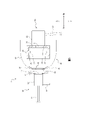
在以下附圖以及說明中闡述了本說明書中所描述之主題之一或多個實施例的細節。從說明、附圖和申請專利範圍,本說明書之主題的其他特徵、態樣與優點將顯得明瞭,其中:圖1為本實施例的硬式內視鏡系統的分解斜視圖。 The details of one or more embodiments of the subject matter described in this specification are set forth in the accompanying drawings and the description below. Other features, aspects and advantages of the subject matter of this specification will become apparent from the description, drawings and claims, wherein: FIG. 1 is an exploded perspective view of the rigid endoscope system of this embodiment.
圖2為本實施例的硬式內視鏡系統的組裝斜視圖。 FIG. 2 is an assembled oblique view of the rigid endoscope system of the present embodiment.
圖3為本實施例的硬式內視鏡系統的分解剖面圖。 FIG. 3 is an exploded cross-sectional view of the rigid endoscope system of the present embodiment.
圖4為本實施例的硬式內視鏡系統的組裝剖面圖。 FIG. 4 is an assembled cross-sectional view of the rigid endoscope system of the present embodiment.
如本文中所使用的,諸如「第一」、「第二」、「第三」、「第四」及「第五」等用語描述了各種元件、組件、區域、層及/或部分,這些元件、組件、區域、層及/或部分不應受這些術語的限制。這些術語僅可用於將一個元素、組件、區域、層或部分與另一個做區分。除非上下文明確指出,否則本文中使用的諸如「第一」、「第二」、「第三」、「第四」及「第五」的用語並不暗示順序或次序。 As used herein, terms such as "first," "second," "third," "fourth," and "fifth" describe various elements, components, regions, layers, and/or sections that are The elements, components, regions, layers and/or sections should not be limited by these terms. These terms are only used to distinguish one element, component, region, layer or section from another. Terms such as "first," "second," "third," "fourth," and "fifth" when used herein do not imply a sequence or order unless the context clearly dictates otherwise.
以下,根據圖式說明本實施例的硬式內視鏡系統1。再者,以下所載的本發明的實施例為表示具體化本發明時的一例,並非將本發明的範圍限定於實施例的記載範圍。因此,本發明可在實施例增加各種變更來實施。
Hereinafter, the
本實施例的硬式內視鏡系統1在消化外科、胸腔外科、脊椎脊髓外科、耳鼻喉科、婦科、泌尿科等,為了觀察對象的觀察、診斷、攝影或治療而被使用。具體而言,硬式內視鏡系統1是照射照射光於觀察對象,並接受
於觀察對象反射的照射光(以下,稱為「反射光」),將接受的反射光經光電轉換來產生影像資料的系統。
The
圖1為本實施例的硬式內視鏡系統1的分解斜視圖。圖2為本實施例的硬式內視鏡系統1的組裝斜視圖。圖3為本實施例的硬式內視鏡系統1的分解剖面圖。圖4為本實施例的硬式內視鏡系統1的組裝剖面圖。如圖1~圖4所示,硬式內視鏡系統1主要具備硬式內視鏡10、攝影機鏡頭20以及攝影機防護袋30。
FIG. 1 is an exploded perspective view of a
硬式內視鏡10是被插入體內照射照射光於觀察對象,並接受反射光的部分。硬式內視鏡10例如由插入部11、本體部12、光輸入部13以及接眼部14構成。
The
插入部11是直線延伸的中空管(例如,不鏽鋼管)。插入部11的前端被插入體內。再者,插入部11的內部配置有光學系統(鏡片、鏡子等)。接著,插入部11的光學系統將由本體部12輸入至基端的照射光從前端照射,將於前端輸入的反射光從基端輸出至本體部12。
The
再者,硬式內視鏡系統1具備對應觀察對象的位置,可分開使用的複數硬式內視鏡10。複數硬式內視鏡10以相對於插入部11的延伸設置方向,在不同方向(例如,0°、12°、70°)輸出照射光,並接受來自不同方向的反射光的方式,來配置插入部11內的光學系統。
Furthermore, the
本體部12連接於插入部11、光輸入部13以及接眼部14。又,於本體部12容置有鏡子或鏡片等構成的光學系統。容納於本體部12的光學系統將通過光輸入部13輸入的照射光引導至插入部11,並將從插入部11輸出的反射光引導至接眼部14。
The
光輸入部13是裝卸從光源裝置(圖未表示)延伸的光導管(光纜)的連接器。光輸入部13將通過光導管從光源裝置供給的照射光,輸出至本體部12。
The
接眼部14是被裝卸於攝影機鏡頭20的部分。接眼部14呈圓盤狀的外型。具體而言,接眼部14的直徑僅略小於攝影機鏡頭20的內視鏡連接部21的內徑尺寸。又,如圖2所示,接眼部14的中央部設有內視鏡側鏡片15。接著,接眼部14將從本體部12輸入的反射光,通過內視鏡側鏡片15輸出至攝影機鏡頭20。
The
攝影機鏡頭20將從硬式內視鏡10輸入的反射光經光電轉換產生影像資料,並將產生的影像資料輸出至顯示器(圖未表示)等。攝影機鏡頭20例如由內視鏡連接部21、本體部22以及傳送纜線23構成。
The
內視鏡連接部21是裝卸硬式內視鏡10的連接器。內視鏡連接部21呈前端開口的有底圓筒狀的外型。內視鏡連接部21的內部由內視鏡連接部21的內側的底壁24、圓筒狀的內側壁25構成。接著,底壁24的中央部設有攝影機側鏡片26。
The
又,如圖3及圖4所示,內視鏡連接部21設有從內側壁25向內側突出的複數突起27、28。再者,複數突起27、28經由手術者操作未表示的操作部,從內側壁25出現。接著,內側壁25的內徑尺寸略大於接眼部14的直徑。另一方面,將從內側壁25突出的複數突起27、28的突出前端連結的假想圓的直徑(以下,稱為「複數突起27、28的內接圓的直徑」)小於接眼部14的直徑。
Furthermore, as shown in FIGS. 3 and 4 , the
在內視鏡連接部21,接眼部14可從前端側插拔。接著,將被插入內視鏡連接部21的接眼部14越過複數的突起27、28,硬式內視鏡10與攝影機鏡頭20被連接。藉此,內視鏡側鏡片15與攝影機側鏡片26彼此面對。其結果,從內視鏡側鏡片15輸出的反射光,通過攝影機側鏡片26輸入至本體部22。另一方面,當操作操作部使複數的突起27、28收入內側壁25,可解除硬式內視鏡10與攝影機鏡頭20的連接。
In the
本體部22連接於內視鏡連接部21及傳送纜線23。又,本體部22容置有光電轉換元件(CCD、CMOS)。本體部22將通過攝影機側鏡片26輸入的反射光,藉由光電轉換元件來光電轉換產生影像資料,並將產生的影像資料輸出至傳送纜線23。接著,在連接於傳送纜線23另一端的顯示器,顯示影像資料所表示的影像。
The
硬式內視鏡10以具有耐熱性及耐壓性的材料來構成。因此,硬式內視鏡10可高壓蒸氣殺菌(壓熱器)。相對於此,攝影機鏡頭20因搭載光電轉換元件等精密機器,不對應高壓蒸氣殺菌。因此,為了無菌的進行手術,以攝影機防護袋30包覆攝影機鏡頭20為必要的。
The
攝影機防護袋30經由包覆攝影機鏡頭20,可在手術室內保持硬式內視鏡系統1為無菌狀態。攝影機防護袋30每次手術使用後即丟棄。因此,攝影機防護袋30期望為簡單的構成,且可容易的裝卸。攝影機防護袋30例如由間隔件31以及防護袋本體32構成。
The camera
間隔件31是介於硬式內視鏡10與攝影機鏡頭20之間的部件。具體而言,間隔件31使硬式內視鏡10與攝影機鏡頭20不接觸,且不介於內視鏡側鏡片15及攝影機側鏡片26之間的配置於接眼部14與內視鏡連接部21之
間。間隔件31例如由聚乙烯對苯二甲酸酯(PET)、聚苯乙烯(PS)等樹脂材料來形成。間隔件31例如由夾持板33以及複數腳部34、35、36、37來構成。夾持板33以及複數腳部34、35、36、37可一體形成,分別形成再組裝亦可。
The
夾持板33是中央形成有貫通孔的板狀部件。夾持板33厚度尺寸設定為較薄(例如,0.1mm~2.0mm的程度)。夾持板33的外型可為正圓、四角形、六角形、八角形等。在本實施例中,以正圓(環狀)的夾持板33來說明。夾持板33的外型尺寸小於內側壁25的內徑尺寸,大於複數突起27、28的內接圓的直徑。又,夾持板33的內徑尺寸(貫通孔的直徑)設定為內視鏡側鏡片15及攝影機側鏡片26的直徑以上。
The clamping
複數腳部34~37設於夾持板33的外緣。又,複數腳部34~37在夾持板33的圓周方向上間隔設置。具體而言,複數腳部34~37在夾持板33的圓周方向上等間隔配置。再者,在本實施例中,四個腳部34~37以90°間隔配置為例作說明,腳部的數量非以四個為限。作為其他示例,三個腳部以120°間隔配置亦可。
The plurality of
又,複數腳部34~37分別從夾持板33的外緣向外(徑向向外)突出。再者,複數腳部34~37分別往夾持板33的厚度方向的一方側(圖1~4的後方側)折曲。在自然狀態下,腳部34~37相對於夾持板33的表面的夾角(以下,稱為「折曲角」)例如設定為10°~45°的程度。接著,複數腳部34~37分別構成為可往夾持板33的厚度方向的另一方側(主要為減少折曲角的方向)彈性變形。
Also, the plurality of
在自然狀態下,連結複數腳部34~37的前端(與夾持板33的連接端相反側的端部)的假想圓的直徑小於內側壁25的內徑尺寸,大於複數突起27、28的內接圓的直徑。另一方面,當複數腳部34~37往減少折曲角的方向彈
性變形,連結複數腳部34~37的前端的假想圓的直徑會成為與內側壁25的內徑尺寸一致(換言之,擴徑)。
In a natural state, the diameter of an imaginary circle connecting the front ends of the plurality of
防護袋本體32是可包覆攝影機鏡頭20的程度大小的膜狀部件。具體而言,防護袋本體32只有包覆硬式內視鏡10及攝影機鏡頭20中的攝影機鏡頭20。防護袋本體32可利用一般的防護袋材料的聚乙烯(polyethylene)以外,亦可利用可伸縮的材料(例如,乳膠(latex)、聚氨酯(polyurethane)等)來構成。
The
在防護袋本體32形成小於夾持板33的外型尺寸,且夾持板33的內徑尺寸以上的貫通孔。接著,使防護袋本體32及夾持板33的貫通孔面對的狀態下,將防護袋本體32裝設(熔接)於間隔件31。具體而言,間隔件31裝設於防護袋本體32的內面側。防護袋本體32的內面是指,以防護袋本體32包覆攝影機鏡頭20時,面對攝影機鏡頭20的面。
A through hole is formed in the
接著,說明攝影機鏡頭20以攝影機防護袋30包覆的順序。首先,手術者在使間隔件31面對內視鏡連接部21的前端的狀態下,以防護袋本體32包覆攝影機鏡頭20。藉此,內視鏡連接部21、本體部22以及傳送纜線23整體被殺菌完成的防護袋本體32包覆。另一方面,硬式內視鏡10位在防護袋本體32的外側。
Next, the procedure of wrapping the
接著,手術者將複數的腳部34~37朝向底壁24的狀態的間隔件31,從內視鏡連接部21的前端的開口至越過複數的突起27、28的位置,插入內視鏡連接部21。接著,手術者將硬式內視鏡10的接眼部14,以內視鏡側鏡片15面對夾持板33的貫通孔的方式,插入內視鏡連接部21直至越過複數的突起27、28的位置。
Next, the operator inserts the
藉此,如圖4所示,防護袋本體32沿著內側壁25進入內視鏡連接部21的內部,間隔件31成為被配置於底壁24以及複數的突起27、28之間的狀態。又,間隔件31在內視鏡連接部21中,被配置於底壁24與接眼部14之間。再者,防護袋本體32通過內側壁25與接眼部14的外緣之間。
Thereby, as shown in FIG. 4 , the
此時,間隔件31經由接眼部14,夾持板33被按壓(夾持)於底壁24。藉此,複數的腳部34~37往折曲角減少的方向彈性變形,複數的腳部34~37的前端壓接於底壁24以及內側壁25的角部。其結果,硬式內視鏡10使間隔件31以及防護袋本體32介於接眼部14以及底壁24之間,來被穩定的固定於攝影機鏡頭20。又,間隔件31整體的厚度尺寸(換言之,從夾持板33的前面至腳部34~37的前端的前後方向的距離)變得比自然狀態時薄。
At this time, the
又,內視鏡側鏡片15與攝影機側鏡片26夾住夾持板33的貫通孔來彼此面對。換言之,內視鏡側鏡片15與攝影機側鏡片26之間沒有界隔攝影機防護袋30的構成要件。更進一步來說,裝設於攝影機鏡頭20的攝影機防護袋30不干涉內視鏡側鏡片15與攝影機側鏡片26之間的反射光的光路。
In addition, the endoscope-
經由上述實施例,例如可達成以下的作用功效。 Through the above-mentioned embodiments, for example, the following functions and effects can be achieved.
經由上述實施例,以防護袋本體32包覆攝影機鏡頭20,由於使硬式內視鏡10位於防護袋本體32的外側,而可不取下攝影機防護袋30來交換硬式內視鏡10。其結果,手術中的硬式內視鏡10的交換變得容易。
Through the above-mentioned embodiment, the
又,經由上述實施例,由於接眼部14與內視鏡連接部21的底壁24之間只有界隔薄板狀的間隔件31,可使內視鏡側鏡片15與攝影機側鏡片26之間的光學距離變短。藉此,可抑制硬式內視鏡系統1的影像品質降低。
In addition, through the above embodiment, since there is only the thin plate-shaped
又,經由上述實施例,攝影機防護袋30並未支持硬式內視鏡10及攝影機鏡頭20,經由以接眼部14與內視鏡連接部21夾持間隔件31,攝影機防護袋30被裝設於硬式內視鏡系統1。因此,間隔件31由於不需要高剛性,而可形成為較薄。
In addition, according to the above-mentioned embodiment, the camera
又,經由上述實施例,因為內視鏡側鏡片15與攝影機側鏡片26之間沒有界隔攝影機防護袋30,可抑制經由硬式內視鏡10所得的影像品質降低,並可適當的包覆攝影機鏡頭20。又,因為間隔件31容置於內視鏡連接部21的內部,而不會妨礙手術者的手術。然而,夾持板33的貫通孔亦可裝設鏡片等。
In addition, through the above-mentioned embodiment, since there is no camera
又,經由上述實施例,因為接眼部14與底壁24之間界隔夾持板33,接眼部14的外緣與內側壁25之間界隔防護袋本體32,硬式內視鏡10及攝影機鏡頭20以非接觸的狀態來連接。其結果,在一邊交換複數的硬式內視鏡10一邊進行手術的情況,可防止硬式內視鏡10直接接觸不乾淨的攝影機鏡頭20。
In addition, through the above-mentioned embodiment, because the clamping
又,經由上述實施例,經由使複數的腳部34~37壓接於底壁24及內側壁25的角部,相對於攝影機鏡頭20可穩定的支持硬式內視鏡10。再者,經由等間隔的配置複數的腳部34~37在夾持板33的圓周方向,可使被支持在攝影機鏡頭20的硬式內視鏡10更穩定。然而,間隔件31亦可不具有複數的腳部34~37。在這種情況,夾持板33的外型尺寸期望設定為相同或略小於內側壁25的內徑尺寸。
In addition, according to the above embodiment, the
又,防護袋本體32以可伸縮的材料(例如,乳膠、聚氨酯)來構成,使位在內視鏡連接部21以外部分伸長來包覆攝影機鏡頭20,可防止包覆
如傳送纜線23的細徑部分的防護袋本體32體積過大。然而,防護袋本體32以作為攝影機防護袋30的一般材料(例如,聚乙烯)來構成亦可。
In addition, the
使用於此且未另外定義,「實質上」及「大約」等用語係用於描述及敘述小變化。當結合於一事件或情況,該用語可包含事件或情況發生精確的當下、以及事件或情況發生至一接近的近似點。例如,當結合於一數值,該用語可包含一變化範圍小於或等於該數值之±10%,如小於或等於±5%、小於或等於±4%、小於或等於±3%、小於或等於±2%、小於或等於±1%、小於或等於±0.5%、小於或等於±0.1%、或小於或等於±0.05%。 As used herein and not otherwise defined, the terms "substantially" and "approximately" are used to describe and describe small changes. When used in connection with an event or circumstance, the term can include the exact moment at which the event or circumstance occurred, as well as the event or circumstance occurring to a near approximate point. For example, when combined with a numerical value, the term can include a range of variation less than or equal to ±10% of the numerical value, such as less than or equal to ±5%, less than or equal to ±4%, less than or equal to ±3%, less than or equal to ±2%, less than or equal to ±1%, less than or equal to ±0.5%, less than or equal to ±0.1%, or less than or equal to ±0.05%.
以上概述了數個實施例的部件、使得在本發明所屬技術領域中具有通常知識者可以更理解本發明實施例的概念。在本發明所屬技術領域中具有通常知識者應該理解、可以使用本發明實施例作為基礎、來設計或修改其他製程和結構、以實現與在此所介紹的實施例相同的目的及/或達到相同的好處。在本發明所屬技術領域中具有通常知識者也應該理解、這些等效的結構並不背離本發明的精神和範圍、並且在不背離本發明的精神和範圍的情況下、在此可以做出各種改變、取代和其他選擇。因此、本發明之保護範圍當視後附之申請專利範圍所界定為準。 The above outlines the components of several embodiments so that the concepts of the embodiments of the present invention may be better understood by those skilled in the art to which the present invention pertains. It should be understood by those of ordinary skill in the art to which the present invention pertains that the embodiments of the present invention may be used as a basis for designing or modifying other processes and structures for carrying out the same purposes and/or achieving the same purposes as the embodiments described herein the benefits of. It should also be understood by those skilled in the art to which the present invention pertains that these equivalent structures do not depart from the spirit and scope of the present invention, and that various modifications can be made herein without departing from the spirit and scope of the present invention. Alterations, substitutions and other options. Therefore, the protection scope of the present invention shall be determined by the scope of the appended patent application.
1:硬式內視鏡系統 1: Rigid endoscope system
10:硬式內視鏡 10: Rigid endoscope
11:插入部 11: Insertion part
12、22:本體部 12, 22: Main body
13:光輸入部 13: Optical input part
14:接眼部 14: eye contact
15:內視鏡側鏡片 15: Endoscope side lens
20:攝影機鏡頭 20: Camera Lens
21:內視鏡連接部 21: Endoscope connecting part
23:傳送纜線 23: Transmission cable
24:底壁 24: Bottom wall
25:內側壁 25: Inner side wall
26:攝影機側鏡片 26: Camera side lens
27、28:突起 27, 28: Protrusions
30:攝影機防護袋 30:Camera protective bag
31:間隔件 31: Spacer
32:防護袋本體 32: Protective bag body
33:夾持板 33: Clamping plate
34、35、36、37:腳部 34, 35, 36, 37: Feet
Claims (4)
Applications Claiming Priority (4)
| Application Number | Priority Date | Filing Date | Title |
|---|---|---|---|
| WOPCT/JP2021/004870 | 2021-02-10 | ||
| PCT/JP2021/004870 WO2022172344A1 (en) | 2021-02-10 | 2021-02-10 | Camera drape |
| PCT/JP2021/016001 WO2022172477A1 (en) | 2021-02-10 | 2021-04-20 | Camera drape |
| WOPCT/JP2021/016001 | 2021-04-20 |
Publications (2)
| Publication Number | Publication Date |
|---|---|
| TW202214169A TW202214169A (en) | 2022-04-16 |
| TWI766822B true TWI766822B (en) | 2022-06-01 |
Family
ID=77172603
Family Applications (1)
| Application Number | Title | Priority Date | Filing Date |
|---|---|---|---|
| TW110143851A TWI766822B (en) | 2021-02-10 | 2021-11-24 | Camera Protection Bag |
Country Status (6)
| Country | Link |
|---|---|
| EP (1) | EP4070715A4 (en) |
| JP (1) | JP6915936B1 (en) |
| KR (1) | KR102448681B1 (en) |
| CN (1) | CN114828728A (en) |
| IL (1) | IL287274A (en) |
| TW (1) | TWI766822B (en) |
Citations (2)
| Publication number | Priority date | Publication date | Assignee | Title |
|---|---|---|---|---|
| US4522196A (en) * | 1982-06-11 | 1985-06-11 | Cunningham Frank W | Reusable, sterile covering for a surgical camera |
| US5274500A (en) * | 1992-07-23 | 1993-12-28 | Kansas City Medical, Inc. | Video camera drape with lens |
Family Cites Families (9)
| Publication number | Priority date | Publication date | Assignee | Title |
|---|---|---|---|---|
| US5433221A (en) * | 1994-10-05 | 1995-07-18 | Adair; Edwin L. | Windowed self-centering drape for surgical camera |
| US5876328A (en) * | 1997-04-23 | 1999-03-02 | Endolap, Inc. | Surgical camera drape assembly and method |
| US5882295A (en) * | 1998-04-13 | 1999-03-16 | Spectrum Medical Industries, Inc. | Video camera drape |
| JP2005296350A (en) * | 2004-04-12 | 2005-10-27 | Pentax Corp | Endoscopic system |
| DK2575590T4 (en) * | 2010-05-25 | 2019-02-11 | Arc Medical Design Ltd | COVER FOR A MEDICAL SHOPPING DEVICE |
| GB201014127D0 (en) * | 2010-08-25 | 2010-10-06 | P3 Medical Ltd | Drape |
| JP6800694B2 (en) * | 2015-10-14 | 2020-12-16 | デクセリアルズ株式会社 | Optical film, connecting member, drape for endoscopic camera, endoscopic device, medical system, manufacturing method of optical film, manufacturing method of connecting member |
| EP3363343A4 (en) * | 2015-10-16 | 2019-03-13 | Okura Industrial Co., Ltd. | Drape for camera |
| JP7374600B2 (en) | 2019-03-22 | 2023-11-07 | ソニー・オリンパスメディカルソリューションズ株式会社 | Medical image processing device and medical observation system |
-
2021
- 2021-04-20 JP JP2021521866A patent/JP6915936B1/en active Active
- 2021-04-20 EP EP21791236.9A patent/EP4070715A4/en not_active Withdrawn
- 2021-04-20 KR KR1020217038125A patent/KR102448681B1/en active IP Right Grant
- 2021-04-20 CN CN202180004131.5A patent/CN114828728A/en active Pending
- 2021-10-14 IL IL287274A patent/IL287274A/en unknown
- 2021-11-24 TW TW110143851A patent/TWI766822B/en active
Patent Citations (2)
| Publication number | Priority date | Publication date | Assignee | Title |
|---|---|---|---|---|
| US4522196A (en) * | 1982-06-11 | 1985-06-11 | Cunningham Frank W | Reusable, sterile covering for a surgical camera |
| US5274500A (en) * | 1992-07-23 | 1993-12-28 | Kansas City Medical, Inc. | Video camera drape with lens |
Also Published As
| Publication number | Publication date |
|---|---|
| EP4070715A1 (en) | 2022-10-12 |
| TW202214169A (en) | 2022-04-16 |
| KR20220116387A (en) | 2022-08-23 |
| JPWO2022172477A1 (en) | 2022-08-18 |
| KR102448681B1 (en) | 2022-09-28 |
| IL287274A (en) | 2021-12-01 |
| JP6915936B1 (en) | 2021-08-11 |
| EP4070715A4 (en) | 2022-10-12 |
| CN114828728A (en) | 2022-07-29 |
Similar Documents
| Publication | Publication Date | Title |
|---|---|---|
| JP3791916B2 (en) | End hood member for endoscope | |
| JP6640159B2 (en) | Endoscope cap and method of sterilizing endoscope cap | |
| JPH0221041Y2 (en) | ||
| CN105142489B9 (en) | Otoscope | |
| US20030233024A1 (en) | Electronic endoscope for stereoscopic endoscope system | |
| US5329936A (en) | Portable arthroscope with periscope optics | |
| JP2007500542A (en) | Otoscope tip elements and related uses | |
| JP2009254403A (en) | Endoscope system | |
| JP6362719B2 (en) | Mantle tube | |
| US20200154981A1 (en) | Flexible endoscope | |
| JP2008245714A (en) | Observation system used for surgical operation | |
| JP2022520422A (en) | Rigid endoscope device | |
| JP2007307090A (en) | Endoscope, endoscope attachment and endoscope apparatus | |
| TWI766822B (en) | Camera Protection Bag | |
| JP5397863B2 (en) | Parent-child endoscope | |
| JPH07204211A (en) | Medical sterilization cover | |
| WO2022172477A1 (en) | Camera drape | |
| US10918266B2 (en) | Endoscope | |
| US20220331040A1 (en) | Camera Drape | |
| JP2005204827A (en) | Endoscope and endoscope system | |
| JP6167206B1 (en) | Optometry tool | |
| JP6980000B2 (en) | How to use the endoscope cap and the endoscope cap | |
| JP2006288821A (en) | Electronic endoscope | |
| JP2005168941A (en) | Ophthalmic imaging method, ophthalmic optical adapter, and ophthalmic optical apparatus | |
| JP5634928B2 (en) | Endoscope |