RU2779373C1 - Method for correction of posttraumatic deformation of the external ankle under external fixation - Google Patents
Method for correction of posttraumatic deformation of the external ankle under external fixation Download PDFInfo
- Publication number
- RU2779373C1 RU2779373C1 RU2022103091A RU2022103091A RU2779373C1 RU 2779373 C1 RU2779373 C1 RU 2779373C1 RU 2022103091 A RU2022103091 A RU 2022103091A RU 2022103091 A RU2022103091 A RU 2022103091A RU 2779373 C1 RU2779373 C1 RU 2779373C1
- Authority
- RU
- Russia
- Prior art keywords
- fibula
- osteotomy
- fixation
- external
- passed
- Prior art date
Links
- 238000000034 method Methods 0.000 title claims abstract description 29
- 238000012937 correction Methods 0.000 title claims abstract description 15
- 210000003423 ankle Anatomy 0.000 title claims description 20
- 210000002082 fibula Anatomy 0.000 claims abstract description 44
- 239000012634 fragment Substances 0.000 claims abstract description 42
- 210000000988 bone and bone Anatomy 0.000 claims abstract description 23
- 210000003275 diaphysis Anatomy 0.000 claims abstract description 19
- 206010023204 Joint dislocation Diseases 0.000 claims abstract description 17
- 238000006073 displacement reaction Methods 0.000 claims abstract description 16
- 230000007547 defect Effects 0.000 claims abstract description 12
- 238000004904 shortening Methods 0.000 claims abstract description 11
- 210000002303 tibia Anatomy 0.000 claims description 27
- 210000002683 foot Anatomy 0.000 claims description 24
- 210000000544 articulatio talocruralis Anatomy 0.000 claims description 11
- 210000004233 talus Anatomy 0.000 claims description 7
- 206010052428 Wound Diseases 0.000 claims description 4
- 208000027418 Wounds and injury Diseases 0.000 claims description 4
- 210000001699 lower leg Anatomy 0.000 claims description 4
- 210000000459 calcaneus Anatomy 0.000 claims description 3
- 230000008030 elimination Effects 0.000 abstract description 6
- 238000003379 elimination reaction Methods 0.000 abstract description 6
- 230000002349 favourable effect Effects 0.000 abstract description 5
- 230000004927 fusion Effects 0.000 abstract description 5
- 230000000399 orthopedic effect Effects 0.000 abstract description 4
- 239000003814 drug Substances 0.000 abstract description 3
- 230000000694 effects Effects 0.000 abstract description 3
- 239000000126 substance Substances 0.000 abstract 1
- 206010017076 Fracture Diseases 0.000 description 14
- 208000010392 Bone Fractures Diseases 0.000 description 7
- 210000003414 extremity Anatomy 0.000 description 7
- 238000011477 surgical intervention Methods 0.000 description 7
- 230000009467 reduction Effects 0.000 description 6
- 230000015572 biosynthetic process Effects 0.000 description 5
- 238000009434 installation Methods 0.000 description 5
- 238000009940 knitting Methods 0.000 description 5
- 208000027502 Ankle fracture Diseases 0.000 description 3
- 208000001132 Osteoporosis Diseases 0.000 description 3
- 210000003557 bones of lower extremity Anatomy 0.000 description 3
- 230000006835 compression Effects 0.000 description 3
- 238000007906 compression Methods 0.000 description 3
- 210000003041 ligament Anatomy 0.000 description 3
- 238000012545 processing Methods 0.000 description 3
- 238000002224 dissection Methods 0.000 description 2
- 210000001149 inferior tibiofibular joint Anatomy 0.000 description 2
- 239000012528 membrane Substances 0.000 description 2
- 230000002085 persistent effect Effects 0.000 description 2
- 210000001519 tissue Anatomy 0.000 description 2
- 230000009466 transformation Effects 0.000 description 2
- 206010071050 Ankle deformity Diseases 0.000 description 1
- 230000009471 action Effects 0.000 description 1
- 230000002411 adverse Effects 0.000 description 1
- 238000013459 approach Methods 0.000 description 1
- 230000001684 chronic effect Effects 0.000 description 1
- 238000007596 consolidation process Methods 0.000 description 1
- 230000007812 deficiency Effects 0.000 description 1
- 230000007560 devascularization Effects 0.000 description 1
- 238000011156 evaluation Methods 0.000 description 1
- 230000000729 hypotrophic effect Effects 0.000 description 1
- 230000006872 improvement Effects 0.000 description 1
- 208000014674 injury Diseases 0.000 description 1
- 230000010354 integration Effects 0.000 description 1
- 210000002414 leg Anatomy 0.000 description 1
- 210000001930 leg bone Anatomy 0.000 description 1
- 239000000463 material Substances 0.000 description 1
- 210000001872 metatarsal bone Anatomy 0.000 description 1
- 230000008569 process Effects 0.000 description 1
- 230000001172 regenerating effect Effects 0.000 description 1
- 238000012552 review Methods 0.000 description 1
- 210000004872 soft tissue Anatomy 0.000 description 1
- 230000008733 trauma Effects 0.000 description 1
Images

Abstract
Description
Область техникиTechnical field
Техническое решение относится к медицине, в частности, к травматологии, может быть использовано для коррекции посттравматической деформации наружной лодыжки в условиях внешней фиксации при лечении неправильно консолидированных чрессиндесмозных переломов наружной лодыжки.The technical solution relates to medicine, in particular, to traumatology, and can be used to correct post-traumatic deformity of the lateral malleolus under conditions of external fixation in the treatment of improperly consolidated transsyndesmotic fractures of the lateral malleolus.
Уровень техникиState of the art
Известны способы одномоментного удлинения малоберцовой кости путем ее Z-образной остеотомии на уровне дистальной трети диафиза [1], а также поперечной остеотомии без использования костного трансплантата [2] и с замещением дефекта аутотрансплантатом из гребня подвздошной кости [3], или из дистального метаэпифиза большеберцовой кости [4] с последующей фиксацией отломков накостной пластиной.There are known methods of simultaneous lengthening of the fibula by its Z-shaped osteotomy at the level of the distal third of the diaphysis [1], as well as transverse osteotomy without the use of a bone graft [2] and with the replacement of the defect with an autograft from the iliac crest [3], or from the distal metaepiphysis of the tibia bones [4] with subsequent fixation of fragments with a bone plate.
По данным литературы [5], описанные выше корригирующие остеотомии малоберцовой кости являются эффективным способом лечения пациентов с неправильно консолидированными надсиндесмозными переломами наружной лодыжки, характеризующихся нарушением целостности межберцовых связок и межкостной мембраны дистальнее уровня перелома. При чрессиндесмозных переломах наружной лодыжки, без нарушения целостности задней межберцовой связки и межкостной мембраны (по крайней мере, проксимальнее уровня дистального межберцового сочленения), одномоментная коррекция длины наружной лодыжки за счет надлодыжечной остеотомии возможна в ограниченных пределах, обеспечивая при этом натяжение межкостных связок, что в некоторых случаях является благоприятным обстоятельством, за счет избыточного их удлинения. В противном случае, данное действие приводит к увеличению травматичности оперативного вмешательства. Кроме того, нередко наблюдаемая угловая деформация и смещение наружной лодыжки по ширине, сочетающиеся с ее укорочением, не устраняются остеотомией дистальной трети диафиза малоберцовой кости, так как вершина деформации локализуется в зоне чрессиндесмозного перелома.According to the literature [5], the above-described corrective osteotomies of the fibula are an effective way to treat patients with improperly consolidated suprasyndesmotic fractures of the lateral malleolus, characterized by a violation of the integrity of the tibiofibular ligaments and the interosseous membrane distal to the fracture level. In case of transsyndesmotic fractures of the lateral malleolus, without violating the integrity of the posterior tibiofibular ligament and interosseous membrane (at least proximal to the level of the distal tibiofibular joint), one-stage correction of the length of the lateral malleolus due to supramalleolar osteotomy is possible to a limited extent, while ensuring tension of the interosseous ligaments, which in in some cases it is a favorable circumstance, due to their excessive lengthening. Otherwise, this action leads to an increase in the morbidity of the surgical intervention. In addition, the often observed angular deformity and displacement of the outer malleolus along the width, combined with its shortening, are not eliminated by osteotomy of the distal third of the fibula shaft, since the apex of the deformity is localized in the area of transsyndesmosis fracture.
Известны способы одномоментной коррекции длины наружной лодыжки путем ее кософронтальной [6], кососагиттальной [патент RU 2216291 C1], или Z-образной [7] остеотомии на вершине деформации и низведения дистального отломка с последующей фиксацией отломков накостной пластиной.Known methods of simultaneous correction of the length of the outer malleolus by its oblique frontal [6], oblique [patent RU 2216291 C1], or Z-shaped [7] osteotomy at the apex of the deformity and reduction of the distal fragment, followed by fixation of the fragments with a bone plate.
Данные оперативные вмешательства эффективны при лечении неправильно консолидированных чрессиндесмозных переломов наружной лодыжки, однако, требуют выполнения доступа, позволяющего установку пластины и винтов, приводящего к деваскуляризации зоны перелома ,остеотомии, неблагоприятно сказывающейся на регенераторном потенциале отломков и окружающих мягких тканей. Кроме того, данные способы проводятся в условиях остеопороза парафрактурной костной ткани и возможны только по достижении консолидации перелома и неприменимы к ранним стадиям постфрактурного процесса. В случае отсутствия сращения оперативное вмешательство осложняется еще более выраженным остеопорозом и необходимостью освежения концов отломков с образованием обширного дефекта, что создает сомнительные условия для стабильного остеосинтеза и интеграции костного трансплантата, требуемого для замещения дефекта.These surgical interventions are effective in the treatment of improperly consolidated transsyndesmotic fractures of the lateral malleolus, however, they require an access that allows the installation of a plate and screws, leading to devascularization of the fracture zone, osteotomy, which adversely affects the regenerative potential of fragments and surrounding soft tissues. In addition, these methods are carried out in conditions of osteoporosis of the parafracture bone tissue and are possible only upon reaching the consolidation of the fracture and are not applicable to the early stages of the post-fracture process. In the absence of union, surgical intervention is complicated by even more pronounced osteoporosis and the need to refresh the ends of the fragments with the formation of an extensive defect, which creates doubtful conditions for stable osteosynthesis and integration of the bone graft required to replace the defect.
Известны способы коррекции длины наружной лодыжки за счет ее остеотомии и постепенного удлинения с формированием дистракционного регенерата в условиях внешней фиксации [8, 9].There are known methods for correcting the length of the outer malleolus due to its osteotomy and gradual lengthening with the formation of a distraction regenerate under conditions of external fixation [8, 9].
Как и описанные выше способы, они применимы при консолидированных переломах с удовлетворительным качеством кости. В противном случае, сопровождаются риском формирования гипотрофического регенерата и несращения. Кроме того, данные оперативные вмешательства требуют увеличения сроков внешней фиксации за счет периода коррекции.Like the methods described above, they are applicable to consolidated fractures with satisfactory bone quality. Otherwise, they are accompanied by the risk of the formation of a hypotrophic regenerate and nonunion. In addition, these surgical interventions require an increase in the duration of external fixation due to the correction period.
Известен способ лечения застарелых неправильно сросшихся переломов наружной лодыжки (патент RU 2528819 C1), заключающийся в выполнении корригирующей остеотомии на вершине деформации и одномоментной коррекции деформации с последующим остеосинтезом костей голени и стопы аппаратом внешней фиксации. Данный способ характеризуется малоинвазивностью оперативного вмешательства, благоприятными условиями для сращения отломков, управляемостью положения отломков на протяжении всего периода фиксации и возможностью ранней нагрузки на оперированную конечность.A known method for the treatment of chronic improperly fused fractures of the external malleolus (patent RU 2528819 C1), which consists in performing corrective osteotomy at the top of the deformity and simultaneous correction of the deformity, followed by osteosynthesis of the bones of the lower leg and foot with an external fixation device. This method is characterized by a minimally invasive surgical intervention, favorable conditions for the fusion of fragments, controllability of the position of the fragments throughout the entire period of fixation, and the possibility of early loading on the operated limb.
Однако осуществление способа возможно лишь при наличии консолидированного чрессиндесмозного перелома наружной лодыжки и небольшой величины деформации. В противном случае, остеотомия зоны несращения должна сопровождаться обработкой концов отломков с образованием значительного дефекта, что в условиях выраженного остеопороза и дефицита костной ткани создает неблагоприятные условия для сращения и снижает стабильность остеосинтеза. Косая, или Z-образная остеотомия выполняется по второму правилу остеотомий, что приводит к смещению отломков по ширине, или к образованию ступенчатой деформации. Отсутствует возможность постепенного вправления ригидного наружного подвывиха стопы ввиду недостаточной степени жесткости фиксации. However, the implementation of the method is possible only in the presence of a consolidated cross-syndesmous fracture of the lateral malleolus and a small amount of deformation. Otherwise, osteotomy of the nonunion zone should be accompanied by processing of the ends of fragments with the formation of a significant defect, which, in conditions of severe osteoporosis and bone deficiency, creates unfavorable conditions for fusion and reduces the stability of osteosynthesis. An oblique, or Z-shaped osteotomy is performed according to the second rule of osteotomy, which leads to a displacement of the fragments in width, or to the formation of a stepped deformity. There is no possibility of gradual reduction of the rigid external subluxation of the foot due to the insufficient degree of fixation rigidity.
Таким образом, известные способы имеют недостатки и требуют совершенствования.Thus, the known methods have disadvantages and require improvement.
Задачей, на решение которой направлено изобретение, является коррекция деформации наружной лодыжки за счет устранения ротационного и осевого смещений, смещений по ширине и длине, обеспечение стабильной фиксации достигнутого положения фрагментов и создание благоприятных условий для сращения зон остеотомий.The problem to be solved by the invention is to correct the deformity of the outer malleolus by eliminating rotational and axial displacements, displacements in width and length, ensuring stable fixation of the achieved position of the fragments and creating favorable conditions for the fusion of osteotomy zones.
Сущность технического решенияThe essence of the technical solution
Технический результат заключается в обеспечении коррекции деформации наружной лодыжки за счёт устранения ротационного и осевого смещений, смещений по ширине и длине, обеспечении стабильной фиксации достигнутого положения фрагментов и создание благоприятных условий для сращения зон остеотомий.The technical result consists in providing correction of the deformity of the outer malleolus by eliminating rotational and axial displacements, displacements in width and length, ensuring stable fixation of the achieved position of the fragments and creating favorable conditions for the fusion of osteotomy zones.
Технический результат достигается тем, что в способе коррекции посттравматической деформации наружной лодыжки в условиях внешней фиксации, выполняют малоинвазивную коррекцию деформации наружной лодыжки путём двойной остеотомии малоберцовой кости и билокального остеосинтеза, для этого кости фиксируют аппаратом внешней фиксации, при этом устанавливают проксимальную базовую кольцевую опору на уровне проксимального метадиафиза большеберцовой кости, содержащую две спицы, одну спицу проводят через большеберцовую кость, вторую спицу с упорной площадкой проводят в направлении снаружи внутрь через головку малоберцовой кости и большеберцовую кость, затем устанавливают кольцевую опору на уровне границы средней и дистальной третей диафиза голени, содержащую одну спицу с упорной площадкой, проведённую в косо-фронтальной плоскости в направлении изнутри наружу параллельно оси голеностопного сустава, устанавливают кольцевую фиксационно-репозиционную опору на уровне дистального метаэпифиза большеберцовой кости, далее выполняют остеотомию малоберцовой кости на вершине деформации, вводят обоюдоострую спицу в дистальный фрагмент, спицу выводят дистальным концом через зону верхушки наружной лодыжки над кожей, проксимальный конец спицы при этом достигает зоны остеотомии, далее спицу ретроградно проводят в медуллярный канал малоберцовой кости, через шейку таранной кости изнутри наружу параллельно оси голеностопного сустава проводят спицу с упорной площадкой, через тело пяточной кости проводят две спицы и фиксируют на U-образной опоре, через наружную лодыжку сзади наперёд проводят спицу с упорной площадкой и фиксируют к U-образной опоре, через дистальную треть диафиза малоберцовой кости спереди назад проводят спицу с упорной площадкой и крепят к фиксационно-репозиционной опоре, затем выполняют косую остеотомию на уровне средней трети диафиза малоберцовой кости, компенсируют укорочение и выполняют замещение дефекта путем дистракционного остеосинтеза, низводя фрагмент малоберцовой кости в фиксационно-репозиционной опоре со смещением по ширине кпереди без потери контакта между фрагментами, наружный подвывих стопы устраняют натяжением спицы, проведённой через дистальный метаэпифиз большеберцовой кости в косо-фронтальной плоскости в направлении изнутри наружу параллельно оси голеностопного сустава и закреплённой на фиксационно-репозиционной опоре или путем позиционирования U-образной опоры аппарата внешней фиксации, при этом репозицию наружного подвывиха выполняют посредством смещения большеберцовой кости кнаружи и смещения таранной кости кнутри, раны послойно ушивают.The technical result is achieved by the fact that in the method for correcting post-traumatic deformity of the lateral malleolus under conditions of external fixation, minimally invasive correction of the deformity of the lateral malleolus is performed by double osteotomy of the fibula and bilocal osteosynthesis, for this the bones are fixed with an external fixation device, while setting the proximal base ring support at the level of the proximal metadiaphysis of the tibia, containing two spokes, one spoke is passed through the tibia, the second spoke with a thrust pad is passed in the direction from the outside to the inside through the head of the fibula and the tibia, then an annular support is installed at the level of the border of the middle and distal thirds of the diaphysis of the tibia, containing one a pin with a thrust pad, drawn in the oblique frontal plane in the direction from the inside outward parallel to the axis of the ankle joint, an annular fixation-reposition support is installed at the level of the distal metaepiphysis of the tibia the fibula, then an osteotomy of the fibula is performed at the apex of the deformity, a double-edged wire is inserted into the distal fragment, the wire is brought out with its distal end through the area of the apex of the outer malleolus above the skin, the proximal end of the wire reaches the zone of osteotomy, then the wire is retrogradely advanced into the medullary canal of the fibula, through the neck of the talus from the inside outward parallel to the axis of the ankle joint, a spoke with a thrust platform is passed, two spokes are passed through the body of the calcaneus and fixed on a U-shaped support, a spoke with a thrust platform is passed through the outer ankle from the back to the front and fixed to the U-shaped support, through the distal third of the diaphysis of the fibula is passed from front to back with a pin with a thrust platform and attached to a fixation-reposition support, then an oblique osteotomy is performed at the level of the middle third of the diaphysis of the fibula, the shortening is compensated and the defect is replaced by distraction osteosynthesis, bringing the fragment of the fibula into f fixation-reposition support with displacement in width anteriorly without loss of contact between the fragments, external subluxation of the foot is eliminated by tension of the pin passed through the distal metaepiphysis of the tibia in the oblique-frontal plane in the direction from the inside outward parallel to the axis of the ankle joint and fixed on the fixation-reposition support or by positioning the U-shaped support of the external fixation device, while repositioning the external subluxation is performed by displacing the tibia outward and displacing the talus inward, the wounds are sutured in layers.
Способ является малоинвазивным оперативным вмешательством, с известными преимуществами малоинвазивной техники, так как выполняют только два продольных доступа, обеспечивающих достижение кости остеотомом, или полотном осциллирующей пилы, без необходимости широкой диссекции тканей. The method is a minimally invasive surgical intervention, with the known advantages of a minimally invasive technique, since only two longitudinal accesses are performed, ensuring that the bone is reached by an osteotome, or an oscillating saw blade, without the need for wide tissue dissection.
Способ обеспечивает устранение деформации наружной лодыжки, сопровождающейся значительным её укорочением и отклонением продольной оси за счёт билокального остеосинтеза малоберцовой кости, выполнения остеотомии на вершине деформации с устранением осевого и ротационного смещений, а также смещения по ширине, и косой остеотомии на уровне средней трети диафиза малоберцовой кости для компенсации укорочения и замещения дефекта;EFFECT: method provides elimination of external ankle deformity, accompanied by its significant shortening and deviation of the longitudinal axis due to bilocal osteosynthesis of the fibula, performing osteotomy at the top of the deformity with the elimination of axial and rotational displacements, as well as displacements in width, and oblique osteotomy at the level of the middle third of the fibula shaft to compensate for shortening and replacement of a defect;
Способ применим при несращении и ложном суставе наружной лодыжки, так как образовавшийся после обработки концов отломков дефект замещается низведенным проксимальным отломком путем билокального остеосинтеза с обеспечением адекватного контакта на стыке фрагментов;The method is applicable for non-union and false joint of the outer malleolus, since the defect formed after processing the ends of the fragments is replaced by a reduced proximal fragment by bilocal osteosynthesis with adequate contact at the junction of the fragments;
Способ обеспечивает стабильность отломков и управляемость положением фрагментов, а также возможность модульной трансформации и постепенного демонтажа элементов фиксации на протяжении всего периода фиксации, что позволяет выполнять раннюю нагрузку на оперированную конечность и проведение реабилитационных мероприятий.The method ensures the stability of the fragments and controllability of the position of the fragments, as well as the possibility of modular transformation and gradual dismantling of the fixation elements throughout the entire period of fixation, which allows for early loading on the operated limb and rehabilitation measures.
Способ позволяет выполнять как одномоментное, так и постепенное устранение деформации и вправление подвывиха стопы посредством как прямой, так и непрямой репозиции за счет позиционирования спиц, проведенных через наружную лодыжку в кососагиттальной плоскости и диафиз/метадиафиз большеберцовой кости в плоскости оси голеностопного сустава, а также позиционированием опоры, фиксирующей стопу.EFFECT: method allows to perform both simultaneous and gradual elimination of deformity and reduction of foot subluxation by means of both direct and indirect reposition due to positioning of the pins passed through the outer ankle in the oblique sagittal plane and the diaphysis/metadiaphysis of the tibia in the plane of the ankle joint axis, as well as by positioning foot support.
Способ позволяет создать как относительную стабильность за счет интраканальной спицы, предотвращающей смещения фрагментов по ширине и шунтирования нагрузки между проксимальной базовой опорой и опорой, фиксирующей стопу; так и абсолютную стабильность за счет возможности осуществления осевой, встречно-боковой и комбинированной межотломковой космпрессии на уровне обеих остеотомий.The method makes it possible to create both relative stability due to the intracanal spoke, which prevents the fragments from shifting in width and shunting the load between the proximal base support and the support that fixes the foot; and absolute stability due to the possibility of axial, counter-lateral and combined interfragmentary compression at the level of both osteotomies.
Способ обеспечивает установку дополнительных фиксационных элементов для устранения сопутствующих деформаций, позволяя монтаж индивидуальной компоновки аппарата внешней фиксации в зависимости от клинической ситуации.The method provides for the installation of additional fixation elements to eliminate associated deformities, allowing the installation of an individual layout of the external fixation apparatus, depending on the clinical situation.
Техническое решение поясняется графическими материалами.The technical solution is illustrated by graphic materials.
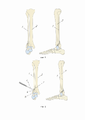
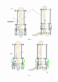
Фиг. 1. Посттравматическая деформация наружной лодыжки, подвывих стопы кнаружи, вид на кости конечности в двух проекциях.Fig. 1. Post-traumatic deformity of the lateral malleolus, outward subluxation of the foot, view of the limb bones in two projections.
Фиг. 2. Корригирующая остеотомия наружной лодыжки, вид на кости конечности в двух проекциях, показано долото.Fig. Fig. 2. Corrective osteotomy of the lateral malleolus, view of the bones of the limb in two projections, a chisel is shown.
Фиг. 3. Остеосинтез костей голени и стопы аппаратом внешней фиксации, корригирующая остеотомия диафиза малоберцовой кости, вид на кости конечности в двух проекциях, показано долото и аппарат Илизарова.Fig. Fig. 3. Osteosynthesis of the bones of the lower leg and foot with an external fixation device, corrective osteotomy of the diaphysis of the fibula, a view of the bones of the limb in two projections, a chisel and the Ilizarov apparatus are shown.
Фиг. 4. Коррекция деформации в аппарате Илизарова, билокальная компрессия зон остеотомий, вид на кости конечности в двух проекциях.Fig. Fig. 4. Deformity correction in the Ilizarov apparatus, bilocal compression of the osteotomy zones, view of the limb bones in two projections.
Фиг. 5. Голень и стопа после коррекции деформации, вид на кости конечности в двух проекциях.Fig. Fig. 5. Lower leg and foot after deformity correction, view of the limb bones in two projections.
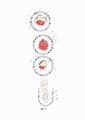
Фиг. 6. Последовательно показаны, уровни и позиции проведения чрескостных элементов (спиц), сверху вниз: на уровне мыщелков больше-берцовой кости; на уровне средней трети голени; на уровне дистального межберцового синдесмоза; на уровне стопы.Fig. 6. The levels and positions of the transosseous elements (pins) are sequentially shown, from top to bottom: at the level of the condyles of the tibia; at the level of the middle third of the leg; at the level of the distal tibiofibular syndesmosis; at foot level.
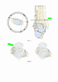
Фиг. 7. Репозиция наружного подвывиха посредством смещения большеберцовой кости кнаружи.Fig. 7. Reposition of the external subluxation by moving the tibia outward.
Фиг. 8. Непрямая репозиция наружного подвывиха посредством смещения таранной кости кнутри.Fig. 8. Indirect reposition of the external subluxation by displacing the talus medially.
Фиг. 9. Рентгенограммы. Обоюдоострая спица введена в малоберцовую кость, до зоны остеотомии.Fig. 9. Radiographs. A double-edged pin is inserted into the fibula, up to the osteotomy zone.
Спецификация:Specification:
1 - малоберцовая кость;1 - fibula;
2 - большеберцовая кость;2 - tibia;
3 - аппарат внешней фиксации;3 - external fixation device;
4 - зона остеотомии;4 - zone of osteotomy;
5 - зона остеотомии;5 - zone of osteotomy;
6 - долото;6 - chisel;
7 - фрагмент кости;7 - bone fragment;
8 - базовая опора;8 - base support;
9 - спица;9 - knitting needle;
10 - спица с упорной площадкой;10 - a spoke with a thrust pad;
11 - опора;11 - support;
12 - спица с упорной площадкой;12 - spoke with thrust pad;
13 - спица;13 - knitting needle;
14 - фиксационно-репозиционная опора;14 - fixation-reposition support;
15 - спица;15 - knitting needle;
16 - фрагмент кости;16 - bone fragment;
17 - обоюдоострая спица;17 - double-edged knitting needle;
18 - спица с упорной площадкой;18 - spoke with thrust pad;
19 - спица;19 - knitting needle;
20 - U-образная опора;20 - U-shaped support;
21 - спица с упорной площадкой;21 - spoke with thrust pad;
22 - фрагмент кости.22 - bone fragment.
Осуществление изобретенияImplementation of the invention
Способ коррекции посттравматической деформации наружной лодыжки в условиях внешней фиксации используют при лечении неправильно консолидированных чрессиндесмозных переломов наружной лодыжки. В основе способа лежит малоинвазивная коррекция многокомпонентной деформации наружной лодыжки путем двойной остеотомии малоберцовой кости 1 и ее билокального остеосинтеза в условиях внешней фиксации аппаратом Илизарова, обеспечивающей управляемую стабильную фиксацию отломков и раннюю функцию оперированной конечности.A method for correcting post-traumatic deformity of the lateral malleolus under conditions of external fixation is used in the treatment of improperly consolidated transsyndesmotic fractures of the lateral malleolus. The method is based on minimally invasive correction of a multicomponent deformity of the lateral malleolus by double osteotomy of the
В способе кости фиксируют аппаратом внешней фиксации 3. Выполняют билокальный остеосинтеза малоберцовой кости 1. Для этого выполняют остеотомию 4 малоберцовой кости 1 на вершине деформации, устраняют осевое и ротационное смещение, смещения по ширине. Репозицию наружного подвывиха выполняют посредством смещения большеберцовой кости кнаружи. Непрямую репозицию наружного подвывиха выполняют посредством смещения таранной кости кнутри. Выполняют косую остеотомию 5 на уровне средней трети диафиза малоберцовой кости 1, компенсируют укорочение и замещение дефекта, путем дистракционного остеосинтеза, низводя фрагмент 22 малоберцовой кости 1.In the method, the bones are fixed with an external fixation device 3. Bilocal osteosynthesis of the
Перед осуществлением остеотомий и коррекции выполняют внешнюю фиксацию костей голени аппаратом внешней фиксации 3, например аппаратом Илизарова.Before performing osteotomy and correction, external fixation of the leg bones is performed with an external fixation apparatus 3, for example, with the Ilizarov apparatus.
Устанавливают проксимальную базовую кольцевую опору 8 на уровне проксимального метадиафиза большеберцовой кости 2, содержащую, по меньшей мере, две спицы 9,10. Одну спицу 9 проводят через большеберцовую кость 2, вторую спицу с упорной площадкой 10 проводят в направлении снаружи-внутрь через головку малоберцовой кости 1 и большеберцовую кость 2 (Фиг. 6).A proximal base
Устанавливают кольцевую опору 11 на уровне границы средней и дистальной третей диафиза голени, содержащую, по меньшей мере, одну спицу с упорной площадкой 12, проведенную в косо-фронтальной плоскости в направлении изнутри-наружу параллельно оси голеностопного сустава (межлодыжечной линии в аксиальной плоскости). Опора 11 может быть усилена проведением спицы 13 через диафизы большеберцовой 2 и малоберцовой костей 1, либо стержнем-шурупом (не показан), введенным в диафиз малоберцовой кости 1 (Фиг. 6).An annular support 11 is installed at the level of the border of the middle and distal thirds of the diaphysis of the tibia, containing at least one spoke with a
Устанавливают кольцевую фиксационно-репозиционную опору 14 на уровне дистального метаэпифиза большеберцовой кости 2, предназначенную для фиксации спиц 15. Опора 14, на начальном этапе не содержит спиц 15, спицы 15 монтируют в процессе выполнения хирургического репозиционного приема.An annular fixation-reposition
Выполняют продольный наружный доступ в проекции вершины деформации наружной лодыжки. Долотом 6 выполняют остеотомю 4 на вершине деформации (Фиг. 2). Распатором и костным рашпилем производят обработку концов костных фрагментов 7,16 с целью создания большей площади контакта на стыке фрагментов 7,16. Далее вводят обоюдоострую спицу 17 в дистальный фрагмент 16, спицу 17 выводят дистальным концом через зону верхушки наружной лодыжки над кожей, проксимальный конец спицы 17 при этом достигает зоны остеотомии. Далее спицу 17 (Фиг. 9), ретроградно проводят в медуллярный канал малоберцовой кости 1. Рана послойно ушивается.Longitudinal external access is performed in the projection of the apex of the deformation of the external ankle.
Через шейку таранной кости изнутри-наружу параллельно оси голеностопного сустава проводят спицу 18 с упорной площадкой. Через тело пяточной кости проводятся две спицы 19. Спицы 19 фиксируют на U-образной опоре 20 с натяжением (Фиг. 6). Для повышения жесткости фиксации возможно проведение дополнительно двух спиц через диафизы I-II и V-III плюсневых костей. Далее выполняют устранение подвывиха стопы путем тракции руками, или при помощи аппарата внешней фиксации 3, используя шарниры и узлы аппарата внешней фиксации 3. По достижению вправления подвывиха и корректного положения стопы дистальнее зоны остеотомии 4 в сагиттальной плоскости через наружную лодыжку сзади наперед проводят спицу 21 с упорной площадкой. Спицу 21 фиксируют на кронштейнах к U-образной опоре 20. При некорректном положении дистального фрагмента 16 наружной лодыжки спица 21, проведенная через нее, крепится к U-образной опоре 20 на резьбовых стержнях и кронштейнах с учетом смещения и низводится за счет укорочения резьбовых стержней.Through the neck of the talus from the inside out, parallel to the axis of the ankle joint, a
Проксимальнее зоны остеотомии 4 в сагиттальной плоскости через дистальную треть диафиза малоберцовой кости 1 спереди-назад проводят спицу 15 с упорной площадкой. Спицу 15 крепят с натяжением на кронштейнах к фиксационно-репозиционной опоре 14 для возможности позиционирования наружной лодыжки по длине.Proximal to the zone of
Выполняют продольный наружный доступ к диафизу малоберцовой кости 1 на уровне средней/дистальной трети в зоне максимального диаметра медуллярного канала. С учетом укорочения выполняют косую остеотомию 5 малоберцовой кости 1 (Фиг. 3) по второму правилу остеотомий: для коррекции длины наружной лодыжки дистальный фрагмент 22 малоберцовой кости 1 низводят в фиксационно-репозиционной опоре 14 со смещением по ширине (кпереди) без потери контакта между фрагментами 7,16. (Фиг. 4). Рана послойно ушивается.Perform a longitudinal external access to the diaphysis of the
При сохраняющемся наружном подвывихе стопы, последний устраняется натяжением спицы 12, проведенной через дистальный метаэпифиз большеберцовой кости 2 в косо-фронтальной плоскости в направлении изнутри-наружу параллельно оси голеностопного сустава (межлодыжечной линии в аксиальной плоскости) и закрепленной на фиксационно-репозиционной опоре 14 (Фиг. 7).With persistent external subluxation of the foot, the latter is eliminated by tensioning the
Альтернативным вариантом вправления подвывиха является одномоментное или постепенное позиционирование U-образной опоры 20 (Фиг. 8).An alternative option for reducing the subluxation is the simultaneous or gradual positioning of the U-shaped support 20 (Fig. 8).
Заявленный способ:Claimed method:
- является малоинвазивным оперативным вмешательством, так как требует двух продольных доступов без необходимости широкой диссекции тканей, обеспечивающих лишь достижение кости остеотомом, или полотном осциллирующей пилы;- is a minimally invasive surgical intervention, as it requires two longitudinal approaches without the need for a wide tissue dissection, which only ensures reaching the bone with an osteotome, or with an oscillating saw blade;
- позволяет коррекцию деформаций наружной лодыжки, сопровождающихся значительным ее укорочением и отклонением оси за счет билокального остеосинтеза малоберцовой кости: остеотомии на вершине деформации с устранением осевого и ротационного смещений, а также смещения по ширине, и косой остеотомии на уровне средней трети диафиза малоберцовой кости для компенсации укорочения и замещения дефекта;- allows correction of deformities of the lateral malleolus, accompanied by its significant shortening and deviation of the axis due to bilocal osteosynthesis of the fibula: osteotomy at the apex of the deformity with the elimination of axial and rotational displacements, as well as displacements in width, and oblique osteotomy at the level of the middle third of the diaphysis of the fibula to compensate shortening and replacement of the defect;
- за счет предыдущего пункта возможен при несращении и ложном суставе наружной лодыжки, так как образовавшийся после обработки концов отломков дефект замещается низведенным проксимальным отломком путем билокального остеосинтеза с обеспечением адекватного контакта на стыке фрагментов;- due to the previous paragraph, it is possible with non-union and false joint of the external ankle, since the defect formed after processing the ends of the fragments is replaced by a reduced proximal fragment by bilocal osteosynthesis, ensuring adequate contact at the junction of the fragments;
- обеспечивает стабильность отломков и управляемость положения фрагментов, а также возможность модульной трансформации и постепенного демонтажа элементов фиксации на про-тяжение всего периода фиксации, что позволяет раннюю нагрузку на оперированную конеч-ность и проведение реабилитационных мероприятий;- ensures the stability of the fragments and controllability of the position of the fragments, as well as the possibility of modular transformation and gradual dismantling of the fixation elements throughout the entire fixation period, which allows early loading on the operated limb and rehabilitation measures;
- позволяет как одномоментное, так и постепенное устранение деформации и вправление подвывиха стопы посредством как прямой, так и непрямой репозиции за счет позиционирования спиц, проведенных через наружную лодыжку в кососагиттальной плоскости и диа-физ/метадиафиз большеберцовой кости в плоскости оси голеностопного сустава, а также по-зиционированием опоры, фиксирующей стопу.- allows both simultaneous and gradual elimination of deformity and reduction of subluxation of the foot through both direct and indirect reposition due to the positioning of the pins passed through the outer ankle in the oblique sagittal plane and the diaphysis / metadiaphysis of the tibia in the plane of the ankle joint axis, as well as positioning of the support that fixes the foot.
- позволяет создать как относительную стабильность за счет интраканальной спицы, предотвращающей смещения фрагментов по ширине и шунтирования нагрузки между проксимальной базовой опорой и опорой, фиксирующей стопу; так и абсолютную стабильность за счет возможности осуществления осевой, встречно-боковой и комбинированной межотломковой космпрессии на уровне обеих остеотомий.- allows you to create both relative stability due to the intracanal needle, which prevents the fragments from shifting in width and shunting the load between the proximal base support and the support that fixes the foot; and absolute stability due to the possibility of axial, counter-lateral and combined interfragmentary compression at the level of both osteotomies.
- позволяет установку дополнительных фиксационных элементов для устранения сопутствующих деформаций, позволяя монтаж индивидуальной компоновки аппарата внешней фиксации в зависимости от клинической ситуации.- allows the installation of additional fixation elements to eliminate associated deformities, allowing the installation of an individual layout of the external fixation device, depending on the clinical situation.
Цитированные источники. Непатентная литератураCited sources. Non-Patent Literature
1. Weber, D & Friederich, Niklaus & Müller, W. (1998). Lengthening osteotomy of the fibula for post-traumatic malunion. Indications, technique and results. International orthopaedics. 22. 149-52. 10.1007/s002640050229.1. Weber, D & Friederich, Niklaus & Müller, W. (1998). Lengthening osteotomy of the fibula for post-traumatic malunion. Indications, technique and results. International orthopaedics. 22.149-52. 10.1007/s002640050229.
2. Chao, Kuo-Hua & Wu, Chia-Chun & Lee, Chian-Her & Chu, Cheng-Mien & Wu, Shing-Shen. (2004). Corrective-Elongation Osteotomy Without Bone Graft for Old Ankle Fracture With Residual Diastasis. Foot & ankle international / American Orthopaedic Foot and Ankle Society [and] Swiss Foot and Ankle Society. 25. 123-7. 10.1177/107110070402500302.2. Chao, Kuo-Hua & Wu, Chia-Chun & Lee, Chian-Her & Chu, Cheng-Mien & Wu, Shing-Shen. (2004). Corrective-Elongation Osteotomy Without Bone Graft for Old Ankle Fracture With Residual Diastasis. Foot & ankle international / American Orthopedic Foot and Ankle Society [and] Swiss Foot and Ankle Society. 25.123-7. 10.1177/107110070402500302.
3. Sinha, Apurv & Sirikonda, Siva & Giotakis, Nikolaos & Walker, Christopher. (2008). Fibular Lengthening for Malunited Ankle Fractures. Foot & ankle international / American Orthopaedic Foot and Ankle Society [and] Swiss Foot and Ankle Society. 29. 1136-40. 10.3113/FAI.2008.1136.3. Sinha, Apurv & Sirikonda, Siva & Giotakis, Nikolaos & Walker, Christopher. (2008). Fibular Lengthening for Malunited Ankle Fractures. Foot & ankle international / American Orthopedic Foot and Ankle Society [and] Swiss Foot and Ankle Society. 29. 1136-40. 10.3113/FAI.2008.1136.
4. El-Rosasy, Mahmoud & Aly, Tarek. (2013). Realignment-lengthening osteotomy for malunited distal fibular fracture. International orthopaedics. 37. 10.1007/s00264-013-1876-7.4. El Rosasy, Mahmoud & Aly, Tarek. (2013). Realignment-lengthening osteotomy for malunited distal fibular fracture. International orthopaedics. 37. 10.1007/s00264-013-1876-7.
5. van Wensen RJ, van den Bekerom MP, Marti RK, van Heerwaarden RJ. Reconstructive osteotomy of fibular malunion: review of the literature. Strategies Trauma Limb Reconstr. 2011 Aug;6(2):51-7. doi: 10.1007/s11751-011-0107-2. Epub 2011 Apr 6. PMID: 21818702; PMCID: PMC3150649.5. van Wensen RJ, van den Bekerom MP, Marti RK, van Heerwaarden RJ. Reconstructive osteotomy of fibular malunion: review of the literature. Strategies Trauma Limb Reconstr. 2011 Aug;6(2):51-7. doi: 10.1007/s11751-011-0107-2. Epub 2011
6. Inori, Fumiaki & Tohyama, Masahiko & Yasuda, Hiroyuki & Konishi, Sadahiko & Waseda, Akeo. (2015). Reconstructive Osteotomy for Ankle Malunion Improves Patient Satisfaction and Function. Case Reports in Orthopedics. 2015. 1-5. 10.1155/2015/549109.6. Inori, Fumiaki & Tohyama, Masahiko & Yasuda, Hiroyuki & Konishi, Sadahiko & Waseda, Akeo. (2015). Reconstructive Osteotomy for Ankle Malunion Improves Patient Satisfaction and Function. Case Reports in Orthopedics. 2015. 1-5. 10.1155/2015/549109.
7. Thangarajah, Tanujan & Lakdawala, Ayaz & Battaloglu, Emir & Malik, Atul & Tillu, Abhay. (2012). Lengthening z-Osteotomy of the Fibula to Correct Persistent Talar Shift Following Open Reduction Internal Fixation of Ankle Fractures. Foot & ankle specialist. 5. 107-10. 10.1177/1938640011434509.7. Thangarajah, Tanujan & Lakdawala, Ayaz & Battaloglu, Emir & Malik, Atul & Tillu, Abhay. (2012). Lengthening z-Osteotomy of the Fibula to Correct Persistent Talar Shift Following Open Reduction Internal Fixation of Ankle Fractures. Foot & ankle specialist. 5. 107-10. 10.1177/1938640011434509.
8. Rozbruch SR, DiPaola M, Blyakher A. Fibula lengthening using a modified Ilizarov method. Orthopedics. 2002 Nov;25(11):1241-4. PMID: 12452340.8. Rozbruch SR, DiPaola M, Blyakher A. Fibula lengthening using a modified Ilizarov method. Orthopedics. 2002 Nov;25(11):1241-4. PMID: 12452340.
9. Johnston, A.J., Andrews, C.T. Fibular lengthening by Ilizarov method secondary to shortening by osteochondroma of distal tibia. Strat Traum Limb Recon 3, 45–48 (2008). https://doi.org/10.1007/s11751-007-0028-29. Johnston, A.J., Andrews, C.T. Fibular lengthening by Ilizarov method secondary to shortening by osteochondroma of distal tibia. Strat Traum Limb Recon 3, 45–48 (2008). https://doi.org/10.1007/s11751-007-0028-2
10. Prissel MA, Daigre JL, Brandão RA, Philbin TM, Hyer CF, Berlet GC. Cross-Sectional Analysis of the Distal Fibular Intramedullary Canal: A Cadaveric Evaluation. Foot Ankle Spec. 2018 Jan 1:1938640017751190. doi: 10.1177/1938640017751190. Epub ahead of print. PMID: 29361841.10. Prissel MA, Daigre JL, Brandão RA, Philbin TM, Hyer CF, Berlet GC. Cross-Sectional Analysis of the Distal Fibular Intramedullary Canal: A Cadaveric Evaluation. Foot Ankle Spec. 2018 Jan 1:1938640017751190. doi: 10.1177/1938640017751190. Epub ahead of print. PMID: 29361841.
Claims (1)
Publications (1)
| Publication Number | Publication Date |
|---|---|
| RU2779373C1 true RU2779373C1 (en) | 2022-09-06 |
Family
ID=
Cited By (1)
| Publication number | Priority date | Publication date | Assignee | Title |
|---|---|---|---|---|
| RU2815476C1 (en) * | 2023-05-03 | 2024-03-18 | Федеральное государственное бюджетное учреждение "Новосибирский научно-исследовательский институт травматологии и ортопедии им. Я.Л. Цивьяна" Министерства здравоохранения Российской Федерации (ФГБУ "ННИИТО им. Я.Л. Цивьяна" Минздрава России) | Method of surgical treatment of deforming arthrosis of ankle joint |
Citations (1)
| Publication number | Priority date | Publication date | Assignee | Title |
|---|---|---|---|---|
| RU2528819C1 (en) * | 2013-07-30 | 2014-09-20 | Государственное автономное учреждение здравоохранения "Республиканская клиническая больница Министерства здравоохранения Республики Татарстан" | Method of treating old pronation-everting malunions of distal shin |
Patent Citations (1)
| Publication number | Priority date | Publication date | Assignee | Title |
|---|---|---|---|---|
| RU2528819C1 (en) * | 2013-07-30 | 2014-09-20 | Государственное автономное учреждение здравоохранения "Республиканская клиническая больница Министерства здравоохранения Республики Татарстан" | Method of treating old pronation-everting malunions of distal shin |
Non-Patent Citations (1)
| Title |
|---|
| МАКАРОВ М.А. и др. Сравнение хирургических доступов при реверсивном эндопротезировании плечевого сустава. Политравма. 2019, 1, стр. 42-56. HADJICOSTAS P. et al., The use of split deltoid-flap in the treatment of massive rotator cuff defects: a retrospective study of 61 patients. Knee Surgery, Sports Traumatology, Arthroscopy. 2008, 16(9), pp. 876-883. LEVY O. et al., A wide and versatile combined surgical approach to the shoulder. Journal of Shoulder and Elbow Surgery. 1999, 8(6), pp. 658-659. GARDNER M.J. et al., Vascular Implications of Minimally Invasive Plating of Proximal Humerus Fractures. Journal of Orthopaedic Trauma. 2006, 20(9), pp. 602-607. * |
Cited By (1)
| Publication number | Priority date | Publication date | Assignee | Title |
|---|---|---|---|---|
| RU2815476C1 (en) * | 2023-05-03 | 2024-03-18 | Федеральное государственное бюджетное учреждение "Новосибирский научно-исследовательский институт травматологии и ортопедии им. Я.Л. Цивьяна" Министерства здравоохранения Российской Федерации (ФГБУ "ННИИТО им. Я.Л. Цивьяна" Минздрава России) | Method of surgical treatment of deforming arthrosis of ankle joint |
Similar Documents
| Publication | Publication Date | Title |
|---|---|---|
| Nihal et al. | Ankle arthrodesis | |
| Hierholzer et al. | Manual on the AO/ASIF tubular external fixator | |
| Sirkin | Plating of tibial pilon fractures | |
| Zhao et al. | Supramalleolar osteotomy with distraction arthroplasty in treatment of varus ankle osteoarthritis with large talar tilt angle: a case report and literature review | |
| Cronier et al. | Early open reduction and internal fixation of pilon fractures | |
| RU2570953C2 (en) | Method for surgical management of patients suffering hallux valgus | |
| RU2484785C2 (en) | Method of treating intra-articular impression fractures of heel bone | |
| RU2779373C1 (en) | Method for correction of posttraumatic deformation of the external ankle under external fixation | |
| RU2779374C1 (en) | Method for correction of posttraumatic deformation of the fib bone under external fixation | |
| RU2177271C2 (en) | Method for performing transosseous fixation of intra- and extramedullary bone transplants in treating false articulations | |
| RU2230513C1 (en) | Method for repeatedly making prosthetic appliance of hip joint | |
| RU2659652C1 (en) | Method for treating patients with fracture-dislocation of the talus | |
| RU2621844C2 (en) | Method for biarticular arthrodesis of talocrural and talocalcanean joints | |
| Basu | External Fixator as an Augment and or Replacement of Internal Fixator | |
| RU2185786C2 (en) | Surgical method for treating the cases of blount disease | |
| RU2319466C1 (en) | Method for surgical treatment of deformations in long tubular bones at exostosis chondrodysplasia in children and teenagers | |
| RU2840204C1 (en) | Method for total calcaneal defect replacement in gunshot wounds | |
| RU2844916C1 (en) | Method of ankle joint arthrodesis | |
| RU2394517C1 (en) | Bone grafting technique in correction of tibia recurvation deformation combined with lengthening of short lower extremity in children | |
| RU2585140C1 (en) | Method of treating refracture of thighbone, complicating periprosthetic fracture with osteoporosis | |
| RU2760992C1 (en) | Arthrodesis method of anterior talocacaneal articulation | |
| RU2285481C2 (en) | Method for plantar reconstruction at polydactyly complicated with hypoplasia of one of its radiuses | |
| RU2549296C1 (en) | Method of arthrodesis of back part of foot joints | |
| Beaman et al. | Deformity correction and distraction arthroplasty for ankle arthritis | |
| RU2825939C1 (en) | Method for correcting axis of first metatarsal bone in hallux valgus |